Pathogenesis of Mycobacterium Tuberculosis, mode of action ...

Acta Tropica 81 (2002) 123–132
Review article
Pathogenesis of Chagas heart disease: role of autoimmunity
David M. Engman *, Juan S. LeonDepartments of Pathology and Microbiology–Immunology, and Feinberg Cardio�ascular Research Institute, Northwestern Uni�ersity
Medical School, Chicago, IL 60611, USA
Received in revised form 26 October 2001; accepted 30 October 2001
Abstract
Chagas heart disease is caused by infection with the protozoan parasite Trypanosoma cruzi. The apparent absenceof parasites from the hearts of most individuals who succumb to this illness has led some to propose an autoimmunebasis for disease pathogenesis. This hypothesis has been extremely difficult to test, because other mechanisms of tissueinflammation may coexist in the setting of active infection. Here we review the proposed mechanisms of Chagasdisease pathogenesis and present new evidence in support of an autoimmune contribution to cardiac inflammation inthe context of these other mechanisms. While we do not yet have a definitive answer to the autoimmunity question,we hope that our views will provide those engaged in the debate fresh perspective on this challenging issue. © 2002Elsevier Science B.V. All rights reserved.
Keywords: Myocarditis; Chagas heart disease; Myosin; Autoimmunity
www.parasitology-online.com
1. Trypanosoma cruzi and Chagas disease
Trypanosoma cruzi is a single-celled eukaryoticparasite with a complex life cycle involving severalstages in both mammals and blood-sucking tri-atomine bugs. Transmission of T. cruzi to humansmay occur when bug excreta, contaminated withthe infective, flagellated trypomastigote form ofthe parasite and released by the bug while it takesa blood meal, contaminate mucous membranes or
breaks in the skin. Once beyond the barrier of theskin, T. cruzi trypomastigotes are able to invade awide variety of host cells where they subsequentlydifferentiate into nonflagellated amastigotes andmultiply in the host cell cytoplasm. At somepoint, the amastigotes differentiate into flagellatedbloodform trypomastigotes, rupture out of thecell, and either penetrate adjacent cells or spreadhematogenously to infect tissues at distant loca-tions. Muscle cells, including those of the heart,are the most heavily infected. A few circulatingbloodforms are always present and may be takenup by a new triatomine during a blood meal,completing the cycle. Individuals residing in mudhuts in rural areas of Latin America are at highest
* Corresponding author. Tel.: +1-312-503-1288; fax: +1-312-503-1265.
E-mail address: [email protected] (D.M. Eng-man).
0001-706X/02/$ - see front matter © 2002 Elsevier Science B.V. All rights reserved.
PII: S 0001 -706X(01 )00202 -9

D.M. Engman, J.S. Leon / Acta Tropica 81 (2002) 123–132124
risk of infection, since the bugs live in thesedwellings and feed on the inhabitants at night;however, transfusion with contaminated bloodand congenital transmission also account forsome new infections. Many infected individualshave entered the USA in recent years as legal andillegal immigrants. Therefore, transfusion-ac-quired Chagas disease may become a significantproblem in this country, especially in areas whereLatin American immigrants have settled in largenumbers (Kirchhoff, 1989).
Chagas disease is a complex illness having bothacute and chronic phases (Kirchhoff, 1993;Tanowitz et al., 1992). In acute Chagas disease alocal inflammatory lesion appears at the sitewhere the metacyclic trypomastigotes enter andundergo their first rounds of multiplication. Afterdissemination, symptoms of cardiac insufficiencydevelop in a small number of patients, reflectingthe underlying severe myocarditis, and mostdeaths from acute Chagas disease are due to heartfailure (Laranja et al., 1956). Meningoencephalitismay also occur, especially in the immunosup-pressed (Hoff et al., 1978). By far the most com-mon manifestations of the disease develop manyyears after the initial infection with T. cruzi(Kirchhoff, 1993). The heart is the organ mostcommonly involved, and sudden death due tocardiac dysrhythmias may occur. Cardiomyopa-thy frequently develops, and cardiomyopathiccongestive heart failure is a common cause ofdeath in these patients. In addition, individualswith Chagas heart disease often develop muralthrombi which embolize and cause cerebrovascu-lar accidents. Finally, megadisease of the esopha-gus and/or colon may develop during chronicinfection, which, in the most severe form, causelife-threatening malnutrition and intractable con-stipation. Approximately 18 million individualsare infected with T. cruzi, with 120 million at risk(Moncayo, 1999).
2. Possible mechanisms of cardiac pathogenesis
Gross examination of the hearts of patientswho have died of heart failure secondary toChagas disease reveals biventricular enlargement
with occasional apical aneurysms and muralthrombi. Histologically, the tissue is characterizedby diffuse interstitial fibrosis, lymphocytic infiltra-tion and myocytolysis—all of which occur in theapparent absence of parasites. It should be statedat this point that parasite DNA is present in theinflammatory foci (Zhang and Tarleton, 1999),and presumably protein antigen as well, even ifintact amastigotes may not be present. More re-cently, amastigotes were found in a high percent-age of cardiac biopsies of chronically-infectedindividuals (Anez et al., 1999). Still, many investi-gators feel that the initial observations still holdtrue and many of the inflammatory lesions inChagas heart disease are devoid of organisms.Fibrotic changes and chronic inflammation arealso found in the conduction system of the heart,which may account for the high incidence ofdysrhythmias among these patients. The patho-genesis of the cardiac lesions of chronic Chagasdisease is not precisely understood and has beenthe subject of considerable controversy for severaldecades. At the present time, there are a numberof basic hypotheses to explain how cardiacpathology develops in the absence of intactparasites.
(i) Parasite-induced myocytolysis is an obviousmechanism, since the host cell lyses after amastig-otes differentiate into bloodform trypomastigotes.(ii) Primary neuronal damage may occur duringacute disease and lead to the development ofchronic phase lesions (Koberle, 1970). (iii) A par-asite-deri�ed product may be secreted by the or-ganism that is toxic for host tissues (Koberle andNador, 1955). (iv) Parasite-induced micro�ascularchanges may lead to cardiac hypoperfusion andfinally to myocyte degeneration and chronic infl-ammation (Factor et al., 1985; Morris et al., 1990;Petkova et al., 2001). (v) Polyclonal B cell acti�a-tion occurring during infection may disrupt nor-mal immune regulatory mechanisms and cancause both immunosuppression and autoimmu-nity (Minoprio, 2001). (vi) Persisting T. cruzi anti-gens may act as foci for specific T-cell mediateddelayed-type hypersensitivity (DTH) processesleading to damage to host tissues (Ben Younes-Chennoufi et al., 1988; Tarleton, 2001; Tarletonand Zhang, 1999). (vii) Autoimmunity may occur

D.M. Engman, J.S. Leon / Acta Tropica 81 (2002) 123–132 125
either by so-called molecular mimicry, in which animmune response to parasite proteins cross-reactswith host tissues, eventually leading to the devel-opment of pathology (Cossio et al., 1974a,b; San-tos-Buch and Teixeira, 1974; Wood et al., 1982)or simply from bystander acti�ation resulting fromsignificant release of self antigen by parasite-medi-ated myocytolysis (Cossio et al., 1984). In thelatter case, it is necessary that the individual ormouse strain be immunogenetically ‘susceptible’(Leon and Engman, 2001). An important pointthat is often ignored is that none of the se�enmechanisms listed abo�e is mutually exclusi�e.Without removing the parasite from the equation,it is impossible to attribute the inflammation toone and only one mechanism.
3. Key problems in understanding Chagas disease
One of the most challenging aspects of Chagasdisease—which is in large part responsible for thedisagreement among experts regarding the mecha-nism of pathogenesis— is the tremendous varia-tion in the outcome of T. cruzi infection. Potentialconsequences of infection range from lifelong,asymptomatic infection in the majority of cases toacute myocarditis and sudden death in a verysmall minority. Development of chronic disease,cardiomyopathy and/or megadisease, occurs inapproximately one-third of infected individualsand at a wide range of times after infection. Thishuge variation in the outcome of T. cruzi infectionindicates that Chagas disease is a mixture ofdistinct clinical illnesses. There are likely manyfactors that contribute to this heterogeneitywhich, although grounded in the genetic variationof both parasite and host, are poorly understoodeven today. Moreover, since individuals may beinfected with a mixture of parasite clones of dif-fering pathogenic potential (see next), it is possi-ble that the disease course in these cases isactually a composite of pathogenically distinctinfections. These mixed infections may result ei-ther from primary infection with a mixed popula-tion or from reinfection.
Another problem is that individual researchgroups have developed their preferred animal
models of Chagas disease, which employ differentcombinations of parasite and mouse strains andvary widely in virtually every aspect imaginable.Some combinations show little if any parasitemiaor target organ damage and may be reflective ofthe majority of infected humans who do remainasymptomatic for life (see next). Others lead todeath of the animal in a matter of days, requiringimmunization or drug treatment to allow survival(Ribeiro dos Santos et al., 1992). Still others leadto target organ inflammation, either within weeksor months and with many, few or no parasitepseudocysts. The timing of disease developmentitself raises a complex issue. Some investigatorsconsider an animal 3–4 months post infection tobe reflective of ‘chronic’ human Chagas disease,but not an animal prior to this time, while othersmay view a 1–2 month animal to be ‘chronic.’ Itis more useful in our opinion not to ascribe anydisease model to human disease, but rather tocarefully define the specific aspect of infection anddisease for which a model may be relevant. Manydifferent labs employ many different models tostudy a very complex disease. This is actually agood thing, which permits the discovery of newinformation about many aspects of T. cruzi infec-tion and Chagas disease. The problem arises whenindividual investigators interpret their findings tobe relevant to ‘Chagas disease’ instead of to thespecific type of Chagas model employed. Anotherproblem is that it is possible, indeed likely, thatseveral of the mechanisms for inflammation de-scribed above coexist in T. cruzi-infected humansand experimental animals and that disease resultsfrom multiple coincident processes.
4. Autoimmunity in Chagas disease
After several decades of productive investiga-tion on the subject, the etiology of Chagas heartdisease, both in humans and in experimental ani-mal models, is not completely understood. Manypathogenic mechanisms, including those describedearlier, have been described for what is in essencea heterogeneous set of infections with highlyvaried outcomes. Among all the inflammatorymechanisms, it is our opinion that parasite-spe-

D.M. Engman, J.S. Leon / Acta Tropica 81 (2002) 123–132126
cific immunity is operative in virtually all casesand it seems logical that it accounts for a gooddeal of the tissue inflammation as well. How-ever, for the remainder of this article, we willfocus on the possibility that pathogenic autoim-munity, induced by molecular mimicry and/orbystander activation, contributes to tissue dam-age (reviewed in Kierszenbaum, 1986; Eisen andKahn, 1991; Kierszenbaum, 1999). This view issupported by a large amount of circumstantialevidence (Acosta and Santos-Buch, 1985; Mc-Cormick and Rowland, 1989; Petry and Eisen,1989; Takle and Hudson, 1989), including someof our own (Leon and Engman, 2001; Leon etal., 2001; Tibbetts et al., 1994).
Perhaps the most compelling evidence sup-porting a role for autoantigen-specific DTH re-sponses in disease pathogenesis is the findingthat CD4+ T cells from chronically-infectedmice mediate the rejection of implanted syn-geneic newborn hearts (Ribeiro dos Santos etal., 1992), although this does not happen whena different combination of parasite and mousestrains is employed (Tarleton et al., 1997). Ofparticular relevance to the new data presentedbelow are reports of autoimmunity specific forcardiac myosin developing in T. cruzi-infectedmice (Rizzo et al., 1988; Tibbetts et al., 1994)and humans (Cunha-Neto et al., 1995, 1996). Itshould be emphasized that not a single reportpublished to date indicates that autoimmunity ispathogenic. This is true of parasite-specific im-munity as well. In both cases, tissue inflamma-tion is accompanied by readily measurablespecific peripheral immunity to parasite and/orself antigens. However, in no case has the anti-gen specificity (parasite, self, nonspecific) oflymphocytes within inflamed tissue sections beendetermined.
The criteria put forth by Kierszenbaum (1986)as essential for the proof that heart disease isautoimmune in nature are based on the viewthat immunologic cross-reactivity between try-panosomes and heart tissue (molecular mimicry)must exist. We feel that this scenario is lesslikely to be true than myocytolysis/antigen re-lease in the susceptible host leading to expan-sion of normally tolerant myosin-reactive T
cells, particularly since myosin autoimmunity isseen in myocarditis associated with other insults,including viral infection, myocardial infarctionand cardiotoxic chemotherapy. ‘Aberrant’ anti-gen exposure per se, is not even necessary, sincepeptides of cardiac myosin, a cytoplasmicprotein, are found complexed with class IIMHC molecules on antigen presenting cells(APC) in normal mouse myocardium (Smithand Allen, 1992).
When is autoimmunity first induced by T.cruzi? One possibility is that autoimmunity isinduced immediately after the initial contact ofthe parasite with the host, during the acutephase of disease. In support of this hypothesis,autoantibodies against actin, laminin andmyosin have been detected in acute human in-fection (Grauert et al., 1993). In addition, au-toantibodies specific for tubulin, actin andmyosin can be detected in acute murine infec-tion (Ternynck et al., 1990; Leon et al., 2001).These results suggest that tissue damage causedby the parasite and/or cross-reactive immunitywith T. cruzi antigens (molecular mimicry) arethe initial trigger for autoimmunity. The poly-clonal, polyspecific nature of the autoantibodyresponse supports the former hypothesis.
Autoimmunity may also develop later in thedisease course. There are reports that serum andsplenocytes from chronically infected mice pro-mote in vitro cell lysis while serum and spleno-cytes from acutely infected animals do not(Acosta and Santos-Buch, 1985; Laguens et al.,1988). The caveat to these reports is that onlythe lytic responses were assessed—not antigen-specific humoral and cellular autoimmunity. It ispossible that non-pathogenic autoimmune re-sponses were present in both acute and chronicdisease yet became pathogenic (directly responsi-ble for inflammation) only in chronic disease.Moreover, several mechanisms can be envisionedfor the elaboration of cytolytic responses thatdo not involve antigen-specific autoimmunity.Persistent, chronic inflammation may be neces-sary to overcome the threshold of cardiac dam-age or produce the correct inflammatoryenvironment for the stimulation and expansionof autoreactive cells.

D.M. Engman, J.S. Leon / Acta Tropica 81 (2002) 123–132 127
5. Mechanisms of induction of autoimmunity
There are several mechanisms to explain au-toimmunity induced by infectious agents (Malkielet al., 1996). All are based on the observation thatan immunocompetent host possesses circulating,autoreactive T cells and B cells that are normallytolerant to self antigens (Dighiero and Rose,1999). (i) Bystander acti�ation may occur in thesetting of a favorable proinflammatory environ-ment induced by T. cruzi parasitization of hosttissue (Talvani et al., 2000). This environment,rich in cytokines, nitric oxide and chemokines,may be sufficient to activate autoreactive T cellsby lowering the threshold of activation (Fe-doseyeva et al., 1999). These cells may then prolif-erate in response to self antigen presented on hostAPC. A contributing factor is myocardial cytoly-sis resulting from T. cruzi infection leading to therelease of self antigen, which promotes increasedpresentation of self peptide and stimulation ofautoreactive cells. In support of this mechanism,there are several examples of autoimmunity oc-curring after cardiac damage, including thosewhich develop after cardiac surgery (de Scheerderet al., 1989), cardiac transplant rejection (Fe-doseyeva et al., 1999), and infection with viruses(Neu et al., 1987). (ii) Cryptic epitopes found inintracellular proteins are not normally presentedin the context of Class I MHC and are, therefore,not normally encountered by circulatinglymphocytes. Upon tissue damage caused by T.cruzi infection, these internal proteins are releasedand available for processing and presentation tobe presented by APC. Circulating T cells maybecome activated and initiate autoimmunity.Cryptic epitopes also may be produced when pro-cessing and presentation of peptides is altered(York et al., 1999). (iii) The hypothesis of molecu-lar mimicry is popular in the T. cruzi field. Asmentioned earlier, this posits that the immuneresponse to a T. cruzi protein ‘crossreacts’ with aself protein sharing the target epitope. The anti-self response is initiated and tissue damage mayresult if the response is of sufficient intensityand/or if bystander activation occurs.
Two candidate molecular mimicry combina-tions are peptides of the T. cruzi B13 protein and
human cardiac myosin (Cunha-Neto et al., 1995)and peptides of T. cruzi cruzipain and skeletalmyosin (Giordanengo et al., 2000). One criticismoften levied against these ‘crossreactive’ proteinsis that, while indirect evidence suggests the pres-ence of host responses against both the parasiteprotein and the putative self protein, there is nodirect evidence demonstrating that the crossreac-tive T. cruzi protein can induce autoimmunity.Although not necessarily proving molecularmimicry, two reports that have addressed thiscriticism showed that immunization with T. cruzilysate (Laguens et al., 1989) or T. cruzi ribosomalP protein induces functional changes in the heart(Motran et al., 1999). Some investigators equateautoimmunity with molecular mimicry, ignoringthe bystander activation mechanism for the gener-ation of anti-self responses. While we will presentnew evidence for molecular mimicry below, wefeel that the bystander activation mechanism isprobably more likely, particularly in an ‘autoim-munity-predisposed’ host. The temporal ‘redirec-tion’ of autoimmunity to different epitopes iscalled epitope spreading and is observed in severalother models of organ-specific inflammation(Vanderlugt et al., 1998).
6. Some new evidence for molecular mimicry
For our studies, we have employed a murinemodel of infection that exhibits (i) severe my-ocarditis, (ii) strong parasite-specific immunityand (iii) strong autoimmunity. A/J mice infectedwith the Brazil strain of T. cruzi develop all threeessential disease properties within a mere 21-daysof infection. The cardiac inflammation is charac-terized by massive mononuclear cell infiltration,myofibrillar edema, fibrosis and occasional para-site pseudocysts (Fig. 1). Of particular importanceis the fact that the quality, magnitude and kineticsof the autoimmune response in these animals aresimilar to those induced by immunization withcardiac myosin in complete Freund’s adjuvant(CFA)—a model in which autoimmunity isclearly pathogenic, and both disease and autoim-munity in the two models show the same hostgenetic susceptibility (Leon et al., 2001). How

D.M. Engman, J.S. Leon / Acta Tropica 81 (2002) 123–132128
autoimmunity develops as a result of T. cruziinfection and how it contributes to tissue inflam-
mation are questions of ongoing study in ourlaboratory.
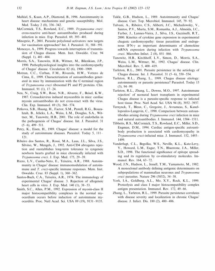
A/J mice infected with the Brazil strain of T.cruzi develop strong myosin-specific DTH andproduction of myosin-specific autoantibodies(Leon et al., 2001). To determine whether thismight result from molecular mimicry rather thanby bystander activation, we immunized A/J micewith an acetone (protein) extract of T. cruzi Brazilstrain epimastigotes emulsified in CFA. Mice werealso immunized with phosphate-buffered saline(PBS)/CFA and myosin/CFA as negative andpositive (Godsel et al., 2001) controls, respec-tively. An additional group was infected with T.cruzi trypomastigotes as another positive control(Leon et al., 2001). Only the T. cruzi infected andmyosin/CFA immunized groups developed my-ocarditis (data not shown; see ref. (Leon et al.,2001) for histopathology). However, three of thegroups developed both myosin-specific DTH andautoantibody responses (Fig. 2). In light of this, itis highly significant both that the magnitude ofthe myosin DTH in myosin/CFA-immunized, T.cruzi-infected, and T. cruzi/CFA-immunized aresimilar, and that the T. cruzi/CFA-immunizedanimals did not develop myocarditis. The findingof strong myosin DTH in the T. cruzi/CFA-im-munized animals implies that there is indeedmolecular mimicry between a T. cruzi protein andcardiac myosin. In light of the magnitude of theDTH, it is somewhat surprising that that thesemice did not develop disease, since the myosin/CFA-immunized mice have such severe myocardi-tis. However, it is probable that autoreactivityitself is not sufficient to give tissue inflammation,and that a proinflammatory environment, inducedby infection (Talvani et al., 2000) or provided byanother inflammatory stimulus (such as lipo-polysaccharide) is also necessary. T. cruzi-specificDTH was high in the T. cruzi-infected and T.cruzi/CFA-immunized animals, as expected.
To investigate the possibility that the Brazilstrain is comprised of multiple clones with indi-vidual pathogenic potential (see above), we gener-ated a panel of clones by limiting dilution. Twelveclones were derived and, although each gives aslightly different outcome when used to infect A/Jmice, none gives the same disease as that given by
Fig. 1. Histopathology of Chagas heart disease. A/J mice wereinfected by intraperitoneal injection of 10 000 trypomastigotesof the Brazil strain of T. cruzi and their hearts were analyzed21 days post infection. Serial sagittal sections were generatedand stained with hematoxylin and eosin. Lesions in infectedmouse hearts were found throughout the myocardium andtypical sections from infected and uninfected animals areshown here. While hearts of uninfected mice are histologicallynormal, with well ordered myofibrils and scattered myocytenuclei and interstitial cells, those of T. cruzi-infected miceshow massive mononuclear infiltration, occasional parasitepseudocysts (not in this section) and myocyte swelling andnecrosis.

D.M. Engman, J.S. Leon / Acta Tropica 81 (2002) 123–132 129
Fig. 2. Induction of myosin-specific cellular and humoral autoimmunity by T. cruzi infection or immunization with a T. cruzi proteinextract. A/J mice were (i) immunized with PBS in CFA, (ii) immunized with myosin in CFA, (iii) immunized with T. cruzi protein(acetone) extract in CFA or (iv) infected with T. cruzi. (Left) 21 days post immunization/infection, myosin- or T. cruzi-specific DTHwas measured using a standard 24-h ear swelling assay (Leon et al., 2001). Briefly, one ear of each mouse was injected with 10 �gtest antigen (myosin or T. cruzi extract in PBS) and the other was injected with 10 �g control antigen (BSA in PBS). The net increasein ear thickness (increase in test minus increase in control) was measured after 24 h. (Right) Serum taken at the same time wasanalyzed by myosin-specific IgG ELISA to measure myosin autoantibodies, as described (Leon et al., 2001). Five mice per groupwere used for assessment of DTH responses and eight per group for serology. Error bars represent standard error of the mean.
the uncloned Brazil strain (data not shown). Asan example, the Brazil 4 clone (Brc4), when usedto infect A/J mice, caused no myocarditis and nomyosin DTH (Fig. 3), although parasite-specificDTH and antibody production were identical tothose of the parental strain (not shown). Thisprovides specific and strong support for the no-tion that natural infection can involve multiple,unique parasite clones with differing pathogenicpotential.
7. Final remarks
The study of animal models of Chagas diseasehas permitted the definition of a number of dis-tinct mechanisms of pathogenesis, including para-site antigen-specific inflammation, parasite-induced myocardial cell necrosis and repair, mi-crovascular spasm and autoimmunity. As is ob-served in human infections, there is a wide
variety of outcomes of the animal infections; asingle strain of mouse may develop many differ-ent types of disease depending on the parasiteisolate used and a single parasite clone can causevery different disease in different strains of mice.Therefore, no single strain–strain combination ormechanism defined within is reflecti�e of all Chagasdisease. While some may view this as a shortcom-ing of the murine models, we believe that it is astrength, since the variation in outcome is pre-cisely what is observed in humans. However, it isequally important that conclusions drawn fromthe study of one strain-strain combination not beinterpreted as representing all of Chagas disease.Furthermore, the different pathogenetic mecha-nisms are not mutually exclusive, a point over-looked by some. Finally, and perhaps mostimportant, when both parasite and host antigensare present in the inflamed myocardium, it isdifficult to determine whether anti-parasite immu-nity, autoimmunity, both or neither are responsi-

D.M. Engman, J.S. Leon / Acta Tropica 81 (2002) 123–132130
Fig. 3. Clones of a pathogenic T. cruzi strain may be nonpathogenic. The pathogenic Brazil strain of T. cruzi was cloned by limitingdilution to produce the Brazil 4 clone (Brc4), among others. A/J mice were (i) injected with PBS, or infected with 10 000trypomastigotes of (ii) the Brc4 clone or (iii) the parental Brazil strain. (Left) 21 days post infection, myosin-specific DTH wasmeasured using a standard 24 h ear swelling assay (Leon et al., 2001). Briefly, one ear of each mouse was injected with 10 �g testantigen (myosin in PBS) and the other was injected with 10 �g control antigen (BSA in PBS). The net increase in ear thickness(increase in test minus increase in control) was measured after 24 h. Five mice per group were used for assessment of DTHresponses. Error bars represent standard error of the mean. (Right) Serial sagittal sections were generated and stained withhematoxylin and eosin.
ble for the tissue damage. It may be logical topresume that immunity to foreign antigen in thetissue is inflammatory and also that one need notinvoke an autoimmune hypothesis to explain thedamage. However, reasonableness and conclusivedetermination are not the same, even withoutconsidering the presence of additional, coexistentinflammatory mechanisms. Many groups havecontributed substantially to our understanding ofChagas disease pathogenesis and it is logical thatparasite persistence is a major factor in tissue
inflammation. We would like to contribute to theissue by rigorously testing the autoimmunity hy-pothesis suggested by vast amounts of circumstan-tial evidence published by others and us in the past.The T. cruzi Brazil/A/J mouse combination offersan attractive model for study in this regard, sinceit possesses very strong autoimmune features. Newapproaches for the selective inhibition of antigen-specific cellular immunity may also help to deter-mine the relative contributions of different types ofimmunity to Chagas disease pathogenesis.

D.M. Engman, J.S. Leon / Acta Tropica 81 (2002) 123–132 131
Acknowledgements
This work was supported by grants form theNational Institutes of Health and the AmericanHeart Association.
References
Acosta, A.M., Santos-Buch, C.A., 1985. Autoimmune my-ocarditis induced by Trypanosoma cruzi. Circulation 71,1255–1261.
Anez, N., Carrasco, H., Parada, H., Crisante, G., Rojas, A.,Fuenmayor, C., Gonzalez, N., Percoco, G., Borges, R.,Guevara, P., Ramirez, J.L., 1999. Myocardial parasitepersistence in chronic chagasic patients. Am. J. Trop. Med.Hyg. 60 (5), 726–732.
Ben Younes-Chennoufi, A., Hontebeyrie-Joskowicz, M., Tri-cottet, V., Eisen, H., Reynes, M., Said, G., 1988. Persis-tence of Trypanosoma cruzi antigens in the inflammatorylesions of chronically infected mice. Trans. R. Soc. Trop.Med. Hyg. 82 (1), 77–83.
Cossio, P.M., Diez, C., Szarfman, A., Kreutzer, E., Candiolo,B., Arana, R.M., 1974a. Chagasic cardiopathy. Demon-stration of a serum gamma globulin factor which reactswith endocardium and vascular structures. Circulation 49(1), 13–21.
Cossio, P.M., Laguens, R.P., Diez, C., Szarfman, A., Segal,A., Arana, R.M., 1974b. Chagasic cardiopathy. Antibodiesreacting with plasma membrane of striated muscle andendothelial cells. Circulation 50 (6), 1252–1259.
Cossio, P.M., Bustouoabad, O., Paterno, E., Lotti, R.,Casanova, M.B., Podesta, M.B., Bolomo, N., Arana,R.M., de Pasqualini, C.D., 1984. Experimental myocarditisinduced in Swiss mice by homologous heart immunizationresembles chronic experimental Chagas’ heart disease. Clin.Immunol. Immunopathol. 33, 165–175.
Cunha-Neto, E., Duranti, M., Gruber, A., Zingales, B., deMessias, I., Stolf, N., Bellotti, G., Patarroyo, M.E., Pil-leggi, F., Kalil, J., 1995. Autoimmunity in Chagas’ diseasecardiomyopathy: biological relevance of a cardiac myosin-specific epitope crossreactive to an immunodominant Try-panosoma cruzi antigen. Proc. Natl Acad. Sci. USA 92,3541–3545.
Cunha-Neto, E., Coelho, V., Guilherme, L., Fiorelli, A., Stolf,N., Kalil, J., 1996. Autoimmunity in Chagas’ disease:identification of cardiac myosin-B13 Trypanosoma cruziprotein crossreactive T cell clones in heart lesions of achronic Chagas’ cardiomyopathy patient. J. Clin. Invest.98 (8), 1709–1712.
de Scheerder, I.K., de Buyzere, M.L., Delanghe, J.R.,Clement, D.L., Wieme, R.J., 1989. Anti-myosin humoralimmune response following cardiac injury. Autoimmunity4 (1–2), 51–58.
Dighiero, G., Rose, N.R., 1999. Critical self-epitopes are keyto the understanding of self-tolerance and autoimmunity.Immunol. Today 20 (9), 423–428.
Eisen, H., Kahn, S., 1991. Mimicry in Trypanosoma cruzi :fantasy and reality. Curr. Opin. Immunol. 3, 507–510.
Factor, S.M., Cho, S., Wittner, M., Tanowitz, H., 1985.Abnormalities of the coronary microcirculation in acutemurine Chagas’ disease. Am. J. Trop. Med. Hyg. 34 (2),246–253.
Fedoseyeva, E.V., Zhang, F., Orr, P.L., Levin, D., Buncke,H.J., Benichou, G., 1999. De novo autoimmunity to car-diac myosin after heart transplantation and its contribu-tion to the rejection process. J. Immunol. 162 (11),6836–6842.
Giordanengo, L., Fretes, R., Diaz, H., Cano, R., Bacile, A.,Vottero-Cima, E., Gea, S., 2000. Cruzipain induces au-toimmune response against skeletal muscle and tissue dam-age in mice. Muscle Nerve 23, 1407–1413.
Godsel, L.M., Wang, K., Schodin, B.A., Leon, J.S., Miller,S.D., Engman, D.M., 2001. Prevention of autoimmunemyocarditis through the induction of antigen-specific pe-ripheral immune tolerance. Circulation 103, 1709–1714.
Grauert, M.R., Houdayer, M., Hontebeyrie-Joskowciz, M.,1993. Trypanosoma cruzi infection enhances polyreactiveantibody response in an acute case of human Chagas’disease. Clin. Exp. Immunol. 93 (1), 85–92.
Hoff, R., Teixeira, R.S., Carvalho, J.S., Mott, K.E., 1978.Trypanosoma cruzi in the cerebrospinal fluid during theacute stage of Chagas’ disease. N. Engl. J. Med. 298 (11),604–606.
Kierszenbaum, F., 1986. Autoimmunity in Chagas’ disease. J.Parasitol. 72, 201–211.
Kierszenbaum, F., 1999. Chagas’ disease and the autoimmu-nity hypothesis. Clin. Microbiol. Rev. 12 (2), 210–223.
Kirchhoff, L.V., 1989. Is Trypanosoma cruzi a new threat toour blood supply? Ann. Intern. Med. 111, 773–775.
Kirchhoff, L.V., 1993. Chagas disease. American trypanosomi-asis. Infect. Dis. Clin. North Am. 7, 487–502.
Koberle, F., 1970. The causation and importance of nervouslesions in American trypanosomiasis. Bull. World HealthOrg. 42 (5), 739–743.
Koberle, F., Nador, E., 1955. Etiologiae patogenia do megae-sofago no Brasil. Rev. Paulista Med. 47, 643–661.
Laguens, R.P., Cabeza Meckert, P.M., Chambo, J.G., 1989.Immunologic studies on a murine model of Chagas disease.Medicina (Buenos Aires) 49, 197–202.
Laguens, R.P., Meckert, P.C., Chambo, J.G., 1988. Antiheartantibody-dependent cytotoxicity in the sera from micechronically infected with Trypanosoma cruzi. Infect. Im-mun. 56, 993–997.
Laranja, F.S., Dias, E., Nobrego, G., Marinda, A., 1956.Chagas’ disease–a clinical, epidemiologic and pathologicstudy. Circulation 14, 1035–1060.
Leon, J.S., Engman, D.M., 2001. Autoimmunity in Chagasheart disease. Int. J. Parasitol. 31 (5–6), 554–560.
Leon, J.S., Godsel, L.M., Wang, K., Engman, D.M., 2001.Cardiac myosin autoimmunity in acute Chagas heart dis-ease. Infect. Immun. 69, 5643–5649.
D.M. Engman, J.S. Leon / Acta Tropica 81 (2002) 123–132132
Malkiel, S., Kuan, A.P., Diamond, B., 1996. Autoimmunity inheart disease: mechanisms and genetic susceptibility. Mol.Med. Today 2 (8), 336–342.
McCormick, T.S., Rowland, E.C., 1989. Trypanosoma cruzi :cross-reactive anti-heart autoantibodies produced duringinfection in mice. Exp. Parasitol. 69, 393–401.
Minoprio, P., 2001. Parasite polyclonal activators: new targetsfor vaccination approaches? Int. J. Parasitol. 31, 588–591.
Moncayo, A., 1999. Progress towards interruption of transmis-sion of Chagas disease. Mem. Inst. Oswaldo. Cruz. 94(Suppl. 1), 401–404.
Morris, S.A., Tanowitz, H.B., Wittner, M., Bilezikian, J.P.,1990. Pathophysiological insights into the cardiomyopathyof Chagas’ disease. Circulation 82 (6), 1900–1909.
Motran, C.C., Cerban, F.M., Rivarola, H.W., Vottero deCima, E., 1999. Characterization of autoantibodies gener-ated in mice by immunization with the C-terminal regionof Trypanosoma cruzi ribosomal P1 and P2 proteins. Clin.Immunol. 91 (1), 17–24.
Neu, N., Craig, S.W., Rose, N.R., Alvarez, F., Beisel, K.W.,1987. Coxsackievirus induced myocarditis in mice: cardiacmyosin autoantibodies do not cross-react with the virus.Clin. Exp. Immunol. 69 (3), 566–574.
Petkova, S.B., Huang, H., Factor, S.M., Pestell, R.G., Bouza-hzah, B., Jelicks, L.A., Weiss, L.M., Douglas, S.A., Wit-tner, M., Tanowitz, H.B., 2001. The role of endothelin inthe pathogenesis of Chagas’ disease. Int. J. Parasitol. 31(5–6), 499–511.
Petry, K., Eisen, H., 1989. Chagas’ disease: a model for thestudy of autoimmune diseases. Parasitol. Today 5, 111–121.
Ribeiro dos Santos, R., Rossi, M.A., Laus, J.L., Silva, J.S.,Silvino, W., Mengels, J., 1992. Anti-CD4 abrogates rejec-tion and reestablishes long-term tolerance to syngeneicnewborn hearts grafted in mice chronically infected withTrypanosoma cruzi. J. Exp. Med. 175, 29–39.
Rizzo, L.V., Cunha-Neto, E., Teixeira, A.R., 1988. Autoim-munity in Chagas’ disease: immunomodulation of autoim-mune and T. cruzi-specific immune responses. Mem. Inst.Oswaldo. Cruz. 83 (Suppl. 1), 360–362.
Santos-Buch, C.A., Teixeira, A.R., 1974. The immunology ofexperimental Chagas’ disease. 3. Rejection of allogeneicheart cells in vitro. J. Exp. Med. 140 (1), 38–53.
Smith, S.C., Allen, P.M., 1992. Expression of myosin-class IImajor histocompatibility complexes in the normal my-ocardium occurs before induction of autoimmune my-ocarditis. Proc. Natl Acad. Sci. USA 89 (19), 9131–9135.
Takle, G.B., Hudson, L., 1989. Autoimmunity and Chagas’disease. Curr. Top. Microbiol. Immunol. 145, 79–92.
Talvani, A., Ribeiro, C.S., Aliberti, J.C., Michailowsky, V.,Santos, P.V., Murta, S.M., Romanha, A.J., Almeida, I.C.,Farber, J., Lannes-Vieira, J., Silva, J.S., Gazzinelli, R.T.,2000. Kinetics of cytokine gene expression in experimentalchagasic cardiomyopathy: tissue parasitism and endoge-nous IFN-� as important determinants of chemokinemRNA expression during infection with Trypanosomacruzi. Microbes Infect. 2 (8), 851–866.
Tanowitz, H.B., Kirchhoff, L.V., Simon, D., Morris, S.A.,Weiss, L.M., Wittner, M., 1992. Chagas’ disease. Clin.Microbiol. Rev. 5, 400–419.
Tarleton, R.L., 2001. Parasite persistence in the aetiology ofChagas disease. Int. J. Parasitol. 31 (5–6), 550–554.
Tarleton, R.L., Zhang, L., 1999. Chagas disease etiology:autoimmunity or parasite persistence? Parasitol. Today 15(3), 94–99.
Tarleton, R.L., Zhang, L., Downs, M.O., 1997. Autoimmunerejection’ of neonatal heart transplants in experimentalChagas disease is a parasite-specific response to infectedhost tissue. Proc. Natl Acad. Sci. USA 94 (8), 3932–3937.
Ternynck, T., Bleux, C., Gregoire, J., Avrameas, S., Kanel-lopoulos-Langevin, C., 1990. Comparison between autoan-tibodies arising during Trypanosoma cruzi infection in miceand natural autoantibodies. J. Immunol. 144, 1504–1511.
Tibbetts, R.S., McCormick, T.S., Rowland, E.C., Miller, S.D.,Engman, D.M., 1994. Cardiac antigen-specific autoanti-body production is associated with cardiomyopathy inTrypanosoma cruzi-infected mice. J. Immunol. 152, 1493–1499.
Vanderlugt, C.L., Begolka, W.S., Neville, K.L., Katz-Levy,Y., Howard, L.M., Eagar, T.N., Bluestone, J.A., Miller,S.D., 1998. The functional significance of epitope spread-ing and its regulation by co-stimulatory molecules. Im-munol. Rev. 164, 63–72.
Wood, J.N., Hudson, L., Jessell, T.M., Yamamoto, M., 1982.A monoclonal antibody defining antigenic determinants onsubpopulations of mammalian neurones and Trypanosomacruzi parasites. Nature 296 (5852), 34–38.
York, I.A., Goldberg, A.L., Mo, X.Y., Rock, K.L., 1999.Proteolysis and class I major histocompatibility complexantigen presentation. Immunol. Rev. 172, 49–66.
Zhang, L., Tarleton, R.L., 1999. Parasite persistence correlateswith disease severity and localization in chronic Chagas’disease. J. Infect. Dis. 180 (2), 480–486.
Copyright © 2022 FDOKUMEN