Identifying the Cells Breaching Self-Tolerance in Autoimmunity
8
The Journal of Immunology Identifying the Cells Breaching Self-Tolerance in Autoimmunity Robert A. Benson,* Agapitos Patakas,* ,† Paola Conigliaro,* ,‡ Catherine M. Rush, x Paul Garside,* Iain B. McInnes, † and James M. Brewer* Activation of auto-reactive T cells by activated dendritic cells (DCs) presenting self-Ag is widely assumed to be the precipitating event in the development of autoimmune disease. However, despite such widely held preconceptions, supporting data are scarce and subjective, particularly in experimental arthropathy. We have adapted a novel murine model of breach of self-tolerance allowing evaluation of the contribution of endogenous DCs to the development of autoimmune responses and disease. For the first time, we reveal the critical role played by conventional DCs, and the timing and location of this process. We further demonstrate the im- portance of this finding by clinically relevant, therapeutic manipulation of conventional DC function, resulting in decreased au- toimmune phenotype and disease severity. The Journal of Immunology, 2010, 184: 6378–6385. R heumatoid arthritis (RA) is an autoimmune mediated disorder characterized by painful articular inflammation and erosion. The systemic nature of this aberrant immune response is highlighted by additional involvement of skin, lungs and vasculature manifesting as rheumatoid nodules, diffuse in- flammation/fibrosis, and increased risk of atherosclerosis (1–5). Despite successful identification of biological targets and their therapeutic translation (anti-TNF, anti-CD20, CTLA-4–Ig, anti– IL-1, anti–IL-6) many patients remain refractory to intervention (6). As with all autoimmune disorders, the ultimate goal should be re-establishment of self-tolerance. However, patient studies have been unable to dissect the critical events mediating the induction of self-reactivity as this event likely occurs many years prior to RA diagnosis. Conversely, most animal models of RA rely on aggressive and artificial self-Ag immunization protocols (7), which do not permit analysis of the very early breach of self- tolerance and prearticular stages of disease. Understanding these early events will be critical if the immune system is to be re-ed- ucated and self-tolerance reinstated, rather than simply treating symptoms. Although approaches, such as the case control con- sortium, have established a number of predictive genetic varia- tions associated with the development of RA (8), they do not inform when to intervene for a given pathway. Understanding the events surrounding the breach of self-tolerance associated with RA will therefore reveal markers associated with the onset of preclinical disease and signal a window of early intervention. Dendritic cells (DCs) are considered the main initiators of naive T cell responses (9), but their contribution to RA remains unclear. Phenotypic studies have identified the subsets and activation states of DCs in RA (10, 11), and current therapeutic regimes can alter their maturation/activation states (12), yet direct evidence impli- cating a particular DC subset in breach of self-tolerance leading to RA pathogenesis is lacking. Despite an incomplete understanding of how DCs are involved in RA pathology, tolerogenic DC pop- ulations are being developed as potential therapeutic tools, with some success being demonstrated in murine models (13–15). In- deed, clinical trials have been initiated in the United Kingdom (http://news.bbc.co.uk/1/hi/health/7560535.stm) and Australia (www.uq.edu.au/news/?article=13128). Although DC therapy ap- pears an attractive proposition, care must be taken as the pivotal role of these cells in directing adaptive immune responses also endows them with the potential to initiate or exacerbate autoim- munity. For example, transfer of exogenous type II collagen (CII) pulsed, myeloid DCs can incite joint pathology in disease sus- ceptible mice (16). As such, it will be critical to define the APC populations that regulate the breach of self-tolerance underlying the induction of RA. Our laboratory has developed a novel murine model of preclinical experimental arthritis allowing delineation of events surrounding loss of self-tolerance (17–19). Transfer of Th1 polarized OVA-specific–TCR transgenic T cells induces synovial hyperplasia and cartilage erosion proximal to joints challenged with heat aggregated OVA (HAO), thus circumventing issues as- sociated with immunization with self-Ags in powerful, non- physiological adjuvant (20, 21). These histological pathologies are also manifest clinically (16–18). These are mild and localized to the affected joint, unlike the aggressive polyarthritis seen in collagen-induced arthritis being more akin to advanced human disease. Joint inflammation in this system is associated with ele- vated titers of autoantibodies, including anti-CII, IgG2a-rheumatoid factor and anticyclic citrullinated peptides (17–19). Further dys- regulation of self-tolerance is evidenced by CII specific T cell proliferative responses on in vitro restimulation. Nonspecific in- flammation can recapitulate many of the histological signs of pathology in this model, however, development of autoreactivity is *Division of Immunology, Infection and Inflammation, Glasgow Biomedical Re- search Centre, Faculty of Medicine and † Centre for Rheumatic Disease, Glasgow Biomedical Research Centre, University of Glasgow, Glasgow, United Kingdom; ‡ Dipartimento di Medicina Interna, Facolta ` di Medicina e Chirurgia, Universita ` degli Studi di Roma “Tor Vergata”, Rome, Italy; and x Vascular Biology Unit, School of Medicine, James Cook University, Townsville, Queensland, Australia Received for publication December 8, 2009. Accepted for publication March 22, 2010. This work was supported by the Chief Scientists Office (Scottish Executive Health Department, Edinburgh, U.K.), and Oliver Bird Rheumatism Programme (Nuffield Foundation, London, U.K.). Address correspondence and reprint requests to Dr. James Brewer, Division of Im- munology, Infection and Inflammation, Glasgow Biomedical Research Centre, Fac- ulty of Medicine, University of Glasgow, 120 University Place, Glasgow G12 8QQ, United Kingdom. E-mail address: [email protected] The online version of this article contains supplemental material. Abbreviations used in this paper: cDC, conventional DC; CII, type II collagen; DC, dendritic cell; DTx, diphtheria toxin; Etan, Etanercept; HAO, heat aggregated OVA; hIgG, human IgG; iso, isotype; LN, lymph node; pDC, plasmacytoid DC; pLN, popliteal LN; RA, rheumatoid arthritis. Copyright Ó 2010 by The American Association of Immunologists, Inc. 0022-1767/10/$16.00 www.jimmunol.org/cgi/doi/10.4049/jimmunol.0903951
-
Upload
independent -
Category
Documents
-
view
1 -
download
0
Transcript of Identifying the Cells Breaching Self-Tolerance in Autoimmunity

The Journal of Immunology
Identifying the Cells Breaching Self-Tolerancein Autoimmunity
Robert A. Benson,* Agapitos Patakas,*,† Paola Conigliaro,*,‡ Catherine M. Rush,x
Paul Garside,* Iain B. McInnes,† and James M. Brewer*
Activation of auto-reactive T cells by activated dendritic cells (DCs) presenting self-Ag is widely assumed to be the precipitating
event in the development of autoimmune disease. However, despite such widely held preconceptions, supporting data are scarce and
subjective, particularly in experimental arthropathy. We have adapted a novel murine model of breach of self-tolerance allowing
evaluation of the contribution of endogenous DCs to the development of autoimmune responses and disease. For the first time, we
reveal the critical role played by conventional DCs, and the timing and location of this process. We further demonstrate the im-
portance of this finding by clinically relevant, therapeutic manipulation of conventional DC function, resulting in decreased au-
toimmune phenotype and disease severity. The Journal of Immunology, 2010, 184: 6378–6385.
Rheumatoid arthritis (RA) is an autoimmune mediateddisorder characterized by painful articular inflammationand erosion. The systemic nature of this aberrant immune
response is highlighted by additional involvement of skin, lungsand vasculature manifesting as rheumatoid nodules, diffuse in-flammation/fibrosis, and increased risk of atherosclerosis (1–5).Despite successful identification of biological targets and theirtherapeutic translation (anti-TNF, anti-CD20, CTLA-4–Ig, anti–IL-1, anti–IL-6) many patients remain refractory to intervention(6). As with all autoimmune disorders, the ultimate goal should bere-establishment of self-tolerance. However, patient studies havebeen unable to dissect the critical events mediating the inductionof self-reactivity as this event likely occurs many years prior toRA diagnosis. Conversely, most animal models of RA rely onaggressive and artificial self-Ag immunization protocols (7),which do not permit analysis of the very early breach of self-tolerance and prearticular stages of disease. Understanding theseearly events will be critical if the immune system is to be re-ed-ucated and self-tolerance reinstated, rather than simply treatingsymptoms. Although approaches, such as the case control con-sortium, have established a number of predictive genetic varia-tions associated with the development of RA (8), they do not
inform when to intervene for a given pathway. Understanding theevents surrounding the breach of self-tolerance associated withRA will therefore reveal markers associated with the onset ofpreclinical disease and signal a window of early intervention.Dendritic cells (DCs) are considered the main initiators of naive
T cell responses (9), but their contribution to RA remains unclear.Phenotypic studies have identified the subsets and activation statesof DCs in RA (10, 11), and current therapeutic regimes can altertheir maturation/activation states (12), yet direct evidence impli-cating a particular DC subset in breach of self-tolerance leading toRA pathogenesis is lacking. Despite an incomplete understandingof how DCs are involved in RA pathology, tolerogenic DC pop-ulations are being developed as potential therapeutic tools, withsome success being demonstrated in murine models (13–15). In-deed, clinical trials have been initiated in the United Kingdom(http://news.bbc.co.uk/1/hi/health/7560535.stm) and Australia(www.uq.edu.au/news/?article=13128). Although DC therapy ap-pears an attractive proposition, care must be taken as the pivotalrole of these cells in directing adaptive immune responses alsoendows them with the potential to initiate or exacerbate autoim-munity. For example, transfer of exogenous type II collagen (CII)pulsed, myeloid DCs can incite joint pathology in disease sus-ceptible mice (16). As such, it will be critical to define the APCpopulations that regulate the breach of self-tolerance underlyingthe induction of RA. Our laboratory has developed a novel murinemodel of preclinical experimental arthritis allowing delineation ofevents surrounding loss of self-tolerance (17–19). Transfer of Th1polarized OVA-specific–TCR transgenic T cells induces synovialhyperplasia and cartilage erosion proximal to joints challengedwith heat aggregated OVA (HAO), thus circumventing issues as-sociated with immunization with self-Ags in powerful, non-physiological adjuvant (20, 21). These histological pathologies arealso manifest clinically (16–18). These are mild and localizedto the affected joint, unlike the aggressive polyarthritis seen incollagen-induced arthritis being more akin to advanced humandisease. Joint inflammation in this system is associated with ele-vated titers of autoantibodies, including anti-CII, IgG2a-rheumatoidfactor and anticyclic citrullinated peptides (17–19). Further dys-regulation of self-tolerance is evidenced by CII specific T cellproliferative responses on in vitro restimulation. Nonspecific in-flammation can recapitulate many of the histological signs ofpathology in this model, however, development of autoreactivity is
*Division of Immunology, Infection and Inflammation, Glasgow Biomedical Re-search Centre, Faculty of Medicine and †Centre for Rheumatic Disease, GlasgowBiomedical Research Centre, University of Glasgow, Glasgow, United Kingdom;‡Dipartimento di Medicina Interna, Facolta di Medicina e Chirurgia, Universita degliStudi di Roma “Tor Vergata”, Rome, Italy; and xVascular Biology Unit, School ofMedicine, James Cook University, Townsville, Queensland, Australia
Received for publication December 8, 2009. Accepted for publication March 22,2010.
This work was supported by the Chief Scientists Office (Scottish Executive HealthDepartment, Edinburgh, U.K.), and Oliver Bird Rheumatism Programme (NuffieldFoundation, London, U.K.).
Address correspondence and reprint requests to Dr. James Brewer, Division of Im-munology, Infection and Inflammation, Glasgow Biomedical Research Centre, Fac-ulty of Medicine, University of Glasgow, 120 University Place, Glasgow G12 8QQ,United Kingdom. E-mail address: [email protected]
The online version of this article contains supplemental material.
Abbreviations used in this paper: cDC, conventional DC; CII, type II collagen; DC,dendritic cell; DTx, diphtheria toxin; Etan, Etanercept; HAO, heat aggregated OVA;hIgG, human IgG; iso, isotype; LN, lymph node; pDC, plasmacytoid DC; pLN,popliteal LN; RA, rheumatoid arthritis.
Copyright� 2010 by TheAmericanAssociation of Immunologists, Inc. 0022-1767/10/$16.00
www.jimmunol.org/cgi/doi/10.4049/jimmunol.0903951
exquisitely dependent on elicitation of an irrelevant Ag-specificT cell response localized in the joint (18). As such the modelrepresents a model of early stage arthritis, reflecting patients withmild clinical presentation but strong evidence of autoreactivity,such as rheumatoid factor. Using this model, we have recentlydemonstrated that plasmacytoid DCs (pDCs) can function to limitself-reactivity and consequent pathology (19). Crucially, the cellpopulation responsible for inciting autoreactive T cell responsesremains to be identified.In this study, we demonstrate conventional DC (cDC) maturation
and Ag presentation immediately prior to breach of self-tolerance,the ability of these cells to transfer autoreactivity, a potential actionof existing biological therapeutics on them and an essential re-quirement for cDCs in the development of self-reactive Tand B cellresponses and RA pathogenesis.
Materials and MethodsAnimals
DO11.10 BALB/c TCR, OT-II C57BL/6 TCR, and CD11cDTR transgenicmice (22), 6- to 8-wk-old female BALB/c, C57BL/6 (Harlan, Bicester,U.K.) were housed in the University of Strathclyde and procedures wereperformed according to the United Kingdom Home Office regulations.
Induction of arthritis
Recipient mice received 2 3 106 in vitro Th1-polarized DO11.10 TCRtransgenic CD4+ T cells i.v. (17). Transferred cells were in vivo expandedby s.c. immunization with OVA/CFA the following day. Ten days later,mice were footpad challenged with HAO, as described by Maffia et al.(17). Th1 Va2+Vb5+ OT-II T cells were used for C57BL/6 recipients inEaGFP and CD11cDTR experiments. EaGFP (100 mg) was administeredwith or without HAO (23) for APC identification experiments. De-velopment of arthritis was followed by paw thickness, using a dial caliper(Kroeplin, Munich, Germany), and by histological assessment at day 7after HAO challenge (17). Disease scoring, based on cell infiltration (0, nocells; 1, ,50 cells; 2, 50–200 cells; and 3, .200 cells), synovial hyper-plasia (0, no proliferation; 2, .3 layers; and 3, severe), and joint erosion(0, no erosion; 1, very mild; 2, marginal; and 3, complete) was performedon three joints for each of five mice per group. Average paw scores foreach parameter were calculated across all three joints, and a total score foreach paw calculated by adding individual parameter scores.
Depletion of cDCs in vivo
cDCs were depleted in CD11cDTR mice (22) in vivo by s.c. footpad in-jection of 20 ng diphtheria toxin (DTx) (Sigma-Aldrich, St. Louis, MO) 1 dbefore s.c injection of HAO. Control CD11cDTR transgenic and C57BL/6mice received matched injection of 50 ml PBS or DTx, respectively.
Isolation and culture of bone marrow DCs
DCs were generated from the bone marrow of C57BL/6 mice by culture inGM-CSF conditioned medium as previously described (16). Routine flowcytometric analysis of DC cultures revealed yields of .80% for cellsstaining both CD11c and MHC class II positive. DCs were cultured for 6 hwith OVA (100 mg/ml), bovine CII (50 mg/ml, Sigma-Aldrich), or both,before addition of LPS (1 mg/ml, Escherichia coli 055:B5 (Sigma-Aldrich)and maturation overnight. Footpad challenge consisted of 23 105 DCs s.c.(a total of 4 3 105 DCs were transferred in the mixed DC group, 2 3 105
OVA and 2 3 105 CII-pulsed DCs).
Flow cytometry
Single-cell suspensions were prepared from axillary, inguinal, cervical, andmesenteric lymph nodes (LNs) from TCR transgenic mice. Popliteal LN(pLN) single-cell suspensions were stained with anti-CD11c, anti-PDCA1,anti-B220, anti–I-A/E (all BD Biosciences, Oxford, U.K.), and YAe(specific for I-Ea 52-68 of I-Ed presented on I-Ab) (eBioscience, SanDiego, CA) for APC identification. cDCs and pDCs were defined asCD11c+PDCA12B2202 and CD11c+PDCA1+B220+, respectively.
In vitro restimulation assays
pLN cells were cultured with either medium, 1 mg/ml OVA, or 50 mg/mlCII. Proliferation was analyzed at 96 h by flow cytometric staining for EdUincorporation (Invitrogen, Molecular Probes, U.K.).
ELISA
Anti-OVA and anti-collagen Abs were detected by ELISA as previouslydescribed (17). An adapted IFN-g ELISA protocol (16, 17) was usedfor detection of IL-17 using anti–IL-17 clones TC11-18H10 and bio-tinylated TC11-8H4 (BD Biosciences). Standard curves for IL-17ELISA were generated using recombinant mouse IL-17A (eBio-science).
Anti–TNF-a treatment
sTNFR-Fc (0.5 mg/kg) (24) (Etanercept, Wyeth Pharmaceuticals, Taplow,U.K.) was given s.c. 1 d prior to, and 1, 3, and 5 d after footpad challenge.Control mice received 0.5 mg/kg hIgG.
Statistical analysis
Results are expressed as mean 6 SD, n = 5. Significance was determinedby Student t test, values of p # 0.05 were considered significant.
ResultsIncreased numbers of MHC class II high cDCs are observed indraining LNs of arthritic mice and are associated with anti-CIIresponses
Using a recently developedmodel of experimental arthritis (17), wesought to delineate the critical events surrounding the de-velopment of autoreactive T and B cell responses and ensuingjoint pathology, identifying contributing APCs. Detailed kineticanalysis of DC markers and numbers in the draining pLN afterHAO challenge revealed that although the proportion of cDCs orpDCs was not significantly altered in HAO versus PBS challengedmice (Fig. 1A, 1B, Supplemental Fig. 1), the total numbers of bothDC populations increased after HAO challenge (Fig. 1C, 1D).cDC expression of MHC class II expression was significantlyhigher in arthritic mice than control animals by day 2 after HAOchallenge (Fig. 1E) and was subsequently maintained (Fig. 1E).pDCs expressed lower levels of MHC class II than cDCs, partic-ularly on days 2 and 3 after HAO challenge (Fig. 1F), returning tolevels comparable to PBS challenge by day 4. There were nosignificant differences in CD80, CD86, or CD40 expression ineither group (data not shown).Elevated cDC number and MHC class II expression preceded
detectable changes in the articular environment, with the first signsof synovial thickening and tissue infiltrate evident 4–5 d after HAOchallenge (Fig. 1G–N, Supplemental Fig. 1), increasing along withsigns of cartilage erosion, and peaking by day 7. This led us tohypothesize that breach of T cell self-tolerance may occur at theseearly time points after stimulation of the OVA response. IncreasedMHC class II expression by cDCs indicated maturation of thesecells and suggested they could be the main cells responsible for Agpresentation associated with footpad challenge.
Increased numbers of Ag-presenting cDCs are observed in thepLN prior to spontaneously arising autoreactive T cell andB cell responses
To address the role of cDCs in Ag presentation directly, we usedthe model Ag EaGFP and the mAb YAe (23) to identify in vivothe major APC populations associated with the development ofautoimmunity and pathology in RA. We focused on day 2postchallenge as the earliest time point at which alterations inAPC populations were detected (Fig. 1). Coadministration ofEaGFP with PBS or HAO revealed significantly more cDCsstaining positively with the peptide Ea/MHC class II-specificmAb, YAe, in HAO-challenged mice versus PBS challengedmice (Fig. 2A, Supplemental Fig. 2). The amount of Ag pre-sented by individual cDCs was not significantly altered in ar-thritic mice, with no change in YAe mean fluorescent intensity
The Journal of Immunology 6379

in the HAO/EaGFP versus the PBS/EaGFP nonarthritic group(Fig. 2B). The small shifts in YAe staining after in vivo EaGFPadministration likely reflect the low frequency at which a singlepeptide/MHC class II complex is presented on the APC surface.No significant presentation of peptide Ea/MHC class II wasobserved on pDCs (Fig. 2A), YAe staining levels were compa-rable between groups with or without EaGFP, regardless ofinflammatory status (Fig. 2B). Thus, the profile of cells pre-senting the model Ag did not differ between nonarthritic andarthritic animals, being confined to the cDC subset 2 d afterchallenge. Despite this, the number of cDCs presenting Eapeptide was elevated after HAO challenge. No detectable anti-CII–serum Ab was detected at this time (Fig. 2C), but breach ofself-tolerance was apparent by day 7, with HAO-challengedmice subsequently developing autoreactive B and T cell re-sponses (Fig. 2C, 2D).
OVA-pulsed DCs induce histological signs of pathology andare sufficient to induce anti-CII T and B cell responses inrecipient mice
Having identified cDCs as a major presenter of model Ag prior todevelopment of autoreactive arthritis,wehypothesized that theywerethe most likely candidates to be presenting tissue-derived Ag in ourmodel and consequently the APC population driving breach of self-tolerance. Indeed, a model of RA in which self-Ag presented byactivated DCs stimulates autoreactive T cells that have escapedthymic selection has been proposed (25, 26). In support of this hy-pothesis, adoptive transfer of self-Ag–bearing DCs can incite auto-immunity underlying diabetes, experimental autoimmune enceph-alitis, and erosive arthritis (16, 27, 28).To determine whether DCs were sufficient to drive this breach of
self-tolerance and drive autoreactive arthritis, Ag-pulsed bonemarrow-derived DCs were used in substitute of HAO. DCs alone,pulsed with one (OVA or CII), or both Ags (OVA and CII) weretested for their ability to induce footpad inflammation, joint pa-thology, and anti-CII–specific immune responses. We also in-cluded a group in which DCs were pulsed separately with OVAand CII, then mixed prior to challenge, allowing us to address therequirement for presentation of Ags by a single DC. One hy-pothesis for the breach of self-tolerance in our model was that the“conditioning” of DCs bearing self-peptide/MHC class II com-plexes by activated OVA-specific T cells could facilitate stimu-lation of autoreactive T cells, occurring via direct interaction if theself-Ag–bearing DCs were also presenting OVA peptides.DCs pulsed with OVA, OVA and CII, and mixed DCs induced
comparable levels of footpad swelling that was significantly greaterthan that observed with unpulsed or CII-pulsed DCs (Fig. 3A).
FIGURE 1. Joint inflammation induced by activated T cells of an ir-
relevant specificity is associated with increased numbers of cDCs and their
expression of MHC class II. Draining pLNs were removed at the indicated
days postchallenge (1–6 d) and FACs analysis performed to identify cDCs
(CD11c+B2202PDCA12) and pDCs (CD11c+B220+PDCA1+) (Supple-
mental Fig. 1). Percentages of cDCs and pDCs (A and B, respectively) in
pLN in PBS versus HAO challenged mice were derived as a function of
total LN cellularity. Absolute numbers of cDCs and pDCs were then
calculated (C and D, respectively), and MHC class II expression by both
populations (E, F). Ankle joints from HAO challenged mice were collected
on days 1, 2, 3, 4, 5, and 6. H&E demonstrated little infiltrate on days 1, 2
and 3 (G–I). Some synovial thickening could be observed on day 4 with
increasing s.c. infiltrate (J, K). Tissue sections from day 5 (L, M) to 6 (N)
revealed increased infiltrate through joint proximal tissue and beginnings
of synovial infiltrate and hyperplasia. Original magnification310. Themean
of n = 5 is shown6 SD in the mean. ppp, 0.01.
FIGURE 2. Elevated numbers of cDCs present model Ag in RA mice.
C57BL/6 mice received Th1 cells and were immunized with OVA/CFA as
before. Mice were footpad challenged with PBS/EaGFP or HAO/EaGFP.
Control groups without EaGFP were also included. YAe staining was then
used to assess presentation of Ea peptide/I-Ab in pLN 2 d after challenge.
Absolute numbers of YAe positive cDCs and pDCs (A) were determined in
addition to intensity of staining (B). Anticollagen responseswere determined
by ELISA on day 2 and 7 serum samples (C) after PBS or HAO footpad
challenge. In vitro restimulation of day 7 HAO challenged mice was used to
demonstrate successful induction of CII-specific T cells (D). Themean of n=
5 is shown6 SD in the mean. pp, 0.05; ppp , 0.01.
6380 DCs IN BREACH OF TOLERANCE
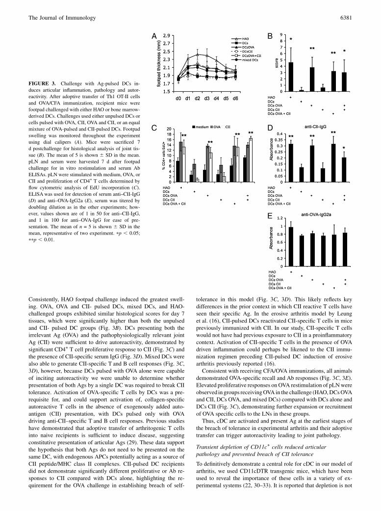
Consistently, HAO footpad challenge induced the greatest swell-ing. OVA, OVA and CII- pulsed DCs, mixed DCs, and HAO-challenged groups exhibited similar histological scores for day 7tissues, which were significantly higher than both the unpulsedand CII- pulsed DC groups (Fig. 3B). DCs presenting both theirrelevant Ag (OVA) and the pathophysiologically relevant jointAg (CII) were sufficient to drive autoreactivity, demonstrated bysignificant CD4+ T cell proliferative response to CII (Fig. 3C) andthe presence of CII-specific serum IgG (Fig. 3D). Mixed DCs werealso able to generate CII-specific T and B cell responses (Fig. 3C,3D), however, because DCs pulsed with OVA alone were capableof inciting autoreactivity we were unable to determine whetherpresentation of both Ags by a single DC was required to break CIItolerance. Activation of OVA-specific T cells by DCs was a pre-requisite for, and could support activation of, collagen-specificautoreactive T cells in the absence of exogenously added auto-antigen (CII) presentation, with DCs pulsed only with OVAdriving anti-CII–specific T and B cell responses. Previous studieshave demonstrated that adoptive transfer of arthritogenic T cellsinto naive recipients is sufficient to induce disease, suggestingconstitutive presentation of articular Ags (29). These data supportthe hypothesis that both Ags do not need to be presented on thesame DC, with endogenous APCs potentially acting as a source ofCII peptide/MHC class II complexes. CII-pulsed DC recipientsdid not demonstrate significantly different proliferative or Ab re-sponses to CII compared with DCs alone, highlighting the re-quirement for the OVA challenge in establishing breach of self-
tolerance in this model (Fig. 3C, 3D). This likely reflects keydifferences in the prior context in which CII reactive T cells haveseen their specific Ag. In the erosive arthritis model by Leunget al. (16), CII-pulsed DCs reactivated CII-specific T cells in micepreviously immunized with CII. In our study, CII-specific T cellswould not have had previous exposure to CII in a proinflammatorycontext. Activation of CII-specific T cells in the presence of OVAdriven inflammation could perhaps be likened to the CII immu-nization regimen preceding CII-pulsed DC induction of erosivearthritis previously reported (16).Consistent with receiving CFA/OVA immunizations, all animals
demonstrated OVA-specific recall and Ab responses (Fig. 3C, 3E).Elevated proliferative responses on OVA restimulation of pLNwereobserved in groups receivingOVA in the challenge (HAO,DCsOVAand CII, DCs OVA, and mixed DCs) compared with DCs alone andDCs CII (Fig. 3C), demonstrating further expansion or recruitmentof OVA specific cells to the LNs in these groups.Thus, cDC are activated and present Ag at the earliest stages of
the breach of tolerance in experimental arthritis and their adoptivetransfer can trigger autoreactivity leading to joint pathology.
Transient depletion of CD11c+ cells reduced articularpathology and prevented breach of CII tolerance
To definitively demonstrate a central role for cDC in our model ofarthritis, we used CD11cDTR transgenic mice, which have beenused to reveal the importance of these cells in a variety of ex-perimental systems (22, 30–33). It is reported that depletion is not
FIGURE 3. Challenge with Ag-pulsed DCs in-
duces articular inflammation, pathology and autor-
eactivity. After adoptive transfer of Th1 OT-II cells
and OVA/CFA immunization, recipient mice were
footpad challenged with either HAO or bone marrow-
derived DCs. Challenges used either unpulsed DCs or
cells pulsed with OVA, CII, OVA and CII, or an equal
mixture of OVA-pulsed and CII-pulsed DCs. Footpad
swelling was monitored throughout the experiment
using dial calipers (A). Mice were sacrificed 7
d postchallenge for histological analysis of joint tis-
sue (B). The mean of 5 is shown 6 SD in the mean.
pLN and serum were harvested 7 d after footpad
challenge for in vitro restimulation and serum Ab
ELISAs. pLN were stimulated with medium, OVA, or
CII and proliferation of CD4+ T cells determined by
flow cytometric analysis of EdU incorporation (C).
ELISA was used for detection of serum anti–CII-IgG
(D) and anti–OVA-IgG2a (E), serum was titered by
doubling dilution as in the other experiments; how-
ever, values shown are of 1 in 50 for anti–CII-IgG,
and 1 in 100 for anti–OVA-IgG for ease of pre-
sentation. The mean of n = 5 is shown 6 SD in the
mean, representative of two experiment. pp , 0.05;
ppp , 0.01.
The Journal of Immunology 6381
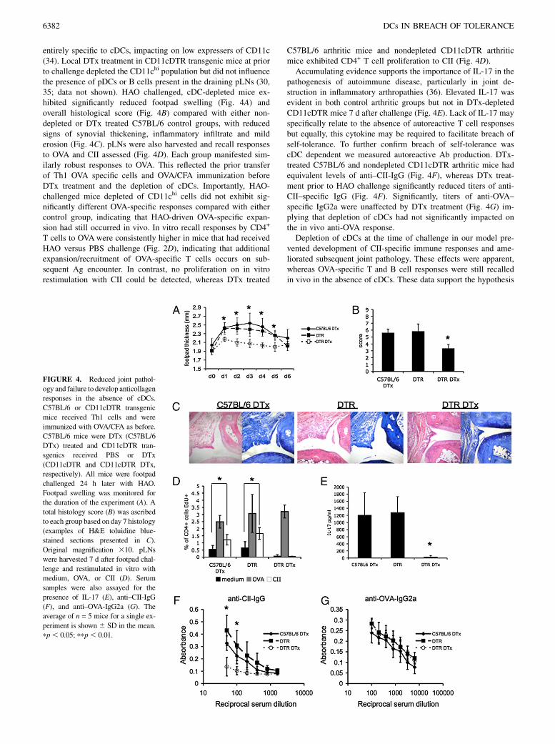
entirely specific to cDCs, impacting on low expressers of CD11c(34). Local DTx treatment in CD11cDTR transgenic mice at priorto challenge depleted the CD11chi population but did not influencethe presence of pDCs or B cells present in the draining pLNs (30,35; data not shown). HAO challenged, cDC-depleted mice ex-hibited significantly reduced footpad swelling (Fig. 4A) andoverall histological score (Fig. 4B) compared with either non-depleted or DTx treated C57BL/6 control groups, with reducedsigns of synovial thickening, inflammatory infiltrate and milderosion (Fig. 4C). pLNs were also harvested and recall responsesto OVA and CII assessed (Fig. 4D). Each group manifested sim-ilarly robust responses to OVA. This reflected the prior transferof Th1 OVA specific cells and OVA/CFA immunization beforeDTx treatment and the depletion of cDCs. Importantly, HAO-challenged mice depleted of CD11chi cells did not exhibit sig-nificantly different OVA-specific responses compared with eithercontrol group, indicating that HAO-driven OVA-specific expan-sion had still occurred in vivo. In vitro recall responses by CD4+
T cells to OVAwere consistently higher in mice that had receivedHAO versus PBS challenge (Fig. 2D), indicating that additionalexpansion/recruitment of OVA-specific T cells occurs on sub-sequent Ag encounter. In contrast, no proliferation on in vitrorestimulation with CII could be detected, whereas DTx treated
C57BL/6 arthritic mice and nondepleted CD11cDTR arthriticmice exhibited CD4+ T cell proliferation to CII (Fig. 4D).Accumulating evidence supports the importance of IL-17 in the
pathogenesis of autoimmune disease, particularly in joint de-struction in inflammatory arthropathies (36). Elevated IL-17 wasevident in both control arthritic groups but not in DTx-depletedCD11cDTR mice 7 d after challenge (Fig. 4E). Lack of IL-17 mayspecifically relate to the absence of autoreactive T cell responsesbut equally, this cytokine may be required to facilitate breach ofself-tolerance. To further confirm breach of self-tolerance wascDC dependent we measured autoreactive Ab production. DTx-treated C57BL/6 and nondepleted CD11cDTR arthritic mice hadequivalent levels of anti–CII-IgG (Fig. 4F), whereas DTx treat-ment prior to HAO challenge significantly reduced titers of anti-CII–specific IgG (Fig. 4F). Significantly, titers of anti-OVA–specific IgG2a were unaffected by DTx treatment (Fig. 4G) im-plying that depletion of cDCs had not significantly impacted onthe in vivo anti-OVA response.Depletion of cDCs at the time of challenge in our model pre-
vented development of CII-specific immune responses and ame-liorated subsequent joint pathology. These effects were apparent,whereas OVA-specific T and B cell responses were still recalledin vivo in the absence of cDCs. These data support the hypothesis
FIGURE 4. Reduced joint pathol-
ogy and failure to develop anticollagen
responses in the absence of cDCs.
C57BL/6 or CD11cDTR transgenic
mice received Th1 cells and were
immunized with OVA/CFA as before.
C57BL/6 mice were DTx (C57BL/6
DTx) treated and CD11cDTR tran-
sgenics received PBS or DTx
(CD11cDTR and CD11cDTR DTx,
respectively). All mice were footpad
challenged 24 h later with HAO.
Footpad swelling was monitored for
the duration of the experiment (A). A
total histology score (B) was ascribed
to each group based on day 7 histology
(examples of H&E toluidine blue-
stained sections presented in C).
Original magnification 310. pLNs
were harvested 7 d after footpad chal-
lenge and restimulated in vitro with
medium, OVA, or CII (D). Serum
samples were also assayed for the
presence of IL-17 (E), anti–CII-IgG
(F), and anti–OVA-IgG2a (G). The
average of n = 5 mice for a single ex-
periment is shown 6 SD in the mean.
pp, 0.05; ppp, 0.01.
6382 DCs IN BREACH OF TOLERANCE

that cDCs directly contribute to the induction of CII-specificresponses, independently of indirect effects of cDCs on the de-velopment of recall OVA responses.
TNF blockade inhibits elevated expression levels of MHC classII by cDCs and prevents breach of self-tolerance
Early and aggressive blockade of TNF-a in RA can result inclinical remission (37, 38) but there is a paucity of data pertainingto possible mechanisms of action. TNF-a blockade can preventthe maturation of circulating myeloid DCs in ankylosing spon-dylitis (39); patients exhibited decreased T cell reactivity and itwas concluded that this related directly to reduced activation byDCs. Indeed, reduced disease severity in RA after anti–TNF-atherapy correlates with decreased DC maturation (12). As we haveattributed maturation of cDCs in our model to the stimulation ofautoreactive responses we sought to assess whether reduced pa-thology and long-term clinical remission seen after TNF-ablockade in patients could reflect reduced cDC maturation andsubsequently, a failure to prime autoreactive T cell responses inour model.Mice were treated with the dimeric recombinant human TNF
receptor p80 Fc fusion protein (sTNFR-Fc) or control Fc matchedhuman Ig (hIgG). Treatment with sTNFR-Fc prevented cDCupregulation of MHC class II expression after HAO challenge (Fig.
5A). No significant differences in MHC class II expression weredetected between cDCs from nonarthritic mice treated with eithercontrol hIgG or sTNFR-Fc (Fig. 5A). The pDCs in the pLN ofarthritic mice expressed lower levels of MHC class II comparedwith nonarthritic animals, with no detectable influence of sTNFR-Fc treatment (Fig. 5B). Having demonstrated that cDCs area major contributor to Ag presentation, can mediate and are crit-ical for, the development of autoreactive responses in this model,we tested whether sTNFR-Fc mediated inhibition of elevated cDCMHC class II could also prevent development of collagen-specificT cell and Ab responses. LN cells were restimulated in vitro withOVA or CII. Only HAO-challenged, hIgG-treated mice showedCD4+ proliferative responses to CII (Fig. 5C, 5D), whereas noresponses were detected in the HAO challenged sTNFR-Fc treatedor PBS challenged groups. The OVA recall response was signifi-cantly increased in HAO challenged arthritic groups, with nodiscernable effect of treatment with sTNFR-Fc (Fig. 5C, 5D). ThesTNFR-Fc inhibited production of anticollagen-IgG with onlybackground levels, comparable with PBS footpad challengedmice, being detected (Fig. 5E). Treatment with sTNFR-Fc did notalter titers of anti–OVA-IgG, with all groups demonstratingcomparable levels of these Abs (data not shown). In vitro treat-ment of bone marrow DCs with sTNFR-Fc prior to addition ofEaGFP did not impact on Ag presentation (Supplemental Fig. 3A,
FIGURE 5. Anti–TNF-a therapy prevents cDC in-
creased MHC class II expression in draining LN,
avoiding T and B cell breach of tolerance to CII. Flow
cytometric analysis of MHC class II expression by
cDCs (A) and pDCs (B) in the pLN of both nonarthritic
and arthritic mice treated with either control hIgG iso
or the sTNFR-Fc fusion protein, Etan. pLN and serum
were harvested 7 d after footpad challenge. pLNs were
restimulated in vitro with medium, OVA, or CII. The
percentage of CD4+ T cells responding was determined
by flow cytometric analysis of EdU incorporation.
FACs plots representative of responses to CII and OVA
are shown (5 mice per group) (C, D). Anti-CII–specific
serum IgG was detected by ELISA (E). The mean 6the SD for n = 5 animals representative of two ex-
periments is shown. pp , 0.05 ppp , 0.01. Etan,
etanercept; iso, isotype;
The Journal of Immunology 6383
3B), as detected by YAe staining. As such the in vivo effectsobserved may relate TNF-dependent tissue maturation and mi-gration rather than reduced Ag presentation.Thus, reduction ofMHC class II expression by cDCs found in the
draining LN of arthritic mice treated with sTNFR-Fc was followedby a failure to mount both CII-specific T cell and Ab responses. Assuch, limiting the maturation of cDCs by TNF-a blockade andpreventing/limiting autoreactive T cell responses may represent oneof the mechanisms whereby early intervention with anti–TNF-abiologics can achieve long-term clinical remission.
DiscussionUnderstanding the factors triggering the activation of previouslyunresponsive, self-reactive T cells will be crucial in delineating thepathological response seen in RA and development of new ther-apeutic regimes. Central to this will be the identification of the APCpopulation initiating the breach of self-tolerance underlying notjust RA, but all autoimmune diseases. Using a recently developedmodel of experimental arthritis (17), we were able to delineate thecritical events surrounding the development of autoreactive T andB cell responses and the ensuing joint pathology. These studieshave demonstrated cDC activation and Ag presentation associatedwith breach of self-tolerance, an ability of these cells to transferautoimmunity, together with a central role for cDCs in the TNF-a–dependent development of self-reactivity and RA pathogenesis.After challenge with HAO in our model of arthritis, similarly
increased numbers of cDCs and pDCs were observed in thedraining LN. However, expression of MHC class II was elevated inthe cDC compartment compared with pDCs. Detection of thisactivated cDC population could have resulted from either stimu-lation of LN resident cells, influx of tissue activated cDCs or, mostlikely, a combination of both. We therefore used EaGFP and themAb YAe (23, 40) to identify in vivo the major APC populationsassociated with the development of autoimmunity and pathologyin RA. Coadministration of EaGFP and HAO identified cDCs asthe primary population presenting Ea peptide, particularly as thispopulation increased significantly in size in arthritic mice. AscDCs were the main cell presenting exogenous Ag, we hypothe-sized that they were the most likely candidates to be presentingtissue-derived Ag in our model.Previous studies demonstrated that adoptive transfer of arthri-
togenic T cells into naive recipients is sufficient to induce disease,suggesting constitutive presentation of articular Ags (29). Jointrelated Ag may drain directly via lymphatics but may also bepresented in the draining LN via the homeostatic migration ofDCs. This raises the possibility that the requirement for cDCs inpriming a CII response in our model may partly reflect a role inAg transport to the draining LN. One hypothesis would be that thestimulation of the existing OVA-specific memory response alterscDC characteristics sufficiently to allow reversal of their usualtolerogenic interaction with self-specific T cells and priming ofautoreactivity. Indeed, a model of RA in which self-Ag presentedby activated DCs stimulates autoreactive T cells that have escapedthymic selection has been proposed (25, 26). In support of thishypothesis, adoptive transfer of Ag-bearing DCs can incite auto-immunity underlying diabetes, experimental allergic encephalitis,and arthritis (16, 27, 28). In this study, we were able to induce CII-specific responses in the OVA TCR transgenic transfer model bychallenge with Ag-bearing DCs. Activation of OVA-specificT cells by DCs was a prerequisite for, and could support activationof, collagen-specific autoreactive T cells when endogenous auto-antigen was available, whereas addition of exogenous CII couldnot. This likely reflects key differences in the prior context inwhich CII reactive T cells have seen their specific Ag. In the
aforementioned study by Leung et al. (16), CII-pulsed DCs re-activated CII-specific T cells in mice previously immunized withCII. In our OVA TCR transgenic transfer model, CII-specificT cells would not have had previous exposure to CII in a proin-flammatory context. Activation of CII-specific T cells in thepresence of OVA driven inflammation could perhaps be likened tothe CII immunization regimen preceding CII-pulsed DC inductionof erosive arthritis previously reported (16).Thus, cDC are activated and present Ag at the earliest stages of
the breach of tolerance in experimental arthritis and their adoptivetransfer can trigger autoreactivity leading to joint pathology. Todefinitively demonstrate a central role for cDC in our model ofarthritis, we used CD11cDTR transgenic mice that have been usedto reveal the importance of these cells in a variety of other systems(22, 30–33). Depletion of these cells at the time of challenge inour model prevented development of CII-specific immune re-sponses and ameliorated subsequent joint pathology. These effectswere apparent, whereas this treatment had little effect on OVA-specific responses, indicating that OVA-specific expansion hadstill occurred in vivo. Our finding does suggest that memory re-sponses can be efficiently mounted in the absence of cDCs. In-deed, the presence of other APCs appeared sufficient to ensureactivation of the memory OVA response, potentially being drivenby an increased frequency of high-affinity B cells generated fromthe OVA/CFA immunization (41). It therefore seems unlikely thata reduced OVA response could account for the lack of autor-eactivity in CD11chi-depleted mice.TNF-a blockade prevented the increase of MHC class II ex-
pression by draining LN cDCs and development of autoreactiveresponses. TNF-a may be required for migration of mature cDCsto the draining LN, preventing the second influx of Ag-presentingDCs important in priming T cell response (23). Equally, matura-tion of LN resident cDCs may also be TNF-a dependent. Themost likely explanation will no doubt prove a combination ofboth, with failure of either scenario preventing activation ofpathogenic CII-specific T cells. The importance of TNF-a to DCmaturation in models of both type 1 diabetes (42) and allores-ponses (43) supports this hypothesis. Indeed, in these two studies,tolerance induction was observed after inhibition of TNF-a ac-tivity, a tantalizing prospect given the success of current anti–TNF-a biologics in RA.This study demonstrates for the first time that cDCs play a central
role in driving arthritogenic autoimmunity and no other APC issufficient for breach of self-tolerance arising via endogenous path-ways initiated by an irrelevant nonarticular Ag. Accumulatingphenotypic studies have identifiedandcharacterizedDCpopulationsin RA and highlighted their potential for contribution to disease inRA. This study directly demonstrates that cDCs mature and presentAg in the context of murine inflammatory arthritis and that de-velopment of autoreactivity in this model is crucially dependent ontheir presence. These studies clarify the processes by which im-munological regulation can be overcome by responses to infectiousagents that have been implicated in the pathogenesis of RA (44–47).
DisclosuresThe authors have no financial conflicts of interest.
References1. Firestein, G. S. 2003. Evolving concepts of rheumatoid arthritis. Nature 423:
356–361.2. Hata, T., and A. Kavanaugh. 2006. Rheumatoid arthritis in dermatology. Clin.
Dermatol. 24: 430–437.3. Nannini, C., J. H. Ryu, and E. L. Matteson. 2008. Lung disease in rheumatoid
arthritis. Curr. Opin. Rheumatol. 20: 340–346.
6384 DCs IN BREACH OF TOLERANCE

4. Libby, P. 2008. Role of inflammation in atherosclerosis associated with rheu-matoid arthritis. Am. J. Med. 121(10, Suppl 1)S21–S31.
5. Michaud, K., and F. Wolfe. 2007. Comorbidities in rheumatoid arthritis. BestPract. Res. Clin. Rheumatol. 21: 885–906.
6. Feldmann, M. 2002. Development of anti-TNF therapy for rheumatoid arthritis.Nat. Rev. Immunol. 2: 364–371.
7. Brand, D. D., A. H. Kang, and E. F. Rosloniec. 2004. The mouse model ofcollagen-induced arthritis. Methods Mol. Med. 102: 295–312.
8. Wellcome Trust Case Control Consortium. 2007. Genome-wide associationstudy of 14,000 cases of seven common diseases and 3,000 shared controls.Nature 447: 661–678.
9. Hommel, M. 2004. On the dynamics of T-cell activation in lymph nodes. Im-munol. Cell Biol. 82: 62–66.
10. Jongbloed, S. L., M. C. Lebre, A. R. Fraser, J. A. Gracie, R. D. Sturrock,P. P. Tak, and I. B. McInnes. 2006. Enumeration and phenotypical analysis ofdistinct dendritic cell subsets in psoriatic arthritis and rheumatoid arthritis. Ar-thritis Res. Ther. 8: R15.
11. Lebre, M. C., S. L. Jongbloed, S. W. Tas, T. J. Smeets, I. B. McInnes, andP. P. Tak. 2008. Rheumatoid arthritis synovium contains two subsets of CD83-DC-LAMP- dendritic cells with distinct cytokine profiles. Am. J. Pathol. 172:940–950.
12. Balanescu, A., E. Radu, R. Nat, T. Regalia, V. Bojinca, R. Ionescu, S. Balanescu,C. Savu, and D. Predeteanu. 2005. Early and late effect of infliximab on cir-culating dendritic cells phenotype in rheumatoid arthritis patients. Int. J. Clin.Pharmacol. Res. 25: 9–18.
13. van Duivenvoorde, L. M., W. G. Han, A. M. Bakker, P. Louis-Plence,L. M. Charbonnier, F. Apparailly, E. I. van der Voort, C. Jorgensen,T. W. Huizinga, and R. E. Toes. 2007. Immunomodulatory dendritic cells inhibitTh1 responses and arthritis via different mechanisms. J. Immunol. 179: 1506–1515.
14. van Duivenvoorde, L. M., P. Louis-Plence, F. Apparailly, E. I. van der Voort,T. W. Huizinga, C. Jorgensen, and R. E. Toes. 2004. Antigen-specific im-munomodulation of collagen-induced arthritis with tumor necrosis factor-stimulateddendritic cells. Arthritis Rheum. 50: 3354–3364.
15. Jaen, O., S. Rulle, N. Bessis, A. Zago, M. C. Boissier, and G. Falgarone. 2008.Dendritic cells modulated by innate immunity improve collagen-induced ar-thritis and induce regulatory T cells in vivo. Immunology 126: 35–44.
16. Leung, B. P., M. Conacher, D. Hunter, I. B. McInnes, F. Y. Liew, andJ. M. Brewer. 2002. A novel dendritic cell-induced model of erosive in-flammatory arthritis: distinct roles for dendritic cells in T cell activation andinduction of local inflammation. J. Immunol. 169: 7071–7077.
17. Maffia, P., J. M. Brewer, J. A. Gracie, A. Ianaro, B. P. Leung, P. J. Mitchell,K. M. Smith, I. B. McInnes, and P. Garside. 2004. Inducing experimental arthritisand breaking self-tolerance to joint-specific antigens with trackable, ovalbumin-specific T cells. J. Immunol. 173: 151–156.
18. Nickdel, M. B., P. Conigliaro, G. Valesini, S. Hutchison, R. Benson, R. V.Bundick, A. J. Leishman, I. B. McInnes, J. M. Brewer, and P. Garside. 2008.Dissecting the contribution of innate and antigen-specific pathways to the breachof self-tolerance observed in a murine model of arthritis. Ann. Rheum. Dis. 68:1059–1066.
19. Jongbloed, S. L., R. A. Benson, M. B. Nickdel, P. Garside, I. B. McInnes, andJ. M. Brewer. 2009. Plasmacytoid dendritic cells regulate breach of self-tolerancein autoimmune arthritis. J. Immunol. 182: 963–968.
20. Trentham, D. E., A. S. Townes, and A. H. Kang. 1977. Autoimmunity to type IIcollagen an experimental model of arthritis. J. Exp. Med. 146: 857–868.
21. Wooley, P. H., H. S. Luthra, J. M. Stuart, and C. S. David. 1981. Type II collagen-induced arthritis in mice. I. Major histocompatibility complex (I region) linkageand antibody correlates. J. Exp. Med. 154: 688–700.
22. Jung, S., D. Unutmaz, P. Wong, G. Sano, K. De los Santos, T. Sparwasser, S. Wu,S. Vuthoori, K. Ko, F. Zavala, et al. 2002. In vivo depletion of CD11c(+) den-dritic cells abrogates priming of CD8(+) T cells by exogenous cell-associatedantigens. Immunity 17: 211–220.
23. Itano, A. A., S. J. McSorley, R. L. Reinhardt, B. D. Ehst, E. Ingulli,A. Y. Rudensky, and M. K. Jenkins. 2003. Distinct dendritic cell populationssequentially present antigen to CD4 T cells and stimulate different aspects ofcell-mediated immunity. Immunity 19: 47–57.
24. Hutchison, S., B. S. Choo-Kang, R. V. Bundick, A. J. Leishman, J. M. Brewer,I. B. McInnes, and P. Garside. 2008. Tumour necrosis factor-alpha blockade sup-presses murine allergic airways inflammation. Clin. Exp. Immunol. 151: 114–122.
25. Thomas, R., and P. E. Lipsky. 1996. Could endogenous self-peptides presentedby dendritic cells initiate rheumatoid arthritis? Immunol. Today 17: 559–564.
26. Thomas, R., and P. E. Lipsky. 1996. Presentation of self peptides by dendriticcells: possible implications for the pathogenesis of rheumatoid arthritis. ArthritisRheum. 39: 183–190.
27. Ludewig, B., B. Odermatt, S. Landmann, H. Hengartner, and R. M. Zinkernagel.1998. Dendritic cells induce autoimmune diabetes and maintain disease via denovo formation of local lymphoid tissue. J. Exp. Med. 188: 1493–1501.
28. Dittel, B. N., I. Visintin, R. M. Merchant, and C. A. Janeway, Jr. 1999. Pre-sentation of the self antigen myelin basic protein by dendritic cells leads toexperimental autoimmune encephalomyelitis. J. Immunol. 163: 32–39.
29. Hata, H., N. Sakaguchi, H. Yoshitomi, Y. Iwakura, K. Sekikawa, Y. Azuma,C. Kanai, E. Moriizumi, T. Nomura, T. Nakamura, and S. Sakaguchi. 2004.Distinct contribution of IL-6, TNF-alpha, IL-1, and IL-10 to T cell-mediatedspontaneous autoimmune arthritis in mice. J. Clin. Invest. 114: 582–588.
30. GeurtsvanKessel, C. H., M. A. Willart, L. S. van Rijt, F. Muskens, M. Kool,C. Baas, K. Thielemans, C. Bennett, B. E. Clausen, H. C. Hoogsteden, et al.2008. Clearance of influenza virus from the lung depends on migratory langerin+CD11b- but not plasmacytoid dendritic cells. J. Exp. Med. 205: 1621–1634.
31. Loof, T. G., M. Rohde, G. S. Chhatwal, S. Jung, and E. Medina. 2007. Thecontribution of dendritic cells to host defenses against Streptococcus pyogenes.J. Infect. Dis. 196: 1794–1803.
32. Cordier-Dirikoc, S., and J. Chabry. 2008. Temporary depletion of CD11c+dendritic cells delays lymphoinvasion after intraperitonal scrapie infection. J.Virol. 82: 8933–8936.
33. Berndt, B. E., M. Zhang, G. H. Chen, G. B. Huffnagle, and J. Y. Kao. 2007. Therole of dendritic cells in the development of acute dextran sulfate sodium colitis.J. Immunol. 179: 6255–6262.
34. Probst, H. C., K. Tschannen, B. Odermatt, R. Schwendener, R. M. Zinkernagel,and M. Van Den Broek. 2005. Histological analysis of CD11c-DTR/GFP miceafter in vivo depletion of dendritic cells. Clin. Exp. Immunol. 141: 398–404.
35. deWalick, S., F. H. Amante, K. A. McSweeney, L. M. Randall, A. C. Stanley,A. Haque, R. D. Kuns, K. P. MacDonald, G. R. Hill, and C. R. Engwerda. 2007.Cutting edge: conventional dendritic cells are the critical APC required for theinduction of experimental cerebral malaria. J. Immunol. 178: 6033–6037.
36. Lubberts, E., M. I. Koenders, and W. B. van den Berg. 2005. The role of T-cellinterleukin-17 in conducting destructive arthritis: lessons from animal models.Arthritis Res. Ther. 7: 29–37.
37. van der Kooij, S. M., S. le Cessie, Y. P. Goekoop-Ruiterman, J. K. De Vries-Bouwstra, D. van Zeben, P. J. Kerstens, J. M. Hazes, D. van Schaardenburg, F. C.Breedveld, B. A. Dijkmans, and C. F. Allaart. 2008. Clinical and radiologicalefficacy of initial versus delayed treatment with infliximab plus methotrexate inpatients with early rheumatoid arthritis. Ann. Rheum. Dis. 68: 1153–1158.
38. Goekoop-Ruiterman, Y. P., J. K. de Vries-Bouwstra, C. F. Allaart, D. van Zeben,P. J. Kerstens, J. M. Hazes, A. H. Zwinderman, H. K. Ronday, K. H. Han,M. L. Westedt, et al. 2008. Clinical and radiographic outcomes of four differenttreatment strategies in patients with early rheumatoid arthritis (the BeSt study):A randomized, controlled trial. Arthritis Rheum. 58(2, Suppl): S126–S135.
39. Pang, L., L. Wang, T. Suo, H. Hao, X. Fang, J. Jia, F. Huang, and J. Tang. 2008.Tumor necrosis factor-alpha blockade leads to decreased peripheral T cell re-activity and increased dendritic cell number in peripheral blood of patients withankylosing spondylitis. J. Rheumatol. 35: 2220–2228.
40. Millington, O. R., V. B. Gibson, C. M. Rush, B. H. Zinselmeyer, R. S. Phillips,P. Garside, and J. M. Brewer. 2007. Malaria impairs T cell clustering and im-mune priming despite normal signal 1 from dendritic cells. PLoS Pathog. 3:1380–1387.
41. Anderson, S. M., M. M. Tomayko, and M. J. Shlomchik. 2006. Intrinsic prop-erties of human and murine memory B cells. Immunol. Rev. 211: 280–294.
42. Lee, L. F., B. Xu, S. A. Michie, G. F. Beilhack, T. Warganich, S. Turley, andH. O. McDevitt. 2005. The role of TNF-alpha in the pathogenesis of type 1diabetes in the nonobese diabetic mouse: analysis of dendritic cell maturation.Proc. Natl. Acad. Sci. USA 102: 15995–16000.
43. Wang, Q., Y. Liu, J. Wang, G. Ding, W. Zhang, G. Chen, M. Zhang, S. Zheng,and X. Cao. 2006. Induction of allospecific tolerance by immature dendritic cellsgenetically modified to express soluble TNF receptor. J. Immunol. 177: 2175–2185.
44. Toussirot, E., and J. Roudier. 2007. Pathophysiological links between rheuma-toid arthritis and the Epstein-Barr virus: an update. Joint Bone Spine 74: 418–426.
45. Balandraud, N., J. Roudier, and C. Roudier. 2004. Epstein-Barr virus andrheumatoid arthritis. Autoimmun. Rev. 3: 362–367.
46. Caliskan, R., S. Masatlioglu, M. Aslan, S. Altun, S. Saribas, S. Ergin, E. Uckan,V. Koksal, V. Oz, K. Altas, et al. 2005. The relationship between arthritis andhuman parvovirus B19 infection. Rheumatol. Int. 26: 7–11.
47. Jorgensen, K. T., A. Wiik, M. Pedersen, C. J. Hedegaard, B. F. Vestergaard,R. Gislefoss, T. K. Kvien, J. Wohlfahrt, K. Bendtzen, and M. Frisch. 2007.Cytokines, autoantibodies, and viral antibodies in premorbid and postdiagnosticsera from patients with rheumatoid arthritis - Case-control study nested ina cohort of Norwegian blood donors. Ann. Rheum. Dis. 67: 860–866.
The Journal of Immunology 6385