ATPase b2-subunit (AMOG) expression abrogates invasion of ...
Upload
khangminh22Category
view
0download
0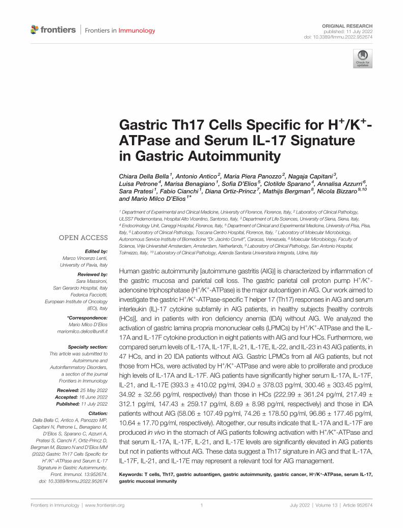
Frontiers in Immunology | www.frontiersin.
Edited by:Marco Vincenzo Lenti,
University of Pavia, Italy
Reviewed by:Sara Massironi,
San Gerardo Hospital, ItalyFederica Facciotti,
European Institute of Oncology(IEO), Italy
*Correspondence:Mario Milco D’Elios
Specialty section:This article was submitted to
Autoimmune andAutoinflammatory Disorders,
a section of the journalFrontiers in Immunology
Received: 25 May 2022Accepted: 16 June 2022Published: 11 July 2022
Citation:Della Bella C, Antico A, Panozzo MP,Capitani N, Petrone L, Benagiano M,
D’Elios S, Sparano C, Azzurri A,Pratesi S, Cianchi F, Ortiz-Princz D,
BergmanM, Bizzaro N and D’Elios MM(2022) Gastric Th17 Cells Specific for
H+/K+-ATPase and Serum IL-17Signature in Gastric Autoimmunity.
Front. Immunol. 13:952674.doi: 10.3389/fimmu.2022.952674
ORIGINAL RESEARCHpublished: 11 July 2022
doi: 10.3389/fimmu.2022.952674
Gastric Th17 Cells Specific for H+/K+-ATPase and Serum IL-17 Signaturein Gastric AutoimmunityChiara Della Bella1, Antonio Antico2, Maria Piera Panozzo2, Nagaja Capitani3,Luisa Petrone4, Marisa Benagiano1, Sofia D’Elios5, Clotilde Sparano4, Annalisa Azzurri 6,Sara Pratesi 1, Fabio Cianchi1, Diana Ortiz-Princz7, Mathijs Bergman8, Nicola Bizzaro9,10
and Mario Milco D’Elios1*
1 Department of Experimental and Clinical Medicine, University of Florence, Florence, Italy, 2 Laboratory of Clinical Pathology,ULSS7 Pedemontana, Hospital Alto Vicentino, Santorso, Italy, 3 Department of Life Sciences, University of Siena, Siena, Italy,4 Endocrinology Unit, Careggi Hospital, Florence, Italy, 5 Department of Clinical and Experimental Medicine, University of Pisa, Pisa,Italy, 6 Laboratory of Clinical Pathology, Toscana Centro Hospital, Florence, Italy, 7 Laboratory of Molecular Microbiology,Autonomous Service Institute of Biomedicine “Dr. Jacinto Convit”, Caracas, Venezuela, 8 Molecular Microbiology, Faculty ofScience, Vrije Universiteit Amsterdam, Amsterdam, Netherlands, 9 Laboratory of Clinical Pathology, San Antonio Hospital,Tolmezzo, Italy, 10 Laboratory of Clinical Pathology, Azienda Sanitaria Universitaria Integrata, Udine, Italy
Human gastric autoimmunity [autoimmune gastritis (AIG)] is characterized by inflammation ofthe gastric mucosa and parietal cell loss. The gastric parietal cell proton pump H+/K+-adenosine triphosphatase (H+/K+-ATPase) is themajor autoantigen in AIG. Our work aimed toinvestigate the gastric H+/K+-ATPase-specific T helper 17 (Th17) responses in AIG and seruminterleukin (IL)-17 cytokine subfamily in AIG patients, in healthy subjects [healthy controls(HCs)], and in patients with iron deficiency anemia (IDA) without AIG. We analyzed theactivation of gastric lamina propria mononuclear cells (LPMCs) by H+/K+-ATPase and the IL-17A and IL-17F cytokine production in eight patients with AIG and four HCs. Furthermore, wecompared serum levels of IL-17A, IL-17F, IL-21, IL-17E, IL-22, and IL-23 in 43 AIG patients, in47 HCs, and in 20 IDA patients without AIG. Gastric LPMCs from all AIG patients, but notthose from HCs, were activated by H+/K+-ATPase and were able to proliferate and producehigh levels of IL-17A and IL-17F. AIG patients have significantly higher serum IL-17A, IL-17F,IL-21, and IL-17E (393.3 ± 410.02 pg/ml, 394.0 ± 378.03 pg/ml, 300.46 ± 303.45 pg/ml,34.92 ± 32.56 pg/ml, respectively) than those in HCs (222.99 ± 361.24 pg/ml, 217.49 ±312.1 pg/ml, 147.43 ± 259.17 pg/ml, 8.69 ± 8.98 pg/ml, respectively) and those in IDApatients without AIG (58.06 ± 107.49 pg/ml, 74.26 ± 178.50 pg/ml, 96.86 ± 177.46 pg/ml,10.64 ± 17.70 pg/ml, respectively). Altogether, our results indicate that IL-17A and IL-17F areproduced in vivo in the stomach of AIG patients following activation with H+/K+-ATPase andthat serum IL-17A, IL-17F, IL-21, and IL-17E levels are significantly elevated in AIG patientsbut not in patients without AIG. These data suggest a Th17 signature in AIG and that IL-17A,IL-17F, IL-21, and IL-17E may represent a relevant tool for AIG management.
Keywords: T cells, Th17, gastric autoantigen, gastric autoimmunity, gastric cancer, H+/K+-ATPase, serum IL-17,gastric mucosal immunity
org July 2022 | Volume 13 | Article 9526741

Della Bella et al. Th17 in Autoimmune Gastritis
INTRODUCTION
Autoimmune gastritis (AIG) is characterized by gastric corpusinflammation that does not usually result in overt disease untilmucosal atrophy development and cobalamin malabsorption (1).The detection of serum anti–parietal cell autoantibodies (PCAs)without any symptoms suggests subclinical AIG (2–4). AIG is apreneoplastic condition, predisposing to the development ofboth gastric adenocarcinoma and type 1 neuroendocrine tumor(1, 5). The proton pump of parietal cell H+/K+-adenosinetriphosphatase (ATPase) is the key autoantigen recognized inboth AIG and experimental autoimmune gastritis (EAIG) (6–10). EAIG can be induced in non-thymectomized animals byimmunization with either purified gastric H+/K+-ATPase orgastric mucosal extracts or by neonatal thymectomy (7–10).Inflammation of the gastric mucosa, acid-secreting parietal celland zymogenic cell loss, and circulating anti-gastric H+/K+-ATPase autoantibodies are hallmarks of EAIG. In both AIGand EAIG, CD4+, CD8+ T cells, macrophages, and B cells are thekey cells present in the gastric infiltrate (1, 2, 11).
T helper 1 (Th1) cells, secreting interferon (IFN)-g, play a centralrole in AIG (12). However, emerging evidence indicates that thepreferential development of Th17 mucosal responses occur in AIGalso. Th17 cells, secreting IL-17 and IL-21, drive Th17 inflammationand have been implicated in the induction of many gastricinflammatory responses, including gastric cancer (13, 14).However, there are no clues on the pathogenic process mediatedby H+/K+-ATPase-specific Th17 lymphocytes in human AIG.
To this aim, we investigated the in vivo production of IL-17A,IL-17F by gastric lamina propria mononuclear cells (LPMCs)obtained from AIG patients. As we wondered whether IL-17A,IL-17F, IL-21, IL-17E, IL-22, and IL-23 levels could be abnormalin patients with AIG, we further measured these cytokines in theserum of AIG patients, iron deficiency anemia (IDA) patientswithout AIG, and healthy controls (HCs).
MATERIALS AND METHODS
PatientsUpon approval of the local ethical committee (Ethics statementn. 14936/CAM_BIO), we investigated the Th17 immuneresponses both at gastric and at serum level in patients withAIG (diagnosed by histology). AIG diagnosis was madeaccording to the updated Sydney–Houston criteria, evaluatingat least five random gastric biopsies with hematoxylin and eosinand Giemsa stains: two in the antrum, two in the body, and onein the angulus (15). In eight patients with AIG (six women andtwo men; mean age 52 years, range 37–64 years) and four healthysubjects (HCs, two women and two men; mean age 46 years,range: 42–48 years), following informed consent, biopsyspecimens were obtained from the gastric mucosa in order tostudy LPMCs. These 8 AIG patients had both anti-intrinisicfactor (IFA) and anti–parietal cell autoantibodies.
In these eight AIG patients and in another 102 patients orcontrols, we studied serum levels of IL-17A, IL-17F, IL-21, IL-
Frontiers in Immunology | www.frontiersin.org 2
17E, IL-22, and IL-23. Of the 110 enrolled subjects, 43 sufferedfrom AIG (diagnosed by histology) (15), 20 (disease control)suffered from IDA and had no AIG (as defined by histology), and47 were HCs. AIG patients were 27 (63%) women and 16 (37%)men, mean age 69.5 ± 11.2 years. The IDA patient group wascomposed of 17 (85%) women and 3 (15%) men, mean age 69.3 ±22.7 years. Healthy subjects were 21 (45%) women and 26 (55%)men with a mean age of 45.0 ± 14.1 years.
Eight out of 43 AIG patients suffered also from autoimmunethyroiditis. No other autoimmune comorbidities were present inAIG, IDA, and HC subjects. None of the patients suffered frompeptic ulcer, gastric cancer, gastric lymphoma nor used protonpump inhibitors in the previous 6 months. Active H. pyloriinfection, as ruled out by histopathology, was not present in anypatient or control.
All AIG and IDA patients and HCs were investigated byserology (Helori CTX, Eurospital, Trieste, Italy), and three AIGpatients were seropositive for H. pylori. All AIG patients hadanti-gastric parietal cell autoantibodies detectable by indirectimmunofluorescence assay on rodent tissue and/or serumintrinsic factor autoantibodies (EliA, Thermo Fisher, Uppsala,Sweden) (11, 16, 17). Sixteen out of 43 AIG patients had bothanti-intrinisic factor and anti–parietal cell autoantibodies, 12AIG patients had serum anti–parietal cell autoantibodies, and 15AIG patients had serum autoantibodies against intrinsic factor.
Proliferative Response to H+/K+-ATPaseby Gastric Lamina PropriaMononuclear cellsGastric specimens from eight AIG patients (having no otherautoimmune disease) and four HCs were used as source ofLPMCs. Specifica l ly , LPMCs were iso la ted by thedithiothreitol- ethylenediaminetetraacetic acid (DTT-EDTA)-collagenase sequence (18, 19). To investigate the proliferativeresponse to gastric H+/K+-ATPase, purified as reported (12),LPMCs were labeled with carboxyfluorescein succinimidyl ester(CFSE) (CellTrace CFSE dye, Invitrogen, USA) followingmanufacturer’s instructions. After CFSE staining, 2 × 10⁵
LPMCs were incubated at 37°C under 5% CO2 with or withoutgastric H+/K+-ATPase (0.3 µg/ml). Flow cytometry on BD FACSCanto II was performed using the FACSDiva software (BectonDickinson, Franklin Lakes, NJ, USA) after 5 days of cell cultureto investigate LPMC proliferation in response to H+/K+-ATPase.
IL-17 Production by GastricMucosa T CellsThe IL-17 production by gastric T cells of AIG patients wasinvestigated by both ELISpot and Fluorescence activated cellsorting (FACS) analysis. T cells of each gastric LPMC werestimulated with H+/K+-ATPase (0.3 µg/ml) or Purified ProteinDerivative (PPD) (10 mg/ml) for 48 h in ELISpot microplatescoated with anti–IL-17A antibody (eBioscience). At the end of theculture period, the number of IL-17 Spot Forming Cells (SFCs) wascounted as described (13). For FACS analysis, we induced cytokineproduction by gastric isolated LPMCs, 2 × 10⁵ cells by stimulationfor 6 h in 37°C with 5% CO2 with the Leukocyte Activation
July 2022 | Volume 13 | Article 952674

Della Bella et al. Th17 in Autoimmune Gastritis
Cocktail-BD GolgiPlug™ (BD Biosciences, San Jose, CA, USA)containing phorbol 12-myristate 13-acetate (PMA), ionomycin, andbrefeldin A in complete medium 5% human serum, as described(20). A basal production for each tested cytokine was determined innon-stimulated cells, treated with the Golgi block alone. T helpercell surface marker was detected by anti-human-CD4 pacific blueconjugated (e-Bioscience, Thermo Fisher Scientific, Waltham, MA,USA), and, after fixation and permeabilization with BD Cytofix/cytoperm fixation/permeabilization kit (BD Biosciences, San Jose,CA, USA), cells were stained with anti-IL-17A FITC, anti-IL-17FPerCP, following manufacturer’s instructions. Flow cytometricanalysis was carried out on BD FACS Canto II using theFACSDiva software (Becton Dickinson, Franklin Lakes, NJ, USA).
Luminex Assay for IL-17A, IL-17F, IL-21,IL-17E, IL-22, and IL-23All sera were investigated for IL-17A, IL-17F, IL-21, IL-17E, IL-22, and IL-23 by Bio-Plex Pro™ (Bio-Rad, Hercules, CA, USA).We used Bio-Plex Manager™ software for determining thecytokine concentration. Detection range of IL-17A is 1.20–19,682.00 pg/ml; for IL-17F, it is 3.04–18,668.00 pg/ml; for IL-21, it is 8.97–147,023.00 pg/ml; for IL-17E, it is 1.00–16,375.00pg/ml; for IL-22, it is 3.88–11,917.00 pg/ml; and for IL-23, it is7.35–120,389.00 pg/ml.
Statistical AnalysesThe sample size required for sensitivity and specificity of theLuminex test was set in relation to the number of AIG patientsadmitted to the Florence reference center for AIG (21).
To calculate qualitative data, we applied descriptive statistics,as well as for standard and mean deviation. To assess thenormality of each independent distribution, we used theShapiro–Wilk test, and then the comparison was made byMann–Whitney U test.
A p < 0.05 was considered statistically significant.Values of cytokines below the lower limit of quantification
(LLOQ) were replaced with one-half the respective LLOQ (22);no patient sample had values above the upper limit ofquantification (ULOQ).
IL-17A, IL-17F, IL-21, and IL-17E Luminex assay accuracy, interms of sensitivity and specificity, was performed by receiveroperating characteristic (ROC) curve analysis. To evaluate thebest cutoff, the area under the curve (AUC) and the Youden’sindex (= Sensitivity ± [1 - Specificity]) were measured (23).
Statistical analyses were performed using IBM®SPSS Statisticversion 27.0.
RESULTS
Gastric T Helper Cells From AutoimmuneGastritis Patients Were Activated by H+/K+-ATPase and Were Able to Proliferateand Produce IL-17A and IL-17FLPMCs isolated from gastric biopsies of eight AIG patients andfrom four HCs were tested for their proliferative response to H+/
Frontiers in Immunology | www.frontiersin.org 3
K+-ATPase after 5 days of incubation with or without thepresence of the stimulus in the medium. FACS analysis wasperformed, and T helper cells were identified as CD4+ cells andevaluated for their level offluorescence to establish the number ofgenerations through which a cell has progressed since the CFSElabel was applied. The percentage of gastric T helper cellproliferation is summarized in Table 1 and Figure 1.
The IL-17 production by gastric T cells of AIG patients wasinvestigated by both ELISpot and FACS analysis. Gastricmucosa-derived T cells from each AIG patient or HC werestimulated with H+/K+-ATPase or PPD for 48 h in ELISpotmicroplates coated with anti–IL-17A antibody. At the end of theculture period, the number of IL-17 SFCs was counted(Figure 1). After specific stimulation with H+/K+-ATPase, asignificant proportion of T helper cells derived from the gastricmucosa of AIG patients produced IL-17A, whereas T cells fromHCs did not.
For FACS analysis, gastric T cells were stimulated for 6 h todetect IL-17A and IL-17F production. Cells were stained formembrane and intracellular markers, and the percentage ofCD4+ T lymphocytes producing IL-17A and/or IL-17F wasevaluated by FACS analysis. The results are indicated inTable 2 and Figure 1.
IL-17A, IL-17F, IL-21, and IL-17E areElevated in the Sera of AutoimmuneGastritis PatientsLevels of serum IL-17A, IL-17F, IL-21, IL-17E, IL-22, and IL-23were measured by Luminex assay in 43 AIG patients, 47 HCs,and 20 IDA without AIG. Multiplexed measurement of the Th17family revealed that IL-17A, IL-17F, IL-21, and IL-17E levelsincrease in AIG patients compared to those in IDA patients andHCs. The total Luminex assay results are detailed in Table 3and Figure 2.
The ROC curve analysis was used to assess the performanceof the Th17 family cytokine Luminex assay in discriminatingbetween healthy and AIG subjects. The ROC curves for IL-17A,
TABLE 1 | Percentage of T helper cell proliferation.
ID % of CD4+ proliferating cells
Medium alone Medium with H+/K+-ATPase
AIG-A 10.4 60.6AIG-B 8.2 49.2AIG-C 15.6 76.8AIG-D 12.8 75.4AIG-E 8.4 54.9AIG-F 9.2 68.3AIG-G 10.3 53.6AIG-H 11.9 74.8HC-I 12.7 13.4HC-L 13.2 11.7HC-M 9.5 10.8HC-N 7.8 8.4
July 2022
For each enrolled subject, 5,000 events were acquired and the CFSE signal was evaluatedon the gated T CD4+ population.CFSE,carboxyfluorescein diacetate succinimidyl ester.
| Volume 13 | Article 952674

Della Bella et al. Th17 in Autoimmune Gastritis
IL-17F, IL-21, and IL-17E are depicted in Figure 3. The testaccuracy for each evaluated cytokine is detailed in Table 4.
DISCUSSION
AIG is characterized by corpus inflammation that may lead togastric atrophy and gastric cancer. Themajor gastric autoantigen isH+/K+-ATPase, and gastric T-cell recognition of H+/K+-ATPaseresults in secretion of Th1 cytokines and activation of perforin- andFasLigand-mediated cytotoxic killing of parietal cells, leading togastric atrophy, a preneoplastic lesion of gastric cancer (12, 24, 25).Several independent reports demonstrated the relevance of Th17 inEAIG (26–28). It was recently reported that intrinsic factor-specificT lymphocytesproducehuge amounts of IL-17 inpatientswithAIGand pernicious anemia (PA) (11). However, it is still not clearwhether the H+/K+-ATPase autoantigen might be able to driveTh17 responses in AIG.
In the current study, we found that CD4+ gastric LPMCsobtained from AIG patients, but not from HCs, were activated byH+/K+-ATPase and were able to proliferate and produce highlevels of IL-17A and IL-17F. These findings show that not onlyTh1 cells but also Th17 cells specific for gastric H+/K+-ATPasedrive inflammation in gastric autoimmunity (12).
To investigate the IL-17 cytokine family serum levels, weanalyzed the serum levels of IL-17A, IL-17F, IL-21, IL-17E, IL-22, and IL-23 in patients with or without AIG. We foundsignificantly higher levels of IL-17A, IL-17F, IL-21, and IL-17E inthe sera of AIG patients compared to HC or IDA ones. The resultsobtained at both gastric and serum levels suggest that IL-17A, IL-17F, IL-21, and IL-17E are relevant cytokines for the
Frontiers in Immunology | www.frontiersin.org 4
immunopathogenesis of AIG. AIG in its late stage ischaracterized by intestinal metaplasia, gastric corpus and fundusdysplasia, and chromaffin cell hyperplasia that are consideredprecursor lesions of gastric cancer, suggesting an important linkbetween AIG and gastric cancer (29). Ample scientific evidenceindicates that gastric Th17 responses were associated with gastricadenocarcinoma and promoted gastric oncogenesis (13, 14, 30–35).In conclusion, we propose that long-lasting Th17 responses mayprecede the onset of gastric cancer and suggest that measurementof IL-17 family cytokines might be useful not only for themanagement of AIG but also for predicting the development ofgastric cancer in AIG patients with gastric atrophy.
TABLE 2 | IL-17A and IL-17F production by LPMCs of 8 AIG patients and 4 HCs.
ID % of CD4+ producing
IL-17A IL-17F IL-17A and IL-17F
AIG-A 8.7 4.5 13.6AIG-B 8.2 5.4 12.8AIG-C 11.4 5.2 15.5AIG-D 7.3 5.6 14.8AIG-E 9.6 7.5 16.3AIG-F 8.0 4.6 11.4AIG-G 7.2 5.3 14.5AIG-H 9.3 4.8 13.6HC-I 2.2 1.4 1.5HC-L 1.8 0.8 0.0HC-M 2.5 1.2 0.6HC-N 1.4 0.5 0.0
July 2022 | Volume
For each sample, 5,000 events were acquired.The percentage of CD4+ T cells stimulated for IL-17A and/or IL-17F production wasdetermined by FACS analysis of intracellular cytokine antibody staining.LPMCs, Lamina propria mononuclear cells; AIG, Autoimmune Gastritis; H, Healthy Controls.
A BD
E FG
I
H
C
FIGURE 1 | Gastric T helper cells from the lamina propria were activated by H+/K+-ATPase and were able to proliferate and produce IL-17. Proliferative response toH+/K+-ATPase, as tested by CFSE flow cytometric analysis of two representative AIG patients (A, B) and two healthy controls (E, F). To assess CD4+ T-cellproliferation to H+/K+-ATPase stimulus, 5,000 events were acquired and the CFSE signal was determined on the CD4+ gate. Each histogram shows the merge ofuntreated (black line) and stimulated (gray histogram) LPMC culture condition. Intracellular cytokine analysis of IL-17A and IL-17F by gastric CD4+ cells of tworepresentative AIG patients (C, D) and two healthy controls (G, H). The 5,000 events were acquired, and IL-17A-FITC and IL-17F-PerCP signals were determinedafter gating for CD4+. Gastric mucosa T cells from each AIG patient (black histogram) or healthy control (HC) (white histogram) were stimulated with H+/K+-ATPaseor PPD for 48 h in ELISpot microplates coated with anti–IL-17A antibody. At the end of the culture period, the number of IL-17A SFCs was counted (I). CFSE,carboxyfluorescein diacetate succinimidyl ester; AIG, Autoimmune Gastritis; HC, Healthy Controls; SFCs, Spot Forming Cells; PPD, Purified Protein Derivative;LPMCs, Lamina propria mononuclear cells; PerCP, Peridinin-Chlorophyll-protein; FITC, Fluorescein-5-isothiocyanate; ATPase; adenosine triphosphatase.
13 | Article 952674

Della Bella et al. Th17 in Autoimmune Gastritis
FIGURE 2 | IL-17A, IL-17F, IL-21, and IL-17E (pg/ml) levels in serum samples of enrolled subjects (AIG, autoimmune gastritis; IDA, iron deficiency anemia withoutAIG; HC, healthy control).
FIGURE 3 | ROC curves of IL-17A, IL-17F, IL-21, and IL-17E Luminex assays. Distributions of the serum amounts of each cytokine were computed by ROC curveanalysis for 47 healthy subjects and 43 autoimmune gastritis patients to assess the test’s accuracy. ROC, Receiver operating characteristic.
TABLE 3 | Luminex assay for Th17 family cytokines.
Cytokine Mean ± SD p (HC vs. AIG) p (HC vs. IDA) p (IDA vs. AIG)
IL-17A AIG 393.3 ± 410.02 0.004 0.889 0.012HC 222.99 ± 361.24IDA 58.06 ± 107.49
IL-17F AIG 394.00 ± 378.03 0.016 0.043 0.001HC 217.49 ± 312.11IDA 74.26 ± 178.50
IL-21 AIG 300.46 ± 303.45 0.039 0.035 <0.001HC 147.43 ± 258.17IDA 96.86 ± 177.46
IL-17E AIG 34.92 ± 32.56 0.047 0.276 0.008HC 8.69 ± 8.98IDA 10.64 ± 17.70
IL-22 AIG 117.60 ± 114.80 0.711 0.238 0.126HC 106.95 ± 66.88IDA 83.66 ± 136.15
IL-23 AIG 368.02 ± 463.52 0.645 0.810 0.728HC 275.06 ± 365.70IDA 279.52 ± 413.86
Frontiers in Immunology | w
ww.frontiersin.org 5 July 2022 | Volume 13Significant p values are highlighted in bold.SD, Standard deviation.Serum samples of 43 patients with autoimmune gastritis (AIG), 47 healthy controls (HCs), and 20 patients without AIG with iron deficiency anemia (IDA) were tested for IL-17A, IL-17F, IL-21, IL-17E, IL-22, and IL-23 and between compared groups.
| Article 952674

Della Bella et al. Th17 in Autoimmune Gastritis
DATA AVAILABILITY STATEMENT
The raw data supporting the conclusions of this article will bemade available by the authors, without undue reservation.
ETHICS STATEMENT
The studies involving human participants were reviewed andapproved by Ethical Committee Area Vasta Centro Firenze. Thepatients/participants provided their written informed consent toparticipate in this study.
AUTHOR CONTRIBUTIONS
CDB, and MMD'E conceived and designed the study. CDB,MPP, NC, MBen, LP, AAz, SP, SD'E, FC, CS, MBer, DO-P
Frontiers in Immunology | www.frontiersin.org 6
performed the experiments. Analysis and interpretation of datawere conducted by CDB, AAn, NB, and MMD'E. CDB, AAn,NB, MBer, and MMD'E wrote, reviewed, and edited themanuscript. All the authors approved the submitted version.
FUNDING
We thank the Italian Ministry of University & Research and theUniversity of Florence for supporting our studies.
ACKNOWLEDGMENTS
We thank Ms. Mary Wilkins for the English editing ofthe manuscript.
REFERENCES
1. Toh BH. Diagnosis and Classification of Autoimmune Gastritis. AutoimmunRev (2014) 13(4-5):459–62. doi: 10.1016/j.autrev.2014.01.048
2. Lenti MV, Rugge M, Lahner E, Miceli E, Toh BH, Genta RM, et al.Autoimmune Gastritis. Nat Rev Dis Primers (2020) 6(1):56. doi: 10.1038/s41572-020-0187-8
3. Massironi S, Zilli A, Elvevi A, Invernizzi P. The Changing Face of ChronicAutoimmune Atrophic Gastritis: An Updated Comprehensive Perspective.Autoimmun Rev (2019) 18(3):215–22. doi: 10.1016/j.autrev.2018.08.011
4. Bizzaro N, Antico A. Diagnosis and Classification of Pernicious Anemia.Autoimmun Rev (2014) 13:565–8. doi: 10.1016/j.autrev.2014.01.042
5. Hsing AW, Hansson LE, McLaughlin JK, Nyren O, Blot WJ, Ekbom A, et al.Pernicious Anemia and Subsequent Cancer. A Population-Based Cohort StudyCancer (1993) 71:745–50. doi: 10.1002/1097-0142(19930201)71:3<745::aid-cncr2820710316>3.0.co;2-1
6. Karlsson FA, Burman P, Lööf L, Mårdh S. Major Parietal Cell Antigen inAutoimmune Gastritis With Pernicious Anemia Is the Acid-Producing H+,K+-Adenosine Triphosphatase of the Stomach. J Clin Invest (1988) 81(2):475–9. doi: 10.1172/JCI113344
7. Scarff KJ, Pettitt JM, Van Driel IR, Gleeson PA, Toh BH. Immunization WithGastric H+/K+-ATPase Induces a Reversible Autoimmune Gastritis.Immunology (1997) 92:91–8. doi: 10.1046/j.1365-2567.1997.00302.x
8. Gleeson PA, Toh BH, van Driel IR. Organ-Specific Autoimmunity Induced byLymphopenia. Immunol Rev (1996) 149:97–125. doi: 10.1111/j.1600-065x.1996.tb00901.x
9. Suri-Payer E, Amar AZ, McHugh R, Natarajan K, Margulies DH, Shevach EM.Post-Thymectomy Autoimmune Gastritis: Fine Specificity and Pathogenicity ofAnti-H/K ATPase-Reactive T Cells. Eur J Immunol (1999) 29(2):669–77.doi: 10.1002/(sici)1521-4141(199902)29:02<669::aid-immu669>3.0.co;2-j
10. Claeys D, Saraga E, Rossier BC, Kraehenbuhl JP. Neonatal Injection of NativeProton Pump Antigens Induces Autoimmune Gastritis in Mice. Gastroenterology(1997) 113(4):1136–45. doi: 10.1053/gast.1997.v113.pm9322508
11. Troilo A, Grassi A, Petrone L, Cianchi F, Benagiano M, Della Bella C, et al.Intrinsic Factor Recognition Promotes T Helper 17/T Helper 1 AutoimmuneGastric Inflammation in Patients With Pernicious Anemia. Oncotarget (2019)10(30):2921–9. doi: 10.18632/oncotarget.26874
12. D'Elios MM, Bergman MP, Azzurri A, Amedei A, Benagiano M, De Pont JJ,et al. H(+),K(+)-Atpase (Proton Pump) Is the Target Autoantigen of Th1-Type Cytotoxic T Cells in Autoimmune Gastritis. Gastroenterology (2001) 120(2):377–86. doi: 10.1053/gast.2001.21187
13. Amedei A, Munari F, Bella CD, Niccolai E, Benagiano M, Bencini L, et al.Helicobacter pylori Secreted Peptidyl Prolyl Cis, Trans-Isomerase Drives Th17Inflammation in Gastric Adenocarcinoma. Intern Emerg Med (2014) 9(3):303–9. doi: 10.1007/s11739-012-0867-9
14. Capitani N, Codolo G, Vallese F, Minervini G, Grassi A, Cianchi F, et al. TheLipoprotein HP1454 of Helicobacter pylori Regulates T-Cell Response byShaping T-Cell Receptor Signalling. Cell Microbiol (2019) 21(5):e13006.doi: 10.1111/cmi.13006
15. Dixon MF, Genta RM, Yardley JH, Correa P. Classification and Grading ofGastritis. The Updated Sydney System. International Workshop on theHistopathology of Gastritis, Houston 1994. Am J Surg Pathol (1996)20:1161–81. doi: 10.1097/00000478-199610000-00001
16. Andres E, Serraj K. Optimal Management of Pernicious Anemia. J Blood Med(2012) 3:97–103. doi: 10.2147/JBM.S25620
17. Miller JL. Iron Deficiency Anemia: A Common and Curable Disease. ColdSpring Harb Perspect Med (2013) 3(7):a011866. doi: 10.1101/cshperspect.a011866
18. Della Bella C, Antico A, Panozzo MP, Capitani N, Benagiano M, Petrone L,et al. Elevated IL-19 Serum Levels in Patients With Pernicious Anemia andAutoimmune Gastritis. Front Immunol (2022) 13:887256. doi: 10.3389/fimmu.2022.887256
19. Luzza F, Parrello T, Monteleone G, Sebkova L, Romano M, Zarrilli R, et al.Up-Regulation of IL-17 Is Associated With Bioactive IL-8 Expression inHelicobacter pylori-Infected Human Gastric Mucosa. J Immunol (2000) 165(9):5332–7. doi: 10.4049/jimmunol.165.9.5332
TABLE 4 | IL-17A, IL-17F, IL-21, and IL-17E Luminex assay performance.
Cytokine AUC Sensitivity Specificity Cutoff value (pg/ml)
IL-17A 0.673 86% 55% 27.1IL-17F 0.670 86% 45% 31.45IL-21 0.730 88% 53% 41.40IL-17E 0.704 65% 85% 20.51
July 2022 | Volum
ROC curve analysis sensitivity and specificity results for each tested cytokine in 43 AIG patients and 47 HCs.ROC, Receiver operating characteristic; AIG, Autoimmune Gastritis; HC, Healthy Controls.
e 13 | Article 952674

Della Bella et al. Th17 in Autoimmune Gastritis
20. Capitani N, Onnis A, Finetti F, Cassioli C, Plebani A, Brunetti J, et al. A CVID-Associated Variant in the Ciliogenesis Protein CCDC28B Disrupts Immune SynapseAssembly. Cell Death Differ (2022) 29(1):65–81. doi: 10.1038/s41418-021-00837-5
21. Bujang MA, Adnan TH. Requirements for Minimum Sample Size forSensitivity and Specificity Analysis. J Clin Diagn Res (2016) 10(10):YE01–a.doi: 10.7860/JCDR/2016/18129.8744
22. Hornung RW, Reed LD. Estimation of Average Concentration in the Presenceof Non Detectable Values. Appl Occup Environ Hyg (1990) 5:46–51.doi: 10.1080/1047322X.1990.10389587
23. Swet JA. Measuring the Accuracy of Diagnostic Systems. Science (1988) 240(4857):1285–93. doi: 10.1126/science.3287615
24. Nguyen TL, Khurana SS, Bellone CJ, Capoccia BJ, Sagartz JE, Kesman RAJr,et al. Autoimmune Gastritis Mediated by CD4+ T Cells Promotes theDevelopment of Gastric Cancer. Cancer Res (2013) 73(7):2117–26.doi: 10.1158/0008-5472.CAN-12-3957
25. D'Elios MM, Appelmelk BJ, Amedei A, Bergman MP, Del Prete G. GastricAutoimmunity: The Role of Helicobacter pylori and Molecular Mimicry.Trends Mol Med (2004) 10(7):316–23. doi: 10.1016/j.molmed.2004.06.001
26. Tu E, Ang DK, Bellingham SA, Hogan TV, Teng MW, Smyth MJ, et al. BothIFN-g and IL-17 Are Required for the Development of Severe AutoimmuneGastritis. Eur J Immunol (2012) 42(10):2574–83. doi: 10.1002/eji.201142341
27. Stummvoll GH, Di Paolo RJ, Huter EN, Davidson TS, Glass D, Ward JM, et al.Th1, Th2, and Th17 Effector T Cell-Induced Autoimmune Gastritis Differs inPathological Pattern and in Susceptibility to Suppression by Regulatory TCells. J Immunol (2008) 181(3):1908–16. doi: 10.4049/jimmunol.181.3.1908
28. Huter EN, Stummvoll GH, Di Paolo RJ, Glass DD, Shevach EM. Pre-DifferentiatedTh1 and Th17 Effector T Cells in Autoimmune Gastritis: Ag-Specific Regulatory TCells are More Potent Suppressors Than Polyclonal Regulatory T Cells. IntImmunopharmacol (2009) 9(5):540–5. doi: 10.1016/j.intimp.2009.01.022
29. Bizzaro N, Antico A, Villalta D. Autoimmunity and Gastric Cancer. Int J MolSci (2018) 19(2):377. doi: 10.3390/ijms19020377
30. D’Elios MM, Amedei A, Azzurri A, Benagiano M, Del Prete G, Bergman MP,et al. Molecular Specificity and Functional Properties of Autoreactive T-CellResponse in Human Gastric Autoimmunity. Int Rev Immunol (2005) 24(1-2):111–22. doi: 10.1080/08830180590884611
31. Yang L, Zhao KL, Qin L, Ji DX, Zhang B, Zheng PF, et al. Notch SignalingPathway Regulates CD4+CD25+CD127dim/- Regulatory T Cells and T
Frontiers in Immunology | www.frontiersin.org 7
Helper 17 Cells Function in Gastric Cancer Patients. Biosci Rep (2019) 39(5):BSR20182044. doi: 10.1042/BSR20182044
32. Suarez G, Romero-Gallo J, Piazuelo MB, Sierra JC, Delgado AG, WashingtonMK, et al. Nod1 Imprints Inflammatory and Carcinogenic Responses Towardthe Gastric Pathogen Helicobacter pylori. Cancer Res (2019) 79(7):1600–11.doi: 10.1158/0008-5472.CAN-18-2651
33. Nguyen PM, Putoczki TL. Could the Inhibition of IL-17 or IL-18 be aPotential Therapeutic Opportunity for Gastric Cancer? Cytokine (2019)118:8–18. doi: 10.1016/j.cyto.2018.01.008
34. Meng X, Zhu S, Dong Q, Zhang S, Ma J, Zhou C. Expression of Th17/TregRelated Molecules in Gastric Cancer Tissues. Turk J Gastroenterol (2018) 29(1):45–51. doi: 10.5152/tjg.2018.17114
35. Zhang H, Yue R, Zhao P, Yu X, Li J, Ma G, et al. Proinflammatory FollicularHelper T Cells Promote Immunoglobulin G Secretion, Suppress Regulatory BCell Development, and Correlate With Worse Clinical Outcomes in GastricCancer. Tumour Biol (2017) 39(6):1010428317705747. doi: 10.1177/1010428317705747
Conflict of Interest: The authors declare that the research was conducted in theabsence of any commercial or financial relationships that could be construed as apotential conflict of interest.
Publisher’s Note: All claims expressed in this article are solely those of the authorsand do not necessarily represent those of their affiliated organizations, or those ofthe publisher, the editors and the reviewers. Any product that may be evaluated inthis article, or claim that may be made by its manufacturer, is not guaranteed orendorsed by the publisher.
Copyright © 2022 Della Bella, Antico, Panozzo, Capitani, Petrone, Benagiano,D’Elios, Sparano, Azzurri, Pratesi, Cianchi, Ortiz-Princz, Bergman, Bizzaro andD’Elios. This is an open-access article distributed under the terms of the CreativeCommons Attribution License (CC BY). The use, distribution or reproduction in otherforums is permitted, provided the original author(s) and the copyright owner(s) arecredited and that the original publication in this journal is cited, in accordance withaccepted academic practice. No use, distribution or reproduction is permitted whichdoes not comply with these terms.
July 2022 | Volume 13 | Article 952674
Copyright © 2022 FDOKUMEN