Potent and reversible interaction of silver with pure Na,K-ATPase and Na,K-ATPase-liposomes
7
J ELSEVIER Biochimica et Biophysica Acta 1190 (1994) 402-408 BBA Biochimie^** et Biophysica Açta Potent and réversible interaction of silver with pure Na,K-ATPase and Na,K-ATPase-liposomes S. Hussain, E. Meneghini, M. Moosmayer, D. Lacotte, B.M. Anner * The Laboratory of Expérimental Cell Therapeutics, Geneva University Médical School, 1, rue Michel- Servet, CH-1211 Gèneva 4, Switzerland (Received 11 August 1993) Abstract The Na,K-ATPase (EC 3.6.1.37) is thé receptor for cardioactive steroids, thé only spécifie inhibitors known at thé présent time for this unique membrane bound transport system. We report hère that silver is thé most rapid and potent inhibitor of isolated Na,K-ATPase ever described. Inhibition of Na,K-ATPase activity by silver is immédiate and strikingly distinct from othcr inhibitors: addition of 1 mM of cysîeine or DMPS réactivâtes thé silver blocked-enzyme immediately. The results reveal that silver interacts with Na,K-ATPase and inhibits differently by an on-off mcchanism involving most likely a few critical sulfhydryl groups. Inhibition of Na-K transport by silver has been demonstratcd also in an artificial membrane, e.g., in liposomes reconstitutcd with pure Na,K-ATPase performing active transport. Silver inhibits thé active N6Rb transport mediatcd by thé pure Na,K-ATPase molécule. The Na,K-ATPase contained in thé liposomes was labeled specifically with 110mAg and appeared to bind two silver ions. Takcn together, thé results show that thé mechanism of silver interaction with Na,K-ATPase might bc différent from other mctals, for instance, mercury. The unique action mechanism of silver suggests a fundamental rôle of a few critical sulfhydryl groups for Na,K-transport. Key words; ATPase, Na + /K+ -; Silver; Liposome; Inhibition, potent; Réversible interaction 1. Introduction The cell surface Na,K-ATPase, an enzymatic expres- sion of thé sodium and potassium transport System, is a most versatile and fundamental membrane receptor composed of a catalytic a-subunit (~ 110 kDa) and a £-subunit glycoprotein (-50-60 kDa) [1-3]. This membrane embedded enzyme performs a vital function as an electrical battery and créâtes an inside-out néga- tive membrane potential [4]. The electrogenic effect results from thé molecular mechanisms of thé pump which extrudes more intracellular Na ions as compared to thé number of absorbed K ions. The membrane potential and thé transmembrane Na-gradient resulting from thé Na,K-ATPase activity furnish thé primary driving force for other membrane associated transport * Corresponding author. Fax:+41 22 7576867. Abbreviations: Na,K-ATPase, sodium-potassium-activated adenosine triphosphatase; DMPS, 2,3-dimercapto-l-propanesulfonic acid. Systems or channels (Na, K and Ça channels, Na/Ca and Na/H antiporters and Na-dependent transporters for phosphate, glucose and amino acids). Ail thèse différent linked functions confer on thé Na,K-ATPase a rôle of a primordial communication system between thé extra and thé intracellular médium [5]. In view of thé functional importance of Na,K-ATPase, thé régula- tion of this system by exo- and endogenous compounds is crucial for understanding thé control of electrolyte balance under various physiological or pathological conditions. Heavy metals such as copper and zinc perform vital structural and functional rôles in cell metabolism and macromolecule interactions [6] and are important com- ponents in several gène regulatory proteins [7,8], In excess, however, thèse as well as non-essential metals, e.g., cadmium, mercury and lead, can be detrimental to cellular processes. The adverse effects of IB and IIB metals are due to their remarkable ability to interact with sulfhydryl groups of proteins [9,10], 0005-2736/94/S07.00 © 1994 Elsevier Science B.V. Ail rights reserved SSDi 0005-2736(93)E0371-G
Transcript of Potent and reversible interaction of silver with pure Na,K-ATPase and Na,K-ATPase-liposomes
J
ELSEVIER Biochimica et Biophysica Acta 1190 (1994) 402-408
BBABiochimie^**et Biophysica Açta
Potent and réversible interaction of silver with pure Na,K-ATPaseand Na,K-ATPase-liposomes
S. Hussain, E. Meneghini, M. Moosmayer, D. Lacotte, B.M. Anner *The Laboratory of Expérimental Cell Therapeutics, Geneva University Médical School, 1, rue Michel- Servet, CH-1211 Gène va 4, Switzerland
(Received 11 August 1993)
Abstract
The Na,K-ATPase (EC 3.6.1.37) is thé receptor for cardioactive steroids, thé only spécifie inhibitors known at thé présenttime for this unique membrane bound transport system. We report hère that silver is thé most rapid and potent inhibitor ofisolated Na,K-ATPase ever described. Inhibition of Na,K-ATPase activity by silver is immédiate and strikingly distinct fromothcr inhibitors: addition of 1 mM of cysîeine or DMPS réactivâtes thé silver blocked-enzyme immediately. The results revealthat silver interacts with Na,K-ATPase and inhibits differently by an on-off mcchanism involving most likely a few criticalsulfhydryl groups. Inhibition of Na-K transport by silver has been demonstratcd also in an artificial membrane, e.g., in liposomesreconstitutcd with pure Na,K-ATPase performing active transport. Silver inhibits thé active N6Rb transport mediatcd by thé pureNa,K-ATPase molécule. The Na,K-ATPase contained in thé liposomes was labeled specifically with 110mAg and appeared to bindtwo silver ions. Takcn together, thé results show that thé mechanism of silver interaction with Na,K-ATPase might bc différentfrom other mctals, for instance, mercury. The unique action mechanism of silver suggests a fundamental rôle of a few criticalsulfhydryl groups for Na,K-transport.
Key words; ATPase, Na+/K+-; Silver; Liposome; Inhibition, potent; Réversible interaction
1. Introduction
The cell surface Na,K-ATPase, an enzymatic expres-sion of thé sodium and potassium transport System, is amost versatile and fundamental membrane receptorcomposed of a catalytic a-subunit (~ 110 kDa) and a£-subunit glycoprotein (-50-60 kDa) [1-3]. Thismembrane embedded enzyme performs a vital functionas an electrical battery and créâtes an inside-out néga-tive membrane potential [4]. The electrogenic effectresults from thé molecular mechanisms of thé pumpwhich extrudes more intracellular Na ions as comparedto thé number of absorbed K ions. The membranepotential and thé transmembrane Na-gradient resultingfrom thé Na,K-ATPase activity furnish thé primarydriving force for other membrane associated transport
* Corresponding author. Fax:+41 22 7576867.Abbreviations: Na,K-ATPase, sodium-potassium-activated adenosinetriphosphatase; DMPS, 2,3-dimercapto-l-propanesulfonic acid.
Systems or channels (Na, K and Ça channels, Na/Caand Na/H antiporters and Na-dependent transportersfor phosphate, glucose and amino acids). Ail thèsedifférent linked functions confer on thé Na,K-ATPasea rôle of a primordial communication system betweenthé extra and thé intracellular médium [5]. In view ofthé functional importance of Na,K-ATPase, thé régula-tion of this system by exo- and endogenous compoundsis crucial for understanding thé control of electrolytebalance under various physiological or pathologicalconditions.
Heavy metals such as copper and zinc perform vitalstructural and functional rôles in cell metabolism andmacromolecule interactions [6] and are important com-ponents in several gène regulatory proteins [7,8], Inexcess, however, thèse as well as non-essential metals,e.g., cadmium, mercury and lead, can be detrimental tocellular processes. The adverse effects of IB and IIBmetals are due to their remarkable ability to interactwith sulfhydryl groups of proteins [9,10],
0005-2736/94/S07.00 © 1994 Elsevier Science B.V. Ail rights reservedSSDi 0005-2736(93)E0371-G

S. Hussain et al. / Biochimica et Biophysica Acta 1190 (1994) 402-408 403
Metals might affect primarily membrane transportSystem while crossing thé plasma membrane. Na,K-ATPase is a principal member of thé ATPase family[11] and contains numerous sulfhydryl groups whichcould be thé target for métal interaction. There are fewreports available on effect of metals on Na,K-ATPaseactivity. Inhibition of Na,K-ATPase by lead in rat tis-sues has been reportée earlier [12,13]. Rajanna et al.[14] reported effect of lead on Na,K-ATPase of ratbrain tissues and its protection by thiol groups. Otherexperiments hâve shown that cadmium is a potentNa,K-ATPase inhibitor [15]. Nechay and Saunders [16]hâve confîrmed thèse findings with hurnan Na,K-ATPase. Ballatori et [17] showed that relatively lowmercury concentrations (10 ^M) inhibit thé active 8flRbuptake within 5 min in suspension cultures of skatehepatocytes indicating interaction with Na,K-ATPase.Ail thèse studies were previously shown with Na,K-ATPase in animal tissues.
Hère we report that silver is thé most potent in-hibitor of isolated Na,K-ATPase ever described despitethé présence of 1 mM EDTA:. Silver interacts withNa,K-ATPase by an on-off mechanism in thé conditionused for activity measurement. To analyse further théeffects of silver on thé active transport ûf Na,K-ATPase,purified Na,K-ATPase molécules were incorporated ina functional state into thé membranes of artificiallyformed phospholipid vesicles (liposomes) and thé silvereffect on thé active 86Rb transport measured. Silverinduced K-transport inhibition was demonstrated.
The results described in thé présent study show thatsilver inhibits Na,K-ATPase by a unique pathway andin addition delineate thé rôle of cysteine and N-acetylcysteine as possible physiological chelators forpreventing thé interaction of silver with this vital trans-port System.
2. Materials and methods
MaterialsAgNO3 and EDTA were from Fluka. NADH (grade
I), Na2ATP and pyruvatekinase/lactate dehydro-genase were purchased from Boehringer, phospho-erto/pyruvate-cyclohexane-Tris from Sigma. Lithiumdodecyl sulfate (LOS) solution (25%wt/vol) was ob-tained from Serva. Cholic acid were purchased fromMerck. Phosphatidylcholine (grade Ha) and phospha-tidylserine were from Lipid Products, Nutfield, UK.86RbCI and n0mAgNO3 were purchased from NewEngland Nuclear and Amersham. DMPS, cysteine, N-acetylcysteîne were from Merck. Ail chemicals andsalts used were of thé highest purity available; onlybidistilled water was used. AgNO3, JV-acetylcysteineand cysteine solutions were freshly prepared. The
Pierce bicinchonic acid assay kit (Rockford, IL) forprotein détermination was obtained from Sigma.
Purification of Na,K-ATPaseThe enzyme was purified from thé outer medulla of
lamb kidney by a dodecyl sulfate extraction micropro-cedure adapted from Dzhandzhugazyan and J0rgensen[18] as follows: 11.2 mg of microsomal protein wereincubated in 7 ml of a solution containing (in mM)3-disodium ATP, 25 imidazole, 1 tris(hydroxymeth-yOaminomethane (Tris)-EDTA, and 2.5 lithium dode-cyl sulfate (LDS) for 20 min at 25°C. The enzyme wasthen recovered as a pellet after a centrifugation for 110min at 250000 Xg in a fixed-angle rotor through a 20ml gradient (4 ml 15% sucrose, 16 ml 15% sucrose, 25mM imidazole, 1 mM Tris-EDTA (pH 7.5), 0°C), sus-pended in 0.5 ml 1% sucrose, 25 mM imidazole and 1mM Tris-EDTA (pH 7.2), and stored at -70°C. TheNa,K-ATPase acîivity was determined by thé linked-enzyme assay as described below. The purity of théenzyme was assessed by gel electrophoresis followed bylaser densitometric scan of thé Commassie blue-stainedsubunits as shown previously [19]. The lamb prépara-tion was used for thé présent study. Rat and rabbitNa,K-ATPase interacted similarly with silver.
Na,K-ATPase activityThe Na,K-ATPase activity was measured at 37°C by
thé enzyme-linked assay as follows: 1-2 mg of enzymeprotein was added to thé cuvette containing 1 ml ofenzyme solution (0.3 mM NADH, 2.5 mM phospho-eno/pyruvate-cyclohexamine-Tris, 8 ^tl pyruvate ki-nase/lactate dehydrogenase suspension, 30 mM imida-zole, 1 mM Tris-EDTA, 2.5 mM ATP, 5 mM MgCl2
100 mM NaCl, 10 mM KC1 (pH 7.2)). The oxidationrate of NADH was recorded at 340 nm wavelength inthé automated enzyme kinetic accessory of a PhilipsUnicam SP1800 or Varian Cary 210 spectrophotom-eter. Calculation was done by considering that onemolécule of oxidised NADH is équivalent to thé pro-duction of 1 ADP molécule. The original recording wascopied on transparancies then reversed to recopy on apaper to yield a time scale going from left to right. Thecopied recording were then redrawn to enlarge expéri-mental points designed by thé pen recorder at 1 min ifthé recorder has been synchronized with thé cuvettechanger or to add thé 1 min values manually when thérecording has been continuously made without cuvettechanger.
Inhibition by silverAgNO3 stock solution (100 mM) was prepared
freshly in water and added to thé cuvette containing 1ml of above enzyme-solution plus enzyme to thé finalvarious concentrations mentioned in results. Activitywas recorded as described above. Control experiments
404 S. Hussain et al. /Biochimica et Biophysica Acta 1190 (1994) 402-408
were run by adding only ADP to thé enzyme solutionin présence of AgNO-, in order to make sure that thélinked enzyme System was not affected by thé métal atthé concentrations used in thé présent study.
Treatment with DMPS, cysteine and N-acetylcysteineStock solutions (100 mM) of cysteine, N-acetylcy-
steine and DMPS were prepared and added to théenzyme in thé cuvette to yield 1 mM final concentra-tions. Reversai of silver inhibition by thèse thiol com-pounds was assesed by measuring Na,K-ATPase activ-ity continuously by thé linked enzyme assay as de-scribed above.
86, 110mRb uptake and ' mAg binding to Na,K-ATPase recon-stituted in liposomes
Functional Na,K-ATPase reconstituted in ATP-filledliposomes were prepared as previously described [20].Briefly, 300 tig purified Na,K-ATPase was suspendedin 60 /xl of a solution containing (in mM): 30 histidine,1 Tris-EDTA, 50 Na2ATP, 5 MgCl2, and 23 cholicacid (pH 7.2), 0°C, was centrifuged for 10 min at100 000 X g in an Airfuge Beckman at 0°C and théresulting supernatant was added to 50 ^1 lipid solution(5 mM MgCl2, 30 mM histidine, 1 mM Tris-EDTA, 23mM cholic acid, 0.8 mg phosphatidylcholine, 0.2 mgphosphatidylserine (pH 7.2)) at 0°C. Na,K-ATPase con-taining liposomes were formed by a 15-h dialysis at 0°Cin 10 ml solution (50 mM Na2ATP, 5 mM MgCl2, 1mM Tris-EDTA, 30 mM histidine (pH 7.2)) and cho-lestyramine résine to bind cholate. External ATP wasremoved by two centrifugations at 100000 X # at 0°C ina Beckman airfuge. The washed ATP-filled liposomeswere suspended in 100 /il of 100 mM NaCl, 5 mMMgCl2, 1 mM EDTA, 30 mM histidine (pH 7.2), 0°C
5 fj.i of solution containing 20 ^M "0mAgN03, in 100mM NaCl, 5 mM MgCl2, 30 mM histidine, 1 mMTris-EDTA (pH 7.2), were added to aliquots of lipo-somes which were then incubated from 10 min at 25°C;100 /xi of stop solution (100 mM NaCl, 30 mM imida-zole, 1 mM Tris-EDTA (pH 7.2), 0°C) were added tothé liposome suspension and thé free isotope was re-moved in a Sephadex G-50 médium column (1x20cm) in stop-solution at 0°C. The washed liposomescontaining thé "0mAg-labeled Na,K-ATPase were col-lected within thé first 10 min at a flow rate of 0.8ml/min and their ll(lmAg content was determined bybêta counting in scintillation fluid.
It was previously demonstrated that 86Rb can beused as a précise analogue for K fluxes in this System[21]. ATP-filled Na,K-ATPase liposomes were pre-pared [26] and %Rb uptake measured by thé additionof external 'Rb and by détermination of thé intralipo-somal *6Rb by thé procédure described for 11UmAg-lipo-somes by gel filteration on 1 X 20-cm Sephadex médiumcolumns at 0°C described [22].
The number of liposomes was calculated on thébasis of extensive ultrastructural data [27] yielding anaverage diameter of 100 nm for a single liposomewhich corresponds to a volume of 382 10~21 1 or 382 zl;from thé fraction of 86Rb entrapped by ail liposomes(10 1 per ml on thé average), a liposome number of2.62 • 10u per ml can be calculated [20,22],
3. Results
Effect of silver on Na,K-ATPase activityThe effect of silver on Na,K-ATPase activity was
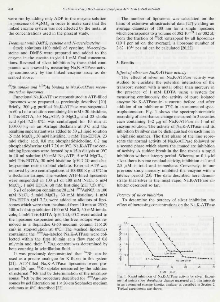
studied to elucidate thé potential interaction of thétransport System with a métal other than mercury inthé présence of 1 mM EDTA using a System formeasuring continuously thé activity of pure isolatedenzyme Na,K-ATPase in a cuvette before and afteraddition of an inhibitor at 37°C in an automated spec-trophotometer. Fig. 1 shows thé spectrophotometricrecording of absorbance change measured in 3 cuvetteseach containing 1-2 ^g of Na,K-ATPase in 1 ml ofenzyme solution. The activity of Na,K-ATPase and ilsinhibition by silver can be distinguished on each Une ina biphasic manner. The first phase of thé line repre-sents thé normal activity of Na,K-ATPase followed bya second phase which shows thé immédiate inhibitionof activity. A sudden break in thé line reveals a rapidinhibition without latency period. Whereas at 0.1 /J.Msilver there is some residual activity, inhibition at 1 and2.5 /tM is total and immédiate. Conversely, in ourprevious study mercury inhibited thé enzyme with alatency period [23]. The data described hère démon-strate that silver is thé most rapid Na,K-ATPase in-hibitor described so far.
Potency of silver inhibitionTo détermine thé potency of silver inhibition, thé
effect of increasing concentrations on thé Na,K-ATPase
2.5nM Silver0.1 [J M Silver
TIME (min)Fig. 1. Rapid inhibition of Na,K-ATPase activity by silver. Expéri-mental points show absorbance change measured at 1-min intervaisin an automated enzyme kinetics analyzer as described in Section 2.Typîcal experiments are shown.

S. Hussain et al. / Biochimica et Biophysica Acta 1190 (1994) 402-408 405
activîty was determined (Fig. 2A). A dose dépendentinhibition of Na,K-ATPase by silver was observed withan IC50 of 9 nM; at 500 nM silver, thé activity wasbelow 2%.
A Hill plot was done to détermine nature andputative binding sites of silver interaction (Fig. 2B).The slope of thé Une shows a Hill coefficient of abouttwo in thé beginning of thé reaction. However, withincreasing concentrations, thé Hill coefficient wasfound below 1 indicating négative cooperativity.
Reversai of siluer-inhibitionSince silver was found to be a potent inhibitor of
Na,K-ATPase, we intended to find out whether DMPS,a known métal chelator, reactivated enzyme activity.DMPS was added to thé enzyme at 1 mM final concen-tration and thé activity of Na,K-ATPase was measured(Fig. 3A); thé two lînes represent thé duplicate valuesof Na,K-ATPase activity. The first phase of thé lineshows a normal activity of Na,K-ATPase, thé secondphase indicates thé immédiate inhibition (90%) ofNa,K-ATPase at 100 nM silver and thé third phaserepresents thé fui! reversai of thé Na,K-ATPase activ-
-2 \ Log [Silver] ,M
Fig. 2. (A) Dose response curve of Na,K-ATPase inhibition by silver.The enzyme activity before and after addition of silver was measuredas described in Section 2. Vertical bars dénote S.E. of thé mean forthree separate déterminations. (B) Hill transformation of thé curveand détermination of thé interaction coefficient (n) of silver effecton Na,K-ATPase.
T I M E (m in )
Fig. 3. (A) Reversai of silver inhibition by DMPS. (B) Reversai ofsilver inhibition by cysteine. (C) Reversai of silver inhibition byA'-acetylcysteine. Na,K-ATPase was measured as described in Sec-tion 2.
ity by 1 mM of thé dithiol DMPS, a potent métalantidote.
Cysteine is known to interact with metals with ilsfree -SH group [33]. In our previous experiments, cys-teine was able to protect Na,K-ATPase from mercuryinhibition [24] but unable to extract it once ît had beenbound to Na,K-ATPase [23]. The normal activity ofNa,K-ATPase, its inhibition by silver and an approxi-mately 80% reversai by cysteine is seen in Fig. 3Billustrating that thé monothiol cysteine is able to ex-tract silver that has been bound to Na,K-ATPase.
7V-Acetylcysteine is an endogenous product of cys-teine metabolism which might be playing a rôle inmétal detoxification owing to thé présence of -SHgroups. N-Acetylcysteine has been shown to protectmercury-induced nephrotoxicity in vivo although it isnot known by what mechanism [25]. Thus, it is crucialto know whether TV-acetylcysteine has any rôle in se-questering metals in in vitro Systems. jV-Acetylcysteine(1 mM) was added to thé enzyme inhibited by silverbut only about 20-30% recovery was seen (Fig. 3C).Although A'-acetylcysteine has a free -SH group, its
406 5. Hussain et al. / Biochimica et Biophysica Acta 1190(1994) 402-408
10
t, 6 9
Time ( min )
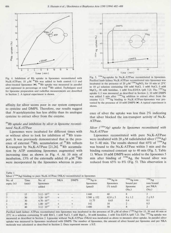
Fig. 4. Inhibition of Rb uptake in liposomes reconstituted withNa,K-ATPase; 10 ^M a6Rb was added to both control (o) andsilver-treated liposomes (•), 86Rb uptake was measured in paralleland expressed in percentage ot total H6Rb added. Techniques usedfor liposome préparation and radioflux measurements are describedin Section 2. A typîcal experiment is shown.
affinity for silver seems poor in our System comparedto cysteine and DMPS. Therefore, our results suggestthat N-acetylcysteine bas less ability than its analoguecysteine to extract silver from thé enzyme.
Rb uptake and inhibition by silver in liposome reconsti-tuted Na,K-ATPase
Liposomes were incubated for différent times withor without silver to look for inhibition of 86Rb trans-port. It was previously demonstrated that in thé prés-ence of external 86Rb, accumulation of 86Rb reflectsK-transport by Na,K-ATPase [21,26]. 86Rb accumula-tion by ATP containing liposomes augmented withincreasing time as shown in Fig. 4. At 10 min of
86
80.DMPS
0 10 20 30 40
Time (min)
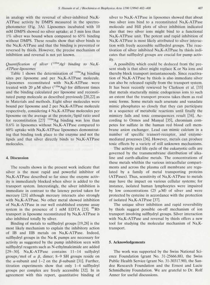
Fig. 5. mAg-uptake by Na,K-ATPase reconstituted in liposomes.Purified lamb kidney Na,K-ATPase reconstituted into liposomes wasincubated in thé présence of 20 /iM ""mAgNO3 for 10 min at 25°Cin 10 fj,\n containing 100 mM NaCI, l mM NaCI, 5 mMMgCI2, 30 mM histidine, 1 mM Tris-EDTA (pH 7.2). The UOmAguptake (o) was measured as described in Section 2; 10 mM DMPSwas added 5 min after mAg addition to extract silver from théenzyme (n). Ag binding to Na,K-ATPase-liposomes was pre-vented by thé présence of 10 mM DMPS (•). A typical experiment isshown.
ence of silver thé uptake was less than 2% indicatingthat silver blocked thé ion-transport activity of Na,K-ATPase.
Silver ("0mAg) uptake by liposomes reconstituted withN a, K-ATPase
Liposomes reconstituted with pure Na,K-ATPasewere incubated with 20 ,u,M radiolabeled silver (110mAg)for 5-40 min. The results showed that 65% of 110mAgwas bound to thé Na,K-ATPase within 5 min and thébinding remained constant up to 40 min (Fig. 5, Table1). When 10 mM DMPS were added to thé liposomes 5
incubation, 13% of thé externally added 10 /iM Rb min after binding of ] Ag, thé bound silver waswere incorporated by thé liposomes whereas in près- reduced from 65% to 8% (Fig. 5). This observation is
Table 1Silver (110mAg) binding to pure Na,K-ATPase (NKA) reconstituted in liposomes
No. ofexpts. (n)
21211
Time(min)
1530
'303040
No. of NKAliposomesadded
3.12 -1012 +3.20- 10 12 +4.70 • 1012
1.00 -1012 +3.12 '1012 +
DMPS 110mAginliposomes(pmol)
14401580 ±152
11.75+ 142
1400
'10mAginliposomes(% total)
6062.0 + 50.63
1858
UUmAgionsbound perliposome(No.)
88±1.20.428
l'On'Ag
ions boundper NKA(No.)
22 + 0.30.10.52
Purified lamb kidney ATPase reconstituted into liposomes was incubated in thé présence of 20 ju,M of silver (ll(lmAg) for 15, 30 and 40 min at25°C in a solution containing 50 mM RbCl, 1 mM NaCI, 5 mM MgCl2, 30 mM histidine, 1 mM Tris-EDTA (pH 7.2). The 1IOmAg uptake wasmeasured as described in Section 2. Liposome without Na,K-ATPase (NKA) was incubated as above to measure silver uptake. In parallel silveruptake was measured in liposomes incubated with DMPS. The number of liposomes, thé amount of silver bound per liposome and per NKAmolécule was calculated as described in Section 2. Data represent means + S.E.
S. Hussain et al. /Biochimica et Biophysica Acta 1190 (1994) 402-408 407
în analogy with thé reversai of silver-inhibited Na,K-ATPase activity by DMPS measured in thé spectro-photometer (Fig. 3A). Liposomes incubated with 10mM DMPS showed no silver uptake; at 5 min less than1% silver was bound when compared to 65% bindîngwithout DMPS. Thèse results show that silver binds tothé Na,K-ATPase and that thé binding is prevented orreversed by thiols. However, thé précise mechanism ofinhibition and reversai is not yet known.
Quantification of silver (!10mAg) binding to Na,K-A TPase-lîposomes
Table 1 shows thé détermination of ' "mAg bindingsites per liposome and per Na,K-ATPase molécule.Liposomes reconstituted with Na,K-ATPase weretreated with 20 ^.M silver (11UmAg) for différent timesand thé binding calculated per liposome and reconsti-tuted pump molécule as described previously [20,22] asin Materials and methods. Eight silver molécules werebound per liposome and 2 per Na,K-ATPase moléculerespectively, considering 4 Na,K-ATPase molécules perliposome on thé average at thé protein/lipid ratio usedfor reconstitution [27] "0rnAg binding was less than0.6% in liposomes without Na,K-ATPase compared to60% uptake with Na,K-ATPase liposomes demonstrat-ing that binding took place to thé enzyme and not thélipids and that silver directly binds to Na,K-ATPasemolécules.
4. Discussion
The results shown in thé présent work indicate thatsilver is thé most rapid and powerful inhibitor ofNa,K-ATPase described so far since thé enzyme activ-ity is immediately blocked when silver is added to thétransport System. Interestingly, thé silver inhibition isimmédiate in contrast to thé latency period taken formercury [23] although mercury interacts also stronglywith Na,K-ATPase. No other métal showed inhibitionof Na,K-ATPase in our well established enzyme assaySystem in thé présence of 1 mM EDTA [23]. 86Rbtransport in liposome reconstituted by Na,K-ATPase isalso inhibited totally by silver.
Binding of metals to sulfhydryl groups [19,28] is thémost likely mechanism to explain thé inhibitory actionof IB and IIB metals on Na,K-ATPase. Indeed,sulfhydryl groups in thé Na-K pumps are necessary foractivity as suggested by thé pump inhibition seen withsulfhydryl reagents such as N-ethylmaleimide are added[29-30]. Na,K-ATPase contains 11-14 sulfydrylgroups/mol of a 0, dimer; 8-9 SH groups réside onthé a-subunît and 1-2 on thé 0-subunit [31]. Further,titration experiments show that only 1-4 sulfhydrylgroups per complex are freely accessible [32], In inagreement with this report, quantitative binding of
silver to Na,K-ATPase in liposomes showed that abouttwo silver ions bind to a reconstituted Na,K-ATPasemolécule and Hill plots of silver inhibition indicatedalso that two silver ions might bind to a functionalNa,K-ATPase unit. The potent and rapid inhibition ofNa,K-ATPase is most likely attributed to silver interac-tion with freely accessible sulfhydryl groups. The réac-tivation of silver inhibited Na,K-ATPase by thiols indi-cates that sulfhydryl groups are crucial for pump activ-ity.
A possibility which could be deduced from thé pré-sent study is that silver might replace K. or Na ions andthereby block transport instantaneously. Since réactiva-tion of Na,K-ATPase by thiols is also immédiate silvercan also be released rapidly from thé transport System.It has been recently rewieved by Clarkson et al. [33]that metals stucturally mimic endogenous ions to suchan extent that thé transport process takes them as realionic forms. Some metals such arsenate and vanadatemimic phosphates so closely that they can participatein a séquence of metabolic reactions until finally thémimicry fails and toxic conséquences resuit [34], Ac-cording to Ormos and Manyal [35], chromium com-petes for sulfate in thé human red blood cell mem-brane anion exchanger. Lead can mimic calcium in anumber of spécifie transort-receptor, and enzyme-mediated processes [36]. However, metals can producetoxic effects by a variety of still unknown mechanisms.
The activity and life cycle of thé eukaryotic cells aregoverned by thé transmembrane distribution of alka-line and earth-alkaline metals. The concentrations ofthèse metals whithin thé various intracellular compart-ments and across thé plasma cell membrane are regu-lated by a family of métal transporting proteins(ATPases). Thus, sensitivity of Na,K-ATPase to metalsmay hâve thé impact on various cellular Systems; forinstance, isolated human lymphocytes were impairedby low concentrations (25 /J.M) of silver and wereprotected by cysteine in accordance with thé protectionof isolated Na,K-ATPase [37].
The unique silver inhibition and rapid reversibilityby thiols suggest possible on-off mechanism of iontransport involving sulfhydryl groups. Silver interactionwith Na,K-ATPase and reversai by thiols offers a newtool for studying thé molecular mechanism of Na,K-transport.
5. Acknowledgements
The work was supportcd by thé Swiss National Sci-ence Foundation (grant No. 31-25666.88), thé SwissPublic Health Service (grant No. 31-30317.90), thé San-doz Research Foundation and thé Ernest and LucieSchmidheiny Foundation. We are grateful to Dr. RolfAnner for useful discussions.

408 5. Hussain et al. / Biochimica et Biophysica Acta 1190 (1994) 402-408
6. Références
[1] Schwartz, A., Lindenmayer, G.E. and Allen, J.C. (1975) Phar-macol. Rev. 27, 3-134.
[2] J0rgensen, P.L. (1975) Biochim. Biophys. Acta 401, 399-415.[3] Skou, J.C. (1974) Biochim. Biophys. Acta 339, 234-245.[4] Dixon, J.F. and Hokin, L.E. (1980) J. Biol. Chem. 255, 10681-
10688.[5] Repke, K.H.R. (1988) in Biomemhranes, Basic and Médical
Research (Berga, G.H. and Tiger, J.M., eds.), pp. 161-176,Springer, Berlin.
[6] Vallée, B.L. and Ulmer, D.D. (1972) Annu. Rev. Biochem. 41,91-128.
[7] Hanas, J.S., Hazuda, D.J., Bogenhagen, D.F., Wu, F.H.Y. andWu, C.W. (1983) J. Biol. Chem. 258, 14120-14125
[8] Lohmer, P.H. and Toft, D.O. (1975) Biochem. Biophys. Res.Commun. 67, 8-15
[9] Gerson, R.J. and Shaikh, Z.A. (1982) Biochem. J. 208, 465-472.[10] Passow, H., Rothstein, A. and Clarkson, T.W. (1961) Pharmacol.
Rev. 13, 185-224.[11] Siegel, G.J., Fogt, S.K. and Hurley, M.J. (1977) Adv. Exp. Med.
Biol. 84,465-491.[12] Siegel, G.J. and Fogt, S.K. (1976) Trans. Am. Neurol. Assoc.
101, 175-177.[13] Rajanna, B., Hobson, M., Bansal, S.K. and Desaiah, D. (1983)
Toxicol. Lett. 18, 331-336.[14] Rajanna, B., Chetty C.S., Stewart, T.C. and Rajanna, S. (1991)
Biomed. Environ. Sci. 4, 441-451.[15] Tucker, R.K. and Malte, A. (1980) Bull. Environ. Contam.
Toxicol. 24, 847-852.[16] Nechay, B.R. and Saunders, J.P. (1978) J. Environ. Pathol.
Toxicol. 2, 283-290.[17] Ballatori, N., Chenyang, S. and Boyer, J.L. (1988) Toxicol. Appl.
Pharmacol. 95, 279-291.[18] Dzhandzhugazyan, K.N. and J0rgensen, P.L. (1985) Biochim.
Biophys. Acta 817, 165-173.
[19] Imesch, E., Moosmayer, M. and Anner, B.M. (1992) Am. J.Physiol. (Rénal Fluid Electrolyte Physiol. 31) 262, F837-F842.
[20] Anner, B.M., Rey, H.G., Moosmayer, M., Mesozoeli, I. andHaupert Jr., G.T. (1990) Am. J. Physiol. (Rénal Fluid Elec-trolyte Physiol. 27) 258, F144-F153.
[21] Rey, H.G., Moosmayer, M. and Anner, B.M. (1987) Biochim.Biophys. Acta 900, 27-37.
[22] Anner, B.M. and Moosmayer, M. (1992) Am. J. Physiol. (RénalFluid Electrolyte Physiol. 31) 262, F843-F848.
[23] Anner, B.M., Moosmayer, M. and Imesch, E. (1992) Am. J.Physiol. (Rénal Fluid Electrolyte Physiol. 31) 262, F830-F836.
[24] Anner, B.M., Imesch, E. and Moosmayer, M. (1989) Biochem.Biophys. Res. Commun. 165, 360-367.
[25] Girardi, G. and Eliases, M.M. (1991) Toxicology 67, 155-164.[26] Anner, B.M. and Moosmayer, M. (1985) Biochem. Biophys. Res.
Commun. 129, 102-108.[27] Anner, B.M., Robertson. and Ting-Beall, H.P. (1984) Biochim.
Biophys. Acta 773, 253-261.[28] Jacobsen, K.B. and Turner, J.E. (1980) Toxicology 16, 1-37.[29] Skou, J.C. and Hilberg, C.H. (1965) Biochim. Biophys. Acta 110,
359-369.[30] Wallick, ET., Anner, B.M., Ray, M.V. and Schwartz, A. (1978)
J. Biol. Chem. 253, 8778-8786.[31] Schoner, W., Hasselberg, M. and Kison, R. (1988) Methods
Enzymol. 28, 302-312.[32] Gevondyan, H.M., Gevondyan, V.S., Gavrilyeva, E.E. and
Modyanov, N.M. (1989) FEBS. Lett. 255, 265-268.[33] Clarkson, T.W. (1993) Annu. Rev. Pharmacol. Toxicol. 32, 545-
571.[34] Cantley, L.C., Cantley, L.G. and Josephsen, L.A. (1978) J. Biol.
Chem. 253, 7361-7368.[35] Ormos, G. and Manyal, S. (1978) J. Physiol. 276, 501-513.[36] Kober, T. and Cooper, G.P. (1976) Nature 265, 704-705.[37] Hussain, S., Anner, R.M. and Anner, B.M. (1992) Biochem.
Biophys. Res. Commun. 189, 1444-1449.
![The effect of di]methylsulfoxide on the substrate site of Na +/K +ATPase studied through phosphorylation by inorganic phosphate and ouabain binding](https://static.fdokumen.com/doc/165x107/6323a6724d8439cb620d023f/the-effect-of-dimethylsulfoxide-on-the-substrate-site-of-na-k-atpase-studied.jpg)