Histamine promotes neuronal differentiation of midbrain neural stem cells

High-Affinity Na�
/K�
-Dependent GlutamateTransporter EAAT4 Is Expressed
Throughout the Rat Fore- and Midbrain
ANN MASSIE,1 LIESELOTTE CNOPS,2 ILSE SMOLDERS,1
ROBERT MCCULLUMSMITH,3 RON KOOIJMAN,4 SHIN KWAK,5
LUTGARDE ARCKENS,2AND YVETTE MICHOTTE1*
1Department of Pharmaceutical Chemistry and Drug Analysis, Research Group ofExperimental Pharmacology, Vrije Universiteit Brussel, 1090 Brussels, Belgium
2Laboratory of Neuroplasticity and Neuroproteomics, Katholieke Universiteit Leuven, 3000Leuven, Belgium
3Department of Psychiatry, University of Alabama at Birmingham, Birmingham, Alabama 352944Department of Pharmacology, Vrije Universiteit Brussel, 1090 Brussels, Belgium
5Department of Neurology, Division of Neuroscience, Graduate School of Medicine, University ofTokyo, Tokyo 113, Japan
ABSTRACTExcitatory amino acid transporter 4 (EAAT4), a member of the high-affinity Na�/K�-
dependent glutamate transporter family, is highly enriched in Purkinje cells of the cerebellum,although it is not restricted to these cells. The detailed expression of EAAT4 protein in differentadult rat fore- and midbrain regions was examined. Despite moderate expression levels comparedwith the cerebellum, EAAT4 protein was omnipresent throughout the fore- and midbrain. Withantibodies raised against the N-terminal mouse EAAT4 sequence, the highest protein expressionlevels were observed in the substantia nigra pars compacta, ventral tegmental area, paranigralnucleus, habenulo-interpeduncular system, supraoptic nucleus, lateral posterior thalamic nu-cleus, subiculum, and superficial layers of the superior colliculus. Relatively high levels of EAAT4protein were also detected in the hippocampal principal cells, in the glutamatergic,�-aminobutyric acid (GABA)ergic, dopaminergic and most likely cholinergic cells of all nuclei ofthe basal ganglia, and in neurons of layers II/III and V of the cerebral cortex. The expression ofEAAT4 was confirmed at the mRNA level in some important fore- and midbrain structures by insitu hybridization and reverse transcriptase-polymerase chain reaction (RT-PCR) and estimatedto range from 6.7 to 1.6% of the amount in the cerebellum as measured by real-time PCR. J.Comp. Neurol. 511:155–172, 2008. © 2008 Wiley-Liss, Inc.
Indexing terms: EAAT4; fore- and midbrain; distribution; immunohistochemistry; real-time PCR;
in situ hybridization
High-affinity Na�/K�-dependent glutamate transport-ers are responsible for the reuptake of synaptically re-leased glutamate, the major excitatory neurotransmitterin the central nervous system (CNS), into glial cells orneurons. Five different excitatory amino acid transporters(EAATs) have been cloned so far (Saier, 1999; Slotboom etal., 1999): GLAST (Storck et al., 1992; Tanaka, 1993),GLT-1 (Pines et al., 1992), and EAAC1 (Kanai and Hedi-ger, 1992), corresponding to human EAAT1, -2, and -3,respectively, EAAT4 (Fairman et al., 1995), and EAAT5(Arriza et al., 1997). Through the proper functioning ofthese glutamate transporters, glutamatergic neurotrans-mission is carefully regulated and excitotoxicity is pre-vented (for review, see Danbolt, 2001). This regulation
also includes control over the extent of glutamate spilloverand ensuing activation of extrasynaptic targets at excita-
Grant sponsor: FWO-Flanders; Grant numbers: 1.5.143.07N, postdoc-toral fellowships (to A.M. and I.S.), and research assistant (to L.C.); Grantsponsor: the Vrije Universiteit Brussel.
*Correspondence to: Yvette Michotte, Ph. D., Department of Pharmaceuti-cal Chemistry and Drug Analysis, Research Group of Experimental Pharma-cology, Vrije Universiteit Brussel, Laarbeeklaan 103, 1090 Brussels, Belgium.E-mail: [email protected]
Received 21 December 2005; Revised 24 August 2006; Accepted 11 July2008
DOI 10.1002/cne.21823Published online in Wiley InterScience (www.interscience.wiley.com).
THE JOURNAL OF COMPARATIVE NEUROLOGY 511:155–172 (2008)
© 2008 WILEY-LISS, INC.

tory synapses (Bergles et al., 1999). In addition, glutamatetransporters also behave as glutamate-gated chloridechannels, a property that is particularly prominent inEAAT4 and EAAT5 (Fairman et al., 1995; Vandenberg etal., 1995; Wadiche et al., 1995a,b; Arriza et al., 1997;Torres-Salazar and Fahlke, 2007).
Originally, the five members of this transporter familywere thought to have not only a distinct regional but alsoa distinct cellular distribution pattern. GLAST and GLT-1were reported to be confined to glial cells (Rothstein et al.,1994; Torp et al., 1994; Chaudhry et al., 1995; Lehre et al.,1995). EAAC1 and EAAT4 were considered to be strictneuronal transporters (Rothstein et al., 1994; Yamada etal., 1996; Furuta et al., 1997). However, this interpreta-tion was challenged by several reports, and it soon becameclear that the subdivision of glutamate transporters into“glial” and “neuronal” was no longer tenable (however, forease this subdivision is still generally used). GLT-1 isexpressed in neurons of cortical and hippocampal culture(Mennerick et al., 1998; Wang et al., 1998). Also, in vivo,GLT-1, as well as its splice variants, can be expressed byneurons (Rauen and Kanner, 1994; Schmitt et al., 1996;Torp et al., 1996; Berger and Hediger, 1998; Berger et al.,2005; Chen et al., 2002, 2004). EAAT4 expression has beenreported to be enriched in astrocytes of mouse spinal cordand forebrain (Hu et al., 2003) as well as in rat retinalastrocytes (Ward et al., 2004). Very recently, Liang et al.(2008) described the presence of the mRNA of all EAATs(i.e., EAAT1–5) in astrocyte cultures. Moreover, glia werereported to have the potential to upregulate EAAT4 aftertraumatic brain injury (Yi et al., 2007). EAAT5, thought tobe primarily neuronal, is predominantly expressed in theretina (Arriza et al., 1997) and will therefore not be dis-cussed in further detail.
As for the regional distribution pattern, GLAST immu-noreactivity (IR) is known to be highly abundant in cere-bellar Bergman glia (Lehre et al., 1995; Lehre and Dan-bolt, 1998; Attwell, 2000), whereas GLT-1, which isresponsible for the bulk of glutamate removal, is highlyexpressed in astrocytes of the cerebral cortex and hip-pocampus (Lehre et al., 1995). EAAC1 expression withinthe CNS has been described in the cerebral cortex, hip-pocampus, and basal ganglia.
It is commonly accepted that EAAT4 is largely confinedto Purkinje cells of the cerebellum, with little or no ex-pression in forebrain regions (Yamada et al., 1996; Bar-Peled et al., 1997; Furuta et al., 1997; Itoh et al., 1997;Nagao et al., 1997; Tanaka et al., 1997; Dehnes et al.,1998; Inage et al., 1998). However, Furuta et al. (1997)have reported very low expression levels of EAAT4 proteinin the forebrain and more particularly in the cerebralcortex, hippocampus, and some basal ganglia nuclei. Also,McCullumsmith and Meador-Woodruff (2002) detectedEAAT4 mRNA in the striatum of human subjects. Re-cently, we described the expression of this glutamatetransporter in more detail in neurons of the cerebral cor-tex of cat and mouse (Massie et al., 2001). The presence ofEAAT4 in the cerebral cortex has been confirmed at themRNA (Ward et al., 2004) as well as the protein level (Huet al., 2003). Nevertheless, the expression level in thecerebral cortex has been estimated to be only approxi-mately 3% that of the cerebellum (Ward et al., 2004). Morerecently, EAAT4 has also been described to be expressedin photoreceptors and astrocytes of the retina (Ward et al.,2004; Pignataro et al., 2005). Thus, it is becoming more
and more clear that EAAT4 is more widespread in distri-bution than was initially thought. In addition, severalrecent studies report changes in the expression levels ofEAAT4 mRNA and/or protein in various fore-and mid-brain regions of various brain pathologies (McCullum-smith and Meador-Woodruff, 2002; Rakhade and Loeb,2008; Yi et al., 2007).
To our knowledge, this is the first report describing indetail the cellular and regional distribution of EAAT4protein and mRNA throughout the rat fore- and midbrain.By using an antibody raised against the N-terminal mouseEAAT4 sequence (Yamada et al., 1996), as well as a stain-ing protocol ensuring optimal permeability of cells for theantibodies, EAAT4 protein could be clearly observed inneurons throughout the fore- and midbrain, notwithstand-ing the manifestly higher levels in the Purkinje cell layerof the cerebellum. In addition, the presence of EAAT4mRNA in the fore-and midbrain was confirmed by usingreverse-transcriptase-polymerase chain reaction (RT-PCR) and in situ hybridization. The expression level ofEAAT4 mRNA relative to the cerebellum was determinedfor the cerebral cortex, hippocampus, and striatum byusing real-time PCR.
MATERIALS AND METHODS
Animals
Protocols for animal experiments described in this studywere carried out according to national guidelines on ani-mal experimentation and were approved by the EthicalCommittee for Animal Experimentation of the Faculty ofMedicine and Pharmacy of the Vrije Universiteit Brussel.
All animals used for this study were housed under stan-dard laboratory conditions. Eight adult male Wistar rats(weighing 250–275 g) were sacrificed for the immunohis-tochemical study, six for the Western blotting, and threefor the RT-PCR and real-time PCR experiments.
Mice bearing heterozygous null mutations for EAAT4were a gift of Dr. K. Tanaka (Huang et al., 2004). Breedingwith these mice resulted in litters containing EAAT4�/�,EAAT4�/�, and EAAT4�/� mice. Four 12-week-old malemice of each genotype were used for this study.
Western blotting
After rats or mice were injected with a lethal dose ofpentobarbital, i.p. (Nembutal, Sanofi sante, Brussels, Bel-gium), brains were immediately dissected. A small tissuesample (�4 mm3) was collected from the cerebral cortex,striatum, hippocampus, and cerebellum, snap-frozen indry ice-cooled 2-methylbutane (�60°C), and stored at�70°C until use.
Protein extraction and Western blotting were performedas described before (Massie et al., 2003). Tissue was ho-mogenized in 2% sodium dodecyl sulfate (SDS; Polylab,Antwerp, Belgium), 60 mM Tris base (ICN Biomedicals,Aurora, OH), pH 6.8, 100 mM dithiothreitol (DTT; MerckEurolab, Leuven, Belgium), and 1 mM EDTA (Merck Eu-rolab). After incubation for 30 minutes at 37°C, sampleswere processed four times through 20-gauge needles andthen through a 26-gauge needle, spun at 10,000g at 4°C(Eliasof et al., 1998), and immediately boiled for 10 min-utes. Supernatants were stored at �70°C. Protein concen-trations were determined by using a modified Bradfordmethod (Qu et al., 1997). Equal concentrations of protein
156 A. MASSIE ET AL.
(1.6 �g/lane) were loaded for all forebrain regions; for thecerebellum, four times less protein was loaded.
Proteins were separated by SDS-polyacrylamide gelelectrophoresis (PAGE; 4–12% gel; Invitrogen, Gro-ningen, The Netherlands) under reducing conditions andtransferred to a polyvinylidene fluoride membrane (Sequi-Blot PVDF Membrane, Bio-Rad Laboratories, Nazareth,Belgium) by using an Xcell II Blot module (Novex, Invitro-gen). Nonspecific binding was blocked by incubating themembrane for 1 hour at room temperature in 5% driedmilk (ECL blocking agent, GE Healthcare, Roosendaal,The Netherlands). Blots were incubated overnight at 4°Cwith the immunoaffinity-purified polyclonal antibodies toEAAT4, raised in rabbit against the N-terminal mousesequence (MSSHGNSLFLRESGAGGGCL; 760 �g/ml; di-luted 1:4,000; kindly provided by Dr. M. Watanabe; samebatch as in Yamada et al., 1996). The next day, afterincubation with horseradish-peroxidase-conjugated anti-rabbit antiserum (1:2,000; Dako, Glostrup, Denmark), im-munoreactive proteins were visualized by using enhancedchemiluminescence (ECL; ECLplus kit, GE Healthcare).All washing and dilution steps were performed with Tris-saline (0.01 M, pH 7.4). The MultiMark Multi-ColoredStandard (Novex, Invitrogen) was used as molecularweight standard. Negative controls included omission ofthe immunoaffinity-purified antibody and the secondaryantibody, respectively.
Immunohistochemistry
Rats or mice were deeply anesthetized with an overdoseof Nembutal, i.p., and transcardially perfused with a phys-iological solution followed by freshly depolymerized 4%paraformaldehyde (Sigma-Aldrich, St. Louis, MO) in 0.15M phosphate-buffered saline (PBS; pH 7.42). Brains wereremoved and postfixed in the same fixative overnight,rinsed in tap water for 24 hours, and stored in 0.015 MPBS at 4°C. Free-floating 50-�m frontal and sagittal sec-tions were made with a vibratome and stored in serialorder in 0.015 M PBS at 4°C.
All rinsing steps and incubations of the staining proce-dure were performed in Tris-saline (0.01 M, 0.05% TritonX-100 [Sigma-Aldrich], pH 7.4) or in 0.015 M PBS, alwaysat room temperature, unless mentioned otherwise, andunder gentle agitation. All results shown and discussedwere obtained with the Triton X-100-containing stainingprotocol unless otherwise noted. Incubation steps wereseparated by three rinsing steps of at least 5 minutes. Thesections underwent a permeabilizing treatment consistingof incubation in 0.1% trypsin (Fluka, Buchs, Switzerland)for 1 hour at 37°C prior to a blocking step with normalgoat serum (Chemicon, Temecula, CA, diluted 1:5, 45 min-utes). Thereafter the sections were incubated with theprimary EAAT4 antibodies (diluted 1:2,000; Yamada etal., 1996) for 2 days. On the third day the sections wereprocessed by the avidin-biotin method by using a Vec-tastain ABC kit (Vector, Burlingame, CA), and immu-noreactivity was visualized, after a final rinsing stepwith acetate buffer, by using the glucose oxidase-diaminobenzidine-nickel method (Shu et al., 1988). Sec-tions were mounted on poly-L-lysine-coated slides, de-hydrated through alcohol, cleared with xylene, andcoverslipped with Coverquick (Labonord, Templemars,France). Negative controls consisted of the omission ofthe primary and secondary antibodies, respectively.
Photomicrographs were made of the stained sections,and brightness and contrast were adapted by using AdobePhotoshop 7.0.
Laser microdissection (LMD)
After the brain was snap-frozen, 14-�m sections werecut on a cryostat and mounted on home-made glasses thatwere covered with a poly-ethylene naphtalate (PEN) foiland exposed to UV radiation for 30 minutes prior to use.Glasses were placed in a Leica AS LMD system (Leica,Leitz Instruments, Heidelberg, Germany) with the sectionfacing downward. After adjusting intensity, aperture, andcutting velocity, the pulsed UV laser beam was carefullydirected along the borders of the fasciculus retroflexus ofseveral serial sections by using the 4� objective. The areacut fell by forces of gravity alone into a plastic tube capplaced directly underneath the section. The tube cap wasfilled with lysis solution. Tissue collection was verified byinspecting the tube cap under higher magnification.
RT-PCR
Total RNA was extracted from tissue samples of thefasciculus retroflexus, collected by the LMD technique, aswell as manually isolated tissue samples from cryosec-tions of the cerebellum, cerebral cortex, hippocampus, andstriatum, by using the Versagene total RNA purificationkit (Gentra Systems, Big Lake, MN). RT-PCR was per-formed according to the instructions of the manufacturer(GeneAmp® RNA PCR kit from Applied Biosystems, Fos-ter City, CA). For the reverse transcription oligo(dT) prim-ers were used. The EAAT4-specific primers used for PCRamplification were designed according to the known ratsequence of EAAT4 (GenEmbl. u89608; Lin et al., 1998;upstream primer: 5�-GGAGACTGTGCCTGTACCTGG-3�,downstream primer: 5�-GCAGAGCTGGAAGAGGTACCC-3�, corresponding to nucleotides 906–926 and 1,299–1,319respectively; Eurogentec, Seraing, Belgium). A total of 40reaction cycles (95°C 1 minute, 60°C 1 minute, 70°C 1minute) was preceded by 1 cycle starting at 95°C for 10minutes to activate the polymerase enzyme (AmpliTaq�Gold DNA polymerase, 2.5 U/100 �l; Applied Biosystems)and concluded by a termination step at 70°C for 10 min-utes. As negative control the cDNA in the reaction mix-ture was substituted by water.
Amplified products were analyzed by horizontal agarosegel electrophoresis and visualized by ethidium bromidestaining. A 100-bp ladder (Invitrogen) was used as molec-ular weight reference.
Real-time PCR
Total RNA was extracted, and, after biophotometricanalysis (Eppendorf, VWR International, Leuven, Bel-gium), RT was performed as described above on mRNAsamples of identical quantity. Primers and probes weredesigned by using the Primer Express Program (AppliedBiosystems), based on the rat sequence of EAAT4 (Gen-Embl. u89608; forward primer: 5�-TTGCGCCTGCAGAC-CAT-3�; reverse primer: 5�-AGCGCTGACTGTGAGCAA-GA-3�; Taqman probe: 5�-CGCTTCCTGCGCCGAAATGC-3�) and GAPDH (GenEmbl. NM017008; forward primer:5�-TGCCTGGATCCCTAAAGAGACA-3�; reverse primer:5�-CGCGATATTCAATTGGATACACA-3�; Taqman probe:5�-CCATTTCCAAGACTGACAGCCCCAGA-3�). Primerswere obtained from Eurogentec, and FAM-TAMRA Taq-man probes were purchased from Applied Biosystems.
157EAAT4 IN RAT FORE- AND MIDBRAIN
PCR was performed on the cDNA samples in a 25-�lreaction volume of 1� Taqman Universal PCR MasterMix (Applied Biosystems) with primers at a final concen-tration of 300 nM and probes of 200 nM, using the ABIPrism 7000 Sequence Detection System. Standard curveswere generated by running serial dilutions (1:2) of controlcDNA in duplicate for each gene. Target samples were runin triplicate on the same well-plate under standard am-plification settings (1 cycle of 50°C for 2 minutes, 1 cycle of95°C for 10 minutes, 40 cycles of 95°C for 15 seconds and60°C for 1 minutes).
Data were analyzed with ABI Prism 7000 SDS software(version 1.1). EAAT4 quantities were normalized toGAPDH, an endogenous control, to account for variabilityin the initial concentration of mRNA in the samples and tocompensate for differences in conversion efficiency of theRT reaction. Amounts of transcripts in the cerebral cortex,striatum, hippocampus, and fasciculus retroflexus wereexpressed relative to the cerebellum which was set as thecalibrator (�100%). To confirm reproducibility, real-timePCR was performed twice on each rat (n � 3).
In situ hybridization
After rats were injected with a lethal dose of Nembutal,i.p., brains were immediately dissected and snap-frozen indry ice-cooled 2-methylbutane (�60°C) and then stored at�80°C until use. In situ hybridization experiments wereperformed as described by McCullumsmith and MeadorWoodruff (2002). To generate a subclone for riboprobesynthesis, we amplified a unique region of EAAT4(U89608, 576–959) from a rat cDNA brain library (Edge-Biosystems, Gaithersburg, MD) by using PCR. AmplifiedcDNA segments were extracted (QIAquick Gel Extractionkit; Qiagen, Valencia, CA), subcloned (Zero Blunt TOPOPCR cloning kit; Invitrogen, Carlsbad, CA), and confirmedby nucleotide sequencing (Thermo Sequenase Radiola-beled Termination Cycle Sequencing kit; USB, Cleveland,OH). Riboprobes were synthesized by using 100 �Ci ofdried [35S]-UTP, 2.0 �l 5X transcription buffer, 1.0 �l 0.1M DTT, 1.0 �l each of 10 mM ATP, CTP, and GTP, 2.0 �llinearized plasmid DNA, 0.5 �l RNAse inhibitor, and 1.5�l SP6 or T7 RNA polymerase, and incubated for 2 hoursat 37°C. After this incubation, 1.0 �l DNAse (RNAse free)was added and incubated for 15 minutes at room temper-ature. The reaction mixture was separated through spincolumns (Micro Bio-Spin P-30 Tris Chromatography Col-umns, Bio-Rad, Richmond, CA), and the purified fractionwas eluted. DTT was added to each fraction to a finalconcentration of 0.01 M.
Slides were removed from �80°C storage, fixed in 4%(wt/vol) formaldehyde at room temperature for 1 hour, andbriefly washed in 2X SSC (standard saline citrate: 300 mMNaCl/30 mM sodium citrate, pH 7.2) three times. Theslides were then placed in 0.1 M triethanolamine (pH8.0)/acetic anhydride (400:1 vol/vol) with stirring for 10minutes at room temperature. The final wash was in 2XSSC buffer for 10 minutes followed by dehydration ingraded ethanol washes and air drying. A coverslip con-taining the [35S]-radiolabeled riboprobe diluted 5 � 106
cpm in 0.1 ml per slide of hybridization buffer (50% form-amide, 10% dextran sulfate, 3X SSC, 50 mM Na2HPO4,pH 7.4, 1X Denhardt’s solution [0.02% poliviny pyrroli-done, 1.02& Ficoll, 0.02% bovine serum albumin], 100�g/ml yeast tRNA) and 0.01 M DTT was placed on eachslide. Slides were then placed in a covered tray with filter
paper saturated with 50% formamide buffer and incu-bated at 55°C overnight. Sense and antisense probes weretested to confirm specificity. After 16 hours of hybridiza-tion, the coverslips were removed and the slides werewashed in 2X SSC for 2 minutes and again in 2X SSC for15 minutes, and then immersed in RNAse A (200 �g/ml in10 mM Tris-HCl pH 8.0/0.5 M NaCl) for 30 minutes at37°C. The slides then underwent washes through descend-ing concentrations of SSC, to a final concentration of 0.1XSSC at 55°C for 1 hour twice. Sections were finally dehy-drated in graded ethanol washes, air-dried, and exposed toKodak BIOMAX MR film (Kodak, Rochester, NY) for 3months. All sections were run in the same experiments toeliminate interassay variability. Neighboring sectionswere stained with Richardson’s methylene blue-azure IINissl stain (Richardson et al., 1960). Digital photomicro-graphs of the Nissl-stained sections were made by usingthe Leica IM50 software. Brightness and contrast wereadapted by using Adobe Photoshop 7.0.
RESULTS
Specificity of the antibodies
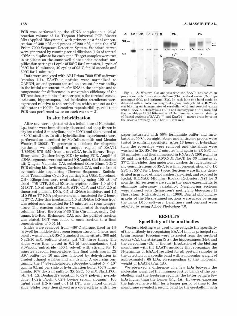
Western blotting was used to investigate the specificityof the antibody in recognizing EAAT4 in four principal ratbrain regions. Proteins were extracted from the cerebralcortex (Cx), the striatum (Str), the hippocampus (Hc), andthe cerebellum (Cb) of the rat. Incubation of the blottingmembrane with the EAAT4 antibody that recognizes theN-terminus of EAAT4 resulted for all protein samples inthe detection of a specific band with a molecular weight ofapproximately 68 kDa, corresponding to the molecularweight of EAAT4 (Fig. 1A).
We observed a difference of a few kDa between themolecular weight of the immunoreactive bands of the cer-ebellum and the forebrain regions, the latter being a fewkDa higher than the former (Fig. 1A). However, exposingthe light-sensitive film for a longer period of time to themembrane revealed a second band for the cerebellum with
Fig. 1. A: Western blot analysis with the EAAT4 antibodies onprotein extracts from rat cerebellum (Cb), cerebral cortex (Cx), hip-pocampus (Hc), and striatum (Str). In each lane one band could bedetected with a molecular weight of approximately 68 kDa. B: West-ern blotting on homogenates of cerebellar (Cb) and cerebral cortex(Fb) of EAAT4 heterozygous (�/�) and homozygous (�/�) mice andtheir wild-type (�/�) littermates. C: Immunohistochemical stainingof frontal sections of EAAT4�/� and EAAT4�/� mouse brain by usingthe EAAT4 antibody. Scale bar � 1 mm in C.
158 A. MASSIE ET AL.
the same molecular weight as the bands of the forebrainregions (data not shown). Because of the very high inten-sity of the lower band, these two bands appear as onebroad and intense band. Possibly, the appearance of thisdouble band can be explained by post-translational modi-fications, the higher band corresponding to a more glyco-sylated and/or phosphorylated form of EAAT4 (Massie etal., 2001). If phosphorylations are mostly localized to theN-terminus of the protein, it might make the epitope ofthe antibodies less accessible, explaining why this highermolecular weight band is less easily detected, comparedwith the lower molecular weight band. At the same time,this might explain why bands are faint and fuzzy in fore-brain regions where we only detected this higher molecu-lar weight band (Fig. 1A). For all conditions, when higherprotein concentrations were loaded, a smear of highermolecular weight bands became noticeable. These bandshave been described abundantly in the literature forEAAT4 (Hu et al., 2003) as well as for the other glutamatetransporter subtypes (Danbolt et al., 1990; Haugeto et al.,1996) as being multimers of the transporter protein. Omit-ting the primary antibodies resulted in a blank lane (datanot shown).
Specificity of the antiserum was further confirmed byWestern blotting and immunohistochemistry on samplesof the cerebellum and cerebral cortex of mice bearingheterozygous or homozygous null mutations for EAAT4and their wild-type littermates. When comparing samplesof cerebral cortex with the same amount of protein, theconcentration of the 68 kDa band in the EAAT4�/� sam-ples is markedly reduced compared with the wild-typesamples, whereas in the EAAT4�/� mice this band isabsent (Fig. 1B, lower arrow). Similar observations weremade in the cerebellum, where the 68-kDa band was re-duced in heterozygous mice and completely absent inEAAT4�/� mice. However, in EAAT4�/� and EAAT4�/�
mice we detected immunoreactive bands with a molecularweight slightly higher than the 68-kDa band found in WTmice (Fig. 1B, upper arrow). This immunoreactive bandwas completely absent in the homogenates of their WTlittermates.
We speculate that insertion of the neoTK targeting vec-tor in the coding region of exon 8 leads to partial tran-scription of this vector and as a consequence the transla-tion of low levels of a larger protein of which theN-terminal region is identical to the N-terminal part ofEAAT4 and thus still recognized by the antibody. Thisidea is in line with the detection of a faint immunoreactivesignal in forebrain slices of EAAT4�/� mice after immu-nohistochemical staining in comparison with EAAT4�/�
mice (Fig. 1C). In samples of the cerebellum, the differencein concentration between the larger protein and the WTprotein is much more pronounced, because the former isonly visible after prolonged exposure of the membrane tothe light-sensitive film (data not shown).
Immunohistochemical distribution ofEAAT4
Overview. In general, EAAT4-positive neurons couldbe detected throughout the whole rat brain, with the mostintense staining in the cerebellum (Fig. 3C,D). In the fore-and midbrain (Figs. 3–6), the strongest signal was presentin the subiculum (S), substantia nigra pars compacta(SNc), ventral tegmental area (VTA), medial habenularnucleus (MHb), fasciculus retroflexus, interpeduncular
nucleus (IP), supraoptic nucleus, paranigral nucleus, lat-eral posterior thalamic nucleus (LPMC), and superficialand zonal layers of the superior colliculus (SC) (Figs. 4–6,Table 1). A relatively strong labeling could also be de-tected in layers II/III and V of the cerebral cortex (Fig.3A,B), the principal cells of the hippocampal formation(Fig. 4), and the basal ganglia (Fig. 6, Table 1).
Overall, when Triton X-100 was included in rinsing andincubation steps (Tris-saline or PBS � 0.3% Triton), im-munopositive neurons were much more abundantlypresent compared with staining without Triton (rinsingand incubation with PBS) (Fig. 3A,B). Moreover, in sec-tions treated with Triton X-100, EAAT4-IR was clearlypresent in the cytoplasm (Figs. 2, 3B). This finding sug-gests that the antigenic sites in the cytoplasm are lessreachable compared with those from EAAT4 proteins incell membranes and dendrites. Conversely, in accordancewith the observations of Dehnes et al. (1998), in the cere-bellar Purkinje cells, we detected intracellular stainingwithout Triton X-100, probably because of the higher con-centrations of EAAT4 in the cerebellum compared withthe fore- and midbrain. As was proposed for EAAC1, thisdeviating localization might imply that EAAT4 can berapidly mobilized from the cytoplasm to the plasma mem-brane, resulting in rapid changes in EAAT4-surface ex-pression near the synapse (Davis et al., 1998; Conti et al.,1998; Kugler and Schmitt, 1999).
Besides the intense somatodendritic labeling, whichmanifests as a uniform dark staining, we could also detecta punctate or granular staining (Fig. 2). This granularstaining is prominent in the neuropil and dendrites butalso in the cell bodies. Some cell bodies are defined only bythese granules (Fig. 2D,E), whereas others show a darkhomogeneous staining of some parts of the cell body or thewhole cell body and are additionally covered by thesegranules (Fig. 2C).
In general, glial labeling was very faint and could beobserved in several white matter regions of the brain, inependymal cells and tanycytes lining the lateral and thirdventricle as well as in the choroid plexus (Table 1). Thetypical arrangement of the glial cells in rows, as observedin the corpus callosum, leads us to think that we arestaining oligodendrocytes rather than astrocytes. Thisglial labeling is not the result of aspecific staining becauseit was absent from the stained EAAT4�/� sections.
Cerebellar cortex. In the cerebellar cortex, Purkinjecells were intensely immunoreactive, as was the molecu-lar layer. When Triton X-100 was not used for incubatingand rinsing the sections (Fig. 3C,D), we could clearly dis-tinguish the dendrites of the Purkinje cells in the molec-ular layer (Mo). At higher power magnification we coulddifferentiate small dots at the end of the branches of thedendrites, probably representing the dendritic spines (Fig.3D, arrows), in accordance with the observations of Nagaoet al. (1997) and Dehnes et al. (1998). When Triton X-100was included in the staining protocol, we observed a verydense and homogeneous punctate staining of this layer(data not shown). With neither protocol could we detectEAAT4 labeling in the granular layer (Gr) or the whitematter.
Septal and basal forebrain regions. Despite theubiquitous distribution of EAAT4 protein in the basalforebrain and the septal complex, EAAT4 expression wasrather low to moderate, compared with other fore- and
159EAAT4 IN RAT FORE- AND MIDBRAIN

TABLE 1. Distribution of EAAT4 Immunoreactivity in the Rat Brain1
Brain region
EAAT4
Neuropil Neurons
Cerebellar cortexPurkinje cells � �����2,3
Molecular layer ����� �Granular layer � �
Septal and basal forebrain regionsLateral septal nucleus � ��Medial septal nucleus � ��Bed nucleus of the striaterminalis
� ��
Nucleus of the vertical limb ofthe diagonal band
� ��
Nucleus of the horizontal limb ofthe diagonal band
� ���3
Substantia innominata � ��3
Septohippocampal nucleus � ��Septofimbral nucleus � �Septohypothalamic nucleus �� ��Triangular septal nucleus � �Subfornical organ � ���
Cerebral cortexIsocortex
Layer I � �Layer II/III ��� ���Layer IV � ��Layer V ��(�) ���3
Layer VI � ��Allocortex
Piriform � ���Cingulate � ��Insular � ��Perirhinal � ��Retrospenial � ��Entorhinal � ��
Hippocampal formationSubiculum ��� ����3
CA1-3Pyramidal cell layer � ���3
Stratum oriens � �3
Stratum radiatum � �3
Dentate gyrusHilus � ��3
Stratum granulosum � ���Stratum moleculare � �
Dorsal thalamus andmetathalamusPosterior thalamic nuclear group � ��3
Anteroventral nucleus � ��Anterodorsal nucleus �� ���Anteromedial nucleus � ��Laterodorsal nucleus � ���Ventrolateral nucleus � ��Ventromedial nucleus � ��Ventroposteromedial and -lateralnucleus
� ��
Mediodorsal intermediodorsalnucleus
� ��
Dorsolateral nucleus � ��Lateral posterior nucleus ���� �Mediodorsal nucleus � ��Centrolateral nucleus � ��Centromedial nucleus � ��Paratenial nucleus � �Anterior paraventricular nucleus ��� ���Posterior paraventricularnucleus
��� ���
Ventral paraventricular nucleus �� ���Ventral endopiriform nucleus � ���4
Dorsal endopiriform nucleus � ��Reticular nucleus �� ���3
Paracentral nucleus � ��Gelatinosus nucleus � ��Rhomboid nucleus �� ��Reuniens nucleus � ��Parafascicular nucleus � ��Posterior intralaminar thalamicnucleus
� ��
Subparafascicular thalamicnucleus
�� ��4
Suprageniculate thalamicnucleus
� ��3
Ventral medial geniculatenucleus
� ��
Dorsal medial geniculate nucleus � ��3
Medial medial geniculatenucleus
� ��
Dorsal lateral geniculate nucleus �� ���Ventral lateral geniculatenucleus
�� ���
EpithalamusMedial habenula ���� �Lateral habenula �� ���
TABLE 1. (Continued)
Brain region
EAAT4
Neuropil Neurons
Hypothalamus and preoptic regionPreoptic area � �Supraoptic nucleus � ����Anteroventral preoptic nucleus � ��Magnocellular preoptic nucleus � ���3
Anterior hypothalamic area �� ��Lateral hypothalamic area � �Paraventricular hypothalamic area �� ���Dorsal hypothalamic area �� ��Ventromedial nucleus �� ���Dorsomedial nucleus �� ���Dorsal hypothalamic nucleus �� �2
Periventricular nucleus �� ��Tuber cinereum area �� ���2
Medial tubercule nucleus �� ���2
Arcuate nucleus ��� ���Perifornical nucleus �� ���Periventricular hypothalamic nucleus �� ��Zona incerta � �Subincertal nucleus � ���
Amygdaloid complexCortical nucleus �� ���Amygdalapiriform transition �� ���Amygdalahippocampal nucleus �� ���Anterior amygdaloid area � ���4
Anterior cortical nucleus �� ���Central nucleus ��� ���Basomedial nucleus �� ��Basolateral nucleus �� ���Lateral nucleus �� ���Medial nucleus �� ��Intercalated nuclei amygdala �� ���3
Basal ganglia, ventral thalamus and associatednucleiNucleus accumbens shell ��� ��Nucleus accumbens core �� ��Striatum (caudate-putamen) ��� ���Globus pallidus �/� ��3
Subthalamic nucleus �� ���3
Substantia nigra pars compacta � ����2,3
Substantia nigra pars reticulata �� ��3,4
Substantia nigra pars laterale �� ��3,4
Entopeduncular nucleus �� �Fundus striate �� ��Ventral pallidum � ��Ventral tegmental area � ����2,3
Mesencephalic regionsInterpeduncular nucleus
Rostral subnucleus ��� �Caudal subnucleus ���� �Rostrolateral subnucleus ���� �Dorsomedial subnucleus �� �Apical subnucleus �� �
Peripeduncular nucleus � ��Anterior pretectal nucleus � ��Posterior pretectal nucleus � ��Nucleus optic tract � ��Deep mesencephalic nucleus � ��3
Red nucleus � ��3
Paranigral nucleus � ����2,3
Central gray �� ��Superior colliculus
Zonal layer ���� �Superficial gray layer ��� �Optic nerve layer � ��3
Intermediate gray layer �� �Intermediate white layer � �
Oculomotor nucleus � ��3
Interfascicular nucleus � ��Rostral linear nucleus raphe � �Caudal linear nucleus raphe � ��2,3
Lateral terminal nucleus accessory optic tract ���� ��Prerubral field � �
White matter GliaFasciculus retroflexus ����Corpus callosum �Anterior commissure �/�Fornix �/�Fimbria hippocampus �/�Internal capsule �/�Optic tract �/�Mammillothalamic tract �/�Stria medularis thal �Commissure superior colliculus �Cerebral peduncle �/�Brachium superior colliculus �Ventral hippocampal commissure �
1Relative intensities of neuropil staining and relative number of labeled neurons. �,low labeling; ��, low to moderate labeling; ���, moderate to high labeling; ����,high labeling; �, no labeling.2Very intensely stained neurons.4Solitary, intensely stained neurons with immunopositive dendrites, intermingled witha larger number of faintly stained cell bodies.3Somatodendritic staining.
160 A. MASSIE ET AL.

midbrain regions (Table 1). We observed relatively stronglabeling in the subfornical organ (SFO) (data not shown).
Cerebral cortex. EAAT4-immunoreactive neuronswere detected in layers II–VI throughout all subdivisionsof the cerebral cortex, the isocortex, and the allocortex(Fig. 3A,B). In general, the strongest labeling was ob-served in the tightly packed cells of layers II/III as well asin the sparsely distributed, large multipolar pyramidalneurons of layer V (Figs. 2A, 3A,B). Apical dendrites of thepyramidal neurons of layer V could be followed into layerII (Figs. 2A, 3A,B). With the Triton-free staining protocol,IR could be detected predominantly in the somatoden-dritic compartment of neurons of layers II/III and layer V(Fig. 3A). In Triton-exposed material, cells were abun-dantly present in all layers of the cerebral cortex, exceptfor layer I (Fig. 3B). The pyramidal cells of layer V are stillmore intensely stained and the lamination pattern stillemerges because of the stronger neuropil staining in lay-ers II/III and V compared with the other layers.
Hippocampal formation. Figure 4 illustrates how IRwas readily apparent in the subiculum (S), in principaland nonprincipal cells of the CA1–3 fields of Ammon’shorn (hippocampus proper or cornu Ammonis), in the stra-tum granulosum (sg) of the dentate gyrus (DG), and inscattered larger cells with neuronal morphology in the
hilus (H). Because axonal fibers and synaptic terminalswere not stained, it was impossible to categorize EAAT4-positive interneurons in terms of the locations of theiraxonal projections.
On the whole, in the hippocampal formation we ob-served a punctate or granular, yet very dense and intense,staining of the cell somata (Fig. 4H–J).
In the subiculum (S; Fig. 4A,B), EAAT4-immunoreactive neurons in the pyramidal cell layer (pyr)were plentiful and intensely stained. Dendrites of all neu-rons could be followed into the molecular layer, wherethey formed a dense meshwork.
In the dentate gyrus (Fig. 4C), EAAT4-positive neuronswere abundantly present in the stratum granulosum (sg)whereas significant numbers of large multipolar cellscould be observed in the hilus (H) and a few cells in thestratum moleculare (sm). Although it is a simple matter toidentify most hippocampal principal cells anatomically,the principal cells of the dentate hilus (i.e., the glutama-tergic mossy cells; Soriano and Frotscher, 1994; Wenzel etal., 1997) are intermingled with hilar interneurons (Ama-ral, 1978) and therefore cannot be differentiated by usingstandard staining methods. Nevertheless, neurons withinthe hilar region showed an intense punctate labeling inaddition to the faint homogeneous staining of the cell body(Fig. 4D).
Within CA1–3, EAAT4 expression was observed in thesomata and initial part of the apical dendrites of pyrami-dal cells (Fig. 4E–J). Labeling of the dendrites was man-ifested as an alignment of a large number of immunopo-sitive puncta (Fig. 4H). Immunoreactive neurons weremore densely packed in a more orderly fashion in thestratum pyramidale (sp) of CA1 (Fig. 4E,H) comparedwith CA2 and CA3 (Fig. 4F,G, I, J). Scattered multipolarneurons were found in the stratum oriens (so) and stra-tum radiatum (sr).
Dorsal thalamus, metathalamus, and epithalamus.
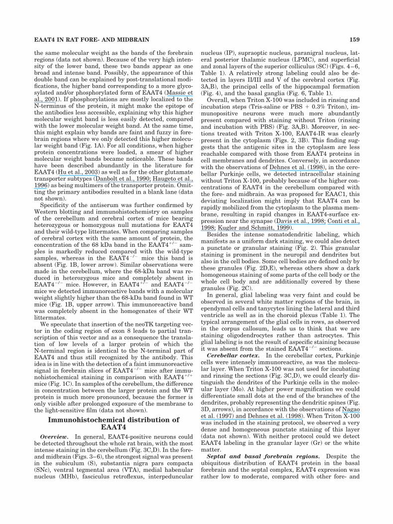
On the whole, EAAT4 expression within the thalamicnuclei was moderate relative to the general staining in-tensity in the fore- and midbrain, with a few exceptions asdescribed below. Very high expression levels could be de-tected in the MHb (Fig. 5A). When the remaining regionsof the forebrain were still immunonegative, staining of theMHb was already intense, as well as staining of the IP(Fig. 5E,F) and the axon bundle connecting both nuclei,namely, the fasciculus retroflexus (fr; Fig. 5B–E). For theMHb we detected a predominant neuropil staining. In thelateral habenular nucleus (LHb) neuropil staining wassignificantly lower. However, in this nucleus immunore-active neurons were abundantly present, contrary to theMHb (Fig. 5A, Table 1). A very intense neuropil stainingcould be observed in the caudal (IPC) and rostrolateralsubnucleus (IPRL) of the IP, whereas staining of the ros-tral (IPR), dorsomedial, and apical subnuclei was rathermoderate (Fig. 5F, Table 1). In frontal (Fig. 5A,B) as wellas sagittal (Fig. 5D,E) sections, the striate immunoreac-tive signal of the fasciculus retroflexus could be tracedfrom the departure of the axon bundle in the habenularnuclei (Hb) until arrival in the IP. On the course from theHb to the IP, branching of the fibers was observed onlyonce (Fig. 5B, arrows).
Hypothalamus and preoptic region. In general, rel-atively high expression levels of EAAT4 protein could beobserved within the hypothalamic region (Table 1). Thedifferent nuclei that compose the hypothalamus could be
Fig. 2. Detail of EAAT4-immunoreactive neurons in the cerebralcortex (A), VTA (B,C), SNc (D), and prerubral field (E) after stainingin the presence of Triton X-100. A: Pyramidal neurons of layer V of thecerebral cortex are intensely and homogeneously stained (large ar-rows). Dendrites of these neurons are visualized as an alignment ofimmunoreactive puncta (arrowheads). Intermingled with the in-tensely stained pyramidal neurons, faintly stained neurons could beseen that are composed of labeled granules (small arrows). Also neu-ropil staining consists of EAAT4-positive granules. B–D: EAAT4-IR inthe VTA (B,C) and SNc (D). Besides very intensely and homoge-neously stained neurons (B, arrow), we could detect neurons withnuclei devoid of this homogeneous and intense IR, although coveredwith immunoreactive puncta (C, arrows). Some of the neurons, as wellas their dendrites, are defined only by these immunoreactive puncta(D). E: Two faintly stained neurons located in the prerubral field showa punctate labeling. Scale bar � 10 �m in A–E.
161EAAT4 IN RAT FORE- AND MIDBRAIN

distinguished very easily after staining. Within each nu-cleus, EAAT4-immunoreactive cell bodies were homoge-neously distributed. Except for the dorsal hypothalamicnucleus, in which large intensely immunoreactive multi-polar neurons were sparsely distributed and in whichimmunoreactivity expanded into the dendrites, immuno-stained cell bodies were rather small and densely packed.Here too, the majority of the cell bodies consisted of a
collection of immunoreactive puncta or granules (data notshown).
Amygdaloid complex. In the majority of the nucleiconstituting the amygdala, a moderate to relatively highexpression level of EAAT4 protein could be observed (Ta-ble 1).
Basal ganglia and associated nuclei. EAAT4-positive neurons could be detected in all structures of the
Fig. 3. EAAT4-IR in cerebral (A,B) and cerebellar (C,D) cortexusing the EAAT4 antiserum in the presence (B) or absence (A,C,D) ofTriton X-100. A,B: Immunopositive neurons could be detected in alllayers of the cerebral cortex. When Triton was used (B) in the stainingprotocol, the number of immunopositive cells was significantly highercompared with Triton-free stainings (A). Apical dendrites of the py-
ramidal neurons in layer V could be followed into layer II. C: EAAT4labeling was highly enriched in the Purkinje cells (arrow) and in themolecular layer (Mo). The granular layer (Gr) was devoid of IR.D: Detail of an immunostained Purkinje cell and dendrites in the Mo.Small dots at the ends of the branches are suggestive of dendriticspines (arrows). Scale bar � 100 �m in A,B; 50 �m in C; 10 �m in D.
Fig. 4. Expression profile of EAAT4 throughout the hippocampalformation, as visualized by using the staining protocol including Tri-ton X-100. A: Overview of EAAT4–IR in a frontal section at Bregma�5.2. B: Relatively high expression levels of EAAT4 could be detectedin neurons located in the pyramidal layer (pyr) of the subiculum (S).Immunoreactive apical dendrites of the pyramidal neurons expandedinto the molecular layer (ml). C: Neurons of the stratum granulosum(sg) of the dentate gyrus (DG) were quite intensely stained, as werescattered neurons in the hilus (H). D: A higher power photomicro-graph of labeled neurons in the hilar region demonstrates the gran-ular staining. E–J: Also, the principal cells in the stratum pyramidale
(sp) of CA1 (E,H), CA2 (F,I), and CA3 (G,J) show a dense granularEAAT4 labeling (arrows), extending into the initial part of the apicaldendrites (H, arrowheads), as well as sparse nonprincipal cells instratum oriens (so) and stratum radiatum (sr). Abbreviations, Ctx,cerebral cortex; Ent, entorhinal cortex; fr, fasciculus retroflexus;LPMC, mediocaudal lateral posterior thalamic nucleus; LT, lateralterminal nucleus accessory optic tract; MG, medial geniculate nu-cleus; pol, polymorphic layer; SC, superior colliculus; sm, stratummoleculare; SNc, substantia nigra pars compacta; SNr, substantianigra pars reticulate; VTA, ventral tegmental area. Scale bar � 500�m in A; 100 �m in B,C; 50 �m in E–G; 25 �m in D; 10 �m in H–J.
162 A. MASSIE ET AL.

Figure 4
163EAAT4 IN RAT FORE- AND MIDBRAIN
basal ganglia, i.e., the striatum (Fig. 6A), globus pallidus(Fig. 6B), subthalamic nucleus (STN; Fig. 6C), substantianigra (SN; Fig. 6D), and entopeduncular nucleus (EP; Fig.6E), with the most intensely stained neurons beingpresent in the SNc (Figs. 4A, 6D). In the VTA, a basalganglia-related structure, labeling was as intense as inthe SNc (Figs. 4A, 6F).
In the striatum (Fig. 6A), immunoreactive intermediate-sized neurons were abundantly present, indicating that
EAAT4 is present in most of the �-aminobutyric acid(GABA)ergic efferent spiny neurons that account for 96% ofthe total neuronal population of the striatum (Ribak et al.,1979; Kita and Kitai, 1988). These immunoreactive cells arehomogeneously distributed without any correspondencewith patches (striosomes)/matrix compartments (Gerfen,1992; Hiroi, 1995). EAAT4 labeling was observed exclusivelyin cell bodies, dendrites being devoid of any staining. Inbetween these intermediate-sized neurons, a few solitary,
Fig. 5. EAAT4-IR in the presence of Triton X-100, in frontal (A–C,F) and sagittal (D,E) sections through the habenular nuclei (Hb;A,D), the fasciculus retroflexus (fr; B–E), and interpeduncular nucleus(IP; E,F). A: In the medial habenular nucleus (MHb) immunoreactiv-ity was confined to the neuropil, whereas in the lateral habenularnucleus (LHb) EAAT4 labeling could be observed in neuronal cellbodies. Arrowheads point to fibers of the fasciculus retroflexus, orig-inating in the habenular nuclei. B: An intense striate-like labeling inthe fasciculus retroflexus can be seen. Arrows point to an immunore-
active branch of the main bundle. C: In more detail, this striatelabeling consists of immunoreactive punctae. D,E: Sagittal sectionsdemonstrate the course of the fasciculus retroflexus, starting in theHb and arriving in the IP. F: Intense staining was also seen in therostrolateral subnucleus of the IP (IPRL) and caudal nucleus of the IP(IPC), whereas staining was less intense in the rostral nucleus of theIP (IPR). Abbreviations: 3V, third ventricle; LHbL, lateral part of thelateral Hb; LHbM, medial part of the lateral Hb. Scale bar � 100 �min A,F; 50 �m in B; 25 �m in C; 200 �m in D,E.
164 A. MASSIE ET AL.

large, very faintly stained neurons could be detected whenTriton X-100 was used (Fig. 6A, arrowhead). These largeneurons probably represent the large aspiny cholinergic in-
terneurons of type II (25–35 �m), which account for 1–2% ofthe total neuron population, because this is the only cell typein the striatum exhibiting this size (Yelnik, 2002). As in the
Fig. 6. Immunohistochemical staining in the presence of TritonX-100, illustrating the distribution of EAAT4 throughout the basalganglia. A: Detail of the striatum. In the middle of a large number ofhomogeneously distributed intermediate-sized neurons, very few,faintly stained large neurons could be distinguished (arrowheads).B,C: Detail of the globus pallidus (B) and the subthalamic nucleus(STN; C). EAAT4-immunoreactive neurons were homogeneously dis-tributed. EAAT4 label was present in the cell bodies and dendrites.D: A large number of intensely labeled neurons could be seen in thesubstantia nigra pars compacta (SNc). In the substantia nigra pars
laterale (SNl) cells were less abundant but still intensely stained.Only a few intensely labeled cells were present in the substantia nigrapars reticulata (SNr), in the middle of a number of faintly stainedneurons. E: In the entopeduncular nucleus (EP), sparse immunoreac-tive neurons could be visualized in a meshwork of immunoreactiveneuropil. F: In the VTA intensely and homogeneously stained neuronswere abundantly present. Abbreviations: cp, cerebral peduncle; MT,medial terminal nucleus accessory optic tract. Scale bar � 50 �m inA,B,D–F; 100 �m in C.
165EAAT4 IN RAT FORE- AND MIDBRAIN

striatum, in the globus pallidus (Fig. 6B) and STN (Fig. 6C),EAAT4-immunoreactive neurons were homogeneously dis-tributed. Staining was present in cell bodies and, contrary tothe striatum, also in dendrites. In the EP, a few immunore-active neurons could be visualized, whereas neuropil stain-ing was moderate (Fig. 6E).
As for the SNc (Figs. 2D, 6D) and VTA (Figs. 2B,C, 6F),very intensely and homogeneously stained, denselypacked neurons were visualized after EAAT4 staining.The nucleus of some immunopositive neurons was devoidof this intense and homogeneous labeling, yet was oftencovered by a number of immunoreactive puncta (Fig. 2C,arrow). Still other neurons and most of the dendrites weredefined only by these immunoreactive puncta (Fig. 2D,E).In the substantia nigra pars laterale (SNl), the stainingpattern of the immunoreactive neurons was similar tothat of the SNc; however, the density of immunoreactiveneurons was significantly smaller compared with the SNc.In contrast, in the substantia nigra pars reticulata (SNr),an occasional intensely stained neuron was present in themiddle of a small number of faintly stained neurons, char-acterized by a punctate labeling (Fig. 6D).
Mesencephalic regions. EAAT4 was omnipresent inthe midbrain region. Besides the aforementioned high ex-pression levels in the IP (Fig. 5F) and VTA (Fig. 6F),EAAT4 was also enriched in the superficial layers of theSC (Fig. 4A). The immunoreactive signal in the SC washigher than average, with a very intense neuropil stainingin the zonal layer and superficial gray layer. In the opticnerve layer, cell bodies as well as dendrites showed clearEAAT4 labeling.
Reverse transcription polymerase chainreaction
The presence of EAAT4 in the fasciculus retroflexus wasfurther investigated on the mRNA level, given the unex-pected abundant occurrence of EAAT4 protein in an axonbundle. After collecting the tissue samples by means of theLMD technique (Fig. 7A–C), making it possible to isolatetissue very precisely from the fasciculus retroflexus with-out contamination from any other nearby brain tissue,RT-PCR was performed with up- and downstream primerscorresponding to rat EAAT4 nucleotide sequences 906–926 and 1,299–1,319, respectively (Lin et al., 1998; Massieet al., 2001). As a control we also included mRNA samplesof the cerebellum, cerebral cortex, striatum, and hip-pocampus. For each condition a fragment was amplifiedwith a length of 414 bp, the expected length based on theknown sequence (Fig. 7D). No fragment was amplifiedwhen the cDNA in the PCR reaction mixture was replacedby water.
Real-time PCR
In order to estimate tissue expression levels of EAAT4mRNA, semiquantitative analysis was performed by usingreal-time PCR. As expected, the cerebellum was found tocontain by far the highest levels of EAAT4 mRNA (Fig.7E). By comparison, after setting the cerebellum as cali-brator, the cerebral cortex contained 6.7 � 1.8%, the hip-pocampus 2.0 � 0.4%, the striatum 1.6 � 0.2%, and thefasciculus retroflexus 4.7 � 1.8% of the total cerebellarEAAT4 mRNA content (n � 3). These mRNA levels are onthe same order of magnitude as those measured by Wardet al. (2004) in the cerebral cortex, i.e., 3.1% relative to thecerebellum.
Fig. 7. A: Photograph showing a frontal section from which thefasciculus retroflexus (arrows) was bilaterally removed by laser micro-dissection. B,C: Detail of the same section before (B) and after (C) thefasciculus retroflexus was laser-collected. D: Conventional end-point RT-PCR with specific primers for EAAT4 on mRNA from the cerebellum(Cb), cerebral cortex (Ctx), hippocampus (Hc), striatum (Str), and laser-captured fasciculus retroflexus (Fr). No band could be detected in thenegative control lane (NC), whereas for all other conditions a band of 414bp was visualized. E: Real-time PCR analysis of EAAT4 mRNA expres-sion in rat cerebellum (Cb), cerebral cortex (Ctx), hippocampus (Hc),striatum (Str), and fasciculus retroflexus (Fr). EAAT4 mRNA expressionlevels were normalized to GAPDH. The amount of transcript in forebrainregions was expressed as mean value (n � 3, � SD) relative to cerebel-lum (� 100%). Scale bar � 2 mm in A; 0.5 mm in B,C.
166 A. MASSIE ET AL.

In situ hybridization
As a last verification of the immunohistochemical datafor EAAT4, in situ hybridization was performed on sec-tions containing some important brain regions (Fig. 8).
EAAT4 mRNA expression was by far highest in the cere-bellar cortex (Fig. 8K). As for the cerebral cortex, we couldclearly distinguish a layered pattern with the highestsignal in layers II/III followed by layer V (Fig. 8A,C,E,G,I).Also, a relatively intense signal was observed in the piri-
Fig. 8. Distribution of EAAT4 mRNA throughout the fore- andmidbrain (A–J) and cerebellum (K). Autoradiograms were generatedafter in situ hybridization by using EAAT4 35S-labeled antisense(A,C,E,G,I,K) and sense (L) riboprobes. Neighboring sections werestained with Richardson’s methylene blue-azure II Nissl stain (B,D,F,H,J). A–J: In the fore -and midbrain, labeling was higher thanaverage in layers II/III and layer V of the cerebral cortex (Cx;A,C,E,G), the substantia nigra pars compacta (SNc; I), the medialgeniculate nucleus (MG; I), and the superior colliculus (SC; I). ClearEAAT4 labeling was observed in the preotpic area (PO; A), the bed
nucleus of the stria terminalis (Bst; A), piriform cortex (Pi; A), hypo-thalamic region (Hyp; C), premammillary nuclei (PM; E), the CA1region of the hippocampus (Hc; G), and the central gray (CG; I). K: Avery high signal was observed in the cerebellum (Cb). Abbreviations:BS, brainstem; CPu, caudate putamen; Hb, habenular nuclei; ic,internal capsule; NST, subthalamic nucleus; SNr, substantia nigrapars reticulate. Scale bar � 2 mm. Scale bar in panel A applies to allin situ hybridization figures (i.e. panel C, E, G, I, K, L), scale bar in Bapplies to all Nissl stains (i.e. D, F, H, J).
167EAAT4 IN RAT FORE- AND MIDBRAIN

form cortex (Fig. 8A,C). As for the hippocampal formation,relatively strong labeling could be seen in all CA regions,with the most intense signal in CA1 and in the dentategyrus (Fig. 8E,G). Concerning the nuclei of the basal gan-glia, high EAAT4 signal was present in the SNc (Fig. 8I).In addition, clear EAAT4 labeling occurred in the preopticarea, the bed nucleus of the stria terminalis, several hy-pothalamic, amygdaloid, and premammillary nuclei (Fig.8C,E), in the central gray, and in the intermediate graylayer of the superior colliculus (Fig. 8I). Labeling with thesense probe resulted in a faint background signal (Fig.8L).
DISCUSSION
In this paper we describe for the first time in detail thewidespread distribution of EAAT4 protein throughout therat fore- and midbrain. EAAT4, which is highly enrichedin the Purkinje cells of the cerebellum, was omnipresentin the rat fore- and midbrain, albeit at protein levelssignificantly lower compared with those in the cerebellum.On the whole, EAAT4-IR was localized not merely in glu-tamatergic and GABAergic neurons, but also in dopami-nergic and probably cholinergic neurons. Besides the neu-ronal localization of EAAT4 protein, a very faint gliallabeling in white matter of several CNS regions as well asin the ventricular walls could be observed. Given the un-expected high expression level of EAAT4 protein in thefasciculus retroflexus, the presence of EAAT4 in this axonbundle was confirmed at the mRNA level and estimated tobe 4.7% that of the cerebellum. In addition, the distribu-tion of EAAT4 in the main brain nuclei was confirmed onthe mRNA level by using conventional RT-PCR as well asin situ hybridization.
In general, given the presence of the glial glutamatetransporters, which are, in most fore- and midbrain re-gions, responsible for the bulk of glutamate reuptake, andgiven the low glutamate transport rate of EAAT4 (Torres-Salazar and Fahlke, 2007), we can imagine that the func-tional significance of EAAT4 in all these regions is notsolely linked to glutamate reuptake activity. EAAT4 haslarge substrate-gated Cl� currents that are not coupled tosubstrate transport. Thus, besides taking up glutamate toterminate glutamate neurotransmission, EAAT4 mightalso modulate neurotransmission by dampening of neuro-nal excitability via the substrate-gated anion conduc-tance, without interfering with glutamate homeostasis. Inaddition, it has been noted that metabotropic glutamatereceptor activation is specifically controlled by neuronalglutamate transporters in the cerebellar cortex (Brasnjoand Otis, 2001), and it was suggested by Otis et al. (2004)that this interaction could influence synaptic plasticity ina synapse-specific manner. Moreover, metabotropic gluta-mate receptors and neuronal glutamate transporters,which are closely associated in the perisynaptic space, canserve together as a physiological mechanism for limitingglutamate spillover from excitatory synapses (Otis et al.,2004). This is further supported by results obtained fromEAAT4-deficient mice, indicating that indeed in the cere-bellum EAAT4 is responsible for effectively preventingglutamate from spilling over to neighboring synapses(Takayasu et al., 2005).
Unfortunately, all studies on mice lacking EAAT4 areuninformative on brain regions outside the cerebellum(Huang et al., 2004; Takayasu et al., 2005, Yamashita et
al., 2006). The reuptake of glutamate by EAAT4 can alsohave a metabolic role. After being transported into thecell, glutamate can be converted into -ketoglutarate byglutamic acid decarboxylase and then enter the tricarbox-ylic acid cycle to produce ATP. The functionality of EAAT4in the fore- and midbrain is further supported by theobservation of Huerta et al. (2006) that the mRNA of theEAAT4-associated interacting proteins KIAA0302 andARHGEF11 is highly expressed throughout the brain.
In the hippocampal formation, a strong somatodendriticlabeling could be observed in the pyramidal cell layer ofthe subiculum. Also, the pyramidal cell layer of CA1–3and the granular cell layer of the dentate gyrus showed astaining higher than average. Besides EAAT4, all otherglutamate transporter subtypes (Lehre et al., 1995; Ku-gler and Schmitt, 1999) as well as glutamate receptorsubtypes (Monaghan et al., 1989; Petralia and Wenthold,1992) are expressed throughout the hippocampus, whichis not surprising given that the glutamatergic as well asthe GABAergic in- and output to all parts of the hippocam-pal formation is quite abundant (Ottersen and Storm-Mathisen, 1984). Moreover, in the stratum radiatum ofCA1, synapses are often found side by side without anyintervening glial processes (Harris and Stevens, 1989;Sorra and Harris, 1993; Lehre and Danbolt, 1998), mak-ing neuronal glutamate reuptake more important relativeto other brain regions (Rothstein et al., 1996).
Concerning the nuclei of the basal ganglia, very strongstaining could be observed in the SNc and in the VTA, abasal ganglia-related structure. Neurons from the SNcreceive, among other inputs, glutamatergic input from themedial prefrontal cortex, the STN, and the pedunculopon-tine region. Also the VTA receives glutamatergic inputfrom a number of different brain structures, including theprefrontal cortex (Carr and Sesack, 2000; Sesack andPickel, 1992; Thierry et al., 1983), the pedunculopontinenucleus (Charara et al., 1996; Kelland et al., 1993), andthe bed nucleus of the stria terminalis (Georges andAston-Jones, 2001, 2002). In the SNc as well as the VTA,EAAT4 is present on dopaminergic neurons. Direct evi-dence comes from staining performed on rats with6-OHDA lesions of the medial forebrain bundle. Fiveweeks after lesioning, a dopaminergic cell loss of 90% canbe observed in the SN and VTA (Sarre et al., 2004), whichcorresponds to the loss of EAAT4-immunoreactive cellsthat we observe in both nuclei (personal observations).The glutamatergic afferents to the SNc and VTA are prob-ably involved in the regulation of these dopaminergic neu-rons. Therefore, several glutamate receptor subtypes (N-methyl-D-aspartate [NMDA] and non-NMDA) have beenfound in both brain regions (Fallon and Loughlin, 1995;Kalivas, 1993). For the same reasons it is not surprisingthat EAAT4 has a high expression level in these neurons.
Regarding the other nuclei of the basal ganglia, as dis-cussed above, we detected a relatively high expressionlevel of EAAT4 in the striatum as well as the STN. Inter-estingly, both nuclei share common characteristics be-cause they both receive cortical and thalamic afferents(Parent, 1986; Canteras et al., 1990) and project to thepallidum and SN (Parent, 1986; Kita and Kitai, 1987). InGABAergic cells, glutamate taken up by EAAT4 can serveas a precursor for neosynthesis of GABA (Furuta et al.,1997; Seal and Amara, 1999) and thus enhance the inhib-itory synaptic strength. In addition, the presence ofEAAT4 on striatal neurons might be part of the glutama-
168 A. MASSIE ET AL.

tergic regulation of the dopaminergic activity in the stri-atum as well as the STN, described by Wullner et al.(1994) and Ampe et al. (2007), respectively. Small frac-tions of ionotropic and metabotropic striatal EAA bindingsites are located on dopaminergic terminals where theymay have a distinct impact on dopaminergic activity. Asfor the expression of EAAT4 on cholinergic neurons, wemight speculate that, again, it is linked to its chloridechannel properties, which make it behave, to some extent,as an inhibitory glutamate receptor (Dehnes et al., 1998).In addition, as described above, EAAT4 can transportglutamate into the neuron, which can then serve as anenergy source.
A moderate expression level could be observed in theglobus pallidus and the EP. In the SNr only very fewintensely labeled neurons could be seen intermingled witha moderate number of faintly stained neurons. However,in addition to the GABAergic input provided by thecaudate-putamen (Chevalier and Deniau, 1990; Deniau etal., 1978), a prominent glutamatergic innervation of theSNr is provided by fibers of the STN (Hammond et al.,1978; Kitai and Kita, 1987; Nakanishi et al., 1987), and allclasses of glutamate receptor subtypes are present in thisarea (Albin et al., 1992).
EAAT4-IR was very pronounced in the habenulo-interpeduncular system, including the fasciculus ret-roflexus. Given this unexpected expression of EAAT4 inan axon bundle, we further investigated the presence ofEAAT4 here. Real-time PCR revealed a relatively highamount of EAAT4 mRNA in the fasciculus retroflexuscompared with the other fore- and midbrain regions ex-amined. Surprisingly, and in sharp contrast to EAAT4, forall other high-affinity glutamate transporters, i.e.,GLAST, GLT-1, and EAAC1, the fasciculus retroflexus isdevoid of immunolabeling. In addition, the glial glutamatetransporters GLAST and GLT-1 are absent from the MHb,whereas EAAC1, like EAAT4, is expressed in this nucleus.However, all glutamate transporters show a considerableexpression level in the LHb (personal observations). TheMHb contains cholinergic and substance P-containingneurons, the former being crowded in the ventral two-thirds of the nucleus whereas the latter are exclusivelylocalized in the dorsal part (Contestabile et al., 1987).Some neurons of the MHb feature dense glutamatergicinnervation (Robertson et al., 1999), and glutamate servesas the excitatory transmitter at MHb-IP synapses (Brownet al., 1983; McGehee et al., 1995). This might explain thevery high expression levels of EAAT4 in both aforemen-tioned nuclei as well as the expression of metabotropicglutamate receptors, as reported before by Kinoshita et al.(1998).
The fasciculus retroflexus however, is an axonal tract.Projecting GABAergic neurons from the LHb sent axonsthrough the mantle of the tract to midbrain cell targets(the SN, VTA, and raphe) (Herkenham and Nauta, 1979;Carlson et al., 2000). In contrast, the MHb projectsthrough the core of the tract, corresponding to the cholin-ergic half of the fasciculus retroflexus (Herkenham andNauta, 1979; Woolf and Butcher, 1989), to the IP. Thispart of the fasciculus retroflexus contains the highest con-centration of nicotinic receptors in brain (London et al.,1985; Perry and Kellar, 1995). Our staining suggests thatthe immunoreactive fibers originate in the LHb as well asthe MHb. These fibers could be followed until arrival inthe IP. We were able to detect branching of the fasciculus
retroflexus only once, which is not surprising given thesmall width of such branches.
The most plausible explanation for the labeling of thisaxon bundle with the EAAT4 antibodies is that not theaxons but the glial processes, which are intimately asso-ciated with the axons, contain EAAT4 protein and mRNA.
The faint glial labeling obtained with the EAAT4 anti-serum was not restricted to glial cells located in the whitematter of several CNS regions. Also, ependymal cells lin-ing the lateral and third ventricle were stained. Hu et al.(2003) detected glial labeling. However, they did not ob-serve colocalization of EAAT4-IR with an oligodendrocytemarker, whereas they did with an astrocyte marker. Ourdata do not exclude the presence of EAAT4 in astrocytes,although the typical arrangement of the majority of thecells in rows strongly suggests that EAAT4 protein islocalized to oligodendrocytes. Also, EAAC1, originally con-sidered to be confined to neurons, was localized to glialcells. In accordance with our data, EAAC1 was expressedin oligodendrocytes of white matter, in ependymal cells,and in epithelial cells of the choroid plexus (Kugler andSchmitt, 1999). In epithelial cells of the choroid plexus noGLAST or GLT-1 could be detected, contrary to the ta-nycytes and ependymal cells (Berger and Hediger, 2000,2001). Therefore, the presence of EAAC1 and EAAT4 inthe choroid plexus might be important to prevent thepassage of glutamate from the blood stream into the cere-brospinal fluid, where the glutamate concentration is verylow, as stated by Kugler and Schmitt (1999).
In conclusion, some areas of high EAAT4-IR coincidewith target areas of dense glutamatergic innervation, e.g.,some corticofugal pathways, as described before forGLAST and GLT-1 (Lehre et al., 1995). However, someareas known to be low in glutamatergic innervation, e.g.globus pallidus, also show a considerable EAAT4 expres-sion level, indeed suggesting that the role of EAAT4 inthese regions goes beyond the canonical role of glutamateremoval. Thus, whether the functional significance of thiswidespread distribution of EAAT4 in the fore- and mid-brain is related to its re-uptake activities or to possibleother functional roles that this transporter can play, onaccount of its chloride channel properties (Sonders andAmara, 1996; Seal and Amara, 1999) or its close associa-tion with metabotropic glutamate receptors (Otis et al.,2004), needs further investigation. After all, besides de-creasing the total glutamate concentrations, EAAT4 canalso prevent excessive excitation and help membrane re-polarization inasmuch as its activation elicits chlorideinflux and consequent local hyperpolarization (Raiteri etal., 2002). Moreover, the interaction of the neuronal glu-tamate transporters with the metabotropic glutamate re-ceptors can influence synaptic plasticity as well as limitthe glutamate spillover from excitatory synapses, as de-scribed for the cerebellum (Otis et al., 2004).
ACKNOWLEDGMENTS
The authors acknowledge Mr. G. De Smet and Ms. R.Vanlaer for excellent technical assistance, Dr. M. Wa-tanabe for providing us with the antibodies, and Dr. K.Tanaka for the EAAT4�/� mice.
169EAAT4 IN RAT FORE- AND MIDBRAIN

LITERATURE CITED
Albin RL, Marcowiec RL, Hollingsworth ZR, Dure LS, Penney JB, YoungAB. 1992. Excitatory amino acid binding sites in the basal ganglia ofthe rat: a quantitative autoradiographic study. Neuroscience 46:35–48.
Amaral DG. 1978. A Golgi study of cell types in the hilar region of thehippocampus in the rat. J Comp Neurol 182:851–914.
Ampe B, Massie A, D’Haens J, Ebinger G, Michotte Y, Sarre S. 2007.NMDA-mediated release of glutamate and GABA in the subthalamicnucleus is mediated by dopamine: an in vivo microdialysis study inrats. J Neurochem 103:1063–1074.
Arriza JL, Eliasof S, Kavanaugh MP, Amara SG. 1997. Excitatory aminoacid transporter 5, a retinal glutamate transporter coupled to a chlo-ride conductance. Proc Natl Acad Sci U S A 94:4155–4160.
Attwell D. 2000. Brain uptake of glutamate: food for thought. J Nutr130:1023S–1025S.
Bar–Peled O, Ben-Hur H, Biegon A, Groner Y, Dewhurst S, Furuta A,Rothstein JD. 1997. Distribution of glutamate transporter subtypesduring human brain development. J Neurochem 69:2571–2580.
Berger UV, Hediger MA. 1998. Comparative analysis of glutamate trans-porter expression in rat brain using differential double in situ hybrid-ization. Anat Embryol (Berl) 198:13–30.
Berger UV, Hediger MA. 2000. Distribution of the glutamate transportersGLAST and GLT-1 in rat circumventricular organs, meninges, anddorsal root ganglia. J Comp Neurol 421:385–399.
Berger UV, Hediger MA. 2001. Differential distribution of the glutamatetransporters GLT-1 and GLAST in tanycytes of the third ventricle.J Comp Neurol 433:101–114.
Berger UV, DeSilva TM, Chen W, Rosenberg PA. 2005. Cellular andsubcellular mRNA localization of glutamate transporter isoformsGLT1a and GLT1b in rat brain by in situ hybridization. J Comp Neurol492:78–89.
Bergles DE, Diamond JS, Jahr CE. 1999. Clearance of glutamate inside thesynapse and beyond. Curr Opin Neurobiol 9:293–298.
Brasnjo G, Otis TS. 2001. Neuronal glutamate transporters control acti-vation of postsynaptic metabotropic glutamate receptors and influencecerebellar long-term depression. Neuron 31:607–616.
Brown DA, Docherty RJ, Halliwell JV. 1983. Chemical transmission in therat interpeduncular nucleus in vitro. J Physiol (Lond) 341:655–670.
Canteras NS, Shammah-Lagnado SJ, Silva AB, Ricardo JA. 1990. Afferentconnections of the subthalamic nucleus: a combined retrograde andanterograde horseradish peroxydase study in the rat. Brain Res 513:43–59.
Carlson J, Armstrong B, Switzer III RC, Ellison G. 2000. Selective neuro-toxic effects of nicotine on axons in fasciculus retroflexus further sup-port evidence that this is a weak link in brain across multiple drugs ofabuse. Neuropharmacology 39:2792–2798.
Carr DB, Sesack SR. 2000. Projections from the rat prefrontal cortex to theventral tegmental area: target specificity in the synaptic associationswith mesoaccumbens and mesocortical neurons. J Neurosci 20:3864–3873.
Charara A, Smith Y, Parent A. 1996. Glutamatergic inputs from thepedunculopontine nucleus to midbrain dopaminergic neurons in pri-mates: Phaseolus vulgaris-leucoagglutinin anterograde labeling com-bined with postembedding glutamate and GABA immunohistochemis-try. J Comp Neurol 364:254–266.
Chaudhry FA, Lehre KP, Campagne MV, Ottersen OP, Danbolt NC,Storm-Mathisen J. 1995. Glutamate transporters in glial plasma mem-branes: highly differentiated localizations revealed by quantitative ul-trastructural immunocytochemistry. Neuron 15:711–720.
Chen W, Aoki C, Mahadomrongkul V, Gruber CE, Wang GJ, Blitzblau R,Irwin N, Rosenberg PA. 2002. Expression of a variant form of theglutamate transporter GLT1 in neuronal cultures and in neurons andastrocytes in the rat brain. J Neurosci 22:2142–2152.
Chen W, Mahadomrongkul V, Berger UV, Bassan, M, DeSilva T, TanakaK, Irwin N, Aoki C, Rosenberg PA. 2004. The glutamate transporterGLT1a is expressed in excitatory axon terminals of mature hippocam-pal neurons. J Neurosci 24:1136–1148.
Chevalier G, Deniau JM. 1990. Disinhibition as a basic process in theexpression of striatal functions. Trends Neurosci 13:277–280.
Contestabile A, Villani L, Fasolo A, Franzoni MF, Gribaudo L, OktedalenO, Fonnum F. 1987. Topography of cholinergic and substance P path-ways in the habenulo-interpeduncular system of the rat. An immuno-cytochemical and microchemical approach. Neuroscience 21:253–270.
Conti F, DeBiasi S, Minelli A, Rothstein JD, Melone M. 1998. EAAC1, a
high-affinity glutamate transporter, is localized to astrocytes andGABAergic neurons besides pyramidal cells in the rat cerebral cortex.Cereb Cortex 8:108–116.
Danbolt NC. 2001. Glutamate uptake. Prog Neurobiol 65:1–105.Danbolt NC, Pines G, Kanner BI. 1990. Purification and reconstitution of
the sodium- and potassium-coupled glutamate transport glycoproteinfrom rat brain. Biochemistry 29:6734–6740.
Davis KE, Straff DJ, Weinstein EA, Bannerman PG, Correale DM, Roth-stein JD, Robinson MB. 1998. Multiple signaling pathways regulatecell surface expression and activity of the excitatory amino acid carrier1 subtype of Glu transporter in C6 glioma. J Neurosci 18:2475–2485.
Dehnes Y, Chaudhry FA, Ullensvang K, Lehre KP, Storm-Mathisen J,Danbolt NC. 1998. The glutamate transporter EAAT4 in rat cerebellarPurkinje cells: a glutamate-gated chloride channel concentrated nearthe synapse in parts of the dendritic membrane facing astroglia. J Neu-rosci 18:3606–3619.
Deniau JM, Hammon C, Riszk A, Feger J. 1978. Electrophysiologicalproperties of identified output neurons of the rat substantia nigra (parscompacta and pars reticulata): evidence for the existence of branchedneurons. Exp Brain Res 32:409–422.
Eliasof S, Arriza JL, Leighton BH, Kavanaugh MP, Amara SG. 1998.Excitatory amino acid transporters of the salamander retina: identifi-cation, localization and function. J Neurosci 18:698–712.
Fairman WA, Vandenberg RJ, Arriza JL, Kavanaugh MP, Amara SG.1995. An excitatory amino-acid transporter with properties of a ligand-gated chloride channel. Nature 375:599–603.
Fallon JH, Loughlin SE. 1995. Substantia nigra. In: Paxinos G, editor. Therat nervous system. San Diego: Academic Press. p 215–237.
Furuta A, Rothstein JD, Martin LJ. 1997. Glutamate transporter proteinsubtypes are expressed differentially during rat CNS development.J Neurosci 17:8363–8375.
Georges F, Aston-Jones G. 2001. Potent regulation of midbrain dopamineneurons by the bed nucleus of the stria terminalis. J Neurosci 21:RC160.
Georges F, Aston-Jones G. 2002. Activation of ventral tegmental area cellsby the bed nucleus of the stria terminalis: a novel excitatory amino acidinput to midbrain dopamine neurons. J Neurosci 22:5173–5187.
Gerfen CR. 1992. The neostriatal mosaic: multiple levels of compartimen-tal organization. Trends Neurosci 15:133–139.
Hammond C, Deniau JM, Rizk A, Feger J. 1978. Electrophysiologicaldemonstration of an excitatory subthalamonigral pathway in the rat.Brain Res 151:235–244.
Harris KM, Stevens JK. 1989. Dendritic spines of CA1 pyramidal cells inthe rat hippocampus: serial electron microscopy with reference to theirbiophysical characteristics. J Neurosci 9:2982–2997.
Haugeto O, Ullensvang K, Levy LM, Chaudhry FA, Honore T, Nielsen M,Lehre KP, Danbolt NC. 1996. Brain glutamate transporter proteinsform homomultimers. J Biol Chem 271:27715–27722.
Herkenham M, Nauta WJH. 1979. Efferent connections of the habenularnuclei in the rat. J Comp Neurol 187:19–48.
Hiroi N. 1995. Compartmental organization of calretinin in the rat stria-tum. Neurosci Lett 197:223–226.
Hu W-H, Walters WM, Xia X-M, Karmally SA, Bethea JR. 2003. Neuronalglutamate transporter EAAT4 is expressed in astrocytes. Glia 44:13–25.
Huang YH, Dykes-Hoberg M, Tanaka K, Rothstein JD, Bergles DE. 2004.Climbing fiber activation of EAAT4 transporters and kainate receptorsin cerebellar Purkinje cells. J Neurosci 24:103–111.
Huerta I, McCullumsmith RE, Haroutunian V, Gimenez-Amaya JM,Meador-Woodruff JH. 2006. Expression of excitatory amino acid trans-porter interacting protein transcripts in the thalamus in schizophre-nia. Synapse 59:394–402.
Inage YW, Itoh M, Wada K, Takashima S. 1998. Expression of two gluta-mate transporters, GLAST and EAAT4, in the human cerebellum: theircorrelation in development and neonatal hypoxic-ischemic damage.J Neuropathol Exp Neurol 57:554–562.
Itoh M, Watanabe Y, Watanabe M, Tanaka K, Wada K, Takashima S.1997. Expression of a glutamate transporter subtype, EAAT4, in thedeveloping human cerebellum. Brain Res 767:265–271.
Kalivas PW. 1993. Neurotransmitter regulation of dopamine neurons inthe ventral tegmental area. Brain Res Rev 18:75–113.
Kanai Y, Hediger MA. 1992. Primary structure and functional character-ization of a high-affinity glutamate transporter. Nature 360:467–471.
Kelland MD, Freeman AS, Rubin J, Chiodo LA. 1993. Ascending afferent
170 A. MASSIE ET AL.

regulation of rat midbrain dopamine neurons. Brain Res Bull 31:539–546.
Kinoshita A, Shigemoto R, Ohishi H, Van der Putten H, Mizuno N. 1998.Immunohistochemical localization of metabotropic glutamate recep-tors, mGluR7a and mGluR7b, in the central nervous system of theadult rat and mouse: a light and electron microscopic study. J CompNeurol 393:332–352.
Kita H, Kitai ST. 1987. Efferent projections of the subthalamic nucleus inthe rat: light and electron microscopic analysis with the PHA-Lmethod. J Comp Neurol 260:435–452.
Kita H, Kitai ST. 1988. Glutamate decarboxylase immunoreactive neuronsin rat neostriatum: their morphological types and populations. BrainRes 447:346–352.
Kitai ST, Kita H. 1987. Anatomy and physiology of the subthalamic nu-cleus: a driving force of the basal ganglia. New York: Plenum Press.
Kugler P, Schmitt A. 1999. Glutamate transporter EAAC1 is expressed inneurons and glial cells in the rat nervous system. Glia 27:129–142.
Lehre KP, Danbolt NC. 1998. The number of glutamate transporter sub-type molecules at glutamatergic synapses: chemical and stereologicalquantification in young adult rat brain. J Neurosci 18:8751–8757.
Lehre KP, Levy LM, Ottersen OP, Storm-Mathisen J, Danbolt NC. 1995.Differential expression of two glial glutamate transporters in the ratbrain: quantitative and immunocytochemical observations. J Neurosci15:1835–1853.
Liang J, Takeuchi H, Doi Y, Kawanokuchi J, Sonobe Y, Jin S, Yawata I, LiH, Yasuoka S, Mizuno T, Suzumura A. 2008. Excitatory amino acidtransporter expression by astrocytes is neuroprotective against micro-glial excitotoxicity. Brain Res 1210:11–19.
Lin CL, Tzingounis AV, Jin L, Furuta A, Kavanaugh MD, Rothstein JD.1998. Molecular cloning and expression of the rat EAAT4 glutamatetransporter subtype. Brain Res Mol Brain Res 63:174–179.
London ED, Waller SB, Wamsley JK. 1985. Autoradiographic localizationof [3H]nicotine binding sites in the rat brain. Neurosci Lett 53:179–184.
Massie A, Cnops L, Jacobs S, Van Damme K, Vandenbussche E, Eysel UT,Vandesande F, Arckens L. 2003. Glutamate levels and transport in cat(Felis catus) area 17 during cortical reorganization following binocularretinal lesions. J Neurochem 84:1387–1397.
Massie A, Vandesande F, Arckens L. 2001. Expression of the high-affinityglutamate transporter EAAT4 in mammalian cerebral cortex. Neuro-report 12:393–397.
McCullumsmith RE, Meador-Woodruff JH. 2002. Striatal excitatory aminoacid transporter transcript expression in schizophrenia, bipolar disor-der, and major depressive disorder. Neuropsychopharmacology 26:368–375.
McGehee D, Heath M, Gelber S, Devay P, Role LW. 1995. Nicotine en-hancement of fast excitatory synaptic transmission in CNS by presyn-aptic receptors. Science 269:1692–1697.
Mennerick S, Dhond RP, Benz A, Xu W, Rothstein JD, Danbolt NC,Isenberg KE, Zorumski CF. 1998. Neuronal expression of the gluta-mate transporter GLT-1 in hippocampal microcultures. J Neurosci18:4490–4499.
Monaghan DT, Bridges RJ, Cotman CW. 1989. The excitatory amino acidreceptors: their classes, pharmacology, and distinct properties in thefunction of the central nervous system. Annu Rev Pharmacol Toxicol29:365–402.
Nagao S, Kwak S, Kanazawa I. 1997. EAAT4, a glutamate transporterwith properties of a chloride channel, is predominantly localized inPurkinje cell dendrites, and forms parasagittal compartments in ratcerebellum. Neuroscience 78:929–933.
Nakanishi H, Kita H, Kitai ST. 1987. Intracellular study of rat substantianigra pars reticulata neurons in an in vitro slice preparation: electricalmembrane properties and response characteristics to subthalamicstimulation. Brain Res 437:45–55.
Otis TS, Brasnjo G, Dzubay JA, Pratap M. 2004. Interactions betweenglutamate transporters and metabotropic glutamate receptors at exci-tatory synapses in the cerebellar cortex. Neurochem Int 45:537–544.
Ottersen OP, Storm-Mathisen J. 1984. Neurons containing or accumulat-ing transmitter amino acids. In: Bjorklund A, Hokfelt T, Kuhar MJ,editors. Handbook of chemical neuroanatomy. Vol. 3. Classical trans-mitters and transmitter receptors in the CNS, Part II. New York:Elsevier Science Publishers. p 141–216.
Parent A. 1986. Comparative neurobiology of the basal ganglia. New York:John Wiley & Sons.
Perry DC, Kellar KJ. 1995. [3H]Epibatidine labels nicotinic receptors in rat
brain: an autoradiographic study. J Pharmacol Exp Ther 275:1030–1034.
Petralia RS, Wenthold RJ. 1992. Light and electron immunocytochemicallocalization of AMPA-selective glutamate receptors in the rat brain.J Comp Neurol 318:329–354.
Pignataro L, Sitaramayya A, Finnemann SC, Sarthy VP. 2005. Nonsynap-tic localization of the excitatory amino acid transporter 4 in photore-ceptors. Mol Cell Neurosci 28:440–51.
Pines G, Danbolt NC, Bjoras M, Zhang Y, Bendahan A, Eide L, Koepsell H,Storm-Mathisen J, Seeberg E, Kanner BI. 1992. Cloning and expres-sion of a rat brain L-glutamate transporter. Nature 360:464–467.
Qu Y, Moons L, Vandesande F. 1997. Determination of serotonin, cat-echolamines and their metabolites by direct injection of supernatantsfrom chicken brain tissue homogenate using liquid chromatographywith electrochemical detection. J Chromatogr B 704:351–358.
Raiteri L, Raiteri M, Bonanno G. 2002. Coexistence and function of differ-ent neurotransmitter transporters in the plasma membrane of CNSneurons. Prog Neurobiol 68:287–309.
Rakhade SN, Loeb JA. 2008. Focal reduction of neuronal glutamate trans-porters in human neocortical epilepsy. Epilepsia 49:226–236.
Rauen T, Kanner BI. 1994. Localization of the glutamate transporterGLT-1 in rat and macaque monkey retinae. Neurosci Lett 169:137–140.
Ribak CE, Vaughn JE, Roberts E. 1979. The GABA neurons and their axonterminals in rat corpus striatum as demonstrated by GAD immunocy-tochemistry. J Comp Neurol 187:261–284.
Richardson KD, Jarret L, Finke EH. 1960. Embedding in epoxy resins forultrathin sectioning in electron microscopy. Stain Technol 35:313–323.
Robertson SJ, Burnashev N, Edwards FA. 1999. Ca2� permeability andkinetics of glutamate receptors in rat medial habenula neurones: im-plications for purinergic transmission in this nucleus. J Physiol 518:539–549.
Rothstein JD, Martin L, Levey AI, Dykes-Hoberg M, Jin L, Wu D, Nash N,Kuncl RW. 1994. Localization of neuronal and glial glutamate trans-porters. Neuron 13:713–725.
Rothstein JD, Dykes-Hoberg M, Pardo CA, Bristol LA, Jin L, Kuncl RW,Kanai Y, Hediger MA, Wang Y, Schielke JP, Welty DF. 1996. Knockoutof glutamate transports reveals a major role for astroglial transport inexcitotoxicty and clearance of glutamate. Neuron 16:675–686.
Saier MH. 1999. Eukaryotic transmembrane solute transport systems. IntRev Cytol 190:61–136.
Sarre S, Yuan H, Jonkers N, Van Hemelrijck A, Ebinger G, Michotte Y.2004. In vivo characterization of somatodendritic dopamine release inthe substantia nigra of 6-hydroxydopamine-lesioned rats. J Neurochem90:29–39.
Schmitt A, Asan E, Puschel B, Jons T, Kugler P. 1996. Expression of theglutamate transporter GLT-1 in neural cells of the rat central nervoussystem: non-radioactive in situ hybridization and comparative immu-nocytochemistry. Neuroscience 71:989–1004.
Seal RP, Amara SG.1999. Excitatory amino acid transporters: a family influx. Annu Rev Pharmacol Toxicol 39:431–456.
Sesack SR, Pickel VM. 1992. Prefrontal cortical efferents in the rat synapseon unlabeled neuronal targets of catecholamine terminals in the nu-cleus accumbens septi and on dopamine neurons in the ventral teg-mental area. J Comp Neurol 320:145–160.
Shu S, Ju G, Fan L. 1988. The glucose oxidase-DAB-nickel method inperoxidase histochemistry of the nervous system. Neurosci Lett 84:169–171.
Slotboom DJ, Konings WN, Lolkema JS. 1999. Structural features of theglutamate transporter family. Microbiol Mol Biol Rev 63:293–307.
Sonders MS, Amara SG. 1996. Channels in transporters. Curr Opin Neu-robiol 6:294–302.
Soriano E, Frotscher M. 1994. Mossy cells of the rat fascia dentate areglutamate-immunoreactive. Hippocampus 4:65–69.
Sorra KE, Harris KM. 1993. Occurrence and three-dimensional structureof multiple synapses between individual radiatum axons and theirtarget pyramidal cells in hippocampal area CA1. J Neurosci 13:3736–3748.
Storck T, Schulte S, Hofmann K, Stoffel W. 1992. Structure, expression,and functional analysis of a Na�-dependent glutamate/aspartatetransporter from rat brain. Proc Natl Acad Sci U S A 89:10955–10959.
Takayasu Y, Iiono M, Kakegawa W, Maeno H, Watase K, Wada K, Yanagi-hara D, Miyazaki T, Komine O, Watanabe M, Tanaka K, Ozawa S.2005. Differential roles of glial and neuronal glutamate transporters inPurkinje cell synapses. J Neurosci 25:8788–8793.
171EAAT4 IN RAT FORE- AND MIDBRAIN
Tanaka K. 1993. Expression cloning of a rat glutamate transporter. Neu-rosci Res 16:149–153.
Tanaka J, Ichikawa R, Watanabe M, Tanaka K, Inoue Y. 1997. Extra-junctional localization of glutamate transporter EAAT4 at excitatoryPurkinje cell synapses. Neuroreport 8, 2461–2464.
Thierry AM, Chevalier G, Ferron A, Glowinski J. 1983. Diencephalic andmesencephalic efferents of the medial prefrontal cortex in the rat:electrophysiological evidence for the existence of branched axons. ExpBrain Res 50:275–282.
Torp R, Danbolt NC, Babaie E, Bjoras M, Seeberg E, Storm-Mathisen J,Ottersen OP. 1994. Differential expression of two glial glutamatetransporters in the rat brain: an in situ hybridization study. EurJ Neurosci 6:936–942.
Torp R, Danbolt NC, Storm-Mathisen J, Ottersen OP. 1996. Differentialcellular and regional distribution of the glutamate transporters GLT-1and rEAAC1: an in situ hybridization analysis in the rat brain. SocNeurosci Abstr 22:356.
Torres-Salazar D, Fahlke C. 2007. Neuronal glutamate transporters varyin substrate transport rate but not in unitary anion channel conduc-tance. J Biol Chem 282:34719–34726.
Vandenberg RJ, Arriza JL, Amara SG, Kavanaugh MP. 1995. Constitutiveion fluxes and substrate binding domains of human glutamate trans-porters. J Biol Chem 270:17668–17671.
Wadiche JI, Amara SG, Kavanaugh MP. 1995a. Ion fluxes associated withexcitatory amino acid transport. Neuron 15:721–728.
Wadiche JI, Arriza JL, Amara SG, Kavanaugh MP. 1995b. Kinetics of ahuman glutamate transporter. Neuron 14:1019–1027.
Wang GJ, Chung HJ, Schnuer J, Pratt K, Zable AC, Kavanaugh MP,
Rosenberg PA. 1998. High affinity glutamate transport in rat corticalneurons in culture. Mol Pharmacol 53:88–96.
Ward MM, Jobling AI, Puthussery T, Foster LE, Fletcher EL. 2004. Local-ization and expression of the glutamate transporter, excitatory aminoacid transporter 4, within astrocytes of the rat retina. Cell Tissue Res315:305–310.
Wenzel HJ, Buckmaster PS, Anderson NL, Wenzel ME, Schwartzkroin PA.1997. Ultrastructural localization of neurotransmitter immunoreactiv-ity in mossy cell axons and their synaptic targets in the rat dentategyrus. Hippocampus 7:559–570.
Woolf N, Butcher L. 1989. Cholinergic systems in the rat brain: IV. De-scending projections of the pontomesencephalic tegmentum. Brain ResBull 23:519–540.
Wullner U, Testa CM, Catania MV, Young AB, Penney JB. 1994. Gluta-mate receptors in striatum and substantia nigra: effects of medialforebrain bundle lesions. Brain Res 645:98–102.
Yamada K, Watanabe M, Shibata T, Tanaka K, Wada K, Inoue Y. 1996.EAAT4 is a post-synaptic glutamate transporter at Purkinje cell syn-apses. Neuroreport 7:2013–2017.
Yamashita A, Makita K, Kuroiwa T, Tanaka K. 2006. Glutamate trans-porters GLAST and EAAT4 regulate postischemic Purkinje cell death:an in vivo study using a cardiac arrest model in mice lacking GLAST orEAAT4. Neurosci Res 55:264–270.
Yelnik J. 2002. Functional anatomy of the basal ganglia. Move Disord17:S15–S21.
Yi JH, Herrero R, Chen G, Hazell AS. 2007. Glutamate transporter EAAT4is increased in hippocampal astrocytes following lateral fluid-percussion injury in the rat. Brain Res 1154:200–205.
172 A. MASSIE ET AL.
Copyright © 2022 FDOKUMEN