Differential sensitivity of the Brattleboro rat to glutamatergic and dopaminergic perturbation
CONEUR-921; NO. OF PAGES 9
Please cite this article in press as: Koos T, et al. Glutamatergic signaling by midbrain dopaminergic neurons: recent insights from optogenetic, molecular and behavioral studies, Curr OpinNeurobiol (2011), doi:10.1016/j.conb.2011.05.010
Available online at www.sciencedirect.com
Glutamatergic signaling by midbrain dopaminergic neurons:recent insights from optogenetic, molecular and behavioralstudiesTibor Koos, Fatuel Tecuapetla and James M Tepper
Although the notion that dopaminergic neurons utilize
glutamate as a co-transmitter has long been supported by
tantalizing molecular, immunocytochemical and
electrophysiological evidence it has only been with the recent
addition of optogenetic and other approaches that the
existence and functional relevance of this mechanism could be
unambiguously demonstrated. Here we discuss the possible
mechanisms of action of glutamate released from
mesoaccumbens dopaminergic neurons based on recently
accumulated evidence. Surprisingly, rather then to confirm a
role in conventional fast excitatory transmission, the latest
evidence suggests that glutamate released from dopaminergic
neurons may primarily act through different unconventional
presynaptic and postsynaptic mechanisms.
AddressCenter for Molecular and Behavioral Neuroscience, Rutgers University,197 University Avenue, Newark, NJ 07102, United States
Corresponding authors: Koos, Tibor ([email protected]), Tepper,James M ([email protected])
Current Opinion in Neurobiology 2011, 21:1–9
This review comes from a themed issue onBehavioral and Cognitive NeuroscienceEdited by Ann Graybiel and Richard Morris
0959-4388/$ – see front matterPublished by Elsevier Ltd.
DOI 10.1016/j.conb.2011.05.010
IntroductionAlthough the precise role(s) of the midbrain dopamin-ergic system in learning are not yet completely under-stood, there is compelling neurophysiological evidencethat these neurons encode information about momentarychanges in the expectation of reinforcement [1]. Thisinformation is represented in brief firing rate changeswhich conform with remarkable precision to the theor-etically predicted properties of a supervisory signalrequired for the adaptive modification of sensory repres-entation and behavior in temporal difference models ofconditioning [1–3]. In order to fulfill this role, dopamin-ergic neurons need to activate postsynaptic signalingmechanisms that exhibit sufficiently fast kinetics topreserve the temporally encoded information about
reinforcement. Although dopaminergic mechanismsmay themselves serve such a function [3,4], recent evi-dence of glutamatergic signaling by midbrain dopamin-ergic neurons suggests the exciting possibility that thisnovel glutamatergic mechanism may also be involved inthe transmission of reward information. Here we reviewthe currently available information about glutamatergicsignaling by mesencephalic dopaminergic neurons andexamine the implications of this phenomenon for thefunctioning of the mesoaccumbens dopaminergic system.
Controversy about functional glutamatergicsignaling by mesencephalic dopaminergicneuronsThe possibility of glutamate release by dopaminergicneurons was first suggested by the detection of phosphateactivated glutaminase, a presumed marker of a glutama-tergic phenotype, in many monoaminergic neurons in-cluding mesencephalic dopaminergic neurons [5].Further molecular evidence was obtained with the detec-tion of an isoform of the vesicular glutamate transporter,VGluT2, in dopaminergic neurons under various con-ditions and using an array of techniques [6–16]. The firstfunctional evidence for co-release of glutamate by dopa-minergic neurons was obtained from recordings inprimary cell cultures of mesencephalic dopaminergicneurons in which autaptic and synaptic glutamatergicresponses were observed [17] – a finding that was laterreplicated and extended [7,10,12,18].
Despite these lines of evidence it remained controversialwhether there was glutamate co-release from dopamin-ergic neurons in the normal adult brain and if so, wassufficient to support functionally significant glutamater-gic communication with postsynaptic targets. The con-troversy arose primarily from the conflicting findings andfunctional interpretations of comparable experimentsthat in most cases resulted from inherent limitations ofavailable methods. For example, the electrophysiologicaldemonstration of glutamatergic transmission by dopamin-ergic neurons in cell culture was weakened by evidenceshowing that VGluT2 expression was strongly upregulated in dopaminergic neurons in culture prep-arations partly owing to the loss of contact mediatedinhibition by postsynaptic targets [7,14]. Similarly, itwas shown that VGluT2 was expressed at low levels invivo and that its expression decreased over development[6,13,14] so that it could be reliably detected only with
www.sciencedirect.com Current Opinion in Neurobiology 2011, 21:1–9
RT-PCR based methods or indirect reporters [6,14].These findings raised the possibility that the releasableglutamate content of adult dopaminergic neurons may beinsufficient to support a functional glutamatergic pheno-type and pointed to alternative developmental functionsor an involvement in neuronal plasticity and repair[6,15,16,19].
In parallel with these studies, in a series of technicallychallenging and creative experiments the existence ofglutamatergic signaling in the forebrain by mesolimbicdopaminergic neurons was also examined in acute slicepreparations [8,20,21]. The important advantages of thisapproach include the possibility of providing direct func-tional evidence for glutamatergic synaptic transmissionand avoiding the potential problems and potential associ-ated with cell culture methods. Although these exper-iments were able to demonstrate that electrical orchemical stimulation of neurons in the VTA can elicitglutamatergic responses in spiny projection neurons(SPN) of the nucleus accumbens that exhibit the con-duction latency and D2 receptor (D2R) mediated regu-lation expected for responses mediated by mesolimbicdopaminergic axons [8,20,21], the complex and poorlyunderstood organization of the VTA and surroundingareas, and in particular, the documented existenceof non-dopaminergic glutamatergic neurons in theVTA[22] and the expression of D2Rs by non-dopamin-ergic VTA neurons [23] left open the possibility that anunknown, possibly significant fraction of the responsesrecorded in the accumbens following stimulation of theVTA originated from non-dopaminergic projections.
Optogenetic disambiguation of theglutamatergic phenotype of dopaminergicneuronsThe controversy about the glutamatergic nature of dopa-minergic neurons has recently been resolved in 2 studiesin which postsynaptic responses of SPNs in the nucleusaccumbens were examined using channelrhodopsin-2(ChR2) mediated optogenetic activation of dopaminergicaxons [24,25]. In these experiments ChR2 was expressedin dopaminergic neurons using viral mediated transfer ofa Cre-lox controlled transgene [26–29] in transgenic micethat express Cre recombinase in neurons expressing thedopamine transporter (DAT; DAT-IRES-Cre) or tyrosinehydroxylase (TH; BAC TH-Cre). This approach ensuresthe specific and selective activation of dopaminergicaxons because the expression of ChR2 in the absenceof Cre recombinase is completely prevented by theinverted orientation of the ChR2 coding sequence andbecause the expression of Cre itself is specific in thesemice to neurons that contain TH or DAT and which aretherefore conclusively dopaminergic [26–29].
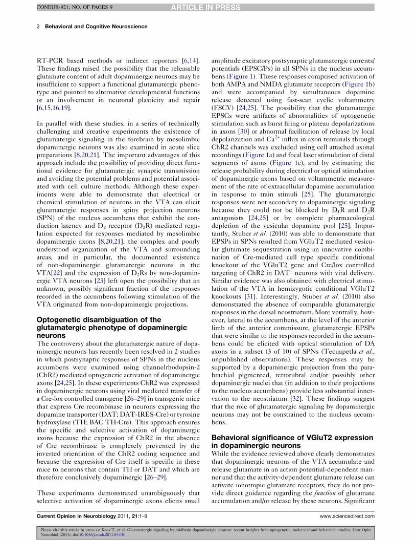
These experiments demonstrated unambiguously thatselective activation of dopaminergic axons elicits small
amplitude excitatory postsynaptic glutamatergic currents/potentials (EPSC/Ps) in all SPNs in the nucleus accum-bens (Figure 1). These responses comprised activation ofboth AMPA and NMDA glutamate receptors (Figure 1b)and were accompanied by simultaneous dopaminerelease detected using fast-scan cyclic voltammetry(FSCV) [24,25]. The possibility that the glutamatergicEPSCs were artifacts of abnormalities of optogeneticstimulation such as burst firing or plateau depolarizationsin axons [30] or abnormal facilitation of release by localdepolarization and Ca2+ influx in axon terminals throughChR2 channels was excluded using cell attached axonalrecordings (Figure 1a) and focal laser stimulation of distalsegments of axons (Figure 1c), and by estimating therelease probability during electrical or optical stimulationof dopaminergic axons based on voltammetric measure-ment of the rate of extracellular dopamine accumulationin response to train stimuli [25]. The glutamatergicresponses were not secondary to dopaminergic signalingbecause they could not be blocked by D1R and D2Rantagonists [24,25] or by complete pharmacologicaldepletion of the vesicular dopamine pool [25]. Impor-tantly, Stuber et al. (2010) was able to demonstrate thatEPSPs in SPNs resulted from VGluT2 mediated vesicu-lar glutamate sequestration using an innovative combi-nation of Cre-mediated cell type specific conditionalknockout of the VGluT2 gene and Cre/lox controlledtargeting of ChR2 in DAT+ neurons with viral delivery.Similar evidence was also obtained with electrical stimu-lation of the VTA in hemizygotic conditional VGluT2knockouts [31]. Interestingly, Stuber et al. (2010) alsodemonstrated the absence of comparable glutamatergicresponses in the dorsal neostriatum. More ventrally, how-ever, lateral to the accumbens, at the level of the anteriorlimb of the anterior commissure, glutamatergic EPSPsthat were similar to the responses recorded in the accum-bens could be elicited with optical stimulation of DAaxons in a subset (3 of 10) of SPNs (Tecuapetla et al.,unpublished observations). These responses may besupported by a dopaminergic projection from the para-brachial pigmented, retrorubral and/or possibly otherdopaminergic nuclei that (in addition to their projectionsto the nucleus accumbens) provide less substantial inner-vation to the neostriatum [32]. These findings suggestthat the role of glutamatergic signaling by dopaminergicneurons may not be constrained to the nucleus accum-bens.
Behavioral significance of VGluT2 expressionin dopaminergic neuronsWhile the evidence reviewed above clearly demonstratesthat dopaminergic neurons of the VTA accumulate andrelease glutamate in an action potential-dependent man-ner and that the activity-dependent glutamate release canactivate ionotropic glutamate receptors, they do not pro-vide direct guidance regarding the function of glutamateaccumulation and/or release by these neurons. Significant
2 Behavioral and Cognitive Neuroscience
CONEUR-921; NO. OF PAGES 9
Please cite this article in press as: Koos T, et al. Glutamatergic signaling by midbrain dopaminergic neurons: recent insights from optogenetic, molecular and behavioral studies, Curr OpinNeurobiol (2011), doi:10.1016/j.conb.2011.05.010
Current Opinion in Neurobiology 2011, 21:1–9 www.sciencedirect.com
Glutamatergic signaling by dopaminergic neurons Koos, Tecuapetla and Tepper 3
CONEUR-921; NO. OF PAGES 9
Please cite this article in press as: Koos T, et al. Glutamatergic signaling by midbrain dopaminergic neurons: recent insights from optogenetic, molecular and behavioral studies, Curr OpinNeurobiol (2011), doi:10.1016/j.conb.2011.05.010
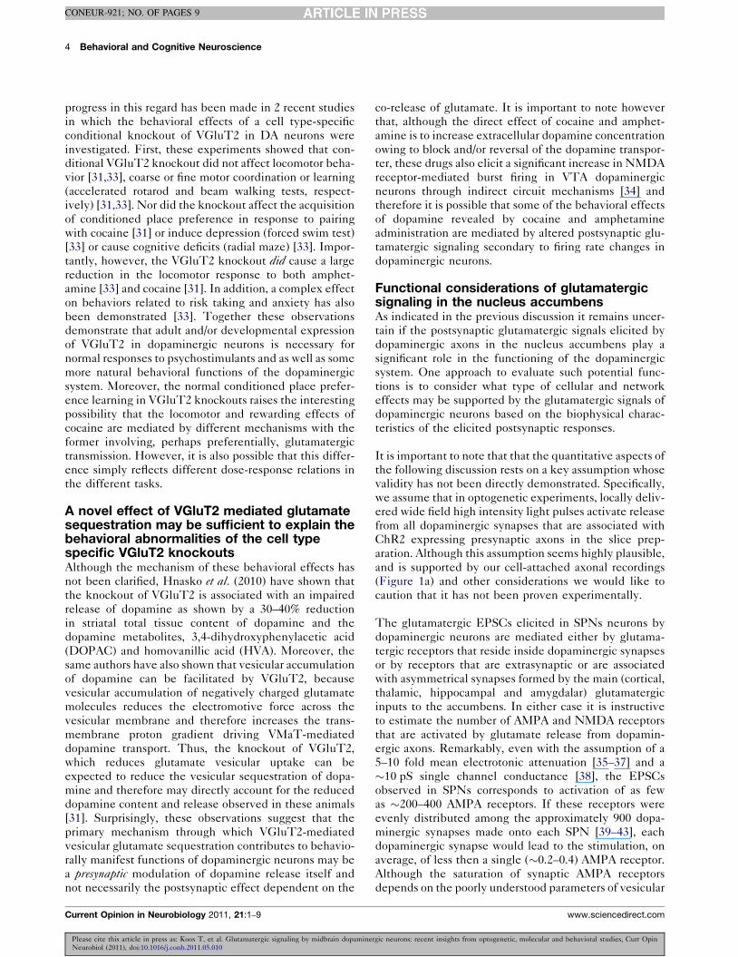
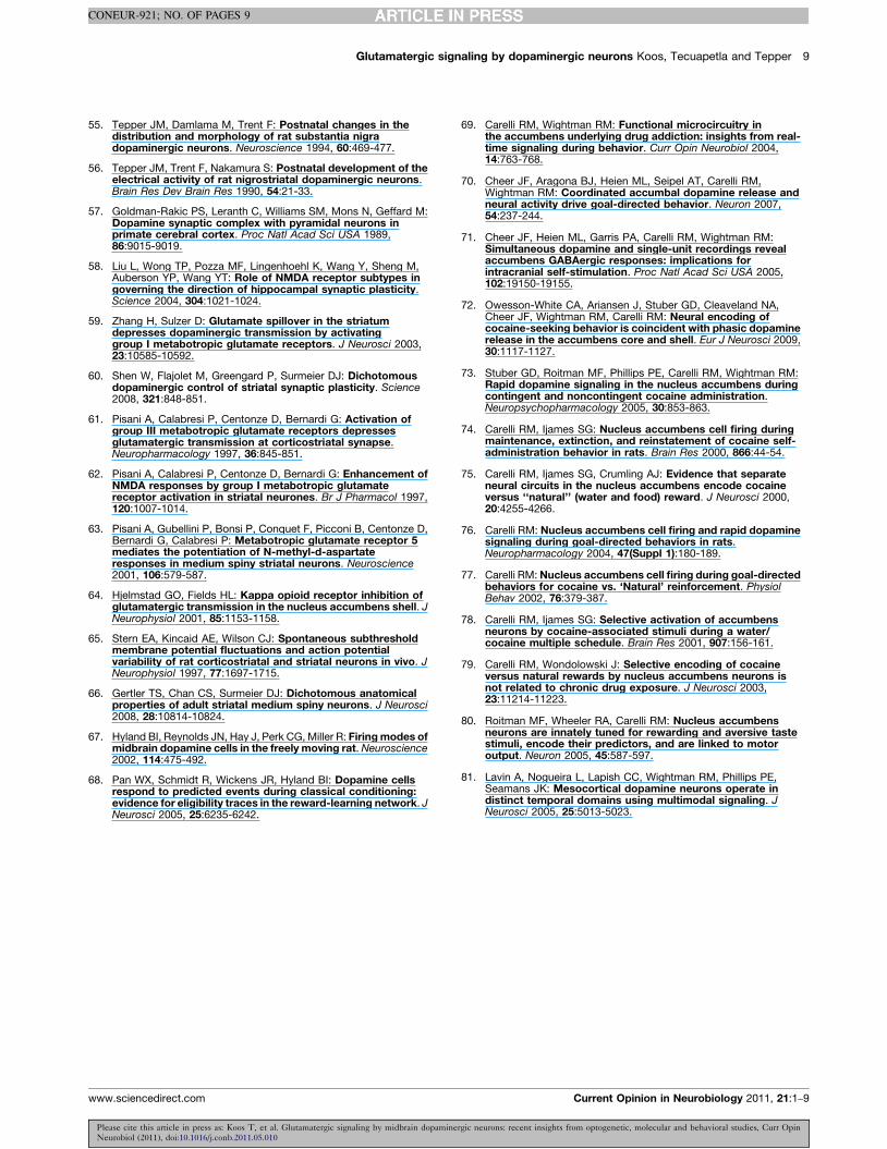
Figure 1
33.3 Hz
10 ms20 pA
Wash
DNQX
Control
(b)(a)
20 pA
5 pA
DA axon
MS soma
10 ms
St
Acc
100 ms10 pAnon-NMDA
NMDA
(d)
-91 mV
-45 mV
5 mV25 ms
30 pA
Im
50 ms
(c)
10 pA
5 ms1460 µm810 µm250 µm
10
50.0 0.5 1.0 1.5
Distance (mm)
Late
ncy
(ms)
Current Opinion in Neurobiology
Biophysical properties of glutamatergic responses elicited with selective stimulation of dopaminergic afferents to projection neurons in the nucleusaccumbens. (a) Optogenetic activation of dopaminergic axons in the nucleus accumbens. A DIC photomicrograph (top left panel) illustrates thelocalization of a typical recording site in the accumbens (note the position of 2 patch pipettes used for simultaneous cell-attached axonal, and wholecell recordings (arrows) and the relative position of the optical fiber (arrowhead), used for focal stimulation near the recording site). An epifluorescencecomposite micrograph obtained with YFP (yellow) and Texas-Red (red) filter sets illustrates a typical axon bleb formed by the severing of adopaminergic axon at the surface of the slice that expresses ChR2-EYFP (yellow structure, arrow) and the cell attached recording pipette (red) used forobtained recordings from this axon. Right panel, top trace is a typical response of a ChR2-EYFP expressing dopaminergic axon exhibiting a singleoptically elicited action potential. Middle traces are simultaneously recorded postsynaptic responses in a nearby SPN (‘MS soma’, green trace is theaverage response, gray traces are individual responses). Bottom trace illustrates reliable firing of action potentials recorded in cell-attached mode in adopaminergic axon in response to a 33.3 Hz train of light pulses. Blue bars indicate light pulses in all panels. (b) Postsynaptic AMPA and NMDAreceptor mediated glutamatergic EPSCs recorded in 2 SPNs in response to optical stimulation of dopaminergic axons (blue bars). Top panel, a singlelight pulse elicits EPSCs (individual EPSCs are in gray, the average EPSC is in green) that are reversibly and completely blocked of the selective AMPAreceptor antagonist DNQX. Bottom panel, shows postsynaptic responses in an SPN to optical train stimuli designed to approximate the pattern ofreward-related dopaminergic activity. Responses recorded at the resting membrane potential (black trace, ‘non-NMDA’) and at +50 mV (blue andgreen traces) are shown. The NMDA receptor mediated component (‘NMDA’, blue trace) was isolated with pharmacological block of AMPA receptors(CNQX, 10 mM) and exhibits significant temporal summation of depressing EPSC components. Note also that most SPNs exhibit NMDA receptormediated EPSCs with significantly smaller amplitudes than seen in this particular neuron [24]. (c) Distal optogenetic laser stimulation of dopaminergicaxons elicits postsynaptic EPSCs in SPNs that are triggered by propagating action potentials, demonstrating that the EPSCs are not artifacts of directCHR2 induced release from axon terminals. Top panel shows 3 average EPSCs elicited by stimulating at increasing distances from the neuron.Corresponding distances are indicated in the panel. Bottom panel shows fitted linear correlations of latencies of EPSCs and simulation distances in 3experiments. Note the slow conduction velocity indicated by the slope of the linear fits. (d) Depolarizing EPSPs and their effects on firing in an SPN.Bottom trace is the average EPSC recorded in voltage clamp at the resting membrane potential of this neuron (–91 mV). Note the unusually large peakamplitude of the EPSC [24,25]. Top traces are voltage recordings with different levels of depolarizing current injected into the neuron. Note that anaction potential (black arrow) could be elicited only when the neuron was depolarized to !5 mV below action potential threshold but not at lower levelsof depolarization (red and green arrows.
www.sciencedirect.com Current Opinion in Neurobiology 2011, 21:1–9
progress in this regard has been made in 2 recent studiesin which the behavioral effects of a cell type-specificconditional knockout of VGluT2 in DA neurons wereinvestigated. First, these experiments showed that con-ditional VGluT2 knockout did not affect locomotor beha-vior [31,33], coarse or fine motor coordination or learning(accelerated rotarod and beam walking tests, respect-ively) [31,33]. Nor did the knockout affect the acquisitionof conditioned place preference in response to pairingwith cocaine [31] or induce depression (forced swim test)[33] or cause cognitive deficits (radial maze) [33]. Impor-tantly, however, the VGluT2 knockout did cause a largereduction in the locomotor response to both amphet-amine [33] and cocaine [31]. In addition, a complex effecton behaviors related to risk taking and anxiety has alsobeen demonstrated [33]. Together these observationsdemonstrate that adult and/or developmental expressionof VGluT2 in dopaminergic neurons is necessary fornormal responses to psychostimulants and as well as somemore natural behavioral functions of the dopaminergicsystem. Moreover, the normal conditioned place prefer-ence learning in VGluT2 knockouts raises the interestingpossibility that the locomotor and rewarding effects ofcocaine are mediated by different mechanisms with theformer involving, perhaps preferentially, glutamatergictransmission. However, it is also possible that this differ-ence simply reflects different dose-response relations inthe different tasks.
A novel effect of VGluT2 mediated glutamatesequestration may be sufficient to explain thebehavioral abnormalities of the cell typespecific VGluT2 knockoutsAlthough the mechanism of these behavioral effects hasnot been clarified, Hnasko et al. (2010) have shown thatthe knockout of VGluT2 is associated with an impairedrelease of dopamine as shown by a 30–40% reductionin striatal total tissue content of dopamine and thedopamine metabolites, 3,4-dihydroxyphenylacetic acid(DOPAC) and homovanillic acid (HVA). Moreover, thesame authors have also shown that vesicular accumulationof dopamine can be facilitated by VGluT2, becausevesicular accumulation of negatively charged glutamatemolecules reduces the electromotive force across thevesicular membrane and therefore increases the trans-membrane proton gradient driving VMaT-mediateddopamine transport. Thus, the knockout of VGluT2,which reduces glutamate vesicular uptake can beexpected to reduce the vesicular sequestration of dopa-mine and therefore may directly account for the reduceddopamine content and release observed in these animals[31]. Surprisingly, these observations suggest that theprimary mechanism through which VGluT2-mediatedvesicular glutamate sequestration contributes to behavio-rally manifest functions of dopaminergic neurons may bea presynaptic modulation of dopamine release itself andnot necessarily the postsynaptic effect dependent on the
co-release of glutamate. It is important to note howeverthat, although the direct effect of cocaine and amphet-amine is to increase extracellular dopamine concentrationowing to block and/or reversal of the dopamine transpor-ter, these drugs also elicit a significant increase in NMDAreceptor-mediated burst firing in VTA dopaminergicneurons through indirect circuit mechanisms [34] andtherefore it is possible that some of the behavioral effectsof dopamine revealed by cocaine and amphetamineadministration are mediated by altered postsynaptic glu-tamatergic signaling secondary to firing rate changes indopaminergic neurons.
Functional considerations of glutamatergicsignaling in the nucleus accumbensAs indicated in the previous discussion it remains uncer-tain if the postsynaptic glutamatergic signals elicited bydopaminergic axons in the nucleus accumbens play asignificant role in the functioning of the dopaminergicsystem. One approach to evaluate such potential func-tions is to consider what type of cellular and networkeffects may be supported by the glutamatergic signals ofdopaminergic neurons based on the biophysical charac-teristics of the elicited postsynaptic responses.
It is important to note that that the quantitative aspects ofthe following discussion rests on a key assumption whosevalidity has not been directly demonstrated. Specifically,we assume that in optogenetic experiments, locally deliv-ered wide field high intensity light pulses activate releasefrom all dopaminergic synapses that are associated withChR2 expressing presynaptic axons in the slice prep-aration. Although this assumption seems highly plausible,and is supported by our cell-attached axonal recordings(Figure 1a) and other considerations we would like tocaution that it has not been proven experimentally.
The glutamatergic EPSCs elicited in SPNs neurons bydopaminergic neurons are mediated either by glutama-tergic receptors that reside inside dopaminergic synapsesor by receptors that are extrasynaptic or are associatedwith asymmetrical synapses formed by the main (cortical,thalamic, hippocampal and amygdalar) glutamatergicinputs to the accumbens. In either case it is instructiveto estimate the number of AMPA and NMDA receptorsthat are activated by glutamate release from dopamin-ergic axons. Remarkably, even with the assumption of a5–10 fold mean electrotonic attenuation [35–37] and a!10 pS single channel conductance [38], the EPSCsobserved in SPNs corresponds to activation of as fewas !200–400 AMPA receptors. If these receptors wereevenly distributed among the approximately 900 dopa-minergic synapses made onto each SPN [39–43], eachdopaminergic synapse would lead to the stimulation, onaverage, of less then a single (!0.2–0.4) AMPA receptor.Although the saturation of synaptic AMPA receptorsdepends on the poorly understood parameters of vesicular
4 Behavioral and Cognitive Neuroscience
CONEUR-921; NO. OF PAGES 9
Please cite this article in press as: Koos T, et al. Glutamatergic signaling by midbrain dopaminergic neurons: recent insights from optogenetic, molecular and behavioral studies, Curr OpinNeurobiol (2011), doi:10.1016/j.conb.2011.05.010
Current Opinion in Neurobiology 2011, 21:1–9 www.sciencedirect.com
glutamate concentration and the number of vesiclesreleased, if these synapses were similar to other gluta-matergic synapses with respect to these properties [44],each would still contain only about 3–4 AMPA receptors(assuming the presence of a fraction of ‘silent’ synapses,the minimal open receptor probability of 0.5 [38] and that20% of the synaptic AMPA receptors are fully occupied[44]). Similar calculations can be made for NMDA recep-tors using the maximal NMDA current amplitudesmeasured by Tecuapetla et al. (2010), and biophysicalproperties of these receptors [38], yielding approximately0.2–0.5 stimulated NMDA receptors/synapse. Althoughthese estimates are liable to significant errors owing to theuncertainty of numerous parameters, the estimated num-ber of receptors activated by glutamate released fromdopaminergic synapses is still very small compared toother (non-silent) glutamatergic synapses [44–46]. Con-sequently if the dopaminergic synapses represent a func-tionally homogenous population (with the possibleexception of a modest fraction of silent synapses) theywould activate so few glutamatergic receptors as to ques-tion whether these synapses can be considered genuinelyglutamatergic in the sense of being specialized for glu-tamatergic transmission by localized targeting of ionotro-pic receptors to the postsynaptic membrane.
An interesting alternative possibility is that dopaminergicsynapses may be highly heterogeneous so that a smallminority is responsible for the bulk of the glutamatergicresponses in SPNs. Consistent with this idea, immuno-cytochemical data suggest that, at least in primary cul-tures [6], only a subset of presynaptic terminals ofdopaminergic neurons contain VGluT2 [6,10]. Moreover,while dopaminergic synapses in the dorsal striatum havebeen found to be exclusively symmetrical using THimmunocytochemistry [40], there is some evidence thata fraction of dopaminergic synapses may exhibit anasymmetrical morphology resembling excitatory synapses[41] (but see [43]). These considerations lend somesupport to the notion that the EPSCs elicited in SPNsby dopaminergic axons may originate from a subset ofspecialized synaptic contacts that contain a postsynapticdensity enriched with glutamatergic receptors. This con-clusion may also provide a simple explanation for theobservation that the peak amplitude ratios of AMPA toNMDA receptor-mediated currents (the AMPA/NMDAratio) elicited by dopaminergic axons and the main glu-tamatergic inputs to SPNs are similar (0.85 vs. 0.71,respectively [47]. If the synapses responsible for theglutamatergic EPSCs elicited from dopaminergic axonsdid not comprise a specialized subset, in the absence ofcompensatory factors, the relative activation of AMPAand NMDA receptors at synaptic and extrasynaptic sitesshould be significantly different and favor the activationof NMDA receptors owing to the difference in theaffinities of these receptors for glutamate and the severespatial restriction of glutamate diffusion [48–54].
In summary, glutamatergic transmission between dopa-minergic axons and SPNs in the nucleus accumbens ismediated either by a small subset of specialized synapsescontaining postsynaptic glutamate receptors or by extra-synaptic or heterosynaptic receptor activation throughglutamate diffusion. In the latter case however, the sub-type, density and/or distribution of extrasynaptic or het-erosynaptic AMPA and NMDA receptors would have tobe favorable for increased relative access or activation ofAMPA receptors [48–54].
What are the operational roles ofglutamatergic signaling in the nucleusaccumbens?From a functional point of view the glutamatergic sig-naling originating from mesotelencephalic dopaminergicaxons within nucleus accumbens neurons may serve atleast 3 different functions. First, these signals may func-tion primarily by eliciting postsynaptic depolarization andconsequently increasing the firing rate, influencing spiketiming, or regulating dendritic excitability and actionpotential back-propagation in SPNs. This possibility willbe discussed in detail in below. Second, glutamatergicsignaling may regulate the development or maintenanceof dopaminergic synapses. Finally, the glutamate releaseform these terminals may function as a heterosynapticmodulator of other inputs through ionotropic and/ormetabotropic mechanisms.
If a specialized subset of glutamatergic synapses of dopa-minergic neurons indeed exist, these contacts mayrepresent a specific stage of synaptogenesis or axongrowth that is predominant early in development butremains observable in adult animals reflecting potentiallycontinuing homeostatic or adaptive remodeling of theneuronal circuitry [6,15,16,19]. This hypothesis is at leastconsistent with the demonstration that dopaminergicaxon growth is controlled by glutamate through ionotropicreceptors [19] and may explain the gradual developmen-tal downregulation of VGluT2 expression that approxi-mately parallels the delayed postnatal development of thedopaminergic system [6,14,55,56]. If this were the case,glutamatergic signaling by dopaminergic axons might beutilizing the molecular mechanisms that evolved to con-trol the long-term functional and structural plasticity ofother glutamatergic synapses in the nervous system andaid target selection and the establishment or reorganiza-tion of dopaminergic synaptic contacts.
In addition or alternatively, activation of extrasynapticor heterosynaptic glutamate receptors may, in principle,regulate the functioning of other synapses. This conceptwas first proposed in the context of a regulatory dopa-minergic cross-talk mechanism of cortico-striatal andcortico-cortical synapses with dopaminergic contactson dendritic spines within synaptic ‘triads’ [40,57],and may be related to the regulation of synaptic efficacy
Glutamatergic signaling by dopaminergic neurons Koos, Tecuapetla and Tepper 5
CONEUR-921; NO. OF PAGES 9
Please cite this article in press as: Koos T, et al. Glutamatergic signaling by midbrain dopaminergic neurons: recent insights from optogenetic, molecular and behavioral studies, Curr OpinNeurobiol (2011), doi:10.1016/j.conb.2011.05.010
www.sciencedirect.com Current Opinion in Neurobiology 2011, 21:1–9
or long-term plasticity through extrasynaptic [58] het-erosynaptic [51,53] or metabotropic glutamate receptoractivation [59–63]. While it is not possible to excludethat glutamate release from dopaminergic terminalsregulates other synapses, the quantitative analysis pre-sented above indicates that the EPSCs recorded inSPN are mediated by the activation of very few gluta-mate receptors. Sine the receptors and their signalingeffects are necessarily distributed over large dendriticareas and among many synaptic contacts, it is unclear iftheir local impact can be sufficient for an efficientneuromodulatory role. We want to emphasize however,the tentative nature of this conclusion since the limitedactivation of ionotropic receptors in the optogenetic invitro experiments may not correctly represent the phys-iological magnitude of glutamate release owing to cur-rently unappreciated factors and furthermore, may bemisleading when attempting to estimate the possiblefunctional significance of the activation of metabotropicreceptors.
Thirdly, the most obvious possible function of glutama-tergic transmission by dopaminergic axons may be theregulation of the firing rate or action potential timing ofaccumbens SPNs. The possible functional significance ofthis effect is perhaps best put into perspective by com-parison of the peak EPSC amplitudes with somaticallyrecorded amplitude of miniature EPSCs originating fromglutamatergic synapses formed by the major excitatoryafferents of SPNs in the accumbens. The amplitude ofAMPA receptor mediated EPSCs elicited with synchro-nous activation of approximately 70% or 99.5% of dopa-minergic axons (defined by the fraction of neuronsexpressing ChR2) was reported to be 24 " 3 pA (6–87 pA) [25] and approximately 38 pA (SD # 14 pA)[25], respectively. Using various methods, miniatureAMPA EPSCs in the accumbens and the dorsolateralneostriatum have been estimated to be 12–15 pA [47,64].Since these EPSCs are likely to be subject to the sameaverage degree of electrotonic attenuation as the EPSCsfrom dopaminergic axons, the entire dopaminergic inner-vation activates the equivalent of 2–4, and at most 10,glutamatergic synapses. Although the precise number ofsynchronously active synapses required for a transitionfrom the down to up state of SPN and for eliciting actionpotentials is not known, an estimate of !140 synapseswas made by Wilson (1995) using a compartmental model[37]. A similar number (!100) can be inferred from ananother study [65] in which the number of miniatureEPSCs required for maintaining a stable up-state in adendritic cylinder containing realistic membrane proper-ties and active conductances was estimated, if the resultsare adjusted by scaling up the estimated number ofsynapses (!30) needed to be active within the timewindow of the membrane time constant of the simulated500 mm long cylinder (25 ms) to be applicable to the 1.2–2 mm total dendritic length of SPNs [66]. We note that
this number is significantly higher than would besuggested by dividing the rheobase measured for adultSPNs in vitro [66] by the unitary EPSC amplitude,because of the short duration of the EPSCs and thecomplex nonlinear electrotonic integration of synapticinputs [36,37,66]. Consequently, the maximal glutama-tergic input to SPNs from the dopaminergic projectionconstitutes, about !2–3% (at the maximum !15%) of theexcitatory drive required for minimal activation of a SPN.Furthermore, during the reward-associated burst firing ofdopaminergic neurons in vivo, the action potentials ofindividual neurons are significantly more dispersed intime than what was induced in slice experiments[1,67,68]. Considering the decay time constant of theAMPA receptor mediated EPSC elicited by dopamin-ergic neurons (!6 ms, Figure 1) [25] the apparent stronguse-dependent depression of the EPSC during trainstimulation (Figure 1c) [25], (but see [20] for otherpossible use-dependent effects), and the temporal distri-bution of action potentials during reinforcement relatedburst firing of dopaminergic neurons (!60 ms) [68], theAMPA mediated current during the dopaminergic burstresponse is probably much smaller in amplitude then thesynchronously elicited EPSC recorded in vitro. Takentogether, these considerations suggest that semi-syn-chronous activity of dopaminergic neurons can beexpected to elicit timed action potentials from SPNsonly when these neurons are depolarized by other inputsto levels approaching action potential threshold and toresult only in a small increase in the firing rate of theseneurons. This conclusion is also supported by the obser-vation that in vitro, synchronous activation of dopamin-ergic axons was capable of eliciting action potentials inMS neurons only when the neurons were depolarized to alevel only a few millivolts below action potentialthreshold with current injection and that this effectwas observable only in neurons that exhibited the largestpeak synaptic currents (Figure 1d) [25]. Recordings fromSPNs in the nucleus accumbens in behaving animals alsosuggest that this input is not a main determinant of thefiring activity of these neurons [69–75]. Although in aminority of neurons in the nucleus accumbens a biphasicexcitatory response is observed following the predictedor electrochemically detected phasic activation of dopa-minergic neurons [70,71,74,76–79], this excitatoryresponse comprises 2 different components of whichonly the early one may be driven by the excitatory effectof the dopaminergic projection while the second is toolong in duration to be supported by this mechanism[70,71,74,76–79]. Moreover, the majority of accumbensneurons respond with a short latency inhibition, which isnot preceded by a brief excitation that might be expectedif these neurons were effectively excited by the dopa-minergic input [70,71,74,76–79]. Finally it is also difficultto reconcile the idea of a significant excitatory effect ofdopaminergic neurons with the observation that accum-bens neurons change their response characteristics
6 Behavioral and Cognitive Neuroscience
CONEUR-921; NO. OF PAGES 9
Please cite this article in press as: Koos T, et al. Glutamatergic signaling by midbrain dopaminergic neurons: recent insights from optogenetic, molecular and behavioral studies, Curr OpinNeurobiol (2011), doi:10.1016/j.conb.2011.05.010
Current Opinion in Neurobiology 2011, 21:1–9 www.sciencedirect.com
between excitation and inhibition as a function of thenature of reinforcement [80].
A promising possibility is that dopaminergic neurons mayexert a significant influence on the excitability of specificdendritic areas, through local depolarization. Since verylittle is known about the active properties of the dendritesof SPNs it is not possible to make predictions about theefficacy of this mechanism [36,37]. Regulation of den-dritic excitability however would provide a potentiallyeffective mechanism for controlling spike-timing de-pendent plasticity of excitatory synapses of SPN andtherefore contribute to the regulation of learning in thebasal ganglia by the dopaminergic projection.
Conclusion and future perspectivesIt is evident from the data reviewed here that the expres-sion of VGluT2 in dopaminergic neurons shapes signifi-cantly the functioning of these neurons and possiblyendows them with the ability to exert a functionallysignificant glutamatergic influence on postsynapticneurons and circuits. Interestingly, the currently availableevidence leaves open the questions if and how thepostsynaptic glutamatergic effects contribute to the func-tioning of the mesotelencephalic dopaminergic system.There are several highly intriguing issues that may berelatively straightforward to address with new technol-ogies and which may in the near future provide a betterinsight into the role of glutamatergic signaling by dopa-minergic neurons. First, imaging action potential-associ-ated dendritic Ca2+ transients of SPNs in combinationwith optogenetic stimulation of dopaminergic axonsshould allow direct examination of the possible glutama-tergic effects on dendritic excitability. Furthermore, inappropriately designed slice preparations selective stimu-lation of dopaminergic and other glutamatergic inputsmay provide an opportunity to directly examine theeffects of phasic activation of the dopaminergic projectionon the induction or maintenance of spike-timing depend-ent synaptic plasticity in SPNs. Finally, optogeneticstimulation of dopaminergic axons should be useful toexamine the glutamatergic responses of various GABA-ergic interneurons in the nucleus accumbens as well asthe responses of neurons in the prefrontal cortex wheresignificant glutamatergic responses may be expectedbased on previous observations [81] and the preferentialexpression of VGlut2 in prefrontally projecting midlinenuclei of the VTA[13].
References and recommended readingPapers of particular interest, published within the annual period ofreview, have been highlighted as:
$ of special interest
$$ of outstanding interest
1. Schultz W: Predictive reward signal of dopamine neurons. JNeurophysiol 1998, 80:1-27.
2. Barto AG, Sutton RS, Anderson CW: Neuronlike adaptiveelements that can solve difficult learning control-problems.IEEE Trans Syst Man Cybern 1983, 13:834-846.
3. Schultz W, Dayan P, Montague PR: A neural substrate ofprediction and reward. Science 1997, 275:1593-1599.
4. Reynolds JN, Hyland BI, Wickens JR: A cellular mechanism ofreward-related learning. Nature 2001, 413:67-70.
5. Kaneko T, Akiyama H, Nagatsu I, Mizuno N:Immunohistochemical demonstration of glutaminase incatecholaminergic and serotoninergic neurons of rat brain.Brain Res 1990, 507:151-154.
6. Berube-Carriere N, Riad M, Dal Bo G, Levesque D, Trudeau LE,Descarries L: The dual dopamine-glutamate phenotype ofgrowing mesencephalic neurons regresses in mature ratbrain. J Comp Neurol 2009, 517:873-891.
7. Bourque MJ, Trudeau LE: GDNF enhances the synaptic efficacyof dopaminergic neurons in culture. Eur J Neurosci 2000,12:3172-3180.
8. Chuhma N, Zhang H, Masson J, Zhuang X, Sulzer D, Hen R,Rayport S: Dopamine neurons mediate a fast excitatory signalvia their glutamatergic synapses. J Neurosci 2004, 24:972-981.
This classical study was the first to provide strong indirect evidence forthe existence of glutamatergic synaptic transmission by dopaminergicneurons in the nucleus accumbens, in adult neurons in a physiologicallyintact preparation.
9. Dal Bo G, Berube-Carriere N, Mendez JA, Leo D, Riad M,Descarries L, Levesque D, Trudeau LE: Enhanced glutamatergicphenotype of mesencephalic dopamine neurons afterneonatal 6-hydroxydopamine lesion. Neuroscience 2008,156:59-70.
10. Dal Bo G, St-Gelais F, Danik M, Williams S, Cotton M, Trudeau LE:Dopamine neurons in culture express VGluT2 explaining theircapacity to release glutamate at synapses in addition todopamine. J Neurochem 2004, 88:1398-1405.
11. Descarries L, Berube-Carriere N, Riad M, Bo GD, Mendez JA,Trudeau LE: Glutamate in dopamine neurons: synaptic versusdiffuse transmission. Brain Res Rev 2008, 58:290-302.
12. Joyce MP, Rayport S: Mesoaccumbens dopamine neuronsynapses reconstructed in vitro are glutamatergic.Neuroscience 2000, 99:445-456.
13. Kawano M, Kawasaki A, Sakata-Haga H, Fukui Y, Kawano H,Nogami H, Hisano S: Particular subpopulations of midbrain andhypothalamic dopamine neurons express vesicular glutamatetransporter 2 in the rat brain. J Comp Neurol 2006, 498:581-592.
14. Mendez JA, Bourque MJ, Dal Bo G, Bourdeau ML, Danik M,Williams S, Lacaille JC, Trudeau LE: Developmental andtarget-dependent regulation of vesicular glutamatetransporter expression by dopamine neurons. J Neurosci 2008,28:6309-6318.
15. Trudeau LE: Glutamate co-transmission as an emergingconcept in monoamine neuron function. J Psychiatry Neurosci2004, 29:296-310.
16. Trudeau LE, Gutierrez R: On cotransmission & neurotransmitterphenotype plasticity. Mol Interv 2007, 7:138-146.
17. Sulzer D, Joyce MP, Lin L, Geldwert D, Haber SN, Hattori T,Rayport S: Dopamine neurons make glutamatergic synapsesin vitro. J Neurosci 1998, 18:4588-4602.
The first demonstration that dopaminergic neurons can form glutama-tergic synapses.
18. Sulzer D, Rayport S: Dale’s principle and glutamate coreleasefrom ventral midbrain dopamine neurons. Amino Acids 2000,19:45-52.
19. Schmitz Y, Luccarelli J, Kim M, Wang M, Sulzer D: Glutamatecontrols growth rate and branching of dopaminergic axons. JNeurosci 2009, 29:11973-11981.
20. Chuhma N, Choi WY, Mingote S, Rayport S: Dopamine neuronglutamate cotransmission: frequency-dependent modulationin the mesoventromedial projection. Neuroscience 2009,164:1068-1083.
Glutamatergic signaling by dopaminergic neurons Koos, Tecuapetla and Tepper 7
CONEUR-921; NO. OF PAGES 9
Please cite this article in press as: Koos T, et al. Glutamatergic signaling by midbrain dopaminergic neurons: recent insights from optogenetic, molecular and behavioral studies, Curr OpinNeurobiol (2011), doi:10.1016/j.conb.2011.05.010
www.sciencedirect.com Current Opinion in Neurobiology 2011, 21:1–9
21. Rayport S: Glutamate is a cotransmitter in ventral midbraindopamine neurons. Parkinsonism Relat Disord 2001, 7:261-264.
22. Yamaguchi T, Sheen W, Morales M: Glutamatergic neurons arepresent in the rat ventral tegmental area. Eur J Neurosci 2007,25:106-118.
23. Margolis EB, Lock H, Hjelmstad GO, Fields HL: The ventraltegmental area revisited: is there an electrophysiologicalmarker for dopaminergic neurons? J Physiol 2006, 577(Pt3):907-924.
24. Stuber GD, Hnasko TS, Britt JP, Edwards RH, Bonci A:Dopaminergic terminals in the nucleus accumbens but notthe dorsal striatum corelease glutamate. J Neurosci 2010,30:8229-8233.
One of the 2 optogenetic studies that first unambiguously demonstratedglutamatergic transmission by dopaminergic neurons. This study isparticularly important because it provides direct evidence that the glu-tamate release is supported by VGluT2 and because it shows thatcomparable EPSCs are absent in the dorsal neostriatum.
25. Tecuapetla F, Patel JC, Xenias H, English D, Tadros I, Shah F,Berlin J, Deisseroth K, Rice ME, Tepper JM, Koos T:Glutamatergic signaling by mesolimbic dopamine neurons inthe nucleus accumbens. J Neurosci 2010, 30:7105-7110.
One of the 2 optogenetic studies that first unambigously demonstratedglutamatergic transmission by dopaminergic neurons. This study alsoestabished important controls that demonstrate that the optogeneticallyelicited EPSCs in accumbens neurons are not abnormal owing to unfor-seen effects of optogenetic stimulation.
26. Zhang F, Aravanis AM, Adamantidis A, de Lecea L, Deisseroth K:Circuit-breakers: optical technologies for probing neuralsignals and systems. Nat Rev Neurosci 2007, 8:577-581.
27. Atasoy D, Aponte Y, Su HH, Sternson SM: A FLEX switch targetschannelrhodopsin-2 to multiple cell types for imaging andlong-range circuit mapping. J Neurosci 2008, 28:7025-7030.
28. Gradinaru V, Thompson KR, Zhang F, Mogri M, Kay K,Schneider MB, Deisseroth K: Targeting and readout strategiesfor fast optical neural control in vitro and in vivo. J Neurosci2007, 27:14231-14238.
29. Tsai HC, Zhang F, Adamantidis A, Stuber GD, Bonci A, de Lecea L,Deisseroth K: Phasic firing in dopaminergic neurons is sufficientfor behavioral conditioning. Science 2009, 324:1080-1084.
The first optogenetic study using Cre/lox mediated targeting of ChR2 todopaminergic neurons using AAV vectors and examining the behavioraleffects of selective activation of dopaminergic neurons.
30. Gunaydin LA, Yizhar O, Berndt A, Sohal VS, Deisseroth K,Hegemann P: Ultrafast optogenetic control. Nat Neurosci 2010,13:387-392.
31. Hnasko TS, Chuhma N, Zhang H, Goh GY, Sulzer D, Palmiter RD,Rayport S, Edwards RH: Vesicular glutamate transportpromotes dopamine storage and glutamate corelease in vivo.Neuron 2010, 65:643-656.
This important study provides behavioral evidence for the functional roleof VGluT2 in dopaminergic neurons. Importantly, this study also demon-strates that Vlut2 expression is neccesary for normal dopamine releaseowing to an effect on synaptic vesicle acidification. This effect is currentlythe best candidate mechanism to explain the known behavioral effects ofcell type specific knockout of VGlut2 in dopaminergic neurons.
32. Ikemoto S: Dopamine reward circuitry: two projection systemsfrom the ventral midbrain to the nucleus accumbens-olfactorytubercle complex. Brain Res Rev 2007, 56:27-78.
33. Birgner C, Nordenankar K, Lundblad M, Mendez JA, Smith C, leGreves M, Galter D, Olson L, Fredriksson A, Trudeau LE et al.:VGluT2 in dopamine neurons is required for psychostimulant-induced behavioral activation. Proc Natl Acad Sci USA 2010,107:389-394.
The first study to demonstrate behavioral deficits in cell type sepcificknowckout if VGlut2 in dopamine neurons. This study also provides adetailed charaterization of many complex behaviors in mice lackingVGLut2 in dopaminergic neurons.
34. Sombers LA, Beyene M, Carelli RM, Wightman RM: Synapticoverflow of dopamine in the nucleus accumbens arises fromneuronal activity in the ventral tegmental area. J Neurosci 2009,29:1735-1742.
35. Koos T, Tepper JM, Wilson CJ: Comparison of IPSCs evoked byspiny and fast-spiking neurons in the neostriatum. J Neurosci2004, 24:7916-7922.
36. Magee JC: Dendritic integration of excitatory synaptic input.Nat Rev Neurosci 2000, 1:181-190.
37. Wilson CJ: Contribution of cortical neurons to the firingpatterns of striatal spiny neurons. In Models of InformationProcessing in the Basal Ganglia. Edited by James CHJL, David G.Beiser. Boston, MA: MIT Press; 1995:29-51.
38. Traynelis SF, Wollmuth LP, McBain CJ, Menniti FS, Vance KM,Ogden KK, Hansen KB, Yuan H, Myers SJ, Dingledine R:Glutamate receptor ion channels: structure, regulation, andfunction. Pharmacol Rev 2010, 62:405-496.
39. Arbuthnott GW, Wickens J: Space, time and dopamine. TrendsNeurosci 2007, 30:62-69.
40. Freund TF, Powell JF, Smith AD: Tyrosine hydroxylase-immunoreactive boutons in synaptic contact with identifiedstriatonigral neurons, with particular reference to dendriticspines. Neuroscience 1984, 13:1189-1215.
41. Hattori T, Takada M, Moriizumi T, Van der Kooy D: Singledopaminergic nigrostriatal neurons form two chemicallydistinct synaptic types: possible transmitter segregationwithin neurons. J Comp Neurol 1991, 309:391-401.
42. Ingham CA, Hood SH, Taggart P, Arbuthnott GW: Plasticityof synapses in the rat neostriatum after unilateral lesion ofthe nigrostriatal dopaminergic pathway. J Neurosci 1998,18:4732-4743.
43. Groves PM, Linder JC, Young SJ: 5-Hydroxydopamine-labeleddopaminergic axons: three-dimensional reconstructions ofaxons, synapses and postsynaptic targets in rat neostriatum.Neuroscience 1994, 58:593-604.
44. Franks KM, Stevens CF, Sejnowski TJ: Independent sources ofquantal variability at single glutamatergic synapses. J Neurosci2003, 23:3186-3195.
45. Nicholson DA, Trana R, Katz Y, Kath WL, Spruston N, Geinisman Y:Distance-dependent differences in synapse number andAMPA receptor expression in hippocampal CA1 pyramidalneurons. Neuron 2006, 50:431-442.
46. Nusser Z, Lujan R, Laube G, Roberts JD, Molnar E, Somogyi P:Cell type and pathway dependence of synaptic AMPA receptornumber and variability in the hippocampus. Neuron 1998,21:545-559.
47. Thomas MJ, Beurrier C, Bonci A, Malenka RC: Long-termdepression in the nucleus accumbens: a neural correlateof behavioral sensitization to cocaine. Nat Neurosci 2001,4:1217-1223.
48. Min MY, Rusakov DA, Kullmann DM: Activation of AMPA, kainate,and metabotropic receptors at hippocampal mossy fibersynapses: role of glutamate diffusion. Neuron 1998, 21:561-570.
49. Rusakov DA, Kullmann DM: Extrasynaptic glutamate diffusionin the hippocampus: ultrastructural constraints, uptake, andreceptor activation. J Neurosci 1998, 18:3158-3170.
50. Rusakov DA, Kullmann DM, Stewart MG: Hippocampalsynapses: do they talk to their neighbours? Trends Neurosci1999, 22:382-388.
51. Rusakov DA, Scimemi A, Walker MC, Kullmann DM: Comment on‘‘role of nmda receptor subtypes in governing the direction ofhippocampal synaptic plasticity’’. Science 2004, 305:1912author reply.
52. Scimemi A, Tian H, Diamond JS: Neuronal transportersregulate glutamate clearance, NMDA receptor activation, andsynaptic plasticity in the hippocampus. J Neurosci 2009,29:14581-14595.
53. Scimemi A, Fine A, Kullmann DM, Rusakov DA: NR2B-containingreceptors mediate cross talk among hippocampal synapses. JNeurosci 2004, 24:4767-4777.
54. Harris AZ, Pettit DL: Recruiting extrasynaptic nmda receptorsaugments synaptic signaling. J Neurophysiol 2008, 99:524-533.
8 Behavioral and Cognitive Neuroscience
CONEUR-921; NO. OF PAGES 9
Please cite this article in press as: Koos T, et al. Glutamatergic signaling by midbrain dopaminergic neurons: recent insights from optogenetic, molecular and behavioral studies, Curr OpinNeurobiol (2011), doi:10.1016/j.conb.2011.05.010
Current Opinion in Neurobiology 2011, 21:1–9 www.sciencedirect.com
55. Tepper JM, Damlama M, Trent F: Postnatal changes in thedistribution and morphology of rat substantia nigradopaminergic neurons. Neuroscience 1994, 60:469-477.
56. Tepper JM, Trent F, Nakamura S: Postnatal development of theelectrical activity of rat nigrostriatal dopaminergic neurons.Brain Res Dev Brain Res 1990, 54:21-33.
57. Goldman-Rakic PS, Leranth C, Williams SM, Mons N, Geffard M:Dopamine synaptic complex with pyramidal neurons inprimate cerebral cortex. Proc Natl Acad Sci USA 1989,86:9015-9019.
58. Liu L, Wong TP, Pozza MF, Lingenhoehl K, Wang Y, Sheng M,Auberson YP, Wang YT: Role of NMDA receptor subtypes ingoverning the direction of hippocampal synaptic plasticity.Science 2004, 304:1021-1024.
59. Zhang H, Sulzer D: Glutamate spillover in the striatumdepresses dopaminergic transmission by activatinggroup I metabotropic glutamate receptors. J Neurosci 2003,23:10585-10592.
60. Shen W, Flajolet M, Greengard P, Surmeier DJ: Dichotomousdopaminergic control of striatal synaptic plasticity. Science2008, 321:848-851.
61. Pisani A, Calabresi P, Centonze D, Bernardi G: Activation ofgroup III metabotropic glutamate receptors depressesglutamatergic transmission at corticostriatal synapse.Neuropharmacology 1997, 36:845-851.
62. Pisani A, Calabresi P, Centonze D, Bernardi G: Enhancement ofNMDA responses by group I metabotropic glutamatereceptor activation in striatal neurones. Br J Pharmacol 1997,120:1007-1014.
63. Pisani A, Gubellini P, Bonsi P, Conquet F, Picconi B, Centonze D,Bernardi G, Calabresi P: Metabotropic glutamate receptor 5mediates the potentiation of N-methyl-d-aspartateresponses in medium spiny striatal neurons. Neuroscience2001, 106:579-587.
64. Hjelmstad GO, Fields HL: Kappa opioid receptor inhibition ofglutamatergic transmission in the nucleus accumbens shell. JNeurophysiol 2001, 85:1153-1158.
65. Stern EA, Kincaid AE, Wilson CJ: Spontaneous subthresholdmembrane potential fluctuations and action potentialvariability of rat corticostriatal and striatal neurons in vivo. JNeurophysiol 1997, 77:1697-1715.
66. Gertler TS, Chan CS, Surmeier DJ: Dichotomous anatomicalproperties of adult striatal medium spiny neurons. J Neurosci2008, 28:10814-10824.
67. Hyland BI, Reynolds JN, Hay J, Perk CG, Miller R: Firing modes ofmidbrain dopamine cells in the freely moving rat. Neuroscience2002, 114:475-492.
68. Pan WX, Schmidt R, Wickens JR, Hyland BI: Dopamine cellsrespond to predicted events during classical conditioning:evidence for eligibility traces in the reward-learning network. JNeurosci 2005, 25:6235-6242.
69. Carelli RM, Wightman RM: Functional microcircuitry inthe accumbens underlying drug addiction: insights from real-time signaling during behavior. Curr Opin Neurobiol 2004,14:763-768.
70. Cheer JF, Aragona BJ, Heien ML, Seipel AT, Carelli RM,Wightman RM: Coordinated accumbal dopamine release andneural activity drive goal-directed behavior. Neuron 2007,54:237-244.
71. Cheer JF, Heien ML, Garris PA, Carelli RM, Wightman RM:Simultaneous dopamine and single-unit recordings revealaccumbens GABAergic responses: implications forintracranial self-stimulation. Proc Natl Acad Sci USA 2005,102:19150-19155.
72. Owesson-White CA, Ariansen J, Stuber GD, Cleaveland NA,Cheer JF, Wightman RM, Carelli RM: Neural encoding ofcocaine-seeking behavior is coincident with phasic dopaminerelease in the accumbens core and shell. Eur J Neurosci 2009,30:1117-1127.
73. Stuber GD, Roitman MF, Phillips PE, Carelli RM, Wightman RM:Rapid dopamine signaling in the nucleus accumbens duringcontingent and noncontingent cocaine administration.Neuropsychopharmacology 2005, 30:853-863.
74. Carelli RM, Ijames SG: Nucleus accumbens cell firing duringmaintenance, extinction, and reinstatement of cocaine self-administration behavior in rats. Brain Res 2000, 866:44-54.
75. Carelli RM, Ijames SG, Crumling AJ: Evidence that separateneural circuits in the nucleus accumbens encode cocaineversus ‘‘natural’’ (water and food) reward. J Neurosci 2000,20:4255-4266.
76. Carelli RM: Nucleus accumbens cell firing and rapid dopaminesignaling during goal-directed behaviors in rats.Neuropharmacology 2004, 47(Suppl 1):180-189.
77. Carelli RM: Nucleus accumbens cell firing during goal-directedbehaviors for cocaine vs. ‘Natural’ reinforcement. PhysiolBehav 2002, 76:379-387.
78. Carelli RM, Ijames SG: Selective activation of accumbensneurons by cocaine-associated stimuli during a water/cocaine multiple schedule. Brain Res 2001, 907:156-161.
79. Carelli RM, Wondolowski J: Selective encoding of cocaineversus natural rewards by nucleus accumbens neurons isnot related to chronic drug exposure. J Neurosci 2003,23:11214-11223.
80. Roitman MF, Wheeler RA, Carelli RM: Nucleus accumbensneurons are innately tuned for rewarding and aversive tastestimuli, encode their predictors, and are linked to motoroutput. Neuron 2005, 45:587-597.
81. Lavin A, Nogueira L, Lapish CC, Wightman RM, Phillips PE,Seamans JK: Mesocortical dopamine neurons operate indistinct temporal domains using multimodal signaling. JNeurosci 2005, 25:5013-5023.
Glutamatergic signaling by dopaminergic neurons Koos, Tecuapetla and Tepper 9
CONEUR-921; NO. OF PAGES 9
Please cite this article in press as: Koos T, et al. Glutamatergic signaling by midbrain dopaminergic neurons: recent insights from optogenetic, molecular and behavioral studies, Curr OpinNeurobiol (2011), doi:10.1016/j.conb.2011.05.010
www.sciencedirect.com Current Opinion in Neurobiology 2011, 21:1–9
Copyright © 2022 FDOKUMEN