Dopaminergic modulation of striatal plateau depolarizations in corticostriatal organotypic...
26
Dopaminergic modulation of striatal plateau depolarizations in corticostriatal organotypic cocultures Kuei Y. Tseng, Center for Neuropharmacology and Neuroscience, Albany Medical College, 47 New Scotland Ave (MC-136), Albany, NY 12208, USA Abigail Snyder-Keller, and Wadsworth Center, New York State Department of Health, Albany, NY 12208, USA Patricio O’Donnell Center for Neuropharmacology and Neuroscience, Albany Medical College, 47 New Scotland Ave (MC-136), Albany, NY 12208, USA,e-mail: [email protected] Abstract Rationale—It has been proposed that dopamine (DA) sustains up states in striatal medium spiny neurons (MSN). Testing this hypothesis requires an in vitro preparation, but up states are typically only observed in vivo. Objectives—In this study, we used corticostriatal organotypic cocultures, a preparation in which up states have been previously observed, to test the DA control of cortically-driven plateau depolarizations. Results—After 7–21 days in vitro in serum-free conditions, plateau depolarizations resembling up states were only observed in cultures with a critical extent of striatal DA innervation. These plateaus were completely blocked by the non-NMDA antagonist CNQX and significantly shortened by the NMDA antagonist APV or the D 1 antagonist SCH23390. Intracellular interruption of Ca ++ or protein- kinase A (PKA) signaling also eliminated the plateaus. The D 2 antagonist eticlopride failed to disrupt the plateaus, but significantly increased MSN excitability. Conclusions—These results suggest that coincident activation of corticostriatal glutamatergic and mesostriatal DA transmission may set ensembles of MSN into prolonged depolarizations through a D 1 enhancement of striatal NMDA function in a Ca ++ and PKA-dependent manner. Keywords Dopamine; Striatum; Electrophysiology; Persistent activity; PKA; D1 Introduction In vivo intracellular recordings of striatal medium spiny neurons (MSN) reveal a membrane potential alternating between two steady-state values. A very negative resting membrane potential (“down state”) is interrupted by periods of sustained depolarization (“up states”) lasting a few hundred milliseconds and dependent on synchronous glutamatergic excitation (Wilson 1993; O’Donnell and Grace 1995). Electrical stimulation of the fimbria–fornix, which carries hippocampal afferents, evokes plateau depolarizations resembling up states in ventral striatal neurons (O’Donnell and Grace 1995), and up–down transitions in MSN are synchronized with cortical electrical activity (Goto and O’Donnell 2001b; Mahon et al. Correspondence to: Patricio O’Donnell. NIH Public Access Author Manuscript Psychopharmacology (Berl). Author manuscript; available in PMC 2008 January 24. Published in final edited form as: Psychopharmacology (Berl). 2007 April ; 191(3): 627–640. NIH-PA Author Manuscript NIH-PA Author Manuscript NIH-PA Author Manuscript
Transcript of Dopaminergic modulation of striatal plateau depolarizations in corticostriatal organotypic...

Dopaminergic modulation of striatal plateau depolarizations incorticostriatal organotypic cocultures
Kuei Y. Tseng,Center for Neuropharmacology and Neuroscience, Albany Medical College, 47 New Scotland Ave(MC-136), Albany, NY 12208, USA
Abigail Snyder-Keller, andWadsworth Center, New York State Department of Health, Albany, NY 12208, USA
Patricio O’DonnellCenter for Neuropharmacology and Neuroscience, Albany Medical College, 47 New Scotland Ave(MC-136), Albany, NY 12208, USA,e-mail: [email protected]
AbstractRationale—It has been proposed that dopamine (DA) sustains up states in striatal medium spinyneurons (MSN). Testing this hypothesis requires an in vitro preparation, but up states are typicallyonly observed in vivo.
Objectives—In this study, we used corticostriatal organotypic cocultures, a preparation in whichup states have been previously observed, to test the DA control of cortically-driven plateaudepolarizations.
Results—After 7–21 days in vitro in serum-free conditions, plateau depolarizations resembling upstates were only observed in cultures with a critical extent of striatal DA innervation. These plateauswere completely blocked by the non-NMDA antagonist CNQX and significantly shortened by theNMDA antagonist APV or the D1 antagonist SCH23390. Intracellular interruption of Ca++ or protein-kinase A (PKA) signaling also eliminated the plateaus. The D2 antagonist eticlopride failed to disruptthe plateaus, but significantly increased MSN excitability.
Conclusions—These results suggest that coincident activation of corticostriatal glutamatergic andmesostriatal DA transmission may set ensembles of MSN into prolonged depolarizations through aD1 enhancement of striatal NMDA function in a Ca++ and PKA-dependent manner.
KeywordsDopamine; Striatum; Electrophysiology; Persistent activity; PKA; D1
IntroductionIn vivo intracellular recordings of striatal medium spiny neurons (MSN) reveal a membranepotential alternating between two steady-state values. A very negative resting membranepotential (“down state”) is interrupted by periods of sustained depolarization (“up states”)lasting a few hundred milliseconds and dependent on synchronous glutamatergic excitation(Wilson 1993; O’Donnell and Grace 1995). Electrical stimulation of the fimbria–fornix, whichcarries hippocampal afferents, evokes plateau depolarizations resembling up states in ventralstriatal neurons (O’Donnell and Grace 1995), and up–down transitions in MSN aresynchronized with cortical electrical activity (Goto and O’Donnell 2001b; Mahon et al.
Correspondence to: Patricio O’Donnell.
NIH Public AccessAuthor ManuscriptPsychopharmacology (Berl). Author manuscript; available in PMC 2008 January 24.
Published in final edited form as:Psychopharmacology (Berl). 2007 April ; 191(3): 627–640.
NIH
-PA Author Manuscript
NIH
-PA Author Manuscript
NIH
-PA Author Manuscript
2001; Tseng et al. 2001). Although there is consensus that strong glutamatergic inputs arerequired for up state onset, there is still some debate as to whether intrinsic voltage-gatedcurrents are necessary to sustain the depolarization. Because many transmitters (particularlymonoamines) can control glutamatergic responses and voltage-gated currents (Cepeda et al.1993; Hernandez-Lopez et al. 1997), it is possible that they also affect striatal up states. Somedata actually suggest that dopamine (DA) may sustain these depolarizations; for example,electrical stimulation of the ventral tegmental area (VTA) elicits plateau depolarizationsresembling spontaneous up states in striatal and prefrontal cortical neurons in vivo, which areshortened by DA antagonists (Lewis and O’Donnell 2000; Goto and O’Donnell 2001a).Complete DA receptor blockade, however, fails to eliminate the onset of the evoked response,suggesting that DA does not mediate the transition to the up state, but contributes to sustainthe depolarization. A major obstacle to study this action of DA is the need to use in vivointracellular recordings to obtain up states, a preparation not amenable to cellularpharmacology. An in vitro preparation in which striatal up states have been recorded is thecorticostriatal organotypic coculture (Plenz and Kitai 1998; Kerr and Plenz 2002). Up statesin organotypic cocultures have been attributed to strong cortical activity impinging on striatalMSN. However, a role of DA on striatal up states could not be conclusively established becausethe cocultures had been incubated in serum-containing media for several weeks. As serum canphosphorylate PKA targets (Snyder et al. 2004) and PKA is involved in cellular responses toserum (Edin et al. 2001), it is conceivable that intracellular processes that would otherwisedepend on DA receptors were activated. Thus, to assess the role of DA on striatal up states,we conducted whole-cell recordings of striatal MSN in corticostriatal (Cx–Str) organotypiccocultures with or without a midbrain piece containing the substantia nigra (SN) and VTA thathad been incubated in serum-free conditions.
Materials and methodsAll experimental procedures were performed according to the USPHS Guide for care and useof laboratory animals and approved by the Institutional Animal Care and Use Committee ofthe Wadsworth Center.
Organotypic culture preparationWe obtained fetal tissue from timed-pregnant Sprague–Dawley rats (sperm presenceconsidered gestation day E0). The brains were rapidly stripped of meninges in Ham’s F12media and kept on ice for further dissection. The ventral mesencephalon was dissected out ofE14–15 fetal brains, and forebrain slices (300 μm) were cut from brains of rat pups rangingfrom E20 to E22 using a vibratome. Coronal sections were dissected further, separatingstriatum (Str) and cortex (Cx). The Cx, Str, and substantia nigra–ventral tegmental area (SN/VTA) were stored in separate small petri dishes containing ice-cold F12 media. Six-well culturetrays containing Costar clear membrane inserts (Costar, Albany, NY, USA) were prepared bypreincubating for 1 h with 1.2 ml/well of Neurobasal medium (Gibco) supplemented withpenicillin (100 U/ml), streptomycin (100 μg/ml), bicarbonate (1.2 mg/ml), glutamine (2 mM),and HEPES (4.5 mg/ml). This initial medium also contained 20% horse serum. Just beforeslice placement, polylysine drops were added directly to the membrane inserts to promoteattachment and restrict movement of the pieces during incubation. The sections were placedon the membranes using a sterile pipette with a cut and flame-polished tip. Cx and/or SN/ VTAwere then placed about 1 mm from selected striatal pieces, with the SN/VTA placed close tothe ventral aspect of the striatum, and Cx adjacent to the dorsal aspect. The cultures wereincubated at 37°C in 5% CO2 from 5 to 26 days, with the medium changed three times perweek. After 3 days in vitro (DIV), cultures were switched to serum-free Neurobasal mediumcontaining B27 supplement (4%, Gibco).
Tseng et al. Page 2
Psychopharmacology (Berl). Author manuscript; available in PMC 2008 January 24.
NIH
-PA Author Manuscript
NIH
-PA Author Manuscript
NIH
-PA Author Manuscript
ElectrophysiologyCocultures were placed in a submersion chamber and perfused at 2 ml/min with artificialcerebrospinal fluid (CSF) (in mM: 125 NaCl, 25 NaHCO3, 10 glucose, 3.5 KCl, 1.25NaH2PO4, 2 CaCl2, 1 MgCl2; pH 7.45, and osmolarity 295±5 mOsm), oxygenated with 95%O2/5% CO2, and maintained at 33–35°C. Striatal neurons were identified under visual guidanceusing infrared-differential interference contrast (IR-DIC) video microscopy with a 40× water-immersion objective mounted on an upright microscope (Olympus BX51). The image wasdetected with an IR-sensitive CCD camera (Dage-MTI) and displayed on a monitor. Patchpipettes (5–8 MΩ) were filled with (in mM): 115 K-gluconate, 10 HEPES, 2 MgCl2, 20 KCl,2 MgATP, 2 Na2-ATP, and 0.3 GTP (pH 7.3, 280±5 mOsm). Whole-cell current-clamprecordings were performed using a computer-controlled amplifier (MultiClamp 700A; AxonInstruments), and acquired with Axoscope 8.1 (Axon Instruments) at a sampling rate of 10KHz. Electrode potentials were adjusted to zero before recording without correcting the liquidjunction potential (estimated at 10–12 mV). All drugs were mixed into oxygenated aCSF andapplied into the recording solution at known concentrations. Both control and drug-containingaCSF were continuously oxygenated throughout the experiments. The D1 antagonistSCH23390, the D2 antagonist eticlopride, and both AMPA and NMDA receptor antagonists(CNQX and DL-APV, respectively) were obtained from Sigma. For intracellular Ca++
chelation, BAPTA (Sigma) was included in the recording micropipette in some experimentsat a concentration of 2 mM. To block PKA activity, 20 μM of the peptide PKI [5–24](Calbiochem, La Jolla, CA, USA) were included in the recording electrode.
Corticostriatal synaptic responses were evoked with a bipolar stimulating electrode placed inthe center of the cortical piece at around 2–3 mm from the recording sites. A pair of twistedteflon-coated nichrome wires (75 μm) was used to stimulate, with the tips separated by 200μm. During baseline recordings, two series of 10 to 15 single pulses (300 μs) were deliveredwith a stimulus isolation unit (ISO-Flex, AMPI, Jerusalem, Israel) controlled by a pulsegenerator (Master-8; AMPI) every 20 s. The stimulation intensity was first adjusted to evokea synaptic response of around 15 to 20 mV in amplitude (typically 0.3–0.5 mA) at the beginningof the baseline recordings. Additional series of stimuli were delivered using constant intensityto assess the time course of different drug effects.
Neurobiotin reaction and immunohistochemistryAfter completion of recordings, cultures were transferred to 4% paraformaldehyde inphosphate-buffered saline (PBS) or to 10% formalin for 18 h. After fixation, cultures wererinsed in PBS and incubated for 3 h in 0.5% Triton/PBS, followed by 2–16 h incubation inavidin–biotin–peroxidase complex (ABC Elite kit; Vector labs) made up in 0.2% Triton/PBS.After rinsing, cultures were reacted in 0.05% diaminobenzidine (DAB) containing 0.0015%H2O2 in the presence or absence of 0.25% nickel ammonium sulfate (for black cells). Thereaction was closely monitored to assess cell staining, and was typically terminated within 5min. In some cases, immunostaining for tyrosine hydroxylase (TH) was conducted in additionto the avidin–biotin reaction. These cultures (remaining on the membrane) were first rinsed inPBS, then in 0.2% Triton X-100/PBS for 30 min, and then incubated in 5% normal goat serum(NGS)/0.2% Triton/PBS for 2 h. Each culture was then incubated in rabbit primary antibodiesto TH (Chemicon; diluted at 1:600 in 2.5% NGS/0.2% Triton X-100/PBS) for 40 h at 4°C. Thecultures were then rinsed in PBS for 45 min and washed for 10 min in 0.1% Triton before a 2-h incubation at room temperature in biotinylated goat anti-rabbit secondary antibodies (1:200;Vector), followed by a 45-min rinse and a 2-h incubation in avidin–biotin complex linked toperoxidase (Elite ABC Kit, Vector). Incubation in 0.05% diaminobenzidine containing0.0015% H2O2 was used to reveal brown or black (0.25% nickel ammonium sulfate added)immunoreactive cells. The best combination was obtained with brown Neurobiotin-labeledcells and black TH-immunoreactive fibers. Stained cultures were viewed and photographed
Tseng et al. Page 3
Psychopharmacology (Berl). Author manuscript; available in PMC 2008 January 24.
NIH
-PA Author Manuscript
NIH
-PA Author Manuscript
NIH
-PA Author Manuscript
with an Olympus Vanox Photomicroscope or Olympus BX-2 microscope with OlympusC-5050 digital camera. Quantification of DA innervation was performed by capturing 350×3503m images centered in each of the striatal pieces, thresholding so that only the fibers appearas black on a white background, and calculating the percent of the area occupied by fibers usingNIH ImageJ.
Data analysisAll values are expressed as mean±SD. The Student’s t test was used for two-group comparisonsinvolving a single continuous variable. The effects along two or more variables were comparedusing repeated measures ANOVA. Differences between experimental conditions wereconsidered statistically significant when P<0.05.
ResultsStriatal DA innervation in Cx–Str–SN/VTA cocultures
Cortical and striatal pieces were cocultured with a SN/VTA piece to explore the impact of DAinnervation on striatal physiology. To ascertain whether a strong DA innervation of the striatalpiece had been established at the time of recording, several cultures were processed for tyrosinehydroxylase (TH) immunocytochemistry. Numerous DA neurons were observed in the SN/VTA, as well as TH-immunoreactive fibers growing into the cocultured striatal piece by 8 daysin vitro (DIV) the earliest age used for recording (Fig. 1). As reported previously (Snyder-Keller et al. 2001,2004), this innervation became distinctively patchy (in all age striata) by 10DIV (Fig. 1).
Striatal MSN recorded from corticostriatal organotypic cocultures exhibit plateaudepolarizations similar to up states
Whole-cell current clamp recordings of striatal neurons were conducted in organotypiccocultures also including a cortical piece (either somatosensory or prefrontal cortex; Cx–Str)or in triple cocultures containing Cx and the SN/ VTA (Cx–Str–SN/VTA). No apparentdifferences were observed between MSN recorded from Cx–Str–SN/VTA coculturescontaining somatosensory or prefrontal cortices and, therefore, the data were pooled for allstatistical analyses. All neurons recorded showed the characteristic ramp depolarization anddelayed action potential firing in response to depolarizing current injection (Fig. 2a) and inwardrectification with negative current injection (Fig. 2c). All neurons successfully filled withNeurobiotin (n=86) were identified as MSN (Fig. 2b). After 7–21 DIV in serum-freeconditions, all striatal neurons recorded from Cx–Str and Cx–Str–SN/VTA coculturesexhibited similar resting membrane potential (Cx–Str: −69.7±2.2 mV; Cx–Str–SN/VTA: −69.7±2.4 mV; mean±SD) and input resistance (Cx–Str: 326.8±169.9 MΩ Cx–Str–SN/VTA: 328.1± 112.9 MΩ). Spontaneous, 10–25 mV plateau potentials resembling in vivo up states wereobserved only in Cx–Str–SN/VTA cocultures (Fig. 3a). These recurrent plateaus occurredirregularly at 0.5±0.3 Hz and lasted 533.4± 141.4 ms (measured as decay to half amplitude,n=33). Histograms of membrane potential distributions could be fitted to a dual Gaussianfunction in about 65% of MSN recorded in Cx–Str–SN/VTA cocultures. Cx–Str coculturesyielded only spontaneous brief depolarizations resembling excitatory postsynaptic potentials(EPSPs) lasting 53.6± 16.2 ms (n=18, P<0.0001 compared to Cx–Str–SN/VTA; Student’s ttest; Fig. 3a). Because these comparisons were made between double and triple cocultures, itis possible that the differences observed arose from the amount of afferent innervation. Toaccount for this factor, a set of recordings was conducted in triple cocultures containing twocortical pieces (Cx–Str–Cx). Striatal MSN recorded from Cx–Str–Cx cultures (n=12) exhibitedsimilar resting membrane potential (−68.7±2.3 mV) and input resistance (361.3±213.88 MΩ)as the other cocultures. Doubling the cortical pieces failed to drive striatal plateaudepolarizations; spontaneous short depolarizations similar to those recorded in Cx–Str
Tseng et al. Page 4
Psychopharmacology (Berl). Author manuscript; available in PMC 2008 January 24.
NIH
-PA Author Manuscript
NIH
-PA Author Manuscript
NIH
-PA Author Manuscript
cocultures (80.0±38.9 ms; P<0.0001 compared to Cx–Str–SN/VTA; Fig. 3a) were observedinstead. The brief depolarizations are likely synaptic responses to active cortical afferents andthe presence of DA in Cx–Str–SN/VTA cocultures may prolong these synaptic depolarizationsinto up states.
Electrical stimulation of the cortical piece evoked brief EPSPs in striatal neurons of Cx–Strcocultures and plateau depolarizations in cultures containing the SN/VTA (Fig. 3b). Theduration of these plateau potentials was 400.7±90.6 ms (measured as decay to half-amplitude;n=28), whereas EPSPs obtained in Cx–Str cocultures lasted 122.2±48.2 ms (P<0.0001compared to Cx–Str–SN/VTA; n=16; Fig. 3b). In triple Cx–Str–Cx cocultures, the evokedEPSPs lasted 167.6±43.3 ms (n=10, P<0.0001 compared to Cx–Str–SN/VTA) and did notextend into a plateau depolarization (Fig. 3b). No apparent differences were observed in theamplitude or latency of evoked responses among neurons recorded from double and triplecocultures (Cx–Str–SN/VTA: 20.5±3.4 mV, 11.2±2.3 ms; Cx–Str: 22.1±4.6 mV, 11.8±1.8 ms;Cx–Str–Cx: 20.5±3.5 mV, 11.6±1.6 ms). These results suggest that activation of cortical inputscan drive striatal plateau depolarizations similar to up states in Cx–Str cocultures following 7–21 DIV in serum-free conditions, but only in the presence of SN/VTA afferents.
Striatal plateau depolarizations require activation of AMPA and NMDA receptorsUp states from striatal MSN in organotypic cocultures do require glutamatergic inputs (Plenzand Kitai 1998). The prolonged nature of evoked plateaus suggests that longer-acting NMDAreceptors may be involved. To determine the role of non-NMDA and NMDA receptors indriving corticostriatal up states, some recordings were performed in the presence of the non-NMDA antagonist CNQX or the NMDA antagonist APV. Bath application of 10 μM CNQXcompletely blocked all evoked responses in both Cx–Str (n=9) and Cx–Str–SN/VTA (n=7)cocultures (Fig. 4). In contrast, APV (50 μM) failed to affect EPSPs in Cx–Str cocultures (datanot shown), but eliminated up states in Cx–Str–SN/VTA cocultures. Bath application of APVreduced the duration of spontaneous plateau depolarizations from 507.5±209.8 ms (baseline)to 115.5±71.9 ms (n=6; P<0.01, compared to baseline; Fig. 5a), and shortened the evokedplateaus from 328.6±30.3 (baseline) to 184.8± 45.1 ms (n=5; P<0.0005, compared to baseline;Fig. 5b,c) without affecting the latency (from 11.1±1.3 to 11.1±1 ms, P=0.9) or amplitude(from 19.8±3.1 to 20.5±2.4 mV, P=0.5) of the response. These results suggest that the onsetof corticostriatal synaptic responses is mediated by EPSPs driven by non-NMDA receptors,and NMDA activation extends the responses into plateau depolarizations. DA innervationseems to be required for this NMDA effect because this potentiation was observed only in Cx–Str cultures with SN/VTA.
Endogenous DA facilitates striatal plateau depolarizations via D1 (not D2) receptorsPrevious studies indicate a strong D1 up-regulation of NMDA responses in striatal MSN(Cepeda et al. 1993, 1998; Cepeda and Levine 1998). It is possible that this interaction isresponsible for the DA-dependence of NMDA plateaus. To investigate a potential role of D1receptors in striatal plateau depolarizations in Cx–Str–SN/VTA cocultures, some recordingswere conducted in the presence of the D1 antagonist SCH23390. Bath application of SCH23390failed to immediately affect spontaneous and evoked plateau depolarizations (not shown).Because all neurons recorded in the midbrain piece were spontaneously active (n=11; includingseven putative DA neurons and four putative non-DA cells, 1.4±1.1 and 1.2±1.2 spikes/s,respectively), a D1 up-regulation of NMDA responses may have already been present in thecultures. It is conceivable that D1 blockade would impair this modulation of NMDA functiononly once the intracellular effects of D1-linked G protein activity wear off. To examine thispossibility, we conducted some experiments incubating (not recording) Cx–Str–SN/VTAcocultures with the D1 antagonist (SCH23390, 5 μM) in the recording chamber for around 65–90 min before attempting to patch a striatal neuron (Fig. 6a). As a control, additional recordings
Tseng et al. Page 5
Psychopharmacology (Berl). Author manuscript; available in PMC 2008 January 24.
NIH
-PA Author Manuscript
NIH
-PA Author Manuscript
NIH
-PA Author Manuscript
were performed following a similar incubation but in the absence of the D1 antagonist (n=9).SCH23390 treatment reduced both spontaneous and cortically evoked plateau potentials intoshort depolarizations resembling those observed in Cx–Str cocultures. These effects wereevident only in Cx–Str–SN/VTA cocultures that had been incubated in SCH23390 for 90–120min (n=6). The duration and frequency of these spontaneous brief depolarizations were 83.6±46.5 ms [P<0.0001, compared to artificial CSF (aCSF) alone, which were 507.7±37 ms] and1.1±0.4 Hz (P<0.008, compared to aCSF alone, which were 0.5± 0.1 Hz), respectively (Fig.6b). Similarly, cortical stimulation evoked brief EPSPs, not plateaus, in cultures incubated withthe D1 antagonist for at least 90 min (aCSF + SCH23390: 132.8±56.5 ms vs aCSF alone: 405.3± 65.9 ms; P<0.0001; Fig. 6c). The latency (10.9±0.5 ms) and amplitude (21.6±5.7 mV) of theresponses were similar to those seen in control recordings. These results indicate that D1receptors can sustain striatal up states, probably via an enhancement of NMDA function.
To examine the role of D2 receptors, additional experiments were conducted in Cx–Str–SN/VTA cocultures in the presence of the D2 antagonist eticlopride. Bath application of eticlopride(20 μM) significantly increased the number of spikes evoked by somatic current injection from1.9±0.8 to 3.5±1.2 after 10–15 min of eticlopride (P<0.0005, n=6, paired Student’s t test; Fig.7a,b), but did not cause membrane potential changes or spontaneous plateau depolarizations.Similarly, the amplitude (from 23.1±4.6 to 24.7± 4.9 mV) and duration (from 407.1±117.3 to481.9± 193.3 ms; Fig. 7c) of cortically evoked responses were not significantly affected byeticlopride. These results suggest that D2 receptors can modulate MSN excitability but are notrequired to sustain DA-dependent plateau depolarizations.
Striatal plateau depolarizations require intracellular Ca++ and protein kinase AIf NMDA-dependent plateau depolarizations also depend on DA, specifically on D1 receptors,they are likely to require D1-activated intracellular pathways and Ca++ (O’Donnell 2003).Indeed, we have recently shown that D1-NMDA-mediated plateau depolarizations in PFCpyramidal neurons require intracellular Ca++ and PKA (Tseng and O’Donnell 2005). Toinvestigate whether these factors are involved in sustaining striatal up states, additionalrecordings were conducted in Cx–Str–SN/VTA cocultures with electrodes containing theCa++ chelator BAPTA (2 mM) or the PKA inhibitor PKI [5–24] (20 μM). Recordings withBAPTA (n=11) yielded brief spontaneous depolarizations of duration within the range of thoseobserved in Cx–Str cultures and significantly shorter than similar recordings without BAPTA(80.4±32.2 ms, P<0.0001; Fig. 8a), without affecting their frequency (0.5±0.3 Hz). Electricalstimulation of the cortical piece evoked EPSPs that did not extend into plateaus (n=10). Theseresponses were shorter than in similar recordings without BAPTA (157.7±35.6 ms; P<0.0001;Fig. 8b), whereas the latency (10.2±2.2 ms) and amplitude (18.2± 4 mV) of the responses werenot different. All these measures were taken at around 10 min after acquiring the whole-cellconfiguration. A comparable effect was observed in striatal neurons recorded in Cx–Str–SN/VTA cocultures with electrodes containing PKI [5–24] (n=10). Intracellular administration of20 μM PKI [5–24] shifted spontaneous plateaus to brief depolarizations resembling the EPSPsobserved in Cx–Str cocultures (89.2±33.2 ms duration; P<0.0001, compared to similarrecordings without PKI [5–24]; Fig. 8c). PKI [5–24] also reduced cortically evoked plateausinto brief EPSPs (149.5±60.5 ms; P<0.0001, compared to similar recordings without PKI [5–24]; Student’s t test; Fig. 8d) without affecting their latency (11.0±0.5 ms) or amplitude (22.1±2.9 mV). This shortening of evoked plateaus was acquired within minutes of the whole-cellconfiguration; the values given correspond to measures taken at around 10 min of recording.These results indicate that intracellular Ca++ and PKA signaling are required for striatal plateaudepolarizations in Cx–Str–SN/VTA cocultures.
Tseng et al. Page 6
Psychopharmacology (Berl). Author manuscript; available in PMC 2008 January 24.
NIH
-PA Author Manuscript
NIH
-PA Author Manuscript
NIH
-PA Author Manuscript
Striatal plateau depolarizations recorded from Cx–Str–SN/ VTA cocultures are not dependenton cortical DA innervation
The presence of DA terminals in both cortical and striatal pieces raised the possibility that aD1 potentiation of NMDA function could occur in either of these regions. Although the densityof DA terminals in the Cx was considerably sparser than that observed in the striatum (Fig.9a,b), it is possible that striatal plateau depolarizations recorded from Cx–Str–SN/VTAcocultures resulted from an increased glutamatergic drive induced by the cortical DAinnervation. To address this possibility, additional experiments were conducted in coculturesin which the SN/ VTA piece was placed next to the cortical piece, to produce a high densityof DA fibers in the Cx and less DA innervation in the striatum. Indeed, a two-way ANOVAcomparison revealed that the density of striatal and cortical TH fibers depends on the relativeplacement of the SN/VTA piece (P<0.0001, region vs configuration, F(1,40)=73.51). Thus,while a strong cortical DA innervation was obtained in all SN/VTA–Cx–Str cocultures(n=11), the striatal DA innervation was comparable to what was normally seen in the Cx ofconventional triple cultures (n=9; Fig. 9a,b). Nearly half (4/9) of pyramidal neurons recordedfrom cortical pieces with significant DA innervation exhibited spontaneous plateaudepolarizations resembling in vivo up states (Fig. 10), suggesting that DA can sustain corticalup states as previously reported (Lewis and O’Donnell 2000; Tseng and O’Donnell 2005).However, striatal MSN in those cultures did not present spontaneous or evoked plateaudepolarizations. Evoked plateau depolarizations in striatal MSN were positively correlatedwith the degree of striatal, but not cortical, DA innervation (Fig. 9c,d). It appears that a criticallevel of TH fiber density (i.e., covering at least 30% of the striatal piece) is required in thestriatum to observe evoked plateaus. MSN recorded from SN/VTA–Cx–Str coculturesexhibited resting membrane potential (−69.9±1.5 mV; n=13), input resistance (301.9± 50.6MΩ), and spontaneous brief depolarizations (67.3± 15.2 ms) comparable to those recorded inCx–Str and Cx–Str–Cx cocultures (Fig. 9e). Similarly, cortical stimulation evoked brief (93.6±23.2 ms) EPSPs (amplitude: 20.9±1.6 mV, latency: 10.9±2.6 ms, n=13) in cultures with theSN/VTA piece next to the cortical piece instead of plateau depolarizations (Fig. 9f). Theseresults indicate that although cortical DA innervation can sustain cortical up states, this is notnecessarily translated into striatal plateau depolarizations in Cx–Str–SN/VTA cocultures.Moreover, a critical level of striatal DA innervation seems to be required to potentiate MSNglutamatergic responses into sustained depolarizations. Thus, MSN up states in corticostriatalcocultures may depend on striatal DA.
Can serum promote plateau depolarizations in Cs–Str cocultures?In contrast to what we observed in the present study, MSN recorded from chronic (30 to 60DIV) Cx–Str organotypic cocultures raised with the roller tube technique have revealedspontaneous plateau depolarizations resembling the in vivo up states, even in the absence ofDA inputs (Plenz and Kitai 1998). Typically, these chronic cultures were kept in serum-containing medium to prolong the survival of neurons. To determine whether the presence ofserum facilitates plateau depolarizations, additional experiments were conducted in Cx–Strcocultures kept in serum-containing medium during the entire period of culturing. After 20–30 DIV in presence of serum, 4/9 MSN recorded from Cx–Str cocultures exhibited sustaineddepolarizations resembling those reported above whereas the remaining five neurons onlyshowed spontaneous EPSPs. Spontaneous depolarizations lasted 53.6±16.2 ms in MSN fromCx–Str cocultures without serum (n=15) and 324.1±250 ms in MSN from Cx–Str coculturesincubated with serum (n=9; p=0.01, Student’s t test; Fig. 11). These results suggest that chronicincubation of organotypic cultures in a serum-containing medium can modify the cellular andnetwork properties of corticostriatal circuitry.
Tseng et al. Page 7
Psychopharmacology (Berl). Author manuscript; available in PMC 2008 January 24.
NIH
-PA Author Manuscript
NIH
-PA Author Manuscript
NIH
-PA Author Manuscript

DiscussionSpontaneous and cortically-evoked plateau depolarizations resembling in vivo up states wererecorded in striatal MSN from corticostriatal cocultures, but only in those also containing aSN/VTA piece providing strong striatal DA innervation or in some of those incubated incontinued exposure to serum. The non-NMDA antagonist CNQX eliminated thesedepolarizations, confirming they are driven by glutamate. The NMDA antagonist APV and theD1 antagonist SCH23390 failed to affect the onset of evoked responses, but shortened theminto depolarizations with a time-course characteristic of AMPA responses instead of plateaus.Recordings with electrodes containing the Ca++ chelator BAPTA or the PKA inhibitor PKI[5–24] did not yield striatal plateaus, but brief depolarizations. These results indicate that MSNsynaptic responses to cortical activation require non-NMDA receptors. NMDA receptors canprolong these synaptic responses into plateau depolarizations, an effect that also involves PKAand intracellular Ca++, and could be facilitated by striatal D1 receptors (Fig. 12).
The plateau depolarizations in organotypic cocultures may represent a phenomenon equivalentto the up states observed in vivo. The duration of MSN plateau depolarizations observed inthis study matches the duration of up states observed in anesthetized animals (Wilson 1993;O’Donnell and Grace 1995; Goto and O’Donnell 2001a,b; Mahon et al. 2001; Tseng et al.2001). Although the temporal aspect of striatal up states has been only assessed in anesthetizedanimals, recordings from cortical neurons in vivo have revealed slow-frequency up states inpyramidal neurons similar to those observed in the striatum (Steriade et al. 1993; Lewis andO’Donnell 2000). In awake animals, recordings from barrel cortex have revealed thatpyramidal neurons also show similar duration up states during quiet wakefulness, andactivation of a natural stimulus (by deflecting vibrissae) caused a transition to the up state thatlasted a few hundred milliseconds if cells were in the down state (Petersen et al. 2003). Thus,persistent depolarizations such as those observed in vivo may be observed in a reducedpreparation in the absence of anesthetics.
Non-NMDA glutamate receptors initiate striatal up states in organotypic cocultures, andcombined DA and NMDA activation maintains them. As reported by Plenz and Kitai (1998),bath application of a non-NMDA antagonist completely eliminated striatal plateaudepolarizations. NMDA antagonists, however, failed to remove the evoked synaptic response,but significantly reduced the duration of plateau depolarizations, suggesting that NMDAreceptors are required to sustain up states in striatal neurons. At the negative resting membranepotential of MSN, cortical inputs are likely to activate only non-NMDA receptors. With theensuing depolarization, NMDA receptors can become activated and sustain the depolarization.A computational model of ventral striatal MSN employing realistic ionic conductances derivedfrom actual recordings yielded up states only when sufficient NMDA activity was included(Wolf et al. 2005). This effect of NMDA, however, can only be seen in our cultures in thepresence of DA innervation and D1 receptor activation, probably reflecting the D1 potentiationof NMDA responses that has been repeatedly observed in striatal MSN (Cepeda et al. 1993,1998). It is likely that in absence of sufficient D1 activation, NMDA responses are not strongenough to maintain the plateaus. We have recently reported that coactivation of D1 and NMDAreceptors in prefrontal cortical slices can also result in pyramidal neuron sustaineddepolarizations resembling in vivo up states (Tseng and O’Donnell 2005). Altogether, thesedata imply that both striatal and cortical up states could be induced with coincident DA andNMDA activation.
Several mechanisms may account for D1 and NMDA-dependent plateau depolarizations instriatal MSN. Intra-cellular Ca++ is an important factor as revealed by the absence of plateausin striatal recordings with electrodes containing the Ca++ chelator BAPTA. This observationis consistent with dendritic Ca++ transients being detected during up states in organotypic
Tseng et al. Page 8
Psychopharmacology (Berl). Author manuscript; available in PMC 2008 January 24.
NIH
-PA Author Manuscript
NIH
-PA Author Manuscript
NIH
-PA Author Manuscript

cocultures (Kerr and Plenz 2002). Activation of the D1–cAMP–PKA signaling pathway mayalso contribute to sustain plateau depolarizations. This could occur by enhancing NMDAfunction (Cepeda and Levine 1998; Cepeda et al. 1998; Wang and O’Donnell 2001; Tseng andO’Donnell 2004) via a PKA-dependent phosphorylation of NMDA receptors (Cepeda andLevine 1998; Cepeda et al. 1998) or by D1-induced trafficking of striatal NMDA receptorsubunits to the postsynaptic membrane (Dunah and Standaert 2001). In addition to enhancingNMDA responses, D1 receptors may activate Ca++ currents that could prolong thedepolarization (Surmeier et al. 1995; Hernandez-Lopez et al. 1997; Vergara et al. 2003). It islikely that the combined D1 effects on intracellular Ca++ and NMDA responses are necessaryfor the expression of plateau depolarizations. This is in line with recent data showing that aD1 potentiation of NMDA responses can elicit up states in prefrontal cortical neurons, an effectthat also requires intracellular Ca++ and PKA (Tseng and O’Donnell 2005). Thus, striatal upstates initiated by non-NMDA receptors may be sustained by a DA enhancement ofpostsynaptic Ca++ and NMDA function via a PKA-dependent pathway (Fig. 12).
The impact of D2 receptors on MSN excitability, plateau depolarizations and synapticresponses was also tested. The D2 antagonist eticlopride blockade failed to affect up states inMSN from Cx–Str–SN/VTA cocultures. However, it did enhance MSN excitability as revealedby the increased number of action potentials evoked by somatic current injection. These resultsare consistent with a report by West and Grace (2002) showing that intrastriatal administrationof D2 receptor antagonists via reverse dialysis did not modify striatal up states, but enhancedMSN excitability and synaptic responses to cortical stimulation in vivo. Thus, striatal D2receptors could regulate both MSN excitability and their response to converging corticostriatalexcitatory drive, but may have little impact on up states.
The presence of MSN plateau depolarizations in Cx–Str organotypic cocultures could dependon DA-glutamate interactions in either the cortical or the striatal piece. Cortical DA innervationcould be responsible for striatal plateaus by yielding enhanced glutamatergic drive to the striatalpiece. This was ruled out because in cocultures with heavy cortical and sparse striatal DAinnervation, cortical up states, but not striatal plateau depolarizations, were observed. Secondand more importantly, the intracellular manipulations blocking MSN up states (i.e., BAPTAand PKA blockers in the pipette) strongly argue for an effect at the MSN level. Therefore, itcan be concluded that MSN plateau depolarizations in this preparation are dependent on localDA actions.
Previous recordings with similar corticostriatal organo-typic cocultures have shownspontaneous up states even in the absence of DA (Plenz and Kitai 1998). Those cultures hadbeen kept in serum-containing medium for the entire incubation period (typically 2 months),while our cultures were serum-free except for the first 3 days. Although serum may serve toprolong the survival of neurons in chronic organotypic cocultures, it also contains many activecomponents that could have a direct effect on intracellular signaling pathways, specifically onPKA (Edin et al. 2001; Snyder et al. 2004). A subset of our data revealed that persistentincubation with serum can yield plateau depolarizations in MSN without a DA innervation. Itis conceivable that in the presence of serum, NMDA currents in MSN become enhanced byserum-related factors, bypassing the required potentiation by D1 receptors. It would be of greatinterest to determine which element present in serum can allow this modulation.
Spontaneous up states recorded in striatal neurons in vivo are strongly dependent onsynchronous cortical activity (Wilson 1993; O’Donnell and Grace 1995; Goto and O’Donnell2001b; Mahon et al. 2001; Tseng et al. 2001) rather than DA levels. Local administration ofDA antagonists (West and Grace 2002) or acute DA depletion (Reynolds and Wickens 2000)failed to eliminate striatal up states suggesting that DA is not required to maintain spontaneousup states in MSN in vivo. As other mono-amines could also activate similar second messenger
Tseng et al. Page 9
Psychopharmacology (Berl). Author manuscript; available in PMC 2008 January 24.
NIH
-PA Author Manuscript
NIH
-PA Author Manuscript
NIH
-PA Author Manuscript
cascades, it is possible that norepinephrine or acetylcholine receptors could have similar effectsin vivo. On the other hand, VTA stimulation with trains of pulses mimicking DA cell burstfiring resulted in prolonged plateau depolarizations resembling spontaneous up states in MSNthat were shortened by blocking both D1 and D2 receptors (Goto and O’Donnell 2001a). Thissuggests that VTA-driven MSN up states involve DA actions that may depend on interactionsbetween intrinsic voltage-gated conductances and the strength of local excitatory inputs(O’Donnell 2003).
Striatal D1 and NMDA-dependent plateau depolarizations may have important consequencesfor information processing in the basal ganglia. It has been proposed that DA regulatescorticostriatal information by enhancing strong and diminishing weak glutamatergic inputs(DeFrance et al. 1985; O’Donnell and Grace 1996; Nicola et al. 2000; Schultz 2002). Thesediverse effects may depend on the interactions observed among specific DA and glutamatereceptors (O’Donnell et al. 1999). At low DA levels, for instance, NMDA responses wouldnot be facilitated, rendering up states less likely to be sustained. During phasic DA increase,however, a strong D1 receptor activation can occur (Gonon 1997). If this coincides with strongglutamatergic inputs, it may set the MSN into a prolonged plateau depolarization. It is possiblethat when a phasic DA signal reaches the striatum, the ongoing MSN activity mediated by non-NMDA receptors is enhanced by virtue of D1–NMDA coactivation resulting in prolonged upstates in appropriate ensembles of MSN (O’Donnell 2003). Because coincident activation ofglutamatergic corticostriatal and dopaminergic mesostriatal systems could occur in thepresence of salient stimuli (Schultz 2002), the D1 modulation of corticostriatal plateaudepolarizations reported in this study may serve as a mechanism by which DA strengthens theflow of relevant cortical information through the basal ganglia.
Acknowledgements
We thank Mr. Gregory Lyng for preparing some of the organotypic cocultures used in the present study. This researchwas supported by the Tourette Syndrome Association (ASK), USPHS grants MH57683 (PO’D), MH60131 (PO’D)and a NARSAD Independent Investigator Award (PO’D).
ReferencesCepeda C, Levine MS. Dopamine and N-methyl-D-aspartate receptor interactions in the neostriatum.
Dev Neurosci 1998;20:1–18. [PubMed: 9600386]Cepeda C, Buchwald NA, Levine MS. Neuromodulatory actions of dopamine in the neostriatum are
dependent upon the excitatory amino acid receptor subtypes activated. Proc Natl Acad Sci USA1993;90:9576–9580. [PubMed: 7692449]
Cepeda C, Colwell CS, Itri JN, Chandler SH, Levine MS. Dopaminergic modulation of NMDA-inducedwhole cell currents in neostriatal neurons in slices: contribution of calcium conductances. JNeurophysiol 1998;79:82–94. [PubMed: 9425179]
DeFrance JF, Marchand JF, Sikes RW, Chronister RB, Hubbard JI. Characterization of fimbria input tonucleus accumbens. J Neurophysiol 1985;54:1553–1567. [PubMed: 2418171]
Dunah AW, Standaert DG. Dopamine D1 receptor-dependent trafficking of striatal NMDA glutamatereceptors to the postsynaptic membrane. J Neurosci 2001;21:5546–5558. [PubMed: 11466426]
Edin ML, Howe AK, Juliano RL. Inhibition of PKA blocks fibroblast migration in response to growthfactors. Exp Cell Res 2001;270:214–222. [PubMed: 11640885]
Gonon F. Prolonged and extrasynaptic excitatory action of dopamine mediated by D1 receptors in the ratstriatum in vivo. J Neurosci 1997;17:5972–5978. [PubMed: 9221793]
Goto Y, O’Donnell P. Network synchrony in the nucleus accumbens in vivo. J Neurosci 2001a;21:4498–4504. [PubMed: 11404437]
Goto Y, O’Donnell P. Synchronous activity in the hippocampus and nucleus accumbens in vivo. JNeurosci 2001b;21:RC131. [PubMed: 11160416]
Tseng et al. Page 10
Psychopharmacology (Berl). Author manuscript; available in PMC 2008 January 24.
NIH
-PA Author Manuscript
NIH
-PA Author Manuscript
NIH
-PA Author Manuscript

Hernandez-Lopez S, Bargas J, Surmeier DJ, Reyes A, Galarraga E. D1 receptor activation enhancesevoked discharge in neostriatal medium spiny neurons by modulating an L-type Ca2+ conductance.J Neurosci 1997;17:3334–3342. [PubMed: 9096166]
Kerr JN, Plenz D. Dendritic calcium encodes striatal neuron output during up-states. J Neurosci2002;22:1499–1512. [PubMed: 11880480]
Lewis BL, O’Donnell P. Ventral tegmental area afferents to the prefrontal cortex maintain membranepotential ‘up’ states in pyramidal neurons via D1 dopamine receptors. Cereb Cortex 2000;10:1168–1175. [PubMed: 11073866]
Mahon S, Deniau JM, Charpier S. Relationship between EEG potentials and intracellular activity ofstriatal and corticostriatal neurons: an in vivo study under different anesthetics. Cereb Cortex2001;11:360–373. [PubMed: 11278199]
Nicola S, Surmeier J, Malenka RC. Dopaminergic modulation of neuronal excitability in the striatumand nucleus accumbens. Annu Rev Neurosci 2000;23:185–215. [PubMed: 10845063]
O’Donnell P. Dopamine gating of forebrain neural ensembles. Eur J Neurosci 2003;17:429–435.[PubMed: 12581161]
O’Donnell P, Grace AA. Synaptic interactions among excitatory afferents to nucleus accumbens neurons:hippocampal gating of prefrontal cortical input. J Neurosci 1995;15:3622–3639. [PubMed: 7751934]
O’Donnell P, Grace AA. Dopaminergic reduction of excitability in nucleus accumbens neurons recordedin vitro. Neuropsychopharmacology 1996;15:87–97. [PubMed: 8797195]
O’Donnell P, Greene J, Pabello N, Lewis BL, Grace AA. Modulation of cell firing in the nucleusaccumbens. Ann NY Acad Sci 1999;877:157–175. [PubMed: 10415649]
Petersen CCH, Hahn TTG, Mehta M, Grinvald A, Sakmann B. Interaction of sensory responses withspontaneous depolarization in layer 2/3 barrel cortex. PNAS 2003;100:13643–13683.
Plenz D, Kitai ST. Up and down states in striatal medium spiny neurons simultaneously recorded withspontaneous activity in fast-spiking interneurons studied in cortex–striatum–substantia nigraorganotypic cultures. J Neurosci 1998;18:266–283. [PubMed: 9412506]
Reynolds JN, Wickens JR. Substantia nigra dopamine regulates synaptic plasticity and membranepotential fluctuations in the rat neostriatum, in vivo. Neuroscience 2000;99:199–203. [PubMed:10938425]
Schultz W. Getting formal with dopamine and reward. Neuron 2002;36:241–263. [PubMed: 12383780]Snyder PM, Olson DR, Kabra R, Zhou R, Steines JC. cAMP and serum and glucocorticoid-inducible
kinase (SGK) regulate the epithelial Na+ channel through convergent phosphorylation of Nedd4-2.J Biol Chem 2004;279:45753–45758. [PubMed: 15328345]
Snyder-Keller A. Pattern of corticostriatal innervation in organotypic cocultures is dependent on the ageof the cortical tissue. Exp Neurol 2004;185:262–271. [PubMed: 14736507]
Snyder-Keller A, Costantini LC, Graber DJ. Development of striatal patch/matrix organization inorganotypic co-cultures of perinatal striatum, cortex and substantia nigra. Neuroscience2001;103:97–109. [PubMed: 11311790]
Steriade M, Nunez A, Amzica F. Intracellular analysis of relations between the slow (<1 Hz) neocorticaloscillation and other sleep rhythms of the electroencephalogram. J Neurosci 1993;13:3266–3283.[PubMed: 8340807]
Surmeier DJ, Bargas J, Hemmings HC, Nairn AC, Greengard P. Modulation of calcium currents by aD1 dopaminergic protein kinase/phosphatase cascade in rat neostriatal neurons. Neuron1995;14:385–397. [PubMed: 7531987]
Tseng KY, Kasanetz F, Kargieman L, Riquelme LA, Murer MG. Cortical slow oscillatory activity isreflected in the membrane potential and spike trains of striatal neurons in rats with chronicnigrostriatal lesions. J Neurosci 2001;21:6430–6439. [PubMed: 11487667]
Tseng KY, O’Donnell P. Dopamine-glutamate interactions controlling prefrontal cortical pyramidal cellexcitability involve multiple signaling mechanisms. J Neurosci 2004;24:5131–5139. [PubMed:15175382]
Tseng KY, O’Donnell P. Post-pubertal emergence of prefrontal cortical up states induced by D1–NMDAco-activation. Cereb Cortex 2005;15:49–57. [PubMed: 15217899]
Tseng et al. Page 11
Psychopharmacology (Berl). Author manuscript; available in PMC 2008 January 24.
NIH
-PA Author Manuscript
NIH
-PA Author Manuscript
NIH
-PA Author Manuscript
Vergara R, Rick C, Hernandez-Lopez S, Laville JA, Guzman JN, Galarraga E, Surmeier DJ, Bargas J.Spontaneous voltage oscillations in striatal projection neurons in a rat corticostriatal slice. J Physiol2003;553:169–182. [PubMed: 12963790]
Wang J, O’Donnell P. D1 dopamine receptors potentiate NMDA-mediated excitability increase in layerV prefrontal cortical pyramidal neurons. Cereb Cortex 2001;11:452–462. [PubMed: 11313297]
West AR, Grace AA. Opposite influences of endogenous dopamine D1 and D2 receptor activation onactivity states and electrophysiological properties of striatal neurons: studies combining in vivointracellular recordings and reverse microdialysis. J Neurosci 2002;22:294–304. [PubMed:11756513]
Wilson CJ. The generation of natural firing patterns in neostriatal neurons. Prog Brain Res 1993;99:277–297. [PubMed: 8108553]
Wolf JA, Moyer JT, Lazarewicz MT, Contreras D, Benoit-Marand M, O’Donnell P, Finkel LH. NMDA/AMPA ratio impacts state transitions and entrainment to oscillations in a computational model of thenucleus accumbens medium spiny projection neuron. J Neurosci 2005;25:9080–9095. [PubMed:16207867]
Tseng et al. Page 12
Psychopharmacology (Berl). Author manuscript; available in PMC 2008 January 24.
NIH
-PA Author Manuscript
NIH
-PA Author Manuscript
NIH
-PA Author Manuscript

Fig. 1.Dopamine innervation and Neurobiotin-labeled cells in Cx–Str–SN/VTA cocultures. aPhotomontage of a Cx–Str–SN/VTA coculture stained for TH: E20 striatum (in middle) withE14 SN (to the left) and E20 cortex (to the right) at 22 DIV. b High-power view of the striatumillustrating a Neurobiotin-labeled cell (black) lying within a patch of TH-immunoreactivefibers (brown). c DA fibers from the SN/VTA (E14) neurons (black) innervating the striatal(E20) region containing a Neurobiotin-labeled cell (brown) at 10 DIV. d High-power view ofa TH immunostaining showing DA neurons within the SN/VTA of a Cx–Str–SN/VTAcoculture. Scale bars: 500 μm (a) and 100 μm (b, c, and d)
Tseng et al. Page 13
Psychopharmacology (Berl). Author manuscript; available in PMC 2008 January 24.
NIH
-PA Author Manuscript
NIH
-PA Author Manuscript
NIH
-PA Author Manuscript
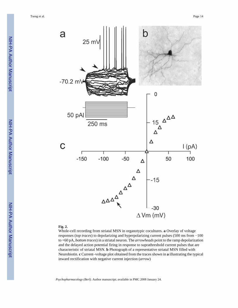
Fig. 2.Whole-cell recording from striatal MSN in organotypic cocultures. a Overlay of voltageresponses (top traces) to depolarizing and hyperpolarizing current pulses (500 ms from −100to +60 pA, bottom traces) in a striatal neuron. The arrowheads point to the ramp depolarizationand the delayed action potential firing in response to suprathreshold current pulses that arecharacteristic of striatal MSN. b Photograph of a representative striatal MSN filled withNeurobiotin. c Current–voltage plot obtained from the traces shown in a illustrating the typicalinward rectification with negative current injection (arrow)
Tseng et al. Page 14
Psychopharmacology (Berl). Author manuscript; available in PMC 2008 January 24.
NIH
-PA Author Manuscript
NIH
-PA Author Manuscript
NIH
-PA Author Manuscript
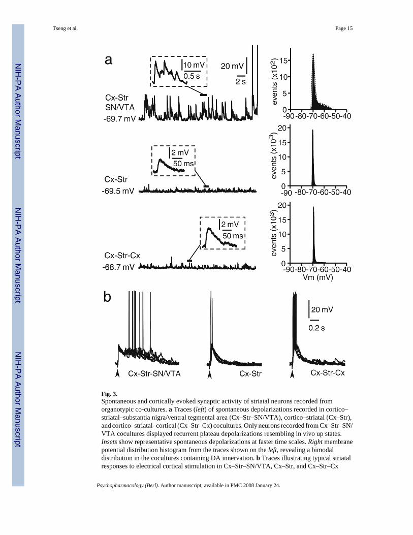
Fig. 3.Spontaneous and cortically evoked synaptic activity of striatal neurons recorded fromorganotypic co-cultures. a Traces (left) of spontaneous depolarizations recorded in cortico–striatal–substantia nigra/ventral tegmental area (Cx–Str–SN/VTA), cortico–striatal (Cx–Str),and cortico–striatal–cortical (Cx–Str–Cx) cocultures. Only neurons recorded from Cx–Str–SN/VTA cocultures displayed recurrent plateau depolarizations resembling in vivo up states.Insets show representative spontaneous depolarizations at faster time scales. Right membranepotential distribution histogram from the traces shown on the left, revealing a bimodaldistribution in the cocultures containing DA innervation. b Traces illustrating typical striatalresponses to electrical cortical stimulation in Cx–Str–SN/VTA, Cx–Str, and Cx–Str–Cx
Tseng et al. Page 15
Psychopharmacology (Berl). Author manuscript; available in PMC 2008 January 24.
NIH
-PA Author Manuscript
NIH
-PA Author Manuscript
NIH
-PA Author Manuscript
cocultures. Plateau depolarizations were evoked only in cultures containing SN/VTA neurons.Only brief depolarizing post-synaptic potentials were observed in Cx–Str and Cx–Str–Cxcocultures. In this and subsequent figures showing synaptic responses, several repetitions wereoverlaid in each example and vertical arrowheads below the traces point to the time of electricalstimulation
Tseng et al. Page 16
Psychopharmacology (Berl). Author manuscript; available in PMC 2008 January 24.
NIH
-PA Author Manuscript
NIH
-PA Author Manuscript
NIH
-PA Author Manuscript
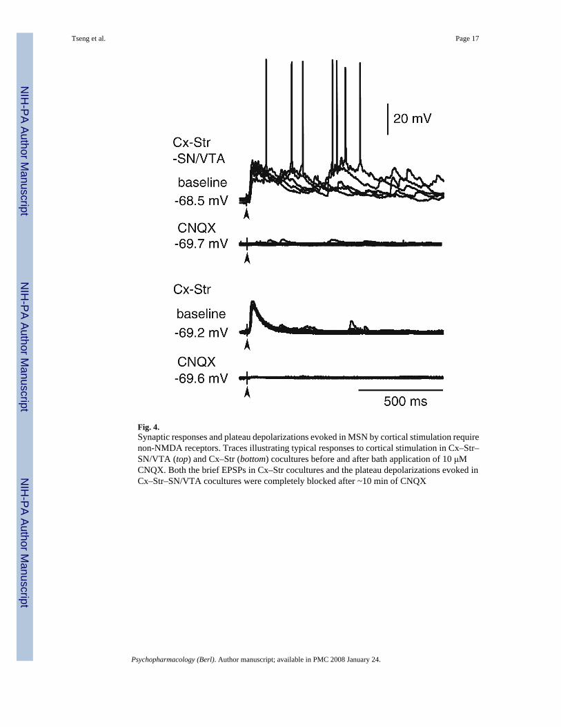
Fig. 4.Synaptic responses and plateau depolarizations evoked in MSN by cortical stimulation requirenon-NMDA receptors. Traces illustrating typical responses to cortical stimulation in Cx–Str–SN/VTA (top) and Cx–Str (bottom) cocultures before and after bath application of 10 μMCNQX. Both the brief EPSPs in Cx–Str cocultures and the plateau depolarizations evoked inCx–Str–SN/VTA cocultures were completely blocked after ~10 min of CNQX
Tseng et al. Page 17
Psychopharmacology (Berl). Author manuscript; available in PMC 2008 January 24.
NIH
-PA Author Manuscript
NIH
-PA Author Manuscript
NIH
-PA Author Manuscript
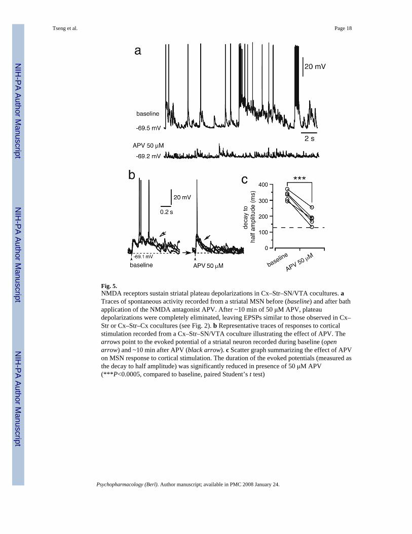
Fig. 5.NMDA receptors sustain striatal plateau depolarizations in Cx–Str–SN/VTA cocultures. aTraces of spontaneous activity recorded from a striatal MSN before (baseline) and after bathapplication of the NMDA antagonist APV. After ~10 min of 50 μM APV, plateaudepolarizations were completely eliminated, leaving EPSPs similar to those observed in Cx–Str or Cx–Str–Cx cocultures (see Fig. 2). b Representative traces of responses to corticalstimulation recorded from a Cx–Str–SN/VTA coculture illustrating the effect of APV. Thearrows point to the evoked potential of a striatal neuron recorded during baseline (openarrow) and ~10 min after APV (black arrow). c Scatter graph summarizing the effect of APVon MSN response to cortical stimulation. The duration of the evoked potentials (measured asthe decay to half amplitude) was significantly reduced in presence of 50 μM APV(***P<0.0005, compared to baseline, paired Student’s t test)
Tseng et al. Page 18
Psychopharmacology (Berl). Author manuscript; available in PMC 2008 January 24.
NIH
-PA Author Manuscript
NIH
-PA Author Manuscript
NIH
-PA Author Manuscript

Fig. 6.Up states recorded from Cx–Str cocultures containing SN/VTA neurons require D1 receptoractivation. a Simplified diagram illustrating the time points of MSN recordings (arrows) inCx–Str–SN/VTA cocultures that were incubated for at least 60 min with SCH23390. Timezero is the onset of incubation. b, d Representative traces from two MSN recorded in presenceof the D1 antagonist SCH23390. The first neuron (b) was obtained from a Cx–Str–SN/VTAcoculture that had been incubated with 5 μM SCH23390 for 60 min. Spontaneous plateaudepolarizations were gradually reduced to shorter events after the subsequent 30–40 min ofrecording. The second neuron (d) was obtained from another Cx–Str–SN/VTA coculturepreincubated with SCH23390 for 2 h. Depolarizing plateaus could not be observed; instead,short depolarizations resembling those observed in Cx–Str cocultures were recorded. The timeafter antagonist onset is indicated to the left of the traces. c Traces of depolarizations evokedby cortical stimulation in the same neurons are shown in b. The evoked responses still exhibiteda plateau depolarization by 65 min after SCH23390 (top); at ~125 min (e); however, the evokedresponse consisted of EPSPs similar to those observed in Cx–Str cocultures. An overlay of theevoked traces at 65 and 125 min after the D1 antagonist is shown in the inset
Tseng et al. Page 19
Psychopharmacology (Berl). Author manuscript; available in PMC 2008 January 24.
NIH
-PA Author Manuscript
NIH
-PA Author Manuscript
NIH
-PA Author Manuscript
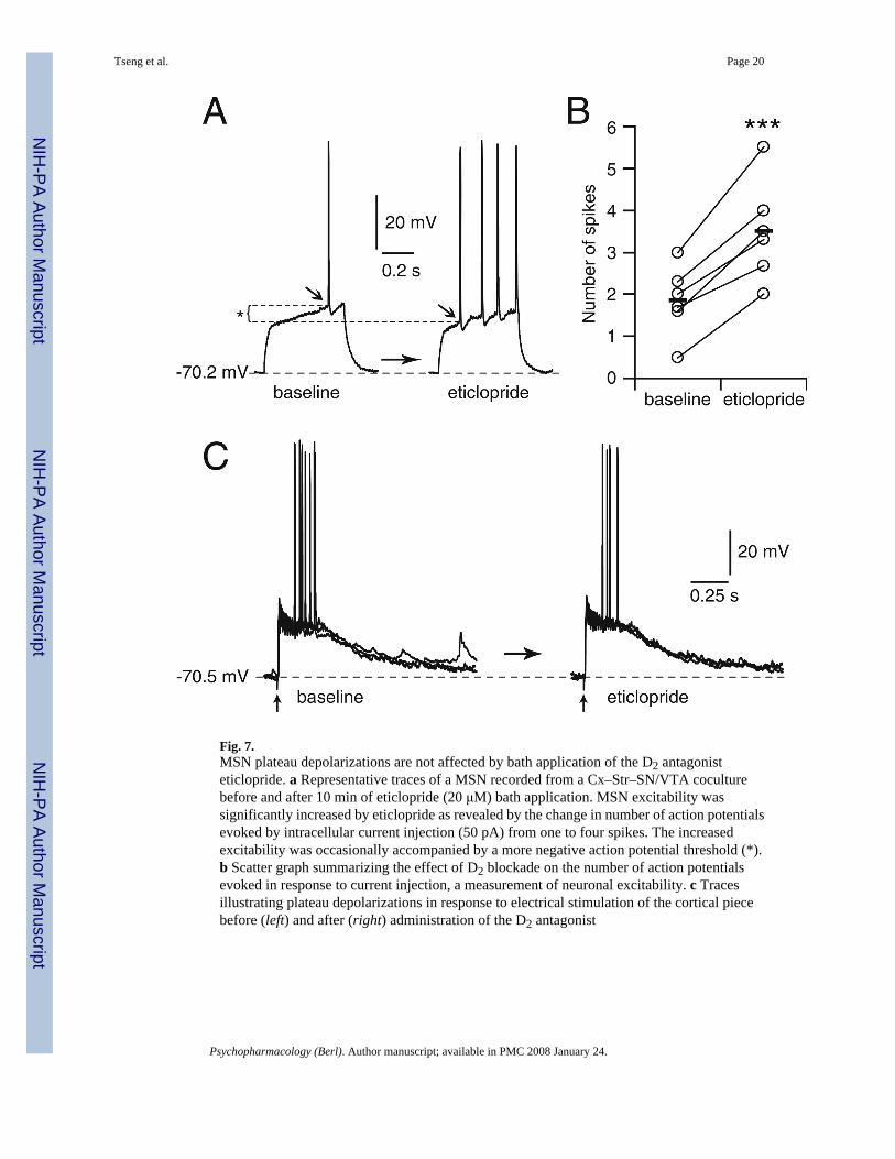
Fig. 7.MSN plateau depolarizations are not affected by bath application of the D2 antagonisteticlopride. a Representative traces of a MSN recorded from a Cx–Str–SN/VTA coculturebefore and after 10 min of eticlopride (20 μM) bath application. MSN excitability wassignificantly increased by eticlopride as revealed by the change in number of action potentialsevoked by intracellular current injection (50 pA) from one to four spikes. The increasedexcitability was occasionally accompanied by a more negative action potential threshold (*).b Scatter graph summarizing the effect of D2 blockade on the number of action potentialsevoked in response to current injection, a measurement of neuronal excitability. c Tracesillustrating plateau depolarizations in response to electrical stimulation of the cortical piecebefore (left) and after (right) administration of the D2 antagonist
Tseng et al. Page 20
Psychopharmacology (Berl). Author manuscript; available in PMC 2008 January 24.
NIH
-PA Author Manuscript
NIH
-PA Author Manuscript
NIH
-PA Author Manuscript

Fig. 8.MSN up states are Ca++ dependent and require PKA. a Representative tracing showingspontaneous activity of a striatal neuron recorded with an electrode containing the Ca++
chelator BAPTA (2 mM). A reduction in the frequency and duration of spontaneous plateaudepolarizations was evident after ~10 min of recording. Inset shows the initial spontaneousactivity of the same neuron 2 min after acquiring the whole-cell configuration. b Traces of thesame neuron shown in a illustrating the effect of intracellular BAPTA (~10 min of recording)on the response evoked by cortical stimulation. The evoked EPSP did not extend into a plateaudepolarization 10 min after acquiring the whole-cell configuration (inset shows an overlay ofseveral traces including the initial ones recorded 4 min after patch acquisition, which didinitially show an evoked plateau). EPSP duration was similar to what was observed incocultures not containing a SN/VTA piece. c, d Striatal up states require protein kinase A.Representative traces of spontaneous (c) and cortically-evoked activity (d) of a striatal MSNobtained from a Cx–Str–SN/VTA coculture and being recorded for ~10 min with an electrodecontaining the protein kinase A inhibitor PKI [5–24] (20 μM). Insets show traces of the sameneuron during the initial 1 to 3 min of patch recording. After ~10 min of recording, the evokedpotential was shortened to values resembling those obtained from Cx–Str cocultures
Tseng et al. Page 21
Psychopharmacology (Berl). Author manuscript; available in PMC 2008 January 24.
NIH
-PA Author Manuscript
NIH
-PA Author Manuscript
NIH
-PA Author Manuscript
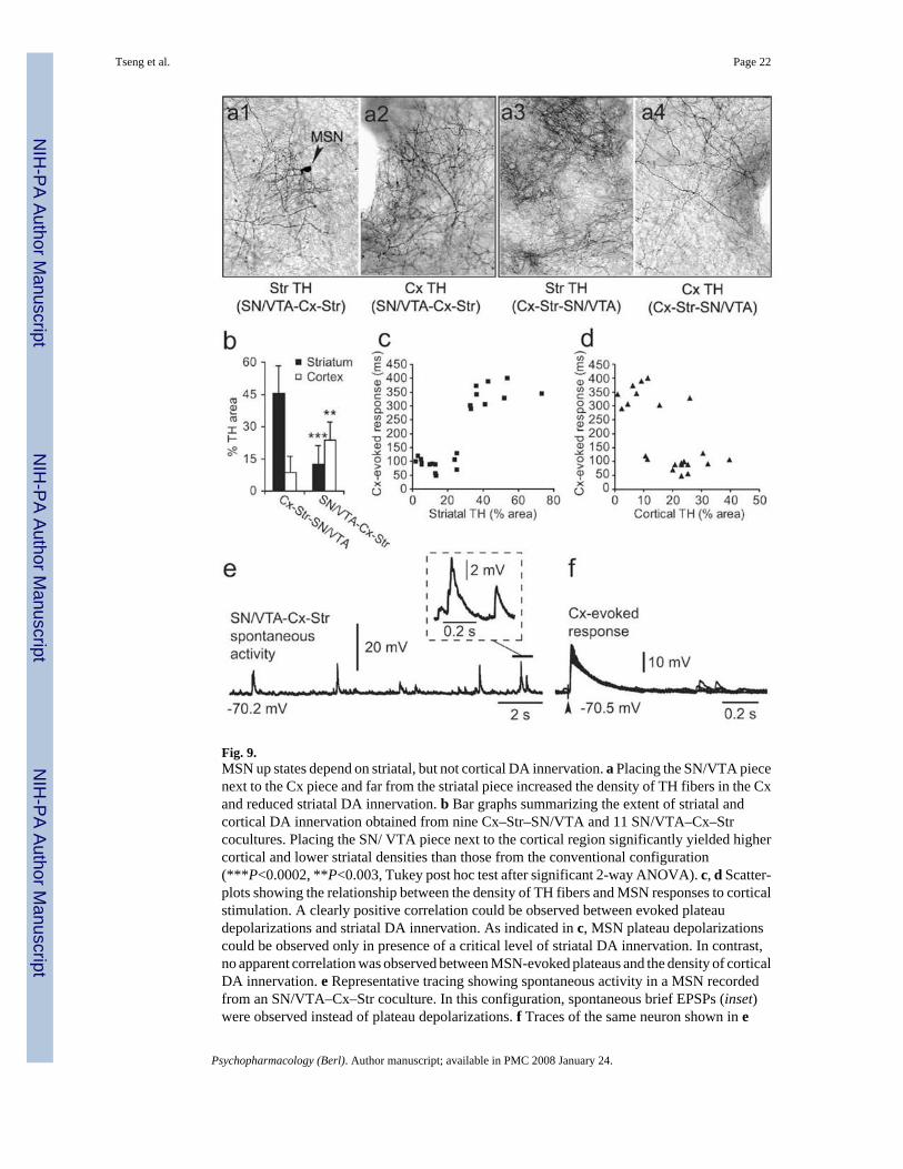
Fig. 9.MSN up states depend on striatal, but not cortical DA innervation. a Placing the SN/VTA piecenext to the Cx piece and far from the striatal piece increased the density of TH fibers in the Cxand reduced striatal DA innervation. b Bar graphs summarizing the extent of striatal andcortical DA innervation obtained from nine Cx–Str–SN/VTA and 11 SN/VTA–Cx–Strcocultures. Placing the SN/ VTA piece next to the cortical region significantly yielded highercortical and lower striatal densities than those from the conventional configuration(***P<0.0002, **P<0.003, Tukey post hoc test after significant 2-way ANOVA). c, d Scatter-plots showing the relationship between the density of TH fibers and MSN responses to corticalstimulation. A clearly positive correlation could be observed between evoked plateaudepolarizations and striatal DA innervation. As indicated in c, MSN plateau depolarizationscould be observed only in presence of a critical level of striatal DA innervation. In contrast,no apparent correlation was observed between MSN-evoked plateaus and the density of corticalDA innervation. e Representative tracing showing spontaneous activity in a MSN recordedfrom an SN/VTA–Cx–Str coculture. In this configuration, spontaneous brief EPSPs (inset)were observed instead of plateau depolarizations. f Traces of the same neuron shown in e
Tseng et al. Page 22
Psychopharmacology (Berl). Author manuscript; available in PMC 2008 January 24.
NIH
-PA Author Manuscript
NIH
-PA Author Manuscript
NIH
-PA Author Manuscript
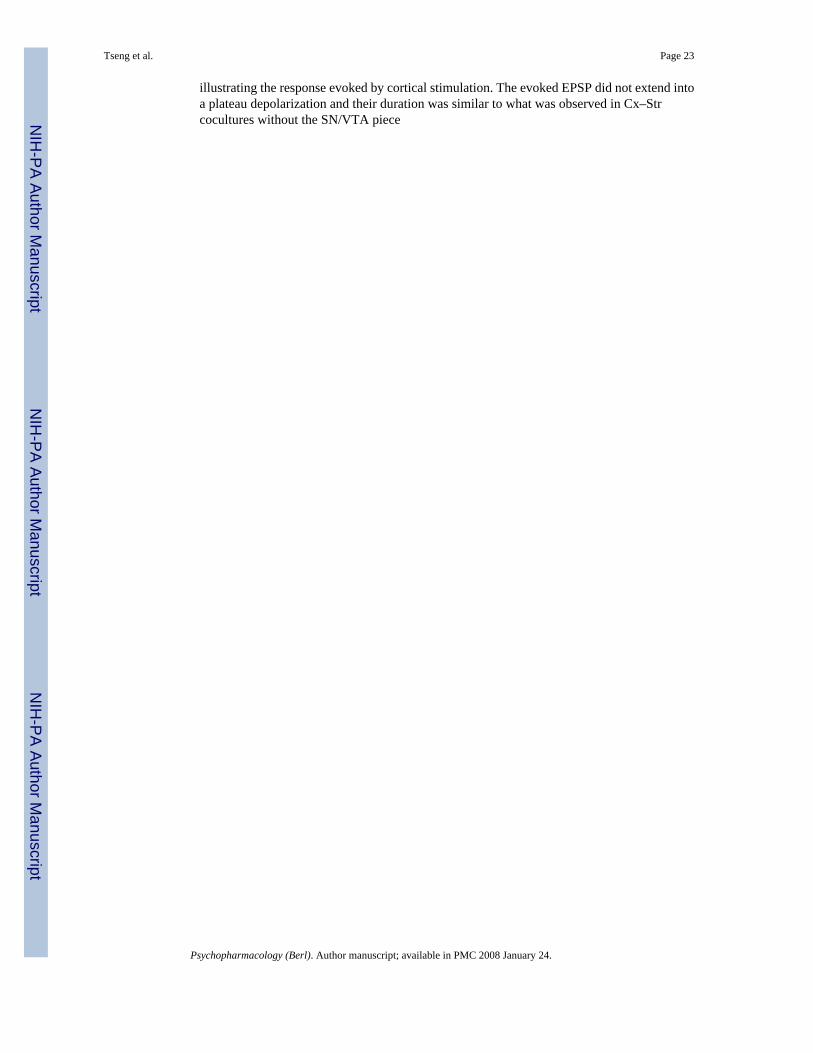
illustrating the response evoked by cortical stimulation. The evoked EPSP did not extend intoa plateau depolarization and their duration was similar to what was observed in Cx–Strcocultures without the SN/VTA piece
Tseng et al. Page 23
Psychopharmacology (Berl). Author manuscript; available in PMC 2008 January 24.
NIH
-PA Author Manuscript
NIH
-PA Author Manuscript
NIH
-PA Author Manuscript
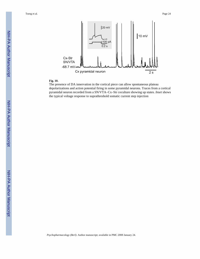
Fig. 10.The presence of DA innervation in the cortical piece can allow spontaneous plateaudepolarizations and action potential firing in some pyramidal neurons. Traces from a corticalpyramidal neuron recorded from a SN/VTA–Cx–Str coculture showing up states. Inset showsthe typical voltage response to suprathreshold somatic current step injection
Tseng et al. Page 24
Psychopharmacology (Berl). Author manuscript; available in PMC 2008 January 24.
NIH
-PA Author Manuscript
NIH
-PA Author Manuscript
NIH
-PA Author Manuscript
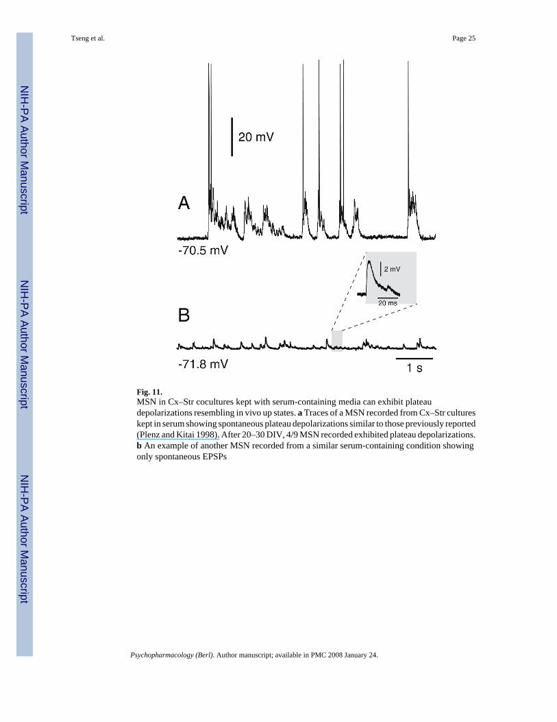
Fig. 11.MSN in Cx–Str cocultures kept with serum-containing media can exhibit plateaudepolarizations resembling in vivo up states. a Traces of a MSN recorded from Cx–Str cultureskept in serum showing spontaneous plateau depolarizations similar to those previously reported(Plenz and Kitai 1998). After 20–30 DIV, 4/9 MSN recorded exhibited plateau depolarizations.b An example of another MSN recorded from a similar serum-containing condition showingonly spontaneous EPSPs
Tseng et al. Page 25
Psychopharmacology (Berl). Author manuscript; available in PMC 2008 January 24.
NIH
-PA Author Manuscript
NIH
-PA Author Manuscript
NIH
-PA Author Manuscript
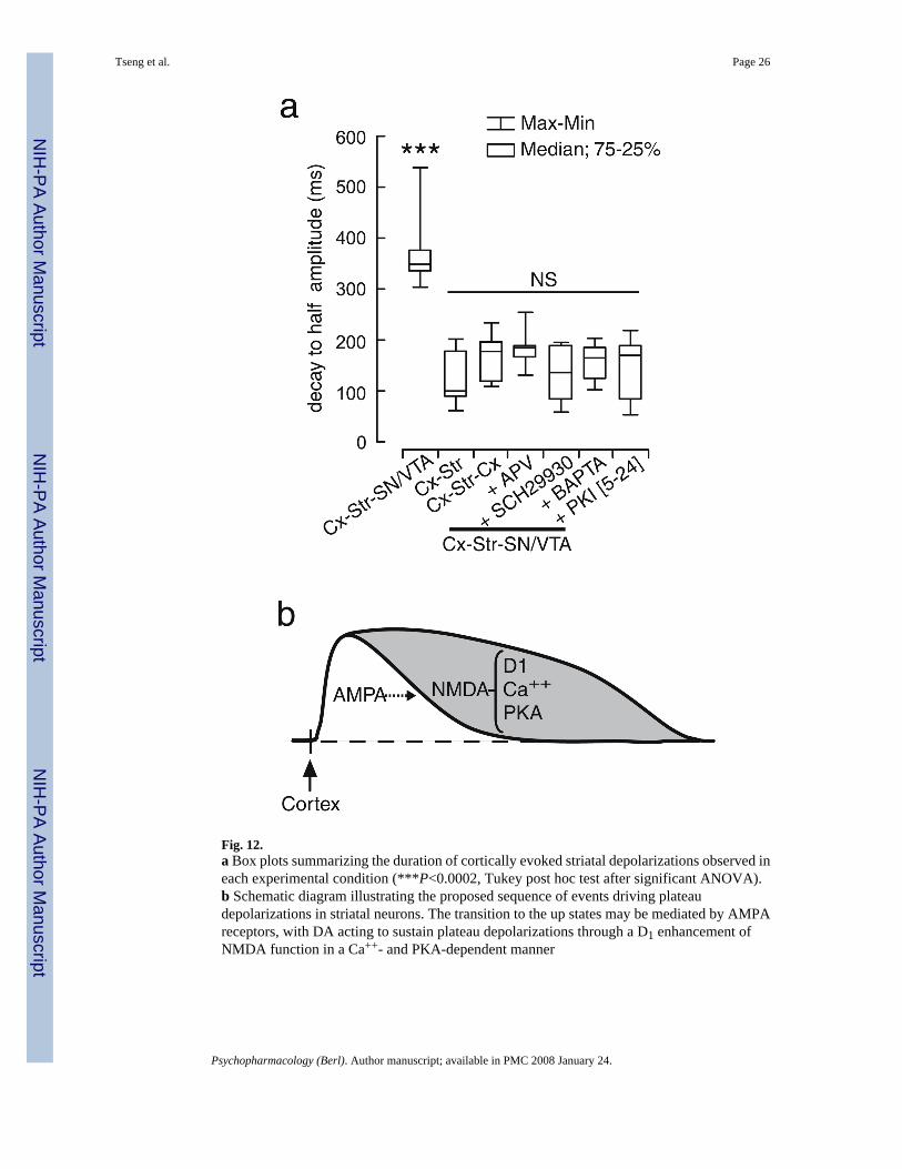
Fig. 12.a Box plots summarizing the duration of cortically evoked striatal depolarizations observed ineach experimental condition (***P<0.0002, Tukey post hoc test after significant ANOVA).b Schematic diagram illustrating the proposed sequence of events driving plateaudepolarizations in striatal neurons. The transition to the up states may be mediated by AMPAreceptors, with DA acting to sustain plateau depolarizations through a D1 enhancement ofNMDA function in a Ca++- and PKA-dependent manner
Tseng et al. Page 26
Psychopharmacology (Berl). Author manuscript; available in PMC 2008 January 24.
NIH
-PA Author Manuscript
NIH
-PA Author Manuscript
NIH
-PA Author Manuscript