The lifelong maintenance of mesencephalic dopaminergic neurons by Nurr1 and engrailed
8
P75 Ret Erk1/2 En1/2 P53 Nurr1 α-Syn. Dj1 DA production & trafficking Mitochondrial dysfuntion, Cell death Metabolic efficiency, Survival Bax Bcl2 Bcl-xL TH DAT VMAT2 The lifelong maintenance of mesencephalic dopaminergic neurons by Nurr1 and engrailed Alavian et al. Alavian et al. Journal of Biomedical Science 2014, 21:27 http://www.jbiomedsci.com/content/21/1/27
Transcript of The lifelong maintenance of mesencephalic dopaminergic neurons by Nurr1 and engrailed
P75 Ret
Erk1/2
En1/2 P53 Nurr1
α-Syn. Dj1
DA production& trafficking
Mitochondrial dysfuntion,Cell death
Metabolic efficiency,Survival
BaxBcl2Bcl-xL
THDAT
VMAT2
The lifelong maintenance of mesencephalicdopaminergic neurons by Nurr1 and engrailedAlavian et al.
Alavian et al. Journal of Biomedical Science 2014, 21:27http://www.jbiomedsci.com/content/21/1/27
Alavian et al. Journal of Biomedical Science 2014, 21:27http://www.jbiomedsci.com/content/21/1/27
REVIEW Open Access
The lifelong maintenance of mesencephalicdopaminergic neurons by Nurr1 and engrailedKambiz N Alavian1,2,3*, Sharmin Jeddi2, Sahar I Naghipour2, Pegah Nabili1, Pawel Licznerski3 and Travis S Tierney4
Abstract
Specific vulnerability and degeneration of the dopaminergic neurons in the substantia nigra pars compacta ofthe midbrain is the pathological hallmark of Parkinson’s disease. A number of transcription factors regulatethe birth and development of this set of neurons and some remain constitutively expressed throughout life.These maintenance transcription factors are closely associated with essential neurophysiological functions andare required ultimately for the long-term survival of the midbrain dopaminergic neurons. The current reviewdescribes the role of two such factors, Nurr1 and engrailed, in differentiation, maturation, and in normalphysiological functions including acquisition of neurotransmitter identity. The review will also elucidate therelationship of these factors with life, vulnerability, degeneration and death of mesencephalic dopaminergicneurons in the context of Parkinson’s disease.
ReviewIntroductionParkinson’s disease (PD) is the second most prevalentneurodegenerative disorder, affecting 1-2% of the popu-lation over 65 and 3-5% of the people over 85 [1]. Thedisease initially manifests with the cardinal motor symp-toms of rest tremor, bradykinesia, rigidity, and posturalinstability. Over time non-motor symptoms such as de-pression, constipation, pain, genitourinary problems, andsleep disorders [1,2] become prominent. The main patho-logical feature of PD is the degeneration of dopaminergicneurons in the substantia nigra pars compacta (SNpc).The other nearby mesencephalic dopaminergic (mesDA)neuronal populations within the ventral tegmental area(VTA) and retrorubral field (RRF) are less susceptible todegeneration and remain relatively unaffected during thecourse of the disease [3,4].A wide variety of animal models have been developed to
study the pathological outcomes of cell death in the ven-tral midbrain and to explore potential therapeutic targetsthat can stabilize or remedy the condition [5,6]. In recentyears, characterization of differential gene expression
* Correspondence: [email protected] of Brain Sciences, Department of Medicine, Imperial College, E508,Burlington Danes, Hammersmith Hospital, DuCane Road, London W12 0NN,UK2The Bahá’í Institute for Higher Education (BIHE), Tehran, IranFull list of author information is available at the end of the article
© 2014 Alavian et al.; licensee BioMed CentralCommons Attribution License (http://creativecreproduction in any medium, provided the orDedication waiver (http://creativecommons.orunless otherwise stated.
profiles between the two main mesDA neuronal popula-tions, VTA and SNpc, has been used to probe the questionof relative susceptibility of neurons to environmental andgenetic vulnerability [7]. Another approach to understandthis susceptibility has been the study of the developmentalcues that contribute directly or indirectly to differentiationof these phenotypes [8].Based on these studies, we postulate that these neurons
perpetually remain in a developmental state that is vulner-able to neurodegeneration because the critical period fornaturally-occurring neuron death that occurs for mostpopulations of neurons never completely closes for theseunique cells in the ventral midbrain. This inherent vulner-ability of mesDA neurons is a consequence of the uniqueexpression profile of these cells which is controlled by acascade of developmental transcription factors that are notonly required for defining the regional identity of the mid-brain and specification of mesDA neurons but also for thelong-term survival and maintenance and normal physio-logical function. Several gain and loss of function studies intransgenic and mutant animals have demonstrated a strongconnection between developmental factors and essentialneuronal functions such as axon guidance, regulation ofsurvival and cell death, defining the neurotransmitterphenotype, neuronal excitability and plasticity [8].These studies have also established that the insuffi-ciency in certain key transcription factors can lead to
Ltd. This is an Open Access article distributed under the terms of the Creativeommons.org/licenses/by/2.0), which permits unrestricted use, distribution, andiginal work is properly credited. The Creative Commons Public Domaing/publicdomain/zero/1.0/) applies to the data made available in this article,
Alavian et al. Journal of Biomedical Science 2014, 21:27 Page 2 of 7http://www.jbiomedsci.com/content/21/1/27
a neurodegenerative phenotype and subsequently toPD-like symptoms [9-12].In this review we will discuss two transcription factors,
engrailed and Nurr1, that are necessary for regulation ofkey events in the development of mesencephalic dopa-minergic neurons as well as their survival and mainten-ance throughout life. In our discussion, we will highlightthe role of these factors as key transcriptional regulatorsof differentiation, maturation and survival of mesDAneurons, the contribution of each factor to the normalphysiological functions in postnatal mesDA neurons, aswell as their possible connection with the vulnerabilityof neurons and etiology of Parkinson’s disease.
Nurr1Orphan nuclear receptor Nurr1 (nuclear receptor sub-family 4, group A, member 2; NR4A2) is a member ofthe steroid/thyroid hormone nuclear receptor and isrelated to a family of factors that modulate transcriptionin response to small lipophilic molecules. The transcrip-tional activity of these factors is regulated through theinteraction of the ligand with the carboxy terminalligand-binding domain of specific nuclear receptors [13].Nurr1 transcription factor is encoded by an immediateearly gene and is predominantly expressed in the brain[14,15]. Structurally, Nurr1 consists of a DNA bindingdomain, a ligand binding domain and two transcriptionactivation function domains at the N- and C-termini(AF1 and AF2, respectively) [16,17]. The DNA bindingdomain of Nurr1 is highly conserved among the nuclearreceptor family members and is comprised of two zincfinger modules. This domain is known to activate tran-scription through binding to an NGFI-B response elem-ent (NBRE) [18]. Nurr1 lacks a classical binding site forcoactivators and the tight packing of side chains fromhydrophobic residues in the ligand-binding domain pre-vents the molecule from having a ligand-binding cavity.The constitutive transcriptional activity of Nurr1, there-fore, can be attributed to the canonical protein fold resem-bling the agonist-bound, transcriptionally active, ligandbinding domains in nuclear receptors [19]. Nurr1 alsoactivates transcription through heterodimerization withthe retionid X receptor (RXR) and in response to the RXRligands [20,21]. The AF1 and AF2 domains are also invol-ved in co-factor recruitment and contribute to the consti-tutive transcriptional activation of Nurr1 [16,22].In mice, the expression of Nurr1 can first be detected
on embryonic day 10 (E10) in the ventral midbrain.Although the expression of the gene is reduced in thepostnatal animals, it continues throughout life and hasbeen shown to regulate several aspects of postmitoticdevelopment [10,23,24]. The role of Nurr1 in the survivaland differentiation of mesDA neurons was initially discov-ered through mouse knockout studies, demonstrating loss
of immunoreactivity for the rate-limiting enzyme in pro-duction of dopamine, tyrosine hydroxylase, in the mesDAneurons from Nurr1 knockout animals [25]. Later onfurther studies established that the neuroepithelial cells,that give rise to mesDA neurons, ventralize normally anddifferentiate into neurons. For example, they expressmesDA neuron specific markers such as Pitx3, Ahd2 andengrailed1/2 and establish nigrostriatal axonal projections,but lack their dopaminergic phenotype [26-28]. Furtherexamination of the transcriptional role of Nurr1 demon-strated that it is an upstream regulator of genes involvedin the synthesis, packaging, transport and reuptake ofdopamine [29,30]. For example, Nurr1 has been shown toinduce the expression of tyrosine hydroxylase (TH) by dir-ectly transactivating the promoter of TH [31]. Regulationof the dopamine transporter (DAT) gene is another keyfunction of Nurr1 in determining the neurotransmitterphenotype of mesDA neurons. This effect of Nurr1 seemsto be independent of its classic heterodimerization part-ner, retinoid X receptor. The expression of TH and DATis regulated by the high affinity binding of Nurr1 to anextended half-hormone response element, NGF1-B re-sponsive element (NBRE), in their 5′-untranslated regions[32,33]. Finally, other studies have shown that Nurr1 isinvolved in conversion of L-DOPA to DA and packagingof DA into synaptic vesicles by regulating the expres-sion of aromatic L-amino acid decarboxylase (AADC)and vesicular monoamine transporter-2 (VMAT2), re-spectively [30].In addition to transcriptional regulation of the genes
involved in dopamine production and trafficking, Nurr1is also a key convert factor in several survival and main-tenance pathways. Nurr1 regulates the rearranged intransfection (Ret) gene from the earliest stages in embry-onic development. Ret tyrosine kinase is the high-affinityligand-binding component of the glial cell line derivedneurotrophic factor (GDNF) receptor complex that isattached to the cell surface via a glycosyl phospha-tidylinositol anchor [34]. GDNF has been shown to pro-tect mesDA neurons against the developmental waves ofapoptosis, neurotoxic insults and cell death [35-38]. UponGDNF binding, autophosphorylation of the tyrosine do-mains of Ret triggers activation of several pathwaysincluding PI3K, MAPK which are required for neuronalsurvival and neurite outgrowth [39]. Interestingly, one ofthe downstream phosphorylation targets of the GDNFsignaling pathway is the cAMP response element binding(CREB) protein, which is known to directly regulate theexpression of Nurr1 by binding to its promoter [40].Homozygous Nurr1 knockout pups die shortly after
birth following degeneration of mesDA neurons [25-27].However, Nurr1 haploinsufficient animals show no signifi-cant loss of mesDA neuron in number or motor functionuntil late in life. The aging (>15 months old) Nurr1+/−
Alavian et al. Journal of Biomedical Science 2014, 21:27 Page 3 of 7http://www.jbiomedsci.com/content/21/1/27
mice have a decrease in the number of their nigral mesDAneurons and their locomotor activities, correlated with thereduced mesolimbic and mesocortical dopamine levels[9,23]. These animals also display increased vulnerabilityand cell death in response to neurotoxic insult [9,41].The increased neuronal degeneration and death inthese animals is likely to be due to mitochondrialdysfunction and the opening of the mitochondrial per-meability transition pore [42]. Transcriptional regulationof the pro-and anti-apoptotic members of the Bcl-2 mito-chondrial family of proteins and interaction with the
P75
Erk1
En1/2
Metabolic efficiency,Survival
Bcl2Bcl-xL
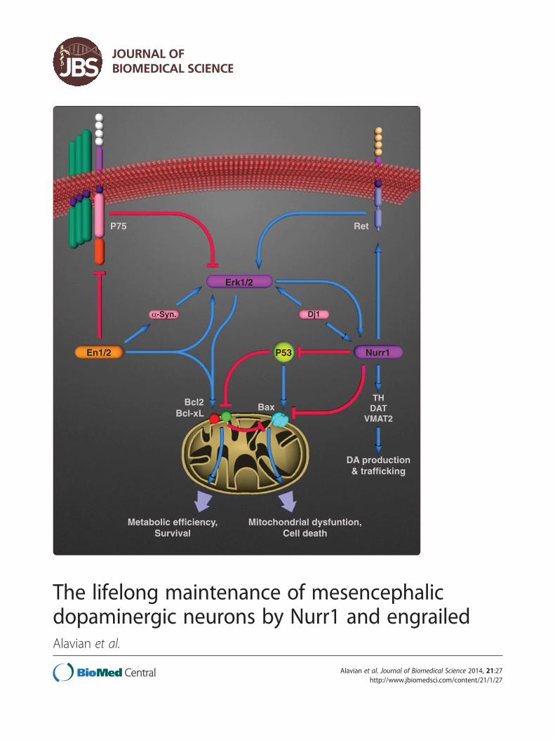
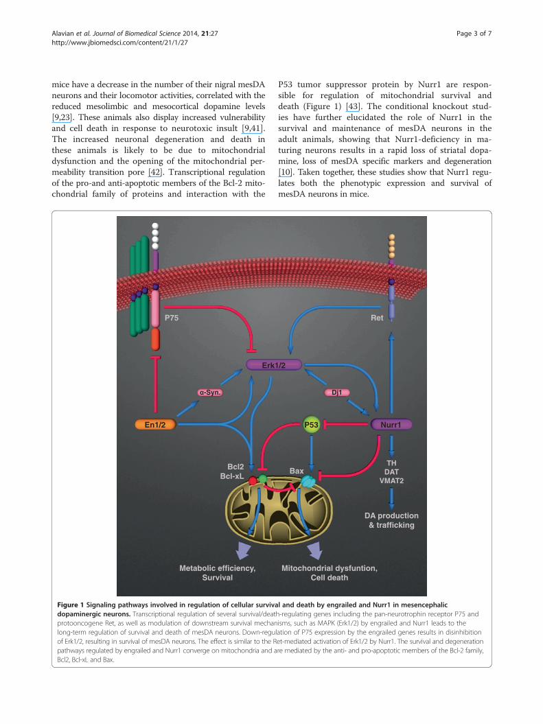
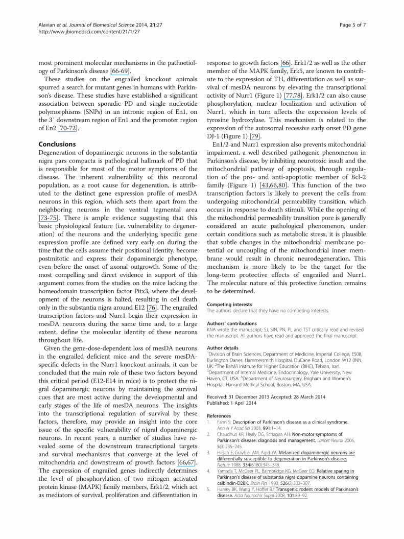
Figure 1 Signaling pathways involved in regulation of cellular survivadopaminergic neurons. Transcriptional regulation of several survival/deathprotooncogene Ret, as well as modulation of downstream survival mechanlong-term regulation of survival and death of mesDA neurons. Down-regulof Erk1/2, resulting in survival of mesDA neurons. The effect is similar to the Rpathways regulated by engrailed and Nurr1 converge on mitochondria and aBcl2, Bcl-xL and Bax.
P53 tumor suppressor protein by Nurr1 are respon-sible for regulation of mitochondrial survival anddeath (Figure 1) [43]. The conditional knockout stud-ies have further elucidated the role of Nurr1 in thesurvival and maintenance of mesDA neurons in theadult animals, showing that Nurr1-deficiency in ma-turing neurons results in a rapid loss of striatal dopa-mine, loss of mesDA specific markers and degeneration[10]. Taken together, these studies show that Nurr1 regu-lates both the phenotypic expression and survival ofmesDA neurons in mice.
Ret
/2
P53 Nurr1
Dj1
DA production& trafficking
Mitochondrial dysfuntion,Cell death
BaxTH
DATVMAT2
l and death by engrailed and Nurr1 in mesencephalic-regulating genes including the pan-neurotrophin receptor P75 andisms, such as MAPK (Erk1/2) by engrailed and Nurr1 leads to theation of P75 expression by the engrailed genes results in disinhibitionet-mediated activation of Erk1/2 by Nurr1. The survival and degenerationre mediated by the anti- and pro-apoptotic members of the Bcl-2 family,
Alavian et al. Journal of Biomedical Science 2014, 21:27 Page 4 of 7http://www.jbiomedsci.com/content/21/1/27
Given the neuoprotective role of Nurr1 demon-strated in animal models and its decreased or dimin-ished expression levels in PD patients [44,45], anumber of human genetic studies of the Nurr1 genewere undertaking to identify potential variants thatmay increase the risk of developing PD. Although theresults from the studies within the past decade havefailed to establish the mutations in Nurr1 as a causeor direct risk factor for PD, higher incidence of severalmutations within the coding and non-coding regions ofNurr1 have been observed in familial and sporadic casesof the disease [46-49].A number of these mutations have been associated
with a marked decrease in the level of Nurr1 expressionand subsequent reduction in the expression of TH [46].The evidence from these studies accompanied by theresults of the postmortem expression analysis in the PDpatient brains suggest dysregulation of Nurr1 as a contrib-uting factor to the onset and progression of neurodegener-ation during the course of PD.
The engrailed transcription factorsThe engrailed genes belong to the family of homeoboxtranscription factors, containing a highly conserved DNAbinding, helix-turn-helix, homeodomain protein fold [50].The morphological significance of the engrailed was firstdescribed in drosophila, where the autosomal mutantdisplays a long cleft of the thorax and irregular venationof the wings [51]. In addition to its early role in bodysegmentation, engrailed homologs in various annelid,mollusks, chordate and arthropod species has revealed aseparate major function in neurogenesis [52-54]. Interest-ingly, homologs of engrailed are conserved among differ-ent phyla, to the degree that replacement of the mouseengrailed-1 gene by its paralog En2 or by the drosophilaengrailed homolog will preserve its function in develop-ment and survival [55,56].The engrailed genes have two distinct functions during
early and late development. The two paralogs of en-grailed, En1 and En2, start their expression around E8 inmice in a broad region at the midbrain/hindbrain border,known as the isthmic organizer [57]. During this period,engrailed is involved in the induction of mesDA neuronsby maintaining expression of Fgf8 in this region [58-60].En1/2 directly regulate the expression of Fgf8 throughinteraction with a DNA-binding intronic fragment onFgf8 [61]. In En1/2 deficient embryos the domain ofexpression of Fgf8 in the isthmus is reduced and leadsan incomplete induction of mesDA neurons in the mid-brain. The specific expression of the engrailed genes inmesDA neurons starts after the neurons are induced andbecome postmitotic, between E11.5 and E14 in mice[62]. After this point, expression of both engrailed genesis required for continued survival of these neurons.
Mouse knockout studies have shown that there is adirect correlation between the dose of expression ofengrailed and its survival effect in mesDA neurons. Micelacking all four alleles of the two transcription factors(En1−/−;En2−/−) show the most severe phenotype. Inthese mice, the induction of dopaminergic neurons inthe ventral midbrain is affected, the differentiated post-mitotic mesDA neurons display significant developmen-tal defects including a lack of axonal outgrowth andeventually die between E12 and E14, matching the time-frame of the expression of engrailed genes in wildtypemesDA neurons [62,63]. The postmitotic survival effectof En1 is more prominent than that of En2, since themice lacking En2 show less severe phenotypes than theEn1−/− animals. All En1−/− animals (regardless of theexpression of En2) exhibit an abnormal distribution ofdopaminergic neurons in ventral midbrain and die atbirth. En1+/+;En2−/− mice, however, are viable andfertile and show no obvious deficiencies with regard totheir dopaminergic neurons [62,64]. The most interest-ing phenotype among the engrailed-deficient animals isthat of the mice heterozygous for En1 and homozygousnull for En2 (En1+/−;En2−/−) where the development ofmesDA neurons seems to be normal until just after birthwhen 70% of the neurons are lost within the first three 3postnatal months. This specific nigral dopaminergic cellloss is accompanied by diminished dopamine levels andrelease within the dorsal striatum, leading to a markeddecrease in locomotion, an increase in freezing episodesand weight loss.Engrailed regulates development, survival and mainten-
ance of mesDA neurons through the cooperative action ofmultiple molecular pathways. Cell death in absence ofengrailed is mediated by at least two molecular pathways,involving the pan-neurotrophin receptor, P75NTR andthe mitochondria. The increase in expression of theneurotrophin receptor P75 is a common phenomenon ob-served in neuronal injury and naturally-occurring neurondeath [65]. Similarly, engrailed deficiency causes an in-crease in the expression of P75 resulting in downregula-tion of the mitogen activated protein kinase (MAPK)survival pathways and deactivation of the extracellularregulated kinases Erk1/2 [66]. A second survival mech-anism regulated by the expression of the engrailedgenes affects the mitochondria. Similar to the Nurr1haploinsufficient cells, the mesDA neurons heterozy-gous for En1 (either in presence or absence of En2) aremore prone to mitochondrial insult and cell deathinduced by inhibition of the complex-I of the electrontransport chain than their wildtype counterparts [66].The dose-dependent survival role of the engrailed genesagainst mitochondrial instability is suggestive of anotherlink between engrailed and the etiology of PD, since mito-chondrial dysfunction has been implicated as one of the
Alavian et al. Journal of Biomedical Science 2014, 21:27 Page 5 of 7http://www.jbiomedsci.com/content/21/1/27
most prominent molecular mechanisms in the pathoetiol-ogy of Parkinson’s disease [66-69].These studies on the engrailed knockout animals
spurred a search for mutant genes in humans with Parkin-son’s disease. These studies have established a significantassociation between sporadic PD and single nucleotidepolymorphisms (SNPs) in an intronic region of En1, onthe 3′ downstream region of En1 and the promoter regionof En2 [70-72].
ConclusionsDegeneration of dopaminergic neurons in the substantianigra pars compacta is pathological hallmark of PD thatis responsible for most of the motor symptoms of thedisease. The inherent vulnerability of this neuronalpopulation, as a root cause for degeneration, is attrib-uted to the distinct gene expression profile of mesDAneurons in this region, which sets them apart from theneighboring neurons in the ventral tegmental area[73-75]. There is ample evidence suggesting that thisbasic physiological feature (i.e. vulnerability to degener-ation) of the neurons and the underlying specific geneexpression profile are defined very early on during thetime that the cells assume their positional identity, becomepostmitotic and express their dopaminergic phenotype,even before the onset of axonal outgrowth. Some of themost compelling and direct evidence in support of thisargument comes from the studies on the mice lacking thehomeodomain transcription factor Pitx3, where the devel-opment of the neurons is halted, resulting in cell deathonly in the substantia nigra around E12 [76]. The engrailedtranscription factors and Nurr1 begin their expression inmesDA neurons during the same time and, to a largeextent, define the molecular identity of these neuronsthroughout life.Given the gene-dose-dependent loss of mesDA neurons
in the engrailed deficient mice and the severe mesDA-specific defects in the Nurr1 knockout animals, it can beconcluded that the main role of these two factors beyondthis critical period (E12-E14 in mice) is to protect the ni-gral dopaminergic neurons by maintaining the survivalcues that are most active during the developmental andearly stages of the life of mesDA neurons. The insightsinto the transcriptional regulation of survival by thesefactors, therefore, may provide an insight into the coreissue of the specific vulnerability of nigral dopaminergicneurons. In recent years, a number of studies have re-vealed some of the downstream transcriptional targetsand survival mechanisms that converge at the level ofmitochondria and downstream of growth factors [66,67].The expression of engrailed genes indirectly determinesthe level of phosphorylation of two mitogen activatedprotein kinase (MAPK) family members, Erk1/2, which actas mediators of survival, proliferation and differentiation in
response to growth factors [66]. Erk1/2 as well as the othermember of the MAPK family, Erk5, are known to contrib-ute to the expression of TH, differentiation as well as sur-vival of mesDA neurons by elevating the transcriptionalactivity of Nurr1 (Figure 1) [77,78]. Erk1/2 can also causephosphorylation, nuclear localization and activation ofNurr1, which in turn affects the expression levels oftyrosine hydroxylase. This mechanism is related to theexpression of the autosomal recessive early onset PD geneDJ-1 (Figure 1) [79].En1/2 and Nurr1 expression also prevents mitochondrial
impairment, a well described pathogenic phenomenon inParkinson’s disease, by inhibiting neurotoxic insult and themitochondrial pathway of apoptosis, through regula-tion of the pro- and anti-apoptotic member of Bcl-2family (Figure 1) [43,66,80]. This function of the twotranscription factors is likely to prevent the cells fromundergoing mitochondrial permeability transition, whichoccurs in response to death stimuli. While the opening ofthe mitochondrial permeability transition pore is generallyconsidered an acute pathological phenomenon, undercertain conditions such as metabolic stress, it is plausiblethat subtle changes in the mitochondrial membrane po-tential or uncoupling of the mitochondrial inner mem-brane would result in chronic neurodegeneration. Thismechanism is more likely to be the target for thelong-term protective effects of engrailed and Nurr1.The molecular nature of this protective function remainsto be determined.
Competing interestsThe authors declare that they have no competing interests.
Authors’ contributionsKNA wrote the manuscript; SJ, SIN, PN, PL and TST critically read and revisedthe manuscript. All authors have read and approved the final manuscript.
Author details1Division of Brain Sciences, Department of Medicine, Imperial College, E508,Burlington Danes, Hammersmith Hospital, DuCane Road, London W12 0NN,UK. 2The Bahá’í Institute for Higher Education (BIHE), Tehran, Iran.3Department of Internal Medicine, Endocrinology, Yale University, NewHaven, CT, USA. 4Department of Neurosurgery, Brigham and Women’sHospital, Harvard Medical School, Boston, MA, USA.
Received: 31 December 2013 Accepted: 28 March 2014Published: 1 April 2014
References1. Fahn S: Description of Parkinson’s disease as a clinical syndrome.
Ann N Y Acad Sci 2003, 991:1–14.2. Chaudhuri KR, Healy DG, Schapira AH: Non-motor symptoms of
Parkinson’s disease: diagnosis and management. Lancet Neurol 2006,5(3):235–245.
3. Hirsch E, Graybiel AM, Agid YA: Melanized dopaminergic neurons aredifferentially susceptible to degeneration in Parkinson’s disease.Nature 1988, 334(6180):345–348.
4. Yamada T, McGeer PL, Baimbridge KG, McGeer EG: Relative sparing inParkinson's disease of substantia nigra dopamine neurons containingcalbindin-D28K. Brain Res 1990, 526(2):303–307.
5. Harvey BK, Wang Y, Hoffer BJ: Transgenic rodent models of Parkinson’sdisease. Acta Neurochir Suppl 2008, 101:89–92.
Alavian et al. Journal of Biomedical Science 2014, 21:27 Page 6 of 7http://www.jbiomedsci.com/content/21/1/27
6. Terzioglu M, Galter D: Parkinson’s disease: genetic versus toxin-inducedrodent models. FEBS J 2008, 275(7):1384–1391.
7. Greene JG: Current status and future directions of gene expressionprofiling in Parkinson’s disease. Neurobiol Dis 2012, 45(1):76–82.
8. Alavian KN, Scholz C, Simon HH: Transcriptional regulation ofmesencephalic dopaminergic neurons: the full circle of life and death.Mov Disord 2008, 23(3):319–328.
9. Jiang C, Wan X, He Y, Pan T, Jankovic J, Le W: Age-dependentdopaminergic dysfunction in Nurr1 knockout mice. Exp Neurol 2005,191(1):154–162.
10. Kadkhodaei B, Ito T, Joodmardi E, Mattsson B, Rouillard C, Carta M,Muramatsu S, Sumi-Ichinose C, Nomura T, Metzger D, Chambon P, LindqvistE, Larsson NG, Olson L, Bjorklund A, Ichinose H, Perlmann T: Nurr1 isrequired for maintenance of maturing and adult midbrain dopamineneurons. J Neurosci 2009, 29(50):15923–15932.
11. Kittappa R, Chang WW, Awatramani RB, McKay RD: The foxa2 genecontrols the birth and spontaneous degeneration of dopamine neuronsin old age. PLoS Biol 2007, 5(12):e325.
12. Sgado P, Alberi L, Gherbassi D, Galasso SL, Ramakers GM, Alavian KN,Smidt MP, Dyck RH, Simon HH: Slow progressive degeneration of nigraldopaminergic neurons in postnatal Engrailed mutant mice. Proc NatlAcad Sci U S A 2006, 103(41):15242–15247.
13. Ribeiro RC, Kushner PJ, Baxter JD: The nuclear hormone receptor genesuperfamily. Annu Rev Med 1995, 46:443–453.
14. Law SW, Conneely OM, DeMayo FJ, O’Malley BW: Identification of anew brain-specific transcription factor, NURR1. Mol Endocrinol 1992,6(12):2129–2135.
15. Zetterstrom RH, Williams R, Perlmann T, Olson L: Cellular expression of theimmediate early transcription factors Nurr1 and NGFI-B suggests a generegulatory role in several brain regions including the nigrostriataldopamine system. Brain Res Mol Brain Res 1996, 41(1–2):111–120.
16. Castro DS, Arvidsson M, Bondesson BM, Perlmann T: Activity of the Nurr1carboxyl-terminal domain depends on cell type and integrity of theactivation function 2. J Biol Chem 1999, 274(52):37483–37490.
17. Nordzell M, Aarnisalo P, Benoit G, Castro DS, Perlmann T: Defining anN-terminal activation domain of the orphan nuclear receptor Nurr1.Biochem Biophys Res Commun 2004, 313(1):205–211.
18. Paulsen RF, Granas K, Johnsen H, Rolseth V, Sterri S: Three related brainnuclear receptors, NGFI-B, Nurr1, and NOR-1, as transcriptional activators.J Mol Neurosci 1995, 6(4):249–255.
19. Wang Z, Benoit G, Liu J, Prasad S, Aarnisalo P, Liu X, Xu H, Walker NP,Perlmann T: Structure and function of Nurr1 identifies a class ofligand-independent nuclear receptors. Nature 2003, 423(6939):555–560.
20. Aarnisalo P, Kim CH, Lee JW, Perlmann T: Defining requirements forheterodimerization between the retinoid X receptor and the orphannuclear receptor Nurr1. J Biol Chem 2002, 277(38):35118–35123.
21. Perlmann T, Wallen-Mackenzie A: Nurr1, an orphan nuclear receptor withessential functions in developing dopamine cells. Cell Tissue Res 2004,318(1):45–52.
22. Flaig R, Greschik H, Peluso-Iltis C, Moras D: Structural basis for thecell-specific activities of the NGFI-B and the Nurr1 ligand-binding domain.J Biol Chem 2005, 280(19):19250–19258.
23. Jankovic J, Chen S, Le WD: The role of Nurr1 in the development ofdopaminergic neurons and Parkinson’s disease. Prog Neurobiol 2005,77(1–2):128–138.
24. Luo Y: The function and mechanisms of Nurr1 action in midbraindopaminergic neurons, from development and maintenance to survival.Int Rev Neurobiol 2012, 102:1–22.
25. Zetterstrom RH, Solomin L, Jansson L, Hoffer BJ, Olson L, Perlmann T:Dopamine neuron agenesis in Nurr1-deficient mice. Science 1997,276(5310):248–250.
26. Saucedo-Cardenas O, Quintana-Hau JD, Le WD, Smidt MP, Cox JJ, De MayoF, Burbach JP, Conneely OM: Nurr1 is essential for the induction of thedopaminergic phenotype and the survival of ventral mesencephalic latedopaminergic precursor neurons. Proc Natl Acad Sci U S A 1998,95(7):4013–4018.
27. Wallen A, Zetterstrom RH, Solomin L, Arvidsson M, Olson L, Perlmann T:Fate of mesencephalic AHD2-expressing dopamine progenitor cells inNURR1 mutant mice. Exp Cell Res 1999, 253(2):737–746.
28. Witta J, Baffi JS, Palkovits M, Mezey E, Castillo SO, Nikodem VM: Nigrostriatalinnervation is preserved in Nurr1-null mice, although dopaminergic
neuron precursors are arrested from terminal differentiation. Brain ResMol Brain Res 2000, 84(1–2):67–78.
29. Flames N, Hobert O: Transcriptional control of the terminal fate ofmonoaminergic neurons. Annu Rev Neurosci 2011, 34:153–184.
30. Hermanson E, Joseph B, Castro D, Lindqvist E, Aarnisalo P, Wallen A,Benoit G, Hengerer B, Olson L, Perlmann T: Nurr1 regulates dopaminesynthesis and storage in MN9D dopamine cells. Exp Cell Res 2003,288(2):324–334.
31. Kim KS, Kim CH, Hwang DY, Seo H, Chung S, Hong SJ, Lim JK, Anderson T,Isacson O: Orphan nuclear receptor Nurr1 directly transactivates thepromoter activity of the tyrosine hydroxylase gene in a cell-specificmanner. J Neurochem 2003, 85(3):622–634.
32. Sacchetti P, Brownschidle LA, Granneman JG, Bannon MJ: Characterizationof the 5’-flanking region of the human dopamine transporter gene.Brain Res Mol Brain Res 1999, 74(1–2):167–174.
33. Sacchetti P, Mitchell TR, Granneman JG, Bannon MJ: Nurr1 enhancestranscription of the human dopamine transporter gene through a novelmechanism. J Neurochem 2001, 76(5):1565–1572.
34. Baloh RH, Enomoto H, Johnson EM Jr, Milbrandt J: The GDNF familyligands and receptors - implications for neural development.Curr Opin Neurobiol 2000, 10(1):103–110.
35. Burke RE: In GDNF as a candidate striatal target-derived neurotrophic factorfor the development of substantia nigra dopamine neurons. 70th edition.Edited by Riederer P, Reichmann H, Youdim MBH, Gerlach M. SpringerVienna: Parkinson’s Disease and Related Disorders; 2006:41–45.
36. Clarkson ED, Zawada WM, Freed CR: GDNF improves survival and reducesapoptosis in human embryonic dopaminergic neurons in vitro. Cell TissueRes 1997, 289(2):207–210.
37. Clarkson ED, Zawada WM, Freed CR: GDNF reduces apoptosis indopaminergic neurons in vitro. Neuroreport 1995, 7(1):145–149.
38. Tomac A, Lindqvist E, Lin LF, Ogren SO, Young D, Hoffer BJ, Olson L:Protection and repair of the nigrostriatal dopaminergic system by GDNFin vivo. Nature 1995, 373(6512):335–339.
39. Airaksinen MS, Saarma M: The gdnf family/signalling, biological functionsand therapeutic value. Nat Rev Neurosci 2002, 3(5):383–394.
40. Barneda-Zahonero B, Servitja JM, Badiola N, Minano-Molina AJ, Fado R, SauraCA, Rodriguez-Alvarez J: Nurr1 protein is required for N-methyl-D-asparticacid (NMDA) receptor-mediated neuronal survival. J Biol Chem 2012,287(14):11351–11362.
41. Le W, Conneely OM, He Y, Jankovic J, Appel SH: Reduced Nurr1 expressionincreases the vulnerability of mesencephalic dopamine neurons toMPTP-induced injury. J Neurochem 1999, 73(5):2218–2221.
42. Imam SZ, Jankovic J, Ali SF, Skinner JT, Xie W, Conneely OM, Le WD: Nitricoxide mediates increased susceptibility to dopaminergic damage inNurr1 heterozygous mice. FASEB J 2005, 19(11):1441–1450.
43. Zhang T, Wang P, Ren H, Fan J, Wang G: NGFI-B nuclear orphan receptorNurr1 interacts with p53 and suppresses its transcriptional activity.Mol Cancer Res 2009, 7(8):1408–1415.
44. Le W, Pan T, Huang M, Xu P, Xie W, Zhu W, Zhang X, Deng H, Jankovic J:Decreased NURR1 gene expression in patients with Parkinson’s disease.J Neurol Sci 2008, 273(1–2):29–33.
45. Liu H, Wei L, Tao Q, Deng H, Ming M, Xu P, Le W: Decreased NURR1 andPITX3 gene expression in Chinese patients with Parkinson’s disease.Eur J Neurol 2012, 19(6):870–875.
46. Le WD, Xu P, Jankovic J, Jiang H, Appel SH, Smith RG, Vassilatis DK:Mutations in NR4A2 associated with familial Parkinson disease.Nat Genet 2003, 33(1):85–89.
47. Wu Y, Peng R, Chen W, Zhang J, Li T, Wang Y, Gou Y, Yuan G: Associationof the polymorphisms in NURR1 gene with Parkinson’s disease.Zhonghua Yi Xue Yi Chuan Xue Za Zhi 2008, 25(6):693–696.
48. Xu PY, Liang R, Jankovic J, Hunter C, Zeng YX, Ashizawa T, Lai D, LeWD: Association of homozygous 7048G7049 variant in the intronsix of Nurr1 gene with Parkinson’s disease. Neurology 2002,58(6):881–884.
49. Zheng K, Heydari B, Simon DK: A common NURR1 polymorphismassociated with Parkinson disease and diffuse Lewy body disease.Arch Neurol 2003, 60(5):722–725.
50. Religa TL, Johnson CM, Vu DM, Brewer SH, Dyer RB, Fersht AR: Thehelix-turn-helix motif as an ultrafast independently folding domain:the pathway of folding of Engrailed homeodomain. Proc Natl AcadSci U S A 2007, 104(22):9272–9277.
Alavian et al. Journal of Biomedical Science 2014, 21:27 Page 7 of 7http://www.jbiomedsci.com/content/21/1/27
51. Eker R: The recessive mutant engrailed in Drosophila melanogaster.Hereditas 1929, 12:217–222.
52. Gibert JM: The evolution of engrailed genes after duplication andspeciation events. Dev Genes Evol 2002, 212(7):307–318.
53. Patel NH, Martin-Blanco E, Coleman KG, Poole SJ, Ellis MC, Kornberg TB,Goodman CS: Expression of engrailed proteins in arthropods, annelids,and chordates. Cell 1989, 58(5):955–968.
54. Wedeen CJ, Weisblat DA: Segmental expression of an engrailed-classgene during early development and neurogenesis in an annelid.Development 1991, 113(3):805–814.
55. Hanks M, Wurst W, Anson-Cartwright L, Auerbach AB, Joyner AL: Rescueof the En-1 mutant phenotype by replacement of En-1 with En-2.Science 1995, 269(5224):679–682.
56. Hanks MC, Loomis CA, Harris E, Tong CX, Anson-Cartwright L, Auerbach A,Joyner A: Drosophila engrailed can substitute for mouse Engrailed1function in mid-hindbrain, but not limb development. Development 1998,125(22):4521–4530.
57. Davis CA, Joyner AL: Expression patterns of the homeo box-containinggenes En-1 and En-2 and the proto-oncogene int-1 diverge duringmouse development. Genes Dev 1988, 2(12B):1736–1744.
58. Scholpp S, Lohs C, Brand M: Engrailed and Fgf8 act synergistically tomaintain the boundary between diencephalon and mesencephalon.Development 2003, 130(20):4881–4893.
59. Shamim H, Mahmood R, Logan C, Doherty P, Lumsden A, Mason I:Sequential roles for Fgf4, En1 and Fgf8 in specification andregionalisation of the midbrain. Development 1999, 126(5):945–959.
60. Ye W, Shimamura K, Rubenstein JL, Hynes MA, Rosenthal A: FGF and Shhsignals control dopaminergic and serotonergic cell fate in the anteriorneural plate. Cell 1998, 93(5):755–766.
61. Gemel J, Jacobsen C, MacArthur CA: Fibroblast growth factor-8 expressionis regulated by intronic engrailed and Pbx1-binding sites. J Biol Chem1999, 274(9):6020–6026.
62. Simon HH, Saueressig H, Wurst W, Goulding MD, O’Leary DD: Fate ofmidbrain dopaminergic neurons controlled by the engrailed genes.J Neurosci 2001, 21(9):3126–3134.
63. Alberi L, Sgado P, Simon HH: Engrailed genes are cell-autonomouslyrequired to prevent apoptosis in mesencephalic dopaminergic neurons.Development 2004, 131(13):3229–3236.
64. Simon HH, Thuret S, Alberi L: Midbrain dopaminergic neurons: control oftheir cell fate by the engrailed transcription factors. Cell Tissue Res 2004,318(1):53–61.
65. Ibanez CF, Simi A: p75 neurotrophin receptor signaling in nervous systeminjury and degeneration: paradox and opportunity. Trends Neurosci 2012,35(7):431–440.
66. Alavian KN, Sgado P, Alberi L, Subramaniam S, Simon HH: Elevated P75NTRexpression causes death of engrailed-deficient midbrain dopaminergicneurons by Erk1/2 suppression. Neural Dev 2009, 4:11.
67. Alvarez-Fischer D, Fuchs J, Castagner F, Stettler O, Massiani-Beaudoin O,Moya KL, Bouillot C, Oertel WH, Lombes A, Faigle W, Joshi RL, Hartmann A,Prochiantz A: Engrailed protects mouse midbrain dopaminergicneurons against mitochondrial complex I insults. Nat Neurosci 2011,14(10):1260–1266.
68. Schapira AH: Mitochondria in the aetiology and pathogenesis ofParkinson’s disease. Lancet Neurol 2008, 7(1):97–109.
69. Schapira AH: Mitochondrial dysfunction in Parkinson’s disease. Cell DeathDiffer 2007, 14(7):1261–1266.
70. Fuchs J, Mueller JC, Lichtner P, Schulte C, Munz M, Berg D, Wullner U,Illig T, Sharma M, Gasser T: The transcription factor PITX3 isassociated with sporadic Parkinson’s disease. Neurobiol Aging 2009,30(5):731–738.
71. Haubenberger D, Reinthaler E, Mueller JC, Pirker W, Katzenschlager R,Froehlich R, Bruecke T, Daniel G, Auff E, Zimprich A: Association oftranscription factor polymorphisms PITX3 and EN1 with Parkinson’sdisease. Neurobiol Aging 2011, 32(2):302–307.
72. Rissling I, Strauch K, Hoft C, Oertel WH, Moller JC: Haplotype analysis of theengrailed-2 gene in young-onset Parkinson’s disease. Neurodegener Dis2009, 6(3):102–105.
73. Chung CY, Seo H, Sonntag KC, Brooks A, Lin L, Isacson O: Cell type-specificgene expression of midbrain dopaminergic neurons reveals moleculesinvolved in their vulnerability and protection. Hum Mol Genet 2005,14(13):1709–1725.
74. Greene JG, Dingledine R, Greenamyre JT: Gene expression profiling of ratmidbrain dopamine neurons: implications for selective vulnerability inparkinsonism. Neurobiol Dis 2005, 18(1):19–31.
75. Grimm J, Mueller A, Hefti F, Rosenthal A: Molecular basis forcatecholaminergic neuron diversity. Proc Natl Acad Sci U S A 2004,101(38):13891–13896.
76. Smidt MP, Smits SM, Bouwmeester H, Hamers FP, Van Der Linden AJ,Hellemons AJ, Graw J, Burbach JP: Early developmental failure ofsubstantia nigra dopamine neurons in mice lacking the homeodomaingene Pitx3. Development 2004, 131(5):1145–1155.
77. Jacobsen KX, MacDonald H, Lemonde S, Daigle M, Grimes DA, Bulman DE,Albert PR: A Nurr1 point mutant, implicated in Parkinson’s disease,uncouples ERK1/2-dependent regulation of tyrosine hydroxylasetranscription. Neurobiol Dis 2008, 29(1):117–122.
78. Sacchetti P, Carpentier R, Segard P, Olive-Cren C, Lefebvre P: Multiplesignaling pathways regulate the transcriptional activity of the orphannuclear receptor NURR1. Nucleic Acids Res 2006, 34(19):5515–5527.
79. Lu L, Sun X, Liu Y, Zhao H, Zhao S, Yang H: DJ-1 upregulates tyrosinehydroxylase gene expression by activating its transcriptional factorNurr1 via the ERK1/2 pathway. Int J Biochem Cell Biol 2012, 44(1):65–71.
80. Lee MA, Lee HS, Cho KG, Jin BK, Sohn S, Lee YS, Ichinose H, Kim SU:Overexpression of midbrain-specific transcription factor Nurr1 modifiessusceptibility of mouse neural stem cells to neurotoxins. Neurosci Lett2002, 333(1):74–78.
doi:10.1186/1423-0127-21-27Cite this article as: Alavian et al.: The lifelong maintenance ofmesencephalic dopaminergic neurons by Nurr1 and engrailed. Journalof Biomedical Science 2014 21:27.
Submit your next manuscript to BioMed Centraland take full advantage of:
• Convenient online submission
• Thorough peer review
• No space constraints or color figure charges
• Immediate publication on acceptance
• Inclusion in PubMed, CAS, Scopus and Google Scholar
• Research which is freely available for redistribution
Submit your manuscript at www.biomedcentral.com/submit