Specific Dopaminergic Neurons for the Formation of Labile Aversive Memory
Upload
independentCategory
view
0download
0
Distinct Mechanisms Underlie Neurotoxin-Mediated Cell Death inCultured Dopaminergic Neurons
Julie Lotharius,1 Laura L. Dugan,2 and Karen L. O’Malley1
1Department of Anatomy and Neurobiology and 2Center for the Study of Nervous System Injury, Washington UniversitySchool of Medicine, St. Louis, Missouri 63110
Oxidative stress is thought to contribute to dopaminergic celldeath in Parkinson’s disease (PD). The neurotoxin 6-hydroxy-dopamine (6-OHDA), which is easily oxidized to reactive oxy-gen species (ROS), appears to induce neuronal death by a freeradical-mediated mechanism, whereas the involvement of freeradicals in N-methyl-4-phenylpyridinium (MPP1) toxicity is lessclear. Using free radical-sensitive fluorophores and vital dyeswith post hoc identification of tyrosine hydroxylase-positiveneurons, we monitored markers of apoptosis and the produc-tion of ROS in dopaminergic neurons treated with either6-OHDA or MPP1. Annexin-V staining suggested that 6-OHDAbut not MPP1-mediated cell death was apoptotic. In accor-dance with this assignment, the general caspase inhibitor Boc-(Asp)-fluoromethylketone only blocked 6-OHDA neurotoxicity.Both toxins exhibited an early, sustained rise in ROS, althoughonly 6-OHDA induced a collapse in mitochondrial membrane
potential temporally related to the increase in ROS. Recently,derivatives of buckminsterfullerene (C60 ) molecules have beenshown to act as potent antioxidants in several models of oxi-dative stress (Dugan et al., 1997). Significant, dose-dependentlevels of protection were also seen in these in vitro models ofPD using the C3 carboxyfullerene derivative. Specifically, C3
was fully protective in the 6-OHDA paradigm, whereas it onlypartially rescued dopaminergic neurons from MPP1-inducedcell death. In either model, it was more effective than glial-derived neurotrophic factor. These data suggest that cell deathin response to 6-OHDA and MPP1 may progress throughdifferent mechanisms, which can be partially or entirely savedby carboxyfullerenes.
Key words: dihydrorhodamine; dihydroethidium; rhodamine123; neuroprotection; MPTP; mesencephalic
Parkinson’s disease (PD) is a progressive disorder characterizedby the loss of nigrostriatal neurons. The resulting striatal defi-ciency leads to the parkinsonian syndrome of bradykinesia, rigid-ity, and motor and postural instability. Although the etiology ofPD remains unknown, an accumulating body of evidence suggeststhat impaired energy metabolism and factors leading to increasedoxidative stress may be involved. For example, mitochondrialdysfunction, lipid peroxidation, increased accumulation of freeiron, and increased superoxide dismutase activity have all beenimplicated in nigral cell death (for review, see Jenner, 1998).
The selective neurotoxins 1-methyl-1,2,3,6-tetrahydropyridine(MPTP) and 6-hydroxydopamine (6-OHDA) have been widelyused to generate animal models of PD. When administered invivo, both toxins cause a parkinsonian condition marked by de-creased dopamine levels and tyrosine hydroxylase (TH) activity,impaired dopamine uptake, and an ensuing loss of dopaminergicneurons. Given the parallels with PD, the mechanism by whichthese compounds lead to dopaminergic cell death is of greatinterest. Earlier studies have shown that 6-OHDA is transportedinto dopaminergic neurons where it is oxidized to produce hy-drogen peroxide, superoxide, and hydroxyl radicals (Cohen andHeikkila, 1974; Graham et al., 1978). 6-OHDA has also been
shown to inhibit mitochondrial Complex I and IV in vitro (Glinkaand Youdim, 1995). In contrast, the mechanism(s) by whichMPTP or its toxic metabolite 1-methyl-4-phenylpyridinium(MPP1) kills cells is less clear.
Numerous studies have suggested that MPP1 blocks NADH-dehydrogenase-linked oxidation, leading to a loss in ATP andsecondarily the formation of superoxide (for review, see Przed-borski and Jackson-Lewis, 1998). These data are supported by theobservation that transgenic mice overexpressing superoxide dis-mutase are resistant to MPTP toxicity (Przedborski et al., 1992).Superoxide can interact with nitric oxide to form the highlyreactive peroxynitrite radical, or it can react with iron or copperto generate hydroxyl radicals. Evidence for the former pathwaycomes from studies showing that inhibition of nitric oxide syn-thase can protect mice (Smith et al., 1994; Przedborski et al.,1996) and baboons (Hantraye et al., 1996) from MPTP neuro-toxicity. In contrast, other studies have suggested that mitochon-drial impairment is not a primary factor in toxin-mediated celldeath in vivo, because Complex I activity undergoes only a small,transient reduction in response to MPTP (Gerlach et al., 1996),and MPP1 kills Rho 0 cells that lack functional mitochondria atdoses equivalent to those that kill normal cells (Khan et al., 1997).Thus, reactive oxygen species (ROS) production attributable toimpaired Complex I activity may not be the instigator of MPTP-induced cell death but rather a byproduct of this process.
To more clearly define the involvement of free radicals in6-OHDA and MPP1 neurotoxicity, we monitored ROS levels,mitochondrial membrane potential, and several markers of apo-ptosis in identified primary dopaminergic neurons. Together withthe dopaminergic neuroprotectant glial cell line-derived neuro-
Received Sept. 28, 1998; revised Nov. 9, 1998; accepted Nov. 25, 1998.This work was supported by the National Parkinson’s Foundation (K.L.O), and by
National Institutes of Health Grants MH45330 (K.L.O.), AG00599 (L.L.D.), andNS37688 (L.L.D.). J.L. was supported in part by the Systems and Molecular Neu-robiology Training Grant 5-T32-GM08151 (National Institutes of Health). We thankDr. G. Kapatos for generously providing the mouse TH antibody; Dr. D. Choi foruse of the Noran confocal microscope; Dr. J. Lichtman for the use of the Fluoviewconfocal microscope; and M. Moffat for assistance with cell culture techniques.Copyright © 1999 Society for Neuroscience 0270-6474/99/191284-10$05.00/0
The Journal of Neuroscience, February 15, 1999, 19(4):1284–1293

trophic factor (GDNF), novel, potent free radical scavengers, thecarboxyfullerenes, were assessed for their ability to attenuate6-OHDA- and MPP1-mediated toxicity. Although both toxinsinduced oxidative stress, in contrast to MPP1, neurons treatedwith 6-OHDA died via apoptosis and were completely protectedby carboxyfullerenes. Thus, MPP1 and 6-OHDA induce distinctmechanisms of cell death in dopaminergic neurons.
MATERIALS AND METHODSMesencephalic culturesThe ventral mesencephalon was removed from embryonic day 14 (E14)CF1 murine embryos (Charles River Laboratories, Wilmington, MA).Tissues were mechanically dissociated, incubated with 0.25% trypsin and0.05% DNase in PBS for 30 min at 37°C, and further triturated using aconstricted Pasteur pipette. For immunocytochemistry, cells were platedat a density of 100,000 cells per 35 mm microwell plate (1.25 3 10 3
cells/mm 2). All plates were precoated overnight with 0.5 mg/ml poly-D-lysine followed by 2.5 mg/ml laminin for 2 hr at room temperature. Initialplating was performed in serum-containing medium consisting of 10%fetal calf serum in DMEM/F1 supplemented with B27 additive (LifeTechnologies, Gaithersburg, MD), 6 gm/l glucose, and antibacterialagents. Near-pure neuronal cultures (,0.5% glia determined by glialfibrillary acidic protein staining) were achieved by subsequently main-taining cells in serum-free Neurobasal medium (Life Technologies) sup-plemented with 0.5 mM L-glutamine, 0.01 mg/ml streptomycin/100 Upenicillin, and 13 B27 supplement. Half of the culture medium wasreplaced with fresh Neurobasal medium every 48 hr. Experiments wereperformed after 7 d in vitro (DIV7) unless specified otherwise.
ChemicalsMPP 1 iodide, 1-aminoindan-1,5-dicarboxylic acid (AIDA), 1,2,3,4-tetrahydro-6-nitro-2,3-dioxo-benzo(f)quinoxaline-7-sulfonamide (NBQX),(5S,10R)-(2)-5-methyl-10,11-dihydro-5H-dibenzo[a,d]cyclohepten-5,10-imine hydrogen malate (MK-801), and nifedipine were all purchased fromResearch Biochemicals (Natick, MA). 6-OHDA, D(1)-glucose, and succi-nate were purchased from Sigma (St. Louis, MO). The C3 isomer ofmalonic acid, C60 , was prepared as described previously (Dugan et al.,1997). Boc-(Asp)-fluoromethylketone (BAF) was obtained from EnzymeSystems Products (Livermore, CA), and human recombinant GDNF(hrGDNF) was from Promega (Madison, WI). 7-Nitroindazole (7-NI) andN G-nitro-L-arginine (L-NNA) were both obtained from Calbiochem-Novabiochem (La Jolla, CA).
Treatment with neuroprotective agentsGlutamate receptor/calcium channel blockers. 1–10 mM MK-801, 30–100mM NBQX, 100–300 mM AIDA, and 1–10 mM nifedipine were added 30min before MPP 1 exposure.
Nitric oxide synthase inhibitors. Neurons were pretreated with 0.5–2.5mM L-NNA or 5–20 mM 7-NI for 3 hr before application of MPP 1.
Metabolic substrates. 0.5–5 mM succinate and 10–30 mM glucose (ap-plied once or every 6 hr) were added simultaneously with MPP 1. Allchemicals, with the exception of 7-NI and NBQX (which were dissolvedin DMSO), were dissolved in water. For 7-NI and NBQX, the finalDMSO concentration did not exceed 0.2%. Cultures were treated withhrGDNF (dissolved in water containing 1 mg/ml BSA and stored at280°C until use) for 3 or 6 hr before MPP 1 exposure. The C3 fullereneisomer was prepared fresh in a 25 mM stock dissolved in water and usedat concentrations ranging from 1 to 100 mM.
Determination of cell viabilityTo determine the effect of 6-OHDA and MPP 1 on dopaminergic cellviability, mesencephalic cultures were processed for TH immunoreactiv-ity. Briefly, cells were rinsed with PBS, fixed in 4% paraformaldehyde,permeabilized in 1% bovine serum albumin/0.1% Triton X-100/PBS for30 min at room temperature, and incubated with a mouse monoclonalanti-TH antibody (1:1000; kindly provided by Dr. Greg Kapatos, WayneState University) for 1 hr at 37°C. Cells were subsequently incubatedwith a biotinylated goat anti-mouse IgG (1:100; Jackson Immunore-search, West Grove, PA) for 40 min at room temperature and visualizedvia an alkaline phosphatase-driven color reaction. For some experi-ments, cells were incubated with a CY3-conjugated anti-mouse IgG(1:250; Jackson Immunoresearch). The number of surviving TH-positive
neurons was counted and normalized to the number of TH cells inuntreated cultures. Nuclear morphology was assessed by incubating cellswith 10 mg/ml Hoechst 33258 (Molecular Probes, Eugene, OR) for 3 minat room temperature followed by fluorescence microscopy using a UV-excitation filter. Loss of membrane asymmetry attributable to phosphati-dyl translocation was assessed using a human recombinant annexin-V-FITC conjugate (Research and Diagnostics, Minneapolis, MN) using amodification of the manufacturer’s instructions developed by L. Dugan.To do so, cells were washed twice with PBS and incubated with 10 mg/mlannexin-V-FITC in binding buffer consisting of 25 mM CaCl2 , 10 mMHEPES-NaOH, and 140 mM NaCl for 10 min at room temperature. Cellsremained in excess binding buffer until imaging. Loss of viability was alsomeasured by incubating cells with 0.04% Trypan blue (Sigma). Manualcell counts were conducted by a person blinded to the experimentalcondition by scoring photomicrographs taken from six consecutive fields(203 objective) across a culture well. This typically yields approximately300 TH-positive cells in control cultures. Time points of 24 and 48 hrwere chosen because cells responding to 6-OHDA or MPP 1, respec-tively, were completely gone. Thus, assessment of remaining cells was notbased on alterations in soma size.
ImagingDopaminergic neurons were identified in situ using the autofluorescentserotonin analog 5–7,dihydroxytryptamine (5,7-DHT; Sigma) (Silva etal., 1988). Briefly, DIV7 mesencephalic cultures were treated with 1 mMMPP 1 for 0.5, 1, 3, 6, 12, and 24 hr and co-incubated with 10 mM5,7-DHT (dissolved in 1% ascorbic acid) and 15 mM dihydrorhodamine123 (DHR) (Molecular Probes; dissolved in DMSO) for 30 min at 37°C.Cultures were washed twice with DMEM/0.1 mg/ml ascorbic acid/12 mMHEPES to remove excess reagents, and dopaminergic cells were firstidentified at 603 using a near-UV excitation filter (Omega Optical;excitation (Ex) 5 330 nm, emission (Em) 5 450 nm) and then imagedwith a laser-scanning confocal microscope (Noran Instruments) using a488 nm excitation filter (Em . 515). The measured, cytoplasmic DHRfluorescence (pixel intensity) of 5,7-DHT-positive cells was obtainedusing a computerized image analysis program (Metamorph, UniversalImaging), averaged, and normalized to control values. A total of 50–75cells was assayed per condition in three separate experiments. Alter-nately, dihydroethidium (DHE) (Molecular Probes) was used to detectsuperoxide formation in toxin-treated cultures. Individual cultures weretreated with 15 mM 6-OHDA or 1 mM MPP 1 for 0, 0.25, 0.5, 1, 3, and 6hr, incubated with 10 mg/ml DHE (dissolved in DMSO) for 15 min at37°C, and fixed with 4% paraformaldehyde for 30 min at room temper-ature. Cultures were then processed for TH immunoreactivity using analkaline phosphatase-mediated color reaction, and fields containing TH-positive neurons were imaged by confocal microscopy (Ex 5 488 nm;Em 5 595 nm). The total ethidium fluorescence from post hoc-identifieddopaminergic neurons was measured using an image analysis system andnormalized to DHE fluorescence from vehicle-treated TH-positive neu-rons. A minimum of 30 cells were assayed per condition in three separateexperiments. To measure changes in mitochondrial membrane potential,neurotoxin-treated cells were loaded with 0.3 mM rhodamine 123 (Rh123) (Molecular Probes; dissolved in DMSO) after 0, 1, 6, 12, and 24 hrand assayed in the same manner as DHR imaging. In experimentsinvolving C3 and BAF, neurons were imaged with a Fluoview confocalmicroscope (Olympus America).
StatisticsDescriptive statistics (mean 6 SEM) of cell survival counts were calcu-lated with statistical software (GraphPad Prism Software). Cell survivalcurves were generated using the mean 6 SEM with data collected fromthree separate cultures. The significance of effects between control cul-tures and drug or neuroprotectant treatments was determined by one-wayANOVA and post hoc Student’s t tests (GraphPad Prism Software). Ifsignificant differences were observed, then post hoc pairwise compari-sons were performed for individual drug concentrations.
RESULTSDopaminergic neurons treated with 6-OHDA displaymorphological and biochemical features of apoptosis,whereas MPP1-treated neurons do notTo determine whether 6-OHDA and MPP1 induce apoptosis inprimary dopaminergic neurons, we monitored the appearance of
Lotharius et al. • Neurotoxin-Mediated Death of Dopamine Neurons J. Neurosci., February 15, 1999, 19(4):1284–1293 1285
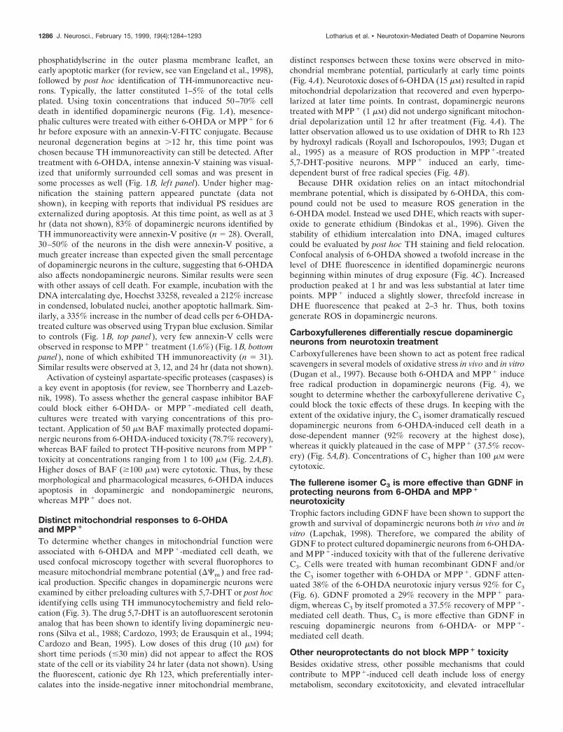
phosphatidylserine in the outer plasma membrane leaflet, anearly apoptotic marker (for review, see van Engeland et al., 1998),followed by post hoc identification of TH-immunoreactive neu-rons. Typically, the latter constituted 1–5% of the total cellsplated. Using toxin concentrations that induced 50–70% celldeath in identified dopaminergic neurons (Fig. 1A), mesence-phalic cultures were treated with either 6-OHDA or MPP1 for 6hr before exposure with an annexin-V-FITC conjugate. Becauseneuronal degeneration begins at .12 hr, this time point waschosen because TH immunoreactivity can still be detected. Aftertreatment with 6-OHDA, intense annexin-V staining was visual-ized that uniformly surrounded cell somas and was present insome processes as well (Fig. 1B, lef t panel). Under higher mag-nification the staining pattern appeared punctate (data notshown), in keeping with reports that individual PS residues areexternalized during apoptosis. At this time point, as well as at 3hr (data not shown), 83% of dopaminergic neurons identified byTH immunoreactivity were annexin-V positive (n 5 28). Overall,30–50% of the neurons in the dish were annexin-V positive, amuch greater increase than expected given the small percentageof dopaminergic neurons in the culture, suggesting that 6-OHDAalso affects nondopaminergic neurons. Similar results were seenwith other assays of cell death. For example, incubation with theDNA intercalating dye, Hoechst 33258, revealed a 212% increasein condensed, lobulated nuclei, another apoptotic hallmark. Sim-ilarly, a 335% increase in the number of dead cells per 6-OHDA-treated culture was observed using Trypan blue exclusion. Similarto controls (Fig. 1B, top panel), very few annexin-V cells wereobserved in response to MPP1 treatment (1.6%) (Fig. 1B, bottompanel), none of which exhibited TH immunoreactivity (n 5 31).Similar results were observed at 3, 12, and 24 hr (data not shown).
Activation of cysteinyl aspartate-specific proteases (caspases) isa key event in apoptosis (for review, see Thornberry and Lazeb-nik, 1998). To assess whether the general caspase inhibitor BAFcould block either 6-OHDA- or MPP1-mediated cell death,cultures were treated with varying concentrations of this pro-tectant. Application of 50 mM BAF maximally protected dopami-nergic neurons from 6-OHDA-induced toxicity (78.7% recovery),whereas BAF failed to protect TH-positive neurons from MPP1
toxicity at concentrations ranging from 1 to 100 mM (Fig. 2A,B).Higher doses of BAF ($100 mM) were cytotoxic. Thus, by thesemorphological and pharmacological measures, 6-OHDA inducesapoptosis in dopaminergic and nondopaminergic neurons,whereas MPP1 does not.
Distinct mitochondrial responses to 6-OHDAand MPP1
To determine whether changes in mitochondrial function wereassociated with 6-OHDA and MPP1-mediated cell death, weused confocal microscopy together with several fluorophores tomeasure mitochondrial membrane potential (DCm) and free rad-ical production. Specific changes in dopaminergic neurons wereexamined by either preloading cultures with 5,7-DHT or post hocidentifying cells using TH immunocytochemistry and field relo-cation (Fig. 3). The drug 5,7-DHT is an autofluorescent serotoninanalog that has been shown to identify living dopaminergic neu-rons (Silva et al., 1988; Cardozo, 1993; de Erausquin et al., 1994;Cardozo and Bean, 1995). Low doses of this drug (10 mM) forshort time periods (#30 min) did not appear to affect the ROSstate of the cell or its viability 24 hr later (data not shown). Usingthe fluorescent, cationic dye Rh 123, which preferentially inter-calates into the inside-negative inner mitochondrial membrane,
distinct responses between these toxins were observed in mito-chondrial membrane potential, particularly at early time points(Fig. 4A). Neurotoxic doses of 6-OHDA (15 mM) resulted in rapidmitochondrial depolarization that recovered and even hyperpo-larized at later time points. In contrast, dopaminergic neuronstreated with MPP1 (1 mM) did not undergo significant mitochon-drial depolarization until 12 hr after treatment (Fig. 4A). Thelatter observation allowed us to use oxidation of DHR to Rh 123by hydroxyl radicals (Royall and Ischoropoulos, 1993; Dugan etal., 1995) as a measure of ROS production in MPP1-treated5,7-DHT-positive neurons. MPP1 induced an early, time-dependent burst of free radical species (Fig. 4B).
Because DHR oxidation relies on an intact mitochondrialmembrane potential, which is dissipated by 6-OHDA, this com-pound could not be used to measure ROS generation in the6-OHDA model. Instead we used DHE, which reacts with super-oxide to generate ethidium (Bindokas et al., 1996). Given thestability of ethidium intercalation into DNA, imaged culturescould be evaluated by post hoc TH staining and field relocation.Confocal analysis of 6-OHDA showed a twofold increase in thelevel of DHE fluorescence in identified dopaminergic neuronsbeginning within minutes of drug exposure (Fig. 4C). Increasedproduction peaked at 1 hr and was less substantial at later timepoints. MPP1 induced a slightly slower, threefold increase inDHE fluorescence that peaked at 2–3 hr. Thus, both toxinsgenerate ROS in dopaminergic neurons.
Carboxyfullerenes differentially rescue dopaminergicneurons from neurotoxin treatmentCarboxyfullerenes have been shown to act as potent free radicalscavengers in several models of oxidative stress in vivo and in vitro(Dugan et al., 1997). Because both 6-OHDA and MPP1 inducefree radical production in dopaminergic neurons (Fig. 4), wesought to determine whether the carboxyfullerene derivative C3
could block the toxic effects of these drugs. In keeping with theextent of the oxidative injury, the C3 isomer dramatically rescueddopaminergic neurons from 6-OHDA-induced cell death in adose-dependent manner (92% recovery at the highest dose),whereas it quickly plateaued in the case of MPP1 (37.5% recov-ery) (Fig. 5A,B). Concentrations of C3 higher than 100 mM werecytotoxic.
The fullerene isomer C3 is more effective than GDNF inprotecting neurons from 6-OHDA and MPP1
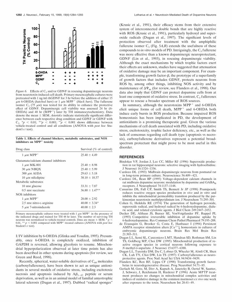
neurotoxicityTrophic factors including GDNF have been shown to support thegrowth and survival of dopaminergic neurons both in vivo and invitro (Lapchak, 1998). Therefore, we compared the ability ofGDNF to protect cultured dopaminergic neurons from 6-OHDA-and MPP1-induced toxicity with that of the fullerene derivativeC3. Cells were treated with human recombinant GDNF and/orthe C3 isomer together with 6-OHDA or MPP1. GDNF atten-uated 38% of the 6-OHDA neurotoxic injury versus 92% for C3
(Fig. 6). GDNF promoted a 29% recovery in the MPP1 para-digm, whereas C3 by itself promoted a 37.5% recovery of MPP1-mediated cell death. Thus, C3 is more effective than GDNF inrescuing dopaminergic neurons from 6-OHDA- or MPP1-mediated cell death.
Other neuroprotectants do not block MPP1 toxicityBesides oxidative stress, other possible mechanisms that couldcontribute to MPP1-induced cell death include loss of energymetabolism, secondary excitotoxicity, and elevated intracellular
1286 J. Neurosci., February 15, 1999, 19(4):1284–1293 Lotharius et al. • Neurotoxin-Mediated Death of Dopamine Neurons
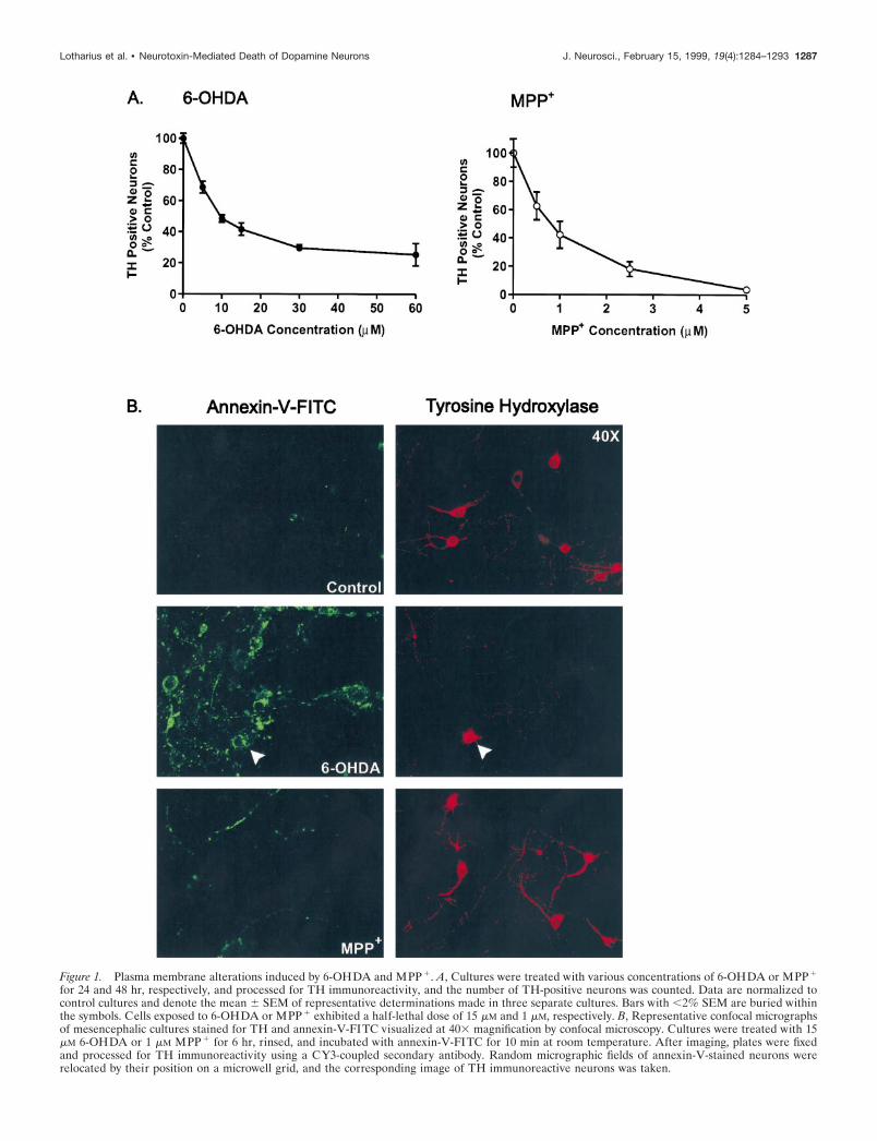
Figure 1. Plasma membrane alterations induced by 6-OHDA and MPP 1. A, Cultures were treated with various concentrations of 6-OHDA or MPP 1
for 24 and 48 hr, respectively, and processed for TH immunoreactivity, and the number of TH-positive neurons was counted. Data are normalized tocontrol cultures and denote the mean 6 SEM of representative determinations made in three separate cultures. Bars with ,2% SEM are buried withinthe symbols. Cells exposed to 6-OHDA or MPP 1 exhibited a half-lethal dose of 15 mM and 1 mM, respectively. B, Representative confocal micrographsof mesencephalic cultures stained for TH and annexin-V-FITC visualized at 403 magnification by confocal microscopy. Cultures were treated with 15mM 6-OHDA or 1 mM MPP 1 for 6 hr, rinsed, and incubated with annexin-V-FITC for 10 min at room temperature. After imaging, plates were fixedand processed for TH immunoreactivity using a CY3-coupled secondary antibody. Random micrographic fields of annexin-V-stained neurons wererelocated by their position on a microwell grid, and the corresponding image of TH immunoreactive neurons was taken.
Lotharius et al. • Neurotoxin-Mediated Death of Dopamine Neurons J. Neurosci., February 15, 1999, 19(4):1284–1293 1287
calcium. Thus, potential neuroprotectants include glutamate recep-tor and/or calcium channel blockers, nitric oxide synthase (NOS)inhibitors, or the addition of various energy supplements. A rangeof concentrations of representative agents for each of these classeswas tested. These included the noncompetitive NMDAR antago-nist MK-801, the AMPA/kainate receptor antagonist NBQX, theGroup I metabotropic glutamate receptor antagonist AIDA, theL-type calcium channel blocker nifedipine, the selective neuronalNOS inhibitor 7-NI, and the nonselective irreversible NOS inhib-itor L-NNA. The maximal protection and the concentration at
which it was achieved are listed in Table 1. Both succinate and thetwo NOS inhibitors used, L-NNA and 7-NI, provided modest butsignificant protection. The various glutamate receptor and calciumchannel blocks failed to attenuate MPP1-induced cell death (Ta-ble 1).
DISCUSSIONThe combined use of apoptotic and free radical-sensitive fluoro-phores together with vital dyes and/or field relocation techniqueshave provided new insights into the cellular changes underlying
Figure 2. The caspase inhibitor BAF rescues cultureddopaminergic neurons from 6-OHDA but not MPP 1-induced cell death. A, Representative fluorescent micro-graphs of mesencephalic cultures stained for TH and vi-sualized using a CY3-conjugated secondary antibody.Cells were exposed to 15 mM 6-OHDA or 1 mM MPP 1 inthe presence or absence of 50 mM BAF and stained after24 and 48 hr, respectively. B, TH cell counts of culturestreated with either toxin and BAF. Values are normalizedto the number of TH-positive neurons in vehicle-treatedcultures and denote the mean 6 SEM made of threeseparate cultures. **p , 0.001 compared with toxin-treated cultures; ••p , 0.001 in relation to vehicle-treatedcontrol (ANOVA with post hoc Student’s t test).
1288 J. Neurosci., February 15, 1999, 19(4):1284–1293 Lotharius et al. • Neurotoxin-Mediated Death of Dopamine Neurons

toxin-mediated dopaminergic cell death. The present findingsdemonstrate that (1) 6-OHDA triggers apoptosis in cultureddopaminergic neurons, whereas MPP1 does not; (2) the effects of6-OHDA in vitro are not selective for dopaminergic neurons; (3)both toxins generate early bursts in ROS, although only 6-OHDAtreatment depolarizes mitochondrial membrane potential(DCm); and (5) the newly described buckminsterfullerene deriv-ative C3 is more effective than GDNF in rescuing cells from6-OHDA neurotoxicity, whereas, similar to GDNF, C3 sup-presses a portion of the MPP1 injury. Taken together these datademonstrate that 6-OHDA and MPP1 kill cultured dopaminer-gic neurons by distinct cellular mechanisms.
Because these toxins have been widely used to create in vitroand in vivo animal models of PD, it is somewhat surprising thatthe process or mechanism(s) by which 6-OHDA and MPP1 killcells has remained equivocal. Several parameters that may haveconfounded this issue include the delivery, severity, and durationof the toxin as well as the overlapping nature of some cell deathprocesses activated during apoptosis or necrosis (Portera-Cailliau et al., 1997). Moreover, apoptosis itself appears to be a“family” of cell death programs that proceed along similar butnonidentical pathways in response to disparate stimuli. The de-
velopment of assays measuring changes in membrane lipid com-position as well as caspase activation have improved our ability toconfirm an apoptotic process. By these criteria, as well as byassays that detect DNA cleavage [terminal deoxynucleotidyltransferase-mediated biotinylated UTP nick end labeling(TUNEL), propidium iodide], 6-OHDA always resulted in apo-ptosis (data not shown). In contrast, even very low concentrationsof MPP1 (#1 mM) never induced externalization of phosphati-dylserine in identified dopaminergic neurons (Fig. 1), nor couldcell death be blocked by the general caspase inhibitor BAF (Fig.2). Moreover, we found no evidence that TH-expressing neuronswere TUNEL positive after MPP1 treatments at doses as high as10 mM (data not shown). Finally, in many apoptotic paradigms, anearly collapse in DCm is observed that precedes nuclear apoptosis(for review, see Green and Reed, 1998). The marked 6-OHDA-mediated collapse in DCm and its absence in MPP1-treateddopaminergic neurons (Fig. 4A) further support the contentionthat 6-OHDA but not MPP1 induces apoptosis in vitro.
Because both MPP1 and 6-OHDA compete with dopaminefor uptake via the plasma membrane transporter (Decker et al.,1993), their actions are generally thought to be selective fordopaminergic neurons. In our culture model, low concentrationsof MPP1 did appear to be specific, whereas even at very earlytime points after 6-OHDA treatment, 10-fold more neuronsexhibited signs of apoptosis than could be accounted for by posthoc TH staining (Fig. 1B). Thus, at least in vitro, 6-OHDAappears to be a nonselective neurotoxin that induces apoptosis indopaminergic and nondopaminergic neurons alike.
A number of previous studies both in vivo and in vitro havesuggested that MPP1 kills cells via apoptosis (Mochizuki et al.,1994; Itano and Nomura, 1995; Cassarino et al., 1998). Becausemost of these studies (1) have used at least 20-fold higher con-centrations of drug, (2) have not confirmed the affected cell typeor in some cases have not used dopaminergic cells, and/or (3)have relied solely on TUNEL positivity as an index of apoptosis,the interpretation that MPP1 elicits apoptosis in bona fide do-paminergic neurons should be regarded with caution. BecauseTUNEL staining is also observed in many models of necrosis(Grasl-Kraupp et al., 1995; van Lookeren Campagne et al., 1995;Negoescu et al., 1996; Portera-Cailliau et al., 1997), additionalcriteria, including caspase blockade or activation, membrane per-turbation, etc., are necessary to definitively establish that anapoptotic process is occurring. These studies highlight the need tofirst establish, in a simple system such as described here, thedirect mechanism of cell loss attributable to MPP1 toxicity tobetter interpret the more direct and complex processes in vivo.
MPP1 toxicity in vivo is almost certainly influenced by striatalas well as cortical inputs. For example, MPP1 has been shown toinduce dopamine efflux and auto-oxidation resulting in hydroxylradical formation (Obata and Chiueh, 1992). Glutamatergic in-puts may be a target for these free radicals, leading to glutamaterelease, impaired reuptake, and further damage to dopaminergicneurons by excitotoxicity. Additionally, the complex metabolismof MPTP itself, the mode of administration, and/or the presenceof other factors that might be missing in cultured neurons can allpotentially alter a cell’s susceptibility to death, further complicat-ing in vivo versus in vitro results.
Green and Reed (1998) have proposed a model by whichmitochondria mediate both apoptotic and nonapoptotic cell deathvia several related mechanisms, including impairment of electrontransport and DCm , release of proteins that activate caspasefamily members, and alterations in cellular redox potential. Thus,
Figure 3. Identification of dopaminergic neurons using 5,7-DHT prela-beling or post hoc TH staining. Top panels, Mesencephalic cultures wereincubated with 5,7-DHT for 30 min at 37°C, rinsed, and imaged byfluorescence microscopy with a computer-controlled camera. Images weretaken at 403 magnification. Cultures were subsequently fixed and stainedfor TH using a CY3-coupled secondary antibody. Field relocation showscolocalization of TH with 5,7-DHT (white arrows). This methodology wasused to assay changes in DHR and Rh 123 fluorescence in dopaminergiccells. Bottom panels, To measure DHE fluorescence in dopaminergicneurons, cultures were stained for TH via a color reaction after incubationwith DHE. Bottom lef t panel, Confocal image of cells exposed to6-OHDA for 30 min and incubated with DHE for 15 min (603). On theright is the corresponding differential interference contrast image ofTH-stained neurons in a relocated field.
Lotharius et al. • Neurotoxin-Mediated Death of Dopamine Neurons J. Neurosci., February 15, 1999, 19(4):1284–1293 1289

mitochondria represent both a target for cell death processes anda source of cytotoxic oxygen radicals. Previous studies wouldsuggest that MPP1 affects the first of these processes by blockingComplex I activity, which leads to decreased ATP levels, alter-ations in membrane permeability, and calcium influx akin toexcitotoxic cell death processes (for review, see Jenner, 1998).Such a model would predict an early loss in DCm , superoxideformation, and a subsequent cellular energy drain (White andReynolds, 1996). In contrast to these predictions, we saw no earlyperturbations of mitochondrial membrane potential (Fig. 4A),nor could we block MPP1-mediated cell death with ionotropic/metabotropic glutamate or calcium channel antagonists (Table 1).Similarly, glycolytic and Complex II substrates, which would beexpected to suppress MPP1 neurotoxicity by providing an alter-native source of ATP, were largely ineffective (Table 1). Thus, thespecific MPP1-induced signals that lead to cell death are notsolely mediated by a loss of mitochondrial ATP production. Inagreement with this hypothesis, Khan et al. (1997) showed re-cently that MPP1 was as toxic to cells without mitochondria as itwas to cells with normal mitochondrial function. Further studiesof temporal events associated with MPP1 toxicity will help toclarify this intriguing issue.
Oxidation of DHR has been used as a measure of mitochon-drial ROS formation (Dugan et al., 1995). As such, it is thoughtto measure radicals such as hydroxyl and peroxynitrite bothdirectly and indirectly via the conversion of superoxide (Hender-son and Chappell, 1993). The successful utilization of DHRdepends on maintenance of DCm, a decline in which precluded itsuse for 6-OHDA experiments. Because MPP1 treatment did notalter DCm (Fig. 3A), we were able to observe a burst in DHRfluorescence in identified dopaminergic neurons as early as 30min after MPP1 treatment before a peak at 3 hr (Fig. 4B). Thistime course paralleled that of DHE (Fig. 4C), which preferen-tially reacts with superoxide. In contrast to in vivo studies (Smithet al., 1994; Przedborski et al., 1996), the NOS inhibitors 7-NI andL-NNA protected only a small percentage (16%) of neurons fromMPP1 treatment (Table 1). These data would suggest that, atleast in vitro, peroxynitrite is only a small component of theMPP1 injury. Rather, superoxide appears to be the primaryradical involved. This is consistent with data from Ramsay andSinger (1992) showing that partial ubiquinone oxidation resultingfrom MPP1 blockade of Complex I can lead to superoxideformation in submitochondrial particles.
We observed a late (12 hr) decline in DCm after MPP1
treatment, consistent with its action as an inhibitor of Complex I.The subsequent return to near normal values at 24 hr could beexplained by the selection of a subpopulation of MPP1-resistantneurons using 5,7-DHT labeling. Although the classic Complex Iinhibitor rotenone generally results in a tight block of electrontransport flow and subsequent membrane depolarization (Sim-bula et al., 1997), MPP1 produces only a partial block of Com-plex I activity (Gluck et al., 1994), which might allow the mem-brane potential to be maintained for some time. Moreover,“reversed” function of the F1/F0ATPase, using ATP hydrolysis topump protons together with ATP generated through glycolysis,could temporarily support DCm (Mitchell and Moyle, 1968).MPP1-induced ROS generation likely reflects inhibition of Com-plex I, which might further the energy impairment produced byMPP1 itself. Thus, oxidative inactivation of key components ofboth the electron transport chain and the glycolytic pathwaymight contribute to a subsequent “metabolic death.” This might
Figure 4. 6-OHDA and MPP 1 differentially affect mitochondrial mem-brane potential (CDm ) and induce ROS formation. A, Time course of CDmin 5,7-DHT-labeled dopaminergic neurons treated with 15 mM 6-OHDA(F) or 1 mM MPP 1 (E). After drug treatments, cells were loaded with 0.3mM Rh 123 for 20 min and assayed using a laser scanning confocal micro-scope. Values correspond to average pixel intensity normalized to baselinefluorescence values from vehicle-treated cells. B, Time-dependent changesin ROS production in dopaminergic neurons treated with 1 mM MPP 1.After treatment with 1 mM MPP 1 for the indicated time period, cells wereincubated with 15 mM DHR for 20 min, rinsed, and imaged by confocalmicroscopy. Values correspond to fluorescence intensity and are normal-ized to vehicle-treated labeled dopaminergic neurons. C, Time-dependentinduction of ROS formation in dopaminergic neurons treated with 15 mM6-OHDA or 1 mM MPP 1. After treatment with either drug for 0, 0.25, 0.5,1, 3, and 6 hr, cells were loaded with 10 mg/ml DHE for 15 min at 37°C,fixed, stained for TH, and assayed by confocal microscopy. Values representnormalized DHE fluorescence from TH-immunoreactive neurons. Dataare mean 6 SEM of determinations made in three separate cultures. *p ,0.01; **p , 0.001, compared with values for vehicle-treated cultures(ANOVA with post hoc Student’s t test).
1290 J. Neurosci., February 15, 1999, 19(4):1284–1293 Lotharius et al. • Neurotoxin-Mediated Death of Dopamine Neurons

explain why antioxidants provide only partial protection againstMPP1 injury.
In contrast, neurons exposed to 6-OHDA showed rapid loss ofmitochondrial membrane potential, followed by rebound hyper-polarization. We believe this reflects early oxidative inactivation
of components from mitochondrial and glycolytic metabolic path-ways by 6-OHDA. Glyceraldehyde-3-phosphate dehydrogenase(GAPDH), for example, can undergo reversible oxidative inacti-vation by H2O2 (Janero et al., 1994). This inhibition might limitthe ability of glycolysis to support DCm in the face of Complex
Figure 5. The C3 fullerene isomer protects cultured dopaminer-gic neurons from 6-OHDA-induced toxicity but only partiallyattenuates MPP 1-mediated cell death. A, Representative fluores-cent micrographs of TH-positive neurons treated with either 15 mM6-OHDA or 1 mM MPP 1 in the presence or absence of 75 mM C3.B, Dose-dependent effects of C3 on dopaminergic neurons treatedwith 15 mM 6-OHDA (F) or 1 mM MPP 1 (E). The number ofsurviving dopaminergic neurons was determined 24 hr (6-OHDA)or 48 hr (MPP 1) later by TH immunocytochemistry and normal-ized to the number of TH neurons in vehicle-treated plates. Dataare mean 6 SEM from determinations made from three cultures.*p , 0.01; **p , 0.001 compared with values from control plates(ANOVA with post hoc Student’s t test).
Lotharius et al. • Neurotoxin-Mediated Death of Dopamine Neurons J. Neurosci., February 15, 1999, 19(4):1284–1293 1291
I /IV inhibition by 6-OHDA (Glinka and Youdim, 1995). Presum-ably, once 6-OHDA is completely oxidized, inhibition ofGAPDH is reversed, allowing glycolysis to resume. Mitochon-drial hyperpolarization observed at later time points has alsobeen described in other systems during apoptosis (for review, seeGreen and Reed, 1998).
Recently, spherical, water-soluble derivatives of C60 molecules(carboxyfullerenes), have been shown to act as unique antioxi-dants in several models of oxidative stress, including excitotoxicnecrosis and apoptosis induced by Ab1–42 peptide or serumdeprivation, as well as in an animal model of familial amyotrophiclateral sclerosis (Dugan et al., 1997). Dubbed “radical sponges”
(Krusic et al., 1991), their efficacy stems from their extensivesystem of interconnected double bonds that are highly reactivewith ROS (Krusic et al., 1991), particularly hydroxyl and super-oxide radicals (Dugan et al., 1997). The significant levels ofprotection observed after treatment with the amphiphilicfullerene isomer C3 (Fig. 5A,B) extends the usefulness of thesecompounds to in vitro models of PD. Intriguingly, the C3 fullerenewas more effective than a known dopaminergic neuroprotectant,GDNF (Lin et al., 1993), in rescuing dopaminergic viability.Although the exact mechanisms by which trophic factors exerttheir effects are unknown, studies have suggested that attenuationof oxidative damage may be an important component. For exam-ple, transforming growth factor-b, the prototype of a superfamilyof growth factors that includes GDNF, protects neurons fromROS by, among other things, inhibiting NOS activity and bymaintenance of DCm (for review, see Flanders et al., 1998). Ourdata also imply that GDNF can protect dopamine cells from atleast one component of oxidative stress. In contrast, C3 fullerenesappear to rescue a broader spectrum of ROS sources.
In summary, although the neurotoxins MPP1 and 6-OHDAelicit unique forms of cell death, MPP1 and 6-OHDA bothinduce early bursts in ROS production. Because impaired ROShomeostasis has been implicated in PD, the development ofantioxidants is a promising therapeutic goal. Given the variousmechanisms of cell death associated with PD, including oxidativestress, excitotoxicity, trophic factor deficiency, etc., as well as thelack of consensus regarding cell death type (apoptosis vs necro-sis), carboxyfullerene derivatives represent a potential broad-spectrum protectant that might prove to be most useful in thisdisorder.
REFERENCESBindokas VP, Jordan J, Lee CC, Miller RJ (1996) Superoxide produc-
tion in rat hippocampal neurons: selective imaging with hydroethidine.J Neurosci 16:1324–1336.
Cardozo DL (1993) Midbrain dopaminergic neurons from postnatal ratin long-term primary culture. Neuroscience 56:409–421.
Cardozo DL, Bean BP (1995) Voltage-dependent calcium channels inrat midbrain dopamine neurons: modulation by dopamine and GABABreceptors. J Neurophysiol 74:1137–1148.
Cassarino DS, Fall CP, Smith TS, Bennett Jr JP (1998) Pramipexolereduces reactive oxygen species production in vivo and in vitro andinhibits the mitochondrial permeability transition produced by the par-kinsonian neurotoxin methylpyridinium ion. J Neurochem 71:295–301.
Cohen G, Heikkila RE (1974) The generation of hydrogen peroxide,superoxide radical, and hydroxyl radical by 6-hydroxydopamine, dialu-ric acid, and related cytotoxic agents. J Biol Chem 249:2447–2452.
Decker DE, Althaus JS, Buxser SE, VonVoigtlander PF, Ruppel PL(1993) Competitive irreversible inhibition of dopamine uptake by6-hydroxydopamine. Res Commun Chem Pathol Pharmacol 79:195–208.
de Erausquin G, Brooker G, Costa E, Hanbauer I (1994) PersistentAMPA receptor stimulation alters [Ca 21]i homeostasis in cultures ofembryonic dopaminergic neurons. Brain Res Mol Brain Res21:303–311.
Dugan LL, Sensi SL, Canzoniero LMT, Handran SD, Rothman SM, LinTS, Goldberg MP, Choi DW (1995) Mitochondrial production of re-active oxygen species in cortical neurons following exposure toN-methyl-D-aspartate. J Neurosci 15:6377–6388.
Dugan LL, Turetsky DM, Du C, Lobner D, Wheeler M, Almli CR, ShenCK, Luh TY, Choi DW, Lin TS (1997) Carboxyfullerenes as neuro-protective agents. Proc Natl Acad Sci USA 94:9434–9439.
Flanders KC, Ren RF, Lippa CF (1998) Transforming growth factor-betas in neurodegenerative disease. Prog Neurobiol 54:71–85.
Gerlach M, Gotz, M, Dirr A, Kupsch A, Janetzky B, Oertel W, Sautter,J, Schwarz, J, Reichmann H, Riederer P (1996) Acute MPTP treat-ment produces no changes in mitochondrial complex activities andindices of oxidative damage in the common marmoset ex vivo one weekafter exposure to the toxin. Neurochem Int 28:41–49.
Figure 6. Effects of C3 and/or GDNF in rescuing dopaminergic neuronsfrom neurotoxin-induced cell death. Primary mesencephalic cultures werepretreated with 1 ng/ml rhGDNF for 3 hr before the addition of either 15mM 6-OHDA (hatched bars) or 1 mM MPP 1 (black bars). The fullereneisomer C3 (75 mM) was tested for its ability to enhance the protectiveeffect of GDNF. Dopaminergic cell viability was assessed 24 hr (6-OHDA) and 48 hr (MPP 1) later by TH immunocytochemistry. Datadenote the mean 6 SEM. Asterisks indicate statistically significant differ-ence between each respective drug condition and GDNF or GDNF withC3. *p , 0.01; **p , 0.001; ••p , 0.001 shows difference betweenvehicle-treated control and all conditions (ANOVA with post hoc Stu-dent’s t test).
Table 1. Effects of channel blockers, metabolic substrates, and NOSinhibitors on MPP1 toxicity
Drug class Survival (% of control)
1 mM MPP1 25.40 6 4.49
Glutamate/calcium channel inhibitors1 mM MK-801 25.40 6 8.9830 mM NBQX 25.40 6 5.99300 mM AIDA 29.63 6 5.1810 mM nifedipine 38.10 6 10.37
Metabolic substrates10 mM glucose 33.31 6 7.870.5 mM succinate 36.80 6 1.47**
NOS inhibitors1 mM MPP1 28.00 6 2.922.5 mM nitro-L-arginine 40.00 6 3.34*5 mM 7-nitroindazole 40.00 6 2.3
Primary mesencephalic cultures were treated with 1 mM MPP1 in the presence ofthe indicated drugs and stained for TH 48 hr later. The number of surviving THneurons was normalized to vehicle-treated controls. Data are mean 6 SEM. * p ,0.01, ** p , 0.001 compared with MPP1-treated cultures (ANOVA with post hocStudent’s t test).
1292 J. Neurosci., February 15, 1999, 19(4):1284–1293 Lotharius et al. • Neurotoxin-Mediated Death of Dopamine Neurons

Glinka Y, Youdim MBH (1995) Inhibition of mitochondrial complexes Iand IV by 6-hydroxydopamine. Eur J Pharmacol 292:329–332.
Gluck MR, Krueger MJ, Ramsay RR, Sablin SO, Singer TP, Nicklas WJ(1994) Characterization of the inhibitory mechanism of 1-methyl-4-phenylpyridinium and 4-phenylpyridine analogs in inner mitochondrialmembrane preparations. J Biol Chem 269:3167–3174.
Grasl-Kraupp B, Ruttkay-Nedecky B, Koudelka H, Bukowska K, BurschW, Schulte-Hermann R (1995) In situ detection of fragmented DNA(TUNEL assay) fails to discriminate among apoptosis, necrosis, andautolytic cell death: a cautionary note. Hepatology 21:1465–1468.
Green DR, Reed JC (1998) Mitochondria and apoptosis. Science281:1309–1312.
Graham DG, Tiffany SM, Bell WR, Gutknecht WF (1978) Autooxida-tion versus covalent binding of quinones as the mechanism of toxicity ofdopamine, 6-hydroxydopamine, related compounds toward C1300 neu-roblastoma cells in vitro. Mol Phamacol 14:644–653.
Hantraye P, Brouillet E, Ferrante R, Palfi S, Dolan R, Matthews RT, BealMF (1996) Inhibition of neuronal nitric oxide synthase preventsMPTP-induced parkinsonism in baboons. Nat Med 2:1017–1021.
Henderson LM, Chappell JB (1993) Dihydrorhodamine 123: a fluores-cent probe for superoxide generation? Eur J Biochem 217:973–980.
Itano Y, Nomura Y (1995) 1-methyl-4-phenyl-pyridinium ion (MPP 1)causes DNA fragmentation and increases the Bcl-2 expression in hu-man neuroblastoma, SH-SY5Y cells, through different mechanisms.Brain Res 704:240–245.
Janero DR, Hreniuk D, Sharif HM (1994) Hydroperoxide-induced oxi-dative stress impairs heart muscle cell carbohydrate metabolism. Am JPhysiol 266:C179–C188.
Jenner P (1998) Oxidative mechanisms in nigral cell death in Parkin-son’s disease. Mov Disord 13:24–34.
Khan U, Filiano B, King M, Przedborski S (1997) Is Parkinson’s disease(PD) an extra-mitochondrial disorder? Neurology 48:A201.
Krusic PJ, Wasserman E, Keizer PN, Morton JR, Preston KF (1991)Radical reactions of C60. Science 254:1183–1185.
Lapchak PA (1998) A preclinical development strategy designed to op-timize the use of glial cell line-derived neurotrophic factor in thetreatment of Parkinson’s disease. Mov Disord 13:49–54.
Lin LF, Doherty DH, Lile JD, Bektesh S, Collins F (1993) GDNF: aglial cell line-derived neurotrophic factor for midbrain dopaminergicneurons. Science 260:1130–1132.
Mitchell P, Moyle J (1968) Proton translocation coupled to ATP hydro-lysis in rat liver mitochondria. Eur J Biochem 4:530–539.
Mochizuki H, Nakamura N, Nishi K, Mizuno Y (1994) Apoptosis isinduced by 1-methyl-4-phenylpyridinium ion (MPP 1) in ventralmesencephalic-striatal co-culture in rat. Neurosci Lett 170:191–194.
Negoescu A, Lorimier P, Labat-Moleur F, Drouet C, Robert C, Guiller-met C, Brambilla C, Brambilla E (1996) In situ apoptotic cell labelingby the TUNEL method: improvement and evaluation on cell prepara-tions. J Histochem Cytochem 4:959–968.
Obata T, Chiueh CC (1992) In vivo trapping of hydroxyl free radicals inthe striatum utilizing intracranial microdialysis perfusion of salicylate:effects of MPTP, MPDP 1, and MPP 1. J Neural Transm 89:139–145.
Portera-Cailliau C, Price DL, Martin LJ (1997) Non-NMDA andNMDA receptor-mediated excitotoxic neuronal deaths in adult brainare morphologically distinct: further evidence for an apoptosis-necrosiscontinuum. J Comp Neurol 3781:88–104.
Przedborski S, Jackson-Lewis V (1998) Mechanisms of MPTP toxicity.Mov Disord 13:35–38.
Przedborski S, Kostic V, Jackson-Lewis V, Naini AB, Simonetti S, FahnS, Carlson E, Epstein CJ, Cadet JL (1992) Transgenic mice withincreased Cu/Zn-superoxide dismutase activity are resistant toN-methyl-4-phenyl-1,2,3,6-tetrahydropyridine-induced neurotoxicity.J Neurosci 12:1658–1667.
Przedborski S, Jackson-Lewis V, Yokoyama R, Shibata T, Dawson VL,Dawson TM (1996) Role of neuronal nitric oxide in l-methyl-4-phenyl-1,2,3,6-tetrahydropyridine (MPTP)-induced dopaminergic neu-rotoxicity. Proc Natl Acad Sci USA 93:4565–4571.
Ramsay RR, Singer TP (1992) Relation of superoxide generation andlipid peroxidation to the inhibition of NADH-Q oxidoreductase byrotenone, piercidin A, and MPP 1. Biochem Biophys Res Commun189:47–52.
Royall JA, Ischoropoulos H (1993) Evaluation of 29,79-dichlorofluoresceinand dihydrorhodamine 123 as fluorescent probes for intracellular H202 incultured endothelial cells. Arch Biochem Biophys 302:348–355.
Silva NL, Mariani AP, Harrison NL, Barker JL (1988) 5,7-dihydroxytryptamine identifies living dopaminergic neurons in mesen-cephalic cultures. Proc Natl Acad Sci USA 85:7346–7350.
Simbula G, Glascott Jr PA, Akita S, Hoek JB, Farber JL (1997) Twomechanisms by which ATP depletion potentiates induction of themitochondrial permeability transition. Am J Physiol 273:C479–C488.
Smith, TS, Swerdlow, RH, Parker WD, Bennett JP (1994) Reduction ofMPP 1-induced hydroxyl radical formation and nigrostriatal MPTPtoxicity by inhibiting nitric oxide synthase. NeuroReport 5:2598–2600.
Thornberry NA, Lazebnik Y (1998) Caspases: enemies within. Science281:1312–1316.
van Engeland M, Nieland LJ, Ramaekers FC, Schutte B, Reuteling-sperger CP (1998) Annexin V-affinity assay: a review on an apoptosisdetection system based on phosphatidylserine exposure. Cytometry31:1–9.
van Lookeren Campagne M, Lucassen PJ, Vermeulen JP, Balazs R(1995) NMDA and kainate induce internucleosomal DNA cleavageassociated with both apoptotic and necrotic cell death in the neonatalrat brain. Eur J Neurosci 7:1627–1640.
White RJ, Reynolds IJ (1996) Mitochondrial depolarization inglutamate-stimulated neurons: an early signal specific to excitotoxinexposure. J Neurosci 16:5688–5697.
Lotharius et al. • Neurotoxin-Mediated Death of Dopamine Neurons J. Neurosci., February 15, 1999, 19(4):1284–1293 1293
Copyright © 2022 FDOKUMEN