REPORT Mutations in HOXD13 Underlie Syndactyly Type V and a Novel Brachydactyly-Syndactyly Syndrome
11
www.ajhg.org The American Journal of Human Genetics Volume 80 February 2007 361 REPORT Mutations in HOXD13 Underlie Syndactyly Type V and a Novel Brachydactyly-Syndactyly Syndrome Xiuli Zhao, * Miao Sun, * Jin Zhao, J. Alfonso Leyva, Hongwen Zhu, Wei Yang, Xuan Zeng, Yang Ao, Qing Liu, Guoyang Liu, Wilson H. Y. Lo, Ethylin Wang Jabs, L. Mario Amzel, Xiangnian Shan, and Xue Zhang HOXD13, the homeobox-containing gene located at the most 5 end of the HOXD cluster, plays a critical role in limb development. It has been shown that mutations in human HOXD13 can give rise to limb malformations, with variable expressivity and a wide spectrum of clinical manifestations. Polyalanine expansions in HOXD13 cause synpolydactyly, whereas amino acid substitutions in the homeodomain are associated with brachydactyly types D and E. We describe two large Han Chinese families with different limb malformations, one with syndactyly type V and the other with limb features overlapping brachydactyly types A4, D, and E and mild syndactyly of toes 2 and 3. Two-point linkage analysis showed LOD scores 13( ) for markers within and/or flanking the HOXD13 locus in both families. In the family v p 0 with syndactyly type V, we identified a missense mutation in the HOXD13 homeodomain, c.950ArG (p.Q317R), which leads to substitution of the highly conserved glutamine that is important for DNA-binding specificity and affinity. In the family with complex brachydactyly and syndactyly, we detected a deletion of 21 bp in the imperfect GCN (where N denotes A, C, G, or T) triplet-containing exon 1 of HOXD13, which results in a polyalanine contraction of seven residues. Moreover, we found that the mutant HOXD13 with the p.Q317R substitution was unable to transactivate the human EPHA7 promoter. Molecular modeling data supported these experimental results. The calculated interactions energies were in agreement with the measured changes of the activity. Our data established the link between HOXD13 and two additional limb phenotypes—syndactyly type V and brachydactyly type A4—and demonstrated that a poly- alanine contraction in HOXD13, most likely, led to other digital anomalies but not to synpolydactyly. We suggest the term “HOXD13 limb morphopathies” for the spectrum of limb disorders caused by HOXD13 mutations. From the Department of Medical Genetics and National Key Laboratory of Medical Molecular Biology, Institute of Basic Medical Sciences, Chinese Academy of Medical Sciences and Peking Union Medical College, Beijing (X. Zhao; M.S.; J.Z.; W.Y.; X. Zeng; Y.A.; Q.L.; G.L.; W.H.Y.L.; X. Zhang); Genetics Research Center, Southeast University School of Medicine, Nanjing, China (X. Zhao; X.S.); Institute of Genetic Medicine, Center for Craniofacial De- velopment and Disorders, The Johns Hopkins University (M.S.; E.W.J.), and Department of Biophysics and Biophysical Chemistry, The Johns Hopkins University School of Medicine (J.A.L.; L.M.A.), Baltimore; Center for Genetic Medicine, Lanzhou University, Lanzhou, China (H.Z.); and The Research Center for Medical Genomics, China Medical University, Shenyang, China (X. Zhang) Received September 29, 2006; accepted for publication November 22, 2006; electronically published January 3, 2007. Address for correspondence and reprints: Dr. Xue Zhang, Department of Medical Genetics, Institute of Basic Medical Sciences, Chinese Academy of Medical Sciences and Peking Union Medical College, 5 Dong Dan San Tiao, Beijing 100005, China. E-mail: [email protected] * These two authors contribute equally to this work. Am. J. Hum. Genet. 2007;80:361–371. 2007 by The American Society of Human Genetics. All rights reserved. 0002-9297/2007/8002-0017$15.00 The homeobox-containing (HOX) genes are a highly con- served transcription-factor family that displays important function in early development. 1,2 In humans, there are 39 HOX genes arranged in four separate clusters: HOXA, HOXB, HOXC, and HOXD. 3 These clusters are located on chro- mosomes 7p15, 17q21, 12q13, and 2q31, respectively, and show a striking colinearity in their 5 r3 genomic position and transcription direction. HOXD13 (MIM *142989; Gen- Bank accession number NM_000523), the most 5 gene of the HOXD cluster, has two coding exons: exon 1 with the imperfect GCN (where N denotes A, C, G, or T) triplet repeats encoding the N-terminal region with a 15-residue polyalanine tract (residues 49–63), and exon 2, which con- tains the homeobox region encoding the C-terminal por- tion with a 60-residue homeodomain (residues 268–327). 4 The homeodomain forms three a-helices. Structural stud- ies of HOX proteins have shown that residues in the rec- ognition helix-3—mainly, the 47th isoleucine (I47), the 50th glutamine (Q50), and the 51st asparagine (N51) of the homeodomain—make base-specific DNA contacts in the major groove, and residues in the N-terminal arm in- teract with the minor groove of DNA, thereby collabora- tively determining DNA-binding specificity and affinity. 5–10 HOXD13 is the first HOX gene known to be linked to human developmental disorders. 11,12 Mutations in HOXD13 are associated with limb deformities in both humans and mice, suggesting a critical role in limb development. Syn- polydactyly (SPD [MIM 186000], or syndactyly type II) is a dominantly inherited limb malformation with incom- plete penetrance and variable expressivity. It is character- ized by soft-tissue syndactyly between fingers 3 and 4 and between toes 4 and 5, with partial or complete digit du- plication within the syndactylous web. Both interfamilial and intrafamilial variations in SPD limb phenotype exist. Individuals with an atypical form of SPD share a distinc- tive set of foot phenotypes, including small spurs of bone between metatarsals 1 and 2. 11 Polyalanine-expansion mu- tations in HOXD13 lead to typical SPD, whereas deletions and missense mutations are associated with atypical SPD. 11– 21 Furthermore, in the spontaneous spdh mouse mutant, which has a phenotype similar to human SPD, a polyala- nine expansion of seven residues in Hoxd13 was de-
Transcript of REPORT Mutations in HOXD13 Underlie Syndactyly Type V and a Novel Brachydactyly-Syndactyly Syndrome
www.ajhg.org The American Journal of Human Genetics Volume 80 February 2007 361
REPORT
Mutations in HOXD13 Underlie Syndactyly Type V and a NovelBrachydactyly-Syndactyly SyndromeXiuli Zhao,* Miao Sun,* Jin Zhao, J. Alfonso Leyva, Hongwen Zhu, Wei Yang, Xuan Zeng, Yang Ao,Qing Liu, Guoyang Liu, Wilson H. Y. Lo, Ethylin Wang Jabs, L. Mario Amzel,Xiangnian Shan, and Xue Zhang
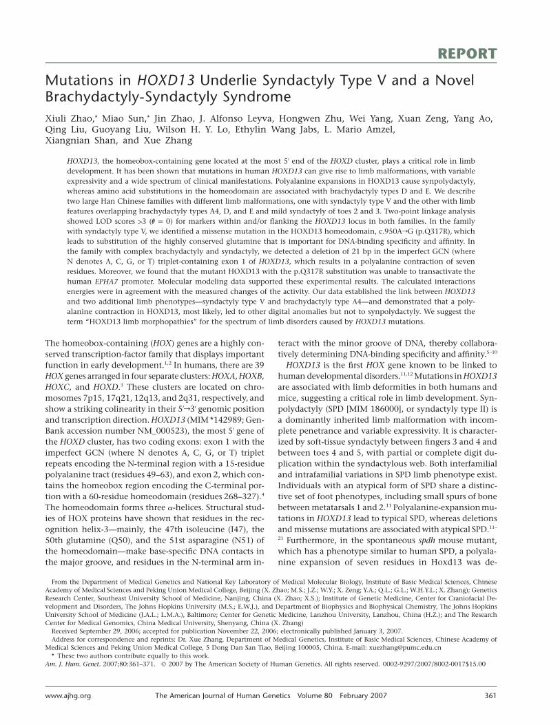
HOXD13, the homeobox-containing gene located at the most 5′ end of the HOXD cluster, plays a critical role in limbdevelopment. It has been shown that mutations in human HOXD13 can give rise to limb malformations, with variableexpressivity and a wide spectrum of clinical manifestations. Polyalanine expansions in HOXD13 cause synpolydactyly,whereas amino acid substitutions in the homeodomain are associated with brachydactyly types D and E. We describetwo large Han Chinese families with different limb malformations, one with syndactyly type V and the other with limbfeatures overlapping brachydactyly types A4, D, and E and mild syndactyly of toes 2 and 3. Two-point linkage analysisshowed LOD scores 13 ( ) for markers within and/or flanking the HOXD13 locus in both families. In the familyv p 0with syndactyly type V, we identified a missense mutation in the HOXD13 homeodomain, c.950ArG (p.Q317R), whichleads to substitution of the highly conserved glutamine that is important for DNA-binding specificity and affinity. Inthe family with complex brachydactyly and syndactyly, we detected a deletion of 21 bp in the imperfect GCN (whereN denotes A, C, G, or T) triplet-containing exon 1 of HOXD13, which results in a polyalanine contraction of sevenresidues. Moreover, we found that the mutant HOXD13 with the p.Q317R substitution was unable to transactivate thehuman EPHA7 promoter. Molecular modeling data supported these experimental results. The calculated interactionsenergies were in agreement with the measured changes of the activity. Our data established the link between HOXD13and two additional limb phenotypes—syndactyly type V and brachydactyly type A4—and demonstrated that a poly-alanine contraction in HOXD13, most likely, led to other digital anomalies but not to synpolydactyly. We suggest theterm “HOXD13 limb morphopathies” for the spectrum of limb disorders caused by HOXD13 mutations.
From the Department of Medical Genetics and National Key Laboratory of Medical Molecular Biology, Institute of Basic Medical Sciences, ChineseAcademy of Medical Sciences and Peking Union Medical College, Beijing (X. Zhao; M.S.; J.Z.; W.Y.; X. Zeng; Y.A.; Q.L.; G.L.; W.H.Y.L.; X. Zhang); GeneticsResearch Center, Southeast University School of Medicine, Nanjing, China (X. Zhao; X.S.); Institute of Genetic Medicine, Center for Craniofacial De-velopment and Disorders, The Johns Hopkins University (M.S.; E.W.J.), and Department of Biophysics and Biophysical Chemistry, The Johns HopkinsUniversity School of Medicine (J.A.L.; L.M.A.), Baltimore; Center for Genetic Medicine, Lanzhou University, Lanzhou, China (H.Z.); and The ResearchCenter for Medical Genomics, China Medical University, Shenyang, China (X. Zhang)
Received September 29, 2006; accepted for publication November 22, 2006; electronically published January 3, 2007.Address for correspondence and reprints: Dr. Xue Zhang, Department of Medical Genetics, Institute of Basic Medical Sciences, Chinese Academy of
Medical Sciences and Peking Union Medical College, 5 Dong Dan San Tiao, Beijing 100005, China. E-mail: [email protected]* These two authors contribute equally to this work.
Am. J. Hum. Genet. 2007;80:361–371. � 2007 by The American Society of Human Genetics. All rights reserved. 0002-9297/2007/8002-0017$15.00
The homeobox-containing (HOX) genes are a highly con-served transcription-factor family that displays importantfunction in early development.1,2 In humans, there are 39HOX genes arranged in four separate clusters: HOXA, HOXB,HOXC, and HOXD.3 These clusters are located on chro-mosomes 7p15, 17q21, 12q13, and 2q31, respectively, andshow a striking colinearity in their 5′r3′ genomic positionand transcription direction. HOXD13 (MIM *142989; Gen-Bank accession number NM_000523), the most 5′ gene ofthe HOXD cluster, has two coding exons: exon 1 with theimperfect GCN (where N denotes A, C, G, or T) tripletrepeats encoding the N-terminal region with a 15-residuepolyalanine tract (residues 49–63), and exon 2, which con-tains the homeobox region encoding the C-terminal por-tion with a 60-residue homeodomain (residues 268–327).4
The homeodomain forms three a-helices. Structural stud-ies of HOX proteins have shown that residues in the rec-ognition helix-3—mainly, the 47th isoleucine (I47), the50th glutamine (Q50), and the 51st asparagine (N51) ofthe homeodomain—make base-specific DNA contacts inthe major groove, and residues in the N-terminal arm in-
teract with the minor groove of DNA, thereby collabora-tively determining DNA-binding specificity and affinity.5–10
HOXD13 is the first HOX gene known to be linked tohuman developmental disorders.11,12 Mutations in HOXD13are associated with limb deformities in both humans andmice, suggesting a critical role in limb development. Syn-polydactyly (SPD [MIM 186000], or syndactyly type II) isa dominantly inherited limb malformation with incom-plete penetrance and variable expressivity. It is character-ized by soft-tissue syndactyly between fingers 3 and 4 andbetween toes 4 and 5, with partial or complete digit du-plication within the syndactylous web. Both interfamilialand intrafamilial variations in SPD limb phenotype exist.Individuals with an atypical form of SPD share a distinc-tive set of foot phenotypes, including small spurs of bonebetween metatarsals 1 and 2.11 Polyalanine-expansionmu-tations in HOXD13 lead to typical SPD, whereas deletionsand missense mutations are associated with atypical SPD.11–
21 Furthermore, in the spontaneous spdh mouse mutant,which has a phenotype similar to human SPD, a polyala-nine expansion of seven residues in Hoxd13 was de-
362 The American Journal of Human Genetics Volume 80 February 2007 www.ajhg.org
tected.22,23 Missense mutations in HOXD13 also underliebrachydactyly types D (BDD [MIM 113200]) and E (BDE[MIM 113300]).24,25 BDD presents with short and broaddistal phalanges of the thumbs and halluces, whereas BDEhas the cardinal feature of one or more shortened meta-carpals and/or metatarsals. Two HOXD13 missense muta-tions within the homeodomain—p.S308C and p.I314L—have been described in families exhibiting features of BDEwith BDD and BDE with mild SPD, respectively.24,25
Syndactyly type V (MIM 186300), defined as “syndac-tyly associated with metacarpal and metatarsal synostosis”by Temtamy and McKusick,26(p302) represents one of therarest types of nonsyndromic syndactyly.27 It is inheritedas an autosomal dominant trait with synostotic fusion ofmetacarpals 4 and 5 as the hallmark.28 Brachydactyly typeA4 (BDA4 [MIM 112800]) is characterized by short middlephalanges of the 2nd and 5th fingers and absence of mid-dle phalanges of the 2nd–5th toes. In this report, we de-scribe limb phenotypes of syndactyly type V and a novelbrachydactyly-syndactyly syndrome that includes BDA4,and we report novel mutations in HOXD13 in two largeHan Chinese families.
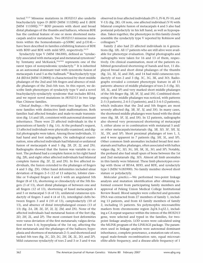
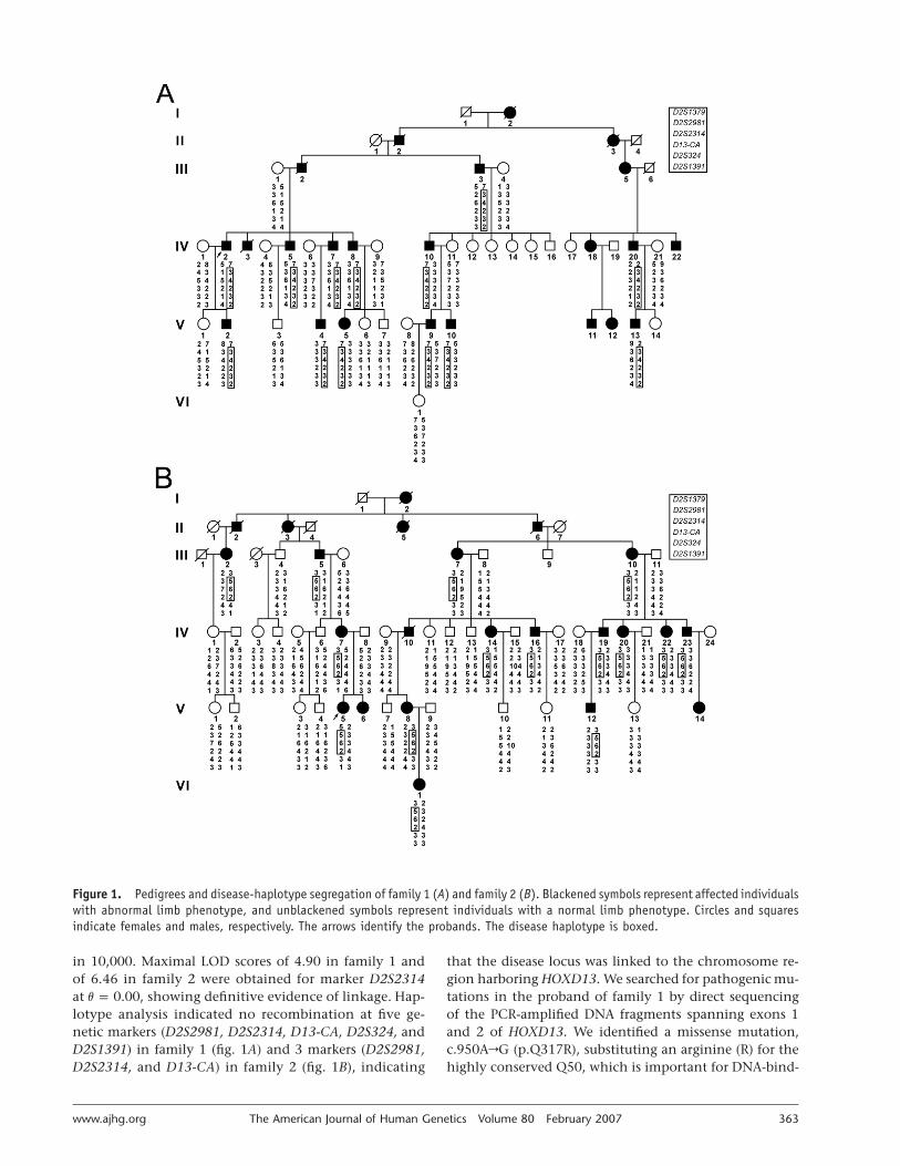
Clinical findings.—We investigated two large Han Chi-nese families with distinctive limb malformations. Bothfamilies had affected females and male-to-male transmis-sion (fig. 1A and 1B), consistent with autosomal dominantinheritance. There were 23 affected individuals in the 6generations of family 1 (fig. 1A). At the proband’s request,13 affected individuals were physically examined, and dig-ital photographs were taken. Among those individuals, 11had hand and foot radiographs. On radiological exami-nation, nine affected individuals had bilateral or unilateralfusion of metacarpals 4 and 5 (fig. 2B, 2F, 2J, and 2N).Radiographs showed that the fusion was variable in ex-tent. The proband had a complete fusion in his right hand(fig. 2B), and eight other affected individuals had bilateralcomplete fusion (fig. 2F, 2J, and 2N). In five affected in-dividuals, the fusion extended to the phalanges of fingers4 and 5 (fig. 2N). Other hand deformities included ulnardeviation of fingers 2–5 (12 of 13 subjects), lobster claw–like or Y-shaped fingers 4 and 5 with an angulated 5thfinger (8 of 13), shortening or clinodactyly of the 5th fin-gers (3 of 13), short distal phalanges of between one andall fingers (12 of 13), shortening of fused metacarpals 4and 5 or metacarpal 5 (6 of 13), unilateral cutaneous syn-dactyly of fingers 3 and 4 (4 of 13), interdigital clefts be-tween fingers 3 and 4 (10 of 13), camptodactyly (10 of13), and absence of distal interphalangeal creases (13 of13) (fig. 2A, 2B, 2E, 2F, 2I, 2J, 2M, and 2N). None of theaffected individuals had metatarsal fusion of the feet (fig.2D, 2H, 2L, and 2P). The most constant foot deformitieswere varus deviation of the first metatarsals; valgus devia-tion of toes 1–4; hyperplasia of the 1st ray, affecting thefirst metatarsals and the phalanges of the halluces; hypo-plasia and shortness of metatarsals 2–5; and shortened andtucked 5th toes (fig. 2C, 2D, 2G, 2H, 2K, 2L, 2O, and 2P).Mild cutaneous syndactyly of toes 2 and 3 or 3 and 4 was
observed in four affected individuals (IV-5, IV-8, IV-10, andV-13) (fig. 2K). Of note, one affected individual (V-9) withbilateral complete fusion of metacarpals 4 and 5 also hadpostaxial polydactyly in his left hand, as well as hypospa-dias. Taken together, the phenotypes in this family closelyresemble the syndactyly type V reported by Robinow andcolleagues.28
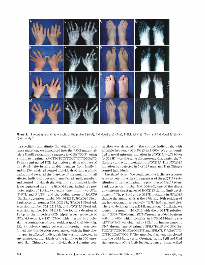
Family 2 also had 23 affected individuals in 6 genera-tions (fig. 1B). All 17 patients who are still alive were avail-able for phenotype evaluation. Digital photographs andradiographs were taken for 16 and 13 of them, respec-tively. On clinical examination, most of the patients ex-hibited generalized shortening of hands and feet, 11 dis-played broad and short distal phalanges of the thumbs(fig. 3A, 3E, 3I, and 3M), and 14 had mild cutaneous syn-dactyly of toes 2 and 3 (fig. 3C, 3G, 3K, and 3O). Radio-graphs revealed a constant phenotypic feature in all 13patients: absence of middle phalanges of toes 2–5 (fig. 3D,3H, 3L, and 3P) and very marked short middle phalangesof the 5th finger (fig. 3B, 3F, 3J, and 3N). Combined short-ening of the middle phalanges was noticed, with patterns2–5 (3 patients), 2-4-5 (3 patients), and 2-3-4-5 (2 patients),which indicates that the 2nd and 5th fingers are mostseverely affected (fig. 3B, 3F, 3J, and 3N). In many cases,the shortened middle phalanges were fused with the distalones (fig. 3B, 3F, 3J, and 3N). In 12 patients, radiographsalso showed very pronounced shortening of metacarpal5, either alone or in combination with metatarsal 5 and/or other metacarpals/metatarsals (fig. 3B, 3D, 3F, 3H, 3J,3L, 3N, and 3P). Short proximal phalanges of toes 1, 3,and 4 were apparent in 7 patients (fig. 3H, 3L, and 3P).Other common limb anomalies included broad first met-atarsals and hallux phalanges, often associated with halluxvalgus (fig. 3C, 3D, 3G, 3H, 3K, 3L, 3O, and 3P). Notably,the proband also had small spurs of bone between the 1stand 2nd metatarsals (fig. 3D). Almost all limb anomaliesin this family were bilateral. These limb phenotypes over-lap with those of BDA4, BDD, and BDE, and syndactylytype I (MIM %185900). No family member showed shortstature or polydactyly.
Molecular genetics.—We performed two-point linkageanalysis and mutation identification after obtaining in-formed consent from participating family members andapproval of Peking Union Medical College InstitutionalReview Board. Blood samples were collected and genomicDNA was extracted from 27 members of family 1, includ-ing 13 patients, and from 43 family members of family2, including 15 patients. Six polymorphic microsatellitemarkers from chromosome region 2q24.3-q32.1, includ-ing a CA-repeat sequence within the intron of the HOXD13gene, were selected and typed in the families, for two-point linkage analysis. LOD scores were calculated usingthe MLINK program of the LINKAGE package. The param-eters used in linkage analysis were autosomal dominantinheritance, complete penetrance, a mutation rate of zero,equal male-female recombination rate, equal microsat-ellite-allele frequency, and a disease-allele frequency of 1
www.ajhg.org The American Journal of Human Genetics Volume 80 February 2007 363
Figure 1. Pedigrees and disease-haplotype segregation of family 1 (A) and family 2 (B). Blackened symbols represent affected individualswith abnormal limb phenotype, and unblackened symbols represent individuals with a normal limb phenotype. Circles and squaresindicate females and males, respectively. The arrows identify the probands. The disease haplotype is boxed.
in 10,000. Maximal LOD scores of 4.90 in family 1 andof 6.46 in family 2 were obtained for marker D2S2314at , showing definitive evidence of linkage. Hap-v p 0.00lotype analysis indicated no recombination at five ge-netic markers (D2S2981, D2S2314, D13-CA, D2S324, andD2S1391) in family 1 (fig. 1A) and 3 markers (D2S2981,D2S2314, and D13-CA) in family 2 (fig. 1B), indicating
that the disease locus was linked to the chromosome re-gion harboring HOXD13. We searched for pathogenic mu-tations in the proband of family 1 by direct sequencingof the PCR-amplified DNA fragments spanning exons 1and 2 of HOXD13. We identified a missense mutation,c.950ArG (p.Q317R), substituting an arginine (R) for thehighly conserved Q50, which is important for DNA-bind-
364 The American Journal of Human Genetics Volume 80 February 2007 www.ajhg.org
Figure 2. Photographs and radiographs of the proband (A–D), individual V-10 (E–H), individual V-13 (I–L), and individual IV-10 (M–P) of family 1.
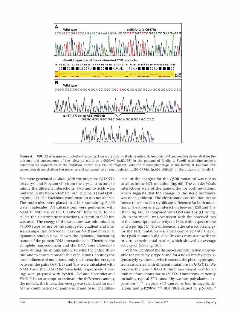
ing specificity and affinity (fig. 4A). To confirm this mis-sense mutation, we introduced into the 950G mutant al-lele a BamHI recognition sequence (5′-GGATCC-3′), usinga mismatch primer (5′-CTTGTCCTTCACTCTTCGGATC-3′) in a semi-nested PCR. Restriction analysis with use ofthis BamHI site in all available members from family 1and in 136 unrelated control individuals of similar ethnicbackground revealed the presence of the mutation in allaffected individuals but not in unaffected family membersand control individuals (fig. 4A). In the proband of family2, we sequenced the entire HOXD13 gene, including a pro-moter region of 1.5 kb, two exons, one intron, two UTRs(5′-UTR and 3′-UTR), and the coding exons of HOXD9(GenBank accession number NM_014213), HOXD10 (Gen-Bank accession number NM_002148), HOXD11 (GenBankaccession number NM_021192), and HOXD12 (GenBankaccession number NM_021193). We found a deletion of21 bp in the imperfect GCN triplet–repeat sequence ofHOXD13 exon 1, c.157_177del, which results in a poly-alanine contraction of seven residues (p.A53_A59del) (fig.4B). By polyacrylamide gel electrophoresis, it was con-firmed that this deletion cosegregated with the limb phe-notypes in affected individuals but was not detected inany unaffected individuals of the family or in 500 unre-lated Han Chinese control individuals. A 4-alanine con-
traction was detected in the control individuals, withan allele frequency of 0.3% (3 in 1,000). We also identi-fied a novel missense mutation in HOXD11—c.734GrA(p.G245D)—on the same chromosome that carries the 7-alanine contraction mutation in HOXD13. This HOXD11mutation was detected in 2 of 139 unrelated Han Chinesecontrol individuals.
Functional study.—We conducted the luciferase reporterassay to determine the consequences of the p.Q317R sub-stitution in transactivating the promoter of EPHA7 (Gen-Bank accession number NM_004440), one of the directdownstream target genes of HOXD13 during limb devel-opment.29 The p.I314L and p.Q317R mutations in HOXD13change the amino acids at the 47th and 50th residues ofthe homeodomain, respectively. “I47L” had been used else-where to designate the p.I314L mutation.25 Similarly, wenamed the mutant HOXD13 with the p.Q317R substitu-tion “Q50R.” The human EPHA7 promoter of 660 bp (from�580 to �80), which contains an HOXD13-binding siteATATTATGG, was obtained by PCR from human genomicDNA through use of primers EPHA7BamF 5′-CGCGGA-TCCTGTTCGCTCGCACCGT-3′ and EPHA7R 5′-AGACTTC-CTTTCCCACTCCC-3′. The amplified fragment was clonedinto the pGL3-basic vector (Promega) at the BglII and KpnIsites upstream of the firefly luciferase gene and was verified
www.ajhg.org The American Journal of Human Genetics Volume 80 February 2007 365
Figure 3. Photographs and radiographs of the proband (A–D), individual III-10 (E–H), individual IV-19 (I–L), and individual IV-23(M–P) of family 2.
by sequencing (fig. 5A). To make the expression vectorsfor FLAG-HOXD13WT (the FLAG-tagged wild type) andFLAG-HOXD13�7A (the FLAG-tagged mutant with the 7-alanine contraction), the full coding regions were gener-ated by genomic PCR from normal and affected individ-uals, respectively. The verified fragments were then clonedinto the HindIII and SmaI sites of the p3#FLAG-CMV-7 plasmid (Sigma), to produce the pFLAG-HOXD13WT
and pFLAG-HOXD13�7A expression vectors, respectively.pFLAG-HOXD13Q50R and pFLAG-HOXD13I47L were con-structed from the pFLAG-HOXD13WT by site-directed mu-tagenesis with use of the QuikChange Site-Directed Mu-tagenesis Kit (Stratagene). The primer pairs used were 5′-CCATTTGGTTTCGGAACCGAAGAGTG-3′ and 5′-CACTC-TTCGGTTCCGAAACCAAATGG-3′ for Q50R and 5′-GACA-AGTGACCCTTTGGTTTCAG-3′ and 5′-CTGAAACCAAAGG-GTCACTTGTC-3′ for I47L. The reporter construct (1.0 mg)was cotransfected with 1.5 mg of one of the four test con-structs (pFLAG-HOXD13WT, pFLAG-HOXD13Q50R, pFLAG-HOXD13I47L, or pFLAG-HOXD13�7A) into the C3H10T1/2mouse embryonic fibroblast cells growing in a 12-wellplate, with use of 1.0 ml VigoFect reagent (Vigorous Bio-technology). The pRL-SV40 Renilla luciferase vector (25ng) was also added in each cotransfection, to normalizethe transfection efficiency. Cells were lysed and assayedfor luciferase activity following the Dual Luciferase pro-tocol (Promega). Consistent with the recent findings thatthe mouse EphA7 promoter could mediate transcriptional
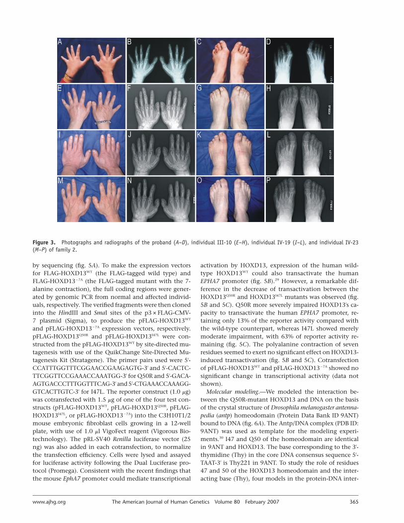
activation by HOXD13, expression of the human wild-type HOXD13WT could also transactivate the humanEPHA7 promoter (fig. 5B).29 However, a remarkable dif-ference in the decrease of transactivation between theHOXD13Q50R and HOXD13I47L mutants was observed (fig.5B and 5C). Q50R more severely impaired HOXD13′s ca-pacity to transactivate the human EPHA7 promoter, re-taining only 13% of the reporter activity compared withthe wild-type counterpart, whereas I47L showed merelymoderate impairment, with 63% of reporter activity re-maining (fig. 5C). The polyalanine contraction of sevenresidues seemed to exert no significant effect on HOXD13-induced transactivation (fig. 5B and 5C). Cotransfectionof pFLAG-HOXD13WT and pFLAG-HOXD13�7A showed nosignificant change in transcriptional activity (data notshown).
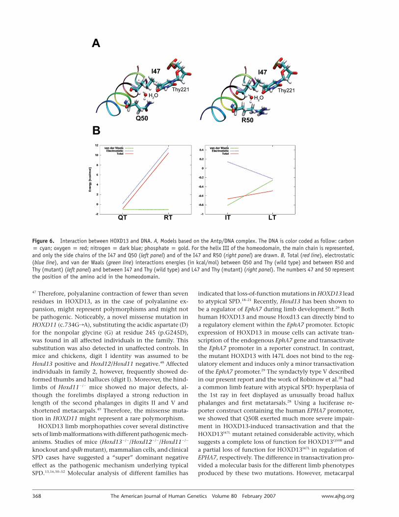
Molecular modeling.—We modeled the interaction be-tween the Q50R-mutant HOXD13 and DNA on the basisof the crystal structure of Drosophila melanogaster antenna-pedia (antp) homeodomain (Protein Data Bank ID 9ANT)bound to DNA (fig. 6A). The Antp/DNA complex (PDB ID:9ANT) was used as template for the modeling experi-ments.30 I47 and Q50 of the homeodomain are identicalin 9ANT and HOXD13. The base corresponding to the 3′-thymidine (Thy) in the core DNA consensus sequence 5′-TAAT-3′ is Thy221 in 9ANT. To study the role of residues47 and 50 of the HOXD13 homeodomain and the inter-acting base (Thy), four models in the protein-DNA inter-
366 The American Journal of Human Genetics Volume 80 February 2007 www.ajhg.org
Figure 4. HOXD13 missense and polyalanine contraction mutations in study families. A, Genomic DNA sequencing demonstrating thepresence and consequence of the missense mutation c.950ArG (p.Q317R) in the proband of family 1. BamHI restriction analysisdemonstrates segregation of the mutation, shown as a 249-bp fragment, with the disease phenotype in the family. B, Genomic DNAsequencing demonstrating the presence and consequence of small deletion c.157-177del (p.A53_A59del) in the proband of family 2.
face were generated in silico (with the programs QUANTA[Accelrys] and Program O31) from the crystal structure, tomimic the different interactions. Two amino acids weremutated in the homeodomain: I47rleucine (L) and Q50rarginine (R). The backbone conformation was not altered.The molecules were placed in a box containing 6,400water molecules. All calculations were performed withNAMD32 with use of the CHARMM33 force field. To cal-culate the electrostatic interactions, a cutoff of 0.20 nmwas used. The energy of the structures was minimized by75,000 steps by use of the conjugated gradient and line-search algorithm of NAMD. Previous NMR and moleculardynamics studies have shown the dynamic, fluctuatingnature of the protein-DNA interactions.34,35 Therefore, thecomplete homeodomain and the DNA were allowed tomove during the minimization, to relax the entire struc-ture and to ensure more-reliable calculations. To study thelocal influence of mutations, only the interaction energiesbetween the pairs Q/R (I/L) and Thy were calculated withNAMD and the CHARMM force field, respectively. Draw-ings were prepared with PyMOL (DeLano Scientific) andVMD.36 In an attempt to estimate the differences amongthe models, the interaction energy was calculated for eachof the combinations of amino acid and base. The differ-
ence in the energies for the Q50R mutation was not assmall as in the I47L mutation (fig. 6B). The van der Waalsinteractions were of the same order for both mutations,which suggests that the change in the steric hindrancewas not significant. The electrostatic contribution to theinteraction showed a significant difference for both muta-tions. The lower energy interaction between R50 and Thy(RT in fig. 6B), as compared with Q50 and Thy (QT in fig.6B) in the model, was consistent with the observed lossof the transcriptional activity: to 13%, with respect to thewild type (fig. 5C). The difference in the interaction energyfor the I47L mutation was small compared with that ofthe Q50R mutation (fig. 6B). This was consistent with thein vitro experimental results, which showed an averageactivity of 63% (fig. 5C).
We have identified the disease-causing mutations respon-sible for syndactyly type V and for a novel brachydactyly-syndactyly syndrome, which extends the phenotypic spec-trum associated with different mutations in HOXD13. Wepropose the term “HOXD13 limb morphopathies” for alllimb malformations due to HOXD13 mutations, currentlyincluding typical SPD caused by various polyalanine ex-pansions,4,12–17 atypical SPD caused by four intragenic de-letions and p.R298W,18–21 BDD/BDE caused by p.S308C,24

www.ajhg.org The American Journal of Human Genetics Volume 80 February 2007 367
Figure 5. Transcriptional activity of wild-type and mutant HOXD13proteins at the human EPH7A promoter. A, Schematic diagram ofthe reporter construct used in transfection assays. B, Transcrip-tional activity mediated by HOXD13WT, HOXD13Q50R, HOXD13I47L, andHOXD13�7A. Bars represent firefly/Renilla luciferase ratios for thedifferent constructs. Values are the means (�SEM) of eight in-dependent experiments. The significance of differences in expres-sion was calculated using the independent-samples T test. P valuesare presented above the bars. C, Relative decrease of transcrip-tional activity mediated by mutant HOXD13 proteins compared withthe wild-type HOXD13WT.
BDE with mild SPD caused by p.I314L,24,25 syndactyly typeV caused by p.Q317R, and BDA4/BDD/BDE with mild 2–3 toe syndactyly caused by a polyalanine contraction.
Syndactyly type V, as a distinct entity, has 4–5 meta-carpal synostosis, a cardinal feature distinguishing it fromother nonsyndromic syndactylies.27,28 Study of family 1, a6-generation Chinese family affected with this rare limbmalformation, enabled us to map the disease locus to thechromosomal region where HOXD13 is located. In thisHan Chinese family, we identified a missense mutation ofHOXD13—c.950ArG (p.Q317R)—that substituted the ba-sic and charged polar amino acid R for the uncharged polarQ at the 50th residue of the homeodomain (Q50). Thismutation was found to cosegregate with the disease phe-notype in the large family but not was present in anyunaffected family members or in 136 normal Han Chinesecontrol individuals. Q50 of the homeodomain is invari-ant in all known HOX proteins (Homeodomain Resource)and has been shown to be a key amino acid for DNA-binding specificity and affinity.5–10,37 Moreover, our func-tional study showed that the mutation had a deleteriouseffect on HOXD13-induced transcriptional activation ofthe human EPHA7 promoter. Finally, molecular modelingdata supported the in vitro experimental results. All these
results established conclusively the pathogenicity of thec.950ArG (p.Q317R) mutation in syndactyly type V.
SPD was the first disease in which polyalanine expan-sion was identified as the disease-causing mechanism.12
This novel type of mutation has been found in nine dif-ferent human genetic diseases.38–40 Like HOXD13 in SPD,polyalanine expansions in these genetic diseases show mei-otic stability over generations, distinguishing it from dy-namic triplet-repeat expansions underlying genetic dis-eases such as Huntington disease and fragile X syndrome.Unequal crossing-over has been suggested as the mecha-nism for polyalanine expansion.41 Polyalanine contrac-tion, the reciprocal allele of this unequal crossing-over,has not previously been associated with a human geneticdisease,39 although Dr. Stephen T. Warren has predictedthat the mutant HOXD13 alleles with truncated polyalan-ine tracts of fewer than eight residues “might lead to otherdigital anomalies.”41(p408) In the large Han Chinese familyaffected with combined BDA4/BDD/BDE and mild 2–3 toesyndactyly, we mapped the disease locus to a chromosomeregion defined by D2S1379 and D2S324 (fig. 1B). This re-gion contains 170 known genes, including the 9 HOXDgenes (Human Genome Browser Gateway). We sequencedall the 5′ HOXD genes that show expression in the limbbud42 and found a polyalanine contraction of seven resi-dues in HOXD13 that segregated perfectly with the digitalanomalies. The contraction was not detectable in 500 un-related and ethnically matched unaffected control indi-viduals. The site and length of the polyalanine tract inHOXD13, like that in the paralogous HOXA13, are highlyconserved among mammals.43 All mammalian HOXD13proteins with sequences available in the databases—in-cluding human, chimpanzee, mouse, rat, and bat—havea 15-residue polyalanine tract in their N-terminal region,which suggests a strong functional and structural con-straint. It has been indicated that a polyalanine tract be-yond a certain threshold will have deleterious effects.40
Mutations in HOXD13 have been associated with BDD andBDE, whereas mild 2–3 toe syndactyly is common in in-dividuals heterozygous for a 7-alanine expansion muta-tion in HOXD13.13,24 Furthermore, homozygous mousemutants with the disrupted Hoxd13 gene showed absenceof the second phalanges in digits II and V, size reductionof the second phalanges in digits III and IV, deformed andthicker metatarsal I with anterior protrusion, and short-ened metacarpals and metatarsals in both of the forelimbsand the hindlimbs.44,45 These, taken together, suggestedthat the 7-alanine contraction mutation in HOXD13 wasmost likely responsible for the novel brachydactyly-syn-dactyly syndrome. In unaffected individuals, other re-searchers have detected 2- and 4-alanine contractions inHOXD13.16,46 In our Han Chinese control individuals, the4-alanine contraction reported46 elsewhere in the Japanesepopulation was detected as a very rare allele. Several othergenes with pathogenic polyalanine expansions also dis-play polyalanine tract-length polymorphisms—that is, ex-pansions and contractions—in the general population.39,-
368 The American Journal of Human Genetics Volume 80 February 2007 www.ajhg.org
Figure 6. Interaction between HOXD13 and DNA. A, Models based on the Antp/DNA complex. The DNA is color coded as follow: carbonp cyan; oxygen p red; nitrogen p dark blue; phosphate p gold. For the helix III of the homeodomain, the main chain is represented,and only the side chains of the I47 and Q50 (left panel) and of the I47 and R50 (right panel) are drawn. B, Total (red line), electrostatic(blue line), and van der Waals (green line) interactions energies (in kcal/mol) between Q50 and Thy (wild type) and between R50 andThy (mutant) (left panel) and between I47 and Thy (wild type) and L47 and Thy (mutant) (right panel). The numbers 47 and 50 representthe position of the amino acid in the homeodomain.
47 Therefore, polyalanine contraction of fewer than sevenresidues in HOXD13, as in the case of polyalanine ex-pansion, might represent polymorphisms and might notbe pathogenic. Noticeably, a novel missense mutation inHOXD11 (c.734GrA), substituting the acidic aspartate (D)for the nonpolar glycine (G) at residue 245 (p.G245D),was found in all affected individuals in the family. Thissubstitution was also detected in unaffected controls. Inmice and chickens, digit I identity was assumed to beHoxd13 positive and Hoxd12/Hoxd11 negative.48 Affectedindividuals in family 2, however, frequently showed de-formed thumbs and halluces (digit I). Moreover, the hind-limbs of Hoxd11�/� mice showed no major defects, al-though the forelimbs displayed a strong reduction inlength of the second phalanges in digits II and V andshortened metacarpals.49 Therefore, the missense muta-tion in HOXD11 might represent a rare polymorphism.
HOXD13 limb morphopathies cover several distinctivesets of limb malformations with different pathogenicmech-anisms. Studies of mice (Hoxd13�/�/Hoxd12�/�/Hoxd11�/�
knockout and spdh mutant), mammalian cells, and clinicalSPD cases have suggested a “super” dominant negativeeffect as the pathogenic mechanism underlying typicalSPD.13,16,50–52 Molecular analysis of different families has
indicated that loss-of-function mutations in HOXD13 leadto atypical SPD.18–21 Recently, Hoxd13 has been shown tobe a regulator of EphA7 during limb development.29 Bothhuman HOXD13 and mouse Hoxd13 can directly bind toa regulatory element within the EphA7 promoter. Ectopicexpression of HOXD13 in mouse cells can activate tran-scription of the endogenous EphA7 gene and transactivatethe EphA7 promoter in a reporter construct. In contrast,the mutant HOXD13 with I47L does not bind to the reg-ulatory element and induces only a minor transactivationof the EphA7 promoter.29 The syndactyly type V describedin our present report and the work of Robinow et al.28 hada common limb feature with atypical SPD: hyperplasia ofthe 1st ray in feet displayed as unusually broad halluxphalanges and first metatarsals.28 Using a luciferase re-porter construct containing the human EPHA7 promoter,we showed that Q50R exerted much more severe impair-ment in HOXD13-induced transactivation and that theHOXD13I47L mutant retained considerable activity, whichsuggests a complete loss of function for HOXD13Q50R anda partial loss of function for HOXD13I47L in regulation ofEPHA7, respectively. The difference in transactivation pro-vided a molecular basis for the different limb phenotypesproduced by these two mutations. However, metacarpal
www.ajhg.org The American Journal of Human Genetics Volume 80 February 2007 369
fusions in syndactyly type V could be a gain-of-functioneffect, because this limb phenotype was absent both inpatients with atypical SPD and in Hoxd13 knockoutmice.4,12–17,44,45 It has been demonstrated elsewhere9,10 thatQ50 is an important determinant of differential DNA-binding specificity among different homeodomains. A sin-gle amino acid substitution to the basic and charged polarlysine (K) could switch the DNA-binding specificities ofdifferent homeodomains.9,10 Guttmacher syndrome (MIM176305) shares some phenotypes with hand-foot-genitalsyndrome (MIM 140000), which is caused by loss-of-func-tion mutations in HOXA13.53 In patients affected withGuttmacher syndrome, the p.Q371L missense mutationwas identified.54 This mutation led to a substitution at Q50of the HOXA13 homeodomain. The presence of additionallimb phenotypes in Guttmacher syndrome suggested thatthe amino acid substitution resulted in both a loss andspecific gain of function.54 It is conceivable that a mixedgain and loss of function of HOXD13Q50R is a reasonableexplanation of the cause of syndactyly type V. The poly-alanine contraction found in the family with BDA4/BDD/BDE and mild 2–3 toe syndactyly is more likely adominant-negative mutation, since Hoxd13�/� mice havesimilar phalangeal and metacarpal/metatarsal defects.44,45
Transheterozygous Hoxd13�/�/Hoxd12�/� mice also exhibita reduction of digits II and V in the forelimbs.44 Further-more, hypoplasia of the middle phalanges of toes 2–5 andbroad hallux phalanges and first metatarsals are consistentin atypical SPD associated with functional haploinsuffi-ciency for the homeodomain of HOXD13.18–21 However,our reporter assay showed no significant difference be-tween HOXD13WT and HOXD13�7A in regulation of EPHA7.Mutant HOXD13 proteins with polyalanine tracts longerthan 22 residues displayed misfolding and cytoplasmicaggregation.40 We observed that the GFP-tagged HOXD13proteins with different polyalanine contractions were alllocated in the nucleus (data not shown). Polyalanine tractsare common in transcription factors and may modulatetranscriptional regulation and protein-protein interac-tion.38,47 Interestingly, longer polyalanine tracts (110 ala-nines) formed b-sheet, whereas shorter ones (7 alanines)adopted a-helix exclusively.55 It is possible that the 7-al-anine contraction in HOXD13 could perturb its bindingto the cofactors or trigger a rapid protein processing.
Our present report and the observations of others allindicate the existence of genotype-phenotype correlationfor the limb morphopathies caused by HOXD13 muta-tions.11,13,16,24 However, the same minor malformationsmay be associated with the different limb clinical abnor-malities—for example, (1) soft-tissue syndactyly of toes2 and 3 in mild SPD due to a polyalanine expansion ofseven residues, in the brachydactyly-syndactyly syndromecaused by a polyalanine contraction of seven residues, andin syndactyly type V, and (2) cutaneous syndactyly of fin-gers 3 and 4 without digit duplication within the web inmild SPD and syndactyly type V. Therefore, different limb
malformations due to distinct classes of HOXD13 muta-tions should be considered a continuum of phenotypes.
Acknowledgments
We are grateful to Drs. Yan Shen, Depei Liu, and Boqin Qiang fortheir encouragement and support. We thank all the family mem-bers for their generous participation. This work was supported bythe National 863 Program of China, the National Natural ScienceFoundation of China Funds for Creative Research Groups grant30421003, Distinguished Young Scholars grant 30125017, andChina Medical Board of New York grant 03-785 (all to X. Zhang).J.A.L. was supported by National Institutes of Health (NIH) grantGM 066895 (to L.M.A.). M.S., Y.A., and X. Zhang are trainees inthe International Collaborative Genetics Research Training Pro-gram (supported by NIH grant D43 TW06176). We also thank Dr.Depei Liu’s group for technical assistance in functional study andDrs.Qi Xu and Ying Liu for providing DNA samples from unre-lated Han Chinese individuals.
Web Resources
Accession numbers and URLs for data presented herein are asfollows:
GenBank, http://www.ncbi.nlm.nih.gov/Genbank/ (for HOXD13[accession number NM_000523], HOXD9 [accession numberNM_014213], HOXD10 [accession number NM_002148],HOXD11 [accession number NM_021192], HOXD12 [acces-sion number NM_021193], and EPHA7 [accession numberNM_004440])
Homeodomain Resource, http://genome.nih.gov/homeodomain/Human Genome Browser Gateway, http://genome.ucsc.edu/
cgi-bin/hgGatewayOnline Mendelian Inheritance in Man (OMIM), http://www.ncbi
.nlm.nih.gov/Omim/ (for HOXD13, SPD, BDD, BDE, syndactylytype V, BDA4, syndactyly type I, Guttmacher syndrome, andhand-foot-genital syndrome)
Protein Data Bank, http://www.rcsb.org/pdb/ (for the Antp [ID9ANT] homeodomain DNA structure)
References
1. Krumlauf R (1994) Hox genes in vertebrate development. Cell78:191–201
2. Capecchi MR (1997) Hox genes and mammalian develop-ment. Cold Spring Harb Symp Quant Biol 62:273–281
3. Scott MP (1993) A rational nomenclature for vertebrate ho-meobox (HOX) genes. Nucleic Acids Res 21:1687–1688
4. Akarsu AN, Stoilov I, Yilmaz E, Sayli BS, Sarfarazi M (1996)Genomic structure of HOXD13 gene: a nine polyalanine du-plication causes synpolydactyly in two unrelated families.Hum Mol Genet 5:945–952
5. Gehring WJ, Qian YQ, Billeter M, Furukubo-Tokunaga K,Schier AF, Resendez-Perez D, Affolter M, Otting G WuthrichK (1994) Homeodomain-DNA recognition. Cell 78:211–223
6. Kissinger CR, Liu BS, Martinblanco E Kornberg TB, Pabo CO(1990) Crystal-structure of an engrailed homeodomain-DNAcomplex at 2.8 A resolution: a framework for understandinghomeodomain-DNA interaction. Cell 63:579–590
7. Fraenkel E, Rould MA, Chambers KA, Pabo CO (1998) En-grailed homeodomain-DNA complex at 2.2 A resolution: a
370 The American Journal of Human Genetics Volume 80 February 2007 www.ajhg.org
detailed view of the interface and comparison with otherengrailed structure. J Mol Biol 284:351–361
8. Clarke ND, Kissinger CR, Desjarlais J, Gilliland GL, Pabo CO(1994) Structural studies of the engrailed homeodomain. Pro-tein Sci 3:1779–1787
9. Treisman J, Gonczy P, Vashishtha M, Harris E, Desplan C(1989) A single amino-acid can determine the DNA-bindingspecificity of homeodomain proteins. Cell 59:553–562
10. Hanes SD, Brent R (1989) DNA specificity of the bicoid ac-tivator protein is determined by homeodomain recognitionhelix residue-9. Cell 57:1275–1283
11. Goodman FR (2002) Limb malformations and the humanHOX genes. Am J Med Genet 112:256–265
12. Muragaki Y, Mundlos S, Upton J, Olsen BR (1996) Alteredgrowth and branching patterns in synpolydactyly caused bymutations in HOXD13. Science 272:548–551
13. Goodman FR, Mundlos S, Muragaki Y, Donnai D, Giovan-nucci-Uzielli ML, Lapi E, Majewski F, McGaughran J, Mc-Keown C, Reardon W, et al (1997) Synpolydactyly pheno-types correlate with size of expansions in HOXD13 poly-alanine tract. Proc Natl Acad Sci USA 94:7458–7463
14. Baffico M, Baldi M, Cassan PD, Costa M, Mantero R, GaraniP, Camera G (1997) Synpolydactyly: clinical and molecularstudies on four Italian families. Eur J Hum Genet Suppl 5:142
15. Kjaer KW, Hedeboe J, Bugge M, Hansen C, Friis-Henriksen K,Vestergaard MB, Tommerup N, Opitz JM (2002) HOXD13polyalanine tract expansion in classical synpolydactyly typeVordingborg. Am J Med Genet 110:116–121
16. Kjaer KW, Hansen L, Eiberg H, Utkus A, Skovgaard LT, LeichtP, Opitz JM, Tommerup N (2005) A 72-year-old Danish puzzleresolved—comparative analysis of phenotypes in familieswith different-sized HOXD13 polyalanine expansion. Am JMed Genet A 138:328–339
17. Zhao XL, Meng JP, Sun M, Ao Y, Wu AH, Lo HY, Zhang X(2005) HOXD13 polyalanine tract expansion in synpolydac-tyly: mutation detection and prenatal diagnosis in a largeChinese family. Zhonghua Yi Xue Yi Chuan Xue Za Zhi 22:5–9
18. Goodman FR, Giovannucci-Uzielli ML, Hall C, Reardon W,Winter R, Scambler P (1998) Deletions in HOXD13 segregatewith an identical, novel foot malformation in two unrelatedfamilies. Am J Hum Genet 63:992–1000
19. Calabrese O, Bigoni S, Gualandi F, Trabanelli C, Camera G,Calzolari E (2000) A new mutation in HOXD13 associatedwith foot pre-postaxial polydactyly. Eur J Hum Genet Suppl8:140
20. Kan SH, Johnson D, Giele H, Wilkie AO (2003) An acceptorsplice site mutation in HOXD13 results in variable hand, butconsistent foot malformations. Am J Med Genet A 121:69–74
21. Debeer P, Bacchelli C, Scambler PJ, De Smet L, Fryns JP, Good-man FR (2002) Severe digital abnormalities in a patient het-erozygous for both a novel missense mutation in HOXD13and a polyalanine tract expansion in HOXA13. J Med Genet39:852–856
22. Johnson KR, Sweet HO, Donahue LR, Ward-Bailey P, BronsonRT, Davisson MT (1998) A new spontaneous mouse mutationof Hoxd13 with a polyalanine expansion and phenotype sim-ilar to human synpolydactyly. Hum Mol Genet 7:1033–1038
23. Albrecht AN, Schwabe GC, Stricker S, Boddrich A, WankerEE, Mundlos S (2002) The synpolydactyly homolog (spdh)
mutation in the mouse—a defect in patterning and growthof limb cartilage elements. Mech Dev 112:53–67
24. Johnson D, Kan S-h, Oldridge M, Trembath RC, Roche P, Es-nouf RM, Giele H, Wilkie AOM (2003) Missense mutationsin the homeodomain of HOXD13 are associated with brach-ydactyly types D and E. Am J Hum Genet 72:984–997
25. Caronia G, Goodman FR, McKeown CME, Scambler PJ, Zap-pavigna V (2003) An I47L substitution in the HOXD13 hom-eodomain causes a novel human limb malformation by pro-ducing a selective loss of function. Development 130:1701–1712
26. Temtamy SA, McKusick VA (1978) Syndactyly. In: Bergsma(ed) The genetics of hand malformations. Alan R. Liss, NewYork, pp 301–322
27. Malik S, Ahmad W, Grzeschik KH, Koch MC (2005) A simplemethod for characterizing syndactyly in clinical practice. Ge-net Couns 16:229–238
28. Robinow M, Johnson GF, Brook GJ (1982) Syndactyly type V.Am J Med Genet 11:475–482
29. Salsi V, Zappavigna V (2006) Hoxd13 and Hoxa13 directly con-trol the expression of the EphA7 ephrin tyrosine kinase re-ceptor in developing limbs. J Biol Chem 281:1992–1999
30. Fraenkel E, Pabo CO (1998) Comparison of X-ray and NMRstructures for the Antennapedia homeodomain-DNAcomplex.Nature Struct Biol 5:692–697
31. Kleywegt GJ, Jones TA (1996) Efficient rebuilding of proteinstructures. Acta Crystallogr D Biol Crystallogr 52:829–832
32. Kale L, Skeel R, Bhandarkar M, Brunner R, Gursoy A, KrawetzN, Phillips J, Shinozaki A, Varadarajan K, Schulten K (1999)NAMD2: greater scalability for parallel molecular dynamics.J Comput Phys 151:283–312
33. MacKerell AD Jr, Bashford D, Bellott M, Dunbrack RL Jr, Evan-seck JD, Field MJ, Fischer S, Gao J, Guo H, Ha S, et al (1998)All-atom empirical potential for molecular modeling and dy-namics studies of proteins. J Phys Chem B 102:3586–3616
34. Qian YQ, Otting G, Wuthrich K (1993) NMR1 detection ofhydration water in the intermolecular interface of a protein-DNA complex. J Am Chem Soc 115:1189–1190
35. Billeter M, Guntert P, Luginbuhl P, Wuthrich K (1996) Hydra-tion and DNA recognition by homeodomains. Cell 85:1057–1065
36. Humphrey W, Dalke A, Schulten K (1996) VMD: visual mo-lecular dynamics. J Mol Graph 14:33–38
37. Baberjee-Basu S, Baxevanis AD (2001) Molecular evolution ofthe homeodomain family of transcription factors. NucleicAcids Res 29:3258–3269
38. Brown LY, Brown SA (2004) Alanine tracts: the expandingstory of human illness and trinucleotide repeats. TIG 20:51–58
39. Amiel J, Trochet D, Clement-Ziza M, Munnich A, Lyonnet S(2004) Polyalanine expansions in human. Hum Mol Genet13:R235–R243
40. Albrecht A, Mundlos S (2005) The other trinucleotide repeat:polyalanine expansion disorders. Curr Opin Genet Dev 15:285–293
41. Warren ST (1997) Polyalanine expansion in synpolydactylymight result from unequal crossing-over of HOXD13. Science275:408–409
42. Dolle P, Duboule D (1989) Two gene members of the murineHOX-5 complex show regional and cell-type specific expres-sion in developing limbs and gonads. EMBO J 8:1507–1515
43. Mortlock D, Sateesh P Innis JW (2000) Evolution of N-ter-
www.ajhg.org The American Journal of Human Genetics Volume 80 February 2007 371
minal sequences of the vertebrate HOXA13 protein. MammGenome 11:151–158
44. Dolle P, Dierich A, LeMeur M, Schimmang T, Schuhbaur B,Chambon P, Duboule D (1993) Disruption of the Hoxd13 geneinduces localized heterochrony leading to mice with neoteniclimbs. Cell 75:431–441
45. Davis AP, Capecchi MR (1996) A mutational analysis of the5′ HoxD genes: dissection of genetic interactions during limbdevelopment in the mouse. Development 122:1175–1185
46. Haga H, Yamada R, Ohnishi Y, Nakamura Y, Tanaka T (2002)Gene-based SNP discovery as part of the Japanese MillenniumGenome Project: identification of 190,562 genetic variationsin the human genome. J Hum Genet 47:605–610
47. Lavoie H, Debeane F, Trinh QD, Turcotte JF, Corbeil-GirardLP, Dicaire MJ, Saint-Denis A, Page M, Rouleau GA, Brais B(2003) Polymorphism, shared functions and convergent evo-lution of genes with sequences coding for polyalanine do-mains. Hum Mol Genet 12:2967–2979
48. Vargas AO, Fallon JF (2005) The digits of the wing of birdsare 1, 2 and 3: a review. J Exp Zool B Mol Dev Evol 304:206–219
49. David AP, Capecchi MR (1994) Axial homeosis and appen-
dicular skeleton defects in mice with targeted disruption ofhoxd-11. Development 120:2187–2198
50. Zakany J, Duboule D (1996) Syndactyly in mice with a tar-geted deficiency in the HoxD complex. Nature 384:69–71
51. Brudeau S, Johnson KR, Yamamoto M, Kuroiwa A, DubouleD (2001) The mouse Hoxd13spdh mutation, a polyalanine ex-pansion similar to human type II synpolydactyly (SPD), dis-rupts the function but not the expression of other Hoxd genes.Dev Biol 237:345–353
52. Albrecht AN, Kornak U, Boddrich A, Suring K, Robinson PN,Stiege AC, Lurz R, Stricker S, Wanker EE, Mundlos S (2004)A molecular pathogenesis for transcription factor associatedpoly-alanine tract expansions. Hum Mol Genet 13:2351–2359
53. Mortlock DP, Innis JW (1997) Mutation of HOXA13 in hand-foot-genital syndrome. Nat Genet 15:179–180
54. Innis JW, Goodman FR, Bacchelli C, Williams TM, MortlockDP, Sateesh P, Scambler PJ, McKinnon W, Guttmacher AE(2002) A HOXA13 allele with a missense mutation in thehomeobox and a ninucleotide deletion in the promoter un-derlies Guttmacher syndrome. Hum Mutat 19:573–574
55. Blondelle SE, Forood B, Houghten A, Perez-Paya E (1997)Polyalanine-based peptides as models for self-associated b-pleated-sheet complexes. Biochemistry 36:8393–8400