Cortical glutamatergic neurons mediate the motor sedative action of diazepam
10
Cortical Glutamatergic Neurons Mediate the Motor Sedative Action of Diazepam A. Zeller, F. Crestani, I. Camenisch, T. Iwasato, S. Itohara, J. M. Fritschy, and U. Rudolph Institute of Pharmacology and Toxicology, University of Zu ¨ rich, Zu ¨ rich, Switzerland (A.Z., F.C., I.C., J.M.F., U.R.); PRESTO, Japan Science and Technology Agency, Saitama, Japan (T.I.); Laboratory for Behavioral Genetics, RIKEN Brain Science Institute, Saitama, Japan (T.I., S.I.); and Laboratory of Genetic Neuropharmacology, McLean Hospital and Department of Psychiatry, Harvard Medical School, Belmont, Massachusetts (U.R.) Received June 6, 2007; accepted October 26, 2007 ABSTRACT The neuronal circuits mediating the sedative action of diaze- pam are unknown. Although the motor-depressant action of diazepam is suppressed in 1(H101R) homozygous knockin mice expressing diazepam-insensitive 1-GABA A receptors, global 1-knockout mice show greater motor sedation with diazepam. To clarify this paradox, attributed to compensatory up-regulation of the 2 and 3 subunits, and to further identify the neuronal circuits supporting diazepam-induced sedation, we generated Emx1-cre-recombinase-mediated conditional mutant mice, selectively lacking the 1 subunit (forebrain-spe- cific 1 / ) or expressing either a single wild-type (H) or a single point-mutated (R) 1 allele (forebrain-specific 1 /H and 1 /R mice, respectively) in forebrain glutamatergic neurons. In the rest of the brain, 1 /R mutants are heterozygous 1(H101R) mice. Forebrain-specific 1 / mice showed en- hanced diazepam-induced motor depression and increased expression of the 2 and 3 subunits in the neocortex and hippocampus, in comparison with their pseudo-wild-type litter- mates. Forebrain-specific 1 /R mice were less sensitive than 1 /H mice to the motor-depressing action of diazepam, but each of these conditional mutants had a similar behavioral response as their corresponding control littermates. Unexpect- edly, expression of the 1 subunit was reduced in forebrain, notably in 1 /R mice, and the 3 subunit was up-regulated in neocortex, indicating that proper 1 subunit expression re- quires both alleles. In conclusion, conditional manipulation of GABA A receptor 1 subunit expression can induce compensa- tory changes in the affected areas. Specifically, alterations in GABA A receptor expression restricted to forebrain glutamater- gic neurons reproduce the behavioral effects seen after a global alteration, thereby implicating these neurons in the motor-sed- ative effect of diazepam. GABA A receptors mediate fast GABAergic inhibition in the adult mammalian central nervous system. GABA A receptors are pentameric ligand-gated ion channels, with the majority of them containing two , two , and one subunit (Barnard et al., 1998; Sieghart and Ernst, 2005). These receptors are the targets of many clinically important drugs (Rudolph and Mo ¨hler, 2006), including benzodiazepines (Rudolph and Mo ¨hler, 2004), barbiturates, neurosteroids (Belelli and Lam- bert, 2005), and general anesthetics (Rudolph and Antkow- iak, 2004). Benzodiazepine binding to GABA A receptors mod- ulates vigilance and anxiety states and a wide range of sensorimotor and cognitive functions. It is noteworthy that diazepam, through 1-GABA A receptor activation, can pro- mote sedation, as measured by its motor-depressant action (Rudolph et al., 1999; McKernan et al., 2000), and antero- grade amnesia, and it displays anticonvulsant properties (Rudolph et al., 1999). This spectrum of effects has been shown genetically by introducing a histidine-to-arginine point mutation at position 101 of the murine GABA A receptor 1 subunit gene. The 1(H101R)-GABA A receptor is insensi- tive to allosteric modulation by benzodiazepine-site ligands, including zolpidem, both in vitro and in vivo, whereas regu- lation by the physiological neurotransmitter GABA is pre- served (Benson et al., 1998; Rudolph et al., 1999; Crestani et al., 2000; Marowsky et al., 2004). The corresponding 1(H101R) mice fail to show the motor-depressant and an- terograde amnesic effect of diazepam, and they are partly resistant to its anticonvulsant action (Rudolph et al., 1999; McKernan et al., 2000). In contrast, the effects of diazepam on sleep EEG are not affected in these mice (Tobler et al., 2001); rather, they depend on 2-GABA A receptors (Kopp et al., 2004). The role of 1-GABA A receptors in mediating the sedative action of benzodiazepine-site ligands was further supported by pharmacological studies using L838-417 (Scott- Stevens et al., 2005). This substance, which acts as a partial This work was supported by grants of the Swiss National Science Founda- tion (to U.R. and J.M.F.). Article, publication date, and citation information can be found at http://molpharm.aspetjournals.org. doi:10.1124/mol.107.038828. ABBREVIATIONS: bp, base pair(s); OD, optical density; IR, immunoreactivity; PV, parvalbumin; CB, calbindin; cre, cre recombinase. 0026-895X/08/7302-282–291$20.00 MOLECULAR PHARMACOLOGY Vol. 73, No. 2 Copyright © 2008 The American Society for Pharmacology and Experimental Therapeutics 38828/3293420 Mol Pharmacol 73:282–291, 2008 Printed in U.S.A. 282 at Rikagaku Kenkyujo on October 1, 2008 molpharm.aspetjournals.org Downloaded from
Transcript of Cortical glutamatergic neurons mediate the motor sedative action of diazepam

Cortical Glutamatergic Neurons Mediate the Motor SedativeAction of Diazepam
A. Zeller, F. Crestani, I. Camenisch, T. Iwasato, S. Itohara, J. M. Fritschy, and U. RudolphInstitute of Pharmacology and Toxicology, University of Zurich, Zurich, Switzerland (A.Z., F.C., I.C., J.M.F., U.R.); PRESTO,Japan Science and Technology Agency, Saitama, Japan (T.I.); Laboratory for Behavioral Genetics, RIKEN Brain ScienceInstitute, Saitama, Japan (T.I., S.I.); and Laboratory of Genetic Neuropharmacology, McLean Hospital and Department ofPsychiatry, Harvard Medical School, Belmont, Massachusetts (U.R.)
Received June 6, 2007; accepted October 26, 2007
ABSTRACTThe neuronal circuits mediating the sedative action of diaze-pam are unknown. Although the motor-depressant action ofdiazepam is suppressed in �1(H101R) homozygous knockinmice expressing diazepam-insensitive �1-GABAA receptors,global �1-knockout mice show greater motor sedation withdiazepam. To clarify this paradox, attributed to compensatoryup-regulation of the �2 and �3 subunits, and to further identifythe neuronal circuits supporting diazepam-induced sedation,we generated Emx1-cre-recombinase-mediated conditionalmutant mice, selectively lacking the �1 subunit (forebrain-spe-cific �1�/�) or expressing either a single wild-type (H) or asingle point-mutated (R) �1 allele (forebrain-specific �1�/H and�1�/R mice, respectively) in forebrain glutamatergic neurons. Inthe rest of the brain, �1�/R mutants are heterozygous�1(H101R) mice. Forebrain-specific �1�/� mice showed en-hanced diazepam-induced motor depression and increased
expression of the �2 and �3 subunits in the neocortex andhippocampus, in comparison with their pseudo-wild-type litter-mates. Forebrain-specific �1�/R mice were less sensitive than�1�/H mice to the motor-depressing action of diazepam, buteach of these conditional mutants had a similar behavioralresponse as their corresponding control littermates. Unexpect-edly, expression of the �1 subunit was reduced in forebrain,notably in �1�/R mice, and the �3 subunit was up-regulated inneocortex, indicating that proper �1 subunit expression re-quires both alleles. In conclusion, conditional manipulation ofGABAA receptor �1 subunit expression can induce compensa-tory changes in the affected areas. Specifically, alterations inGABAA receptor expression restricted to forebrain glutamater-gic neurons reproduce the behavioral effects seen after a globalalteration, thereby implicating these neurons in the motor-sed-ative effect of diazepam.
GABAA receptors mediate fast GABAergic inhibition in theadult mammalian central nervous system. GABAA receptorsare pentameric ligand-gated ion channels, with the majorityof them containing two �, two �, and one � subunit (Barnardet al., 1998; Sieghart and Ernst, 2005). These receptors arethe targets of many clinically important drugs (Rudolph andMohler, 2006), including benzodiazepines (Rudolph andMohler, 2004), barbiturates, neurosteroids (Belelli and Lam-bert, 2005), and general anesthetics (Rudolph and Antkow-iak, 2004). Benzodiazepine binding to GABAA receptors mod-ulates vigilance and anxiety states and a wide range ofsensorimotor and cognitive functions. It is noteworthy thatdiazepam, through �1-GABAA receptor activation, can pro-mote sedation, as measured by its motor-depressant action(Rudolph et al., 1999; McKernan et al., 2000), and antero-
grade amnesia, and it displays anticonvulsant properties(Rudolph et al., 1999). This spectrum of effects has beenshown genetically by introducing a histidine-to-argininepoint mutation at position 101 of the murine GABAA receptor�1 subunit gene. The �1(H101R)-GABAA receptor is insensi-tive to allosteric modulation by benzodiazepine-site ligands,including zolpidem, both in vitro and in vivo, whereas regu-lation by the physiological neurotransmitter GABA is pre-served (Benson et al., 1998; Rudolph et al., 1999; Crestani etal., 2000; Marowsky et al., 2004). The corresponding�1(H101R) mice fail to show the motor-depressant and an-terograde amnesic effect of diazepam, and they are partlyresistant to its anticonvulsant action (Rudolph et al., 1999;McKernan et al., 2000). In contrast, the effects of diazepamon sleep EEG are not affected in these mice (Tobler et al.,2001); rather, they depend on �2-GABAA receptors (Kopp etal., 2004). The role of �1-GABAA receptors in mediating thesedative action of benzodiazepine-site ligands was furthersupported by pharmacological studies using L838-417 (Scott-Stevens et al., 2005). This substance, which acts as a partial
This work was supported by grants of the Swiss National Science Founda-tion (to U.R. and J.M.F.).
Article, publication date, and citation information can be found athttp://molpharm.aspetjournals.org.
doi:10.1124/mol.107.038828.
ABBREVIATIONS: bp, base pair(s); OD, optical density; IR, immunoreactivity; PV, parvalbumin; CB, calbindin; cre, cre recombinase.
0026-895X/08/7302-282–291$20.00MOLECULAR PHARMACOLOGY Vol. 73, No. 2Copyright © 2008 The American Society for Pharmacology and Experimental Therapeutics 38828/3293420Mol Pharmacol 73:282–291, 2008 Printed in U.S.A.
282
at Rikagaku K
enkyujo on October 1, 2008
molpharm
.aspetjournals.orgD
ownloaded from
agonist at �2-, �3- and �5-GABAA receptors and as an an-tagonist at �1-GABAA receptors, displays no sedative prop-erties in rodents(McKernan et al., 2000). However, ocinaplon, a partial ago-nist at all diazepam-sensitive GABAA receptors, has beenreported to produce selective anxiolysis (Lippa et al., 2005)and to depress motor activity at high doses only. The mech-anisms underlying this different profile of action are notknown. Furthermore, the global �1 subunit knockout micetreated with diazepam display enhanced motor sedation com-pared with wild-type littermates (Kralic et al., 2002a,b), in-dicating that �1-GABAA receptors can be substituted. Thesemutants also show increased expression of the GABAA recep-tor �2 and �3 subunits notably in cerebral cortex (Kralic etal., 2002a, 2006). The compensatory up-regulation of other �subunits might underlie the pharmacological phenotype of�1 subunit knockout mice.
To further clarify the molecular mechanisms and neuralcircuits mediating the motor-sedative action of diazepam, wefocused the current study on the pharmacological signifi-cance of �1-GABAA receptors expressed in the forebrain. Toachieve this goal, we investigated genetically engineeredmice with either a constitutive deficit in �1-GABAA recep-tors, or carrying a single diazepam-insensitive �1(H101R)allele, restricted to forebrain glutamatergic neurons for theirresponsiveness to the motor-sedative action of diazepam. Inbehavioral pharmacology, the term sedation refers to a drug-induced diminution in spontaneous activity of experimentalanimals (Trevor and Way, 1995). Conditional gene deletionwas obtained by combining a wild-type �1 subunit alleleflanked by loxP sites (floxed) with a cre transgene expressedfrom an Emx1 promoter or by combining a H101R point-mutated �1 subunit allele with a floxed wild-type �1 subunitallele and the Emx1-cre transgene. These forebrain-specificmutants were analyzed immunohistochemically for possiblechanges in �1, �2, and �3 subunit expression patterns, andthey were tested behaviorally for diazepam-induced changesin spontaneous locomotor activity.
Materials and MethodsAnimals. Forebrain-specific deletion of the �1 subunit was
achieved upon excision of alleles with an exon flanked by loxP sites(floxed) by cre recombinase driven by the Emx1 promoter. To obtainthese mice, Emx1-cre Tg3 PAC transgenic mice [B6-Tg(Emx-cre)described in Iwasato et al. (2004), maintained in Zurich onto theC57BL/6JOlaHsd background] were crossed initially with micehomozygous for the floxed �1 subunit allele [B6.129(FVB)-Gabra1tm1Geh/J, at least six backcrosses onto C57BL/6J (The JacksonLaboratory, Bar Harbor, ME), first described in Vicini et al. (2001)](Fig. 1, A and C). Offspring, heterozygous for the floxed �1 subunitallele and carrying the Emx1-cre transgene, were crossed again withmice homozygous for the floxed �1 subunit allele to obtain the de-sired genotype (Fig. 1, A and C). Because two generations werenecessary to obtain the mutant mice for analysis, the genetic back-ground of the experimental animals was approximately 75%C57BL/6J and 25% C57BL/6JOlaHsd. The Emx1-cre transgene isexpressed principally in glutamatergic cells (but not interneurons) ofthe neocortex and hippocampal formation, and to a lesser extent inseptum, amygdala, allocortex, and olfactory bulb (Iwasato et al.,2004). Homozygous deletion of the �1 subunit floxed alleles wasexpected to result in a region-specific disappearance of the �1 sub-unit during late prenatal and early postnatal development.
To obtain mice with a forebrain-specific �1(H101R) point mutation
(forebrain-specific heterozygous knockin, �1�/R), we first crossedEmx1-cre transgenic mice with homozygous �1(H101R) mice. Alloffspring had one wild-type (H) and one point-mutated (R) �1 allele;those carrying the Emx1-cre transgene were then crossed with micehomozygous for the wild-type floxed �1 subunit allele (Fig. 1, B andC) to obtain four genotypes of experimental animals, includingpseudo-wild-type (�1H/H), forebrain-specific heterozygous knockout(�1�/H), global heterozygous knockin mice (�1H/R), and forebrain-specific heterozygous knockin mice (�1�/R) (Fig. 1, B and C). Theforebrain-specific �1�/R mice carried a single diazepam-insensitive�1(H101R) allele in forebrain glutamatergic cells and both a wild-type floxed �1 allele and a point-mutated diazepam-insensitive�1(H101R) allele in all other cells. In these mice, diazepam wastherefore expected to have no effect on �1-GABAA receptors in fore-brain glutamatergic cells, but it was expected to activate these re-ceptors in the rest of the brain. The other heterozygous mice were�1�/H mice, which had a single wild-type floxed allele in forebrainglutamatergic neurons and two floxed alleles in the rest of the brain(Fig. 1C); therefore, they were expected to display diazepam sensi-tivity throughout the brain. The nomenclature used to distinguishthe six genotypes generated in this study denotes the presence orabsence of the Emx1-driven cre recombinase, the floxed wild-type �1subunit allele, and the point-mutated �1(H101R) subunit allele (Fig.1C). In all cases, H denotes an �1 subunit allele with a histidine inposition 101, and R denotes a point-mutated �1(H101R) subunit.
In some animals, the Emx1-cre transgene can be present in thegermline and induce recombination at this stage. Such recombina-tion can be detected in the liver of the offspring because of the lackof somatic cre expression in this organ (Iwasato et al., 2004). There-fore, to identify mice with germline recombination, we genotypedliver biopsies from all mice used in behavioral and immunohisto-chemical experiments from breeding pairs carrying both the Emx1-cre transgene and the wild-type �1 floxed allele. The frequency ofgermline cre recombination was not dependent on the gender of theparents. Mice showing germline cre recombination (36%) were ex-cluded from the study.
The following polymerase chain reaction primers were used toidentify the cre transgene (5�-TGA CAG CAA TGC TGT TTC ACTGG-3� and 5�-GCA TGA TCT CCG GTA TTG AAA CTC C-3�, provid-ing a product size of 570 bp); germline recombination [5�-CTG TACTGT GTA TAT TAG GAT AAA GTA-3� and 5�-TTC TGC ATG TGGGAC AAA GAC TAT T-3�, providing a product size of 1476 bp whenno recombination occurred and a product size of 296 bp when cre-mediated recombination had occurred and exon 8 was excised], andthe point-mutated �1(H101R) allele [5�-CAA TGG TAG GCT CACTCT GGG AGA TGA TA-3� and 5�-AAC ACA CAC TGG CAG GACTGG CTA GG-3�, product size of approximately 300 bp for the wild-type (H) allele and approximately 350 bp for the (R) allele; the sizedifference was due to the presence of a loxP site in the R allele]. Thepolymerase chain reaction used for the detection of the wild-type �1floxed allele is described at http://jaxmice.jax.org/pub-cgi/protocols/protocols.sh?objtype�protocol&protocol_id�584.
Immunohistochemistry. Adult mice were deeply anesthetizedwith pentobarbital (50 mg/kg i.p.), and then they were perfusedthrough the aorta with 4% paraformaldehyde in 0.15 M phosphatebuffer, pH 7.4. Brains were postfixed for 3 h, cryoprotected in su-crose, frozen, and then cut parasagittally at 40 �m with a slidingmicrotome. Sections were collected in phosphate-buffered saline, andthey were stored in an antifreeze solution. Immunoperoxidase stain-ing was performed to visualize and quantify the distribution ofGABAA receptor �1, �2, or �3 subunits in forebrain-specific mutantmice and their corresponding controls (Fig. 1). Free-floating sectionswere incubated overnight at 4°C with subunit-specific primary anti-bodies diluted in Tris buffer containing 2% normal goat serum and0.2% Triton X-100; see Kralic et al. (2006) for details on the charac-terization of these primary antibodies. Sections were washed andincubated for 30 min at room temperature in biotinylated secondaryantibodies (1:300; Jackson ImmunoResearch Laboratories Inc., West
Cortical Neurons Mediate Diazepam Sedation 283
at Rikagaku K
enkyujo on October 1, 2008
molpharm
.aspetjournals.orgD
ownloaded from

Grove, PA) in the same buffer as the primary antibodies. Afterwashing, sections were incubated in the avidin-biotin-peroxidasecomplex (1:100 in Tris buffer; Vectastatin Elite kit; Vector Labora-tories, Burlingame, CA), and after another wash, they were finallyreacted with diaminobenzidine tetrahydrochloride (Sigma-Aldrich,St. Louis, MO) in Tris buffer, pH 7.7, containing 0.015% hydrogenperoxide. The color reaction was stopped after 5 to 20 min withice-cold buffer. Sections were then mounted on gelatin-coated slidesand air-dried. Finally, they were dehydrated with an ascendingseries of ethanol, cleared in xylene, and coverslipped with Eukitt(Erne Chemie, Dallikon, Switzerland). In separate experiments, dou-ble immunofluorescence staining for the �1 subunit along withmarkers of cortical interneurons (parvalbumin, calbindin, and cal-retinin) was performed in forebrain-specific �1�/� mice and theirpseudo-wild-type littermates (Fig. 1). Sections were incubated in amixture of primary antibodies (mouse anti-parvalbumin, mouse anti-calbindin, rabbit anti-calretinin; Swant, Bellinzona, Switzerland)and guinea pig anti-�1 subunit as described above. After washing,
sections were incubated in a mixture of secondary antibodies coupledto Alexa Fluor 488 (Molecular Probes/Invitrogen, Carlsbad, CA) orCy3 (Jackson ImmunoResearch Laboratories Inc.). After mounting,sections were air-dried and coverslipped with aqueous mountingmedium (Dako Denmark A/S, Glostrup, Denmark). In all experi-ments, sections from wild-type and mutant mice were processed inparallel under identical conditions to minimize variability in stain-ing intensity.
The densitometric analysis was carried out with the MCID M5imaging system (Imaging Research, St. Catharines, ON, Canada)on sections from four animals per genotype processed for immu-noperoxidase staining. Images were digitized with a high-resolu-tion black-and-white camera. Optical density values were cali-brated with gray-scale standards, arbitrarily ranging from 0(white) to 100 (black). Background was measured in the cerebellargranule cell layer for the �3 subunit and in the inferior colliculusfor the �2 subunit and subtracted from the optical density valuesmeasured in the regions of interest. Results, expressed as mean �
Fig. 1. Breeding schemes and description of the genotypes of mice used in the present study. A, breeding scheme to obtain forebrain-specific �1�/�
(HfloxHflox/Emx1-cretg�) mice and their pseudo-wild-type littermates. B, breeding scheme to obtain forebrain-specific heterozygous �1�/R (HfloxR/Emx1-cretg�), �1�/H (HfloxH/Emx1-cretg�), and corresponding control mice (global heterozygous and pseudo-wild type). Hflox, �1 floxed allele; H, �1 wild-typeallele with a codon for histidine at amino acid position 101; R, �1(H101R) point-mutated allele with a codon for arginine at amino acid position 101;Emx1-cretg, absence (�) or presence (�) of cre transgene. C, left, genotypes of all mouse lines right, functional genotype resulting from Emx1-cre-mediated excision of the floxed allele(s) selectively in forebrain principal neurons. For the description of the phenotypes, the floxed alleles are notindicated separately because the loxP sites present in introns did not have an appreciable effect on gene expression.
284 Zeller et al.
at Rikagaku K
enkyujo on October 1, 2008
molpharm
.aspetjournals.orgD
ownloaded from
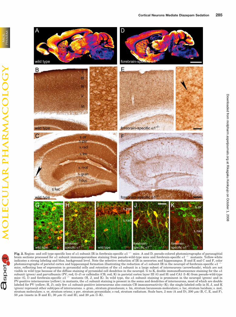
Fig. 2. Region- and cell type-specific loss of �1 subunit IR in forebrain-specific �1�/� mice. A and D, pseudo-colored photomicrographs of parasagittalbrain sections processed for �1 subunit immunoperoxidase staining from pseudo-wild-type mice and forebrain-specific �1�/� mutants. Yellow-whiteindicates a strong labeling and blue, background level. Note the selective reduction of IR in neocortex and hippocampus. B and E and C and F, colorphotomicrographs of parietal cortex and hippocampal formation illustrating the reduction of �1 subunit IR in the neuropil of forebrain-specific �1�/�
mice, reflecting loss of expression in pyramidal cells and retention of the �1 subunit in a large subset of interneurons (arrowheads), which are notvisible in wild type because of the diffuse staining of pyramidal cell dendrites in the neuropil. G to K, double immunofluorescence staining for the �1subunit (green) and parvalbumin (PV; red; G–J) or calbindin (CB; red; K) in parietal cortex layer III (G and H) and CA1 (I–K) from pseudo-wild-typemice (G, I) and forebrain-specific �1�/� mutants (H, J, and K). In wild type, the �1 subunit staining is prominent in the neuropil (green) and inPV-positive interneurons (yellow); in mutants, the �1 subunit staining is present in the soma and dendrites of interneurons, most of which are doublelabeled for PV (yellow; H, J); only few �1 subunit-positive interneurons also contain CB immunoreactivity (K); the single-labeled cells in H, J, and K(green) represent other subtypes of interneurons. s. gran., stratum granulosum; s. lm, stratum lacunosum-moleculare; s. luc, stratum lucidum; s. mol,stratum moleculare; s. or, stratum oriens; s pyr, stratum pyramidale; s rad, stratum radiatum. Scale bars, 2 mm (A and D), 200 �m (B, C, E, and F),50 �m (insets in B and E), 30 �m (G and H), and 20 �m (I–K).
Cortical Neurons Mediate Diazepam Sedation 285
at Rikagaku K
enkyujo on October 1, 2008
molpharm
.aspetjournals.orgD
ownloaded from

S.D., were analyzed using nonparametric Kruskal-Wallis andMann-Whitney U tests.
Behavioral Testing. The effect of diazepam on motor activitywas measured as a determinant of its sedative action (Trevor andWay, 1995) in the different mutant mouse lines. Mice were adaptedto a reversed 12-h day-night cycle (lights off at 8 AM) for at least 2weeks before testing (between 9 AM and 12 PM). Motor activity wasmeasured during the active phase in automated individual circularrunways equipped with photocells (Imetronic, Pessac, France) for anhour, starting 30 min after oral administration of either 10 mg/kgdiazepam or vehicle (0.3% Tween 80 in saline). The dose of diazepamwas chosen based on previous dose-response experiments showing amarked reduction in motor activity in wild-type C57BL/6J mice, butnot in �1(H101R) mutants. Because of the absence of a difference,data from male and female mice were pooled and analyzed usingtwo-way (genotype � treatment) repeated measures analysis of vari-ance followed by post hoc Scheffe’s test. Results are expressed asmean � S.E.
ResultsExpression of GABAA Receptor Subunits in Mice
Lacking the �1 Subunit in Forebrain GlutamatergicNeurons. The immunohistochemical analysis of the regionaldistribution and relative immunoreactivity (IR) levels for the�1, �2, and �3 subunit revealed differences between fore-brain-specific �1�/� mice and their corresponding pseudo-wild-type littermates (HfloxHflox/Emx1-cretg�). In wild-typebrain sections, �1 subunit IR was prominent and nearlyevenly distributed across all cortical areas (Fig. 2A). The �1subunit staining was most pronounced in layers I, III, andIV, as shown in the parietal cortex (Fig. 2B). �1 subunit IRwas also intense and diffuse in all subregions of the hip-pocampal formation, except in the pyramidal and the granulecell layers (Fig. 2C). No structure or single neuron could bedistinguished at low magnification, except in the CA3 stra-
Fig. 3. Region-specific increase of �2 and �3 subunit IR in forebrain-specific �1�/� mice. Pseudo-colored photomicrographs of parasagittal sectionsprocessed for immunoperoxidase staining. A, �2 subunit IR in pseudo-wild-type mice. B, increased �2 subunit IR in the neocortex in forebrain-specific�1�/� mice. C. �3 subunit IR in pseudo-wild-type mice. D, increased �3 subunit IR in the neocortex in forebrain-specific �1�/� mice. Scale bar, 2 mm.
TABLE 1Quantification of GABAA receptor �2 and �3 subunit immunoreactivity in forebrain-specific �1�/� mice compared with wild-type littermates(HfloxHflox/Emx1-cretg�)Optical density (OD) values were measured in sections processed for immunoperoxidase staining (adult mice; n � 4/genotype) using gray-scale standards for calibration.Background was measured in a region of gray matter lacking the expression of these subunits (inferior colliculus for the �2 subunit and cerebellum for the �3 subunit) andsubtracted from the measured values. Values in mutants are expressed as percentage of wild-type control. Statistically significant differences in absolute values are indicatedin bold (p � 0.05; Mann-Whitney U test).
Region Wild-TypeOD �2
Forebrain-Specific �1�/� as aProportion of WT OD �2
Wild-TypeOD �3
Forebrain-Specific �1�/� as aProportion of WT OD �3
% %
Parietal cortex, layer I–III 12 � 5.8 146 33 � 2.4 142Parietal cortex, layer IV 11 � 5.3 172 23 � 1.8 169Parietal cortex, layers V and VI 5 � 4.8 185 35 � 2.3 122Frontal cortex, layers I–III 12 � 4.4 143 39 � 1.9 136Frontal cortex, layers V and VI 9 � 5.0 162 41 � 1.7 131CA1 34 � 6.1 110 22 � 3.9 109CA3 37 � 5.8 101 20 � 5 102Dentate gyrus 43 � 7.7 117 16 � 4.0 125Subiculum 30 � 5.2 118 33 � 1.6 135Striatum 39 � 4.2 93 21 � 3.0 119Thalamic reticular nucleus 30 � 3.1 114Cerebellum, molecular layer 21 � 3.1 101 8 � 1.6 118
286 Zeller et al.
at Rikagaku K
enkyujo on October 1, 2008
molpharm
.aspetjournals.orgD
ownloaded from

tum lucidum where interneurons and their dendrites werevisible (Fig. 2C, arrowhead). In brain sections from forebrain-specific �1�/� mice, a marked decrease in �1 subunit IR wasapparent, and it was restricted to the neocortex and hip-pocampus (Fig. 2D). In these mice, no change in �1 subunitIR could be detected in brain regions in which Emx1-cre isnot expressed, confirming the specificity of the cre recombi-nation driven by the Emx1 promoter. Remarkably, the �1subunit staining was absent from all cortical glutamatergiccells, whereas it was retained in interneurons, as seen athigh magnification (Fig. 2E, arrowhead). This finding waseven more evident in hippocampal sections, where a largepopulation of interneurons selectively showed an intense �1subunit IR against a white background (Fig. 2F). Thus, asexpected, forebrain-specific �1�/� mice displayed a deficit in�1 subunit restricted to glutamatergic neurons. The inter-neuronal nature of �1 subunit-positive cells in the neocortexand hippocampus was verified by double immunofluores-cence staining with parvalbumin (Fig. 2, G–J), calbindin (Fig.2K), and calretinin (data not shown), three calcium-bindingproteins that label largely nonoverlapping subpopulations ofGABAergic interneurons (Freund and Buzsaki, 1996).
Forebrain-specific �1�/� mice showed a regional expres-sion pattern for the �2 and �3 subunit comparable with thatof the pseudo-wild-type controls, but the IR of both subunitswas stronger. In control mice, �2 subunit IR was confined tothe outer layers of the neocortex, whereas it was virtuallyabsent in layers V and VI. In the hippocampal formation, itwas most prominent in the dentate gyrus, followed by CA3and CA1 (Fig. 3A). A significant increase in �2 subunit IRwas apparent in the neocortex of mutants, but not in thehippocampal formation (Fig. 3B; Table 1). The �3 subunit IRin the neocortex of pseudo-wild-type mice was most intensein V and VI, particularly in frontal cortex. In the hippocam-pal formation, it predominated in the CA1 area similarly inpseudo-wild-type and forebrain-specific �1�/� mice (Fig. 3, Cand D), and it was almost absent in the dentate gyrus.Mutant mice showed enhanced levels of �3 subunit IR inneocortex, comparable with the increase in �2 subunit IR(Table 1). It is noteworthy that the �3 subunit, almost absentin layer IV of parietal cortex in wild-type animals, could bedetected in the mutants (Fig. 3D). As in forebrain-specific�1�/� mice, no change in subunit expression was seen inregions where Emx1-cre was not expressed, such as striatum,thalamus, and cerebellum (Table 1). Thus, a deficit of �1subunit in cortical glutamatergic neurons was accompaniedby an increased expression of the �2 and �3 subunits in thecorresponding regions.
Sedative Action of Diazepam in Forebrain-Specific�1�/� Mice. Forebrain-specific �1�/� mice displayedheightened sensitivity to the sedative action of diazepam,as indicated by the greater drug-induced decrease in motoractivity in the mutants compared with the pseudo-wild-type mice [p � 0.05 after F(1,36) � 13.09; p � 0.01] (Fig.4A). No genotype difference was observed with the vehicletreatment.
Expression of GABAA Receptor Subunits in MiceCarrying a Single �1(H101R) Allele in Forebrain Glu-tamatergic Neurons. A second series of experiments wascarried out to obtain mice in which diazepam sensitivity, butnot expression of �1-GABAA receptors, would be selectivelysuppressed in forebrain neurons expressing Emx1. The
breeding scheme adopted (Fig. 1) resulted in four genotypes,including pseudo-wild-type mice (HfloxH/Emx1-cretg�), globalheterozygous knock-in �1H/R mice, forebrain-specific �1�/H
mice (carrying a single floxed �1 subunit allele in forebrain),and forebrain-specific �1�/R [carrying a single point-mutated�1(H101R) subunit allele in forebrain]. The pseudo-wild-typeand �1H/R mice showed an expression pattern for the GABAA
receptor �1, �2, and �3 subunits similar to that seen inpseudo-wild-type HfloxHflox/Emx1-cretg� (Figs. 2A and 3, Aand C). Unexpectedly, in forebrain-specific �1�/H and �1�/R
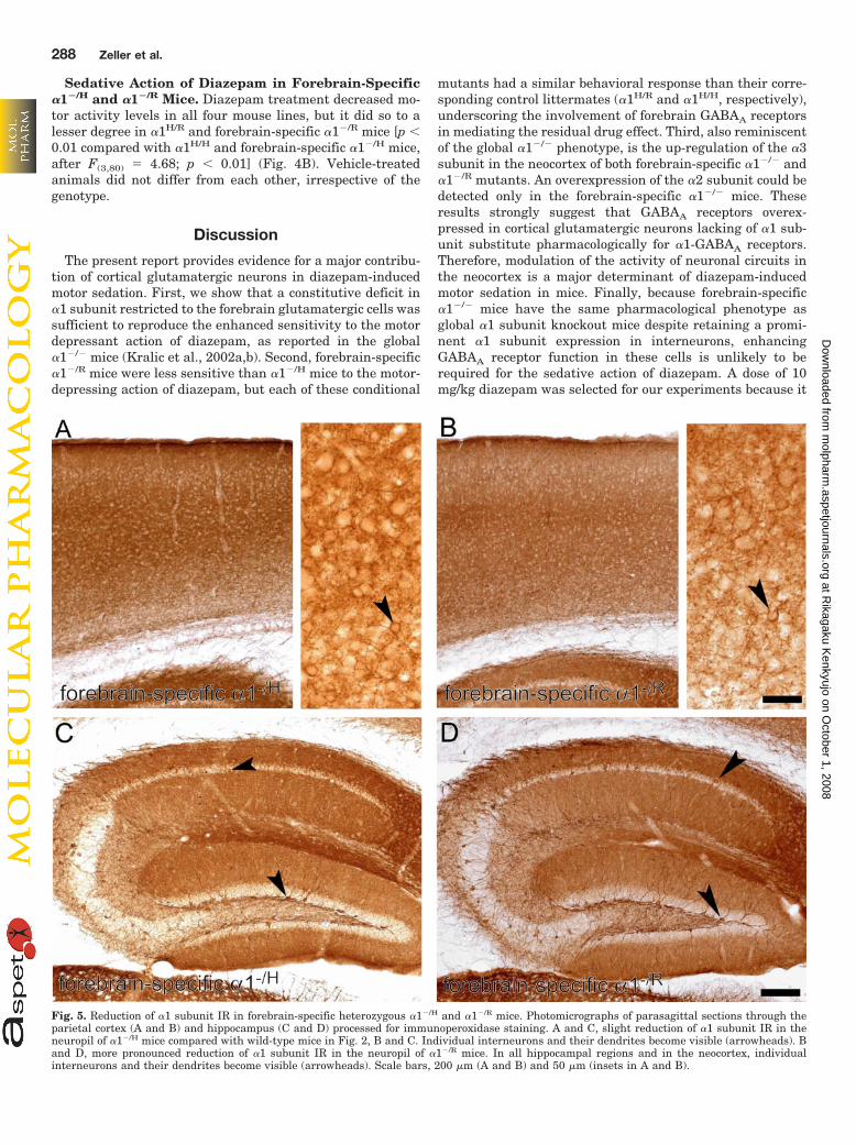
mice, �1 subunit IR was decreased in cortical and hippocam-pal principal cells (Fig. 5). At high magnification, individualinterneurons and their dendrites could be easily visualized atlow magnification in the neocortex (Fig. 5, A and B, arrow-head) and the hippocampus (Fig. 5, C and D, arrowhead) insections from both mutants. The �1 subunit deficit in pari-etal cortex was more pronounced in �1�/R mice (Fig. 5B).However, the prominent labeling of interneurons largelymasked the decrease in pyramidal cells, so that no selectivedensitometric quantification was feasible. Nevertheless,these results indicate that a single, either wild-type or point-mutated, �1 subunit allele in cortical glutamatergic neuronswas insufficient to provide normal expression of the �1subunit.
No consistent alteration in �2 subunit expression patternand IR levels was detected in forebrain-specific �1�/H and�1�/R mice (Table 2). A difference in �3 subunit IR wasobserved specifically in the cerebral cortex of �1�/R mice(Table 2). A trend was seen in �1�/H mice, but the changeswere significant only in layers V and VI of frontal cortex(Table 2). In these mice, weakly stained areas (CA3, dentategyrus, and striatum) exhibited increased staining comparedwith control (Table 2). However, because these changes werenot seen in other mutants and they included regions whereEmx1-cre is not expressed, their significance is uncertain.Overall, we conclude that expression of a single �1(H101R)allele in forebrain glutamatergic neurons is associated with aselective up-regulation of the �3 subunit in the neocortex.
Fig. 4. Motor-sedative effect of diazepam. Motor activity was measuredfor 1 h, starting 30 min after oral administration of either vehicle or 10mg/kg diazepam. A, forebrain-specific �1�/� mice. Note the greater re-duction in mean activity counts in diazepam-treated forebrain-specific�1�/� mice compared with pseudo-wild-type �1H/H mice (n � 19 mice/group; #, p � 0.05 compared with wild-type mice; and ���, p � 0.001compared with vehicle). B, forebrain-specific heterozygous �1�/H and�1�/R mice. Overall, this series of animals displayed lower levels of motoractivity, as seen in vehicle-treated �1H/H mice in comparison with thecorresponding pseudo-wild-type �1H/H animals in A, but this experimen-tal variability did not change the vehicle/diazepam ratio. Diazepam treat-ment induced a decrease in motor activity in all four groups, but the drugeffect was smaller in �1�/R than �1�/H mice, and the effect in each of theseforebrain-specific mutants was not different from that in their corre-sponding �1H/R and �1H/H control littermates (n � 21 mice/group; #, p �0.05 compared with �1H/H and �1�/H mice; and ��, p � 0.01 and ���, p �0.001 compared with vehicle; Scheffe’s test).
Cortical Neurons Mediate Diazepam Sedation 287
at Rikagaku K
enkyujo on October 1, 2008
molpharm
.aspetjournals.orgD
ownloaded from
Sedative Action of Diazepam in Forebrain-Specific�1�/H and �1�/R Mice. Diazepam treatment decreased mo-tor activity levels in all four mouse lines, but it did so to alesser degree in �1H/R and forebrain-specific �1�/R mice [p �0.01 compared with �1H/H and forebrain-specific �1�/H mice,after F(3,80) � 4.68; p � 0.01] (Fig. 4B). Vehicle-treatedanimals did not differ from each other, irrespective of thegenotype.
Discussion
The present report provides evidence for a major contribu-tion of cortical glutamatergic neurons in diazepam-inducedmotor sedation. First, we show that a constitutive deficit in�1 subunit restricted to the forebrain glutamatergic cells wassufficient to reproduce the enhanced sensitivity to the motordepressant action of diazepam, as reported in the global�1�/� mice (Kralic et al., 2002a,b). Second, forebrain-specific�1�/R mice were less sensitive than �1�/H mice to the motor-depressing action of diazepam, but each of these conditional
mutants had a similar behavioral response than their corre-sponding control littermates (�1H/R and �1H/H, respectively),underscoring the involvement of forebrain GABAA receptorsin mediating the residual drug effect. Third, also reminiscentof the global �1�/� phenotype, is the up-regulation of the �3subunit in the neocortex of both forebrain-specific �1�/� and�1�/R mutants. An overexpression of the �2 subunit could bedetected only in the forebrain-specific �1�/� mice. Theseresults strongly suggest that GABAA receptors overex-pressed in cortical glutamatergic neurons lacking of �1 sub-unit substitute pharmacologically for �1-GABAA receptors.Therefore, modulation of the activity of neuronal circuits inthe neocortex is a major determinant of diazepam-inducedmotor sedation in mice. Finally, because forebrain-specific�1�/� mice have the same pharmacological phenotype asglobal �1 subunit knockout mice despite retaining a promi-nent �1 subunit expression in interneurons, enhancingGABAA receptor function in these cells is unlikely to berequired for the sedative action of diazepam. A dose of 10mg/kg diazepam was selected for our experiments because it
Fig. 5. Reduction of �1 subunit IR in forebrain-specific heterozygous �1�/H and �1�/R mice. Photomicrographs of parasagittal sections through theparietal cortex (A and B) and hippocampus (C and D) processed for immunoperoxidase staining. A and C, slight reduction of �1 subunit IR in theneuropil of �1�/H mice compared with wild-type mice in Fig. 2, B and C. Individual interneurons and their dendrites become visible (arrowheads). Band D, more pronounced reduction of �1 subunit IR in the neuropil of �1�/R mice. In all hippocampal regions and in the neocortex, individualinterneurons and their dendrites become visible (arrowheads). Scale bars, 200 �m (A and B) and 50 �m (insets in A and B).
288 Zeller et al.
at Rikagaku K
enkyujo on October 1, 2008
molpharm
.aspetjournals.orgD
ownloaded from

has a robust sedative action, reducing motor activity by ap-proximately two thirds, but still allows us to detect a furtherdecrease in motor activity caused by individual genotypes.
Global deletion of the �1 subunit gene results in a markedcompensatory overexpression of the GABAA receptor �2, �3,and �4 subunits, selectively in those brain regions where the�1 subunit is absent (Kralic et al., 2002a, 2006; SchneiderGasser et al., 2007). Up-regulation probably takes place atthe level of translation, without increase in subunit genetranscription, as shown by several studies (Bosman et al.,2005b). These compensatory changes do not fully restore thefunction of the missing �1 subunit, as evidenced, for example,by the decrease of GABAergic currents in cerebellar slices(Vicini et al., 2001) or the complete loss of GABAA receptorsin Purkinje cells in these mutants (Sur et al., 2001; Kralic etal., 2005; Fritschy et al., 2006). We opted for a conditionalmutation strategy, expecting no compensatory � subunit up-regulation. Nevertheless, deletion of the �1 subunit re-stricted to forebrain principal cells leads to overexpression ofthe �2 and �3 subunit, underscoring the need for homeostaticcompensation to retain normal brain function in the absenceof a major GABAA receptor subtype. The change in subunitexpression was restricted to regions where Emx1-cre-inducedrecombination had occurred, further indicating that GABAA
receptors were probably unaffected in other brain areas ofconditional mutant mice.
The decreased �1 subunit IR in the forebrain of �1�/H and�1�/R mice, which both carry a single �1 subunit allele inforebrain glutamatergic neurons, is reminiscent of the de-creased expression of the �2 subunit occurring mostly inneocortex and hippocampus in mice heterozygous for the �2subunit deletion (�2�/�) (Crestani et al., 1999). These find-ings reveal that certain major GABAA receptor subunits areavailable in limited amounts whenever expressed by a singleallele. In �2�/� mice, no compensatory up-regulation of otherGABAA receptor subunits could be detected, presumably be-cause the remaining �/� subunit variants could form func-tional GABAA receptors in these mutants (Lorez et al., 2000).The partial deficit in �1-GABAA receptors in �1�/R mice
seems to be sufficient to induce compensatory changes, prob-ably because � subunits are required for receptor assembly(Kralic et al., 2006; Rudolph and Mohler, 2006; Studer et al.,2006).
In line with the loss of diazepam binding to GABAA recep-tors containing the �1(H101R) point mutation, forebrain-specific �1�/R mice were less sensitive than �1�/H mice to themotor-sedative action of diazepam, underscoring again thecontribution of cortical circuits to this pharmacological effect.However, these two mutants show diazepam responsivenesssimilar to that of their respective global heterozygote orpseudo-wild-type control (�1H/R and �1H/H). In �1�/H mice,one might argue that the remaining pool of diazepam-sensi-tive �1-GABAA receptors in the cerebral cortex is sufficientfor the full manifestation of the sedative drug action. In �1�/R
mice, the mild reduction in motor activity produced by diaz-epam is best explained by the up-regulation of the �3 sub-unit, which might restore a complement of diazepam-sensi-tive �3-GABAA receptors selectively in neocortical regions.
The consequences of the up-regulation of �2 and �3 subunitin forebrain-specific mutant mice for the function of corticalcircuits remain to be established. Cortical pyramidal cellsexpress multiple GABAA receptor � subunits with a differ-ential subcellular distribution. In particular, �1-GABAA re-ceptors predominate on distal dendrites, whereas �2-GABAA
receptors mediate most of the perisomatic GABAergic inputs(Prenosil et al., 2006). In addition, the �1- and �2 subunitsare located in the synapses of separate subpopulations ofbasket cells (distinguished by expression of parvalbumin andcholecystokinin, respectively) (Nyıri et al., 2001). These dif-ferences probably underlie the contribution of these GABAA
receptor subtypes to distinct neuronal circuits. Although theup-regulation of the �3 subunit suggests that this subunitcould replace the �1 subunit at its original location, a reor-ganization of GABAergic circuits within the cortex cannot beexcluded.
In addition to the circuit-specific localization of GABAA
receptor subtypes, their functional properties are determinedby their subunit composition. Thus, GABAA receptors ex-pressed in the neocortex and hippocampus of global �1�/�
mice have longer decay kinetics (Goldstein et al., 2002; Bos-man et al., 2005b; Schneider Gasser et al., 2007), character-istic of �2- and �3-GABAA receptors expressed early duringdevelopment (Hutcheon et al., 2000). The number of func-tional GABAergic synapses is not changed in the neocortex(Bosman et al., 2005b), but the longer kinetics influences �oscillations (Bosman et al., 2005a). Taken together, up-reg-ulation of the �2 and �3 subunit in forebrain-specific �1�/�
mice might functionally compensate for the loss of the �1subunit when no substance challenges the system, resultingin a normal behavioral response, as seen in vehicle-treatedmice. However, because of the slow kinetics of �3-GABAA
receptors, the effects induced by diazepam in cortical neuronslacking �1-GABAA receptors might be more pronounced thanthose observed in wild type. This difference might be mani-fested behaviorally by the enhanced sensitivity of forebrain-specific �1�/� mutants to the motor-sedative effect of diaze-pam compared with pseudo-wild-type mice (Fig. 4A). Theimportance of GABAA receptor kinetics for normal brainfunction has been underscored by introducing a (S270H)point mutation in the �1 subunit gene that causes a markedslowing of GABAA receptor deactivation (Homanics et al.,
TABLE 2Quantification of the GABAA receptor �2 and �3 subunitimmunoreactivity in forebrain-specific �1�/H and �1�/R mice comparedwith wild-type littermates (HfloxH/ Emx1-cretg� and HfloxR/Emx1-cretg�)Densitometry was performed in sections processed for immunoperoxidase staining(see Table 1). Values are expressed as percentage of wild-type control. Statisticallysignificant differences in absolute values are indicated in bold (p � 0.05; Mann-Whitney U test).
Region
Forebrain-Specific,
as a Proportionof OD �2
Forebrain-Specific,
as a Proportionof OD �3
�1�/H �1�/R �1�/H �1�/R
% %
Parietal cortex, layer I–III 101 118 123 127Parietal cortex, layer IV 129 129 115 142Parietal cortex, layers V and VI 120 109 105 130Frontal cortex, layers I–III 72 112 124 118Frontal cortex, layers V and VI 56 112 140 134CA1 80 97 122 106CA3 84 88 148 109Dentate gyrus 76 109 160 118Subiculum 88 103 124 126Striatum 95 114 156 120Thalamic reticular nucleus 128 126Cerebellum, molecular layer 127 126 126 125
Cortical Neurons Mediate Diazepam Sedation 289
at Rikagaku K
enkyujo on October 1, 2008
molpharm
.aspetjournals.orgD
ownloaded from

2005). The corresponding point-mutated mice exhibit majorphysiological, behavioral, and pharmacological impairments(e.g., loss of sensitivity to the volatile anesthetic isoflurane)probably due to functional abnormalities in neuronal circuitsexpressing �1(S270H)-GABAA receptors (Homanics et al.,2005).
Our results strongly implicate neocortical circuits in themediation of diazepam-induced motor sedation. The sedativeeffect of benzodiazepines is often assessed using tests ofmotor coordination (e.g., rotarod), which probably engageadditional brain circuits, notably the cerebellum (Lalondeand Strazielle, 2001; Levin et al., 2006). However, althoughsuch behavioral paradigms arguably provide a more com-plete measure of the drug effect as a reduction in motoractivity, their validity for predicting sedative effects in hu-man has been questioned (Stanley et al., 2005). A reductionin muscle tone in diazepam-treated mice might possibly af-fect motor activity. However, this effect is unlikely to con-found the present results, because the myorelaxant effect ofdiazepam is mediated by �2- and �5-GABAA receptors, and itcannot be attributed selectively to cortical circuits (Crestaniet al., 2001). Rather, in support for a major involvement ofcortical networks in mediating the motor-depressant effectsof diazepam, it has been shown in rats in vivo that sedativedoses of the volatile anesthetics isoflurane and enfluranereduce cortical firing rate by 65% as a result of increasedGABAA receptor-mediated inhibition (Hentschke et al.,2005). This correlation between behavioral sedation and de-pression of cortical firing rate is consistent with the assump-tion that low doses of volatile anesthetics mediate sedationvia modulation of cortical circuits. Likewise, functional mag-netic resonance imaging experiments in humans show thatlow, sedative doses of the GABAA receptor-specific generalanesthetic propofol reduce neuronal activity prominently incortical networks (Heinke et al., 2004; Heinke and Koelsch,2005). Only when higher, hypnotic doses of propofol are ad-ministered to the subjects, neuronal activity also decreases insubcortical structures, including the thalamus and midbrainreticular formation.
In conclusion, the present study demonstrates that even con-ditional, temporally, and spatially restricted manipulation ofGABAA receptor �1 subunit expression can induce compensa-tory changes selectively in the affected areas. Alterations ofGABAA receptor expression or pharmacology restricted to fore-brain glutamatergic neurons produce the same behavioral ef-fects as seen after a global alteration, thereby implicating theseneurons in the motor-sedative effect of diazepam.
Acknowledgments
We thank Dr. Gregg E. Homanics (University of Pittsburgh, Pitts-burgh, PA) for providing �1 subunit HfloxHflox mice and for helpfulcomments on the manuscript.
ReferencesBarnard EA, Skolnick P, Olsen RW, Mohler H, Sieghart W, Biggio G, Braestrup C,
Bateson AN, and Langer SZ (1998) International Union of Pharmacology. XV.Subtypes of �-aminobutyric acidA receptors: classification on the basis of subunitstructure and function. Pharmacol Rev 50:291–313.
Belelli D and Lambert JJ (2005) Neurosteroids: endogenous regulators of the GABAAreceptor. Nat Rev Neurosci 6:565–575.
Benson JA, Low K, Keist R, Mohler H, and Rudolph U (1998) Pharmacology ofrecombinant �-aminobutyric acidA receptors rendered diazepam-insensitive bypoint-mutated �-subunits. FEBS Lett 431:400–404.
Bosman L, Lodder JC, van Ooyen A, and Brussaard AB (2005a) Role of synapticinhibition in spatiotemporal patterning of cortical activity. Prog Brain Res 147:201–204.
Bosman LW, Heinen K, Spijker S, and Brussaard AB (2005b) Mice lacking the majoradult GABAA receptor subtype have normal number of synapses, but retainjuvenile IPSC kinetics until adulthood. J Neurophysiol 94:338–346.
Crestani F, Lorez M, Baer K, Essrich C, Benke D, Laurent JP, Belzung C, FritschyJM, Luscher B, and Mohler H (1999) Decreased GABAA-receptor clustering resultsin enhanced anxiety and a bias for threat cues. Nat Neurosci 2:833–839.
Crestani F, Low K, Keist R, Mandelli MJ, Mohler H, and Rudolph U (2001) Moleculartargets for the myorelaxant action of diazepam. Mol Pharmacol 59:442–445.
Crestani F, Martin JR, Mohler H, and Rudolph U (2000) Mechanism of action of thehypnotic zolpidem in vivo. Br J Pharmacol 131:1251–1254.
Freund TF and Buzsaki G (1996) Interneurons of the hippocampus. Hippocampus6:347–470.
Fritschy JM, Panzanelli P, Kralic JE, Vogt KE, and Sassoe-Pognetto M (2006)Differential dependence of axo-dendritic and axo-somatic GABAergic synapses onGABAA receptors containing the �1 subunit in Purkinje cells. J Neurosci 26:3245–3255.
Goldstein PA, Elsen FP, Ying SW, Ferguson C, Homanics GE, and Harrison NL(2002) Prolongation of hippocampal miniature inhibitory postsynaptic currents inmice lacking the GABAA receptor �1 subunit. J Neurophysiol 88:3208–3217.
Heinke W, Kenntner R, Gunter TC, Sammler D, Olthoff D, and Koelsch S (2004)Sequential effects of increasing propofol sedation on frontal and temporal corticesas indexed by auditory event-related potentials. Anesthesiology 100:617–625.
Heinke W and Koelsch S (2005) The effects of anesthetics on brain activity andcognitive function. Curr Opin Anaesthesiol 18:625–631.
Hentschke H, Schwarz C, and Antkowiak B (2005) Neocortex is the major target ofsedative concentrations of volatile anesthetics: strong depression of firing ratesand increase of GABAA receptor-mediated inhibition. Eur J Neurosci 21:93–102.
Homanics GE, Elsen FP, Ying SW, Jenkins A, Ferguson C, Sloat B, Yuditskaya S,Goldstein PA, Kralic JE, Morrow AL, et al. (2005) A gain-of-function mutation inthe GABAA receptor produces synaptic and behavioral abnormalities in the mouse.Genes Brain Behav 4:10–19.
Hutcheon B, Morley P, and Poulter MO (2000) Developmental change in GABAAreceptor desensitization kinetics and its role in synapse function in rat corticalneurons. J Physiol 522:3–17.
Iwasato T, Nomura R, Ando R, Ikeda T, Tanaka M, and Itohara S (2004) Dorsaltelencephalon-specific expression of Cre recombinase in PAC transgenic mice.Genesis 38:130–138.
Kopp C, Rudolph U, Low K, and Tobler I (2004) Modulation of rhythmic brainactivity by diazepam: GABAA receptor subtype and state specificity. Proc NatlAcad Sci U S A 101:3674–3679.
Kralic JE, Criswell HE, Ostermann JL, O’Buckley TK, Wilkie ME, Matthews DA,Hamre K, Breese GR, Homanics GE, and Morrow AL (2005) Genetic essentialtremor in �-aminobutyric acidA receptor �1 subunit knockout mice. J Clin Invest115:774–779.
Kralic JE, Korpi ER, O’Buckley TK, Homanics GE, and Morrow AL (2002a) Molec-ular and pharmacological characterization of GABAA receptor �1 subunit knock-out mice. J Pharmacol Exp Ther 302:1037–1045.
Kralic JE, O’Buckley TK, Khisti RT, Hodge CW, Homanics GE, and Morrow AL(2002b) GABAA receptor �1 subunit deletion alters receptor subtype assembly,pharmacological and behavioral responses to benzodiazepines and zolpidem. Neu-ropharmacology 43:685–694.
Kralic JE, Sidler C, Parpan F, Homanics GE, Morrow AL, and Fritschy JM (2006)Compensatory alteration of inhibitory synaptic circuits in cerebellum and thala-mus of �-aminobutyric acid type A receptor �1 subunit knockout mice. J CompNeurol 495:408–421.
Lalonde R and Strazielle C (2001) Motor performance and regional brain metabolismof spontaneous murine mutations with cerebellar atrophy. Behav Brain Res 125:103–108.
Levin SI, Khaliq ZM, Aman TK, Grieco TM, Kearney JA, Raman IM, and MeislerMH (2006) Impaired motor function in mice with cell-specific knockout of sodiumchannel Scn8a (NaV1.6) in cerebellar Purkinje neurons and granule cells. J Neu-rophysiol 96:785–793.
Lippa A, Czobor P, Stark J, Beer B, Kostakis E, Gravielle M, Bandyopadhyay S,Russek SJ, Gibbs TT, Farb DH, et al. (2005) Selective anxiolysis produced byocinaplon, a GABAA receptor modulator. Proc Natl Acad Sci U S A 102:7380–7385.
Lorez M, Benke D, Luscher B, Mohler H, and Benson JA (2000) Single-channelproperties of neuronal GABAA receptors from mice lacking the �2 subunit. JPhysiol 527:11–31.
Marowsky A, Fritschy JM, and Vogt KE (2004) Functional mapping of GABAAreceptor subtypes in the amygdala. Eur J Neurosci 20:1281–1289.
McKernan RM, Rosahl TW, Reynolds DS, Sur C, Wafford KA, Atack JR, Farrar S,Myers J, Cook G, Ferris P, et al. (2000) Sedative but not anxiolytic properties ofbenzodiazepines are mediated by the GABAA receptor �1 subtype. Nat Neurosci3:587–592.
Nyıri G, Freund TF, and Somogyi P (2001) Input-dependent synaptic targeting of�2-subunit-containing GABAA receptors in synapses of hippocampal pyramidalcells of the rat. Eur J Neurosci 13:428–442.
Prenosil GA, Schneider Gasser EM, Rudolph U, Keist R, Fritschy JM, and Vogt KE(2006) Specific subtypes of GABAA receptors mediate phasic and tonic forms ofinhibition in hippocampal pyramidal neurons. J Neurophysiol 96:846–857.
Rudolph U and Antkowiak B (2004) Molecular and neuronal substrates for generalanaesthetics. Nat Rev Neurosci 5:709–720.
Rudolph U, Crestani F, Benke D, Brunig I, Benson JA, Fritschy JM, Martin JR,Bluethmann H, and Mohler H (1999) Benzodiazepine actions mediated by specific�-aminobutyric acidA receptor subtypes. Nature 401:796–800.
Rudolph U and Mohler H (2004) Analysis of GABAA receptor function and dissectionof the pharmacology of benzodiazepines and general anesthetics through mousegenetics. Annu Rev Pharmacol Toxicol 44:475–498.
Rudolph U and Mohler H (2006) GABA-based therapeutic approaches: GABAAreceptor subtype functions. Curr Opin Pharmacol 6:18–23.
290 Zeller et al.
at Rikagaku K
enkyujo on October 1, 2008
molpharm
.aspetjournals.orgD
ownloaded from

Schneider Gasser EM, Duveau V, Prenosil GA, and Fritschy JM (2007) Reorganiza-tion of GABAergic circuits maintains GABAA receptor-mediated transmission ontoCA1 interneurons in �1 subunit-null mice. Eur J Neurosci 25:3287–3304.
Scott-Stevens P, Atack JR, Sohal B, and Worboys P (2005) Rodent pharmacokineticsand receptor occupancy of the GABAA receptor subtype selective benzodiazepinesite ligand L-838417. Biopharm Drug Dispos 26:13–20.
Sieghart W and Ernst M (2005) Heterogeneity of GABAA receptors: revived interestin the development of subtype-selective drugs. Curr Med Chem 5:217–242.
Stanley JL, Lincoln RJ, Brown TA, McDonald LM, Dawson GR, and Reynolds DS(2005) The mouse beam walking assay offers improved sensitivity over the mouserotarod in determining motor coordination deficits induced by benzodiazepines.J Psychopharmacol 19:221–227.
Studer R, von Boehmer L, Haenggi T, Schweizer C, Benke D, Rudolph U, andFritschy JM (2006) Alteration of GABAergic synapses and gephyrin clusters in thethalamic reticular nucleus of GABAA receptor �3 subunit-null mice. Eur J Neu-rosci 24:1307–1315.
Sur C, Wafford KA, Reynolds DS, Hadingham KL, Bromidge F, Macaulay A, Col-
linson N, O’Meara G, Howell O, Newman R, et al. (2001) Loss of the major GABAAreceptor subtype in the brain is not lethal in mice. J Neurosci 21:3409–3418.
Tobler I, Kopp C, Deboer T, and Rudolph U (2001) Diazepam-induced changes insleep: role of the �1 GABAA receptor subtype. Proc Natl Acad Sci U S A 98:6464–6469.
Trevor AJ and Way WL (1995) Sedative-hypnotic drugs, in Basic and ClinicalPharmacology (G Katzung ed) pp 333–349, Appleton & Lange, Norwalk, CT.
Vicini S, Ferguson C, Prybylowski K, Kralic J, Morrow AL, and Homanics GE (2001)GABAA receptor �1 subunit deletion prevents developmental changes of inhibitorysynaptic currents in cerebellar neurons. J Neurosci 21:3009–3016.
Address correspondence to: Dr. Uwe Rudolph, Laboratory of Genetic Neu-ropharmacology, McLean Hospital, Department of Psychiatry, HarvardMedical School, 115 Mill St., Belmont, MA 02478. E-mail: [email protected]
Cortical Neurons Mediate Diazepam Sedation 291
at Rikagaku K
enkyujo on October 1, 2008
molpharm
.aspetjournals.orgD
ownloaded from