Haloperidol protects striatal neurons from dysfunction induced by mutated huntingtin in vivo
Upload
northwesternCategory
view
0download
0
Dorsal Striatal–Midbrain Connectivity in HumansPredicts How Reinforcements Are Used to
Guide Decisions
Thorsten Kahnt1,2*, Soyoung Q Park1*, Michael X Cohen3, Anne Beck1,Andreas Heinz1, and Jana Wrase1
Abstract
& It has been suggested that the target areas of dopaminer-gic midbrain neurons, the dorsal (DS) and ventral striatum(VS), are differently involved in reinforcement learning espe-cially as actor and critic. Whereas the critic learns to predictrewards, the actor maintains action values to guide futuredecisions. The different midbrain connections to the DS andthe VS seem to play a critical role in this functional distinction.Here, subjects performed a dynamic, reward-based decision-making task during fMRI acquisition. A computational mod-el of reinforcement learning was used to estimate the differenteffects of positive and negative reinforcements on future de-cisions for each subject individually. We found that activity
in both the DS and the VS correlated with reward predic-tion errors. Using functional connectivity, we show that theDS and the VS are differentially connected to different mid-brain regions (possibly corresponding to the substantia ni-gra [SN] and the ventral tegmental area [VTA], respectively).However, only functional connectivity between the DS andthe putative SN predicted the impact of different reinforce-ment types on future behavior. These results suggest thatconnections between the putative SN and the DS are criticalfor modulating action values in the DS according to bothpositive and negative reinforcements to guide future decisionmaking. &
INTRODUCTION
Learning which action to take in an uncertain environ-ment to maximize reward and minimize punishment iscritical for survival. Both positive and negative outcomesof current decisions can contribute differentially to theway individuals decide in the future. The dopaminergicmidbrain system and its prominent target areas, espe-cially the striatum, play key roles in this process. Mid-brain dopamine neurons are thought to contribute toreinforcement learning by sending a teaching signalto the striatum that biases action selection accordingto the previous action–outcome history (Samejima &Doya, 2007; Samejima, Ueda, Doya, & Kimura, 2005;Schultz & Dickinson, 2000; Hollerman & Schultz,1998). Specifically, bursts in dopamine after positiveoutcomes are thought to facilitate the current response,whereas dips in dopamine after negative outcomes maysupport the inhibition of the current response (Schultz,2002; Hollerman & Schultz, 1998). The ventral anddorsal compartments of the striatum have been impli-cated in maintaining and updating reward predictions
and action values, respectively. Whereas the ventral part(ventral striatum: VS) learns to predict future rewards,the dorsal compartment (dorsal striatum: DS) maintainsinformation about the outcomes of decisions to enablethat the better option is chosen more often (Atallah,Lopez-Paniagua, Rudy, & O’Reilly, 2007; Schonberg,Daw, Joel, & O’Doherty, 2007; Williams & Eskandar,2006; Samejima et al., 2005; O’Doherty et al., 2004;Joel, Niv, & Ruppin, 2002; Joel & Weiner, 2000). Incomputational reinforcement learning theory, the criticuses a prediction error signal to update predictionsabout future rewards, whereas the actor uses the samesignal to update action values that biases future decisionstoward advantageous options (O’Doherty et al., 2004;Sutton & Barto, 1998).
As neurophysiological investigations in primates andrats have shown, the DS and the VS receive dopaminer-gic input from different but somewhat overlapping mid-brain regions building an ascending midbrain–striatalloop (Ikemoto, 2007; Haber, Fudge, & McFarland, 2000).Specifically, the ventral tegmental area (VTA) is recipro-cally interconnected with the VS, whereas the substantianigra (SN) receives input from the VS and is reciprocallyconnected to the DS (Haber, 2003; Haber et al., 2000).The DS and the VS are also differentially connectedto the frontal cortex. The VS is connected with
1Charite—Universitatsmedizin Berlin (Charite Campus Mitte),Germany, 2Bernstein Center for Computational NeuroscienceBerlin, Germany, 3University of Arizona, Tucson*These authors contributed equally to the work.
D 2008 Massachusetts Institute of Technology Journal of Cognitive Neuroscience 21:7, pp. 1332–1345

medial prefrontal and orbito-frontal cortices, whereasthe DS is connected with dorsal prefrontal and motorcortices (Lehericy et al., 2004; Alexander, Crutcher, &DeLong, 1990). Due to its connections, associationsbetween sensory cues and motor behavior that lead toreward might be strengthened in the striatum by do-paminergic projections from the midbrain (Williams &Eskandar, 2006; Reynolds, Hyland, & Wickens, 2001).
In situations with two different options, one can learnto choose X over Y by either learning that X leads topositive feedback or that Y leads to negative feedback, orboth. In such situations, it is challenging to disentanglethe degree to which positive and negative reinforcementscontribute to learning. However, it is possible to differ-entiate both processes and subjects differ in how theyuse positive and negative outcomes to guide their deci-sions (Frank, Woroch, & Curran, 2005; Frank, Seeberger,& O’Reilly, 2004). These individual differences in learningfrom either positive or negative reinforcements havebeen linked to dopamine genetics, dopamine treatmentand neurological conditions in which dopamine activity ischanged (Cohen, Krohn-Grimberghe, Elger, & Weber,2007; Frank, Moustafa, Haughey, Curran, & Hutchison,2007; Frank, Scheres, & Sherman, 2007; Klein et al., 2007;Frank, 2005). However, in humans, although there is anincreasing amount of research about the striatum and itsinvolvement in reward-learning (Cohen, 2007; Delgado,2007; Pessiglione, Seymour, Flandin, Dolan, & Frith, 2006;Cohen & Ranganath, 2005; O’Doherty, Dayan, Friston,Critchley, & Dolan, 2003), so far, little is known about thespecific contributions of midbrain–striatal connectionsto the way reinforcements are used to guide decisionmaking. A recent study showed prediction error-relatedresponses in the VS and the VTA, and provided evi-dence for a functional coupling between both regions(D’Ardenne, McClure, Nystrom, & Cohen, 2008). Here, wewent beyond this study by showing how these striatal–midbrain circuits might be involved in mediating dynamicadjustments in decision making.
Using fMRI and a computational model of reinforce-ment learning, we aimed to disentangle the use ofpositive and negative reinforcements to guide decisionmaking in a two-arm bandit task with dynamicallychanging reward allocations. Furthermore, by means offunctional connectivity analyses, we investigated specificcontributions of different striatal–midbrain connectionsto how different reinforcements impact future decisionmaking.
Because both the DS and the VS have been suggestedto play specific roles in reinforcement learning by learn-ing the action and reward value, respectively (Williams& Eskandar, 2006; O’Doherty et al., 2004; Joel et al.,2002), we hypothesized that information about rein-forcements is represented in form of reward predictionerrors in the VS and the DS. Additionally, according toanimal studies (Haber, 2003), we hypothesized that theVS and the DS are differentially connected to different
midbrain regions. Finally, we hypothesized that theintegrity of midbrain–striatal connectivity plays a criticalrole in the use of reinforcements to guide future deci-sion making (Belin & Everitt, 2008; Faure, Haberland,Conde, & El Massioui, 2005). Specifically, if reinforce-ment-related activity in the midbrain is used to updateaction values in the DS, the way different subjects usethis information should depend on the functional con-nectivity between the midbrain and the DS, but notnecessarily the VS.
METHODS
Participants
Nineteen right-handed subjects (10 women, aged 24–31 years, mean = 25.7 ± 0.45 years) participated in thestudy. Subjects had normal or corrected-to-normal vi-sion, reported having no psychiatric conditions, andgave written consent to participate. The study wasapproved by the local ethics review committee of theCharite—Universitatsmedizin Berlin.
Experimental Design
During fMRI acquisition, subjects performed a reward-based decision-making task with dynamically changingaction–reward contingencies (Figure 1A). In each of200 trials (100 per session), subjects first saw two ab-stract targets on the screen and were asked to chooseone of them as quickly as possible by pressing the leftor right button with the left or right thumb on a res-ponse box (maximum decision time: 2 sec). A blue boxsurrounding their chosen target and feedback (greensmiling face for positive feedback or a red frowning facefor negative feedback) was simultaneously shown for1 sec. The trials were separated with a jittered intervalof 1 to 6.5 sec. In each session two abstract targetswere randomly selected for each subject and displayedone on the left and the other one on the right side ofthe screen and remained there over the whole session.In each trial, one side leads to a positive feedback andthe other to a negative feedback (no neutral feedback;only positive or negative). The task provided three dif-ferent allocations of reward probability for left versusright responses (i.e., rule) that changed unpredictablyfor the subject during the experiment: 20:80, 50:50,and 80:20 (left/right) probability of reward. The rulereversed after a minimum of 10 trials and after the sub-ject had reached at least 70% accuracy. If the rule wasnot learned after 16 trials, the task went over to thenext condition automatically. Before entering the scan-ner, subjects performed a practice version of the task(without reversal component) to be introduced to theprobabilistic character of the task. Subjects were in-structed to win as often as possible.
Kahnt et al. 1333

Reinforcement Learning Model
Blood oxygenation level-dependent (BOLD) data andbehavioral responses were analyzed using a standardreinforcement learning model (Sutton & Barto, 1998).Similar models have been used previously to analyzebehavioral and neural data (Cohen, 2007; Cohen &Ranganath, 2007; Haruno & Kawato, 2006; Pessiglioneet al., 2006; Cohen & Ranganath, 2005; Samejima et al.,2005).
The model used a reward prediction error (d) toupdate action values or decision weights (w) associatedwith each response (left and right) (Schultz, 2004;Holroyd & Coles, 2002; Egelman, Person, & Montague,1998; Schultz, Dayan, & Montague, 1997). Thus, afterreceiving a positive feedback, the model generates apositive prediction error which is used to increase thesize of the action value of the chosen option (e.g., theright-hand target). In contrast, after receiving negative
feedback, the model generates a negative predictionerror, which is used to decrease the size of the actionvalue of the chosen option, making the model lesslikely to choose that decision option on the followingtrial. Specifically, the model uses the soft-max mecha-nism to generate the probability ( p) of choosing theright-hand target on trial t as the logit transform ofthe difference in the action values in each trial (wt)associated with each target, passed through a biasingsigmoid function (Montague, Hyman, & Cohen, 2004;Egelman et al., 1998).
pðrightÞt ¼ewðrightÞt
ewðrightÞt þ ewðleftÞt
After each trial, a prediction error (d) is calculated asthe difference between the outcome received (r = 0
Figure 1. Experimental design and behavioral results. (A) Structure of the dynamic decision-making task. Subjects first saw two targets for
up to 2 sec (or reaction time). After selecting one with a button press, a blue frame surrounded the chosen target and either positive (reward)
or negative (loss) feedback (reward or loss, left or right middle panel, respectively) was shown for 1 sec. Then, a fixation cross was shownfor 1 to 6.5 sec. (B) Average of individual learning rates a(win) and a(loss). Asterisk indicates significant difference at the p < .001 level and
error bars indicate standard error of mean. (C) Scatterplot depicts relationship between a(win) (x-axis) and learning speed ( y-axis, average number
of trials until the rule was learned). (D) Scatterplot depicts relationship between a(loss) (x-axis) and p(loss/switch) ( y-axis, proportion of loss
trials after which subjects switched to the opposite target to all loss trials). Solid black lines represent best fitting regression lines.
1334 Journal of Cognitive Neuroscience Volume 21, Number 7

or 1 for losses and wins) and the action value for thechosen target:
dt ¼ rt � wðchosenÞt
for example, d = 1 � w(right)t in case of a positive out-come after choosing the right-hand target. The actionvalues are then updated according to:
wtþ1 ¼ wt þ p� aðoutcomeÞ � dt
where p is 1 for the chosen and 0 for the unchosentarget, a(outcome) is a set of learning rates for positive(a(win)) and negative outcomes (a(loss)), which scalethe effect of the prediction error on future action values,with a high learning rate indicating a high impact of thattype of reinforcement on future behavior. Given thatinformation about rewards and punishments are differ-ently used by the brain to guide future behavior, theseparameters should predict different aspects of subjects’behavior and brain processes. Learning rates were in-dividually estimated by fitting the model predictions( p(right)) to subjects’ actual behavior. We used the mul-tivariate constrained minimization function (fmincon)implemented in MATLAB 6.5 for this fitting procedure.Initial values for learning rates were a(win) = a(loss) =0.5 and for action values, w(left) = w(right) = 0.5.
Behavioral Analyses
Two dependent variables of behavioral performancewere used: (1) learning speed and (2) p(loss/switch).Learning speed was defined as the average number oftrials until the rule in 20:80 and 80:20 conditions waslearned (80% correct responses over a sliding windowof 5 trials). p(loss/switch) was defined as the propor-tion of loss trials after which subjects switched to theopposite target, to the total number of loss trials. Thus,learning speed is an indicator of maintaining the re-warded action even in the face of probabilistic losses,whereas p(loss/switch) is an indicator of avoiding theunrewarded option.
To test whether the individually estimated learningrates a(win) and a(loss) predict different aspects ofsubjects’ behavior, both learning rates were simulta-neously regressed against p(loss/switch) and learningspeed, respectively, using multiple regression.
In order to examine the correspondence betweenmodel predictions and subjects’ behavior, model pre-dictions were compared with the actual behavior on atrial-by-trial basis. To do this, we gave the model (pro-vided with individual learning rates) the unique historyof choices and reinforcements of each subject to re-ceive a vector of models’ probability of choosing the
right-hand target ( p(right)) for each trial. Subjects’ trial-wise behavior was coded as zeros and ones for left- andright-hand responses, respectively. Model predictionswere then regressed against the vector of subjects’choices and individual b-coefficients were taken to asecond-level random effect analysis using a one-samplet test.
MRI Acquisition and Preprocessing
Functional imaging was conducted using a 3.0-TeslaGE Signa scanner with an eight-channel head coil to ac-quire gradient-echo, T2*-weighted echo-planar images.For each of the two sessions, 310 volumes (�12 min)containing 29 slices (4 mm thick) were acquired. Theimaging parameter were as follows: repetition time(TR) = 2.3 sec, echo time (TE) = 27 msec, a = 908,matrix size = 96 � 96, and a field of view (FOV) of260 mm, thus yielding an in-plane voxel resolutionof 2.71 mm2. We were unable to acquire data from theventromedial part of the orbito-frontal cortex (OFC) dueto susceptibility artifacts at air–tissue interfaces. A T1-weighted structural dataset was collected for the pur-pose of anatomical localization. The parameters were asfollows: TE = 3.2 msec, matrix size = 196 � 196, FOV =240 mm, 1 mm slice thickness, a = 208. Due to technicalproblems, we were unable to acquire structural scansfrom two subjects.
Functional data were analyzed using SPM5 (WellcomeDepartment of Imaging Neuroscience, Institute of Neu-rology, London, UK). The first three volumes of eachsession were discarded to allow for magnetic saturationeffects. For preprocessing, images were slice time cor-rected, realigned and unwrapped, spatially normalizedto a standard T2* template of the Montreal Neuro-logical Institute (MNI), resampled to 2.5 mm isotropicvoxels, and spatially smoothed with an 8-mm FWHMkernel.
fMRI Data Analyses
To investigate the neural responses to feedback va-lence, independent of learning conditions, we set upa general linear model (GLM) with the onsets of eachfeedback type as regressors. Three feedback types wereincluded: (1) reward outcomes, (2) loss outcomes fol-lowed by a switch (loss/switch), and (3) loss outcomesnot followed by a switch (loss/stay) in behavior. Re-ward trials could not be broken down into similar sub-groups because of too few reward/switch trials. Thestick functions were convolved with a hemodynamicresponse function (HRF) provided by SPM5 to accountfor the sluggishness of the BOLD signal. The regressorswere simultaneously regressed against the BOLD sig-nal in each voxel using the least squares criteria, andcontrast images were computed from the resultingparameter estimates.
Kahnt et al. 1335

To examine neural responses that correlate with on-going reward prediction errors during reinforcementguided decision-making, we set up a second GLM witha parametric design (Buchel, Holmes, Rees, & Friston,1998). In this model, the stimulus functions for rewardand loss feedback were parametrically modulated bythe trial-wise prediction errors derived from the rein-forcement learning model using individually estimatedlearning rates. The modulated stick functions werethen convolved with an HRF to provide the regressorsused in the GLM. These regressors were then orthogo-nalized with respect to the onset regressors of rewardand loss trials and regressed against the BOLD signal.Individual contrast images were computed for predic-tion error-related responses and taken to a second-levelrandom effect analysis using one-sample t test. Thresh-olds were set to p < .001, uncorrected with an extendthreshold of 15 continuous voxels. Because reward pre-diction errors are thought to act as a teaching signal, thisanalysis should reveal regions involved in updating ac-tion values.
To investigate the interplay between striatal subre-gions and midbrain during reinforcement-guided deci-sion making, functional connectivity of the DS and theVS was assessed using the ‘‘psychophysiological interac-tion’’ term (Cohen, Elger, & Weber, 2008; Cohen, Heller,& Ranganath, 2005; Pessoa, Gutierrez, Bandettini, &Ungerleider, 2002; Friston et al., 1997). Two psycho-physiological interaction models were set up to assessfunctional connectivity of the (1) DS and (2) VS inde-pendently. Clusters in the DS and VS, in which activ-ity correlated significantly ( p < .001, k = 15) withreward prediction errors, were used as seed regions ofinterest (ROIs). The method used here relies on corre-lations in the observed BOLD time-series data andmakes no assumptions about the nature of the neu-ral event that contributed to the BOLD signal (Cohenet al., 2008). For each model, the entire time seriesover the experiment was extracted from each subjectin the clusters of the left and right (dorsal or ventral)striatum, respectively. Regressors were then createdby multiplying the normalized time series of the leftor right striatum with condition vectors that containones for six TRs after each Right versus Left hand �Reward versus Loss feedback, respectively, and zerosotherwise. Thus, the four condition vectors for Rightversus Left hand � Reward versus Loss feedbacks wereeach multiplied with the time course of the contralat-eral striatum. These regressors were then used as co-variates in a separate regression. The time series betweenthe left and right hemispheres within each striatalsubregion were highly correlated (averages across runsand subjects were r = .82 and r = .70 in the DS andthe VS, respectively). Therefore, after estimation, pa-rameter estimates of left- and right-hand regressorswere collapsed, and thus, represent the extent towhich feedback-related activity in each voxel corre-
lates with feedback-related activity in the DS and theVS, respectively. In other words, connectivity estimatesrepresent the extent to which activity in the VS andthe DS, respectively, contribute to the responsivenessof distinct other regions to reward or loss. Individualcontrast images for reward > loss feedback were thencomputed for both models and entered into second-level one-sample t tests. To identify significant function-al connectivity, we used a more stringent threshold ofp < .05, family-wise error (FWE) corrected for wholebrain, with a cluster threshold of k = 10 voxels.
In order to confirm the statistical significance of thefinding from the whole-brain analyses of different pat-terns of functional connectivity between the DS and theVS on the one hand, and different midbrain regionson the other, a three-way ANOVA on connectivity esti-mates in midbrain ROIs was performed. For this, ROIswithin the anatomical boundaries of the midbrain weredefined as follows: left and right dorsal/posterior (d/p)midbrain ROIs were defined from significant clustersin the reward > loss contrast of the DS seed model.On the other hand, left and right ventral/anterior (v/a)midbrain ROIs were defined from significant clustersin the reward > loss contrast of the VS seed model.Functional connectivity parameter estimates from bothmodels (DS and VS seed) were then extracted fromthe reward > loss contrast in these four midbrain ROIsand entered into a 2 � 2 � 2 (VS vs. DS seed � v/a vs.d/p midbrain ROI � Left vs. right side) repeated mea-sures ANOVA. We hypothesized that the DS and the VSwere differentially connected to d/p and v/a midbrainregions. Thus, the critical effect in the ANOVA is theStriatal-seed by Midbrain-ROI interaction.
To test whether the impact of positive and negativereinforcements on subsequent decision making de-pends on the integrity of functional DS–d/p midbrainconnectivity, individual functional connectivity parame-ter estimates during reward and loss feedback were cor-related with individual estimates of a(win) and a(loss),respectively. Because a(win) and a(loss) represent theindividual degree of learning from either reinforcement,we predicted that a(win) correlates positively with DS–d/p midbrain connectivity during reward trials and thata(loss) correlates positively with DS–d/p midbrain con-nectivity during loss trials but not vice versa [i.e., a(win)with DS–d/p during loss and a(loss) with DS–d/p dur-ing win). Due to these directed hypotheses, one-tailedtests of significance were used.
Anatomical localizations were carried out by overlay-ing statistical maps on a normalized structural T1-weighted image averaged across subjects and withreference to an anatomical atlas (Duvernoy, 1999).Additionally, MNI coordinates were transformed in theTalairach space and corresponding areas were identifiedwith reference to the atlas provided by Talairach andTournoux (1988). Precise anatomical localization ofmidbrain structures is difficult in fMRI, even at high
1336 Journal of Cognitive Neuroscience Volume 21, Number 7

spatial resolution. As mentioned in the Discussion sec-tion, our results are consistent with anatomical sourcesin the VTA and the SN; however, we refer to theseactivations as ‘‘dorsal/posterior (d/p)’’ (possibly cor-responding to the SN) and ‘‘ventral/anterior (v/a)’’ (pos-sibly corresponding to the VTA). These activations arealso consistent with others who have reported fMRIactivations in midbrain structures (D’Ardenne et al.,2008; Wittmann, Schiltz, Boehler, & Duzel, 2008; Adcock,Thangavel, Whitfield-Gabrieli, Knutson, & Gabrieli, 2006;Bunzeck & Duzel, 2006; Menon & Levitin, 2005; Wittmannet al., 2005; Aron et al., 2004).
RESULTS
Behavioral Results
On average, reaction times were 561 msec (±23) andsubjects won in 71% (±0.5) of trials. Subjects needed7.56 trials (±0.18) on average to learn the rule andswitched after 44.6% (±4.0) of all loss trials to theopposite target.
Average estimated learning rates were 0.70 (±0.04)and 0.49 (±0.06) for a(win) and a(loss), respectively,and differed significantly [t(18) = 4.38, p < .001], in-dicating that positive and negative reinforcementshad different effects on subsequent decision making(Figure 1B). Besides that, a(win) and a(loss) werecorrelated to some degree (r = .58, p < .01), indi-cating that although rewards and punishments contrib-uted differently to reinforcement guided decisionmaking, there seems to be a tendency to learn fromexperience per se that varied between subjects.
To assess whether different learning rates captureddifferent aspects of behavior, a(win) and a(loss) weresimultaneously regressed against the two dependentvariables of this study [learning speed and p(loss/switch)]. A multiple regression of both learningrates on learning speed fitted significantly [R2 = .56,F(2, 16) = 10.36, p < .001] but only a(win) contrib-uted significantly to the regression [ba(win) = �2.69,t(18) = �3.25, p < .005; Figure 1C], whereas a(loss)did not [ba(loss) = �0.46, t(18) = �0.65, p = .46]. Incontrast, in the regression against p(loss/switch) [R2 =.57, F(2, 16) = 10.45, p < .001], a(loss) [ba(loss) =0.49, t(18) = 3.54, p < .005; Figure 1D], but nota(win) [ba(win) = 0.06, t(18) = 0.33, p = .75] contrib-uted significantly. This double dissociation indicatesthat both learning rates captured different behavioralaspects of reinforcement-guided decision making, andthus, their validity.
The reinforcement learning model with individuallearning rates predicted subjects’ behavior quite well;the average b-coefficient was significantly above zero[b = 3.82 ± 1.57, t(18) = 10.62, p < .001]. Indeed,this regression coefficient was statistically significantin each subject (all ts > 10.58). However, there was
still a large variability in the model fits (varianceof b = 2.47), that is, across subjects the model pre-dicted behavior with different accuracy.
fMRI Results
A GLM with the onsets of each feedback type (re-ward, loss/switch, loss/stay) as regressors revealed sig-nificant activity in the left amygdala/hippocampus,the medial prefrontal cortex (mPFC, BA 10; Figure 2A),the precuneus/posterior cingulate cortex (BA 30),and the inferior parietal cortex (BA 40) in the reward >loss contrast (Table S1). The opposite contrast, loss >reward, revealed enhanced activity in the anterior cin-gulate cortex (ACC, BA 32; Figure 2B), extending topremotor areas (BA 8/6), and the lateral OFC (BA 47),extending to the insula cortex (BA 13, Table S1). En-hanced activity in the loss/switch > loss/stay contrastwas revealed in the left and right lateral OFC (BA 47;Figure 2C) as well as in the dorsal ACC (BA 32), bi-lateral parietal areas, and pre- and postcentral gyrus(Table S1). No region showed significant activation inloss/stay > loss/switch trials at the p < .001 level.
Figure 2. BOLD responses to feedback. (A) The mPFC showing
enhanced activity to reward > loss outcomes. (B) The ACC and theinsula showing enhanced activity to loss > reward trials. (C) The left
and right lateral OFC and the dorsal ACC showing enhanced activity
during loss/switch compared to loss/stay trials. Statistical maps are
thresholded at p < .001, uncorrected (k = 15) and overlaid on anormalized structural image averaged across subjects.
Kahnt et al. 1337
Model-based fMRI Results
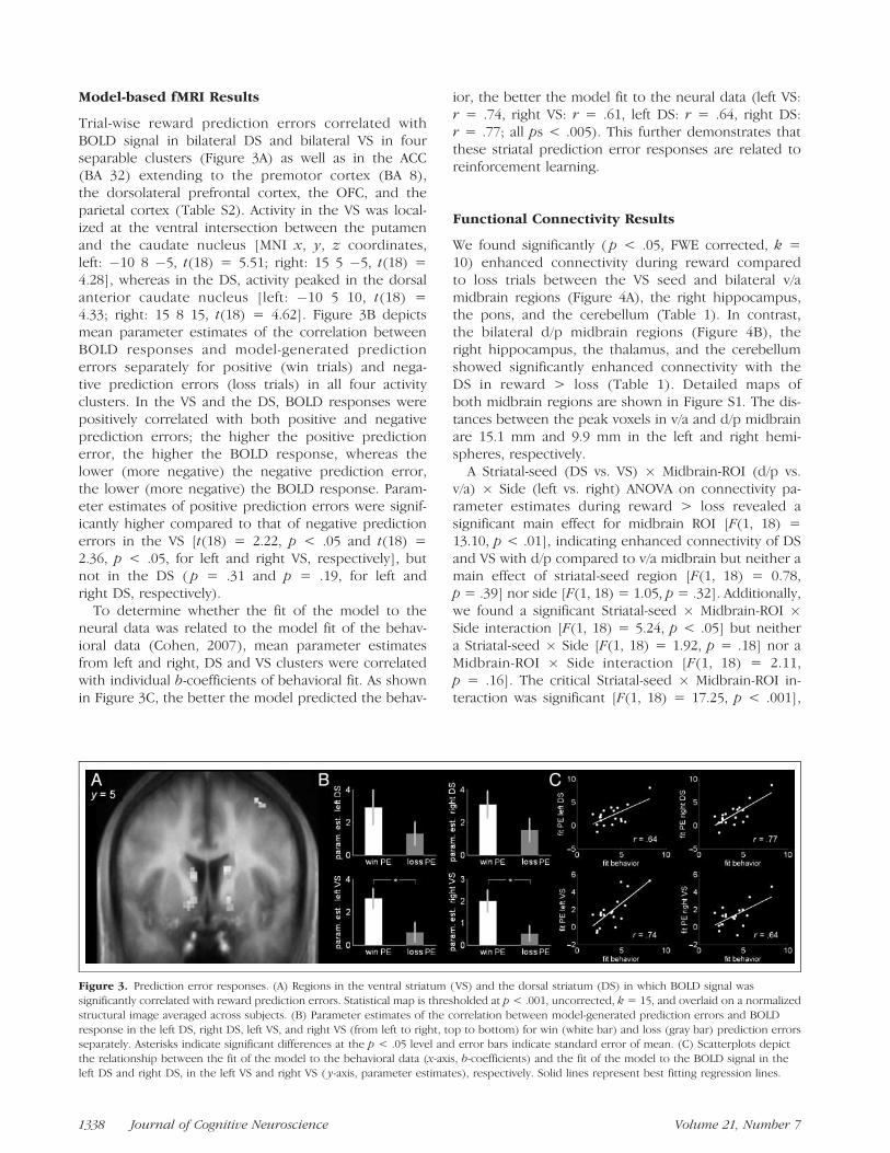
Trial-wise reward prediction errors correlated withBOLD signal in bilateral DS and bilateral VS in fourseparable clusters (Figure 3A) as well as in the ACC(BA 32) extending to the premotor cortex (BA 8),the dorsolateral prefrontal cortex, the OFC, and theparietal cortex (Table S2). Activity in the VS was local-ized at the ventral intersection between the putamenand the caudate nucleus [MNI x, y, z coordinates,left: �10 8 �5, t(18) = 5.51; right: 15 5 �5, t(18) =4.28], whereas in the DS, activity peaked in the dorsalanterior caudate nucleus [left: �10 5 10, t(18) =4.33; right: 15 8 15, t(18) = 4.62]. Figure 3B depictsmean parameter estimates of the correlation betweenBOLD responses and model-generated predictionerrors separately for positive (win trials) and nega-tive prediction errors (loss trials) in all four activityclusters. In the VS and the DS, BOLD responses werepositively correlated with both positive and negativeprediction errors; the higher the positive predictionerror, the higher the BOLD response, whereas thelower (more negative) the negative prediction error,the lower (more negative) the BOLD response. Param-eter estimates of positive prediction errors were signif-icantly higher compared to that of negative predictionerrors in the VS [t(18) = 2.22, p < .05 and t(18) =2.36, p < .05, for left and right VS, respectively], butnot in the DS ( p = .31 and p = .19, for left andright DS, respectively).
To determine whether the fit of the model to theneural data was related to the model fit of the behav-ioral data (Cohen, 2007), mean parameter estimatesfrom left and right, DS and VS clusters were correlatedwith individual b-coefficients of behavioral fit. As shownin Figure 3C, the better the model predicted the behav-
ior, the better the model fit to the neural data (left VS:r = .74, right VS: r = .61, left DS: r = .64, right DS:r = .77; all ps < .005). This further demonstrates thatthese striatal prediction error responses are related toreinforcement learning.
Functional Connectivity Results
We found significantly ( p < .05, FWE corrected, k =10) enhanced connectivity during reward comparedto loss trials between the VS seed and bilateral v/amidbrain regions (Figure 4A), the right hippocampus,the pons, and the cerebellum (Table 1). In contrast,the bilateral d/p midbrain regions (Figure 4B), theright hippocampus, the thalamus, and the cerebellumshowed significantly enhanced connectivity with theDS in reward > loss (Table 1). Detailed maps ofboth midbrain regions are shown in Figure S1. The dis-tances between the peak voxels in v/a and d/p midbrainare 15.1 mm and 9.9 mm in the left and right hemi-spheres, respectively.
A Striatal-seed (DS vs. VS) � Midbrain-ROI (d/p vs.v/a) � Side (left vs. right) ANOVA on connectivity pa-rameter estimates during reward > loss revealed asignificant main effect for midbrain ROI [F(1, 18) =13.10, p < .01], indicating enhanced connectivity of DSand VS with d/p compared to v/a midbrain but neither amain effect of striatal-seed region [F(1, 18) = 0.78,p = .39] nor side [F(1, 18) = 1.05, p = .32]. Additionally,we found a significant Striatal-seed � Midbrain-ROI �Side interaction [F(1, 18) = 5.24, p < .05] but neithera Striatal-seed � Side [F(1, 18) = 1.92, p = .18] nor aMidbrain-ROI � Side interaction [F(1, 18) = 2.11,p = .16]. The critical Striatal-seed � Midbrain-ROI in-teraction was significant [F(1, 18) = 17.25, p < .001],
Figure 3. Prediction error responses. (A) Regions in the ventral striatum (VS) and the dorsal striatum (DS) in which BOLD signal was
significantly correlated with reward prediction errors. Statistical map is thresholded at p < .001, uncorrected, k = 15, and overlaid on a normalized
structural image averaged across subjects. (B) Parameter estimates of the correlation between model-generated prediction errors and BOLD
response in the left DS, right DS, left VS, and right VS (from left to right, top to bottom) for win (white bar) and loss (gray bar) prediction errorsseparately. Asterisks indicate significant differences at the p < .05 level and error bars indicate standard error of mean. (C) Scatterplots depict
the relationship between the fit of the model to the behavioral data (x-axis, b-coefficients) and the fit of the model to the BOLD signal in the
left DS and right DS, in the left VS and right VS ( y-axis, parameter estimates), respectively. Solid lines represent best fitting regression lines.
1338 Journal of Cognitive Neuroscience Volume 21, Number 7

indicating that the DS and the VS are differentiallyfunctionally connected to different midbrain regions.Specifically, as shown in Figure 4C, whereas theVS was connected to both d/p and v/a midbrain re-gions, the DS was significantly more strongly function-ally connected to the d/p compared to v/a midbrain[t(18) = 5.77, p < .001] and the VS was significant-ly more strongly functionally connected to the v/amidbrain than the DS [t(18) = 2.49, p < .05]. ThisStriatal-seed � ROI interaction was significant in boththe left- and right-hemisphere midbrain as indicated bytwo separate Striatal-seed � Midbrain-ROI repeated
measures ANOVAs [F(1, 18) = 18.91, p < .001 andF(1, 18) = 7.91, p < .05, for left and right midbrainROIs, respectively].
According to findings regarding the specific role ofDS in reinforcement learning (Williams & Eskandar,2006) and its connection to the SN (Haber, 2003), wehypothesized that the effect of different reinforcementson future behavior depends on the integrity of DS–d/pmidbrain connections. Specifically, we hypothesized thata(win) should positively correlate with DS–d/p midbrainconnectivity during reward and a(loss) should corre-late positively with DS–d/p midbrain connectivity during
Figure 4. Functional
connectivity with the ventral
striatum (VS) and the dorsal
striatum (DS). Midbrainregions that show enhanced
functional connectivity with
(A) the VS and (B) the DSduring reward compared to
losses. Statistical maps are
thresholded at p < .05, FWE
corrected for multiplecomparisons (whole brain),
k = 10, and overlaid on a
normalized structural image
averaged across subjects. (C)Interaction plots of functional
connectivity estimates
(reward > loss) between theDS and VS seeds and the
dorsal/posterior (d/p) and
ventral/anterior (v/a) midbrain
ROIs. Left: Connectivityestimates of DS (gray line) and
VS (white line) seed plotted as
a function of midbrain ROI
(d/p vs. v/a midbrain). Right:Connectivity estimates in d/p
(gray line) and v/a midbrain
(white line) ROI plotted as afunction of striatal seed (DS vs.
VS). Error bars indicate
standard error of mean.
Asterisks indicate significantdifferences at the *p < .05
and **p < .001 level. (D)
Scatterplots depict the
significant relationshipbetween a(win) (left) and
a(loss) (right) on the one hand
(x-axes) and functional DS–left
d/p midbrain connectivity inwin (left panel) and loss trials
(right panel) on the other
hand ( y-axes). Solid linesrepresent best fitting
regression lines.
Kahnt et al. 1339

loss outcomes but not vice versa. The results confirmedthis hypothesis: In the left hemisphere, a(win) wassignificantly positively correlated with DS–d/p connec-tivity during reward outcomes (r = .40, p < .05, one-tailed; Figure 4D, left) and a(loss) was significantlypositively correlated with DS–d/p connectivity duringloss outcomes (r = .57, p < .05, one-tailed; Figure 4D,right). Critically, the opposite correlations [i.e., a(win)with DS–d/p during loss and a(loss) with DS–d/p dur-ing win] as well as the correlations between learningrates and VS–v/a midbrain connectivity during rewardand loss outcomes were not significant (all ps > .15). In
the right hemisphere, a(loss) was significantly positivelycorrelated with DS–d/p during loss outcomes (r = .39,p < .05, one-tailed), but a(win) was not significantlypositively correlated with DS–d/p connectivity duringreward outcomes ( p = .44, one-tailed). Again, the oppo-site correlations (all ps > .17) as well as the correlationsbetween learning rates and VS–v/a midbrain connectivityduring reward and loss outcomes were not significant(all ps > .08).
Furthermore, we tested for the same pattern ofcorrelations using the behavioral variables p(loss/switch)and learning speed instead of a(loss) and a(win),respectively. The strength of functional connectivitybetween DS and d/p midbrain during loss outcomes wassignificantly positively correlated with p(loss/switch) inboth hemispheres (r = .67, p < .05 and r = .63, p <.05, one-tailed for left and right hemispheres, respec-tively). Accordingly, there was a trend toward a negativecorrelation between the strength of functional DS–d/pmidbrain connectivity during win and learning speed(number of trials to reach learning criteria) in bothhemispheres (r = �.38, p = .06 and r = �.32, p = .09,one-tailed for left and right hemispheres, respectively).
Because the task used in this study was a spatiallearning task and it has been suggested that striatal–hippocampus connections are essential for space-relatedreward learning (Izquierdo et al., 2006; Rossato et al.,2006), we also searched for similar patterns of correla-tions in functional striatal–hippocampus connectivity.However, we did not find any significant correlationbetween striatal–hippocampus connectivity and learn-ing rates in either direction (all ps > .10). Thus, the ob-served pattern of correlations seems to be specific toDS–d/p midbrain connectivity.
DISCUSSION
In this study, we examined functional connectivity be-tween striatal and midbrain subregions and the neuralbasis of how positive and negative reinforcements areused to guide decision making. We found that differ-ent striatal regions, specifically its dorsal and ventralcompartments, in which activity correlated with rewardprediction errors, are differentially connected to mid-brain subregions, particularly with regard to its d/p andv/a compartment. Notably, whereas the VS was function-ally connected to both the v/a and d/p midbrain, the DSwas significantly more strongly functionally connectedto the d/p than the v/a midbrain. We used a computa-tional reinforcement learning model to generate individ-ual estimates of how subjects use rewards and lossesseparately to guide their behavior in a two-arm bandittask. Individual differences in functional connectivitybetween the DS and d/p midbrain predicted the impactof positive and negative outcomes on future decisionmaking. Our results suggest a specific role of striatal–
Table 1. Functional Connectivity Results
MNI
Region NameBrodmann’s
Area (BA) x y z t
DS Connectivity Reward > Loss, FWE Corrected, p < .05,k = 10
L dorsal/posteriormidbrain
�10 �28 �13 8.63
R dorsal/posteriormidbrain
15 �23 �15 11.79
R hippocampus 25 �20 �18 7.54
L parahippocampalgyrus
34 �18 5 �20 8.48
L superior temporalgyrus
22 �53 �13 3 7.92
L insula 13 �43 �23 0 8.09
L thalamus �15 �5 15 10.30
L caudate �10 5 10 9.29
R caudate 13 5 10 8.69
L cerebellum �28 �38 �35 8.69
VS Connectivity Reward > Loss, FWE Corrected, p < .05,k = 10
L ventral/anteriormidbrain
�13 �15 �20 9.04
R ventral/anteriormidbrain
8 �18 �20 7.57
R dorsal/posteriormidbrain
13 �23 �15 8.01
R hippocampus 28 �20 �20 8.14
L pons �5 �30 �28 10.47
L ventral striatum �18 0 �10 7.17
R ventral striatum 15 3 �8 7.94
L cerebellum �10 �45 �28 8.61
R cerebellum 10 �45 �28 8.89
1340 Journal of Cognitive Neuroscience Volume 21, Number 7

midbrain connections in updating action values in thestriatum, and thus, provide novel insights into the neuralmechanisms underlying reinforcement-guided decisionmaking.
We found activity in the mPFC, in the amygdala/hippocampus, and in the posterior cingulate cortex re-lated to positive outcomes, which have been shown tocode the appetitive value of both primary and secondaryreinforcements (Gottfried & Dolan, 2004; Knutson,Fong, Bennett, Adams, & Hommer, 2003). Activity inthe ACC, in the insula, and in the lateral OFC was re-lated to negative reinforcement. It has been shownthat these regions represent the value of aversive andpunishing stimuli (Seymour et al., 2004; O’Doherty,Kringelbach, Rolls, Hornak, & Andrews, 2001). Activityrelated to behavioral switch after negative outcomeswas observed in the lateral OFC and in the ACC.These results are in line with previous studies whichhave investigated BOLD responses to behavioral adjust-ment (Cohen et al., 2008; Wrase et al., 2007; Hampton,Bossaerts, & O’Doherty, 2006; O’Doherty, Critchley,Deichmann, & Dolan, 2003; Cools, Clark, Owen, &Robbins, 2002). In general, these findings replicate theresults of previous studies on reinforcement process-ing, and thus, set the stage for the further investiga-tion of the mechanisms of how reinforcements are usedto guide decision making.
Intuitively, in situations with only two different op-tions, it may seem difficult to disentangle whether anindividual learns to choose the advantageous optionby either learning that one option leads to positivefeedback or that the other leads to negative feedback.Of course, both processes contribute to learning butthere are individual differences in which process dom-inates (Cohen & Ranganath, 2005; Frank et al., 2004,2005). Here, using a computational model of reinforce-ment learning, we were able to disentangle both pro-cesses by estimating learning rates for positive andnegative outcomes that reflect the degree of learningfrom either reinforcement separately (i.e., their effecton subsequent behavior). We found a double dissocia-tion such that either learning rate predicted a differentaspect of subjects’ behavior. Specifically, a(win) pre-dicted the ability to maintain the rewarded action evenin the face of probabilistic losses (learning speed),whereas a(loss) predicted the individual tendency toavoid the unrewarded option [ p(loss/switch)] but notvice versa. This confirms the validity of these learningrates as shown in previous studies (Cohen & Ranganath,2005).
Trial-by-trial prediction errors generated by the re-inforcement learning model using individual learningrates correlated with activity in separable clusters in thedorsal and ventral striatum. Prediction error responsesin the VS have been shown previously in classical aswell as operant conditioning tasks (Cohen, 2007; Kim,Shimojo, & O’Doherty, 2006; Pessiglione et al., 2006;
O’Doherty et al., 2004; McClure, Berns, & Montague, 2003;O’Doherty, Dayan, et al., 2003; Pagnoni, Zink, Montague,& Berns, 2002). These responses, measured with fMRI,are dopamine-modulated (Pessiglione et al., 2006), indi-cating that they indeed reflect the reward-related firingof dopaminergic midbrain neurons known from elec-trophysiologic investigations that are thought to guidelearning and goal-directed behavior (Reynolds et al.,2001; Schultz & Dickinson, 2000; Hollerman & Schultz,1998). Prediction error responses in the DS have beenshown exclusively in learning tasks in which reward de-livery was dependent on operant responses (Schonberget al., 2007; Haruno & Kawato, 2006; Haruno et al., 2004;O’Doherty et al., 2004; Tricomi, Delgado, & Fiez, 2004).Schonberg et al. (2007) showed that prediction errorresponses in the DS but not in the VS distinguishedlearners from nonlearners and correlated with the in-dividual degree of learning. Such results confirm find-ings from animal research, which have raised the notionof an actor–critic model of the basal ganglia, in whichthe VS is involved in reward prediction and the DSuses this information to bias action selection in favorof the advantageous option (Atallah et al., 2007; Williams& Eskandar, 2006; Samejima et al., 2005; O’Doherty et al.,2004; Ito, Dalley, Robbins, & Everitt, 2002; Joel et al.,2002).
Recently, Williams and Eskandar (2006) have shownthat activity in the DS in rhesus monkeys correlates withinstrumental learning and that microstimulation of thedorsal anterior caudate nucleus during the reinforce-ment period enhanced the learning of cue–action asso-ciations, and thus, strengthened action values. The moreinput the DS gets during reinforcement, the morereinforcement information can be used to bias futureactions. Atallah et al. (2007) provided evidence that inrats, whereas the VS is critical for learning, the DS isimportant for selecting the appropriate option. Accord-ing to them, there are two ways the VS might directthe DS. First, the VS could modulate activity in theOFC, which maintains action–reward contingenciesthat, in turn, exerts top–down control over the DS(Frank & Claus, 2006; Joel & Weiner, 1994). Here, thecritical pathway that determines how reinforcements areused by the DS to guide future action would be theOFC–DS connection. Alternatively, the VS might providereinforcement information to the DS by exerting mod-ulatory control over dopaminergic projections from theSN to the DS. The VS projects to the SN, which in turnprojects to the DS (Haber, 2003; Joel & Weiner, 2000).Thus, the critical connection would be the SN–DSpathway. Supporting the latter mechanism, Belin andEveritt (2008) provided evidence that the VS exertscontrol over dorsal striatal processes via dopaminergicmidbrain connections.
Our results confirm these findings: To examine striatal–midbrain connections, we used the striatal regions iden-tified in the prediction error analysis as seed regions in
Kahnt et al. 1341

a functional connectivity analysis. We found significantfunctional connectivity of the DS with d/p midbrainregions, whereas the VS was functionally connected tothe v/a and d/p midbrain. These findings are consistentwith animal studies showing that prediction error re-sponses in the striatum are generated in the midbrain(Schultz & Dickinson, 2000; Hollerman & Schultz, 1998).Furthermore, we found a significant Striatal-seed �Midbrain-ROI interaction, indicating that the VS wasconnected to both midbrain regions, whereas the DSwas connected to the d/p midbrain solely. Neurophysi-ologic investigations in primates have, indeed, shownthat the DS and the VS are differently connected tothe midbrain (Haber, 2003; Haber et al., 2000). Specif-ically, the VS is reciprocally connected to the VTAbut also sends input to the SN, which in turn is recip-rocally connected to the DS (Haber et al., 2000; Lynd-Balta & Haber, 1994b).
The anatomical subdivisions in the midbrain are re-latively small compared to the size of an MRI voxelused in our study so that it is difficult to localize pre-cisely the VTA and the SN. However, the pattern offindings in our study is consistent with BOLD genera-tors in the VTA and the SN. First, the anatomical local-ization of our v/a and d/p midbrain activation is similarto the anatomical localization of the VTA and the SN,respectively in primates (Haber et al., 2000). Second,the pattern of functional connectivity to the DS and theVS itself is consistent with the pattern of anatomicalconnections found in animal studies (Haber et al.,2000; Lynd-Balta & Haber, 1994a, 1994b). Other studieswere also able to investigate distinct midbrain regions,specifically the VTA, using fMRI (D’Ardenne et al.,2008; Wittmann et al., 2005, 2008; Wittmann, Bunzeck,Dolan, & Duzel, 2007; Adcock et al., 2006; Bunzeck& Duzel, 2006; Menon & Levitin, 2005). Additionally,one fMRI study identified functional connectivity be-tween the midbrain and the striatum during learning(Aron et al., 2004). Furthermore, a recent study showeda correlation between reward-related BOLD responsesin the VS and the VTA using high-resolution imaging(D’Ardenne et al., 2008). Therefore, it seems plausibleto assume that the v/a and d/p midbrain identified inour study correspond to the VTA and the SN, respective-ly. Nevertheless, it might be interesting to replicate thispattern of functional connectivity using high-resolutionfMRI.
The degree of functional connectivity between the DSand the d/p midbrain (presumably the SN) during re-ward and loss feedback correlated with individual learn-ing rates, a(win) and a(loss), respectively. This was notthe case for the functional connectivity between the VSand the v/a midbrain (presumably the VTA). The stron-ger the functional connectivity between the DS and theSN during a certain feedback type, the more that feed-back had an effect on future actions; DS–SN connectiv-ity predicted how subjects used reinforcements to
guide future decisions. To our knowledge, this is thefirst human study that supports evidence in animalresearch indicating that inputs from the SN drive instru-mental learning and action selection in the DS (Belin &Everitt, 2008; Williams & Eskandar, 2006; Faure et al.,2005; Joel & Weiner, 2000). Given this mechanism, ourresults imply that the more input the DS gets duringreinforcement, the more this reinforcement biases ac-tion selection. This interpretation is consistent withresults from primate studies (Williams & Eskandar,2006). In our data, this pattern was selective to striatal–midbrain connectivity and was not present in the ob-served striatal–hippocampus connectivity. Connectionsbetween the striatum and the hippocampus, as observedin our study, have been implicated in space-relatedreward learning (Izquierdo et al., 2006; Rossato et al.,2006; Goto & Grace, 2005), but our data suggest thattheir integrity do not determine the degree of learn-ing from different feedback types. Previous studieshave shown that the degree of learning from either rein-forcement depends on various dopaminergic condi-tions, such as dopamine genetics, dopaminergic drugs,and diseases that target dopaminergic transmission spe-cifically in the SN (Cohen et al., 2007; Frank, Moustafa,et al., 2007; Frank, Scheres, et al., 2007; Klein et al., 2007;Frank, 2005). An interesting hypothesis regarding theimpact of different reinforcements on learning is thatthis should also be revealed in a task that incorporateswin versus nonwin and loss versus nonloss outcomes.In this case, a nonloss in the context of a possible lossmight be interpreted as a reward (Kim et al., 2006), andthus, the reward learning rate should also correlatewith the strength of DS–SN connectivity during nonlossfeedback.
A limitation of our experimental design is that it doesnot allow the distinction between stimulus and actionvalues. Therefore, it is possible that action and/or stim-ulus values are updated via SN–DS pathways. However,it is clear that the learning rates used here ref lectmechanisms of updating behavior according to rein-forcements. Future studies could use tasks that investi-gate the updating of stimulus and action representationsseparately. Another limitation is that the correlationbetween functional connectivity estimates and learningrates was modest and should be replicated in futurestudies. However, these correlations are orthogonal tothe definition of the midbrain ROIs that were used. Toour knowledge, this is the first investigation into therelation between functional connectivity and model-estimated behavioral performance. Furthermore, weacknowledge that it is not possible to interpret thedirection of information flow or the underlying neuro-transmitters using functional connectivity measure-ments. However, our results are in line with thenotion that the SN sends information about reinforce-ments via dopaminergic projections to the DS that usesthis information to bias action selection.
1342 Journal of Cognitive Neuroscience Volume 21, Number 7

In conclusion, here we found reward predictionerror responses in the VS and the DS during a dynamicreward-based decision-making task. The DS and the VSwere differentially connected to different midbrain re-gions, the SN and the VTA, respectively. Furthermore,we have shown that the way different reinforcementsare used to guide future decisions critically depends onthe integrity of DS–SN connectivity. Therefore, our re-sults support the hypothesis that the VTA sends a teach-ing signal in form of a reward prediction error to theVS that is used to predict rewards. The VS, on theother hand, projects this signal back to the SN, whichin turn projects it to the DS in order to bias actionselection in favor of the advantageous option (Belin &Everitt, 2008; Haber et al., 2000; Joel & Weiner, 2000).Our results further imply that the latter pathway iscrucial for the way different reinforcements influencefuture decisions, but not the preceding pathways. Thus,VS–VTA connections might be important for reward pre-dictions, whereas DS–SN connections contribute specif-ically to the use of reinforcements to guide actions.These findings might also help to shed light into thelearning deficits observed in patients with Parkinson’sdisease (Shohamy et al., 2004; Myers et al., 2003; Knowlton,Mangels, & Squire, 1996), where dopaminergic neuronsin the SN are diminished (Ito et al., 1999; Owen et al.,1992).
Acknowledgments
This study was supported by the German Research Founda-tion (Deutsche Forschungsgemeinschaft; HE 2597/4-3) andby the Bernstein Center for Computational NeuroscienceBerlin (Bundesministerium fur Bildung und Forschung grant01GQ0411).
Reprint requests should be sent to Thorsten Kahnt, Departmentof Psychiatry and Psychotherapy, Charite—UniversitatsmedizinBerlin (Charite Campus Mitte), Chariteplatz 1, 10117 Berlin,Germany, or via e-mail: [email protected].
REFERENCES
Adcock, R. A., Thangavel, A., Whitfield-Gabrieli, S., Knutson, B.,& Gabrieli, J. D. (2006). Reward-motivated learning:Mesolimbic activation precedes memory formation. Neuron,50, 507–517.
Alexander, G. E., Crutcher, M. D., & DeLong, M. R. (1990).Basal ganglia–thalamocortical circuits: Parallel substrates formotor, oculomotor, ‘‘prefrontal’’ and ‘‘limbic’’ functions.Progress in Brain Research, 85, 119–146.
Aron, A. R., Shohamy, D., Clark, J., Myers, C., Gluck, M. A., &Poldrack, R. A. (2004). Human midbrain sensitivity tocognitive feedback and uncertainty during classificationlearning. Journal of Neurophysiology, 92, 1144–1152.
Atallah, H. E., Lopez-Paniagua, D., Rudy, J. W., & O’Reilly, R. C.(2007). Separate neural substrates for skill learning andperformance in the ventral and dorsal striatum. NatureNeuroscience, 10, 126–131.
Belin, D., & Everitt, B. J. (2008). Cocaine seeking habitsdepend upon dopamine-dependent serial connectivity
linking the ventral with the dorsal striatum. Neuron, 57,432–441.
Buchel, C., Holmes, A. P., Rees, G., & Friston, K. J. (1998).Characterizing stimulus–response functions using nonlinearregressors in parametric fMRI experiments. Neuroimage,8, 140–148.
Bunzeck, N., & Duzel, E. (2006). Absolute coding of stimulusnovelty in the human substantia Nigra/VTA. Neuron, 51,369–379.
Cohen, M. X (2007). Individual differences and the neuralrepresentations of reward expectation and rewardprediction error. Social Cognitive and AffectiveNeuroscience, 2, 20–30.
Cohen, M. X, Elger, C. E., & Weber, B. (2008). Amygdalatractography predicts functional connectivity and learningduring feedback-guided decision making. Neuroimage, 39,1396–1407.
Cohen, M. X, Heller, A. S., & Ranganath, C. (2005). Functionalconnectivity with anterior cingulate and orbitofrontalcortices during decision-making. Brain Research, CognitiveBrain Research, 23, 61–70.
Cohen, M. X, Krohn-Grimberghe, A., Elger, C. E., & Weber, B.(2007). Dopamine gene predicts the brain’s response todopaminergic drug. European Journal of Neuroscience, 26,3652–3660.
Cohen, M. X, & Ranganath, C. (2005). Behavioral and neuralpredictors of upcoming decisions. Cognitive, Affective &Behavioral Neuroscience, 5, 117–126.
Cohen, M. X, & Ranganath, C. (2007). Reinforcement learningsignals predict future decisions. Journal of Neuroscience,27, 371–378.
Cools, R., Clark, L., Owen, A. M., & Robbins, T. W. (2002).Defining the neural mechanisms of probabilistic reversallearning using event-related functional magnetic resonanceimaging. Journal of Neuroscience, 22, 4563–4567.
D’Ardenne, K., McClure, S. M., Nystrom, L. E., & Cohen, J. D.(2008). BOLD responses reflecting dopaminergic signals inthe human ventral tegmental area. Science, 319, 1264–1267.
Delgado, M. R. (2007). Reward-related responses in the humanstriatum. Annals of the New York Academy of Sciences,1104, 70–88.
Duvernoy, H. (1999). The human brain. Surface, bloodsupply, and three-dimensional sectional anatomy(2nd ed.). Vienna: Springer-Verlag.
Egelman, D. M., Person, C., & Montague, P. R. (1998). Acomputational role for dopamine delivery in humandecision-making. Journal of Cognitive Neuroscience, 10,623–630.
Faure, A., Haberland, U., Conde, F., & El Massioui, N.(2005). Lesion to the nigrostriatal dopamine systemdisrupts stimulus–response habit formation. Journal ofNeuroscience, 25, 2771–2780.
Frank, M. J. (2005). Dynamic dopamine modulation in thebasal ganglia: A neurocomputational account of cognitivedeficits in medicated and nonmedicated Parkinsonism.Journal of Cognitive Neuroscience, 17, 51–72.
Frank, M. J., & Claus, E. D. (2006). Anatomy of a decision:Striato-orbitofrontal interactions in reinforcement learning,decision making, and reversal. Psychological Review, 113,300–326.
Frank, M. J., Moustafa, A. A., Haughey, H. M., Curran, T., &Hutchison, K. E. (2007). Genetic triple dissociation revealsmultiple roles for dopamine in reinforcement learning.Proceedings of the National Academy of Sciences, U.S.A.,104, 16311–16316.
Frank, M. J., Scheres, A., & Sherman, S. J. (2007).Understanding decision-making deficits in neurologicalconditions: Insights from models of natural action selection.
Kahnt et al. 1343

Philosophical Transactions of the Royal Society of London,Series B, Biological Sciences, 362, 1641–1654.
Frank, M. J., Seeberger, L. C., & O’Reilly, R. C. (2004). Bycarrot or by stick: Cognitive reinforcement learning inparkinsonism. Science, 306, 1940–1943.
Frank, M. J., Woroch, B. S., & Curran, T. (2005). Error-relatednegativity predicts reinforcement learning and conflictbiases. Neuron, 47, 495–501.
Friston, K. J., Buechel, C., Fink, G. R., Morris, J., Rolls, E., &Dolan, R. J. (1997). Psychophysiological and modulatoryinteractions in neuroimaging. Neuroimage, 6, 218–229.
Goto, Y., & Grace, A. A. (2005). Dopaminergic modulationof limbic and cortical drive of nucleus accumbens ingoal-directed behavior. Nature Neuroscience, 8, 805–812.
Gottfried, J. A., & Dolan, R. J. (2004). Human orbitofrontalcortex mediates extinction learning while accessingconditioned representations of value. Nature Neuroscience,7, 1145–1153.
Haber, S. N. (2003). The primate basal ganglia: Parallel andintegrative networks. Journal of Chemical Neuroanatomy,26, 317–330.
Haber, S. N., Fudge, J. L., & McFarland, N. R. (2000).Striatonigrostriatal pathways in primates form an ascendingspiral from the shell to the dorsolateral striatum. Journalof Neuroscience, 20, 2369–2382.
Hampton, A. N., Bossaerts, P., & O’Doherty, J. P. (2006).The role of the ventromedial prefrontal cortex in abstractstate-based inference during decision making in humans.Journal of Neuroscience, 26, 8360–8367.
Haruno, M., & Kawato, M. (2006). Different neuralcorrelates of reward expectation and reward expectationerror in the putamen and caudate nucleus duringstimulus–action–reward association learning. Journal ofNeurophysiology, 95, 948–959.
Haruno, M., Kuroda, T., Doya, K., Toyama, K., Kimura, M.,Samejima, K., et al. (2004). A neural correlate ofreward-based behavioral learning in caudate nucleus: Afunctional magnetic resonance imaging study of a stochasticdecision task. Journal of Neuroscience, 24, 1660–1665.
Hollerman, J. R., & Schultz, W. (1998). Dopamine neuronsreport an error in the temporal prediction of reward duringlearning. Nature Neuroscience, 1, 304–309.
Holroyd, C. B., & Coles, M. G. (2002). The neural basis ofhuman error processing: Reinforcement learning, dopamine,and the error-related negativity. Psychological Review,109, 679–709.
Ikemoto, S. (2007). Dopamine reward circuitry: Twoprojection systems from the ventral midbrain to the nucleusaccumbens–olfactory tubercle complex. Brain ResearchReviews, 56, 27–78.
Ito, K., Morrish, P. K., Rakshi, J. S., Uema, T., Ashburner, J.,Bailey, D. L., et al. (1999). Statistical parametric mappingwith 18F-dopa PET shows bilaterally reduced striataland nigral dopaminergic function in early Parkinson’sdisease. Journal of Neurology, Neurosurgery andPsychiatry, 66, 754–758.
Ito, R., Dalley, J., Robbins, T., & Everitt, B. (2002).Dopamine release in the dorsal striatum during cocaineseeking behavior under the control of a drug-associated cue.Journal of Neuroscience, 22, 6247–6253.
Izquierdo, I., Bevilaqua, L. R., Rossato, J. I., Bonini, J. S.,Da Silva, W. C., Medina, J. H., et al. (2006). The connectionbetween the hippocampal and the striatal memory systemsof the brain: A review of recent findings. NeurotoxicityResearch, 10, 113–121.
Joel, D., Niv, Y., & Ruppin, E. (2002). Actor–critic models ofthe basal ganglia: New anatomical and computationalperspectives. Neural Networks, 15, 535–547.
Joel, D., & Weiner, I. (1994). The organization of the basalganglia–thalamocortical circuits: Open interconnectedrather than closed segregated. Neuroscience, 63, 363–379.
Joel, D., & Weiner, I. (2000). The connections of thedopaminergic system with the striatum in rats andprimates: An analysis with respect to the functional andcompartmental organization of the striatum. Neuroscience,96, 451–474.
Kim, H., Shimojo, S., & O’Doherty, J. P. (2006). Is avoidingan aversive outcome rewarding? Neural substrates ofavoidance learning in the human brain. PLoS Biology,4, e233.
Klein, T. A., Neumann, J., Reuter, M., Hennig, J., vonCramon, D. Y., & Ullsperger, M. (2007). Geneticallydetermined differences in learning from errors. Science,318, 1642–1645.
Knowlton, B. J., Mangels, J. A., & Squire, L. R. (1996). Aneostriatal habit learning system in humans. Science, 273,1399–1402.
Knutson, B., Fong, G. W., Bennett, S. M., Adams, C. M., &Hommer, D. (2003). A region of mesial prefrontalcortex tracks monetarily rewarding outcomes:Characterization with rapid event-related fMRI.Neuroimage, 18, 263–272.
Lehericy, S., Ducros, M., Van de Moortele, P. F., Francois, C.,Thivard, L., Poupon, C., et al. (2004). Diffusion tensor fibertracking shows distinct corticostriatal circuits in humans.Annals of Neurology, 55, 522–529.
Lynd-Balta, E., & Haber, S. N. (1994a). Primate striatonigralprojections: A comparison of the sensorimotor-relatedstriatum and the ventral striatum. Journal of ComparativeNeurology, 345, 562–578.
Lynd-Balta, E., & Haber, S. N. (1994b). The organization ofmidbrain projections to the striatum in the primate:Sensorimotor-related striatum versus ventral striatum.Neuroscience, 59, 625–640.
McClure, S. M., Berns, G. S., & Montague, P. R. (2003).Temporal prediction errors in a passive learning task activatehuman striatum. Neuron, 38, 339–346.
Menon, V., & Levitin, D. J. (2005). The rewards of musiclistening: Response and physiological connectivity of themesolimbic system. Neuroimage, 28, 175–184.
Montague, P. R., Hyman, S. E., & Cohen, J. D. (2004).Computational roles for dopamine in behavioural control.Nature, 431, 760–767.
Myers, C. E., Shohamy, D., Gluck, M. A., Grossman, S.,Kluger, A., Ferris, S., et al. (2003). Dissociating hippocampalversus basal ganglia contributions to learning and transfer.Journal of Cognitive Neuroscience, 15, 185–193.
O’Doherty, J. P., Critchley, H., Deichmann, R., & Dolan, R. J.(2003). Dissociating valence of outcome from behavioralcontrol in human orbital and ventral prefrontal cortices.Journal of Neuroscience, 23, 7931–7939.
O’Doherty, J. P., Dayan, P., Friston, K., Critchley, H., & Dolan,R. J. (2003). Temporal difference models and reward-relatedlearning in the human brain. Neuron, 38, 329–337.
O’Doherty, J. P., Dayan, P., Schultz, J., Deichmann, R.,Friston, K., & Dolan, R. J. (2004). Dissociable roles of ventraland dorsal striatum in instrumental conditioning. Science,304, 452–454.
O’Doherty, J. P., Kringelbach, M. L., Rolls, E. T., Hornak, J.,& Andrews, C. (2001). Abstract reward and punishmentrepresentations in the human orbitofrontal cortex. NatureNeuroscience, 4, 95–102.
Owen, A. M., James, M., Leigh, P. N., Summers, B. A., Marsden,C. D., Quinn, N. P., et al. (1992). Fronto-striatal cognitivedeficits at different stages of Parkinson’s disease. Brain, 115,1727–1751.
1344 Journal of Cognitive Neuroscience Volume 21, Number 7

Pagnoni, G., Zink, C. F., Montague, P. R., & Berns, G. S.(2002). Activity in human ventral striatum locked to errorsof reward prediction. Nature Neuroscience, 5, 97–98.
Pessiglione, M., Seymour, B., Flandin, G., Dolan, R. J., &Frith, C. D. (2006). Dopamine-dependent prediction errorsunderpin reward-seeking behaviour in humans. Nature,442, 1042–1045.
Pessoa, L., Gutierrez, E., Bandettini, P., & Ungerleider, L.(2002). Neural correlates of visual working memory: fMRIamplitude predicts task performance. Neuron, 35, 975–987.
Reynolds, J. N., Hyland, B. I., & Wickens, J. R. (2001). Acellular mechanism of reward-related learning. Nature, 413,67–70.
Rossato, J. I., Zinn, C. G., Furini, C., Bevilaqua, L. R., Medina,J. H., Cammarota, M., et al. (2006). A link between thehippocampal and the striatal memory systems of the brain.Annals of the Brazilian Academy of Sciences, 78, 515–523.
Samejima, K., & Doya, K. (2007). Multiple representationsof belief states and action values in corticobasal ganglialoops. Annals of the New York Academy of Sciences, 1104,213–228.
Samejima, K., Ueda, Y., Doya, K., & Kimura, M. (2005).Representation of action-specific reward values in thestriatum. Science, 310, 1337–1340.
Schonberg, T., Daw, N. D., Joel, D., & O’Doherty, J. P. (2007).Reinforcement learning signals in the human striatumdistinguish learners from nonlearners during reward-baseddecision making. Journal of Neuroscience, 27, 12860–12867.
Schultz, W. (2002). Getting formal with dopamine and reward.Neuron, 36, 241–263.
Schultz, W. (2004). Neural coding of basic reward terms ofanimal learning theory, game theory, microeconomics andbehavioural ecology. Current Opinion in Neurobiology,14, 139–147.
Schultz, W., Dayan, P., & Montague, P. R. (1997). A neuralsubstrate of prediction and reward. Science, 275, 1593–1599.
Schultz, W., & Dickinson, A. (2000). Neuronal coding of
prediction errors. Annual Review of Neuroscience, 23,473–500.
Seymour, B., O’Doherty, J. P., Dayan, P., Koltzenburg, M.,Jones, A. K., Dolan, R. J., et al. (2004). Temporal differencemodels describe higher-order learning in humans. Nature,429, 664–667.
Shohamy, D., Myers, C. E., Grossman, S., Sage, J., Gluck,M. A., & Poldrack, R. A. (2004). Cortico-striatal contributionsto feedback-based learning: Converging data fromneuroimaging and neuropsychology. Brain, 127, 851–859.
Sutton, R., & Barto, A. (1998). Reinforcement learning: Anintroduction. Cambridge, MA: MIT Press.
Talairach, J., & Tournoux, P. (1988). Co-planar stereotaxicatlas of the human brain. New York: Thieme.
Tricomi, E. M., Delgado, M. R., & Fiez, J. A. (2004). Modulationof caudate activity by action contingency. Neuron, 41,281–292.
Williams, Z. M., & Eskandar, E. N. (2006). Selectiveenhancement of associative learning by microstimulation ofthe anterior caudate. Nature Neuroscience, 9, 562–568.
Wittmann, B. C., Bunzeck, N., Dolan, R. J., & Duzel, E.(2007). Anticipation of novelty recruits reward system andhippocampus while promoting recollection. Neuroimage,38, 194–202.
Wittmann, B. C., Schiltz, K., Boehler, C. N., & Duzel, E.(2008). Mesolimbic interaction of emotional valence andreward improves memory formation. Neuropsychologia,46, 1000–1008.
Wittmann, B. C., Schott, B. H., Guderian, S., Frey, J. U.,Heinze, H. J., & Duzel, E. (2005). Reward-relatedfMRI activation of dopaminergic midbrain is associatedwith enhanced hippocampus-dependent long-term memoryformation. Neuron, 45, 459–467.
Wrase, J., Kahnt, T., Schlagenhauf, F., Beck, A., Cohen,M. X., Knutson, B., et al. (2007). Different neural systemsadjust motor behavior in response to reward andpunishment. Neuroimage, 36, 1253–1262.
Kahnt et al. 1345
Copyright © 2022 FDOKUMEN