Loss of GABAergic cortical neurons underlies the neuropathology of Lafora disease
Upload
unilestemgCategory
view
1download
0
Review
Gabaergic regulation of the neural organization of fear
in the midbrain tectum
Marcus Lira Brandao*, Karina Genaro Borelli, Manoel Jorge Nobre, Julia Maria Santos,
Lucas Albrechet-Souza, Amanda Ribeiro Oliveira, Raquel Chacon Martinez
Laboratorio de Psicobiologia, FFCLRP, University of Sao Paulo, Campus USP, Av. Bandeirantes 3900, 14049-901 Ribeirao Preto, SP, Brazil
Abstract
In midbrain tectum (MT) structures, such as the dorsal periaqueductal gray (dPAG), the superior colliculus (SC) and the inferior colliculus
(IC) GABAergic neurons exert a tonic control on the neural substrates involved in the expression of defensive reactions. In this review, we
summarize behavioral, immunohistochemical (brain Fos distribution) and electrophysiological (auditory evoked potentials) data obtained
with the reduction of GABA transmission by local injections of a GABA receptor blocker (bicuculline, BIC) or a glutamic acid
decarboxylase inhibitor (semicarbazide, SMC) into the MT. Distinct patterns of Fos distribution were obtained following the freezing and
escape reactions induced by MT injections of SMC and BIC, respectively. While only the laterodorsal nucleus of the thalamus was labeled
after SMC-induced freezing, a widespread increase in Fos expression in the brain occurred after BIC-induced escape. Also, injections of
SMC into the IC increased the auditory evoked potentials recorded from this structure. It is suggested that GABAergic mechanisms of MT
are also called into play when sensory gating of the MT is activated during different emotional states.
q 2005 Elsevier Ltd. All rights reserved.
Keywords: Freezing; Escape; Semicarbazide; Bicuculline; GABA; dPAG; Amygdala; SNpr
Contents
1. Introduction . . . . . . . . . . . . . . . . . . . . . . . . . . . . . . . . . . . . . . . . . . . . . . . . . . . . . . . . . . . . . . . . . . . . . . . . . . . . . . . . . . 1299
2. Tonic inhibition of the neural substrates of defensive behavior by GABAergic neurons . . . . . . . . . . . . . . . . . . . . . . . . . . . 1300
3. Sensory gating of the midbrain tectum in the organization of fear . . . . . . . . . . . . . . . . . . . . . . . . . . . . . . . . . . . . . . . . . . . 1301
4. Regulation from substantia nigra pars reticulata . . . . . . . . . . . . . . . . . . . . . . . . . . . . . . . . . . . . . . . . . . . . . . . . . . . . . . . . 1305
5. Regulation by basolateral amygdala . . . . . . . . . . . . . . . . . . . . . . . . . . . . . . . . . . . . . . . . . . . . . . . . . . . . . . . . . . . . . . . . . 1305
6. Final comments . . . . . . . . . . . . . . . . . . . . . . . . . . . . . . . . . . . . . . . . . . . . . . . . . . . . . . . . . . . . . . . . . . . . . . . . . . . . . . . 1308
Acknowledgements . . . . . . . . . . . . . . . . . . . . . . . . . . . . . . . . . . . . . . . . . . . . . . . . . . . . . . . . . . . . . . . . . . . . . . . . . . . . . 1308
References . . . . . . . . . . . . . . . . . . . . . . . . . . . . . . . . . . . . . . . . . . . . . . . . . . . . . . . . . . . . . . . . . . . . . . . . . . . . . . . . . . . 1308
1. Introduction
Electrical stimulation of the midbrain tectum (MT)—
dorsal periaqueductal gray (dPAG) and deep layers of the
superior colliculus (SC)—in the rat elicits unconditioned
0149-7634/$ - see front matter q 2005 Elsevier Ltd. All rights reserved.
doi:10.1016/j.neubiorev.2005.04.013
* Corresponding author. Fax: C55 16 6331609.
E-mail address: [email protected] (M.L. Brandao).
‘fear-like’ behavioral responses, such as alertness, sideways
postures, arching of the back, freezing, fleeing locomotion
and escape leaps (Bittencourt et al., 2004; Brandao et al.,
1982; Graeff et al., 1986; Schenberg et al., 1983). The same
pattern of responses has also been observed with electrical
stimulation of the inferior colliculus (IC) (Brandao et al.,
1988). This defensive behavioral reaction is associated with
sensory changes and autonomic responses, such as increase
in mean blood pressure, heart rate, piloerection, exhophtal-
mus, micturition and defecation. In view of these findings,
the MT has been suggested to play a major role in the neural
Neuroscience and Biobehavioral Reviews 29 (2005) 1299–1311
www.elsevier.com/locate/neubiorev
M.L. Brandao et al. / Neuroscience and Biobehavioral Reviews 29 (2005) 1299–13111300
organization and control of defensive reactions towards
threatening stimuli (for reviews see Brandao et al., 1993,
1999, 2003).
The use of electrical stimulation of brain structures in
these studies frequently leads to problems in the
interpretation of results. Such stimulation frequently
activates fibers of passage, raising the possibility of
stimulation spreading to neighboring structures. In the
case of the IC stimulation, this might include the cuneiform
nucleus. To circumvent this problem researchers have also
examined the effects of microinjecting tiny volumes
(200 nl) of drugs into the MT. The use of this technique
in association with electrophysiological studies has
provided a large body of evidence for the modulatory
influences of an array of neurotransmitters such as GABA,
serotonin, neuropeptides, opioids and excitatory amino
acids (Bittencourt et al., 2004; Brandao et al., 2003; Graeff
et al., 1986). Among them, GABA has been one of the
most studied transmitters regarding its regulatory function
in the defense reaction integrated at the MT level.
In this paper, we review a series of studies aimed at
investigating the role of GABA in sensory gating processes
in the MT during different emotional states produced either
by electrical or local injections of GABA blockers into
the MT, with special emphasis on dPAG and IC. Possible
changes in sensory processes have been neglected as a
possible source of the defensive behavior reaction that is
elicitable via MT stimulation (Brandao et al., 2003; Huston
et al., 1990). However, the MT processes aversive sensory
inputs and transduces them into behavioral and vegetative
nervous system reactions. For example, the IC-induced
defensive behavior is accompanied by changes in auditory-
evoked potentials in this structure, indicative of a
modification of sensory input channels (Brandao et al.,
2001). Thus, the behavioral patterns of the defense reaction
elicited at the MT level are unlikely to be the result of a
localized output process. As in the case of hypothalamic
aggression (Bandler, 1982a,b; Bandler and Flynn, 1971,
1972; Flynn et al., 1971; MacDonne and Flynn, 1966) and
the central activation of the perioral biting reflex, they are
probably linked to changes in sensorimotor gating processes
(Huston et al., 1980; Welzl et al., 1984).
While it is well established that there are four columns in
the central gray, as identified anatomically, their functional
role is still a subject of great debate (Bandler and Carrive,
1988; Bandler and Shipley, 1994; Carrive, 1993). It seems
very likely, however, that different pools of neurons in the
midbrain central gray are responsible for the elaboration of
distinct aspects of the defensive behavior. Dorsolateral and
dorsomedial columns have been associated with freezing,
escape, hypertension, tachycardia, and serotonin-dependent
analgesia, the lateral column with attack and the
ventrolateral column with quiescence, fear conditioned
freezing, recuperative-like behaviors, hypotension, brady-
cardia and opioid-dependent analgesia (Brandao et al.,
2003; Bittencourt et al., 2004; Canteras and Goto, 1999;
De Oca et al., 1998; Walker and Carrive, 2003; Vianna and
Brandao, 2003).
2. Tonic inhibition of the neural substrates of defensive
behavior by GABAergic neurons
GABA exists in appreciable density in the MT, and
GABAergic inhibition controls the firing rate of neurons in
this region (LeBeau et al., 1996, 2001; Roberts and Ribak,
1987; Thompson et al., 1985). As aversive states are
produced by GABA-A blockers and inhibited by GABA-A
agonists locally injected into the dPAG and the IC, it has
been suggested that these structures contain a tonically
active GABAergic network that regulates these states
through GABA-A receptors (Audi and Graeff, 1984;
Behbehani et al., 1990; Brandao et al., 1982, 1986, 1988,
1999; Coimbra and Brandao, 1993; DiScala et al., 1983;
Sandner et al., 1981; Schenberg et al., 1983). Consistent
behavioral evidence has also been provided for an anti-
aversive action of benzodiazepines in the MT. Indeed, local
injections of benzodiazepines into the MT depress the
defensive behavior induced by stimulation of this region
(Audi and Graeff, 1984; Brandao et al., 1982; Melo et al.,
1992; Pandossio and Brandao, 1999).
In this review, we will concentrate on the distinct
sensorimotor effects following the injections of bicuculline
(BIC) and semicarbazide (SMC) into the MT. BIC is a post-
synaptic GABA receptor antagonist, while SMC causes a
reduction in GABA levels due to its inhibition of glutamic
acid decarboxylase (GAD), the enzyme responsible for the
GABA synthesis (Brandao et al., 1986; Killam and Bain,
1957). Injections of SMC or BIC into the MT produce
defensive behavior, which mimics the effects of its electrical
stimulation (Brandao et al., 1982, 1986, 1988; DiScala and
Sandner, 1989). However, while BIC causes a full-blown
behavioral activation with escape responses predominating,
the defensive reaction caused by SMC has a slow onset and
freezing behavior predominates. Freezing and escape are
negatively correlated, suggesting a competition between
these fear-related motor systems. The distinct defensive
responses induced by these drugs could be due to different
degrees of GABA inhibition, as BIC (being a receptor
antagonist) would cause an immediate GABA inhibition
whereas the SMC, by reducing its synthesis, would cause
less intense antagonism.
These same defensive responses may also be produced
by drugs acting at glutamate receptors, as recently reported
in a Fos study from this laboratory. Glutamate injected into
the dPAG caused a selective activation of the laterodorsal
nucleus of the thalamus and other structures involved in the
sensory processing of aversive information, such as the
superior and inferior colliculi. NMDA, similarly injected,
produced a distribution of Fos in the brain that was quite
different from glutamate. NMDA caused widespread
activation throughout the forebrain but only in structures

M.L. Brandao et al. / Neuroscience and Biobehavioral Reviews 29 (2005) 1299–1311 1301
involved in the motor output of defensive behavior in the
brainstem. Therefore, the freezing resulting from the
activation of non-NMDA receptors appears to be related
to the acquisition of aversive information, whereas that
resulting from the activation of NMDA receptors could
serve as a preparatory response for flight (Ferreira-Netto
et al., 2005).
The characteristics of the defense reaction elicited by MT
stimulation will depend on the particular structure under
study so that the neural substrates of fear might subserve
different aspects of the defensive reaction. The main
defensive responses produced by chemical stimulation of
the SC, dPAG and IC are turnings, uncoordinated and
undirected escape behavior, and a more oriented and
coordinated escape, respectively (for a review see Brandao
et al., 1999).
The specific contribution of sensory changes in MT
structures for the production of defensive behavior is less
clear. As we will see, it is likely that GABAergic
mechanisms are involved in the gating of distinct sensory
information of aversive nature, depending on the midbrain
structure which is activated: tactile/nociceptive in the dPAG
or auditory in the inferior colliculus (Brandao et al., 1999;
Schmitt et al., 1986). As to the SC, it remains unresolved
whether this structure is merely sensorimotor or whether it
also possesses attentional (Stein et al., 1975; Drager and
Hubel, 1976) or motivational functions (Dean et al., 1989;
Redgrave et al., 1981). The SC is able to process sensory
integration of visual, auditory and somesthesic information,
aversive or not, so as to produce appropriate orienting
reflexes of eyes, head and trunk via tecto-reticulo-spinal,
tecto-pontine and tecto-cuneiform pathways (Dean et al.,
1989; Mitchell et al., 1988; Redgrave et al., 1981). Recent
studies have provided evidence for the role of these neurons
in the processing of visual information necessary for
attentive reflexes and orienting behavior (turnings), which
are relevant for the expression of defensive behavior
(Bittencourt et al., 2004; Beleboni et al., 2004).
3. Sensory gating of the midbrain tectum
in the organization of fear
Several studies have suggested that GABAergic mech-
anisms in the dPAG may be involved in the gating of
sensory information towards the neural substrate of
defensive response (Schmitt et al., 1985; DePaulis and
Vernes, 1986; Bagri et al., 1989; Nobre et al., 2004).
For example, unilateral microinjections of GABA antagon-
ists in the dPAG produced a hyporeactivity to tactile stimuli
applied to the side of the body ipsilateral to the injected side,
and caused hyperreactivity to stimuli applied to the
contralateral body side (Schmitt et al., 1985). This
hyperreactivity resulted in withdrawal reaction or even
jumping. However, these forms of defensive behaviors were
not accompanied by lunge-and-biting attacks, suggesting
that they represent submissive behavior rather than defense
reactions, in the way the latter has been defined previously
as fleeing and freezing, and also attacks directed at the face
or protruding parts of the body of the opponent (Adams,
1979). Fig. 1 shows the defensive behaviors and its
asymmetrical elicitation in rats injected unilaterally with
bicuculline into the dPAG.
Conversely, unilateral injections of GABA agonists into
the dPAG produced a contralateral hypoactivity with an
ipsilateral hyperreactivity (DiScala et al., 1983). Similarly,
using a social interaction situation, it was shown that
microinjections of GABA antagonists into the dPAG would
increase defensive reactions, such as withdrawal and
jumping to tactile stimulation applied to the contralateral
flank, while the perioral bite reflex was reduced (DePaulis
and Vernes, 1986). These data suggest that GABAergic
mechanisms may intervene, either in the gating of sensory
information towards the PAG substrate involved in the
expression of the defense reaction or in the selection of an
appropriate behavioral output in response to given sensory
information (Adams, 1979; Schmitt et al., 1986).
Mapping techniques, including electrophysiological
recordings, autoradiographic labeling with 2-deoxyglucose,
and Fos immunohistochemistry have revealed arrangements
of isofrequency contours within the central nucleus of the
inferior colliculus (Clerici and Coleman, 1986; Coleman
et al., 1982; Huang and Fex, 1986; Merchan et al., 1994;
Pierson and Snyder-Keller, 1994; Saint Marie et al., 1999;
Schreiner and Langner, 1997). Generally, neurons, which are
tuned to high-frequency stimuli are clustered in band-like
arrays located in ventral IC while those responding best to
low-frequency pure tones are found in more rostral locations.
However, the biological relevance of sensory processing has
also to do with the behavioral meaning of the information
provided. What an individual animal senses may change with
the behavioral program, developmental stage, hormonal
influences and individual expectations. With this in mind,
our studies have attempted to assess the processing of
acoustic information, which is concomitant with the display
of defensive behavior.
A rapid means to assess the state of fear or anxiety of an
animal consists of evaluating the startle reaction (Campeau
and Davis, 1995; Davis et al., 1994). In the startle reflex test,
the animal is placed in a stabilimeter inside an acoustically
attenuated chamber, and the amplitude of the startle
response to loud sounds is measured (Davis et al., 1994).
Moderate, but not high, levels of fear enhance the amplitude
of the startle response (Davis and Astrachan, 1978; Walker
et al., 1997; Santos et al., 2005). While separate evidence
has been provided for the involvement of GABA-mediated
mechanisms in startle and acoustically evoked potentials
(AEP) (Bagri et al., 1989; Brandao et al., 2001; Fendt,
1999), studies combining sensory processing and motor
responses are lacking in this field of research. Thus, taking
advantage of the properties of BIC and SMC in producing
distinct defensive behavior, we were also interested in

Fig. 1. A time course of the behavioral effects produced by microinjections of bicuculline methiodide (BIC—35 ng/0.2 ml) and semicarbazide (SCBZ—6 mg/0.
2 ml) into the dPAG. B. Incidence of the behavioral responses (from top to bottom: contact or bite, head orientation, withdrawal responses and jumps) over time
observed on application of a tactile stimulus ipsilaterally (open symbols) or contralaterally (close symbols) to the injection site, before and after a
microinjection of bicuculline methiodide into the right dPAG. The data are reported as the % incidence of response to tactile stimulus applied to the animal’s
body every minute over 30 min (see Schmitt et al., 1985, for details).
M.L. Brandao et al. / Neuroscience and Biobehavioral Reviews 29 (2005) 1299–13111302
measuring the sensory changes (AEP) concomitant to
freezing induced by SMC.
The processing of sensory information was evaluated by
the AEP recorded from electrodes implanted bilaterally in
the central nucleus of the IC in freely moving rats. The
method used made it possible to record freezing behavior,
startle amplitude and the AEPs (from both sides of the
brain), simultaneously. Microinjections of SMC into the IC,
at doses that induced freezing behavior, caused a clear
enhancement of the AEPs recorded from electrodes
implanted in the central nucleus of this structure with a
concomitant decrease in the startle reflex (Fig. 2).
These effects of SMC on the AEP and startle response
support the notion of the existence of sensory and motor
components for the defense reaction organized at the MT
level. AEPs evoked by sounds have been previously
recorded from the IC of the rat, under drugs and aversive
conditions (Bagri et al., 1989; Brandao et al., 2001; Sandner
et al., 2002). These studies have shown that AEPs are also
increased by local injections of BIC into the IC.
Interestingly, light used as conditioned stimulus (light-CS)
or ultrasound signals presented at the frequency of 22 kHz,
which have been considered to emulate the alarm calls of
nearby predators, caused significant increases in the
AEPs recorded from electrodes implanted into the IC
(Brandao et al., 2001). These effects are similar to those
caused by injections of SMC into this structure, also at doses
that produce freezing (Nobre et al., 2004). This same
chemical stimulation of the IC did not produce any
vocalization at the frequency range studied (16–30 kHz)
(Nobre and Brandao, 2004).
These findings support the contention that IC is a
structure involved in sensory processes relating to aversion.
Neurons of the IC only fired during external acoustic
stimulation or during stimulation of the dPAG, but never
before vocalization onset as occur with the dPAG, one of the
most important vocalmotor structures in the midbrain
(Pieper and Jurgens, 2003). It has been suggested that
these changes in AEP amplitude may be considered to
reflect synaptic modifications within the IC, rather than
upstream of it (Szczepaniak and Moller, 1995). Our findings
provide further evidence for an involvement of a
GABAergic modulation of auditory inputs into the IC
(Li et al., 1998). The possibility that other MT structures
(PAG and SC) show similar effects is open to investigation.
The reduction in the amplitude of the startle reflex
concomitant with the SMC-induced freezing was an
unexpected finding. These effects are the opposite of the
usual increase in startle response induced by aversive
conditioned stimuli. A hypothesis that might account for

Fig. 2. Auditory evoked potentials (AEP) recorded from electrodes implanted into the inferior colliculus and startle responses to pure tones (92.5 dB, 3000 Hz) in rats injected with saline or semicarbazide into
this structure. (A) Photomicrograph of a representative site and location of sites of injections and electrode tips implanted in the inferior colliculus (IC) on cross-sections from the Paxinos and Watson rat brain
atlas (1997). Figures represent the atlas coordinates in mm posterior to bregma. (B) Auditory evoked potentials (AEP) recorded in the central nucleus of the IC of rats. Y axis corresponds to the mean amplitude of
AEPs recorded ipsi and contralateral to the injection sites (mV). X corresponds to the mean latency after the pulse (ms). The graph represents the mean effects of intracerebral injections of saline (sal-thinner line)
and IC microinjections of semicarbazide (Smc—(solid line) on P1 (the first positive wave of the AEP). (C) Averaged collicular auditory evoked potentials following microinjections of saline (sal) and
semicarbazide (Smc—6.0 mg/0.2 ml) or saline into the central nucleus of IC. The recordings were made before, immediately after and 45 min after (recovery) the treatments in the right IC (ipsilateral to the
injection side) and in the left IC (contralateral to the injection side). (D) Effects of microinjections of 6.0 mg/0.2 ml of semicarbazide (Smc) into the central nucleus of the IC on the mean amplitude of startle
response as compared to control animals (sal). The recordings were made before, immediately after and 45 min after (recovery) treatments. *Different from the baseline (p!0.05, Dunnett’s test).
M.L
.B
ran
dao
eta
l./
Neu
roscien
cea
nd
Bio
beh
avio
ral
Review
s2
9(2
00
5)
12
99
–1
31
11
30
3

M.L. Brandao et al. / Neuroscience and Biobehavioral Reviews 29 (2005) 1299–13111304
these striking results is that these effects depend upon the
nature of the stimuli used in these tests. It has been shown that
conditioned stimuli, paired previously with footshocks
during training, lead to increases in fear-potentiated startle
when presented alone during testing. However, this only
occurs as footshock intensity increases from low to moderate
levels. When trained at higher intensities, rats show relatively
poor potentiated startle response (Davis and Astrachan,
1978). In contrast to conditioned stimuli, certain uncondi-
tioned fear stimuli cause a reduction, instead of an increase,
in the startle response. These findings have given rise to the
performance hypothesis, in which startle is suggested to be
non-monotonically related to fear (Davis and Astrachan,
1978; Wecker and Ison, 1986; Walker et al., 1997).
Evidence from several sources has indicated that the
dPAG is involved in the elaboration of potentiated startle.
However, electrical stimulation of the dPAG may increase
motor activity and reduce startle amplitude, simultaneously
(Fendt et al., 1994; Walker et al., 1997). It is also interesting
to note that although electrical stimulation of the IC, by
itself, causes a startle response; prepulse inhibition ensues
when it is applied before a loud sound that itself produces
the startle reflex (Li et al., 1998). Therefore, electrical
stimulation of the IC decreased the startle response in the
same way as SMC microinjections into this region did in our
study. The reduction in startle response during the state of
Fig. 3. Photomicrographs of Fos-immunoreactive cells (dark dots) in coronal
semicarbazide (5 mg/0.2 ml) and bicuculline (40 ng/0.2 ml) into the dPAG. CeA,
dorsomedial part; PMD, dorsal premammillary nucleus of the hypothalamus. Sca
fear induced by SMC observed in this study could result
from an indirect influence of MT mechanisms involved in
active defense on startle, perhaps biasing animals towards
alternative forms of behavior that are incompatible with
startle response. Much evidence exists in support of the
view that MT activation may produce covert preparatory
responses, e.g. changes in muscle tone (Walker et al., 1997;
Watkins et al., 1993), that promote certain classes of
defensive behavior (e.g. freezing) at the expense of others
(e.g. the startle response).
We propose here that GABAergic mechanisms may gate
the processing of aversive sensory information for the
expression of defensive behavior. Injections of SMC into
the dPAG at doses that produce freezing behavior and of
bicuculline that produce escape behavior caused a distinct
pattern of Fos distribution in the brain. SMC-induced
freezing was followed by increased Fos-expression only in
the laterodorsal nucleus of thalamus, while bicuculline
caused a widespread activation in the brain. In the latter
case, the most pronounced labeling was obtained in the CnF,
which is thought to be the main relay station for dPAG- and
SC-mediated defensive responses en route to the medulla
oblongata and spinal cord (Dean et al., 1989; Keay and
Bandler, 2001). The differential distribution of Fos
expression in the brain during freezing and escape is
illustrated in Fig. 3.
sections of the midbrain and diencephalon following microinjections of
central amygdaloid nucleus; vmHdm, ventromedial hypothalamic nucleus,
le bar—200 mm.

M.L. Brandao et al. / Neuroscience and Biobehavioral Reviews 29 (2005) 1299–1311 1305
The present findings add to those of other studies that
also claim a distinct biological meaning for the various
components of the defensive behavioral repertoire
generated in the MT level. It has been reported that
depending on the nature of the aversive stimulus (whether
conditioned or unconditioned), the intensity (moderate or
intense) or the distance (distal or proximal), distinct kinds of
freezing can be obtained by activation of the PAG (for a
review see Vianna and Brandao, 2003). Freezing induced by
stimulation of its ventrolateral column tends to disappear
when the stimulation is terminated, whereas freezing
induced by stimulation of its dorsolateral column remains
high in the absence of stimulation. In view of these results, it
has been suggested that unconditioned fear induced by the
direct electrical stimulation of the dPAG is downstream of
the prosencephalic control, but the post-stimulation freezing
may reflect the process of acquisition of information of
aversive nature that ascends to the amygdala (Martinez
et al., submitted for publication).
The neural substrates of defensive behavior in the dPAG
deserve a deeper discussion with respect to their biological
meaning. Some 25 years ago, Adams (1979) suggested that
the PAG might contain two different, parallel neural
substrates for defense and submission. Defense is the
behavior that includes lunge-and-bite attack, squealing,
upright posture, fleeing, freezing, warning noises and
vocalizations. On the other hand, submission is the behavior
usually observed in laboratory animals under attack by
conspecifics and includes the full submissive posture
(lying on the back) and many of the patterns of defense.
The defensive animal is dangerous, while the submissive
one is vulnerable.
As noted earlier, during freezing induced by activation of
the MT the animals even show a reduction in biting and
attacks, which suggests that this particular form of freezing
behavior may be a submissive posture. Due to similarities in
the motor patterns of submission and defense, their
motivational mechanisms may consist of sets of homo-
geneous neurons with similar neural connections. Although
they have the same locus, submission may have evolved as a
subset of defense during the course of phylogeny. Defense
probably evolved first to deal with predators, and the
submission system may have evolved later to modify
defense behavior when the animal is confronted with a
conspecific, whose offensive behavior would be inhibited by
submissive postures. Thus, while submission clearly
involves integration of aversive information in higher
brain structures, defense does not require this refinement.
Indeed, it has been shown that escape behavior is still
induced by MT stimulation in thalamic rats (Tomaz et al.,
1988). Adams (1979) suggested that the ventromedial
nucleus of the hypothalamus, which interconnects heavily
with the amygdala, seems to possess a mechanism that
switches the behavior of an animal from defense to
submission in particularly dangerous situations. Likely,
concomitant with freezing behavior, ascending information
is relayed in the dPAG column on its way up to the thalamus
and from there to forebrain structures, probably the
amygdala, which may trigger the switch mechanisms at
the hypothalamic level. In support of this, the dPAG sends
information to the laterodorsal nucleus of the thalamus,
from which forebrain structures are called into play
(Ferreira-Netto et al., 2005).
4. Regulation from substantia nigra pars reticulata
The substantia nigra pars reticulata (SNpr) plays an
important role in the inhibitory control of motor behavior
(Chevalier et al., 1981; Chevalier and Denniau, 1990). SNpr
neurons use GABA as a transmitter and project to the SC,
dPAG and IC (Chevalier et al., 1981; Di Chiara et al., 1979;
Imperato and DiChiara, 1981; Yasui et al., 1991). Inhibitory
control of the dPAG and IC may be provided by these
GABAergic projections from the SNpr (Coimbra and
Brandao, 1993; Nobre et al., 2004). Lesions of the SNpr
or local injections of muscimol into the SNpr increase
defensive responses (e.g. escape threshold) induced by
electrical or chemical stimulation of these structures
(Coimbra and Brandao, 1993; Nobre et al., 2004). It has
also been shown that defensive behavior induced by
unilateral injections of NMDA into the IC is ipsilaterally
modulated by prior microinjections of muscimol into the
SNpr (Fig. 4). The results obtained show that unilateral
microinjections of muscimol into the SNpr caused a
significant increase in turning, freezing and jumping
following microinjection of the excitatory amino acid
NMDA into the IC (Nobre et al., 2004). These effects are
probably due to the removal of the inhibitory control exerted
by GABA on defensive circuitry in the MT (Coimbra et al.,
1993; Nobre et al., 2004). Together, these results support the
proposal that nigrocollicular GABAergic fibers constitute a
tonically active inhibitory control for the fear-related
processes organized at the MT level (Brandao et al., 1999,
2003; Huston et al., 1980, 1990).
5. Regulation by basolateral amygdala
Besides sending projections to lower brainstem regions,
the MT also sends information to structures located
rostrally. Reciprocal connections between the dPAG and
the central nucleus of the amygdala have already been
demonstrated (Rizvi et al., 1991).
It has been proposed that distinct circuits exist for
anticipatory responses to distal or conditioned stimuli, and
for fear responses to proximal or unconditioned danger
stimuli (Blanchard and Blanchard, 1969, 1972, 1988; Davis
et al., 1994). It seems that the dPAG is implicated in active
responses to threatening stimuli while the ventrolateral
periaqueductal gray (vPAG) is involved in more passive
responses (De Oca et al., 1998; Fanselow, 1984; Walker and

Fig. 4. Regulation of the defensive behavior elicited by NMDA injections into the central nucleus of the inferior colliculus by GABAergic fibers from the substantia nigra pars reticulata-SNpr. (A and B)
Representative photomicrograph of injection sites into the inferior colliculus (IC) and SNpr, respectively. CG, central gray; PnO, pontine reticular nucleus, pars oralis; MnR, median raphe nucleus; Pn,
pedunculopontine nucleus; SC, superior colliculus; ML, medial lemniscus; MGB, medial geniculate nucleus; Rn, red nucleus. (C) Frequency of rearings, crossings, groomings, jumps and turnings as well as
duration of freezing behavior displayed by rats placed in a circular arena for 30 min after being injected with saline (Sal) or muscimol (Mus-1 nmol/0.2 ml) into the SNpr and 5 min later with saline, muscimol
(1 nmol/0.2 ml) or NMDA (7 nmol/0.2 ml) into the inferior colliculus (meanGSEM). *p!0.05 in relation to the control group (sal–sal). #p!0.05 in relation to salCNMDA group.
M.L
.B
ran
dao
eta
l./
Neu
roscien
cea
nd
Bio
beh
avio
ral
Review
s2
9(2
00
5)
12
99
–1
31
11
30
6
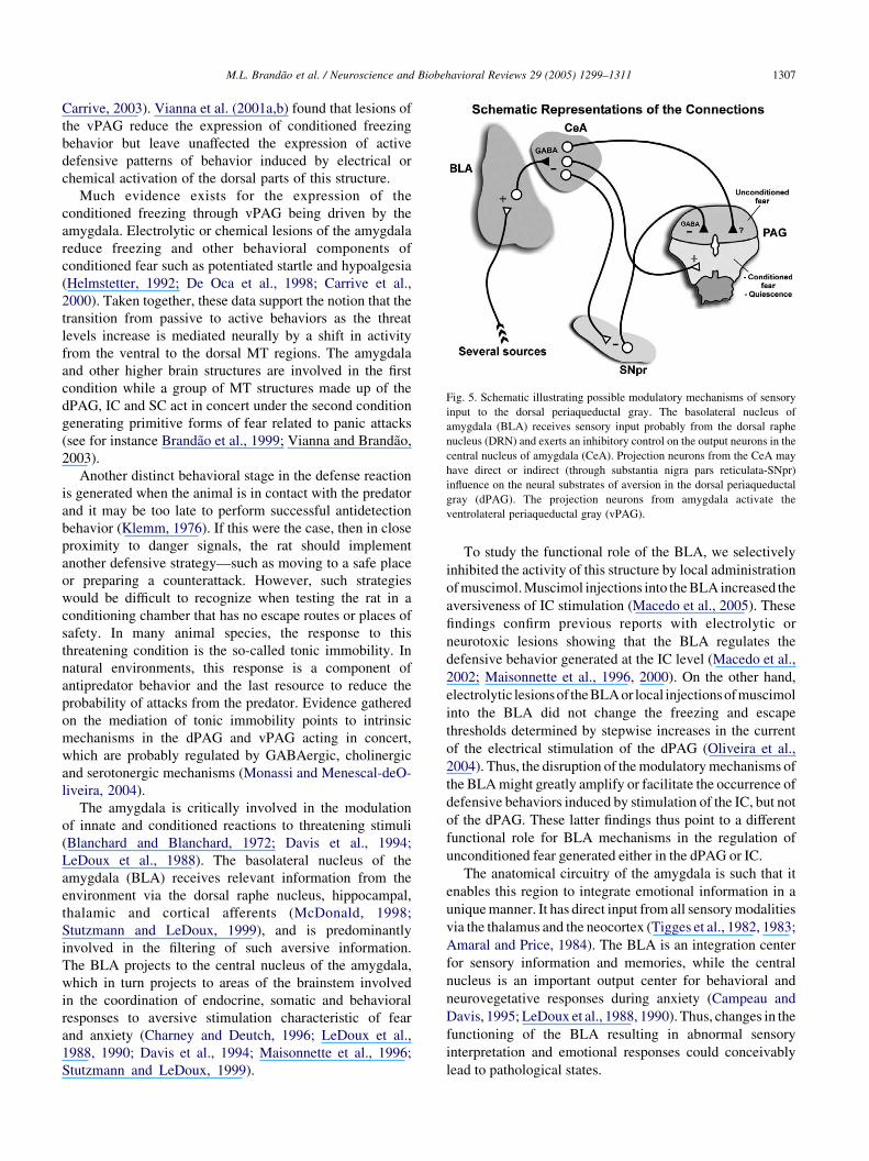
Fig. 5. Schematic illustrating possible modulatory mechanisms of sensory
input to the dorsal periaqueductal gray. The basolateral nucleus of
amygdala (BLA) receives sensory input probably from the dorsal raphe
nucleus (DRN) and exerts an inhibitory control on the output neurons in the
central nucleus of amygdala (CeA). Projection neurons from the CeA may
have direct or indirect (through substantia nigra pars reticulata-SNpr)
influence on the neural substrates of aversion in the dorsal periaqueductal
gray (dPAG). The projection neurons from amygdala activate the
ventrolateral periaqueductal gray (vPAG).
M.L. Brandao et al. / Neuroscience and Biobehavioral Reviews 29 (2005) 1299–1311 1307
Carrive, 2003). Vianna et al. (2001a,b) found that lesions of
the vPAG reduce the expression of conditioned freezing
behavior but leave unaffected the expression of active
defensive patterns of behavior induced by electrical or
chemical activation of the dorsal parts of this structure.
Much evidence exists for the expression of the
conditioned freezing through vPAG being driven by the
amygdala. Electrolytic or chemical lesions of the amygdala
reduce freezing and other behavioral components of
conditioned fear such as potentiated startle and hypoalgesia
(Helmstetter, 1992; De Oca et al., 1998; Carrive et al.,
2000). Taken together, these data support the notion that the
transition from passive to active behaviors as the threat
levels increase is mediated neurally by a shift in activity
from the ventral to the dorsal MT regions. The amygdala
and other higher brain structures are involved in the first
condition while a group of MT structures made up of the
dPAG, IC and SC act in concert under the second condition
generating primitive forms of fear related to panic attacks
(see for instance Brandao et al., 1999; Vianna and Brandao,
2003).
Another distinct behavioral stage in the defense reaction
is generated when the animal is in contact with the predator
and it may be too late to perform successful antidetection
behavior (Klemm, 1976). If this were the case, then in close
proximity to danger signals, the rat should implement
another defensive strategy—such as moving to a safe place
or preparing a counterattack. However, such strategies
would be difficult to recognize when testing the rat in a
conditioning chamber that has no escape routes or places of
safety. In many animal species, the response to this
threatening condition is the so-called tonic immobility. In
natural environments, this response is a component of
antipredator behavior and the last resource to reduce the
probability of attacks from the predator. Evidence gathered
on the mediation of tonic immobility points to intrinsic
mechanisms in the dPAG and vPAG acting in concert,
which are probably regulated by GABAergic, cholinergic
and serotonergic mechanisms (Monassi and Menescal-deO-
liveira, 2004).
The amygdala is critically involved in the modulation
of innate and conditioned reactions to threatening stimuli
(Blanchard and Blanchard, 1972; Davis et al., 1994;
LeDoux et al., 1988). The basolateral nucleus of the
amygdala (BLA) receives relevant information from the
environment via the dorsal raphe nucleus, hippocampal,
thalamic and cortical afferents (McDonald, 1998;
Stutzmann and LeDoux, 1999), and is predominantly
involved in the filtering of such aversive information.
The BLA projects to the central nucleus of the amygdala,
which in turn projects to areas of the brainstem involved
in the coordination of endocrine, somatic and behavioral
responses to aversive stimulation characteristic of fear
and anxiety (Charney and Deutch, 1996; LeDoux et al.,
1988, 1990; Davis et al., 1994; Maisonnette et al., 1996;
Stutzmann and LeDoux, 1999).
To study the functional role of the BLA, we selectively
inhibited the activity of this structure by local administration
of muscimol. Muscimol injections into the BLA increased the
aversiveness of IC stimulation (Macedo et al., 2005). These
findings confirm previous reports with electrolytic or
neurotoxic lesions showing that the BLA regulates the
defensive behavior generated at the IC level (Macedo et al.,
2002; Maisonnette et al., 1996, 2000). On the other hand,
electrolytic lesionsof the BLA or local injectionsofmuscimol
into the BLA did not change the freezing and escape
thresholds determined by stepwise increases in the current
of the electrical stimulation of the dPAG (Oliveira et al.,
2004). Thus, the disruption of the modulatory mechanisms of
the BLA might greatly amplify or facilitate the occurrence of
defensive behaviors induced by stimulation of the IC, but not
of the dPAG. These latter findings thus point to a different
functional role for BLA mechanisms in the regulation of
unconditioned fear generated either in the dPAG or IC.
The anatomical circuitry of the amygdala is such that it
enables this region to integrate emotional information in a
unique manner. It has direct input from all sensory modalities
via the thalamus and the neocortex (Tigges et al., 1982, 1983;
Amaral and Price, 1984). The BLA is an integration center
for sensory information and memories, while the central
nucleus is an important output center for behavioral and
neurovegetative responses during anxiety (Campeau and
Davis, 1995; LeDoux et al., 1988, 1990). Thus, changes in the
functioning of the BLA resulting in abnormal sensory
interpretation and emotional responses could conceivably
lead to pathological states.

M.L. Brandao et al. / Neuroscience and Biobehavioral Reviews 29 (2005) 1299–13111308
The possibility of conjoint inhibitory influences of the
BLA and SNpr on the neural substrates of aversion in the
MT still remains. The SNpr-MT pathway might well be
modulated by descending projections from the amygdaloid
nuclei (Hopkins and Holstege, 1978; Shinonaga et al.,
1992). A diagram illustrating possible modulatory
connections involving amygdaloid nuclei, SNpr, and PAG
is presented in Fig. 5.
6. Final comments
In MT structures such as the dPAG, SC and IC,
GABAergic neurons exert tonic control over the neural
substrates involved in the generation and expression of
defensive reactions. It is still unclear how these
mechanisms are called into play during the processing
of information of an aversive nature when sensory
influences trigger the motor expression of defense. We
can infer that such induced states and behaviors must
either be a precondition for the expression of fear, or a
consequence thereof. We can thus conclude that the MT
‘defense-aversion systems’ as they are called, also
comprise a substrate for, or source of information for
state properties that decide the animal’s behavior on a
scale of anxiety as defined by performance in anxiety
tests. Thus, while freezing behavior is the usual response
when the animal perceives the danger, escape is the
natural reaction when the predator is close. This line of
research has evolved to disclose the structures and the
neural mechanisms that could participate in the acqui-
sition of aversive information and expression of the
different types of defensive behavior.
Studies using electrical stimulation of the MT have
shown that defensive strategies as distinct as avoidance,
freezing and escape are likely to be organized by
different networks. Studies aimed at examination of
both the processing of sensory information and their
behavioral counterparts during the reduction of GABA
transmission caused by local injections of GABA
receptor blockers (e.g. BIC) or GAD inhibitors (e.g.
SMC) into the MT have shown that they induce freezing
and escape behavior, respectively. Moreover, SMC
enhances auditory evoked potential and impair the startle
reaction to loud sound. These results suggest that
reduction of the GABAergic control of the MT results
in an enhanced processing of information of aversive
nature along with defensive behaviors that are incompa-
tible with the startle response. Also, we present evidence
here that Fos expression in the laterodorsal nucleus of
the thalamus is increased during freezing induced by
stimulation of the dPAG suggesting that aversive
information ascends to higher structures during this
emotional condition.
Acknowledgements
This work was supported by FAPESP (Proc. no.
02/03705-0) and CNPq (301069/81–6). KG Borelli, JM
Santos and RC Martinez are recipients of doctorate
scholarships from CAPES. AR Oliveira and L Albrechet-
Souza are recipients of master scholarships from FAPESP
and CAPES, respectively. I also thank Dr Silvio Morato for
the helpful contribution to the writing style of this
manuscript.
References
Adams, D.B., 1979. Brain mechanisms for offense, defense and submission.
Behav. Brain Sci. 2, 201–241.
Amaral, D.G., Price, J., 1984. Amygdala-cortical projections in the monkey
(Macaca fascicularis). J. Comp. Neurol. 230, 465–496.
Audi, E.A., Graeff, F.G., 1984. Benzodiazepine receptors in the
periaqueductal grey mediate anti-aversive drug action. Eur. J.
Pharmacol. 103, 279–285.
Bagri, A., Sandner, G., Di Scala, G., 1989. Effects of unilateral
microinjections of GABAergic drugs into the inferior colliculus on
auditory evoked potentials and on audiogenic seizure susceptibility.
Exp. Neurol. 104, 82–87.
Bandler, R., 1982a. Neural control of aggressive behavior. Trends
Neurosci. 5, 390–393.
Bandler, R., 1982b. Identification of neuronal cell bodies mediating
components of biting attack behavior in the cat; induction of jaw
opening following microinjections of glutamate into hypothalamus.
Brain Res. 245, 192–197.
Bandler, R., Carrive, P., 1988. Integrated defence reaction elicited by
excitatory amino acid microinjection in the midbrain periaqueductal
grey region of the unrestrained cat. Brain Res. 439, 95–106.
Bandler, R., Flynn, J.P., 1971. Visual patterned reflex present during
hypothalamically elicited attack. Science 171, 817–818.
Bandler, R., Flynn, J.P., 1972. Control of somatosensory fields for striking
during hypothalamically elicited attack. Brain Res. 38, 197–201.
Bandler, R., Shipley, M.T., 1994. Columnar organization in the midbrain
periaqueductal gray: modules for emotional expression? Trends
Neurosci. 17, 379–389.
Behbehani, M.M., Jiang, M.R., Chandler, S.D., Ennis, M., 1990. The effect
of GABA and its antagonists on midbrain periaqueductal gray neurons
in the rat. Pain 40, 195–204.
Beleboni, R.O., Carolino, R.O., Pizzo, A.B., Castellan-Baldan, L.,
Coutinho-Netto, J., dos Santos, W.F., Coimbra, N.C., 2004. Pharma-
cological and biochemical aspects of GABAergic neurotransmission:
pathological and neuropsychobiological relationships. Cell Mol.
Neurobiol. 24, 707–728.
Bittencourt, A.S., Carobrez, A.P., Zamprogno, L.P., Tufik, S.,
Schenberg, L.C., 2004. Organization of single components of
defensive behaviors within distinct columns of periaqueductal gray
matter of the rat: role of N-methyl-D-aspartic acid glutamate
receptors. Neurosci. 125, 71–89.
Blanchard, D.C., Blanchard, R.J., 1969. Crouching as an index of fear. J.
Comp. Physiol. Psychol. 67, 370–375.
Blanchard, D.C., Blanchard, R.J., 1972. Innate and conditioned reactions to
threat in rats with amygdaloid lesions. J. Comp. Physiol. Psychol. 81,
281–290.
Blanchard, D.C., Blanchard, R.J., 1988. Ethoexperimental approaches to
the biology of emotion. Ann. Rev. Psychol. 39, 43–68.
Brandao, M.L., De Aguiar, J.C., Graeff, F.G., 1982. GABA mediation of
the anti-aversive action of minor tranquilizers. Pharmacol. Biochem.
Behav. 16, 397–402.

M.L. Brandao et al. / Neuroscience and Biobehavioral Reviews 29 (2005) 1299–1311 1309
Brandao, M.L., DiScala, G., Bouchet, M.J., Schmitt, P., 1986. Escape
behavior produced by the blockade of glutamic decarboxylase (GAD)
in mesencephalic central gray or medial hypothalamus. Pharmacol.
Biochem. Behav. 24, 497–501.
Brandao, M.L., Tomaz, C., Leao-Borges, P.C., Coimbra, N.C., Bagri, A.,
1988. Defense reaction induced by microinjections of bicuculline into
the inferior colliculus. Physiol. Behav. 44, 361–365.
Brandao, M.L., Melo, L.L., Cardoso, S.H., 1993. Mechanisms of defense in
the inferior colliculus. Behav. Brain Res. 58, 49–55.
Brandao, M.L., Anseloni, V.Z., Pandossio, J.E., De Araujo, J.E., Castilho,
V.M., 1999. Neurochemical mechanisms of the defensive behavior in
the dorsal midbrain. Neurosci. Biobehav. Rev. 23, 863–875.
Brandao, M.L., Coimbra, N.C., Osaki, M.Y., 2001. Changes in the
auditory-evoked potentials induced by fear-evoking stimulations.
Physiol. Behav. 72, 365–372.
Brandao, M.L., Troncoso, A.C., Souza Silva, M.A., Huston, J.P., 2003. The
relevance of neuronal substrates of defense in the midbrain tectum to
anxiety and stress: empirical and conceptual considerations. Eur. J.
Pharmacol. 463, 225–233.
Campeau, S., Davis, M., 1995. Involvement of the central nucleus
and basolateral complex of the amygdala in fear conditioning
measured with fear-potentiated startle in rats trained concurrently
with auditory and visual conditioned stimuli. J. Neurosci. 15,
2301–2311.
Canteras, N.S., Goto, M., 1999. Fos-like immunoreactivity in the
periaqueductal gray of rats exposed to a natural predator. Neuroreport
10, 413–418.
Carrive, P., 1993. The periaqueductal gray and defensive behavior:
functional representation and neuronal organization. Behav. Brain
Res. 58, 27–47.
Carrive, P., Lee, J., Su, A., 2000. Lidocaine blockade of amygdala output in
fear conditioned rats reduces Fos-expression in the ventrolateral
periaqueductal gray. Neuroscience 95, 1071–1080.
Charney, D.S., Deutch, A., 1996. A functional neuroanatomy of anxiety and
fear: implications for the pathophysiology and treatment of anxiety
disorders. Crit. Rev. Neurobiol. 10, 419–446.
Chevalier, G., Denniau, J.-M., 1990. Disinhibition as a basic process in the
expression of striatal functions. Trends Neurosci. 13, 277–280.
Chevalier, G., Thierry, A.M., Shibazaki, T., Feger, J., 1981. Evidence for a
GABAergic inhibitory nigrotectal pathway in the rat. Neurosci. Lett.
21, 67–70.
Clerici, W.J., Coleman, J.R., 1986. Resting and high-frequency evoked
2-deoxyglucose uptake in the rat inferior colliculus: developmental
changes and effects of short-term conduction blockade. Brain Res. 392,
127–137.
Coimbra, N.C., Brandao, M.L., 1993. GABAergic nigrocollicular pathways
modulate the defensive behaviour elicited by midbrain tectum
stimulation. Behav. Brain Res. 59, 131–139.
Coleman, J.R., Campbell, M., Clerici, W.J., 1982. Observations on
tonotopic organization within the rat inferior colliculus using
2-deoxy-D[1-3H] glucose. Otolaryngol. Head Neck Surg. 90, 795–
800.
Davis, M., Astrachan, D.I., 1978. Conditioned fear and startle magnitude:
Effects of different footshock and backshock intensities used in training.
J. Exp. Psychol. Anim. Behav. Proc. 4, 95–103.
Davis, M., Raiunnie, D., Cassell, M., 1994. Neurotransmission in the rat
amygdala related to fear and anxiety. Trends Neurosci. 17, 208–214.
De Oca, B.M., DeCola, J.P., Maren, S., Fanselow, M.S., 1998. Distinct
regions of the periaqueductal gray are involved in the acquisition and
expression of defensive responses. J. Neurosci. 18, 3426–3432.
Dean, P., Redgrave, P., Westby, G.W.M., 1989. Event or emergency? two
response systems in the mammalian superior colliculus. Trends
Neurosci. 12, 137–147.
DePaulis, A., Vernes, M., 1986. Elicitation of intraspecific defensive
behaviors in the rat by microinjections of picrotoxin, a g-aminobutyric
acid antagonist, into the midbrain periaqueductal gray matter. Brain
Res. 367, 87–95.
Di Chiara, G., Porceddu, M.L., Morelli, M., Mulas, M.L., Gessa, G.L.,
1979. Evidence for a GABAergic projection from the substantia nigra to
the ventromedial thalamus and to the superior colliculus of the rat.
Brain Res. 176, 273–284.
DiScala, G., Sandner, G., 1989. Conditioned place aversion produced by
microinjections of semicarbazide into the periaqueductal gray of the rat.
Brain Res. 483, 91–97.
DiScala, G., Schmitt, P., Karli, P., 1983. Unilateral injection of GABA
agonists in the superior colliculus: asymmetry to tactile stimulation.
Pharmacol. Biochem. Behav. 19, 281–285.
Drager, U.C., Hubel, D.H., 1976. Topography of visual and somatosensory
projections to mouse superior colliculus. J. Neurophysiol. 39, 91–101.
Fanselow, M.S., 1984. Neural organization of the defensive behavior
system responsible for fear. Psychon. Bull. Rev. 1, 429–438.
Fendt, M., 1999. Enhancement of prepulse inhibition after blockade of
GABA activity within the superior colliculus. Brain Res. 833, 81–85.
Fendt, M., Koch, M., Schnitzler, H.U., 1994. Lesions of the central gray
block the sensitization of the acoustic startle response in rats. Brain Res.
661, 163–173.
Ferreira-Netto, C., Borelli, K.G., Brandao, M.L., 2005. Neural segregation
of Fos-protein distribution in the brain following freezing and escape
behaviors induced by injections of either glutamate or NMDA into the
dorsal periaqueductal gray of rats. Brain Res. 1031, 151–163.
Flynn, J.P., Edwards, S.B., Bandler, R., 1971. Changes in sensory and
motor systems during centrally elicited attack. Behav. Sci. 16, 1–19.
Graeff, F.G., Brandao, M.L., Audi, E.A., Schultz, M.T.B., 1986.
Modulation of the brain aversive system by GABAergic and
serotonergic mechanisms. Behav. Brain Res. 22, 173–180.
Helmstetter, F.G., 1992. The amygdala is essential for the expression of
conditional analgesia. Behav. Neurosci. 106, 518–528.
Hopkins, D.A., Holstege, G., 1978. Amygdaloid projections to the
mesencephalon, pons and medulla oblongata in the cat. Exp. Brain
Res. 32, 529–547.
Huang, C.M., Fex, J., 1986. Tonotopic organization in the inferior
colliculus of the rat demonstrated with the 2-deoxyglucose method.
Exp. Brain Res. 61, 506–512.
Huston, J.P., Nef, B., Papadopoulos, G., Welzl, H., 1980. Activation and
lateralization of sensorimotor field for perioral biting reflex by
intranigral GABA agonist and by systemic apomorphine in the rat.
Brain Res. Bull. 5, 745–749.
Huston, J.P., Steiner, H., Weiler, H.-T., Morgan, S., Schwarting, R.K.W.,
1990. The basal ganglia-orofacial system: studies on neurobehavioral
plasticity and sensory-motor turning. Neurosci. Biobehav. Rev. 14,
433–446.
Imperato, A., DiChiara, G., 1981. Behavioural effects of GABA agonists
and antagonists infused in the mesencephalic reticular formation—deep
layers of superior colliculus. Brain Res. 224, 185–194.
Keay, K.A., Bandler, R., 2001. Parallel circuits mediating distinct
emotional coping reactions to different types of stress. Neurosci.
Biobehav. Rev. 25, 669–678.
Killam, R.F., Bain, J.A., 1957. Convulsant hydrazides. I: in vitro and
in vivo inhibition of vitamin B6 enzymes by convulsant hydrazides. J.
Pharmacol. Exp. Ther. 119, 255–262.
Klemm, W.R., 1976. Identity of sensory and motor systems that are crucial
to the immobility reflex (animal hypnosis). J. Neurosci. Res. 2, 57–69.
LeBeau, F.E.N., Rees, A., Malmierca, M.S., 1996. Contribution of GABA
and glycine-mediated inhibition to the monoaural temporal response
properties of neurons in the inferior colliculus. J. Neurophysiol. 75,
902–919.
LeBeau, F.E.N., Malmierca, M.S., Rees, A., 2001. Iontophoresis in vivo
demonstrates a key role for GABAA and glycinergic inhibition in
shaping frequency response areas in the inferior colliculus of guinea
pig. J. Neurosci. 15, 7303–7312.
LeDoux, J.E., Iwata, J., Cichetti, P., Reis, D.J., 1988. Different projections
of the central amygdaloid nucleus mediate autonomic and behavioral
correlates of conditioned fear. J. Neurosci. 8, 2517–2519.

M.L. Brandao et al. / Neuroscience and Biobehavioral Reviews 29 (2005) 1299–13111310
LeDoux, J.E., Cicchetti, P., Xagoraris, A., Romanski, L.M., 1990. The
lateral amygdaloid nucleus: Sensory interface of the amygdala in fear
conditioning. J. Neurosci. 10, 1062–1069.
Li, L., Priebe, R.P.M., Yeomans, J.S., 1998. Prepulse inhibition of acoustic
or trigeminal startle of rats by unilateral electrical stimulation of the
inferior colliculus. Behav. Neurosci. 112, 1187–1198.
MacDonne, M.F., Flynn, J.P., 1966. Control of sensory fields by stimulation
of hypothalamus. Science 152, 1406–1414.
Macedo, C.E., Castilho, V.M., Souza-Silva, M.A., Brandao, M.L., 2002.
Dual 5-HT mechanisms in basolateral and central nuclei of amygdala in
the regulation of the defensive behavior induced by electrical
stimulation of the inferior colliculus. Brain Res. Bull. 59, 189–195.
Macedo, C.E., Cuadra, G., Molina, V., Brandao, M.L., 2005. Aversive
stimulation of the inferior colliculus changes dopamine and serotonin
extracellular levels in the frontal cortex: Modulation by the basolateral
nucleus of amygdala. Synapse 55, 58–66.
Maisonnette, S.S., Kawasaki, M.C., Coimbra, N.C., Brandao, M.L., 1996.
Effects of lesions of amygdaloid nuclei and substantia nigra on aversive
responses induced by electrical stimulation of the inferior colliculus.
Brain Res. Bull. 40, 93–98.
Maisonnette, S., Villela, C., Carotti, A.P., Landeira-Fernandez, J., 2000.
Microinfusion of nefazodone into the basolateral nucleus of the
amygdala enhances defensive behavior induced by NMDA stimulation
of the inferior colliculus. Physiol. Behav. 70, 243–247.
Martinez, R.C.R., Oliveira, A.R., Brandao, M.L., submitted for publication.
The conditioned and unconditioned fear organized in the periaqueductal
gray are differentially regulated by lateral, basolateral and central nuclei
of amygdala. Neurobiol. Learn. Mem.
McDonald, A.J., 1998. Cortical pathways to the mammalian amygdala.
Prog. Neurobiol. 55, 257–332.
Melo, L.L., Cardoso, S.H., Brandao, M.L., 1992. Antiaversive action of
benzodiazepines on escape behavior induced by electrical stimulation
of the inferior colliculus. Physiol. Behav. 51, 557–562.
Merchan, M.A., Saldana, E., Plaza, I., 1994. Dorsal nucleus of the lateral
lemniscus in the rat: concentric organization and tonotopic projection to
the inferior colliculus. J.Comp. Neurol. 342, 259–278.
Mitchell, I.J., Dean, P., Redgrave, P., 1988. The projection from superior
colliculus to cuneiform area in the rat. II. Defense-like responses to
stimulation with glutamate in cuneiform nucleus and surrounding
structures. Exp. Brain Res. 72, 626–639.
Monassi, C.R., Menescal-de-Oliveira, L., 2004. Serotonin 5-HT2 and
5-HT1A receptors in the periaqueductal gray matter differentially
modulate tonic immobility in guinea pigs. Brain Res. 1009,
169–180.
Nobre, M.J., Brandao, M.L., 2004. Analysis of freezing behavior and
ultrasonic vocalization in response to foot-shocks, ultrasound signals
and GABAergic inhibition in the inferior colliculus: effects of
muscimol and midazolam. Eur. Neuropsychopharmacol. 14, 45–52.
Nobre, M.J., Lopes, M.G., Brandao, M.L., 2004. Defense reaction mediated
by NMDA mechanisms in the inferior colliculus is modulated by
GABAergic nigrocollicular pathways. Brain Res. 999, 124–131.
Oliveira, L., Brandao, M.L., Landeira-Fernandez, J., 2004. Role of
amygdala in conditioned and unconditioned fear generated in the
periaqueductal gray. Neuroreport 15, 2281–2285.
Pandossio, J.E., Brandao, M.L., 1999. Defensive reactions are counteracted
by midazolam and muscimol and elicited by activation of glutamate
receptors in the inferior colliculus of rats. Psychopharmacology 142,
360–368.
Pieper, F., Jurgens, U., 2003. Neuronal activity in the inferior colliculus and
bordering structures during vocalization in the squirrel monkey. Brain
Res. 979, 153–164.
Pierson, M., Snyder-Keller, A., 1994. Development of frequency-selective
domains in inferior colliculus of normal and neonatally noise-exposed
rats. Brain Res. 636, 55–67.
Redgrave, P., Dean, P., Souki, W., Lewis, G., 1981. Gnawing and changes
in reactivity produced by microinjections of picrotoxin into the superior
colliculus of rats. Psychopharmacology 75, 198–203.
Rizvi, T.A., Ennis, M., Behbehani, M.M., Shipley, M.T., 1991.
Connections between the central nucleus of the amygdala and the
midbrain periaqueductal gray: topography and reciprocity. J. Comp.
Neurol. 303, 121–131.
Roberts, R.C., Ribak, C.E., 1987. GABAergic neurons and axon terminals
in the brainstem auditory nuclei of the gerbil. J. Comp. Neurol. 258,
267–280.
Saint Marie, R.J., Luo, L., Ryan, A.F., 1999. Effects of stimulus frequency
and intensity on c-Fos mRNA expression in the adult rat auditory
brainstem. J. Comp. Neurol. 404, 258–270.
Sandner, G., Dessort, D., Schmitt, P., Karli, P., 1981. Distribution of GABA
in the periaqueductal gray matter. Effects of medial hypothalamic
lesions. Brain Res. 224, 279–290.
Sandner, G., Canal, N.M., Brandao, M.L., 2002. Effects of ketamine and
apomorphine on inferior colliculus and caudal pontine reticular nucleus
evoked potentials during prepulse inhibition of the startle reflex in rats.
Behav. Brain Res. 128, 161–168.
Santos, J.M., Gargaro, A.C., Oliveira, A.R., Masson, S., Brandao, M.L.,
2005. Pharmacological dissociation of moderate and high contextual
fear as assessed by freezing behavior and fear-potentiated startle. Eur.
Neuropsychopharmacol. 15, 239–246.
Schenberg, L.C., Aguiar, J.C., Graeff, F.G., 1983. GABA modulation of the
defense reaction induced by brain electrical stimulation. Physiol.
Behav. 31, 429–437.
Schmitt, P., DiScala, G., Brandao, M.L., Karli, P., 1985. Behavioral effects of
microinjections of SR95103; a new GABA-A antagonist; into medial hypo-
thalamus or the mesencephalic central gray. Eur. J. Pharmacol. 117, 149–158.
Schmitt, P., Carrive, P., Discala, G., Jenck, F., Brandao, M., Bagri, A., Moreau,
J.L., Sandner, G., 1986. A neuropharmacological study of the periven-
tricular neural substrate involved in flight. Behav. Brain Res. 22, 181–190.
Schreiner, C.E., Langner, G., 1997. Laminar fine structure of frequency
organization in auditory midbrain. Nature 388, 383–386.
Shinonaga, Y., Takada, M., Mizuno, N., 1992. Direct projections from the
central amygdaloid nucleus to the globus pallidus and substantia nigra
in the cat. Neuroscience 51, 691–703.
Stein, B.E., Magalhaes-Castro, B., Kruger, L., 1975. Superior colliculus:
visuotopic–somatotopic overlap. Science 189, 224–226.
Stutzmann, G.E., LeDoux, J.E., 1999. GABAergic antagonists block the
inhibitory effects of serotonin in the lateral amygdala: a mechanism for
modulation of sensory inputs related to fear conditioning. J. Neurosci. 19, 1–4.
Szczepaniak, W.S., Moller, A.R., 1995. Evidence of decreased GABAergic
influence on temporal integration in the inferior colliculus following
acute noise exposure: a study of evoked potentials in the rat. Neurosci.
Lett. 196, 77–80.
Thompson, G.C., Cortez, A.M., Lam, D.M., 1985. Localization of GABA
immunoreactivity in the auditory brainstem of guinea pigs. Brain Res.
339, 119–122.
Tigges, J., Tigges, M., Cross, M.A., McBride, R.L., Letbetter, W.T.,
Anschel, S., 1982. Subcortical structures projecting to visual cortical
areas in squirrel monkey. J. Comp. Neurol. 209, 29–40.
Tigges, J., Walker, L.C., Tigges, M., 1983. Subcortical projections to the
occipital and parietal lobes of the chimpanzee brain. J. Comp. Neurol.
220, 106–115.
Tomaz, C., Brandao, M., Bagri, A., Carrive, P., Schmitt, P., 1988. Flight
behavior induced by microinjection of GABA antagonists into
periventricular structures in detelencephalated rats. Pharmacol. Bio-
chem. Behav. 30, 337–342.
Vianna, D.M.L., Brandao, M.L., 2003. Anatomical connections of the
periaqueductal gray: specific neural substrates for different kinds of
fear. Braz. J. Med. Biol. Res. 36, 557–566.
Vianna, D.M.L., Landeira-Fernandez, J., Brandao, M.L., 2001a. Dorsolateral -
and ventral regions of the periaqueductal gray matter are involved in
distinct types of fear. Neurosci. Biobehav. Rev. 25, 711–719.
Vianna, D.M.L., Landeira-Fernandez, J., Graeff, F.G., Brandao, M.L.,
2001b. Lesion of the ventral periaqueductal gray reduces conditioned
fear but does not change freezing induced by stimulation of the dorsal
periaqueductal gray. Learn. Mem. 8, 164–169.

M.L. Brandao et al. / Neuroscience and Biobehavioral Reviews 29 (2005) 1299–1311 1311
Walker, P., Carrive, P., 2003. Role of ventrolateral periaqueductal gray
neurons in the behavioral and cardiovascular responses to contextual
fear and poststress recovery. Neuroscience 116, 897–912.
Walker, D.L., Cassella, J.V., Lee, Y., De Lima, T.C.M., Davis, M., 1997.
Opposing roles of the amygdala and dorsolateral periaqueductal gray in
fear-potentiated startle. Neurosci. Biobehav. Rev. 21, 743–753.
Watkins, L., Sherwood, A., Goldstein, D.S., Maixner, W., 1993.
Mechanisms underlying cardiovascular defense reaction evoked by
dorsal periaqueductal gray stimulation. Am. J. Physiol. 265, 1155–
1161.
Wecker, J.R., Ison, J.R., 1986. Effects of motor activity on the elicitation
and modification of the startle reflex in rats. Anim. Learn. Behav. 14,
287–292.
Welzl, H., Schwarting, R., Huston, J.P., 1984. Perioral biting reflex and
turning after intranigral injection of a GABA-or metenkephalin-agonist:
role of the thalamus and superior colliculus. Exp. Brain Res. 55, 438–
444.
Yasui, Y., Nakano, K., Kayahara, T., Mizuno, N., 1991. Non-dopaminergic
projections from the substantia nigra pars lateral to the inferior
colliculus in the rat. Brain Res. 559, 139–144.
Copyright © 2022 FDOKUMEN