Distinct Mechanisms Underlie Neurotoxin-Mediated Cell Death in Cultured Dopaminergic Neurons
Upload
independentCategory
view
2download
0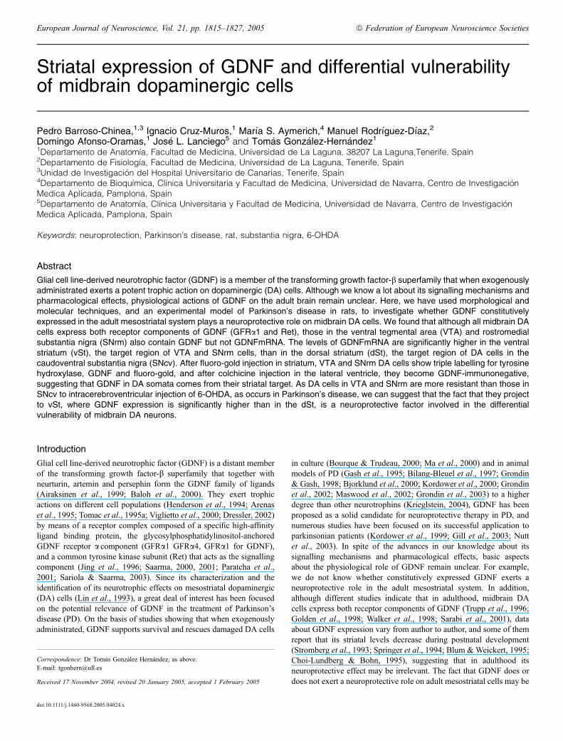
Striatal expression of GDNF and differential vulnerabilityof midbrain dopaminergic cells
Pedro Barroso-Chinea,1,3 Ignacio Cruz-Muros,1 Marıa S. Aymerich,4 Manuel Rodrıguez-Dıaz,2
Domingo Afonso-Oramas,1 Jose L. Lanciego5 and Tomas Gonzalez-Hernandez11Departamento de Anatomıa, Facultad de Medicina, Universidad de La Laguna, 38207 La Laguna,Tenerife, Spain2Departamento de Fisiologıa, Facultad de Medicina, Universidad de La Laguna, Tenerife, Spain3Unidad de Investigacion del Hospital Universitario de Canarias, Tenerife, Spain4Departamento de Bioquımica, Clınica Universitaria y Facultad de Medicina, Universidad de Navarra, Centro de InvestigacionMedica Aplicada, Pamplona, Spain5Departamento de Anatomıa, Clınica Universitaria y Facultad de Medicina, Universidad de Navarra, Centro de InvestigacionMedica Aplicada, Pamplona, Spain
Keywords: neuroprotection, Parkinson’s disease, rat, substantia nigra, 6-OHDA
Abstract
Glial cell line-derived neurotrophic factor (GDNF) is a member of the transforming growth factor-b superfamily that when exogenouslyadministrated exerts a potent trophic action on dopaminergic (DA) cells. Although we know a lot about its signalling mechanisms andpharmacological effects, physiological actions of GDNF on the adult brain remain unclear. Here, we have used morphological andmolecular techniques, and an experimental model of Parkinson’s disease in rats, to investigate whether GDNF constitutivelyexpressed in the adult mesostriatal system plays a neuroprotective role on midbrain DA cells. We found that although all midbrain DAcells express both receptor components of GDNF (GFRa1 and Ret), those in the ventral tegmental area (VTA) and rostromedialsubstantia nigra (SNrm) also contain GDNF but not GDNFmRNA. The levels of GDNFmRNA are significantly higher in the ventralstriatum (vSt), the target region of VTA and SNrm cells, than in the dorsal striatum (dSt), the target region of DA cells in thecaudoventral substantia nigra (SNcv). After fluoro-gold injection in striatum, VTA and SNrm DA cells show triple labelling for tyrosinehydroxylase, GDNF and fluoro-gold, and after colchicine injection in the lateral ventricle, they become GDNF-immunonegative,suggesting that GDNF in DA somata comes from their striatal target. As DA cells in VTA and SNrm are more resistant than those inSNcv to intracerebroventricular injection of 6-OHDA, as occurs in Parkinson’s disease, we can suggest that the fact that they projectto vSt, where GDNF expression is significantly higher than in the dSt, is a neuroprotective factor involved in the differentialvulnerability of midbrain DA neurons.
Introduction
Glial cell line-derived neurotrophic factor (GDNF) is a distant memberof the transforming growth factor-b superfamily that together withneurturin, artemin and persephin form the GDNF family of ligands(Airaksinen et al., 1999; Baloh et al., 2000). They exert trophicactions on different cell populations (Henderson et al., 1994; Arenaset al., 1995; Tomac et al., 1995a; Viglietto et al., 2000; Dressler, 2002)by means of a receptor complex composed of a specific high-affinityligand binding protein, the glycosylphosphatidylinositol-anchoredGDNF receptor a component (GFRa1 GFRa4, GFRa1 for GDNF),and a common tyrosine kinase subunit (Ret) that acts as the signallingcomponent (Jing et al., 1996; Saarma, 2000, 2001; Paratcha et al.,2001; Sariola & Saarma, 2003). Since its characterization and theidentification of its neurotrophic effects on mesostriatal dopaminergic(DA) cells (Lin et al., 1993), a great deal of interest has been focusedon the potential relevance of GDNF in the treatment of Parkinson’sdisease (PD). On the basis of studies showing that when exogenouslyadministrated, GDNF supports survival and rescues damaged DA cells
in culture (Bourque & Trudeau, 2000; Ma et al., 2000) and in animalmodels of PD (Gash et al., 1995; Bilang-Bleuel et al., 1997; Grondin& Gash, 1998; Bjorklund et al., 2000; Kordower et al., 2000; Grondinet al., 2002; Maswood et al., 2002; Grondin et al., 2003) to a higherdegree than other neurotrophins (Krieglstein, 2004), GDNF has beenproposed as a solid candidate for neuroprotective therapy in PD, andnumerous studies have been focused on its successful application toparkinsonian patients (Kordower et al., 1999; Gill et al., 2003; Nuttet al., 2003). In spite of the advances in our knowledge about itssignalling mechanisms and pharmacological effects, basic aspectsabout the physiological role of GDNF remain unclear. For example,we do not know whether constitutively expressed GDNF exerts aneuroprotective role in the adult mesostriatal system. In addition,although different studies indicate that in adulthood, midbrain DAcells express both receptor components of GDNF (Trupp et al., 1996;Golden et al., 1998; Walker et al., 1998; Sarabi et al., 2001), dataabout GDNF expression vary from author to author, and some of themreport that its striatal levels decrease during postnatal development(Stromberg et al., 1993; Springer et al., 1994; Blum &Weickert, 1995;Choi-Lundberg & Bohn, 1995), suggesting that in adulthood itsneuroprotective effect may be irrelevant. The fact that GDNF does ordoes not exert a neuroprotective role on adult mesostriatal cells may be
Correspondence: Dr Tomas Gonzalez Hernandez, as above.E-mail: [email protected]
Received 17 November 2004, revised 20 January 2005, accepted 1 February 2005
European Journal of Neuroscience, Vol. 21, pp. 1815–1827, 2005 ª Federation of European Neuroscience Societies
doi:10.1111/j.1460-9568.2005.04024.x

a key factor in the pathophysiology of PD. Furthermore, its expressionpattern may influence the differential vulnerability displayed bydifferent DA cell populations in PD (Fearnley & Lees, 1991; Gibb &Lees, 1991; Damier et al., 1999) and against different neurotoxins(Chiueh et al., 1985; Schneider et al., 1987; Herrero et al., 1993;Varastet et al., 1994; Rodriguez et al., 2001b). Bearing in mind thesearguments, the aim of the present study was to investigate theexpression pattern of GDNF and its receptor components in the adultmesostriatal system and the possible relation between them andneuroprotection in mesostriatal DA cells. We have used immunohist-ochemistry, in situ hybridization, reverse transcriptase-polymerasechain reaction, injection of retrograde tracers, and an experimentalmodel of PD in rats that reproduces the topographical pattern of celldegeneration observed in PD (Rodriguez et al., 2001a,b).
Materials and methods
Male Sprague–Dawley rats (Panlab, Barcelona) weighing 300–350 gwere housed at 22 �C, two per cage, under normal laboratoryconditions in a standard 12-h light : 12-h dark schedule (lights onbetween 08:00 h and 20:00 h) with free access to food and water.Experimental protocols were in accordance with the EuropeanCommunities Council Directive of 24 November 1986(86 ⁄ 609 ⁄ EEC) regarding the care and use of animals for experimentalprocedures. All efforts were made to reduce the number of animalsused.
Tissue preparation for morphological study
The animals were deeply anaesthetized (chloral hydrate, 400 mg ⁄ kg,i.p.) and transcardially perfused with 0.9% saline (150 mL) and afixative solution (400 mL) of 4% paraformaldehyde in 0.1 m phos-phate-buffered saline pH 7.4 (PBS). The brains were removed andstored in the same fixative at 4 �C for 6 h. Midbrains and forebrainswere initially obtained using a brain blocker (Activational System,Warren, MI), immersed overnight at 4 �C in a cryoprotective solutionof 30% sucrose in the same buffer, and then cut into 30-lm coronalsections with a freezing microtome. Sections were collected in6–8 parallel series and processed by in situ hybridization histo-chemistry (ISH), immunohistochemistry, double-immunofluorescentlabelling or a combination of immunofluorescence and retrogradefluoro-gold transport. Solutions used in the fixation and cryoprotectionof material processed by in situ hybridization were treated with 0.1%diethylpyrocarbonate (DEPC) and autoclaved to inactivate RNases.
Immunohistochemistry and double immunofluorescent labelling
Midbrain sections were processed by single TH, Ret, GFRa1 andGDNF immunocytochemistry and TH-GDNF double immunofluores-cence. For single immunolabelling, sections were immersed for20 min in 3% H2O2 to inactivate endogenous peroxidase, andincubated for 60 min at room temperature (RT) in 4% normal goatserum (NGS, Jackson ImmunoResearch, West Grove, PA), or normaldonkey serum (NDS, Jackson ImmunoResearch) in the case ofGFRa1, in PBS containing 0.05% Triton X-100 (Sigma), andovernight in PBS containing 2% NGS (or 2% NDS) and one of theprimary antibodies, mouse anti-TH monoclonal antibody (1 : 14 000;Sigma, St. Louis, MO), rabbit anti-Ret polyclonal antibody (1 : 400;Santa Cruz Biotechnology, Santa Cruz, CA), goat anti-GFRa1polyclonal antibody (1 : 400, Santa Cruz Biotechnology), or rabbitanti-GDNF polyclonal antibody (1 : 400; Santa Cruz Biotechnology).
After several rinses, the sections were incubated for 2 h in eitherbiotinylated goat anti-mouse antiserum (1 : 1200, Jackson Immuno-Research), biotinylated goat anti-rabbit antiserum (1 : 1200, JacksonImmunoResearch) or biotinylated donkey anti-goat antiserum(1 : 1200, Jackson ImmunoResearch), and 1 : 200 NGS (or 1 : 200NDS) in PBS. Immunoreactions were visible after incubation for 1 hat RT in ExtrAvidin-peroxidase (1 : 5000, Sigma) in PBS, and after10 min in 0.005% 3¢-3¢-diamiobenzidine tetrahydrochloride (DAB,Sigma) and 0.001% H2O2 in cacodylate buffer 0.05 m pH 7.6. Afterseveral rinses in PBS, the slides were dehydrated, cleared in xylene,and coverslipped with DPX (BDH Chemicals, Poole, England).Double-immunofluorescent labelling was studied by using epiflu-
orescence and confocal microscopy. In both cases, the sections werefirst incubated for 1 h in 4% NGS and 0.05% Triton X-100 in PBS,and then overnight in the same solution containing mouse anti-THmonoclonal antibody (1 : 8000) and rabbit anti-GDNF polyclonalantibody (1 : 200). Epifluorescent labelling was visible after incuba-tion for 3 h in Fluorescein Isothiocyanate-conjugated goat anti-mouseIgG (1 : 150; Jackson ImmunoResearch) and Lissamine Rhodamine-conjugated goat anti-rabbit IgG (1 : 150; Jackson ImmunoResearch)in PBS containing 1 : 200 NGS. Confocal images were visible afterincubation for 2 h in Alexa Fluor� 546-conjugated goat anti-mouseIgG (1 : 100; Molecula Probes, Eugene, OR) and Alexa Fluor� 488-conjugated donkey anti-rabbit IgG (1 : 100; Molecula Probes) in PBScontaining 1 : 200 NGS and 1 : 200 NDS. After several rinses,sections for epifluorescence were air dried, coverslipped withVectashield (Vector, Burlingame, CA), and examined under afluoromicroscope (Leica Microsystems; Wetzlar, Germany) usingappropriate filters. Sections for confocal microscopy were air dried,immersed in toluene, coverslipped with Entellan� (Merk; Darmstadt,Germany) and examined under a laser scanning confocal microscope(LSM 510 META, Zeiss; Overkochen, Germany) equipped with anargon laser with excitation wavelengths of 458, 477, 488 and 514 nmand with one helium-neon laser with an excitation wavelength of543 nm. To ensure the appropriate visualization of labelled elementsand to prevent false positive results, the emission from the argon laserat 488 nm was filtered through a band pass filter of 505–530 nm, thisemission was colour–coded in green. The emission from the excitationwith the helium laser at 543 nm was filtered through a band pass filterof 560–615 nm, and colour-coded in red. For each immunocytochem-istry and immunofluorescence, control experiments were performed byremoving the primary antibody, resulting in negative staining.
In situ hybridization histochemistry
The antisense and the control sense rat digoxigenin-labelled GDNFriboprobes used in this study were produced by in vitro transcriptionof GDNF cDNA obtained by PCR. Total rat RNA was isolated fromrat striatum by using Trizol reagent (Life Technologies, Gaithersburg,MD). The cDNA was obtained by incubating 2 lg of total RNA,1 mm dNTP mix, 3 mm DTT, 5 pm hexamers, 40 U RNase inhibitor(Promega, Madison, WI) and 200 U M-MLV reverse transcriptase(Gibco, Carlsbad, CA) in a final volume of 30 lL during 1 h at 37 �C.The full-length GDNF cDNA (644 bp) was amplified by PCR underthe following conditions: 3 lL of total cDNA, 0.3 mm dNTP, 1 mm
MgSO4, 0.2 lm primers and 2.5 U of Platinum ⁄ Pfx ⁄DNA polym-erase (Invitrogen, Carlsbad, CA) in a final volume of 50 lL. Theprimer sequences were: sense 5¢-CTAAGATGAAGTTATGGGATG-3¢and antisense 5¢-AGGGTCAGATACATCCACACC-3¢. The PCRproduct was analysed by electrophoresis, the single band excised,purified and cloned. The GDNF was transcribed with the appropriateRNA polymerases to synthesize either the sense or antisense probes
1816 P. Barroso-Chinea et al.
ª 2005 Federation of European Neuroscience Societies, European Journal of Neuroscience, 21, 1815–1827

using commercial reagents (Roche Diagnostics, Mannheim,Germany). The 50 lL transcription mixture included 1 lg templatecDNA, 1 mm of each ATP, CTP and GTP, 0.7 mm UTP and 0.3 mm
DIG-UTP, 10 mm DTT, 50 U RNase Inhibitor, and 1 U of either T3 orT7 RNA polymerase. After 2 h at 37 �C, the template cDNA wasdigested with 2 U of RNase-free DNAse for 30 min at 37 �C. Then,the riboprobes were precipitated by the addition of 100 lL of 4 m
ammonium acetate and 500 lL ethanol and centrifuged at 4 �C for30 min. The quality of the synthesis was monitored by Northern blot.
Midbrain and forebrain floating sections were incubated twice with0.1% active DEPC ⁄ PBS containing 2% (w ⁄ v) proteinase K for15 min. After pre-equilibration with 5· SSC (0.75 m NaCl, 0.075 m
NaCitrate), the sections were prehybridized at 58 �C for 1 h inhybridization solution (50% formamide, 5· SSC, 40 lg ⁄mL dena-tured salmon DNA). The probes were denatured for 5 min at 80 �C,added to the hybridization mix at 400 ng ⁄mL, and the sections werehybridized in this solution at 58 �C for 16 h. Posthybridization washesincluded 2· SSC at RT for 30 min, 2· SSC at 65 �C for 1 h and0.1· SSC at 65 �C for 1 h. Then, the slides were equilibrated for5 min in TN buffer (100 mm TrisHCl, 150 mm NaCl, pH 7.5), andincubated for 2 h at RT with alkaline-phosphatase conjugated anti-DIG monoclonal antibody (1 : 5000 final dilution in TN with 0.5%blocking reagent, Roche Diagnostics). After two 15 min washes in TNbuffer, the slides were equilibrated for 5 min in TNM buffer (100 mm
TrisHCl, 100 mm NaCl, 50 mm MgCl2, pH 9.5) and incubated insubstrate solution (NBT and BCIP in TNM buffer, Roche Diagnostics)for 8 h. Staining was stopped in TE (10 mm TrisHCl, 1 mm EDTA,pH 8.0), and the slides were dehydrated and mounted. Controlexperiments were performed for each ISH by skipping the antisenseriboprobe or using their control sense probe, which resulted in anabsence of staining.
Fluoro-gold injection
A total of seven rats received a single injection of Fluoro-gold (FG,Florochrome, Denver, CO; 0.1–0.3 lL, 4% w ⁄ v in 0.9% saline) in theleft striatum using a glass micropipette attached to a Hamiltonmicrosyringe. In three rats the injection was localized in the dorsalstriatum and in the other four in the ventral striatum. After a survivaltime of 4–5 days, they were deeply anaesthetized (chloral hydrate,400 mg ⁄ kg, i.p.) and transcardially perfused. The brains wereremoved and processed by TH-GDNF double-immunofluorescenceas described above.
6-Hydroxydopamine (6-OHDA) lesion
Rats were anaesthetized with ketamine (25–40 mg ⁄ kg i.p.; RhoneMerieux; Lyon, France) and xylazine (3–6 mg ⁄ kg i.p.; Bayer,Leverkusen, Germany), and injected in the third ventricle [midline,2 mm posterior to bregma and 8 mm below the dura, according toPaxinos & Watson (1998)] with vehicle (0.9% saline solution with0.3 lg ⁄ lL ascorbic acid, sham group, n ¼ 5) or a single dose of (300 or500 lg) of 6-OHDA (6-hydroxydopamine hydrochloride, Sigma, in7.5 lL of vehicle per injection; 1 lL ⁄minute, 6-OHDA groups, n ¼ 6in each of them). In order to prevent the degeneration of noradrenergiccells, the rats were treated with nortriptyline 20 min before injection(30 mg ⁄ kg in saline, i.p.; nortriptyline hydrochloride, Sigma). Specialcare was taken in anaesthesia because in our experience, nortriptylinesignificantly increases the effect of ketamine, affecting cardiopulmonaryfunctions during surgery. Bearing inmind that the bilateral degenerationof DA cells can cause adipsia and aphagia (Ungerstedt, 1971; Longo,
1973; Zigmond & Stricker, 1973), the intake of water and food wasmonitored during the weeks following the 6-OHDA injection, untilingestion and weight were similar to that of the control rats. Theseanimals were killed after a survival period of 4 weeks and processed byTH immunohistochemistry following the protocol described above.
Division of the midbrain DA formation for the morphologicalstudy
Although the midbrain DA formation is composed of three mainregions: A8, A9 and A10 (Dahlstrom & Fuxe, 1964), our study wasmainly focused on A9 and A10. Bearing in mind cytoarchitectural andneurochemical criteria (McRitchie et al., 1996; Gonzalez-Hernandez& Rodriguez, 2000) and the topographical pattern of degenerationobserved in PD (Bernheimer et al., 1973; Hirsch et al., 1988; Damieret al., 1999) and different models of PD (Chiueh et al., 1985;Schneider et al., 1987; Herrero et al., 1993; Varastet et al., 1994;Rodriguez et al., 2001b), A9 was divided into two subregions: (i) thecaudo-ventro-lateral region of the SN (SNcv), that includes A9 DAcells in the ventrolateral region of the SN pars compacta (SNC) andthe SN pars reticulata (SNR), and (ii) the rostro-dorso-medial region ofthe SNC (SNrm); and A10 into (i) the ventral tegmental area andparabrachial pigmented nucleus (VTA ⁄ PBP), containing medium andlarge cells in the paramedial ventral midbrain and above SNC,respectively, and (ii) the rostral and caudal linear nuclei andinterfascicular nucleus (Li ⁄ IF), formed by small cells in and close tothe midbrain midline. The morphological study was performedseparately in these four subdivisions.
Tissue preparation for reverse transcriptase-polymerase chainreaction (RT-PCR) analysis
The ventral midbrain and striatum were dissected from six freshlyobtained rat brains and RNA was extracted using the acid phenolmethod. The midbrain region containing A9 and A10 [between3.00 mm and 4.00 mm rostral to the interaural axis; Paxinos &Watson (1998] was obtained by using a brain blocker and placed on acold plate. With the aid of a stereomicroscopy and using a scalpel forophthalmic surgery, the dorsal midbrain was removed and two regionswere dissected in the ventral part; a caudoventrolateral one containingthe two lateral thirds of the SNC and SNR (SNcv), and arostrodorsomedial one containing the medial third of the SNC (SNrm)and A10. In the striatum, bearing in mind the topographicaldistribution of mesostriatal DA projections in the rat (Fallon &Moore, 1978; Gerfen et al., 1987; Jimenez-Castellanos & Graybiel,1987; Joel & Weiner, 2000), we distinguished two different regions,the dorsal striatum, which includes the two dorsal thirds of the dorsalstriatum and which is the main target of SNcv, and the ventralstriatum, that includes the ventral third of the dorsal striatum and theaccumbens nucleus and which is the main target of SNrm and A10.
RT-PCR analysis
Two micrograms of total RNA were reverse transcribed after heatdenaturation (5 min, 65 �C) and annealing of oligo-dT primers. AMV-reverse transcriptase (Roche Diagnostics) was used for cDNAsynthesis, under the conditions recommended by the manufacturer,in a final volume of 20 lL. As a control for the amount of cDNAsynthesized in different samples, 1 lL of a 1 : 5 dilution of the cDNAwas used as a template for PCR amplification of b-actin, aconstitutively expressed gene. One microlitre of undiluted cDNA
GDNF and differential vulnerability 1817
ª 2005 Federation of European Neuroscience Societies, European Journal of Neuroscience, 21, 1815–1827
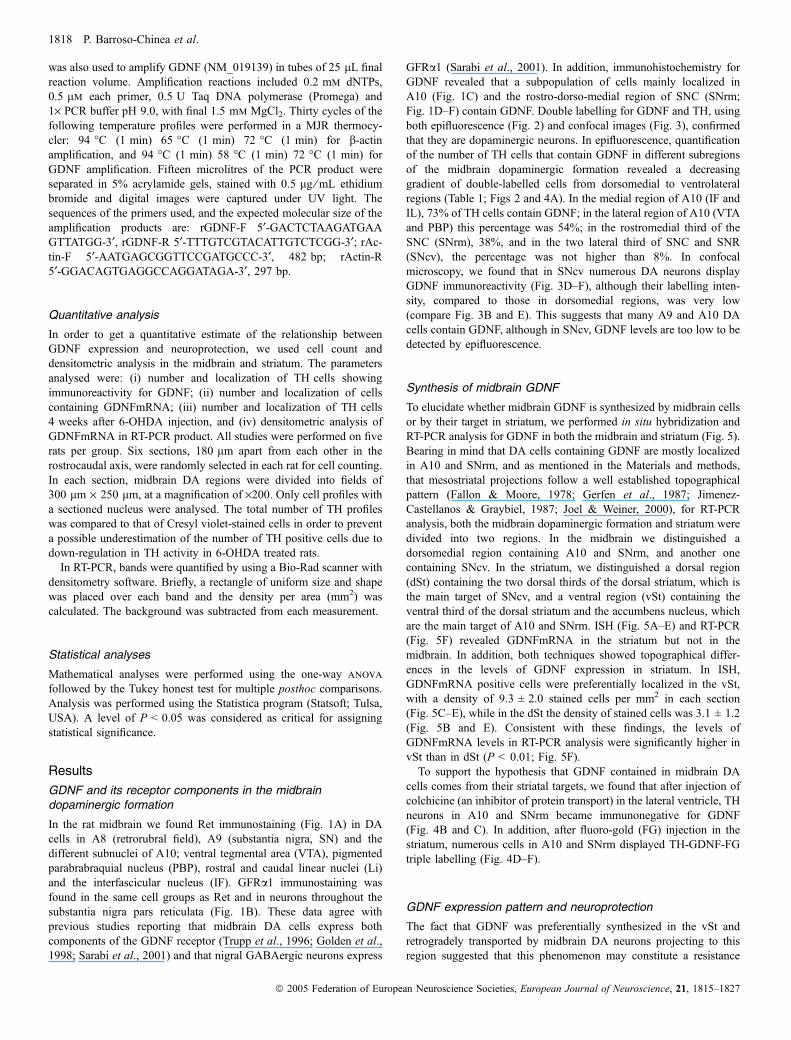
was also used to amplify GDNF (NM_019139) in tubes of 25 lL finalreaction volume. Amplification reactions included 0.2 mm dNTPs,0.5 lm each primer, 0.5 U Taq DNA polymerase (Promega) and1· PCR buffer pH 9.0, with final 1.5 mm MgCl2. Thirty cycles of thefollowing temperature profiles were performed in a MJR thermocy-cler: 94 �C (1 min) 65 �C (1 min) 72 �C (1 min) for b-actinamplification, and 94 �C (1 min) 58 �C (1 min) 72 �C (1 min) forGDNF amplification. Fifteen microlitres of the PCR product wereseparated in 5% acrylamide gels, stained with 0.5 lg ⁄mL ethidiumbromide and digital images were captured under UV light. Thesequences of the primers used, and the expected molecular size of theamplification products are: rGDNF-F 5¢-GACTCTAAGATGAAGTTATGG-3¢, rGDNF-R 5¢-TTTGTCGTACATTGTCTCGG-3¢; rAc-tin-F 5¢-AATGAGCGGTTCCGATGCCC-3¢, 482 bp; rActin-R5¢-GGACAGTGAGGCCAGGATAGA-3¢, 297 bp.
Quantitative analysis
In order to get a quantitative estimate of the relationship betweenGDNF expression and neuroprotection, we used cell count anddensitometric analysis in the midbrain and striatum. The parametersanalysed were: (i) number and localization of TH cells showingimmunoreactivity for GDNF; (ii) number and localization of cellscontaining GDNFmRNA; (iii) number and localization of TH cells4 weeks after 6-OHDA injection, and (iv) densitometric analysis ofGDNFmRNA in RT-PCR product. All studies were performed on fiverats per group. Six sections, 180 lm apart from each other in therostrocaudal axis, were randomly selected in each rat for cell counting.In each section, midbrain DA regions were divided into fields of300 lm · 250 lm, at a magnification of ·200. Only cell profiles witha sectioned nucleus were analysed. The total number of TH profileswas compared to that of Cresyl violet-stained cells in order to preventa possible underestimation of the number of TH positive cells due todown-regulation in TH activity in 6-OHDA treated rats.In RT-PCR, bands were quantified by using a Bio-Rad scanner with
densitometry software. Briefly, a rectangle of uniform size and shapewas placed over each band and the density per area (mm2) wascalculated. The background was subtracted from each measurement.
Statistical analyses
Mathematical analyses were performed using the one-way anova
followed by the Tukey honest test for multiple posthoc comparisons.Analysis was performed using the Statistica program (Statsoft; Tulsa,USA). A level of P < 0.05 was considered as critical for assigningstatistical significance.
Results
GDNF and its receptor components in the midbraindopaminergic formation
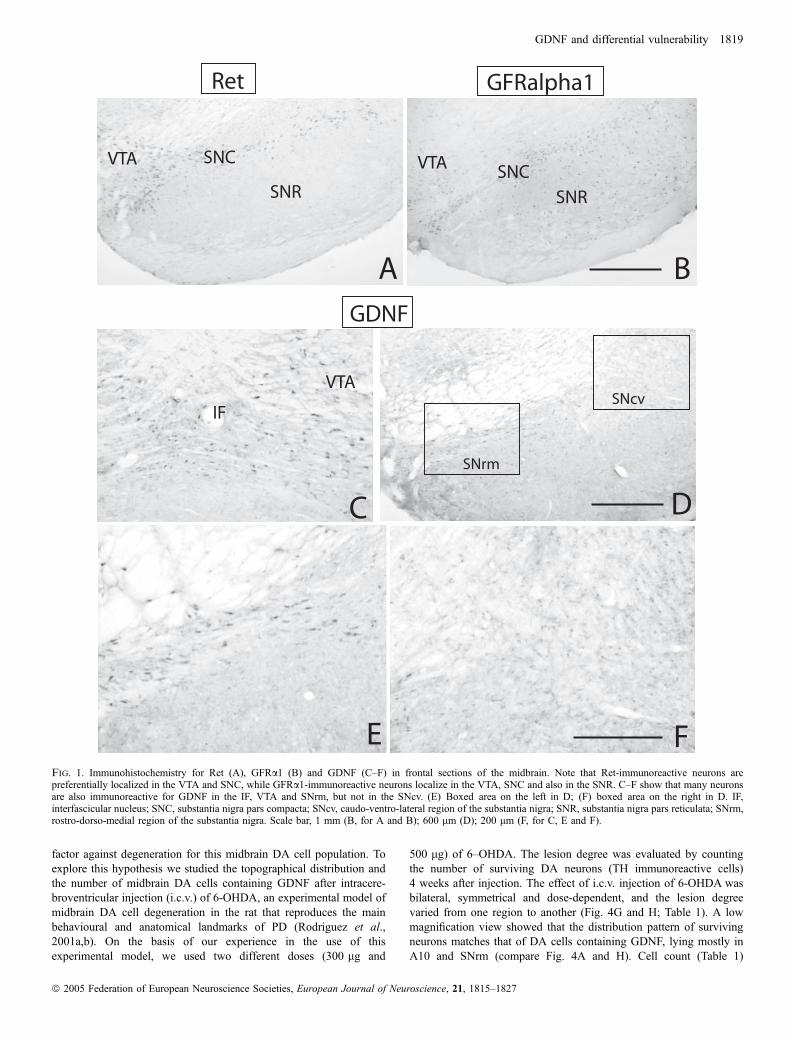
In the rat midbrain we found Ret immunostaining (Fig. 1A) in DAcells in A8 (retrorubral field), A9 (substantia nigra, SN) and thedifferent subnuclei of A10; ventral tegmental area (VTA), pigmentedparabrabraquial nucleus (PBP), rostral and caudal linear nuclei (Li)and the interfascicular nucleus (IF). GFRa1 immunostaining wasfound in the same cell groups as Ret and in neurons throughout thesubstantia nigra pars reticulata (Fig. 1B). These data agree withprevious studies reporting that midbrain DA cells express bothcomponents of the GDNF receptor (Trupp et al., 1996; Golden et al.,1998; Sarabi et al., 2001) and that nigral GABAergic neurons express
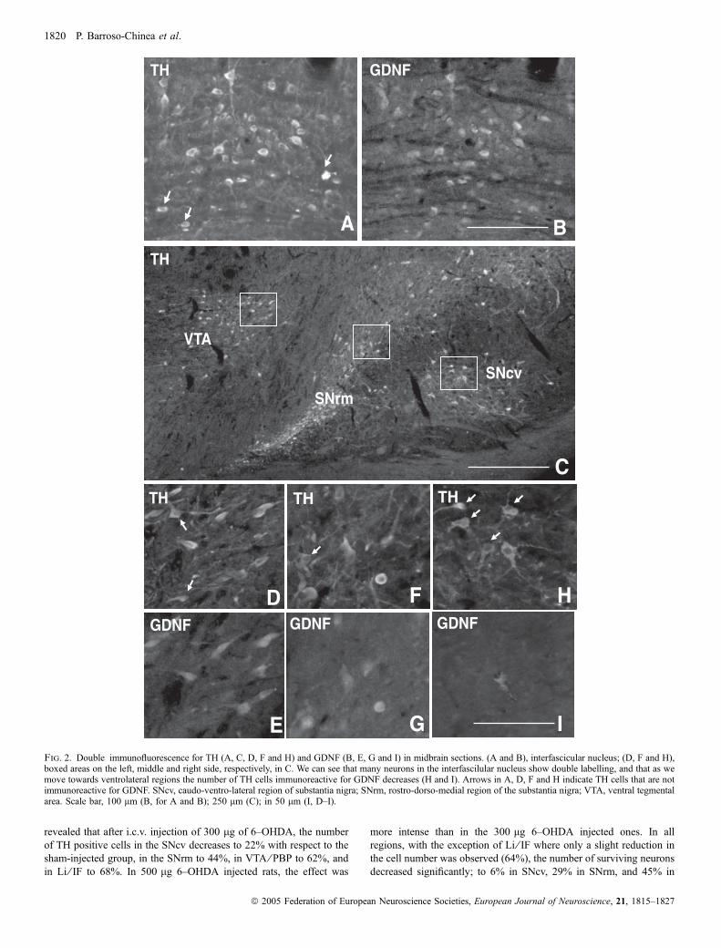
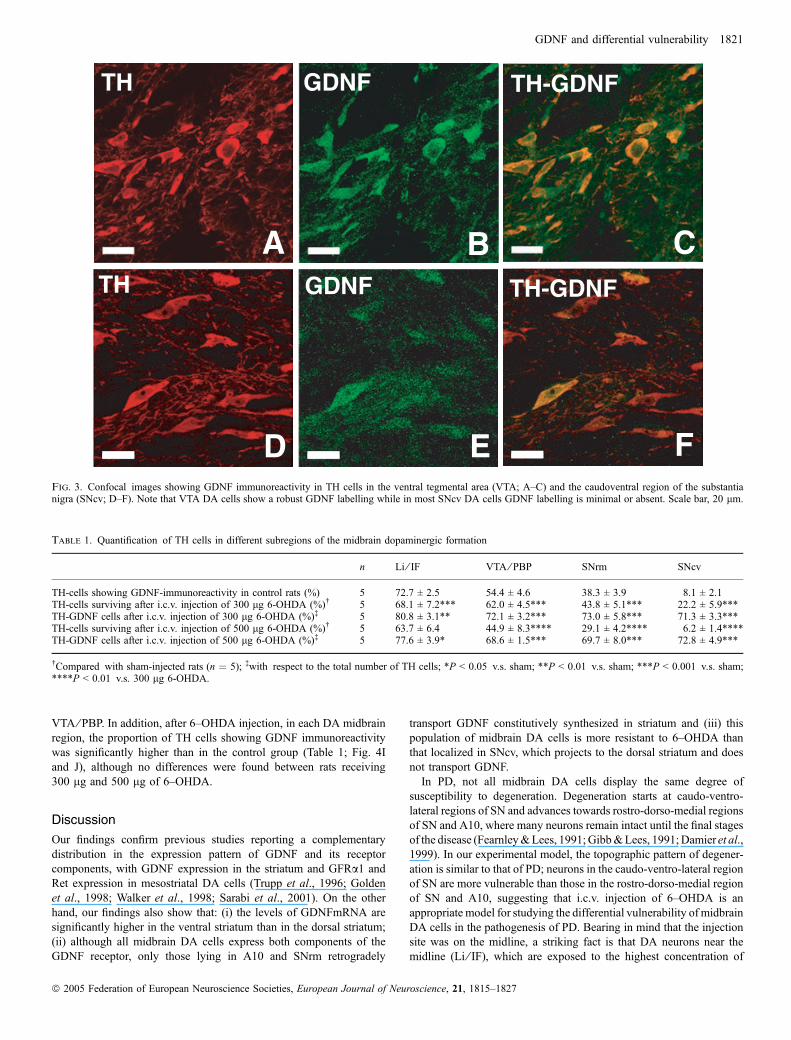
GFRa1 (Sarabi et al., 2001). In addition, immunohistochemistry forGDNF revealed that a subpopulation of cells mainly localized inA10 (Fig. 1C) and the rostro-dorso-medial region of SNC (SNrm;Fig. 1D–F) contain GDNF. Double labelling for GDNF and TH, usingboth epifluorescence (Fig. 2) and confocal images (Fig. 3), confirmedthat they are dopaminergic neurons. In epifluorescence, quantificationof the number of TH cells that contain GDNF in different subregionsof the midbrain dopaminergic formation revealed a decreasinggradient of double-labelled cells from dorsomedial to ventrolateralregions (Table 1; Figs 2 and 4A). In the medial region of A10 (IF andIL), 73% of TH cells contain GDNF; in the lateral region of A10 (VTAand PBP) this percentage was 54%; in the rostromedial third of theSNC (SNrm), 38%, and in the two lateral third of SNC and SNR(SNcv), the percentage was not higher than 8%. In confocalmicroscopy, we found that in SNcv numerous DA neurons displayGDNF immunoreactivity (Fig. 3D–F), although their labelling inten-sity, compared to those in dorsomedial regions, was very low(compare Fig. 3B and E). This suggests that many A9 and A10 DAcells contain GDNF, although in SNcv, GDNF levels are too low to bedetected by epifluorescence.
Synthesis of midbrain GDNF
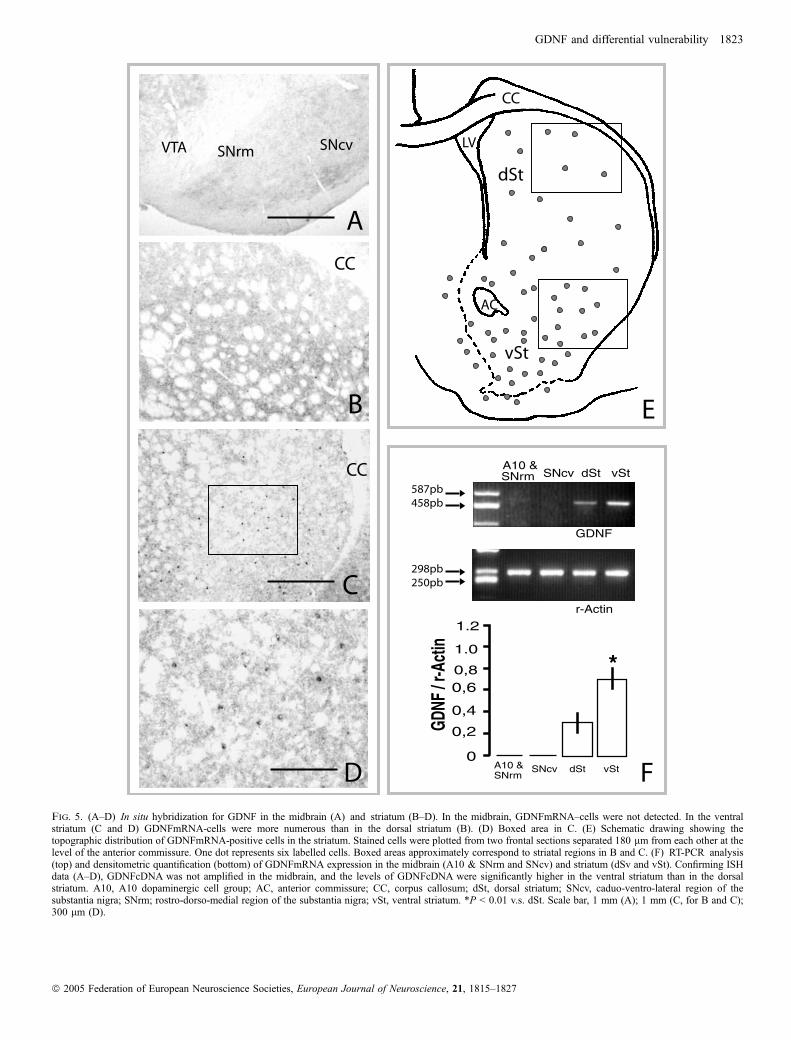
To elucidate whether midbrain GDNF is synthesized by midbrain cellsor by their target in striatum, we performed in situ hybridization andRT-PCR analysis for GDNF in both the midbrain and striatum (Fig. 5).Bearing in mind that DA cells containing GDNF are mostly localizedin A10 and SNrm, and as mentioned in the Materials and methods,that mesostriatal projections follow a well established topographicalpattern (Fallon & Moore, 1978; Gerfen et al., 1987; Jimenez-Castellanos & Graybiel, 1987; Joel & Weiner, 2000), for RT-PCRanalysis, both the midbrain dopaminergic formation and striatum weredivided into two regions. In the midbrain we distinguished adorsomedial region containing A10 and SNrm, and another onecontaining SNcv. In the striatum, we distinguished a dorsal region(dSt) containing the two dorsal thirds of the dorsal striatum, which isthe main target of SNcv, and a ventral region (vSt) containing theventral third of the dorsal striatum and the accumbens nucleus, whichare the main target of A10 and SNrm. ISH (Fig. 5A–E) and RT-PCR(Fig. 5F) revealed GDNFmRNA in the striatum but not in themidbrain. In addition, both techniques showed topographical differ-ences in the levels of GDNF expression in striatum. In ISH,GDNFmRNA positive cells were preferentially localized in the vSt,with a density of 9.3 ± 2.0 stained cells per mm2 in each section(Fig. 5C–E), while in the dSt the density of stained cells was 3.1 ± 1.2(Fig. 5B and E). Consistent with these findings, the levels ofGDNFmRNA levels in RT-PCR analysis were significantly higher invSt than in dSt (P < 0.01; Fig. 5F).To support the hypothesis that GDNF contained in midbrain DA
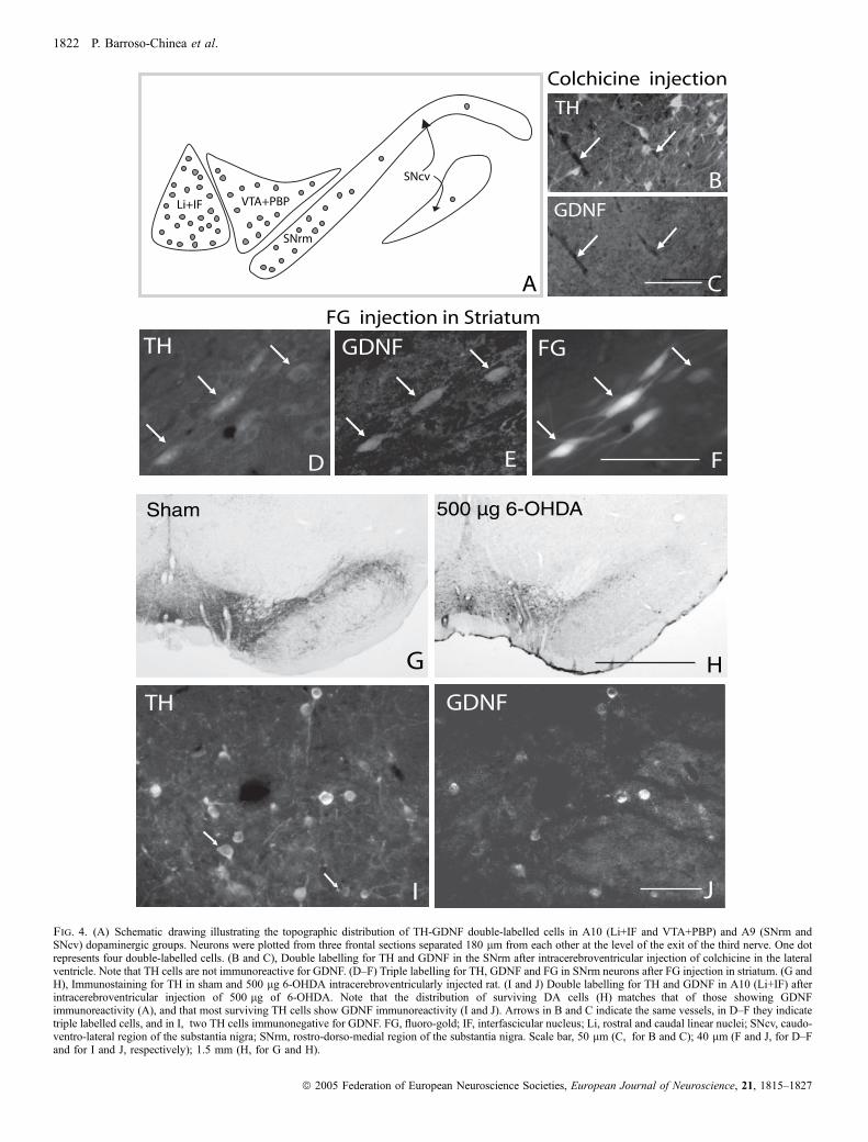
cells comes from their striatal targets, we found that after injection ofcolchicine (an inhibitor of protein transport) in the lateral ventricle, THneurons in A10 and SNrm became immunonegative for GDNF(Fig. 4B and C). In addition, after fluoro-gold (FG) injection in thestriatum, numerous cells in A10 and SNrm displayed TH-GDNF-FGtriple labelling (Fig. 4D–F).
GDNF expression pattern and neuroprotection
The fact that GDNF was preferentially synthesized in the vSt andretrogradely transported by midbrain DA neurons projecting to thisregion suggested that this phenomenon may constitute a resistance
1818 P. Barroso-Chinea et al.
ª 2005 Federation of European Neuroscience Societies, European Journal of Neuroscience, 21, 1815–1827
factor against degeneration for this midbrain DA cell population. Toexplore this hypothesis we studied the topographical distribution andthe number of midbrain DA cells containing GDNF after intracere-broventricular injection (i.c.v.) of 6-OHDA, an experimental model ofmidbrain DA cell degeneration in the rat that reproduces the mainbehavioural and anatomical landmarks of PD (Rodriguez et al.,2001a,b). On the basis of our experience in the use of thisexperimental model, we used two different doses (300 lg and
500 lg) of 6–OHDA. The lesion degree was evaluated by countingthe number of surviving DA neurons (TH immunoreactive cells)4 weeks after injection. The effect of i.c.v. injection of 6-OHDA wasbilateral, symmetrical and dose-dependent, and the lesion degreevaried from one region to another (Fig. 4G and H; Table 1). A lowmagnification view showed that the distribution pattern of survivingneurons matches that of DA cells containing GDNF, lying mostly inA10 and SNrm (compare Fig. 4A and H). Cell count (Table 1)
Fig. 1. Immunohistochemistry for Ret (A), GFRa1 (B) and GDNF (C–F) in frontal sections of the midbrain. Note that Ret-immunoreactive neurons arepreferentially localized in the VTA and SNC, while GFRa1-immunoreactive neurons localize in the VTA, SNC and also in the SNR. C–F show that many neuronsare also immunoreactive for GDNF in the IF, VTA and SNrm, but not in the SNcv. (E) Boxed area on the left in D; (F) boxed area on the right in D. IF,interfascicular nucleus; SNC, substantia nigra pars compacta; SNcv, caudo-ventro-lateral region of the substantia nigra; SNR, substantia nigra pars reticulata; SNrm,rostro-dorso-medial region of the substantia nigra. Scale bar, 1 mm (B, for A and B); 600 lm (D); 200 lm (F, for C, E and F).
GDNF and differential vulnerability 1819
ª 2005 Federation of European Neuroscience Societies, European Journal of Neuroscience, 21, 1815–1827
revealed that after i.c.v. injection of 300 lg of 6–OHDA, the numberof TH positive cells in the SNcv decreases to 22% with respect to thesham-injected group, in the SNrm to 44%, in VTA ⁄ PBP to 62%, andin Li ⁄ IF to 68%. In 500 lg 6–OHDA injected rats, the effect was
more intense than in the 300 lg 6–OHDA injected ones. In allregions, with the exception of Li ⁄ IF where only a slight reduction inthe cell number was observed (64%), the number of surviving neuronsdecreased significantly; to 6% in SNcv, 29% in SNrm, and 45% in
TH
B
TH GDNF
GDNF
A
TH
GDNF
E
D
TH
GDNFGDNF
TH
C
IG
HF
VTA
SNcv
SNrm
Fig. 2. Double immunofluorescence for TH (A, C, D, F and H) and GDNF (B, E, G and I) in midbrain sections. (A and B), interfascicular nucleus; (D, F and H),boxed areas on the left, middle and right side, respectively, in C. We can see that many neurons in the interfascilular nucleus show double labelling, and that as wemove towards ventrolateral regions the number of TH cells immunoreactive for GDNF decreases (H and I). Arrows in A, D, F and H indicate TH cells that are notimmunoreactive for GDNF. SNcv, caudo-ventro-lateral region of substantia nigra; SNrm, rostro-dorso-medial region of the substantia nigra; VTA, ventral tegmentalarea. Scale bar, 100 lm (B, for A and B); 250 lm (C); in 50 lm (I, D–I).
1820 P. Barroso-Chinea et al.
ª 2005 Federation of European Neuroscience Societies, European Journal of Neuroscience, 21, 1815–1827
VTA ⁄ PBP. In addition, after 6–OHDA injection, in each DA midbrainregion, the proportion of TH cells showing GDNF immunoreactivitywas significantly higher than in the control group (Table 1; Fig. 4Iand J), although no differences were found between rats receiving300 lg and 500 lg of 6–OHDA.
Discussion
Our findings confirm previous studies reporting a complementarydistribution in the expression pattern of GDNF and its receptorcomponents, with GDNF expression in the striatum and GFRa1 andRet expression in mesostriatal DA cells (Trupp et al., 1996; Goldenet al., 1998; Walker et al., 1998; Sarabi et al., 2001). On the otherhand, our findings also show that: (i) the levels of GDNFmRNA aresignificantly higher in the ventral striatum than in the dorsal striatum;(ii) although all midbrain DA cells express both components of theGDNF receptor, only those lying in A10 and SNrm retrogradely
transport GDNF constitutively synthesized in striatum and (iii) thispopulation of midbrain DA cells is more resistant to 6–OHDA thanthat localized in SNcv, which projects to the dorsal striatum and doesnot transport GDNF.In PD, not all midbrain DA cells display the same degree of
susceptibility to degeneration. Degeneration starts at caudo-ventro-lateral regions of SN and advances towards rostro-dorso-medial regionsof SN and A10, where many neurons remain intact until the final stagesof the disease (Fearnley&Lees, 1991;Gibb&Lees, 1991;Damier et al.,1999). In our experimental model, the topographic pattern of degener-ation is similar to that of PD; neurons in the caudo-ventro-lateral regionof SN are more vulnerable than those in the rostro-dorso-medial regionof SN and A10, suggesting that i.c.v. injection of 6–OHDA is anappropriate model for studying the differential vulnerability of midbrainDA cells in the pathogenesis of PD. Bearing in mind that the injectionsite was on the midline, a striking fact is that DA neurons near themidline (Li ⁄ IF), which are exposed to the highest concentration of
TH
ATH
GDNF
GDNF
TH-GDNF
TH-GDNF
D
B C
E FFig. 3. Confocal images showing GDNF immunoreactivity in TH cells in the ventral tegmental area (VTA; A–C) and the caudoventral region of the substantianigra (SNcv; D–F). Note that VTA DA cells show a robust GDNF labelling while in most SNcv DA cells GDNF labelling is minimal or absent. Scale bar, 20 lm.
Table 1. Quantification of TH cells in different subregions of the midbrain dopaminergic formation
n Li ⁄ IF VTA ⁄ PBP SNrm SNcv
TH-cells showing GDNF-immunoreactivity in control rats (%) 5 72.7 ± 2.5 54.4 ± 4.6 38.3 ± 3.9 8.1 ± 2.1TH-cells surviving after i.c.v. injection of 300 lg 6-OHDA (%)� 5 68.1 ± 7.2*** 62.0 ± 4.5*** 43.8 ± 5.1*** 22.2 ± 5.9***TH-GDNF cells after i.c.v. injection of 300 lg 6-OHDA (%)� 5 80.8 ± 3.1** 72.1 ± 3.2*** 73.0 ± 5.8*** 71.3 ± 3.3***TH-cells surviving after i.c.v. injection of 500 lg 6-OHDA (%)� 5 63.7 ± 6.4 44.9 ± 8.3**** 29.1 ± 4.2**** 6.2 ± 1.4****TH-GDNF cells after i.c.v. injection of 500 lg 6-OHDA (%)� 5 77.6 ± 3.9* 68.6 ± 1.5*** 69.7 ± 8.0*** 72.8 ± 4.9***
�Compared with sham-injected rats (n ¼ 5); �with respect to the total number of TH cells; *P < 0.05 v.s. sham; **P < 0.01 v.s. sham; ***P < 0.001 v.s. sham;****P < 0.01 v.s. 300 lg 6-OHDA.
GDNF and differential vulnerability 1821
ª 2005 Federation of European Neuroscience Societies, European Journal of Neuroscience, 21, 1815–1827
Li+IF VTA+PBP
SNrm
SNcv
D
HG
B
A C
TH GDNF FG
TH
GDNF
Colchicine injection
500 µg 6-OHDA
E FD
FG injection in Striatum
Sham
TH GDNF
JI
Fig. 4. (A) Schematic drawing illustrating the topographic distribution of TH-GDNF double-labelled cells in A10 (Li+IF and VTA+PBP) and A9 (SNrm andSNcv) dopaminergic groups. Neurons were plotted from three frontal sections separated 180 lm from each other at the level of the exit of the third nerve. One dotrepresents four double-labelled cells. (B and C), Double labelling for TH and GDNF in the SNrm after intracerebroventricular injection of colchicine in the lateralventricle. Note that TH cells are not immunoreactive for GDNF. (D–F) Triple labelling for TH, GDNF and FG in SNrm neurons after FG injection in striatum. (G andH), Immunostaining for TH in sham and 500 lg 6-OHDA intracerebroventricularly injected rat. (I and J) Double labelling for TH and GDNF in A10 (Li+IF) afterintracerebroventricular injection of 500 lg of 6-OHDA. Note that the distribution of surviving DA cells (H) matches that of those showing GDNFimmunoreactivity (A), and that most surviving TH cells show GDNF immunoreactivity (I and J). Arrows in B and C indicate the same vessels, in D–F they indicatetriple labelled cells, and in I, two TH cells immunonegative for GDNF. FG, fluoro-gold; IF, interfascicular nucleus; Li, rostral and caudal linear nuclei; SNcv, caudo-ventro-lateral region of the substantia nigra; SNrm, rostro-dorso-medial region of the substantia nigra. Scale bar, 50 lm (C, for B and C); 40 lm (F and J, for D–Fand for I and J, respectively); 1.5 mm (H, for G and H).
1822 P. Barroso-Chinea et al.
ª 2005 Federation of European Neuroscience Societies, European Journal of Neuroscience, 21, 1815–1827
SNcv dSt vSt
GDNF
r-Actin
SNrmVTA SNcv
CC
CC
A
FD
C
B
298pb250pb
587pb458pb
dSt
CC
AC
LV
vSt
E
0
0,2
0,4
0,6
GDN
F / r
-Act
in
0,8
1.0
1.2
A10 & SNrm
A10 & SNrm
SNcv dSt vSt
*
Fig. 5. (A–D) In situ hybridization for GDNF in the midbrain (A) and striatum (B–D). In the midbrain, GDNFmRNA–cells were not detected. In the ventralstriatum (C and D) GDNFmRNA-cells were more numerous than in the dorsal striatum (B). (D) Boxed area in C. (E) Schematic drawing showing thetopographic distribution of GDNFmRNA-positive cells in the striatum. Stained cells were plotted from two frontal sections separated 180 lm from each other at thelevel of the anterior commissure. One dot represents six labelled cells. Boxed areas approximately correspond to striatal regions in B and C. (F) RT-PCR analysis(top) and densitometric quantification (bottom) of GDNFmRNA expression in the midbrain (A10 & SNrm and SNcv) and striatum (dSv and vSt). Confirming ISHdata (A–D), GDNFcDNA was not amplified in the midbrain, and the levels of GDNFcDNA were significantly higher in the ventral striatum than in the dorsalstriatum. A10, A10 dopaminergic cell group; AC, anterior commissure; CC, corpus callosum; dSt, dorsal striatum; SNcv, caduo-ventro-lateral region of thesubstantia nigra; SNrm; rostro-dorso-medial region of the substantia nigra; vSt, ventral striatum. *P < 0.01 v.s. dSt. Scale bar, 1 mm (A); 1 mm (C, for B and C);300 lm (D).
GDNF and differential vulnerability 1823
ª 2005 Federation of European Neuroscience Societies, European Journal of Neuroscience, 21, 1815–1827
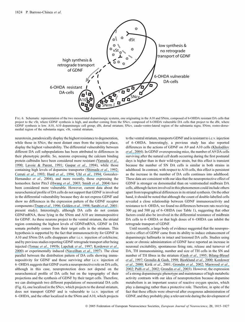
neurotoxin, paradoxically display the highest resistance to degeneration,while those in SNcv, the most distant ones from the injection place,display the highest vulnerability. The differential vulnerability betweendifferent DA cell subpopulations has been attributed to differences intheir phenotypic profile. So, neurons expressing the calcium bindingprotein calbindin have been considered more resistant (Yamada et al.,1990; Lavoie & Parent, 1991; Gaspar et al., 1994), while thosecontaining high levels of dopamine transporter (Shimada et al., 1992;Cerruti et al., 1993; Hurd et al., 1994; Uhl et al., 1994; Gonzalez-Hernandez et al., 2004), and more recently, those expressing thehomeobox factor Pitx3 (Hwang et al., 2003; Smidt et al., 2004) havebeen considered more vulnerable. However, current data about theneurochemical profile of DA cells do not support that GDNF is involvedin the differential vulnerability because they do not express GDNF andshow no differences in the expression pattern of the GDNF receptorcomponents (Trupp et al., 1996; Golden et al., 1998; Sarabi et al., 2001;present study). Interestingly, although DA cells do not containGDNFmRNA, those lying in the SNrm and A10 are immunopositivefor GDNF. As these neurons project to the ventral striatum, the striatalregion containing the highest levels of GDNFmRNA, GDNF in DAsomata probably comes from their target cells in the striatum. Thishypothesis is supported by the fact that immunoreactivity for GDNF inA10 and SNrm DA cells disappears after i.c.v. injection of colchicine,and by previous studies reportingGDNF retrograde transport after beinginjected (Tomac et al., 1995b; Lapchak et al., 1997; Kordower et al.,2000) or experimentally induced (Naveilhan et al., 1997). The closeparallel between the distribution pattern of DA cells showing immu-nopositivity for GDNF and those surviving after i.c.v. injection of6–OHDAsuggests thatGDNF exerts a neuroprotective role onDAcells,although in this case, neuroprotection does not depend on theneurochemical profile of DA cells but on the topography of theirprojections and the synthesis of GDNF by their target cells. Therefore,we can distinguish two different populations of mesostriatal DA cells(Fig. 6), one localized in the SNcv, which projects to the dorsal striatum,does not transport GDNF and is vulnerable to i.c.v. injection of6–OHDA, and the other localized in the SNrm and A10, which projects
to the ventral striatum, transportsGDNF and is resistant to i.c.v. injectionof 6–OHDA. Interestingly, a previous study has also reporteddifferences in the actions of GDNF on A9 and A10 cells (Kholodilovet al., 2004). In GDNF overexpressing mice, the number of A9DA cellssurviving after the natural cell death occurring during the first postnataldays is higher than in their wild-type strain, but this effect is transientbecause the number of SN DA cells is similar in both strains inadulthood. In contrast, with respect to A10 cells, this effect is persistentas the increase in the number of DA cells continues into adulthood.These data are consistent with our idea that the neuroprotective effect ofGDNF is stronger on dorsomedial than on ventromedial midbrain DAcells, although factors involved in this phenomenon could include othersapart from topographical differences in its striatal synthesis. On the otherhand, it should be noted that although the count of double-labelled cellsrevealed a close relationship between GDNF immunoreactivity andresistance to 6–OHDA, we found no differences between rats receiving300 lg and 500 lg of 6–OHDA (see Table 1), suggesting that otherfactors could also be involved in the differential resistance of midbrainDA cells to 6–OHDA or that high doses of 6–OHDA can inhibit theretrograde transport of GDNF.Until recently, a large body of evidence suggested that the neuropro-
tective effect of GDNF came from its ability to induce enhancement ofdopaminergic hallmarks in intact and lesioned DA cells. Studies usingacute or chronic administration of GDNF have reported an increase inneuronal excitability, spontaneous firing rate, release and turnover ofDA, TH immunostaining, number and size of TH cells in the SN andnumber of TH fibres in the striatum (Gash et al., 1995; Bilang-Bleuelet al., 1997; Grondin & Gash, 1998; Bjorklund et al., 2000; Kordoweret al., 2000; Kirik et al., 2001; Grondin et al., 2002; Maswood et al.,2002; Palfi et al., 2002; Grondin et al., 2003). However, the expressionof a strong dopaminergic phenotype and maintenance of high metabolicactivity contrasts with our idea of neuroprotection because dopaminemetabolism is an important source of reactive oxygen species, whichplay a damaging rather than a protective role. Therefore, in spite of thefact that these effects were observed after exogenous administration ofGDNF, and they probably play a relevant role during the development of
dSt
vSt
high synthesis &
A10SNrm
SNcv
retrograde transport of GDNF
no retrograde transport of GDNF
6-OHDA vulnerable DA-cells
low synthesis &
6-OHDA resistant DA-cells
Fig. 6. Schematic representation of the two mesostriatal dopaminergic systems, one originating in the A10 and SNrm, composed of 6-OHDA resistant DA cells thatproject to the vSt, where GDNF synthesis is high, and another coming from the SNcv, composed of 6-OHDA vulnerable DA cells that project to the dSt, whereGDNF synthesis is low. A10, A10 dopaminergic cell group; dSt, dorsal striatum; SNcv, caudo-ventro-lateral region of the substantia nigra; SNrm; rostro-dorso-medial region of the substantia nigra; vSt, ventral striatum.
1824 P. Barroso-Chinea et al.
ª 2005 Federation of European Neuroscience Societies, European Journal of Neuroscience, 21, 1815–1827
DA cells, it is improbable that under physiological conditions they areresponsible for a long-term neuroprotective effect in adulthood.Interestingly, other studies report that sustained infusion (Lu & Hagg,1997) or lentiviral vector delivery (Rosenblad et al., 2003; Georgievskaet al., 2004) of GDNF produce a reduction in the levels of mRNA andprotein TH as well as its enzymatic activity, suggesting that GDNFinduces TH down-regulation by acting directly on its gene expression.On the other hand, it is known that the signalling pathway of GDNF isregulated by protein and lipid kinases and phospholipases (Saarma,2000, 2001; Paratcha et al., 2001; Sariola & Saarma, 2003), bymeans ofwhich it induces THphosphorylation (Salvatore et al., 2004). So, GDNFcan exert its neuroprotective effect acting at transcriptional andtransductional levels in the synthesis of different molecules thatdetermine phenotypic differences in DA cells. In this respect, it isinteresting to note that the levels of THmRNA and DAT mRNA andprotein (Blanchard et al., 1994; Hurd et al., 1994; Haber et al., 1995;Gonzalez-Hernandez et al., 2004) are lower inDA cells lying inA10 andSNrm than in those lying in the SNcv, although the possibility that thesephenotypic differences might be due to differences in GDNF expressionin their target cells remains to be explored.
In spite of the fact that the retrograde transport of GDNF wasdescribed for the first time ten years ago (Tomac et al., 1995b) andthereafter supported by several studies (Lapchak et al., 1997;Naveilhan et al., 1997; Kordower et al., 2000), with respect to themolecular mechanisms underlying this phenomenon, we only knowthat it may be internalized together with GFRa1, independently of Rettyrosine kinase activity (Vieira et al., 2003), and its functionalmeaning remains unknown. In contrast to the neurotrophins (nervegrowth factor, brain derived neurotrophic factor and neurotrophin 3and 4), whose presence in the soma of afferent neurons is substantiatedby the fact that their biological actions require binding to tyrosinekinase receptors in axon terminals and the subsequent internalizationand retrograde transport of the complex to the cell body (Grimes et al.,1996; Howe et al., 2001; Ginty & Segal, 2002), in the case of GDNF,although we know a lot about its complex signalling biology (Saarma,2000, 2001 Paratcha et al., 2001; Sarabi et al., 2001), no pathwayscurrently proposed include internalization and transport of the ligand.Our results show that GDNF is internalized and retrogradelytransported, not only after being injected or when its overexpressionis experimentally induced, but also under physiological conditions.Further studies should be addressed to elucidating the molecularmechanisms responsible for its internalization and transport, and moreimportantly, whether this is a new signalling pathway involved inneuroprotection or only a route for its degradation.
Acknowledgements
Partially supported by the Gobierno Autonomo de Canarias (PI 042004/047)and Ministerio de Educacion y Ciencia de Espana (grant number BFU2004-05756). PBC is supported by a grant from Fundacion Canaria de Investigaciony Salud (FUNCIS), and ICM is supported by a grant from red de Centros deInvestigacion de Enfermedades Neurologicas (CIEN).
Abbreviations
DA, dopaminergic; dSt, dorsal striatum; FG, fluoro-gold; GDNF, glial cell line-derived neurotrophic factor; GFRa1, a subunit of GDNF receptor; IF,interfascicular nucleus; ISH, in situ hybridization; Li, rostral and caudal linearnuclei; PBP, parabrachial pigmented nucleus; PD, Parkinson’s disease; Ret,tyrosine kinase signalling subunit of GDNF receptor; SN, substantia nigra;SNC, substantia nigra, pars compacta; SNcv, caudo-ventro-lateral region of SN;SNR, substantia nigra, pars reticulata; SNrm, rostro-dorso-medial region of theSN; TH, tyrosine hydroxylase; vSt, ventral striatum; VTA, ventral tegmentalarea; 6-OHDA, 6- hydroxydopamine.
References
Airaksinen, M.S., Titievsky, A. & Saarma, M. (1999) GDNF familyneurotrophic factor signaling: four masters, one servant?Mol. Cell Neurosci.,13, 313–325.
Arenas, E., Trupp, M., Akerud, P. & Ibanez, C.F. (1995) GDNF preventsdegeneration and promotes the phenotype of brain noradrenergic neuronsin vivo. Neuron, 15, 1465–1473.
Baloh, R.H., Enomoto, H., Johnson, E.M. Jr & Milbrandt, J. (2000) The GDNFfamily ligands and receptors – implications for neural development. Curr.Opin. Neurobiol., 10, 103–110.
Bernheimer, H., Birkmayer, W., Hornykiewicz, O., Jellinger, K. & Seitelberger,F. (1973) Brain dopamine and the syndromes of Parkinson and Huntington.Clinical, morphological and neurochemical correlations. J. Neurol. Sci., 20,415–455.
Bilang-Bleuel, A., Revah, F., Colin, P., Locquet, I., Robert, J.J., Mallet, J. &Horellou, P. (1997) Intrastriatal injection of an adenoviral vector expressingglial-cell-line-derived neurotrophic factor prevents dopaminergic neurondegeneration and behavioral impairment in a rat model of Parkinson disease.Proc. Natl Acad. Sci. USA, 94, 8818–8823.
Bjorklund, A., Kirik, D., Rosenblad, C., Georgievska, B., Lundberg, C. &Mandel, R.J. (2000) Towards a neuroprotective gene therapy for Parkinson’sdisease: use of adenovirus, AAV and lentivirus vectors for gene transfer ofGDNF to the nigrostriatal system in the rat Parkinson model. Brain Res.,886, 82–98.
Blanchard, V., Raisman-Vozari, R., Vyas, S., Michel, P.P., Javoy-Agid, F.,Uhl, G. & Agid, Y. (1994) Differential expression of tyrosine hydroxylaseand membrane dopamine transporter genes in subpopulations of dopami-nergic neurons of the rat mesencephalon. Brain Res. Mol. Brain Res., 22,29–38.
Blum, M. & Weickert, C.S. (1995) GDNF mRNA expression in normalpostnatal development, aging, and in Weaver mutant mice. Neurobiol. Aging,16, 925–929.
Bourque, M.J. & Trudeau, L.E. (2000) GDNF enhances the synaptic efficacy ofdopaminergic neurons in culture. Eur. J. Neurosci., 12, 3172–3180.
Cerruti, C., Walther, D.M., Kuhar, M.J. & Uhl, G.R. (1993) Dopaminetransporter mRNA expression is intense in rat midbrain neurons and modestoutside midbrain. Brain Res. Mol. Brain Res., 18, 181–186.
Chiueh, C.C., Burns, R.S., Markey, S.P., Jacobowitz, D.M. & Kopin, I.J. (1985)Primate model of parkinsonism: selective lesion of nigrostriatal neurons by1-methyl-4-phenyl-1,2,3,6-tetrahydropyridine produces an extrapyramidalsyndrome in rhesus monkeys. Life Sci., 36, 213–218.
Choi-Lundberg, D.L. & Bohn, M.C. (1995) Ontogeny and distribution of glialcell line-derived neurotrophic factor (GDNF) mRNA in rat. Brain Res. Dev.Brain Res., 85, 80–88.
Dahlstrom, A. & Fuxe, K. (1964) Evidence for the existence of monoamine-containing neurons in the central nervous system – I. Demonstration ofmonoamines in the cell bodies of brain stem neurons. Acta Physiol. Scand.,62, 1–55.
Damier, P., Hirsch, E.C., Agid, Y. & Graybiel, A.M. (1999) The substantianigra of the human brain. II. Patterns of loss of dopamine-containing neuronsin Parkinson’s disease. Brain, 122, 1437–1448.
Dressler, G. (2002) Tubulogenesis in the developing mammalian kidney.Trends Cell Biol., 12, 390–395.
Fallon, J.H. & Moore, R.Y. (1978) Catecholamine innervation of the basalforebrain. IV. Topography of the dopamine projection to the basal forebrainand neostriatum. J. Comp. Neurol., 180, 545–580.
Fearnley, J.M. & Lees, A.J. (1991) Ageing and Parkinson’s disease: substantianigra regional selectivity. Brain, 114, 2283–2301.
Gash, D.M., Zhang, Z., Cass, W.A., Ovadia, A., Simmerman, L., Martin, D.,Russell, D., Collins, F., Hoffer, B.J. & Gerhardt, G.A. (1995) Morphologicaland functional effects of intranigrally administered GDNF in normal rhesusmonkeys. J. Comp. Neurol., 363, 345–358.
Gaspar, P., Ben Jelloun, N. & Febvret, A. (1994) Sparing of the dopaminergicneurons containing calbindin-D28k and of the dopaminergic mesocorticalprojections in weaver mutant mice. Neuroscience, 61, 293–305.
Georgievska, B., Kirik, D. & Bjorklund, A. (2004) Overexpression of glial cellline-derived neurotrophic factor using a lentiviral vector induces time- anddose-dependent downregulation of tyrosine hydroxylase in the intactnigrostriatal dopamine system. J. Neurosci., 24, 6437–6445.
Gerfen, C.R., Herkenham, M. & Thibault, J. (1987) The neostriatal mosaic. II.Patch- and matrix-directed mesostriatal dopaminergic and non-dopaminergicsystems. J. Neurosci., 7, 3915–3934.
Gibb, W.R. & Lees, A.J. (1991) Anatomy, pigmentation, ventral and dorsalsubpopulations of the substantia nigra, and differential cell death inParkinson’s disease. J. Neurol. Neurosurg. Psychiatry, 54, 388–396.
GDNF and differential vulnerability 1825
ª 2005 Federation of European Neuroscience Societies, European Journal of Neuroscience, 21, 1815–1827
Gill, S.S., Patel, N.K., Hotton, G.R., O’Sullivan, K., McCarter, R., Bunnage,M., Brooks, D.J., Svendsen, C.N. & Heywood, P. (2003) Direct braininfusion of glial cell line-derived neurotrophic factor in Parkinson disease.Nature Med., 9, 589–595.
Ginty, D.D. & Segal, R.A. (2002) Retrograde neurotrophin signaling: Trk-ingalong the axon. Curr. Opin. Neurobiol., 12, 268–274.
Golden, J.P., Baloh, R.H., Kotzbauer, P.T., Lampe, P.A., Osborne, P.A.,Milbrandt, J. & Johnson, E.M. Jr (1998) Expression of neurturin, GDNF, andtheir receptors in the adult mouse CNS. J. Comp. Neurol., 398, 139–150.
Gonzalez-Hernandez, T., Barroso-Chinea, P., De La CruZ. Muros, I., Del Mar.Perez-Delgado, M. & Rodriguez, M. (2004) Expression of dopamine andvesicular monoamine transporters and differential vulnerability of mesos-triatal dopaminergic neurons. J. Comp. Neurol., 479, 198–215.
Gonzalez-Hernandez, T. & Rodriguez, M. (2000) Compartmental organizationand chemical profile of dopaminergic and GABAergic neurons in thesubstantia nigra of the rat. J. Comp. Neurol., 421, 107–135.
Grimes, M.L., Zhou, J., Beattie, E.C., Yuen, E.C., Hall, D.E., Valletta, J.S.,Topp, K.S., LaVail, J.H., Bunnett, N.W. & Mobley, W.C. (1996) Endocytosisof activated TrkA: evidence that nerve growth factor induces formation ofsignaling endosomes. J. Neurosci., 16, 7950–7964.
Grondin, R., Cass, W.A., Zhang, Z., Stanford, J.A., Gash, D.M. & Gerhardt,G.A. (2003) Glial cell line-derived neurotrophic factor increases stimulus-evoked dopamine release and motor speed in aged rhesus monkeys.J. Neurosci., 23, 1974–1980.
Grondin, R. & Gash, D.M. (1998) Glial cell line-derived neurotrophic factor(GDNF): a drug candidate for the treatment of Parkinson’s disease.J. Neurol., 245, 35–42.
Grondin, R., Zhang, Z., Yi, A., Cass, W.A., Maswood, N., Andersen, A.H.,Elsberry, D.D., Klein, M.C., Gerhardt, G.A. & Gash, D.M. (2002) Chronic,controlled GDNF infusion promotes structural and functional recovery inadvanced parkinsonian monkeys. Brain, 125, 2191–2201.
Haber, S.N., Ryoo, H., Cox, C. & Lu, W. (1995) Subsets of midbraindopaminergic neurons in monkeys are distinguished by different levels ofmRNA for the dopamine transporter: comparison with the mRNA for the D2receptor, tyrosine hydroxylase and calbindin immunoreactivity. J. Comp.Neurol., 362, 400–410.
Henderson, C.E., Phillips, H.S., Pollock, R.A., Davies, A.M., Lemeulle, C.,Armanini, M., Simmons, L., Moffet, B., Vandlen, R.A. & Simpson, L.C.(1994) GDNF: a potent survival factor for motoneurons present in peripheralnerve and muscle. Science, 266, 1062–1064.
Herrero, M.T., Perez-Otano, I., Oset, C., Kastner, A., Hirsch, E.C., Agid, Y.,Luquin, M.R., Obeso, J.A. & Del Rio, J. (1993) GM-1 ganglioside promotesthe recovery of surviving midbrain dopaminergic neurons in MPTP-treatedmonkeys. Neuroscience, 56, 965–972.
Hirsch, E., Graybiel, A.M. & Agid, Y.A. (1988) Melanized dopaminergicneurons are differentially susceptible to degeneration in Parkinson’s disease.Nature, 334, 345–348.
Howe, C.L., Valletta, J.S., Rusnak, A.S. & Mobley, W.C. (2001) NGF signalingfrom clathrin-coated vesicles: evidence that signaling endosomes serve as aplatform for the Ras-MAPK pathway. Neuron, 32, 801–814.
Hurd, Y.L., Pristupa, Z.B., Herman, M.M., Niznik, H.B. & Kleinman, J.E.(1994) The dopamine transporter and dopamine D2 receptor messengerRNAs are differentially expressed in limbic- and motor-related subpopula-tions of human mesencephalic neurons. Neuroscience, 63, 357–362.
Hwang, D.Y., Ardayfio, P., Kang, U.J., Semina, E.V. & Kim, K.S. (2003)Selective loss of dopaminergic neurons in the substantia nigra of Pitx3-deficient aphakia mice. Brain Res. Mol. Brain Res., 114, 123–131.
Jimenez-Castellanos, J. & Graybiel, A.M. (1987) Subdivisions of thedopamine-containing A8–A9–A10 complex identified by their differentialmesostriatal innervation of striosomes and extrastriosomal matrix. Neuro-science, 23, 223–242.
Jing, S., Wen, D., YuY., Holst, P.L., Luo, Y., Fang, M., Tamir, R., Antonio, L.,Hu, Z., Cupples, R., Louis, J.C., Hu, S., Altrock, B.W. & Fox, G.M. (1996)GDNF-induced activation of the ret protein tyrosine kinase is mediated byGDNFR-alpha, a novel receptor for GDNF. Cell, 85, 1113–1124.
Joel, D. & Weiner, I. (2000) The connections of the dopaminergic system withthe striatum in rats and primates: an analysis with respect to the functionaland compartmental organization of the striatum. Neuroscience, 96, 451–474.
Kholodilov, N., Yarygina, O., Oo, T.F., Zhang, H., Sulzer, D., Dauer, W. &Burke, R.E. (2004) Regulation of the development of mesencephalicdopaminergic systems by the selective expression of glial cell line-derivedneurotrophic factor in their targets. J. Neurosci., 24, 3136–3146.
Kirik, D., Georgievska, B., Rosenblad, C. & Bjorklund, A. (2001) Delayedinfusion of GDNF promotes recovery of motor function in the partial lesionmodel of Parkinson’s disease. Eur. J. Neurosci., 13, 1589–1599.
Kordower, J.H., Emborg, M.E., Bloch, J., Ma, S.Y., Chu, Y., Leventhal, L.,McBride, J., Chen, E.Y., Palfi, S., Roitberg, B.Z., Brown, W.D., Holden, J.E.,Pyzalski, R., Taylor, M.D., Carvey, P., Ling, Z., Trono, D., Hantraye, P.,Deglon, N. & Aebischer, P. (2000) Neurodegeneration prevented bylentiviral vector delivery of GDNF in primate models of Parkinson’sdisease. Science, 290, 767–773.
Kordower, J.H., Palfi, S., Chen, E.Y., Ma, S.Y., Sendera, T., Cochran, E.J.,Mufson, E.J., Penn, R., Goetz, C.G. & Comella, C.D. (1999) Clinicopatho-logical findings following intraventricular glial-derived neurotrophicfactor treatment in a patient with Parkinson’s disease. Ann. Neurol., 46,419–424.
Krieglstein, K. (2004) Factors promoting survival of mesencephalic dopam-inergic neurons. Cell Tissue Res., 318, 73–80.
Lapchak, P.A., Jiao, S., Collins, F. & Miller, P.J. (1997) Glial cell line-derivedneurotrophic factor: distribution and pharmacology in the rat following abolus intraventricular injection. Brain Res., 747, 92–102.
Lavoie, B. & Parent, A. (1991) Dopaminergic neurons expressing calbindin innormal and parkinsonian monkeys. Neuroreport, 2, 601–604.
Lin, L.F., Doherty, D.H., Lile, J.D., Bektesh, S. & Collins, F. (1993) GDNF: aglial cell line-derived neurotrophic factor for midbrain dopaminergicneurons. Science, 260, 1130–1132.
Longo, V.G. (1973) Central effects of 6-hydroxydopamine. Behav. Biol., 9,397–420.
Lu, X. & Hagg, T. (1997) Glial cell line-derived neurotrophic factor preventsdeath, but not reductions in tyrosine hydroxylase, of injured nigrostriatalneurons in adult rats. J. Comp. Neurol., 388, 484–494.
Ma, D., Wang, X. & Hang, J. (2000) NIH 3T3 cells or engineered NIH 3T3cells stably expressing GDNF can protect primary dopaminergic neurons.Neurol. Res., 22, 538–544.
Maswood, N., Grondin, R., Zhang, Z., Stanford, J.A., Surgener, S.P., Gash,D.M. & Gerhardt, G.A. (2002) Effects of chronic intraputamenal infusion ofglial cell line-derived neurotrophic factor (GDNF) in aged Rhesus monkeys.Neurobiol. Aging, 23, 881–889.
McRitchie, D.A., Hardman, C.D. & Halliday, G.M. (1996) Cytoarchitecturaldistribution of calcium binding proteins in midbrain dopaminergic regions ofrats and humans. J. Comp. Neurol., 364, 121–150.
Naveilhan, P.E.I., Shamy, W.M. & Ernfors, P. (1997) Differential regulation ofmRNAs for GDNF and its receptors Ret and GDNFR alpha after sciaticnerve lesion in the mouse. Eur. J. Neurosci., 9, 1450–1460.
Nutt, J.G., Burchiel, K.J., Comella, C.L., Jankovic, J., Lang, A.E., Laws, E.R.Jr, Lozano, A.M., Penn, R.D., Simpson, R.K. Jr, Stacy, M. & Wooten, G.F.(2003) Randomized, double-blind trial of glial cell line-derived neurotrophicfactor (GDNF) in PD. Neurology, 60, 69–73.
Palfi, S., Leventhal, L., Chu, Y., Ma, S.Y., Emborg, M., Bakay, R., Deglon, N.,Hantraye, P., Aebischer, P. & Kordower, J.H. (2002) Lentivirally deliveredglial cell line-derived neurotrophic factor increases the number of striataldopaminergic neurons in primate models of nigrostriatal degeneration.J. Neurosci., 22, 4942–4954.
Paratcha, G., Ledda, F., Baars, L., Coulpier, M., Besset, V., Anders, J., Scott, R.& Ibanez, C.F. (2001) Released GFRalpha1 potentiates downstreamsignaling, neuronal survival, and differentiation via a novel mechanism ofrecruitment of c-Ret to lipid rafts. Neuron, 29, 171–184.
Paxinos, G. & Watson, C. (1998) The Rat Brain in Stereotaxic Coordinates.Academic Press, Orlando, Florida.
Rodriguez, M., Abdala, P., Barroso-Chinea, P., Obeso, J. & Gonzalez-Hernandez, T. (2001a) Motor behavioural changes after intracerebroven-tricular injection of 6-hydroxydopamine in the rat: an animal model ofParkinson’s disease. Behav. Brain Res., 122, 79–92.
Rodriguez, M., Barroso-Chinea, P., Abdala, P., Obeso, J. & Gonzalez-Hernandez, T. (2001b) Dopamine cell degeneration induced by intraven-tricular administration of 6-hydroxydopamine in the rat: similarities with cellloss in parkinson’s disease. Exp. Neurol., 169, 163–181.
Rosenblad, C., Georgievska, B. & Kirik, D. (2003) Long-term striataloverexpression of GDNF selectively downregulates tyrosine hydroxylasein the intact nigrostriatal dopamine system. Eur. J. Neurosci., 17,260–270.
Saarma, M. (2000) GDNF – a stranger in the TGF-beta superfamily? Eur. J.Biochem., 267, 6968–6971.
Saarma, M. (2001) GDNF recruits the signaling crew into lipid rafts. TINS, 24,427–429.
Salvatore, M.F., Zhang, J.L., Large, D.M., Wilson, P.E., Gash, C.R., Thomas,T.C., Haycock, J.W., Bing, G., Stanford, J.A., Gash, D.M. & Gerhardt, G.A.(2004) Striatal GDNF administration increases tyrosine hydroxylasephosphorylation in the rat striatum and substantia nigra. J. Neurochem.,90, 245–254.
1826 P. Barroso-Chinea et al.
ª 2005 Federation of European Neuroscience Societies, European Journal of Neuroscience, 21, 1815–1827
Sarabi, A., Hoffer, B.J., Olson, L. & Morales, M. (2001) GFRalpha-1 mRNA indopaminergic and nondopaminergic neurons in the substantia nigra andventral tegmental area. J. Comp. Neurol., 441, 106–117.
Sariola, H. & Saarma, M. (2003) Novel functions and signalling pathways forGDNF. J. Cell Sci., 116, 3855–3862.
Schneider, J.S., Yuwiler, A. & Markham, C.H. (1987) Selective loss ofsubpopulations of ventral mesencephalic dopaminergic neurons in themonkey following exposure to MPTP. Brain Res., 411, 144–150.
Shimada, S., Kitayama, S., Walther, D. & Uhl, G. (1992) Dopamine transportermRNA: dense expression in ventral midbrain neurons. Brain Res. Mol. BrainRes., 13, 359–362.
Smidt, M.P., Smits, S.M. & Burbach, J.P. (2004) Homeobox gene Pitx3 and itsrole in the development of dopamine neurons of the substantia nigra. CellTissue Res., 318, 35–43.
Springer, J.E., Mu, X., Bergmann, L.W. & Trojanowski, J.Q. (1994) Expressionof GDNF mRNA in rat and human nervous tissue. Exp. Neurol., 127, 167–170.
Stromberg, I., Bjorklund, L., Johansson, M., Tomac, A., Collins, F., Olson, L.,Hoffer, B. & Humpel, C. (1993) Glial cell line-derived neurotrophic factor isexpressed in the developing but not adult striatum and stimulates developingdopamine neurons in vivo. Exp. Neurol., 124, 401–412.
Tomac, A., Lindqvist, E., Lin, L.F., Ogren, S.O., Young, D., Hoffer, B.J. &Olson, L. (1995a) Protection and repair of the nigrostriatal dopaminergicsystem by GDNF in vivo. Nature, 373, 335–339.
Tomac, A., Widenfalk, J., Lin, L.F., Kohno, T., Ebendal, T., Hoffer, B.J. &Olson, L. (1995b) Retrograde axonal transport of glial cell line-derivedneurotrophic factor in the adult nigrostriatal system suggests a trophic role inthe adult. Proc. Natl Acad. Sci. USA, 92, 8274–8278.
Trupp,M., Arenas, E., Fainzilber, M., Nilsson, A.S., Sieber, B.A., Grigoriou, M.,Kilkenny, C., Salazar-Grueso, E., Pachnis, V. &Arumae, U. (1996) Functional
receptor for GDNF encoded by the c-ret proto-oncogene. Nature, 381, 785–789.
Uhl, G.R., Walther, D., Mash, D., Faucheux, B. & Javoy-Agid, F. (1994)Dopamine transporter messenger RNA in Parkinson’s disease and controlsubstantia nigra neurons. Ann. Neurol., 35, 494–498.
Ungerstedt, U. (1971) Striatal dopamine release after amphetamine or nervedegeneration revealed a rotational behavior. Acta Physiol. Scan. Supplement,367, 49–68.
Varastet, M., Riche, D., Maziere, M. & Hantraye, P. (1994) Chronic MPTPtreatment reproduces in baboons the differential vulnerability of mesence-phalic dopaminergic neurons observed in Parkinson’s disease. Neuroscience,63, 47–56.
Vieira, P., Thomas-Crusells, J. & Vieira, A. (2003) Internalization of glial cell-derived neurotrophic factor receptor GFR alpha 1 in the absence of the rettyrosine kinase coreceptor. Cell Mol. Neurobiol., 23, 43–55.
Viglietto, G., Dolci, S., Bruni, P., Baldassarre, G., Chiariotti, L., Melillo, R.M.,Salvatore, G., Chiappetta, G., Sferratore, F., Fusco, A. & Santoro, M. (2000)Glial cell line-derived neutrotrophic factor and neurturin can act as paracrinegrowth factors stimulating DNA synthesis of Ret-expressing spermatogonia.Int. J. Oncol., 16, 689–694.
Walker, D.G., Beach, T.G., Xu, R., Lile, J., Beck, K.D., McGeer, E.G. &McGeer, P.L. (1998) Expression of the proto-oncogene Ret, a component ofthe GDNF receptor complex, persists in human substantia nigra neurons inParkinson’s disease. Brain Res., 792, 207–217.
Yamada, T., McGeer, P.L., Baimbridge, K.G. & McGeer, E.G. (1990) Relativesparing in Parkinson’s disease of substantia nigra dopamine neuronscontaining calbindin-D28K. Brain Res., 526, 303–307.
Zigmond, M.J. & Stricker, E.M. (1973) Recovery of feeding and drinking byrats after intraventricular 6-hydroxydopamine or lateral hypothalamiclesions. Science, 182, 717–720.
GDNF and differential vulnerability 1827
ª 2005 Federation of European Neuroscience Societies, European Journal of Neuroscience, 21, 1815–1827
Copyright © 2022 FDOKUMEN