Developmental expression of high molecular weight tropomyosin isoforms in Mesocestoides corti
Upload
khangminh22Category
view
0download
0
Institute of Biotechnology
and
Division of Pharmacology and PharmacotherapyFaculty of Pharmacy
and
Doctoral School in Health SciencesDoctoral Programme in Drug Research
University of Helsinki
GDNF and Neurturin isoforms in an experimental model of Parkinson’s disease
Anna-Maija Penttinen
ACADEMIC DISSERTATION
To be presented for public examination with the permission of the Faculty of Pharmacy, University ofHelsinki in Auditorium 2, Infocenter Korona, Viikki, on September 23rd 2017, at 10 am

Supervisors Docent Mikko Airavaara, PhDInstitute of BiotechnologyUniversity of HelsinkiFinland
Professor Raimo K Tuominen, MD, PhDDivision of Pharmacology and PharmacotherapyFaculty of PharmacyUniversity of HelsinkiFinland
Reviewers Docent Teemu Aitta-aho, PhDMedicumFaculty of MedicineUniversity of HelsinkiFinland
Docent Sanna Janhunen, PhDOrion PharmaFinland
Opponent Privat docent Liliane Tenenbaum, PhDDepartment of Clinical NeuroscienceLausanne University HospitalSwitzerland
ISBN 978-951-51-3633-6 (paperback)ISBN 978-951-51-3634-3 (PDF, https://ethesis.helsinki.fi)ISSN 2342-3161 (Print)ISSN 2342-317X (Online)UnigrafiaHelsinki 2017

Du stannar på en borderlinesen börjar du gå
Håkan Hellström

TTable of Contents
ABSTRACT
TIIVISTELMÄ
ABBREVIATIONS
ORIGINAL PUBLICATIONS
1 INTRODUCTION 1
2 REVIEW OF THE LITERATURE 2
2.1 Parkinson’s disease and degeneration of dopaminergic neurons 22.1.1 Neurobiology of dopamine and its pathways 22.1.2 Parkinson’s disease 42.1.3 Dopaminergic neurons in Parkinson’s disease 62.1.4 Current therapies for Parkinson’s disease 7
2.2 Animal models of Parkinson’s disease 82.2.1 Toxin-induced models of Parkinson’s disease 82.2.2 Animal models based on genetic studies 12
2.3 Targets for disease-modifying therapy in Parkinson’s disease 142.3.1 Prevention of formation and spreading of Lewy body pathology 152.3.2 Unfolded protein response 182.3.3 Mitochondria and oxidative stress 192.3.4 Neuroinflammation 212.3.5 Augmenting neurotrophic support with GDNF family ligands 21
3 AIMS OF THE STUDY 30
4 MATERIALS AND METHODS 31
4.1 Animals 31
4.2 Stereotaxic surgeries 31
4.3 Behavioral assays 324.3.1 Amphetamine-induced rotations 324.3.2 Cylinder test 32
4.4 Immunohistochemistry 33
4.5 Morphometric analysis 334.5.1 Optical density 334.5.2 Cell counting 33
4.6 Other methods 34
4.7 Statistical analysis 34
5 RESULTS 35
5.1 Cell counting method 35
5.2 Stable 6-OHDA model of Parkinson’s disease 355.2.1 Rotational differences between 6-OHDA doses 35

5.2.2 Immunohistochemistry 37
5.3 Development of NRTN variants with reduced heparin affinity 385.3.1 Modelling NRTN variants 385.3.2 Biological activity of NRTN variants 385.3.3 NRTN variants have improved diffusion capacity and neuroprotective effects in vivo 395.3.4 Neurorestorative effects of NRTN variants (unpubl. results) 40
5.4 Expression GFR 1, GFR 2, and Ret in human brain 40
5.5 GDNF isoforms 415.5.1 scAAV1-mediated overexpression of GDNF isoforms in non-lesioned striatum 415.5.2 Neuroprotective effects of GDNF isoforms in partial 6-OHDA rat model of PD 42
6 DISCUSSION 44
6.1 Matlab-based cell counting 44
6.2 6-OHDA model of Parkinson’s disease 44
6.3 NRTN variants 47
6.4 GDNF isoforms 49
6.5 Protein infusion vs gene therapy 50
7 CONCLUSIONS 52
8 ACKNOWLEDGEMENTS 53
9 REFERENCES 54

AABSTRACT
Parkinson’s disease (PD) is a neurodegenerative disease characterized by intracellular proteinaceousinclusions called Lewy bodies and progressive loss of dopaminergic neurons in the substantia nigra(SN). The first symptoms of PD are non-motor, such as hyposmia and gastrointestinal disturbances,followed by motor symptoms, such as tremor and rigidity. Currently available therapies, medication,surgical procedures and supportive therapies, are symptomatic and do not affect the underlying causeof the disease — the neuronal degeneration. Thus, a new therapy which would restore thedopaminergic phenotype of dying neurons and thus slow down or even halt the progress of the diseaseis needed.
Neurotrophic factors are secretory proteins regulating survival and functioning of the neurons as wellas the formation of new neuronal contacts. Neurotrophic factors have shown great potential in animalmodels of PD, but in clinical trials, the results have been contradictory. One possible explanation forthis is poor diffusion and bioavailability of the therapeutic proteins in the target tissue. The aim of thisstudy was to explore the neuroprotective effects of the isoforms of two of the most potent dopamineneurotrophic factors, GDNF (glial cell line-derived neurotrophic factor) and its homolog neurturin(NRTN) in an experimental model of PD, and to characterize a new stable low-dose 6-hydroxydopamine(6-OHDA) rat PD model.
In the PD model the degeneration of the nigrostriatal pathway was induced by administrating toxicdopamine analog 6-OHDA into the striatum, where the nerve terminals of the dopaminergic neuronsare located. We compared several different administration paradigms to find the optimal parametersto induce a stable lesion model with high success rate. The cell loss induced with low doses (6-9 μg) of6-OHDA was at similar level as the cell loss induced with higher (20 μg) doses of 6-OHDA. Theadvantage of using low 6-OHDA doses is the avoidance of non-specific damage, which occurs withhigher 6-OHDA doses. Moreover, the low-dose induced lesions have high success rate, reducing thenumber of animals needed in the experiments and increasing the reliability of the obtained results.
The spreading of NRTN in the brain tissue was improved by modifying the extracellular matrix bindingsequence of the protein. New NRTN variants were biologically active and were able to initiate signalingvia tyrosine kinase Ret (rearranged during transfection). In the neuroprotection assay in rat 6-OHDAmodel of PD NRTN variant N4 protected the dopaminergic neurons in the SN and fibers in the striatumas well as improved the motor behavior of the animals. In neurorestoration assay, N4 showed a trendin improving the behavioral deficits of the animals. GDNF, on the other hand, was administered to thebrain with viral vectors, enabling long-term protein expression in the target tissue. GDNF has beenwidely studied, but the research has focused on the full-length constitutively secreted -isoform,whereas the biology of the shorter and activity-dependently secreted -GDNF has not been studied invivo before. In the non-lesioned striatum, both isoforms increased striatal dopamine transporter-immunoreactivity. Both isoforms also protected the dopaminergic neurons in SN from 6-OHDA-induced degeneration.
The results show that these new and less studied neurotrophic factor isoforms are able to slow downthe degeneration of the midbrain dopaminergic neurons. In other words, both NRTN variant N4 and
-GDNF are potential disease-modifying factors for PD.
TTIIVISTELMÄ
Parkinsonin tauti on etenevä hermorappeumasairaus, jossa substantia nigran dopamiinia tuottavathermosolut tuhoutuvat. Parkinsonin taudin ensimmäiset oireet ovat nonmotorisia, kuten hajuaistin taisuoliston toimintahäiriöitä, joita seuraavat motoriset oireet, kuten lepovapina ja raajojen jäykkyys.Tällä hetkellä tarjolla olevat hoitomuodot, lääkehoito, kirurginen toimenpide ja fysioterapia,ainoastaan lievittävät Parkinsonin taudin oireita, mutta ne eivät pysäytä tai hidasta oireiden syynäolevaa hermosolujen tuhoutumista. Uusi terapia, joka palauttaisi rappeutuvien hermosolujendopaminergisen fenotyypin ja siten hidastaisi tai jopa pysäyttäisi taudin etenemisen, mullistaisiParkinsonin taudin hoidon.
Hermokasvutekijät ovat elimistön omia proteiineja, jotka säätelevät hermosolujen eloonjäämistä,uusien kontaktien muodostamista ja hermosolujen toimintaa. Hermokasvutekijät ovat osoittautuneetpotentiaalisiksi uusiksi lääkemolekyyleiksi Parkinsonin taudin eläinmalleissa, mutta positiiviset tulokseteivät ole toistuneet kliinisissä kokeissa. Yhtenä syynä tähän on pidetty proteiinien heikkoa leviämistäkohdekudoksessa. Tämän tutkimuksen tarkoituksena oli tutkia kahden hermokasvutekijän,gliasolulinjaperäisen hermokasvutekijän GDNF:n (engl. glial cell line-derived neurotrophic factor) ja sensukulaisproteiinin neurturiinin (NRTN) eri muotojen kykyä suojata keskiaivojen dopamiinia tuottaviahermosoluja Parkinsonin taudin kokeellisessa eläinmallissa sekä karakterisoida matalalla 6-hydroksidopamiini(6-OHDA)annoksella indusoitu Parkinsonin taudin eläinmalli.
Eläinmallissa nigrostriataalisen hermoradan osittainen tuhoutuminen aiheutettiin annostelemallahermovälittäjäaine dopamiinin myrkyllistä analogia, 6-OHDA:a, striatumiin, jossa dopamiiniatuottavien hermosolujen hermopäätteet sijaitsevat. Vertailemalla erilaisia injektioasetuksia löysimmetavan, jolla annosteltuna hermosolujen tuho oli pysyvä ja toistettava. Matalilla 6-OHDA annoksilla (6-9 μg) aiheutettu solutuho osoittautui yhtä suureksi kuin suurilla annoksilla (20 μg) aiheutettu solutuho.Pienten 6-OHDA-annosten aiheuttaman solutuhon on kuitenkin osoitettu olevan spesifisempi kuinsuurten 6-OHDA-annosten aiheuttama solutuho. Toistettava eläinmalli vähentää kokeissa käytettävieneläinten lukumäärää ja lisää tulosten luotettavuutta.
NRTN:n leviämistä kudoksessa parannettiin muokkaamalla jaksoa, jolla neurturiini sitoutuusoluväliaineeseen. Uudet neurturiinivariantit ovat biologisesti aktiivisia ja voivat aloittaasignaalivälityksen. 6-OHDA-eläinmallissa aivoihin annosteltu NRTN-variantti N4 suojasi substantianigran dopamiinia tuottavia hermosoluja ja niiden hermopäätteitä striatumissa sekä kohensi eläintenmotorista toimintakykyä. Myös myöhemmin annosteltuna N4 osoitti potentiaalia palauttaa eläintenmotorinen toimintakyky. GDNF sen sijaan annosteltiin aivoihin virusvektorin avulla, mahdollistaenterapeuttisen proteiinin pitkäkestoisen tuotannon kohdekudoksessa. GDNF on laajalti tutkittuproteiini, mutta tutkimus on keskittynyt vain täyspitkään ja jatkuvasti soluista erittyvään -muotoonja lyhyempi, aktiivisuusriippuvaisesti soluista erittyvä -muoto on vaikutuksiltaan tuntemattomampi.Ehjään striatumiin annosteltuna kumpikin muoto lisäsi dopamiinikuljettajaimmunoreaktiivisuuttastriatumissa. Lisäksi kumpikin muoto suojasi keskiaivojen dopamiinia tuottavia hermosoluja 6-OHDA:naiheuttamalta tuhoutumiselta.
Tulokset osoittavat uusien ja vähemmän tutkittujen hermokasvutekijämuotojen hidastavankeskiaivojen dopamiinia tuottavien hermosolujen tuhoutumista. Toisin sanoen, sekäneurturiinivarianti N4 että -GDNF ovat potentiaalisia terapeuttisia proteiineja Parkinsonin taudinhoitoon.

AABBREVIATIONS
-GDNF pre- -pro-GDNF-GDNF pre- -pro-GDNF
6-OHDA 6-hydroxydopamineAAV Adeno-associated virusANOVA Analysis of varianceATF6 Activating transcription factor 6DAT Dopamine transporterDBS Deep brain stimulationER Endoplasmic reticulumGABA -aminobutyric acidGDNF Glial cell line-derived neurotrophic factorGFL GDNF family ligandGFP Green fluorescent proteinGFR GDNF receptor GP Globus pallidusGPe Globus pallidus externaGPi Globus pallidus internaGRP78 Glucose regulated protein 78IRE1 Inositol-requiring enzyme 1L-DOPA L-dihydroxyphenylalanine, levodopaLPS LipopolysaccharideMFB Medial forebrain bundleMPP+ 1-methyl-4-phenyl-2,3-tetrahydropyridineMPTP 1-methyl-4-phenyl-1,2,3,6-tetrahydropyridinemTOR mammalian target of rapamycinNrf2 Nuclear factor erythroid-2-related factor 2NRTN NeurturinPD Parkinson’s diseasePERK Double stranded RNA-activated protein kinase-like ER kinasePGC-1 Peroxisome proliferator-activated receptor coactivator 1-PINK1 PTEN-induced kinase 1Ret Rearranged during transfectionROS Reactive oxygen speciesscAAV1 Self-complementary adeno-associated virus, serotype 1SN Substantia nigraSNpc Substantia nigra pars compactaSNpr Substantia nigra pars reticulataSTN Subthalamic nucleusTFEB Transcription factor EBTH Tyrosine hydroxylaseUPR Unfolded protein responseUPS Ubiquitin-proasome systemVMAT2 Vesicular monoamine transporter 2VTA Ventral tegmental areaWT Wildtype

OORIGINAL PUBLICATIONS
The thesis is based on the following original publications:
I Runeberg-Roos P, Piccinini E*, Penttinen A-M*, Mätlik K, Heikkinen H, Kuure S, BespalovMM, Peränen J, Garea-Rodríguez E, Fuchs E, Airavaara M, Kalkkinen N, Penn R, Saarma M.(2016) Developing therapeutically more efficient Neurturin variants for treatment ofParkinson’s disease. Neurobiol Dis 96: 335-345
II Penttinen A-M, Suleymanova I*, Albert K*, Anttila J, Voutilainen MH, Airavaara M. (2016)Characterization of new low-dose 6-hydroxydopamine model of Parkinson’s disease in rat. JNeurosci Res 94: 318-328
III Penttinen A-M, Koskela M, Voutilainen MH, Bäck S, Richie CT, Harvey BK, Tuominen RK,Nevalaita L, Saarma M, Airavaara M. Neuroprotective effects of pre- -pro-GDNF and pre- -pro-GDNF isoforms in rat 6-OHDA model of Parkinson’s disease. Manuscript.
*equal contribution
The publications are referred to in the text by their roman numerals. Reprints were made with thepermission of copyright holders.

________________________________________________________________________Introduction
1
11 INTRODUCTION
200 years ago, in his essay “An essay on the shaking palsy”, James Parkinson described patients with aprogressive disease manifested in various motor impairments, such as tremulous motion and bowedposture, and non-motor indications, such as sleep and gastrointestinal disturbances. The malady,which rarely seemed to affect individuals under fifty years old, had a long duration and therefore thedifferent symptoms in each stage of the disease were difficult to connect. Thus, continued observationof the patient was needed to confirm the diagnosis. The origin of the disease was a puzzle, and so wasthe possible treatment. Nevertheless, Parkinson was optimistic about finding a remedy for the disease,at least to stop the progress of it, if not fully reversing it, and warned about considering shaking palsyas an incurable disease (Parkinson, 2002).
A long time has passed since James Parkinson’s suggestions of using mercury or diminishing inordinateactions of vessels in the diseased tissues (Parkinson, 2002), but there still is not a cure for shaking palsy,or Parkinson’s disease (PD) as it is currently known. All the available therapies at the moment aresymptomatic, relieving the symptoms of the patients, but without affecting the progress of the disease(Oertel and Schulz, 2016). The main reason for the lack of effective therapy is the lack of understandingof the molecular mechanisms of PD. Most PD cases are idiopathic, age being the biggest risk factor.Also, genes, environmental factors, and lifestyle have been associated with PD. How these factorsinduce pathogenic cellular events, such as accumulation of fibrillar -synuclein and the formation ofLewy bodies or mitochondrial dysfunction disturbing the cellular homeostasis leading to degenerationof midbrain dopaminergic neurons, is not fully understood (de Lau and Breteler, 2006; Lill, 2016;Przedborski, 2017). Hence, more knowledge of these mechanisms is needed to develop a disease-modifying therapy, which would slow down the progress of the disease.
In the 1950s, soluble tumor agent was observed to enhance the growth of the developing nerve cellsand in fact, to be essential for the survival and development of the neurons (Levi-Montalcini, 1952).Survival-promoting effects of neurotrophic factors make them attractive candidates for the therapy ofneurodegenerative diseases (Lin et al., 1993). Several neurotrophic factor families have beenestablished, glial cell line-derived neurotrophic factor (GDNF) family ligands (GFLs) consisting of GDNF,neurturin (NRTN), artemin, and persephin, being one of them. GFLs have been extensively studied andthey have shown great potential in preclinical models of PD. However, translating the positive resultsfrom animal models to human patients has proven to be difficult (Bartus and Johnson, 2017a; Bartusand Johnson, 2017b; Kirik et al., 2017).

Review of the literature _______________________________________________________________
2
22 REVIEW OF THE LITERATURE
2.1 Parkinson’s disease and degeneration of dopaminergic neurons
Dopamine was suggested to be an independent neurotransmitter in the brain in the late 1950s byArvid Carlsson (Carlsson et al., 1957; Carlsson et al., 1958; Carlsson, 1959). Since Carlsson’sobservations, dopamine has been linked to reward and motivation, motor control, and regulation ofendocrine functions (Björklund and Dunnett, 2007; Wise, 2008; Tekin et al., 2014). Hence,dysregulation of dopamine is associated with several neurological and psychiatric disorders, e.g. PD,schizophrenia, depression, and addiction (Björklund and Dunnett, 2007; Wise, 2008). PD is diagnosedbased on the motor symptoms which appear after the loss of midbrain dopaminergic neurons exceedsa certain threshold. However, when, where, or why exactly this degenerative process starts, is notknown (Braak et al., 2006; Engelender and Isacson, 2017).
2.1.1 Neurobiology of dopamine and its pathways
Dopamine is synthesized from amino acid tyrosine by conversion to L-3,4-dihydroxyphenylalanine (L-DOPA) (Nagatsu et al., 1964) and decarboxylated further to dopamine by L-amino acid decarboxylase.Dopamine synthesis is regulated by tyrosine hydroxylase (TH), which catalyzes the rate-limiting step ofthe dopamine synthesis, conversion of tyrosine to L-DOPA (Levitt et al., 1965). In the cell body TH isdiffusely distributed in the cytoplasm with some association with endoplasmic reticulum (ER) and Golgicomplex (Pickel et al., 1976). In contrast, in the pre-terminal axons TH is localized in dense grannulesand on the rim of vesicles (Pickel et al., 1976). Expression of TH is tightly regulated at thetranscriptional, translational and post-translational level (Iwata et al., 2000; Tekin et al., 2014),whereas the activity of TH is regulated by feedback inhibition. Besides being a competitive inhibitor ofcofactor tetrahydrobiopterin (Fitzpatrick, 1988), dopamine can bind to the ferric iron of the active siteof the enzyme thus preventing the binding of the substrate (Andersson et al., 1988). In addition,activation of dopamine D2 autoreceptors inhibits TH activity by reducing its phosphorylation (Lindgrenet al., 2001; Tekin et al., 2014). In neutral pH dopamine reacts with oxygen producing toxic quinones(Hastings and Zigmond, 1994) and therefore the rate of dopamine synthesis is tightly regulated to meetthe current needs. Produced dopamine is packed to acidic synaptic vesicles by vesicular monoaminetransporter 2 (VMAT2) or metabolized to 3,4-dihydroxyphenylacetic acid, which diffuses out of theneurons. The activity of VMAT2 to transport dopamine to nerve terminals regulates the amount ofreleased dopamine (Pothos et al., 2000). Released dopamine is highly diffuse, the diffusion radius ofdopamine has been estimated to be 8 μm in the striatum (Rice and Cragg, 2008). This enables releaseddopamine to reach large numbers of pre- and postsynaptic, as well as extrasynaptic receptors. Afterrelease dopamine is taken up by dopamine transporter (DAT) which is primarily localized outsidesynapses, along with the dopaminergic fibers (Nirenberg et al., 1996).
In the mammalian midbrain, the dopaminergic cell bodies lie in the substantia nigra pars compacta(SNpc), ventral tegmental area (VTA) and retrorubral field. Cell bodies in the SNpc project mainly tothe dorsal striatum (caudate putamen) forming the so-called nigrostriatal pathway. A small fraction ofSNpc localized cell bodies innervates cortical and limbic areas. Dopaminergic cell bodies located in VTAproject to the nucleus accumbens in the ventral striatum, amygdala, hippocampus, septum andolfactory tubercle, forming the mesolimbic pathway involved in reward and aversion-related cognition,and to the prefrontal cortex forming the mesocortical pathway, involved in executive functions. Thetuberoinfundibular pathway comprises of cell bodies located in the hypothalamus projecting to thepituitary gland, regulating endocrine functions. In addition, cell bodies located in the retrorubral field,the dorsal and caudal extension of SNpc, project to striatal, limbic, and cortical areas (Dahlström andFuxe, 1964; Ungerstedt, 1971a; Björklund and Dunnett, 2007).

_______________________________________________________________Review of the literature
3
The dopaminergic nigrostriatal pathway is of importance for the functionality of basal ganglia,comprising of striatum, both the internal and external part of globus pallidus (GPi and GPe,respectively), subthalamic nucleus (STN), SNpc and substantia nigra pars reticulata (SNpr), and theintralaminar nuclei of thalamus (figure 1). The basal ganglia balances between executing the wantedmovement patterns and inhibiting the unwanted, interfering movements. A key player in the functionof the basal ganglia is the striatum, which receives glutamatergic input from the cortex anddopaminergic input from the SNpc, modulating the responsiveness of striatal -aminobutyric acid(GABA) releasing medium spiny neurons to the cortical input. The major output from the striatum isvia GABAergic medium spiny neurons of the direct and indirect pathways. In the direct pathway,dopamine receptor D1, dynorphin, and substance P expressing GABAergic inhibitory projections reachGPi and SNpr (Gerfen and Young, 1988; Le Moine and Bloch, 1995; DeLong and Wichmann, 2007;Surmeier et al., 2014). This further reduces GABAergic transmission in thalamus, allowingglutamatergic transmission in cortex and facilitating wanted movement patterns (“go”). In contrast inthe indirect pathway, the striatal inhibitory dopamine receptor D2 and endogenous opioid peptideenkephalin expressing GABAergic projections (Gerfen and Young, 1988; Le Moine and Bloch, 1995) toGPe are activated, decreasing the inhibition of STN and further activation of GPi and SNpr viaglutamatergic projections (DeLong and Wichmann, 2007; Surmeier et al., 2014). Thus, the indirectpathway suppresses the interfering unwanted movement patterns by inhibiting thalamus (“no go”).The balance between the direct and indirect pathways determines whether the movement patternsare executed (DeLong and Wichmann, 2007; Surmeier et al., 2014). When the midbrain dopaminesignaling is compromised due to degeneration of dopaminergic cell bodies in the SNpc, the delicatebalance between the direct and indirect pathway is disturbed, shifting the balance towards the indirectpathway. The activity of GPe is decreased, leading to increased firing of STN glutamatergic neuronsand further inhibition of glutamatergic thalamocortical neurons. This basal ganglia dysfunction andabnormal cortical activation pattern are manifested as motor symptoms of PD (Surmeier et al., 2014).
Figure 1. Simplified schematic organization of basal ganglia circuits in A) normal and in B) PD brain (according toDeLong and Wichmann, 2007; Surmeier et al., 2014). In PD, the balance between indirect and direct pathways isimpaired due to a decrease in striatal dopamine. DA, dopamine; GABA, -aminobutyric acid; GLU, glutamate;GPe, globus pallidus externa; GPi, globus pallidus interna, SNpc, substantia nigra pars compacta; SNpr substantianigra pars reticulate; STN, subthalamic nucleus.

Review of the literature _______________________________________________________________
4
22.1.2 Parkinson’s disease
PD has been estimated to affect 7-8 million individuals worldwide with prevalence of about 1% inpopulation over 60 years old, being the second most common neurodegenerative disease (de Lau andBreteler, 2006; Lindholm et al., 2016). In Finland, there are about 15 000 PD patients (Finnish ParkinsonFoundation, www.parkinsonsaatio.fi/parkinsonin-tauti, 14.7.2017). In light of current evidence, thebiggest risk factor for PD is age, it is a rare disease in the population of under 50 years old, but theprevalence increases in the older population, reaching 4% in the population of 85 years old and older.Incidence of the disease (cases per 100 000 person-years) increases sharply after 60 years (de Lau andBreteler, 2006). The clinical phenotype of PD is characterized by different motor manifestations, suchas bradykinesia, muscle rigidity, and resting tremor, which are most probably caused by impairmentof basal ganglia circuitry due to decrease in striatal dopamine levels. However, before the onset ofmotor symptoms patients have nonmotor symptoms, for instance, hyposmia, depression, cognitiveproblems, and sleep disorders, and the origin of these symptoms is currently unknown. Thesenonmotor symptoms can be difficult to recognize and associate with a clinical condition. This furtherdelays the diagnosis, which is determined mainly by the motor symptoms (Postuma et al., 2015;Lindholm et al., 2016). Although the disease has been known for centuries, the first reports aboutparkinsonian condition are from 300BC from India (Ovallath and Deepa, 2013) and in Westernmedicine from 1817 (Parkinson, 2002), the exact mechanisms causing this debilitating status are stillunclear, hampering the development of disease-modifying therapies for PD.
Most of the PD cases are idiopathic cases, and about 5-10% of the cases are caused by highly penetrantrare mutations, causing so-called monogenic forms of PD (table 1). The first autosomal dominantmutation associated with PD was the SNCA gene encoding -synuclein (Polymeropoulos et al., 1997).Since that finding, several other pathogenic SNCA missense and structural (i.e. multiplications)mutations have been linked to PD as well as dominant mutations in other genes (reviewed in Lill, 2016).A common feature in the gene mutation-linked PD cases is early onset: mean age of onset is 39 yearsfor parkin, PTEN-induced kinase 1 (PINK1), or DJ-1 homozygous autosomal recessive mutation carriers(Kilarski et al., 2012). In both parkin and DJ-1 genes, several different missense, as well as structuralmutations, have been associated with PD (Kitada et al., 1998; Bonifati et al., 2003; Lill, 2016). Althoughmost PD cases are a combination of genetics, epigenetics, environmental and lifestyle factors and noclear phenotype has been connected to a specific mutation (Lill, 2016), linking of specific genes to PDhas shed some light on the potential mechanisms of PD pathology.

_______________________________________________________________Review of the literature
5
Table 1. List of genes associated with PD according to OMIM (Online Mendelian Inheritance in Man database,www.omim.org, Parkinson’s disease PS168600).
Locus Protein Function Inheritance OnsetPARK1 -synuclein Presynaptic protein AD Mid to late adulthoodPARK2 Parkin E3 ubiquitin ligase AR JuvenilePARK3 Unknown Unknown ADPARK4 -synuclein
(triplication)Presynaptic protein AD Early
PARK5 UCHL-1 Ubiquitin C-terminal hydrolase ADPARK6 PINK1 Serine/threonine kinase AR EarlyPARK7 DJ-1 Chaperone, protease, redox sensor,
antioxidant scavengerAR Early
PARK8 LRRK2 Kinase AD EarlyPARK9 ATP13A2 ATPase AR JuvenilePARK10 Unknown Unkown UnconfirmedPARK11 GIGYF2 Insulin-like growth factor signaling ADPARK12 Unknown Unknown X-linked/
unknownPARK13 HTRA2 Mitochondrial serine peptidase ADPARK14 PLA2G6 Phospholipase AR EarlyPARK15 FBXO7 E3 ubiquitin ligase component AR EarlyPARK16 Unknown Unknown UnknownPARK17 VPS35 Endosome-trans-Golgi trafficking,
recycling of membrane-associatedproteins
AD
PARK18 EIF4G1 Initiation of translation AD Late adultPARK19 Auxilin Clathrin-mediated endocytosis AR Early/juvenilePARK20 SYNJ1 Phosphatase, synaptic vesicle dynamics AR EarlyPARK21 Unknown Unknown AD Late adultPARK22 CHCHD2 Transcription factor ADPARK23 VPS13C Mitochondrial protein AR EarlyPARK GBA Glucosylceramide metabolism (lysosome) IC, Mu LatePARK ADH1C Ethanol metabolism IC, Mu LatePARK TBP DNA-binding subunit of RNA polymerase
II transcription factorIC, Mu Late
PARK Ataxin-2 Epidermal growth factor receptortrafficking
IC, Mu Late
PARK MAPT Microtubule assembly IC, Mu LatePARK GLUD2 Glutamate metabolism IC, Mu Late
AD, autosomal dominant; ADH1C, alcohol dehydrogenase 1C, gamma subunit; AR, autosomal recessive;ATP13A2, ATPase type 13A2; CHCHD2, coiled-coil-helix-coiled-coil-helix domain-containing protein 2; DJ-1,oncogene DJ-1; DNAJC6, DNAJ/Hsp40 homolog, subfamily C, member 6; EIF4G1, eukaryotic translation initiationfactor 4 gamma 1; FBOX7, F-box only protein 7; GBA, glucocerebrosidase; GIGYF2, GRB10-interacting GYF protein2 (PERQ amino acid rich with GYF-domain-containing protein 2); GLUD2, glutamate dehydrogenase 2; HTRA2,HtrA serine peptidase 2; IC, isolated cases, LRRK2, leucine-rich repeat kinase 2; MAPT, microtubule-associatedprotein tau; Mu, multifactorial; PINK1, PTEN-induced kinase 1; PLA2G6, Phospholipase A2 group 6; SYNJ1,synaptojanin 1; TBP, TATA binding protein; UCHL-1, ubiquitin C-terminal hydrolase; VPS13C, vacuolar proteinsorting 13 homolog C.
Pathological hallmarks of the disease are the degeneration of nigral dopaminergic neurons and thepresence of Lewy bodies, intracellular protein aggregates consisting of a heterogenous mix of over 90proteins, such as ubiquitin and the main component, -synuclein (Kuzuhara et al., 1988; Spillantini etal., 1997; Wakabayashi et al., 2013). However, the complexity of the symptoms observed in patientssuggests that also other neuronal pathways, such as noradrenergic, serotonergic, and cholinergicsystems, are affected in PD (Parkinson, 2002; Lim et al., 2009; Postuma et al., 2015). For example, the

Review of the literature _______________________________________________________________
6
degeneration of noradrenergic neurons in locus ceruleus and serotonergic neurons in Raphe nucleiwith their widely spread projections to many brain areas might pay a role in PD-related mooddisorders, sleep disorders, and pain as well as the impairment of the cholinergic system has beenassociated with gastrointestinal disturbations (Lim et al., 2009).
Braak and colleagues proposed the Lewy pathology to spread from autonomic nerve system toolfactory bulb and lower brain stem in the presymptomatic phase of PD, further to basal midbrain andforebrain in the early symptomatic phase, and in the last, late symptomatic phase to neocortex (Braaket al., 2003). Spreading of Lewy body pathology by cell-to-cell mechanism was further supported bythe findings of Kordower and Li (Kordower et al., 2008; Li et al., 2008), who detected -synucleinpositive Lewy bodies in previously grafted neurons in PD patients. Currently, the spreading of the Lewybody pathology is believed to start from the enteric nervous system (Braak et al., 2006), and alterationsin the gut microbiome have been suggested to be one of the biomarkers in early PD (Scheperjans etal., 2015). However, the exact mechanisms for the propagation of Lewy body pathology have remainedunsolved.
The ascending spreading model of Lewy pathology has also faced some criticism. Evidently, each PDpatient is unique and therefore the clinicopathological phenotype of the patients vary. Thus, therelationship between clinical symptoms and the intraneuronal Lewy body pathology (Burke et al.,2008; Jellinger, 2008; Kalaitzakis et al., 2008; Frigerio et al., 2011) as well as the correlation betweenthe Lewy pathology and neuronal death have been questioned (Burke et al., 2008; Surmeier et al.,2017). Moreover, longitudinal data from patients showing the spreading of Lewy body pathology fromone brain area to another is still lacking (Surmeier et al., 2017). Recently, Engelender and Isacson(Engelender and Isacson, 2017) proposed a new threshold model for PD. In their model, PD isconsidered as a global systemic disease and instead of an ascending spreading pattern, the pathologywould develop simultaneously in multiple systems (i.e. autonomic, peripheral and central nervoussystems). Discrepancies in the functional threshold between the systems would explain why thesymptoms arising from different systems appear at different time points.
22.1.3 Dopaminergic neurons in Parkinson’s disease
As mentioned above, degeneration of dopaminergic neurons in the SNpc is a pathological hallmark ofPD. Several events, such as mitochondrial dysfunction, accumulation of fibrillar -synuclein forminginto Lewy bodies, loss of neurotrophic support and neuroinflammation have been suggested tocontribute to the nigral neuronal degeneration in PD (Lill, 2016; Toulorge et al., 2016; Przedborski,2017). This selective vulnerability of SNpc dopaminergic neurons is believed to emerge from the uniquecharacteristics of the cells, such as their intrinsic pacemaking activity. The ability to release dopaminecontinuously is suggested to be dependent on L-type calcium channel (with pore-forming Cav 1.3subunit) (Chan et al., 2007). The cost of this function is the increased oxidative stress due to the calciuminflux (Guzman et al., 2010). Another feature of the neurons is the exceptionally wide axonalarborization (Matsuda et al., 2009; Pacelli et al., 2015). Long and unmyelinated axons, such as those ofSNpc dopaminergic neurons, have been suggested to be more vulnerable to stress (Braak and DelTredici, 2004). One SNpc neuron has been estimated to fill on average almost 3% of striatal volume,reaching 2.7% of striatal neurons (Matsuda et al., 2009). In rat, this translates to each striatal neuronto be affected by 95-194 SNpc dopaminergic neurons. This kind of redundancy in the number ofneuronal contacts can be thought of as a protective mechanism, as the circuitry can still remainfunctional after some contacts have been lost (Matsuda et al., 2009). However, the large axonalarborization increases the energy demand of the neuron, and indeed, the axons of SNpc neurons aredense in mitochondria (Pacelli et al., 2015). In addition, the basal rate of the mitochondrial oxidativephosphorylation and production of reactive oxygen species (ROS) in SNpc dopaminergic neurons ishigh compared to other, less vulnerable dopaminergic neurons (Pacelli et al., 2015). This leads todecreased capacity to respond to cellular stress and increased energy demand.

_______________________________________________________________Review of the literature
7
The neuronal degeneration in PD has been proposed to be a dying-back process, starting from thenerve terminals and propagating along the axon towards the cell bodies (Hornykiewicz, 1998). In fact,it has been estimated that at the time of diagnosis striatal dopamine has decreased about 70-80%(Cheng et al., 2010; de la Fuente-Fernandez, 2013) and about 30% of the nigral cell bodies havedegenerated, leaving most of the neurons still viable (Fearnley and Lees, 1991; Cheng et al., 2010). Alarge pathological study in post-mortem patient samples showed that the striatum (putamen) is almostdevoid of TH- and DAT-immunoreactivity five years after PD diagnosis (Kordower et al., 2013). Incontrast, in SNpc a small persistent population of dopaminergic neurons is detected decades after thediagnosis (Kordower et al., 2013), supporting the dying back mechanism of the cell degeneration.Indeed, the evidence is pointing at two different degeneration mechanisms: caspase-dependentapoptosis for the cell soma and another, currently unknown mechanism for the preceding selectiveaxon degeneration (Finn et al., 2000; Tagliaferro and Burke, 2016). In a study with mice expressinggreen fluorescent protein (GFP) in dopaminergic cells, the neurons were revealed to lose theirdopaminergic phenotype (i.e. dopaminergic markers) before dying (Cheng et al., 2011). These cellswhich have lost their dopaminergic phenotype but are still viable provide a target for the disease-modifying intervention. Additionally, the data above suggests that protecting the axons instead of cellbodies might be the key to slow down or even halt the disease progression.
The reduced striatal dopamine is manifested as motor symptoms of PD (Cheng et al., 2010; Lindholmet al., 2016). However, the underlying basal ganglia dysfunction is first counterbalanced by bothdopamine-dependent and dopamine-independent mechanisms. The dopamine-dependentmechanisms aim at enhancing the effects of remaining striatal dopamine, for example by increasingdopamine synthesis, slowing down re-uptake of dopamine by decreasing the level of DAT or increasingthe level of postsynaptic dopamine receptors. Conversely, dopamine-independent mechanisms aim atreducing the signaling of the indirect pathway of basal ganglia either by reducing corticostriatal inputor by reducing the inhibition of GPe (Brotchie and Fitzer-Attas, 2009). This compensation delays theonset of motor symptoms and further, diagnosis of the disease.
22.1.4 Current therapies for Parkinson’s disease
The aim of the therapy is to preserve the patient’s quality of life by regaining the independence,functionality as well as social competence. Therapies currently available for PD can be divided intothree categories: pharmacotherapy, functional neurosurgery, and supportive therapy. Although thepharmacotherapy is most often started with dopamine agonists or monoamine oxidase inhibitors, thecornerstone of pharmacotherapy is levodopa (L-DOPA), the dopamine precursor (Birkmayer andHornykiewicz, 1961; Oertel and Schulz, 2016). In the beginning, the response to L-DOPA is usuallystable despite the fluctuation in the plasma concentration. Nonetheless, during the following years,the response in plasma levels of L-DOPA remains continuous, but the clinical response is decreasedand the patients develop dyskinesia (Ahlskog and Muenter, 2001; Oertel and Schulz, 2016). Even withthis disadvantage, L-DOPA is still superior as a monotherapy to all other PD medications in its efficacyand short-term side effect profile (Oertel and Schulz, 2016). To increase the availability of L-DOPA inthe brain and to minimize peripheral side effects L-DOPA is used in combination with aromatic L-aminodecarboxylase inhibitors and catechol-O-methyl transferase inhibitor, which inhibit the peripheralmetabolism of L-DOPA to dopamine thus reducing the peripheral side effects and enabling the use oflower L-DOPA doses (Oertel and Schulz, 2016).
The second form of therapy is functional neurosurgery, i.e. deep brain stimulation (DBS). Stimulationof the target nucleus, STN, ventral intermediate nucleus, or GPi was developed for the treatment ofthe motor symptoms of PD. The mechanism of DBS is still unknown, but the electric stimulation isbelieved to function as a pacemaker, disrupting the disturbed network activity. DBS is often used as alast possibility after L-DOPA response has decreased. Hence, DBS does not replace pharmacotherapy(Oertel and Schulz, 2016). A third type of therapy, supportive therapy, includes various different

Review of the literature _______________________________________________________________
8
functions, such as music therapy, dance, and speech therapy. Physical exercise is known to bebeneficial for PD patients (Rafferty et al., 2017) and therefore it is used as a supportive therapy incombination with DBS and pharmacotherapy. The common denominator for all three therapy modes:pharmacotherapy, surgical, and supportive, is that they are all symptomatic, relieving the symptomsof the disease without an effect to the underlying neuronal degeneration (Oertel and Schulz, 2016).Hence, development of new, disease-modifying therapies which would slow down or even stop theprogress of the degenerative process, would revolutionize the treatment of PD.
22.2 Animal models of Parkinson’s disease
Animal models are essential tools in drug development. Although alternative methods to replaceexperimental animals are being developed, complex structures, such as the brain, are difficult to modelin vitro and therefore animal models are being used and testing in animal models is required for newtherapies. However, modeling a disease is not easy. For example, Parkinson’s disease is known to havefamilial forms caused by a genetic mutation, but most of the cases are sporadic, and the factor(s)triggering the disease is not known (de Lau and Breteler, 2006; Duty and Jenner, 2011; Przedborski,2017). To make things more complicated, animals are not known to have human-equivalent PD. Thus,no perfect animal model has yet been developed, as many of the current animal models fail to exhibitall pathological or behavioral signs of the disease (Duty and Jenner, 2011).
The key term in evaluating animal models is validity. A good animal model should exhibit construct oretiologic validity, meaning similar pathogenesis as the disease, face validity recapitulating thebiochemical, behavioral and pathological features of the disease, and predictive validity, ability todistinguish potential new therapies. On that account, a good animal model of PD should have oxidativestress, inflammation, mitochondrial dysfunction and proteasome inhibition (construct validity),decrease in midbrain dopamine levels leading to motor deficits, such as bradykinesia or akinesia, andnon-motor symptoms (face validity), and the model should respond well to PD therapies (predictivevalidity) (Duty and Jenner, 2011; Przedborski, 2017). Besides being a valid model resembling allpossible features of the disease, animal models should also be reliable. A reliable animal model isreplicable, providing a stable model from animal to animal. In addition to improving the quality of theobtained results with the animal model, high success rate decreases the number of animals neededfor the experiments.
2.2.1 Toxin-induced models of Parkinson’s disease
6-OHDA model of Parkinson’s disease
6-OHDA is a neurotoxin taken up by the cell via monoaminergic transporters DAT and noradrenalinetransporter (Luthman et al., 1989; Duty and Jenner, 2011). Since 6-OHDA does not pass blood-brainbarrier, it is administered intracranially (Ungerstedt, 1968). Most commonly the model is inducedunilaterally, as bilateral injection of 6-OHDA can induce severe welfare problems, such as adipsia andaphagia (Ungerstedt, 1971b). Although cytotoxic mechanisms of 6-OHDA are not fully clear, it seemsto have two main modes of action: formation of free oxygen radicals and inhibition of mitochondrialrespiratory chain complexes I and IV (Glinka et al., 1997; Mazzio et al., 2004, figure 2). Moreover, 6-OHDA reduces striatal levels of antioxidant enzymes, such as glutathione and superoxide dismutase(Perumal et al., 1992; Kunikowska and Jenner, 2001), and elevates iron level in the SN (Oestreicher etal., 1994). Also, activation of microglia has been observed in the striatum and SN after 6-OHDAinjection (Cicchetti et al., 2002) as well as increase in striatal inflammatory markers (Mogi et al., 2000),supporting the construct validity of the 6-OHDA animal model of PD (Duty and Jenner, 2011; Toulorgeet al., 2016).

_______________________________________________________________Review of the literature
9
Figure 2. Simplified presentation of mechanisms of toxin-based animal models of PD (adapted from Duty andJenner, 2011). 6-OHDA, 6-hydroxydopamine; ATP, adenosine triphosphate; DAT, dopamine transporter; LPS,lipopolysaccharide; MPP+, 1-methyl-4-phenyl-2,3-dihydropyridinium; MPTP, 1-methyl-4-phenyl-1,2,3,6-tetrahydropyridine; PSI, carbobenzoxy-L-isoleucyl-L-gamma-t-butyl-L-glutamyl-L-alanyl-L-leucinal; ROS, reactiveoxygen species.
Importantly, the 6-OHDA model also exhibits face validity since administration of 6-OHDA has beenshown to induce degeneration of the nigrostriatal pathway (Kirik et al., 1998; Santiago et al., 2014;Toulorge et al., 2016), and increase firing rate of STN (Breit et al., 2007) and basal ganglia outputregions entopeduncular nucleus and SNpr as well as increase in glutamate levels (Hutchison et al.,1994; You et al., 1996; Biggs et al., 1997; Breit et al., 2007). However, a significant weakness of themodel is the lack of Lewy bodies in the brain, the pathological hallmark of PD (Spillantini et al., 1997;Duty and Jenner, 2011). Parkin-containing aggregates have been reported to be formed in the brain ofthe animals after 6-OHDA administration (Um et al., 2010), but this finding needs further confirmation.
As discussed earlier, PD is a slowly progressing disease. Conversely, 6-OHDA-inducedneurodegeneration evolves faster. The time frame for the cell death depends on the infusion site ofthe toxin. Infusion of 6-OHDA to medial forebrain bundle (MFB) or SN causes almost completedegeneration of the nigrostriatal pathway in one week (Ungerstedt, 1968; Jeon et al., 1995; Zuch etal., 2000). In contrast, striatal injection of 6-OHDA induces rapid degeneration of the nerve terminalsand more slowly progressing partial cell death in SN. Degeneration of the nerve terminals is detectablealready 24 hours after 6-OHDA injection and the lesion develops gradually over the following 2-4 weeks(Sauer and Oertel, 1994; Lee et al., 1996; Kirik et al., 1998).
Thus, 6-OHDA model resembles PD in many aspects. It has construct validity, mitochondrialdysfunction, oxidative stress, and inflammation, as well as face validity with degeneration ofdopaminergic nigrostriatal pathway and biochemical changes. The model has also limitations, fast celldeath and lack of Lewy bodies. Despite its weaknesses, 6-OHDA model has predictive value and it hasbeen used extensively in drug development (e.g. Spencer and Wooten, 1984; Männisto et al., 1992;Wachtel and Abercrombie, 1994).

Review of the literature _______________________________________________________________
10
Behavioral evaluation of the 6-OHDA model of Parkinson’s disease
As discussed above, the animal model of PD should have similar pathogenic, biochemical, andbehavioral features as PD. Different motor behavior tests are used to assess the development of thelesion in living animals. Most of the tests, such as stepping test, drug-induced rotations or cylinder testare based on the imbalance in the dopamine levels between the hemispheres induced by the unilateral6-OHDA injection. Probably the most commonly used test is drug-induced rotations (Ungerstedt andArbuthnott, 1970). Rotations are measured in automated bowls after administration of either a directdopamine receptor agonist, e.g. apomorphine, or an indirect dopamine agonist, e.g. amphetamine(Ungerstedt and Arbuthnott, 1970; Hudson et al., 1993; Kirik et al., 1998). With apomorphine, therotational behavior is detected in animals with almost 90% depletion of striatal dopamine (Hudson etal., 1993), making it more suitable for SN or MFB lesions. In contrast, with amphetamine already 40-50% dopamine depletion is observed (Przedborski et al., 1995; Lee et al., 1996; Kirik et al., 1998).However, the amphetamine-induced rotations have poor correlation with nigral cell loss (Lee et al.,1996; Kirik et al., 1998) and might thus fail to detect the functional improvements in lesioned animals.Another drawback of the rotation assay is the possible sensitization of the animals to amphetamine.The animals might rotate in drug-paired environment without drug administration (Robinson andBecker, 1986; Kalivas and Stewart, 1991; Hudson et al., 1994)
Thus, rotational assay might not be the best test to evaluate the loss of dopamine innervation. Thishas raised the question of how good of a measure the rotational behavior eventually is for testing theefficacy of therapeutic agents (Marin et al., 2006). Therefore use of other tests, such as cylinder test,forelimb placing test or adjusting steps is recommended (Marin et al., 2006; Meredith and Kang, 2006).The advantage of these tests is that they are drug-free, measuring only the animal’s normal behavior.In the cylinder test, animals are placed in a plexiglass cylinder and the use of front paws is recordedupon vertical exploration (Schallert et al., 2000). Animals with unilateral lesions use predominantly theipsilateral front paw. The test has a good correlation only with robust dopamine depletion of 80-90%(Schallert et al., 2000). In contrast to the automated rotation assay, the analysis of cylinder test issubjective and therefore requires a blinded observer to produce reliable results.
MPTP model of Parkinson’s disease
Neurodegenerative effects of systemically administered 1-methyl-4-phenyl-1,2,3,6-tetrahydropyridine (MPTP) were originally recognized in humans (Langston et al., 1983). This findingled to the development of the first non-human primate model of PD: the MPTP model (Burns et al.,1983). MPTP is highly lipophilic, passing the blood-brain barrier thus enabling systemic administration.MPTP itself is not cytotoxic, but it is metabolized in glia and serotonergic neurons to 1-methyl-4-phenyl-2,3-dihydropyridinium (MPP+), which exerts cytotoxic effects (Trevor et al., 1988). Similar to 6-OHDA, MPP+ is taken up by DAT to dopaminergic neurons, where it enters the mitochondria inhibitingmitochondrial complex I, leading to reduced adenosine triphosphate (ATP) production and increasedproduction of ROS (Ramsay and Singer, 1986; Varastet et al., 1994), triggering further apoptotic celldeath of the dopaminergic neurons (Duty and Jenner, 2011, figure 2). Since this resembles themechanisms of PD pathogenesis, the model exhibits construct validity. Interestingly, MPTP is highlytoxic in non-human primates and in some mouse strains, but not in rats, for example. The reason forthis is not known, but differences in the activity of monoamine oxidase B, which is responsible forconverting MPTP to MPP+ (Zimmer and Geneser, 1987), and in the clearance of the toxic metaboliteMPP+ have been suggested to account for the susceptibility discrepancies (Johannessen et al., 1985).
The MPTP model displays significant face validity (Burns et al., 1983; Duty and Jenner, 2011). Repeatedadministration of MPTP to non-human primates causes akinesia, bradykinesia and rigidity and posturalabnormalities resembling motor symptoms of PD patients (Burns et al., 1983; Jenner et al., 1984).Motor disturbances are also reported in MPTP-treated mice (Fornai et al., 2005; Viaro et al., 2010).

_______________________________________________________________Review of the literature
11
Importantly, also the non-motor symptoms of PD, such as excessive salivation and sleep disturbances,have been observed in MPTP-treated primates (Burns et al., 1983; Jenner et al., 1984; Barraud et al.,2009). MPTP administration induces loss of nigral dopamine cells, decrease in striatal dopamine(Varastet et al., 1994; Fornai et al., 2005) and increased extracellular glutamate (Meredith et al., 2009)together with the elevation of inflammatory markers in the striatum and SN (Kurkowska-Jastrzebskaet al., 1999). Non-human primate MPTP model lacks the pathological hallmark of PD, the Lewy bodies(Halliday et al., 2009). In mice, acute and intermittent administration of MPTP did not induce formationof intracellular Lewy body-like inclusions, unlike the long-term continuous infusion of MPTP (Fornai etal., 2005; Shimoji et al., 2005). In addition to construct and face validity, MPTP model has been shownto have predictive validity for assessing symptomatic treatments. For example L-DOPA reversed MPTP-induced hypoactivity (Fredriksson et al., 1990) and catechol-O-methyl transferase inhibitor andmonoamine oxidase B inhibitor potentiated the effects L-DOPA in MPTP-treated mice (Fredriksson andArcher, 1995). In non-human primate MPTP the predictive value is even better (Close et al., 1990;Jenner, 2003; Duty and Jenner, 2011).
The MPTP mouse model has several advantages over 6-OHDA rat model of PD. Whereas MPTP isadministered systemically and induces bilateral degeneration of neurons, successful administration of6-OHDA requires skillful stereotaxic surgery as the toxin is administered intracranially and mostcommonly unilaterally. The effects of MPTP administration depend on several factors, such as gender,weight, and strain of the animals (Duty and Jenner, 2011). Moreover, high mortality rates followingacute bolus injection of MPTP have been reported (Ferger et al., 2000), though this can be avoided byrefining the administration paradigm.
Proteasome inhibition model of Parkinson’s disease
One common problem in the toxin models of PD is the lack of Lewy bodies, which are the pathologicalhallmarks of the disease, reducing the face validity of the animal models. Association of the ubiquitin-proteasome system dysfunctions with PD (Kitada et al., 1998; Leroy et al., 1998; McNaught and Jenner,2001) inspired the development of proteasome inhibition model of PD. Infusion of proteasomeinhibitors, such as lactacystin, to SN has been shown to induce the formation of intracellularproteinaceous -synuclein-immunoreactive inclusion bodies in rodents (Lorenc-Koci et al., 2011;Pienaar et al., 2015). Moreover, intranigral administration of lactacystin induces degeneration of nigraldopaminergic cell bodies, reduces striatal dopamine and causes motor impairments (McNaught et al.,2002; Lorenc-Koci et al., 2011; Pienaar et al., 2015). The toxic effect however might not be restrictedto dopaminergic neurons (Bentea et al., 2017). Microglial activation was also observed aroundlactacystin-injected SN (Pienaar et al., 2015), supporting the face validity of the model. However, theeffects seem to be dependent on the dose and injection site, since such biochemical changes were notdetected after intrastriatal infusion of lactacystin (Lorenc-Koci et al., 2011), raising concerns about thereplicability of the model (Duty and Jenner, 2011). Another disadvantage of the lactacystin model isthat the inhibitors are infused intracranially, thus requiring the stereotaxic surgery similar to 6-OHDAmodel. Interestingly, dopamine agonist pramipexole (Li et al., 2010) and monoamine oxidase Binhibitors rasagiline and selegiline (Zhu et al., 2008), all currently used in pharmacotherapy of PD, haveneuroprotective effects in the proteasome inhibition model of PD. However, no clear disease-modifying effects were observed in clinical trials (Pålhagen et al., 2006; Olanow et al., 2009; Schapiraet al., 2013). Yet the model has some predictive validity since both apomorphine and L-DOPA havebeen shown to have beneficial effects in lactacystin-treated animals (McNaught et al., 2002; Koniecznyet al., 2014). In addition to lactacystin, repeated systemic administration of other proteasomeinhibitors epoximycin and PSI (carbobenzoxy-L-isoleucyl-L-gamma-t-butyl-L-glutamyl-L-alanyl-L-leucinal) have also been shown to induce degeneration of the nigrostriatal pathway manifested asmotor impairments in rats (McNaught et al., 2004), although replication problems have been reported(Kordower et al., 2006b). The replication problems may be the reason why the proteasome inhibitionmodels are not more widely used.

Review of the literature _______________________________________________________________
12
Inflammatory model of Parkinson’s disease
Microglial activation has been observed in the brain of PD patients (McGeer et al., 1988; Ouchi et al.,2005; Gerhard et al., 2006). In mice and rats lipopolysaccharide (LPS) from gram-negative bacteria is apotent stimulator of microglia and astrocytes of the central nervous system. Injection of LPS to SNinduces rapid activation of microglia, upregulation of proinflammatory cytokines, and generation ofROS (Castano et al., 1998; Arai et al., 2004). Progressive and stable loss of nigral dopaminergic neuronsis observed one week after LPS infusion (Castano et al., 1998; Arai et al., 2004), at the same time asanimals start to manifest motor impairments (Hunter et al., 2009). However, the LPS model suffersfrom the same problem as many other toxin models of PD, lack of formation of Lewy bodies (Dutta etal., 2008). Moreover, the predictive value of the model is uncertain as there are no studies availabletesting the effects of currently used PD medication. Since LPS provides a model for theneuroinflammation-mediated degeneration of dopaminergic neurons, the model has been suggestedto have potential in studies regarding the disease progression and in screening for novel diagnosticbiomarkers (Dutta et al., 2008).
22.2.2 Animal models based on genetic studies
Autosomal dominant mutations
Physiologically soluble -synuclein occurs as a tetramer (Bartels et al., 2011), regulating presynapticvesicle release (Burre et al., 2010). However, in PD fibrillar -synuclein accumulates forming insolubleintracellular Lewy bodies (Spillantini et al., 1997; Zhou et al., 2011). Several different -synucleintransgenic mouse lines expressing wildtype (WT) -synuclein or mutated -synuclein (for examplesubstitutions A53T and A30P) have been created. The phenotypes of these mouse lines vary a lot,depending on the promoter under which the transgene is expressed, the level of transgene expression,and the age of used animals (Blesa and Przedborski, 2014). In general, many mouse lines develop mildmotor alterations without degeneration of nigral dopaminergic neurons. The animals have intracellular
-synuclein and ubiquitin-immunoreactive protein inclusions in the dopaminergic neurons, but theseinclusions lack the fibrillar components characteristic to Lewy bodies (Masliah et al., 2000; Paumier etal., 2013; Blesa and Przedborski, 2014). Nevertheless, -synuclein transgenic mouse lines with mildmidbrain dopaminergic neuron loss and motor impairments have also been reported (Lin et al., 2012;Janezic et al., 2013). Besides transgenic animals, overexpression of -synuclein has been targeted tonigral neurons with different viral vectors (Kirik et al., 2002; Lo Bianco et al., 2002; Oliveras-Salva et al.,2013). The animals exhibit both nigral cell loss and striatal denervation, decreased dopamine level instriatum, and motor impairments. Although intracellular -synuclein-immunoreactive inclusions havebeen observed along the nigrostriatal pathway in these animals, these inclusions are ubiquitin-negative and therefore do not recapitulate the phenotype of Lewy bodies (Kirik et al., 2002; Lo Biancoet al., 2002; Oliveras-Salva et al., 2013). Recently the overexpression of commonly used control proteinGFP was reported to cause loss of nigral dopaminergic neurons in rats, suggesting that the effects of
-synuclein overexpression might not be -synuclein-specific (Landeck et al., 2017). Moreover, the useof exogenous promoter might fail to mimic the spatiotemporal expression pattern of -synuclein inPD, questioning the relevance of -synuclein overexpressing animals as PD models (Visanji et al., 2016).
In the newly introduced preform fibril model, the preformed -synuclein fibrils seed the aggregationof endogenous -synuclein, inducing progressive Lewy body-like pathology with nigral cell loss andmotor impairments (Luk et al., 2012). This model might serve as a tool to study the mechanism offormation and cell-to-cell propagation of the fibrillary -synuclein aggregates. However, there are noevidence that exogenous factor is needed to initiate the spreading of the -synuclein aggregates(Visanji et al., 2016). Hence, instead of being models for PD, animals overexpressing WT or mutant -synuclein, or seeded with preformed -synuclein fibrils can serve as tools to study the -synuclein

_______________________________________________________________Review of the literature
13
interactome and to screen new therapeutic strategies to inhibit the aggregation of -synuclein andformation of Lewy bodies (Blesa and Przedborski, 2014; Visanji et al., 2016).
Autosomal recessive mutations
Mutations in parkin and PINK1 genes, which are involved in mitochondrial quality control, areassociated with familial forms of PD (table 1, Kitada et al., 1998; Valente et al., 2004). Reduction inmitochondrial membrane potential induces PINK1 to accumulate to mitochondria. Additionally, PINK1recruits cytosolic parkin to relocate to mitochondria and activates the ubiquitin ligase, initiating theautophagic degradation of damaged mitochondria (Matsuda et al., 2010). A recent report revealedPINK1, parkin, and -synuclein to act cooperatively in mitochondrial dynamics in mild stress situations.Instead of initiating mitophagy, parkin and -synuclein interact to induce PINK1-dependentmitochondrial fusion (Norris et al., 2015).
The parkin gene is comprised of multiple exons and different parkin knockout mouse lines have beengenerated by a deletion in different exons (Blesa and Przedborski, 2014). The mice display some motordeficits and impaired dopamine release as well as reduced DAT levels, but no loss of dopaminergicneurons in SNpc (Itier et al., 2003). Parkin knockout rats did not show any phenotype (Dave et al.,2014). However, bacterial artificial chromosome transgenic mice expressing truncated mutant parkinprotein do exhibit an age-dependent loss of both nigral dopaminergic neurons and striatal nerveterminals, followed by reduced dopamine levels in the striatum and motor deficits (Lu et al., 2009).These animals also have an age-dependent accumulation of endogenous proteinase-resistant -synuclein resembling Lewy body phenotype detected in PD patients (Neumann et al., 2004).Interestingly, transgenic mice overexpressing parkin are less susceptible to MPTP (Bian et al., 2012)and methamphetamine cytotoxicity than WT mice (Liu et al., 2013). In rats, viral vector-mediatedparkin overexpression attenuates the toxic effects of both WT and mutant -synuclein in thenigrostriatal pathway (Lo Bianco et al., 2004; Yamada et al., 2005; Bian et al., 2012). The effects mightbe dose-dependent, as the overexpression of parkin has been reported to induce degeneration ofdopaminergic cells in rats (Van Rompuy et al., 2014). Nonetheless, parkin knockout mice are not theoptimal model of PD, since most of the transgenic mouse lines lack the cell degeneration, which is oneof the hallmarks of the disease. Yet, parkin KO mice might serve as a model for preclinical PD providinginsights into presymptomatic aspects of PD (Itier et al., 2003).
PINK1 knockout mice have an increased number of larger mitochondria in striatum and impairment inthe mitochondrial electron transport chain activity, especially complex I, resulting in mitochondrialdepolarization. Hence, the mice are more susceptible to oxidative stress and apoptosis (Gautier et al.,2008; Morais et al., 2009). PINK1 deletion has also been reported to cause an age-dependent reductionlocomotor activity and brain dopamine content without a decrease in striatal or nigral TH-immunoreactivity (Gispert et al., 2009). In contrast, PINK1 knockout rats develop age-dependentmotor impairment and significant, 25% loss of nigral TH-immunoreactive cells at the age of six monthsand over 50% loss of cells at the age of eight months (Dave et al., 2014).
DJ-1 is a multifunctional protein, including protease activity, transcriptional regulator and mostimportantly, antioxidant scavenger and redox sensor maintaining the mitochondrial homeostasis(Jiang et al., 2016). Pathogenic mutations in the DJ-1 gene cause recessive, early-onset parkinsonism(Bonifati et al., 2003). DJ-1 protects cells against ROS by self-oxidation at a specific cysteine residue(C106) (Kinumi et al., 2004). Although WT DJ-1 is localized in cytosol, pathogenic forms L166P and M26Ithat are localized in mitochondria sensitize cells to oxidative stress-induced cell death (Bonifati et al.,2003; Ren et al., 2012). Moreover, in cells overexpressing PD-associated mutant forms of DJ-1, themitochondria were fragmented and the cells were more prone to ROS-induced oxidative stress. Thus,DJ-1 plays a role in mitochondrial dynamics and protects the cells from oxidative stress (Wang et al.,2012). DJ-1 knockout mice have reduced striatal dopamine release and decreased locomotor activity,

Review of the literature _______________________________________________________________
14
but no degeneration of nigral dopaminergic neurons or -synuclein positive inclusion bodies (Goldberget al., 2005). However, when DJ-1 knockout mice were backcrossed with C57BL/6 mice, marked early-onset loss of nigral TH-immunoreactive cells was observed with mild motor impairment (Rousseaux etal., 2012). On the other hand, the DJ-1 knockout rat has been reported to develop motor impairmentsand loss of nigral TH-immunoreactive cells without any loss of striatal TH-immunoreactivity (Dave etal., 2014). Mice lacking DJ-1 expression are more susceptible to MPTP cytotoxicity, but this can bereversed by adenoviral delivery of DJ-1 (Kim et al., 2005). In line with this, viral vector-mediatedoverexpression of DJ-1 in the nigral cells of the WT C57Bl/6 mice provides protection against MPTP(Paterna et al., 2007).
MitoPark mice have a disrupted mitochondrial transcription factor A gene, causing reducedmitochondrial DNA expression in midbrain dopaminergic neurons and further, dysfunctionalmitochondrial respiratory chain (Ekstrand et al., 2007). The mice display a parkinsonian phenotypewith adult-onset slowly progressive motor impairments, such as reduced locomotor activity andrearing behavior, as well as impairment in the retrograde transport leading to degeneration of thenigrostriatal pathway, with loss of striatal innervation and TH-immunoreactive neurons in SN.However, the intracellular protein aggregates observed in dopaminergic neurons were not -synuclein-immunoreactive (Ekstrand et al., 2007).
In general, the relevance of transgenic animals as models of PD can be questioned. Most of the PDcases are sporadic, under 10% of the cases are due to genetic background (Lill, 2016). The geneticmouse models fail to recapitulate the hallmarks of PD, degeneration of nigrostriatal circuitry andformation of -synuclein-positive Lewy bodies. The phenotype of each mouse model seems to be veryvariable. Some of the genes, such as parkin, have several exons and a knockout mouse line can becreated by deleting one of these exons. Thus, it seems like the phenotype of the mice might dependon the location of the mutation (Blesa and Przedborski, 2014) and the genetic background of themouse line (Rousseaux et al., 2012). In many of the cases, the neurochemical and histological changesmight be age-dependent and the discrepancies between different studies can be explained just by theage differences (Sanchez et al., 2014; Visanji et al., 2016). Yet the models can provide some insights topreclinical, early abnormalities in the nigrostriatal pathway caused by these mutations andconsequently, pathogenic mechanisms leading to cell death. They might also be used as a tool to studycompensatory mechanisms. Nevertheless, many of the knockout mouse lines are more susceptible totoxin-induced cell degeneration (Kim et al., 2005; Haque et al., 2012; Karuppagounder et al., 2016),and therefore the combination of genetic model and a toxin model might be more relevant model ofPD, combining different aspects of PD (Visanji et al., 2016).
22.3 Targets for disease-modifying therapy in Parkinson’s disease
Post-mortem analysis of the PD brain and the genetic studies have shown how wide the etiology of PDis and therefore clinical phenotype of the patients also varies. Studies are pointing at manydysregulated pathways including protein degradation, mitochondrial function, and neurotrophicsignaling, contributing to the impairment in neuronal homeostasis resulting in cell death (Lill, 2016;Toulorge et al., 2016; Przedborski, 2017, figure 3). Understanding the pathogenesis of PD is the key todeveloping new disease-modifying therapies.

_______________________________________________________________Review of the literature
15
Figure 3. Suggested pathways leading to neuronal degeneration in PD (modified from Przedborski, 2017).
22.3.1 Prevention of formation and spreading of Lewy body pathology
Fibrillar -synuclein is the main component of intracellular Lewy bodies (Spillantini et al., 1997).Accumulation of insoluble fibrillar -synuclein has been associated with disturbances in several cellularpathways, inducing for example ER stress (Colla et al., 2012) and oxidative stress (Cremades et al.,2012) leading to cell death. Accumulation of -synuclein has been linked to other neurodegenerativediseases besides PD (Wong and Krainc, 2017). Formation of -synuclein aggregates (Cremades et al.,2012) and the spreading of Lewy body pathology in the brain is a slow process (Braak et al., 2003), andtherefore prevention of Lewy body formation could serve as a strategy for disease-modifying therapyin PD. Moreover, as discussed in section 2.2.2., several -synuclein overexpression models have beendeveloped to study the effects of accumulating -synuclein.
Since the fibrillar form of -synuclein seems to play the key role in the formation of Lewy bodies, oneplausible strategy to reduce Lewy body pathology is to inhibit the aggregation of fibrillar -synuclein.Several small molecules, such as porphyrin phthalocyanine tetrasulfonate, have been shown tostabilize the physiological forms of -synuclein, inhibiting formation of fibrillar protein (Fonseca-Ornelas et al., 2014; Schneeberger et al., 2016). Interestingly, intracellular dopamine has been shownto modulate -synuclein conformation by inhibiting the fibrillization (Conway et al., 2001) andpromoting the formation of physiological -synuclein oligomers (Mazzulli et al., 2006). Thus, reducedintracellular dopamine might lead to formation of fibrillar -synuclein aggregates. This might furthercontribute to the selective vulnerability of SNpc neurons in PD.
Another implemented strategy for inhibiting the aggregation of -synuclein is passive immunization.A monoclonal antibody against -synuclein fibrils decreased the amount of both soluble and vesicle-bound fibrils in the spinal cord of A30P -syn-transgenic mice (Lindström et al., 2014). Interestingly,antibody treatment also had beneficial effects on the motor impairments of the animals. Furthermore,passive immunization can attenuate the propagation of -synuclein-immunoreactive Lewy body-likepathology (Tran et al., 2014). Masliah and colleagues (Masliah et al., 2005) established activeimmunization, i.e. vaccination, to be efficient in clearance of -synuclein aggregates in mice expressinghuman -synuclein. The formed antibodies recognized abnormal -synuclein and promoted thedegradation of the -synuclein-immunoreactive protein inclusions via the lysosomal pathway.However, a potential pitfall in the immunization approach is the risk of inducing an -synuclein-specificautoimmune response (Schneeberger et al., 2016). Therefore vaccines using small peptides mimicking

Review of the literature _______________________________________________________________
16
an epitope of the native -synuclein molecule have been generated. In phase I clinical trials thisapproach was proven to be safe, but the efficacy results have not yet been published (Schneebergeret al., 2016) (clinicaltrials.goc Identifiers NCT02267434, NCT02216188, NCT02618941, NCT01885494).
Given that multiplication of the SNCA gene and the following increase in -synuclein levels is linked toPD in rare cases (Singleton et al., 2003; Chartier-Harlin et al., 2004), strategies to repress -synucleinexpression have been implemented in animal models. Infusion of naked small interfering RNAmolecules to hippocampal neurons in vivo reduced the levels of -synuclein (Lewis et al., 2008), andnigral delivery of viral vectors encoding small hairpin RNA targeted against the SNCA gene also reducedthe level of -synuclein (Sapru et al., 2006; Khodr et al., 2011). Moreover, reduction of -synucleinlevels provided behavioral benefits and protected nigral TH-immunoreactive cells from degenerationin animals overexpressing -synuclein (Khodr et al., 2011; Khodr et al., 2014). However, at highconcentrations, the small hairpin RNA targeted against SNCA had toxic effects on dopaminergicneurons (Khodr et al., 2011). Takahashi and colleagues (Takahashi et al., 2015) tackled this problem byusing “expression control RNA interference” with mismatched RNA providing a moderate level ofinhibition of -synuclein expression. This approach was able to restore the -synuclein level back tonormal in the fibroblasts from PD patients and improve the motor impairments of the Drosophilamodel of PD (Takahashi et al., 2015).
Enhancing protein degradation
Misfolded, damaged and worn-out proteins are removed from the cells by two main proteindegradation pathways: ubiquitin-proteasome system (UPS) and autophagy-lysosome pathways(Opattova et al., 2015). Since aggregation of fibrillar -synuclein is one pathogenic mechanism in PD,enhancing the clearance of the unwanted protein inclusions is an attractive approach for disease-modifying therapy. In normal conditions, -synuclein is degraded mainly via UPS, but with increased
-synuclein burden the autophagy-lysosome pathway is recruited to help in the degradation process(Ebrahimi-Fakhari et al., 2011).
The UPS is responsible for eliminating unusable, mutant, misfolded, defective, terminally modified andaccumulated protein (short-lived proteins) (Opattova et al., 2015). In UPS, misfolded proteins are firsttagged with polyubiquitin chain, which is recognized by the proteasome complex (Chau et al., 1989).The tagged proteins are unfolded and degraded to small peptides in the lumen of the complex(Opattova et al., 2015). Interestingly, both -synuclein and ubiquitin have been observed in Lewybodies in PD brain (Kuzuhara et al., 1988; Spillantini et al., 1997), and several components of the UPS,such as endogenous UPS activators PA28 and PA700, and proteasome subunits, are dysregulated inparkinsonian brain (Toulorge et al., 2016). In addition, mutations in parkin gene encoding E3 ubiquitinligase (Kitada et al., 1998) and UCHL-1 gene encoding ubiquitin C-terminal hydrolase L1, which isresponsible for recycling the ubiquitin monomers, have been associated with PD (Maraganore et al.,1999), although this link has been challenged (Healy et al., 2006). In a mutant -synucleinoverexpressing mouse model of PD, -synuclein has been shown to inhibit UPS function (Chen et al.,2006).
Various strategies have been proposed to activate or maintain functional UPS, but only a fewapproaches have been tested in animal models of PD (Opattova et al., 2015). One of these is theoverexpression of ubiquitin ligase parkin to enhance the ubiquitination of misfolded proteins. In non-human primates, striatal co-overexpression of parkin with human -synuclein attenuated the -synuclein overexpression induced loss of striatal TH- and DAT-immunoreactivity, thus protecting thedopaminergic phenotype. Co-overexpression with parkin also reduced the neuronal accumulation of
-synuclein (Yasuda et al., 2007). In the rat 6-OHDA model of PD, the viral vector-mediatedoverexpression of parkin improved the motor impairments but did not show neuroprotective effectson striatal TH-immunoreactive innervation or nigral TH-immunoreactive neurons (Manfredsson et al.,2007). In the lactacystin animal model of PD, where the proteasome function is inhibited, iron

_______________________________________________________________Review of the literature
17
chelators reduced the presence of ubiquitin-positive inclusions and attenuated the loss of nigralneurons (Zhang et al., 2005).
The autophagy-lysosome pathway is responsible for the degradation of damaged or defectiveintracellular organelles and large protein aggregates (long-lived proteins). Degradable components aretransported to acidic lysosomes where a set of hydrolases degrade the components and the buildingblocks are released back to the cytosol for recycling. Macroautophagy (commonly autophagy) involvesinitiation and elongation of a double-membraned phagophore (i.e. pre-autophagosomal structure),which sequesters cytoplasmic components until it forms a vesicle called autophagosome. Theautophagosome fuses with either an endosome forming an amphisome, or with a lysosome formingan autolysosome. Cytosolic proteins containing a specific degradation signal (KFERQ), which isrecognized by Hsc70 chaperone, are translocated to the lysosomal lumen and degraded enzymaticallyin the chaperone-mediated autophagy pathway. In microautophagy, cytoplasmic components aretaken up into the lysosomes (Rivero-Rios et al., 2016).
Accumulating -synuclein has been suggested to interrupt macroautophagy both in the early phase,inhibiting the formation of autophagosomes (Winslow et al., 2010), and in the late phase, reducing theclearance of autophagosomes (Tanik et al., 2013). In addition, mutant -synuclein can inhibit thechaperone-mediated autophagy by blocking the lysosomal membrane receptors. This results inreduced degradation of -synuclein as well as other proteins (Cuervo et al., 2004). Moreover,mutations in several other PD-linked genes, such as LRRK2, ATP12A2, PINK1, and VPS35, have beenshown to impair the autophagy-lysosome pathway (Rivero-Rios et al., 2016) and many other proteinsinvolved in the pathway are dysregulated in the parkinsonian brain (Toulorge et al., 2016).
Thus, the dysfunctional autophagy-lysosome pathway results in accumulation of misfolded proteins aswell as aggregation of proteins and damaged cell organelles leading to cell death (Cuervo et al., 2004).A variety of pharmacological autophagy-lysosome pathway inducers have been tested in PD models.Rapamycin is an allosteric inhibitor of mTOR (mammalian target of rapamycin) kinase, which is involvedin the regulation of the autophagy-lysosome pathway. Intracranial infusion of rapamycin enhanced theautophagy-lysosome pathway and decreased accumulation of -synuclein in transgenic miceexpressing human -synuclein (Crews et al., 2010). In contrast, non-reducing saccharide trehaloseinduces autophagy via a mTOR-independent mechanism. However, trehalose and rapamycin have anadditive effect in enhancing the clearance of mutant -synuclein via the autophagy-lysosome pathwayin transgenic mouse lines (Sarkar et al., 2007). Interestingly, trehalose alone has beneficial effects inthe MPTP mouse model PD. Pretreatment with trehalose protected striatal dopaminergic neurites andnigral cell bodies from cytotoxicity of chronic MPTP administration (Sarkar et al., 2014). The problemwith trehalose is that it does not penetrate blood-brain barrier, and in the gut, it is gestated to glucose(Rivero-Rios et al., 2016). Xilouri and coworkers used a gene therapy approach to enhance thechaperone-mediated autophagy (Xilouri et al., 2013). Co-overexpression of Lamp2a, a rate-limitingenzyme of the chaperone-mediated autophagy pathway, with human WT -synuclein in nigraldopaminergic neurons reduced the amount of phosphorylated -synuclein as well as formation of -synuclein-immunoreactive Lewy body-like inclusions in the ventral midbrain. Furthermore,enhancement of chaperone-mediated autophagy by Lamp2a overexpression protected the nigraldopaminergic cell bodies and striatal nerve terminals from -synuclein-induced degeneration (Xilouriet al., 2013).
Decressac and colleagues, in contrast, enhanced autophagy by increasing lysosomal capacity(Decressac et al., 2013). Transcription factor EB (TFEB) regulates the expression of genes involved inlysosomal biogenesis and autophagosome formation. Overexpression of -synuclein impairs thefunction of TFEB by sequestering it to the intracellular inclusion bodies. In rats, overexpression of TFEBand beclin1 impeded the motor deficits induced by overexpression of -synuclein in SN. In addition,TFEB protected the nigral dopaminergic neurons and striatal nerve terminals and maintained thestriatal dopamine level (Decressac et al., 2013).

Review of the literature _______________________________________________________________
18
22.3.2 Unfolded protein response
Accumulation of unfolded proteins in the ER, which maintains protein homeostasis, induces ER stress.ER stress triggers the unfolded protein response (UPR) by activating transmembrane sensors inositol-requiring enzyme 1 (IRE1 ), activating transcription factor 6 (ATF6), and double-stranded RNA-activated protein kinase-like ER kinase (PERK) (figure 4, Mercado et al., 2013). Activation of thesesensors leads to activation of transcription factors X-box-binding protein 1, transcriptional activatordomain of ATF6, and activating transcription factor 4, regulating the expression of genes involved inprotein homeostasis, degradation of misfolded proteins, and autophagy. In chronic ER stress, whenUPR pathway is unable to restore homeostasis, apoptosis is triggered (Mercado et al., 2013). In the PDbrain, several ER stress markers are upregulated, supporting the role of ER stress as one of thepathogenic mechanisms (Toulorge et al., 2016). Moreover, several studies have shown activation ofUPR markers in both toxin and genetic animal models of PD (Mercado et al., 2013).
Thus, enhancing the UPR has been considered as a novel strategy for PD therapy. Indeed, activators ofthe UPR pathways have neuroprotective effects in animal models of PD. Gorbatyuk and coworkersenhanced the first step of the UPR by overexpressing glucose regulated protein 78 (GRP78), ER residentchaperone recognizing the unfolded proteins in nigral dopaminergic neurons in rats (Gorbatyuk et al.,2012). Co-overexpression of GRP78 with human -synuclein prevented the degeneration of nigraldopaminergic cells and maintained striatal dopamine concentrations. The beneficial effects of GRP78overexpression in the rotational asymmetry test correlated with dopamine depletion and loss of TH-immunoreactive cells, supporting therapeutic potential of targeting GRP78 in PD (Gorbatyuk et al.,2012). Moreover, overexpression of GRP78 reduced the expression of CCAAT-enhancer-bindingprotein homologous protein (CHOP), a proapoptotic protein in the nigral cells (Gorbatyuk et al., 2012).Another strategy is to target only one pathway of the UPR instead of a more general approach.Administration of methoxyflavone derivative tangeretin activates the PERK pathway of the UPR, whichresulted in protection of nigral TH-positive neurons from MPTP cytotoxicity (Hashida et al., 2012).Valdes and colleagues, in contrast, enhanced the IRE1 pathway by overexpressing a downstreamregulator, transcription factor XPB1 in SNpc in mice. This approach was efficient in protecting nigraldopamine neurons from 6-OHDA toxicity (Valdes et al., 2014). Different compounds have also beentested as pharmacological chaperones (pharmacoperones) to reduce UPR. Pharmacologicalchaperones are small molecules stabilizing the native conformation of a protein and preventing theirmisfolding. For example, nicotine suppresses tunicamycin-induced ER stress and the following UPR inmouse ventral midbrain neurons in vitro (Srinivasan et al., 2016).
The role of the UPR is to maintain the homeostasis and cell viability as an adaptive mechanism. Hence,mild ER stress is actually neuroprotective. This hypothesis is supported by results from Mollereaugroup, showing that predisposition to mild ER stress induced by tunicamycin is neuroprotective against
Figure 4. Unfolded protein response. GRP78 recognizesunfolded proteins in the ER triggering the unfoldedprotein response via IRE1 , ATF6 or PERK Modified from(Mercado et al., 2013). ATF6, activating transcriptionfactor 6; CHOP, CCAAT-enhancer-binding proteinhomologous protein; ER, endoplasmic reticulum; GRP78,glucose regulated protein 78; IRE1 , inositol-requiringenzyme 1 ; PERK, double-stranded RNA-activated proteinkinase-like ER kinase.

_______________________________________________________________Review of the literature
19
6-OHDA-induced cytotoxic insult in mice (Fouillet et al., 2012). Tunicamycin pretreatment increasedthe number of remaining TH-immunoreactive cells in SNpc and antagonized 6-OHDA-inducedrotational asymmetry. Additionally, in human -synuclein expressing Drosophila model of PD,tunicamycin pretreatment improved climbing ability of the flies. In both models, the beneficial effectof ER stress is mediated via an autophagy-dependent mechanism inhibiting apoptosis (Fouillet et al.,2012).
22.3.3 Mitochondria and oxidative stress
Mitochondria are cell organelles responsible for essential cellular functions such as maintainingbalance in the energy metabolism by producing ATP, neuronal survival, calcium buffering, andapoptotic signaling. Several studies suggest mitochondria to play a role in PD pathogenesis.Observation of decreased activity of complex I of mitochondrial electron transport chain (figure 5) inSN of the PD patients (Schapira et al., 1989) was followed by detection of high levels of deletions inmitochondrial DNA in SN neurons in PD patients, but also in aged controls (Bender et al., 2006). In vivoand in vitro studies have shown PD-associated genes and mutations, such as -synuclein pointmutations or PINK1 deficiency, to induce mitochondrial dysfunction either by disrupting mitochondrialdynamics or by inhibiting oxidative phosphorylation. Moreover, PD-related protein parkin is involvedin mitophagy, degradation of dysfunctional mitochondria via autophagy (Narendra et al., 2008; Hu andWang, 2016). Indeed, several mitochondrial and oxidative stress associated genes (Zheng et al., 2010)and proteins (Toulorge et al., 2016) are dysregulated in parkinsonian brain. In addition, as discussedearlier, several animal models of PD have defects in mitochondrial function (Duty and Jenner, 2011;Hu and Wang, 2016). Recently, Amano and colleagues suggested the motor symptoms to be anadaptive mechanism to metabolic dysfunction underlying the neuronal vulnerability (Amano et al.,2014). Whether mitochondrial dysfunction is the cause of the disease or if it correlates with diseaseprogression, remains debated (Franco-Iborra et al., 2016). Thus, the bioenergetics imbalance wouldserve as a plausible therapeutic target in PD.
Figure 5. Electrons passing electron transport chain complexes in the inner membrane of mitochondria createproton gradient which is used by ATPase to create ATP. Modified from (Jiang et al., 2016). ADP, adenosinediphosphate; ATP, adenosine triphosphate; Cyt c, cytochrome c; FAD, Flavin adenine dinucleotide; NAD, nicotineamide adenine dinucleotide; Pi, phosphate; Q, coenzyme Q10.
NADPH oxidase and mitochondrial oxidative phosphorylation are the two major ROS producers in cells.Cellular antioxidants, such as superoxide dismutase, glutathione-S-transferase, and melatonin, takecare of the redox homeostasis, protecting cells from oxidative stress. However, when ROS productionexceeds the cellular antioxidant activity, oxidative stress induces accumulation of cytotoxic compoundsleading to cell death (Jiang et al., 2016). Moreover, dopamine can be oxidized to quinones (Hastingsand Zigmond, 1994), which can further modulate the formation of physiological -synuclein oligomers(Conway et al., 2001; Jiang et al., 2016). Thus, one plausible therapeutic strategy would be to enhancethe function of cellular antioxidants. However, the results obtained with administration of antioxidantvitamin E in the MPTP models of PD have been contradictory (Perry et al., 1985; Perry et al., 1987).Coenzyme Q is an electron carrier in the electron transport chain buffering the excessive ROS

Review of the literature _______________________________________________________________
20
generation. Dietary coenzyme Q was shown to be effective in protecting the nigrostriatal tract againstMPTP-induced degeneration in mice. Coenzyme Q-treated animals had more striatal dopamine andTH-positive fibers left after MPTP treatment compared to control group (Beal et al., 1998). Positiveresults in vivo led to clinical trials, but the coenzyme Q-derived mitoquinone did not have an effect onthe disease progression (Snow et al., 2010).
Nuclear factor erythroid-2-related factor 2 (Nrf2) is a transcription factor that regulates expression ofmany genes involved in the cellular antioxidant response, such as glutathione. In normal conditions,Nrf2 is a cytosolic protein, but it localizes to the nucleus as a response to oxidative stress. In nigralneurons derived from PD patients, Nrf2 is strongly localized to the nucleus (Ramsey et al., 2007).Pharmacological activation of Nrf2 has been shown to be neuroprotective against MPTP (Jazwa et al.,2011), even when the expression is restricted to astrocytes (Chen et al., 2009). Moreover, astrocyte-specific expression of Nrf2 provides neuroprotection in mice overexpressing human mutant -synuclein (A53T) by reducing the total amount of -synuclein and phosphorylated -synuclein byenhancing the degradation of -synuclein via the autophagy-lysosome pathway (Gan et al., 2012).Interestingly, physical exercise activated the Nrf2 pathway in 6-OHDA lesioned mice (Aguiar et al.,2016).
As discussed earlier, large unmyelinated axonal arbor and the continuous pacemaking activity increasethe energy demands of the nigral dopaminergic cells and predisposes the cells to oxidative stress (Chanet al., 2007; Guzman et al., 2010; Pacelli et al., 2015). Thus, antagonizing L-type calcium channels mightserve as a disease-modifying therapy. Indeed, use of blood-brain barrier passing dihydropyridinecalcium channel blockers as hypertensives has been associated with reduced risk of PD (Becker et al.,2008; Pasternak et al., 2012). In both mouse and nonhuman primate MPTP models, pretreatment withcalcium antagonist nimodipine protects nigral dopaminergic cell bodies but not striatal nerve terminalsfrom cytotoxic insult (Kupsch et al., 1995; Kupsch et al., 1996). This discrepancy can be due to differentcytotoxicity mechanisms for striatal and nigral effects of MPTP (Kupsch et al., 1995; Kupsch et al.,1996). However, in 6-OHDA treated mice, pretreatment with another calcium channel antagonistisradipine protected both striatal nerve terminals and nigral cell bodies from degeneration (Ilijic et al.,2011). Currently, the efficacy of isradipine in early stage PD patients is being assessed, but the resultshave not yet been published (clinicaltrials.gov identifier NCT02168842).
One strategy to target mitochondrial dysfunction is to improve mitochondrial biogenesis via atranscriptional coactivator peroxisome proliferator-activated receptor coactivator 1- (PGC-1 ), adominant regulator of mitochondrial function and biogenesis. Targeting of PGC-1 is further supportedby the finding that both PGC-1 itself (Eschbach et al., 2015) and a large set of PGC-1 regulated genesare downregulated in the PD brain (Zheng et al., 2010). Additionally, SNpc dopaminergic neurons havea higher level of PGC-1 mRNA than in VTA neurons (Pacelli et al., 2015), demonstrating the sensitivityof dopaminergic neurons to modifications of mitochondrial gene expression and biogenesis (Ciron etal., 2012). -synuclein, on the other hand, has been shown to repress PGC-1 expression by directinteraction with its promoter (Siddiqui et al., 2012). Downregulation of PGC-1 can further induceaggregation of fibrillar -synuclein and formation of Lewy body-like inclusions and the toxic effects,whereas elevation of PGC-1 activity reduces the aggregation of fibrillar -synuclein and attenuatesthe toxic effects (Eschbach et al., 2015). The SNpc dopaminergic neurons of PGC-1 knockout micehave larger and clustered mitochondria, a phenotype associated with mitochondrial dysfunction andcell death (Ciron et al., 2015). The neurons also display fragmented ER with less mitochondrial contactscompared to WT mice. Studies using PGC-1 knockout mice have shown PGC-1 to protectdopaminergic neurons from cytotoxic insults of MPTP (St-Pierre et al., 2006) and viral vector-mediatedoverexpression of human -synuclein (Ciron et al., 2015) by reducing oxidative stress. However, viralvector-mediated overexpression of PGC-1 in rat nigrostriatal system induced degeneration ofdopaminergic neurons and consequent decrease in striatal dopamine in high doses (Ciron et al., 2012).Since the long-term overexpression of PGC-1 with viral delivery might be unfavorable, use of smallmolecules, such as resveratrol might serve as a more controlled approach (Mudo et al., 2012).

_______________________________________________________________Review of the literature
21
Intraperitoneal administration of resveratrol induced the expression of PGC-1 in WT mice andreduced MPTP-induced neurodegeneration (Mudo et al., 2012).
22.3.4 Neuroinflammation
In PD patients, a strong glial reaction has been observed in the SN (McGeer et al., 1988) and a largenumber of inflammatory-related proteins functioning in different immune response pathways areupregulated in the parkinsonian brain (Toulorge et al., 2016). Indeed, use of non-steroidal anti-inflammatory drugs has been suggested to have weak protective effects against PD (reviewed inMcGeer and McGeer, 2008). In early PD patients, a radiotracer study showed clear correlation betweenmicroglial activation and dopaminergic terminal loss (Ouchi et al., 2005). However, in later stage PDpatients, the microglial activation remains on the same level whereas the dopaminergic terminal lossprogresses over time (Gerhard et al., 2006). Consequently, microglial activation has been suggested toinitiate an immune response, which is then amplified by astrocytes and this has been hypothesized toenhance the ongoing degeneration (Halliday and Stevens, 2011). In an animal model of PD, inductionof inflammatory response by LPS has been shown to cause degeneration of the nigrostriatal pathway(Castano et al., 1998; Arai et al., 2004).
Accumulation of fibrillar -synuclein and formation of Lewy bodies have been suggested to trigger theimmune response in the brain (Reynolds et al., 2008; Van der Perren et al., 2015). Administration ofFK506 (tacrolimus), an immunosuppressant used after organ transplantations to decrease therecipient’s immune response, attenuated neuroinflammation in rats overexpressing A53T mutant -synuclein in nigral dopaminergic neurons. This FK506-mediated suppression of both T-lymphocytepathway and interleukin 2 transcription was able to improve the survival of nigral dopaminergicneurons. However, FK506 did not have an effect on the -synuclein level (Van der Perren et al., 2015).This observation is in contrast to previously published data, where FK506 did reduce -synucleinaggregation and formation of Lewy body-like inclusions as well as neuronal cell death in miceoverexpressing -synuclein (Gerard et al., 2010).
However, since many different inflammatory-related proteins are upregulated, suggesting that manydifferent pathways play a role in the inflammatory response of a PD brain, manipulation of a singleprotein or pathway might not be the optimal strategy to fight inflammation in PD. Instead, thetherapeutic agents should have a broader spectrum of action on the inflammatory processes to protectneurons from degeneration (Hirsch et al., 2003). This hypothesis is supported by a study by Yong et al.,who showed that a vaccine against childhood tuberculosis has neuroprotective effects in a MPTPmouse model of PD (Yong et al., 2011). Bacille Calmetté-Guerin vaccine preserved striatal DAT anddopamine levels while preventing the MPTP-induced microglial activation in SN. Thus, a peripheralimmune response can be beneficial in neuropathological conditions. Together, the results both withFK506 and tuberculosis vaccine suggest that although modulation of the immune response does notattenuate the pathological process, it might alter the environment to be more neurosupportive andhence slow down the progress of the disease.
2.3.5 Augmenting neurotrophic support with GDNF family ligands
Neurotrophic factors play an important role in the neuronal development by regulating neuritebranching, synaptogenesis, and maturation of neuronal phenotype, as well as maintenance andsurvival of neuronal functions and connections of mature neurons (Airaksinen and Saarma, 2002).According to the classical neurotrophic factor theory, small quantities of neurotrophic factors arereleased from the target tissue, and only the neurons which are able to establish contacts with thetarget cells will receive enough trophic support to survive (Levi-Montalcini, 1987). However, most likelythe situation is more complex in the central nervous system. In the PD brain, several neurotrophicfactors are downregulated in the nigrostriatal pathway and the trophic support is missing from the

Review of the literature _______________________________________________________________
22
dopaminergic neurons (Toulorge et al., 2016). Hence, the restoration of the trophic support has beenconsidered as a potential disease-modifying therapy for PD. Classical neurotrophic factors includethree protein families: neurotrophins (nerve growth factor, brain-derived neurotrophic factor, NT-3,and NT-4), neurotrophic cytokines (e.g. ciliary neurotrophic factor, interleukin 6, leukemia inhibitoryfactor, cardiotrophin 1), and GDNF family ligands (GFLs; GDNF, neurturin (NRTN), artemin, andpersephin) (Bespalov and Saarma, 2007), which are the focus of this thesis. The fourth family ofneurotrophic factors comprises of cerebral dopamine neurotrophic factor (Lindholm et al., 2007) andmesencephalic astrocyte-derived neurotrophic factor (Petrova et al., 2003).
GDNF family ligands
GFLs are distant members of transforming growth factor -superfamily. All GFLs are functional asdisulfide-bonded cysteine knot stabilized homodimers (Butte, 2001) and signal via a two-stepmechanism, first uniting two lipid raft-resident glycosylphosphoinositol-anchored GDNF receptor s(GFR ) and recruiting further two Ret (Rearranged during transfection) receptors. Following theformation of heterohexameric complex and transphosphorylation of tyrosine residues, Ret can initiateintracellular signaling cascades such as mitogen-activated protein kinase and phosphoinositide 3-kinase pathways (Hayashi et al., 2000; Airaksinen and Saarma, 2002; Bespalov and Saarma, 2007;Kramer and Liss, 2015). Hence, GFLs do not bind directly to tyrosine kinase Ret, but instead tocoreceptors GFR 1-4. Each GFL has its preferential coreceptor, GDNF binds to GFR 1, NRTN GFR 2,artemin to GFR 3, and persephin to GFR 4, but some cross-talk does occur between GFLs and GFR s(figure 6, Bespalov and Saarma, 2007; Kramer and Liss, 2015). Besides Ret, GFLs can initiate thesignaling cascade via neural cell adhesion molecule (Paratcha et al., 2003) or syndecan-3 (Bespalov etal., 2011).
Figure 6. Initiation of GDNF family ligand signaling. Adapted from (Airaksinen and Saarma, 2002; Kramer and Liss,2015). ARTN, artemin; GDNF, glial cell line-derived neurotrophic factor; GFR , GDNF receptor ; NRTN, neurturin;PSPN, persephin; Ret, rearranged during transfection; TK, tyrosine kinase.
GFLs have an effect on survival of several neuronal subtypes, including sympathetic, parasympathetic,somatosensory, motor, and dopaminergic neurons (Airaksinen and Saarma, 2002). In addition toneuronal populations, GFLs play a role in non-neuronal processes, for example in kidney development
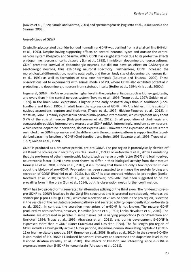
_______________________________________________________________Review of the literature
23
(Davies et al., 1999; Sariola and Saarma, 2003) and spermatogenesis (Viglietto et al., 2000; Sariola andSaarma, 2003).
Neurobiology of GDNF
Originally, glycosylated disulfide-bonded homodimer GDNF was purified from rat glial cell line B49 (Linet al., 1993). Despite having supporting effects on several neuronal types and outside the centralnervous system (Bespalov and Saarma, 2007), GDNF has caught attention due to its protective effectson dopamine neurons since its discovery (Lin et al., 1993). In midbrain dopaminergic neuron cultures,GDNF promoted survival of dopaminergic neurons but did not have an effect on GABAergic orserotonergic neurons, thus exhibiting neuronal specificity. Furthermore, GDNF increased themorphological differentiation, neurite outgrowth, and the cell body size of dopaminergic neurons (Linet al., 1993) as well as formation of new axon terminals (Bourque and Trudeau, 2000). Theseobservations led to experiments with animal models of PD, where GDNF also exhibited potential inprotecting the dopaminergic neurons from cytotoxic insults (Hoffer et al., 1994; Kirik et al., 2000a).
In general, GDNF mRNA is expressed in higher level in the peripheral tissues, such as kidney, gut, testis,and ovary than in the central nervous system (Suvanto et al., 1996; Trupp et al., 1997; Golden et al.,1999). In the brain GDNF expression is higher in the early postnatal days than in adulthood (Choi-Lundberg and Bohn, 1995). In adult brain the expression of GDNF mRNA is highest in the striatum,nucleus accumbens, septum and thalamus (Trupp et al., 1997; Hidalgo-Figueroa et al., 2012). Instriatum, GDNF is mainly expressed in parvalbumin-positive interneurons, which represent only about0.7% of the striatal neurons (Hidalgo-Figueroa et al., 2012). Small population of cholinergic andsomatostatin-positive interneurons express also GDNF mRNA. Interestingly, medium spiny neurons,which receive dopamine innervation, do not express GDNF. However, the expression of GFR is morerestricted than GDNF expression and the difference in the expression patterns is supporting the target-derived paracrine function of GDNF (Choi-Lundberg and Bohn, 1995; Suvanto et al., 1996; Trupp et al.,1997; Golden et al., 1999).
GDNF is produced as a precursor protein, pre-pro-GDNF. The pre-region is proteolytically cleaved offin ER and the pro-region in secretory vesicles (Lin et al., 1993; Lonka-Nevalaita et al., 2010). Consideringthat the pro-forms of other neurotrophic factors, such as nerve growth factor (NGF) and brain-derivedneurotrophic factor (BDNF) have been shown to differ in their biological activity from their matureforms (Lee et al., 2001; Gibon et al., 2016), it is surprising that there are only a few reported studiesabout the biology of pro-GDNF. Pro-region has been suggested to enhance the protein folding andsecretion of GDNF (Piccinini et al., 2013), but GDNF is also secreted without its pro-region (Lonka-Nevalaita et al., 2010; Piccinini et al., 2013). Moreover, pro-GDNF has been suggested to be theprevailing form in the brain (Sun et al., 2014), but this observation needs further confirmation.
GDNF has two pro-isoforms generated by alternative splicing of the third exon. The full-length pre- -pro-GDNF ( -GDNF) localizes in the Golgi-like structures and is secreted constitutively, whereas theshorter pre- -pro-GDNF ( -GDNF), which has a deletion of 26 amino acids in the pro-region, is locatedin the vesicles of the regulated secretory pathway and secreted activity-dependently (Lonka-Nevalaitaet al., 2010). In contrast, the secretion mechanism of -GDNF is not known. The mature GDNFproduced by both isoforms, however, is similar (Trupp et al., 1995; Lonka-Nevalaita et al., 2010). Theisoforms are expressed in parallel in same tissues but in varying proportions (Suter-Crazzolara andUnsicker, 1994; Trupp et al., 1995; Airavaara et al., 2011), e.g. during development -GDNF isexpressed more than -GDNF (Suter-Crazzolara and Unsicker, 1994). The full-length pro-region ofGDNF includes a biologically active 11-mer peptide, dopamine neuron stimulating peptide-11 (DNSP-11 or brain excitatory peptide, BEP) (Immonen et al., 2008; Bradley et al., 2010). In the severe 6-OHDAlesion model of PD, DNSP-11 induced behavioral recovery and increased the dopamine level in thelesioned striatum (Bradley et al., 2010). The effects of DNSP-11 are interesting since -GDNF isexpressed more than -GDNF in human brain (Airavaara et al., 2011).

Review of the literature _______________________________________________________________
24
GDNF exerts its effects via GFR 1 and Ret (Jing et al., 1996) or neural cell adhesion molecule (Paratchaet al., 2003). Alternatively GDNF can initiate the signaling cascade by binding to syndecan-3 Ret-independently (Bespalov et al., 2011). To terminate the signaling GDNF/GFR 1 complex is internalizedthrough sortilin-related receptor SorLA and directed to the endosomes (Glerup et al., 2013). From theendosomes GFR 1 is recycled to the receptor pool and GDNF transported to lysosomes fordegradation.
The importance of GDNF in development is demonstrated in mice lacking GDNF. The homozygousGDNF knockout mice die shortly after birth due to absence of kidneys and enteric neurons (Moore etal., 1996; Pichel et al., 1996; Sanchez et al., 1996). The mice have also defects in dorsal ganglion,sympathetic, and nodose neurons, but the dopaminergic system is intact, suggesting that GDNF is notindispensable for the development of dopaminergic neurons (Moore et al., 1996; Sanchez et al., 1996).In contrast, heterozygous GDNF knockout mice are viable, but have defects in spermatogenesis (Menget al., 2000) and cognition (Gerlai et al., 2001). Whether GDNF is needed for the maintenance ofdopaminergic system in the adulthood, is less clear. When GDNF expression was suppressed in adultmice, the animals exhibited reduced locomotor activity as well as pronounced progressive loss ofcatecholaminergic neurons in SNpc, ventral tegmental area, and locus coeruleus (Pascual et al., 2008).However, these results were challenged recently by Kopra and colleagues, who did not observechanges in the catecholamine system of conditional GDNF knockout mice (Kopra et al., 2015). Thisdiscrepancy between the studies might be due to differences in the genetic backgrounds of the animals(discussed in Pascual and Lopez-Barneo, 2015).
Although there has been a major interest in the dopaminotrophic effects of GDNF, the link betweenPD and GDNF is weak. Chauhan and coworkers reported a significant decrease in GDNF proteinexpression in SNpc neurons in PD brain compared to healthy controls (Chauhan et al., 2001). However,the study was conducted with immunohistochemistry and therefore must be interpreted cautiously.In another study the GDNF levels were studied with enzyme-linked immunosorbent assay, and nodifferences in the GDNF levels in caudate, putamen, or SN between PD patients and healthy controlswere observed (Mogi et al., 2001).
Neurobiology of NRTN
NRTN was discovered by its ability to enhance the survival of sympathetic neurons in culture(Kotzbauer et al., 1996). The following studies presented the neuroprotective properties of NRTN onmidbrain dopaminergic neurons, as NRTN was able to protect dopaminergic neurons as efficiently asGDNF both in vitro and in vivo (Horger et al., 1998). Interestingly, NRTN was found to be morepersistent in brain tissue and it has a more long-lasting effect on dopamine turnover in rat striatumthan GDNF (Hadaczek et al., 2010). However, the reported lack of the capability to induce axonalsprouting and hypertrophy of dopaminergic neurons (Åkerud et al., 1999) might be due to poorsolubility in neutral pH (Rosenblad et al., 1999).
During mouse embryonic development NRTN is highly expressed in peripheral tissues, such as gut,liver, and skin, but also in dorsal root ganglion and motor neurons (Golden et al., 1999). In ventralmidbrain, where the NRTN-responsive dopaminergic neurons are located, NRTN is expressed in earlyembryonic development (until E14). In contrast, NRTN receptor GFR 2 and Ret are expressed in ventralmidbrain throughout whole embryonic development period and in adulthood (Golden et al., 1999).NRTN mRNA is also detected in ventral midbrain in adulthood, but in lower level than receptor mRNA(Golden et al., 1999). Moreover, NRTN mRNA but not GFR 2 is detected in postnatal striatum(Widenfalk et al., 1997). This is in line with the theory of NRTN as a target-derived neurotrophic factor.However, the localization of endogenous NRTN protein has not been studied.
As GDNF and other GFLs, also NRTN is produced as a precursor protein. Cleavage of the pro-regionmight be an important regulatory step in NRTN signaling, since pro-NRTN is not able to initiate the

_______________________________________________________________Review of the literature
25
signaling via GFR 1 nor GFR 2 complexed with Ret (Fjord-Larsen et al., 2005). Moreover, deletion ofthe pro-region improves the secretion of NRTN (Fjord-Larsen et al., 2005). Even though very little isknown about the effects of pro-NRTN, mutations in the pro-region have been associated withHirschprung’s disease, a clinical condition caused by defects in enteric nervous system (Doray et al.,1998).
Mature NRTN is active as a homodimer and binds preferentially to the glycophosphatidylinositol-anchored GFR 2 receptor (Baloh et al., 1997; Klein et al., 1997). In addition to Ret, NRTN/GFR 2-complex can initiate the signaling via neural cell adhesion molecule (Paratcha et al., 2003). NRTN canalso signal GFR and Ret –independently via transmembrane heparan sulfate proteoglycan syndecan-3 (Bespalov et al., 2011). Unlike GDNF, NRTN might be able to signal via integrin -1 too (Schmutzleret al., 2011). Another difference in the GDNF and NRTN signaling has been detected in the kidney.Whereas GDNF is a paracrine morphogen, NRTN has been shown to exert its functions also in anautocrine manner in developing kidney ducts (Davies et al., 1999).
Unlike GDNF knockout mice, NRTN knockout mice are viable and fertile (Heuckeroth et al., 1999). Themice have defects in the enteric nervous system, but unlike GDNF knockout mice, NRTN knockoutshave morphologically and functionally normal kidneys. Furthermore, NRTN knockout animals havedeficits in parasympathetic system and sensory systems, but no abnormalities were characterized inthe central nervous system (Heuckeroth et al., 1999). Thus, other neurotrophic factors and signalingpathways might compensate for the lack of NRTN signaling.
Artemin and persephin
Artemin was identified based on its homology with the NRTN protein sequence (Baloh et al., 1998). Inan in vitro assay, artemin promoted the survival of neurons from both peripheral nervous system andmidbrain dopaminergic neurons as efficiently as GDNF or NRTN (Baloh et al., 1998). These observationshave led to test the efficacy of artemin in the 6-OHDA model of PD, where lentiviral vector-mediatedoverexpression of artemin provided neuroprotective effects against 6-OHDA-induced neurotoxicity(Rosenblad et al., 2000). Persephin was identified using polymerase chain reaction with primersrecognizing homologous sequences in GDNF and NRTN genes (Milbrandt et al., 1998). In contrast toartemin and other GFLs, persephin does not promote survival of peripheral neurons in vitro (Baloh etal., 1998; Milbrandt et al., 1998). Nonetheless, persephin promotes survival of midbrain dopaminergicneurons both in vitro and in vivo (Milbrandt et al., 1998). Recently, lentivirus-mediated overexpressionof persephin was shown to be neuroprotective in the rat 6-OHDA model of PD (Yin et al., 2015).
Therapeutic potential of GDNF and NRTN: Protein infusion studies
Neurotrophic factors do not pass blood-brain barrier and they need to be delivered intracranially.Single intranigral injection of GDNF and NRTN in 6-OHDA animal model of PD showed great potentialin protecting and restoring the dopamine environment of the nigrostriatal pathway. Both neurotrophicfactors promoted the survival of dopaminergic neurons as well as striatal dopamine (Hoffer et al.,1994; Horger et al., 1998). Moreover, repeated intranigral administration of both neurotrophic factorswas well tolerated and provided almost complete protection of TH-immunoreactive cells in animals(Horger et al., 1998). However, intranigral injection of GDNF did not spare striatal innervation, and theeffects were limited to nigral cell bodies (Sauer et al., 1995). Similar effects were observed in mouseMPTP model of PD (Tomac et al., 1995a). In contrast, injection of GDNF to striatum was able to protectthe nigral cell bodies and striatal dopaminergic neurites as well as provide functional benefits in both6-OHDA rat and MPTP mouse models of PD (Tomac et al., 1995a; Kirik et al., 2000a). These resultstogether with the observation that striatally administered GDNF protein is retrogradely transported tothe SNpc (Tomac et al., 1995b; Ai et al., 2003) supported the change of the administration paradigm

Review of the literature _______________________________________________________________
26
to intrastriatal delivery. Indeed, repeated striatal administration of GDNF was more efficient inrestoring the striatal TH-immunoreactive fiber density than nigral administration (Tomac et al., 1995a;Kirik et al., 2000a). Interestingly, repeated intranigral infusion of NRTN was proven to be as potent asGDNF in protecting neurons in SN (Horger et al., 1998), but when delivered intrastriatally, NRTN wasless effective in protecting nigral neurons than GDNF and with intraventricular delivery, NRTN wascompletely ineffective (Rosenblad et al., 1999). This might be due to the diffusion differences sinceNRTN binds tightly to heparin sulfates of the extracellular matrix in the brain tissue, as well as poorsolubility at neutral pH (Rosenblad et al., 1999; Hamilton et al., 2001; Bespalov et al., 2011).
Continuous infusion of GDNF to putamen or ventricle was also beneficial in the non-human primateMPTP model of PD (Grondin et al., 2002). GDNF increased putaminal dopamine concentration and thedensity of striatal TH-immunoreactive fibers as well as the number of nigral dopaminergic neurons(Grondin et al., 2002; Ai et al., 2003). The positive results of GDNF protein infusions led to clinical trials(table 2). Intraventricular infusion of GDNF did not improve the clinical status of the patients andadverse side effects, such as weight loss and nausea were reported (Nutt et al., 2003). In contrast,intraputaminal delivery of GDNF protein improved the clinical status of the patients without majoradverse effects (Gill et al., 2003). However, the results did not replicate in the following study, wherethe patients also developed antibodies against GDNF (Lang et al., 2006) and further trials were halted.
Table 2. GDNF clinical trials.Original study Administration Results Follow-up(Nutt et al.,2003)Phase I/II
Unilateralventricularmonthly bolus
No clinicalimprovementAdverse effects
No evidence of nigrostriatal regeneration orGDNF diffusion (Kordower et al., 1999)
(Gill et al.,2003)Phase I
Bilateral(1 patient unilateral)putaminal continuousinfusion
ClinicalimprovementNo adverseeffects
Clinical improvement, F-DOPA uptakeincreased, medication-induced dyskinesiadecreased (Patel et al., 2005)Increase in TH+ innervation putamen (Loveet al., 2005)
(Lang et al.,2006)Phase II
Bilateral putaminalcontinuous infusion
No clinicalimprovementAntibodiesdetected
GDNF concentrated around the catheter(Salvatore et al., 2006)Statistically underpowered study(Hutchinson et al., 2007)
(Slevin et al.,2005)Phase I
Unilateral putaminalcontinuous infusion
ClinicalimprovementNo adverseeffects
Halted when r-metHuGDNF withdrawn frommarketAll improvements lost in 1 year (Slevin et al.,2007)
Medgenesisa
Phase IIBilateral putaminalintermittent infusion
Noimprovement
NA
NCT01621581b
ongoingPhase I/II
AAV-GDNFputamen
NA NA
aPress release 7.6.2016 (http://www.medgenesis.com/news.htm)bClinicaltrials.gov identifier
Besides intracranial infusion, intranasal administration is another route to deliver therapeutic proteinsto the brain. The advantages of intranasal delivery are that it is non-invasive, bilateral, bypasses blood-brain barrier, and the minimal absorption into systemic circulation reduces the risk of peripheral sideeffects. On the other hand, only a small fraction of intranasally administered protein reaches the brainand therefore the therapeutic molecules have to be administered in high dose and in small volume.Thus, the solubility of the molecule is essential. Intranasal delivered GDNF enters the brain via olfactoryand trigeminal pathways and increases GDNF levels in the whole brain, including striatum and SN onehour after the delivery in rats (Bender et al., 2015). Indeed, intranasal administration of 50 μg of GDNFone hour prior to 6-OHDA protected dopaminergic cell bodies in SN but did not have an effect on

_______________________________________________________________Review of the literature
27
striatal innervation or behavior (Migliore et al., 2014). However, the translation of rodent data tohumans is difficult due to anatomical differences (van Woensel et al., 2013).
Viral vector delivery of neurotrophic factors
Gene therapy provides long-term expression of the therapeutic protein in the target tissue, andtherefore it has been widely studied in preclinical models of PD. Both viral vectors and DNAnanoparticles have been tested with positive effects (Kirik et al., 2000b; Fletcher et al., 2011). Withviral vector delivery, the effects of neurotrophic factors were site-dependent (Kirik et al., 2000b).Striatal overexpression of GDNF was observed to promote functional recovery of 6-OHDA-treatedanimals, whereas nigral overexpression was only protecting nigral cell bodies (Kirik et al., 2000b).Similar results were obtained with CERE-120, adeno-associated virus serotype 2 (AAV2)-based vectorencoding NRTN (Gasmi et al., 2007). Striatal delivery of CERE-120 protected both striatal dopaminergicinnervation as well as nigral cell bodies from 6-OHDA-induced cytotoxicity. It is evident, that GDNF andNRTN can both protect dopaminergic neurons from cytotoxic insults. However, overexpression ofGDNF did not attenuate viral vector-mediated -synuclein-induced neurodegeneration in -synucleinoverexpressing rats (Decressac et al., 2011). The lack of neuroprotective effects might be due to -synuclein-induced downregulation of transcription factor Nurr1 and its downstream target Ret,disrupting the GDNF signaling (Decressac et al., 2012). Nevertheless, the translational value of thesefindings can be questioned, since simultaneous overexpression of two proteins might cause ER stressin vulnerable nigral neurons leading to UPR, masking the neuroprotective effects (Hoffer and Harvey,2011).
Since promising results of viral delivery in rodent models were replicated in non-human primatemodels of PD (Kordower et al., 2000; Palfi et al., 2002; Kordower et al., 2006a; Kells et al., 2010), clinicaltrials were started with CERE-120 (table 3). The first results from the trials were positive: one year afterthe intraputaminal viral vector delivery patients did show improvement in their condition. In addition,no adverse side effects were reported (Marks et al., 2008). To improve the bioavailability of NRTN, infollowing clinical trials CERE-120 was delivered both intraputaminally and nigrally. Although the resultswere declared negative in the trial, re-analysis of the data confirmed a small subpopulation of thepatients to have benefitted from the therapy. The therapeutic efficacy of NRTN was highest in patientswhich were diagnosed with PD less than five years ago (Bartus, 2015; Olanow et al., 2015). Thus, thisis in line with analysis of the PD brain showing clear reduction in striatal dopaminergic fibers alreadyfive years after diagnosis (Kordower et al., 2013) and following impairment in axonal transport (Bartuset al., 2011). A post-mortem sample four years after CERE-120 delivery revealed the patient to havesome NRTN-immunoreactivity in both SN and putamen (Bartus et al., 2015). Currently, safety andefficacy of CERE-120 are being evaluated in patients with moderately advanced PD (clinicaltrials.govidentifier NCT00985517), as well as the safety of AAV2-GDNF is being tested in more advanced PDpatients (clinicaltrials.gov identifier NCT01621581).

Review of the literature _______________________________________________________________
28
Table 3. Neurturin clinical trials.Original study Administration Results Follow-up(Marks et al.,2008)Phase I
Bilateralputaminal
ClinicalimprovementNo adverseeffects
Lack of anterograde transport (Bartus et al.,2015)
(Marks et al.,2010)Phase II
Bilateralputaminal
No clinicalimprovementAdverse effects
Lack of anterograde transport (Bartus et al.,2011; Bartus et al., 2015)Subset of patients improvement after 15-18month follow-up (Olanow et al., 2015)
(Bartus et al.,2013)Phase I/II
Bilateral putaminal+ nigral
ClinicalimprovementNo adverseeffects
NA
(Olanow et al.,2015)Phase II
Bilateral putaminal+ nigral
No clinicalimprovementNo adverseeffects
Clinical improvement in a subset (<5 yearsof diagnosis) of patients (Bartus, 2015)
NCT00985517a
ongoingPhase I/II
Bilateralputaminal + nigral
NA NA
aclinicaltrials.gov identifier
Ex vivo gene therapy of neurotrophic factors has also been applied in preclinical studies. Bone marrow-derived mesenchymal stem cells provide an attractive approach to enhance GDNF signaling in the brainsince the cells can be isolated from an autologous source, the patient himself, genetically manipulatedto overexpress therapeutic proteins, and implanted in the brain. Striatal implantation of mesenchymalstem cells genetically manipulated to express GDNF have been shown to protect striatal innervationfrom a 6-OHDA-induced cytotoxic insult and to improve the rotational asymmetry induced by 6-OHDAadministration (Glavaski-Joksimovic et al., 2010). In another study, neural stem cells expressing NRTNwere engrafted to striatum (Liu et al., 2007). NRTN significantly reduced the rotational imbalance inthe animals after 6-OHDA administration several months after the cytotoxic insult as well as increasedstriatal levels of dopamine and its metabolites. Moreover, retrograde transportation of NRTN to SNprotected the cell bodies from cell death (Liu et al., 2007).
Endogenous GDNF and dopaminergic system
Increasing the expression of endogenous GDNF also has neuroprotective effects against toxin-inducedneurodegeneration. Laganiere and colleagues used AAV-mediated delivery of artificial GDNFtranscription activator to enhance the striatal expression of GDNF. Although activation of GDNF geneprovided significant functional protection against 6-OHDA-induced motor impairments, the protectiveeffect was less clear in SNpc neurons (Laganiere et al., 2010). Another approach to increasing theexpression of endogenous GDNF is to inhibit the transcription of the 3’ untranslated region of the Gdnfgene (Kumar et al., 2015). These GDNF hypermorph mice overexpressing GDNF from the native locushave increased striatal dopamine level and increased number of nigral dopaminergic neurons andstriatal nerve terminals. Although overexpression of GDNF did not protect nigral dopaminergic neuronsfrom the lactacystin-induced degeneration, the striatal dopamine level was preserved in GDNFhypermorph mice. On the other hand, the GDNF hypermorph mice are more susceptible to 6-OHDAcytotoxicity than WT mice due to increased DAT activity (Kumar et al., 2015).

_______________________________________________________________Review of the literature
29
Effects of exogenous GDNF on mitochondrial dysfunction
Most of the data regarding the therapeutic potential of GFLs has been gathered in 6-OHDA or MPTPanimal models of PD and the focus has been on the dopaminotrophic effects, survival of thedopaminergic neurons and neurites. Hence, less is known about the effects of GFL signaling on otheraspects of PD, such as mitochondrial defects. Several studies are pointing at a link between GFLsignaling and mitochondrial function. Striatal administration of GDNF to MPTP-treated mice regulatesseveral PD-associated genes, such as DJ-1 and UCHL-1 (Hong et al., 2009). In the 6-OHDA animal model,administration of GDNF prior to toxin reduces oxidative stress markers in the striatum, suggesting itreduced the formation of ROS (Smith and Cass, 2007). In vitro experiments with neuroblastoma cellshave shown the absence of DJ-1 to suppress the expression of Ret (Foti et al., 2010). When theneuroblastoma cells lacking parkin were treated with GDNF and GFR 1, the reduced cellular ATP levelswere restored (Meka et al., 2015). Similar effects were observed with cells lacking PINK1, wherestimulation of endogenous Ret with GDNF and soluble GFR 1 recovered mitochondrial fragmentationand further, mitochondrial dysfunction (Klein et al., 2014). However, in MitoPark mice, which exhibitrespiratory chain impairment in midbrain dopaminergic neurons leading to a progressive degenerationof nigrostriatal pathway (Ekstrand et al., 2007), application of exogenous GDNF did improve thebehavioral deficits but did not have an effect on the neuronal degeneration. Thus, in this model, GDNFwas not able to fully rescue the mitochondrial dysfunction-induced phenotype of the animals (Sterkyet al., 2013).
Hence, the positive effects of exogenous GDNF and NRTN in the animal models of PD have nottranslated to human trials. Several reasons have been suggested for this, such as sub-optimal designof the catheter (table 2, Salvatore et al., 2006) and inadequate posttranslational modifications of theproteins (Hoane et al., 2000; Fjord-Larsen et al., 2005; Piccinini et al., 2013). The difference in the brainvolume between humans and non-human primates as well as differences in administration paradigmand assessment of the severity of parkinsonism make the translation more difficult (Kordower et al.,1999; Nutt et al., 2003). This raises the question of the predictive validity of current animal models ofPD. As discussed in section 2.2., the animal models fail to simulate the complexity of pathologicalevents associated with PD, and might therefore give “false positive” results. Conversely, failure toexhibit neuroprotective effects in one animal model should be interpreted cautiously. Theneuroprotective and neurorestorative effects of GFLs have been studied mainly in MPTP and 6-OHDAmodels of PD, and only few studies have been conducted in other models. However, different modelsgive insight to different aspects of PD and might therefore give valuable information for the drugdevelopment and for the basic biology of the neurotrophic factors.

Aims of the study____________________________________________________________________
30
33 AIMS OF THE STUDY
Due to their neuroprotective and neurorestorative properties, neurotrophic factors have beenconsidered as a promising new therapy for Parkinson’s disease. The purpose of this thesis was toinvestigate the neuroprotective and neurorestorative properties of novel and less studied isoforms ofneurotrophic factors NRTN and GDNF.
The specific aims of the study were as follows:
- To study the neuroprotective properties of novel neurturin variants with decreased heparinbinding properties. The poor solubility and high affinity of NRTN to heparan sulfatedproteoglycans of the extracellular matrix have been suggested to hamper the diffusion ofNRTN in the brain and further, the therapeutic effects. Thus, we investigated whetherimproved diffusion capacity enhances the therapeutic effects of the protein.
- To develop a stable partial 6-OHDA rat model of Parkinson’s disease with high success rate.Since animals recover spontaneously from 6-OHDA cytotoxic insults, improving the stability ofthe animal model improves the reliability of the obtained results and importantly, decreasesthe number of experimental animals used in the studies.
- To compare the effects of conserved GDNF splice isoforms in the non-lesioned striatum and inpartial 6-OHDA lesion model of Parkinson’s disease. Full-length -GDNF and correspondingmature GDNF are secreted constitutively whereas shorter -GDNF and corresponding matureGDNF are secreted activity-dependently. Hence, the distinctive effects of the isoforms on thenigrostriatal dopaminergic circuitry were evaluated.

_____________________________________ __________________________Materials and methods
31
44 MATERIALS AND METHODS
4.1 Animals
Male Wistar rats (Harlan/Envigo, The Netherlands) were used in all studies. Rats were housed in groupsof 3-5 in 12h/12h light/dark cycle, with standard rodent chow and water ad libitum. Experimentaldesign and procedures regarding animals were reviewed and accepted by the National ExperimentBoard (ESAVI/5459/04.10.03/2011 and ESAVI/7812/04.10.07/2015) and conducted according to EUregulation (EU directive 2010/63/EU) and Finnish legislation (Finnish act on the protection of animalsused for scientific or educational purposes [497/2013] and the Government decree on the protectionof animals used for scientific or educational purposes [564/2013]).
4.2 Stereotaxic surgeries
All stereotaxic procedures were done under isoflurane anesthesia (induction 4-4.5%, sustenance 2.5%isoflurane). Tramadol (1mg/kg, s.c. study I and II) or carprofen (5 mg/kg, s.c., study III) were used as apost-operative analgesic. 6-OHDA injection coordinates (according to Paxinos and Watson 2005) andmicroinjection syringes used in experiments are designated in table 4. Same coordinates were used toinfuse neurotrophic factors (study I, 5 μg of neurturin variants or GDNF in 10 μl) or viral vectors (studyIII, AAV, 3 x 1.5 μl, Figure 7).
Table 4. 6-OHDA injection paradigms used in the experiments.
Dose Volume CoordinatesA/P L/M D/V Needle Study
3 x 1 μg 3 x 1.5 μl+1.60.0-1.2
-2.8-4.1-4.5
-6.0-5.5-5.5
WPI 33G II
3 x 2 μg 3 x 1.5 μl+1.60.0-1.2
-2.8-4.1-4.5
-6.0-5.5-5.5
WPI 33G II, III
3 x 3 μg 3 x 1.5 μl+1.60.0-1.2
-2.8-4.1-4.5
-6.0-5.5-5.5
WPI 33G II
2 x 10 μgprog 2 x 2.5 μl +1.6-0.4
+2.2+3.0
-5.5-5.5 Hamilton 26G II
2 x 10 μgreg 2 x 2.5 μl +1.6-0.4
+2.2+4.0
-5.5-5.5 Hamilton 30G II
1 x 20 μg 5 μl +1.0 +2.7 -5.5 Hamilton 26G II
3 x 7 μg 3 x 1.75 μl+1.60.0-1.2
+2.2+3.5+3.9
-5.5-5.5-5.5
WPI 33G II
1 x 28 μg 7 μl (4 x 1.75 μl) +1.0 +2.8
-6.0-5.5-5.0-4.4
Hamilton 26G I
2 x 10 μgprog= Injection with 26G Hamilton syringe, progressive rotational behavior (see results section 5.2)2 x 10 μgreg= Injection with 30G Hamilton syringe, regressive rotational behavior (see results section 5.2)WPI, World Precision Instruments

Materials and methods_______________________________________________________________
32
Figure 7. Experimental design. Neurotrophic factors were delivered intracranially as a protein infusion either A)two weeks (study I) or B) five weeks (unpubl. results) after 6-OHDA administration. With AAV1 vector delivery,the transgene expression was allowed to continue for three weeks before evaluating the effects in C) non-lesioned animals or D) inducing neurodegeneration with 6-OHDA (study III). 6-OHDA, 6-hydroxydopamine; AAV,adeno-associated virus; IHC, immunohistochemistry; NTF, neurotrophic factors.
44.3 Behavioral assays
4.3.1 Amphetamine-induced rotations
Rotational asymmetry was measured after unilateral 6-OHDA injection. 2.5 mg/kg (s.c.) ofamphetamine sulfate was administered to induce ipsilateral rotational behavior (Ungerstedt andArbuthnott, 1970) after a 30 minute habituation period. Full, uninterrupted 360° turns were countedfor 120 minutes in automatic rotometer bowls (MedAssociates, Inc. St. Albans, VT).
4.3.2 Cylinder test
Cylinder test was used to assess asymmetry in spontaneous forelimb use. Freely moving rats werefilmed for five minutes in a plexiglass cylinder (diameter 20 cm) and the contacts made by eachforepaw with the cylinder wall were scored by blinded observer. Only weight-bearing touches wereconsidered as touches. In study I, the cylinder test was conducted in normal light, but in study III, the

_____________________________________ __________________________Materials and methods
33
test was carried out under red light to increase the activity of the animals. The animals were let toadjust to red light for 30 minutes before testing.
44.4 Immunohistochemistry
For immunohistochemistry, the animals were anesthetized with pentobarbital and perfusedtranscardially with PBS and 4% paraformaldehyde. The removed brains were cut into 40 μm thicksections in series of six and stored at -20°C.
The immunohistochemistry was performed on free-floating brain sections. After washing, theendogenous peroxidase activity was quenched with 0.3% hydrogen peroxide, and in DAT-staining,additional incubation at 80°C was conducted to enhance the antigen recognition. The sections wereincubated with 4% BSA and 0.1% Triton X-100 in PBS to reduce non-specific binding before incubationwith desired primary antibody overnight (table 5). Following the incubation with suitable biotinylatedsecondary antibody, the staining was reinforced with avidin-biotin complex and visualized with 3’, 3’-diaminobenzidine.
In fluorescence staining, the sections were incubated with blocking buffer after washing, incubatedwith primary antibody overnight, and mounted on microscope slides after incubation with secondaryantibody conjugated with a fluorescent dye.
Table 5. Antibodies and their dilutions used in the experiments.
Antigen Host Producer Cataloguenumber Dilution
Fos Rabbit Santa Cruz sc-52 1:1000Dopamine transporter (DAT) Rat Millipore MAB369 1:2000
Glial cell line-derived neurotrophic factor(GDNF) Goat R & D Systems AF-212-NA 1:3000
Green fluorescent protein (GFP) Rabbit Life Technologies A11122 1:2000Tyrosine hydroxylase (TH) Mouse Millipore MAB318 1:2000
4.5 Morphometric analysis
4.5.1 Optical density
The optical density of TH-immunoreactive fibers was analyzed from three (study II), six (study III), ornine (study I) adjacent sections. The density was determined with ImagePro (studies I and III, MediaCybernetics, Rockville, MD, USA), or ImageJ (study II, NIH, Bethesda, MD, USA) from images acquiredeither with a digital camera (Nikon Corp., Tokyo, Japan) attached to a microscope (study I) or with adigital microscope slide scanner (studies II and III, 3D Histech, Budapest, Hungary). Corpus callosumwas used to correct the values for background staining, as it is devoid of staining signal. Data areexpressed as a percentage from the intact side.
4.5.2 Cell counting
The number of TH-immunoreactive cells in the SNpc was estimated from six adjacent sections withMatlab (studies II and III) or from nine adjacent sections with StereoInvestigator (study I,MicroBrightfield, Williston, VT, USA) by blinded observer. In the Matlab method, the cells wererecognized from the digitalized images obtained with 3D Histech scanner (3D Histech, Budapest,Hungary) based on their size in pixels and signal intensity. The cell size was user-defined based on thesize of single cells in example images and kept constant throughout the whole analysis. This way small

Materials and methods_______________________________________________________________
34
objects were excluded from the cell counts and the area covered by larger objects consisting ofoverlying cells was divided by the size of the single cell, thus estimating how many cells were clumpedtogether. Since the signal intensity varies between the samples, the intensity threshold was definedindividually for every sample based on the signal intensity of single cells. The background devoid ofcells and staining signal was kept at zero. StereoInvestigator, on the other hand, is based on stereology,and the cell counting was done with optical fractionator combined with dissector principle andunbiased counting rules from the brain sections mounted on microscope slides (West et al., 1991). Inboth cases, the results are given as a percentage of the unlesioned side.
44.6 Other methods
Other methods used in the studies are presented in table 6. More detailed description of the methodscan be found in the original publications and their supplements. Methods the author has been involvedin are marked with a *.
Table 6. Other methods used in the study.Method Original studyCell-based binding assay ICell culture ICloning I, IIIComputational modelling IDiffusion assays in vivo* IELISA* IIIGFR affinity chromatography IHeparin affinity chromatography IImmunocytochemistry IKidney ex vivo organogenesis IN-terminal sequencing IPCR mutagenesis IPreparation of viral vectors IIIProgramming IIProtein diffusion assay in non-human primates IProtein production IProteolytic resistance IPurification of protein IPurification of viral vectors IIIRET phosphorylation assay IRT-PCR ISurvival assay on embryonic dopaminergic neurons in vitro ITransient cell transfection I, IIIWestern blotting I
4.7 Statistical analysis
Data are expressed as a mean ± standard error of the mean (SEM). Statistical analyses were done bySPSS (IBM). Differences between the treatment groups are analyzed with one-way analysis of variance(ANOVA) followed by Fisher’s Least Significant Difference (LSD), Tukey’s, or Dunn’s multiplecomparison post hoc, or two-way ANOVA or repeated measures ANOVA, or paired t-test. In non-parametric situations, Kruskal-Wallis test was conducted with Bonferroni post hoc analysis. Correlationanalysis was conducted using Pearson correlation test. Results were considered significant at level p 0.05.

__________________________________________________ __________________________Results
35
55 RESULTS
5.1 Cell counting method
A new method for counting the TH-positive cells in the SNpc was set up and validated. Previously usedstereology-based method combined with optical dissector and fractionator enabled the cells to berecognized irrespective of their shape, size or orientation. In contrast, the new Matlab algorithm-basedmethod recognizes the cells by their size in pixels in digitalized images. Despite this methodologicaldifference, the results obtained with the methods have a high correlation (R=0.925, F1,6=35.565,P<0.001).
5.2 Stable 6-OHDA model of Parkinson’s disease
5.2.1 Rotational differences between 6-OHDA doses
6-OHDA is a classical animal model of PD. By using a stable animal model with high success rate, thenumber of animals needed to obtain reliable data is reduced. In study I we used 1 x 28 μg dose, wherefour 7 μg depots of 6-OHDA were injected to the same site, above each other (table 4). When theanimals were rotated two weeks later, only 54% of them rotated sufficiently (table 7) (in study I thelimit was minimum of 220 turns in 120 mins) and were selected for the neuroprotection study (hencethe name 1 x 28 μg2week). The animals were assigned randomly to the treatment groups, and the groupswere balanced based on the rotations so that all groups included animals with high rotations andanimals with low rotations. Thus, the average of the rotations was at similar level in all treatmentgroups. Animals, which did not rotate two weeks after 6-OHDA injection, were re-assessed three weekslater on week five to see whether they developed a lesion later (hence the name 1 x 28 μg5week group).This time only 49% of the remaining animals rotated in the assay (table 7). These animals were dividedinto treatment groups in similar manner as previously based on their rotational behavior(neurorestoration study).
Considering that in study I only about half of the animals injected with 6-OHDA developed a lesionduring the first two weeks and were included in the original neuroprotection experiment, the usedinjection paradigm was not optimal. In study II, we compared seven different 6-OHDA injectionparadigms (table 4) used in our previously unpublished experiments to find out what are the crucialelements to create a stable animal model with high success rate. The data was re-analyzed by theamphetamine-induced rotations and immunohistochemistry. The control group data from study Ineuroprotection experiment (1 x 28 μg2week) and neurorestoration experiment (1 x 28 μg5week) werealso included to the comparison in this thesis. Since the data is a compilation of control groups fromdifferent experiments, the time points vary between the groups. However, all groups were rotatedduring weeks 1-2 to see whether the lesion was successful or not. Animals rotating more than 100turns in two hours were considered to have a lesion (study II; Bäck et al., 2013b), exception in study I,where the limit was 220 turns in 120 minutes). The percentage of animals with a successful lesion wasconsidered as a measure for replicability (success rate) of the model (table 7). In an optimal model, thesuccess rate would be high and most 6-OHDA-injected animals would develop lesion and thus beincluded in the experiments.

Results ____________________________________________________________________________
36
Table 7. Immunohistochemistry results of different lesion paradigms.
Treatmentgroup Category
TH-ir str(% of the intact
side)
DAT-ir str(%of the intact
side)
TH-ir SNpc(% of the intact
side)a
Success rate(%)
Start End3 x 3 μg Progressive 21 ± 5 22 ± 3 29 ± 5 88 100
2 x 10 μgprog 27 ± 5 21 ± 4 23 ± 4 91 1003 x 7 μg 30 ± 2 17 ± 2 22 ± 3 100 1003 x 1 μg Stable 31 ± 9 24 ± 6 43 ± 6 63 803 x 2 μg 28 ± 6 27 ± 5 36 ± 7 75 75
1 x 28 μg2week 13 ± 4 20 ± 4 31 ± 6 54 801 x 28 μg5week NA NA NA 49 572 x 10 μgreg Regressive 66 ± 7 58 ± 6 71 ± 0,2 100 431 x 20 μg 51 ± 6 43 ±5 61 ± 6 100 70
2 x 10 μgprog= progressive rotations, Hamilton 26 G2 x 10 μgreg= regressive rotations, Hamilton 30 G1 x 28 (4 x 7) μg2week= 2 week group (lesion limit 220 turns in 120 mins)1 x 28 (4 x 7) μg5week = 5 week group (lesion limit 220 turns in 120 mins)Success rate: number of animals rotating >100 turns in 120 minsaTH-ir cells analyzed with MatlabDAT-ir, dopamine transporter-immunoreactivity; SNpc, substantia nigra pars compacta; str, striatum; TH-ir,tyrosine hydroxylase-immunoreactivity.
Ideally, unbiased assessment, such as d-amphetamine-induced rotations, should correlate withdopamine depletion. Different paradigms were categorized as progressive, stable or regressive basedon their rotational behavior (figure 8). Although the rotational behavior has been measured atdifferent time points, the difference between the categories is clear. In groups categorized asprogressive, the rotations were increased 100-400% (3 x 3 μg 112%, 2 x 10 μgprog 154%, and 3 x 7 μg387%) from the first time point rotations (week 1-2). These groups had also high success rates in bothfirst and last measured time point (table 7). In stable rotation groups the changes were more subtle (3x 1 μg -17% and 3 x 2 μg 24% , 1 x 28 μg2week 31%, and 1 x 28 μg5week 26%). In 3 x 1 μg and 3 x 2 μggroups the success rate was at moderate level both in first and last time point (table 7). On the otherhand, 1 x 28 μg2week and 1 x 28 μg5week groups had low success rates but stable rotational behavior. Inregressive groups the rotational behavior decreased from the first time point (-84% 2 x 10 μgreg and -51% 1 x 20 μg group). These groups however showed high success rates at the first time point, but thein the last time point (2 x 10 μgreg week 10 and 1 x 20 μg week 8) only 43% and 70% of the animalsrotated over the limit of 100 turns in two hours.

_____________________________________________________________________________Results
37
Figure 8. In study II, the lesion groups were categorized as A) progressive, B) stable, or C) regressive based ontheir rotational behavior. D) The lesion paradigm used in study I also produced stable rotational behavior.
Interestingly, we observed a dose-response in the low-dose 6-OHDA treated groups in the lastmeasured time point, week 12 (3 x 1 μg 432 ± 198 turns, 3 x 2 μg 712 ± 189 turns, and 3 x 3 μg 1258± 315 turns in 120 mins), which is in line with previous studies showing 6-OHDA induced lesions to bedose-dependent (Przedborski et al., 1995; Lee et al., 1996; Bäck et al., 2013b). This was however notobserved with higher doses. Since 3 x 2 μg injection paradigm produced a stable rotational behaviorwith good success rate, it was chosen for the following study III.
55.2.2 Immunohistochemistry
Surprisingly, all 6-OHDA administration paradigms induced the same magnitude of lesion of thenigrostriatal pathway (table 7), with the exception of 1 x 20 μg and 2 x 10 μgreg groups, in which thelesion was much smaller than in other groups. The density of DAT-immunoreactive fibers was also ata similar level in all progressive and stable lesion groups and higher in the regressive lesion groups.Also, the TH-immunoreactivity in the striatum reflected the number of TH-immunoreactive cells inSNpc since there was a high correlation between the measures (R=0.785, p<0.0001). The correlationbetween DAT-immunoreactive fibers and TH-immunoreactive cells in SNpc was even higher (study II,R=0.838, p<0.0001). Even though 6-OHDA induced loss of striatal and nigral TH-immunoreactivity hasbeen shown to be dose-dependent (Lee et al., 1996; Bäck et al., 2013b), this was not observed in ourstudy (II). Instead, when the toxin was injected into multiple sites (e.g. 3 x 1 μg), the lesion size wascomparable to higher doses (e.g. 2 x 10 μg, table 7).
Although the rotational assay has been widely used, the results might be easily over-interpreted (Marinet al., 2006; Meredith and Kang, 2006). As is shown in study II, the rotational behavior does not alwaysreflect the actual loss of TH-immunoreactive cells in SNpc or TH-immunoreactive fibers in the striatum.Despite the difference in the time points when the assay was conducted, there was a clear differencein the rotational behavior between the progressive and the stable groups, but the striatal TH- and DAT-

Results ____________________________________________________________________________
38
immunoreactivity was at the same level in all groups (table 7, correlation between rotations andstriatal TH-immunoreactivity R=0.536, p<0.0001, correlation between rotations and DAT-immunoreactivity R=0.591, p<0.0001). The correlation between the rotations and the number of TH-immunoreactive cells in SNpc was a little bit higher (R=0.662, p<0.0001).
55.3 Development of NRTN variants with reduced heparin affinity
NRTN and GDNF both bind strongly to extracellular matrix and cell surface heparan sulfatedproteoglycans (Hamilton et al., 2001; Bespalov et al., 2011), which can attenuate their diffusion in thebrain parenchyma. This is problematic from the therapeutic perspective since neurotrophic factors donot pass the blood-brain barrier and have to be delivered intracranially. Poor diffusion and thefollowing low bioavailability have been suggested to be one of the reasons why the clinical trials withGDNF have produced only mild effects (Salvatore et al., 2006).
5.3.1 Modelling NRTN variants
New NRTN variants were created to improve the solubility, diffusion capacity and bioavailability of theprotein in the brain tissue. First, the three-dimensional structure of NRTN, a close homolog of GDNF,was modeled using the structure of GDNF (Eigenbrot and Gerber, 1997; Parkash et al., 2008). In themodeled structure, putative heparin binding sequence BBXB or BBXXB, where B is basic amino acidand X is any amino acid (Cardin and Weintraub, 1989), was localized in the heel region(51RRLRQRRRLRRER63), where the required positively charged amino acids are aligned pointing to thesame direction. Since the heel region included several putative consensus sequences, severalmutations were introduced to the region thereby generating several novel variant proteins. VariantsN1 and N2 have substitutions of arginine with alanine and N3 is a combination of these mutations,with the exception of the last restoring the conserved last arginine of the sequence. In N4, the heelregion was substituted with the heel region of PSPN (ARLQGQG), which does not bind to heparin(Bespalov et al., 2011), and elongated (ALVGS) to match the length of the original NRTN heel sequence.
5.3.2 Biological activity of NRTN variants
For purification and initial testing, the variant proteins were expressed in mammalian chinese hamsterovary cells with N-terminal V5-tag (hence the name NV1-NV4) and with an endogenous ER-signalsequence substituted with ER-signal sequence from IgG (Fjord-Larsen et al., 2005). The native pro-sequence was also removed from the proteins to improve secretion (Fjord-Larsen et al., 2005). Apreliminary screen with the Ret-phosphorylation assay showed all the new variants to be biologicallyactive in the presence of either GFR 1 or GFR 2, and heparin affinity chromatography showed allvariants to have reduced affinity to heparin. Moreover, the concentration of NV2 and NV4 was higherthan the concentration of WT NRTN, NV1 or NV3 in the culture media, although the concentrations ofNRTN variants were similar inside the cells. This might reflect the improved resistance to proteolyticdegradation of NV4.
For the larger scale production of variant proteins, both pro-sequence and the V5-tag were removedfrom the variant proteins (variants N1-N4, namely). Untagged proteins were purified with acombination of heparin- and GFR 2-affinity chromatography. Heparin affinity chromatographydemonstrated the reduced affinity for heparin of the variant proteins (table 8). However, two of thevariants, N1 and N3 aggregated and precipitated during the purification and biochemical assays and inhigher concentrations (study I, figure 3). As the proteins were needed in higher concentrations for thein vivo assays, the study focused more on the variants N2 and N4. Similar precipitation problems werealso detected with WT NRTN. A binding assay with chinese ovary cells expressing GFR 2, RET and

_____________________________________________________________________________Results
39
heparan sulfates showed the variants N2 and N4 to have similar affinity to the GFR 2/RET receptorcomplex as WT NRTN (table 8). Moreover, while the WT NRTN attached to the cell surface of the cellslacking the GFR -receptors, N4 did not. If GFR -receptors were present on the cell surface, both WTNRTN and N4 attached to the cell surface. When the cell surface was lacking both heparan sulfates andGFR -receptors, neither WT NRTN nor N4 attached to the cell surface (study I, figure 6).
Table 8. Properties of neurturin variants.
aIn rat diffusion assay, 5 μg of protein was injected to the striatum.bIn cynomolgus monkey diffusion assay, equimolar amounts of protein (GDNF 225 μg/225 μl, N2 170 μg/170 μl,and N4 170 μg/205 μl) was injected to the putamen.cIn marmoset monkey diffusion assay, 5 μg of protein was injected to the putamen.
Ret-phosphorylation assay revealed that the variants are able to activate Ret in a dose-dependentmanner similar to WT NRTN (study I, figure 4). Another dose-dependent assay, in vitro survival assayon dopaminergic neurons from mouse midbrain (E13.5), showed similar patterns between the NRTNvariants and WT NRTN (study I, figure 5A). Interestingly, the neurons express only GFR 1, and notGFR 2 (Cacalano et al., 1998), thus suggesting the NRTN variants are able to start a signaling cascadevia GFR 1/Ret complex. Next, the biological activity of NRTN variants was tested in tissue culture. Incontrast to WT NRTN, both variants N2 and N4 were able to induce extraureteric branching in mouseembryonic kidney cultures (study I, figure 5B). One explanation for this might be the higher stability ofvariant proteins compared to WT NRTN in the culture medium.
55.3.3 NRTN variants have improved diffusion capacity and neuroprotective effects in vivo
The diffusion capacities of NRTN variants were determined in three different species: rat, cynomolgusmonkey, and marmoset monkey (table 8). The results show inevitably how N4 has better diffusioncapacity than N2, WT NRTN or GDNF in brain tissue. Especially in cynomolgus monkey brain, thedifference is striking. The diffusion assay results further support the in vitro studies showing N2 andN4 to have reduced affinity to heparin and heparan sulfated proteoglycans of the extracellular matrixand cell surface.
To study whether this improved diffusion capacity and protein stability would play a role in thetherapeutic efficacy, neuroprotective effects of NRTN variants N2 and N4 were tested in partial 6-OHDA rat model of PD. Compared to previous studies (Lindholm et al., 2007; Piltonen et al., 2009;Voutilainen et al., 2009), a relatively low-dose of 5 μg of proteins were infused to the striatum twoweeks after inducing the degeneration of nigrostriatal pathway with 6-OHDA (1 x 28 μg, table 4). Theneuroprotective effects were tested with drug-free cylinder test and amphetamine-induced rotations(study I, figure 7A). Since NRTN can precipitate in neutral pH (Hadaczek et al., 2010), GDNF was usedas a positive control in the study instead of WT NRTN.
ProteinHeparinaffinity
[NaCl] M
Cell bindingassay
IC50 (nM)
Diffusionrata
(mm3)
Diffusioncynomolgus
monkeyb
(mm3)
Diffusionmarmosetmonkeyc
(mm3)WT NRTN
(commercial, e.coli) 1.07 NA 57 - -
WT NRTN (CHO) 1.08 4.65 - - -WT GDNF
(commercial, e.coli) - - - 619 18.4
N1 0.97 - - - -
N2 0.56 3.13 126 538 -
N3 0.56 - - - -N4 0.48 1.42 217 1228 106.1

Results ____________________________________________________________________________
40
N4 was able to improve the animals’ motor performance after 6-OHDA lesion. In the rotation assay,N4 treated animals recovered from the rotational bias induced by the 6-OHDA lesion by week 12(repeated measures ANOVA F3,40=3.2, p=0.033, Fisher LSD post hoc analysis vehicle vs N4 p<0.01, GDNFvs N4 p<0.05, N2 vs N4 p<0.05, table 9). In the drug-free cylinder test on week 10 (8 weeks after proteininfusion), N4-treated animals displayed an increased use of the contralateral paw compared to vehicletreated animals (Kruskal-Wallis nonparametric ANOVA p=0.00138, Dunn’s multiple comparison vehiclevs N4 p<0.05, table 8). Immunohistochemical findings further supported the behavioral read-outs asthe density of TH-immunoreactive fibers was increased in N4-treated animals (one-way ANOVAF3,40=4.24, p=0.011, Fisher’s LSD post hoc analysis vehicle vs N4 p<0.01, GDNF vs N4 p<0.05, N2 vs N4p<0.01, table 8). In contrast, GDNF did not increase the density of TH-immunoreactive fibers. In SNpc,all treatments, GDNF, N2 and N4 increased the number of TH-immunoreactive cells (one-way ANOVAF3,40=2.951, p=0.0441, vehicle vs GDNF p<0.05, vehicle vs N2 p<0.05, vehicle vs N4 p<0.05, table 9).Thus, all four assays, two behavioral and two immunohistochemical, strongly suggest N4 to havebeneficial effects in the partial 6-OHDA model of PD.
Table 9. Effects of NRTN variants on behavioral correlates in the neuroprotection study.
Treatment Cylinder test(Contralateral paw %)
Rotations(120 min)
TH-ir str(% of the
intact side)
TH-ir SNpca
(% of theintact side)
Week 6 Week 10 Week 8 Week 12 Week 12 Week 12Vehicle 4 ± 1 1 ± 1 778 ± 154 736 ± 188 13 ± 4 18 ± 3GDNF 7 ± 4 13 ± 6 632 ± 145 558 ± 194 21 ± 4 33 ± 5*
N2 2 ± 1 5 ± 3 688 ± 99 483 ± 176 19 ± 3 36 ± 3*N4 18 ± 7 29 ± 10 * 310 ± 85 143 ± 69 **# 41 ± 8**# 37 ± 6*
aTH-ir number of TH-immunoreactive cells in SNpc analyzed with StereoInvestigator*p<0.05 (vs vehicle), **p<0.01 (vs vehicle), # p<0.05 (vs GDNF and N2),SNpc, substantia nigra pars compacta; Str, striatum; TH-ir, tyrosine hydroxylase-immunoreactivity.
55.3.4 Neurorestorative effects of NRTN variants (unpubl. results)
As mentioned in section 5.2.1, the 6-OHDA injection paradigm used in study II (1 x 28 μg) resulted in alow number of lesioned animals. Animals which did not fulfill the rotation criterion (220< turns in 120min) two weeks after 6-OHDA injection were tested again three weeks later (week five). Rats that haddeveloped a lesion by five weeks after 6-OHDA, were injected with vehicle, GDNF or NRTN variants N2or N4. The motor performance was tested with cylinder test on weeks 9 and 16 and withamphetamine-induced rotations on weeks 11 and 16 post-lesion (figure 7B). Neither N2 nor N4 showedneurorestorative effects in cylinder test. In the rotation assay, N4 treated animals rotated less thanvehicle treated animals on week 11, but the difference did not reach statistical difference (table 10).
Table 10. Behavioral effects of the NRTN variants in the neurorestoration study.Treatment Cylinder test (Contralateral paw %) Rotations (120 min)
Week 9 Week 16 Week 11 Week 16Vehicle 9 ± 5 7 ± 5 573 ± 131 457 ± 220GDNF 3 ± 2 0 ± 0 452 ± 101 505 ± 185
N2 3 ± 2 14 ± 14 468 ± 271 608 ± 134N4 15 ± 5 18 ± 14 271 ± 86 330 ± 105
5.4 Expression GFRα1, GFRα2, and Ret in human brain
Together, the data suggest NRTN variant N4 to be biologically active in vitro in the presence of eitherGFR 1/Ret or GRF 2/Ret complex, and to have therapeutic potential in vivo in an animal model of PD.
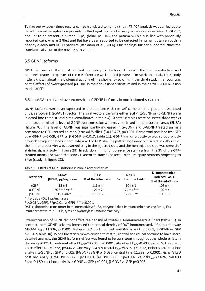
_____________________________________________________________________________Results
41
To find out whether these results can be translated to human trials, RT-PCR analysis was carried out todetect needed receptor components in the target tissue. Our analysis demonstrated GFR 1, GFR 2,and Ret to be present in human SNpc, globus pallidus, and putamen. This is in line with previouslyreported data, where GFR 1 and Ret have been reported to be detected in human putamen both inhealthy elderly and in PD patients (Bäckman et al., 2006). Our findings further support further thetranslational value of the novel NRTN variants.
55.5 GDNF isoforms
GDNF is one of the most studied neurotrophic factors. Although the neuroprotective andneurorestorative properties of the -isoform are well studied (reviewed in Björklund et al., 1997), onlylittle is known about the biological activity of the shorter -isoform. In the third study, the focus wason the effects of overexpressed -GDNF in the non-lesioned striatum and in the partial 6-OHDA lesionmodel of PD.
5.5.1 scAAV1-mediated overexpression of GDNF isoforms in non-lesioned striatum
GDNF isoforms were overexpressed in the striatum with the self-complementary adeno associatedvirus, serotype 1 (scAAV1) vector. The viral vectors carrying either eGFP, -GDNF or ( -GDNF) wereinjected into three striatal sites (coordinates in table 4). Striatal samples were collected three weekslater to determine the level of GDNF overexpression with enzyme-linked immunosorbent assay (ELISA)(figure 7C). The level of GDNF was significantly increased in -GDNF and -GDNF treated animalscompared to GFP-treated animals (Kruskal-Wallis H(3)=15.457, p=0.001. Bonferroni post hoc test GFPvs -GDNF p=0.005, GFP vs -GDNF p=0.017, table 11). GDNF-immunoreactivity was spread widelyaround the injected hemisphere, whereas the GFP-staining pattern was more restricted. In either case,the immunoreactivity was observed only in the injected side, and the non-injected side was devoid ofstaining signal (study III, figure 2B). In addition, immunofluorescence staining from the SN of the GFP-treated animals showed the scAAV1 vector to transduce local medium spiny neurons projecting toSNpr (study III, figure 2C).
Table 11. Effects of GDNF isoforms in non-lesioned striatum.
Treatment ELISAa
[GDNF] pg/mg tissueTH-ir
% of the intact sideDAT-ir
% of the intact side
D-amphetamine-induced Fos-ir
% of the intact sideeGFP 15 ± 6 111 ± 4 104 ± 3 105 ± 4-GDNF 1906 ± 629** 124 ± 7 124 ± 4*** 102 ± 4-GDNF 1115 ± 402* 115 ± 6 122 ± 3** 108 ± 3
aIntact side 40 ± 8 pg/mg tissue*p<0.05 (vs GFP), **p<0.01 (vs GFP), ***p<0.001.DAT-ir, dopamine transporter-immunoreactivity; ELISA, enzyme-linked immunosorbent assay; Fos-ir, Fos-immunoreactive cells; TH-ir, tyrosine hydroxylase-immunoreactivity.
Overexpression of GDNF did not affect the density of striatal TH-immunoreactive fibers (table 11). Incontrast, both GDNF isoforms increased the optical density of DAT-immunoreactive fibers (one-wayANOVA F2,24=11.336, p<0.001, Fisher’s LSD post hoc test -GDNF vs GFP p<0.001, -GDNF vs GFPp=0.002, table 10). When the striatum was divided to rostral, central and caudal sections to have moredetailed analysis, the GDNF isoforms effect was found to be consistent throughout the whole striatum(two-way ANOVA treatment effect F2,72=23.285, p<0.0001; site effect F2,72=0.490, p=0.615; treatmentx site effect F4,72=0.588, p=0.672. One-way ANOVA rostral F2,24=5.315, p=0.012, Fisher’s LSD post hocanalysis -GDNF vs GFP p=0.005, -GDNF vs GFP p=0.026; central F2,24=11.339, p<0.0001, Fisher’s LSDpost hoc analysis -GDNF vs GFP p<0.0001, -GDNF vs GFP p=0.002; caudal:F2,24=7.674, p=0.003Fisher’s LSD post hoc analysis -GDNF vs GFP p=0.001, -GDNF vs GFP p=0.006).

Results ____________________________________________________________________________
42
-GDNF has been suggested to be secreted activity-dependently upon overexpression in vitro (Lonka-Nevalaita et al., 2010). We tested whether administration of amphetamine sulfate would also increasethe secretion of -GDNF in vivo. The possible increase in the secreted GDNF was measured indirectlyusing Fos as a marker for neuronal activity. The number of Fos-immunoreactive cells in the striatumwas assessed two hours after amphetamine sulfate administration (2.5 mg/kg, s.c.). However, thishypothesized effect was not observed as the number of Fos-immunoreactive striatal cells was at thesame level in all treatment groups (table 11). This might be due to low number of animals in theexperiment (n=5) and short follow-up time after d-amphetamine administration.
55.5.2 Neuroprotective effects of GDNF isoforms in partial 6-OHDA rat model of PD
Neuroprotective effects of GDNF isoforms were assessed by inducing a partial lesion of the nigrostriataltract three weeks after viral vector administration. The lesion was induced by injecting 3 x 2 μg of 6-OHDA to the striatum of the animals as designated in table 4. Effects were followed with drug-freecylinder test, amphetamine-induced rotations, and immunohistochemistry (figure 7D). Cylinder testwas performed before lesioning on week three and four weeks after 6-OHDA injection. In the pre-lesion cylinder, GDNF-treated animals exhibited increased exploratory activity (one-way ANOVAF2,43=3.871, p=0.028, Fisher’s LSD post hoc test GFP vs -GDNF p=0.019, GFP vs -GDNF p=0.024, table11). 6-OHDA-induced degeneration of the nigrostriatal tract attenuated this effect, and the activity ofall treatment groups was at similar level four weeks later (two-way ANOVA treatment effectF2,86=3.406, p=0.038; 6-OHDA effect F1,86=29.071, p<0.0001; treatment x 6-OHDA interactionF2,86=1.130, p=0.328, table 12). Although both GDNF isoforms induced the exploratory activity in pre-lesion cylinder test on week three, only -GDNF increased the use of the contralateral (left) paw (one-way ANOVA F2,43=4.492, p=0.017, Fisher’s LSD post hoc test GFP vs -GDNF p=0.005, table 11).However, this effect was abolished by 6-OHDA injection, as the use of contralateral paw was at a similarlevel in all treatment groups four weeks later (Two-way ANOVA treatment effect F2,86=3.215, p=0.045;6-OHDA effect F1,86=41.803, p<0.0001; treatment x 6-OHDA interaction F2,86=0.545, p=0.582, table 11).The results from the rotation assay are in line with the cylinder test results since no differences wereobserved between the treatment groups at either time point. However, -GDNF treated animals didrotate slightly less than GFP- or -GDNF treated animals on both time points, but the difference wasnot significant.
Table 12. Effects of GDNF isoforms on behavioral correlates.
Treatment CylinderContralateral paw (%)
Cylinder activity(number of rearings)
Rotations(120min)
Pre-lesion Post-lesion Pre-lesion Post-lesion Post-lesion Post-lesionWeek 3 Week 7 Week 3 Week 7 Week 5 Week 7
eGFP 48 ± 6 18 ± 6 36 ± 3 28 ± 3 466 ± 111 628 ± 168-GDNF 70 ± 5** 28 ± 9* 46 ± 4 30 ± 3 530 ± 635 635 ± 188-GDNF 56 ± 4 26 ± 8* 46 ± 2 32 ± 2 292 ± 82 427 ± 82
*p<0.05 (vs GFP) **p<0.01 (vs GFP)
Neuroprotective effects of GDNF isoforms were also evaluated with immunohistochemistry. Sevenweeks after viral vector injection and four weeks after 6-OHDA administration the density of striatalTH-immunoreactive fibers was at a similar level in all treatment groups (table 13). Results from theDAT-immunoreactive fiber density were similar with TH, all treatment groups were at similar level.However, when DAT-immunoreactivity pattern was evaluated in more detail by dividing striatum torostral, central, and caudal sections, a significant treatment effect was detected (treatment effectF2,85=4.388, p=0.015; site effect F2,85=0.272, p=0.762; treatment x site interaction F4,85=0.130, p=0.971.One-way ANOVA rostral F2,28=1.358, p=0.274; central: F2,28=1.045, p=0.375; caudal: F2,29=2.343,p=0.114).
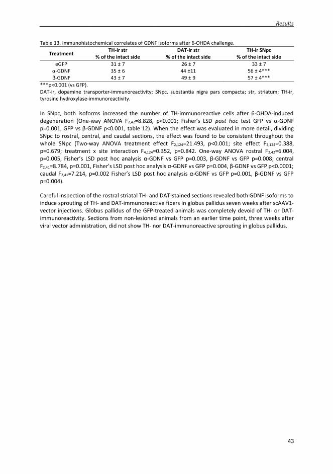
_____________________________________________________________________________Results
43
Table 13. Immunohistochemical correlates of GDNF isoforms after 6-OHDA challenge.
Treatment TH-ir str% of the intact side
DAT-ir str% of the intact side
TH-ir SNpc% of the intact side
eGFP 31 ± 7 26 ± 7 33 ± 7-GDNF 35 ± 6 44 ±11 56 ± 4***-GDNF 43 ± 7 49 ± 9 57 ± 4***
***p<0.001 (vs GFP).DAT-ir, dopamine transporter-immunoreactivity; SNpc, substantia nigra pars compacta; str, striatum; TH-ir,tyrosine hydroxylase-immunoreactivity.
In SNpc, both isoforms increased the number of TH-immunoreactive cells after 6-OHDA-induceddegeneration (One-way ANOVA F2,42=8.828, p<0.001; Fisher’s LSD post hoc test GFP vs -GDNFp=0.001, GFP vs -GDNF p<0.001, table 12). When the effect was evaluated in more detail, dividingSNpc to rostral, central, and caudal sections, the effect was found to be consistent throughout thewhole SNpc (Two-way ANOVA treatment effect F2,124=21.493, p<0.001; site effect F2,124=0.388,p=0.679; treatment x site interaction F4,124=0.352, p=0.842. One-way ANOVA rostral F2,42=6.004,p=0.005, Fisher’s LSD post hoc analysis -GDNF vs GFP p=0.003, -GDNF vs GFP p=0.008; centralF2,41=8.784, p=0.001, Fisher’s LSD post hoc analysis -GDNF vs GFP p=0.004, -GDNF vs GFP p<0.0001;caudal F2,41=7.214, p=0.002 Fisher’s LSD post hoc analysis -GDNF vs GFP p=0.001, -GDNF vs GFPp=0.004).
Careful inspection of the rostral striatal TH- and DAT-stained sections revealed both GDNF isoforms toinduce sprouting of TH- and DAT-immunoreactive fibers in globus pallidus seven weeks after scAAV1-vector injections. Globus pallidus of the GFP-treated animals was completely devoid of TH- or DAT-immunoreactivity. Sections from non-lesioned animals from an earlier time point, three weeks afterviral vector administration, did not show TH- nor DAT-immunoreactive sprouting in globus pallidus.

Discussion__________________________________________________________________________
44
66 DISCUSSION
This work was planned to explore the neuroprotective effects of novel neurotrophic factor variants inthe rat partial 6-OHDA model of PD. Moreover, the new low-dose 6-OHDA models of PD werecharacterized together with a new method to evaluate the number of TH-positive cells in SN. Togetherthe results show how low-doses of 6-OHDA can be used to induce a reliable animal model of PD andthat the less known and less studied variants of neurotrophic factors have neuroprotective potentialand are therefore new candidates for PD drug development.
6.1 Matlab-based cell counting
Stereology has traditionally been the method of choice for evaluating the number of TH-positive cellsin SNpc (Sterio, 1984). Commonly the cell counting has been conducted using a three-dimensionalprobe called an optical dissector, which allows the cell number to be estimated regardless of theirshape, size, and orientation, and the fractionator method, which is a systematic and uniform samplingpattern enabling every part of the sample to be count with an equal probability. The combination ofthese two principles in cell counting results in highly efficient and unbiased estimates of cell numbers(Sterio, 1984; West, 1999). The downside of using this well-established method is that it is very time-consuming. Therefore, we developed a new, algorithm-based method for estimating the cell numbers(study II). There are several critical differences between these two methods. The stereology-basedmethod is used with a microscope, allowing to focus on different layers of the sample (which in thiscase is a 40 μm thick section), and the cells are counted three-dimensionally from the actual brainsections. With the algorithm-based method, the cells are counted two-dimensionally from thedigitalized images taken from the sections. Thus, the quality of images influences the quality of theanalysis and therefore we have used the automated slide scanner to obtain high-quality images.Moreover, in the algorithm-based method, the program recognizes the cells based on their signalintensity and size in pixels, unlike in the stereology-based method, where the user recognizes the cells.Yet another difference is how these two methods take into account the space between the sections.In this study, the 40 μm thick tissue sections were cut in series of six, thus the distance between twoadjacent sections was 240 μm. StereoInvestigator, the computer program used to count cells withstereology, also estimated the number of cells between the sections, thus providing a cell numberestimate for the whole SNpc. The Matlab algorithm did not perform this calculation and only the cellnumbers per section were obtained. However, this might be correctable as the algorithm used for cellcounting is freely available and easy to modify. Due to these differences a lot of information is lostwhile counting cells with our algorithm-based method, and consequently the obtained cell numbersare lower than the estimates obtained with stereology-based methods. Despite these differencesbetween the methods, there is a good correlation between the results obtained with both of thesemethods (study II). It would be interesting to test this Matlab algorithm-based method in thin paraffinsections (~5 μm thick) and compare the results with stereology, as the thin sections contain only onecell layer. In this case, the cells would be counted two-dimensionally in the section with both methodsand less information would be lost with Matlab. Hence, one could expect to have an even highercorrelation between these two methods.
6.2 6-OHDA model of Parkinson’s disease
Since 6-OHDA does not cross the blood-brain barrier, it has to be administered to the brainparenchyma, either to SNpc, where the dopaminergic cell bodies are located, MFB, through which thenigrostriatal tract ascends, or striatum, where the nerve terminals are located (Duty and Jenner, 2011).Whereas injection of 6-OHDA to MFB or SN results in an almost complete (80-90%) loss of nigral TH-positive cells within one week (Ungerstedt, 1968; Jeon et al., 1995; Zuch et al., 2000), injection of 6-

__________________________________________________________________________Discussion
45
OHDA to the striatum produces a more slowly progressive partial loss of nigral cells (Sauer and Oertel,1994; Lee et al., 1996; Kirik et al., 1998). Nerve terminals start to degenerate during the first 24 hoursafter the striatal administration of 6-OHDA, and the lesion is fully developed within the next four weeks(Sauer and Oertel, 1994; Lee et al., 1996; Blandini et al., 2007). Thus, the striatal 6-OHDA injectionmodel is in line with the current “dying back” theory of PD (Burke and O'Malley, 2013), where the lossof striatal dopaminergic nerve terminals outweighs the loss of nigral cell bodies. It has been estimatedthat there is about 30% (adjusted for age) loss of dopaminergic neurons in the SNpc at the time ofsymptom onset in PD patients (Fearnley and Lees, 1991; Cheng et al., 2010). In the striatum, therewould be 50-70% loss of dopaminergic nerve terminals and 70-80% decrease of dopamine (Cheng etal., 2010; de la Fuente-Fernandez, 2013). This further supports the use of the striatal model in testingnew therapeutic agents for PD, since it more closely resembles the early stage of PD, unlike MFB or SNlesion models, which mimic later stages of PD. On the other hand, MFB and SN models might be moresuitable to study the mechanisms of cellular degeneration. Moreover, dorsal striatal targeting of 6-OHDA might provide the most appropriate model of PD in rodents. The dorsal part of the striatummight be the equivalent of the putamen in human and primate brain, as it receives input from motorand sensory neocortical areas in addition to dopaminergic innervation from SN (Björklund et al., 1997;Kirik et al., 1998; Deumens et al., 2002), and the dopamine depletion is most pronounced in theputamen in PD patients (Kish et al., 1988).
In study I we used 1 x 28 μg 6-OHDA dose. In the experiment only 54% of the animals had developeda lesion in two weeks (table 7). Thus, a large number of animals was needed to inject with 6-OHDA tohave sufficient number of animals in each treatment groups for study I. The reason for the low successrate might be the injection paradigm itself. 1 x 28 μg lesions were done as four depots to same injectiontract (table 4), where the microinjection needle was lifted between each depot and this might havecaused the 6-OHDA to flow back up the needle tract (Butcher, 1975). When compared with other used6-OHDA administration paradigms (listed in table 4), the success rate was higher when the 6-OHDAinjections were done as single depot per injection tract (table 7). This is also more common way ofinjecting 6-OHDA (e.g. Lee et al., 1996; Kirik et al., 1998; Bäck et al., 2013b). The advantage of highsuccess rate is that less animals is needed to produce reliable data. Interestingly, in the first measuredtime point the highest success rates were in lesions classified as regressive (2 x 10 μgreg and 1 x 20 μg),in both groups all animals rotated over 100 turns. However, in the last time point, only 43% of theanimals in 2 x 10 μgreg group rotated over the 100 turn limit. In contrast in progressive lesion groups (3x 3 μg, 2 x 10 μgprog, and 3 x 7 μg) the success rates were high in both time points. This comparisonsupports the use of multiple injection sites for creating a 6-OHDA lesion model of PD with success rate.
Stability of the animal model is crucial as it enables longer experiments and sufficient follow-up periodsfor neurorestoration and neuroprotection experiments, reduces the number of animals needed for theexperiments and more importantly, increases the reliability and replicability of the results. The 6-OHDAlesion groups were categorized as progressive, stable or regressive based on their rotational behavior.In progressive lesion groups the rotational behavior of the animals increased in time (3 x 3 μg, 2 x 10μgprog, and 3 x 7 μg), in stable groups the rotational behavior was at similar level during the wholeexperiment (3 x 1 μg, 3 x 2 μg, and 1 x 28 μg2week, and 1 x 28 μg5week), and in regressive groups (2 x 10μgreg and 1 x 20 μg) the rotational behavior decreased in time (figure 8). Several studies have reportedspontaneous recovery from the 6-OHDA lesion (Yang et al., 2009; Bäck et al., 2013a; Bäck et al., 2013b).Spontaneous recovery has been thought to occur by several mechanisms. In the lesioned striatumdopamine is diffused more widely (Robinson et al., 1994) and the amount of dopamine released fromthe remaining nerve terminals is increased (Doucet et al., 1986). These presynaptic compensatorymechanisms take place almost immediately after the 6-OHDA injection, and together with axonalregrowth can provide long-lasting effects (Robinson et al., 1994; Blanchard et al., 1996). In our hands,injection of low doses of 6-OHDA to multiple sites seemed to improve the stability of the lesion (figure8, table 7).

Discussion__________________________________________________________________________
46
One possible explanation for the high rotation rates at the last time point of the progressive lesiongroups might be sensitization to amphetamine. Considering that the experiments are often long, andthe rotation assay is repeated several times, the animals are repeatedly administered amphetamine ina drug-paired environment. This can lead to increased locomotor activity, i.e. the animals rotatewithout amphetamine (Robinson and Becker, 1986; Kalivas and Stewart, 1991). This conditionedresponse has also been detected in MFB-lesioned rats with apomorphine (Hudson et al., 1994). Todiscriminate the conditioned response from the actual drug-induced rotations, we have implementeda 30 minute habituation period before administration of amphetamine. We also decreased the doseof amphetamine from previously used 2.5 mg/kg (Voutilainen et al., 2009; Bäck et al., 2013a; Bäck etal., 2013b) to 2.5mg/kg of amphetamine sulfate, which equals to 1.84 mg/kg of d-amphetamine, andchanged administration from intraperitoneal to subcutaneous. Due to these changes, only one rotationpeak was observed in the assay, despite the time point. However, in our experiments the progressivelesion groups were assessed approximately as many times as stable or regressive groups, and thereforethe sensitization to amphetamine is less likely to explain the differences in rotational behavior.
As discussed above an in section 2.2.1, amphetamine-induced rotations might not be the optimalmethod to assess the lesioned animals. In studies I and III we used cylinder test together with therotational assay to determine the therapeutic effects of neurotrophic factors In our hands, the problemwith cylinder test is the habituation. As the test is based on the exploratory activity, the rats habituateto the environment when the test is repeated and this is detected as a reduction in the vertical activity.To avoid this problem, in study III the cylinder test was conducted under red light. This seemed toincrease the activity of the animals, and a reasonable number of rearings were measured during fiveminutes of testing (GFP-treated animals week 3 36 ±3 rearings and week 7 28 ± 3 rearings).
The classical method for determining the magnitude of the lesion has been counting of nigral TH-immunoreactive cells and striatal density of TH-immunoreactive fibers (Sauer and Oertel, 1994;Bowenkamp et al., 1996; Björklund et al., 1997; Cohen et al., 2011). Third alternative to use as a markerfor dopaminergic neurons is VMAT2 (Decressac et al., 2011; Kumar et al., 2015).Immunohistochemistry is a not fully problem-free approach because the cells first become atrophicand lose their dopaminergic phenotype before degenerating (Sauer and Oertel, 1994; Bowenkamp etal., 1996; Björklund et al., 1997; Cohen et al., 2011). One way of distinguishing these two events, actualcell degeneration and loss of TH-phenotype in nigral cell bodies, is to label dopaminergic cells withfluorescent retrograde axonal tracer FluoroGold (Sauer and Oertel, 1994; Bowenkamp et al., 1996;Cohen et al., 2011). The loss of TH-immunoreactivity has been shown to be maximal about two weeksafter 6-OHDA administration, whereas FluoroGold-signal decreases more slowly, until week four. Thediscrepancy between the number of TH-immunoreactive cells and FluoroGold-positive cells is reducedin time when the lesion has developed properly (Sauer and Oertel, 1994; Cohen et al., 2011). Althoughin our experiments (studies II and II) the immunohistochemical analysis was conducted several (8-16)weeks after 6-OHDA administration, and thus the lesion was fully developed and TH-staining could beconsidered as a reliable marker for the dopaminergic cells (Sauer and Oertel, 1994; Lee et al., 1996;Cohen et al., 2011), another marker, DAT, was also applied to determine the lesion size. In study III thiswas a crucial step since long-term overexpression of GDNF is known to downregulate TH expression(Georgievska et al., 2002a; Georgievska et al., 2004a; Georgievska et al., 2004b). In all our stable lesionmodels tested (3 x 1 μg, 3 x 2 μg, and 1 x 28 μg2week), the loss of TH-immunoreactivity was morepronounced in the striatum compared to SN (study I, II, table 7), suggesting these models to be moreappropriate in modelling the earlier stage of PD and testing new neuroprotective agents.
The effect of 6-OHDA dose on the lesion specificity has been well documented, and high doses of 6-OHDA have been shown to induce gliosis and unspecific loss of neurons around the injection tract(Przedborski et al., 1995; Lee et al., 1996; Kirik et al., 1998), where the local 6-OHDA concentration ishighest (Agid et al., 1973; Butcher, 1975; Javoy et al., 1976) and enlargement of the ipsilateral ventricle(Przedborski et al., 1995). Therefore the use of low doses in inducing the lesion is highly recommended

__________________________________________________________________________Discussion
47
(Kirik et al., 1998). Another thing to consider to minimize the unspecific damage due to injection is thesize of microinjection needle used to infuse 6-OHDA to brain parenchyma. In study II we calculatedthat one injection with 26 G needle (outer diameter 0.47 mm) caused an approximately 2.6%mechanical lesion in the striatum. In contrast, one injection with a smaller 33G microinjection needle(outer diameter 0.21 mm) caused 0.5% mechanical lesion. The choice of the needle size becomes evenmore crucial when the injections are done into multiple sites in brain or to a smaller target (i.e. MFBor SN) or to a smaller animal (i.e. mouse). On the other hand, smaller needle might clog more easily.The third thing to consider for minimizing the mechanical damage is the infusion rate of the toxin.Fulceri and coworkers (Fulceri et al., 2006) have shown how slower 6-OHDA infusion speed spared thenoradrenergic nerve terminals around the needle tract from degeneration. Although 6-OHDA is takenup to the cells via DAT, it has a high affinity for the noradrenalinetransporter (Luthman et al., 1989).Therefore, noradrenaline transporter blockers, such as desipramine (desmethylimipramine), can beadministered prior to 6-OHDA injection to reduce the unspecific damage (Fulceri et al., 2006; Bäck etal., 2013a). Moreover, if the experimenter desires that the SNpc stays intact and devoid of mechanicaldamage, the 6-OHDA should be injected to either MFB or striatum.
To summarize, we have tested several different injection paradigms to find the optimal settings toinduce reliable partial 6-OHDA lesion model of PD with high success rate. Stability and high successrate of the model would reduce the number of animals needed in the experiments to produce reliabledata. After the comparison and for the aforementioned reasons we chose the low 3 x 2 μg dose of 6-OHDA for the study III. This lesion paradigm produced stable rotational behavior for twelve weeks withgood success rate, and showed more pronounced loss of TH- or DAT-immunoreactivity in the striatumcompared to SNpc. However, similar doses as were used in our comparison have been used insuccessfully in experiments elsewhere (Przedborski et al., 1995; Kirik et al., 1998). Thus, there areseveral technical details which can affect the reliability of 6-OHDA model of PD.
66.3 NRTN variants
NRTN and its homolog GDNF both bind to heparan sulfated proteoglycans of the cell surface andextracellular matrix, hampering protein diffusion in the brain tissue (Hamilton et al., 2001; Bespalov etal., 2011). Since neurotrophic factors are secreted in small quantities, binding to cell surface might beused to concentrate them near their receptors (reviewed in Sariola and Saarma, 2003). However, thislow diffusion has been suggested to be one of the reasons for the mixed results in clinical trials(Salvatore et al., 2006; Bartus et al., 2015). Moreover, some animal studies have suggested infusedNRTN to precipitate in tissue resulting in high focal concentration around the infusion site (Hadaczeket al., 2010). To overcome these problems and to improve the diffusion capacity in brain parenchyma,novel NRTN variants were designed.
The structure of NRTN was modeled based on the known structure of GDNF (Eigenbrot and Gerber,1997; Parkash et al., 2008), and mutations were introduced to the heel region, where the putativeheparin-binding site (Cardin and Weintraub, 1989) was located. In the N4 variant, the heel regionsequence was replaced with the heel sequence from persephin, which does not bind heparin (Bespalovet al., 2011). However, the diffusion properties of persephin in the brain tissue have not been studiedin detail. In GDNF the finger regions are more important for the interaction between the neurotrophicfactor and the GFR receptor than the heel region (Eketjäll et al., 1999; Parkash et al., 2008), thussupporting the choice of mutation site. Mutation in the heel region probably did have a small effect onthe orientation of the finger structures which are interacting with GFR receptors, but it did not affectthe biological activity of the variant proteins. However, all NRTN variants have stronger affinity forGFR 2 than for GFR 1, which has been shown previously for WT NRTN (Cik et al., 2000).
Based on the observation of Hoane and colleagues (Hoane et al., 2000) that WT NRTN produced inmammalian cells is more biologically active in vitro than WT NRTN produced in E. coli, NRTN variants

Discussion__________________________________________________________________________
48
were decided to produce in mammalian chinese hamster ovary cells. The difference in biologicalactivity could be explained by post-translational modifications and better quality control of thesecreted proteins. NRTN variants demonstrated biological activity in several in vitro assays.Interestingly, although WT NRTN is not able to induce extraureteric budding in a kidney ex vivoorganogenesis assay, NRTN variants N2 and N4 both induced ectopic ureteric budding and swelling ofthe buds. Higher biological activity in the assays correlates with higher stability of N2 and N4 in theorganogenesis and cell culture media.
Since the composition of heparan sulfate proteoglycans of the extracellular matrix and cell surfacemight vary between cell types, animal species and their developmental stage (reviewed Sarrazin et al.,2011), the diffusion properties of NRTN variants were tested in three different species: rat,cynomolgus, and marmoset monkey. The affinity of N2 and N4 to heparin was at similar level in vitro,but in brain parenchyma N4 diffused better. One possible explanation for this discrepancy might bethe difference in the structure of the variants. In N2 three point mutations were introduced to theheparin binding sequence, whereas in N4 the whole sequence was replaced. Furthermore, theenvironment of the in vivo assay is more complex than in vitro. Thus, the in vivo diffusion capacityreflected the neuroprotective effects of N4. The increased stability and diffusion capacity of NRTNvariants could mean that these proteins reach a higher number of neurons and receptors, inducing thesurvival of axons and not only the somata, and this would be reflected by the ability of N4 to rescuestriatal TH-immunoreactive nerve fibers. On the other hand, the accumulation of NRTN to the nearvicinity of GFR -receptors has been thought to improve signaling (reviewed in Sariola and Saarma,2003). Maybe this is the case with GDNF, as elimination of heparin-binding sequence from the protein( 38N-GDNF) reduced the neuroprotective effects in the 6-OHDA model of PD (Piltonen et al., 2009).This is in contrast to NRTN variants, which showed better neuroprotective effects in the 6-OHDA modelof PD than WT GDNF.
Although wide diffusion around the infusion site is desired to enhance the therapeutic effects,diffusion outside the target area (putamen / SN in PD) might cause side effects. As discussed above,the higher diffusion increases the number of reached target receptors, but simultaneously reduces theconcentration of the therapeutic agent around the needle tract. Which one of the possibilities is morebeneficial depends on the location of the target receptors. Besides diffusion properties and a numberof infusion sites, the vasculature of the brain also affects the spreading of the protein in the brain.Hadeczek and coworkers (Hadaczek et al., 2006) have shown how arterial pulsation generated bycardiac contractions can affect the diffusion of intraparenchymally infused molecules in perivascularspaces around arteries and arterioles. Thus, understanding of the organization of the brain vasculatureand perivascular fluid dynamics is of importance while optimizing the efficiency of therapeuticmolecule transport and diffusion.
In a neuroprotection assay, where low doses of proteins (5 μg) were infused two weeks after 6-OHDAinjections before the lesion had fully developed (Sauer and Oertel, 1994; Lee et al., 1996; Blandini etal., 2007), N4 was able to improve the motor performance both in cylinder test and in amphetamine-induced rotations. The results from both behavioral assays are in line, and remarkably, there is a clearresult in cylinder test, which is a more robust test than amphetamine-induced rotations (Hudson et al.,1993; Kirik et al., 1998; Schallert et al., 2000). The behavioral results from the neurorestoration assayare less clear. This can be due to the low number of animals (n=6-7/group) but also due to the robustlesion used in the study. The neurotrophic factors were administered five weeks after 6-OHDAinjection when the lesion is fully developed, and the high dose of 6-OHDA induced a severe lesion.However, together these results strongly suggest the improvement in the diffusion capacity to improvethe neuroprotective effects of NRTN variants.

__________________________________________________________________________Discussion
49
66.4 GDNF isoforms
Consistent with previous studies showing exogenous GDNF to increase the locomotor activity of theanimals (Hudson et al., 1995; Hebert and Gerhardt, 1997; Kirik et al., 2000a), overexpression of GDNFisoforms in non-lesioned striatum induced the exploratory activity of the animals in the drug-freecylinder test. Interestingly, only -GDNF increased the use of the contralateral paw. This finding is inaccordance with the in vitro observation of different secretory patterns of GDNF isoforms (Lonka-Nevalaita et al., 2010). In this case, the constitutive secretion of -GDNF and corresponding matureGDNF would have an effect on the behavior, whereas the -GDNF and corresponding mature GDNFwould be retained inside the cells and therefore no effect on the use of paws was observed. However,this would also suggest the increased activity of the animals and the use of paws to have differentmechanisms.
Effects of GDNF on TH have been well-documented. A single bolus of GDNF increases TH expression(Hudson et al., 1995) and activity (Lapchak et al., 1997), whereas continuous overexpression of GDNFdecreases striatal TH levels (Georgievska et al., 2002a; Georgievska et al., 2004b; Sajadi et al., 2005)as a response to long-lasting dopamine neuron activity (Iwata et al., 2000; Sajadi et al., 2005). Incontrast, in non-human primates long-term overexpression of GDNF increases TH-immunoreactivity inthe putamen (Kordower et al., 2000; Palfi et al., 2002; Johnston et al., 2009), suggesting thedownregulation of TH to be a species-specific phenomenon. Nevertheless, the effects of GDNF onanother important enzyme regulating the dopamine transmission, DAT, are still largely unknown. Datafrom Salvatore and colleagues (Salvatore et al., 2009) suggests dose-dependence, where low doses ofGDNF protein would not have an effect on striatal DAT expression, but in higher doses GDNFdownregulates DAT. GDNF might regulate the DAT activity by increasing dimerization and protein-protein interactions (Barroso-Chinea et al., 2016). Our results show overexpression of GDNF to havedifferent effects on TH and DAT. In non-lesioned striatum, both isoforms increased the density of DAT-immunoreactive fibers but did not have an effect on TH-immunoreactive fibers (table 11). The increaseof DAT could suggest GDNF-treated animals to be more vulnerable for 6-OHDA-induced cytotoxicitysince 6-OHDA is taken up by DAT. This was observed with mice overexpressing GDNF from the nativelocus (Kumar et al., 2015). The mice have increased activity of DAT, but no increase in the DAT proteinlevel, and increased number of striatal dopaminergic nerve terminals, and thus they are moresusceptible to 6-OHDA cytotoxicity (Kumar et al., 2015). However, our data do not support this, thenumber of TH-immunoreactive cells in SNpc was significantly higher in -GDNF- and -GDNF-treatedanimals than in GFP-treated animals after 6-OHDA administration (table 13).
Several studies where the long-term expression of GDNF has been implemented with the help of viralvectors have been conducted, but the comparison and translation of the obtained results is difficult,since the results vary according to the used dose, administration sites, and used time points (reviewedin Björklund et al., 2000; Kirik et al., 2017). In striatum, GDNF functions as a chemoattractant, inducingaxonal sprouting and reinnervation after a cytotoxic insult, and that might explain why striatal deliveryof GDNF gene protects the nigrostriatal tract and provides functional recovery in the animal models ofPD (Kirik et al., 2000b; Kells et al., 2010). However, neuroprotective effects of striatal GDNF deliverywithout behavioral correlates have also been reported (Georgievska et al., 2002b). This is in line withour results, where both GDNF isoforms protected TH-immunoreactive cell bodies from 6-OHDA-induced degeneration without functional recovery. Nonetheless, using a similar experimental setup(striatal administration of AAV-GDNF four weeks before striatal infusion of 6-OHDA) but with a longerfollow-up time, Kirik and coworkers (Kirik et al., 2000b) showed that the GDNF-treated animals rotatedless with amphetamine four weeks after 6-OHDA lesion, but the difference was not significant beforeweek seven. Therefore the short four-week follow-up time used in our study might explain the lack offunctional effects.
In the experiment the transgene was expressed under cytomegalovirus (CMV) promoter, which hasbeen shown to drive the transgene expression both in neurons and astrocytes (Bockstael et al., 2008).

Discussion__________________________________________________________________________
50
Although the titers of the used viral vectors were similar (scAAV1-CMV-pre- -pro-GDNF 2.14 x 10¹²
vg/ml and scAAV1-CMV-pre- -pro-GDNF 1.73 x 10¹² vg/ml), the GDNF level was much higher in -GDNF overexpressing animals than in animals overexpressing -GDNF. A similar phenomenon has beenobserved when Fletcher and coworkers used DNA nanoparticles to overexpress GDNF isoforms in therat striatum (Fletcher et al., 2011). Furthermore, the expression of endogenous GDNF and exogenousGDNF delivered with DNA nanoparticles is enhanced by 6-OHDA injection (Yurek and Fletcher-Turner,2001; Fletcher et al., 2011). Despite the difference in the protein levels, immunohistochemistryshowed a widely spread GDNF-immunoreactivity pattern in both -GDNF- and -GDNF-treatedanimals. In comparison, GFP-immunoreactivity was detected in a more restricted area as it is retainedinside the cells, unlike GDNF. Fluorescence staining with TH antibody revealed GFP to be expressed inSNpr and not in SNpc, suggesting scAAV1 to transduce GABAergic medium spiny neurons rather thandopaminergic neurons. However, this was not confirmed with GABAergic markers. This GFP-signalpattern is in line with previously published data with CMV promoter (Bockstael et al., 2008). Thetransport of transgene product to SNpr and the observed increase in the number of nigral TH-immunoreactive cells in GDNF-treated animals could imply to the paracrine function of GDNF. Thismight be a better alternative than nigral delivery of GDNF, which has been shown to induce aberrantsprouting (Kirik et al., 2000b; Georgievska et al., 2002a).
Recently, cytotoxic effects of high titers of AAV5-GFP was reported (Xilouri et al., 2013; Landeck et al.,2017). In their study Landeck and colleagues showed that nigral administration of high titer (3.3 x 10¹⁴
gc/ml) AAV5-GFP induced loss of TH-immunoreactive cells in SNpc. In our case, the used titers werelower (7.4 x 10¹³ vg/ml) and no cytotoxic effects were detected after intrastriatal delivery since theloss of TH-immunoreactive cells was about 67% in AAV-GFP-treated animals (study III) and 64% inanimals receiving only 3 x 2 μg dose of 6-OHDA (study II). Cytotoxicity was not observed in striatumeither since the loss of TH-immunoreactivity was 72% after 6-OHDA injection in study II (table 7), and69% after the combination of AAV-GFP and 6-OHDA in study III (table 13).
66.5 Protein infusion vs gene therapy
As mentioned earlier, the focus in neurotrophic factor clinical trials is currently on increasing the targetcoverage of the therapeutic protein. This can be achieved by improving the bioavailability ofneurotrophic factors in the target area by increasing the infusion volume and/or dose, modifying thevector, or by modifying the diffusion properties of the therapeutic agents. Comparison of the obtaineddata is difficult since the results depend on the used model (ex. whether 6-OHDA was injected to MFB,SN, or striatum), time frame, when was the neurotrophic factor infused (e.g. before or after 6-OHDA)and how, as protein infusion or with viral vectors. This variability makes it more difficult to translatethe results. In this thesis, the bioavailability of NRTN in the target tissue was increased by modifyingthe protein sequence (study I) and the bioavailability of GDNF isoforms by viral vector delivery (studyIII).
Gene therapy provides long-lasting expression of transgene in the target tissue. However, with viralvectors the dosing is more difficult because the amount of therapeutic protein produced depends onthe virus vector properties and used promoter (reviewed in Domanskyi et al., 2015). In addition, oncethe vector has been infused to parenchyma, it cannot be removed. Regulated viral vectors, such as theones using TetON/OFF promoters, destabilizing domains, or GeneSwitch, where the expression of thetransgene can be turned on or off with antibiotics or small molecules such as doxycycline ortrimethoprim, might solve this problem (Maddalena et al., 2013; Quintino et al., 2013; Chtarto et al.,2016). High neurotrophic factor doses might not be desirable though, not only due to risk of sideeffects but also because neurotrophic factors are known to be effective in low concentrations (Sariolaand Saarma, 2003).

__________________________________________________________________________Discussion
51
With protein infusion, the dosing may be easier to control than with viral vectors, and in case of sideeffects the administration can be stopped. However, the question arises whether the infusion shouldbe continuous or repeated. As discussed above in section 6.3, in study I we used proteins produced inmammalian cells, as WT NRTN produced in mammalian cells was previously shown to be more activein vitro than the E. coli produced protein, probably due to post-translational modifications (Hoane etal., 2000). Thus, the manufacturing process of the proteins used in the protein infusion studies needsto be taken into account when comparing the obtained results. In study I we showed that by improvingthe diffusion capacity of NRTN we were able to enhance the therapeutic effects. Although theimproved diffusion is a desired property, too diffuse protein might spread outside the target area andinduce unwanted side effects.
Another important consideration is the targeting. Although nigral delivery of neurotrophic factors hasbeen shown to be neuroprotective (Kirik et al., 2000a; Kirik et al., 2000b), overloading the stressedcells with proteins might accelerate the degeneration process instead of delaying it (Hoffer and Harvey,2011). Moreover, nigral overexpression of GDNF can induce aberrant sprouting (Kirik et al., 2000b;Georgievska et al., 2002a). On the other hand, target-derived expression of neurotrophic factors is anattractive approach, as retrograde transport of GDNF has been reported (Tomac et al., 1995b; Ai et al.,2003). However, in the light of current evidence the degeneration process starts from the nerveterminals compromising the axonal transport (Kordower et al., 2013; Tagliaferro and Burke, 2016).Interestingly, as shown in study III, proteins expressed in striatal medium spiny neurons weretransported anterogradely to SNpr, and hence striatally delivered neurotrophic factors may executetheir functions on dopaminergic neurons in SNpc in a paracrine manner.
Ergo, there are numerous factors to consider in the development of efficient neurotrophic factor-based therapy. Besides focusing on developing an efficient and safe therapy, attention should be paidto find new diagnostic tools to identify PD earlier. Detection of early stage PD patients would allowearlier initiation of the treatment before most of the striatal nerve terminals and nigral cell bodieshave degenerated and there are still viable cells left to respond to the therapy.

Conclusions_________________________________________________________________________
52
77 CONCLUSIONS
The aim of the thesis was to study the neuroprotective effects of novel and less studied variants ofneurotrophic factors GDNF and neurturin. The main conclusions from the studies are the following:
- Reduced affinity to heparin in vitro results in improved diffusion capacity and enhancedtherapeutic efficiency of neurturin variants in vivo to protect nigrostriatal dopaminergiccircuitry.
- The stability of the classical striatal 6-OHDA model of PD can be improved by distributing 6-OHDA to multiple injection sites. In this way, even small doses (6-9μg) can produce a stableanimal model with high success rate thus reducing the number of experimental animals.
- Both conserved GDNF isoforms increased the density of DAT-immunoreactive fibers in non-lesioned striatum and increased vertical activity of the animals, but only -GDNF inducedbehavioral asymmetry. Both isoforms protected nigral dopaminergic neurons form 6-OHDA-induced cytotoxicity without behavioral improvements.
Together, the results point that these new isoforms of well-studied neurotrophic factors havetherapeutic potential in PD. Improved diffusion properties of NRTN variants and regulated secretionof -GDNF make them interesting new agents in the search for safe and efficient neurotrophic factor-based therapy.

__________________________________________________________________Acknowledgements
53
88 ACKNOWLEDGEMENTS
This work was conducted at the Insitute of Biotechnology, University of Helsinki in collaboration withDivision of Pharmacology and Pharmacotherapy, Faculty of Pharmacy, University of Helsinki during theyears 2012 to 2017.
Finnish Parkinson Foundation, Finnish Culture Foundation, Instrumentarium Science Foundation,Oskar Öflund Foundation, Finnish Pharmacological Society, Drug Research Foundation, and theDoctoral School of Health together with its Doctoral Programme in Drug Research are gratefullyacknowledged for the personal financial support.
I want to thank my supervisor, docent Mikko Airavaara for the courage to hire a PhD student withoutany background in neuroscience. It’s been quite a journey and I sincerely thank you for giving me thisopportunity.
Professor Raimo Tuominen, Head of the Division of Pharmacology and Pharmacotherapy, isacknowledged for the support, optimism, and all the signatures during these years.
The reviewers of my thesis, docent Sanna Janhunen and docent Teemu Aitta-aho are thanked for thevaluable comments and suggestions regarding my thesis.
Privat docent Liliane Tenenbaum is acknowledged for agreeing to be my opponent in the publicdefence of this thesis.
Professor Mart Saarma is thanked for the support and providing the possibility to work in hislaboratory.
I want to express my gratitude to all my co-authors, your (major) efforts are highly appreciated.Without you I wouldn’t have any papers. Especially Pia, it’s been a pleasure to work with you. TheNRTN paper taught me what it means that a paper should tell a story.
All the past and present members of Team Airavaara and Saarma lab are acknowledged for creating afriendly work environment. Dear mutants and wildtypes, Emmi, my songwriting partnerFroBro/Franci/FNC, Jenni, Kat, Maryna, Polina, and Sara, I’ve enjoyed our adventures in and outsidethe lab enormously. Your support and friendhip means a lot to me.
All the students whom I have supervised and who have helped me in the lab: Antti Huovinen, UlrikaJulku, Jaan Korpikoski, Ilmari Parkkinen, and Aneta Petrušková, your help with stainings, cylinder videosand everything else is gratefully acknowledged.
Susanne Bäck and Kati Rautio at the Division of Pharmacology and Pharmacotherapy are acknowledgedfor all their help and support.
I want to thank my friends for the support and all the good times we’ve had together. I suck at stayingin contact, but it doesn’t mean that I have forgotten about you. Especially Laura, our “culture events”and evening teas have been great escapes from lab and other matters.
Anu & Aki, Aki, Laura, Selja & Siiri, Anne, Juha & Venla, I love the twisted humor we have in our family.Things are never as bad as they seem and they could always be a lot worse. Thank you forcounterbalancing my “science life”. Niin ja oot äiti sinäki semmone kapine.
Mission accomplished.
Helsinki, August 2017

References_________________________________________________________________________
54
99 REFERENCES
Agid, Y., Javoy, F., Glowinski, J., Bouvet, D., and Sotelo, C. (1973) Injection of 6-hydroxydopamine intothe substantia nigra of the rat. II. Diffusion and specificity. Brain Res 58:291-301.
Aguiar, A. S., Jr., Duzzioni, M., Remor, A. P., Tristao, F. S., Matheus, F. C., Raisman-Vozari, R., Latini, A.,and Prediger, R. D. (2016) Moderate-Intensity Physical Exercise Protects Against Experimental 6-Hydroxydopamine-Induced Hemiparkinsonism Through Nrf2-Antioxidant Response Element Pathway.Neurochem Res 41:64-72.
Ahlskog, J. E., and Muenter, M. D. (2001) Frequency of levodopa-related dyskinesias and motorfluctuations as estimated from the cumulative literature. Mov Disord 16:448-458.
Ai, Y., Markesbery, W., Zhang, Z., Grondin, R., Elseberry, D., Gerhardt, G. A., and Gash, D. M. (2003)Intraputamenal infusion of GDNF in aged rhesus monkeys: distribution and dopaminergic effects. JComp Neurol 461:250-261.
Airaksinen, M. S., and Saarma, M. (2002) The GDNF family: signalling, biological functions andtherapeutic value. Nat Rev Neurosci 3:383-394.
Airavaara, M., Pletnikova, O., Doyle, M. E., Zhang, Y. E., Troncoso, J. C., and Liu, Q. R. (2011)Identification of novel GDNF isoforms and cis-antisense GDNFOS gene and their regulation in humanmiddle temporal gyrus of Alzheimer disease. J Biol Chem 286:45093-45102.
Amano, S., Kegelmeyer, D., and Hong, S. L. (2014) Rethinking energy in parkinsonian motor symptoms:a potential role for neural metabolic deficits. Front Syst Neurosci 8:242.
Andersson, K. K., Cox, D. D., Que, L., Jr., Flatmark, T., and Haavik, J. (1988) Resonance Raman studieson the blue-green-colored bovine adrenal tyrosine 3-monooxygenase (tyrosine hydroxylase). Evidencethat the feedback inhibitors adrenaline and noradrenaline are coordinated to iron. J Biol Chem263:18621-18626.
Arai, H., Furuya, T., Yasuda, T., Miura, M., Mizuno, Y., and Mochizuki, H. (2004) Neurotoxic effects oflipopolysaccharide on nigral dopaminergic neurons are mediated by microglial activation, interleukin-1beta, and expression of caspase-11 in mice. J Biol Chem 279:51647-51653.
Baloh, R. H., Tansey, M. G., Golden, J. P., Creedon, D. J., Heuckeroth, R. O., Keck, C. L., Zimonjic, D. B.,Popescu, N. C., Johnson, E. M., Jr., and Milbrandt, J. (1997) TrnR2, a novel receptor that mediatesneurturin and GDNF signaling through Ret. Neuron 18:793-802.
Baloh, R. H., Tansey, M. G., Lampe, P. A., Fahrner, T. J., Enomoto, H., Simburger, K. S., Leitner, M. L.,Araki, T., Johnson, E. M., Jr., and Milbrandt, J. (1998) Artemin, a novel member of the GDNF ligandfamily, supports peripheral and central neurons and signals through the GFRalpha3-RET receptorcomplex. Neuron 21:1291-1302.
Barraud, Q., Lambrecq, V., Forni, C., McGuire, S., Hill, M., Bioulac, B., Balzamo, E., Bezard, E., Tison, F.,and Ghorayeb, I. (2009) Sleep disorders in Parkinson's disease: the contribution of the MPTP non-human primate model. Exp Neurol 219:574-582.
Barroso-Chinea, P., Cruz-Muros, I., Afonso-Oramas, D., Castro-Hernandez, J., Salas-Hernandez, J.,Chtarto, A., Luis-Ravelo, D., Humbert-Claude, M., Tenenbaum, L., and Gonzalez-Hernandez, T. (2016)Long-term controlled GDNF over-expression reduces dopamine transporter activity without affectingtyrosine hydroxylase expression in the rat mesostriatal system. Neurobiol Dis 88:44-54.

_________________________________________________________________________References
55
Bartels, T., Choi, J. G., and Selkoe, D. J. (2011) alpha-Synuclein occurs physiologically as a helicallyfolded tetramer that resists aggregation. Nature 477:107-110.
Bartus, R. T., Herzog, C. D., Chu, Y., Wilson, A., Brown, L., Siffert, J., Johnson, E. M., Jr., Olanow, C. W.,Mufson, E. J., and Kordower, J. H. (2011) Bioactivity of AAV2-neurturin gene therapy (CERE-120):differences between Parkinson's disease and nonhuman primate brains. Mov Disord 26:27-36.
Bartus, R. T., Baumann, T. L., Siffert, J., Herzog, C. D., Alterman, R., Boulis, N., Turner, D. A., Stacy, M.,Lang, A. E., Lozano, A. M., and Olanow, C. W. (2013) Safety/feasibility of targeting the substantia nigrawith AAV2-neurturin in Parkinson patients. Neurology 80:1698-1701.
Bartus, R. T. (2015) Gene therapy for Parkinson's disease: a decade of progress supported byposthumous contributions from volunteer subjects. Neural Regen Res 10:1586-1588.
Bartus, R. T., Kordower, J. H., Johnson, E. M., Jr., Brown, L., Kruegel, B. R., Chu, Y., Baumann, T. L., Lang,A. E., Olanow, C. W., and Herzog, C. D. (2015) Post-mortem assessment of the short and long-termeffects of the trophic factor neurturin in patients with alpha-synucleinopathies. Neurobiol Dis 78:162-171.
Bartus, R. T., and Johnson, E. M., Jr. (2017a) Clinical tests of neurotrophic factors for humanneurodegenerative diseases, part 2: Where do we stand and where must we go next? Neurobiol Dis97:169-178.
Bartus, R. T., and Johnson, E. M., Jr. (2017b) Clinical tests of neurotrophic factors for humanneurodegenerative diseases, part 1: Where have we been and what have we learned? Neurobiol Dis97:156-168.
Beal, M. F., Matthews, R. T., Tieleman, A., and Shults, C. W. (1998) Coenzyme Q10 attenuates the 1-methyl-4-phenyl-1,2,3,tetrahydropyridine (MPTP) induced loss of striatal dopamine and dopaminergicaxons in aged mice. Brain Res 783:109-114.
Becker, C., Jick, S. S., and Meier, C. R. (2008) Use of antihypertensives and the risk of Parkinson disease.Neurology 70:1438-1444.
Bender, A., Krishnan, K. J., Morris, C. M., Taylor, G. A., Reeve, A. K., Perry, R. H., Jaros, E., Hersheson, J.S., Betts, J., Klopstock, T., Taylor, R. W., and Turnbull, D. M. (2006) High levels of mitochondrial DNAdeletions in substantia nigra neurons in aging and Parkinson disease. Nat Genet 38:515-517.
Bender, T. S., Migliore, M. M., Campbell, R. B., John Gatley, S., and Waszczak, B. L. (2015) Intranasaladministration of glial-derived neurotrophic factor (GDNF) rapidly and significantly increases whole-brain GDNF level in rats. Neuroscience 303:569-576.
Bentea, E., Verbruggen, L., and Massie, A. (2017) The Proteasome Inhibition Model of Parkinson'sDisease. J Parkinsons Dis 7:31-63.
Bespalov, M. M., and Saarma, M. (2007) GDNF family receptor complexes are emerging drug targets.Trends Pharmacol Sci 28:68-74.
Bespalov, M. M., Sidorova, Y. A., Tumova, S., Ahonen-Bishopp, A., Magalhaes, A. C., Kulesskiy, E.,Paveliev, M., Rivera, C., Rauvala, H., and Saarma, M. (2011) Heparan sulfate proteoglycan syndecan-3is a novel receptor for GDNF, neurturin, and artemin. J Cell Biol 192:153-169.

References_________________________________________________________________________
56
Bian, M., Liu, J., Hong, X., Yu, M., Huang, Y., Sheng, Z., Fei, J., and Huang, F. (2012) Overexpression ofparkin ameliorates dopaminergic neurodegeneration induced by 1- methyl-4-phenyl-1,2,3,6-tetrahydropyridine in mice. PLoS One 7:e39953.
Biggs, C. S., Fowler, L. J., Whitton, P. S., and Starr, M. S. (1997) Extracellular levels of glutamate andaspartate in the entopeduncular nucleus of the rat determined by microdialysis: regulation by striataldopamine D2 receptors via the indirect striatal output pathway? Brain Res 753:163-175.
Birkmayer, W., and Hornykiewicz, O. (1961) [The L-3,4-dioxyphenylalanine (DOPA)-effect in Parkinson-akinesia]. Wien Klin Wochenschr 73:787-788.
Björklund, A., Rosenblad, C., Winkler, C., and Kirik, D. (1997) Studies on neuroprotective andregenerative effects of GDNF in a partial lesion model of Parkinson's disease. Neurobiol Dis 4:186-200.
Björklund, A., Kirik, D., Rosenblad, C., Georgievska, B., Lundberg, C., and Mandel, R. J. (2000) Towardsa neuroprotective gene therapy for Parkinson's disease: use of adenovirus, AAV and lentivirus vectorsfor gene transfer of GDNF to the nigrostriatal system in the rat Parkinson model. Brain Res 886:82-98.
Björklund, A., and Dunnett, S. B. (2007) Dopamine neuron systems in the brain: an update. TrendsNeurosci 30:194-202.
Blanchard, V., Anglade, P., Dziewczapolski, G., Savasta, M., Agid, Y., and Raisman-Vozari, R. (1996)Dopaminergic sprouting in the rat striatum after partial lesion of the substantia nigra. Brain Res709:319-325.
Blandini, F., Levandis, G., Bazzini, E., Nappi, G., and Armentero, M. T. (2007) Time-course ofnigrostriatal damage, basal ganglia metabolic changes and behavioural alterations followingintrastriatal injection of 6-hydroxydopamine in the rat: new clues from an old model. Eur J Neurosci25:397-405.
Blesa, J., and Przedborski, S. (2014) Parkinson's disease: animal models and dopaminergic cellvulnerability. Front Neuroanat 8:155.
Bockstael, O., Chtarto, A., Wakkinen, J., Yang, X., Melas, C., Levivier, M., Brotchi, J., and Tenenbaum, L.(2008) Differential transgene expression profiles in rat brain, using rAAV2/1 vectors with tetracycline-inducible and cytomegalovirus promoters. Hum Gene Ther 19:1293-1305.
Bonifati, V., Rizzu, P., van Baren, M. J., Schaap, O., Breedveld, G. J., Krieger, E., Dekker, M. C., Squitieri,F., Ibanez, P., Joosse, M., van Dongen, J. W., Vanacore, N., van Swieten, J. C., Brice, A., Meco, G., vanDuijn, C. M., Oostra, B. A., and Heutink, P. (2003) Mutations in the DJ-1 gene associated with autosomalrecessive early-onset parkinsonism. Science 299:256-259.
Bourque, M. J., and Trudeau, L. E. (2000) GDNF enhances the synaptic efficacy of dopaminergicneurons in culture. Eur J Neurosci 12:3172-3180.
Bowenkamp, K. E., David, D., Lapchak, P. L., Henry, M. A., Granholm, A. C., Hoffer, B. J., and Mahalik,T. J. (1996) 6-hydroxydopamine induces the loss of the dopaminergic phenotype in substantia nigraneurons of the rat. A possible mechanism for restoration of the nigrostriatal circuit mediated by glialcell line-derived neurotrophic factor. Exp Brain Res 111:1-7.
Braak, H., Del Tredici, K., Rub, U., de Vos, R. A., Jansen Steur, E. N., and Braak, E. (2003) Staging of brainpathology related to sporadic Parkinson's disease. Neurobiol Aging 24:197-211.

_________________________________________________________________________References
57
Braak, H., and Del Tredici, K. (2004) Poor and protracted myelination as a contributory factor toneurodegenerative disorders. Neurobiol Aging 25:19-23.
Braak, H., de Vos, R. A., Bohl, J., and Del Tredici, K. (2006) Gastric alpha-synuclein immunoreactiveinclusions in Meissner's and Auerbach's plexuses in cases staged for Parkinson's disease-related brainpathology. Neurosci Lett 396:67-72.
Bradley, L. H., Fuqua, J., Richardson, A., Turchan-Cholewo, J., Ai, Y., Kelps, K. A., Glass, J. D., He, X.,Zhang, Z., Grondin, R., Littrell, O. M., Huettl, P., Pomerleau, F., Gash, D. M., and Gerhardt, G. A. (2010)Dopamine neuron stimulating actions of a GDNF propeptide. PLoS One 5:e9752.
Breit, S., Bouali-Benazzouz, R., Popa, R. C., Gasser, T., Benabid, A. L., and Benazzouz, A. (2007) Effectsof 6-hydroxydopamine-induced severe or partial lesion of the nigrostriatal pathway on the neuronalactivity of pallido-subthalamic network in the rat. Exp Neurol 205:36-47.
Brotchie, J., and Fitzer-Attas, C. (2009) Mechanisms compensating for dopamine loss in early Parkinsondisease. Neurology 72:S32-38.
Burke, R. E., Dauer, W. T., and Vonsattel, J. P. (2008) A critical evaluation of the Braak staging schemefor Parkinson's disease. Ann Neurol 64:485-491.
Burke, R. E., and O'Malley, K. (2013) Axon degeneration in Parkinson's disease. Exp Neurol 246:72-83.
Burns, R. S., Chiueh, C. C., Markey, S. P., Ebert, M. H., Jacobowitz, D. M., and Kopin, I. J. (1983) A primatemodel of parkinsonism: selective destruction of dopaminergic neurons in the pars compacta of thesubstantia nigra by N-methyl-4-phenyl-1,2,3,6-tetrahydropyridine. Proc Natl Acad Sci U S A 80:4546-4550.
Burre, J., Sharma, M., Tsetsenis, T., Buchman, V., Etherton, M. R., and Sudhof, T. C. (2010) Alpha-synuclein promotes SNARE-complex assembly in vivo and in vitro. Science 329:1663-1667.
Butcher, L. L. (1975) Degenerative processes after punctate intracerebral administration of 6-hydroxydopamine. J Neural Transm 37:189-203.
Butte, M. J. (2001) Neurotrophic factor structures reveal clues to evolution, binding, specificity, andreceptor activation. Cell Mol Life Sci 58:1003-1013.
Bäck, S., Peränen, J., Galli, E., Pulkkila, P., Lonka-Nevalaita, L., Tamminen, T., Voutilainen, M. H.,Raasmaja, A., Saarma, M., Männisto, P. T., and Tuominen, R. K. (2013a) Gene therapy with AAV2-CDNFprovides functional benefits in a rat model of Parkinson's disease. Brain Behav 3:75-88.
Bäck, S., Raki, M., Tuominen, R. K., Raasmaja, A., Bergstrom, K., and Männisto, P. T. (2013b) Highcorrelation between in vivo [123I]beta-CIT SPECT/CT imaging and post-mortem immunohistochemicalfindings in the evaluation of lesions induced by 6-OHDA in rats. EJNMMI Res 3:46.
Bäckman, C. M., Shan, L., Zhang, Y. J., Hoffer, B. J., Leonard, S., Troncoso, J. C., Vonsatel, P., and Tomac,A. C. (2006) Gene expression patterns for GDNF and its receptors in the human putamen affected byParkinson's disease: a real-time PCR study. Mol Cell Endocrinol 252:160-166.
Cacalano, G., Farinas, I., Wang, L. C., Hagler, K., Forgie, A., Moore, M., Armanini, M., Phillips, H., Ryan,A. M., Reichardt, L. F., Hynes, M., Davies, A., and Rosenthal, A. (1998) GFRalpha1 is an essentialreceptor component for GDNF in the developing nervous system and kidney. Neuron 21:53-62.
Cardin, A. D., and Weintraub, H. J. (1989) Molecular modeling of protein-glycosaminoglycaninteractions. Arteriosclerosis 9:21-32.

References_________________________________________________________________________
58
Carlsson, A., Lindqvist, M., and Magnusson, T. (1957) 3,4-Dihydroxyphenylalanine and 5-hydroxytryptophan as reserpine antagonists. Nature 180:1200.
Carlsson, A., Lindqvist, M., Magnusson, T., and Waldeck, B. (1958) On the presence of 3-hydroxytyramine in brain. Science 127:471.
Carlsson, A. (1959) The occurrence, distribution and physiological role of catecholamines in thenervous system. Pharmacol Rev 11:490-493.
Castano, A., Herrera, A. J., Cano, J., and Machado, A. (1998) Lipopolysaccharide intranigral injectioninduces inflammatory reaction and damage in nigrostriatal dopaminergic system. J Neurochem70:1584-1592.
Chan, C. S., Guzman, J. N., Ilijic, E., Mercer, J. N., Rick, C., Tkatch, T., Meredith, G. E., and Surmeier, D.J. (2007) 'Rejuvenation' protects neurons in mouse models of Parkinson's disease. Nature 447:1081-1086.
Chartier-Harlin, M. C., Kachergus, J., Roumier, C., Mouroux, V., Douay, X., Lincoln, S., Levecque, C.,Larvor, L., Andrieux, J., Hulihan, M., Waucquier, N., Defebvre, L., Amouyel, P., Farrer, M., and Destee,A. (2004) Alpha-synuclein locus duplication as a cause of familial Parkinson's disease. Lancet 364:1167-1169.
Chau, V., Tobias, J. W., Bachmair, A., Marriott, D., Ecker, D. J., Gonda, D. K., and Varshavsky, A. (1989)A multiubiquitin chain is confined to specific lysine in a targeted short-lived protein. Science 243:1576-1583.
Chauhan, N. B., Siegel, G. J., and Lee, J. M. (2001) Depletion of glial cell line-derived neurotrophic factorin substantia nigra neurons of Parkinson's disease brain. J Chem Neuroanat 21:277-288.
Chen, L., Thiruchelvam, M. J., Madura, K., and Richfield, E. K. (2006) Proteasome dysfunction in agedhuman alpha-synuclein transgenic mice. Neurobiol Dis 23:120-126.
Chen, P. C., Vargas, M. R., Pani, A. K., Smeyne, R. J., Johnson, D. A., Kan, Y. W., and Johnson, J. A. (2009)Nrf2-mediated neuroprotection in the MPTP mouse model of Parkinson's disease: Critical role for theastrocyte. Proc Natl Acad Sci U S A 106:2933-2938.
Cheng, H. C., Ulane, C. M., and Burke, R. E. (2010) Clinical progression in Parkinson disease and theneurobiology of axons. Ann Neurol 67:715-725.
Cheng, H. C., Kim, S. R., Oo, T. F., Kareva, T., Yarygina, O., Rzhetskaya, M., Wang, C., During, M., Talloczy,Z., Tanaka, K., Komatsu, M., Kobayashi, K., Okano, H., Kholodilov, N., and Burke, R. E. (2011) Aktsuppresses retrograde degeneration of dopaminergic axons by inhibition of macroautophagy. JNeurosci 31:2125-2135.
Choi-Lundberg, D. L., and Bohn, M. C. (1995) Ontogeny and distribution of glial cell line-derivedneurotrophic factor (GDNF) mRNA in rat. Brain Res Dev Brain Res 85:80-88.
Chtarto, A., Humbert-Claude, M., Bockstael, O., Das, A. T., Boutry, S., Breger, L. S., Klaver, B., Melas, C.,Barroso-Chinea, P., Gonzalez-Hernandez, T., Muller, R. N., DeWitte, O., Levivier, M., Lundberg, C.,Berkhout, B., and Tenenbaum, L. (2016) A regulatable AAV vector mediating GDNF biological effects atclinically-approved sub-antimicrobial doxycycline doses. Mol Ther Methods Clin Dev 5:16027.

_________________________________________________________________________References
59
Cicchetti, F., Brownell, A. L., Williams, K., Chen, Y. I., Livni, E., and Isacson, O. (2002) Neuroinflammationof the nigrostriatal pathway during progressive 6-OHDA dopamine degeneration in rats monitored byimmunohistochemistry and PET imaging. Eur J Neurosci 15:991-998.
Cik, M., Masure, S., Lesage, A. S., Van Der Linden, I., Van Gompel, P., Pangalos, M. N., Gordon, R. D.,and Leysen, J. E. (2000) Binding of GDNF and neurturin to human GDNF family receptor alpha 1 and 2.Influence of cRET and cooperative interactions. J Biol Chem 275:27505-27512.
Ciron, C., Lengacher, S., Dusonchet, J., Aebischer, P., and Schneider, B. L. (2012) Sustained expressionof PGC-1alpha in the rat nigrostriatal system selectively impairs dopaminergic function. Hum MolGenet 21:1861-1876.
Ciron, C., Zheng, L., Bobela, W., Knott, G. W., Leone, T. C., Kelly, D. P., and Schneider, B. L. (2015) PGC-1alpha activity in nigral dopamine neurons determines vulnerability to alpha-synuclein. ActaNeuropathol Commun 3:16.
Close, S. P., Elliott, P. J., Hayes, A. G., and Marriott, A. S. (1990) Effects of classical and novel agents ina MPTP-induced reversible model of Parkinson's disease. Psychopharmacology (Berl) 102:295-300.
Cohen, A. D., Zigmond, M. J., and Smith, A. D. (2011) Effects of intrastriatal GDNF on the response ofdopamine neurons to 6-hydroxydopamine: time course of protection and neurorestoration. Brain Res1370:80-88.
Colla, E., Jensen, P. H., Pletnikova, O., Troncoso, J. C., Glabe, C., and Lee, M. K. (2012) Accumulation oftoxic alpha-synuclein oligomer within endoplasmic reticulum occurs in alpha-synucleinopathy in vivo.J Neurosci 32:3301-3305.
Conway, K. A., Rochet, J. C., Bieganski, R. M., and Lansbury, P. T., Jr. (2001) Kinetic stabilization of thealpha-synuclein protofibril by a dopamine-alpha-synuclein adduct. Science 294:1346-1349.
Cremades, N., Cohen, S. I., Deas, E., Abramov, A. Y., Chen, A. Y., Orte, A., Sandal, M., Clarke, R. W.,Dunne, P., Aprile, F. A., Bertoncini, C. W., Wood, N. W., Knowles, T. P., Dobson, C. M., and Klenerman,D. (2012) Direct observation of the interconversion of normal and toxic forms of alpha-synuclein. Cell149:1048-1059.
Crews, L., Spencer, B., Desplats, P., Patrick, C., Paulino, A., Rockenstein, E., Hansen, L., Adame, A.,Galasko, D., and Masliah, E. (2010) Selective molecular alterations in the autophagy pathway inpatients with Lewy body disease and in models of alpha-synucleinopathy. PLoS One 5:e9313.
Cuervo, A. M., Stefanis, L., Fredenburg, R., Lansbury, P. T., and Sulzer, D. (2004) Impaired degradationof mutant alpha-synuclein by chaperone-mediated autophagy. Science 305:1292-1295.
Dahlström, A., and Fuxe, K. (1964) Localization of monoamines in the lower brain stem. Experientia20:398-399.
Dave, K. D., De Silva, S., Sheth, N. P., Ramboz, S., Beck, M. J., Quang, C., Switzer, R. C., 3rd, Ahmad, S.O., Sunkin, S. M., Walker, D., Cui, X., Fisher, D. A., McCoy, A. M., Gamber, K., Ding, X., Goldberg, M. S.,Benkovic, S. A., Haupt, M., Baptista, M. A., Fiske, B. K., Sherer, T. B., and Frasier, M. A. (2014)Phenotypic characterization of recessive gene knockout rat models of Parkinson's disease. NeurobiolDis 70:190-203.
Davies, J. A., Millar, C. B., Johnson, E. M., Jr., and Milbrandt, J. (1999) Neurturin: an autocrine regulatorof renal collecting duct development. Dev Genet 24:284-292.

References_________________________________________________________________________
60
de la Fuente-Fernandez, R. (2013) Imaging of Dopamine in PD and Implications for Motor andNeuropsychiatric Manifestations of PD. Front Neurol 4:90.
de Lau, L. M., and Breteler, M. M. (2006) Epidemiology of Parkinson's disease. Lancet Neurol 5:525-535.
Decressac, M., Ulusoy, A., Mattsson, B., Georgievska, B., Romero-Ramos, M., Kirik, D., and Björklund,A. (2011) GDNF fails to exert neuroprotection in a rat alpha-synuclein model of Parkinson's disease.Brain 134:2302-2311.
Decressac, M., Kadkhodaei, B., Mattsson, B., Laguna, A., Perlmann, T., and Björklund, A. (2012) alpha-Synuclein-induced down-regulation of Nurr1 disrupts GDNF signaling in nigral dopamine neurons. SciTransl Med 4:163ra156.
Decressac, M., Mattsson, B., Weikop, P., Lundblad, M., Jakobsson, J., and Björklund, A. (2013) TFEB-mediated autophagy rescues midbrain dopamine neurons from alpha-synuclein toxicity. Proc NatlAcad Sci U S A 110:E1817-1826.
DeLong, M. R., and Wichmann, T. (2007) Circuits and circuit disorders of the basal ganglia. Arch Neurol64:20-24.
Deumens, R., Blokland, A., and Prickaerts, J. (2002) Modeling Parkinson's disease in rats: an evaluationof 6-OHDA lesions of the nigrostriatal pathway. Exp Neurol 175:303-317.
Domanskyi, A., Saarma, M., and Airavaara, M. (2015) Prospects of Neurotrophic Factors for Parkinson'sDisease: Comparison of Protein and Gene Therapy. Hum Gene Ther 26:550-559.
Doray, B., Salomon, R., Amiel, J., Pelet, A., Touraine, R., Billaud, M., Attie, T., Bachy, B., Munnich, A.,and Lyonnet, S. (1998) Mutation of the RET ligand, neurturin, supports multigenic inheritance inHirschsprung disease. Hum Mol Genet 7:1449-1452.
Doucet, G., Descarries, L., and Garcia, S. (1986) Quantification of the dopamine innervation in adult ratneostriatum. Neuroscience 19:427-445.
Dutta, G., Zhang, P., and Liu, B. (2008) The lipopolysaccharide Parkinson's disease animal model:mechanistic studies and drug discovery. Fundam Clin Pharmacol 22:453-464.
Duty, S., and Jenner, P. (2011) Animal models of Parkinson's disease: a source of novel treatments andclues to the cause of the disease. Br J Pharmacol 164:1357-1391.
Ebrahimi-Fakhari, D., Cantuti-Castelvetri, I., Fan, Z., Rockenstein, E., Masliah, E., Hyman, B. T., McLean,P. J., and Unni, V. K. (2011) Distinct roles in vivo for the ubiquitin-proteasome system and theautophagy-lysosomal pathway in the degradation of alpha-synuclein. J Neurosci 31:14508-14520.
Eigenbrot, C., and Gerber, N. (1997) X-ray structure of glial cell-derived neurotrophic factor at 1.9 Aresolution and implications for receptor binding. Nat Struct Biol 4:435-438.
Eketjäll, S., Fainzilber, M., Murray-Rust, J., and Ibanez, C. F. (1999) Distinct structural elements in GDNFmediate binding to GFRalpha1 and activation of the GFRalpha1-c-Ret receptor complex. EMBO J18:5901-5910.
Ekstrand, M. I., Terzioglu, M., Galter, D., Zhu, S., Hofstetter, C., Lindqvist, E., Thams, S., Bergstrand, A.,Hansson, F. S., Trifunovic, A., Hoffer, B., Cullheim, S., Mohammed, A. H., Olson, L., and Larsson, N. G.(2007) Progressive parkinsonism in mice with respiratory-chain-deficient dopamine neurons. Proc NatlAcad Sci U S A 104:1325-1330.

_________________________________________________________________________References
61
Engelender, S., and Isacson, O. (2017) The Threshold Theory for Parkinson's Disease. Trends Neurosci40:4-14.
Eschbach, J., von Einem, B., Muller, K., Bayer, H., Scheffold, A., Morrison, B. E., Rudolph, K. L., Thal, D.R., Witting, A., Weydt, P., Otto, M., Fauler, M., Liss, B., McLean, P. J., Spada, A. R., Ludolph, A. C.,Weishaupt, J. H., and Danzer, K. M. (2015) Mutual exacerbation of peroxisome proliferator-activatedreceptor gamma coactivator 1alpha deregulation and alpha-synuclein oligomerization. Ann Neurol77:15-32.
Fearnley, J. M., and Lees, A. J. (1991) Ageing and Parkinson's disease: substantia nigra regionalselectivity. Brain 114 ( Pt 5):2283-2301.
Ferger, B., Teismann, P., Earl, C. D., Kuschinsky, K., and Oertel, W. H. (2000) The protective effects ofPBN against MPTP toxicity are independent of hydroxyl radical trapping. Pharmacol Biochem Behav65:425-431.
Finn, J. T., Weil, M., Archer, F., Siman, R., Srinivasan, A., and Raff, M. C. (2000) Evidence that Walleriandegeneration and localized axon degeneration induced by local neurotrophin deprivation do notinvolve caspases. J Neurosci 20:1333-1341.
Fitzpatrick, P. F. (1988) The pH dependence of binding of inhibitors to bovine adrenal tyrosinehydroxylase. J Biol Chem 263:16058-16062.
Fjord-Larsen, L., Johansen, J. L., Kusk, P., Tornoe, J., Gronborg, M., Rosenblad, C., and Wahlberg, L. U.(2005) Efficient in vivo protection of nigral dopaminergic neurons by lentiviral gene transfer of amodified Neurturin construct. Exp Neurol 195:49-60.
Fletcher, A. M., Kowalczyk, T. H., Padegimas, L., Cooper, M. J., and Yurek, D. M. (2011) Transgeneexpression in the striatum following intracerebral injections of DNA nanoparticles encoding for humanglial cell line-derived neurotrophic factor. Neuroscience 194:220-226.
Fonseca-Ornelas, L., Eisbach, S. E., Paulat, M., Giller, K., Fernandez, C. O., Outeiro, T. F., Becker, S., andZweckstetter, M. (2014) Small molecule-mediated stabilization of vesicle-associated helical alpha-synuclein inhibits pathogenic misfolding and aggregation. Nat Commun 5:5857.
Fornai, F., Schluter, O. M., Lenzi, P., Gesi, M., Ruffoli, R., Ferrucci, M., Lazzeri, G., Busceti, C. L.,Pontarelli, F., Battaglia, G., Pellegrini, A., Nicoletti, F., Ruggieri, S., Paparelli, A., and Sudhof, T. C. (2005)Parkinson-like syndrome induced by continuous MPTP infusion: convergent roles of the ubiquitin-proteasome system and alpha-synuclein. Proc Natl Acad Sci U S A 102:3413-3418.
Foti, R., Zucchelli, S., Biagioli, M., Roncaglia, P., Vilotti, S., Calligaris, R., Krmac, H., Girardini, J. E., DelSal, G., and Gustincich, S. (2010) Parkinson disease-associated DJ-1 is required for the expression ofthe glial cell line-derived neurotrophic factor receptor RET in human neuroblastoma cells. J Biol Chem285:18565-18574.
Fouillet, A., Levet, C., Virgone, A., Robin, M., Dourlen, P., Rieusset, J., Belaidi, E., Ovize, M., Touret, M.,Nataf, S., and Mollereau, B. (2012) ER stress inhibits neuronal death by promoting autophagy.Autophagy 8:915-926.
Franco-Iborra, S., Vila, M., and Perier, C. (2016) The Parkinson Disease Mitochondrial Hypothesis:Where Are We at? Neuroscientist 22:266-277.
Fredriksson, A., Plaznik, A., Sundstrom, E., Jonsson, G., and Archer, T. (1990) MPTP-inducedhypoactivity in mice: reversal by L-dopa. Pharmacol Toxicol 67:295-301.

References_________________________________________________________________________
62
Fredriksson, A., and Archer, T. (1995) Synergistic interactions between COMT-/MAO-inhibitors and L-Dopa in MPTP-treated mice. J Neural Transm Gen Sect 102:19-34.
Frigerio, R., Fujishiro, H., Ahn, T. B., Josephs, K. A., Maraganore, D. M., DelleDonne, A., Parisi, J. E., Klos,K. J., Boeve, B. F., Dickson, D. W., and Ahlskog, J. E. (2011) Incidental Lewy body disease: do some casesrepresent a preclinical stage of dementia with Lewy bodies? Neurobiol Aging 32:857-863.
Fulceri, F., Biagioni, F., Lenzi, P., Falleni, A., Gesi, M., Ruggieri, S., and Fornai, F. (2006) Nigrostriataldamage with 6-OHDA: validation of routinely applied procedures. Ann N Y Acad Sci 1074:344-348.
Gan, L., Vargas, M. R., Johnson, D. A., and Johnson, J. A. (2012) Astrocyte-specific overexpression ofNrf2 delays motor pathology and synuclein aggregation throughout the CNS in the alpha-synucleinmutant (A53T) mouse model. J Neurosci 32:17775-17787.
Gasmi, M., Brandon, E. P., Herzog, C. D., Wilson, A., Bishop, K. M., Hofer, E. K., Cunningham, J. J., Printz,M. A., Kordower, J. H., and Bartus, R. T. (2007) AAV2-mediated delivery of human neurturin to the ratnigrostriatal system: long-term efficacy and tolerability of CERE-120 for Parkinson's disease. NeurobiolDis 27:67-76.
Gautier, C. A., Kitada, T., and Shen, J. (2008) Loss of PINK1 causes mitochondrial functional defects andincreased sensitivity to oxidative stress. Proc Natl Acad Sci U S A 105:11364-11369.
Georgievska, B., Kirik, D., and Björklund, A. (2002a) Aberrant sprouting and downregulation of tyrosinehydroxylase in lesioned nigrostriatal dopamine neurons induced by long-lasting overexpression of glialcell line derived neurotrophic factor in the striatum by lentiviral gene transfer. Exp Neurol 177:461-474.
Georgievska, B., Kirik, D., Rosenblad, C., Lundberg, C., and Björklund, A. (2002b) Neuroprotection inthe rat Parkinson model by intrastriatal GDNF gene transfer using a lentiviral vector. Neuroreport13:75-82.
Georgievska, B., Jakobsson, J., Persson, E., Ericson, C., Kirik, D., and Lundberg, C. (2004a) Regulateddelivery of glial cell line-derived neurotrophic factor into rat striatum, using a tetracycline-dependentlentiviral vector. Hum Gene Ther 15:934-944.
Georgievska, B., Kirik, D., and Björklund, A. (2004b) Overexpression of glial cell line-derivedneurotrophic factor using a lentiviral vector induces time- and dose-dependent downregulation oftyrosine hydroxylase in the intact nigrostriatal dopamine system. J Neurosci 24:6437-6445.
Gerard, M., Deleersnijder, A., Daniels, V., Schreurs, S., Munck, S., Reumers, V., Pottel, H., Engelborghs,Y., Van den Haute, C., Taymans, J. M., Debyser, Z., and Baekelandt, V. (2010) Inhibition of FK506 bindingproteins reduces alpha-synuclein aggregation and Parkinson's disease-like pathology. J Neurosci30:2454-2463.
Gerfen, C. R., and Young, W. S., 3rd (1988) Distribution of striatonigral and striatopallidal peptidergicneurons in both patch and matrix compartments: an in situ hybridization histochemistry andfluorescent retrograde tracing study. Brain Res 460:161-167.
Gerhard, A., Pavese, N., Hotton, G., Turkheimer, F., Es, M., Hammers, A., Eggert, K., Oertel, W., Banati,R. B., and Brooks, D. J. (2006) In vivo imaging of microglial activation with [11C](R)-PK11195 PET inidiopathic Parkinson's disease. Neurobiol Dis 21:404-412.

_________________________________________________________________________References
63
Gerlai, R., McNamara, A., Choi-Lundberg, D. L., Armanini, M., Ross, J., Powell-Braxton, L., and Phillips,H. S. (2001) Impaired water maze learning performance without altered dopaminergic function in miceheterozygous for the GDNF mutation. Eur J Neurosci 14:1153-1163.
Gibon, J., Barker, P. A., and Seguela, P. (2016) Opposing presynaptic roles of BDNF and ProBDNF in theregulation of persistent activity in the entorhinal cortex. Mol Brain 9:23.
Gill, S. S., Patel, N. K., Hotton, G. R., O'Sullivan, K., McCarter, R., Bunnage, M., Brooks, D. J., Svendsen,C. N., and Heywood, P. (2003) Direct brain infusion of glial cell line-derived neurotrophic factor inParkinson disease. Nat Med 9:589-595.
Gispert, S., Ricciardi, F., Kurz, A., Azizov, M., Hoepken, H. H., Becker, D., Voos, W., Leuner, K., Muller,W. E., Kudin, A. P., Kunz, W. S., Zimmermann, A., Roeper, J., Wenzel, D., Jendrach, M., Garcia-Arencibia,M., Fernandez-Ruiz, J., Huber, L., Rohrer, H., Barrera, M., Reichert, A. S., Rub, U., Chen, A., Nussbaum,R. L., and Auburger, G. (2009) Parkinson phenotype in aged PINK1-deficient mice is accompanied byprogressive mitochondrial dysfunction in absence of neurodegeneration. PLoS One 4:e5777.
Glavaski-Joksimovic, A., Virag, T., Mangatu, T. A., McGrogan, M., Wang, X. S., and Bohn, M. C. (2010)Glial cell line-derived neurotrophic factor-secreting genetically modified human bone marrow-derivedmesenchymal stem cells promote recovery in a rat model of Parkinson's disease. J Neurosci Res88:2669-2681.
Glerup, S., Lume, M., Olsen, D., Nyengaard, J. R., Vaegter, C. B., Gustafsen, C., Christensen, E. I., Kjolby,M., Hay-Schmidt, A., Bender, D., Madsen, P., Saarma, M., Nykjaer, A., and Petersen, C. M. (2013) SorLAcontrols neurotrophic activity by sorting of GDNF and its receptors GFRalpha1 and RET. Cell Rep 3:186-199.
Glinka, Y., Gassen, M., and Youdim, M. B. (1997) Mechanism of 6-hydroxydopamine neurotoxicity. JNeural Transm Suppl 50:55-66.
Goldberg, M. S., Pisani, A., Haburcak, M., Vortherms, T. A., Kitada, T., Costa, C., Tong, Y., Martella, G.,Tscherter, A., Martins, A., Bernardi, G., Roth, B. L., Pothos, E. N., Calabresi, P., and Shen, J. (2005)Nigrostriatal dopaminergic deficits and hypokinesia caused by inactivation of the familialParkinsonism-linked gene DJ-1. Neuron 45:489-496.
Golden, J. P., DeMaro, J. A., Osborne, P. A., Milbrandt, J., and Johnson, E. M., Jr. (1999) Expression ofneurturin, GDNF, and GDNF family-receptor mRNA in the developing and mature mouse. Exp Neurol158:504-528.
Gorbatyuk, M. S., Shabashvili, A., Chen, W., Meyers, C., Sullivan, L. F., Salganik, M., Lin, J. H., Lewin, A.S., Muzyczka, N., and Gorbatyuk, O. S. (2012) Glucose regulated protein 78 diminishes alpha-synucleinneurotoxicity in a rat model of Parkinson disease. Mol Ther 20:1327-1337.
Grondin, R., Zhang, Z., Yi, A., Cass, W. A., Maswood, N., Andersen, A. H., Elsberry, D. D., Klein, M. C.,Gerhardt, G. A., and Gash, D. M. (2002) Chronic, controlled GDNF infusion promotes structural andfunctional recovery in advanced parkinsonian monkeys. Brain 125:2191-2201.
Guzman, J. N., Sanchez-Padilla, J., Wokosin, D., Kondapalli, J., Ilijic, E., Schumacker, P. T., and Surmeier,D. J. (2010) Oxidant stress evoked by pacemaking in dopaminergic neurons is attenuated by DJ-1.Nature 468:696-700.

References_________________________________________________________________________
64
Hadaczek, P., Yamashita, Y., Mirek, H., Tamas, L., Bohn, M. C., Noble, C., Park, J. W., and Bankiewicz, K.(2006) The "perivascular pump" driven by arterial pulsation is a powerful mechanism for thedistribution of therapeutic molecules within the brain. Mol Ther 14:69-78.
Hadaczek, P., Johnston, L., Forsayeth, J., and Bankiewicz, K. S. (2010) Pharmacokinetics and bioactivityof glial cell line-derived factor (GDNF) and neurturin (NTN) infused into the rat brain.Neuropharmacology 58:1114-1121.
Halliday, G., Herrero, M. T., Murphy, K., McCann, H., Ros-Bernal, F., Barcia, C., Mori, H., Blesa, F. J., andObeso, J. A. (2009) No Lewy pathology in monkeys with over 10 years of severe MPTP Parkinsonism.Mov Disord 24:1519-1523.
Halliday, G. M., and Stevens, C. H. (2011) Glia: initiators and progressors of pathology in Parkinson'sdisease. Mov Disord 26:6-17.
Hamilton, J. F., Morrison, P. F., Chen, M. Y., Harvey-White, J., Pernaute, R. S., Phillips, H., Oldfield, E.,and Bankiewicz, K. S. (2001) Heparin coinfusion during convection-enhanced delivery (CED) increasesthe distribution of the glial-derived neurotrophic factor (GDNF) ligand family in rat striatum andenhances the pharmacological activity of neurturin. Exp Neurol 168:155-161.
Haque, M. E., Mount, M. P., Safarpour, F., Abdel-Messih, E., Callaghan, S., Mazerolle, C., Kitada, T.,Slack, R. S., Wallace, V., Shen, J., Anisman, H., and Park, D. S. (2012) Inactivation of Pink1 gene in vivosensitizes dopamine-producing neurons to 1-methyl-4-phenyl-1,2,3,6-tetrahydropyridine (MPTP) andcan be rescued by autosomal recessive Parkinson disease genes, Parkin or DJ-1. J Biol Chem 287:23162-23170.
Hashida, K., Kitao, Y., Sudo, H., Awa, Y., Maeda, S., Mori, K., Takahashi, R., Iinuma, M., and Hori, O.(2012) ATF6alpha promotes astroglial activation and neuronal survival in a chronic mouse model ofParkinson's disease. PLoS One 7:e47950.
Hastings, T. G., and Zigmond, M. J. (1994) Identification of catechol-protein conjugates in neostriatalslices incubated with [3H]dopamine: impact of ascorbic acid and glutathione. J Neurochem 63:1126-1132.
Hayashi, H., Ichihara, M., Iwashita, T., Murakami, H., Shimono, Y., Kawai, K., Kurokawa, K., Murakumo,Y., Imai, T., Funahashi, H., Nakao, A., and Takahashi, M. (2000) Characterization of intracellular signalsvia tyrosine 1062 in RET activated by glial cell line-derived neurotrophic factor. Oncogene 19:4469-4475.
Healy, D. G., Abou-Sleiman, P. M., Casas, J. P., Ahmadi, K. R., Lynch, T., Gandhi, S., Muqit, M. M.,Foltynie, T., Barker, R., Bhatia, K. P., Quinn, N. P., Lees, A. J., Gibson, J. M., Holton, J. L., Revesz, T.,Goldstein, D. B., and Wood, N. W. (2006) UCHL-1 is not a Parkinson's disease susceptibility gene. AnnNeurol 59:627-633.
Hebert, M. A., and Gerhardt, G. A. (1997) Behavioral and neurochemical effects of intranigraladministration of glial cell line-derived neurotrophic factor on aged Fischer 344 rats. J Pharmacol ExpTher 282:760-768.
Heuckeroth, R. O., Enomoto, H., Grider, J. R., Golden, J. P., Hanke, J. A., Jackman, A., Molliver, D. C.,Bardgett, M. E., Snider, W. D., Johnson, E. M., Jr., and Milbrandt, J. (1999) Gene targeting reveals acritical role for neurturin in the development and maintenance of enteric, sensory, andparasympathetic neurons. Neuron 22:253-263.

_________________________________________________________________________References
65
Hidalgo-Figueroa, M., Bonilla, S., Gutierrez, F., Pascual, A., and Lopez-Barneo, J. (2012) GDNF ispredominantly expressed in the PV+ neostriatal interneuronal ensemble in normal mouse and afterinjury of the nigrostriatal pathway. J Neurosci 32:864-872.
Hirsch, E. C., Breidert, T., Rousselet, E., Hunot, S., Hartmann, A., and Michel, P. P. (2003) The role ofglial reaction and inflammation in Parkinson's disease. Ann N Y Acad Sci 991:214-228.
Hoane, M. R., Puri, K. D., Xu, L., Stabila, P. F., Zhao, H., Gulwadi, A. G., Phillips, H. S., Devaux, B., Lindner,M. D., and Tao, W. (2000) Mammalian-cell-produced neurturin (NTN) is more potent than purifiedEscherichia coli-produced NTN. Exp Neurol 162:189-193.
Hoffer, B. J., Hoffman, A., Bowenkamp, K., Huettl, P., Hudson, J., Martin, D., Lin, L. F., and Gerhardt, G.A. (1994) Glial cell line-derived neurotrophic factor reverses toxin-induced injury to midbraindopaminergic neurons in vivo. Neurosci Lett 182:107-111.
Hoffer, B. J., and Harvey, B. K. (2011) Is GDNF beneficial in Parkinson disease? Nat Rev Neurol 7:600-602.
Hong, Z., Liu, J., Xia, L., Pan, J., Xiao, Q., Lu, G., Liang, L., and Chen, S. D. (2009) Identification of glial-cell-line-derived neurotrophic factor-regulated proteins of striatum in mouse model of Parkinsondisease. Proteomics Clin Appl 3:1072-1083.
Horger, B. A., Nishimura, M. C., Armanini, M. P., Wang, L. C., Poulsen, K. T., Rosenblad, C., Kirik, D.,Moffat, B., Simmons, L., Johnson, E., Jr., Milbrandt, J., Rosenthal, A., Björklund, A., Vandlen, R. A.,Hynes, M. A., and Phillips, H. S. (1998) Neurturin exerts potent actions on survival and function ofmidbrain dopaminergic neurons. J Neurosci 18:4929-4937.
Hornykiewicz, O. (1998) Biochemical aspects of Parkinson's disease. Neurology 51:S2-9.
Hu, Q., and Wang, G. (2016) Mitochondrial dysfunction in Parkinson's disease. Transl Neurodegener5:14.
Hudson, J., Granholm, A. C., Gerhardt, G. A., Henry, M. A., Hoffman, A., Biddle, P., Leela, N. S.,Mackerlova, L., Lile, J. D., Collins, F., and et al. (1995) Glial cell line-derived neurotrophic factoraugments midbrain dopaminergic circuits in vivo. Brain Res Bull 36:425-432.
Hudson, J. L., van Horne, C. G., Stromberg, I., Brock, S., Clayton, J., Masserano, J., Hoffer, B. J., andGerhardt, G. A. (1993) Correlation of apomorphine- and amphetamine-induced turning withnigrostriatal dopamine content in unilateral 6-hydroxydopamine lesioned rats. Brain Res 626:167-174.
Hudson, J. L., Fong, C. S., Boyson, S. J., and Hoffer, B. J. (1994) Conditioned apomorphine-inducedturning in 6-OHDA-lesioned rats. Pharmacol Biochem Behav 49:147-154.
Hunter, R. L., Cheng, B., Choi, D. Y., Liu, M., Liu, S., Cass, W. A., and Bing, G. (2009) Intrastriatallipopolysaccharide injection induces parkinsonism in C57/B6 mice. J Neurosci Res 87:1913-1921.
Hutchinson, M., Gurney, S., and Newson, R. (2007) GDNF in Parkinson disease: an object lesson in thetyranny of type II. J Neurosci Methods 163:190-192.
Hutchison, W. D., Lozano, A. M., Davis, K. D., Saint-Cyr, J. A., Lang, A. E., and Dostrovsky, J. O. (1994)Differential neuronal activity in segments of globus pallidus in Parkinson's disease patients.Neuroreport 5:1533-1537.
Ilijic, E., Guzman, J. N., and Surmeier, D. J. (2011) The L-type channel antagonist isradipine isneuroprotective in a mouse model of Parkinson's disease. Neurobiol Dis 43:364-371.

References_________________________________________________________________________
66
Immonen, T., Alakuijala, A., Hytonen, M., Sainio, K., Poteryaev, D., Saarma, M., Pasternack, M., andSariola, H. (2008) A proGDNF-related peptide BEP increases synaptic excitation in rat hippocampus.Exp Neurol 210:793-796.
Itier, J. M., Ibanez, P., Mena, M. A., Abbas, N., Cohen-Salmon, C., Bohme, G. A., Laville, M., Pratt, J.,Corti, O., Pradier, L., Ret, G., Joubert, C., Periquet, M., Araujo, F., Negroni, J., Casarejos, M. J., Canals,S., Solano, R., Serrano, A., Gallego, E., Sanchez, M., Denefle, P., Benavides, J., Tremp, G., Rooney, T. A.,Brice, A., and Garcia de Yebenes, J. (2003) Parkin gene inactivation alters behaviour and dopamineneurotransmission in the mouse. Hum Mol Genet 12:2277-2291.
Iwata, S., Nomoto, M., Kaseda, S., Tanoue, S., Shimosaka, M., and Fukuda, T. (2000) TH protein andmRNA in nigrostriatal dopaminergic neurons are down-regulated by continuous but not intermittentapomorphine. Brain Res Mol Brain Res 82:133-136.
Janezic, S., Threlfell, S., Dodson, P. D., Dowie, M. J., Taylor, T. N., Potgieter, D., Parkkinen, L., Senior, S.L., Anwar, S., Ryan, B., Deltheil, T., Kosillo, P., Cioroch, M., Wagner, K., Ansorge, O., Bannerman, D. M.,Bolam, J. P., Magill, P. J., Cragg, S. J., and Wade-Martins, R. (2013) Deficits in dopaminergic transmissionprecede neuron loss and dysfunction in a new Parkinson model. Proc Natl Acad Sci U S A 110:E4016-4025.
Javoy, F., Sotelo, C., Herbet, A., and Agid, Y. (1976) Specificity of dopaminergic neuronal degenerationinduced by intracerebral injection of 6-hydroxydopamine in the nigrostriatal dopamine system. BrainRes 102:201-215.
Jazwa, A., Rojo, A. I., Innamorato, N. G., Hesse, M., Fernandez-Ruiz, J., and Cuadrado, A. (2011)Pharmacological targeting of the transcription factor Nrf2 at the basal ganglia provides diseasemodifying therapy for experimental parkinsonism. Antioxid Redox Signal 14:2347-2360.
Jellinger, K. A. (2008) A critical reappraisal of current staging of Lewy-related pathology in human brain.Acta Neuropathol 116:1-16.
Jenner, P., Rupniak, N. M., Rose, S., Kelly, E., Kilpatrick, G., Lees, A., and Marsden, C. D. (1984) 1-Methyl-4-phenyl-1,2,3,6-tetrahydropyridine-induced parkinsonism in the common marmoset. Neurosci Lett50:85-90.
Jenner, P. (2003) The contribution of the MPTP-treated primate model to the development of newtreatment strategies for Parkinson's disease. Parkinsonism Relat Disord 9:131-137.
Jeon, B. S., Jackson-Lewis, V., and Burke, R. E. (1995) 6-Hydroxydopamine lesion of the rat substantianigra: time course and morphology of cell death. Neurodegeneration 4:131-137.
Jiang, T., Sun, Q., and Chen, S. (2016) Oxidative stress: A major pathogenesis and potential therapeutictarget of antioxidative agents in Parkinson's disease and Alzheimer's disease. Prog Neurobiol 147:1-19.
Jing, S., Wen, D., Yu, Y., Holst, P. L., Luo, Y., Fang, M., Tamir, R., Antonio, L., Hu, Z., Cupples, R., Louis, J.C., Hu, S., Altrock, B. W., and Fox, G. M. (1996) GDNF-induced activation of the ret protein tyrosinekinase is mediated by GDNFR-alpha, a novel receptor for GDNF. Cell 85:1113-1124.
Johannessen, J. N., Chiueh, C. C., Burns, R. S., and Markey, S. P. (1985) Differences in the metabolismof MPTP in the rodent and primate parallel differences in sensitivity to its neurotoxic effects. Life Sci36:219-224.

_________________________________________________________________________References
67
Johnston, L. C., Eberling, J., Pivirotto, P., Hadaczek, P., Federoff, H. J., Forsayeth, J., and Bankiewicz, K.S. (2009) Clinically relevant effects of convection-enhanced delivery of AAV2-GDNF on thedopaminergic nigrostriatal pathway in aged rhesus monkeys. Hum Gene Ther 20:497-510.
Kalaitzakis, M. E., Graeber, M. B., Gentleman, S. M., and Pearce, R. K. (2008) The dorsal motor nucleusof the vagus is not an obligatory trigger site of Parkinson's disease: a critical analysis of alpha-synucleinstaging. Neuropathol Appl Neurobiol 34:284-295.
Kalivas, P. W., and Stewart, J. (1991) Dopamine transmission in the initiation and expression of drug-and stress-induced sensitization of motor activity. Brain Res Brain Res Rev 16:223-244.
Karuppagounder, S. S., Xiong, Y., Lee, Y., Lawless, M. C., Kim, D., Nordquist, E., Martin, I., Ge, P.,Brahmachari, S., Jhaldiyal, A., Kumar, M., Andrabi, S. A., Dawson, T. M., and Dawson, V. L. (2016) LRRK2G2019S transgenic mice display increased susceptibility to 1-methyl-4-phenyl-1,2,3,6-tetrahydropyridine (MPTP)-mediated neurotoxicity. J Chem Neuroanat 76:90-97.
Kells, A. P., Eberling, J., Su, X., Pivirotto, P., Bringas, J., Hadaczek, P., Narrow, W. C., Bowers, W. J.,Federoff, H. J., Forsayeth, J., and Bankiewicz, K. S. (2010) Regeneration of the MPTP-lesioneddopaminergic system after convection-enhanced delivery of AAV2-GDNF. J Neurosci 30:9567-9577.
Khodr, C. E., Sapru, M. K., Pedapati, J., Han, Y., West, N. C., Kells, A. P., Bankiewicz, K. S., and Bohn, M.C. (2011) An alpha-synuclein AAV gene silencing vector ameliorates a behavioral deficit in a rat modelof Parkinson's disease, but displays toxicity in dopamine neurons. Brain Res 1395:94-107.
Khodr, C. E., Becerra, A., Han, Y., and Bohn, M. C. (2014) Targeting alpha-synuclein with a microRNA-embedded silencing vector in the rat substantia nigra: positive and negative effects. Brain Res 1550:47-60.
Kilarski, L. L., Pearson, J. P., Newsway, V., Majounie, E., Knipe, M. D., Misbahuddin, A., Chinnery, P. F.,Burn, D. J., Clarke, C. E., Marion, M. H., Lewthwaite, A. J., Nicholl, D. J., Wood, N. W., Morrison, K. E.,Williams-Gray, C. H., Evans, J. R., Sawcer, S. J., Barker, R. A., Wickremaratchi, M. M., Ben-Shlomo, Y.,Williams, N. M., and Morris, H. R. (2012) Systematic review and UK-based study of PARK2 (parkin),PINK1, PARK7 (DJ-1) and LRRK2 in early-onset Parkinson's disease. Mov Disord 27:1522-1529.
Kim, R. H., Smith, P. D., Aleyasin, H., Hayley, S., Mount, M. P., Pownall, S., Wakeham, A., You-Ten, A. J.,Kalia, S. K., Horne, P., Westaway, D., Lozano, A. M., Anisman, H., Park, D. S., and Mak, T. W. (2005)Hypersensitivity of DJ-1-deficient mice to 1-methyl-4-phenyl-1,2,3,6-tetrahydropyrindine (MPTP) andoxidative stress. Proc Natl Acad Sci U S A 102:5215-5220.
Kinumi, T., Kimata, J., Taira, T., Ariga, H., and Niki, E. (2004) Cysteine-106 of DJ-1 is the most sensitivecysteine residue to hydrogen peroxide-mediated oxidation in vivo in human umbilical vein endothelialcells. Biochem Biophys Res Commun 317:722-728.
Kirik, D., Rosenblad, C., and Björklund, A. (1998) Characterization of behavioral and neurodegenerativechanges following partial lesions of the nigrostriatal dopamine system induced by intrastriatal 6-hydroxydopamine in the rat. Exp Neurol 152:259-277.
Kirik, D., Rosenblad, C., and Björklund, A. (2000a) Preservation of a functional nigrostriatal dopaminepathway by GDNF in the intrastriatal 6-OHDA lesion model depends on the site of administration ofthe trophic factor. Eur J Neurosci 12:3871-3882.

References_________________________________________________________________________
68
Kirik, D., Rosenblad, C., Björklund, A., and Mandel, R. J. (2000b) Long-term rAAV-mediated genetransfer of GDNF in the rat Parkinson's model: intrastriatal but not intranigral transduction promotesfunctional regeneration in the lesioned nigrostriatal system. J Neurosci 20:4686-4700.
Kirik, D., Rosenblad, C., Burger, C., Lundberg, C., Johansen, T. E., Muzyczka, N., Mandel, R. J., andBjörklund, A. (2002) Parkinson-like neurodegeneration induced by targeted overexpression of alpha-synuclein in the nigrostriatal system. J Neurosci 22:2780-2791.
Kirik, D., Cederfjall, E., Halliday, G., and Petersen, A. (2017) Gene therapy for Parkinson's disease:Disease modification by GDNF family of ligands. Neurobiol Dis 97:179-188.
Kish, S. J., Shannak, K., and Hornykiewicz, O. (1988) Uneven pattern of dopamine loss in the striatumof patients with idiopathic Parkinson's disease. Pathophysiologic and clinical implications. N Engl J Med318:876-880.
Kitada, T., Asakawa, S., Hattori, N., Matsumine, H., Yamamura, Y., Minoshima, S., Yokochi, M., Mizuno,Y., and Shimizu, N. (1998) Mutations in the parkin gene cause autosomal recessive juvenileparkinsonism. Nature 392:605-608.
Klein, P., Muller-Rischart, A. K., Motori, E., Schonbauer, C., Schnorrer, F., Winklhofer, K. F., and Klein,R. (2014) Ret rescues mitochondrial morphology and muscle degeneration of Drosophila Pink1mutants. EMBO J 33:341-355.
Klein, R. D., Sherman, D., Ho, W. H., Stone, D., Bennett, G. L., Moffat, B., Vandlen, R., Simmons, L., Gu,Q., Hongo, J. A., Devaux, B., Poulsen, K., Armanini, M., Nozaki, C., Asai, N., Goddard, A., Phillips, H.,Henderson, C. E., Takahashi, M., and Rosenthal, A. (1997) A GPI-linked protein that interacts with Retto form a candidate neurturin receptor. Nature 387:717-721.
Konieczny, J., Czarnecka, A., Lenda, T., Kaminska, K., and Lorenc-Koci, E. (2014) Chronic L-DOPAtreatment attenuates behavioral and biochemical deficits induced by unilateral lactacystinadministration into the rat substantia nigra. Behav Brain Res 261:79-88.
Kopra, J., Vilenius, C., Grealish, S., Harma, M. A., Varendi, K., Lindholm, J., Castren, E., Voikar, V.,Björklund, A., Piepponen, T. P., Saarma, M., and Andressoo, J. O. (2015) GDNF is not required forcatecholaminergic neuron survival in vivo. Nat Neurosci 18:319-322.
Kordower, J. H., Palfi, S., Chen, E. Y., Ma, S. Y., Sendera, T., Cochran, E. J., Cochran, E. J., Mufson, E. J.,Penn, R., Goetz, C. G., and Comella, C. D. (1999) Clinicopathological findings following intraventricularglial-derived neurotrophic factor treatment in a patient with Parkinson's disease. Ann Neurol 46:419-424.
Kordower, J. H., Emborg, M. E., Bloch, J., Ma, S. Y., Chu, Y., Leventhal, L., McBride, J., Chen, E. Y., Palfi,S., Roitberg, B. Z., Brown, W. D., Holden, J. E., Pyzalski, R., Taylor, M. D., Carvey, P., Ling, Z., Trono, D.,Hantraye, P., Deglon, N., and Aebischer, P. (2000) Neurodegeneration prevented by lentiviral vectordelivery of GDNF in primate models of Parkinson's disease. Science 290:767-773.
Kordower, J. H., Herzog, C. D., Dass, B., Bakay, R. A., Stansell, J., 3rd, Gasmi, M., and Bartus, R. T. (2006a)Delivery of neurturin by AAV2 (CERE-120)-mediated gene transfer provides structural and functionalneuroprotection and neurorestoration in MPTP-treated monkeys. Ann Neurol 60:706-715.
Kordower, J. H., Kanaan, N. M., Chu, Y., Suresh Babu, R., Stansell, J., 3rd, Terpstra, B. T., Sortwell, C. E.,Steece-Collier, K., and Collier, T. J. (2006b) Failure of proteasome inhibitor administration to provide amodel of Parkinson's disease in rats and monkeys. Ann Neurol 60:264-268.

_________________________________________________________________________References
69
Kordower, J. H., Chu, Y., Hauser, R. A., Freeman, T. B., and Olanow, C. W. (2008) Lewy body-likepathology in long-term embryonic nigral transplants in Parkinson's disease. Nat Med 14:504-506.
Kordower, J. H., Olanow, C. W., Dodiya, H. B., Chu, Y., Beach, T. G., Adler, C. H., Halliday, G. M., andBartus, R. T. (2013) Disease duration and the integrity of the nigrostriatal system in Parkinson's disease.Brain 136:2419-2431.
Kotzbauer, P. T., Lampe, P. A., Heuckeroth, R. O., Golden, J. P., Creedon, D. J., Johnson, E. M., Jr., andMilbrandt, J. (1996) Neurturin, a relative of glial-cell-line-derived neurotrophic factor. Nature 384:467-470.
Kramer, E. R., and Liss, B. (2015) GDNF-Ret signaling in midbrain dopaminergic neurons and itsimplication for Parkinson disease. FEBS Lett 589:3760-3772.
Kumar, A., Kopra, J., Varendi, K., Porokuokka, L. L., Panhelainen, A., Kuure, S., Marshall, P., Karalija, N.,Harma, M. A., Vilenius, C., Lillevali, K., Tekko, T., Mijatovic, J., Pulkkinen, N., Jakobson, M., Jakobson,M., Ola, R., Palm, E., Lindahl, M., Stromberg, I., Voikar, V., Piepponen, T. P., Saarma, M., and Andressoo,J. O. (2015) GDNF Overexpression from the Native Locus Reveals its Role in the NigrostriatalDopaminergic System Function. PLoS Genet 11:e1005710.
Kunikowska, G., and Jenner, P. (2001) 6-Hydroxydopamine-lesioning of the nigrostriatal pathway inrats alters basal ganglia mRNA for copper, zinc- and manganese-superoxide dismutase, but notglutathione peroxidase. Brain Res 922:51-64.
Kupsch, A., Gerlach, M., Pupeter, S. C., Sautter, J., Dirr, A., Arnold, G., Opitz, W., Przuntek, H., Riederer,P., and Oertel, W. H. (1995) Pretreatment with nimodipine prevents MPTP-induced neurotoxicity atthe nigral, but not at the striatal level in mice. Neuroreport 6:621-625.
Kupsch, A., Sautter, J., Schwarz, J., Riederer, P., Gerlach, M., and Oertel, W. H. (1996) 1-Methyl-4-phenyl-1,2,3,6-tetrahydropyridine-induced neurotoxicity in non-human primates is antagonized bypretreatment with nimodipine at the nigral, but not at the striatal level. Brain Res 741:185-196.
Kurkowska-Jastrzebska, I., Wronska, A., Kohutnicka, M., Czlonkowski, A., and Czlonkowska, A. (1999)The inflammatory reaction following 1-methyl-4-phenyl-1,2,3, 6-tetrahydropyridine intoxication inmouse. Exp Neurol 156:50-61.
Kuzuhara, S., Mori, H., Izumiyama, N., Yoshimura, M., and Ihara, Y. (1988) Lewy bodies areubiquitinated. A light and electron microscopic immunocytochemical study. Acta Neuropathol 75:345-353.
Laganiere, J., Kells, A. P., Lai, J. T., Guschin, D., Paschon, D. E., Meng, X., Fong, L. K., Yu, Q., Rebar, E. J.,Gregory, P. D., Bankiewicz, K. S., Forsayeth, J., and Zhang, H. S. (2010) An engineered zinc finger proteinactivator of the endogenous glial cell line-derived neurotrophic factor gene provides functionalneuroprotection in a rat model of Parkinson's disease. J Neurosci 30:16469-16474.
Landeck, N., Buck, K., and Kirik, D. (2017) Toxic effects of human and rodent variants of alpha-synucleinin vivo. Eur J Neurosci 45:536-547.
Lang, A. E., Gill, S., Patel, N. K., Lozano, A., Nutt, J. G., Penn, R., Brooks, D. J., Hotton, G., Moro, E.,Heywood, P., Brodsky, M. A., Burchiel, K., Kelly, P., Dalvi, A., Scott, B., Stacy, M., Turner, D., Wooten,V. G., Elias, W. J., Laws, E. R., Dhawan, V., Stoessl, A. J., Matcham, J., Coffey, R. J., and Traub, M. (2006)Randomized controlled trial of intraputamenal glial cell line-derived neurotrophic factor infusion inParkinson disease. Ann Neurol 59:459-466.

References_________________________________________________________________________
70
Langston, J. W., Ballard, P., Tetrud, J. W., and Irwin, I. (1983) Chronic Parkinsonism in humans due to aproduct of meperidine-analog synthesis. Science 219:979-980.
Lapchak, P. A., Miller, P. J., and Jiao, S. (1997) Glial cell line-derived neurotrophic factor induces thedopaminergic and cholinergic phenotype and increases locomotor activity in aged Fischer 344 rats.Neuroscience 77:745-752.
Le Moine, C., and Bloch, B. (1995) D1 and D2 dopamine receptor gene expression in the rat striatum:sensitive cRNA probes demonstrate prominent segregation of D1 and D2 mRNAs in distinct neuronalpopulations of the dorsal and ventral striatum. J Comp Neurol 355:418-426.
Lee, C. S., Sauer, H., and Björklund, A. (1996) Dopaminergic neuronal degeneration and motorimpairments following axon terminal lesion by instrastriatal 6-hydroxydopamine in the rat.Neuroscience 72:641-653.
Lee, R., Kermani, P., Teng, K. K., and Hempstead, B. L. (2001) Regulation of cell survival by secretedproneurotrophins. Science 294:1945-1948.
Leroy, E., Boyer, R., Auburger, G., Leube, B., Ulm, G., Mezey, E., Harta, G., Brownstein, M. J.,Jonnalagada, S., Chernova, T., Dehejia, A., Lavedan, C., Gasser, T., Steinbach, P. J., Wilkinson, K. D., andPolymeropoulos, M. H. (1998) The ubiquitin pathway in Parkinson's disease. Nature 395:451-452.
Levi-Montalcini, R. (1952) Effects of mouse tumor transplantation on the nervous system. Ann N YAcad Sci 55:330-344.
Levi-Montalcini, R. (1987) The nerve growth factor 35 years later. Science 237:1154-1162.
Lewis, J., Melrose, H., Bumcrot, D., Hope, A., Zehr, C., Lincoln, S., Braithwaite, A., He, Z., Ogholikhan,S., Hinkle, K., Kent, C., Toudjarska, I., Charisse, K., Braich, R., Pandey, R. K., Heckman, M., Maraganore,D. M., Crook, J., and Farrer, M. J. (2008) In vivo silencing of alpha-synuclein using naked siRNA. MolNeurodegener 3:19.
Levitt, M., Spector, S., Sjoerdsma, A., and Udenfriend, S. (1965) Elucidation of the Rate-Limiting Stepin Norepinephrine Biosynthesis in the Perfused Guinea-Pig Heart. J Pharmacol Exp Ther 148:1-8.
Li, C., Guo, Y., Xie, W., Li, X., Janokovic, J., and Le, W. (2010) Neuroprotection of pramipexole in UPSimpairment induced animal model of Parkinson's disease. Neurochem Res 35:1546-1556.
Li, J. Y., Englund, E., Holton, J. L., Soulet, D., Hagell, P., Lees, A. J., Lashley, T., Quinn, N. P., Rehncrona,S., Björklund, A., Widner, H., Revesz, T., Lindvall, O., and Brundin, P. (2008) Lewy bodies in graftedneurons in subjects with Parkinson's disease suggest host-to-graft disease propagation. Nat Med14:501-503.
Lill, C. M. (2016) Genetics of Parkinson's disease. Mol Cell Probes 30:386-396.
Lim, S. Y., Fox, S. H., and Lang, A. E. (2009) Overview of the extranigral aspects of Parkinson disease.Arch Neurol 66:167-172.
Lin, L. F., Doherty, D. H., Lile, J. D., Bektesh, S., and Collins, F. (1993) GDNF: a glial cell line-derivedneurotrophic factor for midbrain dopaminergic neurons. Science 260:1130-1132.
Lin, X., Parisiadou, L., Sgobio, C., Liu, G., Yu, J., Sun, L., Shim, H., Gu, X. L., Luo, J., Long, C. X., Ding, J.,Mateo, Y., Sullivan, P. H., Wu, L. G., Goldstein, D. S., Lovinger, D., and Cai, H. (2012) Conditionalexpression of Parkinson's disease-related mutant alpha-synuclein in the midbrain dopaminergic

_________________________________________________________________________References
71
neurons causes progressive neurodegeneration and degradation of transcription factor nuclearreceptor related 1. J Neurosci 32:9248-9264.
Lindgren, N., Xu, Z. Q., Herrera-Marschitz, M., Haycock, J., Hokfelt, T., and Fisone, G. (2001) DopamineD(2) receptors regulate tyrosine hydroxylase activity and phosphorylation at Ser40 in rat striatum. EurJ Neurosci 13:773-780.
Lindholm, D., Makela, J., Di Liberto, V., Mudo, G., Belluardo, N., Eriksson, O., and Saarma, M. (2016)Current disease modifying approaches to treat Parkinson's disease. Cell Mol Life Sci 73:1365-1379.
Lindholm, P., Voutilainen, M. H., Lauren, J., Peränen, J., Leppänen, V. M., Andressoo, J. O., Lindahl, M.,Janhunen, S., Kalkkinen, N., Timmusk, T., Tuominen, R. K., and Saarma, M. (2007) Novel neurotrophicfactor CDNF protects and rescues midbrain dopamine neurons in vivo. Nature 448:73-77.
Lindström, V., Fagerqvist, T., Nordstrom, E., Eriksson, F., Lord, A., Tucker, S., Andersson, J.,Johannesson, M., Schell, H., Kahle, P. J., Moller, C., Gellerfors, P., Bergstrom, J., Lannfelt, L., andIngelsson, M. (2014) Immunotherapy targeting alpha-synuclein protofibrils reduced pathology in (Thy-1)-h[A30P] alpha-synuclein mice. Neurobiol Dis 69:134-143.
Liu, B., Traini, R., Killinger, B., Schneider, B., and Moszczynska, A. (2013) Overexpression of parkin inthe rat nigrostriatal dopamine system protects against methamphetamine neurotoxicity. Exp Neurol247:359-372.
Liu, W. G., Lu, G. Q., Li, B., and Chen, S. D. (2007) Dopaminergic neuroprotection by neurturin-expressing c17.2 neural stem cells in a rat model of Parkinson's disease. Parkinsonism Relat Disord13:77-88.
Lo Bianco, C., Ridet, J. L., Schneider, B. L., Deglon, N., and Aebischer, P. (2002) alpha -Synucleinopathyand selective dopaminergic neuron loss in a rat lentiviral-based model of Parkinson's disease. Proc NatlAcad Sci U S A 99:10813-10818.
Lo Bianco, C., Schneider, B. L., Bauer, M., Sajadi, A., Brice, A., Iwatsubo, T., and Aebischer, P. (2004)Lentiviral vector delivery of parkin prevents dopaminergic degeneration in an alpha-synuclein ratmodel of Parkinson's disease. Proc Natl Acad Sci U S A 101:17510-17515.
Lonka-Nevalaita, L., Lume, M., Leppänen, S., Jokitalo, E., Peränen, J., and Saarma, M. (2010)Characterization of the intracellular localization, processing, and secretion of two glial cell line-derivedneurotrophic factor splice isoforms. J Neurosci 30:11403-11413.
Lorenc-Koci, E., Lenda, T., Antkiewicz-Michaluk, L., Wardas, J., Domin, H., Smialowska, M., andKonieczny, J. (2011) Different effects of intranigral and intrastriatal administration of the proteasomeinhibitor lactacystin on typical neurochemical and histological markers of Parkinson's disease in rats.Neurochem Int 58:839-849.
Love, S., Plaha, P., Patel, N. K., Hotton, G. R., Brooks, D. J., and Gill, S. S. (2005) Glial cell line-derivedneurotrophic factor induces neuronal sprouting in human brain. Nat Med 11:703-704.
Lu, X. H., Fleming, S. M., Meurers, B., Ackerson, L. C., Mortazavi, F., Lo, V., Hernandez, D., Sulzer, D.,Jackson, G. R., Maidment, N. T., Chesselet, M. F., and Yang, X. W. (2009) Bacterial artificial chromosometransgenic mice expressing a truncated mutant parkin exhibit age-dependent hypokinetic motordeficits, dopaminergic neuron degeneration, and accumulation of proteinase K-resistant alpha-synuclein. J Neurosci 29:1962-1976.

References_________________________________________________________________________
72
Luk, K. C., Kehm, V., Carroll, J., Zhang, B., O'Brien, P., Trojanowski, J. Q., and Lee, V. M. (2012)Pathological alpha-synuclein transmission initiates Parkinson-like neurodegeneration in nontransgenicmice. Science 338:949-953.
Luthman, J., Fredriksson, A., Lewander, T., Jonsson, G., and Archer, T. (1989) Effects of d-amphetamineand methylphenidate on hyperactivity produced by neonatal 6-hydroxydopamine treatment.Psychopharmacology (Berl) 99:550-557.
Maddalena, A., Tereshchenko, J., Bahr, M., and Kugler, S. (2013) Adeno-associated Virus-mediated,Mifepristone-regulated Transgene Expression in the Brain. Mol Ther Nucleic Acids 2:e106.
Manfredsson, F. P., Burger, C., Sullivan, L. F., Muzyczka, N., Lewin, A. S., and Mandel, R. J. (2007) rAAV-mediated nigral human parkin over-expression partially ameliorates motor deficits via enhanceddopamine neurotransmission in a rat model of Parkinson's disease. Exp Neurol 207:289-301.
Männisto, P. T., Tuomainen, P., and Tuominen, R. K. (1992) Different in vivo properties of three newinhibitors of catechol O-methyltransferase in the rat. Br J Pharmacol 105:569-574.
Maraganore, D. M., Farrer, M. J., Hardy, J. A., Lincoln, S. J., McDonnell, S. K., and Rocca, W. A. (1999)Case-control study of the ubiquitin carboxy-terminal hydrolase L1 gene in Parkinson's disease.Neurology 53:1858-1860.
Marin, C., Rodriguez-Oroz, M. C., and Obeso, J. A. (2006) Motor complications in Parkinson's diseaseand the clinical significance of rotational behavior in the rat: have we wasted our time? Exp Neurol197:269-274.
Marks, W. J., Jr., Ostrem, J. L., Verhagen, L., Starr, P. A., Larson, P. S., Bakay, R. A., Taylor, R., Cahn-Weiner, D. A., Stoessl, A. J., Olanow, C. W., and Bartus, R. T. (2008) Safety and tolerability ofintraputaminal delivery of CERE-120 (adeno-associated virus serotype 2-neurturin) to patients withidiopathic Parkinson's disease: an open-label, phase I trial. Lancet Neurol 7:400-408.
Marks, W. J., Jr., Bartus, R. T., Siffert, J., Davis, C. S., Lozano, A., Boulis, N., Vitek, J., Stacy, M., Turner,D., Verhagen, L., Bakay, R., Watts, R., Guthrie, B., Jankovic, J., Simpson, R., Tagliati, M., Alterman, R.,Stern, M., Baltuch, G., Starr, P. A., Larson, P. S., Ostrem, J. L., Nutt, J., Kieburtz, K., Kordower, J. H., andOlanow, C. W. (2010) Gene delivery of AAV2-neurturin for Parkinson's disease: a double-blind,randomised, controlled trial. Lancet Neurol 9:1164-1172.
Masliah, E., Rockenstein, E., Veinbergs, I., Mallory, M., Hashimoto, M., Takeda, A., Sagara, Y., Sisk, A.,and Mucke, L. (2000) Dopaminergic loss and inclusion body formation in alpha-synuclein mice:implications for neurodegenerative disorders. Science 287:1265-1269.
Masliah, E., Rockenstein, E., Adame, A., Alford, M., Crews, L., Hashimoto, M., Seubert, P., Lee, M.,Goldstein, J., Chilcote, T., Games, D., and Schenk, D. (2005) Effects of alpha-synuclein immunization ina mouse model of Parkinson's disease. Neuron 46:857-868.
Matsuda, N., Sato, S., Shiba, K., Okatsu, K., Saisho, K., Gautier, C. A., Sou, Y. S., Saiki, S., Kawajiri, S.,Sato, F., Kimura, M., Komatsu, M., Hattori, N., and Tanaka, K. (2010) PINK1 stabilized by mitochondrialdepolarization recruits Parkin to damaged mitochondria and activates latent Parkin for mitophagy. JCell Biol 189:211-221.
Matsuda, W., Furuta, T., Nakamura, K. C., Hioki, H., Fujiyama, F., Arai, R., and Kaneko, T. (2009) Singlenigrostriatal dopaminergic neurons form widely spread and highly dense axonal arborizations in theneostriatum. J Neurosci 29:444-453.

_________________________________________________________________________References
73
Mazzio, E. A., Reams, R. R., and Soliman, K. F. (2004) The role of oxidative stress, impaired glycolysisand mitochondrial respiratory redox failure in the cytotoxic effects of 6-hydroxydopamine in vitro.Brain Res 1004:29-44.
Mazzulli, J. R., Mishizen, A. J., Giasson, B. I., Lynch, D. R., Thomas, S. A., Nakashima, A., Nagatsu, T., Ota,A., and Ischiropoulos, H. (2006) Cytosolic catechols inhibit alpha-synuclein aggregation and facilitatethe formation of intracellular soluble oligomeric intermediates. J Neurosci 26:10068-10078.
McGeer, P. L., Itagaki, S., Boyes, B. E., and McGeer, E. G. (1988) Reactive microglia are positive for HLA-DR in the substantia nigra of Parkinson's and Alzheimer's disease brains. Neurology 38:1285-1291.
McGeer, P. L., and McGeer, E. G. (2008) Glial reactions in Parkinson's disease. Mov Disord 23:474-483.
McNaught, K. S., and Jenner, P. (2001) Proteasomal function is impaired in substantia nigra inParkinson's disease. Neurosci Lett 297:191-194.
McNaught, K. S., Björklund, L. M., Belizaire, R., Isacson, O., Jenner, P., and Olanow, C. W. (2002)Proteasome inhibition causes nigral degeneration with inclusion bodies in rats. Neuroreport 13:1437-1441.
McNaught, K. S., Perl, D. P., Brownell, A. L., and Olanow, C. W. (2004) Systemic exposure to proteasomeinhibitors causes a progressive model of Parkinson's disease. Ann Neurol 56:149-162.
Meka, D. P., Muller-Rischart, A. K., Nidadavolu, P., Mohammadi, B., Motori, E., Ponna, S. K., Aboutalebi,H., Bassal, M., Annamneedi, A., Finckh, B., Miesbauer, M., Rotermund, N., Lohr, C., Tatzelt, J.,Winklhofer, K. F., and Kramer, E. R. (2015) Parkin cooperates with GDNF/RET signaling to preventdopaminergic neuron degeneration. J Clin Invest 125:1873-1885.
Meng, X., Lindahl, M., Hyvonen, M. E., Parvinen, M., de Rooij, D. G., Hess, M. W., Raatikainen-Ahokas,A., Sainio, K., Rauvala, H., Lakso, M., Pichel, J. G., Westphal, H., Saarma, M., and Sariola, H. (2000)Regulation of cell fate decision of undifferentiated spermatogonia by GDNF. Science 287:1489-1493.
Mercado, G., Valdes, P., and Hetz, C. (2013) An ERcentric view of Parkinson's disease. Trends Mol Med19:165-175.
Meredith, G. E., and Kang, U. J. (2006) Behavioral models of Parkinson's disease in rodents: a new lookat an old problem. Mov Disord 21:1595-1606.
Meredith, G. E., Totterdell, S., Beales, M., and Meshul, C. K. (2009) Impaired glutamate homeostasisand programmed cell death in a chronic MPTP mouse model of Parkinson's disease. Exp Neurol219:334-340.
Migliore, M. M., Ortiz, R., Dye, S., Campbell, R. B., Amiji, M. M., and Waszczak, B. L. (2014) Neurotrophicand neuroprotective efficacy of intranasal GDNF in a rat model of Parkinson's disease. Neuroscience274:11-23.
Milbrandt, J., de Sauvage, F. J., Fahrner, T. J., Baloh, R. H., Leitner, M. L., Tansey, M. G., Lampe, P. A.,Heuckeroth, R. O., Kotzbauer, P. T., Simburger, K. S., Golden, J. P., Davies, J. A., Vejsada, R., Kato, A. C.,Hynes, M., Sherman, D., Nishimura, M., Wang, L. C., Vandlen, R., Moffat, B., Klein, R. D., Poulsen, K.,Gray, C., Garces, A., Johnson, E. M., Jr., and et al. (1998) Persephin, a novel neurotrophic factor relatedto GDNF and neurturin. Neuron 20:245-253.

References_________________________________________________________________________
74
Mogi, M., Togari, A., Tanaka, K., Ogawa, N., Ichinose, H., and Nagatsu, T. (2000) Increase in level oftumor necrosis factor-alpha in 6-hydroxydopamine-lesioned striatum in rats is suppressed byimmunosuppressant FK506. Neurosci Lett 289:165-168.
Mogi, M., Togari, A., Kondo, T., Mizuno, Y., Kogure, O., Kuno, S., Ichinose, H., and Nagatsu, T. (2001)Glial cell line-derived neurotrophic factor in the substantia nigra from control and parkinsonian brains.Neurosci Lett 300:179-181.
Moore, M. W., Klein, R. D., Farinas, I., Sauer, H., Armanini, M., Phillips, H., Reichardt, L. F., Ryan, A. M.,Carver-Moore, K., and Rosenthal, A. (1996) Renal and neuronal abnormalities in mice lacking GDNF.Nature 382:76-79.
Morais, V. A., Verstreken, P., Roethig, A., Smet, J., Snellinx, A., Vanbrabant, M., Haddad, D., Frezza, C.,Mandemakers, W., Vogt-Weisenhorn, D., Van Coster, R., Wurst, W., Scorrano, L., and De Strooper, B.(2009) Parkinson's disease mutations in PINK1 result in decreased Complex I activity and deficientsynaptic function. EMBO Mol Med 1:99-111.
Mudo, G., Makela, J., Di Liberto, V., Tselykh, T. V., Olivieri, M., Piepponen, P., Eriksson, O., Malkia, A.,Bonomo, A., Kairisalo, M., Aguirre, J. A., Korhonen, L., Belluardo, N., and Lindholm, D. (2012) Transgenicexpression and activation of PGC-1alpha protect dopaminergic neurons in the MPTP mouse model ofParkinson's disease. Cell Mol Life Sci 69:1153-1165.
Nagatsu, T., Levitt, M., and Udenfriend, S. (1964) Tyrosine Hydroxylase. The Initial Step inNorepinephrine Biosynthesis. J Biol Chem 239:2910-2917.
Narendra, D., Tanaka, A., Suen, D. F., and Youle, R. J. (2008) Parkin is recruited selectively to impairedmitochondria and promotes their autophagy. J Cell Biol 183:795-803.
Neumann, M., Muller, V., Kretzschmar, H. A., Haass, C., and Kahle, P. J. (2004) Regional distribution ofproteinase K-resistant alpha-synuclein correlates with Lewy body disease stage. J Neuropathol ExpNeurol 63:1225-1235.
Nirenberg, M. J., Vaughan, R. A., Uhl, G. R., Kuhar, M. J., and Pickel, V. M. (1996) The dopaminetransporter is localized to dendritic and axonal plasma membranes of nigrostriatal dopaminergicneurons. J Neurosci 16:436-447.
Norris, K. L., Hao, R., Chen, L. F., Lai, C. H., Kapur, M., Shaughnessy, P. J., Chou, D., Yan, J., Taylor, J. P.,Engelender, S., West, A. E., Lim, K. L., and Yao, T. P. (2015) Convergence of Parkin, PINK1, and alpha-Synuclein on Stress-induced Mitochondrial Morphological Remodeling. J Biol Chem 290:13862-13874.
Nutt, J. G., Burchiel, K. J., Comella, C. L., Jankovic, J., Lang, A. E., Laws, E. R., Jr., Lozano, A. M., Penn, R.D., Simpson, R. K., Jr., Stacy, M., Wooten, G. F., and factor, I. G. S. G. I. i. G. c. l.-d. n. (2003) Randomized,double-blind trial of glial cell line-derived neurotrophic factor (GDNF) in PD. Neurology 60:69-73.
Oertel, W., and Schulz, J. B. (2016) Current and experimental treatments of Parkinson disease: A guidefor neuroscientists. J Neurochem 139 Suppl 1:325-337.
Oestreicher, E., Sengstock, G. J., Riederer, P., Olanow, C. W., Dunn, A. J., and Arendash, G. W. (1994)Degeneration of nigrostriatal dopaminergic neurons increases iron within the substantia nigra: ahistochemical and neurochemical study. Brain Res 660:8-18.
Olanow, C. W., Rascol, O., Hauser, R., Feigin, P. D., Jankovic, J., Lang, A., Langston, W., Melamed, E.,Poewe, W., Stocchi, F., Tolosa, E., and Investigators, A. S. (2009) A double-blind, delayed-start trial ofrasagiline in Parkinson's disease. N Engl J Med 361:1268-1278.

_________________________________________________________________________References
75
Olanow, W. C., Bartus, R. T., Baumann, T. L., Factor, S., Boulis, N., Stacy, M., Turner, D. A., Marks, W.,Larson, P., Starr, P. A., Jankovic, J., Simpson, R., Watts, R., Guthrie, B., Poston, K., Henderson, J. M.,Stern, M., Baltuch, G., Goetz, C. G., Herzog, C., Kordower, J. H., Alterman, R., Lozano, A. M., and Lang,A. E. (2015) Gene delivery of neurturin to putamen and substantia nigra in Parkinson disease: A double-blind, randomized, controlled trial. Ann Neurol 78:248-257.
Oliveras-Salva, M., Van der Perren, A., Casadei, N., Stroobants, S., Nuber, S., D'Hooge, R., Van denHaute, C., and Baekelandt, V. (2013) rAAV2/7 vector-mediated overexpression of alpha-synuclein inmouse substantia nigra induces protein aggregation and progressive dose-dependentneurodegeneration. Mol Neurodegener 8:44.
Opattova, A., Cente, M., Novak, M., and Filipcik, P. (2015) The ubiquitin proteasome system as apotential therapeutic target for treatment of neurodegenerative diseases. Gen Physiol Biophys 34:337-352.
Ouchi, Y., Yoshikawa, E., Sekine, Y., Futatsubashi, M., Kanno, T., Ogusu, T., and Torizuka, T. (2005)Microglial activation and dopamine terminal loss in early Parkinson's disease. Ann Neurol 57:168-175.
Ovallath, S., and Deepa, P. (2013) The history of parkinsonism: descriptions in ancient Indian medicalliterature. Mov Disord 28:566-568.
Pacelli, C., Giguere, N., Bourque, M. J., Levesque, M., Slack, R. S., and Trudeau, L. E. (2015) ElevatedMitochondrial Bioenergetics and Axonal Arborization Size Are Key Contributors to the Vulnerability ofDopamine Neurons. Curr Biol 25:2349-2360.
Palfi, S., Leventhal, L., Chu, Y., Ma, S. Y., Emborg, M., Bakay, R., Deglon, N., Hantraye, P., Aebischer, P.,and Kordower, J. H. (2002) Lentivirally delivered glial cell line-derived neurotrophic factor increasesthe number of striatal dopaminergic neurons in primate models of nigrostriatal degeneration. JNeurosci 22:4942-4954.
Paratcha, G., Ledda, F., and Ibanez, C. F. (2003) The neural cell adhesion molecule NCAM is analternative signaling receptor for GDNF family ligands. Cell 113:867-879.
Parkash, V., Leppänen, V. M., Virtanen, H., Jurvansuu, J. M., Bespalov, M. M., Sidorova, Y. A., Runeberg-Roos, P., Saarma, M., and Goldman, A. (2008) The structure of the glial cell line-derived neurotrophicfactor-coreceptor complex: insights into RET signaling and heparin binding. J Biol Chem 283:35164-35172.
Parkinson, J. (2002) An essay on the shaking palsy. 1817. J Neuropsychiatry Clin Neurosci 14:223-236;discussion 222.
Pascual, A., Hidalgo-Figueroa, M., Piruat, J. I., Pintado, C. O., Gomez-Diaz, R., and Lopez-Barneo, J.(2008) Absolute requirement of GDNF for adult catecholaminergic neuron survival. Nat Neurosci11:755-761.
Pascual, A., and Lopez-Barneo, J. (2015) Reply to "GDNF is not required for catecholaminergic neuronsurvival in vivo". Nat Neurosci 18:322-323.
Pasternak, B., Svanstrom, H., Nielsen, N. M., Fugger, L., Melbye, M., and Hviid, A. (2012) Use of calciumchannel blockers and Parkinson's disease. Am J Epidemiol 175:627-635.
Patel, N. K., Bunnage, M., Plaha, P., Svendsen, C. N., Heywood, P., and Gill, S. S. (2005) Intraputamenalinfusion of glial cell line-derived neurotrophic factor in PD: a two-year outcome study. Ann Neurol57:298-302.

References_________________________________________________________________________
76
Paterna, J. C., Leng, A., Weber, E., Feldon, J., and Bueler, H. (2007) DJ-1 and Parkin modulate dopamine-dependent behavior and inhibit MPTP-induced nigral dopamine neuron loss in mice. Mol Ther 15:698-704.
Paumier, K. L., Sukoff Rizzo, S. J., Berger, Z., Chen, Y., Gonzales, C., Kaftan, E., Li, L., Lotarski, S.,Monaghan, M., Shen, W., Stolyar, P., Vasilyev, D., Zaleska, M., W, D. H., and Dunlop, J. (2013)Behavioral characterization of A53T mice reveals early and late stage deficits related to Parkinson'sdisease. PLoS One 8:e70274.
Paxinos, G., and Watson , C. (2005) The Rat Brain in Stereotaxic coordinates, 5th Edition: Elsevier.
Perry, T. L., Yong, V. W., Clavier, R. M., Jones, K., Wright, J. M., Foulks, J. G., and Wall, R. A. (1985)Partial protection from the dopaminergic neurotoxin N-methyl-4-phenyl-1,2,3,6-tetrahydropyridine byfour different antioxidants in the mouse. Neurosci Lett 60:109-114.
Perry, T. L., Yong, V. W., Hansen, S., Jones, K., Bergeron, C., Foulks, J. G., and Wright, J. M. (1987) Alpha-tocopherol and beta-carotene do not protect marmosets against the dopaminergic neurotoxicity of N-methyl-4-phenyl-1,2,3,6-tetrahydropyridine. J Neurol Sci 81:321-331.
Perumal, A. S., Gopal, V. B., Tordzro, W. K., Cooper, T. B., and Cadet, J. L. (1992) Vitamin E attenuatesthe toxic effects of 6-hydroxydopamine on free radical scavenging systems in rat brain. Brain Res Bull29:699-701.
Petrova, P., Raibekas, A., Pevsner, J., Vigo, N., Anafi, M., Moore, M. K., Peaire, A. E., Shridhar, V., Smith,D. I., Kelly, J., Durocher, Y., and Commissiong, J. W. (2003) MANF: a new mesencephalic, astrocyte-derived neurotrophic factor with selectivity for dopaminergic neurons. J Mol Neurosci 20:173-188.
Piccinini, E., Kalkkinen, N., Saarma, M., and Runeberg-Roos, P. (2013) Glial cell line-derivedneurotrophic factor: characterization of mammalian posttranslational modifications. Ann Med 45:66-73.
Pichel, J. G., Shen, L., Sheng, H. Z., Granholm, A. C., Drago, J., Grinberg, A., Lee, E. J., Huang, S. P.,Saarma, M., Hoffer, B. J., Sariola, H., and Westphal, H. (1996) Defects in enteric innervation and kidneydevelopment in mice lacking GDNF. Nature 382:73-76.
Pickel, V. M., Joh, T. H., and Reis, D. J. (1976) Monoamine-synthesizing enzymes in centraldopaminergic, noradrenergic and serotonergic neurons. Immunocytochemical localization by light andelectron microscopy. J Histochem Cytochem 24:792-306.
Pienaar, I. S., Harrison, I. F., Elson, J. L., Bury, A., Woll, P., Simon, A. K., and Dexter, D. T. (2015) Ananimal model mimicking pedunculopontine nucleus cholinergic degeneration in Parkinson's disease.Brain Struct Funct 220:479-500.
Piltonen, M., Bespalov, M. M., Ervasti, D., Matilainen, T., Sidorova, Y. A., Rauvala, H., Saarma, M., andMännisto, P. T. (2009) Heparin-binding determinants of GDNF reduce its tissue distribution but arebeneficial for the protection of nigral dopaminergic neurons. Exp Neurol 219:499-506.
Polymeropoulos, M. H., Lavedan, C., Leroy, E., Ide, S. E., Dehejia, A., Dutra, A., Pike, B., Root, H.,Rubenstein, J., Boyer, R., Stenroos, E. S., Chandrasekharappa, S., Athanassiadou, A., Papapetropoulos,T., Johnson, W. G., Lazzarini, A. M., Duvoisin, R. C., Di Iorio, G., Golbe, L. I., and Nussbaum, R. L. (1997)Mutation in the alpha-synuclein gene identified in families with Parkinson's disease. Science 276:2045-2047.

_________________________________________________________________________References
77
Postuma, R. B., Berg, D., Stern, M., Poewe, W., Olanow, C. W., Oertel, W., Obeso, J., Marek, K., Litvan,I., Lang, A. E., Halliday, G., Goetz, C. G., Gasser, T., Dubois, B., Chan, P., Bloem, B. R., Adler, C. H., andDeuschl, G. (2015) MDS clinical diagnostic criteria for Parkinson's disease. Mov Disord 30:1591-1601.
Pothos, E. N., Larsen, K. E., Krantz, D. E., Liu, Y., Haycock, J. W., Setlik, W., Gershon, M. D., Edwards, R.H., and Sulzer, D. (2000) Synaptic vesicle transporter expression regulates vesicle phenotype andquantal size. J Neurosci 20:7297-7306.
Przedborski, S., Levivier, M., Jiang, H., Ferreira, M., Jackson-Lewis, V., Donaldson, D., and Togasaki, D.M. (1995) Dose-dependent lesions of the dopaminergic nigrostriatal pathway induced by intrastriatalinjection of 6-hydroxydopamine. Neuroscience 67:631-647.
Przedborski, S. (2017) The two-century journey of Parkinson disease research. Nat Rev Neurosci18:251-259.
Pålhagen, S., Heinonen, E., Hagglund, J., Kaugesaar, T., Maki-Ikola, O., Palm, R., and Swedish ParkinsonStudy, G. (2006) Selegiline slows the progression of the symptoms of Parkinson disease. Neurology66:1200-1206.
Quintino, L., Manfre, G., Wettergren, E. E., Namislo, A., Isaksson, C., and Lundberg, C. (2013) Functionalneuroprotection and efficient regulation of GDNF using destabilizing domains in a rodent model ofParkinson's disease. Mol Ther 21:2169-2180.
Rafferty, M. R., Schmidt, P. N., Luo, S. T., Li, K., Marras, C., Davis, T. L., Guttman, M., Cubillos, F., Simuni,T., and all, N. P. F. Q. I. I. I. (2017) Regular Exercise, Quality of Life, and Mobility in Parkinson's Disease:A Longitudinal Analysis of National Parkinson Foundation Quality Improvement Initiative Data. JParkinsons Dis 7:193-202.
Ramsay, R. R., and Singer, T. P. (1986) Energy-dependent uptake of N-methyl-4-phenylpyridinium, theneurotoxic metabolite of 1-methyl-4-phenyl-1,2,3,6-tetrahydropyridine, by mitochondria. J Biol Chem261:7585-7587.
Ramsey, C. P., Glass, C. A., Montgomery, M. B., Lindl, K. A., Ritson, G. P., Chia, L. A., Hamilton, R. L.,Chu, C. T., and Jordan-Sciutto, K. L. (2007) Expression of Nrf2 in neurodegenerative diseases. JNeuropathol Exp Neurol 66:75-85.
Ren, H., Fu, K., Mu, C., Zhen, X., and Wang, G. (2012) L166P mutant DJ-1 promotes cell death bydissociating Bax from mitochondrial Bcl-XL. Mol Neurodegener 7:40.
Reynolds, A. D., Glanzer, J. G., Kadiu, I., Ricardo-Dukelow, M., Chaudhuri, A., Ciborowski, P., Cerny, R.,Gelman, B., Thomas, M. P., Mosley, R. L., and Gendelman, H. E. (2008) Nitrated alpha-synuclein-activated microglial profiling for Parkinson's disease. J Neurochem 104:1504-1525.
Rice, M. E., and Cragg, S. J. (2008) Dopamine spillover after quantal release: rethinking dopaminetransmission in the nigrostriatal pathway. Brain Res Rev 58:303-313.
Rivero-Rios, P., Madero-Perez, J., Fernandez, B., and Hilfiker, S. (2016) Targeting theAutophagy/Lysosomal Degradation Pathway in Parkinson's Disease. Curr Neuropharmacol 14:238-249.
Robinson, T. E., and Becker, J. B. (1986) Enduring changes in brain and behavior produced by chronicamphetamine administration: a review and evaluation of animal models of amphetamine psychosis.Brain Res 396:157-198.

References_________________________________________________________________________
78
Robinson, T. E., Mocsary, Z., Camp, D. M., and Whishaw, I. Q. (1994) Time course of recovery ofextracellular dopamine following partial damage to the nigrostriatal dopamine system. J Neurosci14:2687-2696.
Rosenblad, C., Kirik, D., Devaux, B., Moffat, B., Phillips, H. S., and Björklund, A. (1999) Protection andregeneration of nigral dopaminergic neurons by neurturin or GDNF in a partial lesion model ofParkinson's disease after administration into the striatum or the lateral ventricle. Eur J Neurosci11:1554-1566.
Rosenblad, C., Gronborg, M., Hansen, C., Blom, N., Meyer, M., Johansen, J., Dago, L., Kirik, D., Patel, U.A., Lundberg, C., Trono, D., Björklund, A., and Johansen, T. E. (2000) In vivo protection of nigraldopamine neurons by lentiviral gene transfer of the novel GDNF-family member neublastin/artemin.Mol Cell Neurosci 15:199-214.
Rousseaux, M. W., Marcogliese, P. C., Qu, D., Hewitt, S. J., Seang, S., Kim, R. H., Slack, R. S.,Schlossmacher, M. G., Lagace, D. C., Mak, T. W., and Park, D. S. (2012) Progressive dopaminergic cellloss with unilateral-to-bilateral progression in a genetic model of Parkinson disease. Proc Natl Acad SciU S A 109:15918-15923.
Sajadi, A., Bauer, M., Thony, B., and Aebischer, P. (2005) Long-term glial cell line-derived neurotrophicfactor overexpression in the intact nigrostriatal system in rats leads to a decrease of dopamine andincrease of tetrahydrobiopterin production. J Neurochem 93:1482-1486.
Salvatore, M. F., Ai, Y., Fischer, B., Zhang, A. M., Grondin, R. C., Zhang, Z., Gerhardt, G. A., and Gash, D.M. (2006) Point source concentration of GDNF may explain failure of phase II clinical trial. Exp Neurol202:497-505.
Salvatore, M. F., Gerhardt, G. A., Dayton, R. D., Klein, R. L., and Stanford, J. A. (2009) Bilateral effectsof unilateral GDNF administration on dopamine- and GABA-regulating proteins in the rat nigrostriatalsystem. Exp Neurol 219:197-207.
Sanchez, G., Varaschin, R. K., Bueler, H., Marcogliese, P. C., Park, D. S., and Trudeau, L. E. (2014)Unaltered striatal dopamine release levels in young Parkin knockout, Pink1 knockout, DJ-1 knockoutand LRRK2 R1441G transgenic mice. PLoS One 9:e94826.
Sanchez, M. P., Silos-Santiago, I., Frisen, J., He, B., Lira, S. A., and Barbacid, M. (1996) Renal agenesisand the absence of enteric neurons in mice lacking GDNF. Nature 382:70-73.
Santiago, R. M., Barbiero, J., Gradowski, R. W., Bochen, S., Lima, M. M., Da Cunha, C., Andreatini, R.,and Vital, M. A. (2014) Induction of depressive-like behavior by intranigral 6-OHDA is directlycorrelated with deficits in striatal dopamine and hippocampal serotonin. Behav Brain Res 259:70-77.
Sapru, M. K., Yates, J. W., Hogan, S., Jiang, L., Halter, J., and Bohn, M. C. (2006) Silencing of humanalpha-synuclein in vitro and in rat brain using lentiviral-mediated RNAi. Exp Neurol 198:382-390.
Sariola, H., and Saarma, M. (2003) Novel functions and signalling pathways for GDNF. J Cell Sci116:3855-3862.
Sarkar, S., Davies, J. E., Huang, Z., Tunnacliffe, A., and Rubinsztein, D. C. (2007) Trehalose, a novelmTOR-independent autophagy enhancer, accelerates the clearance of mutant huntingtin and alpha-synuclein. J Biol Chem 282:5641-5652.

_________________________________________________________________________References
79
Sarkar, S., Chigurupati, S., Raymick, J., Mann, D., Bowyer, J. F., Schmitt, T., Beger, R. D., Hanig, J. P.,Schmued, L. C., and Paule, M. G. (2014) Neuroprotective effect of the chemical chaperone, trehalosein a chronic MPTP-induced Parkinson's disease mouse model. Neurotoxicology 44:250-262.
Sarrazin, S., Lamanna, W. C., and Esko, J. D. (2011) Heparan sulfate proteoglycans. Cold Spring HarbPerspect Biol 3.
Sauer, H., and Oertel, W. H. (1994) Progressive degeneration of nigrostriatal dopamine neuronsfollowing intrastriatal terminal lesions with 6-hydroxydopamine: a combined retrograde tracing andimmunocytochemical study in the rat. Neuroscience 59:401-415.
Sauer, H., Rosenblad, C., and Björklund, A. (1995) Glial cell line-derived neurotrophic factor but nottransforming growth factor beta 3 prevents delayed degeneration of nigral dopaminergic neuronsfollowing striatal 6-hydroxydopamine lesion. Proc Natl Acad Sci U S A 92:8935-8939.
Schallert, T., Fleming, S. M., Leasure, J. L., Tillerson, J. L., and Bland, S. T. (2000) CNS plasticity andassessment of forelimb sensorimotor outcome in unilateral rat models of stroke, cortical ablation,parkinsonism and spinal cord injury. Neuropharmacology 39:777-787.
Schapira, A. H., Cooper, J. M., Dexter, D., Jenner, P., Clark, J. B., and Marsden, C. D. (1989)Mitochondrial complex I deficiency in Parkinson's disease. Lancet 1:1269.
Schapira, A. H., McDermott, M. P., Barone, P., Comella, C. L., Albrecht, S., Hsu, H. H., Massey, D. H.,Mizuno, Y., Poewe, W., Rascol, O., and Marek, K. (2013) Pramipexole in patients with early Parkinson'sdisease (PROUD): a randomised delayed-start trial. Lancet Neurol 12:747-755.
Scheperjans, F., Aho, V., Pereira, P. A., Koskinen, K., Paulin, L., Pekkonen, E., Haapaniemi, E., Kaakkola,S., Eerola-Rautio, J., Pohja, M., Kinnunen, E., Murros, K., and Auvinen, P. (2015) Gut microbiota arerelated to Parkinson's disease and clinical phenotype. Mov Disord 30:350-358.
Schmutzler, B. S., Roy, S., Pittman, S. K., Meadows, R. M., and Hingtgen, C. M. (2011) Ret-dependentand Ret-independent mechanisms of Gfl-induced sensitization. Mol Pain 7:22.
Schneeberger, A., Tierney, L., and Mandler, M. (2016) Active immunization therapies for Parkinson'sdisease and multiple system atrophy. Mov Disord 31:214-224.
Shimoji, M., Zhang, L., Mandir, A. S., Dawson, V. L., and Dawson, T. M. (2005) Absence of inclusion bodyformation in the MPTP mouse model of Parkinson's disease. Brain Res Mol Brain Res 134:103-108.
Siddiqui, A., Chinta, S. J., Mallajosyula, J. K., Rajagopolan, S., Hanson, I., Rane, A., Melov, S., andAndersen, J. K. (2012) Selective binding of nuclear alpha-synuclein to the PGC1alpha promoter underconditions of oxidative stress may contribute to losses in mitochondrial function: implications forParkinson's disease. Free Radic Biol Med 53:993-1003.
Singleton, A. B., Farrer, M., Johnson, J., Singleton, A., Hague, S., Kachergus, J., Hulihan, M., Peuralinna,T., Dutra, A., Nussbaum, R., Lincoln, S., Crawley, A., Hanson, M., Maraganore, D., Adler, C., Cookson,M. R., Muenter, M., Baptista, M., Miller, D., Blancato, J., Hardy, J., and Gwinn-Hardy, K. (2003) alpha-Synuclein locus triplication causes Parkinson's disease. Science 302:841.
Slevin, J. T., Gerhardt, G. A., Smith, C. D., Gash, D. M., Kryscio, R., and Young, B. (2005) Improvementof bilateral motor functions in patients with Parkinson disease through the unilateral intraputaminalinfusion of glial cell line-derived neurotrophic factor. J Neurosurg 102:216-222.

References_________________________________________________________________________
80
Slevin, J. T., Gash, D. M., Smith, C. D., Gerhardt, G. A., Kryscio, R., Chebrolu, H., Walton, A., Wagner, R.,and Young, A. B. (2007) Unilateral intraputamenal glial cell line-derived neurotrophic factor in patientswith Parkinson disease: response to 1 year of treatment and 1 year of withdrawal. J Neurosurg106:614-620.
Smith, M. P., and Cass, W. A. (2007) GDNF reduces oxidative stress in a 6-hydroxydopamine model ofParkinson's disease. Neurosci Lett 412:259-263.
Snow, B. J., Rolfe, F. L., Lockhart, M. M., Frampton, C. M., O'Sullivan, J. D., Fung, V., Smith, R. A.,Murphy, M. P., Taylor, K. M., and Protect Study, G. (2010) A double-blind, placebo-controlled study toassess the mitochondria-targeted antioxidant MitoQ as a disease-modifying therapy in Parkinson'sdisease. Mov Disord 25:1670-1674.
Spencer, S. E., and Wooten, G. F. (1984) Altered pharmacokinetics of L-dopa metabolism in rat striatumdeprived of dopaminergic innervation. Neurology 34:1105-1108.
Spillantini, M. G., Schmidt, M. L., Lee, V. M., Trojanowski, J. Q., Jakes, R., and Goedert, M. (1997) Alpha-synuclein in Lewy bodies. Nature 388:839-840.
Srinivasan, R., Henley, B. M., Henderson, B. J., Indersmitten, T., Cohen, B. N., Kim, C. H., McKinney, S.,Deshpande, P., Xiao, C., and Lester, H. A. (2016) Smoking-Relevant Nicotine Concentration Attenuatesthe Unfolded Protein Response in Dopaminergic Neurons. J Neurosci 36:65-79.
St-Pierre, J., Drori, S., Uldry, M., Silvaggi, J. M., Rhee, J., Jager, S., Handschin, C., Zheng, K., Lin, J., Yang,W., Simon, D. K., Bachoo, R., and Spiegelman, B. M. (2006) Suppression of reactive oxygen species andneurodegeneration by the PGC-1 transcriptional coactivators. Cell 127:397-408.
Sterio, D. C. (1984) The unbiased estimation of number and sizes of arbitrary particles using thedisector. J Microsc 134:127-136.
Sterky, F. H., Pernold, K., Harvey, B. K., Lindqvist, E., Hoffer, B. J., and Olson, L. (2013) Glial cell line-derived neurotrophic factor partially ameliorates motor symptoms without slowingneurodegeneration in mice with respiratory chain-deficient dopamine neurons. Cell Transplant22:1529-1539.
Sun, X. L., Chen, B. Y., Duan, L., Xia, Y., Luo, Z. J., Wang, J. J., Rao, Z. R., and Chen, L. W. (2014) Theproform of glia cell line-derived neurotrophic factor: a potentially biologically active protein. MolNeurobiol 49:234-250.
Surmeier, D. J., Graves, S. M., and Shen, W. (2014) Dopaminergic modulation of striatal networks inhealth and Parkinson's disease. Curr Opin Neurobiol 29:109-117.
Surmeier, D. J., Obeso, J. A., and Halliday, G. M. (2017) Selective neuronal vulnerability in Parkinsondisease. Nat Rev Neurosci 18:101-113.
Suter-Crazzolara, C., and Unsicker, K. (1994) GDNF is expressed in two forms in many tissues outsidethe CNS. Neuroreport 5:2486-2488.
Suvanto, P., Hiltunen, J. O., Arumae, U., Moshnyakov, M., Sariola, H., Sainio, K., and Saarma, M. (1996)Localization of glial cell line-derived neurotrophic factor (GDNF) mRNA in embryonic rat by in situhybridization. Eur J Neurosci 8:816-822.
Tagliaferro, P., and Burke, R. E. (2016) Retrograde Axonal Degeneration in Parkinson Disease. JParkinsons Dis 6:1-15.

_________________________________________________________________________References
81
Takahashi, M., Suzuki, M., Fukuoka, M., Fujikake, N., Watanabe, S., Murata, M., Wada, K., Nagai, Y.,and Hohjoh, H. (2015) Normalization of Overexpressed alpha-Synuclein Causing Parkinson's DiseaseBy a Moderate Gene Silencing With RNA Interference. Mol Ther Nucleic Acids 4:e241.
Tanik, S. A., Schultheiss, C. E., Volpicelli-Daley, L. A., Brunden, K. R., and Lee, V. M. (2013) Lewy body-like alpha-synuclein aggregates resist degradation and impair macroautophagy. J Biol Chem288:15194-15210.
Tekin, I., Roskoski, R., Jr., Carkaci-Salli, N., and Vrana, K. E. (2014) Complex molecular regulation oftyrosine hydroxylase. J Neural Transm (Vienna) 121:1451-1481.
Tomac, A., Lindqvist, E., Lin, L. F., Ogren, S. O., Young, D., Hoffer, B. J., and Olson, L. (1995a) Protectionand repair of the nigrostriatal dopaminergic system by GDNF in vivo. Nature 373:335-339.
Tomac, A., Widenfalk, J., Lin, L. F., Kohno, T., Ebendal, T., Hoffer, B. J., and Olson, L. (1995b) Retrogradeaxonal transport of glial cell line-derived neurotrophic factor in the adult nigrostriatal system suggestsa trophic role in the adult. Proc Natl Acad Sci U S A 92:8274-8278.
Toulorge, D., Schapira, A. H., and Hajj, R. (2016) Molecular changes in the postmortem parkinsonianbrain. J Neurochem 139 Suppl 1:27-58.
Tran, H. T., Chung, C. H., Iba, M., Zhang, B., Trojanowski, J. Q., Luk, K. C., and Lee, V. M. (2014) Alpha-synuclein immunotherapy blocks uptake and templated propagation of misfolded alpha-synuclein andneurodegeneration. Cell Rep 7:2054-2065.
Trevor, A. J., Castagnoli, N., and Singer, T. P. (1988) The formation of reactive intermediates in theMAO-catalyzed oxidation of the nigrostriatal toxin 1-methyl-4-phenyl-1,2,3,6-tetrahydropyridine(MPTP). Toxicology 49:513-519.
Trupp, M., Ryden, M., Jornvall, H., Funakoshi, H., Timmusk, T., Arenas, E., and Ibanez, C. F. (1995)Peripheral expression and biological activities of GDNF, a new neurotrophic factor for avian andmammalian peripheral neurons. J Cell Biol 130:137-148.
Trupp, M., Belluardo, N., Funakoshi, H., and Ibanez, C. F. (1997) Complementary and overlappingexpression of glial cell line-derived neurotrophic factor (GDNF), c-ret proto-oncogene, and GDNFreceptor-alpha indicates multiple mechanisms of trophic actions in the adult rat CNS. J Neurosci17:3554-3567.
Um, J. W., Park, H. J., Song, J., Jeon, I., Lee, G., Lee, P. H., and Chung, K. C. (2010) Formation of parkinaggregates and enhanced PINK1 accumulation during the pathogenesis of Parkinson's disease.Biochem Biophys Res Commun 393:824-828.
Ungerstedt, U. (1968) 6-Hydroxy-dopamine induced degeneration of central monoamine neurons. EurJ Pharmacol 5:107-110.
Ungerstedt, U., and Arbuthnott, G. W. (1970) Quantitative recording of rotational behavior in rats after6-hydroxy-dopamine lesions of the nigrostriatal dopamine system. Brain Res 24:485-493.
Ungerstedt, U. (1971a) Stereotaxic mapping of the monoamine pathways in the rat brain. Acta PhysiolScand Suppl 367:1-48.
Ungerstedt, U. (1971b) Adipsia and aphagia after 6-hydroxydopamine induced degeneration of thenigro-striatal dopamine system. Acta Physiol Scand Suppl 367:95-122.

References_________________________________________________________________________
82
Wachtel, S. R., and Abercrombie, E. D. (1994) L-3,4-dihydroxyphenylalanine-induced dopamine releasein the striatum of intact and 6-hydroxydopamine-treated rats: differential effects of monoamineoxidase A and B inhibitors. J Neurochem 63:108-117.
Wakabayashi, K., Tanji, K., Odagiri, S., Miki, Y., Mori, F., and Takahashi, H. (2013) The Lewy body inParkinson's disease and related neurodegenerative disorders. Mol Neurobiol 47:495-508.
Valdes, P., Mercado, G., Vidal, R. L., Molina, C., Parsons, G., Court, F. A., Martinez, A., Galleguillos, D.,Armentano, D., Schneider, B. L., and Hetz, C. (2014) Control of dopaminergic neuron survival by theunfolded protein response transcription factor XBP1. Proc Natl Acad Sci U S A 111:6804-6809.
Valente, E. M., Abou-Sleiman, P. M., Caputo, V., Muqit, M. M., Harvey, K., Gispert, S., Ali, Z., Del Turco,D., Bentivoglio, A. R., Healy, D. G., Albanese, A., Nussbaum, R., Gonzalez-Maldonado, R., Deller, T.,Salvi, S., Cortelli, P., Gilks, W. P., Latchman, D. S., Harvey, R. J., Dallapiccola, B., Auburger, G., and Wood,N. W. (2004) Hereditary early-onset Parkinson's disease caused by mutations in PINK1. Science304:1158-1160.
Van der Perren, A., Macchi, F., Toelen, J., Carlon, M. S., Maris, M., de Loor, H., Kuypers, D. R., Gijsbers,R., Van den Haute, C., Debyser, Z., and Baekelandt, V. (2015) FK506 reduces neuroinflammation anddopaminergic neurodegeneration in an alpha-synuclein-based rat model for Parkinson's disease.Neurobiol Aging 36:1559-1568.
Van Rompuy, A. S., Lobbestael, E., Van der Perren, A., Van den Haute, C., and Baekelandt, V. (2014)Long-term overexpression of human wild-type and T240R mutant Parkin in rat substantia nigra inducesprogressive dopaminergic neurodegeneration. J Neuropathol Exp Neurol 73:159-174.
van Woensel, M., Wauthoz, N., Rosiere, R., Amighi, K., Mathieu, V., Lefranc, F., van Gool, S. W., and deVleeschouwer, S. (2013) Formulations for Intranasal Delivery of Pharmacological Agents to CombatBrain Disease: A New Opportunity to Tackle GBM? Cancers (Basel) 5:1020-1048.
Wang, X., Petrie, T. G., Liu, Y., Liu, J., Fujioka, H., and Zhu, X. (2012) Parkinson's disease-associated DJ-1 mutations impair mitochondrial dynamics and cause mitochondrial dysfunction. J Neurochem121:830-839.
Varastet, M., Riche, D., Maziere, M., and Hantraye, P. (1994) Chronic MPTP treatment reproduces inbaboons the differential vulnerability of mesencephalic dopaminergic neurons observed in Parkinson'sdisease. Neuroscience 63:47-56.
West, M. J., Slomianka, L., and Gundersen, H. J. (1991) Unbiased stereological estimation of the totalnumber of neurons in thesubdivisions of the rat hippocampus using the optical fractionator. Anat Rec231:482-497.
West, M. J. (1999) Stereological methods for estimating the total number of neurons and synapses:issues of precision and bias. Trends Neurosci 22:51-61.
Viaro, R., Marti, M., and Morari, M. (2010) Dual motor response to l-dopa and nociceptin/orphanin FQreceptor antagonists in 1-methyl-4-phenyl-1,2,5,6-tetrahydropyridine (MPTP) treated mice:Paradoxical inhibition is relieved by D(2)/D(3) receptor blockade. Exp Neurol 223:473-484.
Widenfalk, J., Nosrat, C., Tomac, A., Westphal, H., Hoffer, B., and Olson, L. (1997) Neurturin and glialcell line-derived neurotrophic factor receptor-beta (GDNFR-beta), novel proteins related to GDNF andGDNFR-alpha with specific cellular patterns of expression suggesting roles in the developing and adultnervous system and in peripheral organs. J Neurosci 17:8506-8519.

_________________________________________________________________________References
83
Viglietto, G., Dolci, S., Bruni, P., Baldassarre, G., Chiariotti, L., Melillo, R. M., Salvatore, G., Chiappetta,G., Sferratore, F., Fusco, A., and Santoro, M. (2000) Glial cell line-derived neutrotrophic factor andneurturin can act as paracrine growth factors stimulating DNA synthesis of Ret-expressingspermatogonia. Int J Oncol 16:689-694.
Winslow, A. R., Chen, C. W., Corrochano, S., Acevedo-Arozena, A., Gordon, D. E., Peden, A. A.,Lichtenberg, M., Menzies, F. M., Ravikumar, B., Imarisio, S., Brown, S., O'Kane, C. J., and Rubinsztein,D. C. (2010) alpha-Synuclein impairs macroautophagy: implications for Parkinson's disease. J Cell Biol190:1023-1037.
Visanji, N. P., Brotchie, J. M., Kalia, L. V., Koprich, J. B., Tandon, A., Watts, J. C., and Lang, A. E. (2016)alpha-Synuclein-Based Animal Models of Parkinson's Disease: Challenges and Opportunities in a NewEra. Trends Neurosci 39:750-762.
Wise, R. A. (2008) Dopamine and reward: the anhedonia hypothesis 30 years on. Neurotox Res 14:169-183.
Wong, Y. C., and Krainc, D. (2017) alpha-synuclein toxicity in neurodegeneration: mechanism andtherapeutic strategies. Nat Med 23:1-13.
Voutilainen, M. H., Back, S., Porsti, E., Toppinen, L., Lindgren, L., Lindholm, P., Peränen, J., Saarma, M.,and Tuominen, R. K. (2009) Mesencephalic astrocyte-derived neurotrophic factor is neurorestorativein rat model of Parkinson's disease. J Neurosci 29:9651-9659.
Xilouri, M., Brekk, O. R., Landeck, N., Pitychoutis, P. M., Papasilekas, T., Papadopoulou-Daifoti, Z., Kirik,D., and Stefanis, L. (2013) Boosting chaperone-mediated autophagy in vivo mitigates alpha-synuclein-induced neurodegeneration. Brain 136:2130-2146.
Yamada, M., Mizuno, Y., and Mochizuki, H. (2005) Parkin gene therapy for alpha-synucleinopathy: arat model of Parkinson's disease. Hum Gene Ther 16:262-270.
Yang, X., Mertens, B., Lehtonen, E., Vercammen, L., Bockstael, O., Chtarto, A., Levivier, M., Brotchi, J.,Michotte, Y., Baekelandt, V., Sarre, S., and Tenenbaum, L. (2009) Reversible neurochemical changesmediated by delayed intrastriatal glial cell line-derived neurotrophic factor gene delivery in a partialParkinson's disease rat model. J Gene Med 11:899-912.
Yasuda, T., Miyachi, S., Kitagawa, R., Wada, K., Nihira, T., Ren, Y. R., Hirai, Y., Ageyama, N., Terao, K.,Shimada, T., Takada, M., Mizuno, Y., and Mochizuki, H. (2007) Neuronal specificity of alpha-synucleintoxicity and effect of Parkin co-expression in primates. Neuroscience 144:743-753.
Yin, X. F., Xu, H. M., Jiang, Y. X., Zhi, Y. L., Liu, Y. X., Xiang, H. W., Liu, K., Ding, X. D., and Sun, P. (2015)Lentivirus-mediated Persephin over-expression in Parkinson's disease rats. Neural Regen Res 10:1814-1818.
Yong, J., Lacan, G., Dang, H., Hsieh, T., Middleton, B., Wasserfall, C., Tian, J., Melega, W. P., andKaufman, D. L. (2011) BCG vaccine-induced neuroprotection in a mouse model of Parkinson's disease.PLoS One 6:e16610.
You, Z. B., Herrera-Marschitz, M., Pettersson, E., Nylander, I., Goiny, M., Shou, H. Z., Kehr, J., Godukhin,O., Hokfelt, T., Terenius, L., and Ungerstedt, U. (1996) Modulation of neurotransmitter release bycholecystokinin in the neostriatum and substantia nigra of the rat: regional and receptor specificity.Neuroscience 74:793-804.

AMPe out__ ________________________________________________________________________
84
Yurek, D. M., and Fletcher-Turner, A. (2001) Differential expression of GDNF, BDNF, and NT-3 in theaging nigrostriatal system following a neurotoxic lesion. Brain Res 891:228-235.
Zhang, X., Xie, W., Qu, S., Pan, T., Wang, X., and Le, W. (2005) Neuroprotection by iron chelator againstproteasome inhibitor-induced nigral degeneration. Biochem Biophys Res Commun 333:544-549.
Zheng, B., Liao, Z., Locascio, J. J., Lesniak, K. A., Roderick, S. S., Watt, M. L., Eklund, A. C., Zhang-James,Y., Kim, P. D., Hauser, M. A., Grunblatt, E., Moran, L. B., Mandel, S. A., Riederer, P., Miller, R. M.,Federoff, H. J., Wullner, U., Papapetropoulos, S., Youdim, M. B., Cantuti-Castelvetri, I., Young, A. B.,Vance, J. M., Davis, R. L., Hedreen, J. C., Adler, C. H., Beach, T. G., Graeber, M. B., Middleton, F. A.,Rochet, J. C., Scherzer, C. R., and Global, P. D. G. E. C. (2010) PGC-1alpha, a potential therapeutic targetfor early intervention in Parkinson's disease. Sci Transl Med 2:52ra73.
Zhou, J., Broe, M., Huang, Y., Anderson, J. P., Gai, W. P., Milward, E. A., Porritt, M., Howells, D., Hughes,A. J., Wang, X., and Halliday, G. M. (2011) Changes in the solubility and phosphorylation of alpha-synuclein over the course of Parkinson's disease. Acta Neuropathol 121:695-704.
Zhu, W., Xie, W., Pan, T., Jankovic, J., Li, J., Youdim, M. B., and Le, W. (2008) Comparison ofneuroprotective and neurorestorative capabilities of rasagiline and selegiline against lactacystin-induced nigrostriatal dopaminergic degeneration. J Neurochem 105:1970-1978.
Zimmer, J., and Geneser, F. A. (1987) Difference in monoamine oxidase B activity between C57 blackand albino NMRI mouse strains may explain differential effects of the neurotoxin MPTP. Neurosci Lett78:253-258.
Zuch, C. L., Nordstroem, V. K., Briedrick, L. A., Hoernig, G. R., Granholm, A. C., and Bickford, P. C. (2000)Time course of degenerative alterations in nigral dopaminergic neurons following a 6-hydroxydopamine lesion. J Comp Neurol 427:440-454.
Åkerud, P., Alberch, J., Eketjäll, S., Wagner, J., and Arenas, E. (1999) Differential effects of glial cell line-derived neurotrophic factor and neurturin on developing and adult substantia nigra dopaminergicneurons. J Neurochem 73:70-78.
Copyright © 2022 FDOKUMEN