Exposure to Leishmania braziliensis Triggers Neutrophil Activation and Apoptosis
Upload
independentCategory
view
1download
0
Differences in substrate specificities between cysteine protease CPBisoforms of Leishmania mexicana are mediated by a few amino acidchanges
Maria A. Juliano1, Darren R. Brooks2, Paul M. Selzer3, Hector L. Pandolfo1, Wagner A. S. Judice1,Luiz Juliano1, Morten Meldal4, Sanya J. Sanderson5, Jeremy C. Mottram2 and Graham H. Coombs5
1Department of Biophysics, Escola Paulista de Medicina, Universidade Federal de Sao Paulo, Brazil; 2Wellcome Centre for Molecular
Parasitology, The Anderson College, University of Glasgow, UK; 3Akzo Nobel, Intervet Innovation GmbH, BioChemInformatics,
Schwabenheim, Germany; 4Center for Solid-Phase Organic Combinatorial Chemistry, Department of Chemistry, Carlsberg
Laboratory, Valby, Denmark; 5Division of Infection and Immunity, Institute of Biomedical and Life Sciences,
Joseph Black Building, University of Glasgow, UK
The CPB genes of the protozoan parasite Leishmania mex-icana encode stage-regulated cathepsin L-like cysteine pro-teases that are important virulence factors and are in atandem array of 19 genes. In this study, we have comparedthe substrate preferences of two CPB isoforms, CPB2.8 andCPB3, and a H84Y mutant of the latter enzyme, to analysethe roles played by the few amino acid differences betweenthe isoenzymes in determining substrate specificity. CPB3differs fromCPB2.8 at just three residues (N60D,D61NandD64S) in the mature domain. The H84Y mutation mimicsan additional change present in another isoenzyme, CPB18.The active recombinant CPB isoenzymes and mutant wereproduced using Escherichia coli and the S1-S3 and S1¢-S3¢subsite specificities determined using a series of fluorogenicpeptide derivatives in which substitutions were made on
positions P3 to P3¢ by natural amino acids. Carboxydipep-tidase activities ofCPB3 andH84Ywere also observed usingthe peptide Abz-FRAK(Dnp)-OH and some of its ana-logues.Thekinetic parameters of hydrolysis byCPB3,H84Yand CPB2.8 of the synthetic substrates indicates that thespecificity of S3 to S3¢ subsites is influenced greatly by themodifications at amino acids 60, 61, 64 and 84. Particularlynoteworthy was the large preference for Pro in the P2¢position for the hydrolytic activity of CPB3, which may berelevant to a role in the activation mechanism of theL. mexicana CPBs.
Keywords: carboxydipeptidase; cysteine protease; fluoro-genic peptides; Leishmania; parasite.
Cysteine proteases (CPs) are present in almost all organismsand are associated with numerous physiological andpathological conditions [1,2]. Cysteine proteases of thepapain superfamily, designated Clan CA, family C1 [3], aresynthesized as zymogens that are activated by cleavage ofthe pro-domain to generate mature enzymes locatedpredominantly within lysosomes. The mature protease foldsinto an ellipsoid conformation with the active site cleftlocated between two structural domains. One domain
consists predominantly of b-barrel folds, while a prominentcentral helix of the second domain is adjacent to, and helpsdefine, the opposite side of the active site cleft [4].
Leishmania mexicana possesses three CPs of the papainsuperfamily, designated CPA and CPB, both of which arecathepsin L-like, andCPC, which is cathepsin B-like [5]. TheCPB proteases exist as multiple isoenzymes, which areencoded by a tandem array of 19 similarCPB genes locatedin a single locus [6,7]. L. mexicana CPB isoenzymes areexpressed as inactive zymogens comprising an 18 aminoacid pre-region that is thought to be rapidly removed by asignal peptidase upon transfer into the endoplasmic reticu-lum, a 106 amino acid pro-region, a 218 amino acid maturedomain that includes the active site, and a C-terminaldomain of either 16 or 100 amino acids [7]. The first twogenes of the array, CPB1 and CPB2, are atypical becausethey encode enzymes with a C-terminal domain of just 16amino acids [7]. Furthermore, CPB1 and CPB2 areexpressed almost exclusively in the infective metacyclicstage, whereas the remaining isogenes, namely CPB3–CPB17 (which include CPB2.8 and CPB3) and CPB18 areexpressed predominantly in amastigotes, whereas CPB19 isa pseudogene [8]. The role of the C-terminal domainremains uncertain. Roles in intracellular targeting to themegasomes, immune evasion and modulation of the
Correspondence toG. H. Coombs, Division of Infection & Immunity,
Institute of Biomedical and Life Sciences, Joseph Black Building,
University of Glasgow, Glasgow, G12 8QQ, UK.
Fax: +44 141 330 3516, Tel.: +44 141 330 4777,
E-mail: [email protected]
Abbreviations: Abz, ortho-amino-benzoyl; AMC, 7-amino-4-methyl-
coumarin; CTE, C-terminal extension; DMF, dimethylformamide;
EDDnp, N-[2,4-dinitrophenyl]-ethylenediamine; K(Dnp), (2,4 di-
nitrophenyl)-e-NH2-lysine; MCA, 4-methylcoumarin-7-amide;
rCPB2.8, recombinantLeishmaniamexicana cysteine protease CPB2.8
lacking the C-terminal extension, originally designated CPB2.8DCTE;Suc-LY-MCA, N-succinyl-Leu-Tyr-7-amido-4-methylcoumarin;
t-Boc, tert-butyloxycarbonyl.
(Received 25 May 2004, revised 12 July 2004, accepted 28 July 2004)
Eur. J. Biochem. 271, 3704–3714 (2004) � FEBS 2004 doi:10.1111/j.1432-1033.2004.04311.x
enzyme’s activity have all been postulated, although defin-itive data are lacking [5].
Information about the functions and importance of theLeishmania enzymes in host–parasite interactions has beenobtained by the generation of mutants deficient in themulticopy CPB gene array (Dcpb). L. mexicana Dcpbmutants have reduced virulence with poor lesion growthin BALB/c mice and induce a protective Th1 response [6,9].Reinsertion of the amastigote-specific CPB2.8 or meta-cyclic-specific CPB2 into Dcpb mutants failed to restoreeither a Th2 response or sustained virulence, and only there-expression of multiple CPB genes from a cosmidsignificantly restored virulence [10].
A recombinant form of the enzyme encoded by CPB2.8but lacking the C-terminal extension, originally designatedCPB2.8DCTE but herein named rCPB2.8 (Table 1 listsnomenclature of all proteins analysed), was expressed [11],and its substrate specificity has been studied extensively[12–15] and several peptide inhibitors have also beenreported for it [16–18]. The CPB3 gene, originally designa-ted cDNACPB as it was isolated from a cDNA library [19],is anotherCPB gene from the central region of the array [7].The corresponding protein, CPB3, when expressed in Dcpbmutants was devoid of the gelatinase activity in nondenat-urating gel electrophoresis that was observed for CPB2.8 [7].These two CPB isoforms differ from each other in themature enzyme domain in only three positions, CPB2.8 hasAsn60, Asp61 and Asp64 whereas CPB3 has Asp60, Asn61and Ser64. Interestingly, CPB18, which also has Asp60,Asn61 and Ser64 but also Tyr84 and Asn18 instead of theHis84 and Asp18 in CPB2.8, is active towards gelatin butdiffers from CPB2.8 in its activity towards some shortpeptidyl-7-amido-4-methylcoumarin substrates [7]. Thus thesubstrate preferences of some CPB isoenzymes seem to bedetermined by just a few amino acids.
In order to explore the effects on substrate utilization ofthe restricted local amino acid variations of the CPB isoen-zymes ofL. mexicana, the recombinant CPB3 (rCPB3), anda recombinant H84Y mutant of CPB3 that was generated,were expressed in Escherichia coli and their S1-S3 and S1¢-S3¢subsite (based on the Schechter & Berger nomenclature [20])specificities investigated in a systematic way using intramole-cularly quenched fluorescence substrates derived from Abz-KLRFSKQ-EDDnp (where Abz is ortho-amino-benzoyland EDDnp is N-[2,4-dinitrophenyl]-ethylenediamine),which were previously used to study the specificity ofrCPB2.8 [13].Moreover, the locationsof the varying residues
were analyzed via molecular modeling to gain insight intohow the changes may impinge upon enzyme activity.
The recombinant cysteine protease cruzain from Trypan-osoma cruzi and rCPB2.8 of L. mexicana are both cathepsinL-like and characteristically endopeptidases. However, wehave shown that these enzymes have carboxydipeptidaseactivities and have compared them with those of humanrecombinant cathepsin B and cathepsin L [21]. Therefore wealso comparatively analyzed the carboxydipeptidase activit-ies of rCPB3 and rH84Y using the internally quenchedfluorescent peptide Abz-FRFK(Dnp)-OH and some of itsanalogues, where K(Dnp) is (2,4 dinitrophenyl)-e-NH2-lysine, in order to characterize further the importance of thevarying amino acids.
Materials and methods
Parasites
Leishmaniamexicana (MNYC/BZ/62/M379) promastigoteswere grown in modified Eagle’s medium (designated com-pleteHOMEMmediumwhen supplementedwith 10% (v/v)heat-inactivated fetal bovine serum, pH 7.5) at 25 �C asdescribed previously [7]. The required antibiotics were addedas follows: hygromycin B (Sigma) at 50 lgÆmL)1, phleomy-cin (Cayla1 , Toulouse, France) at 10 lgÆmL)1, and neomycin(G418, Geneticin, Life Technologies Inc.) at 25 lgÆmL)1.
Molecular modeling
Ahomology-basedproteinmodelofaLeishmaniamajorcath-epsin L-like cysteine protease (GenBank locusU43706, PDBidentification code 1bmj) was built using INSIGHTII [22] soft-ware (Accelrys Inc, San Diego, CA, USA) and the crystalstructuresofpapain [23]andcruzain [24]as referenceproteins.TheL. mexicanaCPB isoformswere thenmodeled by super-impositionusingMIDASPLUS (ComputerGraphicsLaboratory,University of California San Francisco, CA, USA) [25,26].
Mutagenesis, constructs, transfections and productionof recombinant enzymes
Mutations were incorporated into pGL27, a pBluescriptSK– plasmid containing the SWB1a CPB cDNA gene [19]now designated CPB3, using the QuikChange Site-Directedmutagenesis kit (Stratagene) and the following reverse-phase-purified oligonucleotides (only the sense strand
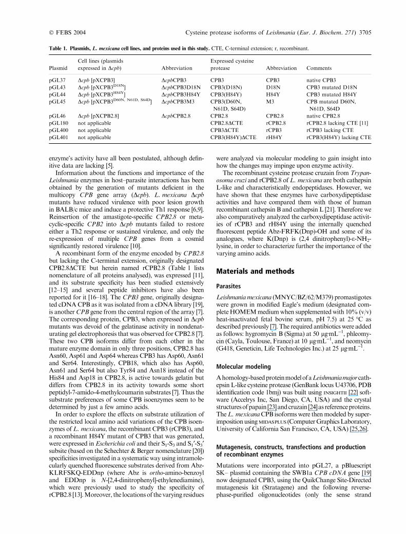
Table 1. Plasmids, L. mexicana cell lines, and proteins used in this study. CTE, C-terminal extension; r, recombinant.
Plasmid
Cell lines (plasmids
expressed in Dcpb) Abbreviation
Expressed cysteine
protease Abbreviation Comments
pGL37 Dcpb [pXCPB3] DcpbCPB3 CPB3 CPB3 native CPB3
pGL43 Dcpb [pXCPB3D18N] DcpbCPB3D18N CPB3(D18N) D18N CPB3 mutated D18N
pGL44 Dcpb [pXCPB3H84Y] DcpbCPB3H84Y CPB3(H84Y) H84Y CPB3 mutated H84Y
pGL45 Dcpb [pXCPB3D60N, N61D, S64D] DcpbCPB3M3 CPB3(D60N,
N61D, S64D)
M3 CPB mutated D60N,
N61D, S64D
pGL46 Dcpb [pXCPB2.8] DcpbCPB2.8 CPB2.8 CPB2.8 native CPB2.8
pGL180 not applicable CPB2.8DCTE rCPB2.8 rCPB2.8 lacking CTE [11]6
pGL400 not applicable CPB3DCTE rCPB3 rCPB3 lacking CTE
pGL401 not applicable CPB3(H84Y)DCTE rH84Y rCPB3(H84Y) lacking CTE
� FEBS 2004 Cysteine protease isoforms of Leishmania (Eur. J. Biochem. 271) 3705

primers are shown and the mutated sites are given inbold): OL416 to generate pGL40: 5¢-GACGCCGGTGAAGAATCAGGGTGCGTG-3¢,OL418 to generate pGL41:5¢-CGAACGGGCACCTGTACACGGAGGACAGC-3¢and OL420 to generate pGL42: 5¢-GCTGCGATGACATGAACGATGGTTGCGACGGCGGGCTGATGC-3¢.
These mutant constructs were verified by sequenceanalysis using an ABI 373 automated DNA sequencer(PerkinElmer). The native and mutant CPB3 genes wereexcised from pBluescript SK– with XbaI–XhoI. Blunt endswere created using Klenow fragment (NEB) and these wereligated to the SmaI site of the pX episomal shuttle vector[27] to generate the pGL plasmids detailed in Table 1. TheCPB2.8 gene [6] was excised from pBluescript SK– (pGL28)as a 2.0 kb EcoRV fragment and ligated to the SmaI site ofpX to generate pGL46 (Table 1). All pX-based constructswere then used to transfect Dcpb [6].
Transfection of L. mexicana promastigotes was as des-cribed previously [6]. Briefly, pX-based constructs wereprepared using Qiagen Tip100 columns as outlined by themanufacturer. Transfection utilized 10 lg of DNA and4 · 107 late-log phase Dcpb promastigotes. Followingelectroporation, cells were allowed to recover in 10 mLcomplete HOMEM medium for 24 h at 25 �C and thentransfectants were selected in complete HOMEM mediumcontaining 25 lg G418ÆmL)1.
To generate recombinant L. mexicana CPBs, the 203 bpKpnI–SacI fragment of pGL180 (pQE-30 CPB2.8DCTE)[11] was replaced with the corresponding fragments frompGL27 and pGL41 to give expression constructs pGL400(pQE-30 CPB3) and pGL401 (pQE-30 H84Y) (Table 1).These encode proteins comprising the pro- and maturedomains of the enzymes, and which lack the pre- andC-terminal domains. The recombinant enzymes were pro-duced without the C-terminal extension (CTE) to aidrefolding from the insoluble inclusion body phase. Theproduction of active, mature recombinant enzyme usingE. coliwas essentially as described previously for isoenzymeCPB2.8 [11]. The concentration of the enzyme stocksolutions (� 11 lM) were determined by active site titrationwith human cystatin C, which was a generous gift fromM. Abrahamson (University of Lund, Sweden), usingZ-FR-7-amido-4-methylcoumarin (Sigma) as the substrate.
Activity analyses of cysteine proteases using gelatinSDS/PAGE
Parasite cysteine protease activities were analysed usingsubstrate SDS/PAGE as described previously [7,28].Parasite cell lysates (107 cells) were subjected to electro-phoresis under nonreducing conditions using 12% (w/v)acrylamide gels containing 0.2% (w/v) gelatin. Followingelectrophoresis, the gel was washed for 1 h with 2.5%(v/v) Triton X-100 and then incubated for 2 h in 0.1 M
sodium acetate, pH 5.5, containing 1 mM dithiothreitol.Gelatin hydrolysis was detected by staining with Coomas-sie Blue R-250 (0.25% w/v). When analysing activitiestowards N-succinyl-Leu-Tyr-7-amido-4-methylcoumarin(Suc-LY-MCA;2 Sigma), the peptidyl amidomethylcou-marin fluorogenic substrate was added to 0.01 mM and,following a 10 min incubation, fluorescence was detectedby exposure of the gel to low intensity UV light [28].
Western blotting
Western blotting utilized polyclonal anti-CPB serum(1 : 2500) raised against rCPB2.8 expressed in and purifiedfrom E. coli [11].
Synthesis of Abz-peptidyl-Q-EDDnp
All the intramolecularly quenched fluorogenic peptidescontain N-[2,4-dinitrophenyl]-ethylenediamine (EDDnp)attached to glutamine. This is a necessary result of thesolid-phase peptide synthesis strategy employed, the detailsof which were provided elsewhere [29]. An automatedbench-top simultaneous multiple solid-phase peptide syn-thesizer (PSSM 8 system; Shimadzu, Tokyo, Japan) wasused for the solid-phase synthesis of all the peptides by theFmoc-procedure. The final de-protected peptides werepurified by semipreparative HPLC using an Econosil C-18column (10 lm, 22.5 · 250 mm) and a two-solvent system:(A) trifluoroacetic acid/H2O (1 : 1000) and (B) trifluoro-acetic acid/acetonitrile/H2O (1 : 900 : 100). The columnwas eluted at a flow rate of 5 mLÆmin)1 with a 10 (or 30))50(or 60)%gradient of solvent B over 30 or 45 min. AnalyticalHPLC was performed using a binary HPLC system fromShimadzu with a SPD-10AV Shimadzu UV-vis detectorand a Shimadzu RF-535 fluorescence detector, coupled toan Ultrasphere C-18 column (5 lm, 4.6 · 150 mm) whichwas eluted with solvent systems A1 (H3PO4/H2O, 1 : 1000)and B1 (acetonitrile/H2O/H3PO4, 900 : 100 : 1) at a flowrate of 1.7 mLÆmin)1 and a 10–80% gradient of B1 over15 min. The HPLC column eluates were monitored by theirabsorbance at 220 nm and by fluorescence emission at420 nm following excitation at 320 nm. Themolecularmassand purity of synthesized peptides were checked byMALDI-TOF mass spectrometry (TofSpec-E, Micromass,Manchester, UK)3 and or peptide sequencing using a proteinsequencer PPSQ-23 (Shimadzu). The concentrations of thesolutions of the substrates were determined by colorimetricdetermination of 2,4-dinitrophenyl group (extinction coef-ficient at 365 nm is 17 300ÆM)1Æcm)1)4 .
Enzymatic hydrolysis of fluorescent quenched substrates
Hydrolysis of the fluorogenic peptide substrates byrCPB2.8, rCPB3 and rH84Y were carried out in 0.1 M
sodium acetate, 2 mM EDTA, 200 mM NaCl, pH 5.5, at37 �C. All kinetic analyses were carried out at 37 �C with5 min enzyme preincubation in 2.5 mM dithiothreitol andby measuring the fluorescence at 420 nm, following excita-tion at 320 nm, using a Hitachi F-2500 spectrofluorometerto follow the Abz-peptidyl-Q-EDDnp substrate hydrolysis.The kinetic parameters were calculated accordingWilkinson[30] as well as by using Eadie–Hofstee plots. The standardderivations of Km and kcat determinations were in no casehigher than 5% of the obtained value.
HPLC analysis of the enzymatic hydrolysis productsof the synthetic fluorogenic substrates
The cleaved peptide bonds in each substrate were identifiedby isolation of the fragments by HPLC reverse-phasechromatography on a C18 column equilibrated in 10%
3706 M. A. Juliano et al. (Eur. J. Biochem. 271) � FEBS 2004

solvent B (90% acetonitrile, 0.1% trifluoroacetic acid, v/v).The columnwas eluted at a flow rate of 1 mLÆmin)1with 10–80% gradient of solvent B over 28 min. The elution profilewasmonitoredbyabsorbanceat 220 nmandbyfluorescenceat 420 nm after excitation at 320 nm. The Abz-containingfragmentswere comparedwithauthentic synthetic sequencesand/or by amino acid sequencing and molecular massdetermination byMALDI-TOFmass spectrometry.
Results
Correlation between structure and activity of CPBisoenzymes using substrate SDS/PAGE
The CPB locus of L. mexicana consists of 19 genes in atandem array [7]. The proteins encoded by three of thesegenes differ in just a few amino acids. The present study wasbased on the finding that CPB2.8 and CPB18 are bothhighly active towards gelatin as a substrate when assessed byin situ substrate SDS/PAGE, whereas CPB3 was inactive[6,7]. CPB3 differs fromCPB2.8 in just three residues (60, 61and 64) whereas the only difference between CPB3 andCPB18 are residues 18 and 84. Thus it was reasoned thatone or more of these changes must play a key role inmodulating the enzyme activity. There appeared twopossible explanations for the activity differences observed:that the amino acid substitutionsmodulated enzyme activitydirectly, or they had an indirect effect by influencing theenzyme’s folding and stability. To investigate the role of thedifferent amino acid residues we generated two mutants ofCPB3 in which residues 18 and 84 were changed to thosepresent in CPB18. We also mutated residues 60, 61 and 64of CPB3 to those present in CBP2.8 – as a positive controlfor the procedure.
Incorporating mutations into the CPB3 gene and intro-ducing the mutated genes into Dcpb by transfection allowedan investigation into the functional roles of these aminoacids. The activity of the enzyme expressed in the parasitewas then analyzed towards gelatin and a small fluorogenicpeptidyl substrate, in both cases using in situ substrate SDS/PAGE. Conversion of three variant residues in the maturedomain of the CPB3 isoenzyme (Asp60, Asn61 and Ser64)to those present in CPB2.8 (Asn60, Glu61 and Glu64) togive a protein designated M3, restored gelatinase activity tothe protease as expected (Fig. 1A, lane 6). This demon-strates that one or more of these residues play an importantrole either in modulating the activity towards gelatin or inenabling the enzyme to reactivate after the electrophoresisprocedure used.Mutation of His84 to Tyr84 (to give H84Y)also restored gelatinase activity to the CPB3 isoenzyme(Fig. 1A, lane 5), whereas mutation of Asp18 to Asn18 (togive D18N) did not (Fig. 1A, lane 4). The level ofre-expressed CPBs was to the same order in all of the celllines, as assessed by Western blotting (Fig. 1B, lanes 3–7),although there were differences in expression levels that mayaccount in part for the differences in proteolytic activitiesapparent (for example between lanes 5–7 of Fig. 1A).Interestingly, the H84Y activity appeared to be somewhatgreater than that of M3. However, such in situ gel assays,although they are useful in providing qualitative results,need to be interpreted with caution with respect toquantitative data.
The mutated forms of CPB3 were also assessed foractivity towards a small fluorogenic peptide using the gel-based assay, which also is useful for assessing activity but isnot very quantitative (Fig. 2). As observed for gelatin, themutants expressing the CPB3 or D18N were inactivetowards Suc-LY-MCA (Fig. 2A, lanes 1 and 2). In contrast,the H84Y isoenzyme hydrolyzed Suc-LY-MCA well(Fig. 2A, lane 3). The M3 enzyme was also active towardsthis fluorogenic compound (Fig. 2A, lane 4). This was to beexpected, and so served as a positive control, as the mutanthas the same mature domain as CPB2.8 (Fig. 2A, lane 5),which is active towards this substrate [7]. The highermolecular mass activities towards Suc-LY-MCA (approxi-mately 35 kDa) correspond to activated precursor forms ofthe isoenzymes [11]. To confirm that similar amounts of there-expressed proteases had been applied to these activitygels, a Western blot was performed on duplicate sampleswith the CPB-specific antiserum (Fig. 2B).
These results indicated that residues 60, 61, 64 and 84influenced the activity of CPB, thus we produced asrecombinant enzymes CPB3 and also H84Y in order tocarry out a fuller analysis of their substrate specificities and
1 2 3 4 5 6 7
1 2 3 4 5 6 7
A
B
30 -
22 -
42 -
30 -
22 -
kDa
kDa
Fig. 1. Gelatin SDS/PAGE and Western blot analyses of L. mexicana
CPB isoenzymes expressed in Dcpb. (A) Extracts from 107 stationary
phase promastigotes were used for gelatin SDS/PAGE. Wild type
parasites (lane 1), Dcpb (lane 2), DcpbCPB3 (lane 3), DcpbCPB3D18N
(lane 4), DcpbCPB3H84Y (lane 5), DcpbCPB3M3 (lane 6) and
DcpbCPB2.8 (lane 7). The higher molecular mass activities (approxi-
mately 35 kDa) evident with some samples resulted from the activation
in situ of precursor forms of the isoenzymes [11]. (B) Extracts from
5 · 106 stationary phase promastigotes were used forWestern blotting
with anti-CPB serum (1 : 2500). Samples were applied to lanes 1–7 as
denoted for (A). Molecular mass markers are shown in kDa. The anti-
CPB serum recognized CPB isoenzymes that migrated as a major band
with a molecular mass of 27 kDa and a minor band of molecular mass
26 kDa in cell lysates of stationary phase promastigotes of wild type
L. mexicana (lane 1). The specificity of the antiserumwas confirmed by
absence of detected proteins in Dcpb (lane 2).
� FEBS 2004 Cysteine protease isoforms of Leishmania (Eur. J. Biochem. 271) 3707

compare them with that of CPB2.8 [13]. All enzymes wereproduced without the C-terminal extension as previously[11].
S1 subsite specificity characterization of CPB3 and H84Y
A series derived from the peptide Abz-KLRFSKQ-EDDnpwere synthesized with systematic variation of Arg at P1using all natural amino acids as previously reported for thesubsite specificity studies of rCPB2.8 [13]. Table 2 showsthe kinetic parameters for the hydrolysis of this series ofpeptides by CPB3 and H84Y, and, for comparison, also thekcat/Km and Ki values of rCPB2.8.
The substrate inhibition with the peptides containinghydrophobic and non-charged amino acids that occurredwith rCPB2.8 was not observed with rCPB3 and rH84Y,and some hydrolysis occurred with all substrates. However,the specificity constants (kcat/Km) values for these proteaseswere considerably lower than those obtained with rCPB2.8.The higher kcat/Km values for rCPB2.8 were due to bothlower Kms and higher kcats. For example, with X ¼ R, therCPB2.8 values were 0.04 lM (Km) and 2.60 s)1 (kcat)compared with the values of 0.3 lM (Km) and 0.48 s)1 (kcat)for rCPB3 (Table 2).
rCPB3 hydrolyzed with highest kcat/Km values thepeptides containing Ser and Thr, followed by those withPhe, Arg, Lys and Tyr. These higher catalytic efficiencies aremainly due to the low Km value rather than the catalyticcomponent. Similar hydrolytic behaviour was observedwith rH84Y and the best substrates were those containingMet, Ser and Thr.
30 -
22 -
42 -
A
B
kDa
21 3 4 5
21 3 4 5
30 -
22 -
42 -
kDa
Fig. 2. Fluorogenic SDS/PAGE and Western blot analyses of
L. mexicana CPBs expressed in Dcpb. (A) Extracts from 107 stationary
phase promastigotes of Dcpb re-expressing the following proteases
were analysed for activity towards Suc-LY-MCA12 . DcpbCPB3 (lane 1),DcpbCPB3D18N (lane 2), DcpbCPB3H84Y (lane 3), DcpbCPB3M3
(lane 4) andDcpbCPB2.8 (lane 5). (B) Extracts from 5 · 106 stationary
phase promastigotes were analysed with anti-CPB serum to show that
roughly equivalent protein loadings were applied for the fluorogenic
SDS/PAGE analyses: samples were applied to lanes 1–5 as denoted for
(A). Molecular mass markers are shown in kDa. The mature CPBs are
arrowed.
Table 2. Kinetic parameters for hydrolysis, by CPB, of the peptides derived fromAbz-KLXFSKQ-EDDnp with modifications in X (P1).Conditions of
hydrolysis: 100 mM NaOAc, 200 mM NaCl, 2 mM EDTA, pH 5.5 and 37 �C. The enzymes were preactivated by 2.5 mM dithiothreitol7 for 5 min.
The cleavage site is indicated by �fl�, and the numbers following give8 the percentage of hydrolysis at each peptide bond. All the other hydrolyses were
at theX–F bond. rCPB2.8 data from reference [13]. The units forKi values are nM, and kcat/Km values are in (mMÆs))1. X indicates the residue varied.
Modification
rCPB2.8 rCPB3 rH84Y
kcat/Km
(mMÆs))1Km
(lM)
kcat(s)1)
kcat/Km
(mMÆs))1Km
(lM)
kcat(s)1)
kcat/Km
(mMÆs))1
R 65000 0.3 0.48 1600 1.6 0.22 138
K 31395 0.1 0.12 1200 0.2 0.04 200
H 36667 0.5 0.40 800 0.5 0.09 180
D 871 0.2 0.03 150 0.1 0.01 100
E 7727 0.8 0.53 663 0.3 0.04 133
C 14060 0.6 0.47 783 4.3 0.26 61
W Ki ¼ 28 1.2 0.12 100 0.3 0.04 133
Y Ki ¼ 26 0.1 0.11 1100 0.2 0.06 300
F Ki ¼ 18 0.1 0.21 2100 0.3 0.08 267
L Ki ¼ 15 kcat/Km ¼ 600 (XflF ¼ 55, FflS ¼ 45) kcat/Km ¼ 700 (XflF ¼ 50, FflS ¼ 50)
I Ki ¼ 9 kcat/Km ¼ 350 (XflF ¼ 15, FflS ¼ 85) kcat/Km ¼ 100 (XflF ¼ 19, FflS ¼ 81)
V Ki ¼ 29 kcat/Km ¼ 250 (XflF ¼ 24, FflS ¼ 76) kcat/Km ¼ 300 (XflF ¼ 28, FflS ¼ 72)
M Ki ¼ 22 0.3 0.15 500 0.05 0.16 3200
A Ki ¼ 27 0.2 0.14 700 0.1 0.12 1200
P Ki ¼ 400 0.8 0.04 50 3.8 0.05 13
S Ki ¼ 24 0.1 0.28 2800 0.04 0.09 2250
T Ki ¼ 16 0.03 0.08 2667 0.04 0.09 2250
N Ki ¼ 32 kcat/Km ¼ 300 (XflF ¼ 67, FflS ¼ 33) kcat/Km ¼ 133 (XflF ¼ 67, FflS ¼ 33)
Q Ki ¼ 43 0.1 0.08 800 0.2 0.15 750
3708 M. A. Juliano et al. (Eur. J. Biochem. 271) � FEBS 2004

Most of the peptides were hydrolyzed by rCPB3 andrH84Y at the X–F peptide bond, where X represents all thesubstitutions of Arg. A second cleavage, at the F–S bond,was observed for the peptides with X ¼ Leu, Ile, Val andAsn. Like cathepsin L, the S2–P2 interaction is determinantin defining the cleavage point and these cysteine proteasesprefer hydrophobic amino acids at P2 position [31,32]. Thusthe cleavage of the F–S bond with Asn at the S2 subsite byrCPB3 and rH84Y is surprising, although this cleavagecorresponded to only 33% of the total (Table 2).
S2 and S3 subsite specificity characterization of rCPB3and rH84Y
The kinetic parameters for hydrolysis of the peptidesmodified at positions P2 and P3 are shown in Table 3. S2specificity is considered critical for the activity of clan CA
5cysteine proteases [31]. The rCPB3 preferred Leu at P2position of the substrate, while Pro is the worst amino acidof those tested for activity. On the other hand, rH84Yhydrolyzed the peptide with Arg at P2 with higher kcat/Km;this was mainly due to kcat contribution because theKm wasrelatively high. The extended binding site of rCPB3 andrH84Y also included the S3 subsite, as the modifications atP3 position of the substrates resulted in significant variationson the kcat/Km values (Table 3). rCPB3 hydrolyzed withbetter efficiency the peptides with Lys and Leu at P3,whereas rH84Y was most efficient with the peptidescontaining Leu and Ala.
S1¢ to S3¢ subsite specificity characterization of rCPB3and rH84Y
The kinetic parameters for hydrolysis of the peptidesmodified at positions P1¢ to P3¢ are shown in Table 4. Thetrend of the kcat/Km values obtained with the substrates with
modifications at P1¢ position were similar between rCPB3and rCPB2.8, however, the latter enzyme hydrolyzed all thepeptides of this series with kcat/Km values at least one orderofmagnitude higher. In contrast to rCPB3 and rCPB2.8, themutant enzyme rH84Y hydrolyzed the peptide with Pheonly poorly. This enzyme hydrolyzed best the peptidecontaining Ala at P1¢, followed by those with Leu and Arg.The peptide with Pro was almost resistant to all threeproteases, although a low rate of hydrolysis was observed atthe P–S bond.
The modification at the P2¢ position of the substratesrevealed the very significant effect of Pro, resulting inconsiderably higher kcat/Km values for the hydrolysis byrCPB3 and rH84Y – even above those with rCPB2.8. Thesehigh values mainly reflected a marked decrease in the Km
value. The kcat/Km values for the hydrolysis of Abz-KLRFPKQ-EDDnp by these two proteases were thehighest found. Such preference for Pro in the S2¢ subsite isa peculiarity of cruzain (of T. cruzi) and Leishmania CPB.These enzymes accept Pro in this position in syntheticsubstrates very well [13,15,33,34] and also in the auto-processing of their pro-enzymes to active enzymes – Pro isthe second amino acid in the mature form of the proteases[11,35]. The importance of S2¢–P2¢ interaction was furtherevidenced by the variations in the kcat/Km values for thehydrolysis of various substrates with modifications at P2¢position by rH84Y – changing the Ser favoured by rCPB2.8in each case resulted in increased activity.
Table 3. Kinetic parameters for hydrolysis, by CPB, of the peptides
derived from Abz-KLRFSKQ-EDDnp, containing modifications at the
Leu (P2) and Lys (P3) residues. Conditions of hydrolysis: 100 mM
NaOAc, 200 mMNaCl, 2 mMEDTA, pH 5.5 and 37 �C. The enzymes
were preactivated by 2.5 mM dithiothreitol for 5 min. rCPB2.8 data
from reference [13]. X indicates the residue varied. The cleavage site is
indicated by �fl�.9
Substrate
rCPB2.8 rCPB3 rH84Y
kcat/Km
(mMÆs))1Km
(lM)
kcat(s)1)
kcat/Km
(mMÆs))1Km
(lM)
kcat(s)1)
kcat/Km
(mMÆs))1
Abz-KXRflFSKQ-EDDnp
L 65000 0.3 0.48 1600 1.6 0.22 138
F 15517 0.9 0.13 144 0.8 0.17 212
A 2204 0.2 0.02 100 0.4 0.04 100
R 3295 0.3 0.04 133 1.4 0.66 471
P 471 0.5 0.01 20 4.8 0.39 81
Abz-XLRflFSKQ-EDDnp
K 65000 0.3 0.48 1600 1.6 0.22 138
L 6167 0.05 0.07 1400 0.05 0.13 2600
A 16769 0.1 0.08 800 0.1 0.25 2500
H 6320 0.7 0.28 400 2.2 0.46 209
R 10421 0.4 0.12 300 0.5 0.29 630
Table 4. Kinetic parameters for hydrolysis, by CPB, of the peptides
derived from Abz-KLRFSKQ-EDDnp, containing modifications at the
Phe (P1¢), Ser (P2¢) and Lys (P3¢) residues. Conditions of hydrolysis:100 mMNaOAc, 200 mMNaCl, 2 mM EDTA, pH 5.5 and 37 �C. Theenzymes were preactivated by 2.5 mM dithiothreitol for 5 min.
rCPB2.8 data from reference [13]. The units for Ki values are nM.
X indicates the residue varied.10 The cleavage site is indicated by ‘fl’.11
Substrates
rCPB2.8 rCPB3 rH84Y
kcat/Km
(mMÆs))1Km
(lM)
kcat(s)1)
kcat/Km
(mMÆs))1Km
(lM)
kcat(s)1)
kcat/Km
(mMÆs))1
Abz-KLRflXSKQ-EDDnp
F 65000 0.3 0.48 1600 1.6 0.22 138
L 14117 0.7 0.28 400 0.06 0.03 500
A 14882 0.2 0.09 450 0.3 0.28 933
R 5438 0.4 0.11 275 0.4 0.17 453
Pa Ki ¼ 490 0.5 0.01 20 0.3 0.03 93
Abz-KLRflFXKQ-EDDnp
S 65000 0.3 0.48 1600 1.6 0.22 138
F 27778 0.2 0.18 977 0.3 0.19 633
A 8909 0.1 0.10 774 0.06 0.11 1833
R 15833 0.3 0.32 944 0.3 0.50 1667
P 11875 0.01 0.18 18000 0.01 0.16 16000
Abz-KLRflFSXQ-EDDnp
K 65000 0.3 0.48 1600 1.6 0.22 138
F Ki ¼ 40 0.5 0.04 80 0.1 0.23 2300
A 12727 0.6 0.20 333 0.6 0.12 200
R 24286 0.4 0.15 375 0.2 0.31 1550
P 11000 0.2 0.02 100 0.6 0.27 450
a Cleavage at P–S bond.
� FEBS 2004 Cysteine protease isoforms of Leishmania (Eur. J. Biochem. 271) 3709

The S3¢ subsite also influenced the binding of rCPB3 andrH84Y. Significant variations in the kcat/Km values wereobserved with the amino acid substitutions at P3¢ position ofthe substrates (Table 4). The peptide with Lys was hydro-lyzed best by rCPB3 (it is also favoured by rCPB2.8),whereas rH84Y was more efficient on the peptides contain-ing Phe and Arg.
Carboxydipeptidase activity of rCPB3 and rH84Y
The kinetic parameters for the carboxydipeptidase activit-ies of rCPB3 and rH84Y on the internally quenchedfluorescent peptide Abz-FRFK(Dnp)-OH and some of itsanalogues are shown in Table 5. The kcat/Km values forhuman recombinant cathepsin L and rCPB2.8 [21] are alsoshown for comparison. The carboxydipeptidase activitiesof rCPB3, and rH84Y were lower than those of cathepsinL and rCPB2.8, although relative activities towards thedifferent substrates were rather similar and in each caseAbz-FRAK(Dnp)-NH2 was the best substrate. The sub-strates with free C-terminal carboxyl group were hydro-lyzed with Km values that are an order of magnitude higherthan those presented in Tables 2–4. The unfavorableeffects of the C-terminal negatively charged carboxylgroup on the protease activity of rCPB3 and rH84Y isdemonstrated by the pH-profiles of the carboxydipeptidaseactivities of these enzymes on the peptides Abz-FRAK(Dnp)-OH and Abz-FRAK(Dnp)-NH2 (Fig. 3).The pH optima of the carboxydipeptidase activitiestowards Abz-FRAK(Dnp)-OH of rCPB3 and rH84Y aredisplaced to 4.0–4.5 and the activity decreases greatly bypH 5.5. This pH range corresponds to the pK ofcarboxylate group formation from the substrate, indicatingthat the protonated carboxyl group fits better to theenzymes than its carboxylate form. This is confirmed bythe pH-profiles for the hydrolysis of Abz-FRAK(Dnp)-NH2. In this case, the pH optimum is in the range 6–8. It isnoteworthy that the pH-profile of carboxydipeptidaseactivity of rCPB2.8 on Abz-FRAK(Dnp)-OH contrastsgreatly with those of rCPB3 and rH84Y. It has optimalactivity at pH 6–8, showing that rCPB2.8 accommodatesmuch better the negatively charged carboxylate group.Interestingly, the pH-profile of hydrolysis of Abz-FRAK(Dnp)-OH by rH84Y presents a second small butsignificant peak of activity around pH 7.5. It is difficult toassign the group responsible for this effect because there
are several groups that change their ionization statusaround this pH.
Analysis of amino acid locations via modelling
The location of the amino acids that differ between theCPB isoenzymes at positions 18, 60, 61, 64 and 84 wasdetermined by constructing a model of the L. mexicanaCPB isoenzymes by superimposing upon that obtained forthe enzymes’ homologue in L. major [22] (Fig. 4). Thecrystal structure of papain [23] and of a recombinantcysteine protease of T. cruzi, cruzain [24], which has ahigh degree of sequence identity to CPB, was used as atemplate for the leishmanial CPB model. The matureregions of the L. mexicana and L. major enzymes used forthe modelling have an overall 80% amino acid sequenceidentity, which reaches even higher values within thestructurally conserved regions and especially within theactive site (Table 6). Therefore, their protein structuresappear to be very similar as determined by secondarystructure alignments. The polypeptide backbone of thesecysteine proteases folds into a series of a-helices andb-sheets and the active site cleft is located between twostructural domains. The C-terminal extension is not shownon the model, as the structure of this domain has not beensolved.
The protease activity of all papain-like cysteine proteasesis associated with the catalytic triad (L. mexicana residuesCys25, His163 and Asn183; Fig. 4) but the substratespecificity is defined by the binding affinities of the subsites.The catalytic residues and the S1 and S1¢ subsites are highlyconserved between the three parasite species, and keyresidues at the threemajor subsites are completely conservedbetween the L. mexicana CPB isoenzymes and the L. majorhomologue (Table 6). Consequently, differences in activitiesbetween the L. mexicana CPB isoenzymes must be associ-atedwith amino acid variations inmore peripheral positionsof the molecule.
The model revealed the location of the amino acids thatdiffer between the CPB isoenzymes under study and somediate the observed activity changes (the residues arehighlighted in Fig. 4B). Residues 60, 61 and 64 are locatedabove the a-helix that forms a wall of the active site cleft.Amino acid 18 is relatively close to the active site cleft andalso near to one disulphide bridge (Cys22–Cys63), whereasresidue 84 is sited on a surface loop of one domain of the
Table 5. Kinetic constant parameters for carboxydipeptidase activities. Conditions of hydrolysis: 100 mM NaOAc, 200 mM NaCl, 2 mM EDTA,
pH 5.5 and 37 �C. The enzymes were preactivated by 2.5 mM dithiothreitol for 5 min. Cath L and rCPB2.8 data are from reference [21].
Substrates
Cath L rCPB2.8 rCPB3 rH84Y
kcat/Km
(mMÆs))1kcat/Km
(mMÆs))1Km
(mM)
kcat(s)1)
kcat/Km
(mMÆs))1Km
(mM)
kcat(s)1)
kcat/Km
(mMÆs))1
Abz-FRFK(Dnp)-OH 256 306 3.3 0.23 70 3.9 0.51 130
Abz-RRFK(Dnp)-OH 2.2 4.5 Ki ¼ 4.3 lM Resistant
Abz-ARFK(Dnp)-OH 0.5 1.1 Ki ¼ 2.3 lM 4.7 0.004 0.85
Abz-FRK(Dnp)W-OH 477 625 0.7 0.14 210 1.9 0.24 121
Abz-FRAK(Dnp)-OH 667 389 2.9 0.35 118 2.2 0.46 203
Abz-FRAK(Dnp)-NH2 5739 4909 1.2 0.90 780 0.85 1.14 1340
3710 M. A. Juliano et al. (Eur. J. Biochem. 271) � FEBS 2004

enzyme but adjacent to another disulphide bridge (Cys56–Cys101).
Discussion
The CPB cysteine proteases of L. mexicana have beenshown to be virulence factors [6], therefore it isimportant to understand the relative contributions thatdifferent isoenzymes play in the pathogenicity of the
parasite. Those isoenzymes that have been characterizedso far are highly conserved (98% identical) and yet someactivity differences were apparent [7]. The aim of thisstudy was to determine the extent to which these aminoacid variations generated activity differences. The CPBsequences obtained to date all possess identical residuesaligning the substrate subsites so it seemed that otherresidues outside of the active site cleft must affectactivity.
Abz-FRAK(Dnp)-NH2Abz-FRAK(Dnp)-OH
pH
3 4 5 6 7 80
400
800
1200
1600
2000
2400
2800
109
pH3 4 5 6 7 8 9 1
0
200
400
600
800
1000
1200
1400
1600
1800
2000
0
pH
3 4 5 6 7 8 9 100
200
400
600
800
1000
pH3 4 5 6 7 8 9 10
0
20
40
60
80
100
120
140
160
180
pH
3 4 5 6 7 8 9 100
40
80
120
160
200
240
pH
3 4 5 6 7 8 9 10
kcat
/Km
(m
M-1
.s-1
)kc
at/K
m (
mM
-1.s
-1)
kcat
/Km
(m
M-1
.s-1
)
kcat
/Km
(m
M-1
.s-1
)kc
at/K
m (
mM
-1.s
-1)
kcat
/Km
(m
M-1
.s-1
)
0
2000
4000
6000
8000
10000
12000
14000
16000C
PB
3H
84Y
CP
B2.
8
Fig. 3. pH-profile activity (kcat/Km) for the hydrolysis of Abz-FRAK(Dnp)-OH and Abz-FRAK(Dnp)-NH2 by rCPB2.8, rCPB3 and rH84Y. The
reactions were carried out in standard buffer containing 25 mM acetic acid, 25 mM Mes, 75 mM Tris base, 25 mM glycine, and 2 mM EDTA. The
pH range was 3.5–10 and adjusted with 2 M NaOH or HCl. The enzymes were preactivated by 2.5 mM dithiothreitol for 5 min at 37 �C.
� FEBS 2004 Cysteine protease isoforms of Leishmania (Eur. J. Biochem. 271) 3711

The activity results presented show that the few aminoacid variations known to exist between some isoenzymes ofCPB of L. mexicana are indeed important in modifying thesubstrate specificities of the CPB isoenzymes. Both rCPB3and rH84Y have lower activity towards some substratesthan does rCPB2.8, but they are able to accommodate awider range of amino acids at P1 (Table 2). The kineticparameters of hydrolysis by rCPB3, rH84Y and rCPB2.8 ofthe substrates with variations at P1 position indicated thatthe specificity of S1 subsite is greatly influenced by themodifications at 60, 61, 64 and 84. However, the enzymeshave an extended binding site that goes at least from S3 toS3¢ and importantly each CPB isoform also shows signifi-cant differences in the specificity of each subsite (Tables 3and 4). Thus, the overall conclusion is that the differentisoenzymes do indeed have different hydrolytic capabilitiesand presumably this is important for the parasite’s survival.This view is supported by the recent observation that theexpression of multiple CPB genes encoding cysteineproteases, rather than just one, is required for L. mexicanavirulence in vivo [10].
A particularly noteworthy finding was the effect of Pro inthe P2¢ position in decreasing considerably the Km valuewith rCPB3 and rH84Y. Clearly these isoenzymes greatlyfavour Pro at this site. This may reflect an important role forthe enzymes in the activation of CPB in the parasite, as Pro
is the second amino acid of the mature domain in all of theCPB isoenzymes for which the structure is known.
The variation of amino acid residues 60, 61 and 64between the CPB isoenzymes examined in this work involveamino acids with charged side chains and this necessarilyresults in significant modifications on the electrostaticpotential on the surface of the enzymes. The modelinganalysis (Fig. 4) shows clearly that residues 60, 61 and 64are located near the catalytic groove of the enzyme and so itis likely that the localized charge variations resulting fromthe introduction or removal of residues with charged sidegroups to these positions could be the basis for thedifferences observed with respect to the utilization ofsubstrates. The pH-profile differences for the carboxydi-peptidase activities of rCPB3 and rCPB2.8 clearly suggestthat the latter isoform accommodates the substrate carb-oxylate group better than the former isoform, perhapsindicating that residue 60 (Asn in CPB2.8 but Asp in CPB3)is particularly important for this binding. These findingsagree well with a study of the pH-activity profile of cruzain,a related cathepsin L-like cysteine protease of T. cruzi,which highlighted the importance of several ionizablegroups and suggested that Asn60 is potentially involved insubstrate recognition [36]. Thus the data obtained providefurther evidence for the role of electrostatic potential indefining the substrate specificity of the CPB isoforms.
84
6061
64
18
SS63
22
25
163
183
SS204
156
SS101
56
A B
Fig. 4. Homology-based protein model of the
mature domain of L. mexicana CPB. The
protein is shown as a ribbon structure (spi-
rals, a-helices; arrows, b-sheets). (A) High-
lighted in yellow are the location of the active
site triad (Cys25, His163 and Asn183) and
disulphide bonds (Cys22–Cys63, Cys56–
Cys101 and Cys156–Cys204). (B) Highlighted
in red are the residues that differ between
CPB2.8, CPB3 and CPB18.
Table 6. Key active site residues of cathepsin L-like cysteine proteases.Themajor variations between the active sites of parasite CPs and papain occur
between the S subsites. Additional S and S� subsites are not listed because they are so far not identified by the cocrystallization of a peptide substrate
and the proteases of the parasites.
Protease S2 subsite S1 subsite Catalytic triad S1¢ subsite
Papain W69, S205, F207 Y67, P68, V133, A160 C25, H159, N175 Q19, W177
Cruzain N69, E208, S210 L67, M68, A138, G163 C25, H162, N182 Q19, W177
L. mexicana CPB L69, Y209, V211 L67, M68, A139, G164 C25, H163, N183 Q19, W185
L. major CPB L69, Y209, V211 L67, M68, A139, G164 C25, H163, N183 Q19, W185
3712 M. A. Juliano et al. (Eur. J. Biochem. 271) � FEBS 2004

Residue 84 is located near the surface of the maturedomain structure and so it is less easy to understand how itplays a role in affecting the binding of substrates and theirhydrolysis. However, it is positioned near to a disulphidebridge (Cys56–Cys101) and it is conceivable that thereplacement of His with Tyr may impinge upon thisstructure and so change the active site is some way. Clearlythe findings on the activity of this mutant compared withrCPB3 suggest that mutation had some effects, althoughmore minor than those resulting from the three changes(N60D, D61N and D64S) between CPB2.8 and the otherisoenzymes.
The results show that rCPB3 has activity towards peptidesubstrates and yet the enzyme showed no activity insubstrate SDS/PAGE analysis, whereas both CPB2.8 andCPB18 were both highly active in similar analyses (Figs 1and 2). This suggests not only that one or more of aminoacid changes H60D, D61N and D64S plays a key role inmodulating the enzyme activity directly but may also beable to do so indirectly by affecting the enzyme’s refoldingand/or stability under the conditions employed for thegelatin SDS/PAGE. Moreover, H84Y but not D18N cancounteract this effect. It is too early to be able to interpretthe way is which this is achieved, but the results show thecomplexity of the interactions that occur both within themature protein and in the acquisition of its tertiarystructure.
In conclusion, the data reported suggest that the set ofCPB isoenzymes with only a few sequence modificationshave the modifications at strategic positions such that theenzyme’s substrate specificity is changed and that thesevariations between isoenzymes provide the parasite with anarray of hydrolytic activity that is needed for its interactionwith the mammalian host, and ensure its survival andsuccess as a parasite.
Acknowledgements
This work was supported by Fundacao de Amparo Pesquisa do Estado
de Sao Paulo (FAPESP), Conselho Nacional de Desenvolvimento
Cientıfico e Tecnologico (CNPq) and Human Frontiers for Science
Progress (RG 00043/2000-M). J.C.M. andG.H.C. are supported by the
Medical Research Council.
References
1. Bromme, D. & Kaleta, J. (2002) Thiol-dependent cathepsins:
pathophysiological implications and recent advances in inhibitor
design. Curr. Pharm. Des. 8, 1639–1658.
2. Sajid,M.&McKerrow, J.H. (2002) Cysteine proteases of parasitic
organisms. Mol. Biochem. Parasitol. 120, 1–21.
3. Barrett, A.J. & Rawlings, N.D. (2001) Evolutionary lines of
cysteine peptidases. Biol. Chem. 382, 727–733.
4. McGrath, M.E. (1999) The lysosomal cysteine proteases. Annu.
Rev. Biophys. Biomol. Struct. 28, 181–204.
5. Mottram, J.C., Brooks, D.R. & Coombs, G.H. (1998) Roles of
cysteine proteinases of trypanosomes and Leishmania in host–
parasite interactions. Curr. Opin. Microbiol. 1, 455–460.
6. Mottram, J.C., Souza, A.E., Hutchison, J.E., Carter, R., Frame,
M.J. & Coombs, G.H. (1996) Evidence from disruption of the
lmcpb gene array ofLeishmania mexicana that cysteine proteinases
are virulence factors. Proc. Natl Acad. Sci. USA 93, 6008–6013.
7. Mottram, J.C., Frame,M.J., Brooks, D.R., Tetley, L., Hutchison,
J.E., Souza, A.E. & Coombs, G.H. (1997) The multiple cpb
cysteine proteinase genes of Leishmania mexicana encode iso-
enzymes which differ in their stage-regulation and substrate pre-
ferences. J. Biol. Chem. 272, 14285–14293.
8. Brooks, D.R., Denise, H., Westrop, G.D., Coombs, G.H. &
Mottram, J.C. (2001) The stage-regulated expression of Leish-
mania mexicana CPB cysteine proteases is mediated by an
intercistronic sequence element. J. Biol. Chem. 276, 47081–
47089.
9. Alexander, J., Coombs, G.H. &Mottram, J.C. (1998) Leishmania
mexicana cysteine proteinase-deficient mutants have attenuated
virulence for mice and potentiate a T Helper 1 (TH1) response.
J. Immunol. 161, 6794–6801.
10. Denise, H., McNeil, K.S., Brooks, D.R., Alexander, J., Coombs,
G.H. & Mottram, J.C. (2003) The expression of multiple CPB
cysteine protease genes is required for Leishmania mexicana viru-
lence in vivo. Infect. Immun. 71, 3190–3195.
11. Sanderson, S.J., Pollock, K.G., Hilley, J.D., Meldal, M., St. Hil-
aire, P.M., Juliano, M.A., Juliano, L., Mottram, J.C. & Coombs,
G.H. (2000) Expression and characterisation of a recombinant
cysteine proteinase of Leishmania mexicana. Biochem. J. 347,
383–388.
12. Alves, L.C.,Melo, R.L., Sanderson, S.J., Mottram, J.C., Coombs,
G.H., Caliendo, G., Santagada, V., Juliano, L. & Juliano, M.A.
(2001) S1 subsite specificity of a recombinant cysteine proteinase,
CPB, of Leishmania mexicana compared with cruzain, human
cathepsin L and papain using substrates containing non-natural
basic amino acids. Eur. J. Biochem. 268, 1206–1212.
13. Alves, L.C., Judice, W.A.S., St. Hilaire, P.M., Meldal, M.,
Sanderson, S.J., Mottram, J.C., Coombs, G.H., Juliano, L. &
Juliano, M.A. (2001) Substrate specificity of recombinant cysteine
proteinase, CPB, of Leishmania mexicana. Mol. Biochem. Para-
sitol. 116, 1–9.
14. Alves, L.C., Melo, R.L., Cezari, M.H., Sanderson, S.J., Mottram,
J.C., Coombs, G.H., Juliano, L. & Juliano, M.A. (2001) Analysis
of the S2 subsite specificities of the recombinant cysteine protei-
nases CPB of Leishmania mexicana, and cruzain of Trypanosoma
cruzi, using fluorescent substrates containing non-natural basic
amino acids. Mol. Biochem. Parasitol. 117, 137–143.
15. St. Hilaire, P.M., Alves, L.C., Sanderson, S.J., Mottram, J.C.,
Juliano, M.A., Juliano, L., Coombs, G.H. & Meldal, M. (2000)
The substrate specificity of a recombinant cysteine protease from
Leishmania mexicana: application of a combinatorial peptide
library approach. Chembiochem. 1, 115–122.
16. Alves, L.C., St. Hilaire, P.M., Meldal, M., Sanderson, S.J., Mot-
tram, J.C., Coombs, G.H., Juliano, L. & Juliano, M.A. (2001)
Identification of peptides inhibitory to recombinant cysteine pro-
teinase, CPB, of Leishmania mexicana. Mol. Biochem. Parasitol.
114, 81–88.
17. St. Hilaire, P.M., Alves, L.C., Herrera, F., Renil, M., Sanderson,
S.J., Mottram, J.C., Coombs, G.H., Juliano, M.A., Juliano, L.,
Arevalo, J. & Meldal, M. (2002) Solid-Phase library synthesis,
screening and selection of tight-binding reduced peptide bond
inhibitors of a recombinant Leishmania mexicana cysteine pro-
tease B. J. Med. Chem. 45, 1971–1982.
18. Graven, A., St. Hilaire, P.M., Sanderson, S.J., Mottram, J.C.,
Coombs, G.H. & Meldal, M. (2001) Combinatorial library of
peptide isosters based on Diels-Alder reactions: Identification of
novel inhibitors against a recombinant cysteine protease from
Leishmania mexicana. J. Comb. Chem. 3, 441–452.
19. Souza, A.E., Waugh, S., Coombs, G.H. & Mottram, J.C. (1992)
Characterization of a multicopy gene for a major stage-specific
cysteine proteinase of Leishmania mexicana. FEBS Lett. 311,
124–127.
� FEBS 2004 Cysteine protease isoforms of Leishmania (Eur. J. Biochem. 271) 3713

20. Schechter, I. & Berger, A. (1967) On the size of the active site
in proteases. I. Papain. Biochem. Biophys. Res. Commun. 27,
157–162.
21. Judice, W.A., Puzer, L., Cotrin, S.S., Carmona, A.K., Coombs,
G.H., Juliano, L. & Juliano, M.A. (2004) Carboxydipeptidase
activities of recombinant cysteine peptidases. Eur. J. Biochem. 271,
1046–1053.
22. Selzer, P.M., Chen, X.W., Chan, V.J., Cheng, M.S., Kenyon,
G.L., Kuntz, I.D., Sakanari, J.A., Cohen, F.E. &McKerrow, J.H.
(1997) Leishmania major: Molecular modeling of cysteine pro-
teases and prediction of new nonpeptide inhibitors.Exp. Parasitol.
87, 212–221.
23. Kamphuis, I.G., Kalk, K.H., Swarte, M.A. & Drenth, J. (1984)
Structure of papain refined at 1.65 A resolution. J. Mol. Biol. 179,
233–256.
24. McGrath,M.E., Eakin, A.E., Engel, J.C.,McKerrow, J.H., Craik,
C.S. & Fletterick, R.J. (1995) The crystal structure of cruzain: a
therapeutic target for Chagas’ disease. J. Mol. Biol. 247, 251–259.
25. Ferrin, T.E., Huang, C.C., Jarvis, L.E. & Langridge, R. (1988)
The midas display system. J. Mol. Graphics 6, 13–27.
26. Huang, C.C., Pettersen, E.F., Klein, T.E., Ferrin, T.E. & Lan-
gridge, R. (1991) Conic – a fast renderer for space-filling molecules
with shadows. J. Mol. Graphics 9, 230–236.
27. LeBowitz, J.H., Coburn, C.M., McMahon-Pratt, D. & Beverley,
S.M. (1990) Development of a stableLeishmania expression vector
and application to the study of parasite surface-antigen genes.
Proc. Natl Acad. Sci. USA 87, 9736–9740.
28. Robertson, C.D. & Coombs, G.H. (1990) Characterization of 3
groups of cysteine proteinases in the amastigotes of Leishmania
mexicana mexicana. Mol. Biochem. Parasitol. 42, 269–276.
29. Hirata, I.Y., Cezari, M.H.S., Nakaie, C., Boschcov, P., Ito, A.S.,
Juliano, M.A. & Juliano, L. (1994) Internally quenched
fluorogenic protease substrates: solid-phase synthesis and fluor-
escence spectroscopy of peptides containing ortho-aminobenzoyl/
dinitrophenyl groups as donor-acceptor pairs. Lett. Peptide Sci. 1,
299–308.
30. Wilkinson, G.N. (1961) Statistical estimations in enzyme kinetics.
Biochem. J. 80, 324–332.
31. Nagler, D.K., Tam, W., Storer, A.C., Krupa, J.C., Mort, J.S. &
Menard, R. (1999) Interdependency of sequence and positional
specificities for cysteine proteases of the papain family. Biochem-
istry 38, 4868–4874.
32. Portaro, F.C.V., Santos, A.B.F., Cezari, M.H.S., Juliano, M.A.,
Juliano, L. & Carmona, E. (2000) Probing the specificity of
cysteine proteinases at subsites remote from the active site: analysis
of P4, P3, P2¢ and P3¢ variations in extended substrates. Biochem. J.
347, 123–129.
33. Serveau, C., Lalmanach, G., Hirata, I., Scharfstein, J., Juliano,
M.A. & Gauthier, F. (1999) Discrimination of cruzipain, the
major cysteine proteinase of Trypanosoma cruzi, and mammalian
cathepsins B and L, by a pH-inducible fluorogenic substrate of
trypanosomal cysteine proteinases. Eur. J. Biochem. 259, 275–280.
34. Serveau, C., Lalmanach, G., Juliano, M.A., Scharfstein, J., Juli-
ano, L. & Gauthier, F. (1996) Investigation of the substrate spe-
cificity of cruzipain, the major cysteine proteinase of Trypanosoma
cruzi, through the use of cystatin-derived substrates and inhibitors.
Biochem. J. 313, 951–956.
35. Eakin, A.E., Mills, A.A., Harth, G., McKerrow, J.H. & Craik,
C.S. (1992) The sequence, organization, and expression of the
major cysteine protease (cruzain) from Trypanosoma cruzi. J. Biol.
Chem. 267, 7411–7420.
36. Stoka, V., McKerrow, J.H., Cazzulo, J.J. & Turk, V. (1998)
Substrate inhibition of cruzipain is not affected by the C-terminal
domain. FEBS Lett. 429, 129–133.
3714 M. A. Juliano et al. (Eur. J. Biochem. 271) � FEBS 2004
Copyright © 2022 FDOKUMEN