
Oral Absorption Promoters: Opportunities, Issues, and Challenges,
Upload
nottinghamCategory
view
1download
0
Archives of Biochemistry and Biophysics 427 (2004) 8–15
ABBwww.elsevier.com/locate/yabbi
Expression of calpastatin isoforms in muscle and functionalityof multiple calpastatin promoters
Tim Parr,* Kirsty K. Jewell, Paul L. Sensky, John M. Brameld,Ronald G. Bardsley, and Peter J. Buttery
Division of Nutritional Biochemistry, School of Biosciences, University of Nottingham, Sutton Bonington Campus, Loughborough,
Leicestershire LE12 5RD, UK
Received 13 January 2004, and in revised form 6 April 2004
Available online 6 May 2004
Abstract
Calpastatin is the specific endogenous inhibitor of calpain proteinase that is encoded by a single gene. Transient transfection
assays in both a non-fusing skeletal muscle and non-muscle cell-line demonstrated that the putative porcine calpastatin promoter
regions 50 to exons 1xa, 1xb, and 1u were functional. Both real-time quantitative and semi-quantitative RT-PCR on porcine skeletal
muscle total RNA indicated that steady-state expression of Type I and III mRNAs containing exons 1xa and 1u, respectively, was at
equivalent levels whilst the expression of Type II mRNA containing exon 1xb was significantly less (p < 0:001). Immunoprobing of
Western blotted muscle extracts with an antibody raised against a peptide sequence encoded by exon 1xa indicated that Type I
protein was expressed and that there was significantly more Type I protein in cardiac than skeletal muscle (p < 0:001). The resultssuggest that the expression of the single calpastatin gene was differentially controlled at several levels.
� 2004 Elsevier Inc. All rights reserved.
Keywords: Calpain; Calpastatin; Promoter analysis; Skeletal muscle; Cardiac muscle
Calpains were first characterised as intracellular cal-cium-dependent cysteine proteinases that are present in
all mammalian cells [1,2]. The conventional, or ubiqui-
tous, l- and m-calpain isoforms appear mainly to cat-
alyse the limited proteolysis of cytoskeletal and
membrane proteins and are regulated both by calcium
ion concentration and the specific protein inhibitor
calpastatin. In striated muscle, the calpain/calpastatin
system has been implicated in the regulation of proteinturnover and growth [3], myoblast fusion [4], cardio-
myopathies [5], and in meat texture development [6].
Calpastatin has four homologous C-terminal inhibi-
tory domains (I–IV) downstream of a non-inhibitory
leader domain (L)1 of largely unknown function [7].
Recently, an additional N-terminal peptide sequence
* Corresponding author. Fax: +44-115-951-6122.
E-mail address: [email protected] (T. Parr).1 Abbreviations used: L, leader domain; ECL, enhanced chemilu-
minescence; DMEM, Dulbecco’s modified Eagle’s medium; FCS,
foetal calf serum.
0003-9861/$ - see front matter � 2004 Elsevier Inc. All rights reserved.
doi:10.1016/j.abb.2004.04.001
(XL) was identified in bovine [8]. Different XL and Lleader sequences have subsequently been shown to
generate a series of tissue-specific variants by alternative
splicing of a single gene to yield calpastatins Type I–IV
[9]. The murine calpastatin gene comprises 32 exons
distributed over �60 kb DNA [9], although the first 6
exons of the porcine gene already extend over �60 kb
DNA [10].
A putative bovine calpastatin promoter was isolatedby Cong et al. [8] and shown to be functional in cell
transfection studies and inducible by dibutyryl cAMP.
Subsequent work based primarily on analysis of mRNA
transcripts predicted four potential promoter regions in
the murine gene [9], three of which have been identified
in the partial porcine gene structure [10]. In the present
report, a series of promoter constructs of the porcine
sequences 50 to exons 1xa, 1xb, and 1u, which are be-lieved to be the transcription initiation sites of Type I,
II, and III calpastatin mRNAs [9,10], respectively, were
generated to characterise these putative promoter re-
gions. Analysis of mRNA transcripts originating from

T. Parr et al. / Archives of Biochemistry and Biophysics 427 (2004) 8–15 9
these three promoters was carried out by ‘‘real-time’’quantitative RT-PCR along with a partial characteri-
sation of calpastatin protein expression through the use
of an isoform-specific antibody.
Materials and methods
Experimental animals
Animal studies were conducted according to the
provisions of the UK Home Office Animals (Scientific
Procedures) act of 1986. All the pigs used were from the
same group of animals that were matched for breed and
age. The animals were slaughtered by electrical stunning
and severance of the carotid arteries. Samples of ven-
tricular and longissimus dorsi muscle were removedfrom the carcass within 5min of slaughter and frozen in
liquid nitrogen for subsequent storage at )70 �C.
Reverse transcriptase-PCR of calpastatin transcripts
Total RNA was prepared from porcine longissimus
dorsi (skeletal muscle) using the RNeasy Midi Kit
(Qiagen). Real-time quantitative RT-PCR was used forthe analysis of Type I, II, and III calpastatin mRNAs
(GenBank Accession Nos. AJ583407, AJ583408, and
AJ583409). Primers and dual-labelled fluorescent oli-
gonucleotide probes (50FAM, 30TAMRA) were pur-
chased from Sigma-Genosys and are listed in Table 1.
All primer and probe combinations, except that for
Type III transcripts, amplified across an exon boundary.
A pool of first strand cDNA was made from 0.5 lg ofskeletal muscle total RNA using random hexamers
(Promega) as primers, essentially as described previously
[10]. For real-time PCR using fluorescent probes, reac-
tion mixtures contained 12.5 ll of 2� Universal Master
Mix (Applied Biosystems), 5 ll cDNA template (1:40
dilution of reverse transcriptase generated cDNA pool
[10]) in a final volume of 25 ll containing primers
Table 1
Primer and probe combinations used for real-time quantitative RT-PCR
Target
gene
Target
region
Amplicon
length
Primer/
Probe
Calpastatin Exon 1xa 91 Forward
Reverse
Probe
Calpastatin Exon 1xb 104 Forward
Reverse
Probe
Calpastatin Exon 1u 60 Forward
Reverse
Probe
Actin Actin 100 Forward
Reverse
(300 nM) and probe (200 nM). Actin expression wasmonitored as an internal standard using real-time
quantitative RT-PCR and SYBR Green Fluorescence
on the cDNA template as described above (Applied
Biosystems). All real-time PCRs were carried out in
triplicate on an ABI PRISM 7700 Sequence Detection
System (Applied Biosystems) using standard default
thermal cycling conditions. A pool of porcine skeletal
muscle cDNA was used to create a standard curve forquantification of the transcripts using a relative stan-
dard curve method as described by Applied Biosystems
[11]. From this the Ct value of a particular variant could
be converted to ng total RNA equivalent used for first
strand synthesis. Semi-quantitative end-point RT-PCR
analysis was carried out as described previously [10] on
calpastatin transcript variants with novel 50termini and
alternative usage of exon 3.
Western blot analysis of muscle extracts with anti-
calpastatin antisera
Porcine skeletal and cardiac muscle samples were
pulverised in liquid nitrogen then homogenised as de-
scribed by Geesink et al. [12]. Homogenates were spun
at 13,000g for 15min at 4 �C after which the supernatantwas heated in a boiling water bath for 5min and then
recentrifuged at 13,000g for 15min at 4 �C. To concen-
trate the heated extract, the solution was dialysed
overnight against distilled water. Ice-cold trichloroacetic
acid was added to 13.3% (w/v) and the precipitate was
centrifuged at 13,000g for 15min at 4 �C. The pellet waswashed five times with ice-cold acetone then dried.
Muscle extracts and concentrated pellets were analy-sed by 8% SDS–PAGE and Western blotted [10].
The Western blots were probed with a polyclonal anti-
serum (1:1000) raised against a peptide sequence
(MSQPGQKPAASPRP) which was encoded by the
open reading frame in 1xa exon of porcine calpastatin
[10]. Bands were visualised using CDP-Star Western-
blotting reagents (Amersham Biotech). Western blots
Primer
name
Nucleotide sequence (50–30)
1xaF CCGGCGAGCTGCTGC
1xabR TGATCCTTTCTGTTCTCCTGACTTG
1xaP CCCGCCACACCCAGGAGCA
1xbF GCAAATTGGTTGTCGCCAT
1xabR TGATCCTTTCTGTTCTCCTGACTTG
1xbP CGTCTTGTACCACCAGCTTGCGAATG
1uF CCGAGCCCAACCAGGAAT
1uR CTTCCCGGCCGAGAGACT
1uP CAAACATCCCCAAACGCCGCTG
actF GTGGCCCTGGACTTCGAG
actR TTGCCGATGGTGATGACCT

10 T. Parr et al. / Archives of Biochemistry and Biophysics 427 (2004) 8–15
were reprobed with a generic anti-porcine calpastatinantiserum [10,13] at 1:1000 dilution and visualised using
enhanced chemiluminescence (ECL) reagents (Amer-
sham Biotech) as described previously [10].
Generation of promoter constructs and transfections
A porcine BAC clone PigE BAC 045d24 (synthesised
by S. Anderson and A. Archibald, Roslin Institute,Edinburgh, UK) containing a 50-terminal region of the
porcine calpastatin gene [10] (GenBank Accession Nos.
AJ583410 and AJ583411) was used to generate the pu-
tative promoter constructs by PCR using the primers
listed in Table 2 and Expand High Fidelity PCR System
(Roche). Amplicons were ligated into pGL3 Basic vector
(Promega), sequenced to confirm identity with the ori-
ginal BAC clone, and then used in transfections.Transfections were carried out using L6G8 myoblasts
(ECACC No. 92102117), a muscle cell-line originally
derived from rat skeletal muscle, and HEK293 cells, a
cell-line originally derived from human embryonic kid-
ney (kindly donated by Dr. K. Mellits, University of
Nottingham). Frozen stocks of cells were thawed and
cultured in 75 cm2 flasks in Dulbecco’s modified Eagle’s
medium (DMEM) supplemented with 8% foetal calfserum (FCS), 4mM glutamine, 100U penicillin/ml and
100 lg streptomycin/ml (growth medium). Cells were
harvested (trypsin/EDTA), resuspended in growth me-
dium, plated onto 48-well culture plates (Falcon), and
transfected when 50–70% confluent. Four hours prior to
transfection, the medium was replaced with either 200 llprewarmed growth medium (L6G8) or with 200 ll se-rum-free DMEM containing 0.5% BSA and antibiotics(HEK293). For each construct, an aliquot of serum-free
DMEM (no BSA or antibiotics added) was thoroughly
mixed with the required quantity of GeneJuice Trans-
fection Reagent (Novagen) at a GeneJuice:DNA ratio of
6:1 (ll/lg). After 5min incubation at room temperature,
Table 2
Primer combinations used to generate promoter constructs
Primer Nucleotide sequence (50–30)
ApromB AAAACGCGTGTAATTGGGTTCATGGCTTGG
BpromB AAAACGCGTTCGTAATGTATAATAGTGCCG
CpromB AAAACGCGTGCGTTGTTCAGTCAGCCAGTC
EpromB AAAACGCGTACCGCCCCGCCGTAACTCTG
AncaspromB AAAAGATCTTCTGCTTCTGGCTTCCCGGCC
Prom1xaa AAAACGCGTGGGGAGAGGGGGCAACACTG
Prom1xab AAAACGCGTGTATAGGGGGTTAAGTTTGGG
Prom1xac AAAACGCGTATTCCCTTCTGCCAAGTTCCAC
Anc1xa AAAAGATCTCGGGAGGGCACAGCGGAGAC
Prom1xba AAAACGCGTCGTGGCTGCCTGGGGAAATA
Prom1xbb AAAACGCGTGCACTCCTGCCAGAGCCAGAG
Anc1xb AAAAGATCTGGCGACAACCAATTTGCTCTG
Forward primers have MluI sites (ACG CGT) at 50 end. Reverse (Anc)
0.02 lg pRL-SV40 DNA (transfection efficiency control)was added to each tube followed immediately by 0.2 lg ofeither promoter reporter construct DNA, pGL3-SV40
Control (positive control) or pGL3 Basic DNA (negative
control). After 15min incubation at room temperature,
8 ll GeneJuice/DNA mixture was added to cells. Four
hours after transfection, themedium on theHEK293 cells
was replaced with 500 ll DMEM including 8% FCS and
the incubationwas allowed toproceed. Firefly andRenillaluciferase activities were determined 24 h (L6G8) and 48 h
(HEK293) after transfection using Dual-Luciferase Re-
porter Assay System (Promega). The transfection effi-
ciency was corrected for by the ratio of firefly luciferase
activity to Renilla luciferase activity and the level of
promoter activity was calculated relative to the pGL3-
SV40 control DNA which was taken as being 100%.
Data analysis
Band intensities were quantified using the FluorS-
Max Imaging system and Quantity-One software (Bio-
Rad). Real-time PCR data were analysed using SDS
software (Applied Biosystems). Data were compared
using a one-way ANOVA using Genstat for Windows (v
6, IACR-Rothamsted).
Results
Expression of variant calpastatin transcripts in skeletal
muscle
Skeletal muscle calpastatin mRNA variants with al-ternative 50termini containing 1xa, 1xb, and 1u exons
were monitored using real-time quantitative RT-PCR.
Although the absolute level of expression could not be
determined, there was no significant difference between
the relative expression of 1xa (Type I) and 1u (Type III)
Forward/reverse
primer to exon
Construct synthesised
by forward primer
GAA Forward 1u Aprom1u
TAG Forward 1u Bprom1u
Forward 1u Cprom1u
Forward 1u Eprom1u
Reverse 1u —
G Forward 1xa Aprom1xa
AATG Forward 1xa Bprom1xa
AT Forward 1xa Cprom1xa
T Reverse 1xa —
Forward 1xb Aprom1xb
Forward 1xb Bprom1xb
Reverse 1xb —
primers have BglII sites (AGA TCT) at 50termini.

Fig. 1. (A) Schematic representation of the predicted products generated from porcine calpastatin mRNAs with alternative 50 sequences (templates
indicated by box) using RT-PCR. The position of the 50 end exon unique to each calpastatin variant and exon3 is indicated in the template. The real-
time RT-PCR primers are indicated by the arrows below the template and the circle indicates the position of the probe. The semi-quantitative RT-
PCR primers are indicated by the arrows above the template. The predicted size of products generated by semi-quantitative RT-PCR with or without
exon 3 is listed. (B) Real-time RT-PCR analysis of Type I, II, and III calpastatin mRNA expression in porcine skeletal muscle (n ¼ 9). Values are
expressed as the ratio of ng total RNA equivalent used for first strand synthesis calpastatin mRNA to actin mRNA (internal standard). Data are
expressed as means+SEM. ***p < 0:001 Type II compared with Type I and Type III (C) Semi-quantitative RT-PCR products amplified from
skeletal muscle total RNA samples analysed on a 1.5% agarose gel. The products amplified from Type I, II, and III transcripts are indicated above
each lane for individual samples and marker sizes are shown.
T. Parr et al. / Archives of Biochemistry and Biophysics 427 (2004) 8–15 11
containing transcripts (Fig. 1B). The detection of cal-
pastatin transcripts containing exon 1xb (Type II) was
at the limits of sensitivity and was significantly lower
(p < 0:001) than both Type I and Type III (Fig. 1B). The
position of the probes and primers used in the real-time
PCR (Fig. 1A) gave no information on whether these
variants contained exon 3 which encodes for a regionwithin L-domain that is alternatively spliced [12]. Semi-
quantitative end-point RT-PCR analysis was carried out
on porcine skeletal muscle total RNA using a series of
variant-specific primers which spanned exon 3 (Fig. 1C).
Whilst both Type I and III transcripts were detected
there was no Type II. Based on predicted amplicon size,
only Type I transcripts contained both exon 3 plus and
exon 3 minus variants whilst the Type III transcript wasexon 3 minus. There was no significant difference in the
total band intensity for Type I and Type III mRNAs as
well as between the exon 3 plus and minus bands of
Type I transcripts (p > 0:2, n ¼ 5, data not shown).
Type I calpastatin in skeletal muscle
Immunoblots probed with the generic anti-calpasta-tin antibody detected a 135 kDa immunoreactive band
in skeletal muscle and a doublet of 145 and 135 kDa in
cardiac muscle as previously reported [12,14] (Fig. 2A).
A replicate blot probed with the anti-1xa peptide anti-
body detected a 145 kDa band only in cardiac tissue
(Fig. 2A). By concentrating the skeletal muscle extracts
by trichloroacetic acid precipitation an immunopositive
band could be detected at approximately 145 kDa, along
with a minor band at approximately 65 kDa (Fig. 2B).The 65 kDa band could either be a non-specific cross-
reaction of the antibody or a proteolytic fragment of
calpastatin, which is very susceptible to proteolysis
[1,12]. Any proteolytic cleavage must be taking place
towards the C terminus to produce this fragment with
the N-terminal epitope intact, allowing the anti-1xa
peptide antibody to detect this large peptide fragment.
The use of this anti-1xa peptide antibody has indicatedfor the first time that the peptide sequence encoded by
exon 1xa can be expressed in cardiac and skeletal mus-
cle. It is noteworthy that the 1xa-specific antibody does
not detect the band of 135 kDa in either cardiac or
skeletal muscle, unlike the generic anti-calpastatin an-
tibody; accordingly the 135 kDa band is therefore un-
likely to represent a Type I protein lacking exon 3.
Comparisons between bands on immunoblots of pre-cipitated skeletal muscle extracts and cardiac extracts
indicated that the 1xa-peptide immunopositive band was
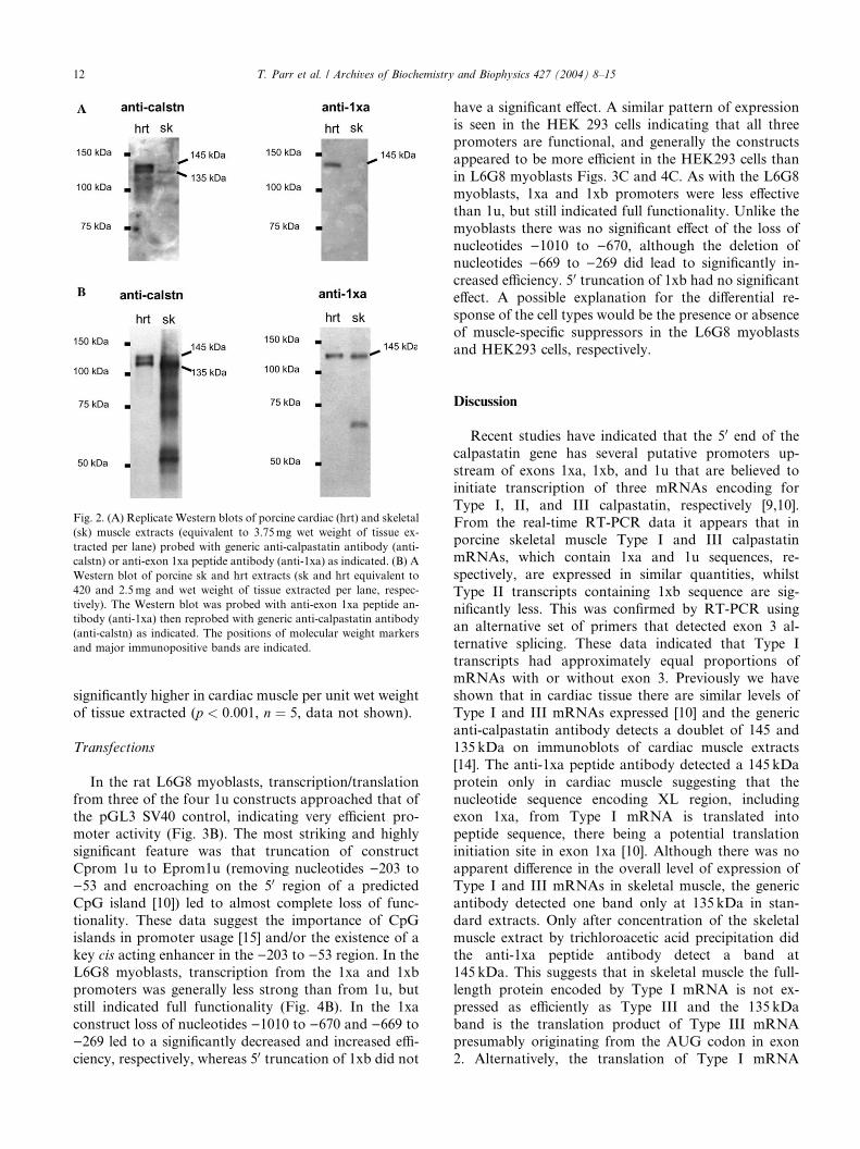
Fig. 2. (A) Replicate Western blots of porcine cardiac (hrt) and skeletal
(sk) muscle extracts (equivalent to 3.75mg wet weight of tissue ex-
tracted per lane) probed with generic anti-calpastatin antibody (anti-
calstn) or anti-exon 1xa peptide antibody (anti-1xa) as indicated. (B) A
Western blot of porcine sk and hrt extracts (sk and hrt equivalent to
420 and 2.5mg and wet weight of tissue extracted per lane, respec-
tively). The Western blot was probed with anti-exon 1xa peptide an-
tibody (anti-1xa) then reprobed with generic anti-calpastatin antibody
(anti-calstn) as indicated. The positions of molecular weight markers
and major immunopositive bands are indicated.
12 T. Parr et al. / Archives of Biochemistry and Biophysics 427 (2004) 8–15
significantly higher in cardiac muscle per unit wet weight
of tissue extracted (p < 0:001, n ¼ 5, data not shown).
Transfections
In the rat L6G8 myoblasts, transcription/translationfrom three of the four 1u constructs approached that of
the pGL3 SV40 control, indicating very efficient pro-
moter activity (Fig. 3B). The most striking and highly
significant feature was that truncation of construct
Cprom 1u to Eprom1u (removing nucleotides )203 to
)53 and encroaching on the 50 region of a predicted
CpG island [10]) led to almost complete loss of func-
tionality. These data suggest the importance of CpGislands in promoter usage [15] and/or the existence of a
key cis acting enhancer in the )203 to )53 region. In the
L6G8 myoblasts, transcription from the 1xa and 1xb
promoters was generally less strong than from 1u, but
still indicated full functionality (Fig. 4B). In the 1xa
construct loss of nucleotides )1010 to )670 and )669 to
)269 led to a significantly decreased and increased effi-
ciency, respectively, whereas 50 truncation of 1xb did not
have a significant effect. A similar pattern of expressionis seen in the HEK 293 cells indicating that all three
promoters are functional, and generally the constructs
appeared to be more efficient in the HEK293 cells than
in L6G8 myoblasts Figs. 3C and 4C. As with the L6G8
myoblasts, 1xa and 1xb promoters were less effective
than 1u, but still indicated full functionality. Unlike the
myoblasts there was no significant effect of the loss of
nucleotides )1010 to )670, although the deletion ofnucleotides )669 to )269 did lead to significantly in-
creased efficiency. 50 truncation of 1xb had no significant
effect. A possible explanation for the differential re-
sponse of the cell types would be the presence or absence
of muscle-specific suppressors in the L6G8 myoblasts
and HEK293 cells, respectively.
Discussion
Recent studies have indicated that the 50 end of the
calpastatin gene has several putative promoters up-
stream of exons 1xa, 1xb, and 1u that are believed to
initiate transcription of three mRNAs encoding for
Type I, II, and III calpastatin, respectively [9,10].
From the real-time RT-PCR data it appears that inporcine skeletal muscle Type I and III calpastatin
mRNAs, which contain 1xa and 1u sequences, re-
spectively, are expressed in similar quantities, whilst
Type II transcripts containing 1xb sequence are sig-
nificantly less. This was confirmed by RT-PCR using
an alternative set of primers that detected exon 3 al-
ternative splicing. These data indicated that Type I
transcripts had approximately equal proportions ofmRNAs with or without exon 3. Previously we have
shown that in cardiac tissue there are similar levels of
Type I and III mRNAs expressed [10] and the generic
anti-calpastatin antibody detects a doublet of 145 and
135 kDa on immunoblots of cardiac muscle extracts
[14]. The anti-1xa peptide antibody detected a 145 kDa
protein only in cardiac muscle suggesting that the
nucleotide sequence encoding XL region, includingexon 1xa, from Type I mRNA is translated into
peptide sequence, there being a potential translation
initiation site in exon 1xa [10]. Although there was no
apparent difference in the overall level of expression of
Type I and III mRNAs in skeletal muscle, the generic
antibody detected one band only at 135 kDa in stan-
dard extracts. Only after concentration of the skeletal
muscle extract by trichloroacetic acid precipitation didthe anti-1xa peptide antibody detect a band at
145 kDa. This suggests that in skeletal muscle the full-
length protein encoded by Type I mRNA is not ex-
pressed as efficiently as Type III and the 135 kDa
band is the translation product of Type III mRNA
presumably originating from the AUG codon in exon
2. Alternatively, the translation of Type I mRNA

Fig. 3. (A) Diagrammatic representation of the partial 50porcine gene structure indicating the exon positions and the deletion mutants of porcine 1u
promoter–luciferase constructs. (B,C) Transfection efficiency of different 1u promoter–luciferase constructs in L6G8 myoblasts (B) and HEK 293
cells (C). Values are expressed relative to transfection of pGL3 control DNA and corrected for transfection efficiency relative to transfection of cells
with Renilla (pRL)+SEM. ***p < 0:001 Eprom1u compared with Aprom1u, Bprom1u, and Cprom1u.
T. Parr et al. / Archives of Biochemistry and Biophysics 427 (2004) 8–15 13
could be from the downstream AUG codon in exon 2
to give a 135 kDa protein. A further possibility is thatthe peptide originating from the upstream AUG co-
don in exon 1xa is subsequently cleaved to a truncated
protein in a similar way to that reported for bovine
Type II mRNA [8].
From the analysis of transcript expression, both
previous studies and the present data have suggested
that the 1u and 1xa promoters are used predominantly
more than the 1xb promoter in skeletal and cardiacmuscle [10]. Apart from the bovine promoter (1xb)
study [8], the functionality of these alternative promot-
ers has not yet been described in direct cell transfection
studies. In the present study, results indicated that all
but one of the constructs tested were functional and
remarkably efficient compared to the pGL3 SV40 con-
trol, especially in HEK 293 cells (Figs. 3 and 4). The one
exception was construct Eprom1u, in which only 51 ntof 50 sequence upstream of the transcriptional start site
in exon 1u was retained. The 1u exon encodes 50 un-translated sequence only, becoming spliced to exon 2
which contains an in-frame AUG encoding the N ter-
minus of Type III calpastatin [9,10]. The inclusion of an
additional 151 50 nucleotides in Cprom1u restored full
activity, suggesting the presence of key regulatory fea-ture(s) in this region. The additional 151 nt contains
high-scoring consensus sequences for one CRE and one
MyoD motif. The CRE is unlikely to be essential, since
there is a similar CRE in the Eprom1u construct that
appears almost completely inactive. Similarly the MyoD
motif may not be essential, because the same pattern of
responses was seen in the non-muscle HEK293 cells.
However, truncation of Cprom1u to Eprom1u does re-move 151 nt of a high-scoring CpG island, which is
therefore proposed to be an important point of control
for the 1u promoter.
The 1xa and 1xb promoter constructs are approxi-
mately 50% less effective than 1u in both cell types. For
the 1xa promoter, the shortest construct Cprom1xa
containing 250 nt of 50 sequence is most effective, with
the suggestion that negative enhancer sequences couldbe located further upstream. The serial 50 deletions of
the 1xb promoter carried out in this study did not have a
significant effect on expression.
It is concluded that all three promoter sequences
studied in the present report are potentially functional

Fig. 4. (A) Diagrammatic representation of the partial 50porcine gene structure indicating the exon positions and the deletion mutants of porcine 1xa
and 1xb promoter–luciferase constructs. (B,C) Transfection efficiency of different 1xa and 1xb promoter–luciferase constructs in L6G8 myoblasts (B)
and HEK 293 cells (C). Values are expressed relative to transfection of pGL3 control DNA and corrected for transfection efficiency relative to
transfection of cells with Renilla (pRL)+SEM. Deletions which result in significant differences in activity are indicated (*p < 0:05).
14 T. Parr et al. / Archives of Biochemistry and Biophysics 427 (2004) 8–15
with high efficiency and that efficiency can be varied by
the inclusion or removal of nucleotide sequences. It is
important to point out that the rat L6G8 myoblastsused in this study are a non-fusing skeletal muscle cell-
line so that direct comparison between the promoter
analysis carried out in these cells and promoter function
in fully differentiated muscle cells in situ cannot be
made. However, within these sequences are a number of
high-scoring cis acting transcriptional motifs whose
importance will need to be determined by site-directed
mutagenesis in combination with a range of physiolog-ical treatments. In skeletal muscle, the level of tran-
scripts originating from these promoters shows a low
level of Type II calpastatin mRNA, indicating that ei-
ther the 1xb promoter is less active or that Type II
mRNA is rapidly degraded. Although the levels of Type
I and III mRNAs are equal in skeletal muscle, the ex-
pression of Type I calpastatin protein (145 kDa) is less
than Type III (135 kDa). These observations suggest adifferential level of control of the calpastatin gene
products at several levels that may have significance for
calpastatin’s role in inhibiting calpain-mediated prote-
olysis both physiologically and in meat texture devel-opment.
Acknowledgments
We thank Tracey Simpson for her excellent technical
assistance. This work was funded by the Biotechnologyand Biological Sciences Research Council (BBSRC),
UK.
References
[1] D.E. Goll, V.F. Thompson, H. Li, W. Wei, J. Cong, Physiol. Rev.
83 (2003) 731–801.
[2] H. Sorimachi, K. Suzuki, J. Biochem. 129 (2001) 653–664.
[3] D.E. Goll, V.F. Thompson, R.G. Taylor, A. Ouali, Can. J. Anim.
Sci. 78 (1998) 503–512.

T. Parr et al. / Archives of Biochemistry and Biophysics 427 (2004) 8–15 15
[4] S. Barnoy, L. Supino-Rosin, N.S. Kosower, Biochem. J. 351
(2000) 413–420.
[5] Y. Sorimachi, K. Harada, T.C. Saido, T. Ono, S. Kawashima, K.
Yoshida, J. Biochem. 122 (1997) 743–748.
[6] P.L. Sensky, T. Parr, R.G. Bardsley, P.J. Buttery, Proc. Br. Soc.
Anim. Sci. 2001 (2001) 239–243.
[7] E. Takano, M. Maki, H. Mori, M. Hatanaka, M. Marti, K.
Titani, R. Kannagi, T. Ooi, T. Murachi, Biochemistry 27 (1988)
1964–1972.
[8] M. Cong, V.F. Thompson, D.E. Goll, P.B. Antin, J. Biol. Chem.
273 (1998) 660–666.
[9] J. Takano, M. Watanabe, K. Hitomi, M. Maki, J. Biochem. 128
(2000) 83–92.
[10] T. Parr, P.L. Sensky, R.G. Bardsley, P.J. Buttery, Arch. Biochem.
Biophys. 395 (2001) 1–13.
[11] User Bulletin #2:ABI PRISM 7700 Sequence Detection System,
Applied Biosystems, Foster City, CA, USA, 1997.
[12] G.H. Geesink, D. Nonneman, M. Koohmaraie, Arch. Biochem.
Biophys. 356 (1998) 19–24.
[13] P.L. Sensky, T. Parr, A.K. Lockley, R.G. Bardsley, P.J.
Buttery, J.D. Wood, C. Warkup, J. Anim. Sci. 77 (1999) 2956–
2964.
[14] T. Parr, P.L. Sensky, M.K. Arnold, R.G. Bardsley, P.J. Buttery,
Arch. Biochem. Biophys. 374 (2000) 299–305.
[15] H. McQueen, V.H. Clark, A.P. Bird, M. Yerle, A.L. Archibald,
Genome Res. 7 (1997) 924–931.
Copyright © 2022 FDOKUMEN