Differential characterization of three alternative spliced isoforms of DPPX

Journal of Cellular Biochemistry 92:1044–1061 (2004)
Expression and Function of Periostin-Isoforms in Bone
Judith Litvin,1,2* Abdul-Hafez Selim,1 Michael O. Montgomery,1 Kiyoko Lehmann,1 Mario C. Rico,1
Hugh Devlin,3 Daniel P. Bednarik,4 and Fayez F. Safadi1
1Department of Anatomy and Cell Biology, Temple University School of Medicine,Philadelphia, Pennsylvania 191402Cardiovascular Research Center, Temple University School of Medicine,Philadelphia, Pennsylvania 191403Department of Restorative Dentistry, Temple University, School of Dentistry, Philadelphia,Pennsylvania 191404Artesian Therapeutics, Inc., Gaithersburg, Maryland
Abstract Periostin was originally identified in MC3T3-E1 osteoblast-like cells. We have identified an isoform ofperiostin referred to as periostin-like-factor (PLF). It is homologous to other proteins such as fasciclin I (fas I), MPB70, bIG-H3, and Algal-CAMs. All of these proteins are implicated in regulating cell adhesion. PLF and the other isoforms ofperiostin differ in their C-terminal sequences. PLF and periostin differ in two specific regions, between 673 and 699 aminoacids (aa) and 785–812 aa. Periostin isoforms are expressed in vivo and in vitro during the stages of osteoblastdifferentiation and maturation. Their mRNAs are present in pre-osteoblast cells as detected by in situ hybridization, andthe proteins are between 86 and 93 kD in size as determined by Western blot analysis. Antisense oligonucleotides andantibodies directed against the isoforms of periostin were used to block the activity of these proteins. In both cases, thelevels of osteoblast-specific-differentiation markers were markedly reduced suggesting a role for these proteins inosteoblast differentiation. J. Cell. Biochem. 92: 1044–1061, 2004. � 2004 Wiley-Liss, Inc.
Key words: bone; periostin isoforms; MC3T3-E1; osteoblast differentiation
Understanding the role of molecules in sig-naling pathways that regulate the process ofosteoblast differentiation is the focus of ourresearch. We identified an isoform of periostinusing the READS technique to isolate differen-tially expressed mRNAs during murine devel-opment. Many proteins expressed in boneincluding cbfa1, alkaline phosphatase, andcollagen are present as multiple isoforms, eachisoform contributing independently to thestructure and/or function of bone. Becauseisoforms clearly have distinct roles in tissuesand organs, we pursued our studies to deter-
mine the pattern of expression of periostinisoforms during bone development and suggestpossible functions for this molecule.
Osteoblast-specific factor 2 (OSF2), alsoreferred to as periostin, was first identifiedusing subtractive hybridization techniques onMC3T3-E1 osteoblast-like cells and was thou-ght at that time to be bone-specific [Takeshitaet al., 1993]. Periostin/OSF2 has a signalpeptide, is secreted into the matrix, and reg-ulatesMC3T3-E1 cell adhesion, an event that isessential and requisite for differentiation ofosteoblasts [Horiuchi et al., 1999]. It is approxi-mately 90 kD, does not contain a trans-membrane domain and has four potentialN-glycosylation sites that are not glycosylatedin bone cells [Horiuchi et al., 1999]. Isoforms ofperiostin have been identified in mice andhumans and are over-expressed by stromal cellsin several human ovary, breast, colon, andbraintumors [Terasaka et al., 1989; Skonier et al.,1992; Huber and Sumper, 1994; LeBaron et al.,1995; Ulstrup et al., 1995; Lal et al., 1999;Sasaki et al., 2001]. In osteoblasts and tumors,
� 2004 Wiley-Liss, Inc.
Grant sponsor: American Heart Association, Pennsylvania/Delaware Affiliate (to J.L.).
*Correspondence to: Judith Litvin, PhD, Associate Profes-sor, Department of Anatomy and Cell Biology, TempleUniversity Medical School, 3420 N. Broad St., MRB 617,Philadelphia, PA 19140. E-mail: [email protected]
Received 5 January 2004; Accepted 3 March 2004
DOI 10.1002/jcb.20115

periostin supports cellular adhesion andspreading in vitro [Horiuchi et al., 1999; Gillanet al., 2002]. It has been shown that purifiedrecombinant periostin supports adhesion ofovarian epithelial cells that can be inhibited bymonoclonal antibodies against avb3 or avb5integrin, but not by anti-b1 integrin antibody.Furthermore, avb3 integrin, but not b1 integrinshave been shown to co-localize at focal adhesionplaques formed on periostin in CSOC cells(ovarian epithelial cells). Cells plated on peri-ostin form fewer stress fibers and are moremotile compared to those plated on fibronectin[Gillan et al., 2002].A similar molecule identified in insects,
fasciclin I (fas I), is homologous to periostin[Zinn et al., 1988]. It is involved in axonalguidance, providing a pathway from aneuron toits target. Two forms of fas I have been des-cribed. They are 48% identical, approximately70 kD, are spatially and temporally regulatedduring embryonic development and are pro-posed as regulators of neural cell adhesion[Hortsch and Goodman, 1990]. fas I is asso-ciated with the cell membrane and may beinvolved in cell–cell interactions [Zinn et al.,1988; Hortsch and Goodman, 1990; McAllisteret al., 1992; Hu et al., 1998]. The fas Ihomozygous Drosophila mutant [McAllisteret al., 1992] showed that fas I not only mediatedadhesion and guidance of axons, but was alsoinvolved in signal transduction pathwaysnecessary for axonal guidance and growth coneextension. Other proteins homologous to peri-ostin identified in many other systems arebelieved to be critical mediators of differentialadhesivity and attachment. For example, (a)MPB70 which may be significant in the inter-action of bacteria and host cells [Terasaka et al.,1989; Ulstrup et al., 1995], (b) bIG-H3 which isimplicated inmediating growth and differentia-tion of cells when stimulated with TGF-b[Skonier et al., 1992; LeBaron et al., 1995],and (c) Algal-CAMs involved in cell adhesion inthe alga Volvox [Huber and Sumper, 1994].Periostin and fas I contain four homologous
domains referred to as ‘Repeat Domains’ eachcontaining 150 amino acids (aa), and an N-terminal signal sequence. Within each RepeatDomain there are two regions of sequence thatare highly conserved and as suggested byHoriuchi et al. [1999] may be involved inprotein–protein interactions. The function ofmany of these motifs has not been tested and,
therefore, one cannot make any structure–function correlations. Because so little is knownabout the function of the periostin isoforms inbone, we examined whether periostin isoformsplay a role in osteoblast differentiation andfunction.
In addition, we identified periostin-like-factor(PLF), an isoform of periostin, by the ‘READS’differential display ofmRNAs isolated at variousstages during mouse heart development. Theperiostin isoforms including PLF are alsoexpressed in bone. Based on their pattern ofexpression and sequence analysis we havechosen to study PLF and the related periostinisoforms in bone. PLF is related to OSF2/periostin but differs in its C-terminal sequence,suggesting that it is an alternatively splicedisoform.Thefindingspresentedhere focus on theexpression and putative function(s) of periostinisoforms inosteoblastdevelopmentand function.
MATERIALS AND METHODS
Identification of PLF
Mouse embryos were isolated at 7.5 day post-conception (pc) [Downs andDavies, 1993; Auda-Boucher et al., 2000] and carefully dissectedaway from the closely attached placental anduterine tissues. Hearts were collected from13.5 day pc embryos, neonatal, and adult mice.Total RNA was prepared using TRIZOL (Invi-trogen,Carlsbad,CA).TheseRNAswereused inthe ‘READS’ technique [Prashar and Weiss-man, 1999] to identify cDNAs that were differ-entially expressed in embryos at 7.5 day pc, 13.5day pc, neonatal and adult heart tissue. Briefly,double-stranded cDNA was digested by restric-tion enzymes and ligated to an adaptersequence that allowed for PCR amplification of30-end fragments of the cDNAs. The amplifiedcDNAs were separated on native polyacryla-mide gels. Differentially expressed cDNA wereexcised, amplified, sequenced, and analyzed forhomology in GenBank.
Cloning of the Full-Length PLF cDNA
ThePLFREADSEST(expressed sequence tag)of 169 nucleotides was 99% identical over itsentire length to the published sequence ofPeriostin (GenBank accession no.: NM_015784).To obtain the full-length cDNA, we extractedtotal RNA from 1-day-old neonatal mouse hearttissue solubilized in TRIZOL. Oligo-dT primedfirst strand cDNAwas generated from this RNA
Periostin Isoforms in Bone 1045

and used to amplify the full-length PLF cDNA.The 30 oligonucleotide primer (50GAGAGAAAA-CATTTGTATTGCAAGAAGC) was designedbased on the READS EST sequence. The 50 endoligonucleotide primer (50GGCTGAAGATGG-GGCTGAAGATGGTTCCTCTCCTGC) was ho-mologous to the sequence at the 50 end of thepublished OSF-2/Periostin. For PCR amplifica-tion, the XL-Gene Amp PCR kit (AppliedBiosystems, Foster City, CA) was used withthe following parameters: initial denaturationat 948C for 1 min, followed by 25 cycles ofdenaturation at 948C for 15 s, and annealingand extension at 668C for 10 min, and finallyextension at 728C for 10 min. The full-lengthPLF cDNAwas ligated into the pgem-3zf- vector(Promega, Madison, WI), and was sequenced inboth directions to prevent errors in the nucleicacid sequence.
RT-PCR of Periostin and PLF
To determine the temporal expression ofperiostin and PLF during bone developmentand in MC3T3-E1 cells, RNA from embryonicday 13.5 calvaria andMC3T3-E1 cells on days 7,14, and 21 were analyzed by RT-PCR. Foramplification the following specific primers wereused: Primer no. 1 (P1) forward: 50-GATAAAATA-CATCCAAATCAAGTTTGCTCG-30, P2 reverse:50-CGTGGATCACTTCTGTCACCGTTTCGC-30,P3 forward: 50-CTGAAAAACAGACTCGGGAA-GAACG-30, and P4 reverse: 50-AAACTCT-GTGGTCTGGCCTCTGGG-30. These primersflank regions of difference between PLF andperiostin (Fig. 1D). The amplification param-eters were as follows: initial denaturation at948C for 3 min followed by 25 cycles ofdenaturation at 948C for 30 s, and annealingand extension at 67.78C for 45 s, and a finalextension at 728C for 10 min. Amplified PCRproducts were separated on 3% NuSeive 3:1agarose gels. The DNA bands were excisedsequenced and analyzed for homologues.
Northern Blot Analysis
Tissues or cells were collected and solubilizedin TRIZOL for isolation of total RNA. Tenmicrograms of total RNA were separated on1% formaldehyde-denatured agarose gels,transferred to Nytran membranes, and probedwith radiolabeled PLF full-length cDNA. Wewere interested in examining the expression ofthe periostin isoforms. Therefore, a PLF-speci-fic region was not used. The probe used was
expected to recognize all of the isoforms ofperiostin. The Nytran was exposed to X-Rayfilm and the image analyzed by densitometry todetermine the level of mRNA. In order to adjustfor equal loading of RNA in each lane the blotswere stripped and re-probed with an 18S rRNAradiolabeled cDNA probe, and the amount ofmRNAwas represented as a ratio of mRNA/18SrRNA.
In Situ Hybridization
Mouse embryos were fixed in 4% paraformal-dehyde, dehydrated, embedded in paraffin, andsectioned. Seven mm sections were deparaffi-nized and rehydrated. Sections were processedas described previously [Wilkinson, 1995;Redkar et al., 2001]. Briefly, sections weretreated with 10 mg/ml proteinase K for 10 minat 378C, re-fixed in glutaraldehyde, prehybri-dized, and then hybridized with the digoxy-genin labeled PLF anti-sense (AS) riboprobe at558C (generated as recommended by the man-ufacturer: Boehringer Mannheim Biochemica,Indianapolis, IN). The probe used was expectedto recognize all of the isoforms of periostin.Following the substrate reaction, stained sec-tions were photographed using a Nikon micro-scope. Controls included tissues treated with adigoxygenin-labeled- sense probe.
Generation of Anti-Periostin IsoformPolyclonal Antibody
A peptide (KKIPANKRVQGPRRRSREGRSQ)present in all known isoforms of periostin,located at the carboxy-terminus of the PLF aasequence was used to generate polyclonal anti-bodies (Invitrogen). The antibody was affinitypurified using the antigenic peptide.
Western Blot Analysis
Long bones (tibiae and femurs) and calvariawere harvested from embryos between 13.5 and19.5 days pc and neonates. Bone powder washomogenized in RIPA protein extraction buffer(50 mM Tris-HCl, pH 7.5; 135 mM NaCl; 1%Triton X-100; 0.1% sodium deoxycholate; 2 mMEDTA; 50 mM NaF; 2 mM sodium orthovana-date; 10 mg/ml aprotinin; 10 mg/ml leupeptin;and 1 mM PMSF). For MC3T3-E1 cells, plateswere rinsed three-times with ice-cold PBS andthe cell layers were then harvested into ice-cold RIPA buffer. Homogenates were incubatedfor 60 min at 48C on a rocker, centrifuged, andsupernatants were collected and stored at
1046 Litvin et al.
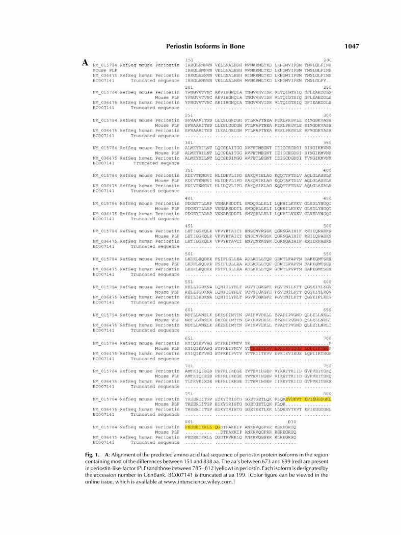
Fig. 1. A: Alignment of the predicted amino acid (aa) sequence of periostin protein isoforms in the regioncontainingmost of the differences between 151 and 838 aa. The aa’s between 673 and 699 (red) are presentin periostin-like-factor (PLF) and those between785–812 (yellow) in periostin. Each isoform is designatedbythe accession number in GenBank. BC007141 is truncated at aa 199. [Color figure can be viewed in theonline issue, which is available at www.interscience.wiley.com.]
Periostin Isoforms in Bone 1047

�808C. Total protein concentration was mea-sured using the bicinchoninic acid (BCA) pro-tein assay (Pierce, Rockford, IL).
Protein samples were mixed with an equalvolume of 2� SDS sample buffer, boiled for10 min, and subjected to 8% SDS–PAGE andWestern blot analysis as described previously[Tokuda et al., 2003]. The nitrocellulose mem-brane was blocked with 5% milk, Tris-bufferedsaline (TBS)-0.2%Tween-20 (TTBS) for 2 h, andincubated with primary antibody (0.5 mg/ml) for2 h. The blot was then washed three-times inTTBS and incubated with 0.1 mg/ml of HRP-conjugated goat anti-rabbit IgG secondary anti-body (Pierce) for 2 h at room temperature.Signal was detected by ECL (Pierce).
Cell Culture
Primary osteoblast cultures. Primaryosteoblast cultures were established usingneonatal rats as previously described [Selimet al., 2003]. Isolated cells were plated at 5� 105
cells/100mmplate inMEM (Earle’s) containing10% fetal bovine serum (FBS, Invitrogen). Onthe third day of culture, the initial platingmedium was replaced with MEM containing10% FBS, 10 mM b-glycerophosphate and ascor-bic acid (25 mg/ml) (Sigma, St. Louis, MO).
MC3T3-E1 osteoblast cultures. Themouse osteoblast cell line MC3T3-E1 was ob-tained from ATCC (Manasass, VA) and routi-nely maintained in growth medium consistingof aMEM containing 10% FBS, 100 U/mlpenicillin, and 100 mg/ml streptomycin. On thethird day of culture 25 mg/ml ascorbic acid and
10 mM b-glycerophosphate were added to themediumandmediawas changed every 3–4days[Owen et al., 1990].
Treatment of MC3T3-E1 cells with ASoligonucleotides. Equal numbers of MC3T3-E1 cells were transfected as previouslydescribed [Bonnelye et al., 2001]. Briefly, cellswere transfected on days 0, 5, 10, and 15 with0–5 mM AS periostin-oligonucleotide 50-GA-GAGGAACCATCTTCAGCCCTGAGCTCCG-30
and using oligofectamine as directed by themanufacturer (Invitrogen). During the trans-fection period of 4 h, cells were grown in OPTI-MEM serum-free media (Invitrogen) afterwhich, cells were grown in aMEM containing10% FBS, ascorbic acid (50 mg/ml) and a-glycerophosphate (10 mM). Cells were har-vested on days 7, 14, and 21, and extracted withTRIZOL for total RNA. DNase-treated totalRNA was used for RT-PCR.
Treatment of MC3T3-E1 cells with anti-periostin isoform antibody. For antibodytransfection, MC3T3-E1 cells were cultured asdescribed above and treated as described pre-viously [Selim et al., 2003]. Briefly, when cellsreached 50–60% confluence, they were trans-fected with a 5 or 2.5 mg/ml of anti-periostinantibody usingChariot reagent according to themanufacturer’s protocol (Activemotif, Carls-bad, CA). Control cells were either untreatedor transfected with non-immune IgG andChariot reagent. For evaluation of transfectionefficiency, MC3T3-E1 cells were transfectedwith b-galactosidase (b-gal), fixed and stain-ed according to the manufacturer’s protocol.
Fig. 1. B: Theexon–intronarrangement for periostin (a) andPLF (b). Thearrow in (a) corresponds to exon21and that in (b) corresponds to exon 17.
1048 Litvin et al.

Percentage of b-galactosidase transfected cellsto total number of cells was calculated fortransfection efficiency.PCR analysis using gene-specific pri-
mers. Two mg total RNA isolated fromMC3T3-
E1 cell cultures were reverse transcribed tocDNA at 428C for 50 min in a volume of 20 mlusing Superscript II as instructed by themanufacturer (Invitrogen). One microliter ali-quots of the cDNAwere amplified in a 50 ml PCR
Fig. 1. C: The cassette arrangement of periostin and PLF. Each cassette is color coded. Cassettes are madeup of either one or two exons.
Periostin Isoforms in Bone 1049

reaction mixture containing 1 nM primer set,and Stoffel Fragment of DNA polymerase(Applied Biosystems). The forward and reverseprimers were as follows: collagen type 1: 50-TCTCCACTCTTCTAGTTCCT-30 and 50-TTGG-GTCATTTCCACATGC-30, osteocalcin: 50-TCT-GACAAACCTTCAGTCC-30 and 50-AAATAGT-GATACCGTAGATGCG-30, G3PDH: 50-ACCA-CAGTCCATGCCATCAC-30 and 50-TCCACCA-CCCTGTTGCTGTA-30, cbfa1: 50-TCTGACAA-ACCTCATGTCC-30, and 50-AAATAGTGATA-CCGTAGATGCG-30, osteopontin: 50-ACACTT-TCACTCCAATCGTCC-30 and 50-TGCCCTTT-CCGTTGTTGTCC-30, respectively. PCR wasperformed using Perkin-Elmer GeneAmp PCR
System 9700 (Applied Biosystems) under thefollowing conditions: initial denaturation at948C for 3 min, amplification through 25 cyclesof 948C for 30 s followed by annealing andextension at the temperatures specified belowfor each gene for 45 s followed by a finalextension step at 728C for 10 min. PCR para-meters for each gene-specific primer varied onlyin annealing and extension temperature whichwere as follows: collagen type I: 558C, osteocal-cin: 558C, G3PDH: 65.38C, Cbfa1: 608C, andosteopontin: 558C. The expected product size foreach was collagen type I: 250, osteocalcin: 198,G3PDH: 452, Cbfa1: 632, and osteopontin:239 bp, respectively. PCR products were ana-
Fig. 1. D: Diagram representing the predicted aa sequence of PLF and periostin. The red and yellow boxesrepresent the 673–699 and 785–812 aa stretches that differ in these two isoforms. Primers on either sideof these regionsandused forRT-PCRare labeledasP1–P4.E: Periostin isoformexpression invivoand invitro.Total RNA frommouse 13.5 day pc calvaria (a & e) and MC3T3-E1 cells on days 7 (b & f), 14 (c & g) and 21(d & h) in culture were reverse transcribed and amplified by PCR using P1 and 2 (lanes a–d), P3 and 4(lanes e–h). [Color figure can be viewed in the online issue, which is available at www.interscience.wiley.com.]
1050 Litvin et al.

lyzed by 1% agarose gel electrophoresis stainedwith ethidium bromide. A 100-bp ladder wasused as a molecular weight marker (Promega).DNA gels of RT-PCR products were analyzed bydensitometry. Densitometric analysis of nor-malized values (specific PCR product/G3PDH)were used as a semi-quantitative analysis of thePCR products.
RESULTS
Generation and Analysisof Full-Length PLF cDNA
The 169 bp sequence we identified from theREADs technique was 99% identical over itsentirety to the published periostin (GenBankaccession no.: NM_015784). Takeshita et al.[1993] showed that periostin was bone-specific;however, our clone was isolated from the heart.We, therefore, attempted to ascertain whetherour clonewas the same as periostin identified inosteoblasts. Horiuchi et al. [1999] had alreadyproposed that there were several periostinisoforms that differ in their 30 translated sequ-ence. It was, therefore, reasonable to assumethat there might be a difference in the bonecDNA identified by Takeshita et al. [1993] andthe heart cDNA. In order to get the full-lengthsequence, we isolated it by RT-PCR from thesame cardiac RNA used in the READS techni-que. We used the published sequence of perios-tin to develop a 50 PCRprimer, because based onHoriuchi’s work, we did not expect there to beany difference at the 50 end. The 30 primer washomologous to the 30 end of the 169 bp READSEST.The full-length PLF cDNA was generated
and sequenced in both directions. The resulting3,012 bp cDNA is a unique form of OSF2/periostin that will be referred to as PLF.Examination of the NCBI database revealedonly highhomology toBC007141.BC007141 is acDNA sequence isolated from a mouse mam-mary tumor that contains a significant frameshift (caused by an extra ‘‘t’’ at position 592relative to the start codon and absent in otherisoforms of periostin), that produces a STOPcodon, which precludes the sequence from pro-ducing a full-length protein (Fig. 1A). We alsofound weaker homologies to two murine1 and
three human OSF2/periostin sequences. Themurine and human sequences are differentfrom that of PLF (Fig. 1A,B). For the analysisthat follows we will refer to the translatedprotein sequences of the isoforms in Figure 1Aand will number the aa sequences based on theReference Sequence2 for mouse periostin(NM_015784). The predicted aa sequence ofmousePLF thatwe identified contains a peptideregion of 27 aa (673–699 aa) that is absent fromboth mouse periostin (NM_015784) and the RefSeq for human periostin (NM_006475). Furthertowards the COOH terminus, there is a 28 aapeptide (785–812 aa) that is absent from ourtranslated PLF cDNA but present in the mouseand human periostins (NM_015784 andNM_006475, respectively). There are variousother mouse and human periostin isoformsequences in GenBank which contain varia-tions on these differences, none of which havethe 27 aa peptide (673–699 aa) found in mousePLF. These different proteins are most likelyisoforms resulting from alternatively splicedRNAs. An analysis of the exon–intron (Fig. 1Band Table I) arrangement of periostin[Fig. 1B(a)] and PLF [Fig. 1B(b)] showed thatnucleotides that code for the 673–699 aa regionin mouse PLF comprise exon 17 (present inperiostin intron 16–17) on chromosome 3 andthe nucleotides that code for 785–812 aacomprise exon 21 of periostin (NM_015784).
Horiuchi et al. [1999] isolated multiple seg-ments at the 30 end of the periostin cDNA byPCR. Based on the sequence analysis of thissegment they showed that there were fourpossible isoforms of periostin generated by acombination of six different cassettes, a–f[Horiuchi et al., 1999; Fig. 1C]. Our sequenceanalysis of the full-length PLF cDNA andpredicted aa sequence showed that it mostresembles Horuichi’s isoform 3 of mouse peri-ostin [Horiuchi et al., 1999]. Comparing the twosequencesofPLFandperiostin [Takeshitaetal.,
2NCBI Reference Sequence (Ref Seq): ‘‘The ReferenceSequence collection aims to provide a comprehensive,integrated, non-redundant set of sequences, includinggenomic DNA, transcript (RNA), and protein products, formajor research organisms. Themain features of the Ref Seqcollection include non-redundancy, explicitly linked nucleo-tide and protein sequences, updates to reflect currentknowledge of sequence data and biology, data validationand format consistency, distinct accession series, andongoing curation by NCBI staff and collaborators, withreview status indicated on each record’’.
1GenBank nomenclature: Murine OSF2¼NM015784 andBC031449, human OSF2¼NM006475, AY140646, andHUMOSF2P1.
Periostin Isoforms in Bone 1051

1993; Horiuchi et al., 1999], there are some keydifferences. In the region at the 30 end, analyzedby Horiuchi et al. [1999], isoform 3 and PLFcontain cassettes a–d and f and lack cassette e.The mouse periostin Ref Seq contains cassettesa, c–f and lacks cassette b. Thus PLF contains a27 aa sequence that is not found in mouseperiostin and mouse periostin contains a 28 aasequence that is not found in PLF. Horiuchi’scassettes are made up of either single or doubleexons in the periostin gene on chromosome 3 inthe mouse (Fig. 1C).
Since the sequence of aa in these regions arehighly conserved across species it is reasonable
to assume that the isoforms are functionallysignificant. For instance, in PLF, between aa784 and 806 there is a sequence recognized by‘motif analysis’ as a putative nuclear localiza-tion signal (NLS). This sequence is altered inperiostin by the presence of cassette ‘e’ so that itis no longer recognized by motif analysis as aNLS.
To investigate the complexity of periostinmRNAs in embryos and cultured MC3T3-E1cells, total RNAwas analyzed by RT-PCR usingprimers P1-4, flanking the regions that differedbetween periostin and PLF (Fig. 1D for primerposition). In 13.5 daymouse embryonic calvariaand MC3T3-E1 cells on days 7, 14, and 21, twoPCRproducts of 250 and 197 bp usingP1 andP2were identified. Using P3 and P4, bands at 300and 209 bp were identified (Fig. 1E). Sequenceanalysis of these cDNAbands identified themaseither PLF or periostin (Fig. 1E).
mRNA Expression is Temporally and SpatiallyRegulated During Mouse Embryogenesis
For an overall temporal pattern of expressionof the periostin isoforms during mouse embry-ogenesis, we probed total mouse embryonicRNA. Therewas a distinct pattern of expressionduring embryogenesis with significantlyreduced levels of expression at days 11.5 and12.5 pc in the whole embryo (Fig. 2). In MC3T3-E1 and primary osteoblast cultures, periostinisoform mRNA expression was detected over aperiod of 21 days (Fig. 3). Expression markedlydecreased in primary osteoblasts over the 21days in culture, whereas in MC3T3-E1 cellsmRNA levels increased and remained relativelyhigh (Fig. 3B).
In situ hybridization was used to determinethe spatial location of periostin isoforms duringcartilage and bone development at day 16.5 pc.mRNAs coding for these isoformswere localizedto mesenchymal tissues containing pre-osteo-blasts that surround cartilage primordia of theribs (Fig. 4E,F), vertebrae (Fig. 4B,D), and thelimb (Fig. 4G,H). mRNA was also detected incells comprising the cartilage primordia of theupper and lower jaws (Fig. 5A–D), specificallyin the mesenchymal/preosteoblasts in the hardpalate, in undifferentiated taste bud precursorcells (Fig. 5C,E), as well as in ameloblasts andodontoblasts in thedeveloping tooth (Fig. 5B,E).The control sections did not show any digox-ygenin reactivity.
TABLE I. Exon–Intron Arrangementfor Periostin (NM_015784) and
Periostin-Like-Factor*
NM_015784Periostin
(bp)
Periostin-Like Factor(PLF) (bp)
Exon 1 (ATG) 125 Exon 1 (ATG) 125Intron 1–2 1,359 Intron 1–2 1,359Exon 2 99 Exon 2 99Intron 2–3 3,178 Intron 2–3 3,178Exon 3 65 Exon 3 65Intron 3–4 1,532 Intron 3–4 1,532Exon 4 158 Exon 4 158Intron 4–5 1,490 Intron 4–5 1,490Exon 5 165 Exon 5 165Intron 5–6 884 Intron 5–6 884Exon 6 147 Exon 6 147Intron 6–7 486 Intron 6–7 486Exon 7 142 Exon 7 142Intron 7–8 1,031 Intron 7–8 1,031Exon 8 213 Exon 8 213Intron 8–9 553 Intron 8–9 553Exon 9 135 Exon 9 135Entron 9–10 800 Entron 9–10 800Exon 10 149 Exon 10 149Intron 10–11 1,164 Intron 10–11 1,164Exon 11 137 Exon 11 137Intron 11–12 882 Intron 11–12 882Exon 12 131 Exon 12 131Intron 12–13 522 Intron 12–13 522Exon 13 131 Exon 13 131Intron 13–14 90 Intron 13–14 90Exon 14 103 Exon 14 103Intron 14–15 151 Intron 14–15 151Exon 15 68 Exon 15 68Intron 15–16 328 Intron 15–16 328Exon 16 46 Exon 16 46Intron 16–17 5,781 Intron 16–17 2,805
Exon 17 82Intron 17–18 2,894
Exon 18 90 Exon 18 90Intron 18–19 1,006 Intron 18–19 1,006Exon 19 90 Exon 19 90Intron 19–20 634 Intron 19–20 634Exon 20 78 Exon 20 78Intron 20–21 385 Intron 20–21 4,053Exon 21 84Intron 21–22 3,584Exon 22 42 Exon 22 42Intron 22–23 881 Intron 21–22 881Exon 23 773 Exon 23 773
*Exons 17 and 21 are specific to PLF and periostin, respectively.
1052 Litvin et al.

Protein Expression is Temporally Regulated inBone During Mouse Embryogenesis
We detected a difference in the pattern ofperiostin protein isoform expression during
embryogenesis in bone and in culturedMC3T3-E1 cells. The antibody directed againstperiostin isoforms clearly detected multipleprotein isoforms between 86 and 93 kD in bothcases, but expression during embryogenesiswas temporally regulated in that it was notdetected by day 19.5 pc using Western blotanalysis (Fig. 6A) aswell as by in situ hybridiza-tion (data not shown). In MC3T3-E1 cells, ex-pression increased over the 21 days in culture.In addition, the number of isoforms expressedincreased with days in culture (Fig. 6A,B).Periostin isoforms in MC3T3-E1 cells werelocalized to the cytoplasm by immunostaining(Fig. 6C). The presence of a functional NLS issupported by protein detected in the nuclei ofMC3T3-E1 cells immunostained with the sameantibody used for Western blot analysis(Fig. 6C).
Reduction in the Amount of Periostin mRNAResulted in a Concomitant Decrease in the Levels
of Osteoblast Differentiation Markers
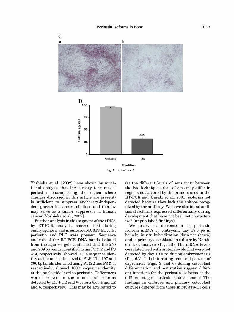
Since theperiostin proteinswere expressed inembryos and cultured osteoblast cells duringthe process of differentiation, we used an ASoligonucleotide and anti-periostin isoform poly-clonal antibody to examine their effects onosteoblast differentiation markers. The effec-tiveness of the AS oligonucleotide was assessedby reduction in the amount of periostin isoformmRNA (Fig. 7A). Several bone-specific markersused here are conventionally used as differ-entiation markers [Stein et al., 1990]. ThemRNA levels of collagen type 1, osteocalcin,osteopontin, alkaline phosphatase, and cbfa1were appreciably reduced in AS treated cellscompared to scrambled oligonucleotide controlsby RT-PCR analysis (Fig. 7B). In addition,alkaline phosphatase production (Fig. 7C) andcalcium levels (Fig. 7D) showed a reductionin both parameters in AS treated cells. TheMTT assay (Sigma) was used to prove that theantisense oligonucleotides didnothave aneffecton cell viability (datanot shown). Thesefindingswere corroboratedwhen thepolyclonal antibodywas used to block the activity of the proteinisoforms of periostin (Fig. 8B). Antibody treat-ment was effective in cells for up to 7 days. It isclear that by day 21 the antibody treatment wasnot effective possibly because of degradationover time. Efficiency of antibody transfectionassessed by b-galactosidase staining was ap-proximately 85% (Fig. 8A).
Fig. 2. Periostin isoform expression is developmentally regu-lated. A: 10 mg total RNA from whole mouse embryos from day9.5 pc to day 18.5 pcwas probedwith 32P labeled full-length PLFcDNA (�3 kb). Below is shown the ethidium bromide stainedformaldehyde-denatured gel. Blots were stripped and re-probedwith 32P labeled 18S cDNA as a control for loading and transfer(data not shown). B: Graph of the periostin isoform mRNA/18Sratio obtained by densitometric analysis of the X-ray films. Thenumbers on the x-axis correspond to the age of the embryofrom9.5 to 18.5 days pc, and the values on the y-axis correspondto the mRNA ratio (periostin isoform mRNAs/18S). [Color figurecan be viewed in the online issue, which is available at www.interscience.wiley.com.]
Periostin Isoforms in Bone 1053

DISCUSSION
We have recently isolated an additional iso-form of periostin that we refer to as PLF. Thisisoform is expressed in bone, specifically inosteoblast cells, determined by RT-PCR andsequence analyses. We have pursued the studyof this family of proteins because the specificfunction(s) of each isoform of periostin has notbeen examined and little is known aboutperiostin itself. This study is the first steptowards an in-depth analysis of periostin iso-
forms expressed during bone development andosteoblast differentiation.
Periostinwas initially identified byTakeshitaet al. [1993] using subtractive hybridization andwas shown to be expressed exclusively in bone.Horiuchi et al. [1999] proposed that periostinregulated recruitment of osteogenic precursorsand showed that it regulated adhesion ofMC3T3-E1 cells. Structurally it shared homol-ogywith fas I [Zinn et al., 1988] which in insectsregulates axonal guidance, a process involvingboth differential adhesion and migration.
Fig. 3. Expression of periostin mRNAs decreased over time inosteoblast cultures.A: Primary osteoblasts isolated fromneonatalrat calvaria and MC3T3-E1 cells were grown in culture for 7–21days. Total RNA was analyzed for the expression of periostinisoforms by Northern blot analysis. Blots were stripped and re-
probed with 32P labeled 18S cDNA as a control for loading andtransfer. B: Bar graph of the periostin isoform mRNA/18S ratioobtained by densitometric analysis of the X-ray films. [Colorfigure can be viewed in the online issue, which is available atwww.interscience.wiley.com.]
1054 Litvin et al.

Recently, Kruzynska-Frejtag et al. [2001]showed that periostin was expressed in theembryonic and fetal endocardial cushions thatlater divide the heart into four chambers. bIG-H3, a secreted protein containing the fourrepeats present in periostin and fas I have asimilar pattern of expression [Ferguson et al.,2003]. The function(s) of this family of proteins,all containing the conserved fas I repeatmotifs is
unclear. They appear to be expressed inmultipletissues during embryonic development and inadults [Zinn et al., 1988; Terasaka et al., 1989;Skonier et al., 1992; Takeshita et al., 1993;Huber and Sumper, 1994; LeBaron et al., 1995;Ulstrup et al., 1995; Horiuchi et al., 1999; Lalet al., 1999; Kruzynska-Frejtag et al., 2001;Sasaki et al., 2001; Gillan et al., 2002; Fergusonet al., 2003]. Experiments described in this study
Fig. 4. Localization of mRNAs of periostin isoforms indeveloping bone. Mouse embryonic 16.5 day pc sections wereprobed with a digoxygenin labeled antisense full-length PLFriboprobe. A: Low magnification; montage. B: The region in theblack box in (A) at 100� shows developing vertebrae,C¼ cartilage. D: A single developing vertebra at 200� indicatesthat developing pre-osteoblasts but not chondrocytes expressperiostin mRNAs. Pre-osteoblasts migrating away from the body
of the vertebra, to form the vertebral processes, are indicated bythe arrowheads. E & F: A view of presumptive ribs at 400�. Pre-osteoblasts express periostin mRNAs whereas chondrocytes donot. The red box in (A) is shown at higher magnification in (F).Cells indicated by the arrow in (F) are pre-osteoblasts migratingfrom thecartilaginous rib into the sternum.G: Developing limbat20� magnification (H) image in (G) shown at 200�, mesench-ymal cells expressing periostin isoforms are observed.
Periostin Isoforms in Bone 1055

Fig. 5. mRNAs of periostin isoforms are expressed in themesenchymal primordia of specific bony structures in the headregion in the16.5daymouse embryo.A: Lowmagnification view(20�) of the head region. The asterisk is located at the tip of thetongue (T) in the oral cavity in (A,B, andC). The region in the boxin (A) is shown at higher magnification in (D). B: View of theupper and lower jawsand the tongue (200�). Thearrow indicatesa region of the hard palate shown at higher magnification in (C).The box indicates a region of the lower jaw shown at higher
magnification in (E). C: The arrow point to cells in the secondarypalate, and the arrow head to cells in the tongue, expressingmRNAs of periostin isoforms (400�). D: The arrow points to aregion that lies between the soft and hard palates that isunstained. The arrow head points to stained cells in the tongue(200�). E: The ameloblasts (arrow head) and odontoblasts(arrow) of the developing tooth express the mRNAs of periostinisoforms (400�).
1056 Litvin et al.

suggest that they are developmentally regulatedand involved in osteoblast differentiation.Horiuchi et al. [1999] isolated multiple seg-
ments at the 30 end of the periostin cDNA byPCR. Based on the sequence analysis of thissegment they showed that there were fourpossible isoforms of periostin generated by acombination of six different cassettes, a–f[Horiuchi et al., 1999]. Our sequence analysisof the full-length PLF cDNA and predicted aasequence showed that it most resembles Hor-uichi’s isoform 3 of mouse periostin [Horiuchiet al., 1999]. Noteworthy are a few key differ-ences between the PLF and periostin sequences[Takeshita et al., 1993; Horiuchi et al., 1999]. Inthe region at the 30 end, analyzed by Horiuchiet al. [1999], isoform 3 and PLF contain casset-
tes a–d and f and lack cassette e. Takeshita’smouse periostin sequence contains cassettes a,c–f, and lacks cassette b. Thus PLF contains a27 aa sequence (cassette b) that is not found inmouse periostin and mouse periostin contains a28 aa sequence (cassette e) that is not found inPLF. These cassettes correlatewith exons in theperiostin gene (chromosome 3 in the mouse).Some of the cassettes contain more than oneexon. The differences amongst the periostinisoforms may have significant functional con-sequences. For instance, in PLF, between aa789 and 806 there is a sequence recognized bymotif analysis as a putative NLS. This sequenceis altered in mouse periostin by the presence ofcassette ‘e,’ so that it is no longer recognized bysequence analysis as a NLS. In addition,
Fig. 6. Expression of protein isoforms of periostin are devel-opmentally regulated. A: Protein extracts from 13.5 dayembryonic calvaria, 15.5, 17.5, and 19.5 day embryonic and2-day-old neonatal long bones were separated by SDS–PAGE.The nitrocellulose was reacted with anti-periostin antibody. B:Protein extracts from MC3T3-E1 cells at 3, 7, 10, 14, 17, and 21
days in culture analyzed by Western blot analysis using anti-periostin isoform polyclonal antibody. C: MC3T3-E1 cellsstained with anti-periostin isoform antibody (a: red) and DAPIspecific for DNA in nuclei (b: blue). Staining detected in both thecytoplasm and nucleus.
Periostin Isoforms in Bone 1057

Fig. 7. The effect of periostin antisense oligonucleotide onosteoblast differentiation. A: MC3T3-E1 cells were transfectedwith varying amounts of AS oligonucleotide and the amplifiedproducts of G3PDH and periostin are shown after 3 days post-transfection. AS oligonucleotide effectively reduced mRNAlevels of periostin isoforms. B: PCR analysis on day 7, 14, and21 post-transfection. C: MC3T3-E1 cells treated with antisense-periostin-oligonucleotide and stained for alkaline phosphataseafter 14 days in culture. AS treated cells (b), showed significantly
reduced levels of ALPH production compared to scrambleoligonucleotide treated control (a). Magnification 200�. D:Calciumdeposition (measured as described in Safadi et al., 2003)in MC3T3-E1 cells treated with antisense-periostin-oligonucleo-tide after 21 days in culture. AS treated cells showed significantlyreduced levels of calciumdeposition compared to controls. (datapresented are the mean and SEM, xxx¼ significant reduction incalcium deposition, P<0.001 using a Student’s t-test).
1058 Litvin et al.
Yoshioka et al. [2002] have shown by muta-tional analysis that the carboxy terminus ofperiostin (encompassing the region wherechanges discussed in this article are present)is sufficient to suppress anchorage-indepen-dent-growth in cancer cell lines and therebymay serve as a tumor suppressor in humancancer [Yoshioka et al., 2002].Further analysis in this segment of the cDNA
by RT-PCR analysis, showed that duringembryogenesis and in culturedMC3T3-E1 cells,periostin and PLF were present. Sequenceanalysis of the RT-PCR DNA bands isolatedfrom the agarose gels confirmed that the 250and 209 bp bands identified using P1&2 andP3& 4, respectively, showed 100% sequence iden-tity at the nucleotide level to PLF. The 197 and300 bp bands identified using P1&2 andP3&4,respectively, showed 100% sequence identityat the nucleotide level to periostin. Differenceswere observed in the number of isoformsdetected by RT-PCR andWestern blot (Figs. 1Eand 6, respectively). This may be attributed to
(a) the different levels of sensitivity betweenthe two techniques, (b) isoforms may differ inregions not covered by the primers used in theRT-PCR and [Sasaki et al., 2001] isoforms notdetected because they lack the epitope recog-nized by the antibody. We have also found addi-tional isoforms expressed differentially duringdevelopment that have not been yet character-ized (unpublished findings).
We observed a decrease in the periostinisoform mRNA by embryonic day 19.5 pc inbone by in situ hybridization (data not shown)and in primary osteoblasts in culture by North-ern blot analysis (Fig. 3B). The mRNA levelscorrelatedwell with protein levels that were notdetected by day 19.5 pc during embryogenesis(Fig. 6A). This interesting temporal pattern ofexpression (Figs. 3 and 6) during osteoblastdifferentiation and maturation suggest differ-ent functions for the periostin isoforms at thedifferent stages of osteoblast development. Thefindings in embryos and primary osteoblastcultures differed from those in MC3T3-E1 cells
Fig. 7. (Continued)
Periostin Isoforms in Bone 1059

where message expression was not markedlyreduced over time (Fig. 3B) as in embryos andprimary osteoblasts, and protein amount andthe number of isoforms increased over time inculture (Fig. 6B). These differences suggest
that there are inherent differences betweenmaturation of osteoblasts during embryogen-esis (in vivo), as compared to maturation of theMC3T3-E1 cells in the petri dish. Nonetheless,the presence of the periostin protein isoformsover time in MC3T3-E1 cells in culture andduring embryogenesis, suggest a role for theseproteins during the initial steps of differentia-tion. The presence of periostin isoforms earlyin osteoblast development suggests a role inthe recruitment of these cells and possibly inthe proliferation of these cells. Their presenceat later stages suggests a role in differentia-tion. Others also suggest a role for periostin[Horiuchi et al., 1999] and bIG-H3 [Fergusonet al., 2003] in the recruitment of mesenchymalcells to osteogenic lineage and in the adhesion ofMC3T3-E1 cells [Horiuchi et al., 1999].
To test the role of periostin isoforms inosteoblast differentiation,MC3T3-E1 cells weretreated with AS oligonucleotides and an anti-body directed against all known isoforms ofperiostin. The use of antibodies to inhibit pro-tein activity, is an accepted technique, usedsuccessfully by others [Selim et al., 2003].Markers of differentiation known to beexpressed in normal osteoblasts, at increasinglevels during the differentiation process, weremarkedly reduced by antibody and AS oligonu-cleotide treatment, when compared to controls.In particular, cbfa1, a transcription factorknown to regulate the expression of osteocalcin,a matrix component and marker of bonedifferentiation was markedly reduced suggest-ing that the periostin isoforms may be involvedin steps upstream of cbfa1. The isoforms maybe able to signal and initiate intracellularevents required to begin the process of differ-entiation possibly by binding integrin receptors[Gillan et al., 2002] and/or translocation to thenucleus.
This investigation has introduced the perios-tin isoform PLF, and has examined the expres-sion and possible function of periostin isoforms.In summary, multiple isoforms of periostin areexpressed in vivo during embryogenesis andin vitro in MC3T3-E1 cells and primary osteo-blast cultures. The expression of these isoformsappears to be developmentally regulated andthe data suggest that they are involved inregulating the process of differentiation. Futurestudies will address the mechanism(s) of actionof periostin and PLF in osteoblast differentia-tion in vitro and bone formation in vivo.
Fig. 8. The effect of periostin polyclonal antibodyon osteoblastdifferentiation.RT-PCRanalysisonRNAisolatedondays7and21after treatmentofMC3T3-E1cellswithanti-periostin isoformanti-body andCHARIOTS. Controlswere treatedwithmouse IgGandCHARIOTS. A: Cells stained for b-galatosidase 24 h after beingtreated with b-galatosidase and CHARIOTS, suggests efficientincorporation of protein into the cells.B: PCR of specificmarkersof osteoblast differentiation on days 7 and 21 post-treatmentwith either 5 or 2.5mg/ml of anti-periostin or IgGandCHARIOTS.
1060 Litvin et al.

REFERENCES
Auda-Boucher G, Bernard B, Fontaine-Perus J, Rouaud T,Mericksay M, Gardahaut MF. 2000. Staging of thecommitment of murine cardiac cell progenitors. Dev Biol225(1):214–225.
Bonnelye E, Merdad L, Kung V, Aubin JE. 2001. Theorphan nuclear estrogen receptor-related receptor alpha(ERRalpha) is expressed throughout osteoblast differ-entiation and regulates bone formation in vitro. J CellBiol 153(5):971–984.
Downs KM, Davies T. 1993. Staging of gastrulating mouseembryos by morphological landmarks in the dissectingmicroscope. Development 118(4):1255–1266.
Ferguson JW, Mikesh MF, Wheeler EF, LeBaron RG.2003. Developmental expression patterns of Beta-ig(betaIG-H3) and its function as a cell adhesion protein.Mech Dev 120(8):851–864.
Gillan L, Matei D, Fishman DA, Gerbin CS, Karlan BY,Chang DD. 2002. Periostin secreted by epithelial ovariancarcinoma is a ligand for alpha(V)beta(3) and alpha(V)-beta(5) integrins and promotes cell motility. Cancer Res62(18):5358–5364.
Horiuchi K, Amizuka N, Takeshita S, Takamatsu H,Katsuura M, Ozawa H, Toyama Y, Bonewald LF,Kudo XX. 1999. Identification and characterization of anovel protein, Periostin, with restricted expression toperiosteum and periodontal ligament and increasedexpression by transforming growth factor beta. J BoneMiner Res 14(7):1239–1249.
Hortsch M, Goodman CS. 1990. Drosophila fasciclin I, aneural cell adhesion molecule, has a phosphatidylinositollipid membrane anchor that is developmentally regu-lated. J Biol Chem 265(25):15104–15109.
Hu S, Sonnenfeld M, Stahl S, Crews ST. 1998. MidlineFasciclin: A Drosophila Fasciclin-I-related membraneprotein localized to the CNS midline cells and trachea.J Neurobiol 35(1):77–93.
Huber O, Sumper M. 1994. Algal-CAMs XX: Isoforms of acell adhesion molecule in embryos of the alga Volvoxwith homology to Drosophila fasciclin I. EMBO J 13(18):4212–4222.
Kruzynska-Frejtag A, Machnicki M, Rogers R, MarkwaldRR, Conway SJ. 2001. Periostin (an osteoblast-specificfactor) is expressed within the embryonic mouse heartduring valve formation. Mech Dev 103(1–2):183–188.
Lal A, Lash AE, Altschul SF, Velculescu V, Zhang L,McLendon RE, Marra MA, Prange C, Morin PJ, PolyakK,PapadopoulosN,VogelsteinB,KinzlerKW,StrausbergRL, Riggins GJ. 1999. A public database for gene expres-sion in human cancers. Cancer Res 59(21):5403–5407.
LeBaron RG, Bezverkov KI, Zimber MP, Pavelec R,Skonier J, Purchio AF. 1995. Beta IG-H3, a novelsecretory protein inducible by transforming growthfactor-beta, is present in normal skin and promotes theadhesion and spreading of dermal fibroblasts in vitro.J Invest Dermatol 104(5):844–849.
McAllister L, Goodman CS, Zinn K. 1992. Dynamicexpression of the cell adhesion molecule fasciclin Iduring embryonic development in Drosophila. Develop-ment 115(1):267–276.
Owen TA, Aronow M, Shalhoub V, Barone LM, Wilming L,Tassinari MS, Kennedy MB, Pockwinse S, Lian JB,Stein GS. 1990. Progressive development of the rat
osteoblast phenotype in vitro: Reciprocal relationships inexpression of genes associated with osteoblast prolifera-tion and differentiation during formation of the boneextracellular matrix. J Cell Physiol 143(3):420–430.
Prashar Y, Weissman SM. 1999. READS: A method fordisplay of 30-end fragments of restriction enzyme-digested cDNAs for analysis of differential gene expres-sion. Methods Enzymol 303:258–272.
Redkar A, Montgomery M, Litvin J. 2001. Fate mapof early avian cardiac progenitor cells. Development128(12):2269–2279.
Safadi FF, Xu J, Smock SL, Kanaan RA, Selim AH,Odgren PR, Marks SC, Jr., Owen TA, Popoff SN. 2003.Expression of connective tissue growth factor in bone:Its role in osteoblast proliferation and differentiationin vitro and bone formation in vivo. J Cell Physiol 196(1):51–62.
Sasaki H, Auclair D, Kaji M, Fukai I, Kiriyama M,Yamakawa Y, Fujii Y, Chen LB. 2001. Serum level of theperiostin, a homologue of an insect cell adhesion mole-cule, in thymoma patients. Cancer Lett 172(1):37–42.
Selim AA, Abdelmagid SM, Kanaan RA, Smock S,Owen TA, Popoff SN, Safadi FF. 2003. Anti-osteoactivinantibody osteoblast differentiation and function in vitro.Critical reviews in eukaryotic gene expression. Crit RevEukaryot Gene Expr 13(2–4):265–275.
Skonier J, Neubauer M, Madisen L, Bennett K, PlowmanGD, Purchio AF. 1992. CDNA cloning and sequenceanalysis of big-h3, a novel gene induced in a humanadenocarcinoma cell line after treatment with transform-ing growth factor-beta. DNA Cell Biol 11(7):511–522.
Stein GS, Lian JB, Owen TA. 1990. Relationship of cellgrowth to the regulation of tissue-specific gene expres-sion during osteoblast differentiation. FASEB J 4(13):3111–3123. Review.
Takeshita S, Kikuno R, Tezuka K, Amann E. 1993.Osteoblast-specific factor 2: Cloning of a putative boneadhesion protein with homology with the insect proteinfasciclin I. Biochem J 294(Pt. 1):271–278.
TerasakaK,Yamaguchi R,MatsuoK,Yamazaki A,Nagai S,Yamada T. 1989. Complete nucleotide sequence ofimmunogenic protein MPB70 from Mycobacterium bovisBCG. FEMS Microbiol Lett 49(2–3):273–276.
Tokuda H, Niwa M, Ito H, Oiso Y, Kato K, Kozawa O. 2003.Involvement of stress-activated protein kinase/c-JunN-terminal kinase in endothelin-1-induced heat shockprotein 27 in osteoblasts. Eur J Endocrinol 149(3):239–245.
Ulstrup JC, Jeansson S, Wiker HG, Harboe M. 1995.Relationship of secretion pattern and MPB70 homologywith osteoblast-specific factor 2 to osteitis followingMycobacterium bovis BCG vaccination. Infect Immun63(2):672–675.
Wilkinson DG. 1995. RNA detection using non-radioactivein situ hybridization. Curr Opin Biotechnol 6(1):20–23.Review.
Yoshioka N, Fuji S, Shimakage M, Kodama K, Hakura A,Yutsudo M, Inoue H, Nojima H. 2002. Suppression ofanchorage-independent growth of human cancer celllines by the TRIF52/periostin/OSF-2 gene. Exp Cell Res279(1):91–99.
Zinn K, McAllister L, Goodman CS. 1988. Sequenceanalysis and neuronal expression of fasciclin I in grass-hopper and Drosophila. Cell 53(4):577–587.
Periostin Isoforms in Bone 1061
Copyright © 2022 FDOKUMEN