Synaptic Zn2+ potentiates the effects of cocaine on striatal ...
15
ARTICLE OPEN Synaptic Zn 2+ potentiates the effects of cocaine on striatal dopamine neurotransmission and behavior Juan L. Gomez 1 , Jordi Bonaventura 1 , Jacqueline Keighron 2 , Kelsey M. Wright 1 , Dondre L. Marable 1 , Lionel A. Rodriguez 1 , Sherry Lam 1 , Meghan L. Carlton 1 , Randall J. Ellis 1 , Chloe J. Jordan 3 , Guo-hua Bi 3 , Oscar Solis 1 , Marco Pignatelli 4 , Michael J. Bannon 5 , Zheng-Xiong Xi 3 , Gianluigi Tanda 2 and Michael Michaelides 1,6 ✉ This is a U.S. government work and not under copyright protection in the U.S.; foreign copyright protection may apply 2021 Cocaine binds to the dopamine (DA) transporter (DAT) to regulate cocaine reward and seeking behavior. Zinc (Zn 2+ ) also binds to the DAT, but the in vivo relevance of this interaction is unknown. We found that Zn 2+ concentrations in postmortem brain (caudate) tissue from humans who died of cocaine overdose were significantly lower than in control subjects. Moreover, the level of striatal Zn 2+ content in these subjects negatively correlated with plasma levels of benzoylecgonine, a cocaine metabolite indicative of recent use. In mice, repeated cocaine exposure increased synaptic Zn 2+ concentrations in the caudate putamen (CPu) and nucleus accumbens (NAc). Cocaine-induced increases in Zn 2+ were dependent on the Zn 2+ transporter 3 (ZnT3), a neuronal Zn 2+ transporter localized to synaptic vesicle membranes, as ZnT3 knockout (KO) mice were insensitive to cocaine-induced increases in striatal Zn 2+ . ZnT3 KO mice showed significantly lower electrically evoked DA release and greater DA clearance when exposed to cocaine compared to controls. ZnT3 KO mice also displayed significant reductions in cocaine locomotor sensitization, conditioned place preference (CPP), self-administration, and reinstatement compared to control mice and were insensitive to cocaine-induced increases in striatal DAT binding. Finally, dietary Zn 2+ deficiency in mice resulted in decreased striatal Zn 2+ content, cocaine locomotor sensitization, CPP, and striatal DAT binding. These results indicate that cocaine increases synaptic Zn 2+ release and turnover/metabolism in the striatum, and that synaptically released Zn 2+ potentiates the effects of cocaine on striatal DA neurotransmission and behavior and is required for cocaine-primed reinstatement. In sum, these findings reveal new insights into cocaine’s pharmacological mechanism of action and suggest that Zn 2+ may serve as an environmentally derived regulator of DA neurotransmission, cocaine pharmacodynamics, and vulnerability to cocaine use disorders. Translational Psychiatry (2021)11:570 ; https://doi.org/10.1038/s41398-021-01693-0 INTRODUCTION Zinc (Zn 2+ ) is an essential trace element necessary for normal brain function [1–6]. Within the brain it exists in two forms; a “fixed”, protein-bound form, which serves as a catalytic co-factor or as a structural component to Zn 2+ binding proteins, and comprises ~90% of total brain concentration, or a “free”, or labile/ chelatable form, comprising ~10% of total brain concentration. Labile Zn 2+ is also referred to as “synaptic” Zn 2+ as it is localized to synaptic vesicles in a subset of glutamatergic neurons, also referred to as “zincergic” neurons [7]. Synaptic Zn 2+ is found in several regions such as cortex (Ctx, layers 2/3, 5, and 6), hippocampus, striatum, amygdala and is generally not present in thalamus [7, 8]. Synaptic Zn 2+ levels are dependent on the Zn 2+ transporter 3 (ZnT3) [9, 10], a neuronal-specific Zn 2+ transporter localized to the membrane of synaptic vesicles. ZnT3 knockout (KO) mice lack the ability to package Zn 2+ into these vesicles and consequently lack synaptic Zn 2+ release [9, 10]. ZnT3 KO mice are viable, fertile, and do not show major behavioral abnormalities across spatial learning, memory, or sensorimotor tasks, though they do exhibit small deficits in skilled reaching tasks [11, 12], fear learning [13], and sensory deficits [14, 15]. In terms of function, synaptic Zn 2+ is released upon neuronal activation, serves as a key regulator of neurotransmitter signaling, and as such has also been referred to as a neurotransmitter [1–6]. Tonic extracellular Zn 2+ concentrations are estimated to be at <25 nM [16]. However, upon physiological zincergic neuron activation and subsequent release, phasic Zn 2+ concentrations can increase up to 10 μM [17]. In this way, Zn 2+ is thought to exert various effects on neurotransmission by binding to synaptic proteins that contain either low- or high-affinity Zn 2+ -binding sites [6]. One such protein is the dopamine transporter (DAT), which contains four high-affinity Zn 2+- binding sites on its extracellular domain [18–22]. Studies have shown that cocaine binds to the DAT to inhibit synaptic DA reuptake, which leads to an increase of extracellular Received: 31 August 2020 Revised: 15 October 2021 Accepted: 20 October 2021 1 Biobehavioral Imaging and Molecular Neuropsychopharmacology Unit, National Institute on Drug Abuse Intramural Research Program, Baltimore, MD 21224, USA. 2 Medication Development Program, National Institute on Drug Abuse Intramural Research Program, Baltimore, MD 21224, USA. 3 Addiction Biology Unit, National Institute on Drug Abuse Intramural Research Program, Baltimore, MD 21224, USA. 4 Department of Psychiatry and Taylor Family Institute for Innovative Psychiatric Research, Washington University School of Medicine, St Louis, MO 63110, USA. 5 Department of Pharmacology, Wayne State University School of Medicine, Detroit, MI 48201, USA. 6 Department of Psychiatry and Behavioral Sciences, Johns Hopkins University School of Medicine, Baltimore, MD 21205, USA. ✉ email: [email protected] www.nature.com/tp Translational Psychiatry 1234567890();,:
-
Upload
khangminh22 -
Category
Documents
-
view
3 -
download
0
Transcript of Synaptic Zn2+ potentiates the effects of cocaine on striatal ...

ARTICLE OPEN
Synaptic Zn2+ potentiates the effects of cocaine on striataldopamine neurotransmission and behaviorJuan L. Gomez1, Jordi Bonaventura 1, Jacqueline Keighron2, Kelsey M. Wright 1, Dondre L. Marable1, Lionel A. Rodriguez1,Sherry Lam1, Meghan L. Carlton1, Randall J. Ellis1, Chloe J. Jordan 3, Guo-hua Bi3, Oscar Solis1, Marco Pignatelli4,Michael J. Bannon 5, Zheng-Xiong Xi3, Gianluigi Tanda 2 and Michael Michaelides 1,6✉
This is a U.S. government work and not under copyright protection in the U.S.; foreign copyright protection may apply 2021
Cocaine binds to the dopamine (DA) transporter (DAT) to regulate cocaine reward and seeking behavior. Zinc (Zn2+) also binds tothe DAT, but the in vivo relevance of this interaction is unknown. We found that Zn2+ concentrations in postmortem brain(caudate) tissue from humans who died of cocaine overdose were significantly lower than in control subjects. Moreover, the level ofstriatal Zn2+ content in these subjects negatively correlated with plasma levels of benzoylecgonine, a cocaine metabolite indicativeof recent use. In mice, repeated cocaine exposure increased synaptic Zn2+ concentrations in the caudate putamen (CPu) andnucleus accumbens (NAc). Cocaine-induced increases in Zn2+ were dependent on the Zn2+ transporter 3 (ZnT3), a neuronal Zn2+
transporter localized to synaptic vesicle membranes, as ZnT3 knockout (KO) mice were insensitive to cocaine-induced increases instriatal Zn2+. ZnT3 KO mice showed significantly lower electrically evoked DA release and greater DA clearance when exposed tococaine compared to controls. ZnT3 KO mice also displayed significant reductions in cocaine locomotor sensitization, conditionedplace preference (CPP), self-administration, and reinstatement compared to control mice and were insensitive to cocaine-inducedincreases in striatal DAT binding. Finally, dietary Zn2+ deficiency in mice resulted in decreased striatal Zn2+ content, cocainelocomotor sensitization, CPP, and striatal DAT binding. These results indicate that cocaine increases synaptic Zn2+ release andturnover/metabolism in the striatum, and that synaptically released Zn2+ potentiates the effects of cocaine on striatal DAneurotransmission and behavior and is required for cocaine-primed reinstatement. In sum, these findings reveal new insights intococaine’s pharmacological mechanism of action and suggest that Zn2+ may serve as an environmentally derived regulator of DAneurotransmission, cocaine pharmacodynamics, and vulnerability to cocaine use disorders.
Translational Psychiatry (2021) 11:570 ; https://doi.org/10.1038/s41398-021-01693-0
INTRODUCTIONZinc (Zn2+) is an essential trace element necessary for normalbrain function [1–6]. Within the brain it exists in two forms; a“fixed”, protein-bound form, which serves as a catalytic co-factoror as a structural component to Zn2+ binding proteins, andcomprises ~90% of total brain concentration, or a “free”, or labile/chelatable form, comprising ~10% of total brain concentration.Labile Zn2+ is also referred to as “synaptic” Zn2+ as it is localizedto synaptic vesicles in a subset of glutamatergic neurons, alsoreferred to as “zincergic” neurons [7]. Synaptic Zn2+ is found inseveral regions such as cortex (Ctx, layers 2/3, 5, and 6),hippocampus, striatum, amygdala and is generally not presentin thalamus [7, 8].Synaptic Zn2+ levels are dependent on the Zn2+ transporter 3
(ZnT3) [9, 10], a neuronal-specific Zn2+ transporter localized to themembrane of synaptic vesicles. ZnT3 knockout (KO) mice lack theability to package Zn2+ into these vesicles and consequently lacksynaptic Zn2+ release [9, 10]. ZnT3 KO mice are viable, fertile, and
do not show major behavioral abnormalities across spatiallearning, memory, or sensorimotor tasks, though they do exhibitsmall deficits in skilled reaching tasks [11, 12], fear learning [13],and sensory deficits [14, 15].In terms of function, synaptic Zn2+ is released upon neuronal
activation, serves as a key regulator of neurotransmitter signaling,and as such has also been referred to as a neurotransmitter [1–6].Tonic extracellular Zn2+ concentrations are estimated to be at<25 nM [16]. However, upon physiological zincergic neuronactivation and subsequent release, phasic Zn2+ concentrationscan increase up to 10 µM [17]. In this way, Zn2+ is thought to exertvarious effects on neurotransmission by binding to synapticproteins that contain either low- or high-affinity Zn2+-bindingsites [6]. One such protein is the dopamine transporter (DAT),which contains four high-affinity Zn2+-binding sites on itsextracellular domain [18–22].Studies have shown that cocaine binds to the DAT to inhibit
synaptic DA reuptake, which leads to an increase of extracellular
Received: 31 August 2020 Revised: 15 October 2021 Accepted: 20 October 2021
1Biobehavioral Imaging and Molecular Neuropsychopharmacology Unit, National Institute on Drug Abuse Intramural Research Program, Baltimore, MD 21224, USA. 2MedicationDevelopment Program, National Institute on Drug Abuse Intramural Research Program, Baltimore, MD 21224, USA. 3Addiction Biology Unit, National Institute on Drug AbuseIntramural Research Program, Baltimore, MD 21224, USA. 4Department of Psychiatry and Taylor Family Institute for Innovative Psychiatric Research, Washington University Schoolof Medicine, St Louis, MO 63110, USA. 5Department of Pharmacology, Wayne State University School of Medicine, Detroit, MI 48201, USA. 6Department of Psychiatry andBehavioral Sciences, Johns Hopkins University School of Medicine, Baltimore, MD 21205, USA. ✉email: [email protected]
www.nature.com/tpTranslational Psychiatry
1234567890();,:

DA [23]. This mechanism underlies the direct subjective responsesthat accompany cocaine use [24] and is critical to cocaine self-administration (SA) in laboratory models, and cocaine reward andabuse liability in humans [25]. In vitro assays have shown thatwhen Zn2+ binds to the DAT it promotes a conformation thatinhibits DA uptake and, when cocaine is present, Zn2+ canincrease cocaine’s affinity and can modulate its potency to inhibitDA uptake [18–22]. Interestingly, synaptic Zn2+ is present in thestriatum [26, 27] where the DAT is found in the highestconcentration [28], and synaptic Zn2+ depletion in this regionimproves motor deficits and memory impairments caused by lossof striatal DAergic fibers [29]. These prior studies suggest thatsynaptic Zn2+ may modulate in vivo DAT function and cocaine-DAT pharmacodynamic interactions in the striatum. However, theextent to which such interactions occur is unclear.In addition to ZnT3, synaptic Zn2+ levels also depend on the
environmental availability of Zn2+ which is obtained exclusivelyvia the diet. Consequently, dietary factors which limit Zn2+ intakeinfluence both synaptic Zn2+ levels and ZnT3 expression [30, 31].In fact, as the body lacks a specialized system for its storage, Zn2+
needs to be consumed continuously to avoid a state of deficiencywhich itself can have profound changes in both the periphery aswell as in brain function [32]. Human drug users generally do notfollow a lifestyle that prioritizes nutritional needs and are knownto exhibit nutritional deficiencies including dysregulated bloodand hair Zn2+ content [33–38]. However, whether such deficits areinvolved in the neurobiological changes associated with cocaineuse or use of other drugs is unknown.
METHODSSubjectsDe-identified postmortem human brain specimens were collectedduring the routine autopsy process as described in detail previously[39, 40]. Briefly, the cause and manner of death were determined byforensic pathologists following medico-legal investigations that eval-uated the circumstances of death including medical records, policereports, autopsy results, and toxicological data. Inclusion in the cocainecohort (n= 20; 10 Caucasian, 10 African-American) was based oncocaine abuse as the cause of death, a documented history of drugabuse, and a toxicology positive for high levels of the cocainemetabolite benzoylecgonine and, in most cases, the short-lived cocaineadulterant levamisole, both indicative of recent cocaine use prior todeath. Control subjects (n= 20; 10 Caucasian, 10 African-American) diedas a result of cardiovascular disease or gunshot wound, had nodocumented history of drug abuse, and tested negative for cocaine andother drugs of abuse. Exclusion criteria for the study included a knownhistory of neurological or psychiatric illness, death by suicide, estimatedpostmortem interval exceeding 20 h, evidence of neuropathology (e.g.,encephalitis, stroke), or chronic illness (e.g., cirrhosis, cancer, HIV,prolonged hospitalization). The final groups did not differ with regard toage for race, nor with regard to brain pH, a well-established measure ofsample quality and perimortem agonal state [41]. Tissue from onecocaine user was not included due to very high cocaine metabolitelevels.Given that the human tissue samples available consisted only of males,
and therefore all human data were limited to this sex, we focused on malemice for all other mechanistic experiments. Male C57Bl/6J mice wereacquired from Jackson Labs at 8 weeks of age. Breeding pairs of Slc30a3(ZnT3) KO mice were obtained from Dr. Thanos Tzounopoulos at theUniversity of Pittsburgh and bred at the National Institute on Drug Abuse(NIDA) (Baltimore, MD) on a C57Bl/6J background. Mice were genotypedby Transnetyx (Cordova, TN). All mice were male and matched for age andweight. Mice were single-housed during experimental testing in atemperature and humidity-controlled environment on a regular light cycle(on at 7 am and off at 7 pm). Food and water were available ad libitum andmice were acclimated prior to any behavioral procedures by handling. Allexperimental procedures were carried out in accordance with the NationalInstitutes of Health Guide for the Care and Use of Laboratory Animals andwere approved by the Animal Care and Use Committee of NIDA.
Total reflection X-ray spectroscopy (TXRF)Tissue samples were collected and weighed in 1.5 ml Eppendorf tubes. Theweight of the tissue was directly used to calculate element concentrationsin µg/kg units. Each tissue sample was dissolved in 100 µL of nitric acid(Sigma: NX0408) with 2 µL of a gallium standard (conc. 1000 ppm). Eachsample was assessed in duplicate for TXRF elemental analysis using an S2Picofox (Bruker, Billerica, MA). This instrument exposes the sample to anX-ray beam and measures fluorescence radiation specific to the element(s)of interest. Human samples were prepared from postmortem tissuecollected from the anterior caudate. Wild-type (WT) or ZnT3 KO micereceived either saline, a single cocaine injection (20mg/kg, i.p), or 8repeated daily cocaine (20 mg/kg, i.p) injections and were euthanized 24 hafter the last injection. Mice fed the 30 ppm and 5 ppm Zn2+ diets wereexposed to each respective diet for 35 days and then euthanized. Mousesamples were prepared by slicing flash-frozen tissue on a cryostat (100 µmsections from Bregma 1.00 to 0.00mm) or dissecting the Ctx and striatumfrom each section.
Synchrotron X-ray fluorescence microspectroscopy (µXRFS)Brain concentrations and distributions of Zn2+ from C57BL/6J miceinjected with saline or cocaine (10mg/kg, i.p.) every other day for 8 daysand euthanized 24 h after the last injection were measured at the X26abeamline at the National Synchrotron Light Source (NSLS) at BrookhavenNational Laboratory (Upton, NY). The synchrotron X-ray beam was tuned to12 keV using a Si(111) channel-cut monochromotor. The monochromaticbeam was then collimated to 350 μm× 350 μm and then focused toapproximately 6 μm× 10 μm using Rh-coated silicon mirrors in aKirkpatrick–Baez (KB) geometry. The sample was placed at a 45° angle tothe incident X-ray beam and X-ray fluorescence was detected with anenergy dispersive, 9-element germanium array detector (Canberra,Meriden, CT) oriented at 90° to the incident beam. The sample wasapproximately 6 cm from the detector. A light microscope objective(Mitutoyo, M Plan Apo 5X) was coupled to a digital CCD camera for sampleviewing. Energy dispersive spectra were collected by raster-scanning thesample through the X-ray beam using a dwell time of 0.3 s/pixel and a stepsize of 10 μm. Zn Kα, fluorescence counts were then extracted frombackground-corrected energy dispersive spectra. All data were normalizedto variations in incident photon flux by normalizing to changes in I0measured by ion chamber upstream of the KB optics. XRFS calibrationstandards on Nuclepore® polycarbonate aerosol membranes expressingknown (±5%) concentrations of Zn (48.4 µg/cm2) were also imaged inparallel to the samples (Micromatter, Vancouver, BC) and used to expressresults as µg/cm2. Image analysis was carried out using ImageJ (NationalInstitutes of Health, Bethesda, MD). Regions of interest (ROI) were drawnonto the Ctx, caudate putamen (CPu), nucleus accumbens (NAc), andmeasurements for each ROI were obtained.
Zinc–selenium autometallography (ZnSeAMG)WT and ZnT3 KO mice were anesthetized with a ketamine-xylazine cocktail(ket = 60mg/kg + xyl = 12mg/kg) and injected (i.p.) with 15mg/kg sodiumselenite (Sigma Aldrich: 214485) and placed on a heating pad whileanesthetized for 60min. Mice were then perfused with 0.1M phosphatebuffer for 5min. Brain tissue was dissected, and flash frozen in dry ice cooledisopentane and stored at −80 °C until sectioning. Coronal brain sections(20 µm) were thaw mounted at the level of the striatum and hippocampus onpositively charged glass slides. Slides were stored at −20 °C until staining.Slides were loaded in non-metallic staining racks and allowed to reach roomtemperature. Slides were fixed in 95% ethanol for 15min followed byhydration in 70% (2min) and 50% (2 min) ethanol ending in 3 × 2min distilledwater rinses. Slides were dipped in 0.5% gelatin and air dried prior to physicaldevelopment. Developer was made by mixing Gum Arabic (50% solution,100ml), citrate buffer (2.0M, 20ml), hydroquinone (1.7 g in 30ml DDH2O),silver lactate (0.22 g in 30ml H20), and DDH2O (200ml). Developer was pouredonto slides, incubated for 60min in the dark then quickly checked at 10minintervals until sections are dark brown. Slides were washed in slowly flowingtap water (37 °C) for 10min to remove gelatin then rinsed 3 × 2min in distilledwater. Slides were then incubated in 5% sodium thiosulphate (12min) andrinsed 2 × 2 min in distilled water and post-fixed in 70% ethanol (30 min).Optional counter stain using cresyl violet or toluidine blue (5 min) followed byrinse 4 × 30 s rinse in distilled water. Finally, slides were dehydrated in 95%ethanol (5 min), 100% ethanol 2 × 5 min, xylene 2 × 5 min, and coverslippedwith permount. Stained sections were imaged using brightfield microscopy.
J.L. Gomez et al.
2
Translational Psychiatry (2021) 11:570

65Zn uptake experiments using positron emissiontomography (PET)WT and ZnT3 KO mice were anesthetized with isoflurane and placed in aprone position on the scanner bed of a nanoScan PET/CT (Mediso, USA)injected intravenously (~150 µL) with 65ZnCl2 (~2.2 MBq) and PET datawere acquired for 2 h followed by a CT scan. After scanning, animals werereturned to their home cage. Scans were repeated on days 1, 3, 7, and 14.For cocaine experiments, C57Bl/6J mice were injected with 65ZnCl2 asabove and then injected immediately with saline or cocaine (20mg/kg, i.p).Saline and cocaine injections continued daily for 7 days. Mice werescanned on Day 1 and Day 7 after 65ZnCl2 injection as above. In all cases,the PET data were reconstructed and corrected for dead-time andradioactive decay. Qualitative and quantitative assessments of PET imageswere performed using the PMOD software environment (PMOD Technol-ogies, Zurich Switzerland). Time-activity curves were generated usingmanually drawn volumes of interest using the CT image as a reference.Standardized uptake values (SUV) were calculated using the formula SUV(i)= C(i)/(ID × BW) where C(i) is the activity value at a given time point (inkBq/cc), ID is the injected dose (in MBq) and BW is the animal’s bodyweight (in kg). For voxel-wise analyses we used Statistical ParametricMapping (SPM12, London, UK) as previously described [42]. First, all theimages were co-registered and masked to the reference mouse atlas inPMOD. Regional changes in uptake were assessed relative to global(whole-brain) uptake. For the cocaine experiments, a repeated measures(RM) analysis of variance (ANOVA) model was used that defined saline vs.cocaine-treated mice scanned at 1 and 7 days post 65ZnCl2 injection.Images were subtracted after intensity normalization to 100 by theproportional scaling method. After estimation of the statistical model, acontrast (Cocaine > Vehicle) was applied to reveal the effects of interest.These effects were overlaid on the reference MRI. An uncorrected P valueof 0.05 with a cluster threshold value of 50 were used as thresholds todetermine statistical significance.
Ex vivo 65Zn autoradiographyOne day after the last PET scan, WT and ZnT3 KO mice were euthanizedand brain tissue was dissected, flash frozen in isopentane, and stored at−80 °C until sectioning. Tissue was sectioned and thaw mounted onpositively charged glass slides. Slides were placed on BAS-IP SR 2040 ESuper Resolution phosphor screens (GE Healthcare) for 14 days andimaged using a phosphor imager (Typhoon FLA 7000; GE Healthcare).
Radioligand binding assaysBrains from euthanized C57Bl/6J mice were removed and striata dissectedand quickly frozen until use. The tissue was weighed and suspended in tentimes (w/v) of ice-cold Tris-HCl buffer (50 mM, pH 7.4). The suspension washomogenized with a Polytron homogenizer (Kinematica, Basel, Switzer-land) under ice. Homogenates were centrifuged at 48,000 × g (50min, 4 °C)and washed twice in the same conditions to isolate the membranefraction. Protein was quantified by the bicinchoninic acid method (Pierce).For competition experiments, membrane suspensions (50 µg of protein/ml) were incubated in 50mM Tris-HCl (pH 7.4) 0.5 nM of [3H]WIN-35428(Perkin-Elmer) and increasing concentrations of the indicated competingdrugs (WIN-35428 or cocaine) in the presence or the absence of 100 nM,10 μM or 1mM of ZnCl2 during 2 h at RT. Nonspecific binding wasdetermined in the presence of 100 µM cocaine. In all cases, free andmembrane-bound radioligand were separated by rapid filtration throughWhatman (Clifton, NJ) GF/B filters, pre-soaked in 0.05% polyethyleneimineby using a Brandel R48 filtering manifold (Brandel Inc., Gaithersburg, MD).The filters were washed twice with 5ml of cold buffer and transferred toscintillation vials. Beckman Ready Safe scintillation cocktail (3.0 ml) wasadded, and the vials were counted the next day with a Beckman 6000liquid scintillation counter (Beckman Coulter Instruments, Fullerton, CA) at50% efficiency.
In vitro autoradiography using [3H]WIN-35,428Brain tissue from WT and ZnT3 KO mice was dissected, flash frozen inisopentane, and stored at −80 °C until sectioning. Tissue was sliced on acryostat at 16 µm and thaw mounted on positively charged glass slidesand stored at -20 °C until autoradiography. Incubation Buffer consisted of50 mM Tris-HCl (7.4 pH) and 100 mM NaCl in deionized water. [3H]WIN-35,428 Total binding buffer (S.A. 82.9 Ci/mmol, Conc. 1 mCi/ml) was madein incubation buffer at a concentration of 10 nM. ZnCl2 binding buffer wasmade using the Total binding buffer stock and adding ZnCl2 for a
concentration of 10 µM. Slides were pre-incubated in ice-cold incubationbuffer for 20min then transferred to respective radioactive incubationbuffers (i.e. total or total+ZnCl2) for 120min on ice. Slides were thenwashed 2 × 1min in ice-cold 50 mM Tris-HCl (pH= 7.4) then dipped (30 s)in ice-cold deionized water. Slides were dried under stream of cool air andplaced on BAS-IP TR 2025 E Tritium Screen (GE Healthcare) for 5 days andimaged using a phosphor imager (Typhoon FLA 7000; GE Healthcare).Sections were analyzed using Multigauge software (Fujifilm, Japan).
In vivo fast scan cyclic voltammetry (FSCV)FSCV procedures follow those of recently published work from ourlaboratory in anesthetized mice using electrical stimulation [43]. Briefly,glass sealed 100 µm carbon-fiber microelectrodes were pre-calibrated withknown concentrations of dopamine and changes in pH to allow for aprincipal component analysis (PCA) of the raw data using HDCV (UNC,Chapel Hill, NC). Dopamine was identified by cyclic voltammogram using avoltage scan from −0.4 to 1.3 V at 400 V/s. During the experiment anexternal stimulus was applied using the tungsten electrode every 5 mincomprised of 24 pulses 4ms in width at 60 Hz and 180 µA while theworking electrode was implanted in the striatum (AP: +1.5 mm; ML:±1.0mm; DV: −3.2 to −3.7 mm from bregma). After PCA data wereanalyzed to determine the DAMax and DA clearance rate using a custommacro written in Igor Carbon Pro which identified peaks greater than 3×root mean square noise and fit to Eq. 1 where DAMax represents the peakDA concentration measured, k is the rate constant, and t is time [43].
DA tð Þ ¼ DAMaxe�k t�t0ð Þ (1)
Cocaine locomotor sensitizationEach session during the development phase was 30min, and mice wereonly exposed to one session per day with locomotor activity quantified asdistance traveled (cm). All injections were administered (i.p.). Mice werefirst habituated to the locomotor activity chambers (Opto-varimex ATM3,Columbus Instruments). On the next two sessions, mice were injected withsaline and placed in the chambers. On the following five sessions, separategroups were injected with either saline or cocaine (10 mg/kg) in acounterbalanced design. Mice were then allowed 7 days of withdrawal inthe colony room and then returned to the behavior room for testingexpression of sensitization. Briefly, all mice were allowed access to theactivity chambers for 60min followed by increasing doses of cocaine(saline, 5, 10, 20 mg/kg) every 60min. Data collection was paused butchambers were not cleaned in-between cocaine dosing, each mouse waspicked up, injected, and placed back in chamber to continue datacollection.
Cocaine conditioned place preference (CPP)The task consisted of ten sessions, one per day, in chambers with twovisually distinct sides, one with clear walls and white floor and one withcheckered walls and black floor. The sides were divided by a door with andwithout access to the other side. Locomotor activity was measured by wayof time spent in each chamber as well as total distance traveled (Opto-varimex ATM3, Columbus Instruments). In the first session, the mice couldexplore both sides of a conditioning box for 15min to determine inherentside preference, designated as the Pre-Test. Using this data, the cocaine-paired side was pseudo-randomized so that mice with a preference for oneside (>60%) were cocaine-paired on the other, non-preferred side. Micewith no side preference were cocaine-paired in a counterbalanced fashion.Separate groups of mice were conditioned with either a 5, 10 or 20mg/kgdose of cocaine. In an alternating fashion for 8 days, mice were injected(i.p.) with either saline or cocaine and placed in the predetermined drug/no drug side of the chamber for 30min. The mice had no physical accessto the other side but were still able to see through the clear divider wall.Each mouse had a total of 4 saline-paired days and 4 cocaine-paired days.The last session was the same as the first and designated the Test session.Time spent in the cocaine-paired chamber during the Pre-Test session(prior to conditioning) was subtracted from time spent in the cocaine-paired chamber during the Test session and expressed as thePreference score.
Mouse intravenous cocaine self-administrationMice were implanted with jugular vein catheters under ketamine/xylazineanesthesia and using aseptic surgical techniques. A 6.0 cm length
J.L. Gomez et al.
3
Translational Psychiatry (2021) 11:570

MicroRenathane (ID 0.012″, OD 0.025″; Braintree Scientific Inc., Braintree,MA, USA) catheter was inserted 1.2 cm into the right jugular vein andanchored to a 24-gauge steel cannula (Plastics One, Roanoke, VA, USA) thatwas bent at a 100° angle and mounted to the skull with cyanoacrylate glueand dental acrylic. A 2.5 cm extension of flexible tubing was connected tothe distal end of the cannula. The mice were allowed 5–7 days for recovery,during which time 0.05 ml of a 0.9% saline solution containing 20 IU/mlheparin and 0.33 mg/ml gentamycin was infused daily through thecatheter to prevent catheter clotting and infection. Thereafter, 0.05 ml of0.9% saline solution containing 20 IU/ml heparin was infused immediatelyprior to and immediately following each daily SA session. When needed,i.v. brevital (a barbiturate) was used to test catheter patency between theSA sessions. During cocaine SA sessions, the flexible tubing extension wasconnected to a perfusion pump (Med Associates, Fairfax, VT) via a PE50tubing connector. After daily SA sessions, the free end of the cannula guidewas always kept sealed.Operant test chambers (Med Associates, Fairfax, VT) contained two
levers (active and inactive) located 2.5 cm above the floor as well as a cuelight above each lever. A house light mounted on the opposite side of thechamber signaled the start of each 3 h session and remained illuminateduntil the session ended. For SA sessions, a liquid swivel mounted on abalance arm above the chamber allowed for i.v. drug delivery in freelymoving mice. Depression of the active lever resulted in the activation of aninfusion pump; depression of the inactive lever was recorded but had noscheduled consequences. Each infusion was paired with two discrete cues:illumination of the cue light above the active lever, and a cue tone thatlasted for the duration of the infusion. Experimental events were controlledby a PC programmed in Medstate Notation and connected to a MedAssociates interface.After recovery from surgery, mice were placed into operant chambers
and allowed to lever press for i.v. cocaine SA under a fixed-ratio 1reinforcement schedule (i.e., each lever press leads to one cocaineinfusion) for 3 h daily. Each cocaine infusion lasted 4.2 s, during whichadditional active lever responses were recorded but had no consequences(i.e., non-reinforced active lever response). Mice were trained initially for ahigh unit dose of cocaine (1 mg/kg/infusion) to potentiate acquisition ofSA until stable SA was achieved, which was defined as earning at least 20infusions per 3 h session and an active/inactive lever press ratio exceeding2:1. Then the mice were switched to a multiple-dose schedule to observethe dose-dependent cocaine SA according to a descending cocaine dosesequence from the initial dose of 1 mg/kg/infusion (sessions 1–13) to0.5 mg/kg/infusion (sessions 14–20), 0.25 mg/kg/infusion (sessions 21–23),0.125mg/kg/infusion (sessions 24–27), and 0.0625mg/kg/infusion (ses-sions 28–29). Mice that did not reach stability criteria, lost catheter patencyor showed excessive high-level inactive lever responding (>100 pressesper session) were excluded from further experimentation. To preventcocaine overdose, maximally allowed cocaine infusions were 50 (0.1 and0.5 mg/kg/infusion), 100 (0.25mg/kg/infusion), 200 (0.125 mg/kg/infusion),or 400 (0.0625mg/kg/infusion), respectively during each 3-h session. Thenumber of cocaine infusions earned, and active and inactive leverresponses were recorded for each session. The last 2–3 days of cocaineSA data at each dose were averaged and used to compare dose-responseperformance between WT and KO mice.After the completion of the above cocaine dose–response experiment,
the animals were switched to cocaine SA under PR reinforcementschedule. During PR conditions, the work requirement (lever presses)needed to receive a cocaine infusion was raised progressively within eachtest session according to the following PR series: 1, 2, 4, 6, 9, 12, 15, 20, 25,32, 40, 50, 62, 77, 95, 118, 145, 178, 219, 268, 328, 402, 492, and 603 untilthe breakpoint was reached. The breakpoint was defined as the maximalworkload (i.e., number of lever presses) completed for the last cocaineinfusion prior to a 1-h period during which no infusions were obtained bythe animal. Animals were tested for cocaine SA under PR reinforcement atthree doses (starting at 0.25, then 1 and then 0.5 mg/kg/infusion) fromdays 30 to 38.After the completion of the PR experiments, the same groups of animals
continued for cocaine extinction and reinstatement tests. During extinc-tion, syringe pumps were turned off and the cocaine-associated cue lightand tone were unavailable. Thus, lever pressing was recorded but had noscheduled consequences. Extinction training continued for about 20 daysuntil the extinction criteria were met (i.e., lever responding <20% of the SAbaseline) for at least 3 sessions. Mice then received a 10mg/kg i.p cocaineinjection to evoke reinstatement of drug-seeking behavior. Duringreinstatement testing, active lever presses lead to re-exposure to the cuelight and tone previously paired with cocaine infusions, but not to actual
cocaine infusions. Active and inactive lever responses were recorded foreach extinction and reinstatement session. Lever pressing behavior duringthe cocaine-primed session was compared to the average lever pressiesduring the last 3 days of extinction.
Custom dietsDiets were formulated by Research Diets, Inc via use of AIN-93M maturerodent diet. The diets were compositionally identical, but one diet had anadequate amount of Zn2+ (30 ppm) and the other diet had a deficientamount of Zn2+ (5 ppm). Zn2+ concentration in each diet was confirmedin-house via random sampling of chow pellets and TXRF (S2 Picofox,Bruker, Billerica, MA).
Body weight and food intake measurementsC56BL/6J mice arrived at the NIDA mouse colony and were allowed oneweek of environmental acclimation with regular chow and water availablead lib. After the acclimation period, mice were given a diet containingeither 30 ppm Zn2+ or 5 ppm Zn2+ for a minimum of 35 days prior to anybehavioral manipulations. Mice stayed on the diet until the completion ofthe experiment. Mice were individually housed and weighed three timesper week along with food weight to track the amount of food consumed.
ImmunohistochemistryCoronal sections were sliced on a cryostat (30 µm), collected in six-wellplates with PBS, and stored at 4 °C until use. Sections were transferred to12-well plates and permeabilized in washing buffer (PBS + Triton X-1000.1%) for 10 min at room temperature on a shaker. Tissue was blocked inblocking buffer (BSA 3% + PBS + Triton X-100 0.1%) for 60 min at RT onshaker. Tissue was incubated overnight in primary ZnT3 antibody (1:500)(anti-rabbit, #197 003, Synaptic Systems, Goettingen, Germany) at 4 °C.Tissue was washed with washing buffer 3 x 10 min at RT then incubated insecondary antibody (Alexa 488-Rabbit #A1134, Thermo Fisher (1:400),Topro (1:1200), and DAPI (1:600) for 2 h at RT in the dark. Tissue waswashed with washing buffer 3 × 10 min, transferred to dish with PBS,mounted on positively charged glass slides, and coverslipped withaqueous mounting medium (90% glycerol + 30 mM Tris-HCl, pH 8.0)and imaged using confocal microscopy.
StatisticsSample sizes were estimated based on prior experience with the specificassays/experiments and expected effect sizes. Mice (with the exception ofZnT3 KO) were randomly selected from littermates into experimentalgroups. Experiments were performed unblinded. Depending on experi-ment, we used linear regression, unpaired t-tests, one sample non-parametric (Wilcoxon) tests, one or two-way ANOVA or a mixed effectsmodel (to account for missing data) taking RM into account whenappropriate. Significant main or interaction effects were followed byHolm–Sidak pairwise comparisons. All statistical tests were evaluated at thep ≤ 0.05 level. Estimation statistics were used for experiments with lowsample size (e.g., n < 5) or with data that were not normally distributed(www.estimationstats.com) [44].
RESULTSStriatal Zn2+ is low in human cocaine users and correlateswith cocaine intakeThe highest DAT density in the brain is found in the striatum [28],a region heavily implicated in cocaine addiction [45–48]. Humandrug users are characterized by nutritional deficiencies anddysregulated blood and hair Zn2+ content [33–38] but whetherthey show deficits in striatal Zn2+ is unknown. To assess this, weperformed elemental profiling using TXRF in postmortem striataltissue derived from humans whose primary cause of death wasattributed to cocaine abuse (n= 19) or matched controls (n= 20)(Fig. 1A). Cocaine users and controls did not differ in age (Fig. 1B)or brain pH content (Fig. 1C). We found that cocaine users hadsignificantly lower (unpaired t-test, t= 2.87; p= 0.006) striatalZn2+ levels compared to controls (Fig. 1D). This difference wasselective to Zn2+ as we did not detect significant differences inother elements (Fig. S1). Striatal Zn2+ levels in these subjects
J.L. Gomez et al.
4
Translational Psychiatry (2021) 11:570

significantly and negatively correlated (linear regression; F(1, 17)= 4.5; p= 0.04) with plasma concentrations of benzoylecgonine,(Fig. 1E) a stable cocaine metabolite indicative of recent cocaineuse [49].
Cocaine increases synaptic Zn2+ release and uptake in thestriatumTo obtain mechanistic insights in the relationship betweencocaine exposure and striatal Zn2+ levels, we examined striatalZn2+ content in mice exposed to cocaine. Unlike TXRF,synchrotron X-ray fluorescence microspectroscopy (µSXRF) allowsvisualization and quantification of Zn2+ in brain slices [50, 51]. Wefirst verified that the distribution of Zn2+, as detected by µSXRF inthe hippocampus, where synaptic Zn2+ levels are high, coincidedwith distributions of ZnT3 immunolabeling and histochemicallyreactive synaptic Zn2+ [52] (Fig. S2). We then performed µSXRF instriatal sections from mice exposed to daily cocaine injections (10mg/kg, intraperitoneal (IP), 4 days, n = 4) and euthanized 24 hlater. We found that cocaine-exposed mice had significantlygreater total Zn2+ levels in the CPu [unpaired mean difference:Veh vs. Coc= 111; (95.0% CI 66.4, 139), p= 0.003] and in the NAc[unpaired mean difference: Veh vs. Coc= 65.3; (95.0% CI 47.4,98.2); p < 0.001] compared to vehicle-injected mice (n = 5) (Fig.2A, B). There was no significant difference in Ctx [unpaired meandifference: Veh vs. Coc = 77.8; (95.0% CI 85.1, 141); p= 0.09].ZnT3 KO mice lack the ability to package Zn2+ into synaptic
vesicles and by extension cannot release synaptic Zn2+ [10]. Wehypothesized that the increase in cocaine-induced striatal Zn2+
we observed in mice was mediated via ZnT3. To assess this, wefirst confirmed that ZnT3 KO mice lack histochemically reactive[52] synaptic Zn2+. In agreement with prior findings [53], we found
that synaptic Zn2+ was enriched in discrete areas including medialcortical regions, dorsomedial CPu, and medial NAc in WT mice(Fig. 2C) and as expected, ZnT3 KO mice completely lackedsynaptic Zn2+ (Fig. 2D). We then exposed WT and ZnT3 KO mice todaily vehicle (n= 6 WT, n= 9 KO), a single (20 mg/kg/day, IP)cocaine injection (SC) (n= 4 WT, n= 4 KO) or repeated cocaine(RC) (20 mg/kg/day for 8 days, IP) (n= 5 WT, n= 4 KO) injectionsand euthanized them 24 h later followed by dissection of medialfrontal Ctx and striatum and assessment of Zn2+ using TXRF.Vehicle-treated ZnT3 KO mice had significantly lower Zn2+ thanvehicle-treated WT mice in Ctx (Fig. S3), where ZnT3 expressionand synaptic Zn2+ pools are high (Fig. 2C). In contrast, vehicle-treated WT and ZnT3 KO mice did not differ in striatal Zn2+, assynaptic Zn2+ levels in this region are low (Fig. 2E, F) anddifferences under these basal circumstances were below thedetection limit of TXRF (Fig. 2E). WT mice exposed to a SC injectiondid not differ in striatal Zn2+ compared to vehicle-treated WT mice[shared control (WT VEH) comparison; unpaired mean difference:vehicle vs. SC = 1.52; (95.0% CI -0.31, 2.84), p = 0.07] (Fig. 2E, F). Inagreement with results from our µSXRF experiments (Fig. 2A, B),WT mice exposed to RC injections had significantly greater striatalZn2+ content than vehicle-treated WT mice [unpaired meandifference: vehicle vs. RC= 3.41; (95.0% CI 0.76, 6.87), p= 0.03](Fig. 2E, F). In contrast, cocaine exposure did not lead to anydifferences in striatal Zn2+ in ZnT3 KO mice (Fig. 2E, F).As a trace element, Zn2+ is challenging to study in vivo.
Consequently, in vivo brain Zn2+ kinetics have not beenpreviously described. However, ex vivo studies using radioactive65ZnCl2 have shown that Zn2+ has unique brain kinetics, uptake,and biodistribution profiles. Specifically, systemic injection of65ZnCl2 takes about 6 days to reach maximal brain uptake [54].
A
0.5 1.0 1.5 2.0 2.5 3.0
8
9
10
11
Benzoylecgonine (µg/ml)
Zn2+
(ppm
)
0
*
ED
Control
Cocaine
0
5
10
15
Zn2+
(ppm
) **
Control
Cocaine
0
20
40
60
Age
(yrs
)
Control
Cocaine
6.0
6.2
6.4
6.6
6.8
7.0
pH
B C
Fig. 1 Striatal Zn2+ is low in human cocaine users and correlates with cocaine intake. A Schematic showing sampled region (caudate) frompostmortem human brain samples. B Cocaine users and control subjects did not differ in age or (C) in tissue pH. (D) Cocaine users showedsignificantly lower striatal Zn2+ levels compared to control subjects. (E) Striatal Zn2+ levels in cocaine users significantly correlated withplasma benzoylecgonine levels (ppm – parts per million). **p ≤ 0.01, *p ≤ 0.05. All data expressed as Mean ±SEM.
J.L. Gomez et al.
5
Translational Psychiatry (2021) 11:570

Similarly, another study estimated Zn2+ turnover in the brain to bea slow process, taking ~7 days [55]. Importantly, the ex vivo braindistribution of systemically administered 65ZnCl2 is dependent onneuronal activity [56] and overlaps with both distribution of ZnT3protein and histochemically reactive Zn2+ [54], suggesting thatthe majority of Zn2+ uptake to the brain is in the form of synapticZn2+. In agreement with this, studies have shown that 65ZnCl2 istransported throughout the brain in a trans-synaptic manner [57].Finally, like synaptic Zn2+ [31], 65ZnCl2 brain uptake is dependenton dietary Zn2+ intake [58]. Collectively, these studies indicate that65ZnCl2 brain uptake can be used as a measure of synaptic Zn2+
turnover/metabolism. Interestingly, 65Zn produces annihilationphotons at 511 keV at a low (~3%) abundance and has a physicalhalf-life of ~244 days. We reasoned that these physicalcharacteristics would be sufficient for noninvasive and long-itudinal measurements of 65ZnCl2 uptake and kinetics usingpositron emission tomography (PET) and thus could providein vivo assessments of brain Zn2+ turnover/metabolism. Toconfirm, WT (n= 2) and ZnT3 KO (n= 2) mice were injectedintravenously (IV) with 2 µCi/g 65ZnCl2 and scanned using PET.Owing to the slow kinetics and long biological and physical half-lives of Zn2+ [54], mice were scanned longitudinally at differentdays after 65ZnCl2 injection (Fig. 2G). 65Zn showed rapid brainuptake, exhibited slow brain clearance, and was detected in thebrain up to 14 days after injection (Fig. 2G, H and Fig. S4). ZnT3 KOmice had lower brain uptake of 65ZnCl2 at 7- and 14 days post
injection compared to WT mice (Fig. 2H). This was confirmedex vivo using autoradiography, which showed that 65Zn braindistribution overlapped with distribution of synaptic Zn2+ andZnT3 protein in WT mice (Fig. 2I and Fig. S5). Notably, whereasZnT3 KO mice did show some minimal 65ZnCl2 uptake, perhapsrepresenting nonspecific uptake or a result of other zinctransporters expressed in the brain [59], the distribution of65ZnCl2 uptake in KO mice was largely absent from ZnT3-richregions, indicating that it did not represent the synaptic Zn2+
pool. Next, to examine effects of cocaine on 65ZnCl2 brain uptake,we injected mice with 65ZnCl2 as above followed by daily vehicle(n= 4) or cocaine (n= 3) injections (20 mg/kg/day, IP, 8 days) andperformed PET. Compared to vehicle, cocaine significantly (one-way anova, treatment main effect, t contrast (1, 4)=2.13; p =0.049) increased uptake of 65ZnCl2 in brain regions with high ZnT3and synaptic Zn2+ levels, including the prelimbic, cingulate,sensory cortices, NAc, hippocampus, and amygdala but decreasedit in areas of low ZnT3 and synaptic Zn2+ levels like thalamus(Fig. 2J).
Synaptic Zn2+ release increases the in vivo potency of cocaineon striatal DA neurotransmissionIn vitro studies indicate that Zn2+ binds to the DAT and causes (i)DA reuptake inhibition and (ii) increased cocaine-DAT binding[18, 21, 22, 60, 61]. We reproduced these findings using mousestriatal membranes (n= 6 mice/pooled) whereby a physiologically
3 7 140.0
0.2
0.4
0.6
0.8
1.0
Days
65Zn
bra
in u
ptak
e(m
SUV)
Wild-typeZnT3 knockout
G
H
J
I
min days
65ZnCl2
Time 0 90 1 3 7 14
ZnT3 KnockoutWild-type
900nCi/g0
Wild-typeZnT3
Knockout
Day 14
kBq/
cc0
350
Vehicle CocaineA
VEHCOC
95% C
I0
50
100
150
200
0
50
100
150
200
NAcA
U
Difference B
etween M
eans
**
B
VEHCOC
95% C
I0
50
100
150
200
250
0
50
100
150
200
250
CPu
AU
Difference B
etween M
eans
***
Wild-type
ZnT3 knockout
C
D
E F
VEH SC RCVEH SC RC
0
5
10
15
20
25
Zn2+
(ppm
)
WTKO
-10 -5 0 5 10
WT VEH - WT SC
WT VEH - WT RC
WT VEH - KO VEH
WT VEH - KO SC
WT VEH - KO RC
95% CI
Difference between group means
*
Cocaine>Vehicle (hot-orange) Cocaine<Vehicle (cool - blue)
Fig. 2 Cocaine increases synaptic Zn2+ release and uptake in the striatum. A Representative synchrotron X-ray fluorescencemicrospectroscopy (µSXRF) Zn2+ maps from vehicle- or cocaine-treated mice. B Cocaine-treated mice had significantly greater Zn2+ (AU-arbitrary units) than vehicle-treated mice in caudate putamen (CPu) and nucleus accumbens (NAc). C Representative Timm- and cresyl violet-co-stained sections from a wild-type (WT) and D a ZnT3 knockout (KO) mouse. E Zn2+ content measured using TXRF in wild-type and ZnT3knockout mice exposed to vehicle (VEH), a single cocaine injection (SC) or repeated cocaine injections (RC) injections. F Wild-type miceexposed to RC had significantly greater Zn2+ levels compared to VEH-treated WT mice. G PET experimental timeline and representativehorizontal 65Zn PET/CT images from wild-type and ZnT3 knockout mice scanned at 14 days after 65ZnCl2 administration. H 65ZnCl2 brainuptake expressed as mean standard uptake value (mSUV) in wild-type and knockout mice scanned at 3, 7 and 14 days after injection.I Representative 65ZnCl2 autoradiograms from wild-type and ZnT3 knockout mice scanned using PET at day 15 after 65ZnCl2 administrationand then euthanized for postmortem verification. J Statistical contrasts from voxel-wise statistical parametric mapping analyses from vehicle-or cocaine-treated mice exposed to 65ZnCl2 PET imaging. ppm parts per million. *p ≤ 0.05, **p ≤ 0.01, ***p ≤ 0.001. All data expressed as Mean± SEM.
J.L. Gomez et al.
6
Translational Psychiatry (2021) 11:570
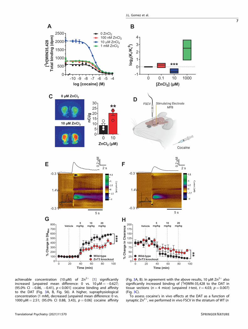
achievable concentration (10 µM) of Zn2+ [1] significantlyincreased [unpaired mean difference: 0 vs. 10 µM=−0.627;(95.0% CI −0.86, −0.41), p < 0.001] cocaine binding and affinityto the DAT (Fig. 3A, B, Fig. S6). A higher, supraphysiologicalconcentration (1 mM), decreased [unpaired mean difference: 0 vs.1000 µM= 2.51; (95.0% CI 0.88, 3.43), p= 0.06] cocaine affinity
(Fig. 3A, B). In agreement with the above results, 10 µM Zn2+ alsosignificantly increased binding of [3H]WIN-35,428 to the DAT intissue sections (n= 4 mice) (unpaired t-test, t= 4.03; p= 0.007)(Fig. 3C).To assess cocaine’s in vivo effects at the DAT as a function of
synaptic Zn2+, we performed in vivo FSCV in the striatum of WT (n
J.L. Gomez et al.
7
Translational Psychiatry (2021) 11:570

= 4) and ZnT3 KO (n= 4) mice. Electrical stimulation of the medialforebrain bundle was used to evoke DA release in the striatumfollowed by escalating cocaine injections (5, 10, 20 mg/kg, IP) (Fig.3D). WT and ZnT3 KO mice did not differ in baseline DA release orclearance (Fig. S7). DA release: compared to vehicle, we found that5, 10, and 20mg/kg cocaine significantly increased electrically-evoked extracellular DA release in WT mice, but ZnT3 KO miceonly showed a significant increase at 20 mg/kg cocaine (Fig. S7).ZnT3 KO mice also showed significantly lower electrically evokedextracellular DA release compared to WT mice (two-way RManova; genotype × time interaction, F(19, 114)= 5.46; p < 0.001)(Fig. 3E, G). DA clearance: compared to Vehicle, 5, 10, and 20mg/kgcocaine significantly decreased DA clearance in WT mice ascompared to Vehicle, but ZnT3 KO mice only showed significantlylower DA clearance at 20 mg/kg cocaine (Fig. S7). ZnT3 KO miceshowed significantly greater DA clearance in response to cocainecompared to WT mice (two-way RM anova; genotype × timeinteraction, F(19, 114)= 2.35 p= 0.0029) (Fig. 3F, H).
Synaptic Zn2+ release potentiates cocaine locomotorsensitization, reward, seeking and is required for cocaine-induced increases in DATWe hypothesized that cocaine-induced synaptic Zn2+ releasewould modulate cocaine-related behavior. To examine this, wefirst tested ZnT3 KO mice for development and expression ofcocaine-induced locomotor sensitization. WT (n= 15) and ZnT3KO mice (n= 16) both developed locomotor sensitization to dailyinjections of cocaine (10 mg/kg/day, IP, 5 days) (two-way RManova with Holm–Sidak multiple comparisons, genotype × sessioninteraction, F(3, 59)= 7.07; p= 0.004) (Fig. 4A). However, ZnT3 KOmice (n= 16) showed significantly lower locomotor activitycompared to cocaine-treated WT mice on Day 1 (t= 3.09; p=0.01) and Day 5 (t= 3.22; p= 0.009) of the procedure. Asexpected, WT mice injected with cocaine showed significantlygreater locomotor activity than vehicle-treated WT (n= 16) miceon Day 1 (t= 5.54; p < 0.001) and Day 5 (t= 8.71; p < 0.001). KOmice injected with cocaine showed significantly greater locomotoractivity than vehicle-treated KO (n= 16) mice on Day 5 (t= 5.68; p< 0.001) but not on Day 1 (t= 2.48; p= 0.08). One week later,vehicle-treated and cocaine-treated mice were tested for expres-sion of cocaine locomotor sensitization via exposure to escalatingcocaine injections (5, 10, and 20mg/kg, IP). WT mice with priorcocaine exposure (cocaine-treated) (n= 9) showed significantlygreater (2-way RM anova with Holm–Sidak multiple comparisons,genotype × time interaction, F(79, 1343)= 1.85; p < 0.001) expres-sion of cocaine locomotor sensitization at 5 (66 min; t= 3.65; p=0.02, 69 min; t= 3.72; p = 0.01) and 10mg/kg cocaine (123 min; t= 5.24; p < 0.001, 126 min; t= 6.03; p < 0.001, 129 min; t= 5.19; p< 0.001, 132 min; t= 3.75; p= 0.01, 135min; t= 3.54; p= 0.03)compared to vehicle-treated WT mice (n= 10) (Fig. 4B). Incontrast, cocaine-treated KO mice (n= 10) did not show asignificant increase in locomotor activity (two-way RM anova,genotype × time interaction, F(79, 1422)= 0.9197; p= 0.67)compared to vehicle-treated KO mice (n= 10) (Fig. 4C). NeitherWT nor ZnT3 KO cocaine-treated mice differed from the
corresponding vehicle-treated mice at a 20mg/kg dose of cocaine(Fig. 4B, C).Cocaine exposure increases DAT levels [47, 48, 62] which is
thought to serve as a compensatory adaptation to DAT blockadeafter RC exposure. We hypothesized that ZnT3 KO mice, whichlack cocaine-induced Zn2+ release and expression of cocaine-induced locomotor sensitization, would be insensitive to cocaine-induced increases in striatal DAT. To test this, mice from the abovesensitization experiments were euthanized 24 h after the lastcocaine injection, the striatum was dissected, and DAT bindingassays were performed using [3H]WIN-35,428 (three repetitionsper curve, in triplicate) (one-way ANOVA: F(3, 8)= 6.23; p= 0.01).Pairwise comparisons (Holm–Sidak) showed that vehicle-treatedWT mice (n= 6) did not differ from vehicle- or cocaine-treatedZnT3 KO mice (n= 6) in [3H]WIN-35,428 binding (Fig. 4D).However, cocaine-treated WT mice (n= 6) showed a trend towardgreater [3H]WIN-35,428 binding (t= 2.21; p= 0.058) compared tovehicle-treated WT mice (n= 6) and significantly greater [3H]WIN-35,428 binding than both vehicle-treated (n= 6) (t= 4.14; p=0.009) and cocaine-treated KO mice (n= 6) (t= 3.12; p= 0.02).Next, we tested whether synaptic Zn2+ release is involved in
cocaine reward by assessing the extent to which ZnT3 KO micedevelop cocaine CPP. As expected, we found that WT miceshowed significant preference for a chamber paired with cocaine(one sample Wilcoxon (non-parametric) test with theoreticalmedian set to 0) at 5 (n= 8) (p= 0.007; 95% CI: 41.3–336.1), 10(n= 9) (p= 0.003; 95% CI: 59.2–297.3), and 20 (n= 8) (p= 0.01;95% CI: −9.8 to 258.4) mg/kg. In contrast, ZnT3 KO mice did notshow significant preference for the chamber paired with cocaineat any dose: 5 mg/kg (n= 7) (p= 0.21; 95% CI: −118.8 to 256.2),10 mg/kg (n= 9) (p= 0.07; 95% CI: −47.2 to 208.5) and 20mg/kg(n= 7) (p= 0.10; 95% CI: −140.6 to 159.3) (Fig. 4E).Finally, we examined whether synaptic Zn2+ is involved in
intravenous cocaine SA. WT mice (n= 9) showed robustacquisition of cocaine SA (1 mg/kg/infusion for 13 days), asevidenced by significantly greater (Mixed effects anova withHolm–Sidak multiple comparisons); genotype × session interac-tion, F(12, 190)= 6.12; p < 0.001) and sustained responding onthe cocaine-reinforced active lever over the non-reinforcedinactive lever on days 11 and 12 (session 11: t= 6.48; p <0.001, session 12: t= 4.25; p= 0.009) (Fig. 4F). In contrast, ZnT3KO mice (n= 9) failed to acquire cocaine SA at the 1 mg/kg/infusion dose (Fig. 4H) as they did not show any significantdifferences (two-way RM anova with Holm–Sidak multiplecomparisons, genotype × session interaction, F(12, 192)= 1.27;p= 0.23) in cocaine-reinforced vs. inactive lever pressing. Allmice were then switched to a lower cocaine dose (0.5 mg/kg/infusion for 7 days). WT mice (n= 7) immediately reached themaximum allowed number (50) of infusions (Fig. 4G) and showedsignificantly greater cocaine-reinforced presses (two-way RManova with Holm–Sidak multiple comparisons, lever main effect,F(1, 12)= 116.7; p < 0.001) compared to inactive lever presses(session 1: t= 12.54; p < 0.001, session 2: t= 5.03; p= 0.004,session 3: t= 7.91; p < 0.001, session 4: t= 9.39; p < 0.001, session5: t= 15.45; p < 0.001, session 6: t= 23.33; p < 0.001, session 7: t= 10.42; p < 0.001). At the 0.5 mg/kg/infusion dose, ZnT3 KO
Fig. 3 Synaptic Zn2+ release increases the in vivo potency of cocaine on striatal DA neurotransmission. A Competition binding curves ofcocaine and ZnCl2 against [
3H]WIN-35,428 in mouse striatal tissue. B 10 µM ZnCl2 significantly increased and 1 mM ZnCl2 decreased affinity ofcocaine in mouse striatum (Ki (±SD)) values in nM; Cocaine: 0 µM Zn, 63 ± 39; 0.1 µM Zn, 77 ± 57; 10 µM Zn, 43 ± 31; 1000 µM Zn, 611 ± 843 andWIN-35,428: 0 µM Zn, 9 ± 1.2; 0.1 µM Zn, 9.2 ± 0.8; 10 µM Zn, 5.1 ± 1.1; 1000 µM Zn, 13.4 ± 1.8). C Autoradiograms at the level of mouse striatumshowing that 10 µM ZnCl2 significantly increased [3H]WIN-35,428 binding. D Experimental design of fast scan cyclic voltammetry (FSCV)experiment. E Representative FSCV color plots from wild-type and F ZnT3 knockout mice showing changes in electrically-evoked dopamine(DA) after a 10mg/kg IP cocaine injection. G FSCV time-course plots showing significantly lower percent change in electrically-evoked DAMaxand H faster DA Clearance rate in ZnT3 knockout compared to wild-type mice as a function of vehicle or escalating IP cocaine injections. *p ≤0.05, **p ≤ 0.01, ***p ≤ 0.001. All data expressed as Mean ± SEM.
J.L. Gomez et al.
8
Translational Psychiatry (2021) 11:570

mice took longer (3 sessions) to learn to discriminate thecocaine-reinforced active lever over the inactive lever and alsotook longer (five sessions) to reach 50 infusions (Fig. 4I).Eventually, ZnT3 KO mice (n = 7) showed significantly greatercocaine-reinforced presses (two-way RM anova with Holm–Sidakmultiple comparisons, lever × time interaction, F(6, 71) = 3.9; p =0.002) compared to inactive lever presses (session 3: t = 4.74; p =0.007, session 4: t = 7.03; p = 0.002, session 5: t = 8.86; p < 0.001,session 6: t = 9.82; p < 0.001, session 7: t = 4.01; p = 0.04). Afteracquisition of cocaine SA, mice were tested at lower doses ofcocaine. WT and KO mice did not differ in the number of cocaineinfusions at these lower doses (Fig. S8). However, compared to
WT mice (n = 6), KO mice (n = 5) showed significantly lowercocaine intake (mixed effects RM analysis with Holm–Sidakmultiple comparisons, genotype × dose interaction, F(4, 44) =2.92) at 1 mg/kg/inf. (t = 3.26; p = 0.001) and at 0.125 mg/kg/inf.(t = 2.03; p = 0.04) doses (Fig. 4J). Mice were then assessed forextinction of cocaine SA, but no genotype differences wereobserved (Fig. 4K). After mice had extinguished their leverresponding for cocaine, they were tested for reinstatement(relapse) responding to a cocaine priming injection (10 mg/kg,IP). WT mice (n = 5) showed reinstatement of cocaine SA andsignificantly greater active lever presses (two-way RM anova withHolm–Sidak multiple comparisons, genotype × dose interaction,
1 50
50001000015000200002500030000
Session
Dis
tanc
e T
rave
led
(cm
/30
min
)WT VEH KO VEHWT COC KO COC
**
***60 120 180 240
0500
100015002000250030003500
Wild-type
Time (min)Di
stan
ce T
rave
led
(cm
/3 m
in)
Vehicle-treatedCocaine-treated
0
*
5mg/kg
10mg/kg
20mg/kg
*
60 120 180 2400
500100015002000250030003500
Knockout
Time (min)
Dist
ance
Tra
vele
d(c
m/3
min
)
Vehicle-treatedCocaine-treated
0
5mg/kg
10mg/kg
20mg/kg
A B C
ED
WT WT KO KO0
50
100
150
[3 H]W
IN35
,428
bin
ding
norm
. to
WT
acut
e
*** Cocaine-treatedVehicle-treated
F G
K L
H
J
I
1 3 5 7 9 11 130
10
20
30
40
50 Knockout
Sessions
Res
pons
es/3
hr
InfusionsInactive
1 3 5 7 9 11 130
10
20
30
40
50 Wild-type
Sessions
Res
pons
es/3
hr
InfusionsInactive *
*
1 3 5 70
10
20
30
40
50
Sessions
*
1 3 5 70
10
20
30
40
50
Sessions
*
M
5 10 20 5 10 20-200-100
0100200300400
mg/kgPr
efer
ence
sco
re(s
ec)
WTKO
** ** *
1 3 5 7 9 11 13 15 17 190
50100150200250300
Session
Leve
r pre
sses
WT ActiveKO ActiveWT Inactive
KO Inactive
0 10 0 100
25
50
75
100
125
Cocaine (mg/kg)
Act
ive
pres
ses
WT
* KO
0 10 0 100
25
50
75
100
125
Cocaine (mg/kg)
Inac
tive
pres
ses KO
WT
0.062
0.125 0.2
5 0.5 10
10
20
30
40
Dose (mg/kg/inf)
Inta
ke (m
g)
WTKO
***
J.L. Gomez et al.
9
Translational Psychiatry (2021) 11:570

F(1, 10) = 5.09; p= 0.04) after cocaine priming (t = 3.17; p =0.005) compared to ZnT3 KO mice (n = 5), which did notreinstate (Fig. 4L, M). WT mice (n = 5) and ZnT3 KO mice (n = 5)did not differ in inactive lever responding (two-way RM anovawith Holm-Sidak multiple comparisons, genotype × doseinteraction, F(1, 10) = 0.17; p = 0.68) during cocaine-primedreinstatement.
Dietary Zn2+ deficiency decreases brain Zn2+ and attenuatescocaine locomotor sensitization, reward, and striatal DATbindingSynaptic Zn2+ levels and ZnT3 expression are modulated bydietary Zn2+ intake [30, 31]. Chronic drug abuse (includingcocaine) in humans is associated with malnutrition and dysregu-lated peripheral Zn2+ levels [33–38]. However, whether dietaryZn2+ availability can modulate the behavioral effects of cocainehas not been previously reported.To examine this, we fed mice, for approximately one month,
diets that were custom-formulated by a commercial vendor tocontain either 30 (considered an adequate level of Zn2+ intake) or5 (a low amount of Zn2+ intake) ppm Zn2+. We first verified theZn2+ content of each diet using TXRF. According to ourmeasurements, the 30 ppm diet contained ~28 ppm Zn2+ andthe 5 ppm diet contained ~8 ppm Zn2+ (Fig. 5A) (we will howeverrefer to them here as 30 and 5 ppm Zn2+ diets to remainconsistent with prior studies that have used similar formulations).Mice fed a 5 ppm Zn2+ diet (n = 32) did not differ in body weightfrom mice fed a 30 ppm Zn2+ diet (n = 32) (Fig. 5B). Mice fed a 5ppm Zn2+ diet (n = 32) showed a significant decrease (two-wayRM anova with Holm-Sidak multiple comparisons; genotype ×time interaction, F(15, 930) = 10.07; p < 0.001) in food intake3 days (t = 11.37; p < 0.001) after diet initiation compared to micefed a 30 ppm diet (n = 32) though soon afterward normalized tothe same levels of intake (Fig. 5C). The low Zn2+ diet was effectivein decreasing brain Zn2+ content as mice fed a 5 ppm Zn2+ diet (n= 7) showed significantly lower Zn2+ levels in frontal Ctx(unpaired t-test; t = 3.15; p = 0.01) compared to mice fed a 30pm Zn2+ diet (n = 5) as assessed via TXRF (Fig. 5D).Next, we performed cocaine locomotor sensitization in 30 ppm
and 5 ppm mice using the same procedures as in the above ZnT3KO experiments. Mice fed 5 ppm or 30 ppm Zn2+ diets did notdiffer in the development of cocaine locomotor sensitization (Fig.5E). 30 ppm diet-fed mice injected with cocaine (n = 14) showedsignificantly greater locomotor activity (two-way RM anova withHolm-Sidak multiple comparisons; genotype × session interaction,F(3, 52) = 23.57; p < 0.001) than 30 ppm diet-fed mice injectedwith vehicle (n = 14) at Day 1 (t = 8.29; p < 0.001) and Day 5 (t =14.27; p < 0.001). Similarly, 5 ppm mice injected with cocaine (n =
14) showed significantly greater locomotor activity compared to 5ppm mice injected with vehicle (n = 14) at Day 1 (t = 6.95; p <0.001) and Day 5 (t = 13.95; p < 0.001). 30 ppm mice injected withcocaine also showed significantly greater locomotor activity atDay 5 than at Day 1 (t = 9.98; p < 0.001) and 5 ppm mice injectedwith cocaine also showed significantly greater locomotor activityat Day 5 than at Day 1 (t = 10.06; p < 0.001) (Fig. 5E). Seven dayslater, mice were examined for expression of cocaine locomotorsensitization. We found that cocaine-treated mice fed the 30 ppmdiet (n = 6) showed significantly greater expression of cocainelocomotor sensitization (two-way RM anova with Holm–Sidakmultiple comparisons; genotype × time interaction, F(79, 790) =3.98; p < 0.001) at 5 (66 min; t = 5.69; p < 0.001, 69 min; t = 4.68; p< 0.001, 72 min; t = 3.76; p = 0.01, 75 min; t = 3.78; p = 0.01,78 min; t = 4.29; p = 0.001, 81 min; t = 3.79; p = 0.01, 84 min; t =4.23; p = 0.002), 10 mg/kg (123 min; t = 5.37; p < 0.001, 126 min; t= 7.42; p < 0.001, 129 min; t = 6.99; p < 0.001, 132 min; t = 6.58; p= 0.01, 135 min; t = 5.08; p = 0.03, 138 min; t = 5.36; p = 0.03,141 min; t = 4.39; p = 0.03, 144 min; t = 4.14; p = 0.03, 147 min; t= 3.78; p = 0.03) and 20mg/kg cocaine (183 min; t = 3.43; p =0.04) compared to vehicle-treated mice fed the same 30 ppm diet(n = 13) (Fig. 5F). In contrast, cocaine-treated mice fed the 5 ppmdiet (n = 6) did not show a significant difference in expression oflocomotor sensitization compared to vehicle-treated mice fed the5 ppm diet (n = 6) (two-way RM anova with Holm-Sidak multiplecomparisons; genotype × time interaction, F(79, 790) = 0.9342; p= 0.64) (Fig. 5G).Next, we tested whether mice fed 30 ppm and 5 ppm Zn2+ diets
differed in cocaine preference using the same CPP procedure as inZnT3 KO mice. Mice fed the 30 ppm Zn2+ diet showed significantpreference (one sample Wilcoxon test with theoretical median setto 0) for the chamber paired with cocaine at 5 mg/kg (n = 8) (p =0.007; 95% CI: 45.8–205.5), 10 mg/kg (n = 8) (p = 0.007; 95% CI:42.6–245.4), and 20 mg/kg (n = 7) (p = 0.003; 95% CI: −6.0 to149.6). Mice fed a 5 ppm Zn2+ diet showed significant preference(one sample Wilcoxon test with theoretical median set to 0) for thechamber paired with cocaine at 10 mg/kg (n = 8) (p = 0.039; 95%CI: −79.8 to 298.0), and 20 mg/kg (n = 8) (p = 0.007; 95% CI:26.2–185.9) but not at 5 mg/kg (n = 8) (p = 0.10; 95% CI: −30.1 to125.7) (Fig. 5H).Finally, we examined whether dietary Zn2+ availability had any
effects on DAT binding in the striatum. Mice fed 30 ppm and 5ppm Zn2+ diets and exposed to escalating cocaine injections (5,10, and 20mg/kg, IP, one injection/h) were euthanized 24 h afterthe last injection and their brains were assessed for striatal [3H]WIN-35,428 binding. We found that mice exposed to cocaine anda 5 ppm Zn2+ diet (n = 6) showed significantly lower striatal DATbinding (Fig. 5I) (unpaired t-test; t= 9.63; p < 0.001) (3 repetitions
Fig. 4 Synaptic Zn2+ release potentiates cocaine locomotor sensitization, reward, seeking and is required for cocaine-induced increasesin DAT. A ZnT3 knockout (KO) mice injected with 10 mg/kg cocaine (COC) showed significantly lower locomotor activity compared tococaine-injected wild-type (WT) mice. Compared to vehicle, cocaine significantly increased locomotor activity in both WT and KO mice on Day5 but only in WT mice on Day 1. B Cocaine-treated WT mice showed significantly greater expression of cocaine locomotor sensitization at 5and 10 mg/kg cocaine compared to vehicle-treated WT mice. C Cocaine-treated ZnT3 KO mice do not differ in expression of cocainelocomotor sensitization compared to vehicle-treated KO mice. D Cocaine-treated WTmice had significantly greater DAT binding than cocaine-treated ZnT3 KO mice and vehicle-treated KO mice and showed a trend toward significantly greater DAT binding than vehicle-treated WTmice. E WT mice showed significant preference for a chamber paired with cocaine at 5, 10, and 20 mg/kg. ZnT3 KO mice did now showpreference for a chamber paired with cocaine at any dose. F Wild-type mice exposed to a training dose of cocaine (1 mg/kg/inf.) showedsignificantly greater cocaine-reinforced presses compared to inactive lever presses. G Once trained, WT mice were exposed to a lower cocainedose (0.5 mg/kg/inf.), immediately reached the maximum number of presses allowed per session, and showed significantly greater cocaine-reinforced presses compared to inactive lever presses on all sessions. H ZnT3 knockout mice exposed to a training dose of cocaine (1 mg/kg/inf.) did not show any significant differences between cocaine-reinforced and inactive lever pressing. I ZnT3 knockout mice exposed to a lowercocaine dose (0.5 mg/kg/inf.) showed significantly greater cocaine-reinforced presses compared to inactive lever presses during the last fivesessions. J ZnT3 KO mice showed significantly lower cocaine intake at 1 mg/kg/inf. and at 0.125mg/kg/inf. compared to WT mice. K WT andZnT3 KO mice did not differ in extinction of cocaine self-administration. L After extinction of cocaine self-administration behavior, WT miceshowed reinstatement of cocaine self-administration and significantly greater active lever presses after cocaine priming compared to ZnT3 KOmice. M WT and ZnT3 KO mice did not differ in inactive lever responding during cocaine-primed reinstatement. *p ≤ 0.05. All data exceptE expressed as Mean ± SEM. Panel E is expressed as Median ± 95% CI.
J.L. Gomez et al.
10
Translational Psychiatry (2021) 11:570
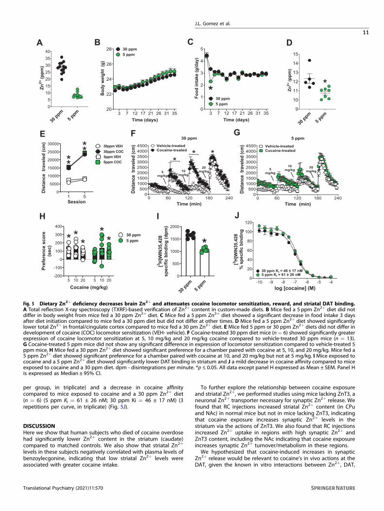
per group, in triplicate) and a decrease in cocaine affinitycompared to mice exposed to cocaine and a 30 ppm Zn2+ diet(n = 6) (5 ppm Ki = 61 ± 26 nM; 30 ppm Ki = 46 ± 17 nM) (3repetitions per curve, in triplicate) (Fig. 5J).
DISCUSSIONHere we show that human subjects who died of cocaine overdosehad significantly lower Zn2+ content in the striatum (caudate)compared to matched controls. We also show that striatal Zn2+
levels in these subjects negatively correlated with plasma levels ofbenzoylecgonine, indicating that low striatal Zn2+ levels wereassociated with greater cocaine intake.
To further explore the relationship between cocaine exposureand striatal Zn2+, we performed studies using mice lacking ZnT3, aneuronal Zn2+ transporter necessary for synaptic Zn2+ release. Wefound that RC injections increased striatal Zn2+ content (in CPuand NAc) in normal mice but not in mice lacking ZnT3, indicatingthat cocaine exposure increases synaptic Zn2+ levels in thestriatum via the actions of ZnT3. We also found that RC injectionsincreased Zn2+ uptake in regions with high synaptic Zn2+ andZnT3 content, including the NAc indicating that cocaine exposureincreases synaptic Zn2+ turnover/metabolism in these regions.We hypothesized that cocaine-induced increases in synaptic
Zn2+ release would be relevant to cocaine’s in vivo actions at theDAT, given the known in vitro interactions between Zn2+, DAT,
H I J
1 50
5000
10000
15000
20000
25000
30000
Session
Dis
tanc
e tr
avel
ed (c
m)
30ppm VEH
5ppm VEH30ppm COC
5ppm COC***
*
60 120 180 2400
50010001500200025003000350040004500
30 ppm
Time (min)
Dist
ance
tra
vele
d (c
m) Vehicle-treated
Cocaine-treated
0
** *
5mg/kg
10mg/kg
20mg/kg
60 120 180 2400
50010001500200025003000350040004500
5 ppm
Time (min)
Dist
ance
tra
vele
d (c
m) Vehicle-treated
Cocaine-treated
0
5mg/kg
10mg/kg
20mg/kg
30 ppm
5 ppm
0
500
1000
1500
2000
[3 H]W
IN35
,428
spec
ific
bind
ing
(dpm
)
*
F
A B
E
C
G
3 7 12 17 21 26 31 3520
22
24
26
28
Time (days)
Body
wei
ght
(g)
30 ppm5 ppm
3 7 12 17 21 26 31 350
1
2
3
4
5
Time (days)
Food
inta
ke (g
/day
)
30 ppm5 ppm
*
30 ppm
5 ppm
05
10152025303540
Zn2+
(ppm
)
30 ppm
5 ppm
9
10
11
12
13
14
15
Zn2+
(ppm
)
*
D
-10 -9 -8 -7 -6 -5 -40
20
40
60
80
100
120
30 ppm Ki = 46 ± 17 nM5 ppm Ki = 61 ± 26 nM
log [cocaine] (M)
[3 H]W
IN35
,428
% s
peci
fic b
indi
ng
5 10 20 5 10 20-200
-100
0
100
200
300
400
Cocaine (mg/kg)
Pref
eren
ce s
core
(sec
)
30 ppm5 ppm* *
**
*
Fig. 5 Dietary Zn2+ deficiency decreases brain Zn2+ and attenuates cocaine locomotor sensitization, reward, and striatal DAT binding.A Total reflection X-ray spectroscopy (TXRF)-based verification of Zn2+ content in custom-made diets. B Mice fed a 5 ppm Zn2+ diet did notdiffer in body weight from mice fed a 30 ppm Zn2+ diet. C Mice fed a 5 ppm Zn2+ diet showed a significant decrease in food intake 3 daysafter diet initiation compared to mice fed a 30 ppm diet but did not differ at other times. D Mice fed a 5 ppm Zn2+ diet showed significantlylower total Zn2+ in frontal/cingulate cortex compared to mice fed a 30 pm Zn2+ diet. E Mice fed 5 ppm or 30 ppm Zn2+ diets did not differ indevelopment of cocaine (COC) locomotor sensitization (VEH- vehicle). F Cocaine-treated 30 ppm diet mice (n = 6) showed significantly greaterexpression of cocaine locomotor sensitization at 5, 10 mg/kg and 20 mg/kg cocaine compared to vehicle-treated 30 ppm mice (n = 13).G Cocaine-treated 5 ppm mice did not show any significant difference in expression of locomotor sensitization compared to vehicle-treated 5ppm mice. H Mice fed a 30 ppm Zn2+ diet showed significant preference for a chamber paired with cocaine at 5, 10, and 20 mg/kg. Mice fed a5 ppm Zn2+ diet showed significant preference for a chamber paired with cocaine at 10, and 20 mg/kg but not at 5 mg/kg. I Mice exposed tococaine and a 5 ppm Zn2+ diet showed significantly lower DAT binding in striatum and J a mild decrease in cocaine affinity compared to miceexposed to cocaine and a 30 ppm diet. dpm - disintegrations per minute. *p ≤ 0.05. All data except panel H expressed as Mean ± SEM. Panel His expressed as Median ± 95% CI.
J.L. Gomez et al.
11
Translational Psychiatry (2021) 11:570

and cocaine. Using in vitro assays, we first confirmed that aphysiologically-relevant concentration of Zn2+ increased theability of cocaine to bind to DAT. Then, to extend the relevanceof this finding to an in vivo context, we used in vivo FSCV andfound that deletion of ZnT3, and hence, loss of synaptic Zn2+
release, attenuated cocaine’s effects on striatal DA neurotransmis-sion. Consistent with this observation, mice lacking ZnT3 wereinsensitive to cocaine-induced increases in striatal DAT bindingand showed attenuated behavioral responses to cocaine in severalprocedures such as locomotor sensitization, CPP, and IV SA,indicating that synaptic Zn2+ promotes cocaine sensitization,reward, and cocaine-seeking behavior.Zn2+ is obtained almost exclusively from the diet. Taking our
above findings into account, we reasoned that low environmentalavailability of Zn2+ and specifically, reduced dietary Zn2+ intake,would attenuate behavioral responses to cocaine. As predicted,we found that mice exposed to a diet with low Zn2+ contentshowed lower brain Zn2+ levels, decreased cocaine-inducedincreases in striatal DAT binding, and attenuated behavioralresponses to cocaine compared to mice fed a diet with adequateZn2+ content. This indicates that low dietary Zn2+ intakedecreases brain Zn2+ levels, and attenuates cocaine locomotorsensitization, cocaine preference, and cocaine-induced increasesin striatal DAT.Taken together, our above findings suggest that synaptic Zn2+
release in the striatum plays a critical role in cocaine’s effects onstriatal DA neurotransmission and consequently in the neurobio-logical and behavioral adaptations associated with cocaineexposure. A summary depicting this proposed mechanism isshown in Fig. 6.The results from our experiments in mice suggest that the Zn2+
deficits we identified in humans who died from cocaine overdosemay arise from a combination of inadequate nutrition (i.e. lowdietary Zn2+ intake) and increased Zn2+ turnover/metabolismbrought upon by chronic cocaine use, although we cannot rule outindividual or combined contributions of diet, cocaine use, or use of
other drugs/substances in these effects. The negative correlationbetween striatal Zn2+ content and plasma benzoylecgonine levelsin postmortem human samples, taken together with the rest of ourdata, may reflect the notion that individuals with inherently lowstriatal Zn2+ levels would be less sensitive to the effects of cocaineand therefore would need to compensate by consuming more ofthe drug. This would implicate Zn2+ as an environmental factor thatcould influence vulnerability to the effects of cocaine andpotentially to the development of cocaine use disorders oraddiction.Our results have important implications for both general DA-
dependent behaviors and especially for the prevention andtreatment of cocaine addiction. Specifically, our findings suggestthat dietary Zn2+ intake, and potentially, impaired Zn2+ absorp-tion or excretion mechanisms, are implicated in cocaine reward,seeking, and relapse. Consequently, we suggest that the Zn2+
status of patients with cocaine addiction should be taken intoconsideration, especially since Zn2+ deficiency varies in preva-lence across social demographics and is found in higherproportion in developing countries [32].Our findings also expand the current understanding of cocaine’s
neurobiological effects. DA and glutamate systems converge at thelevel of the striatum to modulate behaviors that influence cocaineseeking and abuse [46, 63]. Synaptic Zn2+, mediated by ZnT3, ispackaged along with glutamate in the same synaptic vesicles and isreleased by glutamatergic terminals in the striatum upon neuronalactivation. As such, the cocaine-induced synaptic Zn2+ increases wereport here would be expected to accompany the well-describedincrease in synaptic glutamate neurotransmission in the striatumfollowing RC exposure [45, 46, 64, 65]. Indeed, cocaine-inducedincreases in synaptic glutamate are thought to result, in part, fromreductions in extrasynaptic glutamate arising from decreasedcystine-glutamate exchange in response to RC use [63, 66].Specifically, cocaine-induced reductions in extrasynaptic glutamateweaken signaling at presynaptic metabotropic glutamate receptors,thereby increasing synaptic glutamate levels [64] and resulting in
Fig. 6 Schematic representation of the putative mechanism by which presynaptic Zn2+ modulates the effects of cocaine on dopamineand glutamate neurotransmission in the striatum in wild-type (WT) and ZnT3 knockout mice. The image on the left shows the effect ofsynaptic Zn2+ on increasing cocaine’s efficacy by binding to the DAT on dopaminergic neurons. It also acknowledges the putative effects ofZn2+ on glutamate neurotransmission in the context of cocaine via its interaction with AMPA and NMDA receptors. The image on the rightillustrates the effect of ZnT3 deletion, and consequently the loss of presynaptic Zn2+ release, in attenuating the effects of cocaine on striataldopamine and glutamate neurotransmission.
J.L. Gomez et al.
12
Translational Psychiatry (2021) 11:570

excitatory synaptic strengthening. This mechanism may underliethe increases in cocaine-induced synaptic Zn2+ that we observe.Zn2+ binds to NMDA and AMPA receptors [67, 68] and it is likely
that, in addition to affecting DA neurotransmission via binding toDAT, cocaine-induced increases in synaptic Zn2+ release may alsoexert direct allosteric interactions at ionotropic glutamatereceptors to influence cocaine-dependent glutamate neurotrans-mission, synaptic plasticity, and behaviors such as cocainelocomotor sensitization and cocaine priming-induced reinstate-ment of cocaine seeking [45, 46, 65, 69]. However, the precisemechanism for how cocaine-induced Zn2+ release would lead tosuch effects is complicated by the fact that Zn2+ can exertbidirectional effects at AMPA receptors [70], it can inhibit bothsynaptic and extrasynaptic NMDA receptors [68], and finally, thatcocaine induces projection- and cell type-specific adaptations inthe NAc [71], that not all NAc glutamatergic synapses release Zn2+,
and that the specific zincergic synapses, and the postsynaptic cellsthey target in regions such as the NAc have not yet been defined.For all these reasons, the specific effects of cocaine-induced Zn2+
release on glutamatergic signaling are unclear and require furtherinvestigation.Another possibility may be that Zn2+ interacts with cystine-
glutamate exchange mechanisms directly to produce changes incocaine behaviors. Indeed, we found here that ZnT3 KO micefailed to reinstate cocaine-primed SA of cocaine, suggesting thatsynaptic Zn2+ release is necessary for cocaine-primed seeking.N-acetylcysteine (NAC) is a derivative of the amino acid cysteineand has been proposed as a treatment for substance use disorders[72]. NAC prevents cocaine-primed reinstatement of drug seekingafter cocaine SA by stimulating cystine-glutamate exchange [63].Importantly, NAC also has metal-chelating capabilities (Zn2+ bindswith high affinity to cysteines [73]) and systemic administration ofNAC in mice has been shown to decrease peripheral Zn2+ levels[74]. Accordingly, it may be possible that in addition to the effectsof NAC in decreasing cocaine-primed seeking via activation ofcystine-glutamate exchange, its effects may also be driven, in part,via direct interaction with synaptic Zn2+. In this case, NAC wouldeffectively act as a Zn2+ chelating agent, decreasing synaptic Zn2+
levels, which would attenuate cocaine’s effects at the DAT anddecrease cocaine-seeking. However, this could also occur con-comitantly to NAC’s activation of cystine-glutamate exchange,which would increase extrasynaptic glutamate to restore pre-synaptic metabotropic glutamate receptor tone and therebyreduce synaptic glutamate and potentially, co-released Zn2+.In addition to cocaine, other psychostimulants interact with the
DAT to produce their effects of behavior. We propose that thecocaine-dependent changes in Zn2+ that we describe here are notspecific to cocaine but may also be elicited by other psychosti-mulants that modulate DAT function and increase corticostriatalglutamate neurotransmission [45, 46, 75], and potentially otherdrugs of abuse, including alcohol [76] which is strongly associatedwith Zn2+ deficiency [76].Finally, in addition to its critical role in addiction, the DAT is the
primary molecular target for stimulant medications used in thetreatment of attention-deficit hyperactivity disorder (ADHD).Studies indicate that medication response is reduced in Zn2+-deficient ADHD patients [77]. Our findings here offer a mechanisticexplanation for these clinical observations and suggest that Zn2+
supplementation may improve the efficacy of stimulant-basedADHD medications.In conclusion, our findings expand current knowledge regard-
ing the direct pharmacological mechanism of action of cocaineand the neurobiological mechanisms involved in cocaine’sbehavioral effects by identifying a critical role for the traceelement Zn2+ as an environmentally derived modulator of cocaineaffinity, potency, plasticity, reward, and seeking.
DATA AVAILABILITYAll data are available within the article or its supplementary material.
REFERENCES1. Frederickson CJ, Suh SW, Silva D, Thompson RB. Importance of zinc in the central
nervous system: the zinc-containing neuron. J Nutr. 2000;130:1471s–1483s.2. Gower-Winter SD, Levenson CW. Zinc in the central nervous system: from
molecules to behavior. Biofactors. 2012;38:186–93.3. Pfeiffer CC, Braverman ER. Zinc, the brain and behavior. Biol Psychiatry.
1982;17:513–32.4. Sensi SL, Paoletti P, Koh JY, Aizenman E, Bush AI, Hershfinkel M. The neurophy-
siology and pathology of brain zinc. J Neurosci. 2011;31:16076–85.5. Cuajungco MP, Lees GJ. Zinc metabolism in the brain: relevance to human
neurodegenerative disorders. Neurobiol Dis. 1997;4:137–69.6. McAllister BB, Dyck RH. Zinc transporter 3 (ZnT3) and vesicular zinc in central
nervous system function. Neurosci Biobehav Rev. 2017;80:329–50.7. Pe´rez-Clausell J, Danscher G. Intravesicular localization of zinc in rat tele-
ncephalic boutons. A histochemical study. Brain Res. 1985;337:91–98.8. Brown CE, Dyck RH. Distribution of zincergic neurons in the mouse forebrain. J
Comp Neurol. 2004;479:156–67.9. Palmiter RD, Cole TB, Quaife CJ, Findley SD. ZnT-3, a putative transporter of zinc
into synaptic vesicles. Proc Natl Acad Sci USA. 1996;93:14934–9.10. Cole TB, Wenzel HJ, Kafer KE, Schwartzkroin PA, Palmiter RD. Elimination of zinc
from synaptic vesicles in the intact mouse brain by disruption of the ZnT3 gene.Proc Natl Acad Sci USA. 1999;96:1716–21.
11. Thackray SE, McAllister BB, Dyck RH. Behavioral characterization of female zinctransporter 3 (ZnT3) knockout mice. Behav Brain Res. 2017;321:36–49.
12. Cole TB, Martyanova A, Palmiter RD. Removing zinc from synaptic vesicles doesnot impair spatial learning, memory, or sensorimotor functions in the mouse.Brain Res. 2001;891:253–65.
13. Martel G, Hevi C, Friebely O, Baybutt T, Shumyatsky GP. Zinc transporter 3 isinvolved in learned fear and extinction, but not in innate fear. Learn Mem.2010;17:582–90.
14. Kumar M, Xiong S, Tzounopoulos T, Anderson CT. Fine control of sound fre-quency tuning and frequency discrimination acuity by synaptic zinc signaling inmouse auditory cortex. J Neurosci. 2019;39:854–65.
15. Patrick WuHP, Dyck RH. Signaling by synaptic zinc is required for whisker-mediated, fine texture discrimination. Neuroscience. 2018;369:242–7.
16. Frederickson CJ, Giblin LJ, Krężel A, McAdoo DJ, Muelle RN, Zeng Y, et al.Concentrations of extracellular free zinc (pZn)e in the central nervous systemduring simple anesthetization, ischemia and reperfusion. Exp Neurol.2006;198:285–93.
17. Frederickson CJ, Giblin LJ, Rengarajan B, Masalha R, Frederickson CJ, Zeng Y, et al.Synaptic release of zinc from brain slices: factors governing release, imaging, andaccurate calculation of concentration. J Neurosci Methods. 2006;154:19–29.
18. Wu Q, Coffey LL, Reith ME. Cations affect [3H]mazindol and [3H]WIN 35,428binding to the human dopamine transporter in a similar fashion. J Neurochem.1997;69:1106–18.
19. Scholze P, Norregaard L, Singer EA, Freissmuth M, Gether U, Sitte HH. The role ofzinc ions in reverse transport mediated by monoamine transporters. J Biol Chem.2002;277:21505–13.
20. Liang YJ, Zhen J, Chen N, Reith ME. Interaction of catechol and non-catecholsubstrates with externally or internally facing dopamine transporters. J Neu-rochem. 2009;109:981–94.
21. Hong WC, Amara SG. Membrane cholesterol modulates the outward facingconformation of the dopamine transporter and alters cocaine binding. J BiolChem. 2010;285:32616–26.
22. Norregaard L, Frederiksen D, Nielsen EO, Gether U. Delineation of an endogenouszinc-binding site in the human dopamine transporter. Embo J. 1998;17:4266–73.
23. Kuhar MJ, Ritz MC, Boja JW. The dopamine hypothesis of the reinforcing prop-erties of cocaine. Trends Neurosci. 1991;14:299–302.
24. Volkow ND, Wang GJ, Fischman MW, Foltin RW, Fowler JS, Abumrad NN, et al.Relationship between subjective effects of cocaine and dopamine transporteroccupancy. Nature. 1997;386:827–30.
25. Wise RA, Bozarth MA. A psychomotor stimulant theory of addiction. Psychol Rev.1987;94:469–92.
26. Mengual E, Casanovas-Aguilar C, Pérez-Clausell J, Giménez-Amaya JM. Hetero-geneous and compartmental distribution of zinc in the striatum and globuspallidus of the rat. Neuroscience. 1995;66:523–37.
27. Frederickson CJ, Rampy BA, Reamy-Rampy S, Howell GA. Distribution of histo-chemically reactive zinc in the forebrain of the rat. J Chem Neuroanat.1992;5:521–30.
J.L. Gomez et al.
13
Translational Psychiatry (2021) 11:570

28. Ciliax BJ, Drash GW, Staley JK, Haber S, Mobley CJ, Miller GW, et al. Immunocy-tochemical localization of the dopamine transporter in human brain. J CompNeurol. 1999;409:38–56.
29. Sikora J, Kieffer BL, Paoletti P, Ouagazzal AM. Synaptic zinc contributes to motorand cognitive deficits in 6-hydroxydopamine mouse models of Parkinson’s dis-ease. Neurobiol Dis. 2020;134:104681.
30. Grabrucker S, Jannetti L, Eckert M, Gaub S, Chhabra R, Pfaender S, et al. Zincdeficiency dysregulates the synaptic ProSAP/Shank scaffold and might contributeto autism spectrum disorders. Brain. 2013;137:137–52.
31. Takeda A, Hirate M, Tamano H, Nisibaba D, Oku N. Susceptibility to kainate-induced seizures under dietary zinc deficiency. J Neurochem. 2003;85:1575–80.
32. Prasad AS. Discovery of human zinc deficiency: its impact on human health anddisease. Adv Nutr. 2013;4:176–90.
33. Santolaria-Fernandez FJ, Gomez-Sirvent JL, Gonzalez-Reimers CE, Batista-LopezJN, Jorge-Hernandez JA, Rodriguez-Moreno F, et al. Nutritional assessment ofdrug addicts. Drug Alcohol Depend. 1995;38:11–18.
34. Comai S, Bertazzo A, Vachon J, Daigle M, Toupin J, Cote G, et al. Trace elementsamong a sample of prisoners with mental and personality disorders andaggression: correlation with impulsivity and ADHD indices. J Trace Elem Med Biol.2019;51:123–9.
35. Sadlik J, Pach J, Winnik L, Piekoszewski W. [Concentration of zinc, copper andmagnesium in the serum of drug addicts]. Przegl Lek. 2000;57:563–4.
36. Cheng FL, Wang H, Wu J, Ning MX, Hu LF, Su YL. [Determination and correlationanalysis of trace elements in hair of dependence drug addicts]. Guang Pu Xue YuGuang Pu Fen Xi. 2005;25:116–8.
37. Ruiz Martinez M, Gil Extremera B, Maldonado Martin A, Cantero-Hinojosa J,Moreno-Abadia V. Trace elements in drug addicts. Klin Wochenschr.1990;68:507–11.
38. Hossain KJ, Kamal MM, Ahsan M, Islam SN. Serum antioxidant micromineral (Cu,Zn, Fe) status of drug dependent subjects: influence of illicit drugs and lifestyle.Subst Abus Treat Prev Policy. 2007;2:12. p
39. Bannon MJ, Johnson MM, Michelhaugh SK, Hartley ZJ, Halter SD, David JA, et al. Amolecular profile of cocaine abuse includes the differential expression of genesthat regulate transcription, chromatin, and dopamine cell phenotype. Neu-ropsychopharmacology. 2014;39:2191–9.
40. Zhou Y, Michelhaugh SK, Schmidt CJ, Liu JS, Bannon MJ, Lin Z. Ventral midbraincorrelation between genetic variation and expression of the dopamine trans-porter gene in cocaine-abusing versus non-abusing subjects. Addict Biol.2014;19:122–31.
41. Stan AD, Ghose S, Gao XM, Roberts RC, Lewis-Amezcua K, Hatanpaa KJ, et al.Human postmortem tissue: what quality markers matter? Brain Res.2006;1123:1–11.
42. Michaelides M, Pascau J, Gispert JD, Delis F, Grandy DK, Wang GJ, et al. DopamineD4 receptors modulate brain metabolic activity in the prefrontal cortex andcerebellum at rest and in response to methylphenidate. Eur J Neurosci.2010;32:668–76.
43. Keighron JD, Quarterman JC, Cao J, DeMarco EM, Coggiano MA, Gleaves A, et al.Effects of (R)-modafinil and modafinil analogues on dopamine dynamics assessedby voltammetry and microdialysis in the mouse nucleus accumbens shell. ACSChem Neurosci. 2019;10:2012–21.
44. Ho J, Tumkaya T, Aryal S, Choi H, Claridge-Chang A. Moving beyond P values: dataanalysis with estimation graphics. Nat Methods. 2019;16:565–6.
45. Scofield MD, Heinsbroek JA, Gipson CD, Kupchik YM, Spencer S, Smith AC, et al.The nucleus accumbens: mechanisms of addiction across drug classes reflect theimportance of glutamate homeostasis. Pharm Rev. 2016;68:816–71.
46. Schmidt HD, Pierce RC. Cocaine-induced neuroadaptations in glutamate trans-mission: Potential therapeutic targets for craving and addiction. Ann N Y AcadSci. 2010;1187:35–75.
47. Proebstl L, Kamp F, Manz K, Krause D, Adorjan K, Pogarell O, et al. Effects ofstimulant drug use on the dopaminergic system: a systematic review and meta-analysis of in vivo neuroimaging studies. Eur Psychiatry. 2019;59:15–24.
48. Little KY, Zhang L, Desmond T, Frey KA, Dalack GW, Cassin BJ. Striatal dopami-nergic abnormalities in human cocaine users. Am J Psychiatry. 1999;156:238–45.
49. Jufer RA, Wstadik A, Walsh SL, Levine BS, Cone EJ. Elimination of cocaine andmetabolites in plasma, saliva, and urine following repeated oral administration tohuman volunteers. J Anal Toxicol. 2000;24:467–77.
50. Qin Z, Caruso JA, Lai B, Matusch A, Becker JS. Trace metal imaging with highspatial resolution: applications in biomedicine. Metallomics. 2011;3:28–37.
51. Linkous DH, Flinn JM, Koh JY, Lanzirotti A, Bertsch PM, Jones BF, et al. Evidencethat the ZNT3 protein controls the total amount of elemental zinc in synapticvesicles. J Histochem Cytochem. 2008;56:3–6. pp
52. Danscher G, Stoltenberg M. Zinc-specific autometallographic in vivo seleniummethods: tracing of zinc-enriched (ZEN) terminals, ZEN pathways, and pools ofzinc ions in a multitude of other ZEN cells. J Histochem Cytochem.2005;53:141–53.
53. Sorensen JC, Slomianka L, Christensen J, Zimmer J. Zinc-containing telencephalicconnections to the rat striatum: a combined Fluoro-Gold tracing and histo-chemical study. Exp Brain Res. 1995;105:370–82.
54. Takeda A, Sawashita J, Okada S. Biological half-lives of zinc and manganese in ratbrain. Brain Res. 1995;695:53–58.
55. Pullen RGL, Franklin PA, Hall GH. 65Zn uptake from blood into brain in the rat. JNeurochemistry. 1991;56:485–9.
56. Takeda A, Hirate M, Oku N. Elimination of zinc-65 from the brain under kainate-induced seizures. Biometals. 2004;17:141–4.
57. Takeda A, Kodama Y, Ohnuma M, Okada S. Zinc transport from the striatum andsubstantia nigra. Brain Res Bull. 1998;47:103–6.
58. Takeda A, Minami A, Takefuta S, Tochigi M, Oku N. Zinc homeostasis in the brainof adult rats fed zinc-deficient diet. J Neurosci Res. 2001;63:447–52.
59. Wang F, Dufner-Beattie J, Kim BE, Petris MJ, Andrews G, Eide DJ. Zinc-stimulatedendocytosis controls activity of the mouse ZIP1 and ZIP3 zinc uptake transpor-ters. J Biol Chem. 2004;279:24631–9.
60. Bjorklund NL, Volz TJ, Schenk JO. Differential effects of Zn2+ on the kinetics andcocaine inhibition of dopamine transport by the human and rat dopaminetransporters. Eur J Pharmacol. 2007;565:17–25.
61. Pifl C, Wolf A, Rebernik P, Reither H, Berger ML. Zinc regulates the dopaminetransporter in a membrane potential and chloride dependent manner. Neuro-pharmacology. 2009;56:531–40.
62. Malison RT, Best SE, van Dyck CH, McCance EF, Wallace EA, Laruelle M, et al.Elevated striatal dopamine transporters during acute cocaine abstinence asmeasured by [123I] beta-CIT SPECT. Am J Psychiatry. 1998;155:832–4.
63. Baker DA, McFarland K, Lake RW, Shen H, Tang XC, Toda S, et al. Neuroadapta-tions in cystine-glutamate exchange underlie cocaine relapse. Nat Neurosci.2003;6:743–9.
64. Moran MM, McFarland K, Melendez RI, Kalivas PW, Seamans JK. Cystine/gluta-mate exchange regulates metabotropic glutamate receptor presynaptic inhibi-tion of excitatory transmission and vulnerability to cocaine seeking. J Neurosci.2005;25:6389–93.
65. Wolf ME. The Bermuda Triangle of cocaine-induced neuroadaptations. TrendsNeurosci. 2010;33:391–8.
66. Baker DA, Xi Z-X, Shen H, Swanson CJ, Kalivas PW. The origin and neuronalfunction of <em>in vivo</em> nonsynaptic glutamate. J Neurosci.2002;22:9134–41.
67. Kalappa BI, Anderson CT, Goldberg JM, Lippard SJ, Tzounopoulos T. AMPAreceptor inhibition by synaptically released zinc. Proc Natl Acad Sci USA.2015;112:15749–54.
68. Anderson CT, Radford RJ, Zastrow ML, Zhang DY, Apfel UP, Lippard SJ, et al.Modulation of extrasynaptic NMDA receptors by synaptic and tonic zinc. ProcNatl Acad Sci USA. 2015;112:E2705–2714.
69. Fouyssac M, Belin D. Beyond drug-induced alteration of glutamate homeostasis,astrocytes may contribute to dopamine-dependent intrastriatal functional shiftsthat underlie the development of drug addiction: a working hypothesis. Eur JNeurosci. 2019;50:3014–27.
70. Blakemore LJ, Trombley PQ. Mechanisms of zinc modulation of olfactory bulbAMPA receptors. Neuroscience. 2019;410:160–75.
71. Zinsmaier AK, Dong Y, Huang YH. Cocaine-induced projection-specific and celltype-specific adaptations in the nucleus accumbens. Mol Psychiatry. 2021. Inpress.
72. Tomko RL, Jones JL, Gilmore AK, Brady KT, Back SE, Gray KM. N-acetylcysteine: apotential treatment for substance use disorders. Curr Psychiatr. 2018;17:30–55.
73. Pace NJ, Weerapana E. Zinc-binding cysteines: diverse functions and structuralmotifs. Biomolecules. 2014;4:419–34.
74. Wolfram T, Schwarz M, Reuß M, Lossow K, Ost M, Klaus S, et al. N-Acetylcysteineas modulator of the essential trace elements copper and zinc. Antioxidants.2020;9:1117.
75. Bonaventura J, Quiroz C, Cai NS, Rubinstein M, Tanda G, Ferre S. Key role of thedopamine D4 receptor in the modulation of corticostriatal glutamatergic neu-rotransmission. Sci Adv. 2017;3:e1601631.
76. Skalny AV, Skalnaya MG, Grabeklis AR, Skalnaya AA, Tinkov AA. Zinc deficiency asa mediator of toxic effects of alcohol abuse. Eur J Nutr. 2018;57:2313–22.
77. Lepping P, Huber M. Role of zinc in the pathogenesis of attention-deficithyperactivity disorder: implications for research and treatment. CNS Drugs.2010;24:721–8.
ACKNOWLEDGEMENTSThe authors thank Dr. Thanos Tzounopoulos (University of Pittsburgh) for sharingZnT3 knockout mice, Dr. Richard Dyck (University of Calgary) for advice regardingZn2+ staining, Dr. Yavin Shaham (NIDA) for experimental advice in behavioral
J.L. Gomez et al.
14
Translational Psychiatry (2021) 11:570

experimentation and for manuscript comments, and Dr. Marisela Morales (NIDA), Dr.Antonio Lanzirotti, Dr. Keith Jones and William Rao at the National Synchrotron LightSource (NSLS) X26a Beamline at Brookhaven National Laboratory for instrumentationaccess.
AUTHOR CONTRIBUTIONSJLG, JB, SL, DLM, KMW, MLC, LAR, RJE, CJ, GB, JK, MP, MB, ZX, GT, OS, and MMdesigned and/or performed experiments and/or analyzed data. ZX, GT, and MMsupervised work. JLG, JB, and MM wrote the paper with input from all coauthors.
FUNDINGThis work was supported by the NIDA Intramural Research Program (DA000069), theNIDA Medication Development Program (DA000611) and the Department of Energy(DOE) GeoSciences grant DE-FG02-92ER14244. Open Access funding provided by theNational Institutes of Health (NIH).
COMPETING INTERESTSThe authors declare no competing interests.
ADDITIONAL INFORMATIONSupplementary information The online version contains supplementary materialavailable at https://doi.org/10.1038/s41398-021-01693-0.
Correspondence and requests for materials should be addressed to MichaelMichaelides.
Reprints and permission information is available at http://www.nature.com/reprints
Publisher’s note Springer Nature remains neutral with regard to jurisdictional claimsin published maps and institutional affiliations.
Open Access This article is licensed under a Creative CommonsAttribution 4.0 International License, which permits use, sharing,
adaptation, distribution and reproduction in anymedium or format, as long as you giveappropriate credit to the original author(s) and the source, provide a link to the CreativeCommons license, and indicate if changes were made. The images or other third partymaterial in this article are included in the article’s Creative Commons license, unlessindicated otherwise in a credit line to the material. If material is not included in thearticle’s Creative Commons license and your intended use is not permitted by statutoryregulation or exceeds the permitted use, you will need to obtain permission directlyfrom the copyright holder. To view a copy of this license, visit http://creativecommons.org/licenses/by/4.0/.
This is a U.S. government work and not under copyright protection in the U.S.; foreigncopyright protection may apply 2021
J.L. Gomez et al.
15
Translational Psychiatry (2021) 11:570