Convergence and Segregation of Ventral Striatal Inputs and Outputs
Upload
khangminh22Category
view
2download
0
Dual role of CDK5 on cognitive deficits and striatal vulnerability in Huntington’s disease
Elena Alvarez Periel
Aquesta tesi doctoral està subjecta a la llicència Reconeixement- NoComercial 4.0. Espanya de Creative Commons. Esta tesis doctoral está sujeta a la licencia Reconocimiento - NoComercial 4.0. España de Creative Commons. This doctoral thesis is licensed under the Creative Commons Attribution-NonCommercial 4.0. Spain License.


DUAL ROLE OF CDK5 ON COGNITIVE DEFICITS
AND STRIATAL VULNERABILITY IN
HUNTINGTON’S DISEASE
Doctoral degree of Biomedicine
Dissertation submitted by:
Elena Alvarez Periel
This work was performed at the Departament de Biomedicina in the
Facultat de Medicina of the Universitat de Barcelona under the
supervision of Dr. Silvia Ginés Padrós
Elena Alvarez Periel Silvia Ginés Padrós
Programa de Doctorat de Biomedicina


I’m no longer accepting things I cannot change.
I’m changing things I cannot accept.
Angela Davis
When the intellectual historians… look back on these decades,
they are likely to acknowledge that the deepest insights into
the nature of mental processes... will have come not from philosophy,
from the arts, or even from psychology or psychoanalysis,
but from biology.
Kandel ER, Pittenger C.
The past, the future and the biology of memory storage.
Philos Trans R Soc Lond B Biol Sci. 1999;354(1392):2027‐52


Als meus pares
A la meva germana
Al meu avi


AGRAÏMENTS


AGRAÏMENTS
És curiós que després d’haver pensat moltes vegades al llarg de la tesi en el que m’agradaria
escriure en aquest moment, ara em trobo una mica en síndrome de pàgina en blanc. I és que,
quan fa poc em van preguntar per què havia fet una tesi, vaig pensar que, tot i haver estat
probablement l’etapa més enriquidora i intensa de la meva vida, tenia la sensació d’haver arribat
aquí una mica per casualitat, i sobretot, gràcies a l’ajuda de molta gent. Per tant, intentaré
començar pel principi i no deixar‐me a ningú.
Si començo pel principi, he de començar per la meva família. Gràcies papa i mama per haver‐me
recolzat sempre en totes les decisions que he pres, per haver confiat en mi i per haver‐me deixat
la llibertat per escollir el que volia fer mantenint sempre tot el vostre suport. Sé que sempre puc
contar amb vosaltres per qualsevol cosa i això és vital per a mi. Lidia, el petardo que ja s’ha fet
gran i s’ha convertit en una personeta amb les idees molt clares a qui admiro molt. Encara que
no ens veiem tant com abans, la complicitat no caduca i sempre estàs allà per passar l’estona,
parlar de qualsevol tonteria, mirar vídeos xorra i inclús discutir una miqueta, que els costums no
s’han de perdre. Gràcies per estar sempre disponibles per mi, ara i sempre.
Gràcies també a la resta de la meva família, als meus tiets Miquel, Carme, Montse, Manolo, als
meus cosins, Xavier, Lourdes, Jordi, Gerard i també a la Maite i el Ramon. La família és molt
important i en aquesta etapa tan intensa, una conversa telefònica o una tarda de tertúlia
recordant anècdotes i parlant de tot i de res, és un remei infal∙lible per desconnectar i carregar
piles i ànims. Gràcies a tots pel vostre suport, els vostres consells i per fer‐me sentir valorada.
Passem ara a un altre tipus de família, els amics. Començant pels més antics, els que portem
compartint experiències des dels 6 anys. Lili, Anna, Miriam, Oscar, Alba, els anys passen i les
vides es compliquen, i cada cop ens veiem menys, però amb vosaltres la connexió és instantània,
fa masses anys que ens coneixem perquè es perdi. Gràcies per ajudar‐me a desconnectar en
totes les nostres quedades i per continuar compartint experiències! Gràcies també Anna per
tots els moments compartits al pis i per totes les llargues converses i tertúlies parlant de tot i de
res.
I va arribar la universitat, i amb ella, també noves amistats. Gràcies Cris, Jèssica, Mariona, Mila,
Bego, Silvia, Mireia, Marta, Lara i Monica pels múltiples sopars, excursions, berenars i fins i tot
algun viatge (l’escapadeta a Porto va ser una recarrega d’energia molt important). Amb la
tonteria ja fa més de 10 anys que ens coneixem i quedar amb vosaltres sempre és sinònim de
passar‐ho bé i riure una bona estona. Tot i que hem agafat camins professionals diferents,
enteneu perfectament totes les complicacions i problemes que acompanyen fer una tesi
(sobretot vosaltres Mireia i Lara! Jejeje), i poder‐me desfogar amb vosaltres amb un soparet i
bailoteo ha estat molt important per a mi durant aquest temps. Gràcies a totes i per molts anys
més, que encara tenim pendent el gran viatge!
I després de la universitat va venir l’Erasmus. Una mica per casualitat vaig anar a parar a un
laboratori de neuro, i vaig descobrir que allò m’interessava molt. Va ser la meva primera
experiència a un laboratori professional i vaig tenir la sort de trobar gent molt amable i

AGRAÏMENTS
desinteressada que em van ensenyar molt i em van obrir la porta al món de la recerca. Thanks
Satyam, Robert and Anna for your time, for sharing your experience and especially for your
encouraging, I might not be upon finishing a PhD in neuroscience if I hadn’t had the experience
of staying in your lab. Thank you!
I arribem a l’última etapa que m’ha dut fins aquí i que de passada també em va portar noves
amistats. Núria, Angie, Inés i Laura, ha estat un plaer haver compartit aquesta etapa amb
vosaltres. Amb tota la intensitat que va suposar l’any del màster (encara recordo els treballs de
20 a 22h, i les hores de dibuix durant el curs d’animals jejeje), i totes les quedades que han anat
venint després, i és que totes vam emprendre el camí del doctorat i anar compartint les penes i
alegries de cada una ha estat un plaer. Angie i Laura, em va fer molta il∙lusió poder veure‐us
convertides en doctores, i malgrat la distància, espero poguem continuar compartint moments
juntes. Núria, el fet de fer la tesi tan apropet ha fet que a més haguem compartit múltiples
trobades en passadissos i ascensors, on només faltava un cafetó o una birra per acabar de
millorar la conversa jejeje. Gràcies per estar sempre disposada a escoltar‐me amb un somriure!
Molta sort en la recta final (tot i que estic segura que no la necessites). Inés! Eres una de las
personas más positivas que conozco y desprendes bondad. Ha sido un lujazo compartir esta
etapa contigo y tus sonrisas. Eres el claro ejemplo de que, con ganas y entusiasmo, todo es
posible. Estoy segura de que, hagas lo que hagas, te irá de coña y, sobre todo, conseguirás
contagiar tu energía y motivación a quién te rodee!
I amb el màster va arribar també el que, sense saber‐ho, acabaria sent el lloc on passaria quasi
els propers 6 anys, i on coneixeria persones meravelloses.
Gràcies Silvia, en primer lloc, per haver‐me deixat fer les pràctiques al teu grup. Recordo el
primer dia que vaig venir, molt estressada perquè estàvem a setembre i encara no tenia grup
per fer les pràctiques del màster i amb molta calma i molta claredat em vas fer un esquema dels
projectes del grup i em vas ensenyar el laboratori, casi com si ja fos una més del grup. Però
sobretot, gràcies per donar‐me l’oportunitat de fer la tesi amb tu. Gràcies per estar sempre
disponible per resoldre dubtes de tot tipus i per tranquil∙litzar‐me en els moments de màxim
estrès. Gràcies també per tenir en compte la meva opinió, per deixar‐me llibertat per proposar
coses noves i per totes les hores de discussió científica. Sempre tindré un record especial de
totes les reunions intenses, amb alguna que altre interrupció, i que fins i tot acabaven requerint
pauses per anar al lavabo i obrir algun paquet de galetes. Ha estat una etapa extremadament
enriquidora i he aprés molt més del que em podia imaginar, tant a nivell professional com a
nivell personal, moltes gràcies per l’oportunitat!
Gràcies també a la resta de jefes del grup. Esther, les reunions amb la Silvia no serien el mateix
sense els tetris que hem de fer a vegades per entrar en aquell despatx, jejeje. Gràcies per ser
tan propera, pels teus comentaris i opinions en els seminaris, per la teva ajuda a l’hora de
preparar les pràctiques i per donar‐nos l’oportunitat de fer les xerrades de la setmana del cervell,
AGRAÏMENTS
sempre han sigut experiències molt boniques! Gràcies també Jordi pels teus comentaris i el teu
interès sempre que hem coincidit, i per convidar‐nos a casa teva, va ser un dia molt divertit!
Sense deixar la 4a planta, també vull donar les gràcies a les secres, Núria, Carme i Mercè, per la
vostra eficàcia i sobretot per la vostre amabilitat a l’hora de solucionar tot tipus de problemes
de comandes, congressos, targetes i tot el que calgui, sense perdre mai el somriure. Gràcies
també als postdocs de la 4a, Phil i Mercè per totes les vostres aportacions i consells científics i
per compartir la vostra experiència, tant sigui en seminaris, com fent un cafè.
Gràcies també a tota la gent de la 3a, tant les generacions passades com les noves. Gràcies,
Andy, Mònica i Jordi, per la vostra amabilitat i predisposició sempre que he necessitat qualsevol
cosa. Gràcies també a tots els estudiants que han passat durant aquest temps i els que encara
hi són: la Cristina Vila, la Sandra, el Xavi i la Cristina Salado. Mireia, gràcies per estar sempre
disponible i posar les coses fàcils quan he vingut a consultar‐vos coses. Ana, nunca lo diremos lo
suficiente, el trabajo que haces es admirable, y por si no fuera poco, siempre tienes
predisposición absoluta para ayudarnos en cualquier cosa que necesitamos y con una sonrisa en
los labios. Eres un sol, gracias por todo! Georgina, la meva veïna gracienca, desprens felicitat i
energia! Gràcies per haver‐me ajudat sempre que ho he necessitat, espero continuar gaudint de
moltes més festes de barri amb tu! Andrea, ha sido un placer compartir esta etapa de doctorado
contigo, con las tardes de birras y experiencias como la FENS de entre medio. Mucha suerte en
esta etapa final, estoy segura de que llegarás donde te propongas!
Gràcies també a l’equip de la sala blanca. Gràcies Gemma i Vero per totes les converses de dinar
i de cafè compartides. Cris, eres una persona super alegre, ha sido un placer compartir entre
otras cosas la organización de eventos del lab y alguna que otra birra y bailoteo. Raquel, eres
todo bondad, cargada de experiencia, siempre dispuesta a ayudar e interesándote por los
demás. Te deseo lo mejor en esta nueva etapa, porque si alguien se lo merece, eres tú!
I una mica més avall de la sala blanca, arribem al Cellex, la casa nova que vam estrenar ja fa més
de 5 anys, i on no podríem haver trobar uns millors companys de planta. Gràcies Manel, Cris,
Paqui, Francesc i la resta de gent de la 3B per tenir sempre disponibilitat 100% per qualsevol
consulta, per ser excel∙lents companys de laboratori i per tenir sempre un somriure i alguna que
altre broma, que s’agraeixen moltíssim en els dies de més estrès! Gràcies també a la Maite
Cazorla per estar sempre disponible per solucionar qualsevol problema i per facilitar‐nos les
coses, sempre amb un somriure a la cara!
I arribant al Cellex, arribo al meu grup. Gracias Vero por haber compartido conmigo tus consejos
y experiencia y por haberte interesado siempre por mis proyectos. Eres un ejemplo de cómo con
perseverancia y esfuerzo, todo es posible! Albert, amb tu he coincidit una mica al principi i una
mica al final i em sap greu que no hagi pogut ser més temps perquè admiro molt la teva visió
científica. Molta sort en aquesta nova etapa! Gràcies també a tots els estudiants que han anat
passant durant tots aquests anys pel lab: Lidia, Irina, Sergio, Mireia, Ainhoa i Aida. Alex, Anna i
Esther, que us vagi genial en les noves etapes que vindran! Marc, és curiós com, ensenyant

AGRAÏMENTS
també s’aprèn, i en aquest sentit he après moltes coses en tot el temps que ja portes al lab.
Gràcies pel teu interès en el projecte des del primer dia, sense la teva ajuda no hagués arribat
fins a aquí. Molta sort a partir d’ara, amb esforç i dedicació arribaràs molt lluny! Anika, mucha
suerte también en tu tesis, vales mucho y estoy segura que irá genial!
I és el torn de la 5a, on tot i físicament, hi vam estar poc temps, continua sent casa nostra. I és
impossible pensar en la 5a sense pensar en la Maite. Ets l’ànima de la 5a i, si més no pels predocs,
crec que una mica la nostra mama científica, que ens renya quan ens comportem com porquets
però que sempre està disponible per solucionar qualsevol problema i resoldre qualsevol dubte,
i ens mima portant‐nos xocolata o galetes de tant en tant! Gràcies per totes les vegades que
m’has ajudat, per estar sempre disposada a escoltar els nostres drames i per fer‐ho sempre amb
un somriure. Et trobaré molt a faltar!
I tenim la sort de compartir la 5a amb els Gustavos i, compartir lab amb un grup que estudia un
tema totalment diferent pot ser molt enriquidor. Gràcies a les antigues generacions per tots els
consells, tant necessaris quan arribes a un lloc nou, Adrià, Javi, Jessica, Enric. I a tota la gent que
ha passat durant aquests anys, Thayna, Dasha, Yolanda, Nacho, Carla i Julia. Laura, gràcies per
totes les converses de cafè o de tupper, ets un exemple de superació i espero que et vagi molt
bé, perquè t’ho mereixes! Fabio, gracias por mantenernos siempre alimentados jejeje. Isaac i
Cristina, molta sort en les vostres noves etapes!
I arribem ara al minilab! Ana, gracias por todos tus consejos y por las múltiples charlas sobre mil
temas que hemos compartido. Eres un sol y he aprendido muchísimo de ti! Glòria, ha estat un
plaer haver coincidit amb tu, desprens positivisme i tranquil∙litat. Gràcies per ensenyar‐me a
veure les coses amb perspectiva en els moments complicats. Gari, aún echo de menos tus
anécdotas a la hora del café, tienes las ideas super claras y te sobra energía. Gracias por todos
los momentos compartidos.
A les antigues generacions d’estudiants, Sara, Silvia i Aina, vau entrar carregades d’energia i de
ganes i ens ho vau encomanar. Ha estat un plaer veure‐us construir la vostre carrera científica,
no podia ser de cap altre manera. Raquel, tú también fuiste un torbellino de energía y de
motivación que entró al lab! Muchísima suerte en tu doctorado, con tus ganas, estoy segura de
que te irá genial! I a les noves generacions, Anna, Carla, Ened, Cynthia, teniu les idees clares i
moltes ganes, i amb això arribareu molt lluny, molta sort!
I acabo amb l’equip regletón. Tenir companys de feina meravellosos és una sort, però quan
aquests es converteixen en amics, i pràcticament en família, és un luxe. Hem compartit viatges,
excursions, congressos, dinars, sopars, cafès, birres i festes, així com incomptables hores al
laboratori (pot ser masses i tot, perquè coses com medir‐nos el diàmetre del cap, demanar pizzes
o tastar certes coses, no sé si són massa normals... tot i que tenir la confiança per fer‐ho és de
les coses que més trobaré a faltar).

AGRAÏMENTS
Començo amb les antigues generacions, qui ens va transmetre el bon rollo i companyerisme que
desprèn aquest grup. Mar! Vaig tenir la sort de començar a aquest lab amb tu, i per sobre de la
part més tècnica, em vas transmetre la teva energia, positivisme i passió per la ciència. Encara
recordo els intensius de 16 hores i els atacs de riure que no entenia ningú més (probablement
perquè ens faltava glucosa o oxigen al cervell jejeje). Ha estat un plaer compartir amb tu aquest
projecte (carai si ha costat!), i encara tenim pendent, com a mínim, un bikini juntes, per celebrar‐
ho! Gràcies per tot! Cheru, he aprendido tanto de ti! Tienes una determinación y una seguridad
que siempre me han fascinado. Viéndote trabajar y gracias a tus consejos y experiencia, aprendí
a perderle el miedo a probar cosas nuevas. Gracias por estar siempre ahí para resolver todas mis
dudas y para aconsejarme siempre que lo he necesitado, tanto científica como personalmente,
y sobretodo por seguir estándolo a día de hoy! Carla! Qui ens havia de dir que acabaríem
compartint pis! De tu també vaig aprendre molt mentre estaves al lab, em vas transmetre la
teva passió per la biologia cel∙lular i la ciència bàsica, encara recordo la quantitat d’interrogatoris
que et vaig fer per aprendre a fer fraccionaments! Gràcies també per tots els cafès i birres en els
dies que feien més falta i per totes les converses d’aquests últims mesos. Havia dit moltes
vegades que volia estar sola al pis per escriure la tesi, i m’alegro molt que no hagi estat així,
perquè m’has ajudat molt! Gràcies!
Xavi, sembla mentida que només estesis al lab uns quants mesos! Amb tu vaig aprendre com
pot arribar a ser d’enriquidor col∙laborar amb persones d’altres especialitats. Gràcies per tots
els teus consells i per compartir amb mi la teva experiència! Annemie! Nos unimos mucho
durante mi breve estancia de escritura en la 5a y ya no nos hemos separado. Gracias por
escucharme tanto y darme ánimo y abrazos siempre que lo he necesitado. Eres un amor y
desprendes bondad por todos los lados. Por muchos más cafés y birras compartiendo dramas
juntas! Rafa! Parece mentira que ya haga casi un año que eres doctor! Aunque cuando entré en
el lab tú llevabas solo unos meses, fuiste de las personas a quién más veces consulté, supongo
que porque nunca tienes un no para nadie y siempre tienes una sonrisa para todos. Gracias por
hacerme sentir como en casa desde el primer día, por hacerme reír y por estar siempre ahí. Eres
un sol y te mereces lo mejor!
Laura, vas ser una onada d’aire fresc que va entrar al cellex! Tot i que sempre et dic que tens
l’edat de la meva germana petita, he aprés molt de tu i amb la teva serenor i tranquil∙litat m’has
ajudat molt en aquesta etapa final (ara qui em dirà que mengi quan estic marejada? jejeje). Ha
estat un plaer veure’t créixer com a científica i estic segura que només és el principi, amb la teva
motivació i la teva manera de ser, tens un futur molt prometedor! Marta, costa conèixer‐te en
profunditat, però una cosa queda clara enseguida, ets una bellíssima persona i tens les idees
super clares. I així amb més confiança, també tens una part bromista i casi una mica gamberrilla
que m’encanta. Gràcies per ajudar‐me sempre que ho he necessitat, i molta sort a la recta final
de la tesi, tot i que sent com ets, no et farà falta!

AGRAÏMENTS
Andrés, me encanta meterme contigo (tú tampoco te quedas corto, eh?), pero sabes que viene
desde la confianza y el cariño. Eres un excelente científico, pero eres aún mejor persona. Gracias
por todos los consejos, por las horas y horas de charlas y sobretodo por darme ánimo y hacerme
creer en mí cuando más falta me hacía, eres un gran amigo!
Ger, ha estat una sort compartir aquesta etapa final frenètica amb tu i posar en comú dubtes i
preocupacions. Gràcies per les mil i una vegades que m’has ajudat durant la tesi, ja fos amb
problemes del cell profiler, a l’hora de triar pcs o de revisar analítiques de sang jejeje Mai tens
un no per ningú i ets un pou de sabiduria, però sobretot ets una gran persona. Gràcies per tots
els sopars, birres i riures que hem compartit, i pels que encara han de venir!
Jordi, tens l’estranya capacitat de generar somriures i “ves a cagars” en proporcions bastant
similars, però fins i tot aquests últims acaben destensant situacions d’estrès, cosa que sempre
és d’agrair. Sempre m’has fet sentir molt respectada i això en els moments baixos és molt
important. Gràcies per tots els moments compartits en congressos, birres i sopars que sempre
acaben generant anècdotes, i també per totes les discussions científiques que espero poder
continuar mantenint. Ets un bon científic, però sobretot, una bona persona, i com comentàvem
després de la tercera birra a la nostra casa de Brooklyn, fa falta molta gent així en ciència.
Laura, ho hem dit moltes vegades, sembla mentida que amb el poc cas que ens vam fer durant
el màster, avui ja siguis una amiga per tota la vida! Ja fa un any i mig que vas marxar del lab i
encara et trobo a faltar, els nostres cafés i les nostres xerrades inacabables discutint
experiments, papers i maneres d’entendre la ciència (i algun que altre cotilleo...). Tens els valors
claríssims i una ètica que admiro molt i que fa molta falta en el nostre món. Ha estat un plaer
créixer com a científiques juntes i, malgrat crec que amb tu la ciència ha perdut una gran
investigadora, a la llarga en guanyarem molts, perquè aconseguiràs transmetre la teva vocació i
la teva energia a tots els teus estudiants. Gràcies per haver‐hi estat sempre durant aquests anys,
i per recollir‐me de terra i donar‐me ànims per continuar sempre que ho he necessitat!
Sara! no hace falta decir mucho porque ya nos lo hemos dicho todo… Eres todo energía y
positivismo y se lo pegas a cualquiera que tengas cerca. Compartimos el gusto por la ropa, el
comer, el beber, las fotos y tantas otras cosas, y podemos hablar de cualquier mierda (a veces
literal jejeje). Eres todo bondad y una de las personas más generosas que conozco, tienes una
motivación envidiable y eres una currante como pocas. Por eso estoy segura de que vas a
conseguir cualquier cosa que te propongas. Ha sido un lujazo compartir esta etapa contigo y te
has convertido en una de mis mejores amigas. Gracias por todas las sonrisas y lloros compartidos
y por estar siempre ahí con un abrazo a punto cuando más falta me hacía.
Núria, vam començar juntes i acabarem juntes. Fa tant de temps que ens coneixem i hem passat
tantes hores juntes, que moltes vegades sobren les paraules. Ens vam entendre des del primer
moment i ens vam convertir en un pack (el que va costar que la gent aprengués a distingir‐nos!
jejeje). Ens hem fet costat des del primer dia, hem après juntes, i hem compartit bons i mals
moments. Ets una gran persona, tens una força de voluntat tremenda i ets una gran científica,

AGRAÏMENTS
molt més del que t’atribueixes! Gràcies per ser‐hi sempre, per escoltar totes les meves
preocupacions i anades d’olla i per tranquil∙litzar‐me, no saps fins a quin punt ha estat important
per a mi durant aquests anys. Ha estat un privilegi i una sort compartir aquest viatge amb tu (i
els que vindran!).
I podria continuar agraint a tantes persones que en un moment o altre m’han ajudat: l’Elisenda,
l’Anna i la Maria del confocal, la Isa de citometria, la Lara i el Pep dels estabularis, la Nuri de la
facultat que sempre et rep amb un somriure els dies que toca “trasnochar”, i fins i tot al Manel
de l’Ascot per tenir‐nos preparada sempre una Estrella quan volem desconnectar. Però com que
no acabaria mai, em limitaré a fer un agraïment final a tothom que m’ha ajudat a arribar fins
aquí, perquè està clar que sola, no ho hagués aconseguit. Em serà impossible pensar en vosaltres
i en tots els moments viscuts a la facultat, al cellex i a l’histolab, sense que se m’escapi un
somriure. Gràcies a tots!


RESUM


RESUM
INTRODUCCIÓ
La malaltia de Huntington (MH) és un desordre neurodegeneratiu letal causat per una
mutació autosòmica dominant en el gen que codifica per la proteïna Huntingtina (HTT).
Aquesta mutació consisteix en l’expansió anòmala (<40) del triplet CAG que codifica per
l’aminoàcid glutamina (Q), cosa que es tradueix en l’aparició d’un fragment poliQ a l’extrem
N‐terminal de la proteïna HTT, amb efectes tòxics per les cèl∙lules que l’expressen (Difiglia
et al, 1995; The Huntington’s Disease Collaborative Research Group, 1993; Zuccato et al,
2010). La MH es caracteritza, per una banda, per la presència de símptomes motors
consistents en l’aparició de corees, és a dir, moviments involuntaris; i per una altre banda,
per la presència de símptomes cognitius, que es manifesten abans de l’aparició dels dèficits
motors. Aquests consisteixen en problemes de memòria, aprenentatge i alteracions en
funcions executives. Finalment, també hi ha presència de símptomes psiquiàtrics com apatia
o depressió (Huntington, 1872; Paulsen et al, 2008; Roos, 2010; Walker, 2007). L’aparició
dels símptomes motors està fortament associada a la degeneració específica de neurones
estriatals espinoses de mida mitjana, el que representa una de les característiques principals
de la malaltia (Reiner et al, 2011; Vonsattel et al, 1985). D’altre banda, l’aparició dels
símptomes cognitius s’associa a una alteració en la connexió corticoestriatal i a una disfunció
hipocampal (Begeti et al, 2016; Giralt et al, 2012b; Raymond et al, 2011).
A nivell molecular, s’han descrit diversos mecanismes que contribueixen a una major
vulnerabilitat estriatal, com l’exposició a l’excitotoxicitat o la disfunció mitocondrial
(Zuccato et al, 2010), tot i que els mecanismes que desencadenen la degeneració estriatal
selectiva a causa de la presència de la HTT mutada (HTTm), encara es desconeixen. A més,
s’ha descrit que la presència de la HTTm comporta una alteració de múltiples processos
sinàptics, com ara els nivells de diferents neurotransmissors i els seus respectius receptors,
o bé, la densitat d’espines dendrítiques. D’aquesta manera la HTTm altera la inducció de la
plasticitat sinàptica, que és necessària per a l’aprenentatge i la formació de memòria (Li et
al, 2003; Tyebji and Hannan, 2017). Per aquests motius, la identificació de dianes que
participin simultàniament en la vulnerabilitat estriatal i en la disfunció sinàptica, permetria
actuar tant sobre els dèficits motors, com sobre els dèficits cognitius de la MH, per tal de
prevenir o frenar el desenvolupament de la patologia.
Degut a les seves propietats, una d’aquestes possibles dianes és la cinasa Cdk5 (Cyclin‐
dependent kinase 5). A diferència de la resta de membres de la seva família que participen
en la regulació del cicle cel∙lular en cèl∙lules proliferatives, l’activitat de Cdk5 es restringeix
principalment a les neurones, gràcies a l’expressió específica dels seus activadors p35 i p39
en aquest tipus cel∙lular (Tsai et al, 1994). Així, s’han descrit múltiples funcions neuronals de
Cdk5 que engloben des de la regulació de la migració neuronal durant el desenvolupament

RESUM
fins a la modulació de diversos processos implicats en la plasticitat sinàptica, com la
regulació d’alguns receptors de neurotransmissors o de proteïnes del citoesquelet que
permeten la remodelació d’espines dendrítiques (Su and Tsai, 2011). A part d’aquestes
funcions i en contraposició al que s’ha descrit en cèl∙lules en proliferació, Cdk5 sí que
participa en la regulació del cicle cel∙lular neuronal, però, en aquest cas, inhibint la seva
progressió un cop s’han diferenciat (Zhang and Herrup, 2008). Donat doncs aquest gran
nombre de funcions que Cdk5 duu a terme al sistema nerviós central (SNC), és lògic que
alteracions en la seva activitat s’hagin associat a processos de neurodegeneració. En
resposta a diferents estressors neuronals, la proteïna calpaina s’activa i processa p35 a la
forma p25, que s’ha associat a efectes neurotòxics per la cèl∙lula, alterant l’activitat de Cdk5.
Donat que p25 té una vida mitjana més llarga que p35, s’incrementa el temps d’activació de
la cinasa. A més, s’altera la seva distribució subcel∙lular, ja que es perd el residu de
miristoilació que li permetria ancorar‐se a la membrana plasmàtica, alterant l’especificitat
dels substrats de Cdk5 (Asada et al, 2008; Kusakawa et al, 2000; Patrick et al, 1998, 1999).
Aquest tipus de desregulació s’ha descrit en múltiples processos neuropatològics, com la
malaltia d’Alzheimer o la MH (Cheung and Ip, 2012; McLinden, 2012). En el cas concret de
la MH, estudis previs del nostre grup han descrit que la desregulació de l’activitat de Cdk5
participa en la major vulnerabilitat estriatal davant d’estímuls glutamatèrgics i
dopaminèrgics, i contribueix a la disfunció mitocondrial (Cherubini et al, 2015; Paoletti et al,
2008). Tot i així, la possible implicació d’una desregulació de Cdk5 en la disfunció sinàptica
present en la MH i, conseqüentment, en l’aparició dels dèficits cognitius, no ha estat
examinada.
Amb aquests antecedents, en aquesta Tesi ens hem plantejat, per una banda, estudiar el
paper de Cdk5 en l’aparició dels dèficits cognitius en la MH, analitzant la seva participació
en les alteracions sinàptiques; i per una altre, analitzar si la desregulació de Cdk5 podria
contribuir a una reentrada aberrant al cicle cel∙lular per part de neurones estriatals,
contribuint a la major vulnerabilitat estriatal present en la MH.
RESULTATS I DISCUSSIÓ
1. Paper de Cdk5 en els dèficits cognitius de la malaltia de Huntington
Per tal de dur a terme el primer objectiu, vam crear un nou model murí doble mutant que
expressa la proteïna Htt amb 111 repeticions CAG (knock‐in o KI), i és heterozigot per la
proteïna Cdk5 (Cdk5+/‐) específicament en neurones del SNC. Després de comprovar que
tant els ratolins Cdk5+/‐ com els ratolins dobles mutants o KI:Cdk5+/‐, presentaven
aproximadament un 50% menys d’expressió de Cdk5 en les regions cerebrals de l’escorça,
l’estriat i l’hipocamp, vam procedir a avaluar la seva funció cognitiva a l’edat de 6 mesos,
quan els ratolins model de la MH (KI) presenten símptomes cognitius però no motors (Brito

RESUM
et al, 2014; Puigdellívol et al, 2015). Vam utilitzar dues tasques per avaluar l’aprenentatge
motor i la memòria de procediment, que depenen de la funció corticoestriatal; i dues
tasques per avaluar la memòria de reconeixement i la memòria espacial, que són
dependents de la funció hipocampal. Els nostres resultats van demostrar que la reducció
genètica de Cdk5, revertia els dèficits cognitius, tant corticoestriatals com hipocampals, en
ratolins KI, demostrant una implicació crítica de Cdk5 en l’aparició de les alteracions
d’aprenentatge i memòria en la MH.
A continuació, per tal d’investigar quins mecanismes moleculars participaven en les millores
funcionals observades, vam analitzar els nivells dels receptors glutamatèrgics NMDA
(NMDARs). Els NMDARs tenen un paper fonamental en els processos de plasticitat sinàptica
(Izquierdo, 1991) i diversos estudis han demostrat que Cdk5 és capaç de regular els seus
nivells a través de diferents vies (Li et al, 2001a). En primer lloc, vam mesurar els nivells
proteics de les tres subunitats principals dels NMDARs: NR1, NR2A i NR2B, a l’escorça,
l’estriat i l’hipocamp de ratolins salvatges (WT), KI, Cdk5+/‐ i KI:Cdk5+/‐. Els nostres resultats
van revelar una disminució específica dels nivells de NR2B a l’escorça dels ratolins KI, sense
mostrar alteracions en els nivells de NR1 ni de NR2A. Notablement, aquesta disminució era
revertida en els ratolins KI:Cdk5+/‐. A l’estriat i a l’hipocamp, en canvi, no vam observar
alteracions significatives entre genotips en els nivells de cap de les subunitats de NMDARs
analitzades. Seguidament, per tal de comprovar si la disminució dels nivells totals de NR2B
observada a l’escorça, correlacionava amb una alteració funcional, vam passar a analitzar
específicament els nivells de membrana de NR2B. Amb aquesta finalitat vam realitzar un
assaig de biotinilació, amb el que vam detectar que els nivells de membrana de NR2B també
estaven significativament disminuïts a l’escorça dels ratolins KI. Sorprenentment, tot i que
els nivells totals de NR2B no mostraven cap alteració en l’estriat ni en l’hipocamp dels
ratolins KI, sí que vam observar una reducció significativa en analitzar específicament els
nivells de membrana de NR2B en aquestes regions. De forma destacable, la reducció
genètica de Cdk5 en els ratolins KI:Cdk5+/‐, era capaç de recuperar els nivells de NR2B a la
membrana plasmàtica tant en l’escorça com en l’estriat. En canvi, a l’hipocamp, els ratolins
KI:Cdk5+/‐ mostraven només una restauració parcial dels nivells de NR2B de membrana.
Finalment, vam voler analitzar a través de quina via, la disminució genètica de Cdk5 podia
estar recuperant els nivells de membrana de NR2B. S’ha descrit que Cdk5 és capaç de regular
negativament la localització a la membrana de NR2B directament, a través de la fosforilació
de NR2B (Plattner et al, 2014) i facilitant el seu processament i degradació per part de la
proteïna calpaina (Hawasli et al, 2007), i indirectament, a través de la inhibició de la via
pSrc/pNR2B (Zhang et al, 2008b). Curiosament, els nostres resultats van demostrar que els
ratolins KI presenten una disminució específica de la via pSrc/pNR2B a l’escorça, sense que
les altres dues vies prèviament mencionades es veiessin alterades. Aquesta via en qüestió
evita la internalització de NR2B, a través de l’activació de la proteïna Src (pSrc) i la
RESUM
conseqüent fosforilació de NR2B al residu tirosina 1472 (pNR2B), cosa que evita la seva
internalització. D’acord amb estudis previs on s’ha descrit que Cdk5 és capaç d’inhibir
aquesta via (Zhang et al, 2008b), els nostres resultats van mostrar que la reducció de Cdk5
en els ratolins KI:Cdk5+/‐, revertia la disminució de la via pSrc/pNR2B observada en l’escorça
dels ratolins KI. En contraposició, no vam trobar canvis significatius entre genotips d’aquesta
via ni en l’estriat ni en l’hipocamp.
En conjunt, aquests resultats indiquen que la presència de Httm en els ratolins KI causa una
disminució generalitzada dels nivells de membrana de NR2B tant a l’escorça, com a l’estriat,
com a l’hipocamp. Ara bé, els mecanismes que porten a aquesta disminució varien segons
la regió cerebral analitzada, ja que a l’escorça també s’observa una disminució dels nivells
totals de NR2B, no detectable en l’estriat ni en l’hipocamp. De manera destacable, la
reducció genètica de Cdk5 és capaç de revertir totalment les alteracions corticoestriatals,
cosa que podria explicar la millora en les tasques cognitives dependents d’aquestes regions.
En canvi, el fet que en l’hipocamp, la disminució de Cdk5 no recuperi els nivells de NR2B, tot
i que els ratolins KI:Cdk5+/‐ sí que mostrin una preservació de la funció cognitiva hipocampal,
indica que la disminució genètica de Cdk5 probablement afecta altres vies involucrades en
la regulació de la plasticitat sinàptica en aquesta regió.
Per tal de corroborar aquesta hipòtesi, vam analitzar la densitat d’espines dendrítiques en
neurones piramidals de la regió CA1 de l’hipocamp, ja que Cdk5, a través de la modulació
de diversos substrats implicats en la regulació del citoesquelet, participa en la remodelació
d’espines dendrítiques (Jin et al, 2016; Kim et al, 2006a; Sala and Segal, 2014). D’aquesta
manera vam observar que els ratolins KI presenten una menor densitat d’espines
dendrítiques, i aquesta alteració és recuperada en els ratolins KI:Cdk5+/‐. Sorprenentment, a
diferència dels mecanismes analitzats prèviament, els ratolins Cdk5+/‐ mostraven un
augment en el nombre d’espines dendrítiques en comparació amb els ratolins WT. Ja que
estudis anteriors han descrit que un dels substrats a través dels quals Cdk5 regula la
remodelació d’espines dendrítiques, és la RhoGTPasa Rac1 (Nakayama et al, 2000; Posada‐
Duque et al, 2015), també vam analitzar els nivells de la seva activitat. Els resultats obtinguts
van mostrar que, tot i que ratolins KI presentaven nivells d’activitat Rac1 similars als ratolins
WT, aquests estaven augmentats tant en els ratolins Cdk5+/‐, com en els KI:Cdk5+/‐. Això
encaixaria amb estudis que descriuen que la inhibició de Cdk5 comporta un augment de
l’activitat Rac1 (Posada‐Duque et al, 2015), i explicaria la recuperació de la densitat
d’espines dendrítiques hipocampals que presenten els ratolins KI:Cdk5+/‐.
Així doncs, els nostres resultats demostren que la prevenció dels dèficits cognitius
corticoestriatals correlaciona amb una recuperació dels nivells de membrana de NR2B a
l’escorça i a l’estriat, mentre que la prevenció dels dèficits cognitius hipocampals està
RESUM
associada a una recuperació de la densitat d’espines dendrítiques a l’hipocamp. Ara bé, per
tal de saber si la recuperació del nombre d’espines era un mecanisme específic de
l’hipocamp, vam analitzar també aquest paràmetre a l’escorça. Els resultats obtinguts van
determinar que els ratolins KI també mostraven una disminució significativa del nombre
d’espines dendrítiques en neurones piramidals de la capa V de l’escorça, i de manera similar
a l’hipocamp, els ratolins KI:Cdk5+/‐, recuperaven aquest dèficit. Tot i així, l’anàlisi d’activitat
Rac1 va fer palesa l’existència de diferències entre les dues regions. Així doncs, en l’escorça,
els ratolins KI presentaven uns nivells d’activitat Rac1 inferiors als ratolins WT, tot i que
aquesta disminució no era recuperada totalment en els ratolins KI:Cdk5+/‐. En canvi, el fet
que en l’hipocamp, els ratolins KI:Cdk5+/‐ presentessin un augment d’activitat Rac1 respecte
als ratolins WT i KI, indicaria que la recuperació d’espines dendrítiques hipocampals podria
ser deguda a la pròpia reducció genètica de Cdk5, independentment de la presència de
Httm. Aquesta idea encaixaria amb el fet que els ratolins Cdk5+/‐ també mostraven un
augment d’activitat Rac1 i d’espines dendrítiques respecte als ratolins WT, específicament
en l’hipocamp.
Aquests resultats demostren que Cdk5 té un paper molt complex i diferenciat depenent de
la regió cerebral en qüestió, en l’aparició dels símptomes cognitius en la MH, participant en
la regulació de diversos processos implicats en la plasticitat sinàptica, necessària per
l’aprenentatge i la formació de memòria. Particularment, les nostres observacions
suggereixen que Cdk5 té un paper especialment important en l’escorça, on contribueix a la
recuperació, tant dels nivells de NR2B de membrana, com del nombre d’espines
dendrítiques.
Els resultats obtinguts en l’anàlisi dels nivells de fosforilació de diversos substrats de Cdk5,
per tal de mesurar possibles canvis en l’activitat de la cinasa entre genotips, reforcen
aquesta hipòtesi. Així doncs, els ratolins KI presentaven un augment significatiu en els nivells
de fosforilació de Tau (pTau) en l’escorça, així com una tendència a l’alça en els nivells de
fosforilació de l’inhibidor de la PP1 (pIPP1) i dels residus serina dels substrats de CDKs. En
canvi, els ratolins KI no mostraven alteracions significatives d’aquests substrats ni en l’estriat
ni en l’hipocamp, suggerint que l’activitat Cdk5 podria tenir una alteració preferent en
l’escorça dels ratolins KI. Tot i així, la reducció genètica de Cdk5 no revertia l’increment
observat en els nivells de pTau, tot i que sí que evitava la tendència a l’alça observada en els
nivells de fosforilació dels substrats de CDKs. Aquests resultats suggereixen que els canvis
en l’activitat Cdk5 també són específics de substrat, cosa que podria ser deguda a una
regulació diferencial de l’activitat Cdk5 a nivell subcel∙lular. Per corroborar aquesta hipòtesi,
vam realitzar un fraccionament subcel∙lular per analitzar específicament els nivells de Cdk5
i p35 a la fracció de membrana i a la fracció citosòlica. Els nostres resultats van mostrar que
els nivells de p35 a la membrana plasmàtica estaven significativament reduïts en l’escorça

RESUM
dels ratolins KI, indicant que la presència de Httm estaria disminuint la localització de p35 a
la membrana, i per tant podria estar alterant l’accés de Cdk5 a substrats localitzats en
aquesta fracció cel∙lular. De forma destacada, aquesta disminució era recuperada en els
ratolins KI:Cdk5+/‐. A més, els nivells de p35 a la fracció citosòlica mostraven un augment en
els ratolins Cdk5+/‐ i els KI:Cdk5+/‐, cosa que podria explicar la major localització de p35 a la
membrana plasmàtica en els ratolins KI:Cdk5+/‐. Donat que Cdk5 regula positivament la
degradació de p35 (Patrick et al, 1998), el fet que hi hagi menys expressió de Cdk5
contribuiria a una major disponibilitat de p35, que podria localitzar‐se a la membrana
plasmàtica i, per tant, recuperar l’especificitat de Cdk5 pels seus substrats.
Sorprenentment, en l’estriat i en l’hipocamp no vam observar canvis en els nivells de p35 ni
de Cdk5 entre genotips, ni en la fracció de membrana ni en la fracció citosòlica. Tot i així, el
fet que en aquestes regions sí que haguem observat una recuperació dels dèficits cognitius
i de determinats substrats de Cdk5 en els ratolins KI:Cdk5+/‐, suggeriria que altres
mecanismes de regulació de l’activitat Cdk5, a part de p35, podrien estar alterats en
aquestes regions. De fet, tot i que p35 és el principal activador de Cdk5, la seva activitat
també està regulada per diferents modificacions post‐traduccionals i per altres proteïnes
com la CiclinaE o GSTP1 (Shah and Lahiri, 2014). Així doncs, tot i que no hem detectat p25
en les nostres condicions experimentals, no podem descartar la seva presència o la d’altres
modificacions post‐traduccionals, corroborant així la complexitat de la regulació de
l’activitat Cdk5 i per tant de l’estudi de la seva possible alteració.
En general, els resultats obtinguts en el primer objectiu descriuen el complex paper de Cdk5
com a un nou regulador en l’aparició dels dèficits cognitius en la MH. Aquesta regulació es
dóna de manera diferencial segons la regió cerebral, amb un paper particularment rellevant
en l’escorça, afectant diferents substrats implicats en diversos processos necessaris per la
correcta modulació de la plasticitat sinàptica.
2. Paper de Cdk5 en la reentrada neuronal al cicle cel∙lular com a mecanisme de
vulnerabilitat estriatal
El segon objectiu d’aquesta Tesi pretenia analitzar si la desregulació de Cdk5 en l’estriat en
la MH podria causar que les neurones reentressin al cicle cel∙lular de forma aberrant,
contribuint així a una major vulnerabilitat estriatal. Tot i que Cdk5 és considerada una CDK
atípica per la seva funció principal en neurones diferenciades, també s’ha descrit que la
presència nuclear de Cdk5 és necessària per mantenir la inhibició del cicle cel∙lular neuronal
(Zhang and Herrup, 2008). Una alteració d’aquesta funció pot conduir a una reentrada
aberrant al cicle cel∙lular. Notablement, aquest procés s’ha descrit en diferents malalties
neurodegeneratives com la malaltia d’Alzheimer, i s’ha proposat que podria ser un
mecanisme comú de neurodegeneració, ja que, tot i que un intent de divisió cel∙lular per

RESUM
part d’una neurona acabaria causant la seva mort, es creu que aquest procés podria trigar
mesos i fins i tot anys (Herrup and Yang, 2007).
Pel que fa al paper de Cdk5, concretament, s’ha descrit que la cinasa forma un complex al
nucli junt amb l’inhibidor del cicle cel∙lular, p27, i el factor de transcripció, E2F1, evitant així
que E2F1 s’uneixi al seu cofactor, DP1, i iniciï la transcripció de gens necessaris per a la
progressió del cicle cel∙lular (Zhang and Herrup, 2011). Per aquest motiu, vam realitzar una
co‐immunoprecipitació de Cdk5 i vam analitzar la seva interacció amb p27 en l’estriat de
ratolins KI a l’edat en la que comencen els dèficits motors (8 mesos) i en una etapa més
avançada de la patologia (13 mesos). Tot i que els nivells d’interacció de p27 amb Cdk5
tendien a estar reduïts en l’estriat dels ratolins KI a 8 mesos d’edat, aquests canvis no eren
significatius i no s’observaven a 13 mesos d’edat. Tot i així, com que la interacció de Cdk5 i
p27 es dóna específicament al nucli, també vam realitzar un fraccionament subcel∙lular per
analitzar específicament els nivells nuclears de Cdk5 i p27 a 8 mesos d’edat. Els resultats
obtinguts van revelar una reducció significativa dels nivells nuclears de Cdk5 i p27 en els
ratolins KI, cosa que podria comportar una pèrdua de la funció inhibidora d’aquestes
proteïnes sobre la progressió del cicle cel∙lular neuronal.
Per tal de corroborar aquesta hipòtesi, vam analitzar els nivells de p27 i també de les
proteïnes de la fase G1 del cicle cel∙lular, CiclinaD1 i Cdk4, a l’estriat de ratolins WT i KI a
diferents etapes de la malaltia. Els resultats obtinguts van mostrar que, contràriament a les
observacions fetes en la fracció nuclear, els nivells totals de p27 estaven significativament
augmentats des dels 6 mesos d’edat, quan comencen els dèficits cognitius, fins a les etapes
més avançades de la patologia (18 mesos). Aquestes dades, per tant, indicarien que la
reducció nuclear de p27 probablement no és deguda a una alteració en la seva expressió o
en la seva degradació, sinó a una alteració en el seu transport nuclear. De forma destacada,
Cdk5 requereix unir‐se a p27 per tal d’entrar al nucli, ja que no posseeix una seqüència de
localització nuclear, per tant, una alteració de la localització nuclear de p27, també podria
comportar una disminució dels nivells nuclears de Cdk5, i per tant una pèrdua de la inhibició
de la progressió del cicle cel∙lular. D’acord amb aquesta hipòtesi, vam observar que els
nivells de CiclinaD1 en l’estriat dels ratolins KI, estaven significativament augmentats des
dels 8 mesos d’edat fins a les etapes més avançades de la patologia. En canvi, els nivells de
Cdk4 només presentaven una tendència a l’alça als 18 mesos d’edat, sense presentar
alteracions en etapes prèvies de la malaltia. També vam analitzar els nivells d’aquestes
proteïnes en mostres de putamen de pacients control i de la MH. Tot i això, els resultats
obtinguts en mostres de pacients humans, variaven respecte als obtinguts en els ratolins KI.
Així doncs, els pacients de la MH presentaven un augment significatiu dels nivells de Cdk4,
mentre que els nivells de CiclinaD1 no estaven alterats. Els nivells totals de p27 i d’E2F1
tampoc canviaven, tot i que en mostres de pacients de la MH vam observar un increment

RESUM
d’una forma d’E2F1 amb un pes molecular més baix, el que podria correspondre a la seva
forma processada. Per la seva banda, p27 mostrava alteracions en el seu pes molecular
aparent en la separació electroforètica, específicament en les mostres de pacients de la MH,
cosa que podria indicar la presència de modificacions post‐traduccionals,. Així doncs,
d’acord amb estudis anteriors (Fernandez‐Fernandez et al, 2011; Liu et al, 2015; Pelegrí et
al, 2008), aquest resultats indiquen, que en el context de la MH, hi ha una alteració de
proteïnes del cicle cel∙lular, tot i que aquestes alteracions es donen de forma diferent en el
model murí KI, que en mostres de pacients humans. Aquestes diferències podrien ser
atribuïdes a canvis entre els dos contextos, com ara al fet que en el model KI no hi ha mort
neuronal, mentre que en pacients humans aquesta sí que es dóna. Tot i així, el fet que
l’alteració de proteïnes del cicle cel∙lular es doni en diferents contextos de la patologia,
suggereix que la presència de Httm podria estar induint una reentrada aberrant al cicle
cel∙lular, contribuint a la disfunció i a la mort estriatal.
Per tal d’analitzar amb més profunditat aquesta hipòtesi, vam utilitzar cultius primaris
neuronals estriatals i els vam tractar amb NMDA donat que, l’excitotoxicitat és un dels
principals mecanismes proposats per explicar la vulnerabilitat estriatal específica que es
dóna en la MH (Fan and Raymond, 2007). A més, l’entrada de calci després de l’activació de
NMDARs, és també un dels principals mecanismes que porten a l’activació de calpaina i per
tant al processament de p35 a p25 (Miao et al, 2012; Patrick et al, 1999). Així doncs, després
de determinar quina concentració de NMDA i quina duració del tractament era l’òptima per
generar l’activació de calpaina, sense induir una mort apoptòtica significativa, vam analitzar
l’efecte del tractament amb NMDA sobre les proteïnes del cicle cel∙lular prèviament
analitzades. Els nostres resultats van mostrar que el tractament amb NMDA en cultius
estriatals WT causava una disminució significativa dels nivells proteics de p27, i una
tendència a la baixa dels nivells de Cdk4 i de CiclinaD1 analitzats per Western blot. En el cas
d’E2F1, tot i que els nivells totals no mostraven canvis significatius, el tractament amb
NMDA causava l’aparició d’una banda de pes molecular inferior, probablement producte
del processament d’E2F1. Per tal de confirmar aquesta hipòtesi vam fer un tractament previ
amb un inhibidor de calpaina en cultius estriatals WT, abans de tractar‐los amb NMDA. Així
vam observar que la presència de l’inhibidor de calpaina prevenia l’aparició d’aquesta forma
d’E2F1 de pes molecular inferior. Aquests resultats indiquen que, de manera similar al que
han descrit estudis anteriors, l’activació de calpaina en resposta al tractament amb NMDA,
causaria un processament d’E2F1. Notablement, un augment d’aquest tipus de
processament d’E2F1 s’ha associat a efectes neurotòxics en un model neurodegeneratiu in
vitro (Zyskind et al, 2015). De forma similar, els nostres resultats també van mostrar que la
disminució de p27 després del tractament amb NMDA, també era revertida quan s’inhibia
la calpaina. Tenint en compte que la calpaina també és la responsable de la hiperactivació
de Cdk5, seria interessant en futurs estudis inhibir específicament Cdk5, per tal de

RESUM
determinar si aquests efectes depenen directament de la calpaina, o si la desregulació de
Cdk5 també hi està implicada.
Els nostres resultats també van mostrar que el tractament amb NMDA causava una alteració
de la distribució subcel∙lular de les proteïnes del cicle cel∙lular. Així doncs, p27, que en
condicions basals es trobava al nucli d’una alta proporció de neurones, perdia part de la seva
localització nuclear, cosa que podria induir una desinhibició de la progressió del cicle
cel∙lular. D’altre banda, E2F1 que en condicions normals es trobava concentrada al citosol
de la majoria de neurones, passava a tenir una distribució difosa per tota la neurona després
del tractament amb NMDA, trobant‐se també en el nucli en alguns casos. El tractament amb
NMDA afectava de forma similar a Cdk4, que també mostrava una localització citosòlica en
condicions basals. El fet que E2F1 i Cdk4 siguin proteïnes associades a l’inici de la fase G1 del
cicle cel∙lular, fa que el seu canvi de localització, i especialment la seva localització en el
nucli, pugui reflectir una reentrada neuronal al cicle cel∙lular. Sorprenentment, la distribució
de CiclinaD1, que s’associa a Cdk4 durant les primeres etapes de la fase G1, presentava una
distribució diferent a Cdk4. D’aquesta manera, en condicions basals, CiclinaD1 mostrava una
baixa expressió neuronal, localitzant‐se al nucli en les poques neurones on s’expressava,
cosa que podria reflectir funcions addicionals de CiclinaD1. Després del tractament amb
NMDA, la distribució de CiclinaD1 en les neurones on s’expressava passava a ser també
citosòlica, a més de nuclear. Aquests resultats indiquen que el tractament amb NMDA altera
de manera important la funció de les proteïnes del cicle cel∙lular en neurones estriatals WT.
Pel que fa a l’efecte de la presència de Httm en aquestes alteracions, no vam observar canvis
significatius en els nivells totals de p27, E2F1, Cdk4 o CiclinaD1 entre cultius WT i KI, ni en
condicions basals ni després del tractament amb NMDA. Tot i així, l’anàlisi
immunocitoquímic de l’expressió d’aquestes proteïnes específicament en neurones, van
mostrar algunes alteracions entre genotips. Així doncs, els cultius KI en condicions basals
mostraven més expressió neuronal de Cdk4, així com una tendència a l’alça pel que fa als
nivells d’E2F1. En canvi l’expressió de p27 i de CiclinaD1 presentava una tendència a
disminuir que també s’observava entre cultius WT i KI tractats amb NMDA. Futurs estudis
corroborant aquests resultats i analitzant específicament els nivells nuclears i citosòlics
d’aquestes proteïnes, permetrien una millor caracterització de l’efecte de la Httm sobre les
proteïnes del cicle cel∙lular. Ara bé, el fet que els cultius on s’expressa la Httm tendeixin a
tenir menys p27, podria afavorir una pèrdua de la inhibició de la progressió del cicle cel∙lular,
i coincidiria amb les nostres observacions en l’estriat dels ratolins KI on els nivells nuclears
de p27 es troben disminuïts. De manera similar, la tendència d’E2F1 i Cdk4 a presentar major
expressió neuronal, podria ser un indicador d’una reentrada al cicle cel∙lular en neurones
que expressen la Httm. Tot i així, el fet que l’expressió i localització subcel∙lular de CiclinaD1
sigui tan diferent a Cdk4, suggereix que aquestes proteïnes podrien tenir funcions

RESUM
addicionals a la seva participació al cicle cel∙lular en neurones diferenciades. Això
concordaria amb el fet que tant E2F1 com Cdk4 mostrin una forta expressió en la majoria
de neurones en condicions basals, cosa que no seria d’esperar en cèl∙lules diferenciades. En
efecte, tot i que s’ha descrit que algunes proteïnes del cicle cel∙lular estan implicades en
funcions com la regulació del citoesquelet, la transcripció gènica o inclús amb funcions
sinàptiques (Frank and Tsai, 2009; Lim and Kaldis, 2013; Odajima et al, 2011), encara es té
poca informació sobre quines funcions addicionals tenen aquestes proteïnes en condicions
basals en una neurona. Donat que l’alteració de proteïnes del cicle cel∙lular s’ha descrit, no
només en la MH, sinó també en altres contextos neuropatològics, continuar estudiant el
paper d’aquestes proteïnes en neurones, contribuiria a entendre els efectes de la seva
desregulació i per tant a descriure nous mecanismes implicats en la disfunció neuronal en
aquests contextos.
En el cas de la MH, on l’excitotoxicitat glutamatèrgica representa un dels principals factors
de vulnerabilitat estriatal, els nostres resultats suggereixen que l’alteració de proteïnes del
cicle cel∙lular, afavorint una reentrada neuronal al cicle cel∙lular, podria ser un mecanisme
important involucrat en aquest procés. En aquest context, Cdk5 podria actuar com un enllaç
entre una excessiva senyalització glutamatèrgica i una desregulació de la inhibició del cicle
cel∙lular neuronal.
CONCLUSIONS
Els resultats obtinguts en aquesta Tesi demostren que Cdk5 té un paper important en
l’aparició dels dèficits cognitius corticoestriatals i hipocampals presents en la MH. Aquesta
implicació es dóna a través de la regulació de diferents processos implicats en la plasticitat
sinàptica, com la modulació dels nivells de superfície dels receptors NMDA i la regulació de
proteïnes del citoesquelet implicades en la remodelació d’espines dendrítiques. De forma
destacada, aquesta regulació es dóna de forma diferent segons la regió cerebral, i les nostres
observacions suggereixen que l’alteració d’especificitat de substrats per part de Cdk5 davant
la presència de Httm, podria ser especialment rellevant a l’escorça.
D’altre banda, en aquesta Tesi també hem descrit que, tant en el context de la MH, com en
resposta a l’activació de NMDARs, es dóna una alteració de diverses proteïnes implicades
en la regulació del cicle cel∙lular, cosa que podria portar a una reentrada neuronal al cicle
cel∙lular, causant una disfunció i/o mort neuronal. A més, el fet que els nivells nuclears de
Cdk5 estiguin disminuïts en l’estriat de ratolins simptomàtics de la MH, suggereix que el
paper de Cdk5 com a inhibidor del cicle cel∙lular podria estar alterat en neurones estriatals
de la MH, contribuint a l’alteració de proteïnes del cicle cel∙lular.

RESUM
Així doncs, proposem que la desregulació de proteïnes del cicle cel∙lular en resposta a la
desregulació de Cdk5 i/o a una excessiva activació de NMDARs, podria ser un nou
mecanisme molecular implicat en la vulnerabilitat estriatal en la MH. D’altre banda, el paper
de Cdk5 en l’aparició dels dèficits cognitius i la disfunció sinàptica, atorga a Cdk5 una doble
implicació, i per tant, el converteix en una possible diana terapèutica, tant dels dèficits
motors com cognitius de la MH.


ABBREVIATIONS


ABBREVIATIONS
3‐NP 3‐nitropropionic acid
A1R adenosine A1 receptor
A2AR adenosine A2A receptor
ACSF artificial cerebrospinal fluid
AD Alzheimer's disease
ALS amyotrophic lateral sclerosis
AMPA α‐amino‐3‐hydroxy‐5‐methyl‐4‐isoxazolepropionic acid
ANOVA analysis of variance
AP2 adaptor protein 2
APC/C anaphase‐promoting complex/cyclosome
ARTP accelerating rotarod training procedure
A‐T ataxia‐telangiectasia
ATP adenosine triphosphate
AU airy unit
Aβ beta amyloid
BAC bacterial artificial chromosome
BDNF brain‐derived neurotrophic factor
BrdU bromodeoxyuridine
C/Cys cysteine
Ca2+ calcium
CA1 cornu ammonis 1
CA3 cornu ammonis 3
CaMKII Ca2+/calmodulin‐dependent protein kinase II
CB cerebellum
CBP CREB binding protein
CDK cyclin‐dependent kinase
Cdk5 cyclin‐dependent kinase 5
ChAT choline acetyltransferase enzyme
CKI cyclin‐dependent kinase inhibitor
CNS central nervous system
cm centimetre
CREB cAMP response element‐binding protein
CRMP2 collapsin response mediator protein
CTX cortex
D1R dopamine 1 receptor
D2R dopamine 2 receptor
DA dopamine
DARPP32 dopamine‐ and cAMP‐regulated neuronal phosphoprotein

ABBREVIATIONS
DG dentate gyrus
DRPLA dentatorubral‐pallidoluysian atrophy
DNA deoxyribonucleic acid
EC entorhinal cortex
ECL enhanced chemiluminescence
EDTA ethylenediaminetetraacetic acid
EGTA egtazic acid
EphA4 ephrin type‐A receptor 4
ER endoplasmic reticulum
ERK extracellular signal‐regulated kinases
F‐actin filamentous actin
GABA γ‐Aminobutyric acid
GDP guanosine diphosphate
GKAP guanylate kinase‐associated protein
GLT1 glutamate transporter 1
GPe external globus pallidus
GPi internal globus pallidus
GSTP1 glutathione S‐transferase P
GTP guanosine triphosphate
h hours
HAT histone acetyltransferase
HD Huntington's disease
HDAC histone deacetylases
HEAT huntingtin, elongation factor 3, protein phosphatase 2A and TOR1
HIP hippocampus
HIP14 huntingtin‐interacting protein 14
HTT human huntingtin gene
HTT human huntingtin protein
Htt mouse huntingtin gene
Htt mouse huntingtin protein
iGluR ionotropic glutamate receptor
IKK IκB kinase
JNK c‐Jun N‐terminal kinase
K/Lys lysine
kb kilobase
kDa kilodalton
KI knock‐in
KO knock‐out

ABBREVIATIONS
LTD long‐term depression
LTP long‐term potentiation
M molar
MAPK mitogen‐activated protein kinases
MEF2 myocyte enhancer factor‐2
MEK MAPK/ERK kinase
mGluR metabotropic glutamate receptor
mHTT human mutant huntingtin
mHtt mouse mutant huntingtin
min minute
mm millimetre
mM millimolar
MMR mismatch repair
mRNA messenger ribonucleic acid
MSN medium spiny neuron
MWM Morris water maze
μg microgram
μL microlitre
NeuN neuronal nuclear antigen
NES nuclear export signal
NF neurofilaments
NFκβ nuclear factor kappa‐light‐chain‐enhancer of activated B cells
NHEJ non‐homologous end joining
nm nanometre
NMDA N‐Methyl‐ᴅ‐aspartate
NO nitric oxide
NORT novel object recognition test
p phosphorylation
p75NTR p75 neurotrophin receptor
PAGE polyacrylamide gel electrophoresis
PBS phosphate buffer saline
PCNA proliferating cell nuclear antigen
PCR polymerase chain reaction
PD Parkinson's disease
PDE phosphodiesterase
PFA paraformaldehyde
polyP poly proline
polyQ poly glutamine

ABBREVIATIONS
PP1 protein phosphatase 1
Prp mouse prion promoter
PSD postsynaptic density
Rb retinoblastoma protein
REST RE1‐silencing transcription factor
RhoGDI Rho GDP‐dissociation inhibitor
RNA ribonucleic acid
rpm revolutions per minute
S/Ser serine
S6K ribosomal protein S6 kinase
SBDP spectrin breakdown product
SDS sodium dodecyl sulphate
SEM standard error of mean
sEPSC spontaneous excitatory postsynaptic currents
SGK serine/threonine‐protein kinase
SNc substantia nigra pars compacta
SNr substantia nigra pars reticulata
Sp1 specificity protein 1
SPAR spine‐associated RapGAP
STAT3 signal transduced and activator of transcription 3
STEP striatal‐enriched protein tyrosine phosphatase
STN subthalamic nucleus
STR striatum
SUMO small ubiquitin‐like modifier
T/Thr threonine
TBS‐T tris‐buffered saline‐Tween 20
TrkB tropomyosin receptor kinase B
T‐SAT T‐maze spontaneous alternation task
VAchT vesicular acetylcholine transporter
VS Vonsattel grade
WAVE‐1 WASP‐family verprolin‐homologous protein
WT wild‐type
Y/Tyr tyrosine
YAC yeast artificial chromosome


TABLE OF CONTENTS

TABLE OF CONTENTS
I. INTRODUCTION ...................................................................................................... 3
1. Huntington’s Disease .................................................................................................... 3
1.1 Aetiology ........................................................................................................ 4
1.1.1 Genetics 4
1.1.2 Huntingtin protein 5
1.2 Clinical aspects ............................................................................................... 7
1.2.1 Motor symptoms 7
1.2.2 Cognitive deficits 8
1.2.3 Psychiatric symptoms 8
1.3 Neuropathology .............................................................................................. 9
1.3.1 Corticostriatal pathology 9
1.3.2 Hippocampal pathology 11
1.4 HD mice models ............................................................................................ 12
1.4.1 Truncated N‐terminal models 13
1.4.2 Full‐length models 14
1.4.2.1 Transgenic models 14
1.4.2.2 Knock‐in models 15
2. Molecular mechanisms underlying striatal vulnerability and cognitive symptoms in
HD ................................................................................................................................... 16
2.1 Striatal vulnerability ....................................................................................... 16
2.1.1 Canonical mechanisms 16
2.1.1.1 BDNF 16
2.1.1.2 Excitotoxicity 18
2.1.1.3 Aggregate formation and protein homeostasis 19
2.1.1.4 Mitochondrial dysfunction 20
2.1.1.5 Transcriptional dysregulation 20
2.1.1.6 Kinases/phosphatases dysregulation 21
2.1.2 Non‐canonical mechanisms 21
2.1.2.1 Cell cycle re‐entry as a new mechanism 21
2.2 Synaptic dysfunction ....................................................................................... 23
2.2.1 Synaptic transmission 24
2.2.2 Dendritic spines 25
2.2.3 Synaptic proteins 26
2.2.3.1 NMDA receptors 27
TABLE OF CONTENTS
3. Cdk5 ............................................................................................................................ 29
3.1 Cdk5 regulation ............................................................................................ 29
3.2 Cdk5 function ............................................................................................... 31
3.2.1 Cdk5 role in synaptic plasticity 32
3.2.1.1 Cdk5 regulation of NMDA receptors 34
3.2.1.2 Cdk5 modulation of dendritic spines 35
3.2.2 Cdk5 role in cell cycle 36
3.3 Cdk5 alteration in neurodegenerative disorders and neuropathological
conditions ........................................................................................................... 37
3.3.1 Cdk5 deregulation in HD 37
II. AIMS .................................................................................................................... 43
III. METHODS ........................................................................................................... 47
1. Mice models ............................................................................................................... 47
1.1 Huntington’s disease model ......................................................................... 47
1.2 Double mutant KI:Cdk5+/‐ model .................................................................. 47
2. Human samples .......................................................................................................... 48
3. Striatal primary cultures ............................................................................................. 49
4. Behavioural assessment ............................................................................................. 49
4.1 Corticostriatal‐dependent tasks ................................................................... 49
4.2 Hippocampal‐dependent tasks .................................................................... 50
5. Protein extraction ....................................................................................................... 51
5.1 From mice and human brain tissue ............................................................. 51
5.2 From primary cultures .................................................................................. 52
5.3 Protein quantification .................................................................................. 52
6. Western blotting ......................................................................................................... 52
7. Subcellular fractionation ............................................................................................ 54
8. Co‐immunoprecipitation ............................................................................................ 54
8.1 Calpain immunoprecipitation ...................................................................... 54
8.2 Cdk5 immunoprecipitation .......................................................................... 55
9. Rac1 activity ................................................................................................................ 55
10. Biotinylation assay .................................................................................................... 56
11. Immunocytochemistry .............................................................................................. 56
12. Primary cultures image obtention and analysis ....................................................... 57

TABLE OF CONTENTS
13. Golgi staining and confocal analysis ......................................................................... 58
14. Statistical analysis ..................................................................................................... 59
IV. RESULTS ............................................................................................................. 63
1. Role of Cdk5 in cognitive deficits appearance in HD .................................................. 63
1.1 Generation and validation of a new transgenic mouse model:
mHtt KI:Cdk5+/‐ mice .......................................................................................... 63
1.2 Behavioural assessment of mHtt KI:Cdk5+/‐ mice ........................................ 64
1.3 Analysis of Cdk5‐dependent mechanisms underlying cognitive
improvement in mHtt:Cdk5+/‐ mice ................................................................... 68
1.3.1 Analysis of NMDA receptors 68
1.3.2 Analysis of dendritic spine density 73
1.3.3 Analysis of Cdk5 activity in mHtt:Cdk5+/‐ mice 76
2. Cell cycle re‐entry as a potential underlying mechanism for Cdk5‐dependent
striatal vulnerability in HD .............................................................................................. 82
2.1 Analysis of Cdk5 function as a cell cycle modulator in HD .......................... 82
2.2 Analysis of cell cycle proteins expression in HD .......................................... 84
2.3 Analysis of cell cycle proteins following NMDAR activation in WT and KI
striatal primary cultures ..................................................................................... 87
V. DISCUSSION ......................................................................................................... 99
1. Role of Cdk5 in HD cognitive impairment .................................................................. 99
2. Cell cycle role in HD striatal vulnerability ................................................................. 106
3. Cdk5 as a dual player in cognitive and motor deficits in HD .................................... 111
VI. CONCLUSIONS .................................................................................................. 117
VII. REFERENCES .................................................................................................... 121
VIII. ANNEX ............................................................................................................ 157


INTRODUCTION

INTRODUCTION
3
Neurodegenerative disorders are characterized by a middle‐late onset and by a progressive
degeneration which starts selectively in a subset of neurons and expands to other brain
regions as the pathology advances. Although affected neuronal populations are different in
each disease, they all lead to the appearance of increasingly disabling motor, cognitive
and/or psychiatric deficits and they all present shared molecular mechanisms such as
presence of protein aggregates, altered autophagy or mitochondrial dysfunction. Despite
many efforts, causes of specific neuronal degeneration and, more importantly, ways of
stopping its progression, are still unknow. For these reasons, in a society with a continuously
increasing life expectancy, it is crucial the maintained study of molecular mechanisms
leading to the appearance of these disorders, to have a better understanding of their
pathophysiology and to find new therapeutic targets able to delay their progression.
Huntington’s disease, despite being considered a rare pathology, is one of the most well‐
known neurodegenerative disorders along with Alzheimer’s disease and Parkinson’s
disease. In this Thesis we have focused on exploring the dual role of the multifaceted cyclin‐
dependent kinase 5 (Cdk5) in the appearance of both cognitive and motor deficits in
Huntington’s disease.
1. Huntington’s Disease
Huntington’s disease (HD) is a rare fatal neurodegenerative disorder caused by the selective
degeneration of striatal medium spiny neurons (MSNs) which leads to the appearance of
choreas or involuntary movements. It is also characterized by the presence of cognitive and
psychiatric deficits caused by corticostriatal disconnection and hippocampal dysfunction. It
has a prevalence of 5‐10 per 100,000 in most Caucasian populations, with reduced incidence
in Asia and Africa, especially in Japan, and higher prevalence in specific populations
originated from a reduced number of predecessors, such as Tasmania and the area around
Lake Maracaibo (Walker, 2007). HD is classically characterized by the appearance of choreas
(from the Ancient Greek word choreia, meaning dance) and first reports describing these
symptoms date back to the mid‐19th century (Vale and Cardoso, 2015). However, it was not
until 1872 that George Huntington described more accurately the pathology, detailing its
other main hallmarks besides chorea: its manifestation during adult life, the diseased
tendency to insanity and suicide and its hereditary condition (Huntington, 1872). Despite
this early knowledge of HD genetic transmission, another century passed before in 1983,
chromosome 4 was identified as the region where the HD causing mutation was located
(Gusella et al, 1983), and it was not until 1993 that the IT15 gene was identified as the HD
cause (The Huntington’s Disease Collaborative Research Group, 1993) making a huge impact
on HD research progress.
INTRODUCTION
4
1.1. Aetiology
1.1.1. Genetics
Nowadays, it is known that HD is caused by an autosomal dominant mutation in the IT15 or
Huntingtin gene (HTT), located in the short arm of chromosome 4p16.3 and covering ~170
kb which include 67 exons. This mutation consists of an abnormal expansion of the CAG
repeat sequence present in exon 1, which codifies for a polyglutamine (polyQ) stretch (The
Huntington’s Disease Collaborative Research Group, 1993). In normal conditions, the gene
contains between 1‐34 CAG repeats. A number of repetitions comprised between 35‐39
causes incomplete penetrance of the disease, whereas expansions exceeding 40 CAG
repeats, cause full penetrance of the pathology (Mcneil et al, 1997; Rubinsztein et al, 1996;
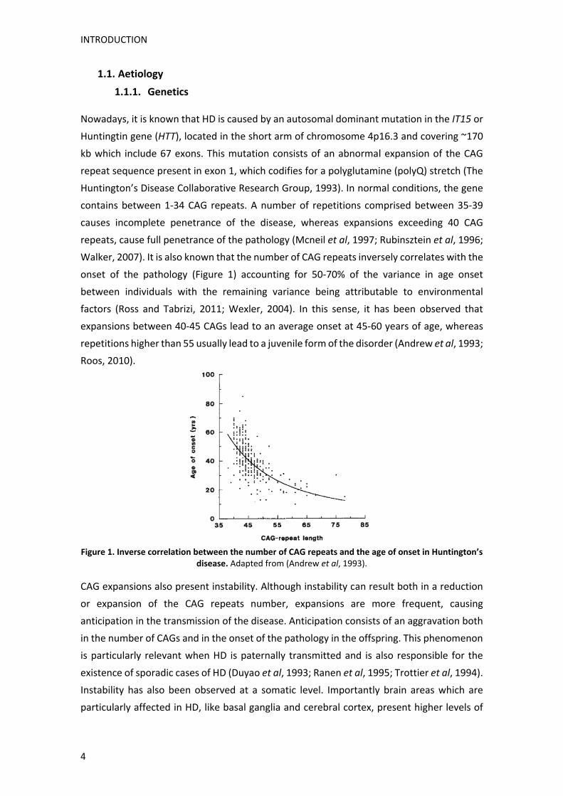
Walker, 2007). It is also known that the number of CAG repeats inversely correlates with the
onset of the pathology (Figure 1) accounting for 50‐70% of the variance in age onset
between individuals with the remaining variance being attributable to environmental
factors (Ross and Tabrizi, 2011; Wexler, 2004). In this sense, it has been observed that
expansions between 40‐45 CAGs lead to an average onset at 45‐60 years of age, whereas
repetitions higher than 55 usually lead to a juvenile form of the disorder (Andrew et al, 1993;
Roos, 2010).
Figure 1. Inverse correlation between the number of CAG repeats and the age of onset in Huntington’s
disease. Adapted from (Andrew et al, 1993).
CAG expansions also present instability. Although instability can result both in a reduction
or expansion of the CAG repeats number, expansions are more frequent, causing
anticipation in the transmission of the disease. Anticipation consists of an aggravation both
in the number of CAGs and in the onset of the pathology in the offspring. This phenomenon
is particularly relevant when HD is paternally transmitted and is also responsible for the
existence of sporadic cases of HD (Duyao et al, 1993; Ranen et al, 1995; Trottier et al, 1994).
Instability has also been observed at a somatic level. Importantly brain areas which are
particularly affected in HD, like basal ganglia and cerebral cortex, present higher levels of
INTRODUCTION
5
mosaicism, correlating increased somatic instability with increased vulnerability in the
pathology (Swami et al, 2009; Telenius et al, 1994).
1.1.2. Huntingtin protein
The product of the IT‐15 gene is Huntingtin (HTT), a 348 kDa protein whose function is still
not fully understood due to several reasons. One of them, is its heterogeneous distribution.
Indeed, HTT is ubiquitously expressed in all human and mammalian cells, with higher
expression in central nervous system (CNS) neurons and testes. It is also broadly distributed
at the subcellular level, as it can be localized in association with the nucleus, endoplasmic
reticulum, Golgi complex and mitochondrion, and it is also present at synapses where it
associates with structures like vesicles or microtubules (Difiglia et al, 1995; Zuccato et al,
2010).
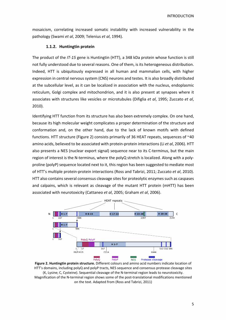
Identifying HTT function from its structure has also been extremely complex. On one hand,
because its high molecular weight complicates a proper determination of the structure and
conformation and, on the other hand, due to the lack of known motifs with defined
functions. HTT structure (Figure 2) consists primarily of 36 HEAT repeats, sequences of ~40
amino acids, believed to be associated with protein‐protein interactions (Li et al, 2006). HTT
also presents a NES (nuclear export signal) sequence near to its C‐terminus, but the main
region of interest is the N‐terminus, where the polyQ stretch is localized. Along with a poly‐
proline (polyP) sequence located next to it, this region has been suggested to mediate most
of HTT’s multiple protein‐protein interactions (Ross and Tabrizi, 2011; Zuccato et al, 2010).
HTT also contains several consensus cleavage sites for proteolytic enzymes such as caspases
and calpains, which is relevant as cleavage of the mutant HTT protein (mHTT) has been
associated with neurotoxicity (Cattaneo et al, 2005; Graham et al, 2006).
Figure 2. Huntingtin protein structure. Different colours and amino acid numbers indicate location of HTT’s domains, including polyQ and polyP tracts, NES sequence and consensus protease cleavage sites
(K, Lysine; C, Cysteine). Sequential cleavage of the N‐terminal region leads to neurotoxicity. Magnification of the N‐terminal region shows some of the post‐translational modifications mentioned
on the text. Adapted from (Ross and Tabrizi, 2011)

INTRODUCTION
6
The N‐terminus fragment can also be subjected to different post‐translational
modifications, including ubiquitination, acetylation, sumoylation, palmitoylation and
phosphorylation. These modifications regulate different HTT aspects like its stability,
subcellular localization, trafficking or provide neuroprotection against caspase cleavage
(Table 1).
Post‐translational modification
Amino acid residue
Functional effect Reference
Ubiquitination K6, K9 and K15 Control of HTT stability, function and intracellular localization
(Kalchman et al, 1996)
Acetylation K444 Targeting to the
macroautophagy pathway (Jeong et al, 2009)
Sumoylation K6, K9 and K15 Control of HTT stability, function and intracellular localization
(Steffan et al, 2004)
Palmitoylation C214 (by HIP14) Control of HTT trafficking and
function (Yanai et al, 2006)
Phosphorylation S421 (by Akt)
S434, 1181, 1201 (by Cdk5)
Neuroprotection and vesicular transport
(Humbert et al, 2002; Luo et al, 2005; Schilling
et al, 2006)
Table 1. HTT is subjected to different post‐translational modifications, with various effects on its function. K, Lysine; C, Cysteine; S, Serine; HIP14, Huntingtin‐interacting protein 14; Cdk5, Cyclin
dependent kinase 5.
All this information, along with generation of animal models, has brought some light at the
multiple functions in which HTT is involved. For instance, it is known that HTT has an
essential role during embryonic development, as a full HTT knockout causes embryonic
lethality before day 8.5 (Duyao et al, 1995; Nasir et al, 1995). It has also been described that
HTT has an anti‐apoptotic role, as its overexpression protects against apoptotic stressors
such as serum deprivation in in vitro models or excitotoxicity in in vivo models (Leavitt et al,
2006; Rigamonti et al, 2000). HTT function is also involved in neuroprotection through
regulation of the neurotrophin BDNF (brain‐derived neurotrophic factor) transcription and
trafficking (Gauthier et al, 2004; Zuccato et al, 2001, 2003), an important prosurvival factor
for striatal neurons produced by cortical neurons (Baquet et al, 2004). Finally, HTT localizes
in nerve terminals and it has been described to interact with several proteins involved in
clathrin‐mediated endocytosis, modulation of dendritic spine morphology and organization
of the postsynaptic density (PSD), thus being involved in synaptic functions (Harjes and
Wanker, 2003).
Despite lack of complete understanding of HTT function, it is assumable that loss of these
roles caused by the presence of the HD mutation could greatly impair different neuronal
processes. Nonetheless, gain of toxic function by the extended polyQ tract is believed to
play a bigger role in HD neuropathology. One of the main arguments in favour of this
hypothesis is that HD is part of nine other human genetic disorders caused by polyglutamine
expansions (spinocerebellar ataxia types 1, 2, 3, 6, 7, 12 and 17; dentatorubropallidoluysian
atrophy (DRPLA) and spinobulbar muscular atrophy (SBMA)), and even though

INTRODUCTION
7
polyglutamine expansions are localized in other proteins, all diseases present a pattern of
selective neurodegeneration, inverse correlation between length of the polyQ tract and
onset of the disease, and neuronal inclusions of polyQ aggregates (Margolis and Ross, 2001;
Walker, 2007).
1.2. Clinical aspects
HD symptoms consist of a triad of progressive motor, cognitive and psychiatric deficits. The
disease onset is stablished as the moment when formal clinical diagnosis of motor
symptoms takes place (Huntington Study Group, 1996) (Figure 3). Its average age is between
30 and 50 years, although it can range from 2 to 85 years (Myers, 2004). Before disease
onset, there is a prodromal phase, characterized by subtle motor, cognitive and behavioural
changes which can take place up to 15 years before the diagnosis. Prior to the prodromal
phase, patients known to carry the mutation, but not presenting any symptoms, are referred
to as presymptomatics (Bates et al, 2015; Ghosh and Tabrizi, 2015). After clinical diagnosis
there is an increasingly disabling progression of symptoms until patients die 15 to 20 years
after motor onset, the most common cause being secondary pneumonia followed by suicide
(Roos, 2010; Roos et al, 1993).
Figure 3. HD clinical progression. Average age onset determined by motor diagnosis is 45 years. The period before is termed premanifest and includes the presymptomatic and prodromal stages.
Individuals functional abilities are affected during prodromal phases and rapidly decrease through the manifest period, as chorea appears and motor and cognitive impairment worsen. Adapted from (Bates
et al, 2015)
1.2.1. Motor symptoms
Motor symptoms have two kinds of manifestations, on one hand appearance of involuntary
movements and, on the other hand, impairment of voluntary movements. Early stages of
the disease are characterized by appearance of chorea, which consists of involuntary,
excessive and short‐lived movements, which start in distal extremities and in the face, and
then spread more proximally becoming larger in amplitude. Dystonia, which are slower
movements caused by sustained muscle contractions, is another feature and leads to
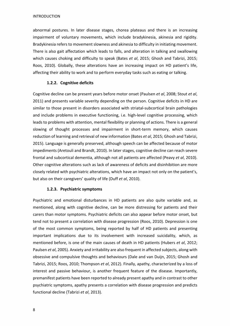
INTRODUCTION
8
abnormal postures. In later disease stages, chorea plateaus and there is an increasing
impairment of voluntary movements, which include bradykinesia, akinesia and rigidity.
Bradykinesia refers to movement slowness and akinesia to difficulty in initiating movement.
There is also gait affectation which leads to falls, and alteration in talking and swallowing
which causes choking and difficulty to speak (Bates et al, 2015; Ghosh and Tabrizi, 2015;
Roos, 2010). Globally, these alterations have an increasing impact on HD patient’s life,
affecting their ability to work and to perform everyday tasks such as eating or talking.
1.2.2. Cognitive deficits
Cognitive decline can be present years before motor onset (Paulsen et al, 2008; Stout et al,
2011) and presents variable severity depending on the person. Cognitive deficits in HD are
similar to those present in disorders associated with striatal‐subcortical brain pathologies
and include problems in executive functioning, i.e. high‐level cognitive processing, which
leads to problems with attention, mental flexibility or planning of actions. There is a general
slowing of thought processes and impairment in short‐term memory, which causes
reduction of learning and retrieval of new information (Bates et al, 2015; Ghosh and Tabrizi,
2015). Language is generally preserved, although speech can be affected because of motor
impediments (Aretouli and Brandt, 2010). In later stages, cognitive decline can reach severe
frontal and subcortical dementia, although not all patients are affected (Peavy et al, 2010).
Other cognitive alterations such as lack of awareness of deficits and disinhibition are more
closely related with psychiatric alterations, which have an impact not only on the patient’s,
but also on their caregivers’ quality of life (Duff et al, 2010).
1.2.3. Psychiatric symptoms
Psychiatric and emotional disturbances in HD patients are also quite variable and, as
mentioned, along with cognitive decline, can be more distressing for patients and their
carers than motor symptoms. Psychiatric deficits can also appear before motor onset, but
tend not to present a correlation with disease progression (Roos, 2010). Depression is one
of the most common symptoms, being reported by half of HD patients and presenting
important implications due to its involvement with increased suicidality, which, as
mentioned before, is one of the main causes of death in HD patients (Hubers et al, 2012;
Paulsen et al, 2005). Anxiety and irritability are also frequent in affected subjects, along with
obsessive and compulsive thoughts and behaviours (Dale and van Duijn, 2015; Ghosh and
Tabrizi, 2015; Roos, 2010; Thompson et al, 2012). Finally, apathy, characterized by a loss of
interest and passive behaviour, is another frequent feature of the disease. Importantly,
premanifest patients have been reported to already present apathy and in contrast to other
psychiatric symptoms, apathy presents a correlation with disease progression and predicts
functional decline (Tabrizi et al, 2013).
INTRODUCTION
9
1.3. Neuropathology
The most characteristic neuropathological hallmark of HD is a marked bilateral atrophy, cell
loss and astrogliosis of the striatum (caudate and putamen), which eventually leads to an
enlargement of the lateral ventricles (Vonsattel and DiFiglia, 1998) (Figure 4). However, 80%
of human HD brains also present an important atrophy of the cerebral cortex with a 21‐29%
of area loss (Vonsattel and DiFiglia, 1998) and a general brain alteration. Hence, human HD
brains present a reduction in whole brain weight, a 29‐34% loss of telencephalic white
matter, and affectation of other brain regions including the globus pallidus, subthalamic
nucleus, substantia nigra, thalamus, hypothalamus, cerebellum and hippocampus (Heinsen
et al, 1996; Jeste et al, 1984; Kremer et al, 1991; Monte et al, 1988; Spargo et al, 1993;
Vonsattel and DiFiglia, 1998).
Figure 4. HD neuropathology. Coronal slices of human HD and control brains. Striatal reduction and
ventricle enlargement, along with cerebral cortex atrophy can be observed in HD brains (left). Schematic illustrations of progressive striatal degeneration through Vonsattel grades, changes in striatal ventricular
profile shape can be appreciated (right). Adapted from (Reiner et al, 2011)
Striatal degeneration presents an ordered progression starting caudally and progressing in
a dorsomedial to ventrolateral direction (Reiner et al, 2011; Roos et al, 1985). Based on this
neuropathological progression, a grading system consisting of five grades (0‐4) was
stablished to evaluate HD severity in correlation with clinical disability. Grade 0‐1 includes
prodromal patients with small alterations compared to controls, while in Grade 2 striatal
atrophy is mild to moderate, with a reduced convex ventricular profile of the caudate, which
becomes flat or concave in Grade 3‐4 (Figure 4) (Reiner et al, 2011; Vonsattel, 2008;
Vonsattel et al, 1985).
1.3.1. Corticostriatal pathology
The striatum is the main input of cortical projections to the basal ganglia, which are a group
of subcortical nuclei comprised by the striatum (caudate and putamen), internal and
external globus pallidus (GPi and GPe), subthalamic nucleus (STN) and substantia nigra pars

INTRODUCTION
10
compacta (SNc) and pars reticulata (SNr). Glutamatergic inputs from the cortex and
dopaminergic inputs from the SNc are processed and relayed to downstream basal ganglia
nuclei (Figure 5). From there, information is transmitted to the thalamus and back to the
cortex, controlling voluntary movements, as well as cognitive and emotional processing
(Obeso et al, 2014; Rothwell, 2011).
MSNs constitute 90‐95% of striatal neurons, with the remaining 5‐10% being GABAergic and
cholinergic interneurons (Kita and Kitai, 1988). MSNs are GABAergic neurons that can be
classically subdivided in two different populations depending on their projection destination
(direct and indirect pathway) and their differential expression of neuropeptides and
dopamine (DA) receptor subtypes. Hence, striatonigral MSNs express D1 DA receptor (D1R),
the neuropeptides substance P and dynorphin and they project to the GPi and SNr forming
the direct pathway. On the other hand, striatopallidal MSNs express D2 DA receptor (D2R),
the neuropeptide enkephalin and they project to the GPe forming the indirect pathway
(Gerfen et al, 1990; Kawaguchi et al, 1990). The direct and indirect pathways regulate
movement initiation and inhibition, respectively, through excitatory (glutamatergic) and
inhibitory (GABAergic) circuits. This classification is important as neurodegeneration in HD
affects primarily MSNs from the indirect pathway. Moreover, dopaminergic inputs from the
SNc to MSNs from both pathways also further modulate movement. DA projections to
striatonigral MSNs are believed to further activate this pathway, whereas DA projections to
striatopallidal MSNs are thought to further inhibit movement (Surmeier et al, 2007).
Figure 5. Schematic representation of the direct and indirect pathways in physiological conditions
(left) and in early and late HD stages (right). Modified from (Calabresi et al, 2014).

INTRODUCTION
11
As illustrated in Figure 5, during HD early phases, the specific loss of striatopallidal MSNs
(D2) from the indirect pathway causes reduced inhibitory signalling to the GPe, increasing
excitatory signalling to the STN and subsequently, enhanced inhibitory signalling arrives to
the GPi and SNr. This leads to reduced inhibitory signalling to the thalamus from these
regions and to increased excitatory signalling to the motor cortex, which causes inability to
control voluntary movements and appearance of hyperkinesia (choreas). In later stages,
however, striatal affection extends to striatonigral MSNs (D1) from the direct pathway as
well, causing reduced inhibitory signalling to the GPi and SNr and subsequent augmented
inhibitory signalling to the thalamus from these regions. This leads to reduced excitatory
signalling to the motor cortex, causing inability to facilitate movement, which results in
rigidity and bradykinesia (Bolam et al, 2000; Bunner and Rebec, 2016; Calabresi et al, 2014;
Obeso et al, 2014; Rothwell, 2011).
The cerebral cortex is structured in six layers. Cortical neurons stablish internal connections
with neurons from other layers and outer connections with other brain areas. Pyramidal
neurons from layers III, V and VI are the main inputs to the striatum (McGeorge and Faull,
1989). HD patients present approximately 30% of cortical global thinning, which is more
pronounced in layers III and V, reaching up to a 43% in layer VI (Hedreen et al, 1991). This
thinning has been associated with specific loss of pyramidal neurons from regions like the
primary motor cortex (Macdonald and Halliday, 2002). Importantly, cortex atrophy begins
during premanifest period in a region‐specific manner, starting with thinning in primary
cortical regions including sensory, motor and visual regions with no significant changes in
whole brain volume. This atrophy then expands to posterior frontal and more occipital and
parietal areas in early HD, reaching a general affection of most cortical regions by late stages
of the disease (Rosas et al. 2008a; Rosas et al. 2008b). Correlation between affected cortical
regions and differential nature of symptoms in patients has also been stablished, providing
an explanation to the complexity and heterogeneity of HD symptoms (Raymond et al, 2011;
Thu et al, 2010). Finally, the fact that alterations in the cortex can be observed as soon as
10‐15 years before the onset of the disease, support the hypothesis that cortical cellular and
synaptic dysfunction plays a key role in early pre‐motor deficits in HD patients (Paulsen et
al, 2008; Raymond et al, 2011).
1.3.2. Hippocampal pathology
The hippocampus is a key element of the limbic system which mediates encoding,
consolidation and retrieval of memory, and participates in regulation of emotions, fear,
anxiety, stress or depression (Bartsch and Wulff, 2015; Pittenger and Duman, 2008; Squire,
1992). As illustrated in Figure 6, the hippocampus receives inputs from the main cortical
association areas through the entorhinal cortex (EC), and the information is relayed through
the DG (dentate gyrus), and the CA3 (cornu ammonis 3) to the CA1 (cornu ammonis 1)

INTRODUCTION
12
pyramidal neurons, which are the major hippocampal output projections to deep layers of
the EC (Bartsch and Wulff, 2015; Ghiglieri et al, 2011).
Figure 6. Hippocampal circuitry representation. Projection neurons from different hippocampal regions
are marked with the stated colours. Adapted from (Saura et al, 2015).
HD patients present a 35% neuronal loss in the CA1 region (Spargo et al, 1993) and mHTT
nuclear inclusions have been observed in several regions from the hippocampal formation
(Herndon et al, 2009). Voxel‐based morphometric studies have also shown that reduction
of hippocampal grey matter correlates with impairment in negative emotion recognition in
HD patients (Ille et al, 2011). However, most evidence of hippocampal early affectation in
HD comes from mice models studies, which have revealed that hippocampal‐dependent
functions, like navigation and spatial and recognition memories, are impaired in most HD
models before motor onset (Brito et al, 2014; Giralt et al, 2011, 2012a, 2012b; Lione et al,
1999). In line with these results, a recent study revealed that early HD patients present
similar deficits in hippocampal‐specific tasks, which correlate with estimated years to
diagnosis, indicating that hippocampal dysfunction is an important and early contributor to
HD cognitive decline (Begeti et al, 2016).
1.4. HD mice models
Despite their limitations, animal models have been and still represent an essential tool in
HD research. Problems arising from research with human patients, like high heterogeneity,
reduced availability of patients or long disease progression, are minimized by inherent
characteristics of animal models, such as shorter lifespans or the possibility to genetically
manipulate them. Thus, use of animal models in HD research has greatly improved
understanding of the molecular mechanisms underlying mHTT toxicity and has allowed
identification of putative therapeutic targets which could delay disease progression.
First developed HD models took advantage of neurotoxins that induced specific striatal
lesioning (Coyle and Schwarz, 1976; McGeer and McGeer, 1976). Glutamate agonists, like
quinolinic acid (Beal et al, 1986), and mitochondrial toxins, like 3‐nitropropionic acid (3‐NP)

INTRODUCTION
13
and malonate (Beal et al, 1993; Brouillet et al, 1993), were commonly used for their
relatively specific toxic effect on MSNs. The discovery of the IT15 gene allowed the
generation of multiple genetic animal models. Even though, mice are the most extensively
used, nowadays HD models from different species are available, including worms
(Caenorhabditis elegans) (Faber et al, 1999), fruit flies (Drosophila melanogaster) (Jackson
et al, 1998), rats (von Horsten et al, 2003) and more recently, sheeps (Jacobsen et al, 2010),
pigs (Yang et al, 2010) and monkeys (Yang et al, 2008). Nonetheless, given HTT’s large size,
the differential effect of distinct CAG lengths or its genomic instability, design of HD models
has been complex and diverse. Thus, several genetic approaches have been used regarding
some of the following factors: use of full‐length mHTT or of the N‐terminal fragment, use of
different CAG repeat lengths, use of human or mice Htt promoter and expression of mHTT
from a transgene or by knocking the CAG mutation into the endogenous murine gene (Figure
7).
Figure 7. Different genetic approaches for HD mice generation. Illustrative scheme showing different genetic design features which differ between HD mice models. Adapted from (Pouladi et al, 2013).
1.4.1. Truncated N‐terminal models
Mice models expressing the truncated N‐terminal fragment of mHTT present a rapid onset
of motor, cognitive and behavioural symptoms, weight loss, reduced lifespan and
generalized degenerative phenotype. Despite being the less genetically accurate models, its
rapid onset and progression makes them a recurrent choice for therapy screening studies
(Ferrante, 2009; Figiel et al, 2012; Gil and Rego, 2009).

INTRODUCTION
14
R6/1 and R6/2 mice express, respectively, one and three randomly inserted copies of the
1.9 kb human genomic fragment containing promoter sequences and exon 1 from HTT gene.
R6/1 mice, expressing 116 CAG repeats and R6/2 mice, expressing 144 CAG repeats, were
the first ever generated HD genetic mice models (Mangiarini et al, 1996). R6/1 mice present
cognitive deficits at 12 weeks of age and motor alterations, including limb clasping and
impaired rotarod performance, at 14‐20 weeks of age. They also present neuronal inclusions
with no neuronal death and an average lifespan of 32‐40 weeks (Brooks et al, 2012;
Mangiarini et al, 1996; Naver et al, 2003). R6/2 mice present similar alterations but with an
earlier onset with cognitive deficits at 3.5 weeks and motor symptoms at 8 weeks of age
(Lione et al, 1999; Stack et al, 2005). Both lines present genomic instability, which can cause
CAG tracts to expand up to 250 and even 400 (Mangiarini et al, 1996; Pouladi et al, 2013).
They also show brain atrophy, even though it is believed to be caused by neuronal
dysfunction rather than by neuronal death, as neuronal apoptosis is not detected in these
models (Yu et al, 2003).
N171‐Q82 mice are another truncated N‐terminal model which differs from the R6 lines by
its expression of a longer sequence including exon 1 and exon 2 with 82 CAG repeats, under
the mouse prion promoter (Prp) regulation, which restricts its expression to neurons
(Schilling et al, 1999). In addition they present loss of striatal neurons by apoptosis (Yu et al,
2003) and a more variable phenotype, which can be a downside as it might require larger
sample sizes (Ferrante, 2009).
1.4.2. Full‐length models
Full‐length mHTT models can be generated by means of two main strategies: introducing a
full‐length human mutant HTT transgene (transgenic models) or knocking a human HTT exon
1 with expanded CAG repeats into the endogenous mouse Htt gene (knock‐in models). They
present a milder disease progression compared to the N‐terminal fragment models,
especially in the case of knock‐in mice, making them a good choice for studying early stages
of the disease.
1.4.2.1. Transgenic models
Transgenic models were created using the YAC (yeast artificial chromosome) and BAC
(bacterial artificial chromosome) technologies, which allow expression of large DNA
sequences. Therefore these models express human genomic mutant HTT transgenes,
including all introns, exons and regulatory elements, thus presenting a more accurate tissue
and temporal genetic expression of mHTT (Gray et al, 2008; Hodgson et al, 1999; Pouladi et
al, 2013; Slow et al, 2003).

INTRODUCTION
15
Several YAC lines with different CAG lengths (48, 72 and 128) have been created, although
YAC128 mice are the most commonly used (Hodgson et al, 1999; Slow et al, 2003). They
present learning deficits at 2 months, motor symptoms at 3 months and aggregates and
neuronal loss at 12‐18 months of age. They also present low inter‐animal variability
(Ferrante, 2009; Figiel et al, 2012; Van Raamsdonk et al, 2005; Slow et al, 2003). BACHD
mice were generated more recently, containing 97 polyglutamine expansions, expressed
from a mixed CAA‐CAG repeat sequence, in order to avoid genomic instability (Gray et al,
2008).
1.4.2.2. Knock‐in models
Knock‐in models were stablished by insertion of the human HTT exon 1 with CAG lengths
ranging from 50 to 200 into the endogenous mouse Htt gene (Wheeler et al, 1999; White et
al, 1997). These mice represent the most genetically accurate models, not only allowing
proper mHtt spatial and temporal expression, but avoiding possible effects arising from
species‐specific sequence differences (Ehrnhoefer et al, 2009; Pouladi et al, 2013). Knock‐in
mice also present a milder phenotype with, usually, a normal lifespan. Onset of symptoms
and disease progression varies depending on the length of the CAG tract, the homozygosity
or heterozygosity of the knock‐in gene and their genetic background (Ferrante, 2009; Figiel
et al, 2012; Lloret et al, 2006). HdhQ111, CAG140, HdhQ150 and zQ175 are some of the
most frequently used knock‐in lines.
In this Thesis we have used HdhQ7/Q111 mice in a C57BL/6 strand. These mice present
depressive‐like behaviour at 2 months of age (unpublished results), corticostriatal and
hippocampal cognitive deficits at 6 months of age, with motor alterations at 8 months and
presence of striatal intranuclear mHTT inclusions starting at 6 months of age (Brito et al,
2014; Giralt et al, 2012a; Lloret et al, 2006; Puigdellívol et al, 2015). No overt neuronal loss
or astrogliosis is detected at motor symptoms onset (Bragg et al, 2017; Brito et al, 2014).

INTRODUCTION
16
2. Molecular mechanisms underlying striatal vulnerability and cognitive
symptoms in HD
HTT’s complexity, as well as its involvement in multiple cellular pathways are some of the
reasons why 25 years after the discovery of the HD mutation, mechanisms underlying
striatal degeneration and cognitive decline are still not fully understood. Nonetheless,
generation of HD mice models has identified some of the molecular mechanisms affected
by the presence of mHTT. Moreover, knowledge that models like the R6 lines display a
severe phenotype without showing overt neuronal loss, has underlined the idea that
neuronal dysfunction precedes neuronal death and, therefore, targeting of affected
pathways in early disease stages could prevent or delay disease progression. In this section
we will present the proposed molecular mechanisms underlying striatal vulnerability and
what is known from synaptic dysfunction greatly responsible for cognitive decline.
2.1. Striatal vulnerability
Understanding why striatal MSNs present specific affectation in HD despite ubiquitous HTT
expression in all body cells, has been and still is one of the main HD conundrums. Even
though years of research have identified several mechanisms which could contribute to
specific striatal dysfunction and degeneration (Figure 8), full comprehension of mHTT‐
induced striatal vulnerability is still lacking. Therefore, further study of the classically
proposed mechanisms, as well as, characterization of new molecular pathways participating
in this process is pivotal to complete our knowledge of striatal pathophysiology in HD.
2.1.1. Canonical mechanisms
2.1.1.1. BDNF
BDNF is a cortically produced neuroprotective factor for striatal neurons. As mentioned
before, HTT participates in BDNF transcription and trafficking and presence of mHTT has
been shown to alter both processes in vitro, in mice models and in human patients. Thus, a
reduction in both BDNF mRNA and protein levels has been observed in the striatum and
other brain regions of different HD models and in human patients (Ferrer et al, 2000;
Zuccato et al, 2001, 2010). mHTT alters BDNF transcription through dysregulation of
different transcription factors, e.g. REST (RE1‐silencing transcription factor) (Zuccato et al,
2003), CREB (cAMP response element‐binding protein), CBP (CREB binding protein) or Sp1
(specificity protein 1) (Cha, 2007; Zuccato et al, 2003), while also decreases anterograde
axonal BDNF vesicle transport from the cortex to the striatum (Gauthier et al, 2004). In
addition, changes in BDNF receptors have also been described in HD, further impairing BDNF
signalling. Thus, levels of the BDNF receptor TrkB, which activates pro‐survival pathways,

INTRODUCTION
17
are reduced in the striatum of different HD mice models and in HD patients. Moreover,
levels of p75NTR, an additional BDNF receptor which can induce pro‐apoptotic pathways,
have been shown to be increased in HD (Brito et al, 2013; Ginés et al, 2006; Plotkin et al,
2014; Zuccato et al, 2008). Therefore, lack of BDNF trophic support along with alterations in
its signalling pathways have been proposed to participate in increased striatal vulnerability
in HD.
Figure 8. Canonical molecular pathways involved in striatal vulnerability in HD. Schematic representation of the principal cellular processes involved in mHTT‐induced striatal vulnerability: transcriptional dysregulation (A), mHTT aggregate formation (B), mitochondrial dysfunction (C),
alterations in autophagy and proteosomal degradation (D), loss of BDNF trophic function, associated with impairment of its transcription and transport and with an imbalance in TrkB and p75 receptors
levels (E), and excitotoxicity, attributed to alterations in glutamate release and uptake and to changes in glutamate receptors levels and localization (F). Modified from (Bates et al, 2015).
INTRODUCTION
18
2.1.1.2. Excitotoxicity
Excitotoxicity refers to excessive excitatory amino acids signalling, mainly glutamate, leading
to toxic calcium overload which promotes neuronal death (Rothman and Olney, 1995).
Observations that treatment with glutamate agonists led to preferential striatal
degeneration causing and HD‐like phenotype, suggested that excitotoxicity might play an
important role in striatal vulnerability in HD. In addition, inherent characteristics of the
striatum as a receiver of mainly excitatory inputs, place it in a vulnerable position to
alterations in corticostriatal glutamate signalling, which have been described in HD. For
instance, R6/1 mice show heightened activity‐induced striatal glutamate release (Nicniocaill
et al, 2001), while astroglial glutamate transporter (GLT1) mRNA and protein levels are
decreased in R6/1 and R6/2 mice (Behrens et al, 2002; Liévens et al, 2001). GLT1 plays a
major role in extracellular glutamate clearance, avoiding excitotoxicity or glutamate spill‐
over to extrasynaptic sites (Takahashi et al, 2015), the latest being associated with activation
of apoptotic pathways (reviewed in (Hardingham and Bading, 2010; Wang et al, 2013)).
Notably, increased localization of NMDA (N‐Methyl‐ᴅ‐aspartate) receptors (NMDAR) at
extrasynaptic sites and subsequent activation of extrasynaptic NMDARs downstream
pathways has been described in the striatum of YAC128 mice at early disease stages prior
to motor onset (Milnerwood et al, 2010). Therefore, mHTT contributes to striatal
excitotoxicity by altering multiple steps of glutamatergic signalling, including glutamate
release and clearance, glutamate receptors levels and localization and activation of their
downstream signalling pathways (Fan and Raymond, 2007).
However, analysis of excitotoxicity responses in HD models have yielded diverse results
depending on the disease stage and the model being studied (Table 2). Therefore, although
enhanced sensitivity to excitotoxicity has been described at early stages in YAC128 and R6/1
and R6/2 mice, these animals present resistance to excitotoxicity at later disease stages
(Graham et al, 2009; Hansson et al, 2001; Levine et al, 1999). Notably, different sensitivity
to excitotoxicity has also been described in the absence of mHTT, depending on the mice
strand, which could further contribute to different excitotoxicity responses between HD
models (Schauwecker and Steward, 1997). Nonetheless, increased sensitivity to
glutamatergic stimulation has been associated with alterations in intracellular calcium
homeostasis (Fernandes et al, 2007) and with impaired mHTT and PSD‐95 interaction (Sun
et al, 2001). As a result of these impairment, increased NR2B and PSD‐95 interaction is
detected causing enhanced NMDARs surface localization, which plays a major role in
excitotoxicity (Fan et al, 2009; Sun et al, 2001). On the other hand, resistance to
excitotoxicity has been proposed as a compensatory mechanism, and it has been associated
with reduction of synaptic proteins like PSD‐95 and citron, which could lead to NMDARs
hypofunction (Jarabek et al, 2004). Importantly, reduction of NMDARs levels as a protective

INTRODUCTION
19
mechanism against excitotoxicity could impair NMDAR‐dependent synaptic plasticity
processes, highlighting the relevance of proper balance in NMDAR function.
HD model Stage Excitotoxicity response Reference
YAC128 presymptomatic enhanced
excitotoxicity sensitivity (Graham et al, 2009)
YAC128 late resistance to excitotoxicity
(Graham et al, 2009)
R6/2 symptomatic enhanced
excitotoxicity sensitivity (Levine et al, 1999)
R6/1 and R6/2
motor onset resistance to excitotoxicity
(Hansson et al, 2001)
R6/2 Early symptomatic
(10 weeks) similar excitotoxicity
response (Estrada‐Sánchez et al,
2009)
R6/2 Late symptomatic
(14 weeks) enhanced
excitotoxicity sensitivity (Estrada‐Sánchez et al,
2009)
CAG94 (KI) presymptomatic enhanced
excitotoxicity sensitivity (Levine et al, 1999)
N171‐82Q resistance to excitotoxicity
(Jarabek et al, 2004)
Table 2. Differential excitotoxicity responses between HD mice models. Examples of reported cases showing contrasting responses to excitotoxic insults depending on the HD model, the disease stage or
the mice strand.
2.1.1.3. Aggregate formation and protein homeostasis
HTT fragmentation is considered one of the earlier steps of HD pathogenesis, as it can be
detected in most HD models and in HD human brains and it takes place before aggregate
formation (Bates et al, 2015; Landles et al, 2010; Lunkes et al, 2002). Small N‐terminus HTT
fragments can be produced via proteolytic cleavage by different caspases (Wellington et al,
2002), calpains (Gafni and Ellerby, 2002; Kim et al, 2001) and other proteases (Lunkes et al,
2002). Post‐translational modifications also affect mHTT cleavage, as it has been described
that HTT phosphorylation at residue Ser434 by Cdk5 (Luo et al, 2005) and at residue Ser421
by Akt and SGK (Warby et al, 2009), are protective against caspase‐mediated HTT cleavage.
Presence of polyglutamine expansions disrupts the balance of protein folding quality control
formed mainly by chaperones and clearance machinery, which have been shown to
progressively decrease in HD causing a global disruption of the proteome (Gidalevitz et al,
2006; Hay et al, 2004; Labbadia and Morimoto, 2013). Moreover, both cellular protein
clearance pathways (autophagy and the ubiquitin‐proteosome system) are also impaired in
HD (reviewed in (Martin et al, 2015; Ortega and Lucas, 2014)). In this context, mHTT
aggregates are formed, and even though there is some debate about aggregate formation
being protective against free soluble N‐terminal fragments, it is usually considered that
aggregates are detrimental for neurons. One of the main reasons is that they sequester
other polyQ tract‐containing proteins, affecting different cellular processes. Hence,

INTRODUCTION
20
intranuclear aggregates have been associated with sequestering of transcription factors and
transcriptional regulators (Cha, 2007), whereas extranuclear aggregates impair functions
like axonal transport (Gunawardena et al, 2003; Li et al, 2001b).
2.1.1.4. Mitochondrial dysfunction
Similar to glutamate agonists, mitochondrial toxins, like 3‐NP and malonate, also induce an
HD‐like phenotype, thus pointing mitochondrial dysfunction as an HD striatal vulnerability
factor. Long before the mHTT mutation was discovered, it had already been observed in HD
brain biopsies, the presence of aberrant dense mitochondria with sparse cristae (Tellez‐
Nagel et al, 1974). Nowadays, it is known that several aspects of mitochondria physiology
are altered. For instance, alterations in mitochondrial transport and biogenesis, resulting
from impaired axonal transport and transcription have been reported, as well as aberrant
mitochondrial dynamics, leading to fragmented mitochondria (Cherubini and Ginés, 2017;
Dubinsky, 2017; Kim et al, 2010). Mitochondria from YAC72 mice and HD patients
lymphoblasts also show reduced calcium uptake capacity (Panov et al, 2002). Importantly,
striatal mitochondria are particularly sensitive to calcium and to permeability transition
induction, a process contributing to mitochondrial dysfunction and activation of apoptotic
pathways in response to calcium and oxidative stress, which is produced for instance after
excitotoxic insults (Brustovetsky et al, 2003, 2005; Crompton, 1999). Therefore, inherent
properties of striatal mitochondria could contribute to HD striatal vulnerability, particularly
to aberrant excitotoxic responses.
2.1.1.5. Transcriptional dysregulation
Transcriptional dysregulation of genes associated with e.g., neurotransmitter receptors,
synaptic transmission, cytoskeletal and structural proteins, intracellular signalling or calcium
homeostasis, can be detected in HD since early stages (reviewed in (Cha, 2007; Zuccato et
al, 2010)). As mentioned before, some of these alterations are caused by sequestering of
transcription factors or their coactivators in mHTT aggregates. Some of these transcription
factors include p53, CBP, which is involved in cell survival and memory consolidation, Sp1,
which regulates expression levels of D2R amongst others, and REST, which as mentioned
before regulates BDNF transcription levels (Dunah et al, 2002; Giralt et al, 2012a; Nucifora
et al, 2001; Steffan et al, 2000; Zuccato et al, 2003). In addition, alterations in histone post‐
translational modifications modulating chromatin structure, have also been described in
different HD models (Glajch and Sadri‐Vakili, 2015). Thus, sequestering in mHTT aggregates
of CBP which displays HAT (histone acetyltransferase) activity, has been associated with
increased chromatin condensation and reduced gene transcription (Steffan et al, 2001).
Moreover, HDACs (histone deacetylases) activity has also been shown to be increased in HD,

INTRODUCTION
21
further contributing to reduced gene transcription (reviewed in (Glajch and Sadri‐Vakili,
2015; Sharma and Taliyan, 2015)).
2.1.1.6. Alteration of kinases and phosphatases
Protein phosphorylation participates in the modulation of most cellular processes, therefore
alterations in the levels of kinases and phosphatases can impact multiple neuronal functions
(Manning et al, 2002; Sacco et al, 2012). Presence of mHTT has been described to alter
several kinases and phosphatases involved in processes such as regulation of neuronal
survival or activation of inflammatory pathways. For instance, both pro‐survival (MEK/ERK)
and pro‐apoptotic (p38 and JNK) pathways of the MAPK family (Keshet and Seger, 2010;
Miloso et al, 2008), as well as, the Akt pathway, which has anti‐apoptotic functions (Song et
al, 2005), have been shown to be altered in HD. However, both excessive activation and
inhibition of these pathways has been described depending on the HD model and disease
progression state. These conflicting results have been attributed to the presence of
compensatory mechanisms in some models at some disease stages, which probably also
underlie differences in neuronal death and sensitivity or resistance to excitotoxicity
between models. Other kinases described to be altered in HD include IKK and NFκβ, which
are associated with increased inflammatory signalling in some HD models (Khoshnan et al,
2004; Reijonen et al, 2010), and Cdk5, which has been associated with increased striatal
vulnerability, participates in multiple neuronal functions (Cheung et al, 2006; Paoletti et al,
2008; Su and Tsai, 2011) and will be discussed with further detail in later sections.
In addition, several phosphatases have also been described to be altered in HD, including
PP1 (Branco‐Santos et al, 2017; Metzler et al, 2010), calcineurin, a calcium‐dependent
phosphatase with high expression in the striatum and hippocampus (Gratuze et al, 2015;
Xifró et al, 2009) and STEP, whose decreased activity has been associated with excitotoxicity
resistance in R6/2 mice whereas increase in its activity has been involved with extrasynaptic
localization of NMDARs (Gladding et al, 2012; Saavedra et al, 2011). In sum, there is a
complex deregulation of kinases and phosphatases in HD, which further contribute to the
previously described alterations in HD. Importantly, their function as downstream effectors
place them in a fitting position to become therapeutic targets in order to minimize side
effects.
2.1.2. Non‐canonical mechanisms
2.1.2.1. Cell cycle re‐entry as a new mechanism
Neurons are classically described as permanent postmitotic cells. For this reason, up until a
few decades ago, little attention had been payed to proteins and checkpoints involved in
cell cycle regulation in neurons (Fig. 9). Nowadays it is known that re‐expression of cell cycle

INTRODUCTION
22
proteins and even DNA duplication can take place in several neurodegenerative disorders in
response to different stressors, although not much is known about its possible role in HD.
Figure 9. Cell cycle phases. Cyclins and CDKs (Cyclin‐dependent kinases), along with CKIs (Cyclin‐dependent kinase inhibitors) regulate transition between different cell cycle phases. During the G1
phase CyclinD1 activates Cdk4/6 which phosphorylates Rb protein allowing E2F1 release. E2F1 binds to DP1 promoting transcription of cell cycle progression genes. CyclinE/Cdk2 regulate G1/S transition and
CyclinA/Cdk2 promote DNA replication activation during S phase. During G2 phase CyclinA/Cdk1 modulate condensation of chromosomes. Finally, CyclinB/Cdk1 modulate G2/M transition, after which the cell duplicates during the M phase, Rb becomes dephosphorylated and the cell cycle can begin
again. CKIs from the Ink family (p15, p16, p18 and p19) and from the Cip/Kip family (p21, p27 and p57) also participate in cell cycle regulation by inhibiting CDKs activation.
Prove that aberrant neuronal cell cycle re‐entry leads to cell death instead of cell division,
was first described in the early 1990s when it was observed that, forcing cell cycle initiation
by expressing the oncogene SV40 T‐antigen under a neuronal promoter, lead to neuronal
demise (Al‐Ubaidi et al, 1992; Feddersen et al, 1992). Since then, it has been described that
stressors such as neurotrophic factor deprivation (Freeman et al, 1994), DNA damage (Park
et al, 1998), excitotoxicity (Park et al, 2000) or presence of amyloid β protein (Copani et al,
1999; Giovanni et al, 1999) induce expression of cell cycle proteins. Similarly, alteration of
cell cycle proteins has been described in ischemia (Love, 2003), amyotrophic lateral sclerosis
(ALS) (Ranganathan et al, 2001), ataxia‐telangiectasia (A‐T) (Yang and Herrup, 2005),
Parkinson’s disease (PD) (Jordan‐Sciutto et al, 2003), and especially in Alzheimer’s disease
(AD) (reviewed in (Lee et al, 2009)). Some of these proteins include CyclinD1, Cdk4,
hyperphosphorylated Retinoblastoma (Rb) and E2F1, which are crucial regulators of the
G1/S transition (Figure 9), but also proliferation markers like PCNA and Ki67. Incorporation
of BrdU has also been described in mature neurons indicating DNA duplication (Herrup and
Busser, 1995). Importantly, it is believed that once a neuron starts expressing cell cycle
proteins and even after it duplicates its DNA, it can take up to months or even years before

INTRODUCTION
23
its death (Frade and Ovejero‐Benito, 2015; Herrup and Yang, 2007), thus contributing to a
long process of neuronal dysfunction prior to neuronal degeneration, common in most
neurodegenerative diseases. In fact, small subsets of aneuploid neurons can be found in
physiological conditions in the brain without causing overt alterations (Arendt, 2012; Frade
and Ovejero‐Benito, 2015).
As previously mentioned, some cellular stressors associated with cell cycle re‐entry, such as
neurotrophic factor deprivation or excitotoxicity play an important role in HD striatal
vulnerability. In addition, alterations in DNA damage, which also can lead to increased cell
cycle re‐entry, have also been described in HD. Impaired non‐homologous end joining
(NHEJ) DNA repair has been described to be impaired in HD due to mHTT interaction with
Ku70 (Enokido et al, 2010), whereas proteins involved in mismatch repair (MMR), like Msh2
have been associated with increased CAG instability (Manley et al, 1999).
Thus, it is possible that cell cycle re‐entry of striatal neurons may contribute to striatal
dysfunction in HD. In accordance, brain samples from HD patients present increased levels
of E2F1, CyclinD1 and Cdk4, which play a key role in G1 phase progression (Fig. 9). Cdk4 levels
have also been described to be increased in the striatum of R6/1 mice, as well as in rats
treated with 3‐NP, along with increased Cdk2, CyclinD1 and E2F1 levels (Fernandez‐
Fernandez et al, 2011; Pelegrí et al, 2008). Importantly, 3‐NP treatment has also been shown
to cause reduction of p27 and increased phosphorylated‐Rb (pRB) levels in the striatum of
treated mice, which can be prevented by CDK inhibitors and NMDARs antagonists in striatal
primary cultures (Akashiba et al, 2008). Finally, differential expression of several microRNAs
has also been shown to affect cell cycle progression of HD immortalized striatal cells
(STHdhQ111) (Das et al, 2013, 2015). Therefore, available data suggests that cell cycle proteins
are altered by mHTT presence but little is known about their changes during disease
progression and about how mHTT triggers their expression.
2.2. Synaptic dysfunction
Synaptic plasticity, i.e., activity‐dependent strength modification of synaptic transmission in
pre‐existing synapses, is necessary and sufficient for the information storage underlying
learning and memory formation (Citri and Malenka, 2008; Martin et al, 2000). The best
characterized synaptic plasticity forms are LTP (long‐term potentiation) and LTD (long‐term
depression), firstly described in the hippocampus. However, there are other forms of
synaptic plasticity, such as an endocannabinoid‐dependent LTD form which has been
identified in different brain regions including D2‐MSNs (reviewed in (Citri and Malenka,
2008)). In any case, all kinds of synaptic plasticity induction rely on normal
electrophysiological properties, proper function and localization of neurotransmitter
receptors and synaptic proteins, and correct cytoskeleton regulation underlying dendritic
INTRODUCTION
24
spine remodelling. The fact that many HTT interactors include pre‐ and post‐synaptic
proteins has led to the idea that mHTT may alter these proteins, generating a complex
synaptic dysfunction.
In accordance, analysis of HD patients brains have shown impairments in cortical and
brainstem LTP‐like plasticity (Crupi et al, 2008), while studies in HD mouse models have
revealed alterations in corticostriatal LTP induction prior to motor onset in R6/2 (Kung et al,
2007) and HdhQ111 mice (Puigdellívol et al, 2015). Changes in synaptic plasticity within the
cortex have also been demonstrated, with R6/1 mice showing reduced LTP induction in the
medial prefrontal cortex, and impaired LTD induction in the perirhinal cortex, alterations
that have been associated with reduced dopamine signalling (Cummings et al, 2006, 2007;
Dallérac et al, 2011). Finally, in the hippocampus, impaired LTP induction in the CA1 has
been described in YAC mice (Hodgson et al, 1999) and in early disease stages in R6/2 mice,
which also present alterations in the DG at later stages, correlating with chronological
appearance of mHTT neuronal intranuclear inclusions in these regions (Murphy et al, 2000).
Hippocampal synaptic plasticity impairments have been associated, in turn, with altered
glutamate and BDNF signalling (Brito et al, 2014; Lynch et al, 2007; Milnerwood et al, 2006;
Simmons et al, 2009). Altogether, even though functional alterations may vary depending
on the studied model and the experimental procedure, aberrant corticostriatal and
hippocampal synaptic plasticity are observed prior to overt motor phenotype and neuronal
death in most HD models, supporting the idea that neuronal dysfunction rather than
neurodegeneration is playing an important role in HD pathology. In accordance, several
neuronal processes implicated in synaptic plasticity show alterations in HD.
2.2.1. Synaptic transmission
Several studies have pointed out that altered corticostriatal transmission is an early HD
hallmark which, similar to motor symptoms progression, manifests in a biphasic way
(reviewed in (Raymond et al, 2011)). It has been observed that in both YAC128 and R6/2
mice, at pre‐motor disease stages, there is an increase in striatal sEPSCs (spontaneous
excitatory postsynaptic currents) (Andre et al, 2011; Cepeda et al, 2003; Joshi et al, 2009).
In addition, although basal glutamate levels are significantly reduced in the striatum and
prefrontal cortex of R6/1 and knock‐in HdhCAG150 mice, respectively, as previously
mentioned, activity‐induced striatal glutamate release is significantly increased in R6/1 mice
(Nicniocaill et al, 2001; Wolfram‐Aduan et al, 2014). On the other hand, in later stages, there
is a reduction in excitatory activity of MSNs, which is believed to arise from MSNs
disconnection from their inputs, as it correlates with alteration of different synaptic markers
and loss of dendritic spines (Cepeda et al, 2003). In agreement, it has been described that
knock‐in Q140 mice present loss of corticostriatal terminals prior to overt striatal
dysfunction (Deng et al, 2013). In addition, at this stage, cortical sEPSCs occur at higher

INTRODUCTION
25
frequencies (Cummings et al, 2009) and it has been described that reduction of cortical
inputs to the striatum or mHTT removal from cortical neurons, is able to delay disease
progression (Cepeda et al, 2003; Gu et al, 2007). In sum, these findings support the idea that
aberrant cortical afferences and/or corticostriatal disconnection play a major role in
impaired corticostriatal function.
2.2.2. Dendritic spines
Dendritic spines are dynamic and heterogeneous structures where most synapses occur.
Remodelling of their shape, size and number is a key step in synaptic plasticity processes
and it relies on cytoskeleton reorganization, which is mainly composed by filamentous actin
(F‐actin). Therefore, proteins involved in positive or negative actin polymerization will
modulate growth or shrinkage of dendritic spines. These proteins include neurotransmitter
receptors (e.g. NR1 and NR2B), neurotrophin receptors (e.g. TrkB and p75NTR), kinases (e.g.
CaMKII and Cdk5) and scaffold proteins (e.g. PSD95) (reviewed in (Sala and Segal, 2014)).
However, downstream signalling of these proteins, usually converge on proteins from the
small GTPases family, which act as molecular switches depending on their active (GTP‐
bound) or inactive (GDP‐bound) state. Rac1 and RhoA are the best characterized members
of this family for their opposite roles in dendritic spine remodelling, with Rac1 acting as a
dendritic spine positive regulator (Nakayama et al, 2000), and RhoA as a negative dendritic
spine modulator (Tashiro et al, 2000).
In this regard, initial studies from HD patients revealed early morphologic and quantitative
alterations in dendritic arbours and spines in the striatum and cortex (Ferrante et al, 1991;
Graveland et al, 1985; Sotrel et al, 1993), which could be associated with presence of mHTT
aggregates on dendritic spines (Gutekunst et al, 1999). Additionally, dendritic spine
alterations are also present in most HD mouse models, including YAC128 mice, which
present reduced spines in MSNs at late disease stages (Xie et al, 2010), R6/2 and R6/1 mice,
which have decreased dendritic spines density in the striatum, cortex and hippocampus
(Bulley et al, 2012; Giralt et al, 2017; Klapstein et al, 2001; Murmu et al, 2013; Spires et al,
2004) and HdhQ111 knock‐in mice which present early decrease of cortical spines (Puigdellívol
et al, 2015) and reduction of striatal and hippocampal dendritic spines (Brito et al, 2014;
Lerner et al, 2012). Importantly, studies tracking individual dendritic spines through the
disease progression have also shown increased spine formation, although their survival is
greatly reduced, indicating aberrant dendritic spine neurogenesis and maintenance (Murmu
et al, 2013).
Importantly, some of these modifications have been associated with alterations in
cytoskeleton regulation. Thus, knock‐in HdhQ111 mice present reduced actin polymerization
at dendritic spines which correlates with failing to stabilize CA1 LTP in presymptomatic

INTRODUCTION
26
stages (Lynch et al, 2007; Star et al, 2002). On the other hand, both Rac1 and RhoA have
been described to be altered in knock‐in HdhQ111 mice. Specifically reduced Rac1 activity
correlating with reduced dendritic spines was observed in the cortex (Puigdellívol et al,
2015), whereas increased RhoA activity was described in the hippocampus (Brito et al,
2014). Moreover, a protein interaction study identified the RhoGTPase signalling pathway
for being enriched in HTT interacting proteins (Tourette et al, 2014). Thus, alterations in
cytoskeleton proteins and their upstream regulators may underlie modifications in dendritic
spines participating in synaptic dysfunction in HD.
2.2.3. Synaptic proteins
Excitatory synapses are characterized by the presence of a postsynaptic density, a
specialised macromolecular complex where neurotransmitter receptors, scaffold proteins
and cytoskeletal components are localized, and which receives inputs from a presynaptic
site (Citri and Malenka, 2008; Sheng, 2001). Presence of mHTT modifies many of these
proteins, contributing to changes in synaptic plasticity. Thus, alterations in presynaptic
processes associated with exocytosis and endocytosis and with neurotransmitter release,
such as reduced levels of acetylcholine metabolism enzymes ChAT (choline
acetyltransferase enzyme) and VAchT (vesicular acetylcholine transporter) have been
described in HD patients and in several HD models (reviewed in (Li et al, 2003; Rozas et al,
2010; Tyebji and Hannan, 2017)). Reduced levels of the endocannabinoid receptor CB1,
which is expressed presynaptically inhibiting neurotransmitter release, has also been
observed at very early stages in HD patients (Glass et al, 2000; Van Laere et al, 2010) and in
HD mice (Blázquez et al, 2011; Chiodi et al, 2012; Ruiz‐Calvo et al, 2018).
However, alterations in the PSD have been more extensively analysed showing deficits in
the expression of several neurotransmitter receptors, including serotonin receptors which
are reduced in the hippocampus and cortex in HD models (reviewed in (Tyebji and Hannan,
2017)) and adenosine receptors (A1R and A2AR) which participate in bioenergetic
homeostasis and are affected at early disease stages (Lee and Chern, 2014; Matusch et al,
2014). GABAA receptors and DA receptors (D1 and D2) also present reduced levels since
initial stages (Chen et al, 2013; Glass et al, 2000). Levels of DA, that are crucial for striatal
LTP and LTD induction (Chen et al, 2013; Citri and Malenka, 2008), are also altered in a
biphasic way, with an early increase, and a reduction at later disease stages (Dallérac et al,
2015; Garrett and Soares‐da‐Silva, 1992; Kish et al, 1987). Finally, glutamate receptors have
been extensively studied in HD, for their pivotal role in synaptic plasticity induction and
because of the association between glutamate agonists treatment and induction of HD‐like
phenotypes in pharmacologic HD models (Coyle and Schwarz, 1976). Nonetheless, even
though alterations in glutamate AMPARs and in mGluRs have been detected in HD (Benn et

INTRODUCTION
27
al, 2007; Cha et al, 1998; Miller and Bezprozvanny, 2010; Wagster et al, 1994), most studies
concerning glutamate receptors have focused on NMDARs.
2.2.3.1. NMDA receptors
Glutamate receptors can be divided in two classes: metabotropic receptors (mGluRs) which
are G‐protein coupled receptors regulating different effector systems (Conn and Pin, 1997),
and ionotropic receptors (iGluRs) which form ion channel pores that allow cationic influx
and include kainate, AMPA and NMDA receptors (Traynelis et al, 2010). Functional NMDARs
are tetrameric structures generally composed by two NR1 subunits and two NR2 subunits
(mostly NR2A and NR2B). Calcium entry following their activation is well known to be pivotal
for development of new synapses and processes of synaptic plasticity underlying learning
and memory formation (Maren and Baudry, 1995). Notably, NMDARs synaptic localization
plays a role in functional outcome of NMDAR‐dependent signalling. Thus, as mentioned
before, while activation of synaptic NMDARs leads to neuroprotective pathways including
CREB phosphorylation, BDNF transcription and ERK activation; activation of extra‐synaptic
NMDARs results in induction of apoptotic pathways (reviewed in (Hardingham and Bading,
2010; Wang et al, 2013)). Therefore, NMDARs signalling needs to be precisely regulated, as
aberrant NMDAR activation can lead to excitotoxicity, highly associated with HD striatal
vulnerability, but reduction of functional NMDARs can lead to impaired synaptic plasticity.
Although it has been observed that HD patients present reduced NMDAR binding and
density in the putamen (Albin et al, 1990; Young et al, 1988), most NMDAR‐associated
alterations have been studied and described in HD models. Reported alterations of NMDAR
subunits levels vary depending on the HD model, disease stage and subcellular localization
being analysed (Table 3) (Cepeda et al, 2001; Cha et al, 1999; Cowan et al, 2008; Fan et al,
2007; Giralt et al, 2017; Jarabek et al, 2004; Luthi‐Carter et al, 2003; Milnerwood et al, 2010).
A general reduction of NMDARs mRNA and protein levels have been described in R6/2 mice
across different brain regions (Luthi‐Carter et al, 2003), and reduced striatal NR2B levels
have also been reported in YAC128 mice (Cowan et al, 2008). Importantly, specific NMDAR
subunits reduction at the plasma membrane has also been observed in R6/2 mice (Luthi‐
Carter et al, 2003). In accordance, alterations in NMDAR subunits trafficking kinetics have
been described in cultured striatal MSNs from YAC72 mice (Fan et al, 2007). In general,
evidence indicates a general deregulation of NMDARs, including their transcription, protein
levels and subcellular localization, which could contribute to impaired synaptic plasticity in
HD.
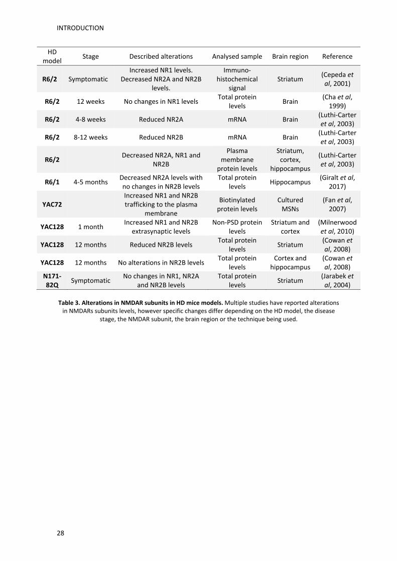
INTRODUCTION
28
HD model
Stage Described alterations Analysed sample Brain region Reference
R6/2 Symptomatic Increased NR1 levels.
Decreased NR2A and NR2B levels.
Immuno‐histochemical
signal Striatum
(Cepeda et al, 2001)
R6/2 12 weeks No changes in NR1 levels Total protein
levels Brain
(Cha et al, 1999)
R6/2 4‐8 weeks Reduced NR2A mRNA Brain (Luthi‐Carter et al, 2003)
R6/2 8‐12 weeks Reduced NR2B mRNA Brain (Luthi‐Carter et al, 2003)
R6/2 Decreased NR2A, NR1 and
NR2B
Plasma membrane
protein levels
Striatum, cortex,
hippocampus
(Luthi‐Carter et al, 2003)
R6/1 4‐5 months Decreased NR2A levels with no changes in NR2B levels
Total protein levels
Hippocampus (Giralt et al,
2017)
YAC72 Increased NR1 and NR2B trafficking to the plasma
membrane
Biotinylated protein levels
Cultured MSNs
(Fan et al, 2007)
YAC128 1 month Increased NR1 and NR2B
extrasynaptic levels Non‐PSD protein
levels Striatum and
cortex (Milnerwood et al, 2010)
YAC128 12 months Reduced NR2B levels Total protein
levels Striatum
(Cowan et al, 2008)
YAC128 12 months No alterations in NR2B levels Total protein
levels Cortex and
hippocampus (Cowan et al, 2008)
N171‐82Q
Symptomatic No changes in NR1, NR2A
and NR2B levels Total protein
levels Striatum
(Jarabek et al, 2004)
Table 3. Alterations in NMDAR subunits in HD mice models. Multiple studies have reported alterations in NMDARs subunits levels, however specific changes differ depending on the HD model, the disease
stage, the NMDAR subunit, the brain region or the technique being used.

INTRODUCTION
29
3. Cdk5 implication in HD
Cdk5, like the others members of the CDK family, is a proline‐directed protein kinase that
phosphorylates Ser or Thr residues followed by a proline amino acid ((Kimura et al, 2014).
However, unlike the rest of the CDK family members, Cdk5 activity does not rely on Cyclins
nor on phosphorylation of its activation loop, but on the neuronal specific activator p35 (Tsai
et al, 1994). Thus, although recent studies have described Cdk5 involvement in maintaining
cell cycle suppression in post‐mitotic neurons, its classical roles are associated with many
crucial CNS functions, particularly synaptic plasticity processes (Angelo et al, 2006; Su and
Tsai, 2011). Moreover, deregulation of Cdk5 activity, mainly caused by calpain‐dependent
cleavage of p35 to p25, is a hallmark of several neurodegenerative disorders and
pathological conditions, including ischemia, Alzheimer’s disease or Huntington’s disease
(McLinden, 2012; Patrick et al, 1998). For these reasons, Cdk5 is a promising putative target
in neurodegenerative disorders and particularly as a dual contributor to both motor and
cognitive dysfunction in HD.
3.1. Cdk5 regulation
Cdk5 is expressed in most tissues with particularly high levels in the brain. However, as its
activators, p35 and p39, are expressed in neurons, its activity is mainly restricted to the CNS
(Tsai et al, 1994; Wu et al, 2000). Levels of Cdk5 activation are also determined by its
activators availability, as their levels are less abundant than Cdk5. Indeed, p35 and p39
present a short half‐life due to its rapid proteosomal degradation (Patrick et al, 1998;
Takasugi et al, 2016). In fact, phosphorylation of p35 by Cdk5 induces p35 proteosomal
degradation and suppresses its calpain‐dependent conversion to p25, thus regulating its
own activity (Kamei et al, 2007; Patrick et al, 1998; Wei et al, 2005). Increased Cdk5‐
dependent p35 proteosomal degradation has been described to take place after calcium
influx induced by glutamate receptors activation (Wei et al, 2005). However, calpain
activation leads to p25 production, the p35 cleaved form, which in contrast to p35 is
resistant to proteosomal degradation, thus constitutively activating Cdk5. In addition, the
myristoylation residue which allows plasma membrane localization of Cdk5/p35 is lost in
p25 leading to Cdk5 redistribution to the cytosol and alteration of its substrate specificity
(Asada et al, 2008; Kusakawa et al, 2000; Patrick et al, 1999) (Figure 10). Production of p25
has been widely associated with neurodegenerative disorders (Camins et al, 2006).
Although p35 and p39 are the main regulators of Cdk5 activity, it can also be regulated by
post‐translational modifications and by interaction with other proteins. For instance, CyclinI
has been described to activate Cdk5 (Brinkkoetter et al, 2009; Hagmann et al, 2015),
whereas interaction with CyclinE, CyclinD1 or GSTP1 (glutathione S‐transferase P1) has been
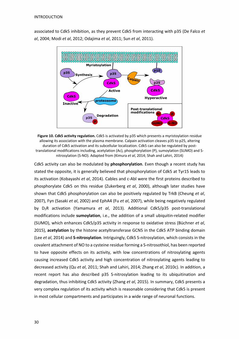
INTRODUCTION
30
associated to Cdk5 inhibition, as they prevent Cdk5 from interacting with p35 (De Falco et
al, 2004; Modi et al, 2012; Odajima et al, 2011; Sun et al, 2011).
Figure 10. Cdk5 activity regulation. Cdk5 is activated by p35 which presents a myristoylation residue allowing its association with the plasma membrane. Calpain activation cleaves p35 to p25, altering duration of Cdk5 activation and its subcellular localization. Cdk5 can also be regulated by post‐
translational modifications including, acetylation (Ac), phosphorylation (P), sumoylation (SUMO) and S‐nitrosylation (S‐NO). Adapted from (Kimura et al, 2014; Shah and Lahiri, 2014)
Cdk5 activity can also be modulated by phosphorylation. Even though a recent study has
stated the opposite, it is generally believed that phosphorylation of Cdk5 at Tyr15 leads to
its activation (Kobayashi et al, 2014). Cables and c‐Abl were the first proteins described to
phosphorylate Cdk5 on this residue (Zukerberg et al, 2000), although later studies have
shown that Cdk5 phosphorylation can also be positively regulated by TrkB (Cheung et al,
2007), Fyn (Sasaki et al, 2002) and EphA4 (Fu et al, 2007), while being negatively regulated
by D2R activation (Yamamura et al, 2013). Additional Cdk5/p35 post‐translational
modifications include sumoylation, i.e., the addition of a small ubiquitin‐related modifier
(SUMO), which enhances Cdk5/p35 activity in response to oxidative stress (Büchner et al,
2015), acetylation by the histone acetyltransferase GCN5 in the Cdk5 ATP binding domain
(Lee et al, 2014) and S‐nitrosylation. Intriguingly, Cdk5 S‐nitrosylation, which consists in the
covalent attachment of NO to a cysteine residue forming a S‐nitrosothiol, has been reported
to have opposite effects on its activity, with low concentrations of nitrosylating agents
causing increased Cdk5 activity and high concentration of nitrosylating agents leading to
decreased activity (Qu et al, 2011; Shah and Lahiri, 2014; Zhang et al, 2010c). In addition, a
recent report has also described p35 S‐nitrosylation leading to its ubiquitination and
degradation, thus inhibiting Cdk5 activity (Zhang et al, 2015). In summary, Cdk5 presents a
very complex regulation of its activity which is reasonable considering that Cdk5 is present
in most cellular compartments and participates in a wide range of neuronal functions.

INTRODUCTION
31
3.2. Cdk5 function
Since its first discovery after being cloned based on sequence homology with other CDKs
(Hellmich et al, 1992; Lew et al, 1992; Meyerson et al, 1992), much has been learned about
Cdk5 complex function. Dozens of Cdk5 substrates involved in multiple processes have been
described, underlining Cdk5 importance in neuronal function. First hints at Cdk5 role came
from observations that Cdk5 knockout mice resulted in perinatal death which correlated
with lack of cortical laminar structure and impaired axonal transport of neurofilaments (NF)
(Ohshima et al, 1996), thus implying an essential role for Cdk5 in developmental neuronal
migration. In fact, recent studies in a ferret model have shown that inhibition of Cdk5
Figure 11. Cdk5 functions in the CNS. Modulation of multiple substrates allows Cdk5 to participate in
the regulation of a plethora of essential processes in the CNS. Adapted from (Hori and Hoshino, 2017; Su and Tsai, 2011).
impairs normal cortical folding during development (Shinmyo et al, 2017). Nowadays, it is
known that Cdk5 participates in numerous neuronal processes (Figure 11). Cdk5
phosphorylates several transcription factors, such as STAT3 (Fu et al, 2004) or MEF2, whose
pro‐survival signalling is inhibited upon Cdk5 phosphorylation (Gong et al, 2003), and
participates in processes such as pain signalling (reviewed in (Utreras et al, 2009)) and in
development of neuromuscular junctions by modulating clustering of acetylcholine
receptors and neuregulin signalling (Fu et al, 2001, 2005; Lin et al, 2005). Finally, Cdk5 plays
an important role in synaptic plasticity modulation. Hence, it modulates dopamine
INTRODUCTION
32
signalling both by regulating dopamine synthesis and by inhibiting its signalling via
phosphorylation of DARPP32, a striatal‐enriched protein important for dopamine‐
dependent striatal LTP and LTD induction (Bibb et al, 1999; Calabresi et al, 2000; Moy and
Tsai, 2004). This function is particularly relevant in the striatum where Cdk5‐dependent
dopamine signalling modulation has been associated with cocaine reinforcement effects
(Benavides et al, 2007; Bibb et al, 2001a). Cdk5 also phosphorylates several presynaptic
substrates, such as Munc18, dynamin‐I or synapsin‐I, modulating exocytosis and recycling
of synaptic vesicles (reviewed in (Cheung et al, 2006; McLinden, 2012)), and several
postsynaptic substrates which will be discussed in more detail hereafter.
3.2.1. Cdk5 role in synaptic plasticity
Cdk5 role in synaptic plasticity was firstly discovered after observations that
pharmacological Cdk5 inhibition led to blocking of LTP induction in hippocampal slices and
to impairment of context‐dependent fear conditioning (Fischer et al, 2002, 2003; Li et al,
2001a). Since then, multiple studies have been performed using different genetic models to
inhibit Cdk5 or its activator p35. Functional effects of these models differ greatly, showing
both improvement and impairment of synaptic plasticity and learning and memory,
depending on the brain region being targeted, on the duration of the inhibition period and
even on the mouse strain being used (Table 4 and (Fischer et al, 2002, 2003)). For example,
p35 knockout mice present lower threshold for seizure‐induced lethality (Chae et al, 1997).
Similar results have been described after genetic Cdk5 inhibition in the CNS for a long period
of time, which leads to appearance of seizures associated with lower epileptiform activity
threshold. In contrast, Cdk5 genetic deletion for a short period of time improves spatial
learning which correlates with increased hippocampal LTP and NMDAR‐mediated excitatory
currents (Hawasli et al, 2007, 2009). Opposite effects depending on the duration of Cdk5
activity modification have also been found in mice models over‐expressing p25. Even though
increased levels of p25 have been associated with several neurodegenerative disorders, it
has been reported that transient p25 over‐expression facilitates LTP induction and
hippocampal‐dependent memory. In fact, it has been described that p25 is generated in
physiological conditions upon neuronal activation in a NR2B‐ and CaMKIIα‐dependent
manner (Seo et al, 2014). However, over‐expression of p25 for longer periods leads to
memory impairment and neuronal loss (Fischer et al, 2005). Importantly, prolonged p25
over‐expression in the hippocampus and cortex causes neuronal loss, whereas p25 increase
in the striatum causes synaptic alterations without neuronal death being detected (Cruz et
al, 2003; Meyer et al, 2008). Differential brain region‐specific effects have also been
observed after Cdk5 inhibition. Thus, specific Cdk5 deletion in the hippocampal CA1 causes
increased PDE levels leading to decreased cAMP‐dependent signalling, conversely Cdk5
inhibition in the striatum decreases PDE activity leading to increased cAMP levels (Guan et

INTRODUCTION
33
al, 2011; Plattner et al, 2015). This data illustrates the complex role of Cdk5 as a modulator
of synaptic plasticity and its homeostasis. In this sense, Cdk5 has also been shown to
regulate GKAP and SPAR, two proteins involved in synaptic scaling, this is, strength
adjustment of all excitatory synapses in a neuron in order to stabilize its firing (Roselli et al,
2011; Seeburg et al, 2008; Turrigiano, 2008). Moreover, a recent study has shown that Cdk5
represses NMDAR‐ and AMPAR‐dependent signalling in a particularly rapid response to
altered synaptic transmission (Sheng et al, 2016). Hence, Cdk5 fine‐tuned function appears
necessary for proper balance between enough synaptic activation, to ensure learning and
memory processes, and excessive neuronal activity, responsible for excitotoxicity.
Action Method Tissue Effect on synaptic plasticity Reference
Cdk5 inhibition
Pharmacological: Roscovitine
Hippocampal slices
Blocking of LTP induction and NMDA‐evoked currents in CA1 hippocampal neurons
(Li et al, 2001a)
Cdk5 inhibition
Pharmacological: Roscovitine and butyrolactone I
Brain slices No effect on LTP induction (Wang et al,
2004)
Cdk5 inhibition
Pharmacological: Roscovitine
Brain slices Increased percentage of population spikes that
exhibit LTP
(Miranda‐Barrientos et al, 2014)
Cdk5 inhibition
Pharmacological: Butyrolactone I microinjection
Septum and hippocampus
Impairment of context‐dependent fear conditioning and of its facilitation in response to stress
(Fischer et al, 2002, 2003)
Cdk5 inhibition
Pharmacological: Butyrolactone I microinjection
Hippocampus Facilitation of extinction of aversive freezing
behaviour (Sananbenesi et al, 2007)
Cdk5 inhibition
Genetic by shRNA‐miR
microinjection Hippocampus Increased LTP
(Posada‐Duque et al,
2016)
Cdk5 inhibition
Short genetic KO (2‐4 weeks)
CNS Improved spatial learning, enhanced hippocampal LTP and NMDAR‐mediated excitatory postsynaptic
currents
(Hawasli et al, 2007)
Cdk5 inhibition
Chronic genetic KO (8 weeks)
CNS Lower threshold for epileptiform activity and
seizures (Hawasli et al,
2009)
Cdk5 inhibition
Genetic KO Forebrain Hyperactivity in the open field, reduced anxiety, reduced behavioural despair, impaired spatial
learning in the MWM and in contextual fear memory
(Su et al, 2013)
Cdk5 inhibition
Genetic KO Forebrain Altered behavioural responses to acute or chronic
stress (Plattner et al,
2015)
Cdk5 inhibition
Genetic KO Hippocampus Impairment in memory formation and retrieval (Guan et al,
2011)
Cdk5 inhibition
Genetic KO Forebrain and dorsolateral striatum
Impairment of dopamine‐facilitated LTP, increased locomotor activity and attenuated motor learning
(Hernandez et al, 2016)
p35 inhibition
Transient genetic KO
General Impairment in spatial learning and memory, reduced anxiety‐like behaviour and defective LTD induction in
the hippocampus
(Mishiba et al, 2014)
p35 inhibition
Genetic KO General Lower threshold for LTP induction in hippocampal
slices (Wei et al, 2005)
p35 inhibition
Genetic KO General Deficits in spatial learning and memory, impaired
LTD and depotentiation of hippocampal LTP (Ohshima et al, 2005)

INTRODUCTION
34
Table 4. Functional effects of Cdk5 activity modulation on synaptic plasticity. Inhibition of Cdk5 or p35 and over‐expression of p25 have been described to have both positive and negative effects on synaptic plasticity induction, depending on the method used to modulate Cdk5 activity, the duration of this
modulation or the brain region being targeted. KO (knock‐ou), MWM (Morris Water Maze).
3.2.1.1. Cdk5 regulation of NMDA receptors
One of the Cdk5‐dependent mechanisms most frequently associated with its role in synaptic
plasticity regulation, is modulation of NMDARs. As mentioned before, they play an
important role in LTP and LTD processes and Cdk5 has been shown to regulate their surface
levels through different pathways. Thus, Cdk5 has been described to negatively regulate
phosphorylation levels of NR1 (Ser897), as Cdk5 inhibition leads to increased phospho‐NR1
levels and increased NMDAR activity (Chergui et al, 2004). NR2A total and phosphorylation
(Ser1232) levels have also been described to be positively regulated by Cdk5 in models of
ischemia, neuropathic pain and glaucoma (Chen et al, 2011; Wang et al, 2003; Yang et al,
2014). However, most studies describing Cdk5‐dependent NMDAR regulation focus on the
NR2B subunit (Figure 12). Accordingly, Cdk5 has been shown to phosphorylate NR2B at
residues Ser1284 and Ser1116. Ser1284 phosphorylation levels are increased after
hippocampal ischemic insults without being affected by neuronal activity, whereas Ser1116
phosphorylation levels, which lead to reduced NR2B surface levels, become reduced after
neuronal activation (Lu et al, 2015; Plattner et al, 2014). Cdk5 can also regulate NR2B surface
levels by physically interacting with NR2B and calpain, contributing to calpain‐dependent
NR2B degradation and consequent reduction of total and NR2B surface levels (Hawasli et al,
2007). Finally, Cdk5 is able to indirectly modulate NR2B surface levels by promoting its
internalization via the clathrin adaptor AP‐2. Phosphorylation of PSD‐95 by Cdk5 reduces Src
colocalization with PSD‐95 and its auto‐activation (phosphor‐Src (Tyr416)), leading to
reduced Src‐dependent NR2B phosphorylation at Tyr1472, which allows AP‐2 binding and
subsequent internalization (Zhang et al, 2008b). Direct Cdk5 phosphorylation of PSD‐95 has
also been shown to negatively regulate PSD‐95 clustering, which could also affect NMDARs
p25 over‐expression
Transient genetic over‐expression (1‐2 weeks)
General Enhancement of LTP and facilitation of hippocampal‐
dependent memory (Fischer et al,
2005)
p25 over‐expression
Prolonged genetic over‐expression
(6 weeks) General
Impairment of learning in fear conditioning and in the water maze paradigm
(Fischer et al, 2005)
p25 over‐expression
Genetic over‐expression
Forebrain
No alterations in the learning phase of the MWM but improved performance in the reversal phase.
Reduced contextual conditioning and enhanced tone fear conditioning
(Angelo et al, 2003)
p25 over‐expression
Genetic over‐expression
Forebrain Impaired LTP in male mice. Faster spatial learning in
females in the MWM (Ris et al, 2005)
p25 over‐expression
Genetic over‐expression
Forebrain Impaired contextual fear conditioning in female, but
not male (Mizuno et al,
2006)

INTRODUCTION
35
and other proteins which rely on PSD‐95 to localize at the PSD (Morabito et al, 2004). In
general, most of these mechanisms lead to a Cdk5‐dependent reduction of NMDARs subunit
levels, which could act as a “brake” for excessive NMDAR activation, but could also lead to
synaptic plasticity impairments if these mechanisms were not properly regulated.
Figure 12. Cdk5 regulation of NMDAR. Schematic representation of the Cdk5‐dependent pathways described to modulate NR2B surface levels.
3.2.1.2. Cdk5 modulation of dendritic spines
In adult neurons Cdk5 also plays an important role in cytoskeleton dynamics regulation, by
directly modulating cytoskeletal proteins, such as Tau, which is phosphorylated by Cdk5 at
different residues (Paudel et al, 1993) and F‐actin, which is stabilized by direct interaction
with p35 (He et al, 2011). Cdk5 also phosphorylates several substrates involved in
cytoskeletal and dendritic spine remodelling, such as S6K or CRMP2 (Jin et al, 2016; Lai et
al, 2015), and multiple substrates which positively and negatively modulate the members
of the small GTPases family, RhoA and Rac1. Specifically, it has been described that Cdk5
positively activates RhoA by phosphorylating ephexin1 and CaMKv (Fu et al, 2007; Liang et
al, 2016), while negatively regulating it during development through phosphorylation and
stabilization of p27 (Kawauchi et al, 2006). On the other hand, Cdk5 positively modulates
Rac1 by phosphorylating Kalirin‐7 (Xin et al, 2008), whereas Cdk5 phosphorylation of
RasGRF2, neurabin‐1, WAVE‐1 and Rac1 downstream Pak1, negatively regulate Rac1‐
dependent dendritic spine outgrowth (Causeret et al, 2007; Kesavapany et al, 2004; Kim et
al, 2006b; Nikolic et al, 1998). Thus, although Cdk5 is able to both promote and inhibit
dendritic spine density, it has been described that, in agreement with its positive regulation
of several Rac1 inhibitors, inhibition of hippocampal Cdk5 in vivo leads to increased Rac1
activity (Posada‐Duque et al, 2015). Once more, this function allows Cdk5 to modulate
synaptic strength on another level.
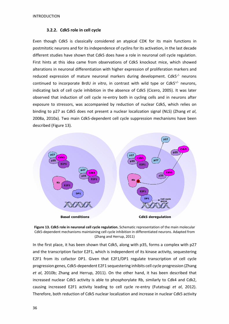
INTRODUCTION
36
3.2.2. Cdk5 role in cell cycle
Even though Cdk5 is classically considered an atypical CDK for its main functions in
postmitotic neurons and for its independence of cyclins for its activation, in the last decade
different studies have shown that Cdk5 does have a role in neuronal cell cycle regulation.
First hints at this idea came from observations of Cdk5 knockout mice, which showed
alterations in neuronal differentiation with higher expression of proliferation markers and
reduced expression of mature neuronal markers during development. Cdk5‐/‐ neurons
continued to incorporate BrdU in vitro, in contrast with wild type or Cdk5+/‐ neurons,
indicating lack of cell cycle inhibition in the absence of Cdk5 (Cicero, 2005). It was later
observed that induction of cell cycle re‐entry both in cycling cells and in neurons after
exposure to stressors, was accompanied by reduction of nuclear Cdk5, which relies on
binding to p27 as Cdk5 does not present a nuclear localization signal (NLS) (Zhang et al,
2008a, 2010a). Two main Cdk5‐dependent cell cycle suppression mechanisms have been
described (Figure 13).
Figure 13. Cdk5 role in neuronal cell cycle regulation. Schematic representation of the main molecular Cdk5‐dependent mechanisms maintaining cell cycle inhibition in differentiated neurons. Adapted from
(Zhang and Herrup, 2011)
In the first place, it has been shown that Cdk5, along with p35, forms a complex with p27
and the transcription factor E2F1, which is independent of its kinase activity, sequestering
E2F1 from its cofactor DP1. Given that E2F1/DP1 regulate transcription of cell cycle
progression genes, Cdk5‐dependent E2F1 sequestering inhibits cell cycle progression (Zhang
et al, 2010b; Zhang and Herrup, 2011). On the other hand, it has been described that
increased nuclear Cdk5 activity is able to phosphorylate Rb, similarly to Cdk4 and Cdk2,
causing increased E2F1 activity leading to cell cycle re‐entry (Futatsugi et al, 2012).
Therefore, both reduction of Cdk5 nuclear localization and increase in nuclear Cdk5 activity
INTRODUCTION
37
could cause loss of cell cycle inhibition in postmitotic neurons. Finally, it has been described
that glutamate‐dependent calpain activation, leads to Cdk5 dependent Cdh1
phosphorylation and inhibition. As Cdh1 is a cofactor of the E3 ubiquitin ligase APC/C
(anaphase‐promoting complex/cyclosome), APC/C activity is reduced, causing accumulation
of CyclinB1, reduction of p27 and activation of Cyclin D1/Cdk4 (Cataldo et al, 2013; Maestre
et al, 2008; Veas‐Pérez de Tudela et al, 2015). Calpain‐dependent p25 production has also
been described to inhibit HDAC1 activity which leads to increased DNA damage and cell
cycle proteins re‐expression (Kim et al, 2008). These findings indicate that deregulation of
Cdk5 could lead to neuronal cell cycle re‐entry through different mechanisms.
3.3. Cdk5 alteration in neurodegenerative disorders and neuropathological
conditions
As reviewed so far, due to its relevant role in so many neuronal processes, deregulation of
Cdk5 can have a big impact in neuronal function. Hence, deregulation of Cdk5 has been
described in many pathological conditions, especially in Alzheimer’s disease (AD). In fact,
first description of p35 conversion to p25 was detected in brains from AD patients, where it
was described to play a key role in Tau hyperphosphorylation (Patrick et al, 1999). Since
then, deregulation of Cdk5 has been associated to other AD hallmarks such as Aβ formation,
alterations in synaptic plasticity, cell cycle re‐entry or mitochondrial dysfunction (reviewed
in (Liu et al, 2016)). Moreover, several studies have shown that inhibition of Cdk5 is able to
ameliorate Tau pathology and cognitive deficits in AD mice models (reviewed in (Bhounsule
et al, 2017; López‐Tobón et al, 2011)).
Cdk5 deregulation associated to p25 production, as well as neurodegeneration amelioration
following Cdk5/p25 inhibition, has also been described in models of Parkinson’s disease and
ischemic injury (Gutiérrez‐Vargas et al, 2015; Meyer et al, 2014; Smith et al, 2003; Zhang et
al, 2016). In addition, aberrant Cdk5 activity has been associated with other
neurodegenerative diseases such as amyotrophic lateral sclerosis (ALS) and prion disease
(Nguyen et al, 2001; Rouget et al, 2015) and even with psychiatric disorders like major
depression and schizophrenia (Engmann et al, 2011; Ramos‐Miguel et al, 2013; Zhu et al,
2012).
3.3.1. Cdk5 deregulation in Huntington’s disease
In HD, Cdk5 has been associated to several processes involved in striatal vulnerability (Figure
14). For instance, it has been described that physiological Cdk5 phosphorylation of HTT at
Ser434 impairs caspase‐mediated HTT cleavage and therefore its aggregation, while
phosphorylation at Ser1181 and Ser1201 confers cell protection against polyQ‐induced
toxicity (Anne et al, 2007; Luo et al, 2005). Interestingly, Htt phosphorylation at S1181/1201,

INTRODUCTION
38
which is induced by DNA damage, has also been associated with BDNF axonal transport and
release (Ben M’Barek et al, 2013).
Importantly, reduced Cdk5 and p35 levels have been reported in the striatum of HD patients
and in knock‐in HdhQ111 mice (Anne et al, 2007; Paoletti et al, 2008), whereas increased p25
levels have been observed in the striatum of YAC128 after quinolinic acid injections, and in
human HD patients and in knock‐in HdhQ111 mice, which also present increased phospho‐
Tau levels, thus indicating aberrant Cdk5 activation in HD (Paoletti et al, 2008; Park et al,
2012). Increased p25 levels have also been described in HD‐like pharmacological models
using kainic acid or 3‐NP (Crespo‐Biel et al, 2007; Putkonen et al, 2011). Moreover, aberrant
Cdk5 activation has been shown to contribute to mHTT‐mediated striatal vulnerability to
dopaminergic and glutamatergic activation (Paoletti et al, 2008) and in increased
mitochondrial fragmentation (Cherubini et al, 2015). Thus, these studies indicate that Cdk5
dysfunction participates in HD striatal vulnerability pathways, like excitotoxicity and
mitochondrial dysfunction, however its role in other mechanisms such as cell cycle re‐entry,
have not been analysed. Importantly, despite Cdk5 pivotal function as a synaptic plasticity
modulator, no studies have determined its possible involvement in HD cognitive deficits.
Figure 14. Cdk5 association with processes involved in HD striatal vulnerability. Relationship between Cdk5 and processes associated with HD striatal vulnerability. Cdk5 has been described to contribute to striatal vulnerability factors, such as mitochondrial fragmentation, mHTT aggregate formation or BDNF transport and release, whereas some mechanisms described to be altered in HD, like deregulation of
calcium homoeostasis or DNA damage, modify Cdk5 activity.


AIMS


AIMS
43
Huntington’s disease is characterized by the presence of motor deficits, mostly associated
to striatal vulnerability, and by cognitive impairments, caused by aberrant corticostriatal
connectivity and hippocampal dysfunction, which are related with alterations in synaptic
plasticity. For this reason, characterization of molecular mechanisms involved both in
neuronal degeneration and synaptic plasticity dysfunction, could help to design new
therapeutic approaches to target all together motor and cognitive deficits in HD. In this
Thesis, we have focused on Cdk5 as one of these molecular targets.
On one hand, we aimed to study Cdk5 contribution to cognitive decline in HD. Cdk5 has a
pivotal role in synaptic plasticity regulation, and deregulation of Cdk5 activity has been
described in HD. Therefore, we hypothesized that Cdk5 alterations could support cognitive
deficits appearance in HD. On the other hand, Cdk5 deregulation has been associated with
aberrant cell cycle re‐entry of differentiated neurons in response to several neuronal
stressors, and Cdk5 activity deregulation has been described to heighten striatal
vulnerability in HD. Hence, we aimed to study whether neuronal cell cycle re‐entry resulting
from Cdk5 deregulation could contribute to striatal vulnerability in HD. Therefore, the aims
of this Thesis are:
1. TO ANALYSE THE ROLE OF CDK5 IN COGNITIVE DEFICITS IN HD.
1.1 To generate and validate a new transgenic mouse model that expresses mHtt and
has genetically reduced Cdk5 levels.
1.2 To analyse the effects of Cdk5 genetic reduction on cognitive deficits development
in mHtt KI mice.
1.3 To analyse Cdk5 role in synaptic plasticity alterations underlying cognitive
impairment in mHtt KI mice.
2. TO ANALYSE NEURONAL CELL CYCLE RE‐ENTRY AS A PUTATIVE MECHANISM FOR
CDK5‐DEPENDENT STRIATAL VULNERABILITY IN HD.
2.1 To analyse whether Cdk5 function as a cell cycle modulator is altered in mHtt KI mice.
2.2 To analyse if mHtt presence affects cell cycle proteins expression in HD.
2.3 To determine whether alteration of cell cycle proteins contributes to increased
NMDA excitotoxic response in mHtt KI striatal primary cultures.


METHODS


METHODS
47
1. MICE MODELS
All mice were housed with access to food and water ad libitum in a colony room kept at 19‐
22°C and 40‐60% humidity, under a 12h light/dark cycle. All mice used were males, in order
to avoid estrus hormonal alterations, and were housed together in numerical birth order in
groups of mixed genotypes. Data was recorded for analysis by microchip mouse number and
genotype was determined by PCR analysis. All procedures were performed in compliance
with the National Institutes of Health Guide for the Care and Use of Laboratory Animals, and
approved by the local animal care committee of the Universitat de Barcelona (76/15) and
Generalitat de Catalunya (00/1094), in accordance with the Directive 2010/63/EU of the
European Commission.
1.1. Huntington’s disease model
Knock‐in HdhQ7/Q111 mice, maintained on a C57BL/6 genetic background, and originally
obtained from Dr. M. MacDonald’s lab (Lloret et al, 2006), were used as HD mice model for
all experiments. These mice present cognitive deficits at 6 months of age, motor deficits at
8 months of age and a normal life span (Brito et al, 2014; Puigdellívol et al, 2015).
1.2. Double mutant KI:Cdk5+/‐ model
Newly generated transgenic double mutant KI:Cdk5+/‐ mice were obtained from knock‐in
HdhQ7/Q111 mice and homozygous floxed Cdk5 mice (Cdk5flox/flox) as shown in Figure 15.
Cdk5flox/flox mice were obtained from Dr. P. Greengard’s lab and were generated by flanking
encoding Cdk5 catalytic domain regions with loxP sequences (floxed) using homologous
recombination (Hawasli et al, 2007). Cre‐CaMKII+/‐ mice were obtained from Dr. C. Saura’s
lab and they express Cre recombinase protein under the α‐CaMKII promoter, restricting Cre
recombinase expression to adult forebrain neurons (Dragatsis and Zeitlin, 2000).
We crossed homozygous Cdk5 floxed and homozygous knock‐in HdhQ111/Q111 mice to obtain
heterozygous Cdk5 floxed and heterozygous knock‐in mice (Cdk5flox/+; HdhQ7/Q111 mice).
These mice were crossed again with homozygous Cdk5 floxed mice to obtain homozygous
Cdk5 floxed and heterozygous knock‐in mice (Cdk5flox/flox; HdhQ7/Q111 mice). We crossed
homozygous Cdk5 floxed and heterozygous knock‐in mice with Cre‐expressing mice to
obtain the final genotypes: Cdk5flox/+, HdhQ7/Q7, Cre‐/‐ (HdhQ7/Q7;Cdk5+/+ or wild‐type (WT)),
Cdk5flox/+, HdhQ7/Q111, Cre‐/‐ (HdhQ7/Q111;Cdk5+/+ or mHtt knock‐in (KI)), Cdk5flox/+, HdhQ7/Q7,
Cre+/‐ (HdhQ7/Q7;Cdk5+/‐ or Cdk5 heterozygous (Cdk5+/‐)), Cdk5flox/+, HdhQ7/Q111, Cre+/‐
(HdhQ7/Q111;Cdk5+/‐ or double mutant (KI:Cdk5+/‐)). All mice were maintained on a C57BL/6
genetic background.

METHODS
48
Figure 15. Generation of double mutant KI:Cdk5+/‐ mice. The stated crossing were performed to obtained the final four used genotypes: wild‐type (WT), knock‐in (KI), Cdk5+/‐ and double mutant
KI:Cdk5+/‐ mice.
2. HUMAN SAMPLES
Putamen samples from HD patients and control individuals were obtained from the
Neurological Tissue Bank of the Biobank‐Hospital Clínic‐Institut d’Investigacions
Biomèdiques August Pi i Sunyer (IDIBAPS) following the guidelines and approval of the local
ethics committee (Hospital Clínic de Barcelona’s Clinical Research Ethics Committee). Details
from control and HD patients are provided in Table 5.
Registration number
Clinical diagnosis Sex Age
CS‐0810 Control Female 81 CS‐1491 Control Male 83 CS‐1563 Control Male 79 CS‐1570 Control Female 86 CS‐1679 Control Female 90 CS‐1697 Control Male 78 CS‐1733 Control Male 76 CS‐1774 Control Female 74

METHODS
49
CS‐1334 HD ‐ VS 1, 40 CAG repeats Male 73 CS‐1630 HD ‐ VS 2, 20/40 CAG repeats Male 76 CS‐1638 HD ‐ VS 2 Male 72 CS‐1758 HD ‐ VS 2‐3, 42 CAG repeats Male 68 CS‐1120 HD ‐ VS 3, 48 CAG repeats Male 55 CS‐1294 HD ‐ VS 3, 45 CAG repeats Male 53 CS‐1438 HD ‐ VS 3, 40 CAG repeats Male 85 CS‐1193 HD ‐ VS 3‐4 Male 55
Table 5. Human post‐mortem brain samples. Information relative to human control and HD patients brain samples indicating sex, age, Vonsattel grades and number of CAG repeats when available.
3. STRIATAL PRIMARY CULTURES
Striatums from E18 WT and mHtt KI mice embryos were dissected and mechanically
dissociated. Densities of 160,000 cells and 500,000 cells were seeded onto 12 mm coverslips
in 24‐well plates for immunocytochemistry, and onto 12‐well plates for Western blot
analysis, respectively. Plates were pre‐coated with 0.1 mg/mL poly‐D‐lysine (P0899, Sigma)
and dissociated cells were cultured with Neurobasal Medium (21103‐049, Gibco)
supplemented with 1% Glutamax (35050‐038, Gibco) and 2% B27 (17504‐044, Gibco) for 21
days in vitro (DIV). Cultures were maintained at 37°C in a humidified atmosphere containing
5% CO2. Embryos genotype was determined by PCR analysis.
When stated, 21 DIV cultures were treated with NMDA (M3262, Sigma) at 10µM, 50µM or
100µM for 1h, 3h or 24h as specified in each experiment. To inhibit calpain activity, cultures
were pre‐treated with 10µM ALLN (Calbiochem) for 1 hour prior to NMDA treatment.
Cultures were fixed or harvested immediately after treatment.
4. BEHAVIOURAL ASSESSMENT
Separate cohorts of 6‐month old WT, mHtt KI, Cdk5+/‐ and mHtt KI:Cdk5+/‐ mice, were used
to evaluate corticostriatal function using the accelerating rotarod training procedure (ARTP)
and the swimming T‐Maze of strategy shifting and hippocampal‐dependent memory using
the T‐maze spontaneous alternation task (T‐SAT) and the novel object recognition test
(NORT), respectively. To reduce the effect of stress and anxiety on subsequent tasks, the
least stressful and anxiety‐inducing tasks were performed first as indicated: ARTP, T‐SAT,
NORT, swimming T‐Maze of strategy shifting. All experiments were performed blind to the
experimental condition.
4.1. Corticostriatal‐dependent tasks
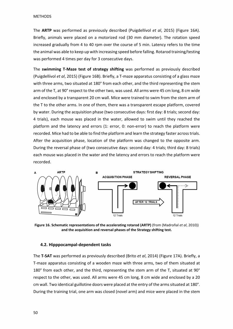
METHODS
50
The ARTP was performed as previously described (Puigdellívol et al, 2015) (Figure 16A).
Briefly, animals were placed on a motorized rod (30 mm diameter). The rotation speed
increased gradually from 4 to 40 rpm over the course of 5 min. Latency refers to the time
the animal was able to keep up with increasing speed before falling. Rotarod training/testing
was performed 4 times per day for 3 consecutive days.
The swimming T‐Maze test of strategy shifting was performed as previously described
(Puigdellívol et al, 2015) (Figure 16B). Briefly, a T‐maze apparatus consisting of a glass maze
with three arms, two situated at 180° from each other, and the third representing the stem
arm of the T, at 90° respect to the other two, was used. All arms were 45 cm long, 8 cm wide
and enclosed by a transparent 20 cm wall. Mice were trained to swim from the stem arm of
the T to the other arms. In one of them, there was a transparent escape platform, covered
by water. During the acquisition phase (two consecutive days: first day: 8 trials; second day:
4 trials), each mouse was placed in the water, allowed to swim until they reached the
platform and the latency and errors (1: error, 0: non‐error) to reach the platform were
recorded. Mice had to be able to find the platform and learn the strategy faster across trials.
After the acquisition phase, location of the platform was changed to the opposite arm.
During the reversal phase of (two consecutive days: second day: 4 trials; third day: 8 trials)
each mouse was placed in the water and the latency and errors to reach the platform were
recorded.
Figure 16. Schematic representations of the accelerating rotarod (ARTP) (from (Madroñal et al, 2010)) and the acquisition and reversal phases of the Strategy shifting test.
4.2. Hipppocampal‐dependent tasks
The T‐SAT was performed as previously described (Brito et al, 2014) (Figure 17A). Briefly, a
T‐maze apparatus consisting of a wooden maze with three arms, two of them situated at
180° from each other, and the third, representing the stem arm of the T, situated at 90°
respect to the other, was used. All arms were 45 cm long, 8 cm wide and enclosed by a 20
cm wall. Two identical guillotine doors were placed at the entry of the arms situated at 180°.
During the training trial, one arm was closed (novel arm) and mice were placed in the stem

METHODS
51
arm of the T (home arm) and allowed to explore this arm and the other available arm
(familiar arm) for 10 min, after which they were returned to the home cage. After 5 h (long‐
term memory), mice were placed in the stem arm of the T‐maze and allowed to freely
explore all three arms for 5 min. The arm preference was measured as the percentage of
time exploring the novel arm.
The NORT was performed as previously described (Brito et al, 2014) (Figure 17B). Briefly, a
40 cm diameter and 40 cm high white circular arena, was used. First, mice were habituated
to the arena in the absence of objects (2 days, 10 min/day). On the third day, two similar
objects were presented to each mouse for 10 min. Later, they were returned to their home
cage. 24 hours later, the same animals were re‐tested for 5 min in the arena with a familiar
and a new object. The object preference was measured as the percentage of time exploring
the new object.
Figure 17. Schematic representations of the T‐maze spontaneous alternation task (T‐SAT) and the novel object recognition test (NORT).
5. PROTEIN EXTRACTION
5.1. From mice and human brain tissue
WT, mHtt KI, Cdk5+/‐ and mHtt KI:Cdk5+/‐ mice were killed by cervical dislocation at the stated
age in each experiment. Brains were quickly removed, desired regions were dissected,
frozen in dry ice and stored at ‐80°C until use.
In Aim 1, same sets of samples were used for Western blot analysis and
immunoprecipitation studies simultaneously. To this end, brain tissue was homogenized in
cold lysis buffer (140 mM NaCl, 10 mM HEPES (pH 7.4), 1 mM EGTA, 0.1 mM MgCl2, 1% X‐
100 Triton, 0.5% NP‐40 supplemented with 1 mM sodium orthovanadate and protease
inhibitor cocktail (Sigma‐Aldrich)) with a dounce homogenizer, left 1 hour at 4°C in ice and
cleared by centrifugation at 13,000g for 15 min at 4°C to collect the supernatants.
In Aim 2, different sets of samples were used for Western blot analysis and
immunoprecipitation studies. Therefore, brain tissue used solely for Western blot studies,
was sonicated in cold lysis buffer (150 mM NaCL, 20 mM Tris, 50 mM NaF, 1% NP‐40, 10%

METHODS
52
glycerol supplemented with 1 mM sodium orthovanadate and protease inhibitor cocktail
(Sigma‐Aldrich)) and centrifuged at 16,000g for 15 minutes at 4°C to collect the
supernatants.
5.2. From primary cultures
Striatal primary cultures grown in 12‐well plates at DIV21 were rinsed with PBS, harvested
in Lysis Buffer (50 mM Tris‐HCl (pH = 7.5), 150 mM NaCl, 2 mM EDTA, 1% NP‐40
supplemented with 1 mM sodium orthovanadate and protease inhibitor cocktail (Sigma‐
Aldrich)) and centrifuged at 16,100g, for 10 minutes at 4°C to collect the supernatants.
5.3. Protein quantification
Protein concentration was assessed with a colorimetric assay following detergent
solubilization similar to the Lowry assay (Lowry et al, 1951) using the Detergent‐Compatible
Protein Assay kit (Bio‐Rad Laboratories) as indicated by the manufacturer’s instructions. 15
minutes after adding the provided reagents to the samples, absorbance at 650‐750 nm was
measured in a Synergy 2TM Multi‐Mode Multiplate Reader (BioTek). A standard curve
obtained from a 16 µg/µL BSA (bovine serum albumin) stock was used as reference,
duplicates were analysed per each sample and only samples with a standard deviation lower
than 0.5 were used for further experiments.
6. WESTERN BLOTTING
Protein extracts (10‐20 g) were mixed with 5X SDS sample buffer, boiled for 5 min, resolved
on 6‐10% SDS‐PAGE and transferred to nitrocellulose membranes (Whatman Schleicher &
Schuell). After 1h incubation in blocking buffer containing 10% non‐fat powdered milk in
TBS‐T (50 mM Tris‐HCl, 150 mM NaCl, pH 7.4, 0.05% Tween 20), membranes were blotted
overnight at 4°C with the indicated primary antibodies:
Antigen Dilution Molecular
Weight (kDa) Source Reference
Cdk5 (J‐3) 1:1000 30 Santa Cruz
Biotechnologies sc‐6247
p35 (C‐19) 1:500 35 Santa Cruz
Biotechnologies sc‐820
NR1 1:1000 150 Chemicon MAB363
NR2A 1:1000 180 Millipore AB1555
NR2B 1:1000 180 Cell Signaling Technology 4207
µ‐calpain 1:1000 80 Sigma C5736
pNR2B (S1116) 180 James A. Bibb Lab
pSrc (Y416) 1:1000 60 Cell Signaling Technology 2101

METHODS
53
Src 1:2000 60 Cell Signaling Technology 2110
pNR2B (Y1472) 1:1000 180 Cell Signaling Technology 4208
Rac1 1:2000 20 BD Biosciences 612652
(PHF)‐1 1:1000 50 Peter Davis Lab
pCDK substrates 1:2000 50‐250 Cell Signaling Technology 9477
pIPP1 (S67) 1:500 19 Santa Cruz
Biotechnologies sc‐14268
Lamin B1 1:1000 67 Abcam
RhoGDIα 1:1000 30 Santa Cruz
Biotechnologies sc‐359
p27 1:1000 27 BD Biosciences 610241
E2F1 1:500 60 Santa Cruz
Biotechnologies sc‐251
Cyclin D1 1:1000 30 Abcam ab134175
Cdk4 1:1000 30 Santa Cruz
Biotechnologies sc‐260
spectrin 1:2000 250/150 Chemicon MAB1622
Cleaved caspase 3 1:1000 17 Cell Signaling Technology 9661
Table 6. Primary antibodies used for Western blot. Dilution, molecular weight, source and reference for each used primary antibody are indicated.
Membranes were then rinsed three times with TBS‐T and incubated with horseradish
peroxidase‐conjugated mouse or rabbit secondary antibody (1:3000; Promega) for 1h at
room temperature. After washing for 30 min with TBS‐T, membranes were developed using
the enhanced chemiluminescence ECL kit (Santa Cruz Biotechnology).
Loading control was performed by reproving the membranes with the following antibodies
depending on the sample being analysed:
Antigen Sample Molecular
Weight (kDa) Source Reference
α‐actin Total lysate and cytosolic
fraction 42 MP Biomedicals 69100
Transferrin receptor
Plasma membrane fraction
95 Invitrogen 13‐6800
Lamin A/C Nuclear fraction 50‐60 Santa Cruz
Biotechnologies sc‐20681
Table 7. Primary antibodies used as loading control. Sample, molecular weight, source and reference for each used primary antibody are indicated.
ImageJ software was used to quantify the different densitometry immunoreactive bands
relative to the intensity of the loading control band in the same membranes within a linear
range of detection for the ECL reagent. Data is expressed as the mean SEM of band density.
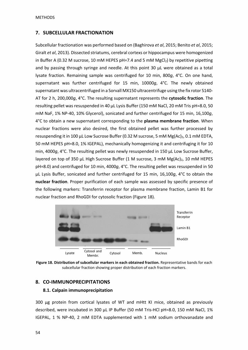
METHODS
54
7. SUBCELLULAR FRACTIONATION
Subcellular fractionation was performed based on (Baghirova et al, 2015; Benito et al, 2015;
Giralt et al, 2013). Dissected striatums, cerebral cortexs or hippocampus were homogenized
in Buffer A (0.32 M sucrose, 10 mM HEPES pH=7.4 and 5 mM MgCl2) by repetitive pipetting
and by passing through syringe and needle. At this point 30 µL were obtained as a total
lysate fraction. Remaining sample was centrifuged for 10 min, 800g, 4°C. On one hand,
supernatant was further centrifuged for 15 min, 10000g, 4°C. The newly obtained
supernatant was ultracentrifuged in a Sorvall MX150 ultracentrifuge using the fix rotor S140‐
AT for 2 h, 200,000g, 4°C. The resulting supernatant represents the cytosolic fraction. The
resulting pellet was resuspended in 40 μL Lysis Buffer (150 mM NaCl, 20 mM Tris pH=8.0, 50
mM NaF, 1% NP‐40, 10% Glycerol), sonicated and further centrifuged for 15 min, 16,100g,
4°C to obtain a new supernatant corresponding to the plasma membrane fraction. When
nuclear fractions were also desired, the first obtained pellet was further processed by
resuspending it in 100 μL Low Sucrose Buffer (0.32 M sucrose, 5 mM Mg(Ac)2, 0.1 mM EDTA,
50 mM HEPES pH=8.0, 1% IGEPAL), mechanically homogenizing it and centrifuging it for 10
min, 4000g, 4°C. The resulting pellet was newly resuspended in 150 μL Low Sucrose Buffer,
layered on top of 350 μL High Sucrose Buffer (1 M sucrose, 3 mM Mg(Ac)2, 10 mM HEPES
pH=8.0) and centrifuged for 10 min, 4000g, 4°C. The resulting pellet was resuspended in 50
μL Lysis Buffer, sonicated and further centrifuged for 15 min, 16,100g, 4°C to obtain the
nuclear fraction. Proper purification of each sample was assessed by specific presence of
the following markers: Transferrin receptor for plasma membrane fraction, Lamin B1 for
nuclear fraction and RhoGDI for cytosolic fraction (Figure 18).
Figure 18. Distribution of subcellular markers in each obtained fraction. Representative bands for each
subcellular fraction showing proper distribution of each fraction markers.
8. CO‐IMMUNOPRECIPITATIONS
8.1. Calpain immunoprecipitation
300 µg protein from cortical lysates of WT and mHtt KI mice, obtained as previously
described, were incubated in 300 µL IP Buffer (50 mM Tris‐HCl pH=8.0, 150 mM NaCl, 1%
IGEPAL, 1 % NP‐40, 2 mM EDTA supplemented with 1 mM sodium orthovanadate and

METHODS
55
protease inhibitor cocktail (Sigma‐Aldrich)) with 5 µL of calpain primary antibody or anti‐
mouse IgG (Jackson ImmunoResearch) for the negative control, overnight at 4°C with
rotation. The following day 40 µL protein A‐sepharose (sc‐2001, Santa Cruz Biotechnology)
were added in each tube and further incubated overnight at 4°C with rotation. Samples were
centrifuged for 5 minutes, at 1500g at 4°C and rinsed three times in decreasing dilutions of
IP Buffer in PBS. Finally, beads were incubated in 10 µL SDS loading buffer with 10% β‐
mercaptoethanol, boiled at 100°C for 10 minutes, spun down and analysed by Western
blotting as previously described. HRP‐conjugated protein A/G (32490, Thermo) was used to
detect the primary antibodies avoiding IgG light and heavy chains detection.
8.2. Cdk5 immunoprecipitation
200 µg protein from striatal lysates from WT and mHtt KI mice, obtained as previously
described, were incubated in 300 µL IP Lysis Buffer (140 mM NaCl, 10 mM HEPES (pH 7.4),
1 mM EGTA, 0.1 mM MgCl2, 1% X‐100 Triton, 0.5% NP‐40) with 2 µL agarose‐conjugated
Cdk5 primary antibody (sc‐173, Santa Cruz Biotechnology) or with 2 µL protein A‐sepharose
(sc‐2001, Santa Cruz Biotechnology) for negative controls, overnight at 4°C with rotation.
Samples were centrifuged for 5 minutes, at 1500g at 4°C and rinsed three times in
decreasing dilutions of IP Lysis Buffer in PBS. Finally, beads were incubated in 10 µL SDS
loading buffer with 10% β‐mercaptoethanol, boiled at 100°C for 10 minutes, spun down and
analysed by Western blotting as previously described.
9. RAC1 ACTIVITY ASSAY
Rac1 activity assay was performed as previously described (Puigdellívol et al, 2015). Briefly,
hippocampus and cortex from WT, mHtt KI, Cdk5+/‐ and mHtt KI:Cdk5+/‐ mice were lysed in
Pull‐down lysis buffer (25 mM HEPES, pH=7.5, 150 mM NaCl, 10 mM MgCl2, 1 mM EDTA, 1%
NP‐40, 2% Glycerol) and centrifuged for 15 minutes at 16,000 g at 4°C. Cleared lysates were
quantified and 600 μg of total protein were incubated with 10 μg PAK‐GST protein beads
(Cytoskeleton Inc.) for 1 hour at 4°C in rotation. Then, PAK‐GST beads were pelleted by
centrifugation at 5000 g, for 1 minute at 4°C, washed 3 times with Pull‐down lysis buffer, re‐
suspended with 5X SDS sample buffer with β‐mercaptoethanol and boiled for 5 minutes.
The resulting samples were loaded to perform Western blotting analysis as previously
described and levels of GTP‐bound Rac1 were determined. Controls were incubated with 10
μM EDTA and 200 μM GTPγS (positive control) or 1 mM GDP (negative control) at 30°C and
the reaction was stopped with 60 mM MgCl2 after 30 minutes.
METHODS
56
10. BIOTINYLATION ASSAY
Biotinylation assay was performed as previously described (Gabriel et al, 2014). Briefly, WT,
mHtt KI, Cdk5+/‐ and mHtt KI:Cdk5+/‐ mice were killed by cervical dislocation at 8 months of
age. Brains were quickly removed, placed in oxygenated ice‐cold ACSF buffer (125 mM NaCl,
2.5 mM KCl, 1.2 mM NaH2PO4, 1.2 mM MgCl2, 2.4 mM CaCl2, 26 mM NaHCO3 and 11 mM
glucose) and processed in a vibratome to obtain 400 μm brain slices. The slices were
incubated for 30 minutes with 0.5 mg/ml sulfo‐NHS‐SS‐Biotin (Sigma), rinsed with ice‐cold
ACSF and with quench buffer (ACSF with 100mM glycine) 3‐6 times. Then, slices were
incubated with quench buffer for 20 minutes and rinsed 3 times with ACSF buffer. Striatum,
cerebral cortex and hippocampus from each slice were dissected, and lysed with lysis buffer
(150 mM NaCl, 20 mM Tris (pH = 8.0), 50 mM NaF, 1% NP‐40, 10% glycerol supplemented
with 1 mM sodium orthovanadate and protease inhibitor cocktail (Sigma‐Aldrich)). Samples
were centrifuged for 15 minutes, at 16000g at 4°C and supernatants were collected. After
protein quantification, 150 µg of protein were incubated in Binding Buffer (100 mM K2PO4,
150 mM NaCl and 1% NP‐40 (pH=7.2)) with 40 μL of NeutrAvidin beads (Thermo Scientific)
at 4°C in overnight rotation. Then, samples were centrifuged 2 minutes at 2500g at 4°C and
rinsed 4 times with decreasing amounts of Binding Buffer in PBS. Finally, pellets were re‐
suspended with 5X SDS sample buffer with β‐mercaptoethanol and boiled for 10 minutes.
Biotinylated samples were loaded to perform Western blotting analysis as previously
described and NR2B surface levels were determined.
11. IMMUNOCYTOCHEMISTRY
Striatal primary cultures were grown in coverslips and fixed at DIV21 with 4%
paraformaldehyde (PFA) for 10 minutes, rinsed with PBS and incubated with 0.1 M Glycine
for 15 minutes. Coverslips were rinsed 3 times in PBS, incubated in blocking solution (0.3%
Triton X‐100, 1% BSA and 1% Normal Donkey Serum in PBS) for 45 minutes at room
temperature and incubated overnight at 4°C with blocking solution containing the
corresponding primary antibodies:
Antigen Dilution Host Species Source Reference
p27 1:500 mouse BD Biosciences 610241
E2F1 1:100 mouse Santa Cruz Biotechnologies sc‐251
Cdk4 1:100 rabbit Santa Cruz Biotechnologies sc‐260
CyclinD1 1:100 rabbit Abcam ab134175
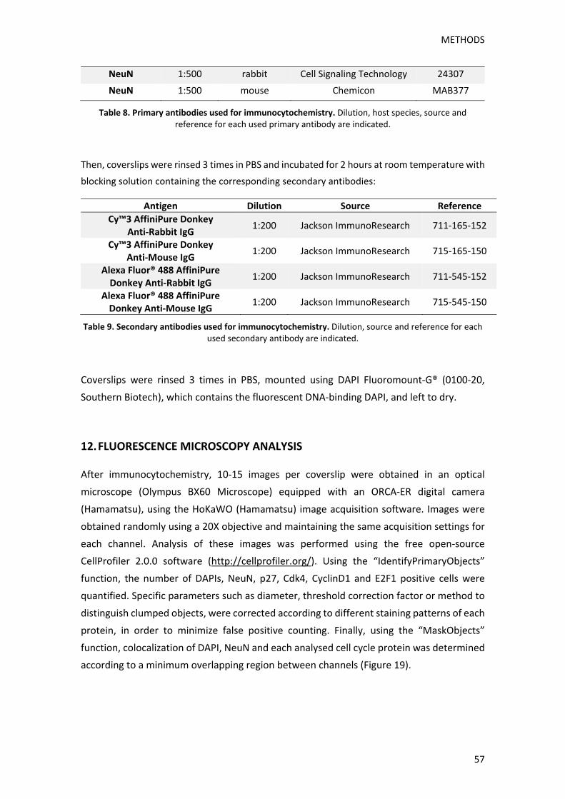
METHODS
57
NeuN 1:500 rabbit Cell Signaling Technology 24307
NeuN 1:500 mouse Chemicon MAB377
Table 8. Primary antibodies used for immunocytochemistry. Dilution, host species, source and reference for each used primary antibody are indicated.
Then, coverslips were rinsed 3 times in PBS and incubated for 2 hours at room temperature with
blocking solution containing the corresponding secondary antibodies:
Antigen Dilution Source Reference
Cy™3 AffiniPure Donkey Anti‐Rabbit IgG
1:200 Jackson ImmunoResearch 711‐165‐152
Cy™3 AffiniPure Donkey Anti‐Mouse IgG
1:200 Jackson ImmunoResearch 715‐165‐150
Alexa Fluor® 488 AffiniPure Donkey Anti‐Rabbit IgG
1:200 Jackson ImmunoResearch 711‐545‐152
Alexa Fluor® 488 AffiniPure Donkey Anti‐Mouse IgG
1:200 Jackson ImmunoResearch 715‐545‐150
Table 9. Secondary antibodies used for immunocytochemistry. Dilution, source and reference for each used secondary antibody are indicated.
Coverslips were rinsed 3 times in PBS, mounted using DAPI Fluoromount‐G® (0100‐20,
Southern Biotech), which contains the fluorescent DNA‐binding DAPI, and left to dry.
12. FLUORESCENCE MICROSCOPY ANALYSIS
After immunocytochemistry, 10‐15 images per coverslip were obtained in an optical
microscope (Olympus BX60 Microscope) equipped with an ORCA‐ER digital camera
(Hamamatsu), using the HoKaWO (Hamamatsu) image acquisition software. Images were
obtained randomly using a 20X objective and maintaining the same acquisition settings for
each channel. Analysis of these images was performed using the free open‐source
CellProfiler 2.0.0 software (http://cellprofiler.org/). Using the “IdentifyPrimaryObjects”
function, the number of DAPIs, NeuN, p27, Cdk4, CyclinD1 and E2F1 positive cells were
quantified. Specific parameters such as diameter, threshold correction factor or method to
distinguish clumped objects, were corrected according to different staining patterns of each
protein, in order to minimize false positive counting. Finally, using the “MaskObjects”
function, colocalization of DAPI, NeuN and each analysed cell cycle protein was determined
according to a minimum overlapping region between channels (Figure 19).
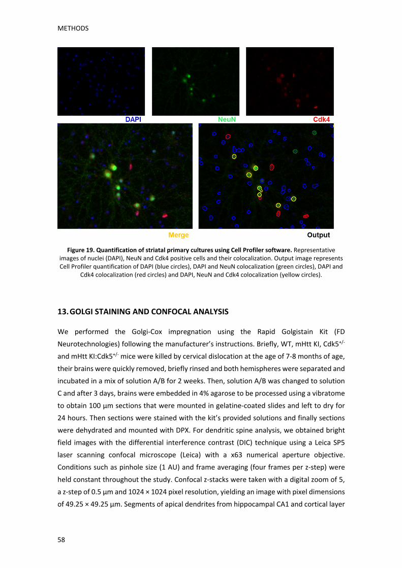
METHODS
58
Figure 19. Quantification of striatal primary cultures using Cell Profiler software. Representative images of nuclei (DAPI), NeuN and Cdk4 positive cells and their colocalization. Output image represents Cell Profiler quantification of DAPI (blue circles), DAPI and NeuN colocalization (green circles), DAPI and
Cdk4 colocalization (red circles) and DAPI, NeuN and Cdk4 colocalization (yellow circles).
13. GOLGI STAINING AND CONFOCAL ANALYSIS
We performed the Golgi‐Cox impregnation using the Rapid Golgistain Kit (FD
Neurotechnologies) following the manufacturer’s instructions. Briefly, WT, mHtt KI, Cdk5+/‐
and mHtt KI:Cdk5+/‐ mice were killed by cervical dislocation at the age of 7‐8 months of age,
their brains were quickly removed, briefly rinsed and both hemispheres were separated and
incubated in a mix of solution A/B for 2 weeks. Then, solution A/B was changed to solution
C and after 3 days, brains were embedded in 4% agarose to be processed using a vibratome
to obtain 100 μm sections that were mounted in gelatine‐coated slides and left to dry for
24 hours. Then sections were stained with the kit’s provided solutions and finally sections
were dehydrated and mounted with DPX. For dendritic spine analysis, we obtained bright
field images with the differential interference contrast (DIC) technique using a Leica SP5
laser scanning confocal microscope (Leica) with a x63 numerical aperture objective.
Conditions such as pinhole size (1 AU) and frame averaging (four frames per z‐step) were
held constant throughout the study. Confocal z‐stacks were taken with a digital zoom of 5,
a z‐step of 0.5 μm and 1024 × 1024 pixel resolution, yielding an image with pixel dimensions
of 49.25 × 49.25 μm. Segments of apical dendrites from hippocampal CA1 and cortical layer

METHODS
59
V pyramidal neurons with no overlap with other branches were selected for analysis of spine
density. Distal spines to the soma of the neuron were included in the study, no distinction
between dendritic spine types was made. Spine density was measured manually in the
stacks using the ImageJ Plugin Cell Counter. Spines were marked in the appropriate focal
plane preventing any double counting of spines. Spines were counted in dendritic segments
range from 15 to 40 μm of length. Z‐Stacks were deconvolved using the ImageJ Parallel
Iterative Deconvolution plugin to improve voxel resolution and reduce optical aberration
along the z‐axis.
14. STATISTICAL ANALYSIS
All data are expressed as mean ± S.E.M. Statistical analysis was performed by using the
unpaired Student’s t‐test (95% confidence), Wald’s test, One‐way ANOVA, Two‐way ANOVA,
and the appropriate post‐hoc tests as indicated in the figure legends. Values of p < 0.05 were
considered as statistically significant.


RESULTS


RESULTS
63
1. ROLE OF CDK5 IN COGNITIVE DEFICITS APPEARANCE IN HD
Cdk5 is an important modulator of synaptic plasticity, learning and memory processes, and
accordingly, reduction or inhibition of either Cdk5 or its activator p35 impacts these crucial
functions (Angelo et al, 2006; Barnett and Bibb, 2011; Lai and Ip, 2009). In agreement, in
neurodegenerative diseases, such as AD, deregulation of Cdk5 function contributes to
several neuropathological hallmarks, while its inhibition improves cognitive deficits in AD
mice models (Castro‐Alvarez et al, 2014; Lopes and Agostinho, 2011; McLinden, 2012).
Cognitive impairment is an important feature of Huntington’s disease, as it deeply affects
the patient’s quality of life. Surprisingly, although Cdk5 has been shown to be altered in HD,
no studies have analysed its involvement in HD cognitive disturbances. For this reason, we
have generated new transgenic mice expressing mutant huntingtin but with reduced Cdk5
expression, in order to analyse their cognitive performance and to determine Cdk5 role in
HD synaptic plasticity alterations.
1.1 GENERATION AND VALIDATION OF A NEW TRANSGENIC MOUSE MODEL: mHTT
KI:CDK5+/‐ MICE
Double mutant mHtt KI:Cdk5+/‐ mice express one copy of mHtt with 111Q in the murine Htt
gene and are conditionally heterozygous for Cdk5 (Figure 15). This is achieved by means of
the Cre recombinase system. Cdk5 heterozygous mice have loxP sequences in its catalytic
domain and express Cre recombinase under regulation of the αCaMKII promoter, thus
restricting its expression mainly to postnatal forebrain neurons (Dragatsis and Zeitlin, 2000).
In order to validate this new transgenic mice, we analysed total protein levels of Cdk5 in the
cortex, striatum and hippocampus of WT, mHtt KI, Cdk5+/‐ and mHtt KI:Cdk5+/‐ mice. Our
results showed that Cdk5+/‐ and mHtt KI:Cdk5+/‐ mice presented approximately 50%
reduction of Cdk5 levels in all analysed brain regions (Fig. 20A). Importantly, no alterations
between genotypes were observed in the cerebellum, were αCaMKII has especially low
expression (Fig. 20A). Given that Cdk5 is able to regulate degradation of its own activator
p35, we also analysed p35 protein levels. No significant changes were observed between
genotypes in any of the analysed brain regions, thus indicating that 50% reduction of Cdk5
expression does not have a major effect in p35 levels (Fig. 20B). These results validate our
new generated mouse model to study the role of Cdk5 in knock‐in HD mice.

RESULTS
64
Fig. 20. Total Cdk5 levels are significantly reduced in the cortex, striatum and hippocampus of Cdk5+/‐ and KI:Cdk5+/‐ mice. Representative Western blots from cortical, striatal, hippocampal and cerebellar lysates probed for Cdk5 (A) and p35 (B) in WT (n = 3‐7), mHtt KI (n = 3‐8), Cdk5+/‐ (n = 3‐7) and mHtt KI:Cdk5+/‐ (n = 3‐7) mice. Histograms represent the mean ± SEM and are normalized to WT values.
Statistical analysis was performed using One‐way ANOVA with Bonferroni post hoc comparisons. * p < 0.05, *** p < 0.001 compared to WT mice. $ p < 0.05, $$$ p < 0.001 compared to mHtt KI mice.
1.2 BEHAVIOURAL ASSESSMENT OF mHTT KI:CDK5+/‐ MICE
In order to analyse Cdk5 contribution to the appearance of HD cognitive deficits, we
analysed corticostriatal and hippocampal‐dependent behavioural tasks in 6 months old WT,
mHtt KI, Cdk5+/‐ and KI:Cdk5+/‐ mice, an age in which previous studies from our group have
shown that mHtt KI mice exhibit cognitive decline without presenting motor coordination
alterations (Brito et al, 2014; Puigdellívol et al, 2015).
1.2.1 Cdk5 genetic reduction prevents corticostriatal deficits in mHtt KI mice
Motor learning and procedural memory were evaluated as indicators of corticostriatal
cognitive function, using the accelerating rotarod (ARTP) and the swimming T‐maze test of
strategy shifting, respectively. In the ARTP, all genotypes were able to learn the rotarod task
as evidenced by an increased latency to fall across trials (Fig. 21A). However, mHtt KI mice

RESULTS
65
exhibited a significant reduction in the latency to fall compared to WT and Cdk5+/‐ mice,
denoting an impairment in motor skill learning. In contrast, mHtt KI:Cdk5+/‐ mice showed a
comparable performance to WT mice, indicating that reduced Cdk5 expression can
counteract mHtt‐induced deficits. To exclude implication of motor coordination deficits in
ARTP impairment in mHtt KI mice, we analysed spontaneous locomotor activity in the open
field. No differences in the distance travelled were observed between genotypes (Fig. 21B).
Figure 21. Motor learning deficits in mHtt KI mice are ameliorated by genetic reduction of Cdk5
expression. A. Latency to fall in the accelerating rotarod task for WT (n = 17), mHtt KI (n = 26), Cdk5+/‐ (n = 9) and mHtt KI:Cdk5+/‐ (n = 12) mice. Data represents the mean ± SEM. Statistical analysis was
performed using Two‐way ANOVA with repeated measures. ** p < 0.01 compared to WT mice and $$ p < 0.01 compared to mHtt KI mice. B. Distance travelled in the open field arena during 15 minutes for WT (n = 12), mHtt KI (n = 16), Cdk5+/‐ (n = 8) and mHtt KI:Cdk5+/‐ (n = 12) mice. Histograms represent the
mean ± SEM. Statistical analysis was performed using One‐way ANOVA.
Next, procedural memory was analysed using the Swimming T‐Maze test. During the
acquisition phase the escape platform was localized in the T‐maze right arm. Latency to
reach the platform and number of “errant” trials were scored (Fig. 22A, C). All genotypes
showed a similar decrease in the latency to reach the platform after the first trial and a
constant level of performance (∼10 s to reach the platform; Fig. 22A) by the last trial. Next,
the ability to change strategy was assessed in the reversal phase, by switching the platform
from the T‐maze right arm to the left one. Latency to reach the platform was significantly
higher in mHtt KI mice compared to WT mice (Fig. 22B) and correlated with increased
number of errant trials (Fig. 22D). Similar to the ARTP results, mHtt KI:Cdk5+/‐ mice showed
a similar performance in the reversal phase of the swimming T‐Maze test compared to WT
mice (Fig. 22B, D), indicating preserved procedural learning function. Together, these data
demonstrate that genetic reduction of Cdk5 expression in mHtt KI mice improves
corticostriatal‐dependent learning deficits.

RESULTS
66
Figure 22. Procedural memory impairments in mHtt KI mice are ameliorated by genetic reduction of Cdk5 expression. Latency to reach the platform (A and B) and error trials (C and D) in the Swimming T‐maze test of Strategy shifting in WT (n = 17), mHtt KI (n = 27), Cdk5+/‐ (n = 9) and mHtt KI:Cdk5+/‐ (n = 14) mice during the acquisition phase (A and C) and the reversal phase (B and D). Data represent the mean ± SEM. Statistical analysis was performed using Two‐way ANOVA with repeated measures to analyse the latency to reach the platform. Logistic regression analysis using the Wald statistical test from IBM SPSS Statistics was used to analyse the error probability to reach the platform in the correct arm. ** p < 0.01
compared to WT mice. $ p < 0.05, $$ p < 0.01 compared to mHtt KI mice.
1.2.2 Reduction of Cdk5 rescues hippocampal memory deficits on mHtt KI mice
To determine Cdk5 role in hippocampal‐dependent deficits, spatial and recognition
memories were evaluated using the T‐maze spontaneous alternation task (T‐SAT) and the
Novel Object Recognition task (NORT), respectively, which are both based on mice natural
exploratory tendency. During the training trial of the T‐SAT, all genotypes spent similar time
in the open or familiar arm (Fig. 23A). During the testing trial, however, mHtt KI mice
exhibited significantly reduced exploration time of the novel arm, indicating spatial memory
impairments (Fig. 23B). Genetic reduction of Cdk5 in mHtt KI:Cdk5+/‐ mice attenuated this
spatial memory decline as shown by higher preference to explore the novel versus the
familiar arm (Fig. 23B).
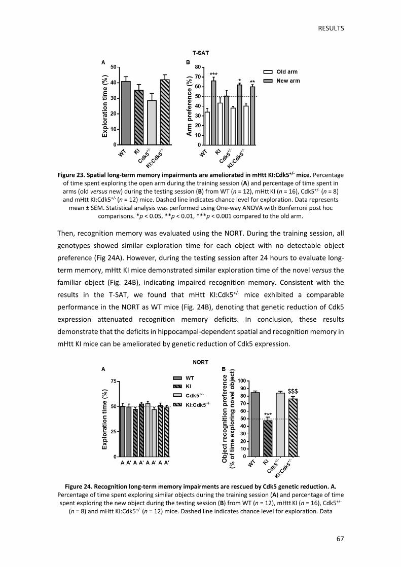
RESULTS
67
Figure 23. Spatial long‐term memory impairments are ameliorated in mHtt KI:Cdk5+/‐ mice. Percentage of time spent exploring the open arm during the training session (A) and percentage of time spent in arms (old versus new) during the testing session (B) from WT (n = 12), mHtt KI (n = 16), Cdk5+/‐ (n = 8) and mHtt KI:Cdk5+/‐ (n = 12) mice. Dashed line indicates chance level for exploration. Data represents mean ± SEM. Statistical analysis was performed using One‐way ANOVA with Bonferroni post hoc
comparisons. *p < 0.05, **p < 0.01, ***p < 0.001 compared to the old arm.
Then, recognition memory was evaluated using the NORT. During the training session, all
genotypes showed similar exploration time for each object with no detectable object
preference (Fig 24A). However, during the testing session after 24 hours to evaluate long‐
term memory, mHtt KI mice demonstrated similar exploration time of the novel versus the
familiar object (Fig. 24B), indicating impaired recognition memory. Consistent with the
results in the T‐SAT, we found that mHtt KI:Cdk5+/‐ mice exhibited a comparable
performance in the NORT as WT mice (Fig. 24B), denoting that genetic reduction of Cdk5
expression attenuated recognition memory deficits. In conclusion, these results
demonstrate that the deficits in hippocampal‐dependent spatial and recognition memory in
mHtt KI mice can be ameliorated by genetic reduction of Cdk5 expression.
Figure 24. Recognition long‐term memory impairments are rescued by Cdk5 genetic reduction. A.
Percentage of time spent exploring similar objects during the training session (A) and percentage of time spent exploring the new object during the testing session (B) from WT (n = 12), mHtt KI (n = 16), Cdk5+/‐
(n = 8) and mHtt KI:Cdk5+/‐ (n = 12) mice. Dashed line indicates chance level for exploration. Data

RESULTS
68
represents mean ± SEM. Statistical analysis was performed using One‐way ANOVA with Bonferroni post hoc comparisons. ***p < 0.001 compared to WT mice. $$$ p < 0.001 compared to mHtt KI mice.
1.3 ANALYSIS OF CDK5‐DEPENDENT MECHANISMS UNDERLYING COGNITIVE
IMPROVEMENT IN mHTT KI:CDK5+/‐ MICE
In view of our results showing that Cdk5 genetic reduction can restore corticostriatal and
hippocampal function in mHtt KI mice, we aimed to identify molecular mechanisms that
could underlie this recovery. Therefore, we analysed synaptic markers known to be altered
in HD and, at the same time, known to be regulated by Cdk5, such as NMDA receptor levels
and dendritic spine density. In addition, as little information is known about Cdk5 alterations
in HD in the cortex and hippocampus, we further analysed alterations in Cdk5 activity in
mHtt KI mice, as well as, the effect of Cdk5 genetic reduction on Cdk5 activity regulation.
1.3.1 ANALYSIS OF NMDA RECEPTORS
In the first place, we focused on NMDARs since many studies have shown their alteration in
HD (Luthi‐Carter et al, 2003), they play a pivotal role in synaptic plasticity (Izquierdo, 1991;
Tang et al, 1999) and, Cdk5 is known to regulate NMDARs levels and function through
different pathways (Plattner et al, 2014; Sahin et al, 2008; Zhang et al, 2008b).
1.3.1.1 Cortical NR2B levels are decreased in mHtt KI mice and are restored by genetic
reduction of Cdk5
We analysed total levels of the main NMDARs subunits: NR1, NR2A and NR2B in the cortex,
striatum and hippocampus of WT, mHtt KI, Cdk5+/‐ and mHtt KI:Cdk5+/‐ mice. No significant
changes between genotypes were detected neither in the striatum nor in the hippocampus
in the levels of any of the analysed NMDAR subunits (Fig. 25B, C). However, a significant
reduction of NR2B was found in the cortex of mHtt KI mice. Importantly, this decrease was
prevented in mHtt KI:Cdk5+/‐ mice as a result of Cdk5 genetic reduction (Fig. 25A). No
significant changes were detected in cortical NR1 and NR2A levels between genotypes.

RESULTS
69
Figure 25. Reduced cortical NR2B levels in mHtt KI mice are recovered by Cdk5 genetic reduction.
Representative Western blots showing total NR1, NR2A and NR2B levels in the cortex (A), striatum (B) and hippocampus (C) of WT (n = 5‐7), mHtt KI (n = 5‐7), Cdk5+/‐ (n = 6‐7) and mHtt KI:Cdk5+/‐ (n = 6‐7)
mice. All data represent mean ± SEM and are normalized to WT levels. Statistical analysis was performed using One‐way ANOVA with Newman‐Keuls post‐hoc comparisons. * p < 0.05 compared to WT mice. $ p
< 0.05 compared to mHtt KI mice.
1.3.1.2 Corticostriatal NR2B surface levels are reduced in mHtt KI mice and restored by
genetic reduction of Cdk5
In order to corroborate whether alterations in total NR2B levels correlated with a functional
outcome, we analysed specifically NR2B surface levels. To this end, we performed a
biotinylation assay and we analysed NR2B surface levels in the cortex, striatum and
hippocampus of WT, mHtt KI, Cdk5+/‐ and mHtt KI:Cdk5+/‐ mice. We also observed a
significant reduction of cortical NR2B surface levels in mHtt KI mice, which was restored in
mHtt KI:Cdk5+/‐ mice (Fig. 26A). Surprisingly, even though no changes in NR2B total levels
were detected in the striatum, we did find a significant reduction of NR2B surface levels in
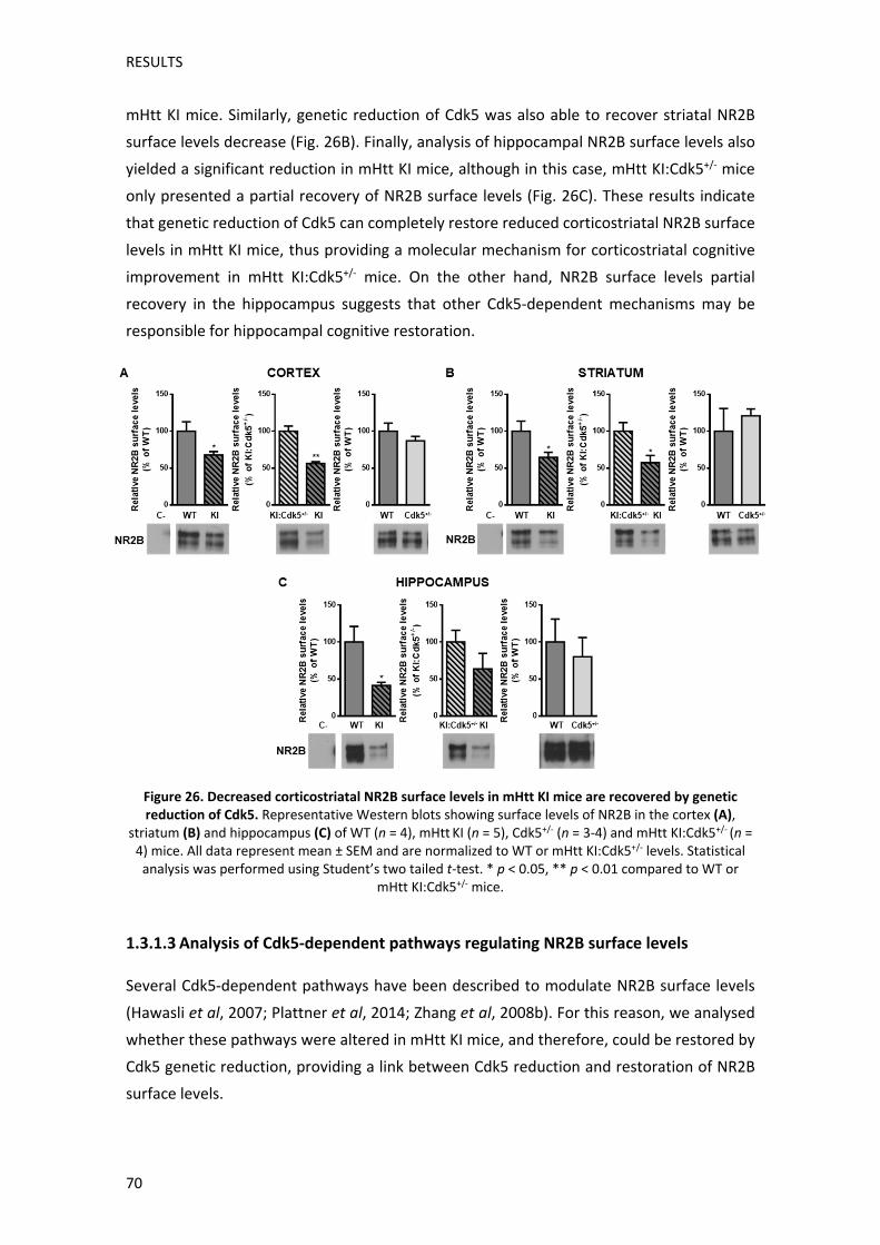
RESULTS
70
mHtt KI mice. Similarly, genetic reduction of Cdk5 was also able to recover striatal NR2B
surface levels decrease (Fig. 26B). Finally, analysis of hippocampal NR2B surface levels also
yielded a significant reduction in mHtt KI mice, although in this case, mHtt KI:Cdk5+/‐ mice
only presented a partial recovery of NR2B surface levels (Fig. 26C). These results indicate
that genetic reduction of Cdk5 can completely restore reduced corticostriatal NR2B surface
levels in mHtt KI mice, thus providing a molecular mechanism for corticostriatal cognitive
improvement in mHtt KI:Cdk5+/‐ mice. On the other hand, NR2B surface levels partial
recovery in the hippocampus suggests that other Cdk5‐dependent mechanisms may be
responsible for hippocampal cognitive restoration.
Figure 26. Decreased corticostriatal NR2B surface levels in mHtt KI mice are recovered by genetic reduction of Cdk5. Representative Western blots showing surface levels of NR2B in the cortex (A),
striatum (B) and hippocampus (C) of WT (n = 4), mHtt KI (n = 5), Cdk5+/‐ (n = 3‐4) and mHtt KI:Cdk5+/‐ (n = 4) mice. All data represent mean ± SEM and are normalized to WT or mHtt KI:Cdk5+/‐ levels. Statistical analysis was performed using Student’s two tailed t‐test. * p < 0.05, ** p < 0.01 compared to WT or
mHtt KI:Cdk5+/‐ mice.
1.3.1.3 Analysis of Cdk5‐dependent pathways regulating NR2B surface levels
Several Cdk5‐dependent pathways have been described to modulate NR2B surface levels
(Hawasli et al, 2007; Plattner et al, 2014; Zhang et al, 2008b). For this reason, we analysed
whether these pathways were altered in mHtt KI mice, and therefore, could be restored by
Cdk5 genetic reduction, providing a link between Cdk5 reduction and restoration of NR2B
surface levels.
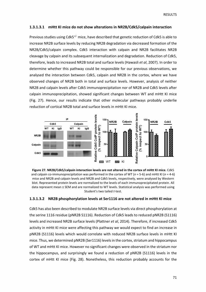
RESULTS
71
1.3.1.3.1 mHtt KI mice do not show alterations in NR2B/Cdk5/calpain interaction
Previous studies using Cdk5+/‐ mice, have described that genetic reduction of Cdk5 is able to
increase NR2B surface levels by reducing NR2B degradation via decreased formation of the
NR2B/Cdk5/calpain complex. Cdk5 interaction with calpain and NR2B facilitates NR2B
cleavage by calpain and its subsequent internalization and degradation. Reduction of Cdk5,
therefore, leads to increased NR2B total and surface levels (Hawasli et al, 2007). In order to
determine whether this pathway could be responsible for our previous observations, we
analysed the interaction between Cdk5, calpain and NR2B in the cortex, where we have
observed changes of NR2B both in total and surface levels. However, analysis of neither
NR2B and calpain levels after Cdk5 immunoprecipitation nor of NR2B and Cdk5 levels after
calpain immunoprecipitation, showed significant changes between WT and mHtt KI mice
(Fig. 27). Hence, our results indicate that other molecular pathways probably underlie
reduction of cortical NR2B total and surface levels in mHtt KI mice.
Figure 27. NR2B/Cdk5/calpain interaction levels are not altered in the cortex of mHtt KI mice. Cdk5 and calpain co‐immunoprecipitation was performed in the cortex of WT (n = 5‐6) and mHtt KI (n = 4‐6) mice and NR2B and calpain levels and NR2B and Cdk5 levels, respectively, were analysed by Western blot. Represented protein levels are normalized to the levels of each immunoprecipitated protein. All data represent mean ± SEM and are normalized to WT levels. Statistical analysis was performed using
Student’s two tailed t‐test.
1.3.1.3.2 NR2B phosphorylation levels at Ser1116 are not altered in mHtt KI mice
Cdk5 has also been described to modulate NR2B surface levels via direct phosphorylation at
the serine 1116 residue (pNR2B S1116). Reduction of Cdk5 leads to reduced pNR2B (S1116)
levels and increased NR2B surface levels (Plattner et al, 2014). Therefore, if increased Cdk5
activity in mHtt KI mice were affecting this pathway we would expect to find an increase in
pNR2B (S1116) levels which would correlate with reduced NR2B surface levels in mHtt KI
mice. Thus, we determined pNR2B (Ser1116) levels in the cortex, striatum and hippocampus
of WT and mHtt KI mice. However no significant changes were observed in the striatum nor
the hippocampus, and surprisingly we found a reduction of pNR2B (S1116) levels in the
cortex of mHtt KI mice (Fig. 28). Nonetheless, this reduction probably accounts for the

RESULTS
72
decrease in total NR2B levels observed specifically in the cortex. In sum, these results
indicate that deregulation of pNR2B (S1116) is probably not associated with reduced NR2B
surface levels in mHtt KI mice.
Figure 28. Cortical pNR2B (S1116) levels are decreased in mHtt KI mice. Representative Western blots of cortical, striatal and hippocampal lysates probed for pNR2B (S1116) in WT (n = 6‐7) and mHtt KI (n = 5‐7) mice. Histograms represent the mean ± SEM and are normalized to WT levels. Statistical analysis
was performed using Student’s two tailed t‐test. *** p < 0.001 compared to WT.
1.3.1.3.3 pSrc and pNR2B (Y1472) levels are significantly decreased in the cortex of
mHtt KI mice and are restored by Cdk5 genetic reduction
Finally, it has also been described that Cdk5 can indirectly regulate NR2B surface levels
through phosphorylation of PSD95 which reduces Src/PSD95 interaction, necessary for Src
auto‐activation. Reduced Src activity (pSrc Y416) leads to reduced NR2B phosphorylation at
the tyrosine 1472 residue (pNR2B Y1472), which allows AP2 binding and subsequent NR2B
internalization (Zhang et al, 2008b). In order to analyse if alterations in this pathway could
be responsible for reduced NR2B surface levels in mHtt KI mice, we analysed levels of pSrc
(Y416) and pNR2B (Y1472) in the cortex, striatum and hippocampus of WT and mHtt KI mice.
We found that mHtt KI mice present reduced levels of pSrc (Y416) and pNR2B (Y1472) in the
cortex, without showing alterations in total Src levels (Fig. 28A). Therefore, we also analysed
pSrc (Y416) and pNR2B (Y1472) levels in Cdk5+/‐ and mHtt KI:Cdk5+/‐ mice. Importantly, we
observed that reduced pSrc (Y416) and pNR2B (Y1472) levels were recovered as a result of
Cdk5 genetic reduction in mHtt KI:Cdk5+/‐ mice (Fig. 29A). However, no significant changes
were detected neither in the striatum nor the hippocampus between genotypes (Fig. 29B,
C). These findings indicate that Cdk5‐dependent pSrc/pNR2B pathway restoration in the
cortex could account for the recovery of cortical NR2B surface levels and thus, for restored
corticostriatal cognitive performance in mHtt KI:Cdk5+/‐ mice. Nonetheless, other Cdk5‐
dependent mechanisms are probably involved in the recovery of striatal NR2B surface levels
in mHtt KI:Cdk5+/‐ mice and in the recovery of hippocampal‐dependent cognitive decline.

RESULTS
73
Figure 29. Reduced cortical pSrc (Y416) and pNR2B (Y1472) phosphorylation levels in mHtt KI mice are recovered in mHtt KI:Cdk5+/‐ mice. Representative Western blots showing levels of pSrc (Y416), total Src and pNR2B (Y1472) in the cortex (A), striatum (B) and hippocampus (C) of WT (n = 5‐7), mHtt KI (n = 6‐7), Cdk5+/‐ (n = 5‐7) and mHtt KI:Cdk5+/‐ (n = 5‐7) mice. All data represent mean ± SEM and are normalized to
WT levels. Statistical analysis was performed using One‐way ANOVA with Newman‐Keuls post hoc comparisons. * p < 0.05 compared to WT mice. $ p < 0.05 compared to mHtt KI mice.
1.3.2 ANALYSIS OF DENDRITIC SPINE DENSITY
Cdk5 is also an important modulator of several cytoskeleton associated proteins, and
therefore of dendritic spine remodelling (Kim et al, 2006b; Posada‐Duque et al, 2015). Given
that previous studies from our group have described that mHtt KI mice present deficits in
dendritic spine density in the cortex since 2 months of age and in the hippocampus at 8
months of age (Brito et al, 2014; Puigdellívol et al, 2015), we decided to analyse if Cdk5
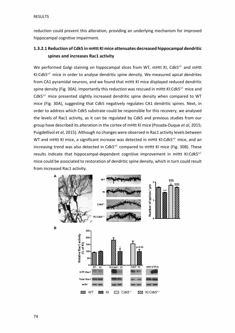
RESULTS
74
reduction could prevent this alteration, providing an underlying mechanism for improved
hippocampal cognitive impairment.
1.3.2.1 Reduction of Cdk5 in mHtt KI mice attenuates decreased hippocampal dendritic
spines and increases Rac1 activity
We performed Golgi staining on hippocampal slices from WT, mHtt KI, Cdk5+/‐ and mHtt
KI:Cdk5+/‐ mice in order to analyse dendritic spine density. We measured apical dendrites
from CA1 pyramidal neurons, and we found that mHtt KI mice displayed reduced dendritic
spine density (Fig. 30A). Importantly this reduction was rescued in mHtt KI:Cdk5+/‐ mice and
Cdk5+/‐ mice presented slightly increased dendritic spine density when compared to WT
mice (Fig. 30A), suggesting that Cdk5 negatively regulates CA1 dendritic spines. Next, in
order to address which Cdk5 substrate could be responsible for this recovery, we analysed
the levels of Rac1 activity, as it can be regulated by Cdk5 and previous studies from our
group have described its alteration in the cortex of mHtt KI mice (Posada‐Duque et al, 2015;
Puigdellívol et al, 2015). Although no changes were observed in Rac1 activity levels between
WT and mHtt KI mice, a significant increase was detected in mHtt KI:Cdk5+/‐ mice, and an
increasing trend was also detected in Cdk5+/‐ compared to mHtt KI mice (Fig. 30B). These
results indicate that hippocampal‐dependent cognitive improvement in mHtt KI:Cdk5+/‐
mice could be associated to restoration of dendritic spine density, which in turn could result
from increased Rac1 activity.

RESULTS
75
Figure 30. Decreased hippocampal dendritic spine density is attenuated by genetic reduction of Cdk5. A. Representative images of a Golgi‐impregnated CA1 hippocampal neuron and of apical dendrites of
CA1 pyramidal neurons from WT, mHtt KI, Cdk5+/‐ and mHtt KI:Cdk5+/‐ mice. Histogram showing quantitative analysis of dendritic spine density per micrometre of dendritic length. Statistical analysis
was performed using One‐way ANOVA with Newman‐Keuls post hoc comparisons (30 dendrites from 3‐5 animals per genotype were analysed). *p < 0.05, ***p < 0.001 compared to WT mice. $$$ p < 0.001
compared to mHtt KI mice. B. Representative Western blots showing levels of GTP‐bound Rac1 immunoprecipitated from hippocampal lysates, indicating Rac1 activity. Histograms represent the mean
± SEM and are normalized to mHtt KI values (n=4‐5 mice per genotype). Statistical analysis was performed using Student’s two tailed t‐test. * p < 0.05 compared to mHtt KI.
1.3.2.2 Reduced cortical dendritic spines are recovered by Cdk5 reduction
We have observed that recovery of corticostriatal‐dependent deficits correlates with
restoration of NR2B surface levels and that restoration of hippocampal‐dependent decline
correlates with recovery of dendritic spine density, suggesting a brain region specificity in
Cdk5‐dependent substrate regulation. For this reason, in order to determine if restoration
of dendritic spine density was a hippocampus‐specific mechanism, we also performed Golgi
staining of corticostriatal slices and we measured dendritic spine density in cortical layer V
pyramidal neurons. Similar to our observations in the hippocampus, mHtt KI mice presented
a significant reduction of cortical dendritic spines which was recovered in mHtt KI:Cdk5+/‐
mice as a result of Cdk5 genetic reduction (Fig. 31A). However, we did not find any significant
changes between WT and Cdk5+/‐ mice (Fig. 31A), indicating that genetic reduction of Cdk5
per se, does not influence cortical dendritic spine density. We also analysed Rac1 activity in
cortical lysates and, in accordance with previous studies (Puigdellívol et al, 2015), we found
a significant reduction of Rac1 between WT and mHtt KI mice (Fig. 31B). However, no
significant changes were found between mHtt KI and mHtt KI:Cdk5+/‐ mice or Cdk5+/‐ mice
(Fig. 31B). These results indicate that in contrast to the hippocampus, mHtt KI mice present
a reduction of Rac1 activity in the cortex, however Cdk5 reduction does not affect cortical
Rac1 activity, suggesting that other Cdk5 substrates might be responsible for recovery of
dendritic spine density in the cortex of mHtt KI:Cdk5+/‐ mice, reinforcing our hypothesis of a
brain region‐dependent modulation of Cdk5 substrates.

RESULTS
76
Figure 31. Decreased cortical dendritic spine density in mHtt KI mice is attenuated in mHtt KI:Cdk5+/‐
mice. A. Representative images of a Golgi‐impregnated cortical pyramidal neuron and of apical dendrites of layer V pyramidal neurons from WT, mHtt KI, Cdk5+/‐ and mHtt KI:Cdk5+/‐ mice. Histogram showing quantitative analysis of dendritic spine density per micrometre of dendritic length. Statistical
analysis was performed using One‐way ANOVA with Newman‐Keuls post hoc comparisons (30 dendrites from 3‐5 animals per genotype were analysed). ***p < 0.001 compared to WT mice. $$$ p < 0.001 compared to mHtt KI mice. B. Representative Western blots showing levels of GTP‐bound Rac1
immunoprecipitated from cortical lysates indicating Rac1 activity. Histograms represent the mean ± SEM and are normalized to mHtt KI values (n=4‐5 mice per genotype). Statistical analysis was performed
using Student’s two tailed t‐test. * p < 0.05 compared to mHtt KI.
1.3.3 ANALYSIS OF CDK5 ACTIVITY IN mHTT KI:CDK5+/‐ MICE
Our results indicate that genetic reduction of Cdk5 restores cognitive decline in mHtt KI
mice, via normalization of NMDARs levels and dendritic spine density in a brain region‐
specific manner. Since Cdk5 activity remains unexplored in the cortex and hippocampus of
HD mice, we wanted to analyse its activity to further correlate Cdk5‐dependent modulation
of synaptic substrates presented up to this point with alterations in Cdk5 activity.
1.3.3.1 Phosphorylation levels of Cdk5 substrates are altered preferentially in the
cortex of mHtt KI mice.
As a readout of putative changes in Cdk5 activity between genotypes, we analysed
phosphorylation levels of different Cdk5 substrates. Cdk5 plays an important role in Tau
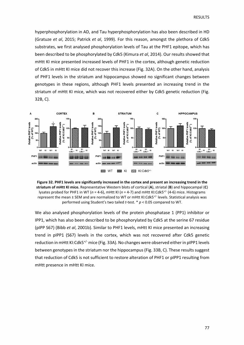
RESULTS
77
hyperphosphorylation in AD, and Tau hyperphosphorylation has also been described in HD
(Gratuze et al, 2015; Patrick et al, 1999). For this reason, amongst the plethora of Cdk5
substrates, we first analysed phosphorylation levels of Tau at the PHF1 epitope, which has
been described to be phosphorylated by Cdk5 (Kimura et al, 2014). Our results showed that
mHtt KI mice presented increased levels of PHF1 in the cortex, although genetic reduction
of Cdk5 in mHtt KI mice did not recover this increase (Fig. 32A). On the other hand, analysis
of PHF1 levels in the striatum and hippocampus showed no significant changes between
genotypes in these regions, although PHF1 levels presented an increasing trend in the
striatum of mHtt KI mice, which was not recovered either by Cdk5 genetic reduction (Fig.
32B, C).
Figure 32. PHF1 levels are significantly increased in the cortex and present an increasing trend in the striatum of mHtt KI mice. Representative Western blots of cortical (A), striatal (B) and hippocampal (C) lysates probed for PHF1 in WT (n = 4‐6), mHtt KI (n = 4‐7) and mHtt KI:Cdk5+/‐ (4‐6) mice. Histograms represent the mean ± SEM and are normalized to WT or mHtt KI:Cdk5+/‐ levels. Statistical analysis was
performed using Student’s two tailed t‐test. * p < 0.05 compared to WT.
We also analysed phosphorylation levels of the protein phosphatase 1 (PP1) inhibitor or
IPP1, which has also been described to be phosphorylated by Cdk5 at the serine 67 residue
(pIPP S67) (Bibb et al, 2001b). Similar to PHF1 levels, mHtt KI mice presented an increasing
trend in pIPP1 (S67) levels in the cortex, which was not recovered after Cdk5 genetic
reduction in mHtt KI:Cdk5+/‐ mice (Fig. 33A). No changes were observed either in pIPP1 levels
between genotypes in the striatum nor the hippocampus (Fig. 33B, C). These results suggest
that reduction of Cdk5 is not sufficient to restore alteration of PHF1 or pIPP1 resulting from
mHtt presence in mHtt KI mice.

RESULTS
78
Figure 33. pIPP1 (S67) levels show no significant alterations between genotypes. Representative
Western blots of cortical (A), striatal (B) and hippocampal (C) lysates probed for pIPP1 (Ser67) in WT (n = 5‐6), mHtt KI (n = 5‐6) and mHtt KI:Cdk5+/‐ (6‐7) mice. Histograms represent the mean ± SEM and are
normalized to WT or mHtt KI:Cdk5+/‐ levels. Statistical analysis was performed using Student’s two tailed
Finally, we checked the general phosphorylation state of CDK‐dependent serine residues
(pCDK substrates) in WT, mHtt KI and mHtt KI:Cdk5+/‐ mice. In this case we also found that
in the cortex, mHtt KI mice presented an increasing trend in pCDK (Ser) substrates when
compared to WT mice. Importantly, this trend was also observed when comparing mHtt KI
with mHtt KI:Cdk5+/‐ mice, indicating that increased phosphorylation of pCDK (Ser)
substrates might be partially reverted as a result of genetic reduction of Cdk5 (Fig. 34A).
However, no changes were observed neither in the striatum nor the hippocampus between
genotypes (Fig. 34B,C). Notably, intensity changes in certain molecular weight bands could
be observed between genotypes in all regions in pCDK (Ser) substrates Western blot analysis
(Fig. 34), which might reflect changes in phosphorylation levels of specific Cdk5 substrates.
Figure 34. pCDK (Ser) substrates levels present an increasing trend in the cortex of mHtt KI mice.
Representative Western blots of cortical (A), striatal (B) and hippocampal (C) lysates probed for pCDK (Ser) substrates in WT (n = 4‐6) mHtt KI (n = 4‐6) and mHtt KI:Cdk5+/‐ (6‐7) mice. Histograms represent

RESULTS
79
the mean ± SEM and are normalized to WT or mHtt KI:Cdk5+/‐ levels. Statistical analysis was performed using Student’s two tailed t‐test.
These results indicate that alteration of Cdk5 substrates phosphorylation levels in mHtt KI
mice occurs preferentially in the cortex and to a lesser extent in the striatum, suggesting a
preferential dysfunction of Cdk5 activity in the cortex over other analysed brain regions.
However, even within the cortex, Cdk5 activity presents differential regulation depending
on the substrate.
1.3.3.2 p35 plasma membrane levels are significantly reduced in the cortex of mHtt KI
which is recovered by Cdk5 genetic reduction
Given that we have observed differential alteration of Cdk5 substrates, we wondered
whether this might result from modifications in subcellular distribution of Cdk5 and its
activator, as correct subcellular localization of Cdk5 and p35 is pivotal for its proper function
(Asada et al, 2008; Patrick et al, 1999). To this end, we performed subcellular fractionation
to obtain plasma membrane and cytosolic fractions from the cortex, striatum and
hippocampus of WT, mHtt KI, Cdk5+/‐ and mHtt KI:Cdk5+/‐ mice.
Our results showed that p35 levels were significantly reduced in the cortical plasma
membrane fraction of mHtt KI mice (Fig. 35A), and that cortical Cdk5 plasma membrane
levels also presented a decreasing trend (Fig. 35B), indicating that normal Cdk5/p35 function
might be altered in the cortex of mHtt KI mice. Importantly, this reduction in p35 plasma
membrane levels was recovered to levels similar to WT in mHtt KI:Cdk5+/‐ mice.
Nonetheless, besides Cdk5 genetic reduction, no significant changes were detected in the
striatal or hippocampal plasma membrane fractions between genotypes (Fig. 35). These
results fit with our prior observations of preferential changes in Cdk5 substrates
phosphorylation in the cortex of mHtt KI mice.

RESULTS
80
Figure 35. Cortical p35 plasma membrane levels are significantly reduced in mHtt KI mice.
Representative Western blots of cortical, striatal and hippocampal plasma membrane fractions probed for Cdk5 (A) and p35 (B) in WT (n = 5‐6), mHtt KI (n = 4‐7), Cdk5+/‐ (n = 4‐5) and mHtt KI:Cdk5+/‐ (n = 4‐6) mice. Histograms represent the mean ± SEM and are normalized to WT levels. One‐way ANOVA with Newman‐Keuls post hoc comparisons. * p < 0.05 compared to WT. $ p < 0.05, $$ p < 0.01 compared to
mHtt KI mice.
We also analysed cytosolic Cdk5 and p35 levels, and although we did not find any significant
differences between WT and mHtt KI mice, we detected a significant increase of cytosolic
p35 levels in the cortex of Cdk5+/‐ mice when compared to mHtt KI mice. Cytosolic p35 levels
also presented an increasing trend in mHtt KI:Cdk5+/‐ mice (Fig. 36B). Similar to data
obtained in plasma membrane fraction, no changes were observed in the striatal and
hippocampal cytosolic fractions (Fig. 36), suggesting that additional Cdk5 activity regulation
mechanisms such as post‐translational modifications, could underlie differential regulation
of Cdk5 substrates in these regions. Levels of the cleaved p35 form, p25, could not be
detected neither in the plasma membrane nor in the cytosolic fraction in our experimental
conditions.

RESULTS
81
Figure 36. Cortical p35 cytosol levels are increased in Cdk5+/‐ mice. Representative Western blots of
cortical, striatal and hippocampal cytosol fractions probed for Cdk5 (A) and p35 (B) in WT (n = 4‐6), mHtt KI (n = 5‐7), Cdk5+/‐ (n = 4‐5) and mHtt KI:Cdk5+/‐ (n = 4‐5) mice. Histograms represent the mean ± SEM and are normalized to WT levels. One‐way ANOVA with Newman‐Keuls post hoc comparisons. * p < 0.05, ** p < 0.01, *** p < 0.001 compared to WT. $ p < 0.05, $$ p < 0.01, $$$ p < 0.001 compared to
mHtt KI mice.
RESULTS
82
2. CELL CYCLE REENTRY AS A POTENTIAL UNDERLYING MECHANISM FOR CDK5‐
DEPENDENT STRIATAL VULNERABILITY IN HD
Cdk5 deregulation contributing to neuronal death, has been described in several
neurodegenerative disorders, including HD. Particularly, alteration of Cdk5 in HD has been
associated with increased striatal vulnerability to dopaminergic and glutamatergic
activation (Paoletti et al, 2008). Nonetheless, not much is known about the underlying
mechanisms linking Cdk5 deregulation with increased striatal vulnerability. For this reason,
we decided to study whether Cdk5 deregulation in HD might alter Cdk5 role in the
maintenance of cell cycle inhibition in postmitotic neurons. Cell cycle re‐entry of
differentiated neurons has been proposed as a possible common mechanism in several
neurodegenerative disorders. It has been described, especially in Alzheimer’s disease, that
deregulation of cell cycle inhibition in neurons, leads to expression of cell cycle proteins and
to cell cycle re‐entry, which leads, eventually, to neuronal dysfunction and death (Herrup
and Yang, 2007). Coincidentally, some articles have described alteration of some cell cycle
proteins in HD human samples and in HD mice models (Fernandez‐Fernandez et al, 2011;
Liu et al, 2015; Pelegrí et al, 2008). However, little is known about the molecular
mechanisms leading to these alterations or the possible involvement of Cdk5 in cell cycle
deregulation in HD.
2.1 ANALYSIS OF CDK5 FUNCTION AS A CELL CYCLE MODULATOR IN HD
Cdk5 has been described to maintain cell cycle progression inhibition in the nucleus of
differentiated neurons. Specifically, it is known that Cdk5 along with p35 forms a complex
with the CDKs inhibitor p27 and the transcription factor E2F1, preventing E2F1 interaction
with its cofactor DP1, and therefore avoiding transcription of cell cycle proliferation genes
(Zhang and Herrup, 2011). For this reason, we analysed interaction and nuclear levels of
these proteins in the striatum of WT and KI mice at early symptomatic motor stages (8
months of age).
2.1.1 Cdk5 and p27 interaction is not significantly altered in the striatum of
symptomatic mHtt KI mice
To analyse if presence of mHtt alters the formation of the Cdk5/p27/E2F1 complex, we
performed Cdk5 immunoprecipitation and we analysed its interaction with p27 as we were
not able to detect E2F1 after Cdk5 immunoprecipitation. Our results showed a decreasing
trend, although not significant, in Cdk5/p27 interaction in the striatum of 8‐month‐old mHtt
KI mice (Fig. 37A). We also analysed this interaction at later symptomatic stages (13 months)
and we found no significant changes either (Fig. 37B). These results suggest that Cdk5/p27
interaction is not significantly altered in the striatum of mHtt KI mice. However, as
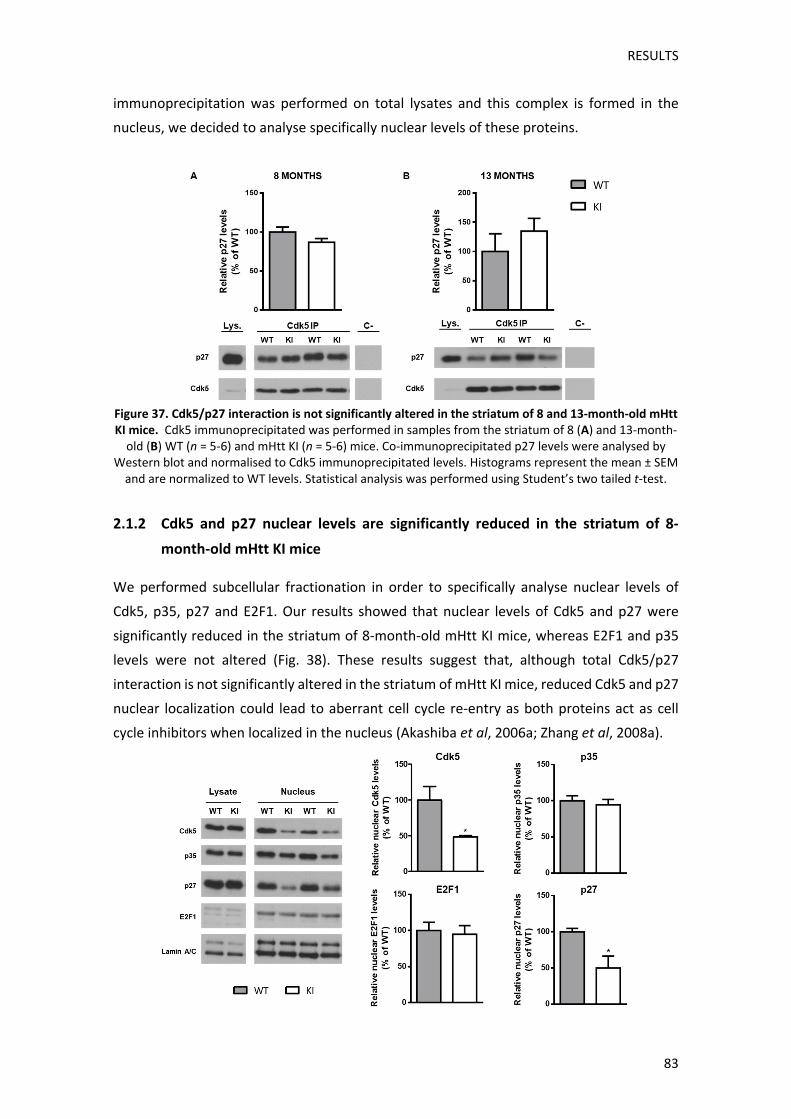
RESULTS
83
immunoprecipitation was performed on total lysates and this complex is formed in the
nucleus, we decided to analyse specifically nuclear levels of these proteins.
Figure 37. Cdk5/p27 interaction is not significantly altered in the striatum of 8 and 13‐month‐old mHtt KI mice. Cdk5 immunoprecipitated was performed in samples from the striatum of 8 (A) and 13‐month‐old (B) WT (n = 5‐6) and mHtt KI (n = 5‐6) mice. Co‐immunoprecipitated p27 levels were analysed by
Western blot and normalised to Cdk5 immunoprecipitated levels. Histograms represent the mean ± SEM and are normalized to WT levels. Statistical analysis was performed using Student’s two tailed t‐test.
2.1.2 Cdk5 and p27 nuclear levels are significantly reduced in the striatum of 8‐
month‐old mHtt KI mice
We performed subcellular fractionation in order to specifically analyse nuclear levels of
Cdk5, p35, p27 and E2F1. Our results showed that nuclear levels of Cdk5 and p27 were
significantly reduced in the striatum of 8‐month‐old mHtt KI mice, whereas E2F1 and p35
levels were not altered (Fig. 38). These results suggest that, although total Cdk5/p27
interaction is not significantly altered in the striatum of mHtt KI mice, reduced Cdk5 and p27
nuclear localization could lead to aberrant cell cycle re‐entry as both proteins act as cell
cycle inhibitors when localized in the nucleus (Akashiba et al, 2006a; Zhang et al, 2008a).

RESULTS
84
Figure 38. Nuclear Cdk5 and p27 levels are significantly reduced in the striatum of mHtt KI mice. Cdk5, p35, p27 and E2F1 levels were determined in nuclear fractions from the striatum of 8‐month‐old WT (n = 3‐4) and mHtt KI (n = 3‐4) mice. Lamin A/C was used as loading control. Histograms represent the mean ± SEM and are normalized to WT levels. Statistical analysis was performed using Student’s two
tailed t‐test. * p < 0.05 compared to WT.
2.2 ANALYSIS OF CELL CYCLE PROTEINS EXPRESSION IN HD
Previous studies have described alteration of some cell cycle proteins in human HD brains
and in R6/1 mice (Fernandez‐Fernandez et al, 2011; Pelegrí et al, 2008). However, little is
known about their alteration in the less severe knock‐in HD mouse model. For this reason,
in order to determine if reduction of Cdk5 and p27 nuclear levels could also involve aberrant
expression of other cell cycle proteins, we analysed the levels of p27, CyclinD1 and Cdk4,
which are expressed during early G1 cell cycle phase, at different disease stages in the
striatum of mHtt KI mice.
2.2.1 p27 and CyclinD1 levels are increased in the striatum of mHtt KI mice since
early symptomatic stages
We analysed the levels of p27, CyclinD1 and Cdk4 in striatal lysates from motor
asymptomatic (2‐4 months), presymptomatic (6 months) and early (8 months) and late (13‐
18 months) symptomatic stages in mHtt KI mice. In contrast with our observations in nuclear
p27 levels, our results showed that p27 presented a slight but significant increase at 6
months of age in mHtt KI mice. This increase became more apparent with increasing disease
progression stages until 18 months of age (Fig. 39A), indicating that reduced nuclear p27
levels are probably caused by altered subcellular distribution rather than by total decrease
of p27 levels.
Analysis of G1 phase proteins Cdk4 and CyclinD1, showed that even though Cdk4 protein
levels only displayed an increasing trend in mHtt KI mice at late disease stages with no
significant changes in previous phases (Fig. 39B), CyclinD1 levels were increased since early
stages. Thus, CyclinD1 levels presented an increasing trend at 6 months. This increase
became significant at 8 months and was maintained at 13 and 18 months of age, when
CyclinD1 levels presented almost a 50% increase in mHtt KI mice when compared to WT
mice (Figure 39C). These results indicate that expression of cell cycle proteins present an
alteration in the striatum of mHtt KI mice at ages that correlate with disease motor onset,
which could lead to aberrant cell cycle re‐entry.

RESULTS
85
Figure 39. p27 and CyclinD1 levels are significantly increased in the striatum of mHtt KI mice since early symptomatic disease stages. Representative Western blots of striatal lysates probed for p27 (A), Cdk4 (B) and CyclinD1 (C) in WT (n = 5‐13) and mHtt KI (n = 5‐12) mice. Histograms represent the mean ± SEM and are normalized to WT levels. Statistical analysis was performed using Student’s two tailed t‐
test. * p < 0.05, ** p < 0.01 compared to WT.

RESULTS
86
2.2.2 Cdk4 levels are significantly increased in the putamen of HD patients
Finally, we also determined the levels of these proteins in putamen samples from HD
patients. In contrast with our observations in HD mice models, p27 levels were not
significantly increased in putamen samples from HD patients, although p27 blot bands
showed notable alterations in their pattern (Fig. 40A). These alterations were reproducible
and only observed for p27 suggesting possible presence of post‐translational p27
modifications in samples from HD patients leading to increased molecular weight. Similarly,
even though we did not find any significant changes in total E2F1 levels, we detected a
significant increase of a lower molecular weight band (Fig. 40A). Interestingly, it has been
described that E2F1 can be cleaved by calpains in neurotoxic conditions, producing a 50 kDa
band (Zyskind et al, 2015), suggesting that E2F1 in the putamen of HD patients could
experience increased cleavage compared to controls. On the other hand, in contrast with
our results in HD models, analysis of CyclinD1 levels in HD patients yielded no significant
alterations respect to control samples. However, Cdk4 levels did show a significant increase
in the putamen of HD patients (Fig. 40B). These results suggest that cell cycle proteins also
present alterations in the putamen of HD patients. However, these alterations differ from
the ones observed in HD models, which could be attributed to the fact that mHtt KI mice do
not present neuronal death, in contrast with HD human brains.

RESULTS
87
Figure 40. Cdk4 levels are significantly increased in the putamen of HD patients. Representative Western blots of putamen lysates probed for p27 and E2F1 (A), and Cdk4 and CyclinD1 (B) in Control (n = 6‐7) and HD (n = 5‐7) patients. Histograms represent the mean ± SEM and are normalized to Control levels. Statistical analysis was performed using Student’s two tailed t‐test. * p < 0.05 compared to
Control.
2.3 ANALYSIS OF CELL CYCLE PROTEINS FOLLOWING NMDAR ACTIVATION IN WT AND
KI STRIATAL PRIMARY CULTURES
Our results show that mHtt induces alteration of cell cycle proteins levels in HD mice and in
HD patients, which might be associated with decreased Cdk5 nuclear levels. One of the
pathways leading to Cdk5 deregulation is calpain‐dependent p35 cleavage following
NMDARs activation (Miao et al, 2012; Patrick et al, 1999). Incidentally, NMDARs activation
also plays a major role in excitotoxicity in HD (Fan and Raymond, 2007) and, particularly,
deregulation of Cdk5 has been associated with increased striatal vulnerability in HD
following glutamatergic activation (Paoletti et al, 2008). Therefore, we aimed to analyse
whether NMDA treatment, leading to Cdk5 hyperactivation, might participate in cell cycle
proteins alteration in striatal primary cultures from WT and mHtt KI mice.
2.3.1 NMDA treatment alters expression levels of cell cycle proteins
We tested different treatment durations and different NMDA concentrations, in DIV21 WT
striatal primary cultures, in order to determine appropriate conditions leading to p35
cleavage to p25. We performed treatments consisting on 100 μM NMDA for 1h, 50 μM
NMDA for 3h and 10 μM NMDA for 24h. We analysed the levels of spectrin breakdown
products (SBDP) which are commonly used as calpain activation indicators (Vanderklish and
Bahr, 2000). Our results showed that all NMDA treatments led to increased SBDP levels (Fig.
41A), indicating NMDA activation and subsequent calpain activation in our treatment
conditions. Next, we analysed the levels of p35 cleaved product, p25. Western blot analysis
showed that 50 μM NMDA for 3h, and particularly 100 μM NMDA for 1h, caused p25

RESULTS
88
production. This effect, however, was lost in the 10 μM NMDA for 24h treatment (Fig. 41A).
Finally, we analysed the levels of cleaved caspase 3, as an apoptotic marker, after 1h 100
μM NMDA treatment, which produced the most significant response in the previously
measured parameters. We did not detect any significant changes between control and
NMDA treated cultures (Fig. 41B), suggesting that this treatment does not lead to overt
neuronal apoptosis.
Figure 41. Efficiency evaluation of different NMDA treatments conditions. Representative Western
blots of WT striatal primary cultures lysates probed for spectrin and p25 (A) and cleaved caspase 3 (B) in Control (n = 3), 100 μM NMDA for 1h (n = 2), 50 μM NMDA for 3h (n = 4) and 10 μM NMDA for 24h (n = 4) conditions. Histograms represent the mean ± SEM and are normalized to Control levels. Statistical
analysis was performed using One‐way ANOVA with Newman‐Keuls post hoc comparisons. ** p < 0.01, *** p < 0.001 compared to Control.
Next, we also analysed the effect of these different NMDA treatments on the levels of the
cell cycle proteins, p27, E2F1, CyclinD1 and Cdk4 in WT striatal cultures. Western blot
analysis, showed that 50 μM NMDA treatment for 3h, and particularly, 100 μM NMDA
treatment for 1h, induced significant p27 reduction. A decreasing trend in Cdk4 and CyclinD1
protein levels could also be observed after 100 μM NMDA for 1h (Fig. 42). Finally, even
though total E2F1 levels did not present alterations in response to NMDA treatment, we
observed an additional E2F1 lower molecular weight band in cultures treated with NMDA,
indicating that, according to previous studies (Zyskind et al, 2015), E2F1 could be cleaved
under these conditions.
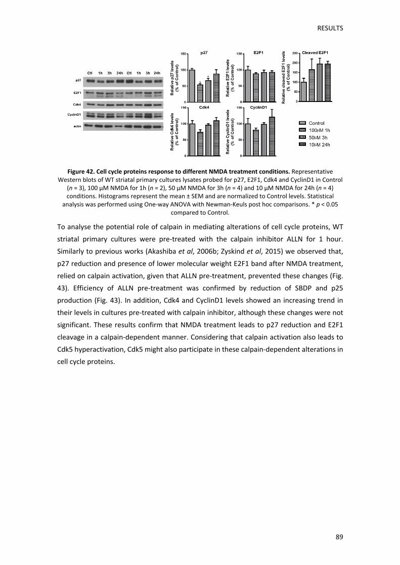
RESULTS
89
Figure 42. Cell cycle proteins response to different NMDA treatment conditions. Representative Western blots of WT striatal primary cultures lysates probed for p27, E2F1, Cdk4 and CyclinD1 in Control
(n = 3), 100 μM NMDA for 1h (n = 2), 50 μM NMDA for 3h (n = 4) and 10 μM NMDA for 24h (n = 4) conditions. Histograms represent the mean ± SEM and are normalized to Control levels. Statistical
analysis was performed using One‐way ANOVA with Newman‐Keuls post hoc comparisons. * p < 0.05 compared to Control.
To analyse the potential role of calpain in mediating alterations of cell cycle proteins, WT
striatal primary cultures were pre‐treated with the calpain inhibitor ALLN for 1 hour.
Similarly to previous works (Akashiba et al, 2006b; Zyskind et al, 2015) we observed that,
p27 reduction and presence of lower molecular weight E2F1 band after NMDA treatment,
relied on calpain activation, given that ALLN pre‐treatment, prevented these changes (Fig.
43). Efficiency of ALLN pre‐treatment was confirmed by reduction of SBDP and p25
production (Fig. 43). In addition, Cdk4 and CyclinD1 levels showed an increasing trend in
their levels in cultures pre‐treated with calpain inhibitor, although these changes were not
significant. These results confirm that NMDA treatment leads to p27 reduction and E2F1
cleavage in a calpain‐dependent manner. Considering that calpain activation also leads to
Cdk5 hyperactivation, Cdk5 might also participate in these calpain‐dependent alterations in
cell cycle proteins.
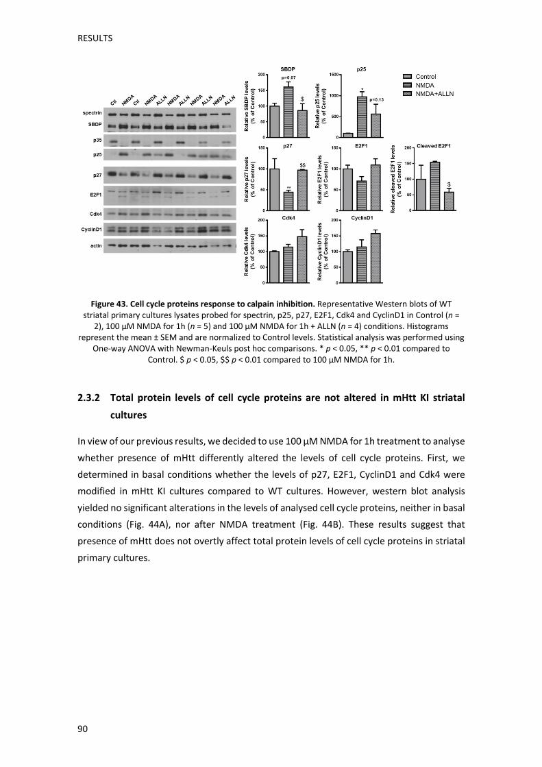
RESULTS
90
Figure 43. Cell cycle proteins response to calpain inhibition. Representative Western blots of WT striatal primary cultures lysates probed for spectrin, p25, p27, E2F1, Cdk4 and CyclinD1 in Control (n =
2), 100 μM NMDA for 1h (n = 5) and 100 μM NMDA for 1h + ALLN (n = 4) conditions. Histograms represent the mean ± SEM and are normalized to Control levels. Statistical analysis was performed using
One‐way ANOVA with Newman‐Keuls post hoc comparisons. * p < 0.05, ** p < 0.01 compared to Control. $ p < 0.05, $$ p < 0.01 compared to 100 μM NMDA for 1h.
2.3.2 Total protein levels of cell cycle proteins are not altered in mHtt KI striatal
cultures
In view of our previous results, we decided to use 100 μM NMDA for 1h treatment to analyse
whether presence of mHtt differently altered the levels of cell cycle proteins. First, we
determined in basal conditions whether the levels of p27, E2F1, CyclinD1 and Cdk4 were
modified in mHtt KI cultures compared to WT cultures. However, western blot analysis
yielded no significant alterations in the levels of analysed cell cycle proteins, neither in basal
conditions (Fig. 44A), nor after NMDA treatment (Fig. 44B). These results suggest that
presence of mHtt does not overtly affect total protein levels of cell cycle proteins in striatal
primary cultures.
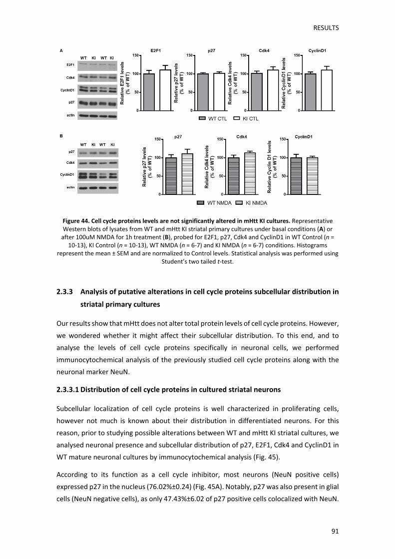
RESULTS
91
Figure 44. Cell cycle proteins levels are not significantly altered in mHtt KI cultures. Representative Western blots of lysates from WT and mHtt KI striatal primary cultures under basal conditions (A) or after 100uM NMDA for 1h treatment (B), probed for E2F1, p27, Cdk4 and CyclinD1 in WT Control (n =
10‐13), KI Control (n = 10‐13), WT NMDA (n = 6‐7) and KI NMDA (n = 6‐7) conditions. Histograms represent the mean ± SEM and are normalized to Control levels. Statistical analysis was performed using
Student’s two tailed t‐test.
2.3.3 Analysis of putative alterations in cell cycle proteins subcellular distribution in
striatal primary cultures
Our results show that mHtt does not alter total protein levels of cell cycle proteins. However,
we wondered whether it might affect their subcellular distribution. To this end, and to
analyse the levels of cell cycle proteins specifically in neuronal cells, we performed
immunocytochemical analysis of the previously studied cell cycle proteins along with the
neuronal marker NeuN.
2.3.3.1 Distribution of cell cycle proteins in cultured striatal neurons
Subcellular localization of cell cycle proteins is well characterized in proliferating cells,
however not much is known about their distribution in differentiated neurons. For this
reason, prior to studying possible alterations between WT and mHtt KI striatal cultures, we
analysed neuronal presence and subcellular distribution of p27, E2F1, Cdk4 and CyclinD1 in
WT mature neuronal cultures by immunocytochemical analysis (Fig. 45).
According to its function as a cell cycle inhibitor, most neurons (NeuN positive cells)
expressed p27 in the nucleus (76.02%±0.24) (Fig. 45A). Notably, p27 was also present in glial
cells (NeuN negative cells), as only 47.43%±6.02 of p27 positive cells colocalized with NeuN.
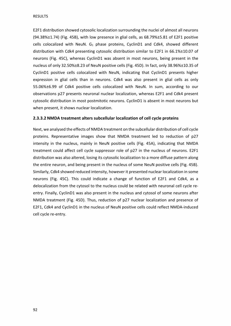
RESULTS
92
E2F1 distribution showed cytosolic localization surrounding the nuclei of almost all neurons
(94.38%±1.74) (Fig. 45B), with low presence in glial cells, as 68.79%±5.81 of E2F1 positive
cells colocalized with NeuN. G1 phase proteins, CyclinD1 and Cdk4, showed different
distribution with Cdk4 presenting cytosolic distribution similar to E2F1 in 66.1%±10.07 of
neurons (Fig. 45C), whereas CyclinD1 was absent in most neurons, being present in the
nucleus of only 32.50%±8.23 of NeuN positive cells (Fig. 45D). In fact, only 38.96%±10.35 of
CyclinD1 positive cells colocalized with NeuN, indicating that CyclinD1 presents higher
expression in glial cells than in neurons. Cdk4 was also present in glial cells as only
55.06%±6.99 of Cdk4 positive cells colocalized with NeuN. In sum, according to our
observations p27 presents neuronal nuclear localization, whereas E2F1 and Cdk4 present
cytosolic distribution in most postmitotic neurons. CyclinD1 is absent in most neurons but
when present, it shows nuclear localization.
2.3.3.2 NMDA treatment alters subcellular localization of cell cycle proteins
Next, we analysed the effects of NMDA treatment on the subcellular distribution of cell cycle
proteins. Representative images show that NMDA treatment led to reduction of p27
intensity in the nucleus, mainly in NeuN positive cells (Fig. 45A), indicating that NMDA
treatment could affect cell cycle suppressor role of p27 in the nucleus of neurons. E2F1
distribution was also altered, losing its cytosolic localization to a more diffuse pattern along
the entire neuron, and being present in the nucleus of some NeuN positive cells (Fig. 45B).
Similarly, Cdk4 showed reduced intensity, however it presented nuclear localization in some
neurons (Fig. 45C). This could indicate a change of function of E2F1 and Cdk4, as a
delocalization from the cytosol to the nucleus could be related with neuronal cell cycle re‐
entry. Finally, CyclinD1 was also present in the nucleus and cytosol of some neurons after
NMDA treatment (Fig. 45D). Thus, reduction of p27 nuclear localization and presence of
E2F1, Cdk4 and CyclinD1 in the nucleus of NeuN positive cells could reflect NMDA‐induced
cell cycle re‐entry.

RESULTS
93
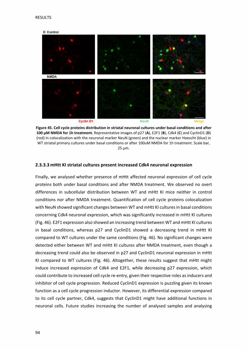
RESULTS
94
Figure 45. Cell cycle proteins distribution in striatal neuronal cultures under basal conditions and after 100 μM NMDA for 1h treatment. Representative images of p27 (A), E2F1 (B), Cdk4 (C) and CyclinD1 (D) (red) in colocalization with the neuronal marker NeuN (green) and the nuclear marker Hoescht (blue) in WT striatal primary cultures under basal conditions or after 100uM NMDA for 1h treatment. Scale bar,
25 µm.
2.3.3.3 mHtt KI striatal cultures present increased Cdk4 neuronal expression
Finally, we analysed whether presence of mHtt affected neuronal expression of cell cycle
proteins both under basal conditions and after NMDA treatment. We observed no overt
differences in subcellular distribution between WT and mHtt KI mice neither in control
conditions nor after NMDA treatment. Quantification of cell cycle proteins colocalization
with NeuN showed significant changes between WT and mHtt KI cultures in basal conditions
concerning Cdk4 neuronal expression, which was significantly increased in mHtt KI cultures
(Fig. 46). E2F1 expression also showed an increasing trend between WT and mHtt KI cultures
in basal conditions, whereas p27 and CyclinD1 showed a decreasing trend in mHtt KI
compared to WT cultures under the same conditions (Fig. 46). No significant changes were
detected either between WT and mHtt KI cultures after NMDA treatment, even though a
decreasing trend could also be observed in p27 and CyclinD1 neuronal expression in mHtt
KI compared to WT cultures (Fig. 46). Altogether, these results suggest that mHtt might
induce increased expression of Cdk4 and E2F1, while decreasing p27 expression, which
could contribute to increased cell cycle re‐entry, given their respective roles as inducers and
inhibitor of cell cycle progression. Reduced CyclinD1 expression is puzzling given its known
function as a cell cycle progression inductor. However, its differential expression compared
to its cell cycle partner, Cdk4, suggests that CyclinD1 might have additional functions in
neuronal cells. Future studies increasing the number of analysed samples and analysing
RESULTS
95
specifically nuclear and cytosolic levels of cell cycle proteins in WT and mHtt KI cultures,
might shed some light to mHtt‐induced cell cycle proteins alterations.
Figure 46. mHtt KI cultures present higher Cdk4 neuronal expression in Control conditions. Percentage of neurons expressing Cdk4, E2F1, p27 or CyclinD1 in WT cultures in control conditions or after 1h 100
µM NMDA treatment. 10‐20 photos each containing an average of 30 neurons were analysed per embryo, 2 embryos per condition are represented. * p < 0.05, ** p < 0.01 compared to WT Control. $ p <
0.05, $$ p < 0.01 compared to KI Control.


DISCUSSION


DISCUSSION
99
Cdk5 is a multifaceted kinase with essential roles in the CNS participating in learning and
memory processes (Angelo et al, 2006; Cheung et al, 2006; Lai and Ip, 2009). Extensive
studies have shown Cdk5 contribution to neuronal dysfunction or death in response to
several cell stressors such as Aβ, excitotoxicity or oxidative stress (Mushtaq et al, 2016;
Patrick et al, 1999; Shelton and Johnson, 2004). Accordingly, deregulation of Cdk5 has been
described in several neuropathological conditions (Cheung and Ip, 2012; McLinden, 2012;
Su and Tsai, 2011). Based on these data and given that Huntington’s disease is characterized
by striatal neurodegeneration and by corticostriatal and hippocampal dysfunction
associated with both motor and cognitive impairments, Cdk5 appears as a fitting target to
simultaneously ameliorate both types of symptoms.
For this reason, in this Thesis, we have evaluated Cdk5 role in cognitive impairment and in
synaptic alterations and we have further analysed its role in striatal vulnerability by studying
cell cycle re‐entry of striatal neurons in HD.
1. Role of Cdk5 in HD cognitive impairment
Appropriate learning and memory formation rely on precise regulation of multiple neuronal
substrates that modulate processes such as, neurotransmitter release, neurotransmitter
receptors levels or cytoskeletal remodelling. Because of its multiple synaptic substrates,
Cdk5 acts as a hub in the regulation of these synaptic events. In accordance, inhibition of
Cdk5 or its activator, p35, have a deep impact on learning and memory. Importantly, both
impaired and enhanced cognitive performance have been reported as a result of Cdk5
inhibition, indicating that Cdk5 activity must be precisely regulated for proper synaptic
plasticity regulation (reviewed in (Hawasli and Bibb, 2007)). Despite Cdk5 deregulation has
been largely studied in several neuropathological conditions (Barnett and Bibb, 2011; Cruz
and Tsai, 2004; McLinden, 2012), its involvement in cognitive decline in neurodegenerative
disorders has not been broadly explored. Notwithstanding, it has been described that
inhibition of Cdk5 in a mice model of Alzheimer’s disease or in ischemic injury, ameliorates
cognitive deficits present in both models (Gutiérrez‐Vargas et al, 2015; Posada‐Duque et al,
2015).
One of the earliest pathological hallmarks of HD is cognitive impairment, which has a huge
impact on the patients’ life (Li et al, 2003; Nithianantharajah and Hannan, 2013; Smith et al,
2005). Additionally, given that these deficits are more associated with altered synaptic
transmission and neuronal dysfunction rather than with neuronal death, therapeutic
treatments aiming to restore these early disturbances may present superior benefits than
those targeting late disease events, when neuronal death is already apparent. Though
previous studies from our group have demonstrated Cdk5 deregulation in the striatum of
HD models and patients (Paoletti et al, 2008), no studies have examined its involvement in

DISCUSSION
100
HD cognitive decline. In order to address this issue, we generated a new transgenic mice
model, which expresses one copy of Htt with 111 CAGs and is conditionally heterozygous
for Cdk5. We found that these new mice have preserved corticostriatal and hippocampal‐
dependent cognitive performance evaluated at 8 months of age, when their mHtt knock‐in
(KI) littermates present deficits in these functions, which denotes a critical role of Cdk5
dysfunction on cognitive disturbances in mHtt KI mice.
Cdk5 regulates multiple synaptic substrates that may contribute to preserve cognitive
function in mHtt KI:Cdk5+/‐ mice. Therefore, we focused on studying those Cdk5 molecular
targets known to be altered in HD. First, we concentrated on NMDARs, since several studies
have reported Cdk5‐dependent NMDAR regulation associated with synaptic plasticity and
memory (Hawasli et al, 2007; Li et al, 2001a; Plattner et al, 2014; Zhang et al, 2008b), while
alterations on NMDARs expression and function have been largely reported in HD (Fan and
Raymond, 2007; Luthi‐Carter et al, 2003; Tyebji and Hannan, 2017). We found a significant
reduction of NR2B surface levels in the cortex, striatum and hippocampus of mHtt KI mice,
in accordance with previous studies showing a decrease of NR2A and NR2B mRNA and
plasma membrane levels in the striatum, cortex and hippocampus of exon‐1 transgenic R6/2
mice (Cepeda et al, 2001; Luthi‐Carter et al, 2003). Similarly, reduced NR2B total levels have
also been described in the striatum of full‐length YAC128 mice, while no significant changes
were found in the cortex or the hippocampus (Cowan et al, 2008). In this sense, we also
detected brain region‐dependent reduction of NR2B total levels, although our observations
show that this decrease is specific for the cortex in mHtt KI mice. These results suggest that
reduced NR2B surface levels in the striatum and hippocampus of mHtt KI mice probably
involve impaired NMDAR trafficking, while decreased cortical NR2B surface levels may also
include, altered subunit transcription and/or degradation. In fact, it has been described that
NMDARs present altered trafficking to the plasma membrane in MSNs from YAC72 mice
even though total levels of NR1 and NR2B are not significantly altered. This alteration in
NMDARs trafficking involves increased rate of both insertion to the plasma membrane and
internalization, which correlates with enhanced NMDAR current (Fan et al, 2007).
Therefore, our results are in line with the idea that alteration of NMDARs is a common
feature of HD models, affecting different aspects of NMDARs regulation (transcription,
trafficking or degradation) in a different way depending on the HD model and the brain
region analysed.
Notably, we observed that genetic reduction of Cdk5 was able to partially or completely
restore these synaptic alterations via brain region‐dependent pathways. Previous studies
have described that Cdk5 indirectly regulates NR2B surface levels through phosphorylation
of PSD‐95, leading to reduced activation of Src (pSrc (Y416)), reduced pNR2B (Y1472) levels
and increased NR2B internalization (Zhang et al, 2008b). Even though we were not able to
DISCUSSION
101
directly analyse phosphorylation levels of PSD‐95 on the Cdk5‐specific phosphorylation
residues, mHtt KI mice showed reduced pSrc (Y416), pNR2B (Y1472) and reduced NR2B
surface levels in the cortex. The fact that these alterations were completely recovered in
mHtt KI:Cdk5+/‐ mice, strongly indicates Cdk5 involvement in aberrant NR2B
function/expression in mHtt KI mice. Conspicuously, we found no alteration of the other
described Cdk5‐dependent NMDAR regulation mechanisms, i.e., phosphorylation of NR2B
at S1116 and promotion of NR2B degradation via calpain (Hawasli et al, 2007; Plattner et al,
2014), which highlights the specificity of the pSrc/pNR2B pathway in Cdk5‐dependent
cortical NR2B reduction in HD. Interestingly, we detected no significant changes in any of
these Cdk5‐dependent NMDARs modulatory pathways in the striatum, despite recovery of
NR2B surface levels was observed in this region in mHtt KI:Cdk5+/‐ mice. Therefore, it is
possible that genetic reduction of Cdk5 in the striatum contributes to restore NR2B surface
levels via recovery of other synaptic proteins involved in regulation of NMDARs trafficking
or in their surface localization, such as PSD95. Nevertheless, changes in glutamatergic
signalling from the cortex to the striatum have been proposed to correlate with alterations
in striatal postsynaptic proteins in HD, implying an important role for cortical modifications
in striatal dysfunction (Cepeda et al, 2003). Therefore, it can be hypothesized that
restoration of cortical NR2B levels could contribute to normalize cortical function and
therefore, cortical inputs to the striatum, which might prevent or delay alterations of striatal
synaptic proteins. Finally, the partial recovery of NR2B surface levels in the hippocampus of
mHtt KI mice following genetic reduction of Cdk5, suggests that Cdk5 deregulation might
not play a major role in modulating hippocampal NR2B surface levels in mHtt KI mice.
Additionally, this result also indicates that despite NMDARs pivotal role in synaptic plasticity,
other Cdk5‐dependent mechanisms likely participate in cognitive decline restoration in
mHtt KI:Cdk5+/‐ mice.
For this reason, we also analysed dendritic spine density, another process tightly associated
with synaptic plasticity modulation. Our findings corroborated previous results from our
group showing that in 8 months‐old mHtt KI mice, CA1 hippocampal pyramidal neurons
exhibit reduced dendritic spine density (Brito et al, 2014). Similarly, we also observed
reduced dendritic spines in layer V cortical neurons, in agreement with previous data from
our group (Puigdellívol et al, 2015). Notably, Cdk5 genetic reduction prevented dendritic
spine density decrease in CA1 pyramidal neurons and in cortical layer V pyramidal neurons.
Similar to our results on NMDARs regulation, the molecular mechanisms underlying
normalization of hippocampal and cortical dendritic spine density also differed depending
on the brain region analysed. Thus, even though Rac1 activity, known to be modulated by
Cdk5, was reduced in the cortex of mHtt KI mice, Cdk5 genetic reduction did not completely
prevent this decrease. These results suggest that, besides Rac1, other Cdk5 substrates, such
as Kalirin or S6K (Lai et al, 2015; Xin et al, 2008), would be involved in restoration of cortical
DISCUSSION
102
dendritic spines in mHtt KI:Cdk5+/‐ mice. On the other hand, despite similar hippocampal
Rac1 activity was found in WT and mHtt KI mice, genetic reduction of Cdk5 led to increased
levels of hippocampal Rac1 activity, both in Cdk5+/‐ and in mHtt KI:Cdk5+/‐ mice. These results
are in agreement with previous studies describing increased Rac1 activity after Cdk5
inhibition in the hippocampus of an AD mouse model (Posada‐Duque et al, 2015), and also
with our results showing that Cdk5+/‐ mice present increased dendritic spine density
specifically in the hippocampus. Altogether, these data suggest that the recovery of cortical
dendritic spines following Cdk5 reduction may involve prevention of mHtt‐induced
alterations in Cdk5 substrates that modulate dendritic spine density. In contrast, the
reversion of hippocampal dendritic spines loss is likely a consequence of Cdk5 genetic
reduction per se.
In line with our observations showing brain region‐dependent modulation of Cdk5
substrates involved in synaptic plasticity and memory, previous studies have shown similar
findings. For instance, it has been described that Cdk5 inhibition in the hippocampal CA1
region leads to decreased cAMP signalling due to increased PDE activity (Guan et al, 2011),
whereas, inhibition of Cdk5 in the striatum causes increased cAMP levels attributed to Cdk5
phosphorylation of PDE4 (Plattner et al, 2015). Moreover, even though p25‐overexpressing
mice present a general neurodegenerative phenotype, neuronal loss is only observed in the
hippocampus and the cortex, while striatal neurons only present morphological alterations
without showing overt neuronal death (Cruz et al, 2003; Meyer et al, 2008).
Particularly, our findings showed preferential modification of Cdk5 substrates in the cortex
of mHtt KI mice. Assessment of possible brain region‐specific alterations of Cdk5 activity
further corroborated these results. Therefore, the analysis of phosphorylation levels of Cdk5
substrates showed no significant changes between WT and mHtt KI in the hippocampus nor
in the striatum, although Tau phosphorylation (pTau) showed an increasing trend in the
striatum of mHtt KI mice. Conversely, phosphorylation levels of Tau were significantly
increased in the cortex of mHtt KI mice compared with WT mice, while phosphorylation
levels of IPP1 (pIPP1) and CDK substrates at serine residues (pCDK substrates) also showed
an increasing trend. Importantly, while the increase in pTau and pIPP1 levels in mHtt KI was
not modified following genetic reduction of Cdk5, levels of pCDK substrates showed a
decreasing trend. The fact that changes in pCDK substrates between genotypes were more
apparent in specific molecular weight bands, while others remained similar, suggests
different Cdk5 substrate modulation between genotypes. Accordingly, previous studies
have shown that in the striatum of p25‐overexpressing mice, phosphorylation levels of Tau
and IPP1 are increased, whereas Cdk5‐dependent DARPP32 phosphorylation is decreased
(Meyer et al, 2008). This differential regulation might result from changes in Cdk5
subcellular localization, and therefore of substrates availability.
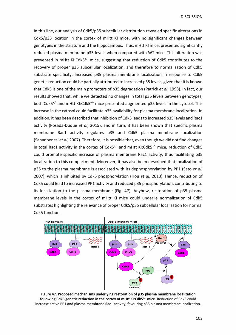
DISCUSSION
103
In this line, our analysis of Cdk5/p35 subcellular distribution revealed specific alterations in
Cdk5/p35 location in the cortex of mHtt KI mice, with no significant changes between
genotypes in the striatum and the hippocampus. Thus, mHtt KI mice, presented significantly
reduced plasma membrane p35 levels when compared with WT mice. This alteration was
prevented in mHtt KI:Cdk5+/‐ mice, suggesting that reduction of Cdk5 contributes to the
recovery of proper p35 subcellular localization, and therefore to normalization of Cdk5
substrate specificity. Increased p35 plasma membrane localization in response to Cdk5
genetic reduction could be partially attributed to increased p35 levels, given that it is known
that Cdk5 is one of the main promoters of p35 degradation (Patrick et al, 1998). In fact, our
results showed that, while we detected no changes in total p35 levels between genotypes,
both Cdk5+/‐ and mHtt KI:Cdk5+/‐ mice presented augmented p35 levels in the cytosol. This
increase in the cytosol could facilitate p35 availability for plasma membrane localization. In
addition, it has been described that inhibition of Cdk5 leads to increased p35 levels and Rac1
activity (Posada‐Duque et al, 2015), and in turn, it has been shown that specific plasma
membrane Rac1 activity regulates p35 and Cdk5 plasma membrane localization
(Sananbenesi et al, 2007). Therefore, it is possible that, even though we did not find changes
in total Rac1 activity in the cortex of Cdk5+/‐ and mHtt KI:Cdk5+/‐ mice, reduction of Cdk5
could promote specific increase of plasma membrane Rac1 activity, thus facilitating p35
localization to this compartment. Moreover, it has also been described that localization of
p35 to the plasma membrane is associated with its dephosphorylation by PP1 (Sato et al,
2007), which is inhibited by Cdk5 phosphorylation (Hou et al, 2013). Hence, reduction of
Cdk5 could lead to increased PP1 activity and reduced p35 phosphorylation, contributing to
its localization to the plasma membrane (Fig. 47). Anyhow, restoration of p35 plasma
membrane levels in the cortex of mHtt KI mice could underlie normalization of Cdk5
substrates highlighting the relevance of proper Cdk5/p35 subcellular localization for normal
Cdk5 function.
Figure 47. Proposed mechanisms underlying restoration of p35 plasma membrane localization following Cdk5 genetic reduction in the cortex of mHtt KI:Cdk5+/‐ mice. Reduction of Cdk5 could
increase active PP1 and plasma membrane Rac1 activity, favouring p35 plasma membrane localization.
DISCUSSION
104
Even though previous studies from our group showed that mHtt KI mice exhibit increased
p25 levels in the striatum, we were not able to detect presence of p25 in our conditions.
This could be attributed to different technical conditions or to the fact that previous studies
were performed in homozygous mHtt KI mice at 9 months of age, which might manifest a
more severe phenotype. Nonetheless, it is important to remember that, despite Cdk5
activity is usually assessed based on p35 and p25 levels, its regulation is much more
complex. Thus, proteins like CyclinI, GSTP1 and CyclinE have also been described to
modulate Cdk5 activity, as well as several posttranslational modifications including
phosphorylation, acetylation and S‐nitrosylation (Shah and Lahiri, 2014). Therefore, we
cannot rule out that some of these regulatory mechanisms may participate in Cdk5
alteration in HD and, subsequently, in functional recovery in mHtt KI:Cdk5+/‐ mice. For
instance, aberrant S‐nitrosylation has been described in several neurodegenerative
disorders such as AD, and has been associated to excessive NMDAR activation and to
mitochondrial dysfunction (Nakamura et al, 2015; Nakamura and Lipton, 2017), which are
also present in HD (Zuccato et al, 2010). Given that both Cdk5 and p35 have been described
to be S‐nitrosylated (Qu et al, 2011; Zhang et al, 2015), it is possible that changes in Cdk5
and/or p35 S‐nitrosylation might be additional regulatory pathways contributing to Cdk5
deregulation in HD. In sum, the complex regulation of Cdk5 at the molecular level, adds up
to the already intricate Cdk5 function on a subcellular level and between different brain
regions, which, altogether, complicates the analysis of Cdk5 deregulation in pathological
conditions and reveals the need of further studying Cdk5 function on a physiological context.
Indeed, most studies analysing the physiological brain function of Cdk5 have concentrated
mainly in the striatum and hippocampus, while reports in the cortex are mostly restricted
to Cdk5 role in neuronal migration during neurodevelopment, with few studies in the adult
brain. In this sense, our observations of increased cytosolic p35 as a result of Cdk5 genetic
reduction, specifically in the cortex, might reflect differential regulatory feedback between
Cdk5 and p35 in this brain region compared to the striatum or the hippocampus. This
hypothesis could explain our results showing that, at least in an HD context, Cdk5 genetic
reduction modifies Cdk5 substrates preferentially in the cortex (Table 10). Given that
cortical alterations are common in different neurodegenerative disorders including AD, a
deeper study of Cdk5 physiological function in the cortex, might be critical to understand
neuropathologies involving Cdk5 deregulation in this region.
Importantly, cortex affectation in HD starts years prior to disease onset (Paulsen et al, 2008;
Rosas et al, 2008a), and, for instance, specific mHtt expression in the striatum does not
reproduce some HD hallmarks like locomotor deficits (Gu et al, 2007), indicating the
relevance of aberrant cortical inputs for mHtt‐induced striatal vulnerability. In fact, previous
studies from our group have demonstrated synaptic disturbances in the cortex prior to
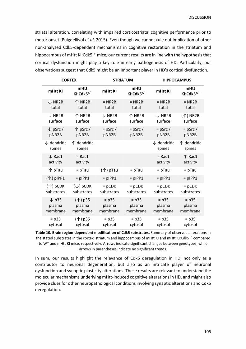
DISCUSSION
105
striatal alteration, correlating with impaired corticostriatal cognitive performance prior to
motor onset (Puigdellívol et al, 2015). Even though we cannot rule out implication of other
non‐analysed Cdk5‐dependent mechanisms in cognitive restoration in the striatum and
hippocampus of mHtt KI:Cdk5+/‐ mice, our current results are in line with the hypothesis that
cortical dysfunction might play a key role in early pathogenesis of HD. Particularly, our
observations suggest that Cdk5 might be an important player in HD’s cortical dysfunction.
Table 10. Brain region‐dependent modification of Cdk5 substrates. Summary of observed alterations in
the stated substrates in the cortex, striatum and hippocampus of mHtt KI and mHtt KI:Cdk5+/‐ compared
to WT and mHtt KI mice, respectively. Arrows indicate significant changes between genotypes, while
arrows in parentheses indicate no significant trends.
In sum, our results highlight the relevance of Cdk5 deregulation in HD, not only as a
contributor to neuronal degeneration, but also as an intricate player of neuronal
dysfunction and synaptic plasticity alterations. These results are relevant to understand the
molecular mechanisms underlying mHtt‐induced cognitive alterations in HD, and might also
provide clues for other neuropathological conditions involving synaptic alterations and Cdk5
deregulation.
CORTEX STRIATUM HIPPOCAMPUS
mHtt KI mHtt
KI:Cdk5+/‐ mHtt KI
mHtt KI:Cdk5+/‐
mHtt KI mHtt
KI:Cdk5+/‐
↓ NR2B total
↑ NR2B total
= NR2B total
= NR2B total
= NR2B total
= NR2B total
↓ NR2B surface
↑ NR2B surface
↓ NR2B surface
↑ NR2B surface
↓ NR2B surface
(↑) NR2B surface
↓ pSrc / pNR2B
↑ pSrc / pNR2B
= pSrc / pNR2B
= pSrc / pNR2B
= pSrc / pNR2B
= pSrc / pNR2B
↓ dendri c spines
↑ dendri c spines
↓ dendri c
spines ↑ dendri c
spines
↓ Rac1 activity
= Rac1 activity
= Rac1 activity
↑ Rac1 activity
↑ pTau = pTau (↑) pTau = pTau = pTau = pTau
(↑) pIPP1 = pIPP1 = pIPP1 = pIPP1 = pIPP1 = pIPP1
(↑) pCDK substrates
(↓) pCDK substrates
= pCDK substrates
= pCDK substrates
= pCDK substrates
= pCDK substrates
↓ p35 plasma
membrane
(↑) p35 plasma
membrane
= p35 plasma
membrane
= p35 plasma
membrane
= p35 plasma
membrane
= p35 plasma
membrane
= p35 cytosol
(↑) p35 cytosol
= p35 cytosol
= p35 cytosol
= p35 cytosol
= p35 cytosol
DISCUSSION
106
2. Cell cycle role in HD striatal vulnerability
Cdk5 deregulation has been associated with neuronal dysfunction and/or death in multiple
neuropathological conditions (Liu et al, 2016; Lopes and Agostinho, 2011; Patrick et al,
1999). In HD, Cdk5 has been involved in several known striatal vulnerability factors, such as
mitochondrial dysfunction (Cherubini et al, 2015) or increased sensitivity to glutamatergic
and dopaminergic signalling (Paoletti et al, 2008; Park et al, 2012). On the other hand, Cdk5
has been implicated in neuronal cell cycle re‐entry in response to neurotoxic factors such as
β‐amyloid and prion peptides (Lopes et al, 2009; Niu et al, 2012). Coincidentally, cell cycle
dysregulation has also been associated with alterations present in HD such as excitotoxicity
and DNA damage (Kruman et al, 2004; Park et al, 2000). For these reasons, we wanted to
assess whether Cdk5 deregulation could also lead to cell cycle deregulation in HD, and the
implication of neuronal cell cycle re‐entry in HD striatal vulnerability.
To act as a cell cycle inhibitor, it has been described that, Cdk5 needs to be located in the
nucleus of differentiated neurons (Zhang et al, 2008a). Interestingly, our results showed that
nuclear Cdk5 levels were significantly reduced in the striatum of 8‐month‐old mHtt KI mice,
suggesting that cell cycle inhibition by Cdk5 could be altered by mHtt presence. We also
observed reduction of nuclear p27 levels in mHtt KI mice, which could underlie reduction of
Cdk5 levels, given that Cdk5 nuclear localization relies on p27 interaction, as Cdk5 does not
possess a NLS (Zhang et al, 2010a). Reduced p27 nuclear levels could be caused, in turn, by
impaired p27 nuclear import as a result of mHtt presence, which is in agreement with
reports describing alterations in nucleocytoplasmic trafficking in HD (Grima et al, 2017;
Truant et al, 2007). In contrast, analysis of total p27 levels in striatal lysates revealed a
significant increase in mHtt KI mice from 6 months of age onwards. These results indicate
that reduced p27 nuclear levels are likely not caused by alterations in p27 transcription or
degradation and therefore, correlate with the hypothesis that reduced p27 nuclear levels
might be associated with altered nucleocytoplasmic transport. On the other hand, increase
in total p27 levels might be related to reduced p27 degradation, as it has been described
that the transcriptional coactivator Jab1 which degrades p27, is sequestered in mHtt
aggregates in HD patient’s brains (Cong et al, 2012). Given that mHtt KI mice already present
mHtt aggregates at 6 months of age (Lloret et al, 2006), the same mechanism might underlie
our observations.
Regardless of the underlying mechanisms, reduced Cdk5 and p27 nuclear localization might
affect formation of the nuclear Cdk5/p27/E2F1 complex. It has been described that
formation of this complex prevents cell cycle progression by sequestering the transcription
factor E2F1 from activating necessary genes for cell cycle initiation (Zhang and Herrup,
2011). Hence, even though we found no significant changes in global Cdk5 and p27

DISCUSSION
107
interaction, reduced Cdk5 and p27 nuclear localization might result in reduced formation of
the Cdk5/p27/E2F1 complex, and subsequently, it could lead to aberrant cell cycle re‐entry
of HD striatal neurons (Fig. 48).
Figure 48. Proposed mechanism underlying Cdk5‐dependent cell cycle proteins alteration in HD. Presence of mHtt aggregates in mHtt KI mice could cause sequestering of Jab1 contributing to increased
p27 levels. mHtt could also impair p27, and consequently Cdk5, nuclear localization, disrupting formation of the Cdk5/p27/E2F1 complex, which could allow E2F1 to activate transcription of cell cycle
genes.
In this regard, we observed that the early G1 phase protein, CyclinD1 presented a trend
towards increased protein levels in the striatum of 6‐month‐old mHtt KI mice. This increase
became significant at motor onset (8 months) and was maintained at later disease stages
(13 and 18 months), indicating a sustained alteration of CyclinD1 levels by the presence of
mHtt. At 18 months of age, levels of the CyclinD1 partner, Cdk4, also presented an increasing
trend. Accordingly, previous studies have found increased Cdk4 protein levels as well as
increased Cdk4 and CyclinE neuronal expression in the striatum of 7‐month‐old R6/1 mice
(Fernandez‐Fernandez et al, 2011). CyclinB1 has also been detected in the striatum of R6/2
mice in contrast with WT mice which show no expression of this protein (Liu et al, 2015).
Therefore, this data suggests that alteration of cell cycle proteins expression might be a
common mechanism resulting from mHtt presence in both full‐length and N‐terminal
fragment HD mice models.
Importantly, we also observed alteration of cell cycle proteins in the putamen of HD human
patients. Specifically, we detected a significant increase of Cdk4 protein levels, whereas
CyclinD1 levels were not significantly altered. Human HD putamen samples also presented
p27 electrophoretic mobility alterations, which might reflect specific post‐translational
modifications leading to increased molecular weight, such as sumoylation or ubiquitination
DISCUSSION
108
(Hilgarth and Sarge, 2005; Xu and Jaffrey, 2013). In addition, E2F1 exhibited an increase in a
lower molecular weight band, which could be associated with increased E2F1 cleavage, as it
has been described that in NMDAR‐mediated toxicity conditions, E2F1 is cleaved to a stable
55‐kDa band (Zyskind et al, 2015). Previous studies had already described alteration of cell
cycle proteins in samples from HD patients brains (Fernandez‐Fernandez et al, 2011; Pelegrí
et al, 2008), further corroborating the hypothesis that mHtt presence causes aberrant
expression of cell cycle proteins. Nonetheless, the fact that alteration of specific proteins
differs between HD mice models and human patients, suggests that neuropathological
differences such as presence or absence of neuronal death or of mHtt aggregates, might
lead to differential deregulation of cell cycle proteins.
In fact, deregulation of cell cycle proteins has been directly or indirectly associated in
previous studies with mHtt aggregate formation. For instance, presence of perinuclear mHtt
aggregates leading to nuclear envelope disruption has been described to cause cell cycle re‐
entry (Liu et al, 2015). Moreover, alteration of ER‐stress response in HD, which is activated
in response to misfolded proteins, has also been associated with alteration of cell cycle
proteins in HD models (Fernandez‐Fernandez et al, 2011). On the other hand, 3‐NP
treatment has been described to induce alteration of cell cycle proteins both in vivo and in
vitro (Akashiba et al, 2008; Pelegrí et al, 2008). Importantly, even though 3‐NP treatment
causes death of both striatal and cortical neurons, only striatal neurons show p27 reduction
and increased calpain activity after 3‐NP treatment, and are partially neuroprotected by
CDKs inhibitors (Akashiba et al, 2008). These data indicate that cell cycle re‐entry might be
an additional mechanism resulting from inherent striatal susceptibility properties, and
therefore could play a role in HD increased striatal vulnerability.
In this sense, excitotoxicity represents one of the main striatal vulnerability factors in HD
(Fan and Raymond, 2007) and, it has also been associated with aberrant expression of cell
cycle proteins (Park et al, 2000). Excitotoxic responses caused by excessive NMDARs
activation is also one of the mechanisms leading to Cdk5 hyperactivation via p35 cleavage
to p25 (Lee et al, 2000; Patrick et al, 1998). For these reasons, we explored whether
alteration of cell cycle proteins in HD, might be associated with NMDA activation and/or
with subsequent Cdk5 hyperactivation. Our results showed that treatment of mature striatal
primary cultures with 100 µM NMDA for 1h, led to a general reduction of cell cycle proteins
expression in neurons. These data contrast with the hypothesis that excitotoxic stress might
lead to re‐expression of cell cycle proteins. However, we also observed important
alterations in the subcellular distribution of the analysed cell cycle proteins. Thus, NMDA
treatment led to a reduction of nuclear levels of p27, which could prevent its role as a cell
cycle suppressor contributing to cell cycle initiation. Cytosolic localization of the cell cycle
promoter E2F1 in basal striatal cultures was also altered following NMDA treatment. A
DISCUSSION
109
diffuse distribution along the entire neuron, including presence in the nucleus of some
neurons, was observed, which might reflect an activation of E2F1 function as a transcription
factor of cell cycle progression genes. Similar distribution alterations were observed for the
early G1 phase kinase Cdk4, which could also reflect cell cycle initiation of striatal neurons
after NMDA treatment. On the other hand, CyclinD1 distribution, which showed mostly
nuclear localization in basal conditions, presented cytosolic, in addition to nuclear
localization, after NMDA treatment. Therefore, our results indicate that, although NMDA
treatment causes a general reduction of cell cycle proteins expression, it also leads to a
significant alteration of their subcellular distribution, which undoubtedly affects its
physiological function and could lead to cell cycle re‐entry.
Nonetheless, the fact that CyclinD1 was expressed only in the nucleus of a small subset of
neurons under basal conditions, might reflect additional functions of CyclinD1 on certain
striatal neurons. In fact, not much is known about cell cycle proteins function in
differentiated neurons, given that, with the exception of cell cycle inhibitors, they would be
expected to show little expression in non‐proliferating cells. Therefore, the fact that most
cell cycle proteins are expressed in neurons, sometimes in cellular compartments other than
the nucleus, indicates their implication in alternative functions, complicating the
extrapolation of functional effects from changes in their levels and localization. For instance,
increase of E2F1 levels has been associated with neuronal death in neurodegeneration in in
vitro models. Importantly, this function has been attributed to cytosolic localization of E2F1,
without alteration of some of the main E2F1 transcription products, suggesting a cell cycle‐
independent function for E2F1 in a neurodegenerative context (Wang et al, 2010). In this
sense, our results show that under basal conditions, in contrast with other analysed cell
cycle progression proteins, like Cdk4 and CyclinD1, E2F1 was expressed in the cytosol of 94%
of striatal neurons, suggesting that E2F1 cytosolic expression is normal in striatal neurons.
Other functions like regulation of cell motility and migration, DNA repair, transcription
modulation or even synaptic plasticity and memory formation, have also been ascribed to
cell cycle proteins like p27, CyclinD1 or CyclinE (Frank and Tsai, 2009; Larrea et al, 2009; Lim
and Kaldis, 2013; Odajima et al, 2011). In this sense, it is also possible that some of the
alterations we have observed in cell cycle proteins levels and distribution after NMDA
treatment may affect some of these cell cycle‐independent functions, leading to neuronal
dysfunction through additional mechanisms besides neuronal cell cycle re‐entry.
We also observed that some of the NMDA‐dependent alterations in cell cycle proteins were
dependent on calpain activation. Thus, in accordance with previous studies (Akashiba et al,
2006b; Zyskind et al, 2015) we observed that reduced p27 levels and E2F1 cleavage after
NMDA treatment were prevented when calpain was inhibited. It would be interesting to
analyse if calpain inhibition also prevents changes in subcellular localization of these

DISCUSSION
110
proteins. Additionally, given that calpain inhibition also avoids p25 production and
subsequent Cdk5 hyperactivation, future studies specifically inhibiting Cdk5, might reveal if
NMDA‐dependent cell cycle proteins alterations are directly dependent on calpain or if Cdk5
hyperactivation is also involved. As calpain is activated by intracellular calcium (Khorchid
and Ikura, 2002), and deregulation of calcium homeostasis is an HD hallmark (Giacomello et
al, 2013), aberrant calpain activation might contribute to calpain‐dependent cell cycle
proteins alteration in HD.
In this sense, our preliminary results regarding mHtt effect on cell cycle proteins indicate
that mHtt led to higher expression of cytosolic Cdk4 and E2F1 in neuronal cells under basal
conditions, while the small subset of neurons expressing CyclinD1 showed a decreasing
trend in mHtt KI cultures. In contrast, NMDA treated mHtt KI cultures showed similar
alterations when compared with NMDA treated WT cultures, with the exception of p27,
which showed a decreasing trend in mHtt KI cultures, indicating that mHtt presence could
further reduce NMDA‐induced p27 decrease, contributing to cell cycle re‐entry. Given that
p27 localization in striatal neurons is mainly nuclear, this data would fit with our
observations in the striatum of mHtt KI mice which also show decreased p27 nuclear levels.
Therefore, future studies contrasting these results and analysing whether mHtt affects
subcellular distribution of cell cycle proteins, might shed some light to the role of neuronal
cell cycle re‐entry in HD.
Notwithstanding, it is noteworthy that, even though re‐expression of cell cycle proteins has
been associated with aberrant cell cycle re‐entry, it has also been proposed to play a role in
neuronal DNA repair responses. Neurons are particularly vulnerable to DNA damage caused
by toxic species resulting from their high metabolic rate. Given that most DNA repair
mechanisms rely on cell cycle progression, activation of certain cell cycle phases has been
proposed to be necessary for activation of certain DNA repair responses (Schwartz et al,
2007). Importantly, altered DNA repair responses have been described in HD (Enokido et al,
2010) and have been associated with increased genomic instability, which shows high
correlation with neuronal dysfunction in HD (Manley et al, 1999). Thus, altered cell cycle
proteins expression in HD might also be associated with DNA repair responses and therefore
with genomic instability.
Altogether, alteration of cell cycle proteins has been described in several neuropathological
conditions and has been associated with cell cycle re‐entry of differentiated neurons
contributing to neuronal dysfunction or death. Nonetheless, it is evident that a lot of
information is still lacking regarding physiological functions of cell cycle proteins in
differentiated neurons and therefore interpretation of functional effects resulting from
their alteration can be complex. Alteration of cell cycle proteins has received little attention,
particularly in HD, even though several molecular mechanisms involved with cell cycle re‐

DISCUSSION
111
entry are associated with increased striatal vulnerability. Therefore, further study of this
mechanism might contribute to understanding, not only molecular pathways contributing
to striatal affectation in HD, but to neuronal dysfunction processes in other
neurodegenerative disorders, therefore providing new therapeutic targets for their
treatment.
3. Cdk5 as a dual player in neuronal vulnerability and synaptic plasticity
Over the last decades Cdk5 has become a pivotal player in synaptic plasticity modulation
and in neurodegenerative processes associated to several pathological conditions.
However, its relevant function goes in hand with its complexity making difficult full
understanding of its regulation both under physiological and pathological conditions.
The results obtained in this Thesis reflect this intricacy. Particularly, we propose that
involvement of Cdk5 deregulation in striatal vulnerability in HD might be preferentially
associated to calpain activation and p35 cleavage to p25. This situation would be favoured
by calcium homeostasis dysregulation caused, in part, by excitotoxicity and mitochondrial
dysfunction, present in HD (Cherubini and Ginés, 2017; Fan and Raymond, 2007; Giacomello
et al, 2011). Our observations suggest that alteration of cell cycle proteins might be an
additional mechanism participating in this scenario. On the other hand, deregulation of Cdk5
associated to reduced p35/Cdk5 plasma membrane localization might have a more
prevalent role in altered Cdk5 synaptic substrates availability, and therefore to synaptic
plasticity alterations. Importantly, this deregulation on a subcellular level might not be
necessarily associated with production of p25 and therefore could go undetected when
analysing total levels of Cdk5/p35 or some of the substrates commonly used as Cdk5 activity
indicators. We propose that this kind of deregulation is particularly relevant in cortical
dysfunction in HD, and therefore could also play an important role in other pathological
processes affecting this region (Fig. 49).
In conclusion, our results point Cdk5 as a potential target for simultaneous treatment of
synaptic plasticity alterations and neuronal vulnerability present in HD, but also highlight
the relevance of further studying Cdk5 intricate function which could contribute to obtaining
therapeutic targets for other neuropathological conditions involving Cdk5 deregulation.
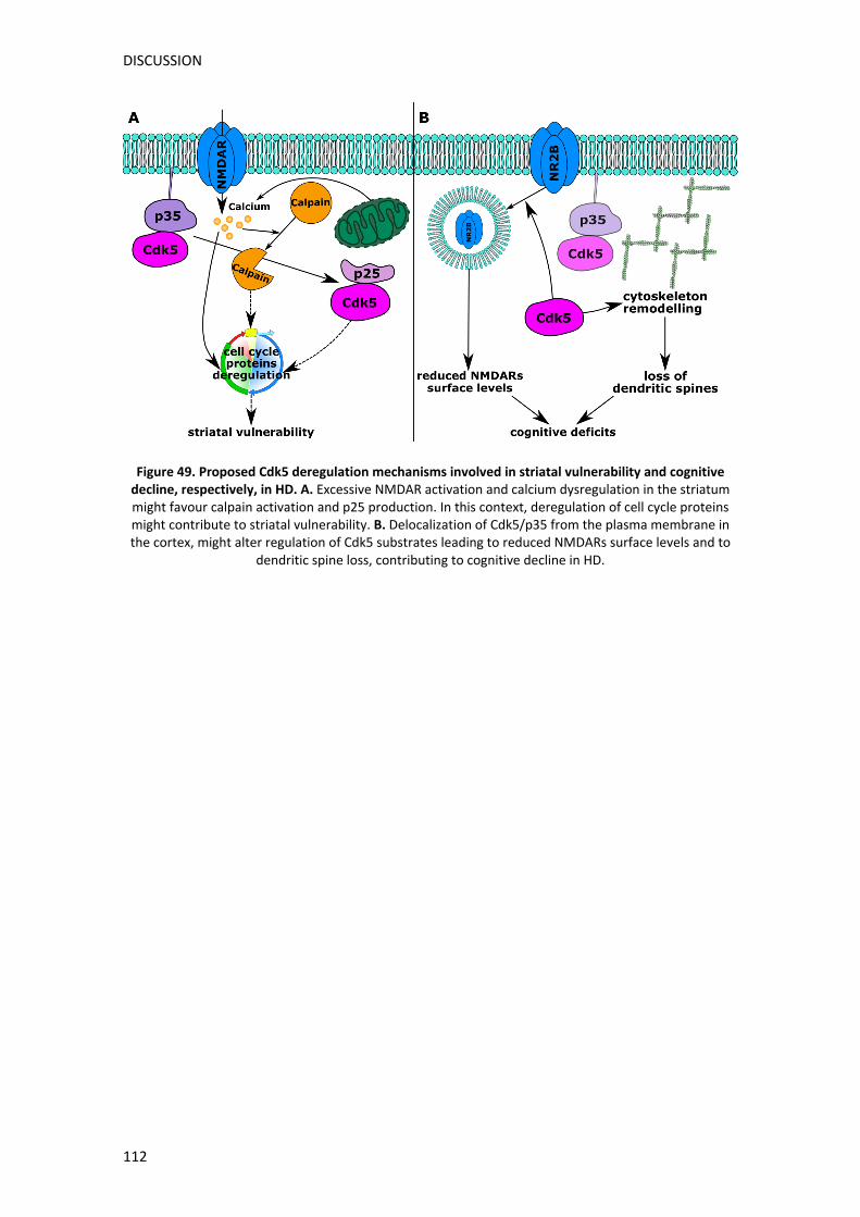
DISCUSSION
112
Figure 49. Proposed Cdk5 deregulation mechanisms involved in striatal vulnerability and cognitive decline, respectively, in HD. A. Excessive NMDAR activation and calcium dysregulation in the striatum might favour calpain activation and p25 production. In this context, deregulation of cell cycle proteins might contribute to striatal vulnerability. B. Delocalization of Cdk5/p35 from the plasma membrane in the cortex, might alter regulation of Cdk5 substrates leading to reduced NMDARs surface levels and to
dendritic spine loss, contributing to cognitive decline in HD.


CONCLUSIONS

CONCLUSIONS
117
As a result of the presented studies, the main conclusions of this Thesis are:
1. Cdk5 is a new important contributor to corticostriatal and hippocampal‐dependent
cognitive deficits in mHtt KI mice.
2. Prevention of cognitive deficits by Cdk5 genetic reduction in mHtt KI mice is
associated with brain region‐specific modulation of synaptic plasticity substrates.
3. Rescue of corticostriatal deficits in double mutant mHtt KI:Cdk5+/‐ mice is associated
with restoration of mHtt induced‐synaptic alterations in the cortex. This recovery is
mediated by normalization of dendritic spine density and NR2B surface levels
through the pSrc/pNR2B pathway. In contrast, prevention of hippocampal deficits
associates with restoration of dendritic spine density which correlates with
increased Rac1 activity, resulting from Cdk5 genetic reduction.
4. mHtt alters Cdk5/p35 subcellular localization specifically in the cortex of mHtt KI
mice, which correlates with preferential alteration of Cdk5 substrates in this region.
5. Symptomatic mHtt KI mice exhibit reduced Cdk5 and p27 nuclear levels in the
striatum, suggesting that mHtt may induce cell cycle re‐entry by impairing their
function as cell cycle inhibitors.
6. mHtt causes alteration of G1 phase cell cycle proteins expression in the striatum of
mHtt KI mice and in the putamen of HD patients, which could further contribute to
aberrant cell cycle re‐entry.
7. Alteration of cell cycle proteins in response to Cdk5 deregulation and/or excessive
NMDARs activation, might be a new molecular mechanism involved in striatal
vulnerability in HD.


REFERENCES

REFERENCES
121
Akashiba H, Ikegaya Y, Nishiyama N, Matsuki N (2008). Differential involvement of cell cycle reactivation between striatal and cortical neurons in cell death induced by 3‐nitropropionic acid. J Biol Chem 283: 6594–6606.
Akashiba H, Matsuki N, Nishiyama N (2006a). p27 small interfering RNA induces cell death through elevating cell cycle activity in cultured cortical neurons: a proof‐of‐concept study. Cell Mol life Sci 63: 2397–404.
Akashiba H, Matsuki N, Nishiyama N (2006b). Calpain activation is required for glutamate‐induced p27 down‐regulation in cultured cortical neurons. J Neurochem 99: 733–744.
Al‐Ubaidi MR, Hollyfield JG, Overbeek PA, Baehr W (1992). Photoreceptor degeneration induced by the expression of simian virus 40 large tumor antigen in the retina of transgenic mice. Proc Natl Acad Sci U S A 89: 1194–1198.
Albin RL, Young AB, Penney JB, Handelin B, Balfour R, Anderson KD, et al (1990). Abnormalities of striatal projection neurons and N‐methyl‐D‐aspartate receptors in presymptomatic Huntington’s disease. N Engl J Med 322: 1293–8.
Andre VM, Cepeda C, Fisher YE, Huynh M, Bardakjian N, Singh S, et al (2011). Differential Electrophysiological Changes in Striatal Output Neurons in Huntington’s Disease. J Neurosci 31: 1170–1182.
Andrew S, Goldberg Y, Kremer B, Telenius H, Theilmann J, Adam S, et al (1993). The relationship between trinucleotide (CAG) repeat lenght and clinical features of Huntington’s disease. Nat Genet 4: 398–403.
Angelo M, Plattner F, Giese KP (2006). Cyclin‐dependent kinase 5 in synaptic plasticity, learning and memory. J Neurochem 99: 353–370.
Angelo M, Plattner F, Irvine EE, Giese KP (2003). Improved reversal learning and altered fear conditioning in transgenic mice with regionally restricted p25 expression. Eur J Neurosci 18: 423–431.
Anne SL, Saudou F, Humbert S (2007). Phosphorylation of huntingtin by cyclin‐dependent kinase 5 is induced by DNA damage and regulates wild‐type and mutant huntingtin toxicity in neurons. J Neurosci 27: 7318–7328.
Arendt T (2012). Cell cycle activation and aneuploid neurons in Alzheimer’s disease. Mol Neurobiol 46: 125–135.
Aretouli E, Brandt J (2010). Episodic memory in dementia: Characteristics of new learning that differentiate Alzheimer’s, Huntington’s, and Parkinson’s diseases. Arch Clin Neuropsychol 25: 396–409.
Asada A, Yamamoto N, Gohda M, Saito T, Hayashi N, Hisanaga SI (2008). Myristoylation of p39 and p35 is a determinant of cytoplasmic or nuclear localization of active cycline‐dependent kinase 5 complexes. J Neurochem 106: 1325–1336.
Baghirova S, Hughes BG, Hendzel MJ, Schulz R (2015). Sequential fractionation and
REFERENCES
122
isolation of subcellular proteins from tissue or cultured cells. MethodsX 2: 440–445.
Baquet ZC, Gorski JA, Jones KR (2004). Early Striatal Dendrite Deficits followed by Neuron Loss with Advanced Age in the Absence of Anterograde Cortical Brain‐ Derived Neurotrophic Factor. J Neurosci 24: 4250–4258.
Barnett DGS, Bibb JA (2011). The role of Cdk5 in cognition and neuropsychiatric and neurological pathology. Brain Res Bull 85: 9–13.
Bartsch T, Wulff P (2015). The hippocampus in aging and disease: From plasticity to vulnerability. Neuroscience 309: 1–16.
Bates GP, Dorsey R, Gusella JF, Hayden MR, Kay C, Leavitt BR, et al (2015). Huntington disease. Nat Rev Dis Prim 1: 1–21.
Beal MF, Brouillet E, Jenkins B, Henshaw R, Rosen B, Hyman BT (1993). Age‐Dependent Striatal Excitotoxic Lesions Produced by the Endogenous Mitochondrial Inhibitor Malonate. J Neurochem 61: 1147–1150.
Beal MF, Kowall NW, Ellison DW, Mazurek MF, Swartz KJ, Martin JB (1986). Replication of the neurochemical characteristics of Huntington’s disease by quinolinic acid. Nature 321: 168–171.
Begeti F, Schwab LC, Mason SL, Barker RA (2016). Hippocampal dysfunction defines disease onset in Huntington’s disease. J Neurol Neurosurg Psychiatry 87: 975–981.
Behrens PF, Franz P, Woodman B, Lindenberg KS, Landwehrmeyer GB (2002). Impaired glutamate transport and glutamate‐glutamine cycling: downstream effects of the Huntington mutation. Brain 125: 1908–1922.
Benavides DR, Quinn JJ, Zhong P, Hawasli AH, DiLeone RJ, Kansy JW, et al (2007). Cdk5 Modulates Cocaine Reward, Motivation, and Striatal Neuron Excitability. J Neurosci 27: 12967–12976.
Benito E, Urbanke H, Ramachandran B, Barth J, Halder R, Awasthi A, et al (2015). HDAC inhibitor‐dependent transcriptome and memory reinstatement in cognitive decline models. J Clin Invest 125: 3572–3584.
Benn CL, Slow EJ, Farrell LA, Graham R, Deng Y, Hayden MR, et al (2007). Glutamate receptor abnormalities in the YAC128 transgenic mouse model of Huntington’s disease. Neuroscience 147: 354–372.
Bhounsule AS, Bhatt LK, Prabhavalkar KS, Oza M (2017). Cyclin dependent kinase 5: A novel avenue for Alzheimer’s disease. Brain Res Bull 132: 28–38.
Bibb JA, Chen J, Taylor JR, Svenningsson P, Nishi A, Snyder GL, et al (2001a). Effects of chronic exposure to cocaine are regulated by the neuronal protein Cdk5. Nature 410: 376–380.
Bibb JA, Nishi A, O’Callaghan JP, Ule J, Lan M, Snyder GL, et al (2001b). Phosphorylation of Protein Phosphatase Inhibitor‐1 by Cdk5. J Biol Chem 276: 14490–14497.

REFERENCES
123
Bibb JA, Snyder GL, Nishi A, Yan Z, Meijer L, Fienberg a a, et al (1999). Phosphorylation of DARPP‐32 by Cdk5 modulates dopamine signalling in neurons. Nature 402: 669–671.
Blázquez C, Chiarlone A, Sagredo O, Aguado T, Pazos MR, Resel E, et al (2011). Loss of striatal type 1 cannabinoid receptors is a key pathogenic factor in Huntington’s disease. Brain 134: 119–136.
Bolam J, Hanley J, Booth P, Bevan M (2000). Synaptic organisation of the basal ganglia. J Anat 196: 527–542.
Bragg RM, Coffey SR, Weston RM, Ament SA, Cantle JP, Minnig S, et al (2017). Motivational, proteostatic and transcriptional deficits precede synapse loss, gliosis and neurodegeneration in the disease B6.HttQ111/+ model of Huntington’s disease. Sci Rep 7: 1–17.
Branco‐Santos J, Herrera F, Poças GM, Pires‐Afonso Y, Giorgini F, Domingos PM, et al (2017). Protein phosphatase 1 regulates huntingtin exon 1 aggregation and toxicity. Hum Mol Genet 26: 3763–3775.
Brinkkoetter PT, Olivier P, Wu JS, Henderson S, Krofft RD, Pippin JW, et al (2009). Cyclin I activates Cdk5 and regulates expression of Bcl‐2 and Bcl‐XL in postmitotic mouse cells. J Clin Invest 119: 3089–3101.
Brito V, Giralt A, Enriquez‐Barreto L, Puigdellívol M, Suelves N, Zamora‐Moratalla A, et al (2014). Neurotrophin receptor p75 NTR mediates Huntington’s disease – associated synaptic and memory dysfunction. J Clin Invest 124: 4411–4428.
Brito V, Puigdellívol M, Giralt A, Toro D del, Alberch J, Ginés S (2013). Imbalance of p75NTR/TrkB protein expression in Huntington’s disease: implication for neuroprotective therapies. Cell Death Dis 4: e595.
Brooks SP, Janghra N, Workman VL, Bayram‐Weston Z, Jones L, Dunnett SB (2012). Longitudinal analysis of the behavioural phenotype in R6/1 (C57BL/6J) Huntington’s disease transgenic mice. Brain Res Bull 88: 94–103.
Brouillet E, Jenkins BG, Hyman BT, Ferrante RJ, Kowall NW, Srivastava R, et al (1993). Age‐Dependent Vulnerability of the Striatum to the Mitochondrial Toxin 3‐Nitropropionic Acid. J Neurochem 60: 356–359.
Brustovetsky N, Brustovetsky T, Purl KJ, Capano M, Crompton M, Dubinsky JM (2003). Increased susceptibility of striatal mitochondria to calcium‐induced permeability transition. J Neurosci 23: 4858–4867.
Brustovetsky N, LaFrance R, Purl KJ, Brustovetsky T, Keene CD, Low WC, et al (2005). Age‐dependent changes in the calcium sensitivity of striatal mitochondria in mouse models of Huntington’s disease. J Neurochem 93: 1361–1370.
Büchner A, Krumova P, Ganesan S, Bähr M, Eckermann K, Weishaupt JH (2015). Sumoylation of p35 Modulates p35/Cyclin‐Dependent Kinase (Cdk) 5 Complex Activity. NeuroMolecular Med 17: 12–23.

REFERENCES
124
Bulley SJ, Drew CJG, Morton AJ (2012). Direct visualisation of abnormal dendritic spine morphology in the hippocampus of the R6/2 transgenic mouse model of Huntington’s disease. J Huntingtons Dis 1: 267–273.
Bunner KD, Rebec G V (2016). Corticostriatal Dysfunction in Huntington’s Disease: The Basics. Front Hum Neurosci 10: 317.
Calabresi P, Gubellini P, Centonze D, Picconi B, Bernardi G, Chergui K, et al (2000). Dopamine and cAMP‐Regulated Phosphoprotein 32 kDa Controls Both Striatal Long‐Term Depression and Long‐Term Potentiation, Opposing Forms of Synaptic Plasticity. J Neurosci 20: 8443–8451.
Calabresi P, Picconi B, Tozzi A, Ghiglieri V, Filippo M Di (2014). Direct and indirect pathways of basal ganglia: A critical reappraisal. Nat Neurosci 17: 1022–1030.
Camins A, Verdaguer E, Folch J, Canudas AM, Pallas M (2006). The role of CDK5/P25 formation/inhibition in neurodegeneration. Drug News Perspect 19: 453–460.
Castro‐Alvarez JF, Uribe‐Arias SA, Kosik KS, Cardona‐Gómez GP (2014). Long‐ and short‐term CDK5 knockdown prevents spatial memory dysfunction and tau pathology of triple transgenic Alzheimer’s mice. Front Aging Neurosci 6: 1–10.
Cataldo F, Peche LY, Klaric E, Brancolini C, Myers MP, Demarchi F, et al (2013). CAPNS1 regulates USP1 stability and maintenance of genome integrity. Mol Cell Biol 33: 2485–96.
Cattaneo E, Zuccato C, Tartari M (2005). Normal Huntingtin Function: An Alternative Approach to Huntington’s Disease. Nat Rev Neurosci 6: 919–930.
Causeret F, Jacobs T, Terao M, Heath O, Hoshino M, Nikolic M (2007). Neurabin‐I Is Phosphorylated by Cdk5: Implications for Neuronal Morphogenesis and Cortical Migration. Mol Biol Cell 18: 4327–4342.
Cepeda C, Ariano MA, Calvert CR, Flores‐Hernández J, Chandler SH, Leavitt BR, et al (2001). NMDA receptor function in mouse models of Huntington disease. J Neurosci Res 66: 525–39.
Cepeda C, Hurst RS, Calvert CR, Hernández‐Echeagaray E, Nguyen OK, Jocoy E, et al (2003). Transient and progressive electrophysiological alterations in the corticostriatal pathway in a mouse model of Huntington’s disease. J Neurosci 23: 961–969.
Cha JH, Kosinski CM, Kerner JA, Alsdorf SA, Mangiarini L, Davies SW, et al (1998). Altered brain neurotransmitter receptors in transgenic mice expressing a portion of an abnormal human huntington disease gene. Proc Natl Acad Sci U S A 95: 6480–5.
Cha JHJ (2007). Transcriptional signatures in Huntington’s disease. Prog Neurobiol 83: 228–248.
Cha JJ, Frey AS, Alsdorf SA, Kerner JA, Kosinki CM, Mangiarini L, et al (1999). Altered Neurotransmitter Receptor Expression in Transgenic Mouse Models of

REFERENCES
125
Huntington’s Disease. Philos Trans R Soc B Biol Sci 354: 981–989.
Chae T, Kwon YT, Bronson R, Dikkes P, En L, Tsai LH (1997). Mice lacking p35, a neuronal specific activator of Cdk5, display cortical lamination defects, seizures, and adult lethality. Neuron 18: 29–42.
Chen J, Miao Y, Wang X‐H, Wang Z (2011). Elevation of p‐NR2A(S1232) by Cdk5/p35 contributes to retinal ganglion cell apoptosis in a rat experimental glaucoma model. Neurobiol Dis 43: 455–64.
Chen JY, Wang EA, Cepeda C, Levine MS (2013). Dopamine imbalance in Huntington’s disease: A mechanism for the lack of behavioral flexibility. Front Neurosci 7: 1–14.
Chergui K, Svenningsson P, Greengard P (2004). Cyclin‐dependent kinase 5 regulates dopaminergic and glutamatergic transmission in the striatum. Proc Natl Acad Sci 101: 2191–2196.
Cherubini M, Ginés S (2017). Mitochondrial fragmentation in neuronal degeneration: Toward an understanding of HD striatal susceptibility. Biochem Biophys Res Commun 483: 1063–1068.
Cherubini M, Puigdellívol M, Alberch J, Ginés S (2015). Cdk5‐mediated mitochondrial fission: A key player in dopaminergic toxicity in Huntington’s disease. Biochim Biophys Acta ‐ Mol Basis Dis 1852: 2145–2160.
Cheung ZH, Chin WH, Chen Y, Ng YP, Ip NY (2007). Cdk5 is involved in BDNF‐stimulated dendritic growth in hippocampal neurons. PLoS Biol 5: 865–877.
Cheung ZH, Fu AKY, Ip NY (2006). Synaptic Roles of Cdk5: Implications in Higher Cognitive Functions and Neurodegenerative Diseases. Neuron 50: 13–18.
Cheung ZH, Ip NY (2012). Cdk5: A multifaceted kinase in neurodegenerative diseases. Trends Cell Biol 22: 169–175.
Chiodi V, Uchigashima M, Beggiato S, Ferrante A, Armida M, Martire A, et al (2012). Unbalance of CB1 receptors expressed in GABAergic and glutamatergic neurons in a transgenic mouse model of Huntington’s disease. Neurobiol Dis 45: 983–991.
Cicero S (2005). Cyclin‐Dependent Kinase 5 Is Essential for Neuronal Cell Cycle Arrest and Differentiation. J Neurosci 25: 9658–9668.
Citri A, Malenka RC (2008). Synaptic plasticity: Multiple forms, functions, and mechanisms. Neuropsychopharmacology 33: 18–41.
Cong SY, Pepers BA, Zhou TT, Kerkdijk H, Roos RA, Ommen GJ van, et al (2012). Huntingtin with an expanded polyglutamine repeat affects the Jab1‐p27(Kip1) pathway. Neurobiol Dis 46: 673–681.
Conn PJ, Pin J‐P (1997). Pharmacology and Functions of Metabotropic Glutamate Receptors. Annu Rev Pharmacol Toxicol 37: 205–237.
Copani A, Condorelli F, Caruso A, Vancheri C, Sala A, Giuffrida Stella AM, et al (1999).

REFERENCES
126
Mitotic signaling by β‐amyloid causes neuronal death. FASEB J 13: 2225–34.
Cowan CM, Fan MMY, Fan J, Shehadeh J, Zhang LYJ, Graham RK, et al (2008). Polyglutamine‐modulated striatal calpain activity in YAC transgenic Huntington disease mouse model: impact on NMDA receptor function and toxicity. J Neurosci 28: 12725–12735.
Coyle JT, Schwarz R (1976). Lesion of striatal neurones with kainic acid provides a model for Huntington’s chorea. Nature 263: 244–246.
Crespo‐Biel N, Camins A, Pelegrí C, Vilaplana J, Pallàs M, Canudas AM (2007). 3‐Nitropropionic acid activates calpain/cdk5 pathway in rat striatum. Neurosci Lett 421: 77–81.
Crompton M (1999). The mitochondrial permeability transition pore and its involvement in cell death. Biochem J 341: 233–249.
Crupi D, Ghilardi MF, Mosiello C, Rocco A Di, Quartarone A, Battaglia F (2008). Cortical and brainstem LTP‐like plasticity in Huntington’s disease. Brain Res Bull 75: 107–114.
Cruz JC, Tsai LH (2004). A Jekyll and Hyde kinase: Roles for Cdk5 in brain development and disease. Curr Opin Neurobiol 14: 390–394.
Cruz JC, Tseng HC, Goldman JA, Shih H, Tsai LH (2003). Aberrant Cdk5 activation by p25 triggers pathological events leading to neurodegeneration and neurofibrillary tangles. Neuron 40: 471–483.
Cummings DM, André VM, Uzgil BO, Gee SM, Fisher YE, Cepeda C, et al (2009). Alterations in cortical excitation and inhibition in genetic mouse models of Huntington’s disease. J Neurosci 29: 10371–10386.
Cummings DM, Milnerwood AJ, Dallérac GM, Vatsavayai SC, Hirst MC, Murphy KPSJ (2007). Abnormal cortical synaptic plasticity in a mouse model of Huntington’s disease. Brain Res Bull 72: 103–107.
Cummings DM, Milnerwood AJ, Dallérac GM, Waights V, Brown JY, Vatsavayai SC, et al (2006). Aberrant cortical synaptic plasticity and dopaminergic dysfunction in a mouse model of Huntington’s disease. Hum Mol Genet 15: 2856–2868.
Dale M, Duijn E van (2015). Anxiety in Huntington’s Disease. J Neuropsychiatry Clin Neurosci 27: 262–271.
Dallérac GM, Levasseur G, Vatsavayai SC, Milnerwood AJ, Cummings DM, Kraev I, et al (2015). Dysfunctional dopaminergic neurones in mouse models of Huntington’s disease: A role for SK3 channels. Neurodegener Dis 15: 93–108.
Dallérac GM, Vatsavayai SC, Cummings DM, Milnerwood AJ, Peddie CJ, Evans KA, et al (2011). Impaired Long‐Term Potentiation in the Prefrontal Cortex of Huntington’s Disease Mouse Models: Rescue by D1 Dopamine Receptor Activation. Neurodegener Dis 8: 230–239.

REFERENCES
127
Das E, Jana NR, Bhattacharyya NP (2013). MicroRNA‐124 targets CCNA2 and regulates cell cycle in STHdhQ111/HdhQ111 cells. Biochem Biophys Res Commun 437: 217–224.
Das E, Jana NR, Bhattacharyya NP (2015). Delayed Cell Cycle Progression in STHdh(Q111)/Hdh(Q111) Cells, a Cell Model for Huntington’s Disease Mediated by microRNA‐19a, microRNA‐146a and microRNA‐432. MicroRNA 4: 86–100.
Deng YP, Wong T, Bricker‐Anthony C, Deng B, Reiner A (2013). Loss of corticostriatal and thalamostriatal synaptic terminals precedes striatal projection neuron pathology in heterozygous Q140 Huntington’s disease mice. Neurobiol Dis 60: 89–107.
Difiglia M, Sapp E, Chase K, Schwarz C, Meloni A, Young C, et al (1995). Huntingtin Is a Cytoplasmic Protein Associated with Vesicles in Human and Rat Brain Neurons. Neuron 14: 1075–1081.
Dragatsis I, Zeitlin S (2000). CaMKIIα‐cre Transgene Expression and Recombination Patterns in the Mouse Brain. Genesis 26: 133–135.
Dubinsky JM (2017). Towards an Understanding of Energy Impairment in Huntington’s Disease Brain. J Huntingtons Dis 6: 267–302.
Duff K, Paulsen JS, Beglinger LJ, Langbehn DR, Wang C, Stout JC, et al (2010). “Frontal” Behaviors Before the Diagnosis of Huntington’s Disease and Their Relationship to Markers of Disease Progression: Evidence of Early Lack of Awareness. J Neuropsychiatry Clin Neurosci 22: 196–207.
Dunah AW, Jeong H, Griffin A, Kim YM, Standaert DG, Hersch SM, et al (2002). Sp1 and TAFII130 transcriptional activity disrupted in early Huntington’s disease. Science (80‐ ) 296: 2238–2243.
Duyao M, Ambrose C, Myers R, Novelletto A, Persichetti M, Frontali M, et al (1993). Trinucleotide repeat length instability and age of onset in Huntington’s disease. Nat Genet 4: 387–392.
Duyao M, Auerbach A, Ryan A, Persichetti F, Barnes GT, Mcneil SM, et al (1995). Inactivation of the Mouse Huntington’s Disease Gene Homolog Hdh. Science (80‐ ) 269: 407–410.
Ehrnhoefer DE, Butland SL, Pouladi MA, Hayden MR (2009). Mouse models of Huntington disease: variations on a theme. Dis Model Mech 2: 123–129.
Engmann O, Hortobágyi T, Pidsley R, Troakes C, Bernstein HG, Kreutz MR, et al (2011). Schizophrenia is associated with dysregulation of a Cdk5 activator that regulates synaptic protein expression and cognition. Brain 134: 2408–2421.
Enokido Y, Tamura T, Ito H, Arumughan A, Komuro A, Shiwaku H, et al (2010). Mutant huntingtin impairs Ku70‐mediated DNA repair. J Cell Biol 189: 425–443.
Estrada‐Sánchez AM, Montiel T, Segovia J, Massieu L (2009). Glutamate toxicity in the striatum of the R6/2 Huntington’s disease transgenic mice is age‐dependent and
REFERENCES
128
correlates with decreased levels of glutamate transporters. Neurobiol Dis 34: 78–86.
Faber PW, Alter JR, MacDonald ME, Hart AC (1999). Polyglutamine‐mediated dysfunction and apoptotic death of a Caenorhabditis elegans sensory neuron. Proc Natl Acad Sci U S A 96: 179–84.
Falco M De, Fedele V, Luca L De, Penta R, Cottone G, Cavallotti I, et al (2004). Evaluation of cyclin D1 expression and its subcellular distribution in mouse tissues. J Anat 205: 405–412.
Fan J, Cowan CM, Zhang LYJ, Hayden MR, Raymond L a (2009). Interaction of postsynaptic density protein‐95 with NMDA receptors influences excitotoxicity in the yeast artificial chromosome mouse model of Huntington’s disease. J Neurosci 29: 10928–38.
Fan MMY, Fernandes HB, Zhang LYJ, Hayden MR, Raymond L a (2007). Altered NMDA receptor trafficking in a yeast artificial chromosome transgenic mouse model of Huntington’s disease. J Neurosci 27: 3768–3779.
Fan MMY, Raymond LA (2007). N‐Methyl‐d‐aspartate (NMDA) receptor function and excitotoxicity in Huntington’s disease. Prog Neurobiol 81: 272–293.
Feddersen RM, Ehlenfeldt R, Yunis WS, Clark HB, Orr HT (1992). Disrupted cerebellar cortical development and progressive degeneration of Purkinje cells in SV40 T antigen transgenic mice. Neuron 9: 955–966.
Fernandes HB, Baimbridge KG, Church J, Hayden MR, Raymond L a (2007). Mitochondrial sensitivity and altered calcium handling underlie enhanced NMDA‐induced apoptosis in YAC128 model of Huntington’s disease. J Neurosci 27: 13614–13623.
Fernandez‐Fernandez MR, Ferrer I, Lucas JJ (2011). Impaired ATF6α processing, decreased Rheb and neuronal cell cycle re‐entry in Huntington’s disease. Neurobiol Dis 41: 23–32.
Ferrante RJ (2009). Mouse models of Huntington’s disease and methodological considerations for therapeutic trials. Biochim Biophys Acta 1792: 506–20.
Ferrante RJ, Kowall NW, Richardson EP (1991). Proliferative and degenerative changes in striatal spiny neurons in Huntington’s disease: a combined study using the section‐Golgi method and calbindin D28k immunocytochemistry. J Neurosci 11: 3877–3887.
Ferrer I, Goutan E, Marín C, Rey MJ, Ribalta T (2000). Brain‐derived neurotrophic factor in Huntington disease. Brain Res 866: 257–261.
Figiel M, Szlachcic WJ, Switonski P, Gabka A, Krzyzosiak W (2012). Mouse Models of Polyglutamine Diseases: Review and Data Table. Part I. Mol Neurobiol 46: 393–429.
Fischer A, Sananbenesi F, Pang PT, Lu B, Tsai LH (2005). Opposing roles of transient and prolonged expression of p25 in synaptic plasticity and hippocampus‐dependent

REFERENCES
129
memory. Neuron 48: 825–838.
Fischer A, Sananbenesi F, Schrick C, Spiess J, Radulovic J (2002). Cyclin‐dependent kinase 5 is required for associative learning. J Neurosci 22: 3700–3707.
Fischer A, Sananbenesi F, Schrick C, Spiess J, Radulovic J (2003). Regulation of contextual fear conditioning by baseline and inducible septo‐hippocampal cyclin‐dependent kinase 5. Neuropharmacology 44: 1089–1099.
Frade JM, Ovejero‐Benito MC (2015). Neuronal cell cycle: the neuron itself and its circumstances. Cell cycle 14: 712–720.
Frank CL, Tsai L (2009). Alternative Functions of Core Cell Cycle Regulators in Neuronal Migration, Neuronal Maturation, and Synaptic Plasticity. Neuron 62: 312–326.
Freeman RS, Estus S, Johnson EM (1994). Analysis of cell cycle related gene expression in postmitotic neurons: selective induction of cyclin D1 during programmed cell death. Neuron 12: 343–355.
Fu AKY, Fu W‐Y, Ng AKY, Chien WWY, Ng Y‐P, Wang JH, et al (2004). Cyclin‐dependent kinase 5 phosphorylates signal transducer and activator of transcription 3 and regulates its transcriptional activity. Proc Natl Acad Sci U S A 101: 6728–33.
Fu AKY, Fu WY, Cheung J, Tsim KWK, Ip FCF, Wang JH, et al (2001). Cdk5 is involved in neuregulin‐induced AChR expression at the neuromuscular junction. Nat Neurosci 4: 374–381.
Fu AKY, Ip FCF, Fu W‐Y, Cheung J, Wang JH, Yung W‐H, et al (2005). Aberrant motor axon projection, acetylcholine receptor clustering, and neurotransmission in cyclin‐dependent kinase 5 null mice. Proc Natl Acad Sci U S A 102: 15224–9.
Fu W‐Y, Chen Y, Sahin M, Zhao X‐S, Shi L, Bikoff JB, et al (2007). Cdk5 regulates EphA4‐mediated dendritic spine retraction through an ephexin1‐dependent mechanism. Nat Neurosci 10: 67–76.
Futatsugi A, Utreras E, Rudrabhatla P, Jaffe H, Pant HC, Kulkarni AB (2012). Cyclin‐dependent kinase 5 regulates E2F transcription factor through phosphorylation of Rb protein in neurons. Cell Cycle 11: 1603–1610.
Gabriel LR, Wu S, Melikian HE (2014). Brain slice biotinylation: an ex vivo approach to measure region‐specific plasma membrane protein trafficking in adult neurons. J Vis Exp 1506: 1–6.
Gafni J, Ellerby LM (2002). Calpain Activation in Huntington’s Disease. J Neurosci 22: 4842–4849.
Garrett MC, Soares‐da‐Silva P (1992). Increased cerebrospinal fluid dopamine and 3,4‐dihydroxyphenylacetic acid levels in Huntington’s disease: evidence for an overactive dopaminergic brain transmission. J Neurochem 58: 101–106.
Gauthier LR, Charrin BC, Borrell‐Pagès M, Dompierre JP, Rangone H, Cordelières FP, et al (2004). Huntingtin controls neurotrophic support and survival of neurons by

REFERENCES
130
enhancing BDNF vesicular transport along microtubules. Cell 118: 127–138.
Gerfen CR, Engber TM, Mahan LC, Susel Z, Chase TN, Monsma FJ, et al (1990). D1 and D2 dopamine receptor‐regulated gene expression of striatonigral and striatopallidal neurons. Science (80‐ ) 250: 1429–32.
Ghiglieri V, Sgobio C, Costa C, Picconi B, Calabresi P (2011). Striatum‐hippocampus balance: From physiological behavior to interneuronal pathology. Prog Neurobiol 94: 102–114.
Ghosh R, Tabrizi SJ (2015). Clinical Aspects of Huntington’s Disease. Behav Neurobiol Huntington’s Dis Park Dis 3–31doi:10.1007/7854_2013_238.
Giacomello M, Hudec R, Lopreiato R (2011). Huntington’s disease, calcium, and mitochondria. BioFactors 37: 206–218.
Giacomello M, Oliveros JC, Naranjo JR, Carafoli E (2013). Neuronal Ca2+ dyshomeostasis in Huntington disease. Prion 7: 76–84.
Gidalevitz T, Ben‐Zvi A, Ho KH, R. BH, Morimoto RI (2006). Progressive disruption of cellular protein folding in models of polyglutamine diseases. Science (80‐ ) 311: 1471–1474.
Gil JM, Rego AC (2009). The R6 lines of transgenic mice: A model for screening new therapies for Huntington’s disease. Brain Res Rev 59: 410–431.
Ginés S, Bosch M, Marco S, Gavaldà N, Díaz‐Hernández M, Lucas JJ, et al (2006). Reduced expression of the TrkB receptor in Huntington’s disease mouse models and in human brain. Eur J Neurosci 23: 649–658.
Giovanni A, Wirtz‐Brugger F, Keramaris E, Slack R, Park DS (1999). Involvement of cell cycle elements, cyclin‐dependent kinases, pRb, and E2F∙DP, in B‐amyloid‐induced neuronal death. J Biol Chem 274: 19011–6.
Giralt A, Brito V, Chevy Q, Simonnet C, Otsu Y, Cifuentes‐Díaz C, et al (2017). Pyk2 modulates hippocampal excitatory synapses and contributes to cognitive deficits in a Huntington’s disease model. Nat Commun 8: 1–16.
Giralt A, Puigdellívol M, Carretón O, Paoletti P, Valero J, Parra‐Damas A, et al (2012a). Long‐term memory deficits in Huntington’s disease are associated with reduced CBP histone acetylase activity. Hum Mol Genet 21: 1203–1216.
Giralt A, Saavedra A, Alberch J, Pérez‐Navarro E (2012b). Cognitive dysfunction in Huntington’s disease: Humans, mouse models and molecular mechanisms. J Huntingtons Dis 1: 155–173.
Giralt A, Saavedra A, Carretón O, Arumí H, Tyebji S, Alberch J, et al (2013). PDE10 inhibition increases GluA1 and CREB phosphorylation and improves spatial and recognition memories in a Huntington’s disease mouse model. Hippocampus 23: 684–695.
Giralt A, Saavedra A, Carretón O, Xifró X, Alberch J, Pérez‐navarro E (2011). Increased

REFERENCES
131
PKA signaling disrupts recognition memory and spatial memory: Role in Huntington’s disease. Hum Mol Genet 20: 4232–4247.
Gladding CM, Sepers MD, Xu J, Zhang LYJ, Milnerwood AJ, Lombroso PJ, et al (2012). Calpain and STriatal‐Enriched protein tyrosine Phosphatase (STEP) activation contribute to extrasynaptic NMDA receptor localization in a Huntington’s disease mouse model. Hum Mol Genet 21: 3739–3752.
Glajch KE, Sadri‐Vakili G (2015). Epigenetic Mechanisms Involved in Huntington’s Disease Pathogenesis. J Huntingtons Dis 4: 1–15.
Glass M, Dragunow M, Faull RLM (2000). The pattern of neurodegeneration in Huntington’s disease: A comparative study of cannabinoid, dopamine, adenosine and GABA(A) receptor alterations in the human basal ganglia in Huntington’s disease. Neuroscience 97: 505–519.
Gong X, Tang X, Wiedmann M, Wang X, Peng J, Zheng D, et al (2003). Cdk5‐mediated inhibition of the protective effects of transcription factor MEF2 in neurotoxicity‐induced apoptosis. Neuron 38: 33–46.
Graham RK, Deng Y, Slow EJ, Haigh B, Bissada N, Lu G, et al (2006). Cleavage at the Caspase‐6 Site Is Required for Neuronal Dysfunction and Degeneration Due to Mutant Huntingtin. Cell 125: 1179–1191.
Graham RK, Pouladi MA, Joshi P, Lu G, Deng Y, Wu N, et al (2009). Differential Susceptibility to Excitotoxic Stress in YAC128 Mouse Models of Huntington Disease between Initiation and Progression of Disease. J Neurosci 29: 2193–2204.
Gratuze M, Noël A, Julien C, Cisbani G, Milot‐Rousseau P, Morin F, et al (2015). Tau hyperphosphorylation and deregulation of calcineurin inmousemodels of Huntington’s disease. Hum Mol Genet 24: 86–99.
Graveland GA, Williams RS, DiFiglia M (1985). Evidence for degenerative and regenerative changes in neostriatal spiny neurons in Huntington’s disease. Science (80‐ ) 227: 770–3.
Gray M, Shirasaki DI, Cepeda C, Andre VM, Wilburn B, Lu X‐H, et al (2008). Full‐Length Human Mutant Huntingtin with a Stable Polyglutamine Repeat Can Elicit Progressive and Selective Neuropathogenesis in BACHD Mice. J Neurosci 28: 6182–6195.
Grima JC, Daigle JG, Arbez N, Lloyd TE, Ross CA, Rothstein JD, et al (2017). Mutant Huntingtin Disrupts the Nuclear Pore Complex. Neuron 94: 93–107.
Gu X, André VM, Cepeda C, Li SH, Li XJ, Levine MS, et al (2007). Pathological cell‐cell interactions are necessary for striatal pathogenesis in a conditional mouse model of Huntington’s disease. Mol Neurodegener 2: 1–11.
Guan JS, Su SC, Gao J, Joseph N, Xie Z, Zhou Y, et al (2011). Cdk5 is required for memory function and hippocampal plasticity via the camp signaling pathway. PLoS One 6: e25735.
REFERENCES
132
Gunawardena S, Her LS, Brusch RG, Laymon RA, Niesman IR, Gordesky‐Gold B, et al (2003). Disruption of axonal transport by loss of huntingtin or expression of pathogenic polyQ proteins in Drosophila. Neuron 40: 25–40.
Gusella J, Wexler N, Conneally P, Naylor S, Anderson MA, Tanzi R, et al (1983). A polymorphic DNA marker genetically linked to Huntington’s disease. Nature 306: 234–8.
Gutekunst C a, Li SH, Yi H, Mulroy JS, Kuemmerle S, Jones R, et al (1999). Nuclear and neuropil aggregates in Huntington’s disease: relationship to neuropathology. J Neurosci 19: 2522–2534.
Gutiérrez‐Vargas J a, Múnera A, Cardona‐Gómez GP (2015). CDK5 knockdown prevents hippocampal degeneration and cognitive dysfunction produced by cerebral ischemia. J Cereb Blood Flow Metab 1115545314: 1–13.
Hagmann H, Taniguchi Y, Pippin JW, Kauerz H‐M, Benzing T, Shankland SJ, et al (2015). Cyclin I and p35 determine the subcellular distribution of Cdk5. Am J Physiol ‐ Cell Physiol 308: C339–C347.
Hansson O, Guatteo E, Mercuri NB, Bernardi G, Li XJ, Castilho RF, et al (2001). Resistance to NMDA toxicity correlates with appearance of nuclear inclusions, behavioural deficits and changes in calcium homeostasis in mice transgenic for exon 1 of the huntington gene. Eur J Neurosci 14: 1492–1504.
Hardingham GE, Bading H (2010). Synaptic versus extrasynaptic NMDA receptor signalling: implications for neurodegenerative disorders. Nat Rev Neurosci 11: 682–96.
Harjes P, Wanker EE (2003). The hunt for huntingtin function: Interaction partners tell many different stories. Trends Biochem Sci 28: 425–433.
Hawasli AH, Benavides DR, Nguyen C, Kansy JW, Hayashi K, Chambon P, et al (2007). Cyclin‐dependent kinase 5 governs learning and synaptic plasticity via control of NMDAR degradation. Nat Neurosci 10: 880–6.
Hawasli AH, Bibb JA (2007). Alternative roles for Cdk5 in learning and synaptic plasticity. Biotechnol J 2: 941–948.
Hawasli AH, Koovakkattu D, Hayashi K, Anderson AE, Powell CM, Sinton CM, et al (2009). Regulation of hippocampal and behavioral excitability by cyclin‐dependent kinase 5. PLoS One 4: e5808.
Hay DG, Sathasivam K, Tobaben S, Stahl B, Marber M, Mestril R, et al (2004). Progressive decrease in chaperone protein levels in a mouse model of Huntington’s disease and induction of stress proteins as a therapeutic approach. Hum Mol Genet 13: 1389–1405.
He L, Zhang Z, Yu Y, Ahmed S, Cheung NS, Qi RZ (2011). The neuronal p35 activator of Cdk5 is a novel F‐actin binding and bundling protein. Cell Mol Life Sci 68: 1633–1643.

REFERENCES
133
Hedreen JC, Peyser CE, Folstein SE, Ross CA (1991). Neuronal loss in layers V and VI of cerebral cortex in Huntington’s disease. Neurosci Lett 133: 257–261.
Heinsen H, Rüb U, Gangnus D, Jungkunz G, Bauer M, Ulmar G, et al (1996). Nerve cell loss in the thalamic centromedian‐parafascicular complex in patients with Huntington’s disease. Acta Neuropathol 91: 161–168.
Hellmich MR, Pant HC, Wada E, Battey JF (1992). Neuronal cdc2‐like kinase: a cdc2‐related protein kinase with predominantly neuronal expression. Proc Natl Acad Sci U S A 89: 10867–71.
Hernandez A, Tan C, Mettlach G, Pozo K, Plattner F, Bibb JA (2016). Cdk5 Modulates Long‐Term Synaptic Plasticity and Motor Learning in Dorsolateral Striatum. Sci Rep 6: 29812.
Herndon ES, Hladik CL, Shang P, Burns DK, Raisanen J, White CL (2009). Neuroanatomic profile of polyglutamine immunoreactivity in Huntington disease brains. J Neuropathol Exp Neurol 68: 250–61.
Herrup K, Busser JC (1995). The induction of multiple cell cycle events precedes target‐related neuronal death. Development 121: 2385–95.
Herrup K, Yang Y (2007). Cell cycle regulation in the postmitotic neuron: oxymoron or new biology? Nat Rev Neurosci 8: 368–78.
Hilgarth RS, Sarge KD (2005). Detection of Sumoylated Proteins. Ubiquitin‐Proteasome Protoc 329–337doi:10.1385/1‐59259‐895‐1:329.
Hodgson JG, Agopyan N, Gutekunst CA, Leavitt BR, Lepiane F, Singaraja R, et al (1999). A YAC mouse model for Huntington’s disease with full‐length mutant huntingtin, cytoplasmic toxicity, and selective striatal neurodegeneration. Neuron 23: 181–192.
Hori K, Hoshino M (2017). Neuronal migration and AUTS2 syndrome. Brain Sci 7: .
Horsten S von, Schmitt I, Nguyen HP, Holzmann C, Schmidt T, Walther T, et al (2003). Transgenic rat model of Huntington’s disease. Hum Mol Genet 12: 617–624.
Hou H, Sun L, Siddoway BA, Petralia RS, Yang H, Gu H, et al (2013). Synaptic NMDA receptor stimulation activates PP1 by inhibiting its phosphorylation by Cdk5. J Cell Biol 203: 521–535.
Hubers AAM, Reedeker N, Giltay EJ, Roos RAC, Duijn E Van, Mast RC Van Der (2012). Suicidality in Huntington’s disease. J Affect Disord 136: 550–557.
Humbert S, Bryson EA, Cordelières FP, Connors NC, Datta SR, Finkbeiner S, et al (2002). The IGF‐1/Akt pathway is neuroprotective in Huntington’s disease and involves huntingtin phosphorylation by Akt. Dev Cell 2: 831–837.
Huntington G (1872). On Chorea. Med Surg Report A Wkly J 26: 317–321.
Huntington Study Group (1996). Unified Huntington’s disease rating scale: Reliability

REFERENCES
134
and consistency. Mov Disord 11: 136–142.
Ille R, Schäfer A, Scharmüller W, Enzinger C, Schöggl H, Kapfhammer H‐P, et al (2011). Emotion recognition and experience in Huntington disease: a voxel‐based morphometry study. J Psychiatry Neurosci 36: 383–390.
Izquierdo I (1991). Role of NMDA receptors in memory. Trends Pharmacol Sci 12: 128–129.
Jackson GR, Salecker I, Dong X, Yao X, Arnheim N, Faber PW, et al (1998). Polyglutamine‐expanded human huntingtin transgenes induce degeneration of Drosophila photoreceptor neurons. Neuron 21: 633–642.
Jacobsen JC, Bawden CS, Rudiger SR, McLaughlan CJ, Reid SJ, Waldvogel HJ, et al (2010). An ovine transgenic Huntington’s disease model. Hum Mol Genet 19: 1873–1882.
Jarabek BR, Yasuda RP, Wolfe BB (2004). Regulation of proteins affecting NMDA receptor‐induced excitotoxicity in a Huntington’s mouse model. Brain 127: 505–516.
Jeong H, Then F, Jr TJM, Mazzulli JR, Cui L, Savas JN, et al (2009). Acetylation Targets Mutant Huntingtin to Autophagosomes for Degradation. Cell 137: 60–72.
Jeste D V, Barban L, Paris J (1984). Purkinje Cell Density in Huntington’s Disease. Exp Neurol 85: 78–86.
Jin X, Sasamoto K, Nagai J, Yamazaki Y, Saito K, Goshima Y, et al (2016). Phosphorylation of CRMP2 by Cdk5 Regulates Dendritic Spine Development of Cortical Neuron in the Mouse Hippocampus. Neural Plast 6790743: .
Jordan‐Sciutto KL, Dorsey R, Chalovich EM, Hammond RR, Achim CL (2003). Expression patterns of retinoblastoma protein in Parkinson disease. J Neuropathol Exp Neurol 62: 68–74.
Joshi PR, Wu N‐P, Andre VM, Cummings DM, Cepeda C, Joyce JA, et al (2009). Age‐Dependent Alterations of Corticostriatal Activity in the YAC128 Mouse Model of Huntington Disease. J Neurosci 29: 2414–2427.
Kalchman MA, Graham RK, Xia G, Koide HB, Hodgson JG, Graham KC, et al (1996). Huntingtin Is Ubiquitinated and Interacts with a Specific Ubiquitin‐conjugating Enzyme. J Biol Chem 271: 19385–19394.
Kamei H, Saito T, Ozawa M, Fujita Y, Asada A, Bibb JA, et al (2007). Suppression of calpain‐dependent cleavage of the CDK5 activator p35 to p25 by site‐specific phosphorylation. J Biol Chem 282: 1687–1694.
Kawaguchi Y, Wilson CJ, Emson PC (1990). Projection subtypes of rat neostriatal matrix cells revealed by intracellular injection of biocytin. J Neurosci 10: 3421–38.
Kawauchi T, Chihama K, Nabeshima YI, Hoshino M (2006). Cdk5 phosphorylates and stabilizes p27kip1 contributing to actin organization and cortical neuronal migration. Nat Cell Biol 8: 17–26.
REFERENCES
135
Kesavapany S, Amin ND, Zheng Y, Nijhara R, Jaffe H, Sihag R, et al (2004). p35/Cyclin‐Dependent Kinase 5 Phosphorylation of Ras Guanine Nucleotide Releasing Factor 2 (RasGRF2) Mediates Rac‐Dependent Extracellular Signal‐Regulated Kinase 1/2 Activity, Altering RasGRF2 and Microtubule‐Associated Protein 1b Distribution in Neurons. J Neurosci 24: 4421–4431.
Keshet Y, Seger R (2010). The MAP kinase signaling cascades: a system of hundreds of components regulates a diverse array of physiological functions. Methods Mol Biol 661: 3–38.
Khorchid A, Ikura M (2002). How calpain is activated by calcium. Nat Struct Biol 9: 239–241.
Khoshnan A, Ko J, Watkin EE, Paige LA, Reinhart PH, Patterson PH (2004). Activation of the IκB Kinase Complex and Nuclear Factor‐κB Contributes to Mutant Huntingtin Neurotoxicity. J Neurosci 24: 7999–8008.
Kim D, Frank CL, Dobbin MM, Tsunemoto RK, Tu W, Peng PL, et al (2008). Deregulation of HDAC1 by p25/Cdk5 in Neurotoxicity. Neuron 60: 803–817.
Kim J, Moody JP, Edgerly CK, Bordiuk OL, Cormier K, Smith K, et al (2010). Mitochondrial loss, dysfunction and altered dynamics in Huntington’s disease. Hum Mol Genet 19: 3919–3935.
Kim Y, Sung JY, Ceglia I, Lee K‐W, Ahn J‐H, Halford JM, et al (2006a). Phosphorylation of WAVE1 regulates actin polymerization and dendritic spine morphology. Nature 442: 814–7.
Kim Y, Sung JY, Ceglia I, Lee K‐W, Ahn J‐H, Halford JM, et al (2006b). Phosphorylation of WAVE1 regulates actin polymerization and dendritic spine morphology. Nature 442: 814–7.
Kim YJ, Yi Y, Sapp E, Wang Y, Cuiffo B, Kegel KB, et al (2001). Caspase 3‐cleaved N‐terminal fragments of wild‐type and mutant huntingtin are present in normal and Huntington’s disease brains, associate with membranes, and undergo calpain‐dependent proteolysis. Proc Natl Acad Sci U S A 98: 12784–9.
Kimura T, Ishiguro K, Hisanaga S‐I (2014). Physiological and pathological phosphorylation of tau by Cdk5. Front Mol Neurosci 7: 65.
Kish SJ, Shannak K, Hornykiewicz O (1987). Elevated serotonin and reduced dopamine in subregionally divided Huntington’s disease striatum. Ann Neurol 22: 386–389.
Kita H, Kitai ST (1988). Glutamate decarboxylase immunoreactive neurons in rat neostriatum: their morphological types and populations. Brain Res 447: 346–352.
Klapstein GJ, Fisher RS, Zanjani H, Cepeda C, Jokel EVES, Gloria J, et al (2001). Electrophysiological and Morphological Changes in Striatal Spiny Neurons in R6/2 Huntington’s Disease Transgenic Mice. J Neurophysiol 86: 2667–2677.
Kobayashi H, Saito T, Sato K, Furusawa K, Hosokawa T, Tsutsumi K, et al (2014).
REFERENCES
136
Phosphorylation of Cyclin‐dependent kinase 5 (Cdk5) at Tyr‐15 is inhibited by Cdk5 activators and does not contribute to the activation of Cdk5. J Biol Chem 289: 19627–19636.
Kremer HPH, Roos RAC, Dingjan GM, Bots GTAM, Bruyn GW, Hofman MA (1991). The hypothalamic lateral tuberal nucleus and the characteristics of neuronal loss in Huntington’s disease. Neurosci Lett 132: 101–104.
Kruman II, Wersto RP, Cardozo‐Pelaez F, Smilenov L, Chan SL, Chrest FJ, et al (2004). Cell Cycle Activation Linked to Neuronal Cell Death Initiated by DNA Damage. Neuron 41: 549–561.
Kung VWS, Hassam R, Morton AJ, Jones S (2007). Dopamine‐dependent long term potentiation in the dorsal striatum is reduced in the R6/2 mouse model of Huntington’s disease. Neuroscience 146: 1571–1580.
Kusakawa GI, Saito T, Onuki R, Ishiguro K, Kishimoto T, Hisanaga SI (2000). Calpain‐dependent proteolytic cleavage of the p35 cyclin‐dependent kinase 5 activator to p25. J Biol Chem 275: 17166–17172.
Labbadia J, Morimoto RI (2013). Huntington’s disease: Underlying molecular mechanisms and emerging concepts. Trends Biochem Sci 38: 378–385.
Laere K Van, Casteels C, Dhollander I, Goffin K, Grachev I, Bormans G, et al (2010). Widespread decrease of type 1 cannabinoid receptor availability in Huntington disease in vivo. J Nucl Med 51: 1413–1417.
Lai KO, Ip NY (2009). Recent advances in understanding the roles of Cdk5 in synaptic plasticity. Biochim Biophys Acta ‐ Mol Basis Dis 1792: 741–745.
Lai KO, Liang Z, Fei E, Huang H, Ip NY (2015). Cyclin‐dependent kinase 5 (Cdk5)‐dependent phosphorylation of p70 ribosomal S6 kinase 1 (S6K) is required for dendritic spine morphogenesis. J Biol Chem 290: 14637–14646.
Landles C, Sathasivam K, Weiss A, Woodman B, Moffitt H, Finkbeiner S, et al (2010). Proteolysis of mutant huntingtin produces an exon 1 fragment that accumulates as an aggregated protein in neuronal nuclei in Huntington disease. J Biol Chem 285: 8808–8823.
Larrea MD, Wander SA, Slingerland JM (2009). p27 as Jekyll and Hyde: Regulation of cell cycle and cell motility. Cell Cycle 8: 3455–3461.
Leavitt BR, Raamsdonk JM Van, Shehadeh J, Fernandes H, Murphy Z, Graham RK, et al (2006). Wild‐type huntingtin protects neurons from excitotoxicity. J Neurochem 96: 1121–1129.
Lee C, Chern Y (2014). Adenosine receptors and Huntington’s disease. Int Rev Neurobiol 119: 195–232.
Lee H gon, Casadesus G, Zhu X, Castellani RJ, McShea A, Perry G, et al (2009). Cell cycle re‐entry mediated neurodegeneration and its treatment role in the pathogenesis

REFERENCES
137
of Alzheimer’s disease. Neurochem Int 54: 84–88.
Lee J, Yun N, Kim C, Song MY, Park KS, Oh YJ (2014). Acetylation of cyclin‐dependent kinase 5 is mediated by GCN5. Biochem Biophys Res Commun 447: 121–127.
Lee MS, Kwon YT, Li M, Peng J, Friedlander RM, Tsai LH (2000). Neurotoxicity induces cleavage of p35 to p25 by calpain. Nature 405: 360–364.
Lerner RP, Trejo Martinez L del CG, Zhu C, Chesselet MF, Hickey MA (2012). Striatal atrophy and dendritic alterations in a knock‐in mouse model of Huntington’s disease. Brain Res Bull 87: 571–578.
Levine M, Klapstein GJ, Koppel A, Gruen E, Cepeda C, Vargas ME, et al (1999). Enhanced sensitivity to N‐methyl‐D‐aspartate receptor activation in transgenic and knockin mouse models of Huntington’s disease. J Neurosci Res 58: 515–32.
Lew J, Beaudette K, Litwin CM, Wang JH (1992). Purification and characterization of a novel proline‐directed protein kinase from bovine brain. J Biol Chem 267: 13383–13390.
Li BS, Sun MK, Zhang L, Takahashi S, Ma W, Vinade L, et al (2001a). Regulation of NMDA receptors by cyclin‐dependent kinase‐5. Proc Natl Acad Sci U S A 98: 12742–12747.
Li H, Li S, Yu Z, Shelbourne P, Li X (2001b). Huntingtin Aggregate‐Associated Axonal Degeneration is an Early Pathological Event in Huntington ’ s Disease Mice. J Neurosci 21: 8473–8481.
Li JY, Plomann M, Brundin P (2003). Huntington’s disease: A synaptopathy? Trends Mol Med 9: 414–420.
Li W, Serpell LC, Carter WJ, Rubinsztein DC, Huntington JA (2006). Expression and Characterization of Full‐length Human Huntingtin, an Elongated HEAT Repeat Protein. J Biol Chem 281: 15916–15922.
Liang Z, Zhan Y, Shen Y, Wong CCL, Yates JR, Plattner F, et al (2016). The pseudokinase CaMKv is required for the activity‐dependent maintenance of dendritic spines. Nat Commun 7: 1–13.
Liévens JC, Woodman B, Mahal A, Spasic‐Boscovic O, Samuel D, Kerkerian‐Le Goff L, et al (2001). Impaired glutamate uptake in the R6 Huntington’s disease transgenic mice. Neurobiol Dis 8: 807–821.
Lim S, Kaldis P (2013). Cdks, cyclins and CKIs: roles beyond cell cycle regulation. Development 140: 3079–3093.
Lin W, Dominguez B, Yang J, Aryal P, Brandon EP, Gage FH, et al (2005). Neurotransmitter acetylcholine negatively regulates neuromuscular synapse formation by a Cdk5‐dependent mechanism. Neuron 46: 569–579.
Lione L a, Carter RJ, Hunt MJ, Bates GP, Morton a J, Dunnett SB (1999). Selective discrimination learning impairments in mice expressing the human Huntington’s disease mutation. J Neurosci 19: 10428–37.

REFERENCES
138
Liu KY, Shyu YC, Barbaro BA, Lin YT, Chern Y, Thompson LM, et al (2015). Disruption of the nuclear membrane by perinuclear inclusions of mutant huntingtin causes cell‐cycle re‐entry and striatal cell death in mouse and cell models of Huntington’s disease. Hum Mol Genet 24: 1602–1616.
Liu SL, Wang C, Jiang T, Tan L, Xing A, Yu JT (2016). The Role of Cdk5 in Alzheimer’s Disease. Mol Neurobiol 53: 4328–4342.
Lloret A, Dragileva E, Teed A, Espinola J, Fossale E, Gillis T, et al (2006). Genetic background modifies nuclear mutant huntingtin accumulation and HD CAG repeat instability in Huntington’s disease knock‐in mice. Hum Mol Genet 15: 2015–2024.
Lopes JP, Agostinho P (2011). Cdk5: Multitasking between physiological and pathological conditions. Prog Neurobiol 94: 49–63.
Lopes JP, Oliveira CR, Agostinho P (2009). Cdk5 acts as a mediator of neuronal cell cycle re‐entry triggered by amyloid‐β and prion peptides. Cell Cycle 8: 97–104.
López‐Tobón A, Castro‐Álvarez JF, Piedrahita D, Boudreau RL, Gallego‐Gómez JC, Cardona‐Gómez GP (2011). Silencing of CDK5 as potential therapy for Alzheimer’s disease. Rev Neurosci 22: 143–152.
Love S (2003). Neuronal expression of cell cycle‐related proteins after brain ischaemia in man. Neurosci Lett 353: 29–32.
Lowry OH, Rosebrough NJ, Farr AL, Randall RJ (1951). Protein measurement with the folin phenol reagent. J Biol Chem 193: 265–275.
Lu W, Ai H, Peng L, Wang J jie, Zhang B, Liu X, et al (2015). A novel phosphorylation site of N‐methyl‐d‐aspartate receptor GluN2B at S1284 is regulated by Cdk5 in neuronal ischemia. Exp Neurol 271: 251–258.
Lunkes A, Lindenberg KS, Ben‐Haem L, Weber C, Devys D, Landwehrmeyer GB, et al (2002). Proteases acting on mutant huntingtin generate cleaved products that differentially build up cytoplasmic and nuclear inclusions. Mol Cell 10: 259–269.
Luo S, Vacher C, Davies JE, Rubinsztein DC (2005). Cdk5 phosphorylation of huntingtin reduces its cleavage by caspases: Implications for mutant huntingtin toxicity. J Cell Biol 169: 647–656.
Luthi‐Carter R, Apostol BL, Dunah AW, DeJohn MM, Farrell LA, Bates GP, et al (2003). Complex alteration of NMDA receptors in transgenic Huntington’s disease mouse brain: Analysis of mRNA and protein expression, plasma membrane association, interacting proteins, and phosphorylation. Neurobiol Dis 14: 624–636.
Lynch G, Kramar EA, Rex CS, Jia Y, Chappas D, Gall CM, et al (2007). Brain‐Derived Neurotrophic Factor Restores Synaptic Plasticity in a Knock‐In Mouse Model of Huntington’s Disease. J Neurosci 27: 4424–4434.
M’Barek K Ben, Pla P, Orvoen S, Benstaali C, Godin JD, Gardier AM, et al (2013). Huntingtin Mediates Anxiety/Depression‐Related Behaviors and Hippocampal

REFERENCES
139
Neurogenesis. J Neurosci 33: 8608–8620.
Macdonald V, Halliday G (2002). Pyramidal cell loss in motor cortices in Huntington’s disease. Neurobiol Dis 10: 378–386.
Madroñal N, López‐Aracil C, Rangel A, Río JA del, Delgado‐García JM, Gruart A (2010). Effects of enriched physical and social environments on motor performance, associative learning, and hippocampal neurogenesis in mice. PLoS One 5: e11130.
Maestre C, Delgado‐Esteban M, Gomez‐Sanchez JC, Bolaños JP, Almeida A (2008). Cdk5 phosphorylates Cdh1 and modulates cyclin B1 stability in excitotoxicity. EMBO J 27: 2736–45.
Mangiarini L, Sathasivam K, Seller M, Cozens B, Harper A, Hetherington C, et al (1996). Exon 1 of the HD gene with an expanded CAG repeat is sufficient to cause a progressive neurological phenotype in transgenic mice. Cell 87: 493–506.
Manley K, Shirley TL, Flaherty L, Messer A (1999). Msh2 deficiency prevents in vivo somatic instability of the CAG repeat in Huntington disease transgenic mice. Nat Genet 23: 471–473.
Manning G, Whyte DB, Martinez R, Hunter T, Sudarsanam S (2002). The Protein Kinase Complement of the Human Genome. Science (80‐ ) 298: 1912–1934.
Maren S, Baudry M (1995). Properties and Mechanisms of Long‐Term Synaptic Plasticity in the Mammalian Brain: Relationships to Learning and Memory. Neurobiol Learn Mem 63: 1–18.
Margolis RL, Ross CA (2001). Expansion explosion: New clues to the pathogenesis of repeat expansion neurodegenerative diseases. Trends Mol Med 7: 479–482.
Martin DDO, Ladha S, Ehrnhoefer DE, Hayden MR (2015). Autophagy in Huntington disease and huntingtin in autophagy. Trends Neurosci 38: 26–35.
Martin SJ, Grimwood PD, Morris RGM (2000). Synaptic plasticity and memory: An Evaluation of the Hypothesis. Annu Rev Neurosci 23: 649–711.
Matusch A, Saft C, Elmenhorst D, Kraus PH, Gold R, Hartung H‐P, et al (2014). Cross sectional PET study of cerebral adenosine A1 receptors in premanifest and manifest Huntington’s disease. Eur J Nucl Med Mol Imaging 41: 1210–1220.
McGeer EG, McGeer PL (1976). Duplication of biochemical changes of Huntington’s chorea by intrastriatal injections of glutamic and kainic acids. Nature 263: 517–519.
McGeorge AJ, Faull RLM (1989). The organization of the projection from the cerebral cortex to the striatum in the rat. Neuroscience 29: 503–537.
McLinden K a. (2012). At the Fulcrum in Health and Disease: Cdk5 and the Balancing Acts of Neuronal Structure and Physiology. Brain Disord Ther 1: 1–11.
Mcneil SM, Novelletto A, Srinidhi J, Barnes G, Kornbluth I, Altherr MR, et al (1997). Reduced penetrance of the Huntington’s disease mutation. Hum Mol Genet 6: 775–

REFERENCES
140
779.
Metzler M, Gan L, Mazarei G, Graham RK, Liu L, Bissada N, et al (2010). Phosphorylation of Huntingtin at Ser421 in YAC128 Neurons Is Associated with Protection of YAC128 Neurons from NMDA‐Mediated Excitotoxicity and Is Modulated by PP1 and PP2A. J Neurosci 30: 14318–14329.
Meyer DA, Richer E, Benkovic SA, Hayashi K, Kansy JW, Hale CF, et al (2008). Striatal dysregulation of Cdk5 alters locomotor responses to cocaine, motor learning, and dendritic morphology. Proc Natl Acad Sci U S A 105: 18561–18566.
Meyer DA, Torres‐Altoro MI, Tan Z, Tozzi A, Filippo M Di, DiNapoli V, et al (2014). Ischemic Stroke Injury Is Mediated by Aberrant Cdk5. J Neurosci 34: 8259–8267.
Meyerson M, Enders GH, Wu CL, Su LK, Gorka C, Nelson C, et al (1992). A family of human cdc2‐related protein kinases. EMBO J 11: 2909–2917.
Miao Y, Dong LD, Chen J, Hu XC, Yang XL, Wang Z (2012). Involvement of Calpain/p35‐p25/Cdk5/NMDAR signaling pathway in Glutamate‐induced neurotoxicity in cultured rat retinal neurons. PLoS One 7: e42318.
Miller BR, Bezprozvanny I (2010). Corticostriatal circuit dysfunction in Huntington’s disease: intersection of glutamate, dopamine and calcium. Future Neurol 5: 735–756.
Milnerwood AJ, Cummings DM, Dallérac GM, Brown JY, Vatsavayai SC, Hirst MC, et al (2006). Early development of aberrant synaptic plasticity in a mouse model of Huntington’s disease. Hum Mol Genet 15: 1690–1703.
Milnerwood AJ, Gladding CM, Pouladi MA, Kaufman AM, Hines RM, Boyd JD, et al (2010). Early Increase in Extrasynaptic NMDA Receptor Signaling and Expression Contributes to Phenotype Onset in Huntington’s Disease Mice. Neuron 65: 178–190.
Miloso M, Scuteri a, Foudah D, Tredici G (2008). MAPKs as mediators of cell fate determination: an approach to neurodegenerative diseases. Curr Med Chem 15: 538–548.
Miranda‐Barrientos J, Nieto‐Mendoza E, Hernández‐Echeagaray E (2014). The Cdk5 inhibitor Roscovitine increases LTP induction in corticostriatal synapses. ASN Neuro 6: 111–125.
Mishiba T, Tanaka M, Mita N, He X, Sasamoto K, Itohara S, et al (2014). Cdk5/p35 functions as a crucial regulator of spatial learning and memory. Mol Brain 7: 82.
Mizuno K, Plattner F, Peter Giese K (2006). Expression of p25 impairs contextual learning but not latent inhibition in mice. Neuroreport 17: 1903–1905.
Modi PK, Komaravelli N, Singh N, Sharma P (2012). Interplay between MEK‐ERK signaling, cyclin D1, and cyclin‐dependent kinase 5 regulates cell cycle reentry and apoptosis of neurons. Mol Biol Cell 23: 3722–30.
REFERENCES
141
Monte SM De, Vonsattel J, Richardson EP (1988). Morphometric Demonstration of Atrophic Changes in the Cerebral Cortex, White Matter, and Neostriatum in Huntington’s Disease. J Neuropathol Exp Neurol 47: 516–525.
Morabito MA, Sheng M, Tsai LH (2004). Cyclin‐dependent kinase 5 phosphorylates the N‐terminal domain of the postsynaptic density protein PSD‐95 in neurons. J Neurosci 24: 865–876.
Moy LY, Tsai LH (2004). Cyclin‐dependent Kinase 5 phosphorylates serine 31 of tyrosine hydroxylase and regulates its stability. J Biol Chem 279: 54487–54493.
Murmu R, Li W, Holtmaat A, Li J (2013). Dendritic spine instability leads to progressive neocortical spine loss in a mouse model of Huntington’s disease. J Neurosci 33: 12997–3009.
Murphy KP, Carter RJ, Lione LA, Mangiarini L, Mahal A, Bates GP, et al (2000). Abnormal synaptic plasticity and impaired spatial cognition in mice transgenic for exon 1 of the human Huntington’s disease mutation. J Neurosci 20: 5115–23.
Mushtaq G, Greig NH, Anwar F, Al‐Abbasi FA, Zanzami MA, Al‐Talhi HA, et al (2016). Neuroprotective Mechanisms Mediated by CDK5 Inhibition. Curr Pharm Des 22: 527–534.
Myers RH (2004). Huntington’s Disease Genetics. Am Soc Exp Neurother Inc 1: 255–262.
Nakamura T, Lipton SA (2017). “sNO”‐Storms Compromise Protein Activity and Mitochondrial Metabolism in Neurodegenerative Disorders. Trends Endocrinol Metab 28: 879–892.
Nakamura T, Prikhodko OA, Pirie E, Nagar S, Akhtar MW, Oh CK, et al (2015). Aberrant protein S‐nitrosylation contributes to the pathophysiology of neurodegenerative diseases. Neurobiol Dis 84: 99–108.
Nakayama AY, Harms MB, Luo L (2000). Small GTPases Rac and Rho in the maintenance of dendritic spines and branches in hippocampal pyramidal neurons. J Neurosci 20: 5329–38.
Nasir J, Floresco SB, Kusky JRO, Diewert VM, Richman JM, Zeisler J, et al (1995). Targeted Disruption of the Huntington’s Disease Gene Results in Embryonic Lethality and Behavioral and Morphological Changes in Heterozygotes. Cell 81: 811–823.
Naver B, Stub C, Møller M, Fenger K, Hansen AK, Hasholt L, et al (2003). Molecular and behavioral analysis of the R6/1 Huntington’s disease transgenic mouse. Neuroscience 122: 1049–1057.
Nguyen MD, Larivière RC, Julien JP (2001). Deregulation of Cdk5 in a mouse model of ALS: Toxicity alleviated by perikaryal neurofilament inclusions. Neuron 30: 135–147.
Nicniocaill B, Haraldsson B, Hansson O, Connor WTO, Brundin P (2001). Altered striatal amino acid neurotransmitter release monitored using microdialysis in R6/1

REFERENCES
142
Huntington transgenic mice. Eur J Neurosci 13: 206–210.
Nikolic M, Chou MM, Lu W, Mayer BJ, Tsai LH (1998). The p35/Cdk5 kinase is a neuron‐specific Rac effector that inhibits Pak1 activity. Nature 395: 194–198.
Nithianantharajah J, Hannan AJ (2013). Dysregulation of synaptic proteins, dendritic spine abnormalities and pathological plasticity of synapses as experience‐dependent mediators of cognitive and psychiatric symptoms in Huntington’s disease. Neuroscience 251: 66–74.
Niu Y, Li H, Herrup K, Zhang J (2012). Neuronal Cell Cycle Regulation of Cdk5 in Alzheimer’s Disease. Brain Disord Ther 1: 1–5.
Nucifora FC, Sasaki M, Peters M, Huang H, Cooper J, Yamada H, et al (2001). Interference by Huntingtin and Atrophin‐1 with CBP‐Mediated Transcription Leading to Cellular Toxicity. Science (80‐ ) 291: 2423–2428.
Obeso JA, Rodriguez‐Oroz MC, Stamelou M, Bhatia KP, Burn DJ (2014). The expanding universe of disorders of the basal ganglia. Lancet 384: 523–531.
Odajima J, Wills ZP, Ndassa YM, Terunuma M, Kretschmannova K, Deeb TZ, et al (2011). Cyclin E Constrains Cdk5 Activity to Regulate Synaptic Plasticity and Memory Formation. Dev Cell 21: 655–668.
Ohshima T, Ogura H, Tomizawa K, Hayashi K, Suzuki H, Saito T, et al (2005). Impairment of hippocampal long‐term depression and defective spatial learning and memory in p35‐/‐ mice. J Neurochem 94: 917–925.
Ohshima T, Ward JM, Huh CG, Longenecker G, Veeranna, Pant HC, et al (1996). Targeted disruption of the cyclin‐dependent kinase 5 gene results in abnormal corticogenesis, neuronal pathology and perinatal death. Proc Natl Acad Sci U S A 93: 11173–8.
Ortega Z, Lucas JJ (2014). Ubiquitin‐proteasome system involvement in Huntington’s disease. Front Mol Neurosci 7: 1–11.
Panov A V., Gutekunst CA, Leavitt BR, Hayden MR, Burke JR, Strittmatter WJ, et al (2002). Early mitochondrial calcium defects in Huntington’s disease are a direct effect of polyglutamines. Nat Neurosci 5: 731–736.
Paoletti P, Vila I, Rifé M, Lizcano JM, Alberch J, Ginés S (2008). Dopaminergic and glutamatergic signaling crosstalk in Huntington’s disease neurodegeneration: the role of p25/cyclin‐dependent kinase 5. J Neurosci 28: 10090–10101.
Park DS, Morris EJ, Padmanabhan J, Shelanski ML, Geller HM, Greene LA (1998). Cyclin‐dependent kinases participate in death of neurons evoked by DNA‐ damaging agents. J Cell Biol 143: 457–467.
Park DS, Obeidat A, Giovanni A, Greene LA (2000). Cell cycle regulators in neuronal death evoked by excitotoxic stress: Implications for neurodegeneration and its treatment. Neurobiol Aging 21: 771–781.
REFERENCES
143
Park KHJ, Lu G, Fan J, Raymond L a, Leavitt BR (2012). Decreasing Levels of the cdk5 Activators , p25 and p35 , Reduces Excitotoxicity in Striatal Neurons. Mol Med 1: 97–104.
Patrick GN, Zhou P, Kwon YT, Howley PM, Tsai LH (1998). p35, the neuronal‐specific activator of cyclin‐dependent kinase 5 (Cdk5) is degraded by the ubiquitin‐proteasome pathway. J Biol Chem 273: 24057–24064.
Patrick GN, Zukerberg L, Nikolic M, la Monte S de, Dikkes P, Tsai L‐H (1999). Conversion of p35 to p25 deregulates Cdk5 activity and promotes neurodegeneration. Nature 402: 615–622.
Paudel HK, Lew J, Ali Z, Wang JH (1993). Brain proline‐directed protein kinase phosphorylates tau on sites that are abnormally phosphorylated in tau associated with Alzheimer’s paired helical filaments. J Biol Chem 268: 23512–23518.
Paulsen JS, Langbehn DR, Stout JC, Aylward E, Ross CA, Nance M, et al (2008). Detection of Huntington’s disease decades before diagnosis: The Predict‐HD study. J Neurol Neurosurg Psychiatry 79: 874–880.
Paulsen JS, Nehl C, Hoth KF, Kanz JE, Benjamin M, Conybeare R, et al (2005). Depression and Stages of Huntington’s Disease. J Neuropsychiatry Clin Neurosci 17: 496–502.
Peavy GM, Jacobson MW, Goldstein JL, Hamilton JM, Kane A, Gamst AC, et al (2010). Cognitive and functional decline in Huntington’s disease: Dementia criteria revisited. Mov Disord 25: 1163–1169.
Pelegrí C, Duran‐Vilaregut J, Valle J del, Crespo‐Biel N, Ferrer I, Pallàs M, et al (2008). Cell cycle activation in striatal neurons from Huntington’s disease patients and rats treated with 3‐nitropropionic acid. Int J Dev Neurosci 26: 665–671.
Pittenger C, Duman RS (2008). Stress, depression, and neuroplasticity: A convergence of mechanisms. Neuropsychopharmacology 33: 88–109.
Plattner F, Hayashi K, Hernández A, Benavides DR, Tassin TC, Tan C, et al (2015). The role of ventral striatal cAMP signaling in stress‐induced behaviors. Nat Neurosci 18: 1094–1100.
Plattner F, Hernández A, Kistler TM, Pozo K, Zhong P, Yuen EY, et al (2014). Memory enhancement by targeting Cdk5 regulation of NR2B. Neuron 81: 1070–1083.
Plotkin JL, Day M, Peterson JD, Xie Z, Kress GJ, Rafalovich I, et al (2014). Impaired TrkB receptor signaling underlies corticostriatal dysfunction in Huntington’s disease. Neuron 83: 178–188.
Posada‐Duque RA, López‐Tobón A, Piedrahita D, González‐Billault C, Cardona‐Gomez GP (2015). p35 and Rac1 underlie the neuroprotection and cognitive improvement induced by CDK5 silencing. J Neurochem 1–17doi:10.1111/jnc.13127.
Posada‐Duque RA, Ramirez O, Härtel S, Inestrosa NC, Bodaleo F, González‐Billault C, et al (2016). CDK5 downregulation enhances synaptic plasticity. Cell Mol Life Sci 1–
REFERENCES
144
20doi:10.1007/s00018‐016‐2333‐8.
Pouladi M a, Morton a J, Hayden MR (2013). Choosing an animal model for the study of Huntington’s disease. Nat Rev Neurosci 14: 708–21.
Puigdellívol M, Cherubini M, Brito V, Giralt A, Suelves N, Ballesteros J, et al (2015). A role for Kalirin‐7 in corticostriatal synaptic dysfunction in Huntington’s disease. Hum Mol Genet 24: 7265–85.
Putkonen N, Kukkonen JP, Mudo G, Putula J, Belluardo N, Lindholm D, et al (2011). Involvement of cyclin‐dependent kinase‐5 in the kainic acid‐mediated degeneration of glutamatergic synapses in the rat hippocampus. Eur J Neurosci 34: 1212–1221.
Qu J, Nakamura T, Cao G, Holland EA, McKercher SR, Lipton SA (2011). S‐Nitrosylation activates Cdk5 and contributes to synaptic spine loss induced by beta‐amyloid peptide. Proc Natl Acad Sci U S A 108: 14330–5.
Raamsdonk JM Van, Pearson J, Slow EJ, Hossain SM, Leavitt BR, Hayden MR (2005). Cognitive Dysfunction Precedes Neuropathology and Motor Abnormalities in the YAC128 Mouse Model of Huntington’s Disease. J Neurosci 25: 4169–4180.
Ramos‐Miguel A, Meana JJ, García‐Sevilla J a. (2013). Cyclin‐dependent kinase‐5 and p35/p25 activators in schizophrenia and major depression prefrontal cortex: basal contents and effects of psychotropic medications. Int J Neuropsychopharmacol 16: 683–689.
Ranen NG, Stine C, Abbott MH, Sherr M, Codori A, Franz ML, et al (1995). Anticipation and Instability of IT‐ 15 (CAG)N Repeats in Parent‐ Offspring Pairs with Huntington Disease. Am J Hum Genet 57: 593–602.
Ranganathan S, Scudiere S, Bowser R (2001). Hyperphosphorylation of the retinoblastoma gene product and altered subcellular distribution of E2F‐1 during Alzheimer’s disease and amyotrophic lateral sclerosis. J Alzheimer’s Dis 3: 377–385.
Raymond LA, André VM, Cepeda C, Gladding CM, Milnerwood AJ, Levine MS (2011). Pathophysiology of Huntington’s disease: Time‐dependent alterations in synaptic and receptor function. Neuroscience 198: 252–273.
Reijonen S, Kukkonen JP, Hyrskyluoto A, Kivinen J, Kairisalo M, Takei N, et al (2010). Downregulation of NF‐κB signaling by mutant huntingtin proteins induces oxidative stress and cell death. Cell Mol Life Sci 67: 1929–1941.
Reiner A, Dragatsis I, Dietrich P (2011). Genetics and neuropathology of Huntington’s disease. Int Rev Neurobiol 98: 325–372.
Rigamonti D, Bauer JH, De‐fraja C, Conti L, Sipione S, Sciorati C, et al (2000). Wild‐Type Huntingtin Protects from Apoptosis Upstream of Caspase‐3. J Neurosci 20: 3705–3713.
Ris L, Angelo M, Plattner F, Capron B, Errington ML, Bliss TVP, et al (2005). Sexual
REFERENCES
145
dimorphisms in the effect of low‐level p25 expression on synaptic plasticity and memory. Eur J Neurosci 21: 3023–3033.
Roos RAC (2010). Huntington’s disease: a clinical review. Orphanet J Rare Dis 5: 40.
Roos RAC, Hermans J, Vegter‐Van Der Vlis M, Ommen GJB Van, Bruyn GW, Roos RA (1993). Duration of illness in Huntington’s disease is not related to age at onset. Neurol Neurosurg Psychiatry 56: 98–100.
Roos RAC, Pruyt JFM, Vries J De, Bots GTAM (1985). Neuronal distribution in the putamen in Huntington’s disease. J Neurol Neurosurg Psychiatry 48: 422–425.
Rosas HD, Salat DH, Lee SY, Zaleta AK, Hevelone N, Hersch SM (2008a). Complexity and heterogeneity: What drives the ever‐changing brain in Huntington’s disease? Ann N Y Acad Sci 1147: 196–205.
Rosas HD, Salat DH, Lee SY, Zaleta AK, Pappu V, Fischl B, et al (2008b). Cerebral cortex and the clinical expression of Huntington’s disease: Complexity and heterogeneity. Brain 131: 1057–1068.
Roselli F, Livrea P, Almeida OFX (2011). Cdk5 is essential for soluble amyloid β‐Induced degradation of GKAP and remodeling of the synaptic actin cytoskeleton. PLoS One 6: e23097.
Ross CA, Tabrizi SJ (2011). Huntington’s disease: From molecular pathogenesis to clinical treatment. Lancet Neurol 10: 83–98.
Rothman SM, Olney JW (1995). Excitotoxicity and the NMDA receptor ‐ still lethal after eight years. Trends Neurosci 18: 57–58.
Rothwell JC (2011). The Motor Functions of the Basal Ganglia. J Integr Neurosci 10: 303–315.
Rouget R, Sharma G, LeBlanc AC (2015). Cyclin‐dependent kinase 5 phosphorylation of familial prion protein mutants exacerbates conversion into amyloid structure. J Biol Chem 290: 5759–5771.
Rozas JL, Gómez‐Sánchez L, Tomás‐Zapico C, Lucas JJ, Fernández‐Chacón R (2010). Presynaptic dysfunction in Huntington’s disease. Biochem Soc Trans 38: 488–492.
Rubinsztein DC, Leggo J, Coles R, Almqvist E, Biancalana V, Cassiman J, et al (1996). Phenotypic Characterization of Individuals with 30‐40 CAG Repeats in the Huntington Disease (HD) Gene Reveals HD Cases with 36 Repeats and Apparently Normal Elderly Individuals with 36‐39 Repeats. Am J Hum Genet 59: 16–22.
Ruiz‐Calvo A, Maroto IB, Bajo‐Grañeras R, Chiarlone A, Gaudioso Á, Ferrero JJ, et al (2018). Pathway‐Specific Control of Striatal Neuron Vulnerability by Corticostriatal Cannabinoid CB1 Receptors. Cereb Cortex 28: 307–322.
Saavedra A, Giralt A, Rué L, Xifró X, Xu J, Ortega Z, et al (2011). Striatal‐enriched protein tyrosine phosphatase expression and activity in Huntington’s disease: a STEP in the resistance to excitotoxicity. J Neurosci 31: 8150–8162.

REFERENCES
146
Sacco F, Perfetto L, Castagnoli L, Cesareni G (2012). The human phosphatase interactome: An intricate family portrait. FEBS Lett 586: 2732–2739.
Sahin B, Hawasli AH, Greene RW, Molkentin JD, Bibb JA (2008). Negative regulation of cyclin‐dependent kinase 5 targets by protein kinase C. Eur J Pharmacol 581: 270–275.
Sala C, Segal M (2014). Dendritic Spines: The Locus of Structural and Functional Plasticity. Physiol Rev 94: 141–188.
Sananbenesi F, Fischer A, Wang X, Schrick C, Neve R, Radulovic J, et al (2007). A hippocampal Cdk5 pathway regulates extinction of contextual fear. Nat Neurosci 10: 1012–9.
Sasaki Y, Cheng C, Uchida Y, Nakajima O, Ohshima T, Yagi T, et al (2002). Fyn and Cdk5 mediate semaphorin‐3A signaling, which is involved in regulation of dendrite orientation in cerebral cortex. Neuron 35: 907–920.
Sato K, Zhu Y, Saito T, Yotsumoto K, Asada A, Hasegawa M, et al (2007). Regulation of Membrane Association and Kinase Activity of Cdk5 – p35 by Phosphorylation of p35. J Neurosci Res 3078: 3071–3078.
Saura CA, Parra‐Damas A, Enriquez‐Barreto L (2015). Gene expression parallels synaptic excitability and plasticity changes in Alzheimer’s disease. Front Cell Neurosci 9: 1–14.
Schauwecker PE, Steward O (1997). Genetic determinants of susceptibility to excitotoxic cell death: Implications for gene targeting approaches. Proc Natl Acad Sci 94: 4103–4108.
Schilling B, Gafni J, Torcassi C, Cong X, Row RH, LaFevre‐Bernt MA, et al (2006). Huntingtin phosphorylation sites mapped by mass spectrometry: Modulation of cleavage and toxicity. J Biol Chem 281: 23686–23697.
Schilling G, Becher MW, Sharp AH, Jinnah HA, Duan K, Kotzuk JA, et al (1999). Intranuclear inclusions and neuritic aggregates in transgenic mice expressing a mutant N‐terminal fragment of huntingtin. Hum Mol Genet 8: 397–407.
Schwartz EI, Smilenov LB, Price MA, Osredkar T, Baker RA, Ghosh S, et al (2007). Cell Cycle Activation in Postmitotic Neurons is Essential for DNA Repair. Cell cycle 6: 318–329.
Seeburg DP, Feliu‐Mojer M, Gaiottino J, Pak DTS, Sheng M (2008). Critical Role of CDK5 and Polo‐like Kinase 2 in Homeostatic Synaptic Plasticity during Elevated Activity. Neuron 58: 571–583.
Seo J, Giusti‐Rodríguez P, Zhou Y, Rudenko A, Cho S, Ota KT, et al (2014). Activity‐dependent p25 generation regulates synaptic plasticity and Aβ‐induced cognitive impairment. Cell 157: 486–498.
Shah K, Lahiri DK (2014). Cdk5 activity in the brain ‐ multiple paths of regulation. J Cell

REFERENCES
147
Sci 127: 2391–400.
Sharma S, Taliyan R (2015). Transcriptional dysregulation in Huntington’s disease: The role of histone deacetylases. Pharmacol Res 100: 157–169.
Shelton SB, Johnson GVW (2004). Cyclin‐dependent kinase‐5 in neurodegeneration. J Neurochem 88: 1313–1326.
Sheng M (2001). Molecular organization of the postsynaptic specialization. Proc Natl Acad Sci U S A 98: 7058–7061.
Sheng Y, Zhang L, Su SC, Tsai LH, Zhu JJ (2016). Cdk5 is a new rapid synaptic homeostasis regulator capable of initiating the early Alzheimer‐like pathology. Cereb Cortex 26: 2937–2951.
Shinmyo Y, Terashita Y, Dinh Duong TA, Horiike T, Kawasumi M, Hosomichi K, et al (2017). Folding of the Cerebral Cortex Requires Cdk5 in Upper‐Layer Neurons in Gyrencephalic Mammals. Cell Rep 20: 2131–2143.
Simmons DA, Rex CS, Palmer L, Pandyarajan V, Fedulov V, Gall CM, et al (2009). Up‐regulating BDNF with an ampakine rescues synaptic plasticity and memory in Huntington’s disease knockin mice. Proc Natl Acad Sci 106: 4906–4911.
Slow EJ, Raamsdonk J van, Rogers D, Coleman SH, Graham RK, Deng Y, et al (2003). Selective striatal neuronal loss in a YAC128 mouse model of Huntington disease. Hum Mol Genet 12: 1555–1567.
Smith PD, Crocker SJ, Jackson‐Lewis V, Jordan‐Sciutto KL, Hayley S, Mount MP, et al (2003). Cyclin‐dependent kinase 5 is a mediator of dopaminergic neuron loss in a mouse model of Parkinson’s disease. Proc Natl Acad Sci U S A 100: 13650–13655.
Smith R, Brundin P, Li J (2005). Synaptic dysfunction in Huntington’s disease : a new perspective. Cell Mol Life Sci 62: 1901–1912.
Song G, Ouyang G, Bao S (2005). The activation of Akt/PKB signaling pathway and cell survival. J Cell Mol Med 9: 59–71.
Sotrel A, Williams RS, Kaufmann WE, Myers RH (1993). Evidence for neuronal degeneration and dendritic plasticity in cortical pyramidal neurons of Huntington’s disease: a quantitative Golgi study. Neurology 43: 2088–2096.
Spargo E, Everall IP, Lantos PL (1993). Neuronal loss in the hippocampus in Huntington’s disease: a comparison with HIV infection. J Neurol Neurosurg Psychiatry 56: 487–91.
Spires TL, Grote HE, Garry S, Cordery PM, Dellen A Van, Blakemore C, et al (2004). Dendritic spine pathology and deficits in experience‐dependent dendritic plasticity in R6/1 Huntington’s disease transgenic mice. Neuroscience 19: 2799–2807.
Squire LR (1992). Memory and the hippocampus: A synthesis from findings with rats, monkeys, and humans. Psychol Rev 99: 195–231.

REFERENCES
148
Stack EC, Kubilus JK, Smith K, Cormier K, Signore SJ Del, Guelin E, et al (2005). Chronology of behavioral symptoms and neuropathological sequela in R6/2 Huntington’s disease transgenic mice. J Comp Neurol 490: 354–370.
Star EN, Kwiatkowski DJ, Murthy VN (2002). Rapid turnover of actin in dendritic spines and its regulation by activity. Nat Neurosci 5: 239–246.
Steffan JS, Agrawal N, Pallos J, Rockabrand E, Trotman LC, Slepko N, et al (2004). SUMO Modification of Huntingtin and Huntington’s Disease Pathology. Science (80‐ ) 304: 100–105.
Steffan JS, Bodai L, Pallos J, Poelman M, McCampbell A, Apostol BL, et al (2001). Histone deacetylase inhibitors arrest polyglutamine‐dependent neurodegeneration in Drosophila. Nature 413: 739–743.
Steffan JS, Kazantsev A, Spasic‐Boskovic O, Greenwald M, Zhu YZ, Gohler H, et al (2000). The Huntington’s disease protein interacts with p53 and CREB‐binding protein and represses transcription. Proc Natl Acad Sci U S A 97: 6763–6768.
Stout JC, Paulsen JS, Queller S, Solomon AC, Whitlock KB, Campbell JC, et al (2011). Neurocognitive Signs in Prodromal Huntington Disease. Neuropsychology 25: 1–14.
Su SC, Rudenko A, Cho S, Tsai LH (2013). Forebrain‐specific deletion of Cdk5 in pyramidal neurons results in mania‐like behavior and cognitive impairment. Neurobiol Learn Mem 105: 54–62.
Su SC, Tsai L‐H (2011). Cyclin‐dependent kinases in brain development and disease. Annu Rev Cell Dev Biol 27: 465–491.
Sun KH, Chang KH, Clawson S, Ghosh S, Mirzaei H, Regnier F, et al (2011). Glutathione‐S‐transferase P1 is a critical regulator of Cdk5 kinase activity. J Neurochem 118: 902–914.
Sun Y, Savanenin A, Reddy PH, Liu YF (2001). Polyglutamine‐expanded Huntingtin Promotes Sensitization of N‐Methyl‐D‐aspartate Receptors via Post‐synaptic Density 95. J Biol Chem 276: 24713–24718.
Surmeier DJ, Ding J, Day M, Wang Z, Shen W (2007). D1 and D2 dopamine‐receptor modulation of striatal glutamatergic signaling in striatal medium spiny neurons. Trends Neurosci 30: 228–235.
Swami M, Hendricks AE, Gillis T, Massood T, Mysore J, Myers RH, et al (2009). Somatic expansion of the Huntington’s disease CAG repeat in the brain is associated with an earlier age of disease onset. Hum Mol Genet 18: 3039–3047.
Tabrizi SJ, Scahill RI, Owen G, Durr A, Leavitt BR, Roos RA, et al (2013). Predictors of phenotypic progression and disease onset in premanifest and early‐stage Huntington’s disease in the TRACK‐HD study: Analysis of 36‐month observational data. Lancet Neurol 12: 637–649.
Takahashi K, Foster JB, Lin C‐LG (2015). Glutamate transporter EAAT2: regulation,

REFERENCES
149
function, and potential as a therapeutic target for neurological and psychiatric disease. Cell Mol Life Sci 72: 3489–3506.
Takasugi T, Minegishi S, Asada A, Saito T, Kawahara H, Hisanaga SI (2016). Two degradation pathways of the p35 Cdk5 (Cyclin‐dependent Kinase) activation subunit, dependent and independent of ubiquitination. J Biol Chem 291: 4649–4657.
Tang YP, Shimizu E, Dube GR, Rampon C, Kerchner G a, Zhuo M, et al (1999). Genetic enhancement of learning and memory in mice. Nature 401: 63–69.
Tashiro A, Minden A, Yuste R (2000). Regulation of dendritic spine morphology by the Rho family of small GTPases: antagonistic roles of Rac and Rho. Cereb Cortex 10: 927–938.
Telenius H, Kremer B, Goldberg Y, Theilmann J, Andrew S, Zeisler J, et al (1994). Somatic and gonadal mosaicism of the Huntington disease gene CAG repeat in brain and sperm. Nat Genet 6: 409–414.
Tellez‐Nagel I, Johnson AB, Terry RD (1974). Studies on brain biopsies of patients with Huntington’s chorea. J Neuropathol Exp Neurol 33: 308–332.
The Huntington’s Disease Collaborative Research Group (1993). A novel gene containing a trinucleotide repeat that is expanded and unstable on Huntington’s disease chromosomes. Cell 72: 971–983.
Thompson JC, Harris J, Sollom AC, Stopford CL, Howard E, Snowden JS, et al (2012). Longitudinal Evaluation of Neuropsychiatric Symptoms in Huntington’s Disease. J Neuropsychiatry Clin Neurosci 24: 53–60.
Thu DCV, Oorschot DE, Tippett LJ, Nana AL, Hogg VM, Synek BJ, et al (2010). Cell loss in the motor and cingulate cortex correlates with symptomatology in Huntington’s disease. Brain 133: 1094–1110.
Tourette C, Li B, Bell R, O’Hare S, Kaltenbach LS, Mooney SD, et al (2014). A large scale huntingtin protein interaction network implicates RHO GTPase signaling pathways in Huntington disease. J Biol Chem 289: 6709–6726.
Traynelis SF, Wollmuth LP, McBain CJ, Menniti FS, Vance KM, Ogden KK, et al (2010). Glutamate receptor ion channels: structure, regulation and function. Pharmacol Rev 62: 405–496.
Trottier Y, Biancalana V, Mandel J (1994). Instability of CAG repeats in Huntington’s disease: relation to parental transmission and age of onset. J Med Genet 377–382.
Truant R, Atwal RS, Burtnik A (2007). Nucleocytoplasmic trafficking and transcription effects of huntingtin in Huntington’s disease. Prog Neurobiol 83: 211–227.
Tsai LH, Delalle I, Caviness VS, Chae T, Harlow E (1994). P35 Is a Neural‐Specific Regulatory Subunit of Cyclin‐Dependent Kinase 5. Nature 371: 419–423.
Turrigiano GG (2008). The Self‐Tuning Neuron: Synaptic Scaling of Excitatory Synapses.
REFERENCES
150
Cell 135: 422–435.
Tyebji S, Hannan AJ (2017). Synaptopathic mechanisms of neurodegeneration and dementia: Insights from Huntington’s disease. Prog Neurobiol 153: 18–45.
Utreras E, Futatsugi A, Pareek TK, Kulkarni AB (2009). Molecular roles of Cdk5 in pain signaling. Drug Discov Today Ther Strateg 6: 105–111.
Vale TC, Cardoso F (2015). Chorea : A Journey through History. Tremor and Other Hyperkinetic Movements 5: 1–6.
Vanderklish PW, Bahr BA (2000). The pathogenic activation of calpain: A marker and mediator of cellular toxicity and disease states. Int J Exp Pathol 81: 323–339.
Veas‐Pérez de Tudela M, Maestre C, Delgado‐Esteban M, Bolaños JP, Almeida A (2015). Cdk5‐mediated inhibition of APC/C‐Cdh1 switches on the cyclin D1‐Cdk4‐pRb pathway causing aberrant S‐phase entry of postmitotic neurons. Sci Rep 5: 18180.
Vonsattel J‐P, Myers RH, Stevens TJ, Ferrante RJ, Bird ED, Richardson EP (1985). Neuropathological Classification of Huntinqton’s Disease. J Neuropathol Exp Neurol 44: 559–577.
Vonsattel J, DiFiglia M (1998). Huntington Disease. J Neuropathol Exp Neurol 57: 369–384.
Vonsattel JPG (2008). Huntington disease models and human neuropathology: Similarities and differences. Acta Neuropathol 115: 55–69.
Wagster M V., Hedreen JC, Peyser CE, Folstein SE, Ross CA (1994). Selective loss of [3H]kainic acid and [3H]AMPA binding in layer VI of frontal cortex in Huntington’s disease. Exp Neurol 127: 70–75.
Walker FO (2007). Huntington’s disease. Lancet 369: 218–28.
Wang J, Liu S, Fu Y, Wang JH, Lu Y (2003). Cdk5 activation induces hippocampal CA1 cell death by directly phosphorylating NMDA receptors. Nat Neurosci 6: 1039–47.
Wang Q, Walsh DM, Rowan MJ, Selkoe DJ, Anwyl R (2004). Block of Long‐Term Potentiation by Naturally Secreted and Synthetic Amyloid β‐Peptide in Hippocampal Slices Is Mediated via Activation of the Kinases c‐Jun N‐Terminal Kinase, Cyclin‐Dependent Kinase 5, and p38 Mitogen‐Activated Protein Kinase. J Neurosci 24: 3370–3378.
Wang Y, Briz V, Chishti A, Bi X, Baudry M (2013). Distinct Roles for µ‐Calpain and m‐Calpain in Synaptic NMDAR‐Mediated Neuroprotection and Extrasynaptic NMDAR‐Mediated Neurodegeneration. J Neurosci 33: 18880–18892.
Wang Y, Shyam N, Ting JH, Akay C, Lindl KA, Jordan‐Sciutto KL (2010). E2F1 localizes predominantly to neuronal cytoplasm and fails to induce expression of its transcriptional targets in human immunodeficiency virus‐induced neuronal damage. Neurosci Lett 479: 97–101.

REFERENCES
151
Warby SC, Doty CN, Graham RK, Shively J, Singaraja RR, Hayden MR (2009). Phosphorylation of huntingtin reduces the accumulation of its nuclear fragments. Mol Cell Neurosci 40: 121–127.
Wei FY, Tomizawa K, Ohshima T, Asada A, Saito T, Nguyen C, et al (2005). Control of cyclin‐dependent kinase 5 (Cdk5) activity by glutamatergic regulation of p35 stability. J Neurochem 93: 502–512.
Wellington CL, Ellerby LM, Gutekunst C‐A, Rogers D, Warby S, Graham RK, et al (2002). Caspase cleavage of mutant huntingtin precedes neurodegeneration in Huntington’s disease. J Cell Biol 22: 749–759.
Wexler NS (2004). Venezuelan kindreds reveal that genetic and environmental factors modulate Huntington’s disease age of onset. Proc Natl Acad Sci U S A 101: 3498–3503.
Wheeler VC, Auerbach W, White JK, Srinidhi J, Auerbach A, Ryan A, et al (1999). Length‐dependent gametic CAG repeat instability in the Huntington’s disease knock‐in mouse. Hum Mol Genet 8: 115–122.
White JK, Auerbach W, Duyao MP, Vonsattel J‐P, Gusella JF, Joyner AL, et al (1997). Huntingtin is required for neurogenesis and is not impaired by the Huntington’s disease CAG expansion. Nat Genet 17: 404–410.
Wolfram‐Aduan A, Altemus M, Wickwire JH, Sandstrom MI (2014). Presymptomatic glutamate levels in prefrontal cortex in the Hdh(CAG150) mouse model of Huntington’s disease. J Huntingtons Dis 3: 387–399.
Wu D, Yu Y, Lee NTK, Yu ACH, Wang JHC, Han Y‐F (2000). The Expression of Cdk5, p35, p39, and Cdk5 kinase Activity in Developing, Adult, and Aged Rat Brains. Neurochem Res 25: 923–929.
Xie Y, Hayden MR, Xu B (2010). BDNF Overexpression in the Forebrain Rescues Huntington’s Disease Phenotypes in YAC128 Mice. J Neurosci 30: 14708–14718.
Xifró X, Giralt A, Saavedra A, García‐Martínez JM, Díaz‐Hernández M, Lucas JJ, et al (2009). Reduced calcineurin protein levels and activity in exon‐1 mouse models of Huntington’s disease: Role in excitotoxicity. Neurobiol Dis 36: 461–469.
Xin X, Wang Y, Ma X, Rompolas P, Keutmann HT, Mains RE, et al (2008). Regulation of Kalirin by Cdk5. J Cell Sci 121: 2601–2611.
Xu G, Jaffrey SR (2013). Proteomic identification of protein ubiquitination events. Biotechnol Genet Eng Rev 29: 73–109.
Yamamura Y, Morigaki R, Kasahara J, Yokoyama H, Tanabe A, Okita S, et al (2013). Dopamine signaling negatively regulates striatal phosphorylation of Cdk5 at tyrosine 15 in mice. Front Cell Neurosci 7: 12.
Yanai A, Huang K, Kang R, Singaraja RR, Arstikaitis P, Gan L, et al (2006). Palmitoylation of huntingtin by HIP14 is essential for its trafficking and function. Nat Neurosci 9:

REFERENCES
152
824–31.
Yang D, Wang CE, Zhao B, Li W, Ouyang Z, Liu Z, et al (2010). Expression of Huntington’s disease protein results in apoptotic neurons in the brains of cloned transgenic pigs. Hum Mol Genet 19: 3983–3994.
Yang L, Gu X, Zhang W, Zhang J, Ma Z (2014). Cdk5 inhibitor roscovitine alleviates neuropathic pain in the dorsal root ganglia by downregulating N‐methyl‐D‐aspartate receptor subunit 2A. Neurol Sci 35: 1365–1371.
Yang S‐H, Cheng P‐H, Banta H, Piotrowska‐Nitsche K, Yang J‐J, Cheng ECH, et al (2008). Towards a transgenic model of Huntington’s disease in a non‐human primate. Nature 453: 921–924.
Yang Y, Herrup K (2005). Loss of Neuronal Cell Cycle Control in Ataxia‐Telangiectasia: A Unified Disease Mechanism. J Neurosci 25: 2522–2529.
Young AB, Greenamyre JT, Hollingsworth Z, Albin R, D’Amato C, Shoulson IRA, et al (1988). NMDA Receptor Losses in Putamen from Patients with Huntington’s Disease. Proc Natl Acad Sci U S A 241: 981–983.
Yu Z‐X, Li S‐H, Evans J, Pillarisetti A, Li H, Li X‐J (2003). Mutant huntingtin causes context‐dependent neurodegeneration in mice with Huntington’s disease. J Neurosci 23: 2193–2202.
Zhang J, Cicero S a, Wang L, Romito‐Digiacomo RR, Yang Y, Herrup K (2008a). Nuclear localization of Cdk5 is a key determinant in the postmitotic state of neurons. Proc Natl Acad Sci U S A 105: 8772–7.
Zhang J, Herrup K (2008). Cdk5 and the non‐catalytic arrest of the neuronal cell cycle. Cell cycle 7: 3487–3490.
Zhang J, Herrup K (2011). Nucleocytoplasmic Cdk5 is involved in neuronal cell cycle and death in post‐mitotic neurons. Cell Cycle 10: 1208–1214.
Zhang J, Li H, Herrup K (2010a). Cdk5 Nuclear Localization Is p27‐dependent in Nerve Cells: Implications for cell cycle suppression and caspase‐3 activation. J Biol Chem 285: 14052–14061.
Zhang J, Li H, Yabut O, Fitzpatrick H, Arcangelo GD, Herrup K (2010b). Cdk5 Suppresses the Neuronal Cell Cycle by Disrupting the E2F1 – DP1 Complex. Cell Cycle 30: 5219–5228.
Zhang P, Fu W‐Y, Fu AKY, Ip NY (2015). S‐nitrosylation‐dependent proteasomal degradation restrains Cdk5 activity to regulate hippocampal synaptic strength. Nat Commun 6: 8665.
Zhang P, Yu P‐C, Tsang AHK, Chen Y, Fu AKY, Fu W‐Y, et al (2010c). S‐Nitrosylation of Cyclin‐Dependent Kinase 5 (Cdk5) Regulates Its Kinase Activity and Dendrite Growth During Neuronal Development. J Neurosci 30: 14366–14370.
Zhang Q, Xie H, Ji Z, He R, Xu M, He Y, et al (2016). Cdk5/p25 specific inhibitory peptide

REFERENCES
153
TFP5 rescues the loss of dopaminergic neurons in a sub‐acute MPTP induced PD mouse model. Neurosci Lett 632: 1–7.
Zhang S, Edelmann L, Liu J, Crandall JE, Morabito M a (2008b). Cdk5 regulates the phosphorylation of tyrosine 1472 NR2B and the surface expression of NMDA receptors. J Neurosci 28: 415–424.
Zhu W‐L, Shi H‐S, Wang S‐J, Xu C‐M, Jiang W‐G, Wang X, et al (2012). Increased Cdk5/p35 activity in the dentate gyrus mediates depressive‐like behaviour in rats. Int J Neuropsychopharmacol 15: 795–809.
Zuccato C, Ciammola A, Rigamonti D, Leavitt BR, Goffredo D, Conti L, et al (2001). Loss of Huntingtin‐Mediated BDNF Gene Transcription in Huntington’s Disease. Science (80‐ ) 293: 493–499.
Zuccato C, Marullo M, Conforti P, MacDonald ME, Tartari M, Cattaneo E (2008). Systematic assessment of BDNF and its receptor levels in human cortices affected by Huntington’s disease. Brain Pathol 18: 225–238.
Zuccato C, Tartari M, Crotti A, Goffredo D, Valenza M, Conti L, et al (2003). Huntingtin interacts with REST/NRSF to modulate the transcription of NRSE‐controlled neuronal genes. Nat Genet 35: 76–83.
Zuccato C, Valenza M, Cattaneo E (2010). Molecular Mechanisms and Potential Therapeutical Targets in Huntington’s Disease. Physiol Rev 90: 905–981.
Zukerberg LR, Patrick GN, Nikolic M, Humbert S, Wu CL, Lanier LM, et al (2000). Cables links Cdk5 and c‐Abl and facilitates Cdk5 tyrosine phosphorylation, kinase upregulation, and neurite outgrowth. Neuron 26: 633–646.
Zyskind JW, Wang Y, Cho G, Ting JH, Kolson DL, Lynch DR, et al (2015). E2F1 in neurons is cleaved by calpain in an NMDA receptor‐dependent manner in a model of HIV‐induced neurotoxicity. J Neurochem 132: 742–755.


ANNEX

ANNEX
157
ANNEX
158

ANNEX
159

ANNEX
160
ANNEX
161

ANNEX
162
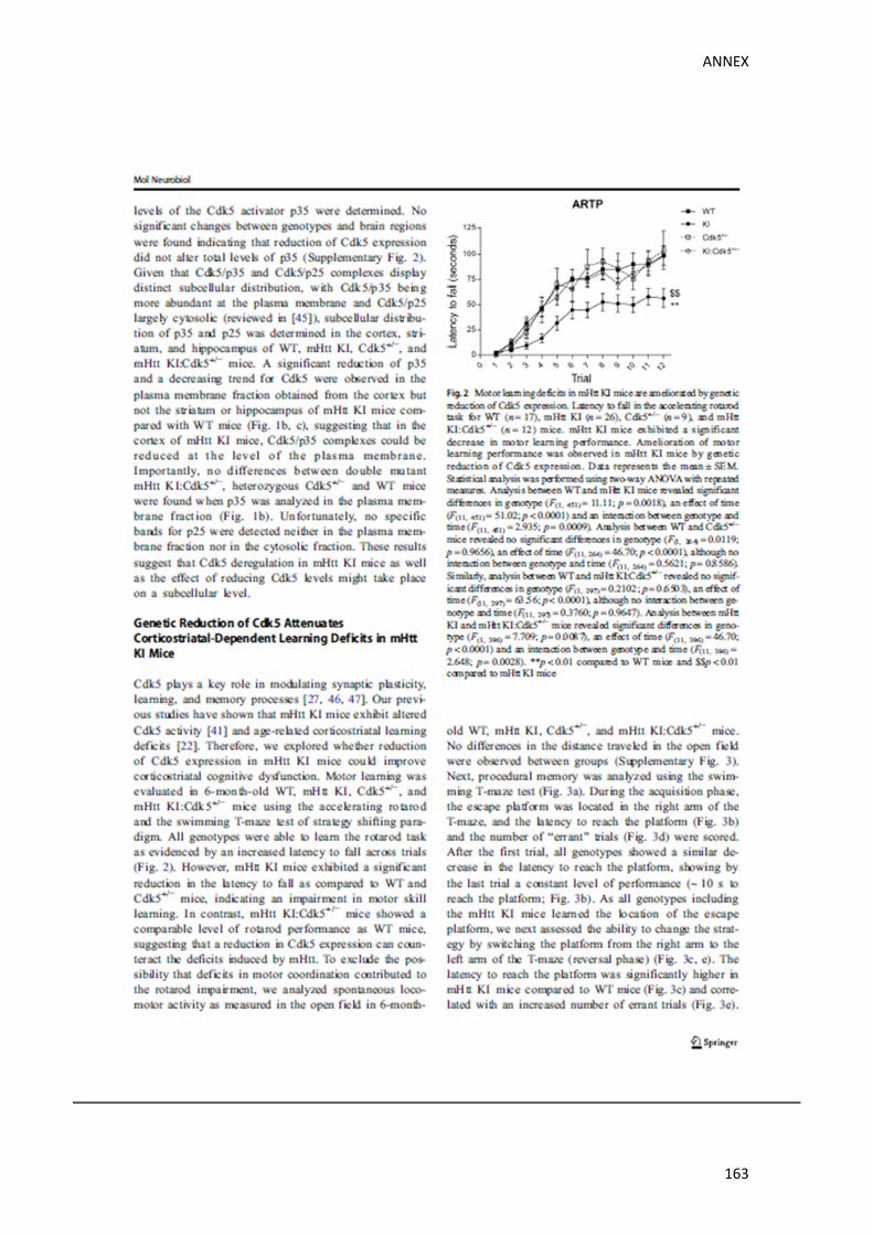
ANNEX
163

ANNEX
164

ANNEX
165
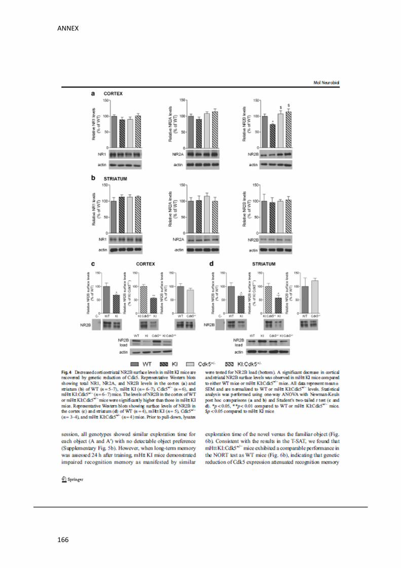
ANNEX
166
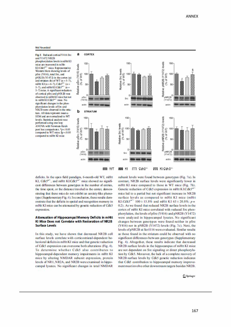
ANNEX
167
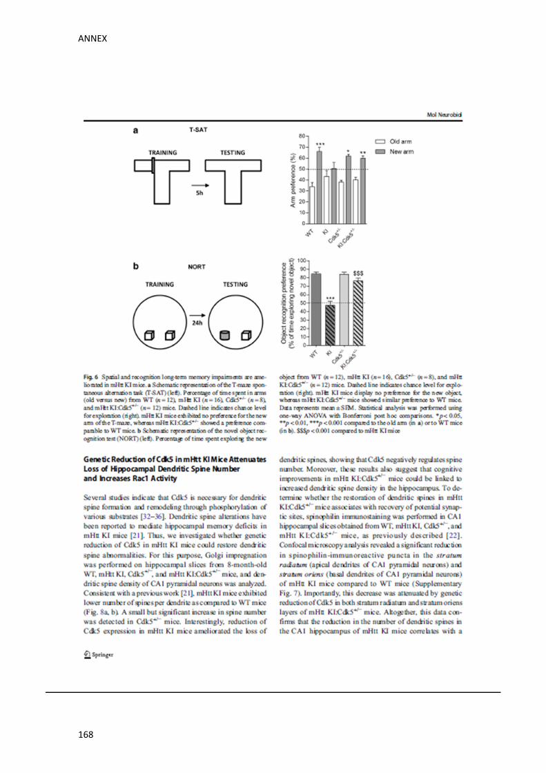
ANNEX
168

ANNEX
169

ANNEX
170

ANNEX
171

ANNEX
172
ANNEX
173

ANNEX
174

ANNEX
175

Copyright © 2022 FDOKUMEN