Convergence and Segregation of Ventral Striatal Inputs and Outputs
15
49 Convergence and Segregation of Ventral Striatal Inputs and Outputs HENK J. GROENEWEGEN, a CHRISTOPHER I. WRIGHT, b ARNO V.J. BEIJER, AND PIETER VOORN Graduate School Neurosciences Amsterdam, Research Institute Neurosciences Vrije Universiteit, Department of Anatomy, Vrije Universiteit, Amsterdam, The Netherlands ABSTRACT: The ventral striatum, which prominently includes the nucleus accumbens (Acb), is a heterogeneous area. Within the Acb of rats, a peripher- ally located shell and a centrally situated core can be recognized that have dif- ferent connectional, neurochemical, and functional identities. Although the Acb core resembles in many respects the dorsally adjacent caudate-putamen complex in its striatal character, the Acb shell has, in addition to striatal fea- tures, a more diverse array of neurochemical characteristics, and afferent and efferent connections. Inputs and outputs of the Acb, in particular of the shell, are inhomogeneously distributed, resulting in a mosaical arrangement of con- centrations of afferent fibers and terminals and clusters of output neurons. To determine the precise relationships between the distributional patterns of var- ious afferents (e.g., from the prefrontal cortex, the basal amygdaloid complex, the hippocampal formation, and the midline/intralaminar thalamic nuclei) and efferents to the ventral pallidum and mesencephalon, neuroanatomical anter- ograde and retrograde tracing experiments were carried out. The results of the double anterograde, double retrograde, and anterograde/retrograde tracing experiments indicate that various parts of the shell (dorsomedial, ventromedi- al, ventral, and lateral) and the core (medial and lateral) have different input– output characteristics. Furthermore, within these Acb regions, various popu- lations of neurons can be identified, arranged in a cluster-like fashion, onto which specific sets of afferents converge and that project to particular output stations, distinct from the input–output relationships of neighboring, cluster- like neuronal populations. These results support the idea that the nucleus ac- cumbens may consist of a collection of neuronal ensembles with different in- put–output relationships and, presumably, different functional characteristics. INTRODUCTION: VENTRAL STRIATUM AND NUCLEUS ACCUMBENS The term ventral striatum denotes an area of the striatum that receives inputs from such limbic structures as the hippocampus, entorhinal cortex, and amygdala, as well as dopaminergic afferents from the ventral mesencephalon. These striatal affer- ents are largely confined to the ventral and medial parts of the striatum, although in a Address for correspondence: H.J. Groenewegen, M.D. Ph.D., Department of Anatomy and Embryology, Faculty of Medicine, Vrije Universiteit, Van der Boechorststraat 7, 1081 BT Amsterdam, The Netherlands. Voice: 31-20-444-8040; fax: 31-20-444-8054; [email protected] b Present address: Department of Neurology, Harvard Medical School, Brigham and Women’s Hospital, Boston MA, USA.
-
Upload
independent -
Category
Documents
-
view
0 -
download
0
Transcript of Convergence and Segregation of Ventral Striatal Inputs and Outputs

49
Convergence and Segregation of VentralStriatal Inputs and Outputs
HENK J. GROENEWEGEN,a CHRISTOPHER I. WRIGHT,b ARNO V.J. BEIJER, AND PIETER VOORN
Graduate School Neurosciences Amsterdam, Research Institute Neurosciences VrijeUniversiteit, Department of Anatomy, Vrije Universiteit, Amsterdam, The Netherlands
ABSTRACT: The ventral striatum, which prominently includes the nucleusaccumbens (Acb), is a heterogeneous area. Within the Acb of rats, a peripher-ally located shell and a centrally situated core can be recognized that have dif-ferent connectional, neurochemical, and functional identities. Although theAcb core resembles in many respects the dorsally adjacent caudate-putamencomplex in its striatal character, the Acb shell has, in addition to striatal fea-tures, a more diverse array of neurochemical characteristics, and afferent andefferent connections. Inputs and outputs of the Acb, in particular of the shell,are inhomogeneously distributed, resulting in a mosaical arrangement of con-centrations of afferent fibers and terminals and clusters of output neurons. Todetermine the precise relationships between the distributional patterns of var-ious afferents (e.g., from the prefrontal cortex, the basal amygdaloid complex,the hippocampal formation, and the midline/intralaminar thalamic nuclei) andefferents to the ventral pallidum and mesencephalon, neuroanatomical anter-ograde and retrograde tracing experiments were carried out. The results of thedouble anterograde, double retrograde, and anterograde/retrograde tracingexperiments indicate that various parts of the shell (dorsomedial, ventromedi-al, ventral, and lateral) and the core (medial and lateral) have different input–output characteristics. Furthermore, within these Acb regions, various popu-lations of neurons can be identified, arranged in a cluster-like fashion, ontowhich specific sets of afferents converge and that project to particular outputstations, distinct from the input–output relationships of neighboring, cluster-like neuronal populations. These results support the idea that the nucleus ac-cumbens may consist of a collection of neuronal ensembles with different in-put–output relationships and, presumably, different functional characteristics.
INTRODUCTION: VENTRAL STRIATUM AND NUCLEUS ACCUMBENS
The term ventral striatum denotes an area of the striatum that receives inputsfrom such limbic structures as the hippocampus, entorhinal cortex, and amygdala, aswell as dopaminergic afferents from the ventral mesencephalon. These striatal affer-ents are largely confined to the ventral and medial parts of the striatum, although in
aAddress for correspondence: H.J. Groenewegen, M.D. Ph.D., Department of Anatomy andEmbryology, Faculty of Medicine, Vrije Universiteit, Van der Boechorststraat 7, 1081 BTAmsterdam, The Netherlands. Voice: 31-20-444-8040; fax: 31-20-444-8054;[email protected]
bPresent address: Department of Neurology, Harvard Medical School, Brigham and Women’sHospital, Boston MA, USA.
50 ANNALS NEW YORK ACADEMY OF SCIENCES
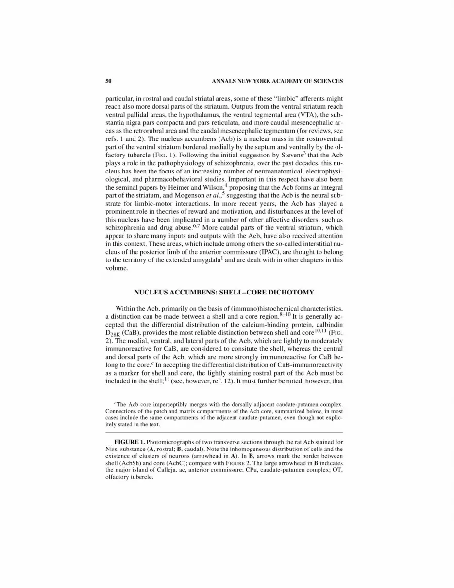
particular, in rostral and caudal striatal areas, some of these “limbic” afferents mightreach also more dorsal parts of the striatum. Outputs from the ventral striatum reachventral pallidal areas, the hypothalamus, the ventral tegmental area (VTA), the sub-stantia nigra pars compacta and pars reticulata, and more caudal mesencephalic ar-eas as the retrorubral area and the caudal mesencephalic tegmentum (for reviews, seerefs. 1 and 2). The nucleus accumbens (Acb) is a nuclear mass in the rostroventralpart of the ventral striatum bordered medially by the septum and ventrally by the ol-factory tubercle (FIG. 1). Following the initial suggestion by Stevens3 that the Acbplays a role in the pathophysiology of schizophrenia, over the past decades, this nu-cleus has been the focus of an increasing number of neuroanatomical, electrophysi-ological, and pharmacobehavioral studies. Important in this respect have also beenthe seminal papers by Heimer and Wilson,4 proposing that the Acb forms an integralpart of the striatum, and Mogenson et al.,5 suggesting that the Acb is the neural sub-strate for limbic-motor interactions. In more recent years, the Acb has played aprominent role in theories of reward and motivation, and disturbances at the level ofthis nucleus have been implicated in a number of other affective disorders, such asschizophrenia and drug abuse.6,7 More caudal parts of the ventral striatum, whichappear to share many inputs and outputs with the Acb, have also received attentionin this context. These areas, which include among others the so-called interstitial nu-cleus of the posterior limb of the anterior commissure (IPAC), are thought to belongto the territory of the extended amygdala1 and are dealt with in other chapters in thisvolume.
NUCLEUS ACCUMBENS: SHELL–CORE DICHOTOMY
Within the Acb, primarily on the basis of (immuno)histochemical characteristics,a distinction can be made between a shell and a core region.8–10 It is generally ac-cepted that the differential distribution of the calcium-binding protein, calbindinD28K (CaB), provides the most reliable distinction between shell and core10,11 (FIG.2). The medial, ventral, and lateral parts of the Acb, which are lightly to moderatelyimmunoreactive for CaB, are considered to consitute the shell, whereas the centraland dorsal parts of the Acb, which are more strongly immunoreactive for CaB be-long to the core.c In accepting the differential distribution of CaB-immunoreactivityas a marker for shell and core, the lightly staining rostral part of the Acb must beincluded in the shell;11 (see, however, ref. 12). It must further be noted, however, that
cThe Acb core imperceptibly merges with the dorsally adjacent caudate-putamen complex.Connections of the patch and matrix compartments of the Acb core, summarized below, in mostcases include the same compartments of the adjacent caudate-putamen, even though not explic-itely stated in the text.
FIGURE 1. Photomicrographs of two transverse sections through the rat Acb stained forNissl substance (A, rostral; B, caudal). Note the inhomogeneous distribution of cells and theexistence of clusters of neurons (arrowhead in A). In B, arrows mark the border betweenshell (AcbSh) and core (AcbC); compare with FIGURE 2. The large arrowhead in B indicatesthe major island of Calleja. ac, anterior commissure; CPu, caudate-putamen complex; OT,olfactory tubercle.

51GROENEWEGEN et al.: CONVERGENCE AND SEGREGATION

52 ANNALS NEW YORK ACADEMY OF SCIENCES
both shell and core exhibit distinct inhomogeneities for cellular density (FIG. 1) andCaB-immunoreactivity (TABLE 1; FIG. 2). The lateral shell exhibits a moderate im-munoreactivity for CaB, whereas medial, ventral, and rostral shell areas show muchlower levels of CaB-immunoreactivity. Within the medial shell, cell cluster areas inNissl-stained sections exhibit almost no CaB-immunoreactivity, whereas in its cau-do-dorsal part, collections of CaB-immunoreactive neurons are present.10 The coreof the Acb, much like the ventromedial parts of the adjacent caudate-putamen com-plex, contains distinct areas of low CaB-immunoreactivity which coincide with thestriatal patches expressing high concentrations of µ-opioid receptors.19,20 TABLE 1summarizes the differential distribution of a number of neurochemical substancesand neurotransmitter receptors in the various parts of the Acb shell and core. At leastfive, but presumably more, different compartments can be recognized in the Acb. Itmust be noted that, for a variety of substances and receptors that are not representedin this table, also, inhomogeneous patterns of distribution have also been described,but that a complete picture of the precise mutual relationships between these patternsis still largely lacking.
TABLE 1a
CaB ENK NAL DA D1 D2 D3 SP NT
CORE +++ ++ + ++ ++ ++ + ++ ++patch ++ +++ +++ + +++ +++matrix +++ ++ + ++ ++ +“rostral zones” +++ +++ +++ + +
SHELL ++ +++ + +++ +++ + ++ +++ +++cell clusters + + +++ + + +cone-shaped area ++ +++ ++ +++ +++ +++
aThe distribution of a limited number of neurochemical substances and neurotransmitterreceptors in shell and core of the Acb (grey rows), and identifiable “compartments” therein,9,10
is represented in a three-level scale: +++, dense; ++, moderate; +, weak. The distribution of thedopamine receptors D1, D2, and D3 has not been described in such detail that a further subdivi-sion within the shell and core is justified. For the various substances, the references contain theoriginal data. CaB, calbindin D28K;9–11,13 ENK, enkephalin;9,14 NAL naloxon;14 DA, dopamine;9
D1, dopamine D1 receptor;15 D2, dopamine D2 receptor;15 D3, dopamine D3 receptor;16 SP,substance P;9,10 NT, neurotensin.17,18
FIGURE 2. Photomicrographs of three transverse sections through the Acb, immun-ostained for CaB. A, rostral; C, caudal. Arrows in B and C indicate the border between shell(AcbSh) and core (AcbC) of the Acb. Note that the shell shows much less CaB immunore-activity than the core but that the shell is inhomogeneous in itself, exhibiting moderate im-munoreactivity in the lateral shell and almost no immunoreactivity for CaB in its medial part(compare also FIG. 4A). The large arrowhead in C indicates the major island of Calleja. Thecore, exhibiting mostly high levels of CaB, contains, in addition, patches of light or moder-ate levels of immunoreactivity for CaB. The rostral part of the Acb (A), with the exceptionof a lateral region, is lightly immunoreactive for CaB and has been included in the shell onthe basis of this characteristic by Jongen-Rêlo et al.11 ac, anterior commissure.

53GROENEWEGEN et al.: CONVERGENCE AND SEGREGATION

54 ANNALS NEW YORK ACADEMY OF SCIENCES
RELATIONSHIPS OF AFFERENTS AND EFFERENTS WITHSHELL AND CORE
The Acb receives inputs from the hippocampal region, basal amygdaloid com-plex, prefrontal cortex, midline and intralaminar thalamus, ventral pallidum, thedopaminergic ventral tegmental (A10) and retrorubral (A8) cell groups, the seroton-ergic median raphe nucleus, and the noradrenergic A2 cell group in the nucleus ofthe solitary tract.1,21,22–24 The projections of none of these structures are restrictedto the Acb, but they extend either into the ventrally adjacent striatal parts of the ol-factory tubercle and/or into the dorsally adjacent caudate-putamen complex andmore caudal ventral striatal areas. Within the Acb, the projections of most of theseafferent structures are inhomogeneously distributed, and they show particular rela-tionships with the shell and core, or subregions therein (FIG. 3). However, there areno afferent systems that are exclusively related to either Acb shell or core. Thus,whereas the projections from the hippocampal subicular and CA1 regions predomi-nently target the medial, ventral, and rostral parts of the Acb shell, these hippocam-pal projections also extend into the medial part of the Acb core (refs. 21 and 22; andBeijer and Groenewegen, unpublished observations).
Likewise, the medial entorhinal area projects predominently to the Acb shell, butthese projections are by no means restricted to the shell, and they appear to also in-vade the Acb core. The lateral entorhinal area projects primarily to the Acb core buttargets also the lateral part of the shell (ref. 25; and Beijer and Groenewegen, unpub-lished observations).
Afferents from different nuclei of the basal amygdaloid complex terminate in dif-ferent parts of the Acb in a highly complex arrangement.24 Whereas the caudal partof the parvicellular basal nucleus sends fibers predominently to the dorsomedialshell, it targets, in addition, the patches in the core. The rostral part of the magnocel-lular basal nucleus sends fibers to the lateral part of the Acb shell and, additionally,to the patches of the lateral Acb core. The midrostrocaudal part of the accessory bas-al nucleus issues fibers to the ventral part of the shell as well as to the matrix of thecore. The caudal parts of the accessory basal and the magnocellular basal nucleiproject to the ventromedial shell, whereas these nuclei have additional projections tothe patches in the medial core of the Acb.24
The thalamic projections from the midline and intralaminar thalamic nuclei to theAcb likewise show specific arrangements in projecting to distinct parts of the Acbshell and either the patch or matrix compartments in the Acb core.22,26 The anteriorpart of the thalamic paraventricular nucleus has a strong projection to the medialshell, as well as to the patches in the medial core of the Acb. More posterior parts ofthe paraventricular nucleus send fibers to more ventral and lateral parts of the shell.The intermediodorsal thalamic nucleus, a caudal representative of the midline tha-lamic nuclei, projects heavily to the Acb core, in particular targeting the matrix andavoiding its patches. The projections from the rostrally located parataenial nucleusinclude predominantly the ventral and rostral parts of the shell, avoiding its most me-dial and lateral parts. Additional projections reach the Acb core, but these are lessdense and not strictly bound to either patch or matrix. The central medial thalamicnucleus and the medial part of the parafascicular nucleus send fibers to the rostralpart of the Acb core and, in addition, extensively to the medial part of the caudate-putamen complex.22,26

55GROENEWEGEN et al.: CONVERGENCE AND SEGREGATION
The projections from different areas in the prefrontal cortex to the Acb are alsotopographically arranged. Moreover, the detailed arrangements with respect to spe-cific compartments within the shell and core appear to depend upon the layer of or-igin of the prefrontal cortical projections.13,23,27 The infralimbic cortex projects to aperipheral, band-like region in the medial and ventral parts of the shell, as well as toa region including the lateral part of the medial shell and the medial part of the coreof the Acb. The lateral shell receives its cortical inputs predominantly from the ven-tral agranular insular area in the depth of the rostral part of the rhinal sulcus. Thedeep laminae (deep layer V) of the ventral prelimbic area send fibers to the dorso-medial part of the shell, as well as to the patches of the Acb core; its superficial lam-inae (superficial layer V and layer III) project to the matrix of the core. Similararrangements exist for the deep versus superficial projections from the dorsal pre-limbic area to, respectively, the patch and matrix compartments of the Acb core.23,
27 However, the projections from the dorsal prelimbic area extend further rostrallyinto the Acb and more dorsally into the medial caudate-putamen than those from theventral prelimbic area. The dorsal agranular insular area in the lateral part of the pre-frontal cortex primarily projects to the core of the Acb and extensive parts of the ven-tral caudate-putamen. Superficial layers project more heavily to the matrix, deeplayers to the patches of the core.23, 28 The projections from the dorsal agranular in-sular and the dorsal prelimbic areas to the Acb core, in part, overlap in caudal partsof the Acb, but the dorsal prelimbic area tends to project more medially and rostrallyin the Acb than the dorsal agranular insular area.
There is a clear, reciprocal point-to-point relationship between the Acb and theventral pallidum: the medial shell and the core project to the ventromedial and thedorsal parts of the ventral pallidum, respectively.29–31 In addition, the lateral shellprojects to the ventrolateral part of the ventral pallidum (ref 31; and Groenewegenand Wright, unpublished observations). The return projections from the ventral pal-lidum to the Acb are organized in a comparable topographical fashion.22,32
The relationships of the Acb with the dopaminergic and nondopaminergic cellgroups in the ventral mesencephalon are rather complex. Whereas the A10 cellgroup in the VTA projects predominantly to the medial and ventral parts of the shell,its fibers are by no means restricted to this area and terminate also in the medial coreand adjacent regions of the caudate-putamen complex. Within the medial shell, aninhomogeneous distribution of dopaminergic fibers is apparent: whereas the cone-shaped area contains the highest density of dopaminergic fibers, the cell clusters thatborder this region receive virtually no such fibers (TABLE 1).33 The retrorubral A8cell group projects more laterally in the Acb, whereas the A9 projections in the Acbdo not reach further ventrally than the core.22,34,35 The accumbal projections to theventral mesencephalon are arranged as follows. The medial shell sends fibers to themedial VTA; more lateral parts of the shell, that is, its ventromedial and ventralparts, innervate the lateral part of VTA, the dorsal tier of the substantia nigra parscompacta and the retrorubral area. A small number of fibers extend even further cau-dally to reach the midbrain tegmentum and lateral part of the central grey mat-ter.2,29,35,36 For the projections from the core, a distinction must be made betweenthe patch and matrix compartments. The patches project to the substantia nigra parscompacta; the matrix sends fibers to a restricted dorsomedial part of the parsreticulata.29,35,37–39

56 ANNALS NEW YORK ACADEMY OF SCIENCES
FIGURE 3. Tentative subdivision of the Acb shell and the topographical arrangementsof inputs and outputs of the Acb. A: In a drawing of a transverse CaB-stained sectionthrough the Acb, the subdivision of the shell into medial, lateral, and intermediate parts isindicated. B: The broad topography of the main afferents and the ventral pallidal efferentsof the core and the medial and lateral parts of the shell is represented in a transverse sectionthrough the Acb (left) and the ventral pallidum (right). ac, anterior commissure; Acb, nucle-us accumbens; AId, dorsal agranular insular cortex; AIv, ventral agranular insular cortex;BAC, basal amygdaloid complex; CA1, cornu Ammonis field 1; CM, central medial nucle-us; IL, infralimbic cortex; IMD, intermediodorsal nucleus; PLd, dorsal prelimbic cortex;PLv, ventral prelimbic cortex; PVa, anterior paraventricular nucleus; PVp, posteriorparaventricular nucleus; SNC, substantia nigra pars compacta; SNR, substantia nigra parsreticulata; Sub, subiculum; VPdl, dorsolateral ventral pallidum; VPvl, ventrolateral ventralpallidum; VPvm, ventromedial ventral pallidum; VTA, ventral tegmental area.

57GROENEWEGEN et al.: CONVERGENCE AND SEGREGATION
An important distinction between the Acb core and shell is that the latter, at leastits medial part, sends projections to the lateral preoptic and lateral hypothalamicareas.29,31,40
SUBDIVISIONS OF THE SHELL OF THE NUCLEUS ACCUMBENS
Taking together the immunohistochemical differentiation, for example, for CaBimmunoreactivity, and the differential arrangements of afferents and efferents of theshell of the Acb, it may be suggested that this region consists of at least three subre-gions. Thus, a distinction can be made between a medial, a ventral, and a lateral shell(FIG. 3A). The medial shell, with its reciprocal relationships with the VTA and itsstrong input from the anterior paraventricular thalamic nucleus, the ventral subicu-lum, and the caudal basal amygdaloid nucleus, is clearly distinct from the lateralshell, which has more direct relationships with the substantia nigra pars compacta,the posterior paraventricular thalamic nucleus, and the rostral magnocellular basalamygdaloid nucleus (FIG. 3B). As discussed above, the ventral, intermedate part ofthe shell receives, at least to a large degree, inputs from yet another set of afferents,among which are the dorsal subiculum and midrostrocaudal parts of the basal andaccessory basal amygdaloid nuclei. The borders between the ventral part of the shellon the one hand and the medial and lateral parts of the shell on the other hand are notsharp, and, as yet, no immunohistochemical markers have been described that dis-tinguish clearly between these areas in the shell. Yet, their specific input–output re-lationships suggest an involvement in different functional aspects of the Acb.
Within the medial shell further distinctions should probably be made, in view ofthe heterogeneities in the distribution of neurochemical substances, neurotransmitterreceptors, and afferent and efferent connections (see refs. above and, e.g., 41 and42). Purely for descriptive reasons, the terms ventromedial and dorsomedial shellhave been introduced (e.g., refs. 13 and 42). Functional studies indicate that the ven-tromedial shell may indeed be a distinct r egion of the Acb.43
Both neuroanatomical12,44,45 and functional studies46,47 suggest that there existalso rostrocaudal differences within the Acb. Zahm and Heimer,12 on the basis of anefferent connectivity pattern distinct from the “typical” projections patterns of bothshell and core, designated the rostral part of the Acb as the so-called “rostral pole.”Further studies are needed to substantiate the specific identity of the rostral part ofthe Acb, distinct from shell and core.
SHELL OF THE NUCLEUS ACCUMBENS: COLLECTION OFENSEMBLES OF NEURONS?
The immunohistochemical and tract-tracing data reviewed above indicate that theshell and core of the Acb should not be considered as anatomical and functional unitsbut rather that they can be further subdivided into different subregions. Thus, the me-dial, intermediate, and lateral parts of the shell of the Acb, as well as the medial andlateral parts of the core, receive different combinations of inputs from cortical andsubcortical sources and project to different pallidal, hypothalamic, and mesenceph-

58 ANNALS NEW YORK ACADEMY OF SCIENCES
alic targets. Moreover, within these subregions of the shell and core, the terminalfields of different sources of inputs and the neurons that give rise to different outputsare very heterogeneously distributed.2,13,24,28,35,48,49 This leads to intricate relation-ships of various afferent systems in different parts of the Acb. For example, the re-sults of double anterograde tracing experiments have shown that within the medialshell and core of the Acb the inhomogeneously distributed afferents from the ante-rior paraventricular thalamic nucleus, the caudal parvicellular basal amygdaloid nu-cleus, and the ventral prelimbic and infralimbic cortices form intricate patterns ofconvergence and segregation.13 Similar arrangements of convergence and noncon-vergence appear to exist in the lateral parts of the shell and core of the Acb with re-spect to afferents from the other parts of the midline thalamus, basal amygdaloidcomplex, and the (lateral) prefrontal cortex.28 The results of retrograde tracing ex-periments, with small injections of retrograde tracers in one of the projection areasof the Acb, show that the Acb output neurons are organized in a clustered fashion.2,
35,49 An important question, that largely remains to be answered, is if and how thecluster-like organization of output neurons is related to the heterogeneous mosaic ofconvergent and segregated inputs of the Acb. In other words, to what extent does thisorganization reflect the existence of specific input–output channels through the Acb?Preliminary results of experiments, in which rats were injected with an anterogradetracer in one of the input structures of the Acb combined with a retrograde tracer inone of the Acb target areas, indicate that indeed particular clusters of output neuronsmay receive rather specific sets of inputs.48,49
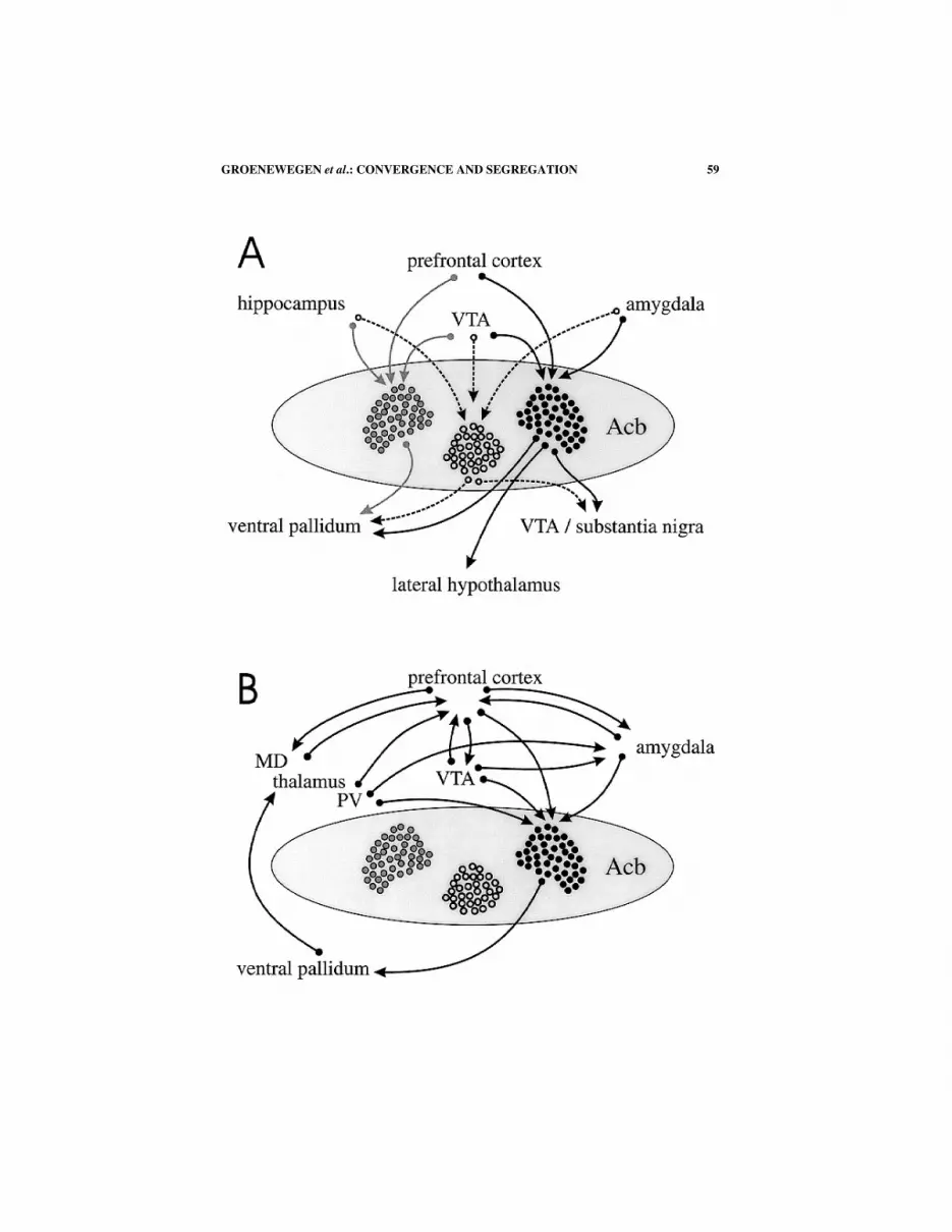
In view of the wide range of behavorial aspects in which the Acb is presumed toplay a role, Pennartz et al.,50 in a recent review, argued that the nucleus consists ofa collection of neuronal ensembles, or groups, with different functional and behav-ioral connotations. An ensemble is thought to be formed by a population of Acb neu-rons that is temporarily and (nearly) synchronously activated by a specific set ofexcitatory inputs. The coherent activity of these inputs could thus lead to the activa-tion of a particular set of outputs of the Acb. Each distinct ensemble may be capableof generating a spatiotemporally coded output that is transferred to a set of targetstructures characteristic for this ensemble, and hence may induce behavioral effectsthat are specifically linked to this particular ensemble; 50 (see also O’Donnell et al.,this volume). It is not a prerequisite for the idea of the existence of neuronal ensem-bles in the Acb that such ensembles of neurons are “bound” to a cluster of spatiallyclosely related neurons. However, it is tempting to speculate that the experimentallyshown clusters of output neurons in the above-mentioned retrograde tracing experi-ments, which receive different and specific combinations of converging inputs, formthe anatomical substrates of such ensembles (FIG. 4A). Because only a strong exci-
FIGURE 4. Schematic representation of the input–output relationships of clusters ofneurons in the shell of the nucleus accumbens. A: Different clusters of neurons (ensem-bles?) receive different combinations of converging inputs and project to distinct targets,including the ventral pallidum, the lateral hypothalamus, and the ventral mesencephalon. B:The various (limbic) cortical and subcortical structures that project to the Acb are stronglyand, in a number of cases, reciprocally interconnected. Note that the Acb, via the ventralpallidum and the mediodorsal thalamic nucleus is involved in a closed thalamocortical–bas-al ganglia loop.1,2 VTA, ventral tegmental area. Acb, nucleus accumbens; MD, me-diodorsal thalamic nucleus; PV, paraventricular thalamic nucleus.
59GROENEWEGEN et al.: CONVERGENCE AND SEGREGATION

60 ANNALS NEW YORK ACADEMY OF SCIENCES
tatory input is thought to be able to electrophysiologically activate striatal outputneurons (see below), it is important to realize that the major cortical, thalamic, andlimbic inputs of the Acb have extensive, and in most cases, reciprocal interconnec-tions (FIG. 4B). This extensive interconnectivity of the afferent structures of the Acbcould be the anatomical basis for the temporarily coherent and synchronous activa-tion of the populations of neurons that constitute the ensembles in the Acb (FIG. 4).
As indicated above, for an understanding of the way in which ensembles of Acbneurons may be activated, it is of great interest to consider the specific physiologicalproperties of the Acb output cells, that is, the medium-sized spiny neurons. Thesespiny output neurons, both in the caudate-putamen complex 51 and the Acb, 52 havea bistable membrane potential, that is, they are mostly in a state with a hyperpolar-ized membrane potential of −85 to −90 mV (“down state”), but occasionally theyreach a state with a relatively depolarized membrane potential of approximately −55mV (“up state”). Only in neurons in the up state, of which the membrane potentialis close to the spike threshold, can firing of action potentials be induced; neurons inthe down state are physiologically “silent.” It is thought that excitatory inputs froma particular source can bring striatal output neurons from the down state into the upstate, but that excitatory inputs from yet another source are necessary to induce firingactivity.50–52 For example, hippocampal activation may bring Acb output neurons intheir up state and, in this way, facilitate the throughout of prefrontal cortical activityvia the Acb, as observed by O’Donnell and Grace,52 in an intracellular electrophys-iological study. Following lesions of the hippocampal afferents to the Acb, the Acboutput neurons remain in their down state, and prefrontal cortical activition is notable to evoke Acb neuronal firing. Therefore, O’Donnell and Grace52 have suggestedthat hippocampal inputs “gate” the prefrontal cortical throughput through the Acb.In an extracellular electrophysiological study, investigating the interactions betweenhippocampal and amygdaloid afferents in the Acb, Mulder et al.53 found thatamygdaloid activation facilitates hippocampal throughput through the nucleus butthat, in contrast, hippocampal activation may close a gate for amygdaloid inputs. Al-though the observations of O’Donnell et al.52 and Mulder et al.53 cannot be com-pletely reconciled with each other, they suggest that various afferents of the Acbinteract with each other at the level of the output neurons, one of the possible conse-quences being that two or more afferents have to be active in temporal and spatialcoherence to activate output neurons of the Acb. In that respect, it is of great impor-tance to study in detail the input–output patterns at the level of the Acb, in order tounderstand the various possible interactions of inputs and, in this way, the activationor modulation of a multitude of outputs of the nucleus.
CONCLUDING REMARKS
The above reviewed data, with respect to the functional anatomical organizationof the Acb, as part of the ventral striatum, indicate that this nucleus consists of var-ious different subregions (e.g., medial and lateral shell and core, etc.) and that withinthese subregions functionally distinct ensembles of neurons exist, possibly orga-nized in anatomical compartments and activated by specific sets of afferents that areprimarily derived from (subregions of) limbic structures. It is thus important to note

61GROENEWEGEN et al.: CONVERGENCE AND SEGREGATION
that the Acb neuronal ensembles must be viewed in close association with groups ofneurons in prefrontal cortical, amygdaloid, and midline/intralaminar thalamic struc-tures (as described above), which, in turn, have strong mutual interconnections (FIG.4B). Such interconnections between structures that are afferent to the Acb may becrucial for the spatially and temporally coherent activation of the presumed ensem-bles in the Acb. How and under which functional circumstances the Acb neuronalensembles are activated, and whether they can influence each other, are all importantquestions that need to be answered in order to further our understanding of the func-tional anatomy of the nucleus accumbens.
REFERENCES
1. HEIMER, L., D.S. ZAHM & G.F. ALHEID. 1995. Basal ganglia. In The Rat Nervous Sys-tem, 2nd Edition. G. Paxinos & C. Watson, Eds.: 579–628. Academic Press. SanDiego, CA.
2. GROENEWEGEN, H.J., C.I. WRIGHT & A.V.J. BEIJER. 1996. The nucleus accumbens:gateway for limbic structures to reach the motor system? In The Emotional MotorSystem, G. Holstege, R. Bandler & C.B. Saper, Eds. Prog. Brain Res. 107: 485–511.
3. STEVENS, J.R. 1973. An anatomy of schizophrenia? Arch. Gen. Psychiatry. 29: 177–189.
4. HEIMER, L. & R.D. WILSON. 1975. The subcortical projections of the allocortex: simi-larities in the neural associations of the hippocampus, the piriform cortex, and theneocortex. In Golgi Centennial Symposium: Perspectives in Neurobiology. M. San-tini, Ed. :177–193. Raven Press. New York.
5. MOGENSON, G.J., D.L. JONES & C.Y. YIM. 1980. From motivation to action: functionalinterface between the limbic system and the motor system. Prog. Psychobiol. 14: 60–97.
6. KOOB, G.F. 1992. Drugs of abuse: anatomy, pharmacology and function of rewardpathways. Trends Pharmacol. Sci. 13: 177–184.
7. ROBBINS, T.W. & B.J. EVERITT. 1996. Neurobehavioural mechanisms of reward andmotivation. Curr. Opin. Neurobiol. 6: 228–236.
8. ZÁBORSZKY, L., G.F. ADELHEID, M.C. BEINFELD, L.E. EIDEN, L. HEIMER & M. PALK-OVITS. 1985. Cholecystokinin innervation of the vental striatum: a morphological andradioimmunological study. Neuroscience 14: 427–453.
9. VOORN, P., C.R. GERFEN & H.J. GROENEWEGEN. 1989. The compartmental organiza-tion of the ventral striatum of the rat: immunohistochemical distribution of enkepha-lin, substance P, dopamine, and calcium binding protein. J. Comp. Neurol. 289: 189–201.
10. ZAHM, D.S. & J.S. BROG. 1992. On the significance of subterritories in the “accum-bens” part of the rat ventral striatium. Neuroscience 50: 751 – 767.
11. JONGEN-RÊLO, A.L., P. VOORN & H.J. GROENEWEGEN. 1994. Immunohistochemicalcharacterization of the shell and core territories of the nucleus accumbens in therat. Eur. J. Neurosci. 6: 1255–1264.
12. ZAHM, D.S. & L. HEIMER. 1993. Specificity in the efferent projections of the nucleusaccumbens in the rat: comparison of the rostral pole projection patterns with thoseof the core and shell. J. Comp. Neurol. 327: 220–232.
13. WRIGHT, C.I. & H.J. GROENEWEGEN. 1995. Patterns of convergence and segregationin the medial nucleus accumbens of the rat: relationships of prefrontal cortical,midline thalamic and basal amygdaloid afferents. J. Comp. Neurol. 361: 383–403.
14. JONGEN–RÊLO, A.L., H.J. GROENEWEGEN & P. VOORN. 1993. Evidence for a multi-compartmental histochemical organization of the nucleus accumbens in the rat. J.Comp. Neurol. 337: 267–276.
15. JONGEN-RÊLO, A.L., G.J. DOCTER, A.J. JONKER & P. VOORN. 1995. Differential local-ization of mRNAs encoding dopamine D1 or D2 receptors in cholinergic neuronsin the core and shell of the rat nucleus accumbens. Mol. Brain Res. 28:169–174.

62 ANNALS NEW YORK ACADEMY OF SCIENCES
16. LE MOINE, C. & B. BLOCH. 1996. Expression of the D3 dopamine receptor in pepti-dergic neurons of the nucleus accumbens: comparison with the D1 and D2 dopam-ine receptors. Neuroscience 73:131–143.
17. ZAHM, D.S. 1987. Neurotensin-immunoreactive neurons in the ventral striatum ofthe adult rat: ventromedial caudate-putamen, nucleus accumbens and olfactorytubercle. Neurosci. Lett. 81: 41–47.
18. ZAHM, D.S. & L. HEIMER. 1988. Ventral striatopallidal parts of the basal ganglia inthe rat: I. Neurochemical compartmentation as reflected by the distributions ofneurotensin and substance P immunoreactivity. J. Comp. Neurol. 272: 516–535.
19. HERKENHAM, M. & C.B. PERT. 1981. Mosaic distribution of opiate receptors,parafascicular projections and acetylcholinesterase in rat striatum. Nature 291:415–418.
20. MANSOUR, A., C.A. FOX, S. BURKE, H. AKIL & S.J. WATSON. 1995. Immunohis-tochemical localization of the cloned mu opioid receptor in the rat CNS. J. Chem.Neuroanat. 8: 283–305.
21. GROENEWEGEN, H.J., E. VERMEULEN-VAN DER ZEE, A. TE KORTSCHOT & M.P. WIT-TER. 1987. Organization of the projections from the subiculum to the ventral stria-tum in the rat: a study using anterograde transport of Phaseolus vulgaris-leucoagglutinin. Neuroscience 23: 103–120.
22. BROG, J.S., A. SALYAPONGSE, A.Y. DEUTCH & D.S. ZAHM. 1993. The patterns ofafferent innervation of the core and shell in the “accumbens” part of the rat ventralstriatum: immunohistochemical detection of retrogradely transported fluoro-gold.J. Comp. Neurol. 338: 255–278.
23. BERENDSE, H.W., Y. GALIS-DE GRAAF & H.J. GROENEWEGEN. 1992. Topographicalorganization and relationship with ventral striatal compartments of prefrontal cor-ticostriatal projections in the rat. J. Comp. Neurol. 316: 314–347.
24. WRIGHT, C.I., A.V.J. BEIJER & H.J. GROENEWEGEN. 1996. Basal amygadaloid com-plex afferents to the rat nucleus accumbens are compartmentally organized. J. Neu-rosci. 16: 1877–1893.
25. TOTTERDELL, S. & G.E. MEREDITH. 1997. Topographical organization of projectionsfrom the entorhinal cortex to the striatum of the rat. Neuroscience 78: 715–729.
26. BERENDSE, H.W. & H.J. GROENEWEGEN. 1990. The organization of the thalamostri-atal projections in the rat, with special emphasis on the ventral striatum. J. Comp.Neurol. 299: 187–228.
27. GERFEN, C.R. 1989. The neostriatal mosaic: striatal patch-matrix organization isrelated to cortical lamination. Science 246: 385–388.
28. WRIGHT, C.I. & H.J. GROENEWEGEN. 1996. Patterns of overlap and segregationbetween insular cortical, intermediodorsal thalamic and basal amygdaloid afferentsin the nucleus accumbens of the rat. Neuroscience 73: 359–373.
29. HEIMER, L., D.S. ZAHM, L. CHURCHILL, P.W. KALIVAS & C. WOHLTMANN. 1991.Specificity in the projection patterns of the accumbal core and shell in the rat. Neu-roscience 41: 89125.
30. ZAHM D.S. & L. HEIMER. 1990. Two transpallidal pathways originating in the ratnucleus accumbens. J. Comp. Neurol. 302: 437–446.
31. USUDA, I., K. TANAKA & T. CHIBA. 1998. Efferent projections of the nucleus accum-bens in the rat with special reference to subdivisions of the nucleus—–biotinylateddextran amine study. Brain Res. 797: 73–93.
32. GROENEWEGEN, H.J., H.W. BERENDSE & S.N. HABER. 1993. Organization of the out-put of the ventral striatopallidal system in the rat. Ventral pallidal efferents. Neu-roscience 57: 113–142.
33. VOORN, P., B. JORRITSMA-BYHAM, C. VAN DIJK & R.M. BUIJS. 1986. The dopaminer-gic innervation of the ventral striatum in the rat: a light- and electron-microscopi-cal study with antibodies against dopamine. J. Comp. Neurol. 251: 84–99.
34. BECKSTEAD, R.M., V.B. DOMESICK & W.J.H. NAUTA. 1979. Efferent connections ofthe substantia nigra and ventral tegmental area in the rat. Brain Res. 175: 191–217.
35. BERENDSE, H.W., H.J. GROENEWEGEN & A.H.M. LOHMAN. 1992. Compartmental dis-tribution of ventral striatal neurons projecting to the ventral mesencephalon in therat. J.Neurosci. 12: 2079–2103.

63GROENEWEGEN et al.: CONVERGENCE AND SEGREGATION
36. NAUTA, W.J.H., G.P. SMITH, R.L.M. FAULL & V.B. DOMESICK. 1978. Efferent con-nections and nigral afferents of the nucleus accumbens septi in the rat. Neuro-science 3: 385–401.
37. GERFEN, C.R., M. HERKENHAM & J. THIBAULT. 1987. The neostriatal mosaic: II.Patch- and matrix-directed mesostriatal dopaminergic and non-dopaminergic sys-tems. J.Neurosci. 7: 3915–3934.
38. DENIAU, J.M., A. MENETREY & A.M. THIERRY. 1994. Indirect nucleus accumbensinput to the prefrontal cortex via the substantia nigra pars reticulata: a combinedanatomical and electrophysiological study in the rat. Neuroscience 61: 533–545.
39. GROENEWEGEN, H.J., H.W. BERENDSE & F.G. WOUTERLOOD. 1994. Organization ofthe projections from the ventral striatopallidal system to ventral mesencephalicdopaminergic neurons. In The Basal Ganglia IV. G. Percheron & J.S. McKenzie,Eds.: 81–93. Plenum Press. New York.
40. GROENEWEGEN, H.J. & F.T. RUSSCHEN. 1984. Organization of the efferent projec-tions of the nucleus accumbens to pallidal, hypothalamic, and mesencephalic struc-tures: a tracing and immunohistochemical study in the cat. J. Comp. Neurol. 223:347–367.
41. CURRAN, E.J. & S.J. WATSON. 1995. Dopamine receptor mRNA expression patternsby opioid peptides in the nucleus accumbens of the rat: a double in situ hybridiza-tion study. J. Comp. Neurol. 361: 57–76.
42. DIAZ, J., D. LÉVESQUE, C.H. LAMMERS, N. GRIFFON, M.P. MARTES, J.C. SCHWARTZ &P. SOKOLOFF. 1995. Phenotypical characterization of neurons expressing thedopamine D3 receptor in the rat brain. Neuroscience 65: 731–745.
43. MALDONADO-IRIZARRY, C.S., C.J. SWANSON & A.E. KELLEY. 1995. Glutamate recep-tors in the nucleus accumbens shell control feeding behavior via the lateral hypo-thalamus. J. Neurosci. 15: 6779–6788.
44. VOORN, P. & G.J. DOCTER. 1992. A rostrocaudal gradient in the synthesis ofenkephalin in nucleus accumbens. NeuroReport 3: 161–164.
45. VOORN, P., G.J. DOCTER, A.L. JONGEN-RÊLO & A.J. JONKER. 1994. Rostrocaudal sub-regional differences in the response of enkephalin, dynorphin and substance P syn-thesis in rat nucleus accumbens to dopamine depletion. Eur. J. Neurosci. 6: 486–496.
46. LADURELLE, N., G. KELLER, B.P. ROQUES & V. DAUGE. 1993. Effects of CCK8 and ofthe CCKB-selective agonist BC264 on extracellular dopamine content in the ante-rior and posterior nucleus accumbens: a microdialysis study in freely moving rats.Brain Res. 628: 254–262.
47. HEIDBREDER, C. & J. FELDON. 1998. Amphetamine-induced neurochemical and loco-motor responses are expressed differentially across the anteroposterior axis of thecore and shell subterritories of the nucleus accumbens. Synapse (NY) 29: 310–322.
48. BEIJER, A.V.J. & H.J. GROENEWEGEN. 1996. Specific anatomical relationships betweenhippocampal and basal amygdaloid afferents and different populations of projectionneurons in the nucleus accumbens of rats. Soc. Neurosci. Abstr. 22: 413.
49. HEIMER, L, G.F. ALHEID, J.S. DE OLMOS, H.J. GROENEWEGEN, S.N. HABER, R.E. HAR-LAN & D.S. ZAHM. 1997. The accumbens: beyond the core-shell dichotomy. J.Neuropsychiatry Clin. Neurosci. 9: 354–381.
50. PENNARTZ, C.M.A., H.J. GROENEWEGEN & F.H. LOPES DA SILVA. 1994. The nucleusaccumbens as a complex of functionally distinct neuronal ensembles: An integra-tion of behavioural, electrophysiological and anatomical data. Prog. Neurobiol. 42:719–761.
51. WILSON, C.J. 1993. The geneartion of natural firing patterns in neostriatal neurons.Prog. Brain Res. 99: 277–297.
52. O'DONNELL, P. & A.A. GRACE. 1995. Synaptic interactions among excitatory affer-ents to nucleus accumbens neurons: hippocampal gating of prefrontal corticalinput. J.Neurosci. 15: 3622–3639.
53. MULDER, A.B., M. GIJSBERTI HODENPIJL & F.H. LOPES DA SILVA. 1998. Electrophys-iology of the hippocampal and amygdaloid projections to the nucleus accumbensof the rat: convergence, segregation, and interaction of inputs. J. Neurosci. 18:5095–5102.