Scopolamine injection into the olfactory bulb impairs short-term olfactory memory in rats
Upload
independentCategory
view
1download
0
Cell, Vol. 123, 669–682, November 18, 2005, Copyright ©2005 by Elsevier Inc. DOI 10.1016/j.cell.2005.08.039
Olfactory Inputs to Hypothalamic NeuronsControlling Reproduction and Fertility
Hayan Yoon,1 L.W. Enquist,2 and Catherine Dulac1,*1Howard Hughes Medical InstituteDepartment of Molecular and Cellular BiologyHarvard UniversityCambridge, Massachusetts 021382Department of Molecular BiologyPrinceton UniversityPrinceton, New Jersey 08544
Summary
In order to gain insight into sensory processing mod-ulating reproductive behavioral and endocrine changes,we have aimed at identifying afferent pathways toneurons synthesizing luteinizing hormone-releasinghormone (LHRH, also known as gonadotropin-releas-ing hormone [GnRH]), a key neurohormone of repro-duction. Injection of conditional pseudorabies virusinto the brain of an LHRH::CRE mouse line led to theidentification of neuronal networks connected to LHRHneurons. Remarkably, and in contrast to established no-tions on the nature of LHRH neuronal inputs, our dataidentify major olfactory projection pathways originatingfrom a discrete population of olfactory sensory neuronsbut fail to document any synaptic connectivity with thevomeronasal system. Accordingly, chemosensory mod-ulation of LHRH neuronal activity and mating behaviorare dramatically impaired in absence of olfactory func-tion, while they appear unaffected in mouse mutantslacking vomeronasal signaling. Further visualizationof afferents to LHRH neurons across the brain offersa unique opportunity to uncover complex polysyn-aptic circuits modulating reproduction and fertility.
Introduction
To ensure reproductive success, animals have evolvedsensory and behavioral strategies to identify suitablemating partners. In many animal species, chemicalcues called pheromones carry species- and gender-specific information required for mating. Detection ofpheromones triggers the activation of likely hardwiredbrain circuits, which leads to stereotyped changes inthe behavioral and endocrine state of the animal. Whatare the neuronal circuits involved in the processing ofthe pheromonal information? How is the information re-sulting from the activation of defined sets of phero-mone receptors translated into specific behavioral andphysiological outputs?
Surgical removal of olfactory structures have broadlyassigned the role of the main olfactory system to thesense of smell, resulting in the detection of a large vari-ety of volatile odorants, while the vomeronasal systemis thought to mediate the detection of most gender andspecies-specific cues involved in the control of matingand aggressive behavior (Dulac and Torello, 2003). Down-
*Correspondence: [email protected]
stream effectors of the pheromone signals reside withinthe hypothalamus, in central control areas of reproduc-tive and defensive behaviors (Kevetter and Winans,1981a, 1981b; Petrovich et al., 2001). The hypothala-mus is an essential integrator of internal and environ-mental cues, ensuring the homeostasis of the organ-ism, the coordination of visceral functions, and theinitiation of genetically preprogrammed behaviors suchas feeding, defense, and reproduction. Thus, specificchanges in hypothalamic activity orchestrate both thelong-lasting endocrine changes and the short-term be-havioral effects elicited by pheromones.
Neurons that synthesize and secrete the decapep-tide, luteinizing hormone-releasing hormone (LHRH),also known as gonadotropin-releasing hormone (GnRH),integrate and control peripheral and central aspects ofreproduction including the onset of puberty (Silvermanet al., 1994; Sisk and Foster, 2004). In the mouse, LHRHis produced by a small and diffuse population of neu-rons scattered in the rostral hypothalamus and medialpreoptic area (MPOA) and in the basal forebrain, includ-ing the septum and diagonal band of Broca (Wu et al.,1997). LHRH secretion into the hypophyseal portal vas-culature regulates the synthesis and secretion of theluteinizing hormone (LH) and the follicular-stimulatinghormone (FSH) by the anterior pituitary gland (Figure1A). LH and FSH, in turn, control the development andthe function of the male and female gonads and therelease of steroids into the bloodstream. Steroids coor-dinate the development of sexual traits and organs andact centrally on brain structures to modulate LHRHsecretion to facilitate sexual behavior. In addition to itsrole as a neurohormone, LHRH is detected in axonalprojections in the amygdala and the midbrain, where itis thought to act directly as a neurotransmitter facilitat-ing sexual receptivity and mating behavior (Merchen-thaler et al., 1989; Witkin et al., 1982). LHRH-expressingneurons integrate a variety of internal and external fac-tors that affect mating behavior and fertility (Figure 1A;Dobson et al., 2003; Genazzani et al., 2000). More spe-cifically, chemosensory cues have been shown in ro-dents to influence the onset of puberty in the youngand to modulate LHRH neuronal activity and LHRH re-lease in the adult. Accordingly, tracing experimentshave consistently pointed to the vomeronasal pathwayas one of the major inputs to anatomical areas contain-ing LHRH-expressing neurons (Meredith, 1998; Petro-vich et al., 2001; Simerly and Swanson, 1986).
We sought to determine precisely the nature of sen-sory network connected to LHRH-expressing neuronsand involved in the modulation of the reproductivefunction in the mouse. To accomplish this goal, we havetaken advantage of the ability of conditional strains ofneurotropic viruses to retrogradely infect synapticallyconnected chains of neurons from a molecularly de-fined population (DeFalco et al., 2001; Enquist andCard, 2003). We describe here the tracing and func-tional validation of an entire brain circuit involved in theneuroendocrine control of reproduction, from its initialsensory detectors in periphery down to the ultimate

Cell670
Figure 1. The Role of LHRH Neurons in the Control of Reproduction and Fertility, a Genetic Approach to the Study of LHRH Afferent Brain Net-works
(A) LHRH neurons constitute a central node for the control of reproduction and fertility. LHRH neurons, in green, integrate internal and externalinformation, such as the animal developmental state, the detection of chemosensory and somatosensory cues, the circadian clock, the levelof stress, and the body energy status. LHRH is released in the medial eminence into the portal vein to direct gonadotropin (LH and FSH)synthesis and release from the anterior pituitary gland. LH and FSH in turn control gonadal development and function and steroid hormonesynthesis and release. Sex steroids from the gonads promote secondary sexual traits and regulate LHRH neuronal activity through positiveand negative feedback loops, for example during the estrus cycle of females. In addition, steroids facilitate social and reproductive behaviorsby acting directly on central neural circuits. Subsets of LHRH neurons project directly within the brain where they regulate sexual receptivityand mating behaviors.(B) LHRH neurons are activated by exposure to pheromones. In situ hybridization for LHRH and immunohistochemistry for phosphorylatedMAPK were performed. Top: alkaline phosphatase reaction shows cytoplasmic labeling for LHRH transcripts in blue and nuclear DAB reactionfor phosphorylated MAPK in brown. In this picture, a neuron expresses only LHRH (white arrowhead) while the other displays LHRH andphosphorylated MAPK double labeling (black arrowhead). Bottom: in male mice, exposure to female urine significantly increased the percen-tage of LHRH neurons expressing phosphorylated MAPK (right bar, 23% ± 4%) compared to exposure to water (left bar, 11% ± 3%) (p <0.025 independent sampled t test).(C) Recombinant PRV strains, PRV152 and Ba2001, and transgenic line used in our study. PRV152 expresses GFP under the control of the

Olfactory Modulation of Reproduction and Fertility671
order to propagate and transfer in a retrograde direc-
CMV promoter, enabling detection of viral infection. Ba2001 requires CRE-mediated recombination to excise a stop cassette, leading to TKand tauGFP expression, and thus, viral transfer from primary infected cells expressing CRE recombinase only. A transgenic mouse wasgenerated in which CRE recombinase is specifically expressed in LHRH neurons. A CRE-containing cassette including a nuclear localizationsignal and a poly adenylation sequence was inserted into the start codon of the LHRH gene by homologous recombination of the BAC, RP23-22J8 which includes 137 kb of 5# upstream and 73 kb downstream sequences around the LHRH coding sequence(D) Double immunostaining with antibodies against LHRH (green) and CRE recombinase (red) demonstrates specific expression of CRE inLHRH neurons of the LHRH::CRE transgenic mouse line. Scale bar, 20 �m.
mals die from a global response to infection reflecting
central effectors in the hypothalamus. Strikingly, ourstudy failed to document any anatomical or functionalconnectivity between LHRH-expressing neurons andstructures of the vomeronasal pathway, thus contra-dicting the established notion that VNO activity exertsa direct influence on LHRH neuronal activity and, inturn, on the endocrine control of reproduction. Instead,our data reveal the existence of other sensory inputs,including a major projection pathway from the primaryolfactory cortex originating from a circumscribed pop-ulation of olfactory sensory neurons in the main olfac-tory epithelium (MOE) and uncover the severe repro-ductive defects of animals lacking MOE function.
Results
Experimental DesignChemosensory inputs have been reported in a numberof rodent species to modulate the activity of LHRH-expressing neurons (Coquelin et al., 1984; Richardsonet al., 2004). In an initial step, we wished to confirmearlier reports in the mouse and monitored the increaseof MAP kinase phosphorylation in LHRH-expressingneurons of adult male mice in response to female pher-omones present in urine. Identification of LHRH-posi-tive and LHRH/MAPK-P double-labeled cells (Figure1B) showed a significant increase in the percentage ofLHRH neurons expressing MAPK-P after exposure tourine (23% ± 4%, n = 637, LHRH-positive neurons mon-itored in eight independent animals) compared to expo-sure to water (11% ± 3%, n = 409, LHRH-positive neu-rons monitored in six independent animals; p < 0.025independent sampled t test), thus providing support toearlier studies.
Attenuated strains of pseudorabies virus (PRV), an αherpes virus, can be used as self-amplifying tracers ofneural circuitry. Derivatives of the attenuated PRV strainBartha retain the ability to infect neurons but are selec-tively neuroinvasive from postsynaptic to presynapticneurons (retrograde spread only) through polysynapticcircuits (Jansen et al., 1993). Anterograde infection(spread from presynaptic to postsynaptic neuron) hasnever been observed with Bartha strains of PRV (En-quist and Card, 2003). In our study, we used the Bartharecombinants PRV152 and Ba2001 (Figure 1C). ThePRV152 strain has been modified to express GFP, per-mitting direct visualization of the infected neurons,while PRV Ba2001 developed by DeFalco et al. (2001) isdependent on CRE-mediated recombination to expressthe neuronal tracer tau-GFP, and the gene thymidinekinase (TK) essential for viral replication in nonmitoticcells such as neurons. Thus Ba2001 requires the initialinfection of CRE recombinase-expressing neurons in
tion through neuronal circuits. We have generated atransgenic mouse line in which CRE recombinase is ex-pressed under the control of the LHRH locus. Briefly, a212 kb bacterial artificial chromosome (BAC) containingthe entire LHRH coding sequence was modified by ho-mologous recombination in order to insert a CRE re-combinase-poly(A) cassette into the ATG of the firstexon of the LHRH gene (Figure 1C).
Double immunostaining of brain sections originatingfrom three independent LHRH::CRE animals with anti-LHRH and anti-CRE recombinase antibodies demon-strated strong anti-LHRH immunoreactivity in 100 outof 104 randomly picked CRE-positive cells, while theremaining four cells displayed weak or questionablestaining (Figure 1D). Moreover, analysis of brain slicesobtained from the progeny of the cross LHRH::CRE toROSA26:: loxP-Stop-loxP-YFP (Srinivas et al., 2001) en-abled us to visualize LHRH and YFP double-positiveneurons in the medial hypothalamus, medial septum,and diagonal band of Broca, all sites of LHRH expres-sion in the adult. In addition, we identified a populationof neurons that were CRE negative, LHRH negative, yetYFP positive within the lateral septum (LS) and the bednucleus of the stria terminalis (BNST; data not shown),originating from transient transcription of the LHRHgene documented in these regions during early devel-opment (Skynner et al., 1999).
These controls let us conclude with confidence thatthe CRE recombinase transgene is faithfully tran-scribed in LHRH neurons and that the CRE transgeneproduct is functional and efficient. Accordingly, stereo-taxic injection of Ba2001 into the brain of the LHRH::CRE line led to viral infection and spread through poly-synaptic circuits in the brain, as revealed by anti-GFPimmunofluorescence (see results below), whereas nosignal was ever detected in control virus injections intothe brain of wild-type C57BL/J6 mice.
LHRH-expressing neurons form a small and diffusepopulation of around 800 cells scattered in the mouseover a large area of the medial hypothalamus and basalforebrain (Wu et al., 1997). Our viral injection procedurewas optimized to infect a large (1/4 to 1/3) fraction ofthe endogenous LHRH neuronal population (see detailsin the Supplemental Data available with this article on-line). The temporal spread of the virus through neuronalcircuits is determined by the number of crossed syn-apses, the density of connections, the amount of avail-able virus, and the nature of the infected neurons in-cluding their resistance to viral cytopathic effect (Cardet al., 1999). In addition, Bartha-infected animals willdie of their infection after 7 to 8 days in our experimen-tal conditions, providing an upper limit to the experi-mental time course. Although the viral pathogenesis isnot fully understood, it is likely that PRV-infected ani-

Cell672
the combination of inflammatory cytokines and otherfactors produced in response to infection (Brittle et al.,2004). Importantly, the lethal process does not dependon the type of neuron or the circuit infected (Enquistand Card, 2003).
Local Septal and Hypothalamic Afferentsto LHRH NeuronsAt an early time point after viral injection (day 2) GFPimmunoreactivity was detected in restricted areas ofthe hypothalamus, including the medial preoptic area(MPOA) and the medial septum (MS) where the injectionwas performed (abbreviations used for the nomencla-ture of brain areas are indicated in Table S1). In addi-tion, we observed strong labeling in the LS, the arcuatenucleus (Arc), the suprachiasmatic nucleus (SCN), andthe lateral preoptic nucleus (LPON), and weaker stain-ing in ventral and dorsal medial hypothalamus (VMHand DMH), the lateral hypothalamus (LH) and the para-ventricular nucleus (PVN) (Figure 2). These results arefully concordant with the known functional circuits ofthe hypothalamus with direct or nearly direct input toLHRH neurons (Dobson et al., 2003; Genazzani et al.,2000). Although some of these structures have beenreported to be sexually dimorphic, our experimental de-sign in which only two animals of each gender wereusually analyzed at any given time point, together withthe inherent variability of the resulting viral infection ineach animal, prevented us from identifying any mean-ingful difference in the number or position of virally in-fected neurons between males and females (Figure 2B).
At day 4 and 5 post-viral injection, more GFP-positiveneurons were identified in nearly every area of the hy-pothalamus (Figures 2B and 2C), with the exception ofthe supraoptic nucleus (SON) and the medial mammil-lary nucleus (MM). At later stages post-viral injection(day 6), the number and extent of GFP-positive neuronswithin the hypothalamus drops significantly (Figure 2B),likely reflecting the cytotoxicity of the virus in neuronswith early infection.
Chemosensory Inputs to LHRH NeuronsThe significant role of chemosensory cues in modulat-ing reproduction and fertility in rodents (Meredith, 1998)is further substantiated by the existence of a major de-scending vomeronasal pathway projecting via the me-dial and posteromedial cortical amygdala and the pos-terior division of the bed nucleus of the stria terminalis(BNSTp; Dong and Swanson, 2004b; Kevetter and Wi-nans, 1981a) to parts of the MPOA containing LHRHneurons and involved in reproduction. More minor de-scending olfactory and multimodal sensory pathwaysproject via the BNSTa (anterior nucleus of the bed nu-cleus of the stria terminalis; Dong and Swanson,2004a), the LS, and LH (Simerly, 2002) and reach otherareas of the MPOA involved in defensive and reproduc-tive behavior.
Systematic analysis of GFP immunoreactivity wasperformed using brain sections containing olfactoryand vomeronasal-related structures (Petrovich et al.,2001; Figure 3A).
Our analysis at early, medium, and late stages of viralinfection demonstrated consistent and readily identifi-
aPlmTdltsMcnttwr5wttolwabpola
bwcasint
sGofmteiPTstcnpM
FaNTtnt
ble GFP immunostaining in the BNSTa, in the BLA andLCN (basolateral and posterolateral cortical amygda-
oid nucleus), and in all five nuclei comprising the pri-ary olfactory cortex: TT; OT; Pir; EC; AON (Figure 3B).he widespread detection of infected neurons in allownstream structures of the main olfactory pathway
ed us in turn to extend our analysis to the main olfac-ory bulb (MOB) and epithelium (MOE), where we ob-erved cohorts of GFP-labeled neurons as well. In theOB, GFP-positive cells were identified in the mitral
ell layer and the granular cell layer (Figure 3B), withumbers and intensity of labeled cells increasing withime post-viral injection (Figure 6A). Accordingly, fromhe fourth day to the seventh day post-viral injection,e could detect an average of 1 to 5 GFP-positive neu-
ons per section of the MOE, up to a total amount of00–600 cells per epithelium. Importantly, this resultas observed very reproducibly across 24 experimen-
al animals specifically analyzed for Ba2001 infection inhe MOE, while no background fluorescence was everbserved in noninfected control animals (n = 2). The
ocation of GFP cells within the MOE sensory layer, asell as double immunostaining with antibody directedgainst the neuronal marker HU enabled us to unam-iguously confirm the nature of the labeled cells asostmitotic olfactory sensory neurons. Thus, the mainlfactory system, including all major subdivisions in the
imbic system and primary olfactory cortex, emerges asmajor afferent to LHRH neurons.Rather unexpectedly, and in striking contrast to the ro-
ust spread of Ba2001 infection along the olfactory path-ays, we consistently failed to observe any GFP-labeledell in any structure of the vomeronasal pathway fromlarge cohort of 20 infected animals representing both
exes and 4 time points of the virus infection. Thesencluded the posteromedial cortical (PMCN) and medialuclei of the amygdala (Me), the BNSTp, the AOB, andhe VNO.
Stereotaxic injections of the nonconditional Barthatrain PRV152 along identical coordinates revealedFP-labeled afferents among all relay stations of thelfactory and vomeronasal pathways (Figure 4). Thisinding is critical, as it removes the possibility that vo-eronasal neurons are not permissive for PRV infec-
ion. In particular, in agreement with published tracingxperiments from similar injection sites, we could read-
ly detect GFP-expressing neurons in the Me andMCN, the BNSTp, and also in the AOB and the VNO.hus, in contrast to widely accepted conclusions re-ulting from retrograde and anterograde tracing be-ween anatomically defined brain structures, LHRHell-specific tracing suggests that LHRH neurons doot receive any synaptic inputs from the vomeronasalathway, while neighboring neurons in the MPOA andS do.
unctional Studies Substantiate Major Olfactorynd Negligible Vomeronasal Inputs to LHRHeurons and to the Control of Mating Behaviorhe absence of anatomical connections observed be-ween the vomeronasal pathway and LHRH-expressingeurons implies a lack of functional relationship be-ween the two neuronal populations. Alternatively, the

Olfactory Modulation of Reproduction and Fertility673
Figure 2. Transfer of Ba2001 and PRV152 inthe Hypothalamus and Septum
(A) Example of GFP expression in Ba2001-infected hypothalamic nuclei: suprachias-matic nucleus (SCN), the arcuate hypothala-mus (Arc), the medial preoptic nucleus(MPON), and the dorsomedial hypothalamus(DMH). Scale bar, 50 �m.(B) Recording of GFP-labeled structures inthe hypothalamus and septum resulting fromstereotaxic injections of Ba2001 and PRV152.The presence of GFP-positive cells in eachstructure was displayed on a graph accord-ing to the intensity of the labeling, the genderof the injected animals, and the tracingperiod after viral infection. Color of each boxrepresents the relative level of GFP expres-sion in the brain nucleus, from 0—no GFPstaining, 1—less than five faintly labeledneurons in the field, 2—less than fivestrongly labeled neurons per field or morethan five weakly labeled neurons per field,3—presence of more than five strongly la-beled neurons. From left to right, columnsrepresent data from 20 individual LHRH::CRE homozygotes injected with Ba2001.Data from each sex were then compiled andmean labeling for males and females shownon the following two columns. The samecolor scheme was used to display the meanvalue, with the addition of a hatched color toindicate areas showing GFP-positive neu-rons in at least at one animal but with a meanvalue lower than 0.5. The last column on theright shows mean values for their labeling inall areas traced by PRV152 injection, basedon the data from four different infections(two males and two females).(C) Mean values of GFP labeling data ob-tained after 2 and 5 days of infection withBa2001 are displayed on a simplified hori-zontal map of the hypothalamus. RelativeGFP expression levels were indicated withthe same color code as above, on symmetri-cally outlined nuclei; left shows results atday 2 postinfection and right at day 5.
chemosensory modulation of LHRH neuronal activitycould be mediated by VNO stimulation through a set ofanatomical connections undetected by our viral trac-ing. The availability of the OCNC-KO and TRPC2-KOmutant lines in which MOE and VNO-evoked responsesare impaired, respectively (Brunet et al., 1996; Leypoldet al., 2002; Stowers et al., 2002; Zhao and Reed, 2001),offers the opportunity to directly investigate the respec-tive roles of each chemosensory modality in the behav-ioral and neuroendocrine controls of reproduction. Inaddition, chemical ablation of the olfactory epitheliumin adult mice by intraperitoneal (i.p.) injection of thechemical dichlobenil has been shown to selectively de-stroy the olfactory sensory epithelium without affectingVNO neurons (John and Key, 2003; Piras et al., 2003),providing us with a sizeable supply of adult olfactory-deficient animals. Initial injections of dichlobenil inadult mice of the OMP-tauLacZ line (Mombaerts et al.,
1996) enabled us a direct assessment of the efficiencyand specificity of the procedure. In agreement withpublished reports, we observed a severe destruction ofthe MOE neuronal layer and MOB glomerular layer inanimals treated with dichlobenil when compared tocontrols, while vomeronasal structures remained intact(Figures 5A–5F). In subsequent experiments, sexuallyinexperienced adult C57BL/J6 males were injected withdichlobenil according to the same procedure, and asystematic assessment of MOE destruction was per-formed in treated animals by in situ hybridization ofMOE sections with OMP antisense RNA probes.
In order to evaluate the contribution of the main ol-factory system in the control of reproduction, we moni-tored the behavior of adult OCNC-KO, MOE-lesioned,and control males when put in the presence of sexuallyreceptive females. Each male was tested multiple timeswith different females in each trial. In contrast to wild-
Cell674
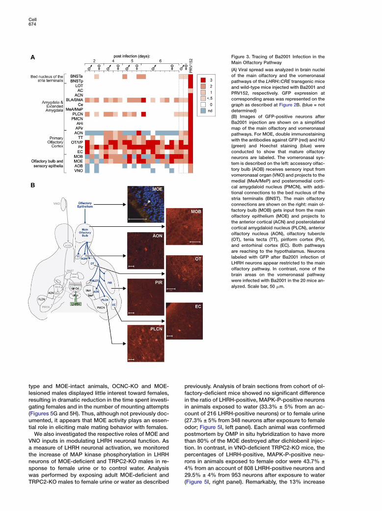
Figure 3. Tracing of Ba2001 Infection in theMain Olfactory Pathway
(A) Viral spread was analyzed in brain nucleiof the main olfactory and the vomeronasalpathways of the LHRH::CRE transgenic miceand wild-type mice injected with Ba2001 andPRV152, respectively. GFP expression atcorresponding areas was represented on thegraph as described at Figure 2B. (blue = notdetermined)(B) Images of GFP-positive neurons afterBa2001 injection are shown on a simplifiedmap of the main olfactory and vomeronasalpathways. For MOE, double immunostainingwith the antibodies against GFP (red) and HU(green) and Hoechst staining (blue) wereconducted to show that mature olfactoryneurons are labeled. The vomeronasal sys-tem is described on the left: accessory olfac-tory bulb (AOB) receives sensory input fromvomeronasal organ (VNO) and projects to themedial (MeA/MeP) and posteromedial corti-cal amygdaloid nucleus (PMCN), with addi-tional connections to the bed nucleus of thestria terminalis (BNST). The main olfactoryconnections are shown on the right: main ol-factory bulb (MOB) gets input from the mainolfactory epithelium (MOE) and projects tothe anterior cortical (ACN) and posterolateralcortical amygdaloid nucleus (PLCN), anteriorolfactory nucleus (AON), olfactory tubercle(OT), tenia tecta (TT), piriform cortex (Pir),and entorhinal cortex (EC). Both pathwaysare reaching to the hypothalamus. Neuronslabeled with GFP after Ba2001 infection ofLHRH neurons appear restricted to the mainolfactory pathway. In contrast, none of thebrain areas on the vomeronasal pathwaywere infected with Ba2001 in the 20 mice an-alyzed. Scale bar, 50 �m.
type and MOE-intact animals, OCNC-KO and MOE-lesioned males displayed little interest toward females,resulting in dramatic reduction in the time spent investi-gating females and in the number of mounting attempts(Figures 5G and 5H). Thus, although not previously doc-umented, it appears that MOE activity plays an essen-tial role in eliciting male mating behavior with females.
We also investigated the respective roles of MOE andVNO inputs in modulating LHRH neuronal function. Asa measure of LHRH neuronal activation, we monitoredthe increase of MAP kinase phosphorylation in LHRHneurons of MOE-deficient and TRPC2-KO males in re-sponse to female urine or to control water. Analysiswas performed by exposing adult MOE-deficient andTRPC2-KO males to female urine or water as described
pfiic(opttpr42(
reviously. Analysis of brain sections from cohort of ol-actory-deficient mice showed no significant differencen the ratio of LHRH-positive, MAPK-P-positive neuronsn animals exposed to water (33.3% ± 5% from an ac-ount of 216 LHRH-positive neurons) or to female urine27.3% ± 5% from 348 neurons after exposure to femaledor; Figure 5I, left panel). Each animal was confirmedostmortem by OMP in situ hybridization to have morehan 80% of the MOE destroyed after dichlobenil injec-ion. In contrast, in VNO-deficient TRPC2-KO mice, theercentages of LHRH-positive, MAPK-P-positive neu-
ons in animals exposed to female odor were 43.7% ±% from an account of 808 LHRH-positive neurons and9.5% ± 4% from 953 neurons after exposure to waterFigure 5I, right panel). Remarkably, the 13% increase

Olfactory Modulation of Reproduction and Fertility675
Figure 4. Olfactory and Vomeronasal Afferents to the Medial Preoptic Areas Determined by PRV152 Infection
Viral transfer in the main olfactory and the vomeronasal pathways were analyzed in wild-type mice injected with PRV152; GFP-positiveneurons in the corresponding areas are shown on the map. Both olfactory (right) and vomeronasal (left) pathways were infected with PRV152.Scale bar, 50 �m.
in the percentage of LHRH neurons expressing MAPK-P after exposure to female urine compared to exposureto water in TRPC2-KO (Figure 5I, right panel) is similarto what we observed in our previous experiment inwild-type mice (11%, see Figure 1B). These resultsstrongly support our anatomical data and indicate thatthe chemosensory modulation of LHRH neuronal activ-ity relies mostly, if not exclusively, on MOE function,while the contribution of VNO input, if any, is negligible.
The Nature of Olfactory Inputs to LHRH NeuronsWe consistently observed a relatively small number ofGFP-positive neurons in the bulb and olfactory epithe-lium in all animals analyzed. The vast majority of theinfected cells were found in the most anteroventralquadrant of the MOB (Figures 6A and 6B), an area thatreceives inputs from zones 2 and 3 of the MOE (Mori etal., 1999). Accordingly, afferent GFP-labeled neurons inthe MOE are confined within an intermediate zonealong the dorsoventral axis of the epithelium (Figures6C–6E and results below). In situ hybridization on analternate MOE section performed with RNA probes rep-resenting the most dorsal and ventral OR zones (zones1 and 4) as well as probes representing the two medial
zones (zones 2 and 3; Sullivan et al., 1996; Vassar etal., 1993) and anti-GFP immunocytochemistry showedthat 2/3 of MOE neurons ultimately connected to LHRHneurons are localized within zones 2 and 3 of the MOE(Figures 6C, 6D, and 6F). Thus, our results are consis-tent with the existence of a defined subpopulation ofolfactory neurons linked to the LHRH network and ex-pressing a discrete subset of the entire olfactory recep-tor repertoire.
Interestingly, this medial area of the olfactory epithe-lium is known to contain an unusual population of ol-factory neurons expressing a guanylyl cyclase receptor(GC-D) instead of the canonical olfactory receptor type(Fulle et al., 1995; Juilfs et al., 1997). However, the clus-tered expression of GC-D in the most ventral portion ofthe MOE, in an area where we very rarely identifiedBa2001 infected GFP-labeled neurons (Figure 6E, blackarrowheads) suggests that correlation between the twoolfactory neuron populations is unlikely.
LHRH Afferents across the BrainA larger survey of the spread of conditional Ba2001across the brain, in particular in the brain stem, basalganglia, hippocampus, cortex, and thalamus provided

Cell676
Figure 5. Behavioral and Physiological Defects in Olfactory-Deficient Mice
(A–F) Chemical ablation of the MOE. To assess the level of lesion in the MOE induced by dichlobenil treatment, X-gal staining was performedon cryosections of the MOE (A–D) and olfactory bulb (E and F) in OMP-tauLacZ mice treated with dichlobenil (A, C, and E) or DMSO (B, D,and F). The OE (olfactory epithelium) and NL (nerve layer) (A–D) and the glomerular layer of the MOB (E and F) are highly disrupted indichlobenil-treated animals compared to controls. X-gal staining in the glomerular layer of the AOB is comparable to that of the control,confirming the specific toxicity of dichlobenil for MOE but not VNO neurons.(G and H) Behavioral tests on olfactory-deficient mice.(G) When exposed to sexually receptive females, OCNC-KO mice spend significantly less time sniffing females (ratio per 20 min trial time:0.033 ± 0.018; mean ± SEM) compared to wild-type mice (0.29 ± 0.044) (p < 0.002, two-way ANOVA with repeated measures). During a 20min test session repeated twice, olfactory mutants failed to display any mounting attempt while wild-type animals consistently attemptedmounting females (7.1 ± 2.4; p < 0.01).(H) Similar experiments were performed in animals in which the MOE was chemically lesioned. In three repeated trials, each lasting for 20min, animals with more than 40% of the MOE still intact spent significantly larger ratio of time (0.31 ± 0.083) sniffing females compared tomice with more severe MOE lesions (0.078 ± 0.038) (p < 0.001, two-way ANOVA with repeated measures). Animals with functional MOEshowed active mounting behavior (8.4 ± 3.9 mounting attempts per trial) while animals with severe MOE lesions displayed very reducedmating behavior (0.078 ± 0.038 attempts per trial; p < 0.02).(I) Activation of LHRH neurons by female odor was tested by double staining for LHRH and phosphorylated MAPK in male mice with MOE

Olfactory Modulation of Reproduction and Fertility677
LHRH neurons across the entire mouse brain shows
lesion and TRPC2-KO mice. In mice with MOE ablation, exposure to female urine did not significantly increase the percentage of LHRHneurons expressing phosphorylated MAPK (27.3% ± 5%) compared to exposure to water (33.3% ± 5%) (p = 0.43, independent sampled ttest). In contrast, TRPC2-KO mice had higher level of LHRH neurons positive for phosphorylated MAPK when they are exposed to femaleurine (43.7% ± 4%) than when they are exposed to water (29.5% ± 4%) (p < 0.02).
olfactory system and the somatosensory systems, at
additional support for the critical role of olfactory cuesin modulating LHRH neuronal activity and uncovered avariety of other inputs, including significant somatosen-sory but virtual absence of visual and auditory inputs.Ba2001 infection in the cortex reveals prominent GFPlabeling of the primary and secondary somatosensorycortex (Figure 7), including the areas corresponding tosomatosensation of the trunk, flank, and face, regionsof the body known to be amenable to the somatosen-sory control of reproductive behavior (Pfaus et al.,1994). In contrast, visual and auditory cortical areas,central targets of retinal ganglion cells, such as the dor-sal lateral geniculate nucleus, the pretectal nucleus andthe superior colliculus (Figure 7) appear devoid of GFPlabeling. Cerebral projections from the motor cortexwere also identified, presumably involved in the volun-tary regulations of reproductive behavior. Furthermore,we identified brain areas known to be part of the cor-tex-hypothalamic and the cortex-basal nuclei-hypo-thalamic pathways such as the substantia nigra, thecaudate putamen, the globus pallidus, and the hippo-campus, a critical relay station for behavioral controlthat receives inputs from the cortex, the amygdala, andthe hypothalamus. In particular, staining was observedin the pyramidal layer and stratum oriens of CA1 andCA3 in the hippocampus and in the subiculum.
Injections of the PRV152 confirmed projections to thevicinity of LHRH neurons from several regions of thebrain stem (Simerly and Swanson, 1986). Among them,only the VTA and PAG, both known to be involved inmale and female reproductive behavior (Marson andFoley, 2004; Sipos and Nyby, 1996) displayed connec-tions to LHRH neurons, as shown by Ba2001 infection.
Discussion
Neuropeptides expressed in control centers of the hy-pothalamus are key regulators of homeostatic and be-havioral processes, such as sleep, feeding, and repro-duction. One of these peptides, the LHRH, is producedby a small and dispersed population of neurons in themedial hypothalamus and forebrain and has been thesubject of intense investigation for its essential role inorchestrating central and peripheral aspects of repro-duction. The existence of such a circumscribed andmolecularly defined neuronal population offered aunique opportunity to genetically uncover the variouscomponents of afferent neuronal circuits participatingin the control of reproduction and fertility across thewhole brain. This in turn should enable us to gain directinsights into the processing of relevant sensory andnonsensory inputs leading to reproductive behaviorsand endocrine changes.
The LHRH Wiring Diagram across the Mouse BrainCell-specific retrograde tracing of afferent pathways to
that LHRH neurons are embedded within an extraordi-narily complex neuronal network, which integrates in-formation from all major hypothalamic subdivisions,and from specific areas of the brain stem, limbic sys-tem, basal ganglia, and motor and sensory cortex.Based on the anatomical tracing of neural circuits con-trolling motivated behavior, Swanson (2000) proposeda wiring diagram classifying the various types of inputsreceived by the behavior control column of the hypo-thalamus. In accord with this model, our data show thatafferents to LHRH neurons, which are part of the so-called neuroendocrine secretomotor system (Swanson,2000), fit into three categories: sensory afferents thatmediate reflex behavior, cortical projections that medi-ate cognition and voluntary behavior, and intrahypo-thalamic and brain stem connections that reflect theanimal internal behavioral state.
We visualized brain stem projections, as well asextensive local intrahypothalamic circuitry, from theArc, the SCN, and other hypothalamic regions thoughtto provide information about the animal internal statesuch as the circadian period, stress, nutrition, and ste-roid levels. Moreover, several afferent nuclei identifiedat early time points post-viral infection, VMH, LH, PVN,and DMH play a central role in mediating reproductivebehavior and have been predicted to be connected toLHRH neurons (Baum, 2002; McCarthy and Becker,2002; Simerly, 2002). At later time points post-viral in-jection, GFP-positive neurons were identified in nearlyevery area of the hypothalamus with the exception ofthe SON and the MM. Clearly, LHRH neurons appearembedded within an extraordinarily complex and wide-spread regulatory network that involves all major hypo-thalamic subdivisions. Thus, the study of the connectiv-ity of a specific cell type, the LHRH neurons confirmsand expands results obtained by classical neuroana-tomical tracing of the MPOA (Simerly and Swanson,1986) that points to this region as a node of regulatorycircuits involved with reproduction.
Similarly, our study uncovered projections to LHRHneurons originating from neurons in the hippocampus,basal ganglia, and in defined areas of the cortex, thatform the various descending cortico-hypothalamicpathways participating in behavior control, revealingthe intensive crosstalk between LHRH neurons andother behavior control systems. The wiring diagram ofLHRH afferents across the brain provides novel insightsinto the biology and homeostasis of reproduction in themouse and lays the groundwork for detailed analysis ofthe circuit function in the control of reproduction.
The Vomeronasal System Is Not Part of the LHRHAfferent NetworkA unique aspect of our study was the direct visualiza-tion of the pattern of sensory projections to LHRH neu-rons. Our results point to major afferents from the main

Cell678
Figure 6. The Nature of Olfactory Inputs to LHRH-Expressing Neurons
(A) One of every four vibratome sections of olfactory bulb (50 �m) was analyzed, and the number of GFP-labeled mitral cells was counted.The y axis represents the number of mitral cells labeled in a quarter of whole olfactory bulb sections, which shows exponential increasethrough days after injection. The red dots represent data from each individual animal at a given time point, while black dots indicate themean value of each group analyzed on a given time point post-viral infection.(B) The location of GFP-positive mitral cells was identified from 12 different animals injected with Ba2001. The location and number of labeledcells was reported on a map of the olfactory bulb divided into seven regions with grids. Sixty-nine percent of the labeled neurons werelocated at the antero and anteroventral parts of the olfactory bulb.(C–E) Coronal cryosections of the main olfactory epithelium (17 �m) were labeled with antibody against GFP and the location of the labeledneurons marked by red dots on a low-magnification view of the sections. The positions of labeled sensory neurons after labeling adjacentsections with OR probes identifying zones 1 and 4 of the MOE (D) and zones 2 and 3 (E) and guanylate cyclase receptor probes (F) arerepresented by black dots.(F) Systematic comparison of the positions of GFP- and OR-positive neurons corresponding to either zones 1 and 4 or zones 2 and 3 wasconducted with MOE sections from seven animals infected with Ba2001 for 7 days. The number of GFP-positive OR neurons identified insideor outside of the areas labeled with the zonal markers was counted, which indicates that the majority of MOE neurons sending inputs toLHRH-expressing cells are located within the medial zones, zones 2 and 3.
the exclusion of vomeronasal, visual, and auditory in-puts. The importance of somatosensory inputs in theregulation of LHRH release is well illustrated by previ-ous reports showing that vaginocervical stimulation in-
dL
t
uces activation of LHRH neurons and increase ofHRH release (Pfaus et al., 1994).The MOE, MOB, as well as all cortical recipients from
he MOB including the AON, TT, EC, Pir, and their

Olfactory Modulation of Reproduction and Fertility679
Figure 7. Tracing of Ba2001 and PRV152 in the Brain Stem, Basal Ganglia, Hippocampus, Cortex, and Thalamus
(A) GFP immunostaining on sections of brain infected with Ba2001 clearly show viral transfer through the lateral globus pallidus (LGP), motorcortex (M1M2), somatosensory cortex, for the front limb (S1FL), and granular/agranular insular cortex (GI/AI). Scale bar, 50 �m.(B) Brain stem, basal ganglia, hippocampus, cortex, and thalamus in the bregma between +2.10 mm and −4.04 mm were analyzed afterBa2001 or PRV152 injection, and the data are presented on the graph as described.
downstream targets, the ventral pallidum, olfactory tu-bercle, and the caudal lateral hypothalamus, were iden-tified by retrograde infection of LHRH neurons. In con-trast, and in contradiction to all established tenets onthe control of LHRH neurons (Meredith, 1998), our viraltracing failed to document any synaptic connection be-tween the vomeronasal pathway and LHRH-expressingneurons. These data are substantiated by the completeabsence of retrogradely labeled neurons by Ba2001 inany nucleus of the identified vomeronasal pathways, inthe medial and cortico-medial amygdala, the BNSTp,the AOB, and the VNO. Neurons in these areas couldbe labeled by the nonconditional tracing strain PRV152,proving that they could replicate PRV.
These unexpected results are actually in agreementwith data obtained from the genetic ablation of VNOfunction in the TRPC2-KO mutant (Leypold et al., 2002;Stowers et al., 2002). TRPC2 is a channel required forthe pheromone-evoked response in the VNO (Liman etal., 1999). TRPC2-KO male mice appear perfectly able
to reproduce, with no reduction in courtship and matingbehavior with females but with profound defect in theirability to distinguish between males and females, anddisplay mating behavior toward both males and fe-males with equal frequency. These results prompted usto offer a new model of VNO function according towhich non-VNO cues are sufficient to trigger mating be-havior, while VNO function ensures the sex specificityof reproduction (Dulac and Torello, 2003; Stowers et al.,2002). Accordingly, data presented here demonstratethe lack of anatomical and functional connection be-tween VNO and LHRH neurons and document instead adirect link between the central regulators of mammalianreproduction, the LHRH neurons, and olfactory and so-matosensory pathways. Accordingly, we have providedbehavioral and functional data that enlightens theessential role of olfactory stimuli in mouse reproductivebehavior and in the modulation of LHRH neuronal ac-tivity.
What is then the nature of the vomeronasal inputs to

Cell680
reproductive centers of the hypothalamus, and how doolfactory and vomeronasal pathways interact for theglobal control of reproduction? Our anatomical tracingexperiments from the MPOA performed with PRV152,and those of published reports (Simerly and Swanson,1986) clearly indicate that some neurons adjacent toLHRH-expressing cells do receive vomeronasal inputs.This undefined neuronal population may play a role inreproduction control as well. Nevertheless, sex discrim-ination leading to mating behavior is likely to imply theconvergence and integration of information from thetwo chemosensory pathways. Intriguingly, our tracingexperiments failed to identify any synaptic connectionbetween the vomeronasal and olfactory networks up-stream of LHRH neurons. Although at this point we can-not rule out the existence of hypothetical synaptic con-nections resistant to Ba2001 infection, we favor twoalternative scenarios. First, paracrine and nonconven-tional mediators can relay vomeronasal inputs to theLHRH network via nonsynaptic mechanisms. For exam-ple nitric oxide, one of several identified diffusiblemessengers, was suggested to control LHRH release(Karanth et al., 2004; Wang et al., 1998). Secondly, theconvergence may occur at the level of downstream tar-gets of LHRH neurons. Specific targets of LHRH neu-rons in the central nervous system have not yet beenclearly defined. However, synergistic roles of the brain-stem, particularly the PAG and the VTA together withthe MPOA in the control of mating behaviors suggestthat these neuronal populations may receive projec-tions from the LHRH neurons and constitute a site ofconvergence between the two chemosensory path-ways (Marson and Foley, 2004; Rizvi et al., 1992; Siposand Nyby, 1996; Swanson, 1982).
The Nature of Olfactory Afferents to LHRH NeuronsRemarkably, our tracing experiments identify a smallpopulation of olfactory sensory neurons located in amedial zone of the olfactory mucosa as the major con-duit of pheromonal cues that regulate LHRH synthesisand release. This neuronal subset may in part overlapwith olfactory neurons shown to respond to volatilecomponents of urine (Lin da et al., 2005). It is thereforelikely that a dedicated population of olfactory receptorsacts as genuine pheromone receptors, a result particu-larly interesting to consider in the context of animalspecies thought to be devoid of a functional vomerona-sal system but displaying intriguing pheromonal-like re-sponses. It has been reported for example that humanaxillary cues affect the timing of ovulation in women(Stern and McClintock, 1998) and the pulsatility ofLHRH release (Preti et al., 2003), providing support tothe idea that the pathway identified in our study mayalso operate in other species.
Experimental Procedures
Transgenic Mouse Line Expressing CRE Recombinasein LHRH NeuronsA CRE cassette was generated by PCR amplification from pSP13-CRE, a CRE recombinase cDNA with Large T nuclear localizationsignal, and SV40 Large T poly(A) and inserted into the start codonin a shuttle vector (Yang et al., 1997) containing 1.8 kb of mousegenomic DNA centered around the start codon of the LHRH se-
qafj
VV2to
SSmipwBw((afM
AVmVMdtsw
Ltan
AFmffawUipbcLwi
CThd(idamwdwce
uence. Homologous recombination between the shuttle vectornd BAC RP23-22J8, containing the LHRH locus, yielded a modi-ied BAC containing the CRE transgene, which was in turn microin-ected into pronuclei of B6/CBA F1 oocytes.
irus Preparationirus was produced (3 × 10⁸ pfu/ml) as described (DeFalco et al.,001) and stored at −80°C before use. A fresh stock of virus washawed and sonicated using a cup sonicator (10 pulses for a totalf 10 s at an amplitude of 80%) before each injection.
tereotaxic Injection of Virusexually inexperienced homozygous LHRH:: CRE and C57BL/J6ice were used for stereotaxic injections conducted as described
n Card et al. (1999), with two exceptions: micropipette with dis-enser top (Drummond Scientific Company) was used and virusas injected at a rate of 20 nl/min. Three hundred nanoliters ofa2000 or Ba2001 (9 × 10⁴ pfu), or 20 nl of PRV152 (6 × 10³ pfu)as injected into eight different spots of the following coordinates
anterior, lateral, depth [mm] relative to bregma): (0.8,0,5.25);0.8,0,5.15); (0.8,0,4.95); (0.8,0,4.75); (0.9,0,5); (0.9,0,4.9); (0.9,0,4.7);nd (0.9,0,4.5). The injection areas were optimized by injection ofluorescent microspheres (Molecular Probes) to cover the MS, VDB,
POA, and MPON.
nalysis of Viral Transferibratome sections (50 �m) were made from brains (Bregma: 2.10m w−4.04 mm [Paxinos and Franklin, 2001]), olfactory bulbs, andNOs. Seventeen micrometer cryosections were prepared fromOE. Antibody against GFP (Molecular Probes) was used at 1:1000
ilution followed by biotinylated anti-rabbit IgG used at 1:200 dilu-ion (Vector Laboratories). Sections were then incubated with Cy3treptavidin (1:1000; Jackson laboratories). Antibody against HUas obtained from Molecular Probes (1:100).Sections were analyzed and images were captured with a
SM510 microscope (Zeiss). Anatomical analysis and nomencla-ure was based on Paxinos and Franklin (2001) except for olfactorynd vomeronasal structures named according to Kevetter and Wi-ans (1981a, 1981b).
nimal Exposure to Female Odorourteen male C57BL/J6 mice (The Jackson Laboratory) and 15ale TRPC2-KO mice, 7–9 weeks of age, were individually housed
or over a week and habituated with a Q-tip swapped with wateror one day before the experiment. Control mice were exposed to
new Q-tip swapped with 100 �l of water, and experimental miceere exposed for 10 to 15 min to 100 �l of urine freshly collected.rine was collected from three different adult C57BL/J6 females
n separate cages. Experimental animals were perfused with 4%araformaldehyde in PBS. Brains were postfixed, washed, and em-edded in Tissue-Tek OCT compound (Sakura). Seventeen mi-rometer coronal cryosections of brains were prepared and doubleHRH in situ hybridization and anti-MAPK-P immunocytochemistryas performed (details of procedures and reagents can be found
n the Supplemental Data).
hemical Ablation of the Olfactory Epitheliumhirty sexually naive male C57BL/J6 mice, 7–8 weeks of age, wereoused individually, from which 28 mice were given i.p. injections ofichlobenil (2,6-dichlobenzonitrile, 25 �g/g body weight) in DMSO
dimethyl sulphoxide, 2 �l/g body weight) and two were given i.p.njections of DMSO (2 �l/g body weight) on days 0, 2, and 4. Onay 7, 20 mice injected with dichlobenil were used to evaluate thectivation of LHRH neurons after exposure to female odor. Eightice injected with dichlobenil and two mice injected with DMSOere used for the behavioral test. MOE tissues of these mice wereissected out following each experiment and in situ hybridizationith an OMP antisense RNA probe was performed. The levels ofhemical lesion in the MOE tissues were evaluated based on OMPxpression and used for further analysis.
Olfactory Modulation of Reproduction and Fertility681
Behavioral AssaysFour OCNC-KO (OCNC knockout hemizygous crossed to LHRH::CRE transgenic line) and four control male mice (LHRH::CRE trans-genic line), 2 to 4 months of age, were each housed in an individualcage with a C57BL/J6 female for a week before the test. Threehours before the experiment, females were removed from the cage.A sexually receptive C57BL/J6 female was introduced into the malehome cage and behavior was recorded for 20 min. This test wasrepeated twice with two different females. ANOVA test was per-formed with Statistica and data were presented as mean ± SEM.
Similar behavioral assay was performed with ten sexually naiveC57BL/J6 males, eight injected with dichlobenil and two injectedwith DMSO. Twenty minute behavior tests were repeated threetimes with three different females. The level of MOE lesion wasassessed by three independent blind observers. Four animals withmore than 40% of the MOE intact were used as controls and sixanimals with more severely disrupted MOEs were used as experi-mental animals.
Supplemental DataSupplemental Data include one table and Supplemental Experi-mental Procedures and can be found with this article online athttp://www.cell.com/cgi/content/full/123/4/669/DC1/.
Acknowledgments
We wish to acknowledge R. Hellmiss for artistic work and J. Du-bauskaite for mouse transgenics. We thank the Dulac lab for help-ful discussions and comments on the manuscript, P. Choi andC. Chen for RNA probes, Drs. T. Kimhi, A. Lanjuin, and S. Santorofor help with experiments, M. Eldridge for producing and testingthe virus stocks, and Dr. B. Roska for useful advice. The pSP13-CRE construct was kindly provided by Dr. A.P. McMahon, and theROSA26::loxP-Stop-loxP-YFP mouse line was originated by Dr. F.Costantini. The OCNC-KO line was provided by Dr. R.R. Reed. Thiswork was supported by the Howard Hughes Medical Institute (C.D.)and by NIH-NIDCD grant R01 DC003903 (C.D.) and NIH grant2RO1-NS33506 (L.W.E.). H.Y. is an HHMI predoctoral fellow.
Received: December 7, 2004Revised: July 29, 2005Accepted: August 26, 2005Published online: November 10, 2005
References
Baum, M.J. (2002). Sexual behavior in the male. In Behavioral En-docrinology, J.B. Becker, S.M. Breedlove, D. Crews, and M.M. Mc-Carthy, eds. (Cambridge, MA: The MIT Press), pp. 153–203.
Brittle, E.E., Reynolds, A.E., and Enquist, L.W. (2004). Two modesof pseudorabies virus neuroinvasion and lethality in mice. J. Virol.78, 12951–12963.
Brunet, L.J., Gold, G.H., and Ngai, J. (1996). General anosmiacaused by a targeted disruption of the mouse olfactory cyclic nu-cleotide-gated cation channel. Neuron 17, 681–693.
Card, J.P., Enquist, L.W., and Moore, R.Y. (1999). Neuroinvasive-ness of pseudorabies virus injected intracerebrally is dependent onviral concentration and terminal field density. J. Comp. Neurol. 407,438–452.
Coquelin, A., Clancy, A.N., Macrides, F., Noble, E.P., and Gorski,R.A. (1984). Pheromonally induced release of luteinizing hormonein male mice: involvement of the vomeronasal system. J. Neurosci.4, 2230–2236.
DeFalco, J., Tomishima, M., Liu, H., Zhao, C., Cai, X., Marth, J.D.,Enquist, L., and Friedman, J.M. (2001). Virus-assisted mapping ofneural inputs to a feeding center in the hypothalamus. Science 291,2608–2613.
Dobson, H., Ghuman, S., Prabhakar, S., and Smith, R. (2003). Aconceptual model of the influence of stress on female reproduc-tion. Reproduction 125, 151–163.
Dong, H.W., and Swanson, L.W. (2004a). Organization of axonal
projections from the anterolateral area of the bed nuclei of the striaterminalis. J. Comp. Neurol. 468, 277–298.
Dong, H.W., and Swanson, L.W. (2004b). Projections from bed nu-clei of the stria terminalis, posterior division: implications for cere-bral hemisphere regulation of defensive and reproductive behav-iors. J. Comp. Neurol. 471, 396–433.
Dulac, C., and Torello, A.T. (2003). Molecular detection of phero-mone signals in mammals: from genes to behaviour. Nat. Rev. Neu-rosci. 4, 551–562.
Enquist, L.W., and Card, J.P. (2003). Recent advances in the use ofneurotropic viruses for circuit analysis. Curr. Opin. Neurobiol. 13,603–606.
Fulle, H.J., Vassar, R., Foster, D.C., Yang, R.B., Axel, R., and Gar-bers, D.L. (1995). A receptor guanylyl cyclase expressed specific-ally in olfactory sensory neurons. Proc. Natl. Acad. Sci. USA 92,3571–3575.
Genazzani, A.R., Bernardi, F., Monteleone, P., Luisi, S., and Luisi,M. (2000). Neuropeptides, neurotransmitters, neurosteroids, andthe onset of puberty. Ann. N Y Acad. Sci. 900, 1–9.
Jansen, A.S., Farwell, D.G., and Loewy, A.D. (1993). Specificity ofpseudorabies virus as a retrograde marker of sympathetic pregan-glionic neurons: implications for transneuronal labeling studies.Brain Res. 617, 103–112.
John, J.A., and Key, B. (2003). Axon mis-targeting in the olfactorybulb during regeneration of olfactory neuroepithelium. Chem.Senses 28, 773–779.
Juilfs, D.M., Fulle, H.J., Zhao, A.Z., Houslay, M.D., Garbers, D.L.,and Beavo, J.A. (1997). A subset of olfactory neurons that selec-tively express cGMP-stimulated phosphodiesterase (PDE2) andguanylyl cyclase-D define a unique olfactory signal transductionpathway. Proc. Natl. Acad. Sci. USA 94, 3388–3395.
Karanth, S., Yu, W.H., Mastronardi, C.A., and McCann, S.M. (2004).Inhibition of stimulated ascorbic acid and luteinizing hormone-releasing hormone release by nitric oxide synthase or guanyl cy-clase inhibitors. Exp. Biol. Med. (Maywood) 229, 72–79.
Kevetter, G.A., and Winans, S.S. (1981a). Connections of the corti-comedial amygdala in the golden hamster. I. Efferents of the “vo-meronasal amygdala”. J. Comp. Neurol. 197, 81–98.
Kevetter, G.A., and Winans, S.S. (1981b). Connections of the corti-comedial amygdala in the golden hamster. II. Efferents of the “ol-factory amygdala”. J. Comp. Neurol. 197, 99–111.
Leypold, B.G., Yu, C.R., Leinders-Zufall, T., Kim, M.M., Zufall, F.,and Axel, R. (2002). Altered sexual and social behaviors in trp2 mu-tant mice. Proc. Natl. Acad. Sci. USA 99, 6376–6381.
Liman, E.R., Corey, D.P., and Dulac, C. (1999). TRP2: a candidatetransduction channel for mammalian pheromone sensory signaling.Proc. Natl. Acad. Sci. USA 96, 5791–5796.
Lin da, Y., Zhang, S.Z., Block, E., and Katz, L.C. (2005). Encodingsocial signals in the mouse main olfactory bulb. Nature 434, 470–477.
Marson, L., and Foley, K.A. (2004). Identification of neural pathwaysinvolved in genital reflexes in the female: a combined anterogradeand retrograde tracing study. Neuroscience 127, 723–736.
McCarthy, M.M., and Becker, J.B. (2002). Sexual behavior in female.In Behavioral Endocrinology, J.B. Becker, S.M. Breedlove, D.Crews, and M.M. McCarthy, eds. (Cambridge, MA: The MIT Press),pp. 117–151.
Merchenthaler, I., Setalo, G., Petrusz, P., Negro-Vilar, A., and Flerko,B. (1989). Identification of hypophysiotropic luteinizing hormone-releasing hormone (LHRH) neurons by combined retrograde label-ing and immunocytochemistry. Exp. Clin. Endocrinol. 94, 133–140.
Meredith, M. (1998). Vomeronasal, olfactory, hormonal con-vergence in the brain. Cooperation or coincidence? Ann. N Y Acad.Sci. 855, 349–361.
Mombaerts, P., Wang, F., Dulac, C., Chao, S.K., Nemes, A., Mendel-sohn, M., Edmondson, J., and Axel, R. (1996). Visualizing an olfac-tory sensory map. Cell 87, 675–686.
Mori, K., Nagao, H., and Yoshihara, Y. (1999). The olfactory bulb:

Cell682
coding and processing of odor molecule information. Science 286,711–715.
Paxinos, G., and Franklin, K.B.J. (2001). The Mouse Brain in Stereo-taxic Coordinates, Second Edition (San Diego, CA: AcademicPress).
Petrovich, G.D., Canteras, N.S., and Swanson, L.W. (2001). Combi-natorial amygdalar inputs to hippocampal domains and hypothala-mic behavior systems. Brain Res. Brain Res. Rev. 38, 247–289.
Pfaus, J.G., Jakob, A., Kleopoulos, S.P., Gibbs, R.B., and Pfaff, D.W.(1994). Sexual stimulation induces Fos immunoreactivity withinGnRH neurons of the female rat preoptic area: interaction with ste-roid hormones. Neuroendocrinology 60, 283–290.
Piras, E., Franzen, A., Fernandez, E.L., Bergstrom, U., Raffalli-Mathieu, F., Lang, M., and Brittebo, E.B. (2003). Cell-specific ex-pression of CYP2A5 in the mouse respiratory tract: effects of olfac-tory toxicants. J. Histochem. Cytochem. 51, 1545–1555.
Preti, G., Wysocki, C.J., Barnhart, K.T., Sondheimer, S.J., andLeyden, J.J. (2003). Male axillary extracts contain pheromones thataffect pulsatile secretion of luteinizing hormone and mood inwomen recipients. Biol. Reprod. 68, 2107–2113.
Richardson, H.N., Nelson, A.L., Ahmed, E.I., Parfitt, D.B., Romeo,R.D., and Sisk, C.L. (2004). Female pheromones stimulate releaseof luteinizing hormone and testosterone without altering GnRHmRNA in adult male Syrian hamsters (Mesocricetus auratus). Gen.Comp. Endocrinol. 138, 211–217.
Rizvi, T.A., Ennis, M., and Shipley, M.T. (1992). Reciprocal connec-tions between the medial preoptic area and the midbrain peri-aqueductal gray in rat: a WGA-HRP and PHA-L study. J. Comp.Neurol. 315, 1–15.
Silverman, A.J., Livne, I., and Witkin, J.W. (1994). The gonadotropin-releasing hormone (GnRH), neuronal systems: immunocytochemis-try and in situ hybridization. In The Physiology of Reproduction,Volume 1, E. Knobil and J.D. Neill, eds. (New York: Raven Press).
Simerly, R.B. (2002). Wired for reproduction: organization and de-velopment of sexually dimorphic circuits in the mammalian fore-brain. Annu. Rev. Neurosci. 25, 507–536.
Simerly, R.B., and Swanson, L.W. (1986). The organization of neuralinputs to the medial preoptic nucleus of the rat. J. Comp. Neurol.246, 312–342.
Sipos, M.L., and Nyby, J.G. (1996). Concurrent androgenic stimula-tion of the ventral tegmental area and medial preoptic area: syner-gistic effects on male-typical reproductive behaviors in housemice. Brain Res. 729, 29–44.
Sisk, C.L., and Foster, D.L. (2004). The neural basis of puberty andadolescence. Nat. Neurosci. 7, 1040–1047.
Skynner, M.J., Slater, R., Sim, J.A., Allen, N.D., and Herbison, A.E.(1999). Promoter transgenics reveal multiple gonadotropin-releas-ing hormone-I-expressing cell populations of different embryologi-cal origin in mouse brain. J. Neurosci. 19, 5955–5966.
Srinivas, S., Watanabe, T., Lin, C.S., William, C.M., Tanabe, Y., Jes-sell, T.M., and Costantini, F. (2001). Cre reporter strains producedby targeted insertion of EYFP and ECFP into the ROSA26 locus.BMC Dev. Biol. 1, 4.
Stern, K., and McClintock, M.K. (1998). Regulation of ovulation byhuman pheromones. Nature 392, 177–179.
Stowers, L., Holy, T.E., Meister, M., Dulac, C., and Koentges, G.(2002). Loss of sex discrimination and male-male aggression inmice deficient for TRP2. Science 295, 1493–1500.
Sullivan, S.L., Adamson, M.C., Ressler, K.J., Kozak, C.A., and Buck,L.B. (1996). The chromosomal distribution of mouse odorant recep-tor genes. Proc. Natl. Acad. Sci. USA 93, 884–888.
Swanson, L.W. (1982). The projections of the ventral tegmental areaand adjacent regions: a combined fluorescent retrograde tracerand immunofluorescence study in the rat. Brain Res. Bull. 9, 321–353.
Swanson, L.W. (2000). Cerebral hemisphere regulation of motivatedbehavior. Brain Res. 886, 113–164.
Vassar, R., Ngai, J., and Axel, R. (1993). Spatial segregation of odor-
a7
Wry
WiN
WNi
YnmN
Zno
nt receptor expression in the mammalian olfactory epithelium. Cell4, 309–318.
ang, H., Li, S., and Pelletier, G. (1998). Role of nitric oxide in theegulation of gonadotropin-releasing hormone and tyrosine hydrox-lase gene expression in the male rat brain. Brain Res. 792, 66–71.
itkin, J.W., Paden, C.M., and Silverman, A.J. (1982). The luteiniz-ng hormone-releasing hormone (LHRH) systems in the rat brain.euroendocrinology 35, 429–438.
u, T.J., Gibson, M.J., Rogers, M.C., and Silverman, A.J. (1997).ew observations on the development of the gonadotropin-releas-
ng hormone system in the mouse. J. Neurobiol. 33, 983–998.
ang, X.W., Model, P., and Heintz, N. (1997). Homologous recombi-ation based modification in Escherichia coli and germline trans-ission in transgenic mice of a bacterial artificial chromosome.at. Biotechnol. 15, 859–865.
hao, H., and Reed, R.R. (2001). X inactivation of the OCNC1 chan-el gene reveals a role for activity-dependent competition in thelfactory system. Cell 104, 651–660.
Copyright © 2022 FDOKUMEN