64 Cu-Labeled Inhibitors of Prostate-Specific Membrane Antigen for PET Imaging of Prostate Cancer

a RESEARCH ARTICLE
MMP-2 Regulates Rat Ventral ProstateDevelopment In VitroAlexandre Bruni-Cardoso,1 Rafaela Rosa-Ribeiro,1 Vinicius D. B. Pascoal,2 Andre A. De Thomaz,3
Carlos L. Cesar,3,4 and Hernandes F. Carvalho1,4*
We have hypothesized that epithelial growth, branching, and canalization in the rodent ventral prostate(VP) would require matrix remodeling, and hence matrix metalloproteinase (MMP) activity. Therefore,the aim of this study was to evaluate the impact of blocking MMP-2, using whole organ culture. siRNAwas employed to inhibit MMP-2 expression, and this was compared to GM6001’s (a broad-spectrum MMPinhibitor) inhibition of general MMPs. These blocks impaired VP morphogenesis. MMP-2 silencingreduced organ size, epithelial area, and the number of tips, as well as caused a dilation of the distal partsof the epithelium. Histology, 3-D reconstruction, biochemistry, and second harmonic generation (SHG)revealed that MMP-2 silencing affected VP architecture by interfering in epithelial cell proliferation,lumen formation, and cellular organization of both epithelium and stroma, besides intense accumulationof collagen fibers. These data suggest that MMP-2 plays important roles in prostate growth, being directlyinvolved with epithelial morphogenesis. Developmental Dynamics 239:737–746, 2010. VC 2010 Wiley-Liss, Inc.
Key words: prostate development; MMP; siRNA; GM6001; collagen; SHG
Accepted 25 October 2009
INTRODUCTION
The rodent prostate gland shows sig-nificant growth and morphologicalchanges immediately after birth, in
response to a testosterone surge(Corbier et al., 1992). In this phase,the epithelial structures grow, differ-entiate, branch, and canalize (Sugi-mura et al., 1986a; Donjacour and
Cunha, 1988; Bruni-Cardoso and Car-valho, 2007; Bruni-Cardoso et al.,2008). These processes involve cellproliferation (Sugimura et al., 1986b;Bruni-Cardoso and Carvalho, 2007),
lumen formation (Vilamaior et al.,2006), epithelial and smooth-musclecell differentiation, and colonization
of spaces previously occupied by stro-mal components (Bruni-Cardoso andCarvalho, 2007), in addition to con-centrating important events in pros-tatic morphogenesis that result in fur-ther growth of the prototype gland atpuberty. This early postnatal develop-mental stage coincides with an impor-tant physiological window that willinfluence later prostate physiologyand susceptibility to disease, espe-cially in response to xenobiotics (Putzet al., 2001; Risbridger et al., 2005).MMPs constitute a family of zinc-de-pendent endopeptidases that prefer-entially cleave extracellular matrix(ECM) proteins. These enzymes playa key role in normal development and
physiology (VanSaun and Matrisian,2006; Page-McCaw et al., 2007; Wise-man et al., 2003; Bruni-Cardoso et al.,2008), as well as in cancer initiationand progression (Matrisian and Bow-den, 1990; Sternlicht and Werb, 2001;Lynch et al., 2005). We have previ-ously demonstrated a higher andlocalized expression of MMP-2 and -9in the rat ventral prostate during thefirst week of postnatal development,and have hypothesized that theseevents are associated with remodelingof ECM to allow epithelial growth(Bruni-Cardoso et al., 2008). Thus,the aim of the present study was toevaluate aspects of MMP-2 functionin the rat ventral-prostate (VP)
Dev
elop
men
tal D
ynam
ics
Additional Supporting Information may be found in the online version of this article.1Department of Cell Biology, State University of Campinas (UNICAMP), Campinas SP, Brazil2Department of Medical Genetics, State University of Campinas (UNICAMP), Campinas SP, Brazil3Quantum Electronics, IFGW, State University of Campinas (UNICAMP), Campinas SP, Brazil4National Institute of Photonics Applied to Cell Biology (INFABIC), State University of Campinas (UNICAMP), Campinas SP, BrazilGrant sponsor: FAPESP; Grant number: 07/07564-6.*Correspondence to: Hernandes F. Carvalho, Department of Cell Biology, UNICAMP, CP6109, 13083-863 Campinas SP,Brazil. E-mail: [email protected]
DOI 10.1002/dvdy.22222Published online 27 January 2010 in Wiley InterScience (www.interscience.wiley.com).
DEVELOPMENTAL DYNAMICS 239:737–746, 2010
VC 2010 Wiley-Liss, Inc.

morphogenesis during the first post-natal week, using specific siRNA forMMP2 in parallel to overall inhibitionby GM6001 in the whole ventral pros-tate in vitro. The efficiency of siRNAtreatment was assessed with RT-PCR,qRT-PCR, and gelatin zymography.Both MMP-2 silencing and GM6001treatment were evaluated by meas-uring the organ and epithelial area,by counting the number of epithelialtips on day 2 of culture, and by exam-ining collagen distribution using sec-ond harmonic generation (SHG) in amultiphoton laser scanning micro-scope. In addition, siRNA-treated VPswere examined by 3D-reconstructionof serial sections, volume changes,and hydroxyproline quantification.Together, the data showed that MMPsin general and MMP-2 in particularare important elements during VPmorphogenesis. Moreover, we showthat siRNA is an applicable and effi-cient tool for studying gene functionin the neonatal whole-prostate organculture.
RESULTS
GM6001 Treatment Inhibits
Ventral Prostate
Morphogenesis
The VP glands cultured on floatingmembranes grew and differentiated,with branching morphogenesis simi-lar to the in vivo situation (Fig. 1A).In order to investigate the role ofMMPs in VP development, we blockedtheir activity with GM6001, a broad-spectrum MMP inhibitor. VPs treatedwith 20 mM GM6001 showed an ini-tial growth delay observed on day 2,which was partially restored on day 6.
The whole organ, epithelial area,and fraction of the organ occupied bythe epithelium were smaller thanthose measured for the control group(Fig. 1B–D) on day 2. Branchingmorphogenesis was also affected.Whereas the VP epithelium of the con-trol group showed extensive branch-ing, resulting in about 40 epithelialtips per gland, GM6001-treated organsshowed fewer tips (�20) per gland(Fig. 1E). The lower concentration (2.5mM) of this inhibitor did not affectprostatic morphogenesis at any pointof the experiment.
Fig. 1. A: Low-magnification optical-microscope views of ventral prostates from Control, 2.5-mM GM6001, and 20-mM GM6001 groups in culture. In these images, the morphogenesis of thetranslucent epithelium can be evaluated by following the same organ along the timeline of theexperiment. The organ cultured with 20 mM GM6001 showed defective development (mainly inthe epithelial structure) in comparison with the control group. Scale bar ¼ 500 mm. B–E: Quanti-tative aspects of the VP development in culture from the vehicle (Control), 2.5-mM, and 20-mMGM6001 groups. Values are expressed as the mean 6 standard deviation. B: Mean total area ofVPs during their development in vitro. C: Mean area occupied by the epithelium. D: Mean frac-tion of the VP represented by the epithelial structures. The 20-mM GM6001 group showed a sig-nificantly smaller total and epithelial area, as well as epithelial/organ ratio, than those obtainedfor the Control group only on day 2 of development. E: Number of epithelial tips on days 0 and2 of culture. The 20-mM GM6001 group had on day 2 practically the same number of epithelialtips found at day 0, whereas for the Control the number of these structures was doubled on day2. Asterisks (*) indicate statistical differences (P < 0.05) between groups, as obtained usingANOVA followed by Tukey’s test. A total of ten VPs per group was used for thesemeasurements.
Dev
elop
men
tal D
ynam
ics
738 BRUNI-CARDOSO ET AL.
MMP-2 Silencing by siRNA
Inhibits Ventral-Prostate
Morphogenesis
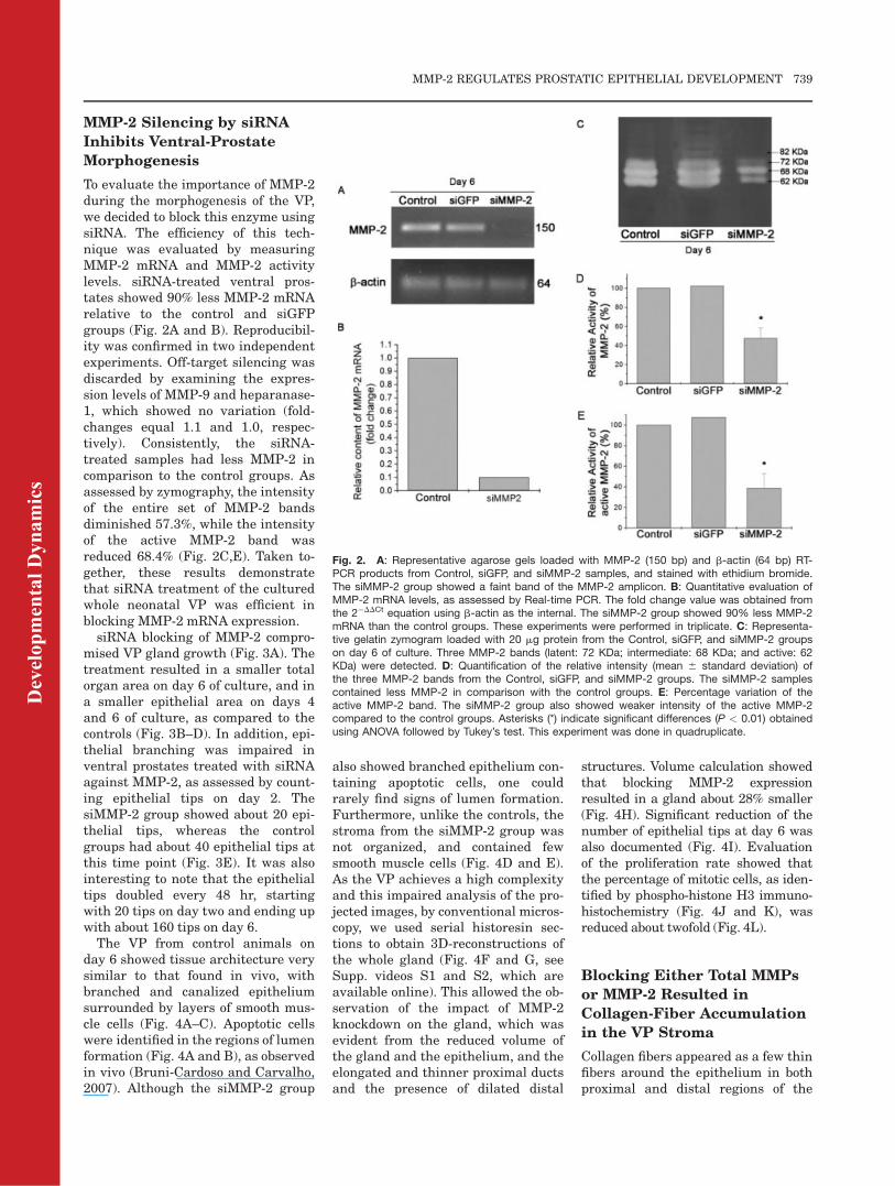
To evaluate the importance of MMP-2during the morphogenesis of the VP,we decided to block this enzyme usingsiRNA. The efficiency of this tech-nique was evaluated by measuringMMP-2 mRNA and MMP-2 activitylevels. siRNA-treated ventral pros-tates showed 90% less MMP-2 mRNArelative to the control and siGFPgroups (Fig. 2A and B). Reproducibil-ity was confirmed in two independentexperiments. Off-target silencing wasdiscarded by examining the expres-sion levels of MMP-9 and heparanase-1, which showed no variation (fold-changes equal 1.1 and 1.0, respec-tively). Consistently, the siRNA-treated samples had less MMP-2 incomparison to the control groups. Asassessed by zymography, the intensityof the entire set of MMP-2 bandsdiminished 57.3%, while the intensityof the active MMP-2 band wasreduced 68.4% (Fig. 2C,E). Taken to-gether, these results demonstratethat siRNA treatment of the culturedwhole neonatal VP was efficient inblocking MMP-2 mRNA expression.
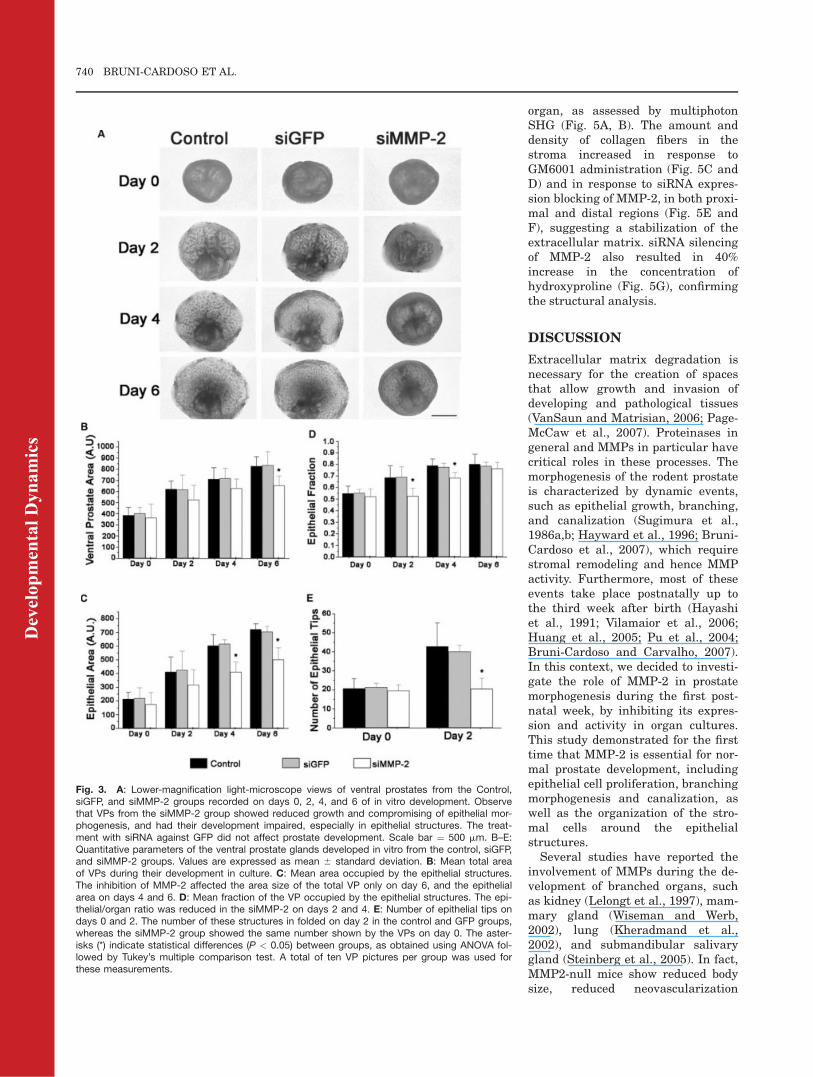
siRNA blocking of MMP-2 compro-mised VP gland growth (Fig. 3A). Thetreatment resulted in a smaller totalorgan area on day 6 of culture, and ina smaller epithelial area on days 4and 6 of culture, as compared to thecontrols (Fig. 3B–D). In addition, epi-thelial branching was impaired inventral prostates treated with siRNAagainst MMP-2, as assessed by count-ing epithelial tips on day 2. ThesiMMP-2 group showed about 20 epi-thelial tips, whereas the controlgroups had about 40 epithelial tips atthis time point (Fig. 3E). It was alsointeresting to note that the epithelialtips doubled every 48 hr, startingwith 20 tips on day two and ending upwith about 160 tips on day 6.
The VP from control animals onday 6 showed tissue architecture verysimilar to that found in vivo, withbranched and canalized epitheliumsurrounded by layers of smooth mus-cle cells (Fig. 4A–C). Apoptotic cellswere identified in the regions of lumenformation (Fig. 4A and B), as observedin vivo (Bruni-Cardoso and Carvalho,2007). Although the siMMP-2 group
also showed branched epithelium con-taining apoptotic cells, one couldrarely find signs of lumen formation.Furthermore, unlike the controls, thestroma from the siMMP-2 group wasnot organized, and contained fewsmooth muscle cells (Fig. 4D and E).As the VP achieves a high complexityand this impaired analysis of the pro-jected images, by conventional micros-copy, we used serial historesin sec-tions to obtain 3D-reconstructions ofthe whole gland (Fig. 4F and G, seeSupp. videos S1 and S2, which areavailable online). This allowed the ob-servation of the impact of MMP-2knockdown on the gland, which wasevident from the reduced volume ofthe gland and the epithelium, and theelongated and thinner proximal ductsand the presence of dilated distal
structures. Volume calculation showedthat blocking MMP-2 expressionresulted in a gland about 28% smaller(Fig. 4H). Significant reduction of thenumber of epithelial tips at day 6 wasalso documented (Fig. 4I). Evaluationof the proliferation rate showed thatthe percentage of mitotic cells, as iden-tified by phospho-histone H3 immuno-histochemistry (Fig. 4J and K), wasreduced about twofold (Fig. 4L).
Blocking Either Total MMPs
or MMP-2 Resulted in
Collagen-Fiber Accumulation
in the VP Stroma
Collagen fibers appeared as a few thinfibers around the epithelium in bothproximal and distal regions of the
Fig. 2. A: Representative agarose gels loaded with MMP-2 (150 bp) and b-actin (64 bp) RT-PCR products from Control, siGFP, and siMMP-2 samples, and stained with ethidium bromide.The siMMP-2 group showed a faint band of the MMP-2 amplicon. B: Quantitative evaluation ofMMP-2 mRNA levels, as assessed by Real-time PCR. The fold change value was obtained fromthe 2�DDCt equation using b-actin as the internal. The siMMP-2 group showed 90% less MMP-2mRNA than the control groups. These experiments were performed in triplicate. C: Representa-tive gelatin zymogram loaded with 20 mg protein from the Control, siGFP, and siMMP-2 groupson day 6 of culture. Three MMP-2 bands (latent: 72 KDa; intermediate: 68 KDa; and active: 62KDa) were detected. D: Quantification of the relative intensity (mean 6 standard deviation) ofthe three MMP-2 bands from the Control, siGFP, and siMMP-2 groups. The siMMP-2 samplescontained less MMP-2 in comparison with the control groups. E: Percentage variation of theactive MMP-2 band. The siMMP-2 group also showed weaker intensity of the active MMP-2compared to the control groups. Asterisks (*) indicate significant differences (P < 0.01) obtainedusing ANOVA followed by Tukey’s test. This experiment was done in quadruplicate.
Dev
elop
men
tal D
ynam
ics
MMP-2 REGULATES PROSTATIC EPITHELIAL DEVELOPMENT 739
organ, as assessed by multiphotonSHG (Fig. 5A, B). The amount anddensity of collagen fibers in thestroma increased in response toGM6001 administration (Fig. 5C andD) and in response to siRNA expres-sion blocking of MMP-2, in both proxi-mal and distal regions (Fig. 5E andF), suggesting a stabilization of theextracellular matrix. siRNA silencingof MMP-2 also resulted in 40%increase in the concentration ofhydroxyproline (Fig. 5G), confirmingthe structural analysis.
DISCUSSION
Extracellular matrix degradation isnecessary for the creation of spacesthat allow growth and invasion ofdeveloping and pathological tissues(VanSaun and Matrisian, 2006; Page-McCaw et al., 2007). Proteinases ingeneral and MMPs in particular havecritical roles in these processes. Themorphogenesis of the rodent prostateis characterized by dynamic events,such as epithelial growth, branching,and canalization (Sugimura et al.,1986a,b; Hayward et al., 1996; Bruni-Cardoso et al., 2007), which requirestromal remodeling and hence MMPactivity. Furthermore, most of theseevents take place postnatally up tothe third week after birth (Hayashiet al., 1991; Vilamaior et al., 2006;Huang et al., 2005; Pu et al., 2004;Bruni-Cardoso and Carvalho, 2007).In this context, we decided to investi-gate the role of MMP-2 in prostatemorphogenesis during the first post-natal week, by inhibiting its expres-sion and activity in organ cultures.This study demonstrated for the firsttime that MMP-2 is essential for nor-mal prostate development, includingepithelial cell proliferation, branchingmorphogenesis and canalization, aswell as the organization of the stro-mal cells around the epithelialstructures.Several studies have reported the
involvement of MMPs during the de-velopment of branched organs, suchas kidney (Lelongt et al., 1997), mam-mary gland (Wiseman and Werb,2002), lung (Kheradmand et al.,2002), and submandibular salivarygland (Steinberg et al., 2005). In fact,MMP2-null mice show reduced bodysize, reduced neovascularization
Fig. 3. A: Lower-magnification light-microscope views of ventral prostates from the Control,siGFP, and siMMP-2 groups recorded on days 0, 2, 4, and 6 of in vitro development. Observethat VPs from the siMMP-2 group showed reduced growth and compromising of epithelial mor-phogenesis, and had their development impaired, especially in epithelial structures. The treat-ment with siRNA against GFP did not affect prostate development. Scale bar ¼ 500 mm. B–E:Quantitative parameters of the ventral prostate glands developed in vitro from the control, siGFP,and siMMP-2 groups. Values are expressed as mean 6 standard deviation. B: Mean total areaof VPs during their development in culture. C: Mean area occupied by the epithelial structures.The inhibition of MMP-2 affected the area size of the total VP only on day 6, and the epithelialarea on days 4 and 6. D: Mean fraction of the VP occupied by the epithelial structures. The epi-thelial/organ ratio was reduced in the siMMP-2 on days 2 and 4. E: Number of epithelial tips ondays 0 and 2. The number of these structures in folded on day 2 in the control and GFP groups,whereas the siMMP-2 group showed the same number shown by the VPs on day 0. The aster-isks (*) indicate statistical differences (P < 0.05) between groups, as obtained using ANOVA fol-lowed by Tukey’s multiple comparison test. A total of ten VP pictures per group was used forthese measurements.
Dev
elop
men
tal D
ynam
ics
740 BRUNI-CARDOSO ET AL.
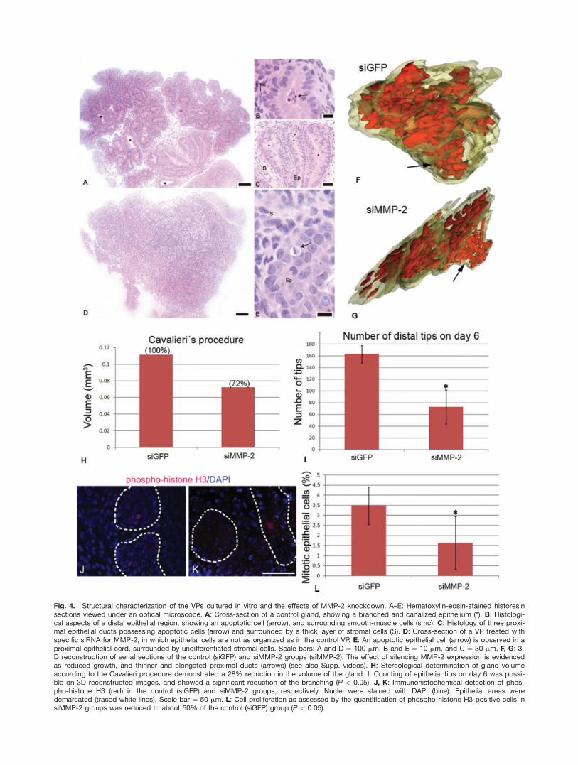
Fig. 4. Structural characterization of the VPs cultured in vitro and the effects of MMP-2 knockdown. A–E: Hematoxylin-eosin-stained historesinsections viewed under an optical microscope. A: Cross-section of a control gland, showing a branched and canalized epithelium (*). B: Histologi-cal aspects of a distal epithelial region, showing an apoptotic cell (arrow), and surrounding smooth-muscle cells (smc). C: Histology of three proxi-mal epithelial ducts possessing apoptotic cells (arrow) and surrounded by a thick layer of stromal cells (S). D: Cross-section of a VP treated withspecific siRNA for MMP-2, in which epithelial cells are not as organized as in the control VP. E: An apoptotic epithelial cell (arrow) is observed in aproximal epithelial cord, surrounded by undifferentiated stromal cells. Scale bars: A and D ¼ 100 mm, B and E ¼ 10 mm, and C ¼ 30 mm. F, G: 3-D reconstruction of serial sections of the control (siGFP) and siMMP-2 groups (siMMP-2). The effect of silencing MMP-2 expression is evidencedas reduced growth, and thinner and elongated proximal ducts (arrows) (see also Supp. videos). H: Stereological determination of gland volumeaccording to the Cavalieri procedure demonstrated a 28% reduction in the volume of the gland. I: Counting of epithelial tips on day 6 was possi-ble on 3D-reconstructed images, and showed a significant reduction of the branching (P < 0.05). J, K: Immunohistochemical detection of phos-pho-histone H3 (red) in the control (siGFP) and siMMP-2 groups, respectively. Nuclei were stained with DAPI (blue). Epithelial areas weredemarcated (traced white lines). Scale bar ¼ 50 mm. L: Cell proliferation as assessed by the quantification of phospho-histone H3-positive cells insiMMP-2 groups was reduced to about 50% of the control (siGFP) group (P < 0.05).

(Kato et al., 2001), decreased primaryductal invasion in the mammarygland (Wiseman et al., 2003), andreduced lung saccular development(Kheradmand et al., 2002). We haverecently shown that MMP-2 and -9activities are higher and localized inthe epithelium and at the epithelial/stromal interface of rat VP during thefirst postnatal week, and have sug-gested that this gelatinolytic activityis correlated with the ECM remodel-ing necessary for VP morphogenesis,in particular to permit epithelialgrowth and its projection into the sur-rounding stroma (Bruni-Cardosoet al., 2008).
In the present study, we found thatinhibition of MMPs with 10 mMGM6001 significantly impaired VPgrowth and morphogenesis by day 2of culture. Despite showing a dose-dependent effect, the GM6001 treat-ment did not significantly affect VPdevelopment concerning the eval-uated parameters in the later stages.Because GM6001 is a specific broad-spectrum inhibitor, it is possible thatsome MMP activity could have an in-hibitory effect on gland morphogene-sis in the period studied, and theirblocking would result in extendedgrowth. Such an inhibitory effect
could manifest through degradationof extracellular matrix componentscontributing to gland development. Infact, it is known that some ECM ele-ments promote branching morpho-genesis, such as tenascin in lung(Gebb and Jones, 2003) and type IIIcollagen in salivary gland (Nakanishiet al., 1998). We cannot rule out thepossibility that MMP-activity is com-pensated by different proteases.
To assess the importance of MMP-2in the morphogenesis of the ventralprostate, we silenced this enzymewith siRNA, using appropriate con-trols (either a transfecting agent orspecific siRNA for GFP). The post-transcriptional silencing of MMP-2 bysiRNA was highly effective, reaching90% reduction of the correspondingmRNA (as obtained by qRT-PCR) andabout 60% of the MMP-2 activity ingelatin zymograms. Probably, the dis-crepancy between these percentagesresults from MMP-2 protein stabilityin the extracellular spaces. The pres-ent study demonstrated reducedgrowth (at day 6) and compromisedepithelial morphogenesis throughoutthe timeline of the experiment in theabsence of MMP-2. As demonstratedby the histological evaluation, VPsdeveloped in vitro showed similar
architecture as seen in vivo (Bruni-Cardoso and Carvalho, 2007; Bruni-Cardoso et al., 2008). Apoptotic cellswere found inside the epithelial struc-tures in the process of canalization. Inspite of the presence of apoptotic cellsin the siMMP-2 group, lumen forma-tion was hindered. It is thus apparentthat, in addition to the creation ofspaces by epithelial cell deletion, theexpansion and consolidation of lumi-nal compartments require MMP-2 ac-tivity at the periphery of the epithe-lial structures consistent withgelatinolytic activity and MMP-2 loca-tion not only at the epithelial tips, butalso at the base of the epitheliumalong the epithelial structures(Bruni-Cardoso et al., 2008).BMP-4 and -7 play a key role in the
VP development by inhibiting its mor-phogenesis (Huang et al., 2005; Cooket al., 2007; Prins and Putz, 2008).Interestingly, multiple BMP familymembers, including BMP-4 andBMP-7, induce an epithelial-to-mes-enchymal transition (EMT) in thehuman pancreatic-cancer cell linePanc-1 (Gordon et al., 2008). More-over, BMP-mediated EMT results inan increase in invasiveness of Panc-1cells, in part through increasedexpression and activity of MMP-2
Fig. 5. Imaging of collagen fibers in the cultured ventral prostate by second harmonic generation (SHG) from the hematoxylin and eosin-stainedsections shown in Figure 4. Eosin fluorescence was included in the images for allowing identification of the tissue structures, especially the epithe-lium (Ep). Collagen fibers in the control prostates appeared as a few thin fibers in the stroma, in both proximal (A) and distal regions (B). GM6001(20 mM) treatment of the cultures resulted in marked accumulation of collagen fibers (C, D) and so did blocking of MMP-2 expression by siRNA(E, F). Scale bar ¼ 30 mm. G: Determination of the concentration of hydroxyproline in the control (siGFP) and siMMP-2 group revealed a significantincrease in response to MMP-2 silencing (P < 0.05).
Dev
elop
men
tal D
ynam
ics
742 BRUNI-CARDOSO ET AL.
(Gordon et al., 2008). AccompanyingEMT, BMP reduces the expression ofthe TGF-b type III receptor (TbRIII).TGF-b inhibits mammary-glandbranching (Nelson et al., 2006) andepithelial mammary-cell EMT in 3Dculture (Nelson et al., 2006, 2008).Sustained TbRIII expression inhibitsBMP-mediated invasion and sup-presses Smad1 activation. Further-more, Smad1 is required for BMP-induced invasiveness and is partiallyresponsible for BMP-mediatedincreases in MMP-2 activity. Webelieve that EMT might occur in themorphogenesis of ventral prostate,since we have recently found morpho-logical features of this process at ven-tral-prostate epithelial growing tipssuch as a few cell junctions, non-polarized cells, and invadopodia/filo-podia-like structures (Bruni-Cardosoet al., 2008).
We believe that the high testoster-one levels occurring immediately af-ter birth (Corbier et al., 1992) mightregulate the expression of MMPs,especially MMP-2. Liao et al. (2003)showed that testosterone regulatesMMP-2 expression in LNCaP andLAPC-4 cells in a dose-dependentmanner. Although MMP-2 expressionis constitutive, the levels of thisenzyme can be altered during normaldevelopment and in processes ofinflammation and tumor progression(Qin et al., 1999). In fact, there is anandrogen-responsive element (ARE)in the MMP-2 promotor gene (Liet al., 2007), indicating that MMP-2expression might be regulated byandrogens during prostatedevelopment.
The present results demonstratedthe stabilization of the collagen ma-trix in the prostate stroma. SHGrevealed an accumulation of collagenfibers, which was confirmed by thequantification of hydroxyproline, inresponse to MMP-2 and overall MMPinhibition, thus compromising VPprostate morphogenesis, likely byrestricting the growth of the epithe-lial structures.
Taken together, the results pre-sented here indicate that MMP-2plays an important role in epithelialgrowth and morphogenesis in the ratventral prostate, and it became clearthat collagen-fiber accumulation (andmatrix stabilization) in response to
MMP-2 inhibition by siRNA and over-all MMP-activity inhibition withGM6001 compromises VP morphogen-esis. While this effect in itself could beresponsible for the impaired epithelialgrowth and differentiation, we cannotrule out the participation of release ofgrowth factors or cryptic regulatorymolecules from the ECM. Thus, fur-ther analysis will be done regardingMMP-mediated ECM remodeling and/or shedding and activation of growthfactors that are directly involved inVP morphogenesis and physiology.
EXPERIMENTAL
PROCEDURES
Animals and Whole-Organ
Culture
Eighty male Wistar rats were pur-chased from CEMIB-UNICAMP. Thepups were decapitated on the day ofdelivery, and their VPs dissectedunder a stereoscopic microscope. Im-mediately after harvesting, the wholeVPs were cultured for 6 days onPTFE membranes (Millipore Corp.,Bedford, MA) floating in 500 mL of thebasal medium (BM) constituted ofDMEM/Ham’s F-12 (1:1; vol:vol) sup-plemented with insulin-transferrin-selenium (Gibco, Grand Island, NY)and 10 nM of testosterone cypionate(Novaquımica, Sao Bernardo doCampo, SP, Brazil), according toLopes et al. (1996).
For each assay, VP images weretaken using a TM- C35DX Zeiss cam-era connected to an Axiovert S100microscope (Zeiss, Thornwood, NY) atday 0 (the day of delivery and the firstday of culture) and days 2, 4, and 6 ofculture.
MMP Inhibition by GM6001
For experiments using the syntheticMMP inhibitor GM6001 (SigmaChemical Co., Saint Louis, MO), theVPs from 30 animals (10 VPs pertreatment) were cultured with either2.5 or 20 mM GM6001 in BM. Anequivalent volume of dimethylsulfox-ide (DMSO, vehicle for GM6001) inbasal medium was used as a control.The basal medium containingGM6001 was changed every 48 hr.
Post-Transcriptional Silenc-
ing of MMP-2 by Short
Interference RNA (siRNA)
The oligonucleotides for MMP-2siRNA were (sense) 50- UGG UGUUGG GGG AGA UUC UCA- 30 and(antisense) 50- AGA AUC UCC CCCAAC ACC AGU - 30, and for GFPsiRNA were (sense) 50- GAC GGGAAC UAC AAG ACA CGU - 30 and(antisense) 50- GUG UCU UGT AGUUCC CGU CAU - 30. These were de-signed with software (http://lgm.fcm.unicamp.br:9001) as described previ-ously (Pereira et al., 2007), using theNCBI sequence for the rat MMP-2(access number: NM_031054.1). Thelyophilized oligonucleotides (Inte-grated DNA Technologies, Coralville,IA) were dissolved (1 mg/mL) in DEPCwater. Twenty-five micrograms ofeach oligonucleotide was mixed andincubated for 5 min at 95�C andcooled for 4 hr at room temperature,for annealing of the complementarystrands.Ventral Prostates from 30 animals
(10 VPs per group) were treated inculture with 50 nM siRNA specific forMMP2 (siMMP-2 group), GFP (siGFPgroup), or only the transfecting agentlipofectamin 2000 (control group)(Invitrogen, Carlsbad, CA). In brief,1.25 mL of the siRNA stock solution(20 mM) was mixed with the same vol-ume of lipofectamin in 100 mL basalmedium, according to the manufac-turer’s instructions. The siRNA/lipo-fectamin mixture was added to 400mL of BM, and loaded on 24-wellplates. The BM containing siRNAwaschanged every 24 hr.The organs were harvested on
day 6 of culture for RNA and pro-tein extraction, and histologicalprocessing.
Evaluation of siRNA
Silencing Efficiency and
Off-Target Effects
The efficiency of MMP-2 silencing bysiRNA was evaluated by RT-PCR,real-time RT-PCR, and gelatin zymog-raphy on day 6. Off-target silencingwas assessed by determining theeffect of siRNA treatment on thenon-related genes MMP-9 andheparanase-1.
Dev
elop
men
tal D
ynam
ics
MMP-2 REGULATES PROSTATIC EPITHELIAL DEVELOPMENT 743

RNA Extraction and Reverse
Transcription-Polymerase
Chain Reaction (RT-PCR)
After culturing for 6 days, 4 VPs fromeach group (control, siGFP, andsiMMP-2) were harvested and pooled,and the total RNA was extractedusing the RNASpin Kit (GE Health-care, Buckinghamshire, UK), quanti-fied by spectrophotometry, andreverse-transcribed using 200 U ofSuperScript III and Oligo (dT)12-18Primer (Invitrogen, Sao Paulo SP,Brazil) according to the manufac-turer’s instructions. b-actin was usedas the internal control for the reac-tions. All the primers were designedwith Gene Runner 3.05 Software andsynthesized by Invitrogen, and theirsequences were the following: forMMP-2 (forward) 50 - CGA CCA CAGGAA GCC ATC - 30 and (reverse) 50 -TCG CCC ATC ATC AAG TTC - 30andfor b-actin (forward) 50- CTG GCCTCA CTG TCC ACC TT - 30 and(reverse) 50- GGG CCG GAC TCATCG TAC T - 30.
The cycling conditions were (1) ini-tial step of 5 min at 94�C; (2) 30 cyclesof 1 min at 94�C, 1 min at 60�C (for b-actin) or 58�C (for MMP-2), and 1 minand 30 sec at 74�C; and (3) final stepof 7 min at 74�C. The reactionsoccurred in 13 mL final volume con-taining 110 ng of cDNA, 0.39 U TaqDNA Polymerase (Promega, MadisonWI), 2 mM MgCl2 and 1.5 mM of eachprimer. The reaction product waselectrophoresed in 2% agarose gel andstained with ethidium bromide.Images were taken with a digitalcamera. Each experiment wasrepeated three times.
Quantitative RT-PCR
Real-time quantitative RT–PCR wasperformed using TaqMan UniversalPCR Master Mix (Applied Biosys-tems, Foster City, CA) in the AppliedBiosystems 7300. Inventoried assays(Primer and FAM-conjugated probes)for b-actin (Rn02532334_s1), b2-microglobulin (Rn00560865_m1)MMP-2 (Rn00667869_m1), MMP-9(Rn00578162_m1), and heparanase-1(Rn00575080_m1) were purchasedfrom Applied Biosystems. cDNA (20ng) was used in each reaction, accord-ing to universal cycling conditions for
the TaqMan system. The results werenormalized using the CT (thresholdcycle) values of the internal control b-actin on the same plate. The equationDCT ¼ CT (target gene) – CT (internalcontrol) was employed for normaliza-tion of the results. In order to quan-tify and acquire the fold-change varia-tion of MMP-2, the mathematicalmodel 2�DDCT was utilized. Both theMMP-2 and b-actin assays had theirefficiency calculated through theequation: E ¼ 10(-1/slope), with result-ing values of 1.05 and 1.03 for MMP-2and b-actin, respectively. The sameprocedure was applied for measuringexpression levels of MMP-9 andheparanase-1, considering the inter-nal control b2-microglobulin, thislatter chosen because it showed thebest efficiency for the former twogenes. All reactions were performedin technical triplicate on the sameplate for each pool, and the experi-ment was repeated twice.
MMP Extraction and Gelatin
Zymography
Twelve VPs cultured for 6 days ineach group were pooled (4 pools, 3VPs each) and homogenized in 50 mMTris–HCl, pH 7.4, 0.2 M NaCl, 0.1%Triton, 10 mM CaCl2, and 1% prote-ase inhibitor cocktail (Sigma Chemi-cal Co.). The homogenates were thenincubated for 2 hr at 4�C and centri-fuged at 2,080g for 20 min at 4�C. Thesupernatants were reserved, and thepellets were suspended once again inthe same solution, heated to 60�C for5 min, and centrifuged at 2,080g for20 min at 4�C. The two supernatantswere pooled, and the soluble proteinwas quantified using the Bradforddye binding assay kit (BioAgency, SaoPaulo, SP, Brazil). Twenty micro-grams of protein from each samplewas electrophoresed on 10% SDSpolyacrylamide gel containing 0.1%gelatin (used as the substrate) at 4�Cunder nonreducing conditions. Afterelectrophoresis, the gel was washedtwice, and then gently shaken in 2.5%Triton X-100 for 30 min at room tem-perature to remove the SDS. The gelwas incubated overnight in a 50 mMTris–HCl buffer pH 7.4, containing 10nM CaCl2 and 0.1 M NaCl, at 37�C.The gel was then stained with Coo-massie brilliant blue R (0.5% dye in
20% methanol and 10% acetic acid)for 1 hr. Unstained bands indicatinggelatinolytic activity were seen afterslight destaining with 30% ethanoland 10% acetic acid. Quantitativeassessment of band intensity wasdone by densitometry, using the soft-ware Scion Image (Scion Corporation,Frederick, MD). The activity deter-mined for the control group was usedas the reference (100%). The experi-ments were done in quadruplicate.Statistical comparison of the resultswas done using ANOVA followed bypost-hoc Tukey’s test, with the level ofsignificance set at P < 0.05, using theMini Tab 14 statistical software.
Morphometric Analysis and
Counting of Epithelial Ducts
A total of 10 digitalized images of thewhole organ were assessed for eachexperimental condition on days 0, 2,4, and 6 of culture. The epithelialarea was outlined and measuredusing the Image Pro Plus software. Asimilar outline of the whole VP wasutilized to estimate the organ areaand the fraction of the VP occupied bythe epithelium (epithelial fraction ¼epithelium area/organ area ratio), asdescribed previously (Pu et al., 2007).The number of epithelial tips andmain epithelial ducts on day 2 wascounted using the Adobe Photoshop7.0 program, according to Cook et al.(2007).Measurements were submitted to
ANOVA followed by the post-hocTukey’s test, using the Mini Tab 14statistical software. Values of P <0.05 were considered statisticallydifferent.
Historesin Embedding,
Histology, 3D-Reconstruction,
Tip Counting, Volume
Calculation, and Second
Harmonic Generation (SHG)
At day 6 in culture, the ventral pros-tates were fixed with 4% paraformal-dehyde in phosphate-buffered salinefor 16 hr. After dehydration in ethanol,the material was embedded in Leicahistoresin (Leica, Heidelberg, Ger-many) following the manufacturer’sinstructions. Serial two-micrometer
Dev
elop
men
tal D
ynam
ics
744 BRUNI-CARDOSO ET AL.

sections were cut, stained with hema-toxylin-eosin, and analyzed under alight microscope for histology. Serialsections were used for both 3D-recon-struction and volume calculation.Serial reconstruction was done byusing the free software Reconstruct(http://synapses.clm.utexas.edu/tools/reconstruct/reconstruct.stm). Thirty to70 sections were employed, and variedaccording to the position of the organwith respect to the sectioning plane.Three reconstructions were obtainedfor the control (siGFP) and siMMP-2organs. Distal tips were counted man-ually on the resulting reconstructions.The volume of the glands was calcu-lated by the Cavalieri procedure, afterdetermination of the section area usingthe Leica Application Suite software(Leica, Heerbrugg, Switzerland).
SHG was identified using an Olym-pus confocal system (IX-81 invertedmicroscope, the FV300 scan head, theFV-5 COMB2 laser combiner, and twoHamamatsu model 3896 PMTs). SHGwas examined by excitation with aTi:Sapphire laser (Tsunami, SpectraPhysics) at 800 nm and 80 MHz repe-tition rate, coupled to the scan headby an external port, and collected bythe condenser in the forward directionwith a bandpass filter at 400 nm(Oriel Corporation, Stratford, CT) anda blue shortpass filter to reject anyfluorescence signal. The Fluoviewsoftware was used to reconstruct theimages.
Proliferation Rate
Cultured VP were fixed in 4% parafor-maldehyde, processed for routine par-affin embedding, and subjected toimmunohistochemistry for phospho-histone H3 (ser10) mitosis marker. Inbrief, 5-mm sections were de-waxedand subjected to antigen retrieval byboiling in 10 mM citrate buffer pH 6.0for 10 min in a microwave oven, andthen treated for 20 min with 20 mg/mL proteinase K in 10 mM Tris-HCl,pH 7.4, buffer at room temperature.Sections were then blocked with 1%BSA and 5% donkey serum in block-ing buffer (10 mM Tris-HCl, pH 7.4,containing 0.1 M MgCl2, 0.5% Tween-20), before incubation with a rabbitanti-phospho-histone H3 (ser10)(Millipore, Temecula, CA) (diluted1:250) in blocking solution overnight
at 4�C. After rinsing, sections wereincubated with an Alexa-fluor 546-conjugated donkey anti-rabbit IgG(Invitrogen, Eugene, OR) (diluted1:500) in blocking solution. Negativecontrols were effected by substitutingthe primary antibody with rabbit IgGat the same concentration as the pri-mary antibody. Sections were thencounterstained with DAPI. Mitoticindex was determined by countingphospho-histone H3-positive nucleiwith respect to the total number ofepithelial cell nuclei in 10 microscopefields taken at random per animal(n¼3) using a 40� objective in a Leicafluorescence microscope. At least 800nuclei were counted per group.
Quantification of
Hydroxyproline
Hydroxyproline content was deter-mined for individual VP after treat-ments with siGFP (control) or siMMP-2, according to the procedure of Stege-mann and Stalder (1967) after hydro-lysis with 6N HCl at 115�C for 16 hr.To determine the tissue concentrationof hydroxyproline, we used the glandvolume calculated according to theCavalieri procedure as describedabove and assuming the specific grav-ity of the gland as 1 g/mL, according tothe determination for the adult VP byDeKlerk and Coffey (1978). Theresults were obtained for 4–5 individu-als in each experimental group, andare presented as nanograms of hydrox-yproline per gram of wet tissue.
ACKNOWLEDGMENTSThe authors thank Ricardo Fochi forhis help with the reconstruction soft-ware. Financial support from the Stateof Sao Paulo Funding Agency andNational Research Council (CNPq)grants to H.F.C. (FAPESP Grant no.07/07564-6) and to C.L.C. is acknowl-edged. A.B.-C. was a recipient of aFAPESP fellowship. INFABIC is co-funded by CNPq and FAPESP.
REFERENCES
Bruni-Cardoso A, Carvalho HF. 2007. Dy-namics of epithelium during canaliza-tion of the rat ventral prostate. AnatRec 290:1223–1232.
Bruni-Cardoso A, Vilamaior PSL, TabogaSB, Carvalho HF. 2008. Localized ma-trix metalloproteinase (MMP) 2 andMMP-9 activity in the rat ventral pros-tate during the first week of postnatal.Histochem Cell Biol 129:805–815.
Cook C, Vezina CM, Allgeier SH, Shaw A,Yu M, Peterson RE, Bushman W. 2007.Noggin is required for normal lobe pat-terning and ductal budding in themouse prostate. Dev Biol 312:217–230.
Corbier P, Edwards DA, Roffi J. 1992. Theneonatal testosterone surge: a compara-tive study. Arch Int Physiol BiochimBiophys 100:127–131.
Donjacour AA, Cunha GR. 1988. Theeffect of androgen deprivation onbranching morphogenesis in the mouseprostate. Dev Biol 128:1–14.
DeKlerk DP, Coffey DS. 1978. Quantita-tive determination of prostatic epithe-lial and stromal hyperplasia by a newtechnique biomorphometrics. InvestUrol 16:240–245.
Hayward SW, Baskin LS, Haughney PC,Cunha AR, Foster BC, Dahiya R, PrinsGS, Cunha GR. 1996. Epithelial devel-opment in the rat ventral prostate, an-terior prostate and seminal vesicle.Acta Anat 155:81–93.
Gebb SA, Jones PL. 2003. Hypoxia andlung branching morphogenesis. AdvExp Med Biol 543:117–125.
Gordon KJ, Kirkbride KC, How T, BlobelGC. 2008. Bone morphogenetic proteinsinduce pancreatic cancer cell in-vasiveness through a Smad1-dependentmechanism that involves matrix metal-loproteinase protein-2. Carcinogenesis30:238–248.
Huang L, Pu Y, Alam S, Birch L, PrinsGS. 2005. The role of Fgf10 signaling inbranching morphogenesis and geneexpression of the rat prostate gland:lobe-specific suppression by neonatalestrogens. Dev Biol 278:396–414.
Hayashi N, Sugimura Y, Kawamura J,Donjacour AA, Cunha GR. 1991. Mor-phological and functional heterogeneityin the rat prostatic gland. Biol Reprod45:308–321.
Kato T, Kure T, Chang J, Gabison EE,Itoh T, Itohara S, Azar DT. 2001.Diminished corneal angiogenesis in gel-atinase A-deficient mice. FEBS Lett508:187–190.
Kheradmand F, Rishi K, Werb Z. 2002.Signaling through the EGF receptorcontrols lung morphogenesis in part byregulating MT1-MMP-mediated activa-tion of gelatinase A/MMP2. J Cell Sci115:839–848.
Lelongt B, Trugnan G, Murphy G, RoncoPM. 1997. Matrix metalloproteinasesMMP2 and MMP9 are produced in earlystages of kidney morphogenesis but onlyMMP9 is required for renal organogene-sis in vitro. J Cell Biol 136:1363–1373.
Li BY, Liao XB, Fujito A, Thrasher JB,Shen FY, Xu PY. 2007. Dual androgen-response elements mediate androgenregulation of MMP-2 expression inprostate cancer cells. Asian J Androl 9:41–50.
Dev
elop
men
tal D
ynam
ics
MMP-2 REGULATES PROSTATIC EPITHELIAL DEVELOPMENT 745

Liao X, Thrasher JB, Pelling J, Holzbeier-lein J, Sang XQ, Li B. 2003. Androgenstimulates matrix metalloproteinase-2expression in human prostate cancer.Endocrinology 144:1656–1663.
Lopes ES, Foster BA, Donjacour AA,Cunha GR. 1996. Initiation of secretoryactivity of rat prostatic epithelium inorgan culture. Endocrinology 137:4225–4234.
Lynch CC, Hikosaka A, Acuff BH, MartinMD, Kawai N, Singh RK, Vargo-GogolaTC, Begtrup JL, Peterson TE, FingletonB, Tomoyuki S, Matrisian LM, Futaku-chi M. 2005. MMP-7 promotes prostatecancer-induced osteolysis via the solubi-lization of RANKL. Cancer Cell 7:485–496.
Matrisian LM, Bowden GT. 1990. Strome-lysin/transin and tumor progression.Semin Cancer Biol 1:107–115.
Nakanishi Y, Nogawa H, Hashimoto Y,Kishi J, Hayakawa T. 1988. Accumula-tion of collagen III at the cleft points ofdeveloping mouse submandibular epi-thelium. Development 104:51–59.
Nelson CM, Vanduijn MM, Inman JL,Fletcher DA, Bissell MJ. 2006.Tissue geometry determines sites ofmammary branching morphogenesis inorganotypic cultures. Science 314:298–300.
Nelson CM, Khauv D, Bissell MJ, Radi-sky DC. 2008. Change in cell shape isrequired for matrix metalloproteinase-induced epithelial-mesenchymal transi-tion of mammary epithelial cells.. J CellBiochem 105:25–33.
Page-McCaw A, Ewald AJ, Werb Z. 2007.Matrix metalloproteinases and the reg-
ulation of tissue remodeling. Nat RevMol Cell Biol 8:221–233.
Prins GS, Putz O. 2008. Molecular signal-ing pathways that regulate prostategland development. Differentiation 76:641–659.
Pu Y, Huang L, Prins GS. 2004. Sonichedgehog-patched Gli signaling in thedeveloping rat prostate gland: lobe-spe-cific suppression by neonatal estrogensreduces ductal growth and branching.Dev Biol 273:257–275.
Pu Y, Huan L, Birch L, Prins GS. 2007.Androgen regulation of prostate mor-phoregulatory gene expression: Fgf10-dependent and -independent pathways.Endocrinology 148:1697–1706.
Putz O, Schwartz CB, Kim S, LeBlancGA, Cooper RL, Prins GS. 2001. Neona-tal low- and high-dose exposure to es-tradiol benzoate in the male rat: I.Effects on the prostate gland. BiolReprod 65:1496–1505.
Qin H, Sun Y, Benveniste EN. 1999. Thetranscription factors Sp1, Sp3, and AP-2 are required for constitutive matrixmetalloproteinase-2 gene expression inastroglioma cells. J Biol Chem 274:29130–29137.
Risbridger GP, Almahbobi GA, Taylor RA.2005. Early prostate development andits association with late-life prostatedisease. Cell Tissue Res 322:173–181.
Stegemann H, Stalder K. 1967. Determi-nation of hydroxyproline. Clin ChimActa 18:267–273.
Steinberg Z, Myers C, Heim VM, LathropCA, Rebustini IT, Stewart JS, LarsenM, Hoffman MP. 2005. FGFR2b signal-ing regulates ex vivo submandibular
gland epithelial cell proliferation andbranching morphogenesis. Development132:1223–1234.
Sternlicht MD, Werb Z. 2001How matrixmetalloproteinases regulate cell behav-ior. Annu Rev Cell Dev Biol 17:465–516.
Sugimura Y, Cunha GR, Donjacour AA.1986a. Morphogenesis of ductal net-works in the mouse prostate. BiolReprod 34:961–971.
Sugimura Y, Cunha GR, Donjacour AA,Bigsby RM, Brody JR. 1986b. Whole-mount autoradiography study of DNAsynthetic activity during postnatal de-velopment and androgen-induced regen-eration in the mouse prostate. BiolReprod 34:985–995.
VanSaun MN, Matrisian LM. 2006. Ma-trix metalloproteinases and cellular mo-tility in development and disease.Birth Defects Res C Embryo Today 78:69–79.
Vilamaior PS, Taboga SR, Carvalho HF.2006. Alternating proliferative and se-cretory activities contribute to the post-natal growth of rat ventral prostate.Anat Rec A Discov Mol Cell Evol Biol288:885–892.
Wiseman BS, Werb Z. 2002. Stromaleffects on mammary gland developmentand breast cancer. Science 296:1046–1049.
Wiseman BS, Sternlicht MD, Lund LR,Alexander CM, Mott J, Bissell MJ,Soloway P, Itohara S, Werb Z. 2003.Site-specific inductive and inhibitoryactivities of MMP-2 and MMP-3 orches-trate mammary gland branching mor-phogenesis. J Cell Biol 162:1123–1133.
Dev
elop
men
tal D
ynam
ics
746 BRUNI-CARDOSO ET AL.
Copyright © 2022 FDOKUMEN