Spatial genomic heterogeneity within localized, multifocal prostate cancer

RESEARCH ARTICLE Open Access
Elevated AKR1C3 expression promotes prostatecancer cell survival and prostate cell-mediatedendothelial cell tube formation: implications forprostate cancer progressioanMikhail G Dozmorov1†, Joseph T Azzarello2,3†, Jonathan D Wren1, Kar-Ming Fung2,4,5, Qing Yang2, Jeffrey S Davis2,Robert E Hurst2,6, Daniel J Culkin2, Trevor M Penning7, Hsueh-Kung Lin1,3,5*
Abstract
Background: Aldo-keto reductase (AKR) 1C family member 3 (AKR1C3), one of four identified human AKR1Cenzymes, catalyzes steroid, prostaglandin, and xenobiotic metabolism. In the prostate, AKR1C3 is up-regulated inlocalized and advanced prostate adenocarcinoma, and is associated with prostate cancer (PCa) aggressiveness.Here we propose a novel pathological function of AKR1C3 in tumor angiogenesis and its potential role inpromoting PCa progression.
Methods: To recapitulate elevated AKR1C3 expression in cancerous prostate, the human PCa PC-3 cell line wasstably transfected with an AKR1C3 expression construct to establish PC3-AKR1C3 transfectants. Microarray andbioinformatics analysis were performed to identify AKR1C3-mediated pathways of activation and their potentialbiological consequences in PC-3 cells. Western blot analysis, reverse transcription-polymerase chain reaction (RT-PCR), enzyme-linked immunosorbent assay (ELISA), and an in vitro Matrigel angiogenesis assays were applied tovalidate the pro-angiogenic activity of PC3-AKR1C3 transfectants identified by bioinformatics analysis.
Results: Microarray and bioinformatics analysis suggested that overexpression of AKR1C3 in PC-3 cells modulatesestrogen and androgen metabolism, activates insulin-like growth factor (IGF)-1 and Akt signaling pathways, as wellas promotes tumor angiogenesis and aggressiveness. Levels of IGF-1 receptor (IGF-1R) and Akt activation as well asvascular endothelial growth factor (VEGF) expression and secretion were significantly elevated in PC3-AKR1C3transfectants in comparison to PC3-mock transfectants. PC3-AKR1C3 transfectants also promoted endothelial cell(EC) tube formation on Matrigel as compared to the AKR1C3-negative parental PC-3 cells and PC3-mocktransfectants. Pre-treatment of PC3-AKR1C3 transfectants with a selective IGF-1R kinase inhibitor (AG1024) or a non-selective phosphoinositide 3-kinases (PI3K) inhibitor (LY294002) abolished ability of the cells to promote EC tubeformation.
Conclusions: Bioinformatics analysis followed by functional genomics demonstrated that AKR1C3 overexpressionpromotes angiogenesis and aggressiveness of PC-3 cells. These results also suggest that AKR1C3-mediated tumorangiogenesis is regulated by estrogen and androgen metabolism with subsequent IGF-1R and Akt activationfollowed by VEGF expression in PCa cells.
* Correspondence: [email protected]† Contributed equally1Arthritis & Clinical Immunology Program, Oklahoma Medical ResearchFoundation, 825 N.E. 13th Street, Oklahoma City, Oklahoma 73104, USAFull list of author information is available at the end of the article
Dozmorov et al. BMC Cancer 2010, 10:672http://www.biomedcentral.com/1471-2407/10/672
© 2010 Dozmorov et al; licensee BioMed Central Ltd. This is an Open Access article distributed under the terms of the CreativeCommons Attribution License (http://creativecommons.org/licenses/by/2.0), which permits unrestricted use, distribution, andreproduction in any medium, provided the original work is properly cited.

BackgroundThe aldo-keto reductase (AKR) enzymes comprise afunctionally diverse gene family [1]. Members of theAKR superfamily are generally monomeric (37 kD) cyto-solic NAD(P)(H)-dependent oxidoreductases that con-vert carbonyl groups to primary or secondary alcohols,and share a common (a/b)8-barrel structural motif(visit: http://www.med.upenn.edu/akr) [2]. In humans,four AKR1C isoforms have been identified; they areknown as AKR1C1 [20a(3a)-hydroxysteroid dehydro-genase (HSD)] [3], AKR1C2 (type 3 3a-HSD) [4,5],AKR1C3 (type 2 3a/type 5 17b-HSD) [6,7], andAKR1C4 (type 1 3a-HSD) [5]. Natural substrates forthese enzymes include steroids [8,9], prostaglandins[10], and lipid aldehydes [11].Originally cloned from human prostate [7] and pla-
cental cDNA libraries [12], AKR1C3 shares greater than86% sequence identity with the other three highlyrelated human AKR1Cs [13]. AKR1C3 catalyzes bothandrogen and estrogen metabolism. The relatively high17b-HSD activity of this enzyme reduces Δ4-androstene-3,17-dione (Δ4-dione; a weak androgen) to yield testos-terone (a potent androgen) [14], and reduces estrone (aweak estrogen) to yield 17b-estradiol (a potent estrogen)[13]. Using its 3a-HSD activity, AKR1C3 reduces 5a-dihydrotesterone (5a-DHT, a potent androgen) to 5a-androstane-3a,17b-diol (3a-diol, a weak androgen) [7].As a result, AKR1C3 may be capable of modulating theamounts of potent androgen (testosterone and 5a-DHT)and estrogen (17b-estradiol) available for the androgenreceptor (AR) and estrogen receptor (ER), respectively.Deregulated AKR1C3 expression has been associated
with multiple human cancers. Suppressed AKR1C3expression has been demonstrated in breast cancer [15]and endometrial cancer [16], whereas elevated expres-sion of this enzyme has been reported in squamous cellcarcinoma of the head and neck [17]. In the prostate,low or undetectable levels of AKR1C3 are observed innormal prostate epithelium [18], whereas levels ofAKR1C3 expression are significantly elevated in loca-lized [19,20], advanced [21], and recurrent [22] prostatecancer (PCa).Based on the observations that expression of AKR1C3
is elevated in both localized and metastatic PCa, theenzyme might modulate significant pathological activ-ities in cancer development or progression. Studies sofar have been focused on the potential androgeniceffects mediated by AKR1C3 in the prostate; and ele-vated AKR1C3 expression in the prostate is thought tobe responsible for testosterone and 5a-DHT accumula-tion and AR trans-activation in cancerous prostate[14,20,21]. In this report, an androgen insensitive, AR-negative human PCa PC-3 cell line was stably trans-fected with AKR1C3 cDNA to establish PC3-AKR1C3
transfectants and to recapitulate elevated expression ofthis enzyme in PCa. Microarray, bioinformatics, and lit-erature analyses were used to explore possible pathologi-cal consequences of elevated AKR1C3 expression in PCacells. In addition to elevated cell growth in PC3-AKR1C3 transfectants [23], AKR1C3 overexpressionpromoted angiogenic capability of PC-3 cells as evi-denced by elevated levels of vascular endothelial growthfactor (VEGF) expression and PC-3 cell-mediatedendothelial cell (EC) tube formation on Matrigel. Theseresults suggest that, AKR1C3-mediated steroid hor-mones or prostaglandin metabolism may promoteaggressiveness of PCa through enhanced tumorangiogenesis.
MethodsReagents and chemicalsPC-3 cells (CRL-1435) derived from a human bonemetastatic tumor and LNCaP-FGC (LNCaP) cells (CRL-1740) were obtained from ATCC (Manassas, VA). AnSV40 large T-antigen transformed human microvascularEC line, HMEC-1 [24], was generously provided by Dr.Michael Ihnat at the University of Oklahoma HealthSciences Center. RPMI 1640 medium, F-12 nutrientmix, MCDB 131 medium, penicillin-streptomycin, fetalbovine serum (FBS), pcDNA3 vector, Trizol, andLipofectamine 2000 were purchased from Invitrogen(Carlsbad, CA). AG1042 and LY294002 were purchasedfrom EMD Chemicals (Gibbstown, NJ). Matrigelbasement membrane matrix was purchased from BDBiosciences (Bedford, MA). Mouse anti-AKR1C3 mono-clonal antibody was produced in our laboratories [25].Rabbit anti-insulin-like growth factor (IGF)-1 receptor(IGF-1R) b, rabbit anti-phospho-IGF-1R b (Tyr 1131),rabbit anti-Akt, and rabbit anti-phospho-Akt (Ser 473)polyclonal antibodies, as well as horseradish peroxidase(HRP)-linked goat anti-rabbit IgG were obtainedfrom Cell Signaling Technology (Danvers, MA). Mouseanti-human b-actin antibody was obtained from Sigma-Aldrich (St. Louis, MO). HRP-conjugated goat anti-mouse IgG was obtained from KPL (Gaithersburg, MD).Bicinchoninic acid (BCA) protein assay kit andenhanced chemiluminescent (ECL) substrate reagentwere obtained from Pierce (Rockford, IL). VEGFenzyme-linked immunosorbent assay (ELISA) kit waspurchased from Ray Biotech (Norcross, GA).
Prostate cell culture and transfectionPC-3 cells were cultured in complete growth mediumconsisting of F-12 nutrient mixture, 7% FBS, and 100units/ml penicillin-100 µg/ml streptomycin. LNCaP cellswere cultured in complete growth medium consisting ofRPMI-1640, 10% FBS, and penicillin-streptomycin.HMEC-1 were maintained in MCDB 131 medium
Dozmorov et al. BMC Cancer 2010, 10:672http://www.biomedcentral.com/1471-2407/10/672
Page 2 of 16
supplemented with 10% FBS and penicillin-streptomycin.All cells were maintained in a humidified cell incubatorat 37°C and 5% CO2. AKR1C3 expression construct(pcDNA3-AKR1C3) was established as previouslydescribed [23]. Transfection of PC-3 cells with eitherpcDNA3 empty vector or pcDNA3-AKR1C3 constructwas achieved using Lipofectamine 2000. Stable transfec-tants were established in the presence of 600 μg/ml G418selection. PC-3 stable transfectants were designated asPC3-mock or PC3-AKR1C3 for cells transfected withpcDNA3 or pcDNA3-AKR1C3 plasmid, respectively.Multiple independent clones were isolated and expandedin the complete growth medium containing G418. PC3-AKR1C3 clones were confirmed with elevated AKR1C3mRNA and protein expression as compared to parentalPC-3 cells and PC3-mock transfectants. LNCaP-mockand LNCaP-AKR1C3 stable transfectants were also simi-larly established.
RNA extraction and quality evaluationTotal RNA was isolated from 2 independent PC3-mockclones and 3 independent PC3-AKR1C3 clones for acomprehensive gene expression analysis using oligonu-cleotide arrays. Briefly, following PC-3 transfectants (5 ×10⁵) seeding and adherence, total RNA was isolatedfrom these clones using Trizol followed by RNeasy®Mini total RNA isolation kit (Qiagen, Valencia, CA)purification. Concentration of the isolated total RNAwas determined with a Nanodrop scanning spectrophot-ometer; and quality of the total RNA was assessed usingthe ratio of 28:18 s rRNA by Agilent 2100 Bioanalyzercapillary gel electrophoresis system (Agilent Technolo-gies, Santa Clara, CA).
RNA labeling, microarray hybridization, and scanningA total of 250 ng total RNA from each clone waslabeled using the Illumina Total Prep RNA Amplifica-tion kit (Ambion, Austin,. TX). Briefly, cDNA wasreverse transcribed from the total RNA after primingwith T7-oligo(dT); and cRNA was synthesized from theT7 promoter in the presence of biotinylated UTP. Bioti-nylated cRNA was hybridized to Illumina HumanWG 6v2 BeadChips containing a total of 48,702 probes (Illu-mina Inc., San Diego, CA). Hybridization was detectedby incubating the microarray chips with streptavidin-Cy3 (Amersham Biosciences, Piscataway, NJ) andscanned on an Illumina BeadArray Reader.
Data normalization and identification of AKR1C3regulated genesRaw expression values were first normalized based on anormal distribution of genes with low expression levelsas previously described [26,27]. Briefly, normalizationwas performed for each array by first plotting a
frequency histogram of the raw expression data for allgenes. The histograms showed a right-skewed unimodaldistribution curve with the mode around zero. A normaldistribution curve representing random variability of thegenes with low expression values was then fitted aroundthe mode, mirroring Gaussian profile of the left part ofthe histogram. Two parameters [mean and standarddeviation (SD)] were defined. The expression valuesthen were normalized to the SD of the normaldistribution after subtraction of the mean and then log10-transformed. The arrays were than adjusted to each otherby robust linear regression [28,29]. Genes with expres-sion values less than 3.0 (or 0.477 in log10 scale) wereconsidered to be not expressed; this is equivalent ofsetting a threshold at 3 SD above noise level. Raw andnormalized gene expression data are available on GeneExpression Omnibus (GEO accession GSE20956).To identify genes that are differentially expressed
between PC3-mock and PC3-AKR1C3 transfectants, anassociative analysis was performed as described by Doz-morov et al. [30]. Briefly, in combined microarray data-sets from both PC3-mock and PC3-AKR1C3transfectants, a reference group of genes expressedabove background with low variability of expression ascompared with variability of all others was identified bya F-test. The variability in expression of these genes wasassumed to represent the technical variability of theassay. Associative t-test (the standard t-test applied tothe comparison of expression variability) was used totest the null hypothesis if a given gene under a givencondition is associated with the reference group. Thosewith statistically significant variability over the referenceset were assumed to represent variability due to the bio-logical variables being manipulated.An additional analysis was performed to minimize
threshold choice effects in different statisticalapproaches in microarray data analysis [31]. Class com-parison between PC3-mock and PC3-AKR1C3 transfec-tants was performed using the BRB ArrayTools. To usethe BRB Array Tools, raw expression values were log2transformed; and quantile normalization was applied.Differentially expressed genes between the two classes,PC3-mock and PC3-AKR1C3 transfectants, were identi-fied by random-variance t-test. The random-variancet-test, implemented in the BRB ArrayTools, is animprovement of the standard t-test since it permitssharing information among genes with within-class var-iation, without assuming that all genes have the samevariance [32]. A stringent significance threshold wasapplied to limit the number of false positive findings;genes were considered statistically significant if theirP-values were less than 0.001. Lists of differentiallyexpressed genes from associative t-test and class com-parison analysis were compared; and genes identified by
Dozmorov et al. BMC Cancer 2010, 10:672http://www.biomedcentral.com/1471-2407/10/672
Page 3 of 16

both analyses were selected for further analysis unlessotherwise stated.
Functional analysis and identification of canonicalpathwaysFor pathway analysis of the microarray data, gene listswere analyzed by Ingenuity Pathway Analysis (IPA; Inge-nuity® Systems, Redwood City, CA, http://www.ingenu-ity.com), a web-based bioinformatics tool. Each gene listwas tested against the full Illumina HumanWG 6 v2dataset to identify significantly overrepresented func-tions and canonical pathways as compared to the samenumber of randomly selected genes. The most signifi-cant functions and canonical pathways with P < 0.05were selected; and genes overrepresented in the func-tions and pathways were identified. The branches ofpathways containing genes that are not expressed onmicroarrays were not considered functional.
Analysis of 5’-flanking regions of the differentiallyexpressed genesTo explore transcriptional regulatory mechanisms of thegenes regulated in PC3-AKR1C3 versus PC3-mocktransfectants, the promoter analysis and interaction net-work toolset (PAINT) http://www.dbi.tju.edu/dbi/tools/paint/) [33] was used to identify overrepresented cis-act-ing elements, or transcription factor binding sites, withinthe identified genes against the same transcription regu-latory elements (TREs) selected randomly from thewhole set of genes on the microarray. Statistical signifi-cance for testing the presence of TREs was set at P <0.05; and additional filtering was performed by using afalse discovery rate (FDR) as described in the text.
Data validation by literature analysisLiterature-based matching of genes to concepts providesan advantage over gene ontology (GO) enrichment analy-sis in that first, GO annotation lags literature publica-tions, and second, more importantly it provides a muchbroader matching of genes to concepts, such as diseases,phenotypes, chemical compounds, and other genes fre-quently associated with the gene set being analyzed. Toperform literature analysis, lists of differentially expressedgenes between PC3-mock and PC3-AKR1C3 transfec-tants were analyzed by a comprehensive, computationalsearch of published MEDLINE abstracts via a softwarepackage called IRIDESCENT [34-36]. There were 7 and39 up- and down-regulated genes in PC3-AKR1C3 trans-fectants, respectively, not mentioned in the literature andwere automatically discarded, since, by definition, theycould not share any literature-based commonalities withother genes. This left 56 up-regulated and 108 down-regulated genes available for analysis. IRIDESCENT usesa thesaurus of names and their synonyms obtained from
public databases such as OMIM, Entrez Gene, GO, CHE-MID, and Disease Ontology to recognize these “objects”and their common spelling variants (e.g., “IL2” and “IL-2”) within text. All available MEDLINE records (approxi-mately 18 million at the time of this writing) were pro-cessed to identify “objects” co-occurring within the sameabstracts, and within the same sentences within thoseabstracts. The number of co-occurrences was tallied,with a weighting based on a previous study [35] of 0.5 forobjects co-mentioned in the same abstract and 0.8 forobjects co-mentioned in the same sentence, providing anetwork of terms connected by their relative strength ofassociation. Once commonalities had been identified inthe literature for a gene set, each was compared to anidentically connected set within a random networkmodel to calculate a ratio of how many connections wereobserved in the actual dataset versus how many connec-tions would be expected by chance (Obs/Exp ratio), pro-viding a measure of statistical enrichment for each term.The significance of enrichment was calculated using aMonte Carlo simulation and the associations reportedwere ≥ 3 SDs from the mean (P ≤ 0.01).
Western blot analysisWestern blot analysis was performed to confirm IGF-1Rand Akt activation in PC3-AKR1C3 transfectants identi-fied by bioinformatics analysis. Briefly, following cell(1 × 10⁶) seeding and adherence, PC3-mock and PC3-AKR1C3 transfectants were lysed with RIPA buffersupplemented with 5 mM EDTA, 0.1 mM phenyl-methylsulphonylfluoride (PMSF), Complete Mini-Protease Inhibitor Cocktail, and PhosSTOP PhosphataseInhibitor Cocktail (Roche Applied Science, Indianapolis,IN). Total cellular proteins were collected following cen-trifugation to remove cellular components and quanti-fied using the BCA protein assay kit. Aliquots (30 µg) ofthe cellular proteins were separated on 10% Tris-HClgels; and separated proteins were transferred onto PVDFmembranes (Bio-Rad; Hercules, CA). The membraneswere incubated with rabbit anti-human phospho-IGF-1Rb (Tyr 1131) antibody (1:250), rabbit anti-human IGF-1R b antibody (1:1,000), rabbit anti-human phospho-Akt(Ser 473) antibody (1:250), or rabbit anti-human Aktantibody (1:1,500) followed by HRP-conjugated goatanti-rabbit IgG (1:2,000) incubation. Immunoreactiveproteins were detected using the ECL reagent. Themembranes were then stripped and incubated with amouse anti-human b-actin monoclonal antibody(1:5,000) followed by HRP-conjugated goat anti-mouseIgG (1:10,000) incubation and ECL development.
Quantification of VEGF expressionVEGF appears to be one of the most important factorsreleased by tumor cells as a signal for angiogenesis.
Dozmorov et al. BMC Cancer 2010, 10:672http://www.biomedcentral.com/1471-2407/10/672
Page 4 of 16

VEGF expression was first compared between PC3-AKR1C3 and PC3-mock transfectants. Levels of VEGFmRNA expression were determined in these transfec-tants using semi-quantitative reverse transcription-poly-merase chain reaction (RT-PCR) method. Total RNAisolation, first-strand cDNA synthesis, PCR amplificationusing pre-designed VEGF isoform-specific primer pairsand pre-determined cycling conditions followed ourreported procedures [37]. PCR amplified products wereseparated on 1% agarose gels and stained with ethidiumbromide. Images of ethidium bromide-stained gels wereacquired with Gel Doc 1000 (Bio-Rad) equipped withQuantity One® imaging software (Bio-Rad). The involve-ment of IGF-1R and phosphoinositide 3-kinases (PI3K)in VEGF mRNA expression in PC3-AKR1C3 transfec-tants was studied by adding 20 µM AG1024 (IGF-1Rselective tyrosine kinase inhibitor) and 10 µM LY294002(non-selective PI3K inhibitor), respectively, to the cellcultures at 2 hours prior to total RNA isolation. Theselected concentrations of AG1024 and LY294002 didnot interfere with cell viability as determined by XTTassay (data not shown).Total VEGF secretion by PC3-mock and PC3-AKR1C3
C1 transfectants was quantified in conditioned mediausing a RayBio® human VEGF ELISA kit following themanufacture’s recommendations. Briefly, 1 × 10⁶ cellswere seeded onto 60 mm cell culture plates in 2 mlcompleted medium in duplicate. At 24 hours followingcell seeding, the conditioned media were removed andcentrifuged at 300 × g to clear cellular components. Ali-quots (100 μl) of the conditioned media were transferredto each well of the 96-well assay plate. Known quantitiesof VEGF A165 were added in separate wells, run in par-allel, and used to construct a standard curve. The platewas read at 450 nm on a μQuant microplate reader(Bio-Tek Instrument, Winooski, VT). Results were nor-malized to total protein present in the media, as deter-mined by the BCA assay, and reported as pg of totalsecreted VEGF/μg total protein.
EC tube formation assayAn image-based, PCa cell-mediated EC tube formationon Matrigel assay was used as an in vitro model toassess enhanced angiogenic capability of PC3-AKR1C3transfectants predicted by bioinformatics. Briefly, PC3-mock and PC3-AKR1C3 transfectants (8 × 10⁴) were re-suspended in 0.25 ml liquid Matrigel and plated intoeach well of 24-well tissue culture plates in duplicate.Following overnight incubation, 8 × 10⁴ HMEC-1 in0.25 ml of the MCDB 131 complete medium were intro-duced on top of the solidified PC-3 transfectant-Matri-gel suspension. The co-cultures on Matrigel weremonitored and photographed at 6, 12, 24, 48, and 72hours following HMEC-1 cell addition using an
Olympus IX51 inverted microscope equipped withSPOT Insight CCD digital camera and SPOT Advancesoftware (Diagnostic Instruments, Sterling Heights, MI).Three images were randomly taken from each well.Images in JPEG format were uploaded into Image Pro-Plus 6 (Media Cybernetics, Bethesda, MD), converted togreen on the RGB color channel to eliminate the redcolor of Matrigel, and flattened. The number of tubeswas counted using the dendrite/tube template in theImage Pro-Plus software; and non-tube forming cellssuspended on Matrigel were mathematically excludedfrom counting [37]. The co-culture HMEC-1 tube for-mation model was also applied to 2 clones of each fromLNCaP-mock and LNCaP-AKR1C3 transfectants.To determine the roles of IGF-1R and PI3K in pros-
tate cell-mediated EC tube formation, parental PC-3cells as well as PC3-mock and PC3-AKR1C3 transfec-tants were first incubated with 20 µM AG1024 or10 µM LY294002 at 37°C for 2 hours. Cells were thenharvested, washed with phosphate-buffered saline (PBS),and suspended in Matrigel followed by HMEC-1 addi-tion, image acquisition, and EC tube quantificationusing the above described procedures.
Statistical analysisStatistical differences between PC3-mock and PC3-AKR1C3 stable transfectants were analyzed using stu-dent t-test. Statistically significant difference was setwhen P < 0.05.
ResultsIdentification of genes that are regulated by elevatedAKR1C3 expression in PC3-AKR1C3 transfectantsGene expression profiles were compared between PC3-mock and PC3-AKR1C3 transfectants using microarrayand bioinformatics techniques to predict possible altera-tions of gene expression in AKR1C3-positive PCa.When a threshold was set at 3 SDs above noise level,approximately 12,000 genes were expressed above thelevel in all arrays. The BRB ArrayTools class comparisonidentified 70 genes being up-regulated and 153 genesbeing down-regulated in PC3-AKR1C3 versus PC3-mock transfectants. The associative analysis identified27 genes being up-regulated and 85 genes being down-regulated by at least 1.5 fold and expressed greater than20 SDs above the noise levels in PC3-AKR1C3 versusPC3-mock transfectants (Additional file 1). Class com-parison and associative analysis provided almost overlap-ping results. The 27 up-regulated genes identified by theassociative analysis were a subset of the 70 genes identi-fied by the BRB ArrayTools. Among the 85 down-regu-lated genes identified by the associative analysis, 76genes were among the 153 genes identified by the BRBArrayTools (Additional file 2). The 27 up-regulated and
Dozmorov et al. BMC Cancer 2010, 10:672http://www.biomedcentral.com/1471-2407/10/672
Page 5 of 16
76 down-regulated genes in PC3-AKR1C3 transfectantsidentified by both analyses were used as “beacon” genesfor further analysis to reduce the possibility of includingfalse positives. However, in some cases, the number of“beacon” genes was insufficient to identify their com-mon functions or other properties. Each “beacon” genemay represent a unique function or feature, and the sta-tistical calculations for overrepresentation of such fea-tures become impossible. In such cases, the 70 up- and153 down-regulated gene lists identified by the BRBArrayTools were used, as described in the text. Theinclusion of these genes was further validated by subse-quent statistical analysis for overrepresentation of com-mon features among genes in the lists.
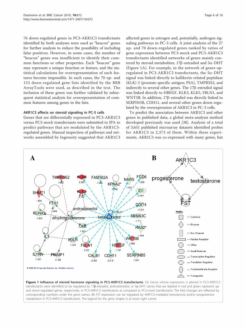
AKR1C3 effects on steroid signaling in PC-3 cellsGenes that are differentially expressed in PC3-AKR1C3versus PC3-mock transfectants were submitted to IPA topredict pathways that are modulated by the AKR1C3-regulated genes. Manual inspection of pathways and net-works assembled by Ingenuity suggested that AKR1C3
affected genes in estrogen and, potentially, androgen sig-naling pathways in PC-3 cells. A joint analysis of the 27up- and 70 down-regulated genes ranked by ratios ofgene expression between PC3-mock and PC3-AKR1C3transfectants identified networks of genes mainly cen-tered by steroid metabolites, 17b-estradiol and 5a-DHT(Figure 1A). For example, in the network of genes up-regulated in PC3-AKR1C3 transfectants, the 5a-DHTsignal was linked directly to kallikrein-related peptidase(KLK) 3 (prostate-specific antigen; PSA), TMPRSS2, andindirectly to several other genes. The 17b-estrsdiol signalwas linked directly to HBEGF, KLK3, KLK5, FBLN1, andWNT5B. In addition, 17b-estradiol was directly linked toSERPIN5B, CDH11, and several other genes down-regu-lated by the overexpression of AKR1C3 in PC-3 cells.To predict the association between AKR1C3 and other
genes in published data, a global meta-analysis methoddeveloped previously was used [38]. Analysis of a totalof 3,651 published microarray datasets identified probesfor AKR1C3 in 2,373 of them. Within these experi-ments, AKR1C3 was co-expressed with many genes, but
A B
Figure 1 Influence of steroid hormone signaling in PC3-AKR1C3 transfectants. (A) Genes whose expression is altered in PC3-AKR1C3transfectants were identified to be regulated by 17b-estradiol, androstenediol, or 5a-DHT. Genes that are labeled in red and green represent up-and down-regulated genes, respectively, in PC3-AKR1C3 transfectants as compared to PC3-mock transfectants. The fold changes are reflected bycorresponding numbers under the gene names. (B) FST expression can be regulated by AKR1C3-mediated testosterone and/or progesteronemetabolism in PC3-AKR1C3 transfectants. The legend for the gene shapes is at lower right corner.
Dozmorov et al. BMC Cancer 2010, 10:672http://www.biomedcentral.com/1471-2407/10/672
Page 6 of 16

Follistatin (FST) showed the most statistically significantpositive correlation with AKR1C3 (Figure 1B). FST wasidentified as one of significantly down-regulated genesin PC3-AKR1C3 transfectants in our dataset (Additionalfile 2). IPA indicated the interaction between AKR1C3and FST may result from their interconnection withprogesterone and testosterone metabolism.
Activation of VDR/RXR and IGF-1 signaling pathways inPC3-AKR1C3 transfectantsFunctional classification predicted biological consequencesof the up-regulated genes in PC3-AKR1C3 transfectants.Among the 27 genes up-regulated in PC3-AKR1C3 trans-fectants, 4 genes were not present in the Ingenuity data-base, 11 genes were poorly functionally annotated to beeligible for statistical calculations, and only 12 genes wereeligible for functional classification. Ingenuity analysisidentified a group of 7 genes (BEX1, FBLN1, FGD3,HBEGF, KLK3, KLK6, and WNT5B) related to cancer, cel-lular function and maintenance, cellular growth, invasion,as well as proliferation with P < 0.001-0.05 (Table 1).Since IPA was not able to identify any significantly
overrepresented pathways based on the limited numberof genes, the 70-gene list identified by the BRB Array-Tools analysis was submitted for pathway analysis. Tominimize false positives, these genes were first filteredto select genes that have been identified in both humanand PC-3 cells. Among the 70 genes, 16 genes were eli-gible for generating networks (Additional file 1). Usingthe IPA tool, several overrepresented canonical pathwayswere identified. VDR/RXR activation (P < 0.0001) wasoverrepresented by cystatin E/M (CTS6), KLK6, andprotein kinase C, eta (PRKCH). The second most
significant pathway was IGF-1 signaling (P < 0.01; Figure2A) represented by IGF binding protein 4 (IGFBP4) andPRKCH. It was noticed that analysis of the 70-gene listwithout filtering does not change the significance of theidentified canonical pathways, but added insulin recep-tor substrate 2 (IRS2) to the IGF-1 signaling pathway.Activation of IGF-1R and its downstream Akt signal-
ing molecule were confirmed by phosphorylated levelsof the respective proteins using Western blot analysis.There was no difference in total IGF-1R and Akt expres-sion between PC3-mock and PC3-AKR1C3 transfec-tants, as also reflected by corresponding mRNAexpression on microarrays (data not shown). In contrast,levels of phosphorylated IGF-1R b (Tyr 1131) and Akt(Ser 473) expression were significantly elevated in allPC3-AKR1C3 clones as compared to PC3-mock trans-fectants or parental cells (Figure 2B).
Suppression of cell death genes and inactivation of thep53 pathway in PC3-AKR1C3 transfectantsGenes whose expression were suppressed in PC3-AKR1C3 versus PC3-mock transfectants were also sub-jected to functional classification to predict biologicaloutcomes in PCa cells. Among the 76 genes down-regu-lated in PC3-AKR1C3 transfectants, 24 of them werenot mapped in the Ingenuity database and 17 geneswere not sufficiently annotated to count in a statisticalcalculation (Additional file 2). Among the remaining 35genes eligible for functional classification, 15 of themwere significantly overrepresented in the cell death func-tional category with P < 0.0005 with 3 genes (ID1,PMAIP1, and SERPINB5) directly related to the deathof PCa cell lines (Table 1).
Table 1 Up- and down-regulated genes and their representing functional groups in PC3-AKR1C3 transfectants
Up-regulated genes Down-regulated genes
Cancer, Cellular growth and proliferation Cell death
BEX1 Brain expressed, X-linked 1 CALB1 Calbindin 1, 28 kDa
FBLN1 Fibulin 1 CD70 CD70 molecule
FGD3 FYVE, RhoGEF and PH domain containing 3 DDIT4 DNA-damage-inducible transcript 4
HBEGF Heparin-binding EGF-like growth factor FAM162A
KLK3 Kallikrein-related peptidase 3 FTH1 Ferritin, heavy polypeptide 1
KLK6 Kallikrein-related peptidase 6 HCLS1 Hematopoietic cell-specific Lyn substrate 1
WNT5B Wingless-type MMTV integration site family, member5B
ID1 Inhibitor of DNA binding 1, dominant negative helix-loop-helixprotein
IL1RN Interleukin 1 receptor antagonist
LDLR Low density lipoprotein receptor (familial hypercholesterolemia)
NEFL Neurofilament, light polypeptide 68 kDa
PERP PERP, TP53 apoptosis effector
PMAIP1 Phorbol-12-myristate-13-acetate-induced protein 1
PRDM1 PR domain containing 1, with ZNF domain
RUNX2 Runt-related transcription factor 2
SERPINB5 Serpin peptidase inhibitor, clade B (ovalbumin), member 5
Dozmorov et al. BMC Cancer 2010, 10:672http://www.biomedcentral.com/1471-2407/10/672
Page 7 of 16

IPA also identified the p53 canonical pathway as themost significant pathway (P < 0.0008) overrepresented byPERP (TP53 apoptosis effector), PMAIP1 (phorbol-12-myristate-13-acetate-induced protein 1, induction of apop-tosis), and SERPINB5 (serpin peptidase inhibitor, clade B,member 5, aka maspin). In addition, alanine and aspartatemetabolism were the second and third most significantlyoverrepresented pathways (P < 0.003), respectively, repre-sented by DLAT (dihydrolipoamide S-acetyltransferase)and NARS (asparaginyl-tRNA synthetase). When the list of153 down-regulated genes identified by the BRB Array-Tools analysis was filtered by Ingenuity and expressed inboth human and PC-3 cells (Additional file 2), 58 geneswere eligible for forming networks and expressed categori-zation. The endoplasmic reticulum stress pathway wasidentified as the most significant (P < 0.004, ATF, HSPA5).The p53 pathway along with alanine and aspartate metabo-lism were also present, but at a less significant level.
Assessment of PC3-AKR1C3 aggressiveness by literatureanalysisLiterature analysis predicted pathological developmentand progression of PC-3 cells overexpressing AKR1C3.Analysis of common biological functions of the 70 up-and 153 down-regulated genes using IRIDESCENTshowed that PSA, b-catenin, ER, and aggressiveness arethe most significant keywords overrepresented amongthe up-regulated genes. Those terms characterized acti-vation of specific biological processes induced by over-expression of AKR1C3 in PC-3 cells, and were morespecific than general functions identified by IPA. BEX1and FBLN were associated with ER, while FBLN1,HBEGF, KLK3, and TMPRSS2 were associated withtumor aggressiveness (Figure 3A). A strong connectionof WNT5B to the keyword “b-catenin” was also identi-fied. Significantly, ER was connected to all these genesdirectly or indirectly (Figure 3A).
A
B
p-IGF-1R (Tyr 1131)
Total IGF-1R
PC-3
PC3-
Moc
k
PC3-AKR1C3
Total Akt
C1 C2 C3
p-Akt (Ser 473)
-actin
Figure 2 Activation of the IGF-1 pathway in PC3-AKR1C3 transfectants. (A) Ingenuity analysis identified that activation of the IGF-1 signalingpathway is statistically significant in PC3-AKR1C3 transfectants. Up-regulated genes, or focus genes, are marked by in filled gray; and uncoloredgenes indicate they are expressed in the microarray datasets under any conditions and may participate in signal transduction. Branches of theIGF-1 pathway that did not have at least one focus gene were not included in the diagram. (B) Western blot analysis of total andphosphorylated IGF-1R b and Akt in PC3-AKR1C3 stable transfectants. The analysis was performed 3 times; and all experiments showedconsistent elevated phosphorylation of IGF-1R b (Tyr 1131) and Akt (Ser 473) in PC3-AKR1C3 transfectants. Image analysis confirmed that levels ofphosphorylated IGF-1R b and Akt are statistically higher in PC3-AKR1C3 transfectants as compared to PC3-mock transfectants.
Dozmorov et al. BMC Cancer 2010, 10:672http://www.biomedcentral.com/1471-2407/10/672
Page 8 of 16

More than 400 keywords were identified to be signifi-cantly overrepresented in the 76 down-regulated genes;and the majority of them were related to carcinogenicprocesses (data not shown). “Tumor suppressor” wasidentified as the most significant keyword connectingthe majority of these genes including SERPINB5 andCHD11 (Figure 3B). “Angiogenesis” was another signifi-cant keyword connecting many of the down-regulatedgenes in PC3-AKR1C3 transfectants (Figure 3B). Inaddition, as identified by Ingenuity, PERP and PMAIP1were strongly related to each other and to “p53 tumorsuppressor” keyword (Figure 3B). Consistently, themajority of these genes were regulated by 17b-estradiol,androstenediol, and 5a-DHT as described in Figure 1.
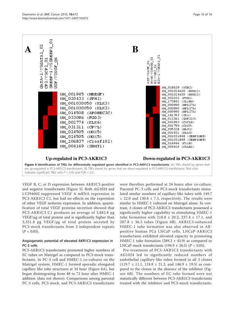
Promoter analysis of up- and down-regulated genes inPC3-AKR1C3 transfectantsThe PAINT analysis predicted whether the differentiallyexpressed genes between PC3-AKR1C3 and PC3-mocktransfectants are regulated by common transcription fac-tors. Based on results from PAINT analysis, GATA-1(aka globin transcription factor 1) was identified as themajor TRE shared by 7 of the 27 up-regulated genes fol-lowed by less overrepresented CP2, SEBP-1, and USF,
with P < 0.05 (Figure 4A). It should be noted that anygiven gene can have more than one TRE within 2 kb of5’-flanking regions queried by PAINT; and some genesappeared to share a given TRE. Additional analysis ofthe 70 genes from the BRB ArrayTools analysis identi-fied GATA-1 and SEBP-1 as major TREs shared amongthe up-regulated genes in PC3-AKR1C3 transfectants(data not shown). Among the 76 genes down-regulatedin PC3-AKR1C3 transfectants, ATF, MyoD, E2, and Elk-1 TREs were found to be major TREs, with P < 0.05and FDR < 0.3 (Figure 4B). Analysis of all 153 down-regulated genes in PC3-AKR1C3 transfectants identifiedby the BRB ArrayTools analysis showed the same set ofTREs.
VEGF expression in PC3-AKR1C3 transfectantsElevated AKR1C3 expression promoted VEGF expres-sion and secretion in PC-3 cells. AKR1C3 had differen-tial effects on the expression of various VEGF isoforms.Semi-quantitative RT-PCR analysis of VEGF isoformmRNA demonstrated that only the mRNA for theVEGF A isoforms significantly elevated in PC3-AKR1C3C1 as compared to PC3-mock transfectants from 3repeats (P < 0.05), whereas there was no difference in
A B
Figure 3 Phenotypic presentation of PC3-AKR1C3 transfectants from literature analysis. Relationships between differentially expressedgenes in PC3-AKR1C3 transfectants and the most significantly overrepresented keywords published with those genes in MEDLINE abstracts andtitles are identified. Genes are circled, keywords are not circled. Thickness of the line correlates with the mutual information between the terms(thicker lines mean more mutual information). (A) Up-regulated genes that have been documented as related to aggressive carcinomas, PSA, b-catenin, and ER. (B) Down-regulated genes that are related to angiogenesis, tumor suppression and, more specifically, tumor suppression via p53.
Dozmorov et al. BMC Cancer 2010, 10:672http://www.biomedcentral.com/1471-2407/10/672
Page 9 of 16
VEGF B, C, or D expression between AKR1C3-positiveand negative transfectants (Figure 5). Both AG1024 andLY294002 suppressed VEGF A mRNA expression inPC3-AKR1C3 C1, but had no effects on the expressionof other VEGF isoforms expression. In addition, quanti-fication of total VEGF proteins secretion showed thatPC3-AKR1C3 C1 produces an average of 3,882.8 pgVEGF/μg of total protein and is significantly higher than3,351.8 pg VEGF/μg of total protein secreted byPC3-mock transfectants from 3 independent repeats(P < 0.05).
Angiogenetic potential of elevated AKR1C3 expression inPC-3 cellsPC3-AKR1C3 transfectants promoted higher numbers ofEC tubes on Matrigel as compared to PC3-mock trans-fectants. In PC-3 cell and HMEC-1 co-cultures on theMatrigel system, HMEC-1 formed sporadic elongatedcapillary like tube structures at 24 hour (Figure 6A), butbegan disintegrating from 48 to 72 hour after HMEC-1addition (data not shown). Comparisons among parentalPC-3 cells, PC3-mock, and PC3-AKR1C3 transfectants
were therefore performed at 24 hours after co-culture.Parental PC-3 cells and PC3-mock transfectants stimu-lated similar numbers of capillary-like tubes with 149.7± 22.0 and 130.8 ± 7.3, respectively. The results weresimilar to HMEC-1 cultured on Matrigel alone. In con-trast, 3 clones of PC3-AKR1C3 transfectants possessed asignificantly higher capability in stimulating HMEC-1tube formation with 218.0 ± 20.2, 237.4 ± 17.1, and287.8 ± 36.5 tubes (Figure 6B). AKR1C3-enhancedHMEC-1 tube formation was also observed in AR-positive human PCa LNCaP cells. LNCaP-AKR1C3transfectants exhibited elevated capacity in promotingHMEC-1 tube formation (289.2 ± 45.9) as compared toLNCaP-mock transfectants (194.9 ± 26.5) (P < 0.05).Pre-treatment of PC3-AKR1C3 transfectants with
AG1024 led to significantly reduced numbers ofendothelial capillary-like tubes formed in all 3 clones(119.7 ± 11.1, 124.8 ± 21.2, and 146.9 ± 19.5) as com-pared to the clones in the absence of the inhibitor (Fig-ure 6B). The numbers of EC tube formed were notstatistically different between PC3-AKR1C3 transfectantstreated with the inhibitor and PC3-mock transfectants.
A B
Up-regulated in PC3-AKR1C3 Down-regulated in PC3-AKR1C3Figure 4 Identification of TREs for differentially regulated genes identified in PC3-AKR1C3 transfectants. (A) TREs shared by genes thatare up-regulated in PC3-AKR1C3 transfectants. (B) TREs shared by genes that are down-regulated in PC3-AKR1C3 transfectants. Red colorindicates significant TREs with P < 0.05 and FDR < 0.3.
Dozmorov et al. BMC Cancer 2010, 10:672http://www.biomedcentral.com/1471-2407/10/672
Page 10 of 16

Similarly, pre-treating PC3-AKR1C3 transfectants withLY294002 reduced the numbers of EC tubes to thelevels observed in parental PC-3 cells and PC3-mocktransfectants (Figure 6B).
DiscussionConsistent with previous reports [19-22], we observedlow levels of AKR1C3 immunoreactivity in normal pro-static epithelium, whereas a strong immunoreactivity isobserved in low grades and advanced PCa using tissuearrays. Forced expression of AKR1C3 was established inhuman PCa PC-3 and LNCaP cell lines to recapitulateelevated AKR1C3 expression in cancer tissues and tostudy potential pathological activities of this enzyme incancerous prostate. Bioinformatics analysis and biologi-cal characterization suggest that elevated AKR1C3expression in PCa cells not only promotes cancer cellgrowth [23] but also enhances angiogenesis. In additionto the classical belief that AKR1C3 is responsible forandrogen conversion and regulation of AR trans-activa-tion, AKR1C3 may also activate 17b-estradiol-mediatedsignaling pathways to promote PCa progression.
Differences in the prevalence of AKR1C3 expressionin PCa were noted between tissue sections preparedfrom individual patients and from tissue arrays. Usingradical prostatectomy samples, Nakamura et al. reported77.1% (54/70) [19] and we reported 81.8% (9/11) posi-tive immunoreactivity [20] in adenocarcinoma. Usingtissue array sections, we showed 33.3% AKR1C3 immu-noreactivity in prostate adenocarcinoma (data notshown) and Stanbrough et al. showed 5.6% (1/18) [21].In contrast, tissue array samples identified as androgen-independent PCa showed a higher 57.9% (11/19) immu-noreactivity for AKR1C3 [21]. Wako et al. also reporteda statistically significant positive correlation betweenAKR1C3 immunoreactivity and PCa stages [22]. PCa isa heterogeneous and multi-focal disease; and the sizes,locations, and stages of PCa tissues obtained for analysiscan affect interpretations. In addition, we previouslydescribed an uneven distribution of AKR1C3 in adeno-carcinoma in prostatectomy specimens [20]. Tissuearray slides consist of core biopsies of 1.0 to 1.5 mm indiameter from paraffin embedded specimens; locationsof the cores obtained for tissue array preparation cansignificantly affect immunohistochemical stainingresults. The differences in AKR1C3 immunoreactivityobtained from sections prepared from an individualpatient and tissue arrays might result from the unevendistribution of AKR1C3 in prostate adenocarcinoma.Since AKR1C3-mediated lipophilic hormone metabolismmay function through a paracrine signaling, it may notrequire all cancer cells to express elevated levels ofAKR1C3 to promote disease progression.In androgen metabolism, AKR1C3 acts as a 3-ketos-
teroid reductase converting 5a-DHT to 3a-diol [7], andas a 17-ketosteroid reductase converting Δ4-dione totestosterone [14]. In addition to androgen metabolism,recombinant AKR1C3 inter-converts estrone and 17b-estradiol [13]. Consistent with AKR1C3-mediated ster-oid metabolism, bioinformatics analysis confirmed thatgenes deregulated in PC3-AKR1C3 transfectants arerelated to Δ4-dione, testosterone, 5a-DHT, and 17b-estradiol. In contrast to other microarray data showing apositive correlation between AKR1C3 and FST, FST wassignificantly suppressed in PC3-AKR1C3 transfectants.Ingenuity analysis of interactions between AKR1C3 andFST shows that they are connected via testosterone andprogesterone. In transfectants, AKR1C3 likely reducesΔ4-dione to accumulate testosterone [14], which in turndecreases FST expression [39]. In addition, FST proteindecreases FST mRNA expression [39], but increases thesecretion of progesterone [40].The connection from AKR1C3 overexpression to the
p53 signaling suggests possible suppression of tumorsuppressive effects of p53 protein and promotion ofprostate cancer aggressiveness. Three down-regulated
VEGF A165
VEGF A121
-actin
VEGF A189
VEGF B186
VEGF B167
VEGF C
VEGF D
PC3-
moc
k
PC3-
AK
R1C
3 C
1
PC3-
AK
R1C
3 C
1+
10 μ
M L
Y29
4002
PC3-
AK
R1C
3 C
1+
20 μ
M A
G10
24
Figure 5 Levels of VEGF mRNA expression in PC3-AKR1C3transfectants. Total RNA was isolated from PC3-mock and PC3-AKR1C3 C1 transfectants as well as from PC3-AKR1C3 C1 clonetreated with either 20 µM AG1024 or 10 µM LY294002. Target VEGFA, B, C, and D mRNA species were PCR amplified using isofrom-specific primer pairs. PCR products were separated by 1% agarosegel electrophoresis; and images of ethidium bromide-stained gelswere acquired.
Dozmorov et al. BMC Cancer 2010, 10:672http://www.biomedcentral.com/1471-2407/10/672
Page 11 of 16

genes, SERPIN5B, PERP and PMAIP1, have been identi-fied as exclusive members of the p53 pathway, which isfurther highlighted by their connections to the “p53tumor suppressor” keyword. Expression of the p53 geneis not detectable in PC3-mock transfectants but isdetectable in AKR1C3 transfectants, albeit at a very lowlevel. This may be due to the fact that PC-3 cells arehemizygous for chromosome 17p [41]; and a base pairdeletion of the single copy of the p53 gene which gener-ates a frame shift and a new in-frame stop codon maynot provide the optimal hybridization between wild-typep53 probes and p53 transcripts in PC-3 cells. SERPIN5Bhas been identified as a suppressor of angiogenesis [42];and down-regulation of this gene in PC3-AKR1C3 trans-fectants suggests the role of AKR1C3 in promotingblood vessel formation. PERP [43] and PMAIP1 [44] areparts of the p53 signaling branch leading to apoptosis;and suppressed expression of these genes suggests thatAKR1C3 may also promote cancer progression throughapoptosis evasion. Other members of the apoptoticbranch, such as FAS and Caspase 6, were also down-regulated in PC3-AKR1C3 transfectants, although theirchanges did not pass our strict statistical criteria.A summary of these findings are presented in Additionalfile 3.
The advantage of using a literature-based analysis overa GO enrichment analysis is that it permits commonal-ities to be identified between genes and their associateddiseases, phenotypes, metabolites and chemical com-pounds and is complementary to GO categories. Thisapproach greatly expands the number and nature ofcommonalities that can be identified and has been usedin other microarray studies [45,46]. The downside isthat associations are probabilistic, which tends to bemore problematic when two terms have only been co-mentioned a few times, whereas in GO they are manu-ally corrected and have a lower false-positive rate.A search for biological or pathological functions of the
up- and down-regulated genes in PC3-AKR1C3 trans-fectants with literature keywords refined and confirmedIngenuity’s findings. “Aggressiveness” is the most signifi-cant process represented by the up-regulated genes inPC3-AKR1C3 transfectants, and directly relates to otherkeywords: “PSA”, “b-catenin” and “ER”. HBEGF identi-fied as one of EGF receptor ligands is strongly asso-ciated with large tumor size, high histoprognosticgrading, and aggressiveness of breast cancer [47].FBLN1, a calcium-binding, acidic glycoprotein of extra-cellular matrix, is positively correlated with ovarian can-cer progression [48]; and elevated expression of KLK3
PC3-mock
PC3-AKR1C3 C1
A
* *
*B
PC3-
moc
k
PC3-AKR1C3
C1 C2 C3
PC3-AKR1C3+
20 μM AG1024
C1 C2 C3
PC3-AKR1C3+
10 μM LY294002
C1 C2 C3
Figure 6 Angiogenic properties of PC-3 cells overexpressing AKR1C3 on Matrigel. (A) PC-3 cell-mediated HMEC-1 tube formation wasperformed in a co-culture system on Matrigel basement membrane matrix. Formation of HMEC-1 tubes was imaged at 24 hours after HMEC-1co-culture with either PC3-mock or PC3-AKR1C3 transfectants. (B) Quantification of HMEC-1 tube formation. The number of EC tubes werecounted; and results were compared between PC3-mock and PC3-AKR1C3 transfectants and presented as mean ± SEM from 12 experiments forthe 4 independent clones. The number of HMEC-1 tubes formed were also determined by pre-treating PC-3 transfectants with 20 µM AG1042 or10 µM LY294002; and results were presented as mean ± SEM from at least 3 independent assays. * indicates P < 0.05 between PC3-mock andPC3-AKR1C3 transfectants.
Dozmorov et al. BMC Cancer 2010, 10:672http://www.biomedcentral.com/1471-2407/10/672
Page 12 of 16

and TMPRSS2 have been associated with PCa aggres-siveness [49]. In the “CTNNB” (or b-catenin) keyword,both WNT5B (a representative Wnt family protein) andb-catenin are well-known pathways in carcinogenesis;and elevated expression of these molecules have beenassociated with AR, ER, and cancer progression [50,51].In the “ER” keyword, HBEGF [52], FBLN1 [53], BEX1[54], and TMPRSS2 [55] are regulated by 17b-estradiolthrough the ER. The up-regulated genes indentified inPC3-AKR1C3 transfectants surrounding these 4 key-words are interconnected and associated with moreaggressive cancer phenotypes.The majority of down-regulated genes in PC3-
AKR1C3 transfectants have been identified as tumor orangiogenesis suppressors. The keyword “tumor suppres-sor” is the most predominant among genes down-regu-lated in PC3-AKR1C3 transfectants. Reduced expressionof tumor suppressors is associated with developmentand progression of malignancies. For example, SER-PINB5 inhibits cell invasion, as well as angiogenesis andpromotes apoptosis [56]. Genetic loss of the stressresponse DDIT4 (or REDD1) gene promotes anchorage-independent cell growth and elicits tumorigenesis in amouse model [57]. Similarly, loss of CDH11 tumor sup-pressor gene shows the highest frequency in retinoblas-toma tumors [58]. Genes related to “angiogenesis”keyword are predominantly negative regulators of angio-genesis, suggesting the activation of angiogenic pro-cesses in PC3-AKR1C3 transfectants. For instance, theRUNX2 transcription factor is negatively correlated toangiogenesis [59]. Overexpression of FST (an inhibitorof activin) in human small cell lung cancer cells sup-presses metastatic colonies and microvessel density inSCID mice [60]. “Angiogenesis” and “tumor suppressor”keywords are interconnected by the down-regulatedgenes identified in PC3-AKR1C3 transfectants.Several TREs identified among up- ad down-regulated
genes in PC3-AKR1C3 transfectants are also related tosteroid metabolism. Steroid hormones such as proges-terone up-regulates GATA-1 expression [61]; and mem-bers of the GATA transcription factor family (GATA-2and GATA-3) are involved in the androgen regulatedPSA expression [62]. CP2 implicated in the control ofmRNA turnover and translation is involved in post-transcriptional regulation of AR expression [63].SREBP-1 (sterol regulatory element binding protein-1)interacts with a 17b-estradiol and insulin-sensitive SRE-1 cis-element [64]; and up-regulation of SREBP-1 isassociated with androgen-independent PCa progression[65]. USF1 (upstream stimulatory factor 1) has beenshown to act cooperatively with SREBP-1 in sterol bio-synthesis [66]. Many of the down-regulated genesshared MyoD or Elk-1 TRE. MyoD (myoblast determi-nation protein 1) is a new component of the ERa
complex [67]. Elk-1 (an ETS-related gene whose activa-tion is regulated by 17b-estradiol [68]) plays a role inprostate cell proliferation [69].Although the involvement of AR in tumor angiogen-
esis of PCa remains unsettled, several reports have sug-gested that angiogenesis can be independent of the AR.For example, expression of interleukin (IL)-8 activatesandrogen-independent growth and angiogenesis inLNCaP cells [70]. Consistent with our results identifyingER trans-activation in AKR1C3-positive PC-3 cells, avariety of estrogenic, anti-estrogenic, and selective ERmodulator (SERM) compounds has been suggested tohave chemopreventive or therapeutic utilities for PCareviewed by Ho [71]. In addition to trans-activation ofthe ER by 17b-estraldiol, 5a-androstane-3b, 17b-diol(3b-diol, an androgen metabolite formed by 3b-HSDactivity of AKR1C3 [72]), has been shown to be capableof trans-activating ERb and regulating PC-3 cell growthin cultures and in xenografts in nude mice [73]. Roles ofAKR1C3-mediated AR and ER trans-activation in PCaprogression require further study.More than a dozen different proteins have been iden-
tified as angiogenic factors, meaning that they arereleased by tumors as signals for angiogenesis. Thesefactors include acidic fibroblast growth factor (FGF),basic FGF, angiogenis, epidermal growth factor (EGF),granulocyte colony-stimulating factor (GM-CSF), hepa-tocyte growth factor (HGF), IL-8, placental growth fac-tor (PGF), platelet-derived growth factor (PDGF), scatterfactor, transforming growth factor (TGF)-a, tumornecrosis factor (TNF)-a, and VEGF. Among theseangiogenic factors, VEGF is one of the most importantfactors, and is a main target of many anti-angiogenicagents. Although elevated VEGF expression in PC3-AKR1C3 transfectants suggests that VEGF might beresponsible for promoting EC tube formation in the invitro angiogenesis model, our results did not rule outthe involvement of other angiogenic factors in promot-ing AKR1C3-positive cell-mediated angiogenesis. Inaddition, mechanisms of the secreted VEGF from PC3-AKR1C3 transfectants in activating VEGF receptors inECs with subsequent capillary-like tube formation willneed to be studied.Although the role for estrogen in PCa progression
remains controversial, our results might provide amechanistic explanation for why a combination of 3a-diol and 17b-estradiol administration promotes prostaticepithelial hyperplasia in castrated beagle dogs mimickinghuman PCa [74]. 17b-estradiol can activate IGF-1 sig-naling pathway in human PCa [75]. IGF-1 [76] and Akt[77], well-known players in promoting PCa progression,are identified as results of elevated AKR1C3 expressionin PC-3 cells. AKR1C3, therefore, can be positioned as acritical regulatory molecule in activating/inactivating
Dozmorov et al. BMC Cancer 2010, 10:672http://www.biomedcentral.com/1471-2407/10/672
Page 13 of 16

steroid hormone receptors and activating growth factorsignaling pathways. AKR1C3 can also be described as amolecule that provides a survival mechanism to promoteangiogenesis in cancer progression. Based on this com-munication, inter-relationships among AKR1C3, ER,IGF-1, Akt, and subsequent VEGF expression deservefurther investigation for tumor angiogenesis andprogression.
ConclusionsElevated expression of AKR1C3 has been identified andconfirmed in some cases of localized and advancedprostate adenocarcinoma. Bioinformatics data and biolo-gical characterization demonstrate that elevatedAKR1C3 expression in PCa cells can promote cancercell growth and cancer cell-mediated angiogenesis. Ourresults strongly suggest AKR1C3-mediated steroid hor-mones metabolism may activate growth factor IGF-1and cytoplasmic Akt signaling pathways as well asVEGF expression. AKR1C3 might be placed upstream ofthe well known growth factor receptor signaling andangiogenesis pathways for promoting PCa progression.
Additional material
Additional file 1: List of genes up-regulated in ACR1C3transfectants. 27 overlapping genes from both analyses presented first,followed by 43 unique genes from BRB ArrayTools analysis. Bold andItalicized were 16 genes expressed in Human and in PC-3 cells.
Additional file 2: List of genes down-regulated in ACR1C3transfectants. 76 overlapping genes from both analyses presented first,followed by 9 unique genes from associative analysis, followed by 77unique genes from BRB ArrayTools analysis. Bold and italicized were 58genes expressed in Human and in PC-3 cells.
Additional file 3: Parts of p53 signaling disturbed in PC-ACR1C3transfectants. Red/green shading indicates up- and down-regulatedgenes, respectively. Grey shading highlights non-changing genes. Whiteshading indicates no information available about expression of thesegenes.
AbbreviationsΔ4-dione: Δ4-androstene-3,17-dione; 3a-diol: 5a-androstane-3a, 17a-diol; 5a-DHT: 5a-dihydrotestosterone; AR: androgen receptor; EC: endothelial cell; ER:estrogen receptor; FDR: false discovery rate; GO: gene ontology; HSD:hydroxysteroid dehydrogenase; IGF-1: insulin-like growth factor-1; IPA:Ingenuity Pathway Analysis; PSA: prostate-specific antigen; TRE: transcriptionregulatory element; VEGF: vascular endothelial growth factor;
AcknowledgementsThis work was supported by NIH DK069808 to REH and 1R01-CA90744 andP30-ES13508 to TMP and by the US Department of Veterans Affairs MeritAward to HKL.
Author details1Arthritis & Clinical Immunology Program, Oklahoma Medical ResearchFoundation, 825 N.E. 13th Street, Oklahoma City, Oklahoma 73104, USA.2Department of Urology, University of Oklahoma Health Sciences Center,Oklahoma City, OK 73104, USA. 3Department of Physiology, University ofOklahoma Health Sciences Center, Oklahoma City, OK 73104, USA.4Department of Pathology, University of Oklahoma Health Sciences Center,
Oklahoma City, OK 73104, USA. 5Oklahoma City Department of VeteransAffairs Medical Center, Oklahoma City, OK 73104, USA. 6Department ofBiochemistry and Molecular Biology, University of Oklahoma Health SciencesCenter, Oklahoma City, OK 73104, USA. 7Center of Excellence inEnvironmental Toxicology, Department of Pharmacology, University ofPennsylvania School of Medicine, Philadelphia, PA 19104, USA.
Authors’ contributionsMGD performed bioinformatics analysis for microarray data. JTA, QY, and JSDestablished stable transfectants and carried out molecular and cellularstudies. JDW executed literature bioinformatics analysis. KMF analyzed theimmunohistochemical data and critically analyzed the manuscript. REH, DJC,and TMP participated in study design and coordination of the study. HKLdesigned the study, carried out the experiments, interpreted the data, andfinalize the manuscript. All authors read and approved the final manuscript.
Competing interestsThe authors declare that they have no competing interests.
Received: 7 October 2009 Accepted: 6 December 2010Published: 6 December 2010
References1. Jez JM, Flynn TG, Penning TM: A new nomenclature for the aldo-keto
reductase superfamily. Biochem Pharmacol 1997, 54:639-647.2. Jez JM, Bennett MJ, Schlegel BP, Lewis M, Penning TM: Comparative
anatomy of the aldo-keto reductase superfamily. Biochem J 1997,326:625-636.
3. Hara A, Matsuura K, Tamada Y, Sato K, Miyabe Y, Deyashiki Y, Ishida N:Relationship of human liver dihydrodiol dehydrogenases to hepatic bile-acid-binding protein and an oxidoreductase of human colon cells.Biochem J 1996, 313:373-376.
4. Dufort I, Soucy P, Labrie F, Luu-The V: Molecular cloning of human type 33α-hydroxysteroid dehydrogenase that differs from 20α-hydroxysteroiddehydrogenase by seven amino acids. Biochem Biophy Res Communication1996, 228:474-479.
5. Deyashiki Y, Ogasawara A, Nakayama T, Nakanishi M, Miyabe Y, Sato K,Hara A: Molecular cloning of two human liver 3α-hydroxysteroid/dihydrodiol dehydrogenase isoenzymes that are identical withchlordecone reductase and bile-acid binder. Biochem J 1994, 299:545-552.
6. Khanna M, Qin KN, Wang RW, Cheng KC: Substrate specificity, genestructure, and tissue-specific distribution of multiple human 3α-hydroxysteroid dehydrogenases. J Biol Chem 1995, 270:20162-20168.
7. Lin HK, Jez JM, Schlegel BP, Peehl DM, Pachter JA, Penning TM: Expressionand characterization of recombinant type 2 3α-hydroxysteroiddehydrogenase (HSD) from human prostate: demonstration ofbifunctional 3α/17β-HSD activity and cellular distribution. Mol Endocrinol1997, 11:1971-1984.
8. Schlegel BP, Pawlowski JE, Hu Y, Scolnick DM, Covey DF, Penning TM:Secosteroid mechanism-based inactivators and site-directedmutagenesis as probes for steroid hormone recognition by 3 alpha-hydroxysteroid dehydrogenase. Biochemistry 1994, 33:10367-10374.
9. Penning TM, Pawlowski JE, Schlegel BP, Jez JM, Lin HK, Hoog SS,Bennett MJ, Lewis M: Mammalian 3α-hydroxysteroid dehydrogenases.Steroids 1997, 61:508-523.
10. Bohren KM, Bullock B, Wermuth B, Gabbay KH: The aldo-keto reductasesuperfamily. cDNAs and deduced amino acid sequences of humanaldehyde and aldose reductases. J Biol Chem 1989, 264:9547-9551.
11. Hyndman D, Bauman DR, Heredia VV, Penning TM: The aldo-keto reductasesuperfamily homepage. Chem Biol Interact 2003, , 143-144: 621-631.
12. Dufort I, Rheault P, Huang XF, Soucy P, Luu-The V: Characteristics of ahighly labile human type 5 17β-hydroxysteroid dehydrogenase.Endocrinology 1999, 140:568-574.
13. Penning TM, Burczynski ME, Jez JM, Hung CF, Lin HK, Ma H, Moore M,Palackal N, Ratnam K: Human 3α-hydroxysteroid dehydrogenase isoforms(AKR1C1-AKR1C4) of the aldo-keto reductase superfamily: functionalplasticity and tissue distribution reveals roles in the inactivation andformation of male and female sex hormones. Biochem J 2000, 351:67-77.
14. Labrie F, Luu-The V, Lin SX, Labrie C, Simard J, Breton R, Belanger A: Thekey role of 17 β-hydroxysteroid dehydrogenases in sex steroid biology.Steroids 1997, 62:148-158.
Dozmorov et al. BMC Cancer 2010, 10:672http://www.biomedcentral.com/1471-2407/10/672
Page 14 of 16

15. Lewis MJ, Wiebe JP, Heathcote JG: Expression of progesteronemetabolizing enzyme genes (AKR1C1, AKR1C2, AKR1C3, SRD5A1,SRD5A2) is altered in human breast carcinoma. BMC Cancer 2004, 4:27.
16. Zakharov V, Lin HK, Azzarello J, McMeekin S, Moore KN, Penning TM,Fung KM: Suppressed expression of type 2 3α/type 5 17β-hydroxysteroiddehydrogenase (AKR1C3) in endometrial hyperplasia and carcinoma. IntJ Clin Exp Pathol 2010, 3:608-617.
17. Martinez I, Wang J, Hobson KF, Ferris RL, Khan SA: Identification ofdifferentially expressed genes in HPV-positive and HPV-negativeoropharyngeal squamous cell carcinomas. Eur J Cancer 2007, 43:415-432.
18. Azzarello J, Fung KM, Lin HK: Tissue distribution of human AKR1C3 and rathomolog in adult genitourinary system. J Histochem Cytochem 2008, 56:853-861.
19. Nakamura Y, Suzuki T, Nakabayashi M, Endoh M, Sakamoto K, Mikami Y,Moriya T, Ito A, Takahashi S, Yamada S, et al: In situ androgen producingenzymes in human prostate cancer. Endocr Relat Cancer 2005, 12:101-107.
20. Fung KM, Samara ENS, Wong C, Metwalli A, Krlin R, Bane B, Liu CZ, Yang JT,Pitha JT, Culkin DJ, et al: Increased expression of type 2 3α-hydroxysteroid dehydrogenase/type 5 17β-hydroxysteroiddehydrogenase (AKR1C3) and its relationship with androgen receptor inprostate carcinoma. Endocr Relat Cancer 2006, 13:169-180.
21. Stanbrough M, Bubley G, Ross K, Golub TR, Rubin MA, Penning TM,Febbo PG, Balk SP: Increased expression of genes converting adrenalandrogens to testosterone in androgen-independent prostate cancer.Cancer Res 2006, 66:2815-2825.
22. Wako K, Kawasaki T, Yamana K, Suzuki K, Jiang S, Umezu H, Nishiyama T,Takahashi K, Hamakubo T, Kodama T, et al: Expression of androgen receptorthrough androgen-converting enzymes is associated with biologicalaggressiveness in prostate cancer. J Clin Pathol 2008, 61:448-454.
23. Wang S, Yang Q, Fung KM, Lin HK: AKR1C2 and AKR1C3 mediatedprostaglandin D2 metabolism augments the PI3K/Akt proliferativesignaling pathway in human prostate cancer cells. Mol Cell Endocrinol2008, 289:60-66.
24. Ades EW, Candal FJ, Swerlick RA, George VG, Summers S, Bosse DC,Lawley TJ: HMEC-1: establishment of an immortalized humanmicrovascular endothelial cell line. J Invest Dermatol 1992, 99:683-690.
25. Lin HK, Steckelbroeck S, Fung KM, Jones AN, Penning TM: Characterizationof a monoclonal antibody for human aldo-keto reductase AKR1C3 (type2 3α-hydroxysteroid dehydrogenase/type 5 17β-hydroxysteroiddehydrogenase); immunohistochemical detection in breast and prostate.Steroids 2004, 69:795-801.
26. Dozmorov I, Knowlton N, Tang Y, Centola M: Statistical monitoring ofweak spots for improvement of normalization and ratio estimates inmicroarrays. BMC Bioinformatics 2004, 5:53.
27. Knowlton N, Dozmorov IM, Centola M: Microarray Data Analysis Toolbox(MDAT): for normalization, adjustment and analysis of gene expressiondata. Bioinformatics 2004, 20:3687-3690.
28. Hua J, Balagurunathan Y, Chen Y, Lowey J, Bittner ML, Xiong Z, Suh E,Dougherty ER: Normalization benefits microarray-based classification.EURASIP J Bioinform Syst Biol 2006, 43056.
29. Gusnanto A, Calza S, Pawitan Y: Identification of differentially expressedgenes and false discovery rate in microarray studies. Curr Opin Lipidol2007, 18:187-193.
30. Dozmorov I, Centola M: An associative analysis of gene expression arraydata. Bioinformatics 2003, 19:204-211.
31. Pan KH, Lih CJ, Cohen SN: Effects of threshold choice on biologicalconclusions reached during analysis of gene expression by DNAmicroarrays. Proc Natl Acad Sci USA 2005, 102:8961-8965.
32. Wright GW, Simon RM: A random variance model for detection ofdifferential gene expression in small microarray experiments.Bioinformatics 2003, 19:2448-2455.
33. Vadigepalli R, Chakravarthula P, Zak DE, Schwaber JS, Gonye GE: PAINT: apromoter analysis and interaction network generation tool for generegulatory network identification. OMICS 2003, 7:235-252.
34. Wren JD: Extending the mutual information measure to rank inferredliterature relationships. BMC Bioinformatics 2004, 5:145.
35. Wren JD, Bekeredjian R, Stewart JA, Shohet RV, Garner HR: Knowledgediscovery by automated identification and ranking of implicitrelationships. Bioinformatics 2004, 20:389-398.
36. Wren JD, Garner HR: Shared relationship analysis: ranking set cohesionand commonalities within a literature-derived relationship network.Bioinformatics 2004, 20:191-198.
37. Azzarello J, Kropp BP, Fung KM, Lin HK: Age-dependent vascularendothelial growth factor expression and angiogenic capability ofbladder smooth muscle cells: implications for cell-seededtechnology in bladder tissue engineering. J Tissue Eng Regen Med2009, 3:579-589.
38. Wren JD: A global meta-analysis of microarray expression data to predictunknown gene functions and estimate the literature-data divide.Bioinformatics 2009, 25:1694-1701.
39. Bilezikjian LM, Corrigan AZ, Blount AL, Vale WW: Pituitary follistatin andinhibin subunit messenger ribonucleic acid levels are differentiallyregulated by local and hormonal factors. Endocrinology 1996,137:4277-4284.
40. Li W, Khorasheh S, Yuen BH, Ling N, Leung PC: Stimulation ofprogesterone secretion by recombinant follistatin-288 in humangranulosa cells. Endocrinology 1993, 132:1750-1756.
41. Carroll AG, Voeller HJ, Sugars L, Gelmann EP: p53 oncogene mutations inthree human prostate cancer cell lines. Prostate 1993, 23:123-134.
42. Zhang M, Volpert O, Shi YH, Bouck N: Maspin is an angiogenesis inhibitor.Nat Med 2000, 6:196-199.
43. Attardi LD, Reczek EE, Cosmas C, Demicco EG, McCurrach ME, Lowe SW,Jacks T: PERP, an apoptosis-associated target of p53, is a novel memberof the PMP-22/gas3 family. Genes Dev 2000, 14:704-718.
44. Oda E, Ohki R, Murasawa H, Nemoto J, Shibue T, Yamashita T, Tokino T,Taniguchi T, Tanaka N: Noxa, a BH3-only member of the Bcl-2 family andcandidate mediator of p53-induced apoptosis. Science 2000,288:1053-1058.
45. Mizumoto N, Hui F, Edelbaum D, Weil MR, Wren JD, Shalhevet D, Matsue H,Liu L, Garner HR, Takashima A: Differential activation profiles of multipletranscription factors during dendritic cell maturation. J Invest Dermatol2005, 124:718-724.
46. Xu Z, Patterson TA, Wren JD, Han T, Shi L, Duhart H, Ali SF, Slikker W Jr: Amicroarray study of MPP+-treated PC12 Cells: Mechanisms of toxicity(MOT) analysis using bioinformatics tools. BMC Bioinformatics 2005,6(Suppl 2):S8.
47. Revillion F, Lhotellier V, Hornez L, Bonneterre J, Peyrat JP: ErbB/HER ligandsin human breast cancer, and relationships with their receptors, the bio-pathological features and prognosis. Ann Oncol 2008, 19:73-80.
48. Roger P, Pujol P, Lucas A, Baldet P, Rochefort H: Increased immunostainingof fibulin-1, an estrogen-regulated protein in the stroma of humanovarian epithelial tumors. Am J Pathol 1998, 153:1579-1588.
49. Bi X, He H, Ye Y, Dai Q, Han Z, Liang Y, Zhong W: Association of TMPRSS2and KLK11 gene expression levels with clinical progression of humanprostate cancer. Med Oncol 2010, 27:145-151.
50. Wang G, Wang J, Sadar MD: Crosstalk between the androgen receptorand β-catenin in castrate-resistant prostate cancer. Cancer Res 2008,68:9918-9927.
51. Saitoh T, Katoh M: Expression and regulation of WNT5A and WNT5B inhuman cancer: up-regulation of WNT5A by TNFa in MKN45 cells and up-regulation of WNT5B by β-estradiol in MCF-7 cells. Int J Mol Med 2002,10:345-349.
52. Song RX, Zhang Z, Chen Y, Bao Y, Santen RJ: Estrogen signaling via alinear pathway involving insulin-like growth factor I receptor, matrixmetalloproteinases, and epidermal growth factor receptor to activatemitogen-activated protein kinase in MCF-7 breast cancer cells.Endocrinology 2007, 148:4091-4101.
53. Moll F, Katsaros D, Lazennec G, Hellio N, Roger P, Giacalone PL, Chalbos D,Maudelonde T, Rochefort H, Pujol P: Estrogen induction andoverexpression of fibulin-1C mRNA in ovarian cancer cells. Oncogene2002, 21:1097-1107.
54. Naderi A, Teschendorff AE, Beigel J, Cariati M, Ellis IO, Brenton JD, Caldas C:BEX2 is overexpressed in a subset of primary breast cancers andmediates nerve growth factor/nuclear factor-kappaB inhibition ofapoptosis in breast cancer cell lines. Cancer Res 2007, 67:6725-6736.
55. Setlur SR, Mertz KD, Hoshida Y, Demichelis F, Lupien M, Perner S, Sboner A,Pawitan Y, Andren O, Johnson LA, et al: Estrogen-dependent signaling ina molecularly distinct subclass of aggressive prostate cancer. J NatlCancer Inst 2008, 100:815-825.
56. Bailey CM, Khalkhali-Ellis Z, Seftor EA, Hendrix MJ: Biological functions ofmaspin. J Cell Physiol 2006, 209:617-624.
57. Guertin DA, Sabatini DM: Defining the role of mTOR in cancer. Cancer cell2007, 12:9-22.
Dozmorov et al. BMC Cancer 2010, 10:672http://www.biomedcentral.com/1471-2407/10/672
Page 15 of 16

58. Marchong MN, Chen D, Corson TW, Lee C, Harmandayan M, Bowles E,Chen N, Gallie BL: Minimal 16q genomic loss implicates cadherin-11 inretinoblastoma. Mol Cancer Res 2004, 2:495-503.
59. Sun L, Vitolo MI, Qiao M, Anglin IE, Passaniti A: Regulation of TGFβ1-mediated growth inhibition and apoptosis by RUNX2 isoforms inendothelial cells. Oncogene 2004, 23:4722-4734.
60. Ogino H, Yano S, Kakiuchi S, Muguruma H, Ikuta K, Hanibuchi M, Uehara H,Tsuchida K, Sugino H, Sone S: Follistatin suppresses the production ofexperimental multiple-organ metastasis by small cell lung cancer cells innatural killer cell-depleted SCID mice. Clin Cancer Res 2008, 14:660-667.
61. da Silva Santos Duarte A, Sales TS, Mengel JO, Costa FF, Saad ST:Progesterone upregulates GATA-1 on erythroid progenitors cells inliquid culture. Blood Cells Mol Dis 2002, 29:213-224.
62. Perez-Stable CM, Pozas A, Roos BA: A role for GATA transcription factorsin the androgen regulation of the prostate-specific antigen geneenhancer. Mol Cell Endocrinol 2000, 167:43-53.
63. Yeap BB, Voon DC, Vivian JP, McCulloch RK, Thomson AM, Giles KM, Czyzyk-Krzeska MF, Furneaux H, Wilce MC, Wilce JA, et al: Novel binding of HuRand poly(C)-binding protein to a conserved UC-rich motif within the 3’-untranslated region of the androgen receptor messenger RNA. J BiolChem 2002, 277:27183-27192.
64. Streicher R, Kotzka J, Muller-Wieland D, Siemeister G, Munck M, Avci H,Krone W: SREBP-1 mediates activation of the low density lipoproteinreceptor promoter by insulin and insulin-like growth factor-I. J Biol Chem1996, 271:7128-7133.
65. Ettinger SL, Sobel R, Whitmore TG, Akbari M, Bradley DR, Gleave ME,Nelson CC: Dysregulation of sterol response element-binding proteinsand downstream effectors in prostate cancer during progression toandrogen independence. Cancer Res 2004, 64:2212-2221.
66. Griffin MJ, Sul HS: Insulin regulation of fatty acid synthase genetranscription: roles of USF and SREBP-1c. IUBMB life 2004, 56:595-600.
67. Jin W, Chen Y, Di GH, Miron P, Hou YF, Gao H, Shao ZM: Estrogen receptor(ER) β or p53 attenuates ERα-mediated transcriptional activation on theBRCA2 promoter. J Biol Chem 2008, 283:29671-29680.
68. Hennessy BA, Harvey BJ, Healy V: 17β-Estradiol rapidly stimulates c-fosexpression via the MAPK pathway in T84 cells. Mol Cell Endocrinol 2005,229:39-47.
69. Nunez C, Cansino JR, Bethencourt F, Perez-Utrilla M, Fraile B, Martinez-Onsurbe P, Olmedilla G, Paniagua R, Royuela M: TNF/IL-1/NIK/NF-κBtransduction pathway: a comparative study in normal and pathologicalhuman prostate (benign hyperplasia and carcinoma). Histopathology2008, 53:166-176.
70. Araki S, Omori Y, Lyn D, Singh RK, Meinbach DM, Sandman Y,Lokeshwar VB, Lokeshwar BL: Interleukin-8 is a molecular determinant ofandrogen independence and progression in prostate cancer. Cancer Res2007, 67:6854-6862.
71. Ho SM: Estrogens and anti-estrogens: key mediators of prostatecarcinogenesis and new therapeutic candidates. J Cell Biochem 2004,91:491-503.
72. Steckelbroeck S, Jin Y, Gopishetty S, Oyesanmi B, Penning TM: Humancytosolic 3α-hydroxysteroid dehydrogenases of the aldo-keto reductasesuperfamily display significant 3β-hydroxysteroid dehydrogenaseactivity: implications for steroid hormone metabolism and action. J BiolChem 2004, 279:10784-10795.
73. Dondi D, Piccolella M, Biserni A, Della Torre S, Ramachandran B, Locatelli A,Rusmini P, Sau D, Caruso D, Maggi A, Ciana P, Poletti A: Estrogen receptorβ and the progression of prostate cancer: role of 5α-androstane-3β,17β-diol. Endocr Relat Cancer 2010, 17:731-742.
74. Walsh PC, Wilson JD: The induction of prostatic hypertrophy in the dogwith androstanediol. J Clin Invest 1976, 57:1093-1097.
75. Pandini G, Genua M, Frasca F, Squatrito S, Vigneri R, Belfiore A: 17β-estradiol up-regulates the insulin-like growth factor receptor through anongenotropic pathway in prostate cancer cells. Cancer Res 2007,67:8932-8941.
76. Chan JM, Stampfer MJ, Ma J, Gann P, Gaziano JM, Pollak M, Giovannucci E:Insulin-like growth factor-I (IGF-I) and IGF binding protein-3 aspredictors of advanced-stage prostate cancer. J Natl Cancer Inst 2002,94:1099-1106.
77. Malik SN, Brattain M, Ghosh PM, Troyer DA, Prihoda T, Bedolla R,Kreisberg JI: Immunohistochemical demonstration of phospho-Akt inhigh Gleason grade prostate cancer. Clin Cancer Res 2002, 8:1168-1171.
Pre-publication historyThe pre-publication history for this paper can be accessed here:http://www.biomedcentral.com/1471-2407/10/672/prepub
doi:10.1186/1471-2407-10-672Cite this article as: Dozmorov et al.: Elevated AKR1C3 expressionpromotes prostate cancer cell survival and prostate cell-mediatedendothelial cell tube formation: implications for prostate cancerprogressioan. BMC Cancer 2010 10:672.
Submit your next manuscript to BioMed Centraland take full advantage of:
• Convenient online submission
• Thorough peer review
• No space constraints or color figure charges
• Immediate publication on acceptance
• Inclusion in PubMed, CAS, Scopus and Google Scholar
• Research which is freely available for redistribution
Submit your manuscript at www.biomedcentral.com/submit
Dozmorov et al. BMC Cancer 2010, 10:672http://www.biomedcentral.com/1471-2407/10/672
Page 16 of 16
Copyright © 2022 FDOKUMEN