Transcription Factor Stat3 Stimulates Metastatic Behavior of Human Prostate Cancer Cells in Vivo,...
14
Tumorigenesis and Neoplastic Progression Transcription Factor Stat3 Stimulates Metastatic Behavior of Human Prostate Cancer Cells In Vivo, whereas Stat5b Has a Preferential Role in the Promotion of Prostate Cancer Cell Viability and Tumor Growth Lei Gu,* Ayush Dagvadorj,* Jacqueline Lutz,* Benjamin Leiby, † Gloria Bonuccelli,* Michael P. Lisanti,* Sankar Addya,** Paolo Fortina,** Abhijit Dasgupta, † Terry Hyslop, † Lukas Bubendorf, ‡ and Marja T. Nevalainen* §¶ From the Departments of Cancer Biology,* Medical Oncology, § and Urology, ¶ Nucleic Acid Facility,** Kimmel Cancer Center, and the Department of Pharmacy and Experimental Therapeutics, † Thomas Jefferson University, Philadelphia, Pennsylvania; and the Institute for Pathology, ‡ University of Basel, Basel, Switzerland Identification of the molecular changes that promote viability and metastatic behavior of prostate cancer is critical for the development of improved therapeutic interventions. Stat5a/b and Stat3 are both constitu- tively active in locally-confined and advanced pros- tate cancer , and both transcription factors have been reported to be critical for the viability of prostate cancer cells. We recently showed that Stat3 promotes metastatic behavior of human prostate cancer cells not only in vitro but also in an in vivo experimental metastases model. In this work , we compare side-by- side Stat5a/b versus Stat3 in the promotion of prostate cancer cell viability , tumor growth , and induction of metastatic colonization in vivo. Inhibition of Stat5a/b induced massive death of prostate cancer cells in cul- ture and reduced both subcutaneous and orthotopic prostate tumor growth , whereas Stat3 had a predom- inant role over Stat5a/b in promoting metastases for- mation of prostate cancer cells in vivo in nude mice. The molecular mechanisms underlying the differen- tial biological effects induced by these two transcrip- tion factors involve largely different sets of genes regulated by Stat5a/b versus Stat3 in human prostate cancer model systems. Of the two Stat5 homologs , Stat5b was more important for supporting growth of prostate cancer cells than Stat5a. This work provides the first mechanistic comparison of the biological ef- fects induced by transcription factors Stat5a/b versus Stat3 in prostate cancer. (Am J Pathol 2010, 176:000 – 000; DOI: 10.2353/ajpath.2010.090653) Presently, there are no effective pharmacological thera- pies for metastatic and castration-resistant prostate can- cer. 1 Prostate cancer develops resistance to androgen- deprivation therapy within two to three years on average. 1 Disseminated prostate cancer, in turn, is the lethal form of the disease. The molecular mechanisms underlying pro- gression of prostate cancer to castration-resistant and metastatic disease are largely unclear, which has inhib- ited rational-based drug design for advanced prostate cancer. Transcription factors Stat5 and Stat3 are both consti- tutively active in clinical prostate cancers 2–8 and have been reported to promote growth of prostate cancer Supported by American Cancer Society (RSG-04-196-01-MGO), De- partment of Defense Prostate Cancer grants (W81XWH-05-01-0062 and W81XWH-06-1-0076), and National Institutes of Health NCI (1RO1CA113580-01A1) to M.T.N. Shared Resources of Kimmel Cancer Center are partially supported by National Institutes of Health grant CA56036-08 (Cancer Center Support grant, to Kimmel Cancer Center). L.G. and A.D. contributed equally to this study. Accepted for publication December 14, 2009. A guest editor acted as Editor-in-Chief for this manuscript. No person at Thomas Jefferson University or Albert Einstein College of Medicine was involved in the peer review process or final disposition for this article. Supplemental material for this article can be found at http://ajp. amjpathol.org. Address reprint requests to Marja T. Nevalainen, M.D., Ph.D., Department of Cancer Biology, Medical Oncology, Urology, Kimmel Cancer Center, Thomas Jefferson University, 233 S. 10 th Street, BLSB 309, Philadelphia, PA 19107. E-mail: [email protected] or marja.nevalainen@ kimmelcancercenter.org. The American Journal of Pathology, Vol. 176, No. 4, April 2010 Copyright © American Society for Investigative Pathology DOI: 10.2353/ajpath.2010.090653 1 Uncorrected Version. Published on February 18, 2010 as DOI:10.2353/ajpath.2010.090653 Copyright 2010 by the American Society for Investigative Pathology.
Transcript of Transcription Factor Stat3 Stimulates Metastatic Behavior of Human Prostate Cancer Cells in Vivo,...

Tumorigenesis and Neoplastic Progression
Transcription Factor Stat3 Stimulates MetastaticBehavior of Human Prostate Cancer Cells In Vivo,whereas Stat5b Has a Preferential Role in thePromotion of Prostate Cancer Cell Viability andTumor Growth
Lei Gu,* Ayush Dagvadorj,* Jacqueline Lutz,*Benjamin Leiby,† Gloria Bonuccelli,*Michael P. Lisanti,* Sankar Addya,** Paolo Fortina,**Abhijit Dasgupta,† Terry Hyslop,† Lukas Bubendorf,‡
and Marja T. Nevalainen*§¶
From the Departments of Cancer Biology,* Medical Oncology,§
and Urology,¶ Nucleic Acid Facility,** Kimmel Cancer Center,
and the Department of Pharmacy and Experimental
Therapeutics,† Thomas Jefferson University, Philadelphia,
Pennsylvania; and the Institute for Pathology,‡ University of
Basel, Basel, Switzerland
Identification of the molecular changes that promoteviability and metastatic behavior of prostate cancer iscritical for the development of improved therapeuticinterventions. Stat5a/b and Stat3 are both constitu-tively active in locally-confined and advanced pros-tate cancer, and both transcription factors have beenreported to be critical for the viability of prostatecancer cells. We recently showed that Stat3 promotesmetastatic behavior of human prostate cancer cellsnot only in vitro but also in an in vivo experimentalmetastases model. In this work, we compare side-by-side Stat5a/b versus Stat3 in the promotion of prostatecancer cell viability, tumor growth, and induction ofmetastatic colonization in vivo. Inhibition of Stat5a/binduced massive death of prostate cancer cells in cul-ture and reduced both subcutaneous and orthotopicprostate tumor growth, whereas Stat3 had a predom-inant role over Stat5a/b in promoting metastases for-mation of prostate cancer cells in vivo in nude mice.The molecular mechanisms underlying the differen-tial biological effects induced by these two transcrip-tion factors involve largely different sets of genesregulated by Stat5a/b versus Stat3 in human prostatecancer model systems. Of the two Stat5 homologs,Stat5b was more important for supporting growth of
prostate cancer cells than Stat5a. This work providesthe first mechanistic comparison of the biological ef-fects induced by transcription factors Stat5a/b versusStat3 in prostate cancer. (Am J Pathol 2010, 176:000–000;
DOI: 10.2353/ajpath.2010.090653)
Presently, there are no effective pharmacological thera-pies for metastatic and castration-resistant prostate can-cer.1 Prostate cancer develops resistance to androgen-deprivation therapy within two to three years on average.1
Disseminated prostate cancer, in turn, is the lethal form ofthe disease. The molecular mechanisms underlying pro-gression of prostate cancer to castration-resistant andmetastatic disease are largely unclear, which has inhib-ited rational-based drug design for advanced prostatecancer.
Transcription factors Stat5 and Stat3 are both consti-tutively active in clinical prostate cancers2–8 and havebeen reported to promote growth of prostate cancer
Supported by American Cancer Society (RSG-04-196-01-MGO), De-partment of Defense Prostate Cancer grants (W81XWH-05-01-0062and W81XWH-06-1-0076), and National Institutes of Health NCI(1RO1CA113580-01A1) to M.T.N. Shared Resources of Kimmel CancerCenter are partially supported by National Institutes of Health grantCA56036-08 (Cancer Center Support grant, to Kimmel Cancer Center).
L.G. and A.D. contributed equally to this study.
Accepted for publication December 14, 2009.
A guest editor acted as Editor-in-Chief for this manuscript. No person atThomas Jefferson University or Albert Einstein College of Medicine wasinvolved in the peer review process or final disposition for this article.
Supplemental material for this article can be found at http://ajp.amjpathol.org.
Address reprint requests to Marja T. Nevalainen, M.D., Ph.D., Departmentof Cancer Biology, Medical Oncology, Urology, Kimmel Cancer Center,Thomas Jefferson University, 233 S. 10th Street, BLSB 309, Philadelphia,PA 19107. E-mail: [email protected] or [email protected].
The American Journal of Pathology, Vol. 176, No. 4, April 2010
Copyright © American Society for Investigative Pathology
DOI: 10.2353/ajpath.2010.090653
1
Uncorrected Version. Published on February 18, 2010 as DOI:10.2353/ajpath.2010.090653
Copyright 2010 by the American Society for Investigative Pathology.

cells.2,5,9–12 Stat5 and Stat3 are members of the seven-member Stat gene family of transcription factors.13 Thereare two highly homologous isoforms of Stat5, 94-kDaStat5a and 92-kDa Stat5b with 93% sequence homology,but are encoded by separate genes.13 Stat5a/b andStat3 share approximately 31% sequence homology andpossess a similar structural organization comprising thefollowing domains: N-terminal, coiled-coil, DNA-binding,SH2, and transactivation domain, which are all importantfor proper functioning.14 The transactivation domain var-ies considerably in both length and sequence betweendifferent Stat family members. The transactivation domainbinds critical co-activators and is therefore directly in-volved in facilitating the initiation of transcription.14,15
Phosphorylation of a C-terminal tyrosine residue activatesStat3 (Y705) and Stat5 (5a, Y694; 5b Y699).14 Activationof Stat3 is supplemented by phosphorylation of a specificserine residue of Stat3 (S727),15 whereas the corre-sponding serine phosphorylation of Stat5 (5a, S725; 5b,S730) might have a negative role in transcriptional activityof Stat5.16–18 Once active, Stats dimerize through aphosphotyrosine–SH2 domain interaction19,20 and trans-locate to the nucleus to regulate transcription by directbinding to the target gene promoters.14 Both Stat5 andStat3 recognize the same palindromic consensus se-quence of TTCN2-4GAA, which is referred to as a GASelement (�-interferon activation sequence).21
Stat5a/b is critical for the viability of human prostatecancer cells in culture, for human prostate xenografttumor growth in vivo2,9,10 and for prostate cancer pro-gression in TRAMP mouse model.22 In clinical prostatecancers, active Stat5a/b protein expression is abundantin prostate cancer cells, but not in normal prostate epi-thelium.2 Moreover, expression of active Stat5a/b in pros-tate cancer is associated with high histological grade ofthe cancer.3,4 We showed recently a co-action betweenStat5a/b and androgen receptor (AR) in prostate cancercells, where Stat5a/b promoted transcriptional activity ofAR, and AR, in turn, increased transcriptional activity ofStat5a/b.23 The transcriptional synergy between Stat5a/band AR supports the results of our earlier study whichshowed that active Stat5a/b expression in primary clinicalprostate cancer predicted early prostate cancer recur-rence.3 Autocrine prolactin in prostate cancer has beendemonstrated to activate Stat5a/b4,9 via Jak2 tyrosinekinase.4 The relative importance of Stat5a versus Stat5bin promotion of prostate cancer cell growth has beenunclear.
Besides Stat5a/b, transcription factor Stat3 is impli-cated in promotion of growth and progression of prostatecancer. Activation of Stat3 has been associated withadvanced stage of prostate cancer,5–8,24 and Stat3 in-teracts with AR in prostate cancer cells.25–28 Importantly,several reports have demonstrated that Stat3 promotesproliferation and inhibits apoptosis in DU145 and LNCaPprostate cancer cell lines.5,11,29,30 Recently, we showedthat Stat3 dramatically increases metastases formation ofhuman prostate cancer cells in nude mice in an experi-mental metastases assay, and Stat3 induced migration ofhuman prostate cancer cells in vitro.31
In this work, we compared side-by-side the effects ofStat5a versus Stat5b versus Stat3 in the promotion ofprostate cancer cell viability in vitro and tumor growth invivo. In addition, we compared Stat5a/b to Stat3 in pro-moting metastatic dissemination of prostate cancer cellsin vivo. The data indicate that the effects of Stat5a/b-inhibition on reduction of prostate cancer cell viability inculture and subcutaneous and orthotopic prostate tumorgrowth in nude mice were greater than those of Stat3.Stat3, in turn, was more effective than Stat5a/b in induc-ing metastases formation of human prostate cancer cellsin vivo. These findings were supported by the gene ex-pression analysis, which indicated that Stat5a/b andStat3, despite both recognizing the GAS consensus se-quence, regulate to a large extent different sets of genesin human prostate cancer cells. When the two Stat5 ho-mologs were compared, Stat5b was more critical thanStat5a in supporting the viability of prostate cancer cells.Finally, Stat5a/b inhibition did not reduce the viability ofnormal human prostate epithelial cells or cancer cell linesoriginating from tissues other than prostate.
Materials and Methods
Cell Culture
DU145, PC-3, LNCaP, and CWR22Rv1 human prostatecancer cells (American Type Culture Collection, Man-assas, VA) were grown in RPMI-1640 medium (Medi-atech, Inc. Herndon, VA). The growth medium contained10% fetal bovine serum (FBS; Quality Biological, Inc.,Gaithersburg, MD), 2 mmol/L L-glutamine and penicillin-streptomycin (Mediatech, Inc.; 50 IU/ml and 50 �g/ml,respectively). LNCaP cells were grown in the presence of0.5 nmol/L dihydrotestosterone (DHT). Normal humanprostate epithelial cells RC165N and RC170N32 weregrown in keratinocyte-serum-free (keratinocyte-SFM;Gibco, Grand Island, NY) medium supplemented withL-glutamine, epidermal growth factor, and bovine pitu-itary extract (Gibco). Human breast cancer (T47D andMCF-7), pancreatic cancer (Hs766T and CAPAN), mela-noma (A2058), liver cancer (HCT116), and lung cancer(A549) cell lines were grown in DMEM (Invitrogen, Carls-bad, CA) supplemented with 10% FBS (Quality Biologi-cal, Inc.), 2 mmol/L L-glutamine, and penicillin-streptomy-cin (Mediatech, Inc.).
Solubilization of Proteins, Immunoprecipitation,and Immunoblotting
Cells were lysed in lysis buffer (10 mmol/L Tris-HCl �pH7.5�, 5 mmol/L EDTA, 50 mmol/L NaCl, 30 mmol/L sodiumpyrophosphate, 50 mmol/L sodium fluoride, 1 mmol/Lsodium orthovanadate, 1% Triton X-100, 1 mmol/L phe-nylmethylsulphonylfluoride, 5 �g/ml aprotinin, 1 �g/mlpepstatin A, and 2 �g/ml leupeptin), and the proteinconcentrations of the whole cell lysates were determinedby the Bradford method (BioRad Laboratories Inc., Her-cules, CA). The cell lysates were immunoprecipitated for2 hours at 4°C with anti-Stat5a or anti-Stat5b pAbs (each
2 Gu et alAJP April 2010, Vol. 176, No. 4

1.2 �g/ml; Advantex Bioreagents, Conroe, TX) or anti-Stat3 pAb (1.2 �g/ml, Santa Cruz Biotechnology, Inc.,Santa Cruz, CA). Antibodies were captured by incubationwith protein A-Sepharose beads (Pharmacia Biotech, Pis-cataway, NJ) for 60 minutes. The filters were blotted withanti-phosphotyrosine-Stat5a/b (Y694/Y699) monoclonalantibody (mAb; 1 �g/ml, Advantex Bioreagents), anti-Stat5a/b mAb (1:250) (Transduction Laboratories, Inc.),anti-Stat3 pAb (1:1000; Santa Cruz Biotechnology), anti-phospho-tyrosine Stat3 pAb (Y705; 1:1000; Cell Signal-ing, Danvers, MA) or anti-actin pAb (1:4000; Invitrogen).The immunoreaction was detected by horseradish per-oxidase–conjugated secondary antibodies in conjunc-tion with enhanced chemiluminescence substrate mix-ture (Amersham, Piscataway, NJ), and exposed to film.
Generation of Adenoviruses for Gene Delivery ofWild-Type and Dominant-Negative Forms ofStat5a/b and Stat3
We cloned pcDNA-CMV-WTStat5b (AdWTStat5b) andpcDNA-CMV-DNStat5a/b (AdDNStat5a/b) into adenoviralvector using BD Adeno-X Expression System 2 (BD Bio-sciences Clontech, Palo Alto, CA) according to the man-ufacturer’s protocol as described previously.22 Adenovi-ruses carrying a human wild-type Stat3 (AdWTStat3) anda dominant-negative Stat3 (AdDNStat3; C-terminally trun-cated at amino acid 715)33 were kindly provided by Dr.Hallgeir Rui at Thomas Jefferson University. Viral stockswere expanded in large-scale cultures, purified by dou-ble cesium chloride gradient centrifugation, and titeredside-by-side by a standard plaque assay method in QBI-293A cells as per the manufacturer’s instructions (Qbiogene,Carlsbad, CA).
siRNA Transfection
DU145 and CWR22Rv1 prostate cancer cells were trans-fected with the scramble siRNA or siRNAs targeted tohuman Stat3, Stat5a, or Stat5b (Dharmacon, Lafayette,CO; 100 pmol of siRNA) using transfection reagent Lipo-fectamine 2000 (Invitrogen).
Cell Growth and Viability Assay
The number of living cells was determined by countingthe attached cells using a hemacytometer and trypanblue exclusion. In addition, the cell viability was deter-mined by 3-(4,5 Dimethylthiazol-2-yl)�5-(3-carboxyme-thoxyphenyl)�2-(4-sulfophenyl)-2H-tetrazolium assay (Pro-mega, Madison, WI). In brief, after plating, the cells wereinfected next day with AdWTStat5b, AdDNStat5a/b, AdWT-Stat3, AdDNStat3, or AdLacZ at multiplicity of infection(MOI) 5. After 72 hours, the proportion of viable cells in eachtreatment group in sextuplicates was determined by tet-razolium conversion to its formazon dye (Promega) at 490nm (POLARstar OPTIMA, BMG Labtech).
Subcutaneous Prostate Cancer XenograftTumors
Castrated male athymic mice (Taconic; Germantown,NY) were cared for according to the institutional guide-lines. Sustained-release DHT pellets (90-day release, 1pellet/mouse; Innovative Research of America, Sarasota,FL) were implanted subcutaneously (s.c) three days be-fore the tumor cell inoculations to normalize the circulat-ing DHT levels. Briefly, DU145 cells were infected 24hours before the inoculation to the mice with AdWTStat3,AdDNStat3, AdWTStat5b, AdDNStat5a/b, or AdLacZ atMOI 5. DU145 cells (20 � 106) were mixed with one halfof the total injection volume (0.2 ml) with Matrigel (BDBioscience, San Jose, CA), and injected s.c. to the flanksof the nude mice (1 site/mouse). When the tumorsreached 15 to 20 mm in diameter, the mice were sacri-ficed, and the tumor tissues were harvested. The volumesof the tumors were measured twice a week and calcu-lated using the formula V � (�/6) � d1� (d2)2, with d1 andd2 being two perpendicular tumor diameters.
Orthotopic Inoculation of Human ProstateCancer Cells
Athymic male nude mice (6 to 8 weeks; Taconic, Ger-mantown, NY) were anesthesized with 2% isoflurane, andan incision was made in the lower abdomen through skinand peritoneum to access the dorsolateral prostate. Asuspension of DU145 cells (1 � 106) in 20 �l of PBS wasinjected into the dorsalateral prostate, and the woundwas closed by single-stitch technique using a 4–0 su-ture. Before (24 hours) orthotopic inoculation, DU145cells had been infected with AdLacZ, AdWTStat3,AdDNStat3, AdWTStat5b, or AdDNStat5a/b at MOI 5.Mice were sacrificed 8 weeks after the tumor cell inocu-lation, and the tumors were measured and photo-graphed. The volumes of the tumors were calculatedusing the formula V � (�/6) � d1 � (d2)2, with d1 and d2
being two perpendicular tumor diameters.
Tail-Vein Injections of Human Prostate CancerCells
Castrated male athymic mice (Taconic) were implantedwith DHT pellets (90-day release, 1 pellet/mouse; Inno-vative Research of America) to normalize the circulatinglevels of DHT. DU145 cells were infected with AdLacZ,AdWTStat3, AdWTStat5a, or AdWTStat5b at MOI 5. After24 hours, 1 � 106 cells were suspended in 0.2 ml of PBSand injected into the lateral tail vein using 27-gaugeneedle. The mice were sacrificed 8 weeks after inocula-tion, and the lungs were perfused with 1.5 ml of 15% IndiaInk dye in 3.7% formalin. Lungs were then removed andbleached in Fekete’s destaining solution (70% ethanol,3.7% formaldehyde, 0.75 mol/L glacial acetic acid). Lungsurfaces were photographed, and the numbers of metas-tases nodules were scored.
Stat5a/b and Stat3 in Prostate Cancer Progression 3AJP April 2010, Vol. 176, No. 4
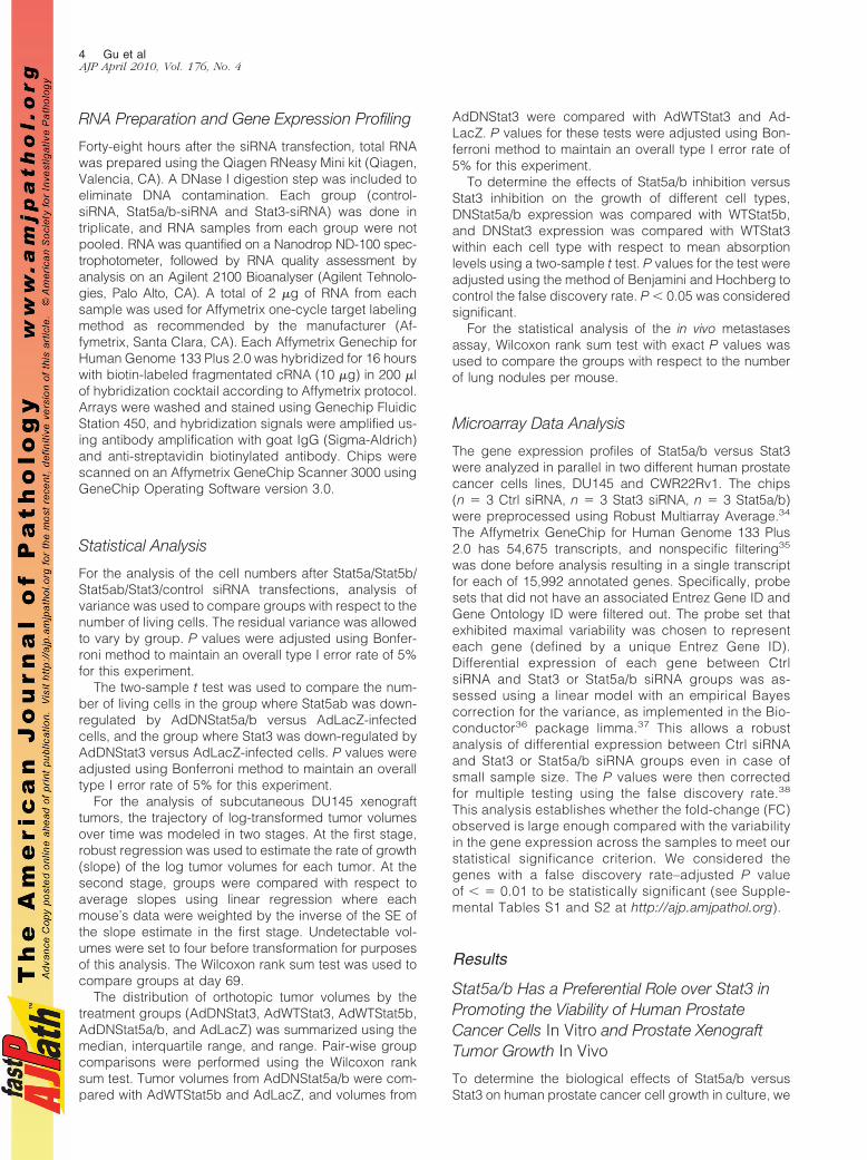
RNA Preparation and Gene Expression Profiling
Forty-eight hours after the siRNA transfection, total RNAwas prepared using the Qiagen RNeasy Mini kit (Qiagen,Valencia, CA). A DNase I digestion step was included toeliminate DNA contamination. Each group (control-siRNA, Stat5a/b-siRNA and Stat3-siRNA) was done intriplicate, and RNA samples from each group were notpooled. RNA was quantified on a Nanodrop ND-100 spec-trophotometer, followed by RNA quality assessment byanalysis on an Agilent 2100 Bioanalyser (Agilent Tehnolo-gies, Palo Alto, CA). A total of 2 �g of RNA from eachsample was used for Affymetrix one-cycle target labelingmethod as recommended by the manufacturer (Af-fymetrix, Santa Clara, CA). Each Affymetrix Genechip forHuman Genome 133 Plus 2.0 was hybridized for 16 hourswith biotin-labeled fragmentated cRNA (10 �g) in 200 �lof hybridization cocktail according to Affymetrix protocol.Arrays were washed and stained using Genechip FluidicStation 450, and hybridization signals were amplified us-ing antibody amplification with goat IgG (Sigma-Aldrich)and anti-streptavidin biotinylated antibody. Chips werescanned on an Affymetrix GeneChip Scanner 3000 usingGeneChip Operating Software version 3.0.
Statistical Analysis
For the analysis of the cell numbers after Stat5a/Stat5b/Stat5ab/Stat3/control siRNA transfections, analysis ofvariance was used to compare groups with respect to thenumber of living cells. The residual variance was allowedto vary by group. P values were adjusted using Bonfer-roni method to maintain an overall type I error rate of 5%for this experiment.
The two-sample t test was used to compare the num-ber of living cells in the group where Stat5ab was down-regulated by AdDNStat5a/b versus AdLacZ-infectedcells, and the group where Stat3 was down-regulated byAdDNStat3 versus AdLacZ-infected cells. P values wereadjusted using Bonferroni method to maintain an overalltype I error rate of 5% for this experiment.
For the analysis of subcutaneous DU145 xenografttumors, the trajectory of log-transformed tumor volumesover time was modeled in two stages. At the first stage,robust regression was used to estimate the rate of growth(slope) of the log tumor volumes for each tumor. At thesecond stage, groups were compared with respect toaverage slopes using linear regression where eachmouse’s data were weighted by the inverse of the SE ofthe slope estimate in the first stage. Undetectable vol-umes were set to four before transformation for purposesof this analysis. The Wilcoxon rank sum test was used tocompare groups at day 69.
The distribution of orthotopic tumor volumes by thetreatment groups (AdDNStat3, AdWTStat3, AdWTStat5b,AdDNStat5a/b, and AdLacZ) was summarized using themedian, interquartile range, and range. Pair-wise groupcomparisons were performed using the Wilcoxon ranksum test. Tumor volumes from AdDNStat5a/b were com-pared with AdWTStat5b and AdLacZ, and volumes from
AdDNStat3 were compared with AdWTStat3 and Ad-LacZ. P values for these tests were adjusted using Bon-ferroni method to maintain an overall type I error rate of5% for this experiment.
To determine the effects of Stat5a/b inhibition versusStat3 inhibition on the growth of different cell types,DNStat5a/b expression was compared with WTStat5b,and DNStat3 expression was compared with WTStat3within each cell type with respect to mean absorptionlevels using a two-sample t test. P values for the test wereadjusted using the method of Benjamini and Hochberg tocontrol the false discovery rate. P � 0.05 was consideredsignificant.
For the statistical analysis of the in vivo metastasesassay, Wilcoxon rank sum test with exact P values wasused to compare the groups with respect to the numberof lung nodules per mouse.
Microarray Data Analysis
The gene expression profiles of Stat5a/b versus Stat3were analyzed in parallel in two different human prostatecancer cells lines, DU145 and CWR22Rv1. The chips(n � 3 Ctrl siRNA, n � 3 Stat3 siRNA, n � 3 Stat5a/b)were preprocessed using Robust Multiarray Average.34
The Affymetrix GeneChip for Human Genome 133 Plus2.0 has 54,675 transcripts, and nonspecific filtering35
was done before analysis resulting in a single transcriptfor each of 15,992 annotated genes. Specifically, probesets that did not have an associated Entrez Gene ID andGene Ontology ID were filtered out. The probe set thatexhibited maximal variability was chosen to representeach gene (defined by a unique Entrez Gene ID).Differential expression of each gene between CtrlsiRNA and Stat3 or Stat5a/b siRNA groups was as-sessed using a linear model with an empirical Bayescorrection for the variance, as implemented in the Bio-conductor36 package limma.37 This allows a robustanalysis of differential expression between Ctrl siRNAand Stat3 or Stat5a/b siRNA groups even in case ofsmall sample size. The P values were then correctedfor multiple testing using the false discovery rate.38
This analysis establishes whether the fold-change (FC)observed is large enough compared with the variabilityin the gene expression across the samples to meet ourstatistical significance criterion. We considered thegenes with a false discovery rate–adjusted P valueof � � 0.01 to be statistically significant (see Supple-mental Tables S1 and S2 at http://ajp.amjpathol.org).
Results
Stat5a/b Has a Preferential Role over Stat3 inPromoting the Viability of Human ProstateCancer Cells In Vitro and Prostate XenograftTumor Growth In Vivo
To determine the biological effects of Stat5a/b versusStat3 on human prostate cancer cell growth in culture, we
4 Gu et alAJP April 2010, Vol. 176, No. 4

first established the phosphorylation and total proteinlevels of Stat5a, Stat5b, and Stat3 in CWR22Rv1, LNCaP,and DU145 human prostate cancer cell lines. Immuno-precipitation and immunoblotting of Stat5a/b show per-sistent activation and expression of Stat5a and Stat5b inall three cell lines (Figure 1A). Immunoprecipitation ofStat3 indicated persistent Stat3 activation in only DU145cells, whereas Stat3 protein was expressed in DU145,LNCaP, and CWR22Rv1 cells (Figure 1A).
Because persistent activation of both Stat5 and Stat3was detected in DU145 cells, we decided to use this cellline as our first model system to compare the biologicaleffects caused by inhibition of Stat5a/b versus Stat3. Wedesigned siRNAs to inhibit selectively Stat5a/b or Stat3 inDU145 prostate cancer cells. First, we verified by West-ern blotting that siRNAs targeted to Stat5a/b did notaffect Stat3 protein expression or Stat3 phosphorylation(Figure 1B). Stat3 siRNAs, in turn, did not affect the expres-sion level of Stat5a/b as demonstrated in Figure 1B. DU145cells were transfected with siRNAs against Stat5a/b or Stat3for 48 hours, with scrambled siRNA as control, and Stat5a/bwas immunoprecipitated and blotted with anti-Stat5a/bmAb. Whole cell lysates of the same samples were immu-noblotted for phosphorylated Stat3, total Stat3, and actin(Figure 1B). In the next set of experiments, DU145 cellswere transfected with siRNAs against Stat5a/b or Stat3for 72 hours, after which the cells were photographed(Figure 1, C and D), and the number of attached cellswas determined. Inhibition of Stat5ab by RNA interfer-ence resulted in a 70% decrease in attached and viableDU145 cells (P � 0.0001), whereas Stat3 inhibition de-creased the number of viable cells by 20% comparedwith cells transfected with control siRNA (P � 0.15; Fig-ure 1C). Microscope photographs indicate dead andfloating cells in the wells transfected with Stat5a/b siRNA(Figure 1D). Our previous work has demonstrated thatprostate cancer cell death induced by Stat5 inhibition isapoptotic involving extensive DNA fragmentation andCaspase-3 and �9 activation.2,9,10 In summary, theseresults demonstrate that Stat5a/b has a more importantrole than Stat3 in sustaining the viability of DU145 humanprostate cancer cells in culture.
To determine the relative importance of the two Stat5homologs for promotion of the viability of human prostatecancer cells, siRNAs specific to Stat5a or Stat5b wereintroduced to DU145 cells. The specificity of the siRNAswas verified by immunoprecipitation and Western blotting(Figure 1B). Inhibition of Stat5b by RNA interference re-sulted in almost similar decrease in the number of at-tached and viable DU145 cells compared with the groupthat was transfected with siRNAs targeting simulta-neously both Stat5a and Stat5b (P � 0.0001; Figure 1, Cand D). In contrast, inhibition of Stat5a by Stat5a siRNAsdid not affect significantly the cell viability compared withthe controls (P � 1.0; Figure 1, C and D).
As the second methodological approach, Stat5a/b orStat3 were inhibited by adenoviral (Ad) gene deliveryof dominant-negative (DN) mutants of Stat5a/b10 andStat3,33 respectively, with AdLacZ, AdWTStat5b,10
AdWTStat3,33 and mock-infected cells as controls. Thetransactivation domain was deleted from Stat5a and
Stat3 to create mutants of Stat5a/b and Stat3 that wouldinhibit transcriptional activity of Stat5a/b and Stat3, re-spectively.2,10,33 All of the adenoviral constructs werepropagated and titered side-by-side and tested by Westernblotting for transfer of equal gene expression levels for eachconstruct in human prostate cancer cells. DU145 cells wereinfected with AdWTStat5b, AdDNStat5a/b, AdWTStat3, orAdDNStat3, with AdLacZ (all at MOI 5) or mock-infectedcells as controls, and the cells were photographed and thenumber of attached cells was determined after 72 hours(Figure 1, E and F). Inhibition of Stat5a/b by adenoviralexpression of DNStat5a/b decreased the number of viableand attached DU145 cells by 87% compared with AdLacZ-infected cells (P � 0.0001), whereas inhibition of Stat3reduced the cell number by 30% (P � 0.0014; Figure 1E).Microscope photographs verify extensive cell death inAdDNStat5a/b-infected wells as evidenced by cell rounding,detachment, and shrinkage (Figure 1F). Collectively, theseresults suggest Stat5b is the key regulator of DU145 prostatecancer cell viability in culture rather than Stat5a or Stat3.
Given the preferential role of Stat5a/b in promotion ofprostate cancer cell viability in cell culture, we hypothe-sized that Stat5a/b also has a predominant role in theregulation of prostate xenograft tumor growth in nudemice. To test this hypothesis, we inhibited Stat5a/b byAdDNStat5a/b and Stat3 by AdDNStat3 in DU145 pros-tate cancer cells. Adenoviral gene delivery was per-formed 24 hours before inoculation of the cancer cellssubcutaneously into flanks of nude mice. The mice hadbeen castrated, and sustained-release DHT pellets wereimplanted to normalize the circulating androgen levels.Once tumors started to form on day 9, the tumor sizeswere measured once a week until day 69 of the experi-ment. The tumor volumes (P � 0.024) and the growth rate(P � 0.016) of prostate tumors both were significantlysuppressed in Stat5a/b-inhibited group compared withthe control group (AdWTStat5b), whereas the effects ofStat3 inhibition on the tumor volumes (P � 0.52) or thegrowth rate (P � 0.90) of subcutaneously grown prostatexenograft tumors were less consistent (Figure 2A).
In the second set of experiments, we compared theeffects of Stat5a/b inhibition versus Stat3 on orthotopicprostate tumor growth in nude mice (Figure 2B). DU145cells were infected with AdDNStat5a/b, AdWTStat5b,AdDNStat3, AdWTStat3, or AdLacZ 24 hours before in-oculation to the dorsolateral prostates of athymic nudemice. After 8 weeks, the mice were sacrificed, the uro-genital blocks were photographed, and the prostate tu-mor sizes were measured. When Stat5a/b was inhibited,the tumors were significantly smaller when comparedwith the tumors formed from AdWTStat5b-infected cells(P � 0.0032). Inhibition of Stat3 by AdDNStat3 moder-ately decreased the tumor sizes compared with theAdWTStat3-group (P � 0.112) or LacZ-group (P � 1.0;Figure 2B), but did not reach statistical significance. Inconclusion, these results suggest a preferential role forStat5a/b rather than Stat3 in promotion of subcutane-ous and orthotopic xenograft human prostate tumors innude mice.
Stat5a/b and Stat3 in Prostate Cancer Progression 5AJP April 2010, Vol. 176, No. 4

A
CWR22Rv1
LNCaP
DU145
IP:
anti-Stat5a
anti-Stat5b
anti-Stat5abanti-pYStat5ab
anti-Stat5abanti-pYStat5ab
anti-pYStat3
anti-Stat3
anti-actin
anti-Stat5abStat5aStat5b
WCL:
WB:anti-Stat5ab
anti-pYStat3
anti-Stat3
anti-actin
anti-Stat5ab
B
anti-Stat5a
anti-Stat5b
Stat5a
Stat5b
Stat5a
b
Control
Medium
Stat3
Control
siRNA (100 pmole)
WCL
WCL
WCL
IP:
0.0
0.5
1.0
1.5
2.0
2.5
Num
ber o
f aliv
e ce
lls(x
106 c
ells
)
Medium
Stat5a-siRNA
Stat5b-siRNA
Ctrl-siRNA
Stat5ab-siRNA
C
Medium Ctrl-siRNA
Stat3-siRNA
Stat5a-siRNA Stat5b-siRNA
D
Stat3-siRNAStat5ab-siRNA
0
0.2
0.4
0.6
0.8
1.0
1.2
Num
ber o
f aliv
e ce
lls(x
106 c
ells
)
Mock
AdWTStat5b
AdDNStat5abAdLacZ
AdWTStat3
AdDNStat3AdDNStat3AdWTStat3
AdWTStat5b AdDNStat5ab
Mock AdLacZ
E F
Figure 1. Inhibition of Stat5a/b caused a robust decrease in the number of viable DU145 human prostate cancer cells, whereas the effects of Stat3 inhibition wereless pronounced. A: Transcription factors Stat5 and Stat3 are both constitutively active in DU145 prostate cancer cells. Stat5a or Stat5b were immunoprecipitated(IP) from exponentially growing CWR22Rv1, LNCaP, and DU145 cells using anti-Stat5a or anti-Stat5b pAbs, and blotted (WB) with anti–phospho-Stat5a/b(anti-pYStat5a/b) antibody, as indicated. Whole cell lysates (WCL) were blotted with anti-phospho-Stat3 (anti-pYStat3) antibody. Filters were stripped andre-blotted with anti-Stat5ab or anti-Stat3 mAbs, and whole cell lysates before IP were immunoblotted with anti-actin pAb. B: To verify the specificity of the siRNAs,DU145 cells were transfected with siRNA targeted to Stat5a, Stat5b, or Stat3, with scrambled siRNAs or nontransfected cells as controls. The cells were harvested48 hours after the siRNA transfection. Stat5a and Stat5b were immunoprecipitated with anti-Stat5a and anti-Stat5b pAbs, respectively, and blotted with anti-Stat5a/bmAb, as indicated. Whole cell lysates (WCL) of the same samples were blotted with anti–phospho-Stat3 (anti-pYStat3), anti-Stat3, or anti-actin pAbs. C: Inhibitionof Stat5b by RNA interference decreased the number of viable DU145 cells, whereas the effects of Stat5a or Stat3 inhibition were less evident. (i) The number oflive cells after inhibition of Stat5a, Stat5b, Stat5a/b, or Stat3 by specific siRNAs, control group was incubated with transfection reagent only. After trypan blueexclusion, the attached viable DU145 cells were counted 72 hours after the siRNA transfection. The bars indicate mean � SD of triplicate wells. D: Microscopephotographs of the DU145 cells 72 hours after the transfection show the cell morphology. siRNAs targeted to Stat5b or Stat5a/b induced massive detachment androunding of DU145 cells, whereas Stat5a or Stat3 siRNA caused hardly detectable effects on the cell numbers or the cell morphology. E: Inhibition of Stat5a/b byadenoviral (Ad) gene delivery of a dominant-negative (DN) Stat5a/b decreased significantly the number of viable prostate cancer cells, whereas Stat3 inhibitioneffects were only minor. DU145 cells were infected with AdWTStat5b, AdDNStat5a/b, AdWTStat3, AdDNStat3, or with AdLacZ at MOI 5 using mock-infected cellsas an additional control. After trypan blue staining, the cells were photographed and counted after 72 hours. The bars indicate mean � SD of triplicate wells.F: Microscope photographs demonstrate the cell morphology after 72 hours infection of the cells with AdDNStat5a/b, AdWTStat5b, AdDNStat3, AdWTStat3 versusAdLacZ or mock-infected cells. Inhibition of Stat5a/b induced detachment, rounding, and shrinkage of the cells, which are all characteristics of apoptosis.
6 Gu et alAJP April 2010, Vol. 176, No. 4

A
AdDNStat5ab AdWTStat5b AdWTStat3AdDNStat3
AdDNStat5ab AdWTStat5bAdWTStat3
AdDNStat3
Tum
or v
olum
e (m
m3 )
1 2 3 4 5 6 7 1 2 3 4 5 6 7 1 2 3 4 5 6 7 1 2 3 4 5 6 7
69625541342320
9
Day
s
900.0
800.0
700.0
600.0
500.0
400.0
300.0
200.0
100.0
0.0
13
Tum
or v
olum
e (m
m3 )
AdWTStat3 AdDNStat5ab AdWTStat5b AdLacZAdDNStat3
B
0
200
400
600
800
1000
1200
AdWTStat3 AdDNStat5ab AdWTStat5b AdLacZAdDNStat3
C
0 0.2 0.4 0.6 0.8 1 1.2
RC170N
RC165N
PC3
CWR22Rv1
Ad-DNStat3Ad-WTStat3Ad-DNStat5abAd-WTStat5b
Absorbance (490 nm)
Prostate carcinoma
Prostate carcinoma
Normal prostate epithelium
Normal prostate epithelium
LNCaPProstate carcinoma
Ad-DNStat3Ad-WTStat3Ad-DNStat5abAd-WTStat5b
Ad-DNStat3Ad-WTStat3Ad-DNStat5abAd-WTStat5b
Ad-DNStat3Ad-WTStat3Ad-DNStat5abAd-WTStat5b
Ad-DNStat3Ad-WTStat3Ad-DNStat5abAd-WTStat5b
D
0 0.4 0.8 1.2 1.6 2.0
T47D
MCF-7
CAPAN
Hs766T
Absorbance (490 nm)
Pancreatic carcinoma
Pancreatic carcinoma
Breast carcinoma
Breast carcinoma
Ad-DNStat3Ad-WTStat3Ad-DNStat5abAd-WTStat5b
Ad-DNStat3Ad-WTStat3Ad-DNStat5abAd-WTStat5b
Ad-DNStat3Ad-WTStat3Ad-DNStat5abAd-WTStat5b
Ad-DNStat3Ad-WTStat3Ad-DNStat5abAd-WTStat5b
A549
HCT116
HepG2
A2058
Hepatocellularcarcinoma
Colorectal carcinoma
Lung carcinoma
Skin melanoma
Ad-DNStat3Ad-WTStat3Ad-DNStat5abAd-WTStat5b
Ad-DNStat3Ad-WTStat3Ad-DNStat5abAd-WTStat5b
Ad-DNStat3Ad-WTStat3Ad-DNStat5abAd-WTStat5b
Ad-DNStat3Ad-WTStat3Ad-DNStat5abAd-WTStat5b
E
RC16
5NRC
170N
A205
8He
pG2
HCT1
16A5
49
Hs76
6TCA
PAN
MCF
-7T4
7DIP:
anti-Stat5a
anti-Stat5b
WB:
anti-Stat5ab
anti-Stat5ab
anti-actin
anti-Stat3WCL
anti-pYStat5ab
anti-pYStat3
anti-pYStat5ab
Figure 2. Inhibition of Stat5a/b, not Stat3, has a major effect in blocking DU145 prostate cancer subcutaneous and orthotopic xenograft tumor growth in athymicnude mice. A: Subcutaneous tumor growth of DU145 prostate cancer cells infected with adenovirus (Ad) expressing DNStat5a/b, WTStat5b, DNStat3 or WTStat3.Twenty-four hours after the infection with AdDNStat5a/b, AdWTStat5b, AdDNStat3, or AdWTStat3 at MOI 5, the cells were inoculated subcutaneously into flanksof castrated athymic nude mice supplied with sustained-release 5�-dihydrotestosterone (DHT)-pellets (n � 7 per group, 1 tumor per mouse, 20 � 106 DU145 cellsper site, 1 DHT pellet/mouse). The tumor growth was measured once a week for 69 days. Tumor volumes were calculated using the formula V � (�/6) � d1�(d2)
2, with d1 and d2 being two perpendicular tumor diameters. B: Orthotopic growth of DU145 prostate cancer cells infected with adenovirus (Ad) expressingDNStat3, WTStat3, DNStat5a/b, WTStat5b, or LacZ. DU145 cells were inoculated orthotopically in the dorsolateral prostates of athymic nude mice (1 � 106 DU145cells per injection) supplied with DHT-pellets (1 pellet per mouse). Prior (24 hours) to the injection, the DU145 cells had been infected with AdDNStat5a/b,AdWTStat5b, AdDNStat3, AdWTStat3, or AdLacZ at MOI 5. Mice were sacrificed 8 weeks after tumor cell inoculation, and the tumor volumes were calculated usingthe formula V � (�/6) � d1� (d2)
2, with d1 and d2 being two perpendicular tumor diameters. C: Inhibition of Stat5a/b does not affect the viability of normalhuman prostate epithelial cell lines, but caused a decrease in the number of viable LNCaP and CWR22Rv1 cells. Normal prostate epithelial cell lines, RC165N andRC170N, and human prostate cancer cell lines, LNCaP, CWR22Rv1, and PC-3, were infected with AdWTStat5b, AdDNStat5a/b, AdWTStat3, AdDNStat3, or AdLacZat MOI 5. The cell viability was determined 72 hours after the adenoviral gene delivery by MTS (3-�4,5-dimethylthiazolyl-2��2,5-diphenyl-tetrazolium bromide)metabolic activity assay. D: Melanoma (A5028), hepatocellular (HepG2), colorectal (HCT116), lung (A549), pancreatic (Hs766T and CAPAN), and breast (MCF-7and T47D) cancer cell lines were infected with AdWTStat5b, AdDNStat5a/b, AdWTStat3, AdDNStat3, or AdLacZ at MOI 5, and the cell viability was determined72 hours after the adenoviral gene delivery by MTS assay. The bars indicate mean � SD of triplicate determinations. E: Stat5a and Stat5b were immunoprecipitatedusing anti-Stat5a and anti-Stat5b pAbs, respectively, and immunoprecipitates and whole cell lysates (WCL) were blotted with anti-Stat5a/b mAb or anti-Stat3 pAb,as indicated. Anti-actin immunoblotting of whole cell lysates indicates equal level of proteins in the immunoprecipitations.
Stat5a/b and Stat3 in Prostate Cancer Progression 7AJP April 2010, Vol. 176, No. 4

Inhibition of Stat5a/b or Stat3 Does Not Affectthe Viability of Normal Human Prostate EpithelialCells
We next tested the biological effects of Stat5a/b or Stat3inhibition on the growth of other human prostate cancer celllines LNCaP, CWR22Rv1, and PC-3. LNCaP andCWR22Rv1, which are both Stat5a/b and Stat3-positive,responded to Stat5a/b inhibition by significant reduction ofviable cells (LNCaP; P � 0.0163, CWR22Rv1; P � 0.0058),but did not respond (LNCaP; P � 0.5532) or respondedonly moderately to Stat3 inhibition (CWR22Rv1; P �0.0213). Inhibition of Stat5a/b or Stat3 did not affect theviability of PC-3 cells, which do not express Stat5a/b orStat3 because of gene deletions,39 as expected (Fig-ure 2C).
To determine whether transcription factors Stat5a/b orStat3 are critical for the viability of normal prostate epi-thelium, RC165N and RC170N normal human prostateepithelial cell lines32 were infected with adenovirusesexpressing DNStat5a/b, DNStat3, WTStat5b, or WTStat3(Figure 2C). RC165N and RC170N have been estab-lished from benign prostate tissue, and both cell lineshave been shown to express cytokeratin CK8, PSA, AR,and NKX3.1 characteristic to prostate luminal secretorycells.32 Importantly, neither of the nonmalignant prostateepithelial cell lines responded to inhibition of Stat5a/b orStat3 by decreased cell viability (Figure 2C). However,the Stat3 protein expression in the nonmalignant prostateepithelial cell lines was below the detection level by West-ern blotting. Although Stat5b protein expression was de-tected in nonmalignant prostate epithelial cell lines andStat5a expression was below the detection level with ourantibodies, phospho-Stat5 immunoblotting indicatedweak Stat5a phosphorylation in the RC165N and RC170Ncells (Figure 2E).
Because transcription factor Stat5a/b may provide a ther-apeutic target protein for prostate cancer, we next aimed toestablish how specific the effects of Stat5a/b inhibition areon the viability of prostate cancer cells compared with othercancers (Figure 2D). We first assessed the frequency ofconstitutive activation of Stat5a/b and Stat3 by immunohis-
tochemistry in paraffin-embedded specimens of breast, co-lon, lung cancers, and melanomas (Table 1). Stat5a/b andStat3 were in the active state in the majority of breast can-cers (Stat5a/b 67%; Stat3 91%), and melanomas (Stat5a/b59%; Stat3 50%), whereas 37% colon cancers expressedactivated Stat5a/b and 100% active Stat3. Furthermore,87% of lung cancers expressed active Stat5a/b and 22%active Stat3 (Table 1). Stat5a/b or Stat3 inhibition byAdDNStat5a/b or AdDNStat3, respectively, did not sup-press the viability of breast (MCF-7, T47D), melanoma(A2058), hepatocellular (HepG2), colorectal (HCT116),pancreas (Hs766T, CAPAN), or lung cancer (A549) cells(Figure 2D). Western blotting of immunoprecipitated Stat5aand Stat5b indicated Stat5a/b expression in all of the celllines except HCT116 and PC-3 cells,2 whereas Stat3 wasexpressed in all cancer cell lines (Figure 2E). Taken to-gether, these results suggest that Stat5b inhibition leads tocell death specifically in prostate cancer cell lines, but not incell lines established from normal prostate epithelium.Moreover, the data suggest that although Stat5a/b may beconstitutively active in cancers originating from other tissuetypes than prostate, its functional significance in those can-cers is different from in prostate cancer.
Stat3 Is the Key Inducer of ExperimentalMetastases Formation of Human ProstateCancer Cells, whereas the Effects of Stat5a/bon Induction of Metastases Nodules WereModest
We recently showed that Stat3 induces a robust increasein lung metastases formation of human prostate cancerafter tail-vein injections in athymic nude mice.31 There-fore, we next compared the effects of Stat5a/b to Stat3 inpromoting the in vivo metastasis formation of human pros-tate cancer cells. We performed an experimental metas-tases assay by infecting DU145 cells with adenovirusesexpressing WTStat5a, WTStat5b, WTStat3, or LacZ atMOI 5. Twenty-four hours after the adenoviral gene de-livery, we injected DU145 cells in athymic nude micethrough the tail veins. The lungs were harvested after 8
Table 1. Frequency of Stat5a/b and Stat3 Activation in other Cancer Types than Prostate Cancer
Number of patients % Number of patients %
Breast cancer Breast cancerStat5a/b activation status 30 100 Stat3 activation status 34 100Negative 10 33 Negative 9 9Positive 20 67 Positive 31 91
Colon cancer Colon cancerStat5a/b activation status 16 100 Stat3 activation status 19 100Negative 10 63 Negative 0 0Positive 6 37 Positive 19 100
Lung cancer (squamous) Lung cancer (squamous)Stat5a/b activation status 16 100 Stat3 activation status 18 100Negative 2 13 Negative 14 78Positive 14 87 Positive 4 22
Melanoma MelanomaStat5a/b activation status 17 100 Stat3 activation status 20 100Negative 7 41 Negative 10 50Positive 10 59 Positive 10 50
8 Gu et alAJP April 2010, Vol. 176, No. 4

weeks and stained with India Ink, bleached with Feketesolution, and scored for surface lung metastases. Asdemonstrated in Figure 3A, the number of metastases inmice injected with DU145 cells infected with AdWTStat3was increased by approximately 50-fold when comparedwith mice injected with DU145 cell that had been infectedwith either AdLacZ. In contrast, WTStat5a increased thenumber of metastases by only fivefold and WTStat5b byfourfold in comparison with DU145 cells expressingLacZ. Quantitatively, injection of DU145 with AdWTStat3cells resulted in an average of 137 (SEM � 9.5) metas-tases per lung, as compared with 2.6 per lung usingDU145 cells infected with AdLacZ (SEM � 1.2) orAdWTStat5a (mean, 13, SEM � 6.5) or WTStat5b (mean,9, SEM � 2.4). Figure 3B demonstrates visually the whitemetastasis nodules in the lungs of nude mice. In conclu-sion, the data suggest that Stat3, not Stat5a/b, has apredominant role in promoting metastatic disseminationof human prostate cancer cells in an in vivo experimentalmetastases assay. This is the first side-by-side compar-ison of Stat5a/b to Stat3 in promotion of metastatic po-tential of human prostate cancer cells in vivo.
Gene Expression Profiles Regulated by Stat5a/bVersus Stat3 in Human Prostate Cancer Cells
To assess gene expression profiles of Stat5a/b and Stat3in human prostate cancer cells, we inhibited Stat5 gene
expression 48 hours before the gene expression analysisby Stat5a/b siRNA and Stat3 gene expression by Stat3siRNA using scrambled siRNA as control (Ctrl) in twodifferent human prostate cancer cell lines, DU145 andCWR22Rv1 (Figure 4A). Overall, 1409 genes in DU145cells and 1344 genes in CWR22Rv1 cells were differen-tially expressed between Ctrl siRNA versus Stat5a/b andStat3 siRNA groups using false discovery rate � 0.01 onthe full dataset of 15,992 genes (See Supplemental Ta-bles S1 and S2 at http://ajp.amjpathol.org).
Importantly, only 15% of the 1409 genes in DU145cells were regulated by both Stat5a/b and Stat3, whereas25% were regulated by Stat3 only and 60% were regu-lated by Stat5 only (Figure 4B). Similarly, only 11% of the1344 genes in CWR22Rv1 cells were regulated by bothtranscription factors, whereas 30% were regulated byStat3 only and 59% by Stat5a/b only (Figure 5A). Todefine functional groups within the Stat5a/b- and Stat3-regulated genes, we used the descriptions from the GeneOntology annotations as our tool. The Gene Ontology isconstructed in a hierarchical manner with categories cor-responding to each gene ontology identifier (GO ID) be-ing potentially subdivided into more precise subcatego-ries, each with its own GO ID. “The metastasis group” ofgenes was defined as a set of 88 GO IDs and theirsubcategories (a total of 1975 GO IDs) corresponding togenes encoding proteins related to metastases pro-cesses. Correspondingly, “the apoptosis group” of geneswas defined as a set of 21 GO IDs and their subcatego-ries (a total of 469 GO IDs) and “the proliferation group”as a set of 47 GO IDs and their subcategories (a total of943 GO IDs). Somewhat surprisingly, in each of our threecomparisons (the genes regulated by Stat5 only, Stat3only, or by both Stat5a/b and Stat3), the majority of thedifferentially expressed genes in both DU145 (n � 1409)and CWR22Rv1 cells (n � 1344) between Ctrl siRNA andStat5a/b or Stat3 siRNA groups were metastases-related.This was followed by genes related to proliferation,whereas the minority of the genes were apoptosis-related(Figures 4C and 5B).
The heatmaps demonstrate a hierarchical clustering ofthe expression values of the most differentially expressedgenes (as determined by the smallest P values) related tometastases, apoptosis or proliferation for the genes reg-ulated by Stat5 only (Figure 4D; DU145, Figure 5C;CWR22Rv1), Stat3 only (Figure 4E; DU145, Figure 5D;CWR22Rv1), or the genes regulated by both Stat5a/band Stat3 (Figure 4F; DU145, Figure 5E; CWR22Rv1).Red represents higher expression and green representslower expression in the heatmaps, and the genes (rows)are re-ordered based on hierarchical clustering using thecorrelation metric. The heatmaps clearly illustrate theunique gene expression patterns of Stat5a/b versus Stat3in both DU145 and CWR22Rv1 prostate cancer cell lines.Specifically, the majority of the genes that are up-regu-lated for example by Stat5a/b, and thus coded by red,are either down-regulated or not altered in the groupswhere Stat3 expression was selectively inhibited (Figures4 and 5). In addition, the heatmaps (Figures 4 and 5)demonstrate the genes related to metastases, apoptosis,or proliferation, which are similarly regulated by both
AdWTStat3 AdLacZ
num
ber o
f lun
g no
dule
s /m
ouse
AdWTStat3
AdWTStat5a
AdLacZ
2 50
60
127
165
0 0
15
44
07
0 2
49
8
24
4 70
13 101
13
12612
8
0
20
40
60
80
100
120
140
160
180
1 2 3 4 5 1 2 3 4 1 2 3 4 5 6 7 8 9 1 2 3 4 5 6 7 8 9
AdWTStat5a AdWTStat5b Mean:S.E.M.
2.6 136.5 13 9
1.2 9.5 6.5 2.4
AdWTStat5b
A
B
Figure 3. Stat3 has a preferential role in promoting prostate cancer metas-tases formation in an experimental metastases assay. A: Athymic nude micewere injected with DU145 cells infected with adenovirus expressing LacZ,WTStat3, WTStat5a, or WTStat5b at MOI 5 (1 � 106 cells per mouse) throughthe tail-vein. After 8 weeks, the lungs were harvested and stained with IndiaInk, bleached with Fekete’s solution, and scored for surface lung metastases.B: Representative photographs of India Ink–stained lungs derived fromathymic nude mice injected with LacZ, WTStat3, WTStat5a, or WTStat5bexpressing DU145 cells after 8 weeks.
Stat5a/b and Stat3 in Prostate Cancer Progression 9AJP April 2010, Vol. 176, No. 4

AStat3 Stat5
14583
853348 208
DU145
DU145 cell line
Percentage of all significant genes
Proliferation
Apoptosis
MetastasisStat3 and Stat5
Apoptosis
MetastasisStat3 only
Proliferation
Apoptosis
MetastasisStat5 only
0 2 4 6 8 10 12
ProliferationDU145 CWR22Rv1
Ctrl-siRNA
Stat3-siRNA
Stat5a/b-siRNA
Ctrl-siRNA
Stat3-siRNA
Stat5a/b-siRNA
Stat5a/b
Stat3
Actin
WCL
B C
D
Stat
5 si
RN
A #
1St
at5
siR
NA
#2
Stat
5 si
RN
A #
3St
at3
siR
NA
#1
Stat
3 si
RN
A #
2St
at3
siR
NA
#3
Ctr
l #1
Ctr
l #2
Ctr
l #3
HHEX SFN SPHK1 FBXO5 PRDM4 NEK11 ASNS AURKC TFAM KLF4 PLAGL1 RPA2 DNAJB9 IFI16 PDCD4 RBMS1 CCND1 CETN3
LIF INSIG1 TSPAN1 CUL4B IL31RA LRP5 CAMK2D CHPT1 NUP62 KRT4 MAP2K6 PTPN6 IFI16 RHOT1 ASNS PLAGL1 APAF1 TP53 KIAA1967 ATG12 SFN SPHK1 PTPN6 NUP62 HSP90B1 BCL2L2 TIA1 PIK3R2 YWHAZ TIAF1 SORT1 NME3 TP53 S100P SYNE1 NFATC1 TUBGCP3 CASP8 PLSCR1 HMOX1
INA ASNS AOC3 RHOT1 RCAN1 IRF9 MX1 NRTN SFN PRDM4 RRAGC EDG7 HMGB2 MIER1 NUDT21 MAP3K7IP2 G3BP2 NEK11 VLDLR STAM RBM9 C1GALT1 DDAH2
IVL CETN3 FGD4 ZDHHC13 PTPRG IL13RA1 NDFIP1 CSNK2A1 PAG1 INADL SCEL RAB1A PRKCA NRG2 MFGE8 LRP5 DDR1 KIF5C TSPAN1 ABLIM1 VEGFC GPR39 RAB5B CALM3 GMDS CLIP2 ARF3 EPOR RAB31 NES PARD3 GGCX FBXO8 MAP2K4 YWHAZ BNIP3L KATNB1 VCL BCAM CPLX1 NLRP1 PTPN6 IGFBP4 RTN1 GRB14 DYNLL1 F11R CLIC3 PCTK3 FLNB GJA1 PTGES TACSTD2
RRAGCC13orf15CCNE2HMGB2ERN1TP53CDK2MLH1FRAT2TUBE1CDK5R1BTG2WARSOSGIN1PARD3PMS2L5NF2TGFBR1SMC2SLC29A2DLG3CCNG2CAPN1MST1RCD24RBM9IGFBP4TACSTD2EMP1LRPAP1PIM2MX1RIPK1KLF4CDK5R1ERN1CEBPGCASP8CASP2RRAGCNLRP1EMP1BAG1IL31RAPRKCAPDCD4BNIP3LNRG2CD24DDAH2CD44AURKCFGF12RIPK1SQSTM1MED30CDK5R1ADMGCH1GALSCYL3TUBE1MX2IFI16PARP9CASP2THBDERN1SPHK1HHEXAPAF1FBXO5PLSCR4MAP3K4RAC1WASF1MAML1GRK5PCDHA10RALGPS1IGF2RBMPR1APRKAR2BCGNWNT7BOPN1SWOXTRLIFEXT2CREBL2AKAP12PIP5K1ATGFBR1STAT5BCDONCD24S100A2TIAF1MST1RGNB1PDE9ASORT1RAB27ACOL12A1APPBP2MAPKAPK3NR2F6IL31RACOL6A1RND2PKD2MAP3K13ARHGAP29SOS2TFPINF2TMED10BTRCGPR56CELSR1NUP62TSPAN6SPOCK2BAG1SPTAN1MAP2K6CLDN7PPLPTPRJFADS1GPRC5AGPERS100A4KRT4
Proliferation
Apoptosis
Metastasis
DU145 cells:Stat5a/b-regulated genes
E
Stat
5 si
RN
A #
1St
at5
siR
NA
#2
Stat
5 si
RN
A #
3St
at3
siR
NA
#1
Stat
3 si
RN
A #
2St
at3
siR
NA
#3
Ctr
l #1
Ctr
l #2
Ctr
l #3
AFAP1L2TNFRSF11ATNFRSF9CKLFGNG4CCNG1CDK6CDC23DLG5ARTNZFP42NAMPTFMR1HDGFCDC25BLIFRCCNA1SMUG1VEGFAEMG1CCDC88APAWRSERTAD2SGPL1BCL2A1C16orf5TNFRSF10BNFKBIAMYBL2OPTNTNFRSF9MAGI3SOCS3MAEAEI24DOCK1DAPK1GLO1PAWRVEGFASRGNCASP4TP53I3BCL2L11RTN4ARHGAP26PAK2AFAP1L2IL32NFKBIACEP63LTBRASA2TGM2CKLFTNFRSF11ACDK6LMNB1ARL4CCLDN12OPTNCSF2LTBP2C16orf5NR2F1TNFRSF10BDLG5SPRED2RAB3BGNG4PAK1RIN2ERAP1KIF3BOPN3SDCBPEML2EIF2AK3TAX1BP3LPXNF2RL2NAMPTDOCK1WASF3SRP72NR0B1MAEAGMFBIFNGR1PTGES3SOCS3CREBBPDAPK1STAT3SERPINH1BCL7BPRMT2HDGFMBPEFNA3MAGI3NPTNARTNCCNA1PIP5K3LIFRTBL1XPCDHB2RTN4RAB18CCDC88ADERL1EPHA4RABL2BVEGFA
Proliferation
Apoptosis
Metastasis
DU145 cells:Stat3-regulated genes
Stat
5 si
RN
A #
1St
at5
siR
NA
#2
Stat
5 si
RN
A #
3St
at3
siR
NA
#1
Stat
3 si
RN
A #
2St
at3
siR
NA
#3
Ctr
l #1
Ctr
l #2
Ctr
l #3
CDC6
CUZD1
TCF19
CDC34
FEN1
LAMC1
MDFIC
NAP1L1
HDAC4
RRM2
RBPJ
POLE3
FGFBP1
CITED2
FOXN3
GPNM
MAPRE2
TGFB2
ZAK
TSPAN2
MPHOSPH9
CASP7
DYRK2
TGFB2
ZAK
CYFIP2
CD14
CUZD1
MAP3K3
PCGF5
EPB41L4B
RAB8B
HMMR
LAMC1
MDFIC
MAPK9
GPR177
RHOQ
CYLD
CTNNB1
LEPR
FEN1
SYPL1
SRD5A1
RAP2B
OXSR1
GOLT1B
CASP7
PDCL
DYRK2
PVRL3
PLXNC1
CDH1
CITED2
SYT1
FOXN3
ENPP2
MAPRE2
CDH3
CYFIP2
CALD1
RAB4B
CD14
LAT2
NDFIP2
PDE5A
WISP2
TCF7L2
MAP3K12
TNNC1
TGFB2
ITPR1
FGFBP1
LANCL1
ZAK
CDS1
SEMA4F
TSPAN2
UTRN
THBS1
IFNAR1
Proliferation
Apoptosis
Metastasis
DU145 cells:Stat3- and Stat5a/b-regulated genes
F
Figure 4. Identification of Stat5 and/or Stat3-regulated genes in DU145 human prostate cancer cells. A: Inhibition of Stat5a/b or Stat3 protein expression humanprostate cancer cells. DU145 and CWR22Rv1 cells were transfected with siRNA targeted to Stat5a/b or Stat3, with scrambled siRNA as control. Cells were harvested48 hours after the transfection, lysed, and immunoblotted with anti-Stat5a/b mAb or anti-Stat3 pAb, as indicated. The lower parts of the filters were blotted withanti-actin antibody to demonstrate equal loading (bottom panel). B: Venn-diagrams of Stat5a/b- and/or Stat3-regulated genes in DU145 human prostate cancercells. C: The relative proportions of Stat5a/b versus Stat3 vs. Stat5-Stat3–regulated genes in DU145 prostate cancer cells related to metastases, proliferation, orapoptosis out of total of 1409 genes differentially expressed between control siRNA and Stat5a/b or Stat3 siRNA using false-discovery rate �0.01 on the full datasetof 15,992 genes. The functional groups were identified using the Gene Ontology annotations. D: A heatmap showing the top differentially expressed genes byStat5a/b (determined by the smallest P values) related to metastases, apoptosis, or proliferation. Red represents higher expression, whereas green represent lowerexpression in the heatmap. For comparison, the levels of the gene expression for the corresponding genes in the groups where Stat3 was inhibited are shownin the middle of the heatmap. E: A heatmap showing the top differentially expressed genes by Stat3 (determined by the smallest P values) related to metastases,apoptosis, or proliferation. Red represents higher expression, whereas green represent lower expression in the heatmap. For comparison, the levels of the geneexpression for the corresponding genes in the groups where Stat5a/b was inhibited are shown in the left side of the heatmap. F: A heatmap showing the topdifferentially expressed genes by Stat5a/b and Stat3 (determined by the smallest P values) related to metastases, apoptosis, or proliferation. Red represents higherexpression, whereas green represent lower expression in the heatmap.
10 Gu et alAJP April 2010, Vol. 176, No. 4

Stat5a/b and Stat3 (See Supplemental Tables S1 and S2at http://ajp.amjpathol.org). In both cell lines, inhibition ofStat5a/b led to down-regulation of PDCD4, a protein thatmediates the inhibition of neoplastic transformation.40 In
addition, inhibition of Stat5a/b induced in both cell linesthe expression of Kruppel-like factor 4, which is a knownrepressor of CyclinD1 and mediates growth suppres-sion.41,42 VEGF-C and collagen types VI and XII were
A
Stat3 Stat5
14648
786 407 151
CWR22Rv1 cell line
Percentage of all significant genes
Proliferation n
Apoptosis
Metastasis
Stat3 and Stat5
Proliferation n
Apoptosis
Metastasis
Stat3 only
Proliferation n
Apoptosis
Metastasis
Stat5 only
0 2 4 6 8 10
B
Stat
5 si
RN
A #
1 St
at5
siR
NA
#2
Stat
5 si
RN
A #
3 St
at3
siR
NA
#1
Stat
3 si
RN
A #
2 St
at3
siR
NA
#3
Ctr
l #1
Ctr
l #2
Ctr
l #3
ERBB3 GRN KLF4 NRP1 RHOB BTG2 PTCH1 PAM SEPT6 FSCN1 FTH1 CALCA CHFR ILF3 CDC23 CDKN2D CAMK2D TXNDC1 CETN3 ARHGEF2 PDCD4 HSPA2 BRCA2 BCCI P BMPR2 PRIM2 CITED2 CCND3 POLA1 BUB1B SMC2 TUSC2 TYMS POLD3 CUL4B BAG1 CD24 TNFRSF25 TNFRSF19 DUSP22 ERBB3 KLF4 RHOB ATG12 CDKN2D IFRD1 PRKCA RNF7 BIRC5 BCL6 CEBPG IKIP IL6R NRP1 PAK1 RHOB RXFP3 EEF1A1 EPPK1 ERBB3 STAT6 PDIA2 TRHDE MIER1 ABCA1 RPS6KA1 VASP DSCAM BAG1 S100P IL10RB CD24 EPHA4 PLA2G4C BASP1 DMD BCAM ATP6AP2 TNFRSF19 ADRB2 CLDN3 CEACAM6 RIMS1 NI N RAB31 C1GALT1 ITGA1 RANBP1 TXNDC1 RGS4 NUDT4 RAB1A ABI 2 CETN3 NAMPT CSNK2A1 ASNS BIRC5 PAFAH1B1 CLIC4 SOS2 RABI F PCDHA10 TYMS BUB1B RAB5B TXNRD1 ARHGAP29 CITED2 CDC27 CHEK1 PCDHA6 CREBL2 BRCA2 CLIP1 CALM3 PRKCA ALDH1A1 HIP1 STC2 CALCA EIF5 GNAS PPP1R12A GRB14 PIP5K1A ERBB4
TGFB1 SESN1 TCFL5 TMEM97 IL6R ZEB1 CD24 GPNMB POP1 SH3BP4 MXD4 DUSP22 CSRP2 ERBB4 DNAJB9 IHPK2 DLG3 EML4 NAMPT RBMS1 TGFBR1 STRN3 ASNS HELLS AREG BIRC5 CLIP1 CDC27 PAFAH1B1 BCL6 EXO1 POLA2 ISG20 CHEK1 WHSC1L1 CCNG2 ANXA1 FAS BI D PDIA2 ANXA4 RRAGA TNFRSF10B TGFB1 IHPK2 TIA1 TXNDC1 HIP1 FEM1B PDCD4 BCL2L2 ASNS IMPA2 TNFRSF10B INPP5A HIP1R TG RHOQ F11R ANXA4 RRAGA LITAF DUSP22 CLIC3 ITPR3 TGFB1 MTSS1 HMOX1 MCC ANXA1 NQO1 CSNK1D FAS CDC42EP3 GPR143 PTCH1 BI D TNFRSF25 FSCN1 HLA−CGRN SQSTM1 ARHGEF2 PAG1 VLDLR TGFBR1 AREG TFG EML4 RAB4A IL13RA1 BMPR1A BMPR2 PRKAR2B STAT5B SLC20A1 MLLT4 DUSP2 CORO1C PLSCR1 STAM DUSP6 NUDT1 SPRED1 OPN1SW TUSC2 PKD2 APPBP2 TMED10 GNB1 RAB22A BTRC MAP2K4 SNX6 CD2AP KLRA1 OGT KIF5C MAP2 CDC42 HBEGF SAR1A SEPT11 NLGN1 GRB10 DSC2 CAMKK2
Proliferation
Apoptosis
Metastasis
CWR22Rv cells: Stat5a/b-regulated genes
C
Stat
5 si
RN
A #
1 St
at5
siR
NA
#2
Stat
5 si
RN
A #
3 St
at3
siR
NA
#1
Stat
3 si
RN
A #
2 St
at3
siR
NA
#3
Ctr
l #1
Ctr
l #2
Ctr
l #3
SEPT7 CLEC11A TAF5 CUL3 NUP98 PNKP TRIB1 NCAPD2 CDT1 TP53 HMGB2 UBE2C ING1 ERN1 HOXC10 RASSF1 IL8 DLG7 CUL1 PPP2R3B KIF11 CENPE CDC6 EVI 5 CDKN1B KLF10 KLF6 BTC LIFR POLE3 ZNF239 PRLR ERN1 CUL1 TP53 MTL5 ANXA5 CUL3 CASP7 CDKN1B SGPL1 DYRK2 RTN4 GLO1 MAEA FOXO3 HTATIP2 SNCA RP6−213H19.1 RAPGEF2 ANXA5 STAT2 HMMR EPAS1 CGN CTNND1 LGR5 ROCK1 ITGB8 RAB8B TNNI3 NLK ERAP1 GPER TLE3 LMNB1 CCR10 HMGB2 UBE2C KIF11 ERN1 IL8 PRLR CRIPAK GSG2 TP53 S100A10 DLG7 CLDN12 TROAP EPB41L4B RASSF1 NRTN FARP1 PDCL TRAF2 NARF MAP3K7IP2 DYRK2 KLHL20 CASP7 GJC1 SEPT7 IPO7 EIF2AK3 SYPL1 KLF10 PVRL3 DUSP10 SPOCK2 RAB18 GRIA2 CLDN1 CNTN1 PLAUR ITGAV GPR85 TSGA14 KLK3 PTGES3 HTATIP2 FEZ2 STAT3 GRM 3 GPR177 LIFR DERL1 PIP5K3 SRP72 GPSM 2 BCL7B RTN4 TBL1X MAEA WASF3 LRM P GLUL NELL2 RASL10A GNG11 DYNLRB1 TPM 1 CD36
Proliferation
Apoptosis
Metastasis
CWR22Rv cells: Stat3-regulated genes
D
Stat
5 si
RN
A #
1 St
at5
siR
NA
#2
Stat
5 si
RN
A #
3 St
at3
siR
NA
#1
Stat
3 si
RN
A #
2 St
at3
siR
NA
#3
Ctr
l #1
Ctr
l #2
Ctr
l #3
LAMC1
CDK6
IRF2
CDKN1A
NAP1L1
MDFIC
TSPAN31
LRP12
BCAT1
HGF
CDKN2B
MPHOSPH9
FOSL2
CDKN1A
UBE4B
BNIP3L
YWHAZ
IGFBP3
PMAIP1
ABAT
CGA
GDF15
LEPR
SRD5A1
OXSR1
CDK6
LAMC1
PLSCR4
MAP7
TAX1BP3
ARHGAP1
CYLD
MDFIC
MAP3K3
MAPK9
CDKN1A
CSNK1E
RAC1
PVRL2
NKIRAS1
GOLT1B
YWHAZ
FBXO8
IFNAR1
CDKN2B
BNIP3L
TCF7L2
PMAIP1
CALCB
EML1
HGF
LRP12
NDFIP2
IGFBP3
AXL
Proliferation
Apoptosis
Metastasis
CWR22Rv cells: Stat3- and Stat5a/b-regulated genes
E
Figure 5. Identification of Stat5 and/or Stat3-regulated genes in CWR22Rv1 human prostate cancer cells. A: Venn diagrams of Stat5a/b- and/or Stat3-regulated genes inCWR22Rv1 human prostate cancer cells. B: Transcriptional profiles of Stat5a/b versus Stat3-regulated genes in CWR22Rv1 prostate cancer cells related to metastases,proliferation, or apoptosis out of total of 1344 genes differentially expressed between control siRNA and Stat5a/b or Stat3 siRNA using false-discovery rate �0.01 on thefull dataset of 15,992 genes. The functional groups were identified using the Gene Ontology annotations. C: A heatmap showing the top differentially expressed genesby Stat5a/b (determined by the smallest P values) in CWR22Rv1 cells related to metastases, apoptosis, or proliferation. Red represents higher expression, whereas greenrepresent lower expression in the heatmap. For comparison, the levels of the gene expression for the corresponding genes in the groups where Stat3 was inhibited areshown in the middle of the heatmap. D: A heatmap showing the top differentially expressed genes by Stat3 (determined by the smallest P values) in CWR22Rv1 cellsrelated to metastases, apoptosis, or proliferation. Red represents higher expression, whereas green represent lower expression in the heatmap. For comparison, the levelsof the gene expression for the corresponding genes in the groups where Stat5a/b was inhibited are shown on the left side of the heatmap. E: A heatmap showing thetop differentially expressed genes by Stat5a/b and Stat3 (determined by the smallest P values) in CWR22Rv1 cells related to metastases, apoptosis, or proliferation. Redrepresents higher expression, whereas green represent lower expression in the heatmap.
Stat5a/b and Stat3 in Prostate Cancer Progression 11AJP April 2010, Vol. 176, No. 4

identified among the metastases-related genes regulatedby Stat5a/b in DU145 cells, whereas inhibition of Stat3 ledto down-regulation of Reticulon 4, BCL7B, and Macro-phage Erythroblast Attacher43 in both prostate cancercell lines. We verified by quantitative PCR of a set of sixgenes that the changes in mRNA levels correspondedwith the Affymetrix data (Table 2; see also SupplementalFigure S1 at http://ajp.amjpathol.org). In summary, thedata presented here demonstrate that the gene expres-sion profiles of transcription factors Stat5a/b and Stat3differ in human prostate cancer cells.
Discussion
Protein kinase signaling pathways, such as Jak2-Stat5a/bor Jak2-Stat3, are of significant interest in the search fornew molecular targets for therapy development for pros-tate cancer. In this work, we compared side-by-side thebiological effects of two transcription factors of the Statfamily, Stat5a/b and Stat3, in prostate cancer. We dem-onstrate that Stat5b has a key role over Stat3 in promotingthe viability of prostate cancer cells in culture and growthof subcutaneous and orthotopic prostate tumors in nudemice. Stat3, in turn, is the predominant promoter of me-tastases formation in an experimental metastases assayin vivo. Of the two Stat5 homologs, Stat5b was moreimportant for prostate cancer cell viability than Stat5a.Intriguingly, Stat5a/b regulates largely a different set ofgenes than Stat3 in prostate cancer cells, which providesthe molecular basis for the different biological effectsmediated by these two closely related Stat transcriptionfactors in prostate cancer cells.
This is the first side-by-side comparison of the biolog-ical effects of Stat5a/b versus Stat3 in human prostatecancer cells in vitro and in vivo. This is important becauseit is well established that Stat5a/b and Stat3 are bothconstitutively active in advanced clinical prostate can-cers.3–6,23 The relative importance of Stat5a/b versus Stat3in promoting prostate cancer cell viability and tumor growthhas been unclear. Here, our results demonstrate that inhi-bition of Stat5a/b induced extensive death of prostate can-cer cells in culture in three different human prostate cancercell lines (DU145, CWR22Rv1, and LNCaP), whereas theeffects of Stat3 inhibition on prostate cancer cell viability
were modest. Specifically, inhibition of Stat3 had no ef-fect on the number of LNCaP cells, and the effect of Stat3inhibition on the number of DU145 or CWR22Rv1 cellswas clearly less pronounced than that of Stat5 inhibitionin the side-by-side comparison. PC-3 cells, which lackStat5a/b and Stat3 because of gene deletions,39 did notrespond to Stat5a/b or Stat3 inhibition. The data pre-sented here were not dependent on the methodologyused because similar results were obtained by two dif-ferent methodological approaches, adenoviral gene de-livery of DN mutants of Stat5a/b or Stat3, and Stat5a/bversus Stat3 siRNA inhibition. Moreover, we verified byWestern blotting that the siRNAs targeting Stat5a/b orStat3 did not have cross-reactivity between the threetranscription factors. In addition to the effects in cellculture conditions, inhibition of Stat5a/b reduced bothsubcutaneous and orthotopic prostate tumor growth innude mice, whereas the effects of Stat3 inhibition wereless pronounced. The results of this study also point outthat of the two Stat5 homologs, Stat5b may be moreimportant for prostate cancer cell viability and survivalthan Stat5a. Future work will need to confirm this obser-vation in tumor growth studies in vivo.
The second key finding of this work was the demon-stration of a predominant role of Stat3, not Stat5a/b, inpromoting metastatic dissemination of human prostatecancer cells in an in vivo experimental metastases assay.We recently demonstrated that Stat3, through Jak2 acti-vation, induced a migratory phenotype of DU145 andPC-3 cells through regulation of the actin cytoskeletonand the microtubule network.31 Moreover, Stat3 pro-moted metastases formation of human prostate cancercells in vivo. This finding led us to compare Stat3 versusStat5 in induction of metastatic behavior of human pros-tate cancer cells. Here we showed that Stat3 induced a50-fold increase in the number of metastases in the lungsof nude mice after tail-vein injection of DU145 prostatecancer cells, whereas Stat5a/b increased the number ofmetastases by four- to fivefold. It is important to note thatthe Stat3 induction of lung metastases formation is un-likely to be attributable to Stat3 promotion of survival ofhuman prostate cancer cells in circulation and in the lungmicroenvironment, because the effects of Stat3 inhibitionon prostate cancer cell viability were only minor. These
Table 2. Primers for Quantitative PCR Analysis
Gene Direction Sequence
IL6R Forward 5�-GGGCTCTGAAGGAAGGCAAGACA-3�Reverse 5�-CGGTGGGGAGATGAGAGGAACA-3�
PDCD4 Forward 5�-TATGATGTGGAGGAGGTGGATGTGA-3�Reverse 5�-CCTTTCATCCAAAGGCAAAACTACAC-3�
KLF-4 Forward 5�-AGAGGAGCCCAAGCCAAAGAGG-3�Reverse 5�-CCACAGCCGTCCCAGTCACAGT-3
MAEA Forward 5�-GAAGCTCAGCGTCCTCAAGA-3�Reverse 5�-CTGCTATGCTCTTTGAGGTGC-3�
YWHAZ Forward 5�-CGTTACTTGGCTGAGGTTGC-3�Reverse 5�-CAGAGAAGTTAAGGGCCAGAC-3�
VEGF-A Forward 5�-CACTGAGGAGTCCAACATCAC-3�Reverse 5�-TTCTTGTCTTGCTCTATCTTTCTTTG-3�
Plexin C1 Forward 5�-GCACCGTGGTAATGAACAGG-3�Reverse 5�-GAACGAGTTTGTAGAAAACAGG-3�
12 Gu et alAJP April 2010, Vol. 176, No. 4

observations support the concept that Stat3 induces met-astatic behavior of prostate cancer cells by distinct mo-lecular mechanisms that need to be defined in detail.
To understand the molecular background of the differ-ential biological effects of Stat5a/b versus Stat3 in pros-tate cancer, we examined the gene expression profiles ofStat5a/b versus Stat3 in two different human prostatecancer cell lines. The most important knowledge gainedfrom these studies is that Stat5a/b and Stat3 for the mostpart regulate different sets of genes in human prostatecancer cells. Both Stat5a/b and Stat3 recognize the GASconsensus sequence21 and, therefore, the differentialgene expression patterns regulated by Stat5a/b versusStat3 are likely a result of a number of different factors.These include different expression levels of co-activators,co-repressors, as well as negative regulators of Stat signal-ing such as phosphatases, PIAS proteins, and cytokine-inducible suppressors of cytokine signaling proteins.Moreover, the spacing between palindromic half-siteswithin the promoter is important in Stat5a/b versus Stat3selection by GAS elements. In addition to the sequence,the extent of palindromic sites and also the nucleotides inbetween and outside the palindrome half-sites can con-tribute to the strength and selectivity of Stat5/3 binding toGAS elements.21 Somewhat surprisingly, the majority ofthe genes regulated by Stat5a/b as well as Stat3 weremetastases-related. This may, however, be affected bythe selection of the GO IDs for each functional group ofthe gene expression analysis.
Finally, Stat5a/b and Stat3 are in the active state in asignificant fraction of human melanomas, breast, lung, andcolon cancers. At the same time, inhibition of Stat5a/b orStat3 did not affect the viability of cell lines established frommelanomas, liver, colon, pancreas, or lung cancers, sug-gesting that the biological roles of Stat5a/b and Stat3 inthese cancer types are different from in prostate cancer. Itis known, however, that presence of active Stat5a/b inbreast cancer predicts favorable clinical outcome,44
whereas active Stat5a/b expression in prostate cancer pre-dicts early disease recurrence.3 The differential biologicaleffects mediated by Stat5 in various tissues are likely to beattributable to several factors such as epigenetic regulationof chromatin structure and the availability of Stat5-responsive genes for transcription, expression of co-activators and co-repressors in a given tissue, as well asexpression levels of negative regulators of Stat5 action.
In conclusion, this work provides the first functionaland gene expression profile comparison of transcriptionfactors Stat5a/b and Stat3 in prostate cancer. The resultsindicate different roles for Stat5a/b and Stat3 in the pro-gression of prostate cancer: Stat5a/b promotes viabilityand survival of the cells, whereas Stat3 stimulates meta-static behavior of cancer cells. Ongoing work aims todetermine whether Jak2 is the common denominator foractivation of both Stat5a/b and Stat3 in clinical prostatecancers using organ cultures of clinical prostate cancersas the experimental model system. This is important be-cause the newly developed Jak2 inhibitors45 may providethe therapeutic means to target both transcription factorsand inhibit two different aspects of prostate cancer pro-gression at the same time.
Acknowledgments
We thank Dr. Hallgeir Rui (Kimmel Cancer Center �KCC�,Thomas Jefferson University, Philadelphia, PA) for AdWTStat3,AdDNStat3, AdWTJak2, and AdDNJak2 constructs. Wealso thank Dr. Erik Knudsen (KCC) for the cell lines A2058,HepG2, HCT116, and A549 used in this study, Dr. JohngRhimm (Center for Prostate Disease Research, CPDR) forRC165N and RC170N, and Dr. Jonathan Brody (KCC) forthe pancreatic cell lines Hs766T and CAPAN.
References
1. Damber JE, Aus G: Prostate cancer. Lancet 2008, 371:1710–17212. Ahonen TJ, Xie J, LeBaron MJ, Zhu J, Nurmi M, Alanen K, Rui H,
Nevalainen MT: Inhibition of transcription factor Stat5 induces celldeath of human prostate cancer cells. J Biol Chem 2003, 278:27287–27292
3. Li H, Zhang Y, Glass A, Zellweger T, Gehan E, Bubendorf L, GelmannEP, Nevalainen MT: Activation of signal transducer and activator oftranscription-5 in prostate cancer predicts early recurrence. Clin Can-cer Res 2005, 11:5863–5868
4. Li H, Ahonen TJ, Alanen K, Xie J, LeBaron MJ, Pretlow TG, Ealley EL,Zhang Y, Nurmi M, Singh B, Martikainen PM, Nevalainen MT: Activa-tion of signal transducer and activator of transcription 5 in humanprostate cancer is associated with high histological grade. CancerRes 2004, 64:4774–4782
5. Mora LB, Buettner R, Seigne J, Diaz J, Ahmad N, Garcia R, Bowman T,Falcone R, Fairclough R, Cantor A, Muro-Cacho C, Livingston S, KarrasJ, Pow-Sang J, Jove R: Constitutive activation of Stat3 in human prostatetumors and cell lines: direct inhibition of Stat3 signaling induces apopto-sis of prostate cancer cells. Cancer Res 2002, 62:6659–6666
6. Campbell CL, Jiang Z, Savarese DM, Savarese TM: Increased ex-pression of the interleukin-11 receptor and evidence of STAT3 acti-vation in prostate carcinoma. Am J Pathol 2001, 158:25–32
7. Dhir R, Ni Z, Lou W, DeMiguel F, Grandis JR, Gao AC: Stat3 activationin prostatic carcinomas. Prostate 2002, 51:241–246
8. Horinaga M, Okita H, Nakashima J, Kanao K, Sakamoto M, Murai M:Clinical and pathologic significance of activation of signal transducerand activator of transcription 3 in prostate cancer. Urology 2005,66:671–675
9. Dagvadorj A, Collins S, Jomain JB, Abdulghani J, Karras J, Zellweger T,Li H, Nurmi M, Alanen K, Mirtti T, Visakorpi T, Bubendorf L, Goffin V,Nevalainen MT: Autocrine prolactin promotes prostate cancer cellgrowth via Janus kinase-2-signal transducer and activator of transcrip-tion-5a/b signaling pathway. Endocrinology 2007, 148:3089–3101
10. Dagvadorj A, Kirken RA, Leiby B, Karras J, Nevalainen MT: Transcrip-tion factor signal transducer and activator of transcription 5 promotesgrowth of human prostate cancer cells in vivo. Clin Cancer Res 2008,14:1317–1324
11. Barton BE, Karras JG, Murphy TF, Barton A, Huang HF: Signal trans-ducer and activator of transcription 3 (STAT3) activation in prostatecancer: direct STAT3 inhibition induces apoptosis in prostate cancerlines. Mol Cancer Ther 2004, 3:11–20
12. Lee SO, Lou W, Hou M, de Miguel F, Gerber L, Gao AC: Interleukin-6promotes androgen-independent growth in LNCaP human prostatecancer cells. Clin Cancer Res 2003, 9:370–376
13. Ihle JN: The Stat family in cytokine signaling. Curr Opin Cell Biol 2001,13:211–217
14. Levy DE, Darnell JE Jr: Stats: transcriptional control and biologicalimpact. Nat Rev Mol Cell Biol 2002, 3:651–662
15. Darnell JE Jr: STATs and gene regulation. Science 1997, 277:1630–1635
16. Kirken RA, Malabarba MG, Xu J, Liu X, Farrar WL, Hennighausen L,Larner AC, Grimley PM, Rui H: Prolactin stimulates serine/tyrosinephosphorylation and formation of heterocomplexes of multiple Stat5isoforms in Nb2 lymphocytes. J Biol Chem 1997, 272:14098–14103
17. Kirken RA, Malabarba MG, Xu J, DaSilva L, Erwin RA, Liu X,Hennighausen L, Rui H, Farrar WL: Two discrete regions of interleu-kin-2 (IL2) receptor beta independently mediate IL2 activation of a
Stat5a/b and Stat3 in Prostate Cancer Progression 13AJP April 2010, Vol. 176, No. 4

PD98059/rapamycin/wortmannin-insensitive Stat5a/b serine kinase.J Biol Chem 1997, 272:15459–15465
18. Yamashita H, Nevalainen MT, Xu J, LeBaron MJ, Wagner KU, ErwinRA, Harmon JM, Hennighausen L, Kirken RA, Rui H: Role of serinephosphorylation of Stat5a in prolactin-stimulated beta- casein geneexpression. Mol Cell Endocrinol 2001, 183:151–163
19. Becker S, Groner B, Muller CW: Three-dimensional structure of theStat3beta homodimer bound to DNA. Nature 1998, 394:145–151
20. Chen X, Vinkemeier U, Zhao Y, Jeruzalmi D, Darnell JE, Jr., Kuriyan J:Crystal structure of a tyrosine phosphorylated STAT-1 dimer bound toDNA. Cell 1998, 93:827–839
21. Decker T, Kovarik P, Meinke A: GAS elements: a few nucleotides witha major impact on cytokine-induced gene expression. J InterferonCytokine Res 1997, 17:121–134
22. Kazansky AV, Spencer DM, Greenberg NM: Activation of signal trans-ducer and activator of transcription 5 is required for progression ofautochthonous prostate cancer: evidence from the transgenic ade-nocarcinoma of the mouse prostate system. Cancer Res 2003,63:8757–8762
23. Tan SH, Dagvadorj A, Shen F, Gu L, Liao Z, Abdulghani J, Zhang Y,Gelmann EP, Zellweger T, Culig Z, Visakorpi T, Bubendorf L, KirkenRA, Karras J, Nevalainen MT: Transcription factor Stat5 synergizeswith androgen receptor in prostate cancer cells. Cancer Res 2008,68:236–248
24. Huang HF, Murphy TF, Shu P, Barton AB, Barton BE: Stable expres-sion of constitutively-activated STAT3 in benign prostatic epithelialcells changes their phenotype to that resembling malignant cells. MolCancer 2005, 4:2
25. Chen T, Wang LH, Farrar WL: Interleukin 6 activates androgen re-ceptor-mediated gene expression through a signal transducer andactivator of transcription 3-dependent pathway in LNCaP prostatecancer cells. Cancer Res 2000, 60:2132–2135
26. Matsuda T, Junicho A, Yamamoto T, Kishi H, Korkmaz K, Saatcioglu F,Fuse H, Muraguchi A: Cross-talk between signal transducer and activa-tor of transcription 3 and androgen receptor signaling in prostate carci-noma cells. Biochem Biophys Res Commun 2001, 283:179–187
27. Ueda T, Bruchovsky N, Sadar MD: Activation of the androgen recep-tor N-terminal domain by interleukin-6 via MAPK and STAT3 signaltransduction pathways. J Biol Chem 2002, 277:7076–7085
28. Yang L, Wang L, Lin HK, Kan PY, Xie S, Tsai MY, Wang PH, Chen YT,Chang C: Interleukin-6 differentially regulates androgen receptortransactivation via PI3K-Akt. STAT3, and MAPK, three distinct signalpathways in prostate cancer cells Biochem Biophys Res Commun2003, 305:462–469
29. DeMiguel F, Lee SO, Lou W, Xiao X, Pflug BR, Nelson JB, Gao AC:Stat3 enhances the growth of LNCaP human prostate cancer cells inintact and castrated male nude mice. Prostate 2002, 52:123–129
30. Lou W, Ni Z, Dyer K, Tweardy DJ, Gao AC: Interleukin-6 inducesprostate cancer cell growth accompanied by activation of stat3 sig-naling pathway. Prostate 2000, 42:239–242
31. Abdulghani J, Gu L, Dagvadorj A, Lutz J, Leiby B, Bonuccelli G,Lisanti MP, Zellweger T, Alanen K, Mirtti T, Visakorpi T, Bubendorf L,
Nevalainen MT: Stat3 promotes metastatic progression of prostatecancer. Am J Pathol 2008, 172:1717–1728
32. Gu Y, Li H, Miki J, Kim KH, Furusato B, Sesterhenn IA, Chu WS,McLeod DG, Srivastava S, Ewing CM, Isaacs WB, Rhim JS: Pheno-typic characterization of telomerase-immortalized primary non-malig-nant and malignant tumor-derived human prostate epithelial celllines. Exp Cell Res 2006, 312:831–843
33. Sultan AS, Xie J, LeBaron MJ, Ealley EL, Nevalainen MT, Rui H: Stat5promotes homotypic adhesion and inhibits invasive characteristics ofhuman breast cancer cells. Oncogene 2005, 24:746–760
34. Irizarry RA, Hobbs B, Collin F, Beazer-Barclay YD, Antonellis KJ,Scherf U, Speed TP: Exploration, normalization, and summaries ofhigh density oligonucleotide array probe level data. Biostatistics2003, 4:249–264
35. Falcon S, Gentleman R: Using GOstats to test gene lists for GO termassociation. Bioinformatics 2007, 23:257–258
36. Gentleman RC, Carey VJ, Bates DM, Bolstad B, Dettling M, Dudoit S,Ellis B, Gautier L, Ge Y, Gentry J, Hornik K, Hothorn T, Huber W, IacusS, Irizarry R, Leisch F, Li C, Maechler M, Rossini AJ, Sawitzki G, SmithC, Smyth G, Tierney L, Yang JY, Zhang J: Bioconductor: opensoftware development for computational biology and bioinformat-ics. Genome Biol 2004, 5:R80
37. Smyth GK: Linear models and empirical bayes methods for assessingdifferential expression in microarray experiments. Stat Appl GenetMol Biol 2004, 3:Article3
38. Benjamini Y, Hochberg, Y: Controlling the false discovery rate: A prac-tical and powerful approach to multiple testing. 1995, pp 289–300
39. Clark J, Edwards S, Feber A, Flohr P, John M, Giddings I, Crossland S,Stratton MR, Wooster R, Campbell C, Cooper CS: Genome-wide screen-ing for complete genetic loss in prostate cancer by comparative hybrid-ization onto cDNA microarrays. Oncogene 2003, 22:1247–1252
40. Yang HS, Jansen AP, Nair R, Shibahara K, Verma AK, Cmarik JL,Colburn NH: A novel transformation suppressor. Pdcd4, inhibits AP-1transactivation but not NF-kappaB or ODC transactivation Oncogene2001, 20:669–676
41. Chen X, Johns DC, Geiman DE, Marban E, Dang DT, Hamlin G, SunR, Yang VW: Kruppel-like factor 4 (gut-enriched Kruppel-like factor)inhibits cell proliferation by blocking G1/S progression of the cellcycle. J Biol Chem 2001, 276:30423–30428
42. Shie JL, Chen ZY, Fu M, Pestell RG, Tseng CC: Gut-enriched Krup-pel-like factor represses cyclin D1 promoter activity through Sp1motif. Nucleic Acids Res 2000, 28:2969–2976
43. Hanspal M, Hanspal JS: The association of erythroblasts with mac-rophages promotes erythroid proliferation and maturation: a 30-kDheparin-binding protein is involved in this contact. Blood 1994,84:3494–3504
44. Nevalainen MT, Xie J, Torhorst J, Bubendorf L, Haas P, Kononen J,Sauter G, Rui H: Signal transducer and activator of transcription-5 acti-vation and breast cancer prognosis. J Clin Oncol 2004, 22:2053–2060
45. Gozgit JM, Bebernitz G, Patil P, Ye M, Parmentier J, Wu J, Su N, WangT, Ioannidis S, Davies A, Huszar D, Zinda M: Effects of the JAK2inhibitor. AZ960, on Pim/BAD/BCL-xL survival signaling in the humanJAK2 V617F cell line SET-2. J Biol Chem 2008, 283:32334–32343
14 Gu et alAJP April 2010, Vol. 176, No. 4