A Deep Learning Model Development to Predict Safety ... - MDPI
Upload
khangminh22Category
view
0download
0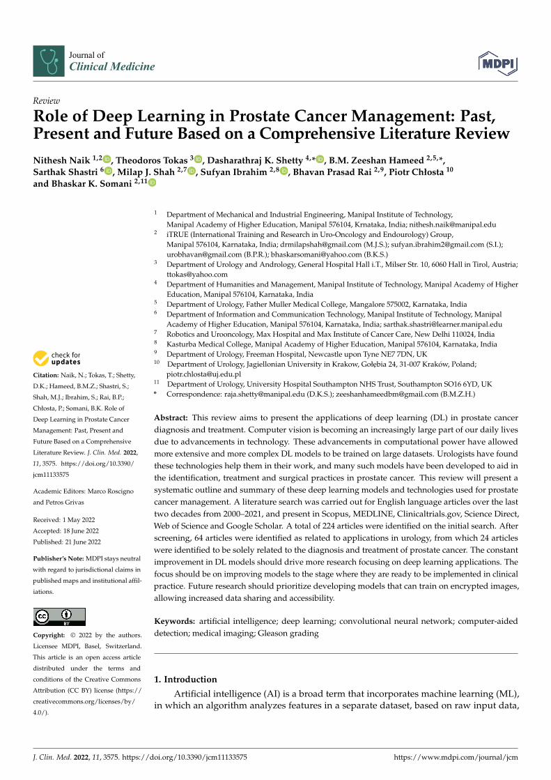
Citation: Naik, N.; Tokas, T.; Shetty,
D.K.; Hameed, B.M.Z.; Shastri, S.;
Shah, M.J.; Ibrahim, S.; Rai, B.P.;
Chłosta, P.; Somani, B.K. Role of
Deep Learning in Prostate Cancer
Management: Past, Present and
Future Based on a Comprehensive
Literature Review. J. Clin. Med. 2022,
11, 3575. https://doi.org/10.3390/
jcm11133575
Academic Editors: Marco Roscigno
and Petros Grivas
Received: 1 May 2022
Accepted: 18 June 2022
Published: 21 June 2022
Publisher’s Note: MDPI stays neutral
with regard to jurisdictional claims in
published maps and institutional affil-
iations.
Copyright: © 2022 by the authors.
Licensee MDPI, Basel, Switzerland.
This article is an open access article
distributed under the terms and
conditions of the Creative Commons
Attribution (CC BY) license (https://
creativecommons.org/licenses/by/
4.0/).
Journal of
Clinical Medicine
Review
Role of Deep Learning in Prostate Cancer Management: Past,Present and Future Based on a Comprehensive Literature ReviewNithesh Naik 1,2 , Theodoros Tokas 3 , Dasharathraj K. Shetty 4,* , B.M. Zeeshan Hameed 2,5,*,Sarthak Shastri 6 , Milap J. Shah 2,7 , Sufyan Ibrahim 2,8 , Bhavan Prasad Rai 2,9, Piotr Chłosta 10
and Bhaskar K. Somani 2,11
1 Department of Mechanical and Industrial Engineering, Manipal Institute of Technology,Manipal Academy of Higher Education, Manipal 576104, Krnataka, India; [email protected]
2 iTRUE (International Training and Research in Uro-Oncology and Endourology) Group,Manipal 576104, Karnataka, India; [email protected] (M.J.S.); [email protected] (S.I.);[email protected] (B.P.R.); [email protected] (B.K.S.)
3 Department of Urology and Andrology, General Hospital Hall i.T., Milser Str. 10, 6060 Hall in Tirol, Austria;[email protected]
4 Department of Humanities and Management, Manipal Institute of Technology, Manipal Academy of HigherEducation, Manipal 576104, Karnataka, India
5 Department of Urology, Father Muller Medical College, Mangalore 575002, Karnataka, India6 Department of Information and Communication Technology, Manipal Institute of Technology, Manipal
Academy of Higher Education, Manipal 576104, Karnataka, India; [email protected] Robotics and Urooncology, Max Hospital and Max Institute of Cancer Care, New Delhi 110024, India8 Kasturba Medical College, Manipal Academy of Higher Education, Manipal 576104, Karnataka, India9 Department of Urology, Freeman Hospital, Newcastle upon Tyne NE7 7DN, UK10 Department of Urology, Jagiellonian University in Krakow, Gołebia 24, 31-007 Kraków, Poland;
[email protected] Department of Urology, University Hospital Southampton NHS Trust, Southampton SO16 6YD, UK* Correspondence: [email protected] (D.K.S.); [email protected] (B.M.Z.H.)
Abstract: This review aims to present the applications of deep learning (DL) in prostate cancerdiagnosis and treatment. Computer vision is becoming an increasingly large part of our daily livesdue to advancements in technology. These advancements in computational power have allowedmore extensive and more complex DL models to be trained on large datasets. Urologists have foundthese technologies help them in their work, and many such models have been developed to aid inthe identification, treatment and surgical practices in prostate cancer. This review will present asystematic outline and summary of these deep learning models and technologies used for prostatecancer management. A literature search was carried out for English language articles over the lasttwo decades from 2000–2021, and present in Scopus, MEDLINE, Clinicaltrials.gov, Science Direct,Web of Science and Google Scholar. A total of 224 articles were identified on the initial search. Afterscreening, 64 articles were identified as related to applications in urology, from which 24 articleswere identified to be solely related to the diagnosis and treatment of prostate cancer. The constantimprovement in DL models should drive more research focusing on deep learning applications. Thefocus should be on improving models to the stage where they are ready to be implemented in clinicalpractice. Future research should prioritize developing models that can train on encrypted images,allowing increased data sharing and accessibility.
Keywords: artificial intelligence; deep learning; convolutional neural network; computer-aideddetection; medical imaging; Gleason grading
1. Introduction
Artificial intelligence (AI) is a broad term that incorporates machine learning (ML),in which an algorithm analyzes features in a separate dataset, based on raw input data,
J. Clin. Med. 2022, 11, 3575. https://doi.org/10.3390/jcm11133575 https://www.mdpi.com/journal/jcm
J. Clin. Med. 2022, 11, 3575 2 of 12
without being explicitly programmed, and returns a specific classification [1]. Deep learn-ing (DL) is a subset of ML which uses multilayer artificial neural networks (ANNs) tolearn hierarchical representations. Unlike classic ML algorithms such as support vectornetworks (SVN) and random forest (RF), DL learns features from input data without relyingsubstantially on domain knowledge developed by engineers [2]. Deep learning uses neuralnetworks with many layers where the first layer is the input layer connected to multiplehidden layers that are finally connected to the output layer. Neural networks use a seriesof algorithms to recognize hidden relationships in a data set by a process similar to thehuman brain. Each of the interconnected layers comprises numerous nodes, which arecalled perceptrons. Model perceptrons are arranged to form an interconnected network ina multi-layered perceptron. The input layer, upon receiving the input, transfers patternsobtained to the hidden layers. The hidden layers are activated based on the input parame-ters received. Hidden layers fine-tune the inputs received until the error is minimal, afterwhich the values of the neurons are passed to the output layer. The activation functioncalculates the output value, and the neural network produces its result.
Deep learning models help in diagnosing and treating urological conditions andhave proved their ability to detect prostate cancer, bladder tumors, renal cell carcinoma,along with ultrasound image analysis. A general schematic diagram of DL models canbe seen in Figure 1. Deep learning models have also displayed their ability to detect theneedle/trocar pressure during insertion, which is an essential aspect of laparoscopic androbotic urological surgeries.
J. Clin. Med. 2022, 11, 3575 2 of 13
1. Introduction Artificial intelligence (AI) is a broad term that incorporates machine learning (ML),
in which an algorithm analyzes features in a separate dataset, based on raw input data, without being explicitly programmed, and returns a specific classification [1]. Deep learning (DL) is a subset of ML which uses multilayer artificial neural networks (ANNs) to learn hierarchical representations. Unlike classic ML algorithms such as support vector networks (SVN) and random forest (RF), DL learns features from input data without relying substantially on domain knowledge developed by engineers [2]. Deep learning uses neural networks with many layers where the first layer is the input layer connected to multiple hidden layers that are finally connected to the output layer. Neural networks use a series of algorithms to recognize hidden relationships in a data set by a process similar to the human brain. Each of the interconnected layers comprises numerous nodes, which are called perceptrons. Model perceptrons are arranged to form an interconnected network in a multi-layered perceptron. The input layer, upon receiving the input, transfers patterns obtained to the hidden layers. The hidden layers are activated based on the input parameters received. Hidden layers fine-tune the inputs received until the error is minimal, after which the values of the neurons are passed to the output layer. The activation function calculates the output value, and the neural network produces its result.
Deep learning models help in diagnosing and treating urological conditions and have proved their ability to detect prostate cancer, bladder tumors, renal cell carcinoma, along with ultrasound image analysis. A general schematic diagram of DL models can be seen in Figure 1. Deep learning models have also displayed their ability to detect the needle/trocar pressure during insertion, which is an essential aspect of laparoscopic and robotic urological surgeries.
Figure 1. Deep learning framework for image classification.
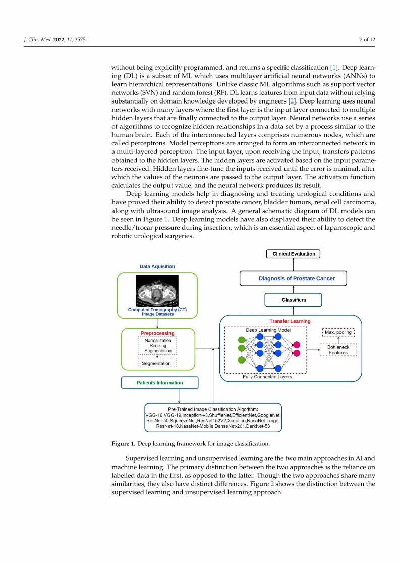
Supervised learning and unsupervised learning are the two main approaches in AI and machine learning. The primary distinction between the two approaches is the reliance on labelled data in the first, as opposed to the latter. Though the two approaches share
Figure 1. Deep learning framework for image classification.
Supervised learning and unsupervised learning are the two main approaches in AI andmachine learning. The primary distinction between the two approaches is the reliance onlabelled data in the first, as opposed to the latter. Though the two approaches share manysimilarities, they also have distinct differences. Figure 2 shows the distinction between thesupervised learning and unsupervised learning approach.
J. Clin. Med. 2022, 11, 3575 3 of 12
J. Clin. Med. 2022, 11, 3575 3 of 13
many similarities, they also have distinct differences. Figure 2 shows the distinction between the supervised learning and unsupervised learning approach.
Figure 2. Supervised learning and unsupervised learning approach.
What is supervised learning? The use of labelled datasets distinguishes supervised learning from other forms of
machine learning. Using these datasets, algorithms can be trained to better classify data or predict results. The model’s accuracy can be measured and improved over time using labelled inputs and outputs.
Based on data mining, supervised learning can be categorised into two types: classification and regression: (a) classification tasks rely on an algorithm to reliably assign test data to specified groups. For example: supervised learning algorithms can differentiate spam from the rest of the incoming emails. Classification methods include linear classifiers, support vector machines, decision trees, and random forests. (b) Regression is used to learn about the relationship between dependent and independent variables. Predicting numerical values based on various data points is possible with regression models. Linear regression, logistic regression, and polynomial regression are all common regression algorithms.
What is unsupervised learning? For the analysis and clustering of unlabeled data sets, unsupervised learning makes
use of machine learning methods. These algorithms, which are referred to as ‘unsupervised’, find hidden patterns in data without the aid of human intervention. Three key tasks are performed by unsupervised learning models: (a) clustering, (b) association, and (c) dimensionality reduction.
Using data mining techniques such as clustering, it is possible to create groups of unlabeled data that are similar or dissimilar. Similar data points are grouped together according to the K value in K-means clustering algorithms. This method is useful for a variety of things, including image segmentation and image compression. Another unsupervised learning technique is association, which employs a separate set of criteria to discover connections among the variables in a dataset.
Figure 2. Supervised learning and unsupervised learning approach.
What is supervised learning?The use of labelled datasets distinguishes supervised learning from other forms of
machine learning. Using these datasets, algorithms can be trained to better classify dataor predict results. The model’s accuracy can be measured and improved over time usinglabelled inputs and outputs.
Based on data mining, supervised learning can be categorised into two types: classi-fication and regression: (a) classification tasks rely on an algorithm to reliably assign testdata to specified groups. For example: supervised learning algorithms can differentiatespam from the rest of the incoming emails. Classification methods include linear classifiers,support vector machines, decision trees, and random forests. (b) Regression is used to learnabout the relationship between dependent and independent variables. Predicting numericalvalues based on various data points is possible with regression models. Linear regression,logistic regression, and polynomial regression are all common regression algorithms.
What is unsupervised learning?For the analysis and clustering of unlabeled data sets, unsupervised learning makes
use of machine learning methods. These algorithms, which are referred to as ‘unsupervised’,find hidden patterns in data without the aid of human intervention. Three key tasksare performed by unsupervised learning models: (a) clustering, (b) association, and (c)dimensionality reduction.
Using data mining techniques such as clustering, it is possible to create groups ofunlabeled data that are similar or dissimilar. Similar data points are grouped togetheraccording to the K value in K-means clustering algorithms. This method is useful fora variety of things, including image segmentation and image compression. Anotherunsupervised learning technique is association, which employs a separate set of criteria todiscover connections among the variables in a dataset.
Dimensionality reduction is a learning approach used when the number of features(or dimensions) in a dataset is too large. It minimizes the quantity of data inputs while yetmaintaining the integrity of the data. To enhance image quality, auto encoders often utilizethis technique to remove noise from visual data before it is processed further.

J. Clin. Med. 2022, 11, 3575 4 of 12
Over the last decade, imaging technology has significantly improved, which has madeit easier for us to apply computer vision technologies for the classification and detectionof diseases [3]. With the advancements in graphics processing units (GPUs) and theircomputational power to perform parallel processing, computer vision processing is moreaccessible today. Deep learning is also being used for data management, chatbots, andother facilities that aid in medical practice. Natural language processing (NLP) practicesused in finding patterns in multimodal data have been shown to increase the learningsystem’s accuracy of diagnosis, prediction, and performance [4]. However, identifyingessential clinical elements and establishing relations has been difficult as these records areusually unordered and disorganized. Urology has been at the forefront of accepting newertechnologies to achieve better patient outcomes. This comprehensive review aims to givean insight into the applications of deep learning in Urology.
2. Search Strategy
In October 2021, Pubmed/MEDLINE, Scopus, Clinicaltrials.gov, Science Direct, Webof Science and Google Scholar were used to undertake a review of all English languageliterature published in the previous two decades (2000–2021). The search technique wasbased on PICO (Patient Intervention Comparison Outcome) criteria, in which patientswere treated with AI models (I) or classical biostatistical models (C), and the efficacy of AImodels was evaluated (O) [5].
Specifically, the search was conducted by using a combination of the following terms:‘artificial intelligence’, ‘AI’, ‘machine learning’, ‘ML’, ‘convolutional networks’, ‘CNN’,‘deep learning’, ‘DL’, ‘magnetic resonance imaging’, ‘prostate’, ‘prostate cancer’, ‘MRI’,‘Sorensen–Dice coefficient’, ‘DSC’, ‘area under the ROC curve’, ‘AUC’, ‘Sorensen–Diceindex’, ‘SDI’ and ‘computed tomography’, ‘CT’ [6].
2.1. Inclusion Criteria
1. Articles on the application of deep learning in prostate cancer diagnosis and treatment.2. Full-text articles, clinical trials and meta-Analysis on outcomes of analysis in Urology
using deep learning.
2.2. Exclusion Criteria
1. Animal, laboratory, or cadaveric studies2. Reviews, editorials, commentaries or book chapters
The literature review was carried out using the inclusion and exclusion criteria men-tioned. Articles were screened based on the titles and abstracts. Articles were then selectedand their entire text was analyzed. For further screening of other published literature, thereferences list of the selected articles was individually and manually checked.
3. ResultsEvidence Synthesis
A total of 224 distinct articles were discovered during the initial search. Following theinitial screening, 97 articles remained, with 64 left after a second screening as related toapplications of deep learning in Urology. Among these articles, 24 were identified to besolely related to prostate cancer, these abstracts satisfied our inclusion criteria and were thenincluded in the final review. The summary of all the previous studies from the literature isshown in Tables 1 and 2 on the diagnosis and treatment of prostate cancer, respectively.

J. Clin. Med. 2022, 11, 3575 5 of 12
Table 1. Summary of studies on diagnosis of prostate cancer using deep learning models.
Author Year Objective Sample Size(n = Patients/Images)
StudyDesign Model AUC DSC SDI MAE Sn Sp
A. MRI images
Takeuchi et al. [7] 2018 To predict PCa using DL andmultilayer ANN 334 patients Prospective Stepwise ANN
5-hidden-layers0.76 (Step
200) N/A N/A N/A N/A N/A
Schelb et al. [8] 2019
To compare clinical evaluationperformance with
segmentation-optimized DLsystem in PCa diagnosis.
312 patients;T2W and diffusion
images usedRetrospective U-Net N/A N/A N/A N/A 96% 22%
Shao et al. [9] 2021
For PCa diagnosis usingProsRegNet (DL system)
using MRI andhistopathological data.
152 patients;T2W images and HPE
slices used.Prospective ProsRegNet and
CNNGeometric N/ACohort 1: 0.979Cohort 2: 0.971Cohort 3: 0.976
N/A N/A N/A N/A
Hiremath et al. [10] 2021
To detect csPCa usingintegrated nomogram using DL,
PI-RADS grading andclinical factors.
592 patients;T2W and ADC images
usedRetrospective AlexNet and
DenseNet 0.76 N/A N/A N/A N/A N/A
Hiremath et al. [11] 2020
To assess the test-retestrepeatability of U-Net (DLsystem) in identification
of csPCa.
112 patients;ADC/DWI images
usedProspective U-Net 0.8 0.8 N/A N/A N/A N/A
Schelb et al. [12] 2019The use DL algorithm (U-Net)for detection, localization, and
segmentation of csPCa
259 patients; T2W andDW images used. Retrospective U-Net N/A N/A N/A N/A 98% 24%
Yan et al. [13] 2021
For deep combination learningof multi-level features for MRprostate segmentation using a
propagation DNN
80 patients; only T2Wimages used Retrospective MatConvNet N/A 0.84 N/A N/A N/A N/A

J. Clin. Med. 2022, 11, 3575 6 of 12
Table 1. Cont.
Author Year Objective Sample Size(n = Patients/Images)
StudyDesign Model AUC DSC SDI MAE Sn Sp
Khosravi et al. [14] 2021
To develop an AI-based modelfor the early detection of PCa
using MR pictures tagged withhistopathology information.
400 patients; T2Wimages used Retrospective GoogLenet 0.89 N/A N/A N/A N/A N/A
Shiradkar et al. [15] 2020
To find any links between T1Wand T2W MR fingerprinting
data and the appropriate tissuecompartment ratios in PCa and
prostatitis wholemount histology.
14 patients;T1W and T2 W
images usedRetrospective U-Net 0.997 N/A N/A N/A N/A N/A
Winkel et al. [16] 2020
To incorporate DL andbiparametric imaging for
autonomous detection andclassification of
PI-RADS lesions.
49 patients;T2W and DWI used Prospective ProstateAI N/A N/A N/A N/A 87% 50%
B. Pathology
AlDubayan et al.[17] 2020
To detect germline harmfulmutations in PCa using
DL techniques.1295 patients Retrospective
DeepVariant and
GenomeAnalysis Toolkit
0.94 N/A N/A N/A CI:0.91–0.97 N/A
Kott et al. [18] 2021To apply DL methods on biopsy
specimen for histopathologicdiagnosis and Gleason grading.
85 images25 patients Prospective 18-layer CNN 0.83 N/A N/A N/A N/A N/A
Lucas et al. [19] 2019To determine Gleason pattern
and grade group in biopsyspecimen using DL
96 images38 patients Retrospective Inception-v3
CNN 0.92 N/A N/A N/A 90% 93%
J. Clin. Med. 2022, 11, 3575 7 of 12
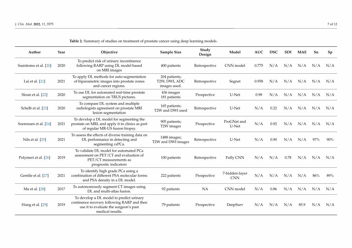
Table 2. Summary of studies on treatment of prostate cancer using deep learning models.
Author Year Objective Sample Size StudyDesign Model AUC DSC SDI MAE Sn Sp
Sumitomo et al. [20] 2020To predict risk of urinary incontinence
following RARP using DL model basedon MRI images
400 patients Retrospective CNN model 0.775 N/A N/A N/A N/A N/A
Lai et al. [21] 2021To apply DL methods for auto-segmentationof biparametric images into prostate zones
and cancer regions.
204 patients;T2W, DWI, ADC
images used.Retrospective Segnet 0.958 N/A N/A N/A N/A N/A
Sloun et al. [22] 2020 To use DL for automated real-time prostatesegmentation on TRUS pictures.
436 images181 patients Prospective U-Net 0.98 N/A N/A N/A N/A N/A
Schelb et al. [23] 2020To compare DL system and multiple
radiologists agreement on prostate MRIlesion segmentation
165 patients;T2W and DWI used Retrospective U-Net N/A 0.22 N/A N/A N/A N/A
Soerensen et al. [24] 2021To develop a DL model for segmenting the
prostate on MRI, and apply it in clinics as partof regular MR-US fusion biopsy.
905 patients;T2W images Prospective ProGNet and
U-Net N/A 0.92 N/A N/A N/A N/A
Nils et al. [25] 2021To assess the effects of diverse training data on
DL performance in detecting andsegmenting csPCa.
1488 images;T2W and DWI images Retrospective U-Net N/A 0.90 N/A N/A 97% 90%
Polymeri et al. [26] 2019
To validate DL model for automated PCaassessment on PET/CT and evaluation of
PET/CT measurements asprognostic indicators
100 patients Retrospective Fully CNN N/A N/A 0.78 N/A N/A N/A
Gentile et al. [27] 2021To identify high grade PCa using a
combination of different PSA molecular formsand PSA density in a DL model.
222 patients Prospective 7-hidden-layerCNN N/A N/A N/A N/A 86% 89%
Ma et al. [28] 2017 To autonomously segment CT images usingDL and multi-atlas fusion. 92 patients NA CNN model N/A 0.86 N/A N/A N/A N/A
Hung et al. [29] 2019
To develop a DL model to predict urinarycontinence recovery following RARP and then
use it to evaluate the surgeon’s pastmedical results.
79 patients Prospective DeepSurv N/A N/A N/A 85.9 N/A N/A

J. Clin. Med. 2022, 11, 3575 8 of 12
4. Discussion4.1. Diagnosis of Prostate Cancer Using MRI Images
Eleven studies have evaluated the application of deep learning in diagnosingprostate cancer.
Takeuchi et al. (2019) developed a DL model to predict prostate cancer using a multi-layer ANN. The model was trained on images obtained from 232 patients and validated itsaccuracy on images obtained from 102 patients. On a test dataset, the model achieved AUCof 0.76, thereby, suggesting that neural network achieved better results as compared to alogistic regression model. However, this accuracy needs to be improved to be implementedin clinical practice [7].
Khosravi et al. (2021) used DL models to differentiate malignant and benign tumorsand high- and low-risk tumors which achieved an AUC of 0.89 and 0.78, respectively. Thestudy concluded that new images captured did not require manual segmentation and couldbe implemented in clinical practice [14].
Hiremath et al. (2020) used diffusion-weighted imaging fitted with monoexponen-tial function, ADCm, employing a deep learning architecture (U-Net) to investigate theshort-term test-retest repeatability of U-Net in slice- and lesion-level identification andsegmentation of clinically significant prostate cancer (csPCa: Gleason grade group > 1)(U-Net). The training dataset included 112 PCa patients who had two prostate MRI examson the same day. Two U-Net-based CNNs were trained using this dataset. The studyperformed three-fold cross-validation on the training set and evaluated its performanceand repeatability on testing data. The CNNs with U-Net-based architecture demonstratedan intraclass correlation coefficient (ICC) between 0.80–0.83, agreement of 66–72%, andDSC of 0.68–0.72 for a slice- and lesion-level detection. These findings lay the groundworkfor DL architecture’s test-retest and repeatability in identifying and segmenting clinicallyrelevant prostate cancer on apparent diffusion coefficient maps. [11].
To summarize, MR images are most commonly used to study the applications of DLin image-based diagnosis of prostate cancer (PCa). Though the accuracy of the DL modelsappears to be satisfactory, the generalizability of the results across varied demographicsstill needs to be tested before implementing into general clinical practice.
4.2. Histopathological Diagnosis of Prostate Cancer Using DL Models
Three studies have evaluated the application of deep learning in the diagnosis ofprostate cancer.
Kott et al. (2019) developed a DL algorithm for histopathologic diagnosis. They alsoperformed Gleason grading of the prostate cancer biopsies. This histopathologic diagnosisand Gleason grading process are considered lengthy and time-consuming. Using MLmodels, this process can be made significantly faster and more efficient. The study wasperformed using 85 prostate biopsies from 25 patients with further magnification of up to20x performed. The study used a deep residual CNN model with fivefold cross-validation.The DL model achieved 91.5 and 85.4% accuracy at coarse and fine-level classification,respectively. The study concluded that the model achieved excellent performance for thediagnosis as mentioned earlier; however, it needs to be tested on a larger sample size forexternal validation [18].
Lucas et al. (2019) performed a study using DL models for automatic Gleason patternclassification to identify grade groups from prostate biopsies. The study used a datasetcontaining 96 prostate biopsies from 38 patients. The Inception-v3 convolutional neuralnetwork was trained to generate probability maps. The model has a 92% accuracy in distin-guishing between non-atypical and malignant regions, with a sensitivity and specificity of90 and 93%, respectively. The study successfully demonstrates the feasibility of trainingCNN models to differentiate between Gleason patterns in heterogeneous biopsies [19].

J. Clin. Med. 2022, 11, 3575 9 of 12
The DL models have shown promising results in the histopathological diagnosis ofPCa. This can definitely be added as an adjunct tool for the histopathologists to reduce theburden in terms of time and workload. However due to lack of external validation of thesemodels, their applicability cannot be generalized as of yet.
4.3. Diagnosis of Prostate Cancer Using MR Based Segmentation Techniques
Four studies have evaluated the application of DL in the diagnosis of prostate cancer.Lai et al. (2021) developed a DL CNN model to segment prostate zones and cancer
regions from MRI images. The study was performed using the PROSTATEx dataset contain-ing MRI scans from 204 patients. A SegNet model was modified and fine-tuned to performadequately on the dataset. The study achieved a dice similarity coefficient of 90.45% for thetransition zone, 70.04% for the peripheral zone, and 52.73% for the prostate cancer region.The study concluded that automatic segmentation using a DCNN model has the potentialto assist in prostate cancer diagnosis [21].
Sloun et al. (2021) performed prostate segmentation of transrectal ultrasound using theDL model on MRI images. The study used three datasets with MRI-transrectal ultrasoundimages collected at different institutions. The study trained a U-Net model on the dataset of436 images and achieved a median accuracy of 98%. While performing zonal segmentation,the model achieved a pixel-wise accuracy of 97 and 98% for the peripheral and transitionimages. The model can also self-assess its segmentation, allowing it to identify incorrectsegmentations swiftly. The process of performing manual segmentation of prostate MRIimages places a burden on clinicians. The authors concluded that using DL models canallow for fast and accurate segmentation of MRI images from different scanners [22].
Schelb et al. (2020) produced a comparison of prostate MRI lesion segmentationbetween a DL model and multiple radiologists. The study was performed using MRIimages collected from 165 patients suspected to have prostate cancer. The study used U-Net models trained on the dataset of MRI images to perform segmentation. The mean Dicecoefficient for manual segmentation was between 0.48–0.52, while the U-Net segmentationsexhibited a Dice coefficient of 0.22. The authors concluded that smaller segmentationsizes could explain the lower Dice coefficients of the U-Net model. They also discuss howthe overlapping lesions between multiple rates can be used as a secondary measure forsegmentation quality in future studies [23].
Soerensen et al. (2021) performed a study to determine if DL improves the speedand accuracy of prostate gland segmentation from MRI images. The study used imagesfrom 905 subjects who underwent prostate MRI scans at 29 institutions. The study traineda ProGNet model on 805 cases and tested it on 100 independent and 56 external cases.The study found that the ProGNet model outperformed the U-Net model. The study alsofound that the ProGNet model achieved a Dice similarity coefficient of 0.93, outperformingradiology technicians, producing results at 35 s/case. The study concluded that DL modelscould be used for segmentation in targeted biopsy in routine urological clinical practice [24].
As proven, ProGNet model outperformed not only the U-Net model but also theradiology technicians in terms of speed and accuracy. However, it should be noted thatauthors have not compared the ProGNet model to trained and experienced urologistsand radiologists. Furthermore, the accuracy of the model has to be tested across differentMRI scanners.
4.4. Diagnosis of Prostate Cancer Using CT Images
Four studies have evaluated the application of DL in the diagnosis of prostate cancerand prostatectomy. Polymeri et al. (2019) used a DL algorithm to automate prostate cancerquantification on positron emission tomography–computed tomography (PET/CT) scans.The study looked at the possibility of PET/CT measurements as prognostic biomarkers. Thetraining of the DL model was performed on CT scan images of 100 patients. In 45 patientswith biopsy-proven hormone-naive prostate cancer, the DL algorithm was validated. Themodel was evaluated based on the Sørensen–Dice index (SDI) score. The SDI scores
J. Clin. Med. 2022, 11, 3575 10 of 12
achieved were 0.78 and 0.79 for automatic and manual volume segmentation, respectively.The study demonstrated DL applications in quantifying PET/CT prostate gland uptakeand its association with overall survival. The results obtained showed agreement betweenautomated and manual PET/CT measurements. The DL model demonstrated that PET/CTindicators were strongly linked to overall survival [26].
Ma et al. (2017) performed automatic prostate segmentation using DL and multi-atlas fusion. A dataset of 92 prostate CT scans was used to conduct and assess the study.When compared to the radiologists’ manual segmentation, the model had a Dice similaritycoefficient of 86.80%. The study concluded that the DL-based method can provide avaluable tool for automatic segmentation and aid clinical practice [28].
Not many studies have been performed to check the applications of DL modelsusing PET/CT images to highlight their advantages in the same aspect. However, thenascent applications appear promising in terms of development of DL-based biomarkerand prognostic models.
4.5. Robot-Assisted Treatment Practices
The study by Hung et al. evaluated the application of DL in the treatment of PCa andRARP. Hung et al. created a DL model to predict urinary continence recovery followingradical prostatectomy with robotic assistance. The study was performed on images ob-tained from 79 patients. The study trained a DeepSurv model on the dataset and achieveda concordance index (C-index) of 0.6 and a mean absolute error (MAE) of 85.9 in predict-ing continence. The authors concluded that using automated performance metrics andclinicopathological data, the DeepSurv model could predict continence after the prostate-ctomy. According to the findings, experienced surgeons had greater continence rates at3 and 6 months following the prostatectomy [29].
The application of automated performance metrics (APMs) and its impact on clinicaloutcome variables was very well highlighted in this study, underlining the evidence thatsurgical skills impact clinical outcomes. However, this was a single-institution study andrequires external validation for the same.
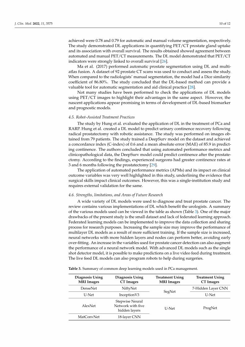
4.6. Strengths, limitations, and Areas of Future Research
A wide variety of DL models were used to diagnose and treat prostate cancer. Thereview contains various implementations of DL which benefit the urologists. A summaryof the various models used can be viewed in the table as shown (Table 3). One of the majordrawbacks of the present study is the small dataset and lack of federated learning approach.Federated learning models can be implemented to improve the data collection and sharingprocess for research purposes. Increasing the sample size may improve the performance ofmultilayer DL models as a result of more sufficient training. If the sample size is increased,neural networks with more hidden layers and nodes can perform better, avoiding earlyover-fitting. An increase in the variables used for prostate cancer detection can also augmentthe performance of a neural network model. With advanced DL models such as the singleshot detector model, it is possible to make predictions on a live video feed during treatment.The live feed DL models can also program robots to help during surgeries.
Table 3. Summary of common deep learning models used in PCa management.
Diagnosis UsingMRI Images
Diagnosis UsingCT Images
Treatment UsingMRI Images
Treatment UsingCT Images
DenseNet NiftyNetSegNet
7-Hidden Layer CNN
U-Net InceptionV3 U-Net
AlexNetStepwise Neural
Network with fivehidden layers U-Net ProgNet
MatConvNet 18-layer CNN

J. Clin. Med. 2022, 11, 3575 11 of 12
5. Conclusions
As per our review, the most common application of DL techniques has been in thediagnosis of prostate cancer using MR image-based segmentation techniques. Althoughthe ProgNet model outperformed trained radiologists in prostate cancer detection, wecannot generalize these results. In conclusion, for clinical application, the DL models’ per-formance may still need improvement. As the performance of these models increases, theywill become much more implementable, with many models surpassing human accuracyand efficiency.
Author Contributions: Concept and Study design: B.M.Z.H., B.K.S., T.T., N.N. and B.P.R.; Methodsand experimental work: T.T., S.S., M.J.S. and P.C.; Results analysis and conclusions: B.M.Z.H., B.K.S.,N.N., S.I. and B.P.R.; Manuscript preparation: N.N., S.S., S.I., M.J.S. and D.K.S. All authors have readand agreed to the published version of the manuscript.
Funding: This research received no specific grant from any funding agency in the public, commercial,or not-for-profit sectors.
Institutional Review Board Statement: Not applicable.
Informed Consent Statement: Not applicable.
Data Availability Statement: Not applicable.
Conflicts of Interest: The authors declare no conflict of interest.
References1. Pannala, R.; Krishnan, K.; Melson, J.; Parsi, M.A.; Schulman, A.R.; Sullivan, S.; Trikudanathan, G.; Trindade, A.J.; Watson, R.R.;
Maple, J.T.; et al. Artificial intelligence in gastrointestinal endoscopy. VideoGIE 2020, 5, 598–613. [CrossRef]2. Chen, H.; Sung, J. Potentials of AI in medical image analysis in Gastroenterology and Hepatology. J. Gastroenterol. Hepatol. 2021,
36, 31–38. [CrossRef] [PubMed]3. Ganatra, N.; Patel, A. A comprehensive study of deep learning architectures, applications and tools. Int. J. Comput. Sci. Eng. 2018,
6, 701–705. [CrossRef]4. Asgari, S.; Scalzo, F.; Kasprowicz, M. Pattern recognition in medical decision support. BioMed Res. Int. 2019, 2019, 2. [CrossRef]
[PubMed]5. Eriksen, M.; Frandsen, T. The impact of patient, intervention, comparison, outcome (PICO) as a search strategy tool on literature
search quality: A systematic review. J. Med. Libr. Assoc. 2018, 106, 420–431. [CrossRef] [PubMed]6. Liberati, A.; Altman, D.G.; Tetzlaff, J.; Mulrow, C.; Gøtzsche, P.C.; Ioannidis, J.P.; Clarke, M.; Devereaux, P.J.; Kleijnen, J.; Moher, D.
The PRISMA statement for reporting systematic reviews and meta-analyses of studies that evaluate healthcare interventions:Explanation and elaboration. BMJ 2009, 339, b2700. [CrossRef] [PubMed]
7. Takeuchi, T.; Hattori-Kato, M.; Okuno, Y.; Iwai, S.; Mikami, K. Prediction of prostate cancer by deep learning with multilayerartificial neural network. Can. Urol. Assoc. J. 2018, 13, E145–E150. [CrossRef] [PubMed]
8. Schelb, P.; Kohl, S.; Radtke, J.P.; Wiesenfarth, M.; Kickingereder, P.V.; Bickelhaupt, S.; Kuder, T.A.; Stenzinger, A.; Hohenfellner, M.;Schlemmer, H.-P.; et al. Classification of cancer at prostate MRI: Deep learning versus clinical PI-RADS assessment. Radiology2019, 293, 607–617. [CrossRef]
9. Shao, W.; Banh, L.; Kunder, C.A.; Fan, R.E.; Soerensen, S.J.; Wang, J.B.; Teslovich, N.C.; Madhuripan, N.; Jawahar, A.; Ghanouni,P.; et al. ProsRegNet: A deep learning framework for registration of MRI and histopathology images of the prostate. Med. ImageAnal. 2021, 68, 101919. [CrossRef]
10. Hiremath, A.; Shiradkar, R.; Fu, P.; Mahran, A.; Rastinehad, A.R.; Tewari, A.; Tirumani, S.H.; Purysko, A.; Ponsky, L.; Madabhushi,A. An integrated nomogram combining deep learning, Prostate Imaging-Reporting and Data System (PI-RADS) scoring, andclinical variables for identification of clinically significant prostate cancer on biparametric MRI: A retrospective multicentre study.Lancet Digit. Health 2021, 3, e445–e454. [CrossRef]
11. Hiremath, A.; Shiradkar, R.; Merisaari, H.; Prasanna, P.; Ettala, O.; Taimen, P.; Aronen, H.J.; Boström, P.J.; Jambor, I.; Madabhushi,A. Test-retest repeatability of a deep learning architecture in detecting and segmenting clinically significant prostate cancer onapparent diffusion coefficient (ADC) maps. Eur. Radiol. 2020, 31, 379–391. [CrossRef] [PubMed]
12. Schelb, P.; Wang, X.; Radtke, J.P.; Wiesenfarth, M.; Kickingereder, P.; Stenzinger, A.; Hohenfellner, M.; Schlemmer, H.-P.; Maier-Hein, K.H.; Bonekamp, D. Simulated clinical deployment of fully automatic deep learning for clinical prostate MRI assessment.Eur. Radiol. 2021, 31, 302–313. [CrossRef] [PubMed]
13. Yan, Y.; Shao, L.; Liu, Z.; He, W.; Yang, G.; Liu, J.; Xia, H.; Zhang, Y.; Chen, H.; Liu, C.; et al. Deep learning with quantitativefeatures of magnetic resonance images to predict biochemical recurrence of radical prostatectomy: A multi-center study. Cancers2021, 13, 3098. [CrossRef] [PubMed]

J. Clin. Med. 2022, 11, 3575 12 of 12
14. Khosravi, P.; Lysandrou, M.; Eljalby, M.; Li, Q.; Kazemi, E.; Zisimopoulos, P.; Sigaras, A.; Brendel, M.; Barnes, J.; Ricketts, C.; et al.A deep learning approach to diagnostic classification of prostate cancer using pathology-radiology fusion. J. Magn. Reson. Imaging2021, 54, 462–471. [CrossRef] [PubMed]
15. Shiradkar, R.; Panda, A.; Leo, P.; Janowczyk, A.; Farre, X.; Janaki, N.; Li, L.; Pahwa, S.; Mahran, A.; Buzzy, C.; et al. T1 and T2 MRfingerprinting measurements of prostate cancer and prostatitis correlate with deep learning–derived estimates of epithelium,lumen, and stromal composition on corresponding whole mount histopathology. Eur. Radiol. 2020, 31, 1336–1346. [CrossRef][PubMed]
16. Winkel, D.J.; Wetterauer, C.; Matthias, M.O.; Lou, B.; Shi, B.; Kamen, A.; Comaniciu, D.; Seifert, H.-H.; Rentsch, C.A.; Boll, D.T.Autonomous detection and classification of PI-RADS lesions in an MRI screening population incorporating multicenter-labeleddeep learning and biparametric imaging: Proof of concept. Diagnostics 2020, 10, 951. [CrossRef]
17. Aldubayan, S.H.; Conway, J.R.; Camp, S.Y.; Witkowski, L.; Kofman, E.; Reardon, B.; Han, S.; Moore, N.; Elmarakeby, H.; Salari,K.; et al. Detection of pathogenic variants with germline genetic testing using deep learning vs standard methods in patients withprostate cancer and melanoma. JAMA 2020, 324, 1957. [CrossRef]
18. Kott, O.; Linsley, D.; Amin, A.; Karagounis, A.; Jeffers, C.; Golijanin, D.; Serre, T.; Gershman, B. Development of a deep learningalgorithm for the histopathologic diagnosis and gleason grading of prostate cancer biopsies: A pilot study. Eur. Urol. Focus 2021,7, 347–351. [CrossRef]
19. Lucas, M.; Jansen, I.; Savci-Heijink, C.D.; Meijer, S.L.; de Boer, O.J.; van Leeuwen, T.G.; de Bruin, D.M.; Marquering, H.A. Deeplearning for automatic Gleason pattern classification for grade group determination of prostate biopsies. Virchows Arch. 2019, 475,77–83. [CrossRef]
20. Sumitomo, M.; Teramoto, A.; Toda, R.; Fukami, N.; Fukaya, K.; Zennami, K.; Ichino, M.; Takahara, K.; Kusaka, M.; Shiroki, R.Deep learning using preoperative magnetic resonance imaging information to predict early recovery of urinary continence afterrobot-assisted radical prostatectomy. Int. J. Urol. 2020, 27, 922–928. [CrossRef]
21. Lai, C.-C.; Wang, H.-K.; Wang, F.-N.; Peng, Y.-C.; Lin, T.-P.; Peng, H.-H.; Shen, S.-H. Autosegmentation of prostate zones andcancer regions from biparametric magnetic resonance images by using deep-learning-based neural networks. Sensors 2021, 21,2709. [CrossRef] [PubMed]
22. Van Sloun, R.J.; Wildeboer, R.R.; Mannaerts, C.K.; Postema, A.W.; Gayet, M.; Beerlage, H.P.; Salomon, G.; Wijkstra, H.; Mischi, M.Deep learning for real-time, automatic, and scanner-adapted prostate (zone) segmentation of transrectal ultrasound, for example,magnetic resonance imaging–transrectal ultrasound fusion prostate biopsy. Eur. Urol. Focus 2021, 7, 78–85. [CrossRef] [PubMed]
23. Schelb, P.; Tavakoli, A.A.; Tubtawee, T.; Hielscher, T.; Radtke, J.-P.; Görtz, M.; Schütz, V.; Kuder, T.A.; Schimmöller, L.; Stenzinger,A.; et al. Comparison of prostate MRI lesion segmentation agreement between multiple radiologists and a fully automatic deeplearning system. RöFo—Fortschr. Geb. Röntgenstrahlen Bildgeb. Verfahr. 2020, 193, 559–573. [CrossRef] [PubMed]
24. Soerensen, S.J.C.; Fan, R.E.; Seetharaman, A.; Chen, L.; Shao, W.; Bhattacharya, I.; Kim, Y.H.; Sood, R.; Borre, M.; Chung, B.I.; et al.Deep learning improves speed and accuracy of prostate gland segmentations on magnetic resonance imaging for targeted biopsy.J. Urol. 2021, 206, 604–612. [CrossRef] [PubMed]
25. Netzer, N.; Weißer, C.; Schelb, P.; Wang, X.; Qin, X.; Görtz, M.; Schütz, V.; Radtke, J.P.; Hielscher, T.; Schwab, C.; et al. Fullyautomatic deep learning in bi-institutional prostate magnetic resonance imaging: Effects of cohort size and heterogeneity. Investig.Radiol. 2021, 56, 799–808. [CrossRef] [PubMed]
26. Polymeri, E.; Sadik, M.; Kaboteh, R.; Borrelli, P.; Enqvist, O.; Ulén, J.; Ohlsson, M.; Trägårdh, E.; Poulsen, M.H.; Simonsen,J.A.; et al. Deep learning-based quantification of PET/CT prostate gland uptake: Association with overall survival. Clin. Physiol.Funct. Imaging 2019, 40, 106–113. [CrossRef]
27. Gentile, F.; Ferro, M.; Della Ventura, B.; La Civita, E.; Liotti, A.; Cennamo, M.; Bruzzese, D.; Velotta, R.; Terracciano, D. Optimisedidentification of high-grade prostate cancer by combining different PSA molecular forms and PSA density in a deep learningmodel. Diagnostics 2021, 11, 335. [CrossRef]
28. Ma, L.; Guo, R.; Zhang, G.; Tade, F.; Schuster, D.M.; Nieh, P.; Master, V.; Fei, B. Automatic segmentation of the prostate on CTimages using deep learning and multi-atlas fusion. Med. Imaging 2017, 10133, 101332O.
29. Hung, A.J.; Chen, J.; Ghodoussipour, S.; Oh, P.J.; Liu, Z.; Nguyen, J.; Purushotham, S.; Gill, I.S.; Liu, Y. A deep-learning modelusing automated performance metrics and clinical features to predict urinary continence recovery after robot-assisted radicalprostatectomy. BJU Int. 2019, 124, 487–495. [CrossRef]
Copyright © 2022 FDOKUMEN