Two Zebrafish eIF4E Family Members Are Differentially Expressed and Functionally Divergent

Carcinogenesis vol.29 no.12 pp.2279–2288, 2008doi:10.1093/carcin/bgn221Advance Access publication September 22, 2008
Phosphorylation of eIF4E by MNKs supports protein synthesis, cell cycle progressionand proliferation in prostate cancer cells
Andrea Bianchini1,3, Maria Loiarro1,3, Pamela Bielli1,3,Roberta Busa1,3, Maria Paola Paronetto1,3, FabrizioLoreni2, Raffaele Geremia1 and Claudio Sette1,3,�
1Department of Public Health and Cell Biology and 2Department of Biology,University of Rome Tor Vergata, Via Montpellier 1, 00133 Rome, Italy and3Neuroembryology Unit, Fondazione Santa Lucia, Via di Fosso del Fiorano64, 00143 Rome, Italy
�To whom correspondence should be addressed. Tel: þ39 06 72596260;Fax: þ39 06 72596268;Email: [email protected]
Deregulation of the phosphatidyl inositol trisphosphate kinase/AKT/mammalian target of rapamycin (mTOR) and RAS/mitogen-activated protein kinase (MAPK)/MNK pathways frequently occursin human prostate carcinomas (PCas) and leads to aberrant mod-ulation of messenger RNA (mRNA) translation. We have investi-gated the relative contribution of these pathways to translationalregulation and proliferation of PCa cells. MNK-dependent phos-phorylation of eIF4E is elevated in DU145 cells, which have lowbasal levels of AKT/mTOR activity due to the expression of thetumor suppressor PTEN. In contrast, eIF4E phosphorylation islow in PC3 and LNCaP cells with mutated PTEN and constitu-tively active AKT/mTOR pathway, but it can be strongly inducedthrough inhibition of mTOR activity by rapamycin or serum de-pletion. Remarkably, we found that inhibition of MNKs stronglyreduced the polysomal recruitment of terminal oligopyrimidinemessenger RNAs (TOP mRNAs), which are known targets ofmTOR-dependent translational control. Pull-down assays of theeIF4F complex indicated that translation initiation was differ-ently affected by inhibition of MNKs and mTOR. In addition,concomitant treatment with MNK inhibitor and rapamycin ex-erted additive effects on polysomal recruitment of TOP mRNAsand protein synthesis. TheMNK inhibitor was more effective thanrapamycin in blocking proliferation of PTEN-expressing cells,whereas combination of the two inhibitors suppressed cell cycleprogression in both cell lines. Microarray analysis showed thatMNK affected translation of mRNAs involved in cell cycle pro-gression. Thus, our results indicate that a balance between theactivity of the AKT/mTOR and the MAPK/MNK pathway inPCa cells maintains a defined translational level of specific mRNAsrequired for ribosome biogenesis, cell proliferation and stressresponse and might confer to these cells the ability to overcomenegative insults.
Introduction
Prostate cancer (PCa) remains the second leading cause of death in thewestern male population. Although initially counteracted by androgendeprivation therapy, PCa cells often evolve to become independent ofandrogens for their growth (1). At this stage, no effective cure isavailable for the patients, rendering the discovery of new therapeuticapproaches a clinical priority. A common feature in PCa is repre-sented by inactivating mutations in PTEN (2). This gene encodesfor the phosphatase that hydrolyses the phosphatidyl inositoltrisphosphate and turns off the proliferative signal exerted by thephosphatidyl inositol trisphosphate kinase (PI3K) in response to growth
factors (3). Mouse models have shown that haploinsufficiency in thePTEN locus predisposes to the onset and the progression of PCa (4,5).Mechanistically, inactivation of PTEN leads to increased basal levelsof phosphatidyl inositol trisphosphate in the cell and constitutiveactivation of the AKT pathway.
The oncogenic potential of the AKT pathway relies on the down-stream serine/threonine kinase mammalian target of rapamycin(mTOR) (6,7). The involvement of mTOR in neoplastic transforma-tion appears to depend on its regulatory activity toward the translationinitiation complex eIF4F. By this way, mTOR stimulates ribosomebiogenesis in response to nutrients and growth factors (8,9). TheeIF4F complex consists of eIF4E, the protein that binds to the5#-cap of messenger RNAs (mRNAs), eIF4G, a scaffold protein andeIF4A, a helicase that unwinds the 5#-UTR of mRNAs allowing rec-ognition of the first AUG codon. mTOR phosphorylates the eIF4E-binding proteins (4E-BPs) causing their dissociation from eIF4E andpromoting the assembly of eIF4F (8). Additional mTOR substrates arethe ribosomal protein S6 kinase and eIF4B (8,10), which stimulatesthe RNA-unwinding activity of eIF4A. Due to its central role in cellproliferation and oncogenesis, inhibitors of mTOR, such as rapamycinand its derivatives CCI-779 and RAD001, have been enrolled in clin-ical trials for several neoplastic diseases, including PCa (7,11). How-ever, in spite of their potent activity against various tumor cell types,these inhibitors often elicit activation of additional pathways thatsupport cancer cell survival. Prolonged treatment of cancer cells orpatients with mTOR inhibitors causes a positive feedback on PI3Kactivity that leads to phosphorylation of AKT and eIF4E (12,13).While activation of AKT has been described to elicit anti-apoptoticresponses in cancer cells through several mechanisms (14), the role ofeIF4E phosphorylation is less understood. Mouse models have shownthat eIF4E phosphorylation on serine 209 depends on the activity oftwo kinases, MNK1 and 2, that are not essential during development(15). Nevertheless, eIF4E phosphorylation correlates with cancer cellproliferation (16) and eIF4E protein levels are upregulated in manytumors thereby favoring the translation of mRNAs for proteinsinvolved in cell proliferation and survival (17). Remarkably, a recentreport demonstrated that the oncogenic potential of eIF4E strictlydepends on phosphorylation of serine 209 by MNK in a mouse lym-phoma model (18). Thus, phosphorylation of eIF4E appears tobe pivotal to neoplastic transformation rather than a consequence ofthe aberrant regulation of signaling pathways in cancer cells.Although these results highlight the relevance of the MNK/eIF4Epathway in lymphoma, no information on the occurrence and rele-vance of this regulatory phosphorylation event in PCa is available.Moreover, although phosphorylation of eIF4E was hypothesizedto affect translational control, only one potential target, the anti-apoptotic protein Mcl-1, was identified in the former study (18).
Herein, we have investigated the functional interaction between theAKT/mTOR and MNK/eIF4E pathways in PCa cells. We found thatupregulation of eIF4E phosphorylation occurs under conditions inwhich the AKT/mTOR pathway is stably or transiently downregu-lated. Both pathways are required for efficient cap-dependent trans-lation and for cell proliferation. Finally, by using the PTEN-positiveDU145 PCa cells, we show that, similarly to the mTOR pathway, theMNK/eIF4E pathway influences translation of mRNAs for proteinsrequired for cell cycle transitions and cell growth. Thus, our resultsindicate that the AKT/mTOR and MNK/eIF4E pathways are linkedby a compensatory feedback that supports PCa cell proliferation.
Materials and methods
Cell culture and treatments
PC3 cells were grown in Dulbecco’s modified Eagle’s medium (Gibco BRL)supplemented with 10% fetal bovine serum (FBS) (BioWhittaker Cambrex
Abbreviations: 4E-BP, eIF4E-binding proteins; FBS, fetal bovine serum; IEG,immediate early gene; MAPK, mitogen-activated protein kinase; mRNA, mes-senger RNA; mTOR, mammalian target of rapamycin; PCa, prostate carcinoma;PCR, polymerase chain reaction; PI3K, phosphatidyl inositol trisphosphate ki-nase; TOP mRNA, terminal oligopyrimidine messenger RNA.
� The Author 2008. Published by Oxford University Press. All rights reserved. For Permissions, please email: [email protected] 2279
at Universita' degli studi di R
oma Foro Italico on Septem
ber 24, 2014http://carcin.oxfordjournals.org/
Dow
nloaded from

Bioscience). LNCaP and DU145 cells were grown in RPMI medium (BioWhit-taker Cambrex Bioscience) supplemented with 10% FBS. Cells were seeded at5 � 104/cm2. After 30–36 h, growing cells (50–70% confluence) were starvedin medium without FBS for 16 h unless specified. The MNK inhibitor 4-amino-5-(4-fluoroanilino)-pyrazolo[3,4-d]pyrimidine (Calbiochem), rapamycin (Alexis),U0126 and LY294002 (Calbiochem) were preincubated 15 min before theaddition of FBS. After stimulation, cells were washed twice with ice-coldphosphate-buffered saline and extracted as described below.
For the in vivo labeling experiments, 2 � 105 cells per 35 mm well wereincubated in the presence of FBS and/or inhibitors as indicated. In the last30 min, [35S] cell labeling mix (PRO-MIX, Amersham, .1000 Ci/mmol) wasadded to a final concentration of 10 lCi/ml. Cells were lysed in phosphate-buffered saline–sodium dodecyl sulfate buffer (150 mM NaCl, 2.7 mM KCl,8 mM Na2HPO4, 1.4 mm KH2PO4 and 0.1% sodium dodecyl sulfate) and pro-teins were precipitated in 10% trichloroacetic acid. After three washes with 5%cold trichloroacetic acid, the insoluble material was collected on GFC filters(Whatman) and the incorporated radioactivity was measured in scintillationfluid. For the cell cycle analysis, treated cells were collected in phosphate-buffered saline, incubated with 70% ethanol for 2 h, then treated with RNAseA(15 min at 37�C) and 10 lg/ml propidium iodide (30 min at 37�C) and ana-lyzed on an FACSCalibur Flow Cytometer (Becton Dickinson, San Jose, CA).
Preparation of cell extracts and western blot analysis
Cells were lysed directly on the plate by the addition of lysis buffer as de-scribed previously (19). Protein concentration was determined by using Brad-ford reagent (Bio-Rad). Cell extracts were used for western blot analysis asdescribed previously (20) with the following primary antibodies (1:1000 dilu-tions): rabbit anti-eIF4E, rabbit anti-pSer473-AKT, mouse anti-p Thr389S6K1,rabbit anti-4E-BP1, rabbit anti-p44/42 mitogen-activated protein kinase(MAPK) (Thr202 and Tyr204), rabbit anti-eIF4G and rabbit anti-rpS6 (CellSignaling Technology); rabbit anti-extracellular regulated kinase 2 (Santa CruzBiotechnology); rabbit anti-pSer235/236 rpS6 and rabbit anti-pSer209 eIF4E(BioSource International); mouse anti b-tubulin (Sigma–Aldrich); mouse anti-c-JUN (BD Biosciences). After incubation with secondary anti-mouse or anti-rabbit IgGs conjugated to horseradish peroxidase (Amersham), immunostainedbands were detected by chemiluminescent method (Santa Cruz Biotechnology).
Polysome separation and extraction of RNA
Whole-cell extracts were prepared in the presence of 10 lg/ml cycloheximideand 30 U/ml of RNase inhibitor as described previously (19). An aliquot ofeach fresh lysate was used for western blot analysis and another aliquot wasextracted with TRIZOL reagent (Invitrogen) as a source for total RNA. Theremaining extracts were centrifuged at 13 000 r.p.m. for 10 min in a microfugeat 4�C and the clarified supernatants (2 mg of total proteins) were loaded ontoa 15–50% linear sucrose gradient as described (19). The absorbance at 254 nmof the collected fractions was monitored and polyribosome-containingfractions were pooled. RNA was isolated with RNeasy Mini Kit (Qiagen),resuspended in RNase-free water (Sigma–Aldrich) and immediately frozenat �80�C for further analysis.
Polymerase chain reaction analysis
RNA (0.5–1 lg) from total or polysomal preparation was used for reversetranscription–polymerase chain reaction (PCR) using M-MLV reverse tran-scriptase (Invitrogen) and random examers (Roche). Ten percent of the reversetranscription reaction was used as a template. All primer sequences are listed insupplementary Table 1 (available at Carcinogenesis Online). Amplification ofb-actin was used as an internal control. Quantitative real-time PCR amplifica-tion reactions were carried out in triplicate using Biorad iQTM SYBR-greenSupermix according to the manufacturer’s instruction. At least two housekeep-ing genes among HPRT, GAPDH and b-actin were used to obtain the DDCtvalues for the calculation of fold increases. In order to obtain a measure of theribosomal loading efficiencies, we calculated the ratio between polysomalmRNA and total mRNA. For each drug treatment, the value of this ratio isexpressed as a percentage of that in FBS-stimulated sample.
GeArray prostate cancer biomarkers
Total and polysomal RNAs were isolated as described above and used asa template to generate biotin16-UTP-labeled cRNA probes using the TrueLabelling Kit (Superarray, Bethesda, MD). The cRNA probes were hybridizedat 60�C with the SuperArray Prostate Cancer Biomarkers membranes andsignals were revealed using the SuperArray Detection Kit. Data were analyzedby densitometry using the Scanalyze software and following the manufac-turer’s instruction.
7-Methyl-GTP-Sepharose chromatography
For the isolation of eIF4E and associated proteins, cells were lysed in buffercontaining 50 mM N-2-hydroxyethylpiperazine-N#-2-ethanesulfonic acid, pH
7.4, 75 mM NaCl, 10 mM MgCl2, 1 mM dithiothreitol, 8 mM ethylene glycol-bis[b-aminoethyl ether]-N,N,N’,N’-tetraacetic acid, 10 mM b-glycerophos-phate, 0.5 mM Na3VO4, 0.5% Triton-X-100 and protease inhibitor cocktail. Cellextracts were incubated for 10 min on ice and centrifuged at 12 000g for 10min at 4�C. The supernatants were precleared for 1 h on Sepharose beads(Sigma–Aldrich). After centrifugation for 1 min at 1000g, supernatants wererecovered and incubated for 2 h at 4�C with 7-methyl-GTP-Sepharose (Amer-sham) under constant shaking. Beads were washed three times with lysis bufferand absorbed proteins were eluted in sodium dodecyl sulfate–polyacrylamidegel electrophoresis sample buffer.
Cell proliferation assay
The CellTiter A96 MTS (Promega) assay was used to monitor cell viability andproliferation as described previously (21).
Results
Low levels of activity of the PI3K/AKT/mTOR pathway are correlatedwith eIF4E phosphorylation in prostate cancer cells
PTEN is frequently mutated in PCa cells, leading to constitutive ac-tivation of the PI3K/AKT/mTOR pathway (3). Since cap-dependenttranslation is regulated by the AKT/mTOR and MAPK/MNK path-ways through regulation of their common target eIF4E, we investi-gated whether eIF4E phosphorylation levels were affected by thestatus of PTEN in PCa cells. In LNCaP and PC3 cells, which arePTEN null, AKT is constitutively active and phosphorylated, whereaseIF4E is weakly phosphorylated (Figure 1A). In contrast, in DU145,which express wild-type PTEN, we observed lower levels ofAKT phosphorylation and higher levels of eIF4E phosphorylation.To further test whether low levels of AKT activity trigger eIF4Ephosphorylation in PCa cells, we set out to interfere with the PI3K/AKT/mTOR pathway in PC3 and LNCaP cells (Figure 1B). Treat-ment with the mTOR inhibitor rapamycin (1 or 4 h) or with serum-depleted medium (6 or 16 h) reduced AKT phosphorylation andmTOR activity, as demonstrated by decreased phosphorylation ofits direct (4E-BP1, note the increase in the a isoform) and indirect(rpS6) substrates. Remarkably, both treatments strongly inducedphosphorylation of eIF4E at all time points analyzed. These resultssupport the hypothesis that a transient or stable decrease in PI3K/AKT/mTOR activity induces a response that causes eIF4E phosphor-ylation in PCa cells.
Next, we checked whether stimulation with growth factors inducedactivation of the AKT/mTOR and MNK/eIF4E pathways in similarfashion in cells with different PTEN status. Serum-deprived cellswere treated with FBS for up to 1 h and analyzed by western blot.As shown in Figure 1C, although both cell lines readily responded togrowth factors, the kinetic of phosphorylation of several proteinsdiffered. Unlike in PC3 cells, the AKT/mTOR pathway was com-pletely inhibited in starved DU145 cells and its activation did not leadto complete phosphorylation of the mTOR target 4E-BP1 even after 1h, as shown by the persistence of the non-phosphorylated a isoform(Figure 1C). In contrast, we observed that eIF4E was constitutivelyphosphorylated in DU145 cells, whereas its phosphorylation slowlyincreased upon time in PC3 cells. These results highly suggested thatthe relative activity of the AKT/mTOR and of the MNK/eIF4E path-ways are under a controlled balance in PCa cells.
The AKT/mTOR and MNK/eIF4E pathways stimulate the polysomalrecruitment of terminal oligopyrimidine messenger RNAs in PCacells
The AKT/mTOR and MNK/eIF4E pathways converge on the regula-tion of cap-dependent mRNA translation (8). For instance, growthfactor-induced polysomal recruitment of specific mRNAs containinga polypyrimidine tract in their 5#-UTR, the terminal oligopyrimidinemessenger RNAs (TOP mRNAs), is mostly dependent on the mTORpathway (22–25). It is currently unknown, however, whether theMNK/eIF4E pathway also plays a role in this rapid mitogenic response.To answer this question, cell extracts from serum-deprived PC3 cells orfrom cells stimulated for 1 h with FBS were fractionated on sucrosegradients to separate the heaviest actively translating polysomes from
A.Bianchini et al.
2280
at Universita' degli studi di R
oma Foro Italico on Septem
ber 24, 2014http://carcin.oxfordjournals.org/
Dow
nloaded from

monosomes and from ribonucleoprotein particles fractions (supplemen-tary Figure 1A is available at Carcinogenesis Online). PC3 cells wereselected because in this cell line we could observe an increase in bothAKT/mTOR-dependent phosphorylations and eIF4E phosphorylation(Figure 1C). In addition to TOP mRNAs, we also checked the poly-somal recruitment of immediate early genes (IEGs), whose transcrip-tion is strongly induced upon mitogenic stimulation. Semiquantitativeand quantitative real-time PCR demonstrated that FBS induced a 2-foldincrease in polysomal recruitment of the TOP mRNAs rpL32 andrpS19, without significant increase in the total mRNA levels. In con-trast, accumulation of mRNAs for IEGs (c-FOS, c-JUN, NR4A1 andEGR1) on the polysomes followed their transcriptional increase (sup-plementary Figure 1B and C is available at Carcinogenesis Online).Hence, FBS induce polysomal recruitment of TOP mRNAs from a pre-existing pool and of newly transcribed IEG mRNAs.
Next, we set out to selectively interfere with AKT/mTOR andMAPK/MNK signaling pathways to determine their role on polyso-mal recruitment of TOP and IEG mRNAs. We observed that themTOR inhibitor rapamycin specifically inhibited phosphorylation ofrpS6 and 4E-BP1 (c isoform) (supplementary Figure 2A is availableat Carcinogenesis Online), without affecting the transcriptional acti-vation or stability of IEG mRNAs (supplementary Figure 2B and C isavailable at Carcinogenesis Online and data not shown). On the otherhand, the MNK inhibitor inhibited exclusively phosphorylation ofeIF4E, without affecting the AKT/mTOR pathway (supplementary
Figure 2A is available at Carcinogenesis Online) or mRNA transcrip-tion or stability (supplementary Figure 2B and CM is available atCarcinogenesis Online and data not shown). In contrast, inhibitionof PI3K by LY294002 or MEK1/2 by U0126 either affected IEGmRNA transcription or incompletely abolished eIF4E phosphoryla-tion (supplementary Figure 2A and B is available at CarcinogenesisOnline). Thus, to determine the contribution of the AKT/mTOR andMNK/eIF4E pathways on polysomal recruitment of IEG and TOPmRNAs, we selected rapamycin and MNK inhibitor that had a path-way-specific effect without affecting de novo transcription.
PC3 cells were stimulated with FBS in the presence or absence ofrapamycin or MNK-4inhibitor and the polysomal or total RNA frac-tions were purified from each sample. The ribosomal loading efficien-cies were calculated as the ratios between polysomal and total mRNA.As expected, rapamycin decreased the recruitment of TOP mRNAs(rpS19 and rpL32) on polysomes (Figure 2A). Interestingly, a similareffect was also exerted by treatment with MNK inhibitor, whichblocks eIF4E phosphorylation without affecting phosphorylation of4E-BP1 and rpS6 (Figure 2B). This result indicates that phosphory-lation of eIF4E is also required for cap-dependent translation of TOPmRNAs in PCa cells. On the other hand, polysomal loading of IEGmRNAs was not impaired by these inhibitors (Figure 2A), demon-strating that their translation in response to growth factors completelyescapes the control exerted by the AKT/mTOR and MAPK/MNKpathways. Western blot analyses showed that rapamycin and MNK
Fig. 1. Low levels of activity of the PI3K/AKT/mTOR pathway stimulate eIF4E phosphorylation in prostate cancer cells. (A) Western blot analysis of extractsfrom PC3, LNCaP or DU145 cells grown in medium supplemented with 10% FBS. Antibodies used are indicated on the right side of each panel. (B) Western blotanalysis of time-dependent effects of rapamycin (10 nM) and serum depletion on the AKT/mTOR and MNK/eIF4E pathways in PC3 and LNCaP cells. (C)Western blot analysis of time-dependent activation of AKT/mTOR and MNK/eIF4E pathways following stimulation with 10% FBS in PC3 and DU145 cells. Theantibodies used are indicated on the right. Cell extract loading was normalized with anti-tubulin (lower panel).
mRNA translation in prostate cancer cells
2281
at Universita' degli studi di R
oma Foro Italico on Septem
ber 24, 2014http://carcin.oxfordjournals.org/
Dow
nloaded from

inhibitor did not affect the accumulation of c-JUN protein induced byFBS stimulation (Figure 2B), confirming at the protein level the lackof effect on posttranscriptional regulation of this transcription factor.
Rapamycin and MNK inhibitor exert additive effects on polysomalrecruitment of TOP mRNAs
Since both rapamycin and MNK inhibitor affected the polysomal re-cruitment of TOP mRNAs, we asked whether they influence the samestep in the assembly of the eIF4F complex by pull-down assays with7-methyl-GTP-Sepharose beads. FBS caused an increase in the asso-ciation eIF4G with eIF4E (Figure 3A). This effect was suppressed byrapamycin, which increased the binding of hypophosphorylated 4E-BP1 to eIF4E and its competition with eIF4G (Figure 3A). In contrast,pretreatment with the MNK inhibitor did not inhibit the associationof eIF4G with eIF4E. Similar amounts of eIF4E were bound to7-methyl-GTP-Sepharose under all conditions. These results indicatethat, although phosphorylation of 4E-BP1 by mTOR and eIF4E byMNK are both required for the polysomal recruitment of TOPmRNAs in PC3 cells, they probably affect different biochemical steps.
To test this hypothesis, we investigated whether rapamycin andMNK inhibitor exert additive effects on polysomal recruitment ofTOP or IEG mRNAs. First, we determined the effect of mTOR orMNK inhibition on the polysomal profile of PC3 cells. After starva-tion, the absorbance profile of sucrose gradient fractionations revealedthat the ribosomal subunits 40 and 60 S, the monosome 80 S and thepolysomes were all detected in PC3 cell extracts (Figure 3C). Stim-ulation with FBS for 1 h caused a decrease in the 80 S peak and anincrease in the polysomal peaks. This shift was abolished by pretreat-ment with rapamycin, which caused the maintenance of the profile ofstarved cells even in the presence of FBS. Treatment with the MNKinhibitor caused an even more dramatic increase in the 80 S mono-somal peak than rapamycin. Moreover, cotreatment with both inhib-itors induced an additive effect on the monosomal fractions anda larger decrease of the polysomal peaks. Western blot analyses ofthe phosphorylation status of target proteins confirmed the specificityof effect of the two inhibitors (Figure 3B). In line with the effect onthe polysomal profile, the analysis of the distribution of the rpL32 andrpS19 by quantitative real-time PCR indicated that cotreatment withMNK inhibitor and rapamycin exerted an additive effect also on thepolysomal recruitment of TOP mRNAs (Figure 3D). In contrast, themRNA for NR4A1, and for other IEGs (data not shown), was notaffected even in the presence of this stronger inhibition of translationinitiation, demonstrating that these mRNAs are completely indepen-dent from the activation of the AKT/mTOR or MAPK/MNK path-ways in PCa cells.
Rapamycin and MNK inhibitor exert additive effects on proteinsynthesis
Since both rapamycin and MNK inhibitor affected polysomal recruit-ment of mRNAs for proteins involved in ribosome biogenesis, wetested whether they displayed long-term effects in PCa cells. First,we measured the rate of protein synthesis by 35S-methionine incor-poration in starved cells treated for either 1 or 16 h with FBS. Theshort-term treatment did not significantly affect protein synthesis inboth PC3 and DU145 cells (Figure 4A and B). Under these conditions,rapamycin mildly reduced the rate of synthesis, whereas MNK in-hibitor had no effect in PC3 cells (Figure 4A). In contrast, in DU145cells, inhibition of MNK was more effective than rapamycin in de-creasing protein synthesis (Figure 4B). Prolonged incubation withFBS (16 h) was able to weakly stimulate 35S-methionine incorpora-tion in both cell lines. Both rapamycin and MNK inhibitor preventedthis increase in protein synthesis at this time point. Again, we ob-served a different effect in PCa cells, possibly dependent on the PTENstatus. In the PTEN-null PC3 cells, rapamycin exerted a stronger in-hibition than MNK inhibitor (Figure 4A), whereas the opposite wasobserved in the PTEN-expressing DU145 cells (Figure 4B). Remark-ably, combined treatment with the two inhibitors exerted an additiveeffect in PC3 cells, confirming the results obtained with polysomalloading of TOP mRNAs. Under these conditions, the rate of proteinsynthesis was lowered well below the levels observed in cells de-prived of serum (Ctrl white bar in Figure 4). The inhibitors maintainedthe specificity of effect on protein phosphorylation after 16 h treat-ment in both cell lines (Figure 4C and D). These results suggest thatthe mTOR/4EBP1 and the MNK/eIF4E pathways are both required tosustain protein synthesis in PCa cells. Moreover, the relative contri-bution of each pathway is affected by the status of PTEN.
Concomitant inhibition of mTOR and MNK suppresses PCa cell cycleprogression and cell proliferation
The additive effect observed on TOP mRNA translation and proteinsynthesis in PC3 cells suggested that inhibition of eIF4E phosphory-lation might reinforce the cytostatic effect of rapamycin in PCa cells.To test this hypothesis, we measured proliferation in the presence ofboth inhibitors. Indirect MTS assays (upper panels) and direct cellcounts (lower panels) showed that rapamycin and MNK inhibitor onlypartially decreased the rate of PC3 proliferation when administeredalone (Figure 4E and F). However, the effect was much more pro-nounced when the inhibitors were supplied together. Indeed, concom-itant inhibition of mTOR and MNKs almost suppressed the growth ofPC3 cells in 72 h of treatment (Figure 4E). On the other hand, in-hibition of MNK alone strongly affected proliferation of DU145 cells,
Fig. 2. Effect of rapamycin and MNK inhibitor on polysomal recruitment of TOP and IEG mRNAs. (A) Analysis by real-time PCR of the effect of 50 nMrapamycin or 10 lM MNK inhibitor on FBS-induced polysomal recruitment of IEGs (NR4A1, EGR-1, c-JUN and c-FOS) and TOP (rpL32 and rpS19) mRNAs.The values of samples treated with FBS and inhibitors are expressed as percentage of the FBS stimulated. Data represent mean ± SD from three independentexperiments. At least two different housekeeping genes were used for each determination. (B) Western blot analysis of the effects of the inhibitors treatment onAKT/mTOR and MAPK pathways. Cell extract loading was normalized with anti-tubulin (lower panel).
A.Bianchini et al.
2282
at Universita' degli studi di R
oma Foro Italico on Septem
ber 24, 2014http://carcin.oxfordjournals.org/
Dow
nloaded from

and it almost completely suppressed growth when it was combinedwith rapamycin (Figure 4F). These results confirm that in PCa cellswith normal PTEN expression, proliferation is mainly sustained bythe MNK/eIF4E pathway.
Next, we tested whether the AKT/mTOR and MAPK/MNK path-ways specifically affected cell cycle progression in PCa cells. FACSanalysis indicated that rapamycin caused accumulation of PC3 cells inthe G1 phase, whereas MNK inhibitor alone did not exert any effect.However, cotreatment with rapamycin and MNK inhibitor reinforcedthe block in G1/S transition exerted by the mTOR inhibitor alone(Figure 5A). This effect could be explained by the increased phos-phorylation of eIF4E in PC3 cells treated with rapamycin and thecomplete reversion of this response in the presence of MNK inhibitor
(Figure 4C; compare lanes 5 and 9). In fact, in DU145, where eIF4E isconstitutively phosphorylated (Figure 1C), both inhibitors affected theG1/S transition cells and the MNK inhibitor exerted a slightly strongereffect than rapamycin (Figure 5C). Moreover, concomitant inhibitionof the AKT/mTOR and MNK/eIF4E pathways almost completelysuppressed cell cycle progression of DU145 cells, causing accumula-tion of �90% of the cells in the G1 phase and virtually no cells inS phase. Consistently, the effects observed by FACS analysis weremirrored by the expression levels of specific cyclins (Figure 5B and D).Western blot analyses showed that cotreatment with rapamycin andMNK inhibitor strongly reduced the levels of cyclin D1, cyclin Aand cyclin B, indicating that PCa cells exit cell cycle progressionunder these conditions. Our results suggest that, in addition to mTOR
Fig. 3. Effect of rapamycin and MNK inhibitor on eIF4F complex assembly and TOP mRNAs translation. (A) 7-methyl-GTP-Sepharose pull-down assay of theeffect of 50 nM rapamycin or 10 lM MNK inhibitor on FBS-induced eIF4F complex assembly. The proteins adsorbed to 7-methyl-GTP-Sepharose beads wereanalyzed in western blot with the antibodies listed to the right of each panel. (B) Western blot analysis of the effect of the inhibitors treatment on Akt/mTOR andMAPK pathways. Cell extract loading was normalized with anti-tubulin. (C) Absorption profiles (A254) from sucrose gradient fractionations of FBS-stimulatedPC3 cell samples pretreated or not with 50 nM rapamycin and/or 10 lM MNK inhibitor. The polysomes, the ribonucleoprotein particles-containing fractions andthe three different ribosomal peaks are indicated. (D) Analysis by real-time PCR of cotreatment of rapamycin and MNK inhibitor on polysomal recruitment ofrpL32, rpS19 and NR4A1 mRNAs. Data are expressed as percentage of the FBS-stimulated sample ± SD from three independent experiments. At least twodifferent housekeeping genes were used for each determination.
mRNA translation in prostate cancer cells
2283
at Universita' degli studi di R
oma Foro Italico on Septem
ber 24, 2014http://carcin.oxfordjournals.org/
Dow
nloaded from

Fig. 4. Concomitant inhibition of mTOR and MNKs limits protein synthesis and cell proliferation. Protein synthesis was measured by 35S-amino acidincorporation in PC3 cells (A) and DU145 cells (B). Cells were starved for 16 h before treatment with FBS and kinase inhibitors for either 1 or 16 h as indicated inthe figure and explained in the text. Rapamycin was used at 10 nM, MNK inhibitor at 10 lM. Results of 35S-amino acid incorporation are illustrated in the leftpanels and represent the mean ± SE of three experiments. Western blot analyses of extracts from PC3 (C) or DU145 cells (D) stimulated with FBS and kinaseinhibitors for 16 h are illustrated in the right panels. Western blots were performed with antibodies specific for the proteins indicated on the right side of each panel.Rapamycin was used at 1 or 10 nM, MNK inhibitor at 1 or 10 lM, as indicated. Representative results of three experiments are shown. Cell proliferation assayswere performed using the MTS assay (upper graph) or by counting the cells (lower graph) in PC3 (E) and DU145 (F) prostate cancer cells. T 5 0 h and T 5 72 hrepresent the samples grown in 10% FBS at the beginning and at the end of the experiment, respectively. The other samples represent cell growth for 72 h in thepresence of FBS and the indicated kinase inhibitors. Results are the mean ± SD of three experiments.
A.Bianchini et al.
2284
at Universita' degli studi di R
oma Foro Italico on Septem
ber 24, 2014http://carcin.oxfordjournals.org/
Dow
nloaded from

inhibitors, pharmacological inhibition of MNK is a valuable tool toregulate PCa cell proliferation and its efficacy might depend on thelevel of activity of the PI3K/AKT pathway.
The MNK/eIF4E pathway is involved in translation of cell cycleproteins in DU145 cells
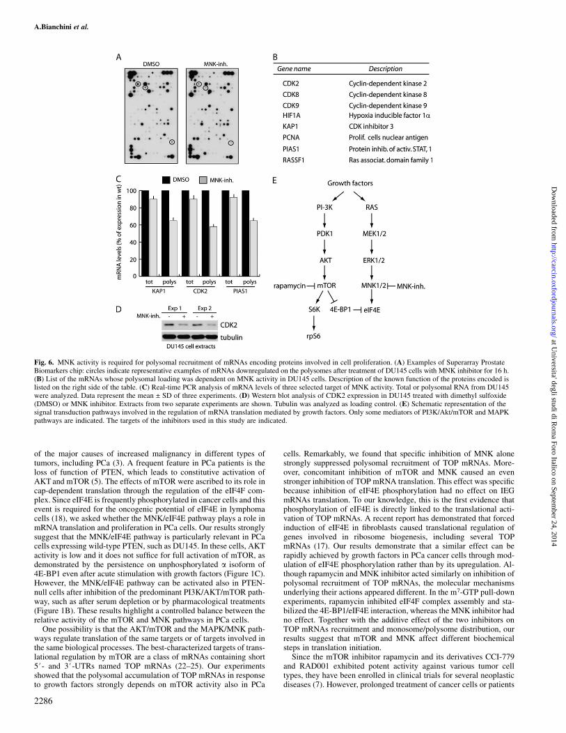
mTOR activity regulates translation of specific mRNAs in differentcell types (25–27). On the other hand, with the exception of MCL-1(18), no information is available on the requirement of eIF4E phos-phorylation for translational regulation of specific genes. To deter-mine whether, in addition to TOP mRNAs, eIF4E phosphorylationalso affected other mRNAs, we performed a microarray analysis.DU145 cells were chosen because of their higher sensitivity to theMNK inhibitor. Polysomal mRNAs were purified from cells culturedfor 16 h in the presence or absence of MNK inhibitor and hybridizedto a microarray chip containing 263 genes with known relevance toPCa (21). We observed that treatment with MNK inhibitor did notaffect the overall pattern of mRNAs present on the polysomes(Figure 6A). Nevertheless, several mRNAs were selectively down-regulated (Figure 6B). Most of these mRNAs encode for proteinsinvolved in cell cycle progression (CDK2, CDK8, CDK9 andKAP1), which might explain the strong effect on cell proliferationobserved in DU145 cells by the MNK inhibitor. In addition, RASSF1is a regulator of cell cycle and apoptosis and its locus is often aber-rantly methylated in PCa (28), whereas PCNA is a cofactor of DNApolymerases that recruits crucial players to the replication fork (29).PIAS1 regulates protein sumoylation and affects checkpoints of theexit from G1 and G2 phases of the cell cycle (30). HIF1a is a tran-
scription factor that induces angiogenesis in PCa and its expression isregulated by the AKT/mTOR pathway in the PTEN-negative PC3cells (31). The microarray results were validated by real-time PCRanalysis for three of the targets: KAP1, CDK2 and PIAS1. TheseRNAs were decreased in the polysomal pool after treatment withMNK inhibitor, whereas they were only marginally affected at thetotal level (Figure 6C). Moreover, inhibition of MNK also causeda detectable decrease in CDK2 protein levels (Figure 6D), confirmingthe data obtained at the mRNA level. These results indicate thatphosphorylation of eIF4E is required for efficient translation of spe-cific mRNAs involved in cell cycle progression in PCa cells.
Discussion
In this study, we have investigated the activation and function ofsignaling pathways involved in the regulation of mRNA translationin PCa cells (Figure 6E). Our experiments provide four major con-clusions: (i) the activity of the mTOR and MNK pathways is undera controlled balance in PCa cells and downregulation of the former iscorrelated with activation of the latter; (ii) translation of TOP mRNAsencoding for ribosomal proteins depends on activation of both themTOR and MNK pathways by growth factors; (iii) concomitant in-hibition of the mTOR and MNK pathway strongly suppresses proteinsynthesis, cell cycle progression and proliferation of PCa cells and(iv) inhibition of the MNK pathway leads to translational repressionof mRNAs for proteins involved in cell cycle.
The rationale of this study stems from the widely accepted notionthat deregulation of the PI3K/AKT/mTOR signaling pathway is one
Fig. 5. Cotreatment with rapamycin and MNK inhibitor causes cell cycle exit in PCa cells. PC3 (A and B) and DU145 (C and D) prostate cancer cells were treatedwith the indicated inhibitors for 72 h. Cells were collected and stained with propidium iodide for FACS analysis (A and C) or lysed for western blot analysis (B and D)with antibodies for cyclin D1, cyclin A or cyclin B. Ten micrograms of total extracts were loaded on sodium dodecyl sulfate–polyacrylamide gel electrophoresis.Results are the mean ± SD (A and C) or representative (B and D) of three experiments.
mRNA translation in prostate cancer cells
2285
at Universita' degli studi di R
oma Foro Italico on Septem
ber 24, 2014http://carcin.oxfordjournals.org/
Dow
nloaded from
of the major causes of increased malignancy in different types oftumors, including PCa (3). A frequent feature in PCa patients is theloss of function of PTEN, which leads to constitutive activation ofAKT and mTOR (5). The effects of mTOR were ascribed to its role incap-dependent translation through the regulation of the eIF4F com-plex. Since eIF4E is frequently phosphorylated in cancer cells and thisevent is required for the oncogenic potential of eIF4E in lymphomacells (18), we asked whether the MNK/eIF4E pathway plays a role inmRNA translation and proliferation in PCa cells. Our results stronglysuggest that the MNK/eIF4E pathway is particularly relevant in PCacells expressing wild-type PTEN, such as DU145. In these cells, AKTactivity is low and it does not suffice for full activation of mTOR, asdemonstrated by the persistence on unphosphorylated a isoform of4E-BP1 even after acute stimulation with growth factors (Figure 1C).However, the MNK/eIF4E pathway can be activated also in PTEN-null cells after inhibition of the predominant PI3K/AKT/mTOR path-way, such as after serum depletion or by pharmacological treatments(Figure 1B). These results highlight a controlled balance between therelative activity of the mTOR and MNK pathways in PCa cells.
One possibility is that the AKT/mTOR and the MAPK/MNK path-ways regulate translation of the same targets or of targets involved inthe same biological processes. The best-characterized targets of trans-lational regulation by mTOR are a class of mRNAs containing short5#- and 3#-UTRs named TOP mRNAs (22–25). Our experimentsshowed that the polysomal accumulation of TOP mRNAs in responseto growth factors strongly depends on mTOR activity also in PCa
cells. Remarkably, we found that specific inhibition of MNK alonestrongly suppressed polysomal recruitment of TOP mRNAs. More-over, concomitant inhibition of mTOR and MNK caused an evenstronger inhibition of TOP mRNA translation. This effect was specificbecause inhibition of eIF4E phosphorylation had no effect on IEGmRNAs translation. To our knowledge, this is the first evidence thatphosphorylation of eIF4E is directly linked to the translational acti-vation of TOP mRNAs. A recent report has demonstrated that forcedinduction of eIF4E in fibroblasts caused translational regulation ofgenes involved in ribosome biogenesis, including several TOPmRNAs (17). Our results demonstrate that a similar effect can berapidly achieved by growth factors in PCa cancer cells through mod-ulation of eIF4E phosphorylation rather than by its upregulation. Al-though rapamycin and MNK inhibitor acted similarly on inhibition ofpolysomal recruitment of TOP mRNAs, the molecular mechanismsunderlying their actions appeared different. In the m7-GTP pull-downexperiments, rapamycin inhibited eIF4F complex assembly and sta-bilized the 4E-BP1/eIF4E interaction, whereas the MNK inhibitor hadno effect. Together with the additive effect of the two inhibitors onTOP mRNAs recruitment and monosome/polysome distribution, ourresults suggest that mTOR and MNK affect different biochemicalsteps in translation initiation.
Since the mTOR inhibitor rapamycin and its derivatives CCI-779and RAD001 exhibited potent activity against various tumor celltypes, they have been enrolled in clinical trials for several neoplasticdiseases (7). However, prolonged treatment of cancer cells or patients
Fig. 6. MNK activity is required for polysomal recruitment of mRNAs encoding proteins involved in cell proliferation. (A) Examples of Superarray ProstateBiomarkers chip: circles indicate representative examples of mRNAs downregulated on the polysomes after treatment of DU145 cells with MNK inhibitor for 16 h.(B) List of the mRNAs whose polysomal loading was dependent on MNK activity in DU145 cells. Description of the known function of the proteins encoded islisted on the right side of the table. (C) Real-time PCR analysis of mRNA levels of three selected target of MNK activity. Total or polysomal RNA from DU145were analyzed. Data represent the mean ± SD of three experiments. (D) Western blot analysis of CDK2 expression in DU145 treated with dimethyl sulfoxide(DMSO) or MNK inhibitor. Extracts from two separate experiments are shown. Tubulin was analyzed as loading control. (E) Schematic representation of thesignal transduction pathways involved in the regulation of mRNA translation mediated by growth factors. Only some mediators of PI3K/Akt/mTOR and MAPKpathways are indicated. The targets of the inhibitors used in this study are indicated.
A.Bianchini et al.
2286
at Universita' degli studi di R
oma Foro Italico on Septem
ber 24, 2014http://carcin.oxfordjournals.org/
Dow
nloaded from

with mTOR inhibitors causes a positive feedback on PI3K activitythat leads to phosphorylation of AKT and eIF4E (12,13). This elicitsan anti-apoptotic response that may confer resistance to rapamycintreatment. Herein, we have observed that upregulation of eIF4E phos-phorylation occurs also in PCa cells treated with mTOR inhibitors andit was completely reverted by cotreatment with the MNK inhibitor,indicating a role for MNK in this event. A similar inhibition of eIF4Ephosphorylation could be achieved by concomitant inhibition of ex-tracellular regulated kinase 1/2 and p38 activity, but not by eitherkinase alone (data not shown), suggesting that both upstream MAPKscan fuel MNK activity in PCa cells. In line with the additive effects ofrapamycin and MNK inhibitor on TOP mRNA translation, we foundthat overall protein synthesis was also strongly suppressed by con-comitant treatment of PCa cells with these inhibitors. Moreover, in-hibition of both pathways was required to cause proliferation arrestand exit from the cell cycle, as indicated by the dramatic reduction inthe levels of mitotic cyclins. Remarkably, the MNK inhibitor alonehad a stronger effect than rapamycin on proliferation in DU145 cells,which express functional PTEN, indicating that this inhibitor might beparticularly effective in cancer cells in which AKT is not constitu-tively active. Since eIF4E was highly phosphorylated even in serum-depleted DU145, our results suggest that the constitutive activation ofthis pathway in PTEN-expressing PCa cells is an attempt to compen-sate for the lower levels of activity of the AKT/mTOR pathway.Nevertheless, our observations in PC3 cells strongly suggest that co-treatment with mTOR and MNK inhibitors also limits the growthpotential of PTEN-null PCa cells, especially under conditions inwhich the AKT/mTOR pathway is inhibited. An important advantageof targeting MNK activity in cancer cells is given by the absence ofphenotype in mice deleted of the MNK1 and MNK2 genes (15), in-dicating that MNK activity might be more stringently required forcancer cells than normal cells. Thus, the concomitant use of rapamy-cin and MNK inhibitors might be beneficial to PCa patients in whichthe PI3K/AKT or MAPK/MNK pathways are deregulated.
Intriguingly, phosphorylation of eIF4E is turned on under condi-tions that are clearly opposed, such as after growth factors stimulationor after various types of stresses (16,32). Although not required duringdevelopment (15), phosphorylation of eIF4E on serine 209 correlateswith cancer cell proliferation (16). eIF4E is upregulated in manytumors and its elevated expression promoted cell transformation(17,33). Furthermore, a recent report demonstrated that the oncogenicpotential of eIF4E depends on phosphorylation of serine 209 by MNKand that a similar efficiency in neoplastic transformation could beobtained by upregulation of a constitutively active form of MNK1instead of eIF4E (18). These results strongly indicate that phosphor-ylation of eIF4E plays a crucial role in neoplastic transformation andit is not a side effect caused by altered signaling pathways in thetransformed cells. Phosphorylation of eIF4E was hypothesized toaffect translational control in lymphoma cells, but only one potentialtarget, the anti-apoptotic protein Mcl-1, was identified in that study(18). In addition to TOP mRNAs, using the highly MNK-sensitiveDU145 cell line, we have now identified eight new translationallyregulated targets of the MNK/eIF4E pathway. Many of these mRNAsencode for proteins involved in cell cycle transitions or cell prolifer-ation, such as cyclin-dependent kinases or inhibitors. An interestingtarget of MNK activity is the HIF1a mRNA, which encodes for a tran-scription factor that modulates angiogenesis and confers adaptation tohypoxic conditions leading to increased resistance to therapies (34).HIF1a expression is regulated by the AKT pathway in PC3 cells (31).However, under hypoxic conditions, when the AKT/mTOR pathwayis inhibited, HIFa is one of the few mRNAs that remains associated toactively translating polysomes in PC3 cells (35). Our results suggestthat activation of the MNK pathway might sustain HIF1a translationunder these conditions and compensate for the inhibition of themTOR pathway. Indeed, hypoxia induces a cellular stress and eIF4Ephosphorylation strongly increases upon various stresses in most can-cer cells (16,32).
In conclusion, our results indicate that a fine balance between theactivity of the AKT/mTOR and the MAPK/MNK pathway in PCa
cells maintains translation of specific mRNAs required for ribosomebiogenesis, cell proliferation and stress response and might conferthem the ability to overcome negative insults.
Supplementary material
Supplementary Table 1 and Figures 1 and 2 can be found at http://carcin.oxfordjournals.org/
Funding
Associazione Italiana Ricerca sul Cancro; Ministry of Education(PRIN 2004, PRIN 2006); Istituto Superiore della Sanita (Projectno. 527/B/3A/5); Lance Armstrong Foundation;
Acknowledgements
We wish to thank Dr Manuela Cappellari for assistance with cell cultures andtreatments, Dr Federica Barbagallo for assistance with FACS analyses andDr Daniela Barila (Fondazione Santa Lucia) for the gift of reagents and helpfuldiscussion. M.P.P. is the recipient of a Postdoctoral Fellowship from the Fon-dazione Santa Lucia.
Conflict of Interest Statement: None declared.
References
1.Feldman,B.J. et al. (2001) The development of androgen-independent pros-tate cancer. Nat. Rev. Cancer, 1, 34–45.
2.Sansal,I. et al. (2004) The biology and clinical relevance of the PTENtumor suppressor pathway. J. Clin. Oncol., 22, 2954–2963.
3.Shaw,R.J. et al. (2006) Ras, PI(3)K and mTOR signalling controls tumourcell growth. Nature, 441, 424–430.
4.Trotman,L.C. et al. (2003) Pten dose dictates cancer progression in theprostate. PLoS Biol., 1, E59.
5.Majumder,P.K. et al. (2005) Akt-regulated pathways in prostate cancer.Oncogene, 24, 7465–7474.
6.Wendel,H.G. et al. (2004) Survival signalling by Akt and eIF4E in onco-genesis and cancer therapy. Nature, 428, 332–337.
7.Tsang,C.K. et al. (2006) Targeting mammalian target of rapamycin(mTOR) for health and diseases. Drug Discov. Today, 12, 112–124.
8.Hay,N. et al. (2004) Upstream and downstream of mTOR. Genes Dev., 18,1926–1945.
9.Wullschleger,S. et al. (2006) TOR signaling in growth and metabolism.Cell, 124, 471–484.
10.Raught,B. et al. (2004) Phosphorylation of eucaryotic translation initiationfactor 4B Ser422 is modulated by S6 kinases. EMBO J., 23, 1761–1769.
11.Garcia,J.A. et al. (2008) Mammalian target of rapamycin inhibition asa therapeutic strategy in the management of urologic malignancies. Mol.Cancer Ther., 7, 1347–1354.
12.Sun,S.Y. et al. (2005) Activation of Akt and eIF4E survival pathways byrapamycin-mediated mammalian target of rapamycin inhibition. CancerRes., 65, 7052–7058.
13.O’Reilly,K.E. et al. (2006) mTOR inhibition induces upstream receptortyrosine kinase signaling and activates Akt. Cancer Res., 66, 1500–1508.
14.Manning,B.D. et al. (2007) AKT/PKB signaling: navigating downstream.Cell, 129, 1261–1274.
15.Ueda,T. et al. (2004) Mnk2 and Mnk1 are essential for constitutive andinducible phosphorylation of eukaryotic initiation factor 4E but not for cellgrowth or development. Mol. Cell. Biol., 24, 6539–6549.
16.De Benedetti,A. et al. (2004) eIF-4E expression and its role in malignanciesand metastases. Oncogene, 23, 3189–3199.
17.Mamane,Y. et al. (2007) Epigenetic activation of a subset of mRNAs byeIF4E explains its effects on cell proliferation. PLoS ONE, 2, e242.
18.Wendel,H.G. et al. (2007) Dissecting eIF4E action in tumorigenesis. GenesDev., 21, 3232–3237.
19.Paronetto,M.P. et al. (2006) The nuclear RNA-binding protein Sam68translocates to the cytoplasm and associates with the polysomes in mousespermatocytes. Mol. Biol. Cell, 17, 14–24.
20.Sette,C. et al. (2002) Tr-kit-induced resumption of the cell cycle in mouseeggs requires activation of a Src-like kinase. EMBO J, 21, 5386–5395.
mRNA translation in prostate cancer cells
2287
at Universita' degli studi di R
oma Foro Italico on Septem
ber 24, 2014http://carcin.oxfordjournals.org/
Dow
nloaded from

21.Busa,R. et al. (2007) The RNA-binding protein Sam68 contributes toproliferation and survival of human prostate cancer cells. Oncogene, 26,4372–4382.
22.Meyuhas,O. et al. (2000) Translational control of TOP mRNAs. InSonenberg,N., Hershey,J.W.B. and Mathews,M.B. (eds.) TranslationalControl of Gene Expression. Cold Spring Harbor Laboratory Press, ColdSpring Harbor, NY, pp. 671–693.
23. Jefferies,H.B.J. et al. (1997) Rapamycin suppresses 5#TOP mRNA trans-lation through inhibition of p70s6k. EMBO J., 16, 3693–3704.
24.Caldarola,S. et al. (2004) Translational regulation of terminal oligopyrimi-dine mRNAs induced by serum and amino acids involves distinct signalingevents. J. Biol. Chem., 279, 13522–13531.
25.Bilanges,B. et al. (2007) Tuberous sclerosis complex proteins 1 and 2control serum-dependent translation in a TOP-dependent and -independentmanner. Mol. Cell. Biol., 27, 5746–5764.
26.Gera,J.F. et al. (2004) AKT activity determines sensitivity to mammaliantarget of rapamycin (mTOR) inhibitors by regulating cyclin D1 and c-mycexpression. J. Biol. Chem., 279, 2737–2746.
27.Grolleau,A. et al. (2002) Global and specific translational control by rapa-mycin in T cells uncovered by microarrays and proteomics. J. Biol. Chem.,277, 22175–22184.
28.Dammann,R. et al. (2005) The tumor suppressor RASSF1A in humancarcinogenesis: an update. Histol. Histopathol., 20, 645–663.
29.Moldovan,G.L. et al. (2007) PCNA, the maestro of the replication fork.Cell, 129, 665–679.
30.Munarriz,E. et al. (2004) PIAS-1 is a checkpoint regulator which affectsexit from G1 and G2 by sumoylation of p73. Mol. Cell. Biol., 24, 10593–10610.
31.Fang,J. et al. (2007) PI3K/PTEN/AKT signaling regulates prostate tumorangiogenesis. Cell. Signal., 19, 2487–2497.
32.Mamane,Y. et al. (2004) eIF4E—from translation to transformation.Oncogene, 2, 3172–3179.
33.Ruggero,D. et al. (2004) The translation factor eIF-4E promotes tumorformation and cooperates with c-Myc in lymphomagenesis. Nat. Med.,10, 484–486.
34.Kimbro,K.S. et al. (2006) Hypoxia-inducible factor-1 in human breast andprostate cancer. Endocr. Relat. Cancer, 13, 739–749.
35.Thomas,J.D. et al. (2007) Identification of mRNAs that continue to asso-ciate with polysomes during hypoxia. RNA, 13, 1116–1131.
Received July 22, 2008; revised September 5, 2008;accepted September 12, 2008
A.Bianchini et al.
2288
at Universita' degli studi di R
oma Foro Italico on Septem
ber 24, 2014http://carcin.oxfordjournals.org/
Dow
nloaded from
Copyright © 2022 FDOKUMEN