Scatter and attenuation correction in technetium-99m brain SPECT
Upload
independentCategory
view
5download
0
Development of an Optimized Activatable MMP-14 targetedSPECT Imaging Probe
Gregory A. Watkinsa, Ella Fung Jonesa,*, M. Scott Shellb, Henry F. VanBrocklina,c, Mei-HsiuPana, Stephen M. Hanrahanc, Jin Jin Fenga, Jiang Hea, Nor Eddine Sounnid, Ken A. Dillb,Christopher H. Contage, Lisa M. Coussensd, and Benjamin L. Franca
a Center for Molecular and Functional Imaging, Department of Radiology and Biomedical ImagingUniversity of California, San Francisco, 185 Berry Street, Suite 350, Box 0946, San Francisco, CA 94107
b Department of Pharmaceutical Chemistry, University of California, San Francisco, San Francisco, CA.94143
c Department of Functional Imaging, Lawrence Berkeley National Laboratory, Berkeley, CA 94720
d Department of Pathology, University of California, San Francisco, San Francisco, CA 94115
e Departments of Pediatrics, Radiology, and Microbiology & Immunology; Molecular Imaging Program,Stanford University, Stanford, CA 94305, United States
AbstractMatrix Metalloproteinase-14 (MT1-MMP or MMP-14) is a membrane-associated proteaseimplicated in a variety of tissue remodeling processes and a molecular hallmark of select metastaticcancers. The ability to detect MMP-14 in vivo would be useful in studying its role in pathologicprocesses and may potentially serve as a guide for the development of targeted molecular therapies.Four MMP-14 specific probes containing a positively charged cell penetrating peptide (CPP) d-arginine octamer (r8) linked with a MMP-14 peptide substrate and attenuating sequences withglutamate (8e, 4e) or glutamate-glycine (4eg and 4egg) repeating units were modeled using anAMBER force field method. The probe with 4egg attenuating sequence exhibited the highest CPP/attenuator interaction, predicting minimized cellular uptake until cleaved. The in vitro MMP-14-mediated cleavage studies using the human recombinant MMP-14 catalytic domain revealed anenhanced cleavage rate that directly correlated with the linearity of the embedded peptide substratesequence. Successful cleavage and uptake of a technetium-99m labeled version of the optimal probewas demonstrated in MMP-14 transfected human breast cancer cells. Two- fold reduction of cellularuptake was found in the presence of a broad spectrum MMP inhibitor. The combination ofcomputational chemistry, parallel synthesis and biochemical screening, therefore, shows promise asa set of tools for developing new radiolabeled probes that are sensitive to protease activity.
KeywordsCancer; MMP-14; Protease sensitive probe; SPECT imaging
*Corresponding author: Ella F. Jones, Department of Radiology and Biomedical Imaging, University of California San Francisco, 185Berry Street, Suite 350, San Francisco, CA 94107, Tel: (415)353-9469, Fax: (415)353-9423, email: [email protected]'s Disclaimer: This is a PDF file of an unedited manuscript that has been accepted for publication. As a service to our customerswe are providing this early version of the manuscript. The manuscript will undergo copyediting, typesetting, and review of the resultingproof before it is published in its final citable form. Please note that during the production process errors may be discovered which couldaffect the content, and all legal disclaimers that apply to the journal pertain.
NIH Public AccessAuthor ManuscriptBioorg Med Chem. Author manuscript; available in PMC 2010 January 15.
Published in final edited form as:Bioorg Med Chem. 2009 January 15; 17(2): 653–659. doi:10.1016/j.bmc.2008.11.078.
NIH
-PA Author Manuscript
NIH
-PA Author Manuscript
NIH
-PA Author Manuscript

1. IntroductionTumor cells employ a host of biochemical mechanisms in order to invade and metastasize.Many of these mechanisms are thought, in part, to involve proteases associated with cellmembrane and extracellular matrix (ECM) molecules that are posited to initiate pro-angiogenicsignaling cascades. Among cancer-associated proteases, matrix metalloproteases (MMPs), aclass of zinc-dependent proteolytic enzymes, have been postulated to be used by cancer cellsto dissolve ECM during neoplastic progression.1 In addition, numerous studies havedocumented a positive correlation between certain MMP expression levels and poor outcomein cancer patients.2 The importance of MMPs in tumor progression not only has guided thedevelopment of MMP inhibitors for therapy, it has also received particular attention as imagingtarget utilizing methods to detect tumor-associated proteolytic activity in vivo. 3–7
The family of human MMPs contains 16 secreted and 7 membrane-tethered enzymes. 8 Asubclass of the membrane-anchored proteinases, termed membrane type (MT) MMPs, playsdominant roles in controlling cancer cell behavior. 9,10 In particular, the up-regulation of themembrane-associated collagenase MMP-14 (MT1-MMP) correlates to the invasiveness ofmany different tumor types.2 MMP-14 not only promotes tumor growth through induction ofangiogenesis and proteolysis of ECM, but it also acts as a critical regulatory switch in theactivation of MMP-2 proenzyme.11 Clinical studies revealed that the expression of MMP-14is associated with poor prognosis in patients with advanced neuroblastoma,12 small cell lungcancer (SCLC),13 tongue squamous cell carcinoma,14 head and neck carcinoma,15 bladder,and ovarian cancer.16,17. MMP-14 has been detected in tumor cells and adjacent stromal cellsin a variety of human tumors including breast. 9 Consequently, MMP-14 overexpression holdsgreat promise as an early biomarker for invasive cancers.
In the past, several in vivo optical imaging probes targeting various MMPs have been reported;the most successful of these efforts have been directed against MMP-2, -7, and -9.18–23Attempts to image MMP activities by non-optical modalities (e.g., positron emissiontomography (PET) or single photon emission computed tomography (SPECT)) using labeledsubstrates or inhibitors, however, have met with limited success in vivo, in part due to the poorspecificity and in vivo stability of the radiolabeled probes.24–31 Our motivation, therefore,was to develop a sensitive nuclear probe for MMP-14 activities for early cancer detection. Thesuccess of such a probe would represent a significant advancement in preclinical and clinicalimaging as it would be a tool able to locate and track the molecular evolution of malignanttissues for use in drug development.
A number of protease imaging strategies have been described previously. One particular classof probes comprising an “activatable” delivery mechanism has been developed by a numberof research groups.21,23,32 These probes share a core structure consisting of a poly-d-argininecell-penetrating peptide (CPP) that is covalently tethered to a negatively charged attenuatingpeptide sequence through a proteolysis sensitive peptide (Figure 1). The intact probe is believedto be prohibited from crossing the cell membrane due to the electrostatic interaction betweenthe positively charged arginine and the tethered intramolecular negatively charged attenuator(I), but other mechanisms may also contribute. However, proteolytic cleavage (II) separatesthe polyarginine sequence from the negatively charged domain, thereby triggering uptake ofthe CPP (III). Protease-rich tissues may be imaged by tagging an imaging reporter group, suchas a fluorophore, gadolinium chelate or radionuclide, to the CPP.32 As a result, the number ofprotease cleavage events may be correlated to the CPP concentration, and its associated tag,within targeted cells. Using agents directed against MMP-2/9 and -7, Rao and coworkers haveselectively tagged cultured fibrosarcoma cells (HT-1080) with quantum dots, 23 while Tsienand co-workers successfully imaged cancers rich in MMP-2 and -9 in murine xenografts, usingoptical and magnetic resonance techniques.21
Watkins et al. Page 2
Bioorg Med Chem. Author manuscript; available in PMC 2010 January 15.
NIH
-PA Author Manuscript
NIH
-PA Author Manuscript
NIH
-PA Author Manuscript

This general strategy is attractive because the catalytic processing of more than one probe byeach enzyme provides a robust mechanism for signal amplification. For the purpose ofMMP-14 imaging, this is a particularly important point if one seeks to detect protease activityprior to the maturation of secondary downstream proteases, e.g., MMP-2 and -9.
The development of an “activatable” SPECT imaging probe specific for MMP-14 is reportedherein. The probe design was undertaken realizing that attaching a large metal chelate fornuclear imaging may alter the topology of the MMP-14 selective peptide sequence andadversely affect the cleavage rate as well as attenuation characteristics of the basic probeplatform. Therefore the combination of molecular modeling, parallel synthesis and bioassayscreens were effectively utilized to optimize the imaging probe construct for MMP-14activities. This work sheds new light on the intramolecular quenching and activationmechanisms of this class of imaging peptides, and demonstrates the value of computationalchemistry relative to imaging probe development.
2. Results and Discussion2.1. MMP-14 Probe Design and Modular Components
The general MMP-14 probe, shown in Figure 2, is a modular design comprised of threecomponents: A) a positively charged d-arginine octamer (r8) cell penetrating peptide (CPP)attached with single amino acid chelate (SAAC) for technetium-99m; B) a MMP-14 specificcleavable substrate (SGRIGF↓LRTA) and C) a negatively charged attenuation sequence. Theseminal component for designing an effective MMP-14 probe was a suitable cleavable peptidesubstrate. A known MMP-14 substrate (SGRIGF↓LRTA) was incorporated into our constructsto maximize the selectivity of the probe for MMP-14-rich tissues. This sequence was originallydescribed by Smith and co-workers, who employed phage display technology to map thesubstrate specificity of matrix metalloproteinases.33 In an enzyme panel study, this peptidesequence is preferentially cleaved by MMP-14 with a kcat/Km value of 777,200 M−1/s, whilethe cleavage by the related MMP-9 is much less efficient, with a kcat/Km of 20,000 M−1/s.
Although poly-d-arginine sequences with chain lengths of 7–14 residues have been widelyemployed as cell membrane penetrating transporters34 in probe constructs, there has been littleconsensus over the degree of attenuation necessary for effective proteolytic cleavage andreducing cell penetration. In designing the MMP-14 probe, the attenuation module wasoptimized considering: 1) the number of negatively charged residues for effective attenuation;2) the structural impact on the cleavable peptide sequence and 3) the ability to release thecleaved probe for cellular uptake. Attenuation of d-arginine octamer (r8) CPP was favorablyachieved by inserting the optimal number of d-glutamate residues in the attenuation domain,such that the cleavable peptide sequence was present in a roughly linear conformation andavailable for docking to the MMP-14 enzyme. A small panel of attenuation sequences,including four d-glutamates (4e), four d-glutamate-glycine repeats (4eg), four d-glutamate-glycine-glycine repeats (4egg) and eight contiguous d-glutamates (8e), were evaluated usingcomputer modeling. The d version of the amino acid residues were chosen to prevent non-specific in vivo proteolysis. Based on prior experience, the choice of four d-glutamatesappropriately spaced in three of the four attenuator sequences adequately attenuates the r8peptide.32
Finally, in order to conduct radionuclide imaging by SPECT, it is necessary to selectively labelthe peptide probe with a suitable isotope. To this end, the single amino acid chelate (SAAC)technology, previously developed by Stephenson, et al., 35,36 has been utilized. The SAACis a Nε-bis(pyridylmethyl)-lysine derivative that may be site-selectively incorporated intosynthetic peptides using standard Fmoc chemistry. The resulting compounds carry an N-3chelator that form stable tricarbonyl complexes with technetium and rhenium.
Watkins et al. Page 3
Bioorg Med Chem. Author manuscript; available in PMC 2010 January 15.
NIH
-PA Author Manuscript
NIH
-PA Author Manuscript
NIH
-PA Author Manuscript

Technetium-99m, a gamma ray emitter, has emissions of sufficient energy (140 KeV) topenetrate the human body, but not too energetic to pass through the SPECT detector material.It is considered to be a near-ideal isotope for SPECT imaging, in part due to its short half-life(6 h), its gamma emission energy and its availability from a commercial generator system.Based on these characteristics, technetium-99m has been chosen in the current probe designto visualize cell penetration upon proteolysis.
2.2. Computational ChemistryHolding the CPP and MMP-14 peptide regions constant and using the cysteine-cysteinedisulfide bond as a linker between modules, the overall structure with four different attenuationsequences containing d-glutamate residues (4e, 4eg, 4egg and 8e) was computed. The extentof charge-complementation was calculated between the CPP and the attenuation sequence byexamining the number of arginine residues that were in contact with the glutamate residues.The average end-to-end backbone length of the cleaved substrate was measured to quantifythe extent of exposed substrate, in an available extended conformation, for effective enzymepocket binding for subsequent cleavage by the MMP-14. Based on these calculations, thedominant conformation for each probe, shown in Figure 3, was determined.
These dominant structures reveal that the 4e probe has minimum interaction between the CPP(blue) and the attenuation sequence (red). Structural calculations confirm the overlap finding(Figure 4A) with the 4e probe possessing the least CPP-attenuator complementation at anaverage of 4.3 ±1.5 arginine residues in contact with d-glutamate while contact in the otherthree probes range from 5.2 ±1.6 to 6.4 ±1.2 residues. Here, the standard deviation reporteddoes not indicate the standard error in the measurement. Instead, it corresponds to the rootmean square fluctuation of the measurement within the computed peptide conformationalensemble, that is due to the natural conformational fluctuations experienced by the peptide atambient conditions. These data suggest that the 4e motif may not be as effective in attenuatingthe translocation of the CPP prior to proteolytic cleavage. As the overall probe structure wasexamined, the CPP-attenuator complementation was also found to influence the conformationof the MMP-14 substrate region, potentially altering the accessibility for proteolytic cleavage.As the end-to-end distance of the MMP-14 substrate is a direct measure of peptide folding, italso indirectly predicts the availability of the peptide substrate for cleavage. The calculationsshow (Figure 4B) that the 4egg probe best adapts the β-strand conformation 33 required forrapid MMP-14 cleavage, exhibiting the longest end-to-end distance of 21.62 ± 4.3 Å. The 4e(16.9 ± 2.9 Å) and 4eg (17.1± 4.3 Å) sequences show a similar degree of folding, whereas the8e probe is the most distorted from linearity (15.0 ± 2.9 Å). This modeling study predicts thatthe 4egg probe has adequate CPP attenuation in the absence of MMP-14 and allows a ®-strandtype conformation for the substrate portion of the probe to bind and interact with the MMP-14catalytic domain.
2.3 Parallel Probe SynthesisA modular approach to the panel design was adapted to accelerate the synthesis of the peptideprobes. Each probe was retro-synthetically divided into two fragments of comparable size. Onepeptide fragment common to all of the probes contains the CPP and the SAAC with anadditional cysteine and glycine appended to the N-terminus. Four variable fragments,containing the SGRIGF↓LRTA sequence and the attenuators, were also appended with glycineand cysteine at the C-terminus. This allowed all five fragments (1 conserved, 4 specific to eachprobe) to be synthesized in parallel and to be subsequently assembled into the probe construct(Figure 2) through simple disulfide chemistry.
Typical synthesis of the various probes yielded the desired constructs along with two homo-dimers. The isolated probes were characterized by analytical HPLC, MALDI-MS and 1H
Watkins et al. Page 4
Bioorg Med Chem. Author manuscript; available in PMC 2010 January 15.
NIH
-PA Author Manuscript
NIH
-PA Author Manuscript
NIH
-PA Author Manuscript

NMR. As expected, the desired MMP-14 probes exhibited retention times between the homo-dimer peaks. MALDI-MS analysis showed the expected molecular ion peak along with thecharacteristic fragmentation pattern produced by homolytic cleavage of the disulfide bond andloss of pyridylmethylene radicals from the SAAC (Figure 5).
2.4 Proteolytic Cleavage Study with Purified MMP-14 EnzymeA sample of each probe was subjected to enzymatic digestion by MMP-14, followed by HPLCanalysis. As expected, each probe generated two clear cleavage products upon proteolysis,indicating that the cleavage event was largely restricted to a single cleavage site. The cleavagerate was characterized by the probe half-lives and initial cleavage velocities based on the first-order kinetics. Significant differences in cleavage rates between the probes were observed. Asshown in Figure 4C, the increasing cleavage rate rank order was 8e < 4e ≈ 4eg < 4egg.Regression analyses confirm that all eight data sets are strongly linear, with r2 values rangingfrom 0.90 to 0.99. These data correlate well with the end-to-end distance measures derivedfrom the molecular modeling studies where the 8e probe MMP-14 substrate was most distortedfrom linearity, the 4e and 4eg probes were similar length yet longer than the 8e probe, whilethe 4egg probe possessed the longest end-to-end distance.
As previously suggested by Smith and co-workers, 33 the SGRIGF↓LRTA cleavage sequenceis believed to possess the β-strand conformation for efficient binding to MMP-14 and tofacilitate the subsequent catalytic reaction. In the present study, protein structure related to theinteraction of the attenuator with the CPP changes the embedded MMP-14 substrateconformation directly affecting the cleavage rate. The molecular modeling of this small libraryof structures efficiently predicts the cleavage rate as a function of substrate linearity. Thus, theattenuation sequence may be chosen for effective attenuation of the CPP and presentation ofan optimized structural conformation for rapid enzymatic cleavage.
2.5 Radiochemistry with 99mTcThe carboxylate terminal of the r8 CPP was linked to a single amino acid chelate (SAAC)through a peptide bond. Based on our results in molecular modeling and MMP-14 cleavagestudy, the optimal probe, 4egg, was chosen to demonstrate labeling efficiency. The 4egg probewas successfully labeled with technetium-99m through the [99mTc(CO)3(OH2)3]+intermediate. The final 99mTc- labeled probe was isolated by HPLC with 97% radiopurity anda radiochemical yield greater than 90%.
The SAAC technology, developed by Zubieta and colleagues, has shown great promise in smallmolecule radiopharmaceutical applications. 35,37,38 The SAAC, a lysine based residue witha built-in chelating system for binding rhenium or technetium tricarbonyl, was chosen overdirect peptide labeling techniques to avoid non-specific binding of the label on the backbonestructure. The SAAC also allows labeling of the molecule as the final step. Labeling anintermediate that is subsequently reacted with the final molecule may reduce yield, while bulkychelating molecules or bifunctional chelates used in direct labeling may significantly effectbiodistribution of the labeled compound. 39 For instance, Polyakov and colleagues havesynthesized a Tat CPP incorporating a peptide-based motif (epsilon-KGC) that provides anN3S donor core for chelating technetium and rhenium. They have successfully demonstratedits uptake in Jurkat cells after labeling with oxotechnetium(V) or oxorhenium(V). 40 Althoughthis approach offers labeling specificity, each donor core requires three additional residues.The current probe design and prototype, on the other hand, utilizes a single residue for eachtechnetium or rhenium tag. In addition, this strategy for radiolabeling provides control overthe isotope delivery system thereby permitting the MMP-14 probe to be labeled withtechnetium-99m (a pure gamma emitter) or rhenium-186 (a beta and gamma emitter) that will
Watkins et al. Page 5
Bioorg Med Chem. Author manuscript; available in PMC 2010 January 15.
NIH
-PA Author Manuscript
NIH
-PA Author Manuscript
NIH
-PA Author Manuscript

not only allow early detection of invasive cancer, but it will also provide a common platformfor radiotherapy.
2.6 In vitro uptake of the 99mTc labeled probeHuman epithelial breast cancer (MDA-MB-231) cells transfected with MMP-14 cDNA tooverexpress MMP-14 41 were used to evaluate the in vitro uptake of the labeled 4egg probe.The effect of probe activation through cleavage by MMP-14 at cell surface was determined bytreatment of the transfected MDA-MB-231 cells with and without the potent broad-based MMPinhibitor, GM1489. Free 99mTc-tricarbonyl complex was compared as a null control. All cellstudies were carried out in triplicate. After the treatment with the labeled 4egg probe, theadhesive cells were carefully washed with DMEM, before being released from the plate withtrypsin. The probe content within each media and cell fraction was then quantified by a gammacounter (Wizard 3, Perkin-Elmer, CT). Each data point represents the fraction of the probeuptake by cells quantified by gamma count before and after incubation and washings of cells(approximately 105 cells per well). As shown in Figure 6, there is a clear difference in thecellular uptake of probe in the presence and absence of the MMP inhibitor. There was little orno uptake of the free [Tc(CO)3]+ by the cells. Despite the larger variation in cells withoutinhibitor (may due to cell handling), the average uptake of the 4egg probe was 2 times greaterin cells without inhibitor compared to those with inhibitor. This demonstrates the successfulcleavage and increased uptake of the activated probe into MMP-14 expressing cells. Theresidual 10–15% uptake in cells treated with MMP inhibitor indicates that there may beincomplete inhibition of MMP-14 activity by the inhibitor, or a basal level of probe leakageattributable to incomplete attenuation of the CPP. The negative results from [99mTc(CO)3]+
suggests that a) there is no non-specific uptake of free [99mTc(CO)3]+ and b) the positive uptakeof the probe is the result of the cleaved probe rather than leakage of free [99mTc(CO)3 ]+ intothe transfected cells. Attempts have been made to use MDA-MB-231 cells without MMP-14transfection as a negative control. However, uptake of the 4egg probe was also observed. Thismay due to cleavage reactions by combination of other MMPs at basal level. 33 Nevertheless,the reduction in uptake by transfected cells in the presence of inhibitor suggests that the 4eggprobe possesses MMP-14 specificity, and it warrants further efforts in probe development.
3.0 ConclusionThe unique application of molecular modeling in optimization of the MMP-14 probe constructhas been demonstrated. Modeling calculations were able to provide useful information on theattenuation properties of four different sequences against the r8 CPP and the length of the MMPsubstrate that is directly related to cleavage rate. Both of attenuation and cleavage must bemaximized in the optimal probe. Although three out of four probes studied exhibited good CPPattenuation, the CPP/attenuator interaction contributed to structural modifications of theMMP-14 substrate altering the cleavage rate. The probe with the longest end-to-end substratelength, 4egg, exhibited the fastest cleavage rate. Evaluation of the 4egg probe in cellsexpressing MMP-14 demonstrated the specific activation of the probe and subsequentaccumulation of the label in the cell. The value of computational modeling to imaging probedesign has been demonstrated and this methodology will be incorporated into future tumorimaging probe platform development.
4.0 Experimental4.1 Molecular Modeling
Computer simulations were used to predict the solution structures of the peptide probesemploying the AMBER96 force field 42 with the implicit solvation model of Onufriev,Bashford, and Case. 43 This force field was validated in a separate study and shown to give
Watkins et al. Page 6
Bioorg Med Chem. Author manuscript; available in PMC 2010 January 15.
NIH
-PA Author Manuscript
NIH
-PA Author Manuscript
NIH
-PA Author Manuscript

accurate structures for peptides with both alpha and beta motifs. 44 For the non-canonicalchelate residue, the program “antechamber” in the AMBER package was used to determineatomic charges and estimate force field constants. The SAAC chelate was included in themolecule without the radiometal for the simulations.
Sampling was performed using the zipping and assembly (Z&A) methodology developed bythe Dill research group. 45 Z&A works by sampling peptides and proteins according to aputative folding mechanism: 46 peptide pieces along the chain independently first form smallbits of structure, which then either nucleate additional structure locally by reeling in nearbysections of chain (zipping), or join together with other such structured peptide pieces to formlarger units (assembly). This mechanism is implemented in simulation by first breaking apeptide into small fragments, which are simulated separately, and then by growing thesefragments by the addition of new residues and other fragments, followed in each case by furthersimulation. Sampling at each stage is performed using replica exchange molecular dynamics.47
For each of the probe candidates, the overall chain was broken into the three fragmentscorresponding to the attenuator, substrate, and translocation domains. These were simulatedseparately in replica exchange for 10ns, and conformations from each fragment from the final1ns were clustered into a maximum of 10 structures using a modified k-means algorithm.Subsequently, clustered conformations from the substrate and translocation domains werecombined (in all permutations), and used in a second 10ns replica exchange simulation,followed by additional clustering. Next, the attenuating domain structures were added,followed by 10ns of sampling, and finally, the terminal chelate residue was added, with 30nsof replica exchange as the last sampling step. The clustered conformations from the last 5nsof the 30ns run (along with their populations) were taken as the final conformational ensemble.In total, each peptide required about 300 aggregate CPU days worth of simulation time onXeon 2.4 GHz processors.
4.2 MMP-14 Probe SynthesisUronium coupling agents (2-(1H-9-azabenzotriazole-1-yl)-1,1,3,3-tetramethyluroniumhexafluorophosphate or tetrafluoroborate; “HATU” and “TBTU”) and Fmoc-protected aminoacids were purchased from Novabiochem, Inc. The Nα-Fmoc-Nε-bis(pyridylmethyl)-lysinewas received from Molecular Insight Pharmaceuticals, Inc. or synthesized as previouslydescribed.42 Solvents, including dichloromethane and dimethylformamide, were purchasedfrom VWR Scientific. All other reagents were obtained from Sigma-Aldrich ChemicalCompany, and used without further purification.
Peptide intermediates were synthesized on a Protein Technologies, Inc. Prelude peptidesynthesizer according to standard Fmoc chemistry protocols. In some cases, Fmocdeproptections were effected using 6% piperazine in dimethylformamide, 48 as opposed to themore traditional 20% piperidine in DMF. All sequences were cleaved from the resin anddeprotected using 92:3:3:1:1 trifluoroacetic acid/triisoproylsilane/water/thioanisole/1-naphthol for 2 h. Crude peptides were obtained by triturating the cleavage solution with ice-cold ether, followed by centrifugation. All peptide starting materials were purified by semi-preparative, reverse-phase HPLC (Jupiter C12 column from Phenomenex; water-acetonitrilegradient, 95% water to 5% water; 0.1% trifluoroacetic acid added to the mobile phase tomaintain a pH ~2.)
The completed probes were assembled as heterodimeric disulfides. Cysteine oxidation wasconducted using the following general procedure: Ac-CGrrrrrrrr(SAAC)-CONH2 (~1.8 mg.;1 μmol) and a suitable attenuator-MMP-14 substrate peptide (~3–4 mg., according to the massof the attenuator sequence; 1 μmol) were dissolved in 0.75 mL of 5:3:2 water/0.2 M borate
Watkins et al. Page 7
Bioorg Med Chem. Author manuscript; available in PMC 2010 January 15.
NIH
-PA Author Manuscript
NIH
-PA Author Manuscript
NIH
-PA Author Manuscript

buffer, pH 8/methanol. The peptides were pre-reduced by addition of 50 μL of 1 mg/mL tris-(2-carboxyethyl)phosphine hydrochloride, dissolved in the same reaction buffer. After 15minutes, the pH was checked to ensure the reaction mixture remained above 8; then, 100 μLof DMSO was added with swirling, followed 5 minutes later by 5 mg. of potassiumhexacyanoferrate (III). The resulting yellow solution was covered with foil, and allowed tostand overnight. Following acidification with 50 μL of acetic acid, the reaction mixture wasdiluted to ca. 1 mL with deionized water, and purified by semi-preparative HPLC (Jupiter C12column from Phenomenex; water-acetonitrile gradient, 95% water to 5% water; 0.1%trifluoroacetic acid added to the mobile phase to maintain a pH ~2.) Typically, three majorpeaks were observed at 220 nm (two peaks at 260 nm), corresponding to the desiredheterodimer and homodimerized reactants. The reactant homodimers could be recovered andreductively recycled for use in future reactions.
4.3 MMP-14 Cleavage KineticsApproximately equimolar samples of each unlabeled probe were prepared in MMP-14hydrolysis buffer (50 mM Tris-HCl, pH 7.5, 150 mM NaCl, 5 mM CaCl2, 0.025% Brij-35)49 as verified by HPLC analysis and peak integration at 260 nm. Each of these peptide stocksolutions (0.8 mL) was transferred to a separate microcentrifuge tube, and equilibrated at 37 °C for 30 minutes. Recombinant MMP-14 catalytic domain was obtained from Calbiochem, ata concentration of 200 μg/mL. This solution was diluted by half with assay buffer, and 4 μLof the resulting solution was transferred to each probe solution. Aliquots of each solution (25μL) were removed at time points of 0, 2.5, 5, 7.5, 10, 20, 40, 80, and 120 minutes, and rapidlytransferred to an HPLC vial insert containing 50 μL of ice-cold quenching reagent (0.1 MEDTA/2 Na+ in 1% HOAc, pH 3). The quenched samples were then analyzed by RP-HPLC(Jupiter C12 analytical column (Phenomenex), flow rate 1.1 mL/minutes 0–5 minutes, 95%water, 5% acetonitrile, 0.1% TFA; 5–35 minutes, ramp to 50% water, 50% acetonitrile, 0.1%TFA; then, re-equilibration at 95% water).
Probe half-lives and initial cleavage velocities were extracted from the analytical HPLC data,assuming enzyme saturation and first-order probe cleavage kinetics early in the reaction timecourse. For the 4e, 4eg, and 4egg probes, linear curves were constructed by plotting the naturallog of the intact probe peak area against time. For these probes, saturation conditions wereassumed to persist until the 20 minute time point. The 8e probe data was analyzed in a similarfashion, and included the 40 minutes time point in the analysis. Calculations were conductedtwice for each probe, using HPLC data obtained at either 220 or 260 nm. The slope of eachlinear equation was taken as a measure of initial cleavage velocity. Probe half-lives wereestimated by dividing the measured probe peak areas at t=0 in half, and substituting the resultingvalues into each regression equation to solve for t.
4.4 99mTc RadiochemistryRadiolabeling of the probes with 99mTc was carried out using an IsoLink tricarbonyl labelingkit (Covidien Inc., MO), via a modified procedure. Briefly, a solution of [99mTcO4] − (1 mL)from a commercial generator (20–100 mCi) was added to a sealed vial containing sodiumboranocarbonate (4.5 mg), sodium tetraborate dodecahydrate (2.85 mg), sodium tartratedehydrate (8.5 mg) and sodium carbonate (7.15 mg). The reaction mixture was warmed in aboiling water bath for 20 min to form an intermediate complex of [99mTc(CO)3(OH2)3]+. ThepH was adjusted to 6–6.5 using 1 M HCl. This stock solution (200 μL) was then reacted withthe unlabeled probe (25–50 nmol dissolved in 100 μL of methanol) at 75 °C for 15–30 minutesThe final 99mTc- labeled probe was isolated by HPLC at high purity (90% radiological yield).
Watkins et al. Page 8
Bioorg Med Chem. Author manuscript; available in PMC 2010 January 15.
NIH
-PA Author Manuscript
NIH
-PA Author Manuscript
NIH
-PA Author Manuscript

4.5 In vitro Cell StudiesHuman epithelial breast cancer (MDA-MB-231) cells transfected with MMP-14 cDNA tooverexpress MMP-14 were cultured and characterized using published methods. 41 They weredistributed into six-well plates (100,000 cells per well). The plates were incubated overnight.The MMP-14 inhibitor buffer was prepared by dissolving EDTA-disodium salt and GM 1469metalloprotease inhibitor in DMEM media, to concentrations of 1.0 mM and 0.2 mM,respectively. Aliquots of inhibitor solution (100 μL) were added to three of the wells; theremaining wells received 100 μL aliquots of blank DMEM media. All wells were thenincubated for 20 min. Six wells (three treated with inhibitor, three untreated) were spiked with33 μL (16.5 μL) of 99mTc radiolabeled probe in PBS. The remaining three wells received 33μL (16.5 μL) of free 99mTc tricarbonyl complex in PBS as a negative control. Following 20min. of incubation time, the medium was removed from each plate, and adhesive cells werewashed with an additional 1 mL of DMEM. All media washes were collected in separatemicrocentrifuge tubes. The adhesive cells were then collected by washing each plate threetimes with trypsin solution (1 mL per plate; 2 × 2 minutes, 1 × 30 minutes), and once withmethanol (1 mL per plate; 2 minutes). Each wash was again collected in a separatemicrocentrifuge tube. The 99mTc content of every collected fraction was then quantified bygamma counter.
AcknowledgementsThis work was supported by the Office of Science, Office of Biological and Environmental Research, BiologicalSystems Science Division, U.S. Department of Energy (DE-FG02-05ER64010). G.W. was supported by a Departmentof Defense Breast Cancer Center of Excellence grant DAMD17-02-1-0693, and L.M.C. acknowledges R01 supportfrom the NIH/NCI and a DOD BCRP Era of Hope Scholar Award (W81XWH-06-1-0416). The authors wish to thankMolecular Insight Pharmaceuticals, Inc. for their gift of the dipyridyl-lysine SAAC.
References1. Seiki M. Cancer Lett 2003;194:1–11. [PubMed: 12706853]2. Sternlicht MD, Bissell MJ, Werb Z. Oncogene 2000;19:1102–13. [PubMed: 10713697]3. Cai W, Rao J, Gambhir SS, Chen X. Mol Cancer Ther 2006;5:2624–33. [PubMed: 17121909]4. Overall CM, Lopez-Otin C. Nat Rev Cancer 2002;2:657–72. [PubMed: 12209155]5. Scherer RL, McIntyre JO, Matrisian LM. Cancer Metastasis Rev 2008;27:679–90. [PubMed:
18465089]6. Van de Wiele C, Oltenfreiter R. Cancer Biother Radiopharm 2006;21:409–17. [PubMed: 17105415]7. Zucker S, Cao J. Nat Med 2001;7:655–6. [PubMed: 11385494]8. Page-McCaw A, Ewald AJ, Werb Z. Nat Rev Mol Cell Biol 2007;8:221–33. [PubMed: 17318226]9. Zucker S, Pei D, Cao J, Lopez-Otin C. Curr Top Dev Biol 2003;54:1–74. [PubMed: 12696745]10. Sounni NE, Noel A. Biochimie 2005;87:329–42. [PubMed: 15781320]11. Strongin AY, Collier I, Bannikov G, Marmer BL, Grant GA, Goldberg GI. J Biol Chem
1995;270:5331–8. [PubMed: 7890645]12. Sakakibara M, Koizumi S, Saikawa Y, Wada H, Ichihara T, Sato H, Horita S, Mugishima H, Kaneko
Y, Koike K. Cancer 1999;85:231–9. [PubMed: 9921997]13. Michael M, Babic B, Khokha R, Tsao M, Ho J, Pintilie M, Leco K, Chamberlain D, Shepherd FA. J
Clin Oncol 1999;17:1802. [PubMed: 10561218]14. Yoshizaki T, Maruyama Y, Sato H, Furukawa M. Int J Cancer 2001;95:44–50. [PubMed: 11241310]15. Yoshizaki T, Sato H, Maruyama Y, Murono S, Furukawa M, Park CS, Seiki M. Cancer 1997;79:139–
44. [PubMed: 8988738]16. Kanayama H, Yokota K, Kurokawa Y, Murakami Y, Nishitani M, Kagawa S. Cancer 1998;82:1359–
66. [PubMed: 9529029]17. Davidson B, Goldberg I, Gotlieb WH, Kopolovic J, Ben-Baruch G, Nesland JM, Reich R. Mol Cell
Endocrinol 2002;187:39–45. [PubMed: 11988310]
Watkins et al. Page 9
Bioorg Med Chem. Author manuscript; available in PMC 2010 January 15.
NIH
-PA Author Manuscript
NIH
-PA Author Manuscript
NIH
-PA Author Manuscript

18. Bremer C, Bredow S, Mahmood U, Weissleder R, Tung CH. Radiology 2001;221:523–9. [PubMed:11687699]
19. Bremer C, Tung CH, Weissleder R. Nat Med 2001;7:743–8. [PubMed: 11385514]20. Chen J, Tung CH, Allport JR, Chen S, Weissleder R, Huang PL. Circulation 2005;111:1800–5.
[PubMed: 15809374]21. Jiang T, Olson ES, Nguyen QT, Roy M, Jennings PA, Tsien RY. Proc Natl Acad Sci U S A
2004;101:17867–72. [PubMed: 15601762]22. McIntyre JO, Fingleton B, Wells KS, Piston DW, Lynch CC, Gautam S, Matrisian LM. Biochem J
2004;377:617–28. [PubMed: 14556651]23. Zhang Y, So MK, Rao J. Nano Lett 2006;6:1988–92. [PubMed: 16968013]24. Breyholz HJ, Schafers M, Wagner S, Holtke C, Faust A, Rabeneck H, Levkau B, Schober O, Kopka
K. J Med Chem 2005;48:3400–9. [PubMed: 15857146]25. Furumoto S, Takashima K, Kubota K, Ido T, Iwata R, Fukuda H. Nucl Med Biol 2003;30:119–25.
[PubMed: 12623110]26. Giersing BK, Rae MT, CarballidoBrea M, Williamson RA, Blower PJ. Bioconjug Chem
2001;12:964–71. [PubMed: 11716687]27. Kopka K, Breyholz HJ, Wagner S, Law MP, Riemann B, Schroer S, Trub M, Guilbert B, Levkau B,
Schober O, Schafers M. Nucl Med Biol 2004;31:257–67. [PubMed: 15013492]28. Medina OP, Kairemo K, Valtanen H, Kangasniemi A, Kaukinen S, Ahonen I, Permi P, Annila A,
Sneck M, Holopainen JM, Karonen SL, Kinnunen PK, Koivunen E. Anticancer Res 2005;25:33–42.[PubMed: 15816516]
29. Oltenfreiter R, Staelens L, Lejeune A, Dumont F, Frankenne F, Foidart JM, Slegers G. Nucl MedBiol 2004;31:459–68. [PubMed: 15093816]
30. Sprague JE, Li WP, Liang K, Achilefu S, Anderson CJ. Nucl Med Biol 2006;33:227–37. [PubMed:16546677]
31. Zheng QH, Stone KL, Mock BH, Miller KD, Fei X, Liu X, Wang JQ, Glick-Wilson BE, Sledge GW,Hutchins GD. Nucl Med Biol 2002;29:803–7. [PubMed: 12453589]
32. Goun EA, Shinde R, Dehnert KW, Adams-Bond A, Wender PA, Contag CH, Franc BL. BioconjugChem 2006;17:787–96. [PubMed: 16704219]
33. Kridel SJ, Sawai H, Ratnikov BI, Chen EI, Li W, Godzik A, Strongin AY, Smith JW. J Biol Chem2002;277:23788–93. [PubMed: 11959855]
34. Wender PA, Mitchell DJ, Pattabiraman K, Pelkey ET, Steinman L, Rothbard JB. Proc Natl Acad SciU S A 2000;97:13003–8. [PubMed: 11087855]
35. Stephenson KA, Zubieta J, Banerjee SR, Levadala MK, Taggart L, Ryan L, McFarlane N, BorehamDR, Maresca KP, Babich JW, Valliant JF. Bioconjugate Chem 2004;15:128–136.
36. Levadala MK, Banerjee SR, Maresca KP, Babich JW, Zubieta J. Synthesis 2004:1759–1766.37. Banerjee SR, Levadala MK, Lazarova N, Wei L, Valliant JF, Stephenson KA, Babich JW, Maresca
KP, Zubieta J. Inorg Chem 2002;41:6417–25. [PubMed: 12444786]38. Banerjee SR, Wei L, Levadala MK, Lazarova N, Golub VO, O’Connor CJ, Stephenson KA, Valliant
JF, Babich JW, Zubieta J. Inorg Chem 2002;41:5795–802. [PubMed: 12401085]39. Bullok KE, Dyszlewski M, Prior JL, Pica CM, Sharma V, Piwnica-Worms D. Bioconjug Chem
2002;13:1226–37. [PubMed: 12440857]40. Polyakov V, Sharma V, Dahlheimer JL, Pica CM, Luker GD, Piwnica-Worms D. Bioconjug Chem
2000;11:762–71. [PubMed: 11087323]41. Tam EM, Morrison CJ, Wu YI, Stack MS, Overall CM. Proceedings of the National Academy of
Sciences of the United States of America 2004;101:6917–6922. [PubMed: 15118097]42. Cornell WD, Cieplak P, Bayly CI, Gould IR, Merz KM, Ferguson DM, Spellmeyer DC, Fox T,
Caldwell JW, Kollman PA. J Am Chem Soc 1995;117:5179–5197.43. Onufriev A, Bashford D, Case DA. Proteins 2004;55:383–94. [PubMed: 15048829]44. Shell MS, Ritterson R, Dill KA. J Phys Chem B 2008;112:6878. [PubMed: 18471007]45. Ozkan SB, Wu GA, Chodera JD, Dill KA. Proc Natl Acad Sci U S A 2007;104:11987–92. [PubMed:
17620603]
Watkins et al. Page 10
Bioorg Med Chem. Author manuscript; available in PMC 2010 January 15.
NIH
-PA Author Manuscript
NIH
-PA Author Manuscript
NIH
-PA Author Manuscript

46. Fiebig KM, Dill KA. Journal of Chemical Physics 1993;98:3475–3487.47. Sugita Y, Okamoto Y. Chemical Physics Letters 1999;314:141–151.48. Wade JD, Mathieu MN, Macris M, Tregear GW. Letters in Peptide Science 2000;7:107–112.49. Gupta PD, Waheed AA. FEBS Letters 1992;300:263–267. [PubMed: 1555654]
Watkins et al. Page 11
Bioorg Med Chem. Author manuscript; available in PMC 2010 January 15.
NIH
-PA Author Manuscript
NIH
-PA Author Manuscript
NIH
-PA Author Manuscript
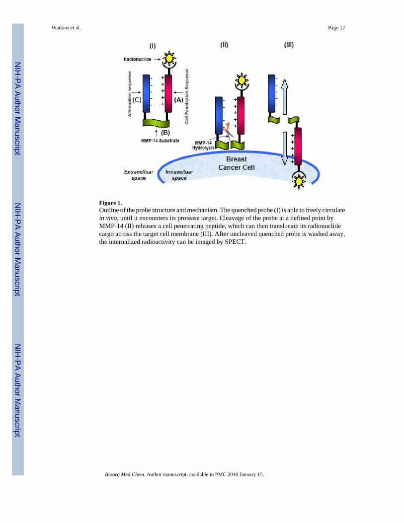
Figure 1.Outline of the probe structure and mechanism. The quenched probe (I) is able to freely circulatein vivo, until it encounters its protease target. Cleavage of the probe at a defined point byMMP-14 (II) releases a cell penetrating peptide, which can then translocate its radionuclidecargo across the target cell membrane (III). After uncleaved quenched probe is washed away,the internalized radioactivity can be imaged by SPECT.
Watkins et al. Page 12
Bioorg Med Chem. Author manuscript; available in PMC 2010 January 15.
NIH
-PA Author Manuscript
NIH
-PA Author Manuscript
NIH
-PA Author Manuscript
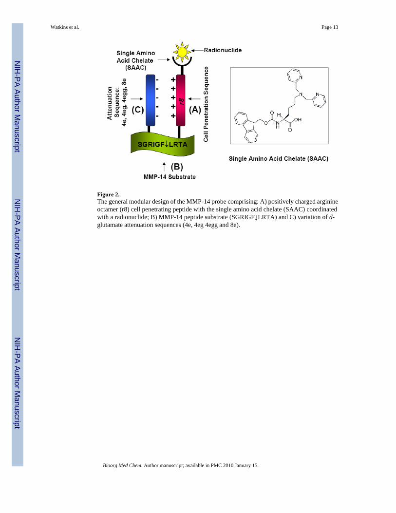
Figure 2.The general modular design of the MMP-14 probe comprising: A) positively charged arginineoctamer (r8) cell penetrating peptide with the single amino acid chelate (SAAC) coordinatedwith a radionuclide; B) MMP-14 peptide substrate (SGRIGF↓LRTA) and C) variation of d-glutamate attenuation sequences (4e, 4eg 4egg and 8e).
Watkins et al. Page 13
Bioorg Med Chem. Author manuscript; available in PMC 2010 January 15.
NIH
-PA Author Manuscript
NIH
-PA Author Manuscript
NIH
-PA Author Manuscript

Figure 3.Structures of MMP-14 probes determined from computer simulations using the zipping andassembly method. In each structure, the backbone is shown in a cartoon representation, coloredas follows: blue = CCP, green = MMP-14 specific substrate region, and red = attenuatingdomain. Both the Single Amino Acid Chelate (SAAC) residue and the cysteine-cysteine linkerunit are shown in bond representation.
Watkins et al. Page 14
Bioorg Med Chem. Author manuscript; available in PMC 2010 January 15.
NIH
-PA Author Manuscript
NIH
-PA Author Manuscript
NIH
-PA Author Manuscript
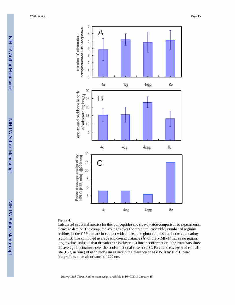
Figure 4.Calculated structural metrics for the four peptides and side-by-side comparison to experimentalcleavage data A: The computed average (over the structural ensemble) number of arginineresidues in the CPP that are in contact with at least one glutamate residue in the attenuatingregion. B: The computed average end-to-end distance (Å) of the MMP-14 substrate region;larger values indicate that the substrate is closer to a linear conformation. The error bars showthe average fluctuations over the conformational ensemble. C: Parallel cleavage studies; half-life (t1/2, in min.) of each probe measured in the presence of MMP-14 by HPLC peakintegrations at an absorbance of 220 nm.
Watkins et al. Page 15
Bioorg Med Chem. Author manuscript; available in PMC 2010 January 15.
NIH
-PA Author Manuscript
NIH
-PA Author Manuscript
NIH
-PA Author Manuscript

Figure 5.A sample MALDI-MS of the 4egg probe. The presence of the molecular ion peak (3972 amu,[MH]+), peptide fragments corresponding to disulfide scission (2191 and 1777 amu), and afragment corresponding to loss of a pyridylmethylene radical from the SAAC-containingparent peptide (1685 amu = 1777 – 92).
Watkins et al. Page 16
Bioorg Med Chem. Author manuscript; available in PMC 2010 January 15.
NIH
-PA Author Manuscript
NIH
-PA Author Manuscript
NIH
-PA Author Manuscript

Figure 6.In vitro uptake of the 4egg probe in MMP-14 expressing MDA-MB-231 cells with and withoutmetalloproteinase inhibitor. [Tc(CO)3]+ was used as a negative control.
Watkins et al. Page 17
Bioorg Med Chem. Author manuscript; available in PMC 2010 January 15.
NIH
-PA Author Manuscript
NIH
-PA Author Manuscript
NIH
-PA Author Manuscript
Copyright © 2022 FDOKUMEN