Loss of PDEF, a prostate-derived Ets factor is associated with aggressive phenotype of prostate...
14
Johnson et al. Molecular Cancer 2010, 9:148 http://www.molecular-cancer.com/content/9/1/148 Open Access RESEARCH © 2010 Johnson et al; licensee BioMed Central Ltd. This is an Open Access article distributed under the terms of the Creative Commons Attribution License (http://creativecommons.org/licenses/by/2.0), which permits unrestricted use, distribution, and reproduction in any medium, provided the original work is properly cited. Research Loss of PDEF, a prostate-derived Ets factor is associated with aggressive phenotype of prostate cancer: Regulation of MMP 9 by PDEF Thomas R Johnson, Sweaty Koul, Binod Kumar, Lakshmipathi Khandrika, Sarah Venezia, Paul D Maroni, Randall B Meacham and Hari K Koul* Abstract Background: Prostate-derived Ets factor (PDEF) is expressed in tissues of high epithelial content including prostate, although its precise function has not been fully established. Conventional therapies produce a high rate of cure for patients with localized prostate cancer, but there is, at present, no effective treatment for intervention in metastatic prostate cancer. These facts underline the need to develop new approaches for early diagnosis of aggressive prostate cancer patients, and mechanism based anti-metastasis therapies that will improve the outlook for hormone-refractory prostate cancer. In this study we evaluated role of prostate-derived Ets factor (PDEF) in prostate cancer. Results: We observed decreased PDEF expression in prostate cancer cell lines correlated with increased aggressive phenotype, and complete loss of PDEF protein in metastatic prostate cancer cell lines. Loss of PDEF expression was confirmed in high Gleason Grade prostate cancer samples by immuno-histochemical methods. Reintroduction of PDEF profoundly affected cell behavior leading to less invasive phenotypes in three dimensional cultures. In addition, PDEF expressing cells had altered cell morphology, decreased FAK phosphorylation and decreased colony formation, cell migration, and cellular invasiveness. In contrast PDEF knockdown resulted in increased migration and invasion as well as clonogenic activity. Our results also demonstrated that PDEF downregulated MMP9 promoter activity, suppressed MMP9 mRNA expression, and resulted in loss of MMP9 activity in prostate cancer cells. These results suggested that loss of PDEF might be associated with increased MMP9 expression and activity in aggressive prostate cancer. To confirm results we investigated MMP9 expression in clinical samples of prostate cancer. Results of these studies show increased MMP9 expression correlated with advanced Gleason grade. Taken together our results demonstrate decreased PDEF expression and increased MMP9 expression during the transition to aggressive prostate cancer. Conclusions: These studies demonstrate for the first time negative regulation of MMP9 expression by PDEF, and that PDEF expression was lost in aggressive prostate cancer and was inversely associated with MMP9 expression in clinical samples of prostate cancer. Based on these exciting results, we propose that loss of PDEF along with increased MMP9 expression should serve as novel markers for early detection of aggressive prostate cancer. Background Prostate cancer is the second leading cause of cancer death in men. In the United States alone, 192,280 new cases of prostate cancers were diagnosed in 2009 and among them around 27,360 deaths occurred. One of the biggest challenges we face in prostate cancer is determin- ing if the cancer is aggressive. Conventional therapies produce a high rate of cure for patients with localized prostate cancer, but there is no cure once the disease has spread beyond the prostate. Reduction in serum prostate- specific antigen (PSA) levels has been proposed as an endpoint biomarker for human prostate cancer interven- tion. However, despite being the mainstay of prostate cancer detection, the value of PSA screening is still * Correspondence: [email protected] 1 Program in Urosciences, Division of Urology-Department of Surgery, University of Colorado Denver School of Medicine, Denver Veterans Administrative Medical Center, and University of Colorado Comprehensive Cancer Center, Building P15 or RC2, C-317, 12700 E 19th Avenue, Aurora, CO 80045, USA Full list of author information is available at the end of the article
-
Upload
independent -
Category
Documents
-
view
0 -
download
0
Transcript of Loss of PDEF, a prostate-derived Ets factor is associated with aggressive phenotype of prostate...
Johnson et al. Molecular Cancer 2010, 9:148http://www.molecular-cancer.com/content/9/1/148
Open AccessR E S E A R C H
ResearchLoss of PDEF, a prostate-derived Ets factor is associated with aggressive phenotype of prostate cancer: Regulation of MMP 9 by PDEFThomas R Johnson, Sweaty Koul, Binod Kumar, Lakshmipathi Khandrika, Sarah Venezia, Paul D Maroni, Randall B Meacham and Hari K Koul*
AbstractBackground: Prostate-derived Ets factor (PDEF) is expressed in tissues of high epithelial content including prostate, although its precise function has not been fully established. Conventional therapies produce a high rate of cure for patients with localized prostate cancer, but there is, at present, no effective treatment for intervention in metastatic prostate cancer. These facts underline the need to develop new approaches for early diagnosis of aggressive prostate cancer patients, and mechanism based anti-metastasis therapies that will improve the outlook for hormone-refractory prostate cancer. In this study we evaluated role of prostate-derived Ets factor (PDEF) in prostate cancer.
Results: We observed decreased PDEF expression in prostate cancer cell lines correlated with increased aggressive phenotype, and complete loss of PDEF protein in metastatic prostate cancer cell lines. Loss of PDEF expression was confirmed in high Gleason Grade prostate cancer samples by immuno-histochemical methods. Reintroduction of PDEF profoundly affected cell behavior leading to less invasive phenotypes in three dimensional cultures. In addition, PDEF expressing cells had altered cell morphology, decreased FAK phosphorylation and decreased colony formation, cell migration, and cellular invasiveness. In contrast PDEF knockdown resulted in increased migration and invasion as well as clonogenic activity. Our results also demonstrated that PDEF downregulated MMP9 promoter activity, suppressed MMP9 mRNA expression, and resulted in loss of MMP9 activity in prostate cancer cells. These results suggested that loss of PDEF might be associated with increased MMP9 expression and activity in aggressive prostate cancer. To confirm results we investigated MMP9 expression in clinical samples of prostate cancer. Results of these studies show increased MMP9 expression correlated with advanced Gleason grade. Taken together our results demonstrate decreased PDEF expression and increased MMP9 expression during the transition to aggressive prostate cancer.
Conclusions: These studies demonstrate for the first time negative regulation of MMP9 expression by PDEF, and that PDEF expression was lost in aggressive prostate cancer and was inversely associated with MMP9 expression in clinical samples of prostate cancer. Based on these exciting results, we propose that loss of PDEF along with increased MMP9 expression should serve as novel markers for early detection of aggressive prostate cancer.
BackgroundProstate cancer is the second leading cause of cancerdeath in men. In the United States alone, 192,280 newcases of prostate cancers were diagnosed in 2009 and
among them around 27,360 deaths occurred. One of thebiggest challenges we face in prostate cancer is determin-ing if the cancer is aggressive. Conventional therapiesproduce a high rate of cure for patients with localizedprostate cancer, but there is no cure once the disease hasspread beyond the prostate. Reduction in serum prostate-specific antigen (PSA) levels has been proposed as anendpoint biomarker for human prostate cancer interven-tion. However, despite being the mainstay of prostatecancer detection, the value of PSA screening is still
* Correspondence: [email protected] Program in Urosciences, Division of Urology-Department of Surgery, University of Colorado Denver School of Medicine, Denver Veterans Administrative Medical Center, and University of Colorado Comprehensive Cancer Center, Building P15 or RC2, C-317, 12700 E 19th Avenue, Aurora, CO 80045, USAFull list of author information is available at the end of the article
© 2010 Johnson et al; licensee BioMed Central Ltd. This is an Open Access article distributed under the terms of the Creative CommonsAttribution License (http://creativecommons.org/licenses/by/2.0), which permits unrestricted use, distribution, and reproduction inany medium, provided the original work is properly cited.
Johnson et al. Molecular Cancer 2010, 9:148http://www.molecular-cancer.com/content/9/1/148
Page 2 of 14
debated. In particular, there is a growing concern regard-ing the over diagnosis of potentially indolent disease [1].Therefore, there remains an urgent need for more accu-rate biomarkers to diagnose aggressive prostate cancer.Thus, identification of new molecular markers/targets foraggressive prostate cancer is important in order toimprove early detection of the aggressive disease and todevelop new therapeutic regimens.
Progression of prostate cancer from focal, androgen-dependent lesions to androgen-independent, metastaticcancer requires deregulation of growth control, invasive-ness and cell motility. Abundant evidence demonstratesroles for Ets transcription factors in many cancers includ-ing prostate. Prostate-derived Ets factor (PDEF), firstdescribed nine years ago as preferentially binding to thenoncanonical Ets core sequence GGAT [2], has recentlyreceived considerable attention due to its potentialimportance in regulating cell motility and invasion [3-5].Recently, proteomic analysis of PDEF overexpressing cellsrevealed 286 proteins in the PDEF-associated proteincomplex in breast cancer [6]. Thus interaction of PDEFwith other partner proteins could help in finding theirrole in maintenance of malignant phenotype. Publishedliterature concerning experimental manipulation ofPDEF expression is paradoxical and limited to tissues ofhigh epithelial content, notably prostate, breast, ovaryand colon [7,8]. PDEF expression has been both positively[3,9] and negatively [10] correlated with breast cancergrade at mRNA or protein levels. It is important to notethat PDEF mRNA and protein levels do not always corre-late, which may have led to different conclusions in someof the studies examining PDEF expression in primarytumors. Turner et al. [4] found that introducing PDEFinto invasive breast cancer cell lines reduced their invad-ing ability. Similarly, siRNA-mediated knockdown ofPDEF in MCF7 cells increased their ability to migrate inthe Transwell assay. Besides its role in cancer metastasis,PDEF expression was also correlated with changes in theactin cytoskeleton and focal adhesion localization, andloss of cellular polarity. Ghadersohi et al. [10] silencedPDEF expression in MCF7 cells, and found that such cellsshowed greatly accelerated xenograft tumor formation inSCID mice. By contrast, Gunawardane et al. [3] showedthat increasing expression of PDEF increased their abilityto migrate in a Transwell assay and stimulated colony for-mation in soft agar. This group also identified a canonicalMAP kinase phosphorylation site at T50 (PAT50P) andshowed that mutation to alanine at this site abolished allthe effects they observed. To date there are few data avail-able formally correlating PDEF expression in mainte-nance of prostate malignant phenotype. Two publishedstudies, one with a prostate cancer cell-line [5] andanother with clinical samples from prostate [11] reachedopposite conclusions with respect to the role of PDEF in
prostate cancer. Clearly additional studies are necessaryto evaluate role of PDEF in prostate cancer biology.
In the current studies, we report here that PDEFexpression is lost, whereas MMP9 expression increasedwith the aggressive behavior of prostate cancer. Overex-pression of PDEF in PC3 cells strongly inhibits colonyformation, cell migration and invasion, and increased celladherence. Furthermore, re-introduction of PDEF in PC3cells led to changes in actin cytoskeleton, altered focaladhesion kinase activity, and reestablished cell polarity inthese cultures as indicated by induction of less invasivespheroid-like structures in three-dimensional culture,Moreover, PDEF expression downregulates MMP9expression, and its promoter activity in PC3 cells. Thus,consideration of both PDEF and MMP9 may have a betterprognosis value for determining the aggressive phenotypeof prostate cancer.
Materials and methodsConstructs and cell linesAll cell lines (PC3, LNCaP, and C4-2B) were purchasedfrom ATCC and maintained according to ATCC guide-lines. Phoenix cells were grown in DMEM containing10% fetal bovine serum. FLAG tag antibody was pur-chased from Sigma (St. Louis, USA). PDEF and phosphoFAK antibodies were from Santa Cruz Biotechnology(CA, USA). PDEF was cloned from PC3 cDNA with anamino-terminal FLAG tag, and inserted into retroviralvectors pBABE and the bicistronic vector QCXIX (Clon-tech). The latter vector was modified to contain a wild-type internal ribosome entry site to increase expressionfrom the second multiple cloning site, into which G418resistance was cloned. Mutations were created using theQuick-Change kit (Stratagene) according to the manufac-turer's instructions. Oligonucleotide primers for PCRwere purchased from Integrated DNA Technologies.
Retrovirus production and infectionPhoenix cells were transfected with 2 μg DNA usingEffectene (Qiagen) according to the manufacturer'sinstructions, and infection was followed according toPhoenix™ Retrovirus Expression System (Orbigen Inc.).After infection, cells were trypsinized, transferred to 150mm dishes, and subjected to puromycin or G418 selec-tion after 48 h incubation.
Thymidine incorporation1 × 105 cells/well were plated in 12-well plates. 48 h later,cells were exposed to medium containing1-3 μCi/ml 3H-thymidine (Perkin Elmer) and incubated for 4 h. After 2washes with cold PBS, cells were fixed in cold methanolfor 5 min followed by an additional methanol wash. Cellswere solubilized in 0.1% SDS/0.2 M NaOH and radioac-tivity determined.
Johnson et al. Molecular Cancer 2010, 9:148http://www.molecular-cancer.com/content/9/1/148
Page 3 of 14
Anchorage independent growth, motility, invasion and attachment assaysGrowth in soft agar was performed as described previ-ously [12]. Invasiveness was determined by the method ofRepesh LA [13]. Cell migration through Transwell mem-branes was performed identically, but without the use ofMatrigel. Wound healing assays were performed by mak-ing a cruciform scratch in a confluent monolayer of cells.Cells were washed, the medium replaced with serum-freemedium, and incubated for 48-72 h. Cells were fixed withmethanol, stained with Giemsa, and photographed.
Attachment assays were performed essentially by themethod of Turner et al. [4] using 96-well plates pretreatedovernight with fibronectin, Matrigel, or bovine serumalbumin at concentrations of 50 ng/ml, 100 ng/ml, and 10mg/ml respectively.
Immunohistochemistry for PDEF and MMP9 expression on prostate tissue array slidesTissue microarray slides containing 9 normal and 40prostate cancer samples of varying pathological gradewere obtained from Imgenex Corporation, San Diego,CA, 92121. Immuno histochemistry for PDEF and MMP9was performed using the avidin-biotin complex methodpreviously described by Hsu et al. [14]. Expression ofPDEF and MMP9 were evaluated by analysis of micro-scopic scans of each tissue. Expression was consideredhigh if greater than 60% of the scanned area scored posi-tive, while expression was considered moderate if 40-60%area scored positive, and expression was considered low ifless than 40% area scored positive.
Reverse transcription PCRTotal RNA was extracted using RNEasy mini kit (Qiagen).1 μg of RNA was used to prepare cDNA using iScript sec-ond strand cDNA synthesis kit (Bio-Rad). 100 ng of syn-thesized cDNA was used for RT-PCR using forward 5-TTGACAGCGACAAGAAGTGG-3 and reverse 5-TCACGTCGTCCTTATGCAAG-3 for MMP9, forward5-ACCACAGTCCATGCCATCAC-3 and reverse 5-TCCACCACCCTGTTGCTGTA-3 for GAPDH. Tran-scripts were separated by agarose gel electrophoresis.
Reporter assayCells were transfected with 1 μg of MMP9 luciferasereporter vector along with 10 ng of Renilla luciferaseexpression plasmid using Effectene (Qiagen, Valencia,CA) according to manufacturer's instructions. Luciferaseactivity was measured using the Dual luciferase kit (Pro-mega Corporation, Madison, WI) with Monolight 2010Luminometer (Analytical Luminescence laboratory, SanDiego, CA).
MMP ZymographyZymogram for MMP9 activity was performed accordingto Bernhard and Muschel [15] using conditionedmedium.
Three dimensional cell culturePDEF overexpressing PC3 cells and respective vectorcontrol cells were grown in growth factor reduced Matri-gel for 10-12 days and then immunofluorescence stainingwas performed according to Debnath et al. [16]
Morphology studiesCell morphology was done on glass chamber slides byimmunofluorescence method. Vector control and PDEFexpressing cells were seeded on multi-chamber slide,fixed with 4% formalin, permeabilized with 0.1% Triton-X-100, blocked in 2% BSA, and change in actin cytoskele-ton was examined by phalloidin staining as per the manu-facturer's instructions (Molecular Probes, Eugene, OR).Pictures were taken using Spin Disc Olympus confocalmicroscope.
Western Blot analysisElectrophoresis and blotting were performed asdescribed previously [17].
Statistical analysisStatistical analyses for tissue culture studies were per-formed using two-dimensional two sample variance T-tests; For data from clinical specimens, statistical analysiswas performed using MANN - WHITNEY U: exact test.p ≤ 0.05 was considered significant.
ResultsPDEF expression is reduced during the transition from low grade to high grade prostate cancerPDEF expression was evaluated by immunohistochemicalexamination in tissue microarray slides containing 40cores of prostate cancer and 9 cores of normal prostate.Results presented in Figure 1 show that PDEF proteinexpression is downregulated during the transition toaggressive prostate cancer. As shown in Figure. 1A, highlevels of PDEF protein are present in normal prostate epi-thelial cells as well as early stage prostate cancer. How-ever, in high grade prostate cancer PDEF protein issignificantly decreased. Significant reduction in PDEFexpression was observed in all cores of prostate tumortissue with high Gleason grade (Gleason score greaterthan 7) as compared to normal prostate tissue as well aslow grade prostate cancer (Gleason score of 7 or below).Moreover, PDEF protein levels showed graded decreasewith increase in pathologically confirmed aggressive dis-ease. Our results show that while 59 ± 3.6% tissue scoredpositive for PDEF in low to moderate grade (Gleason 6 &7) prostate cancer, and 33 ± 3.3% of the tissue scored pos-itive for PDEF in moderately high grade (Gleason 8)tumor, there was little or no expression of PDEF in veryhigh grade (Gleason 9 & 10) prostate cancer(Figure. 1B).Antibody specificity for these assays was determined byImmunofluorescence and Western blot analysis usingcells with and without PDEF expression (Figure S1, Addi-tional file 1). In addition to clinical samples, we also eval-

Johnson et al. Molecular Cancer 2010, 9:148http://www.molecular-cancer.com/content/9/1/148
Page 4 of 14
uated PDEF protein expression in established prostatecancer cell lines with low to high aggressive behavior.These results presented in Figure 1C show that PDEF isexpressed in LNCaP cells (a less aggressive prostate can-cer cell line). However, PDEF expression is reduced inmore aggressive lineages of LNCaP cells (C4-2 and C4-2B). Moreover, PDEF expression is completely lost in twowidely used aggressive prostate cancer cells (DU145 andPC3 cells). Taken together, results of these studies dem-
onstrate that PDEF expression is decreased or lost inprostate cancer cells with aggressive phenotype, and pro-vide novel insights into the characteristics of PDEF pro-tein expression in progression of prostate cancers.Absence of PDEF protein expression in PC3 cells in ourstudies is in apparent disagreement with previous report[5] that showed PDEF expression in PC3 cells,. This dis-crepancy could result from several causes: First, the anti-body used in [5] could be more sensitive, such that they
Figure 1 PDEF protein expression in human prostate tissues and prostate cancer cell lines. A, Representative photo-micrographs of Immuno-histochemical analysis of PDEF expression using prostate tissue micro-array slides (containing both normal and tumor samples of different grades) performed as described in Materials and Methods. B, Quantification of percentage staining for PDEF. C, Representative image showing Western blot analysis on prostate cancer cell lines using anti-PDEF antibody (left panel), and quantitation of the same data (right panel).

Johnson et al. Molecular Cancer 2010, 9:148http://www.molecular-cancer.com/content/9/1/148
Page 5 of 14
were able to detect even negligible amounts of PDEF. Sec-ond PC3 cells change over various passages of culture andmedia conditions, which could explain the differences.
Re-introduction of PDEF inhibited directional migration, decreased cell migration and anchorage independent growth in prostate cancer cellsTo examine the effects of PDEF expression on cell motil-ity, PC3 cells transduced with PDEF or mutant PDEFT50A, or vector alone were assayed for their ability tomigrate through Transwell membranes as described inmaterials and methods. Results presented in Figure 2Aindicate that both wild-type and mutant PDEF-trans-duced cells were significantly inhibited in their ability tomigrate through Transwell pores. We also subjected cellsto an assay for persistence migratory directionality (invitro wound healing, Figure. 2B). Expression of eitherPDEF or the T50A mutant inhibited the ability of PC3cells to fill in gaps in a monolayer compared to vectoralone. These results demonstrated that PDEF signifi-cantly interfered with ability of cells to maintain migra-tory phenotype.
We next examined the effects of PDEF overexpressionon the ability of PC3 cells to form colonies in soft agar.Expression of either PDEF or the T50A mutant equallyinhibited the ability of PC3 cells to form colonies in softagar (Figure. 2C) compared to vector alone.
To address the possibility that altered cell proliferationcontributed to the results of these assays, we measuredDNA synthesis (3H-thymidine incorporation) in controland PDEF transfected PC3. Results of these studies (Fig-ure. 2D) show that decreased clonogenic activity follow-ing PDEF expression was not a consequence of decreasedDNA synthesis. These results demonstrated that PDEFexpression decreased clonogenic ability of the cells inde-pendent of DNA synthesis. Moreover, in sharp contrastto the effects of PDEF on anchorage independent growth,PDEF expression did not significantly affect anchoragedependent growth of prostate cancer cells in culture (datanot shown).
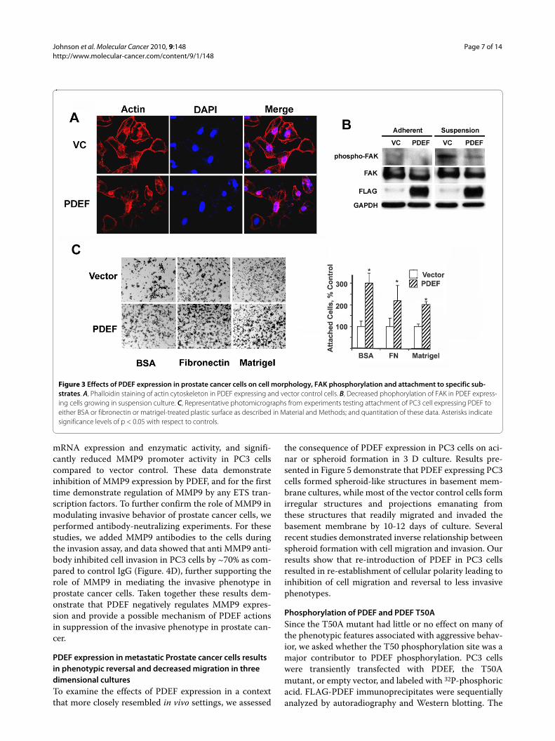
PDEF expression resulted in increased cell adhesion, altered cell morphology and decreased focal adhesion kinase activity in prostate cancer cellsImmunofluorescence studies of PDEF expressing cellsshowed a rounded area of cleared fluorescence ratherthan elongated track as seen on invasive vector controlcells (Figure. 3A). These results indicated that PDEFexpression resulted in alterations to actin cytoskeletonand altered cell morphology. FAK is a non-receptor pro-tein tyrosine kinase, associated with supramolecular focaladhesion complexes. Focal adhesion complex assemblyand disassembly are critical for cell attachment andmovement [18]. The lack of morphologic polarity in
PDEF expressing cells as shown in Figure 3A raised thepossibility that PDEF may affect adhesion complex for-mation. Moreover, in previous studies we observed thatFAK was non-phosphorylated in adherent cultures andFAK phosphorylation was increased in suspension cul-ture [19]. Therefore, we evaluated the effects of re-intro-duction of PDEF in PC3 cells on FAK phosphorylation inPC3 cells in suspension cultures. Results of these studiesrevealed a significant reduction in FAK phosphorylationin PDEF expressing cells grown in suspension culture(Figure. 3B). These results demonstrated that PDEFexpression in PC3 cells resulted in decreased FAK activ-ity, suggesting decreased focal adhesion formation.
Focal adhesion formation and its interaction with theECM play a central role in migration and invasion, sinceincreased adhesion makes cells less motile. To examinethis possibility, we directly measured the effects of PDEFexpression on adhesion of PC3 cells to various ECM sub-strates. For these studies, PC3 cells transfected withPDEF or vector alone were assayed for their ability toattach to fibronectin or Matrigel-coated plastic surfaces.Results presented in Figure 3C indicate that attachmentof PDEF-expressing cells to fibronectin-coated, Matrigel-coated, or control (BSA treated) plastic was significantlyincreased compared to vector-transduced cells. Theseresults are in contrast to the effects of PDEF in breastcancer cells, where PDEF was shown to decrease adhe-sion of the cells to fibronectin and matrigel [4]. Takentogether, these results suggest that PDEF mediated inhi-bition of migration may occur through cytoskeleton dis-organization and ECM interaction.
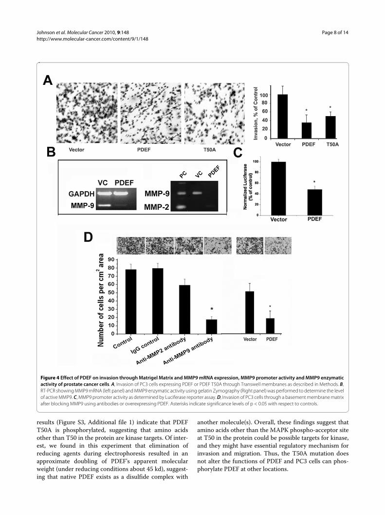
PDEF decreased invasion and inhibited expression of matrix metalloproteinase-9 (MMP9) in prostate cancer cellsTo test the effects of PDEF on cell invasion, we examinedthe effects of PDEF expression on the ability of PC3 cellsto invade simulated basement membrane in vitro, a phe-notype correlated with aggressive behavior. Results pre-sented in Figure 4A indicate that expression of eitherPDEF or the T50A mutant inhibited the ability of PC3cells to invade through Matrigel compared with vectortransfected control cells. In addition to transfection ofPC3 cells with PDEF, we also performed complementaryRNA interference (RNAi) experiments to reduce theendogenous PDEF expression in prostate cancer cells thatexpress PDEF (LNCaP and C4-2B cells), and directlyevaluated the effects of decreased PDEF levels on inva-sion and clonogenic activity of these cells. Results pre-sented in Figure S2, Additional file 1 demonstrated thatSiRNA mediated knock-down of PDEF in these cellsresulted in an increased ability to form colonies in softagar and increased invasion through Matrigel basementmembrane. Taken together with rest of the results these

Johnson et al. Molecular Cancer 2010, 9:148http://www.molecular-cancer.com/content/9/1/148
Page 6 of 14
studies suggest that PDEF may play an important role inprostate cancer metastasis.
Matrix metalloproteinases (MMPs) are a family ofenzymes whose function primarily relates to degradationof extracellular matrix proteins, and are necessary for cellinvasion. Moreover increased MMP activity has beenassociated with tumor metastasis. In our in vitro studies
we observed that only MMP9 was prominently active inPC3 cells. Thus we set out to test the possible role ofMMP9 in mediating the effects of PDEF on cell invasion.For these studies we evaluated the effects of PDEFexpression on MMP9 mRNA expression, promoter activ-ity and enzymatic activity. As can be seen in Figure 4B&4C, PDEF expression completely abolished MMP9
Figure 2 Effect of re-introduction of PDEF on directional migration, trans-well migration and anchorage independent growth in prostate cancer cells. A, Migration of PC3 cells expressing PDEF or PDEF T50A through Transwell membranes as described in Materials and Methods. B, PDEF overexpression decreases directional cell migration (in vitro wound healing migration of these cells). C, Representative photomicrographs from exper-iments testing colony formation as described in Materials and Methods. D, 3H-thymidine incorporation in PC3 PDEF overexpressing cells measured as described in Materials and Methods. E, Relative expression level of PDEF in these cell lines. Asterisks indicate significance levels of p < 0.05 with respect to controls.
Johnson et al. Molecular Cancer 2010, 9:148http://www.molecular-cancer.com/content/9/1/148
Page 7 of 14
mRNA expression and enzymatic activity, and signifi-cantly reduced MMP9 promoter activity in PC3 cellscompared to vector control. These data demonstrateinhibition of MMP9 expression by PDEF, and for the firsttime demonstrate regulation of MMP9 by any ETS tran-scription factors. To further confirm the role of MMP9 inmodulating invasive behavior of prostate cancer cells, weperformed antibody-neutralizing experiments. For thesestudies, we added MMP9 antibodies to the cells duringthe invasion assay, and data showed that anti MMP9 anti-body inhibited cell invasion in PC3 cells by ~70% as com-pared to control IgG (Figure. 4D), further supporting therole of MMP9 in mediating the invasive phenotype inprostate cancer cells. Taken together these results dem-onstrate that PDEF negatively regulates MMP9 expres-sion and provide a possible mechanism of PDEF actionsin suppression of the invasive phenotype in prostate can-cer.
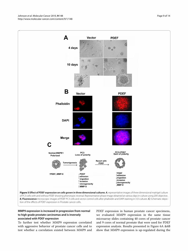
PDEF expression in metastatic Prostate cancer cells results in phenotypic reversal and decreased migration in three dimensional culturesTo examine the effects of PDEF expression in a contextthat more closely resembled in vivo settings, we assessed
the consequence of PDEF expression in PC3 cells on aci-nar or spheroid formation in 3 D culture. Results pre-sented in Figure 5 demonstrate that PDEF expressing PC3cells formed spheroid-like structures in basement mem-brane cultures, while most of the vector control cells formirregular structures and projections emanating fromthese structures that readily migrated and invaded thebasement membrane by 10-12 days of culture. Severalrecent studies demonstrated inverse relationship betweenspheroid formation with cell migration and invasion. Ourresults show that re-introduction of PDEF in PC3 cellsresulted in re-establishment of cellular polarity leading toinhibition of cell migration and reversal to less invasivephenotypes.
Phosphorylation of PDEF and PDEF T50ASince the T50A mutant had little or no effect on many ofthe phenotypic features associated with aggressive behav-ior, we asked whether the T50 phosphorylation site was amajor contributor to PDEF phosphorylation. PC3 cellswere transiently transfected with PDEF, the T50Amutant, or empty vector, and labeled with 32P-phosphoricacid. FLAG-PDEF immunoprecipitates were sequentiallyanalyzed by autoradiography and Western blotting. The
Figure 3 Effects of PDEF expression in prostate cancer cells on cell morphology, FAK phosphorylation and attachment to specific sub-strates. A, Phalloidin staining of actin cytoskeleton in PDEF expressing and vector control cells. B, Decreased phophorylation of FAK in PDEF express-ing cells growing in suspension culture. C, Representative photomicrographs from experiments testing attachment of PC3 cell expressing PDEF to either BSA or fibronectin or matrigel-treated plastic surface as described in Material and Methods; and quantitation of these data. Asterisks indicate significance levels of p < 0.05 with respect to controls.
Johnson et al. Molecular Cancer 2010, 9:148http://www.molecular-cancer.com/content/9/1/148
Page 8 of 14
results (Figure S3, Additional file 1) indicate that PDEFT50A is phosphorylated, suggesting that amino acidsother than T50 in the protein are kinase targets. Of inter-est, we found in this experiment that elimination ofreducing agents during electrophoresis resulted in anapproximate doubling of PDEF's apparent molecularweight (under reducing conditions about 45 kd), suggest-ing that native PDEF exists as a disulfide complex with
another molecule(s). Overall, these findings suggest thatamino acids other than the MAPK phospho-acceptor siteat T50 in the protein could be possible targets for kinase,and they might have essential regulatory mechanism forinvasion and migration. Thus, the T50A mutation doesnot alter the functions of PDEF and PC3 cells can phos-phorylate PDEF at other locations.
Figure 4 Effect of PDEF on invasion through Matrigel Matrix and MMP9 mRNA expression, MMP9 promoter activity and MMP9 enzymatic activity of prostate cancer cells. A, Invasion of PC3 cells expressing PDEF or PDEF T50A through Transwell membranes as described in Methods. B, RT-PCR showing MMP9 mRNA (left panel) and MMP9 enzymatic activity using gelatin Zymography (Right panel) was performed to determine the level of active MMP9. C, MMP9 promoter activity as determined by Luciferase reporter assay. D, Invasion of PC3 cells through a basement membrane matrix after blocking MMP9 using antibodies or overexpressing PDEF. Asterisks indicate significance levels of p < 0.05 with respect to controls.
Johnson et al. Molecular Cancer 2010, 9:148http://www.molecular-cancer.com/content/9/1/148
Page 9 of 14
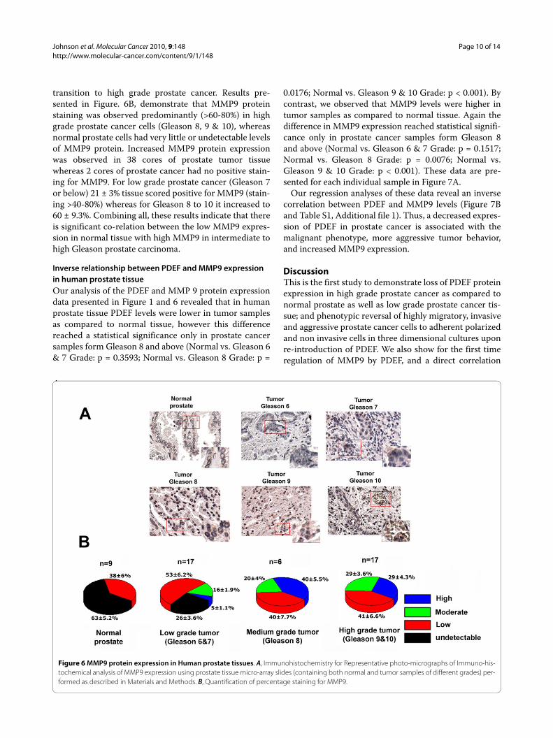
MMP9 expression is increased in progression from normal to high grade prostate carcinomas and is inversely associated with PDEF expressionTo further test whether MMP9 expression correlatedwith aggressive behavior of prostate cancer cells and totest whether a correlation existed between MMP9 and
PDEF expression in human prostate cancer specimens,we evaluated MMP9 expression in the same tissuemicroarray slides containing 40 cores of prostate cancerand 9 cores of normal prostate that were used for PDEFexpression analysis. Results presented in Figure 6A &6Bshow that MMP9 expression is up-regulated during the
Figure 5 Effect of PDEF expression on cells grown in three dimensional cultures. A, representative images of three dimensional matrigel culture of PC3 cells with and without PDEF showing phenotypic reversal. Representative phase image obtained at various days in culture using 20X objective. B, Fluorescence microscopic images of PDEF PC3 cells and vector control cells after phalloidin and DAPI staining in 3 D cultures. C) Schematic depic-tion of the effects of PDEF expression in Prostate cancer cells.
Johnson et al. Molecular Cancer 2010, 9:148http://www.molecular-cancer.com/content/9/1/148
Page 10 of 14
transition to high grade prostate cancer. Results pre-sented in Figure. 6B, demonstrate that MMP9 proteinstaining was observed predominantly (>60-80%) in highgrade prostate cancer cells (Gleason 8, 9 & 10), whereasnormal prostate cells had very little or undetectable levelsof MMP9 protein. Increased MMP9 protein expressionwas observed in 38 cores of prostate tumor tissuewhereas 2 cores of prostate cancer had no positive stain-ing for MMP9. For low grade prostate cancer (Gleason 7or below) 21 ± 3% tissue scored positive for MMP9 (stain-ing >40-80%) whereas for Gleason 8 to 10 it increased to60 ± 9.3%. Combining all, these results indicate that thereis significant co-relation between the low MMP9 expres-sion in normal tissue with high MMP9 in intermediate tohigh Gleason prostate carcinoma.
Inverse relationship between PDEF and MMP9 expression in human prostate tissueOur analysis of the PDEF and MMP 9 protein expressiondata presented in Figure 1 and 6 revealed that in humanprostate tissue PDEF levels were lower in tumor samplesas compared to normal tissue, however this differencereached a statistical significance only in prostate cancersamples form Gleason 8 and above (Normal vs. Gleason 6& 7 Grade: p = 0.3593; Normal vs. Gleason 8 Grade: p =
0.0176; Normal vs. Gleason 9 & 10 Grade: p < 0.001). Bycontrast, we observed that MMP9 levels were higher intumor samples as compared to normal tissue. Again thedifference in MMP9 expression reached statistical signifi-cance only in prostate cancer samples form Gleason 8and above (Normal vs. Gleason 6 & 7 Grade: p = 0.1517;Normal vs. Gleason 8 Grade: p = 0.0076; Normal vs.Gleason 9 & 10 Grade: p < 0.001). These data are pre-sented for each individual sample in Figure 7A.
Our regression analyses of these data reveal an inversecorrelation between PDEF and MMP9 levels (Figure 7Band Table S1, Additional file 1). Thus, a decreased expres-sion of PDEF in prostate cancer is associated with themalignant phenotype, more aggressive tumor behavior,and increased MMP9 expression.
DiscussionThis is the first study to demonstrate loss of PDEF proteinexpression in high grade prostate cancer as compared tonormal prostate as well as low grade prostate cancer tis-sue; and phenotypic reversal of highly migratory, invasiveand aggressive prostate cancer cells to adherent polarizedand non invasive cells in three dimensional cultures uponre-introduction of PDEF. We also show for the first timeregulation of MMP9 by PDEF, and a direct correlation
Figure 6 MMP9 protein expression in Human prostate tissues. A, Immunohistochemistry for Representative photo-micrographs of Immuno-his-tochemical analysis of MMP9 expression using prostate tissue micro-array slides (containing both normal and tumor samples of different grades) per-formed as described in Materials and Methods. B, Quantification of percentage staining for MMP9.

Johnson et al. Molecular Cancer 2010, 9:148http://www.molecular-cancer.com/content/9/1/148
Page 11 of 14
between loss of PDEF and increased expression of MMP9high grade prostate cancer.
The ETS family is one of the largest families of tran-scription factors with 27 genes in human chromosome.The ETS family is present throughout the body and isinvolved in a wide variety of functions including the regu-lation of cellular differentiation, cell cycle control, cellmigration, cell proliferation, apoptosis (programmed celldeath) and angiogenesis. Multiple Ets factors have beenfound to be associated with cancer, such as through genefusion including prostate cancer (2, 20-25). PDEF is selec-
tively localized to the tissues with high epithelial contentincluding prostate, and like other Ets family members hasbeen shown to have diverse biological functions includingtumor suppressor as well as tumor promoter functions.
Our results demonstrate loss of PDEF in high gradeprostate cancer as compared to low grade prostate canceras well as normal prostate tissue. Our results are uniquein a way that they demonstrate for the first time loss ofPDEF is associated with aggressive phenotype in prostatecancer, and suggest that PDEF might serve as a potentialmarker for distinguishing aggressive prostate cancer from
Figure 7 Relationship between PDEF and MMP9 expression in human prostate tissue: A, Expression of PDEF and MMP9 in individual sam-ples B) Regression plot of the data presented in A. Data were collected from the immunohistochemistry performed for PDEF and MMP9 as de-scribed for Figure 1 and 6. (Normal = red; Gleason 6 & 7 = blue; Gleason 8 = green; and Gleason 9 and 10 = black). By regression analysis, there is a significant 0.846 (UNITS) drop in MMP9 for every 1.0 (UNIT) increase in PDEF.
Johnson et al. Molecular Cancer 2010, 9:148http://www.molecular-cancer.com/content/9/1/148
Page 12 of 14
an indolent disease. These findings are in apparent con-trast to the previously published studies that concludedover expression of PDEF in prostate cancer as comparedto normal prostate tissue [11]. It is important to point outhere that previous studies lumped together all cancersamples and compared them with normal tissue, how-ever, they did not attempt subset analysis of PDEF expres-sion between low grade and high grade disease, whichcould have resulted in different conclusions. Our conclu-sion that PDEF expression is a favorable indicator in pros-tate cancer is, however, in agreement with studies thatdemonstrated a positive prognostic value of PDEF inovarian cancer[8].
Our results also show that re-introduction of PDEF inaggressive prostate cancer cells resulted in decreased cellmigration, decreased directional migration as well asdecrease in clonogenic activity and converse was truewhen we knocked down PDEF in prostate cancer cellsexpressing PDEF (Figure S2, Additional file 1). These datasuggest PDEF might serve as a suppressor of tumormigration and clonogenic activity. These results are inagreement with the previous studies with various breastand prostate cancer cells [5,7]. Our results are, howeverin sharp disagreement with studies that suggested thatPDEF might promote migratory phenotype in breast can-cer cells [3].
In order to become motile, cancer cells establish adefined polarity in the direction of movement throughinteraction between lamellipodia (a cytoskeletal actinprojection on mobile edge of the cells) and focal adhe-sions that facilitate adhesion and migration of the cells.We also observed distinct changes in cytoskeleton andcell morphology associated with PDEF expression. Wealso show for the first time in any system that that PDEFexpression increased cell adhesion, and resulted in a sig-nificant reduction in FAK phosphorylation. In previousstudies we have shown an essential role for FAK inaggressive phenotype in prostate cancer cells [19]. Thusmodulation of cytoskeleton organization and FAK activ-ity by PDEF expression may provide potential pathwaysby which PDEF modulates cell behavior. These resultsextend previous observations in several cell types thatshow SiRNA mediated knockdown of PDEF was associ-ated with increased cell migration [3-5]. While the resultsdiscussed so far point to the possible role of PDEF expres-sion in modulating phenotypic behavior of cancer cells,our studies, are the first to use three dimensional culturesto actually demonstrate directly the effects of PDEF oncellular polarity and spheroid formation. Results clearlydemonstrate that re-introduction of PDEF in aggressiveprostate cancer cells resulted in phenotypic reversal froma disorganized, migratory and invasive cell growth to anorganized, non-migratory and non-invasive phenotype.
Chintala et al. reported that the formation of spheroidsis also linked to reduced invasion and expression andactivity of MMP9 [26]. Matrix metalloproteinases (MMP)are a family of enzymes whose function primarily relatesto the degradation of extracellular matrix proteins, andwhich are necessary for cell invasion. Our results pre-sented here show that upon PDEF expression, prostatecancer cells lose their ability to invade Matrigel in Boydenchamber assays. These results are similar to the resultsobserved by Turner et al. in invasive breast cancer cells[4]. However, to the best of our knowledge, our resultsshow for the first time that PDEF downregulates MMP9expression, and its promoter activity in any cell type. Weobserved that expression of PDEF in PC3 cells resulted inloss of MMP9 mRNA expression, decrease in MMP9 pro-moter activity and a significant reduction in the gelati-nolytic activity. Thus our results again highlight a uniqueproperty of PDEF that is distinct from other ETS factors.We carried out additional studies to directly evaluate thefunctional consequence of MMP9 activity in PC3 cells.Results of these studies demonstrate that antibody medi-ated neutralization of MMP 9 reduced the invasion ofPC3 cells through basement membrane matrix similar tothat observed upon PDEF expression. Overall these find-ings provide for a mechanism by which PDEF expressioncould modulate cell polarity and other aggressive behav-ior.
Since we observed negative regulation of MMP9 byPDEF and published results suggested that the activity ofMMP9 is associated with the progression and metastasisof prostate cancer [27], we also evaluated MMP9 expres-sion in the tissue microarray slides containing 40 cores ofprostate cancer and 9 cores of normal prostate that wereused for PDEF expression analysis. Our results demon-strated an increase in MMP9 expression in high gradeprostate cancer, which is in agreement with the previousstudies [27]. We also observed an inverse correlationbetween PDEF expression and MMP9 expression in thesesamples. These results are in agreement with our findingsin tissue culture studies that demonstrated negative regu-lation of MMP9 expression by PDEF. These results alsohighlight the potential use of loss of PDEF expression andincreased MMP9 expression in early detection of aggres-sive prostate cancer.
ConclusionsIn summary results presented herein demonstrate for thefirst time that PDEF, a member of Ets family, is lost inhigh grade prostate cancer and decreased PDEF expres-sion is associated with increased MMP9 expression. Wealso provide direct evidence for the first time demon-strating that PDEF expression results in phenotypicreversal of aggressive prostate cancer cells in three

Johnson et al. Molecular Cancer 2010, 9:148http://www.molecular-cancer.com/content/9/1/148
Page 13 of 14
dimensional cultures. Our studies also provide first dem-onstration in any system of negative regulation of MMP9expression by PDEF. Taken together, our studies suggestthat PDEF, by virtue of suppressing MMP9 expressionand by modulating the ability of cancer cells to form atemporal structure required for migration and invasion,may function as suppressor of tumor metastasis in pros-tate cancer and perhaps other cancers. Our observationof an inverse relationship between PDEF and MMP9expression suggests that expression of PDEF along withdecreased MMP9 could help in early detection of aggres-sive prostate cancer and may facilitate new approaches toprostate cancer treatment.
Additional material
Competing interestsA patent application relating in part to the content of the manuscript is beingprepared.No other interests
Authors' contributionsHKK designed research; TJ cloned PDEF, made stable cell lines and performedmost of the cell migration, adhesion and invasion assays; S.K. performed all ofthe experiments with PDEF and MMP 9 expression in clinical samples and MMP9 gene expression in cell lines. BK performed 3 D culture assays, cytoskeletonimaging, zymograms and the MMP 9 antibody neutralization assays; LK per-formed phospho-FAK assays and helped with cell culture work; SV performedmany colony formation assays and assisted in the retrovirus and cloning work;TJ, BK, SK, PM, RBM and HKK analyzed data; and BK, TJ, LK, SK and HKK wrote thepaper. All authors read and approved the final manuscript.
AcknowledgementsThese studies were supported in part by NIH/NCI-P20 CA103680-Schwartz/Byers Program PI's (H Koul, Pilot-Project PI) and the Department of Surgery, School of Medicine Academic Enrichment Funds. We are grateful to Dina Lev for kindly providing MMP9 luciferase reporter construct used in these studies. We also gratefully acknowledge Mr. Colin Odonnell for Statistical analysis on PDEF and MMP 9 expression in human prostate cancer specimen data.
Author DetailsProgram in Urosciences, Division of Urology-Department of Surgery, University of Colorado Denver School of Medicine, Denver Veterans Administrative Medical Center, and University of Colorado Comprehensive Cancer Center, Building P15 or RC2, C-317, 12700 E 19th Avenue, Aurora, CO 80045, USA
References1. Etzioni R, Penson DF, Legler JM, di Tommaso D, Boer R, Gann PH, Feuer EJ:
Overdiagnosis due to prostate-specific antigen screening: lessons from U.S. prostate cancer incidence trends. J Natl Cancer Inst 2002, 94:981-990.
2. Oettgen P, Finger E, Sun Z, Akbarali Y, Thamrongsak U, Boltax J, Grall F, Dube A, Weiss A, Brown L, Quinn G, Kas K, Endress G, Kunsch C, Libermann TA: PDEF, a novel prostate epithelium-specific transcription factor,
interacts with the androgen receptor and activates prostate-specific antigen gene expression. J Biol Chem 2000, 275:1216-1225.
3. Gunawardane RN, Sgroi DC, Wrobel CN, Koh E, Daley GQ, Brugge JS: Novel role for PDEF in epithelial cell migration and invasion. Cancer Res 2005, 65:11572-11580.
4. Turner DP, Moussa O, Sauane M, Fisher PB, Watson DK: Prostate-derived ETS factor is a mediator of metastatic potential through the inhibition of migration and invasion in breast cancer. Cancer Res 2007, 67:1618-1625.
5. Gu X, Zerbini LF, Otu HH, Bhasin M, Yang Q, Joseph MG, Grall F, Onatunde T, Correa RG, Libermann TA: Reduced PDEF expression increases invasion and expression of mesenchymal genes in prostate cancer cells. Cancer Res 2007, 67:4219-4226.
6. Cho JY, Lee M, Ahn JM, Park ES, Cho JH, Lee SJ, Kim BG, Heo SH, Park HJ, Zerbini LF, Hwang D, Libermann TA: Proteomic Analysis of a PDEF Ets Transcription Factor-Interacting Protein Complex. J Proteome Res 2009, 8:1327-1337.
7. Feldman RJ, Sementchenko VI, Gayed M, Fraig MM, Watson DK: Pdef expression in human breast cancer is correlated with invasive potential and altered gene expression. Cancer Res 2003, 63:4626-4631.
8. Ghadersohi A, Odunsi K, Zhang S, Azrak RG, Bundy BN, Manjili MH, Li F: Prostate-derived Ets transcription factor as a favorable prognostic marker in ovarian cancer patients. Int J Cancer 2008, 123:1376-1384.
9. Turcotte S, Forget MA, Beauseigle D, Nassif E, Lapointe R: Prostate-derived Ets transcription factor overexpression is associated with nodal metastasis and hormone receptor positivity in invasive breast cancer. Neoplasia 2007, 9:788-796.
10. Ghadersohi A, Sood AK: Prostate epithelium-derived Ets transcription factor mRNA is overexpressed in human breast tumors and is a candidate breast tumor marker and a breast tumor antigen. Clin Cancer Res 2001, 7:2731-2738.
11. Sood AK, Saxena R, Groth J, Desouki MM, Cheewakriangkrai C, Rodabaugh KJ, Kasyapa CS, Geradts J: Expression characteristics of prostate-derived Ets factor support a role in breast and prostate cancer progression. Hum Pathol 2007:1628-1638.
12. Kumar B, Koul S, Khandrika L, Meacham RB, Koul HK: Oxidative stress is inherent in prostate cancer cells and is required for aggressive phenotype. Meth Enzymol 1987, 146:341-349. Cancer Res. 2008, 68:1777-1785
13. Repesh LA: A new in vitro assay for quantitating tumor cell invasion. Invasion Metastasis 1989, 9:192-208.
14. Hsu SM, Raine L, Fanger H: Use of avidin-biotin-peroxidase complex (ABC) in immunoperoxidase techniques: a comparison between ABC and unlabeled antibody (PAP) procedures. J Histochem Cytochem 1981, 29:577-580.
15. Bernhard EJ, Muschel RJ: Ras, metastasis, and matrix metalloproteinase 9. Methods Enzymol 2001, 333:96-103.
16. Debnath J, Muthuswamy SK, Brugge JS: Morphogenesis and oncogenesis of MCF-10A mammary epithelial acini grown in three-dimensional basement membrane cultures. Methods 2003, 30:256-268.
17. Koul S, Huang M, Bhat S, Maroni P, Meacham RB, Koul HK: Oxalate exposure provokes HSP 70 response in LLC-PK1 cells, a line of renal epithelial cells: protective role of HSP 70 against oxalate toxicity. Urol Res 2008, 36:1-10.
18. Mitra SK, Hanson DA, Schlaepfer DD: Focal adhesion kinase: in command and control of cell motility. Nat Rev Mol Cell Biol 2005, 6:56-68.
19. Johnson TR, Khandrika L, Kumar B, Venezia S, Koul S, Chandhoke R, Maroni P, Donohue R, Meacham RB, Koul HK: Focal adhesion kinase controls aggressive phenotype of androgen-independent prostate cancer. Mol Cancer Res 2008, 6:1639-1648.
20. Ida K, Kobayashi S, Taki T, Hanada R, Bessho F, Yamamori S, Sugimoto T, Ohki M, Hayashi Y: EWS-FLI-1 and EWS-ERG chimeric mRNAs in Ewing's sarcoma and primitive neuroectodermal tumor. Int J Cancer 1995, 63:500-504.
21. Peeters P, Raynaud SD, Cools J, Wlodarska I, Grosgeorge J, Philip P, Monpoux F, Van Rompaey L, Baens M, Van den Berghe H, Marynen P: Fusion of TEL, the ETS-variant gene 6 (ETV6), to the receptor-associated kinase JAK2 as a result of t(9;12) in a lymphoid and t(9;15;12) in a myeloid leukemia. Blood 1997, 90:2535-2540.
22. Sharrocks AD: The ETS-domain transcription factor family. Nat Rev Mol Cell Biol 2001, 2:827-837.
Additional File 1 Supplementary data. Supporting Information. Materi-als and Methods with respect to following: -Immunofluorescence of cul-tured cells. -Anchorage independent Growth, and invasion assays. -Western blot analysis. -Metabolic Labeling. Figure S1 PDEF expression in cultured cells with or without PDEF. Figure S2 PDEF knockout increased colony for-mation and invasion of LNCaP and LNCaP C4-2B cells. Figure S3 Phosphory-lation of PDEF and PDEF T50A. Table S1 Relative PDEF and MMP9 gene expression in human prostate tissue.
Received: 18 November 2009 Accepted: 15 June 2010 Published: 15 June 2010This article is available from: http://www.molecular-cancer.com/content/9/1/148© 2010 Johnson et al; licensee BioMed Central Ltd. This is an Open Access article distributed under the terms of the Creative Commons Attribution License (http://creativecommons.org/licenses/by/2.0), which permits unrestricted use, distribution, and reproduction in any medium, provided the original work is properly cited.Molecular Cancer 2010, 9:148
http://www.ncbi.nlm.nih.gov/entrez/query.fcgi?cmd=Retrieve&db=PubMed&dopt=Abstract&list_uids=3479675
http://www.ncbi.nlm.nih.gov/entrez/query.fcgi?cmd=Retrieve&db=PubMed&dopt=Abstract&list_uids=2722434
http://www.ncbi.nlm.nih.gov/entrez/query.fcgi?cmd=Retrieve&db=PubMed&dopt=Abstract&list_uids=6166661
http://www.ncbi.nlm.nih.gov/entrez/query.fcgi?cmd=Retrieve&db=PubMed&dopt=Abstract&list_uids=7591257
Johnson et al. Molecular Cancer 2010, 9:148http://www.molecular-cancer.com/content/9/1/148
Page 14 of 14
23. Tomlins SA, Rhodes DR, Perner S, Dhanasekaran SM, Mehra R, Sun XW, Varambally S, Cao X, Tchinda J, Kuefer R, Lee C, Montie JE, Shah RB, Pienta KJ, Rubin MA, Chinnaiyan AM: Recurrent fusion of TMPRSS2 and ETS transcription factor genes in prostate cancer. Science 2005, 310:644-648.
24. Helgeson BE, Tomlins SA, Shah N, Laxman B, Cao Q, Prensner JR, Cao X, Singla N, Montie JE, Varambally S, Mehra R, Chinnaiyan AM: Characterization of TMPRSS2:ETV5 and SLC45A3:ETV5 gene fusions in prostate cancer. Cancer Res 2008, 68:73-80.
25. Rickman DS, Pflueger D, Moss B, VanDoren VE, Chen CX, de la Taille A, Kuefer R, Tewari AK, Setlur SR, Demichelis F, Rubin MA: SLC45A3-ELK4 Is a Novel and Frequent Erythroblast Transformation-Specific Fusion Transcript in Prostate Cancer. Cancer Res 2009, 69:2734-2738.
26. Chintala SK, Sawaya R, Aggarwal BB, Majumder S, Giri DK, Kyritsis AP, Gokaslan ZL, Rao JS: Induction of matrix metalloproteinase-9 requires a polymerized actin cytoskeleton in human malignant glioma cells. J Biol Chem 1998, 273:13545-13551.
27. Zhang L, Shi J, Feng J, Klocker H, Lee C, Zhang J: Type IV collagenase (matrix metalloproteinase-2 and -9) in prostate cancer. Prostate Cancer Prostatic Dis 2004, 7:327-332.
doi: 10.1186/1476-4598-9-148Cite this article as: Johnson et al., Loss of PDEF, a prostate-derived Ets factor is associated with aggressive phenotype of prostate cancer: Regulation of MMP 9 by PDEF Molecular Cancer 2010, 9:148