Evidence for the Participation of the Neuron-Specific CDK5 Activator P35 during Laminin-Enhanced...
12
Evidence for the Participation of the Neuron-Specific CDK5 Activator P35 during Laminin-Enhanced Axonal Growth Gabriela Paglini, 1 Gustavo Pigino, 1 Patricia Kunda, 1 Gerardo Morfini, 1 Ricardo Maccioni, 2 Santiago Quiroga, 3 Adriana Ferreira, 4 and Alfredo Ca ´ ceres 1 1 Instituto Mercedes y Martı ´n Ferreyra, Consejo Nacional de Investigaciones Cientı ´ficas y Te ´ cnicas (CONICET), 5000 Cordoba, Argentina, 2 Laboratorio Biologı ´a Celular y Molecular, Universidad de Chile, 5000 Santiago, Chile, 3 Departamento Quimica Biologica, CONICET, Universidad Nacional de Cordoba, 5000 Cordoba, Argentina, and 4 Institute of Neuroscience and Department of Cell and Molecular Biology, Northwestern University, Chicago, Illinois 60611 Cultures of cerebellar macroneurons were used to study the pattern of expression, subcellular localization, and function of the neuronal cdk5 activator p35 during laminin-enhanced ax- onal growth. The results obtained indicate that laminin, an extracellular matrix molecule capable of selectively stimulating axonal extension and promoting MAP1B phosphorylation at a proline-directed protein kinase epitope, selectively stimulates p35 expression, increases its association with the subcortical cytoskeleton, and accelerates its redistribution to the axonal growth cones. Besides, suppression of p35, but not of a highly related isoform designated as p39, by antisense oligonucleo- tide treatment selectively reduces cdk5 activity, laminin- enhanced axonal elongation, and MAP1b phosphorylation. Taken collectively, the present results suggest that cdk5/p35 may serve as an important regulatory linker between environ- mental signals (e.g., laminin) and constituents of the intracellu- lar machinery (e.g., MAP1B) involved in axonal elongation. Key words: cdk5; p35; neurons; development; axon; growth cones; MAP1b; phosphorylation; antisense oligonucleotides; neuronal cultures Developing neurons project axons toward their targets often over relatively enormous distances. At the distal end of these projec- tions is the growth cone, a structure containing dynamic protru- sions that sample a complex extracellular environment composed of adhesive substrates. Among these are a variety of extracellular matrix components, such as laminin, collagen, fibronectin, vitro- nectin, tenascin, and thrombospondin capable of promoting pro- cess outgrowth, elongation, and/or branching (Reichardt and Tomaselli, 1991). Intracellular mechanisms provide structural elements, most notably microtubules, to maintain elongating neuronal projec- tions and the transport of membranous organelles or cytoskeletal elements to the active growing tip of neuritic processes (Mitchi- son and Kirschner, 1988; Tanaka and Sabry, 1995). As the neu- ronal specific organization of microtubules depends on their high degree of spatial and temporal differentiation, a great deal of attention has been devoted to identif y factors controlling micro- tubule organization in nerve cells. Current evidence favors the view that the regulation of microtubule assembly and stability during process formation is dependent on the expression of a particular set of proteins, known as microtubule-associated pro- teins or MAPs (Matus, 1988; Hirokawa, 1994; Maccioni and Cambiazo, 1995). Interestingly, all of the MAPs that have been directly implicated in axonal formation, such as MAP1b and tau (Ca ´ceres and Kosik, 1990; Brugg et al., 1993; Esmaeli-Azad et al., 1994; Harada et al., 1994; DiTella et al., 1996; Edelman et al., 1996; Takei et al., 1997) are present at the distal end of growing axons, including the central and peripheral regions of growth cones (Ulloa et al., 1994; Brandt et al., 1995; Rocha and Avila, 1995; DiTella et al., 1996). Besides, there is an emerging body of evidence suggest- ing that the in vivo usage of MAPs is stimulated by environmental factors (Drubin et al., 1985; Ferreira and Ca´ceres, 1991; DiTella et al., 1996). Despite that, it is not yet clear how environmental clues, and particularly extracellular matrix molecules, may modulate the f unctional activity of M AP1b or tau. One obvious possibility is the local and temporal regulation of M APs activity by phosphorylation (Avila et al., 1994; Tanaka and Sabry, 1995). To begin testing this hypothesis, in previous studies we have shown that laminin, a molecule capable of selectively enhancing axonal outgrowth and promoting MAP1b phosphorylation, accel- erates the redistribution of the cyclin-dependent kinase 5 (cdk5) to the axonal growth cone and dramatically stimulates its activity (DiTella et al., 1996, Pigino et al., 1997). In addition, our results showed that cdk5 suppression by antisense oligonucleotide treat- ment reduced axonal elongation and decreased the phosphoryla- tion status of M AP1b, as well as its binding to microtubules (Pigino et al., 1997). In the present study we present evidence about the mechanisms by which laminin may regulate cdk5 activity and MAP1b phosphorylation. The results obtained suggest that by regulating the expression and subcellular distribution of p35, a brain-specific activator of cdk5 (Lew et al., 1994; Tsai et al., 1994; Nikolic et al., 1995; Lee et al., 1996a; Chae et al., 1997) laminin stimulates cdk5 activity, MAP1b phosphorylation, and axonal elongation. MATERIALS AND METHODS Cell cultures. Dissociated cultures of cerebellar macroneurons were pre- pared as described previously (Ferreira et al., 1989; Ca ´ceres et al., 1992; Received June 8, 1998; revised Sept. 9, 1998; accepted Sept. 11, 1998. This work was supported by grants from Consejo Nacional de Investigaciones C ientı ´ficas y Te ´cnicas (CONICET), CONICOR, Fundacio ´n Perez-Companc, Fun- dacio ´n Antorchas (ABC Research Grant), and the Howard Hughes Medical Insti- tute under an International Research Scholar Program to A.C. It was also supported by start-up funds from the Northwestern Institute of Neuroscience to A.F. P.K. is supported by a doctoral fellowship from CONICET. We thank Dr. L. Binder, N. Carri, and J. Avila for providing antibodies and J. Wong for cDNA probes. Correspondence should be addressed to Alfredo Ca ´ceres, Instituto Mercedes y Martı ´n Ferreyra, Casilla de Correo 389, 5000 Co ´rdoba, Argentina. Copyright © 1998 Society for Neuroscience 0270-6474/98/189858-12$05.00/0 The Journal of Neuroscience, December 1, 1998, 18(23):9858–9869
-
Upload
independent -
Category
Documents
-
view
1 -
download
0
Transcript of Evidence for the Participation of the Neuron-Specific CDK5 Activator P35 during Laminin-Enhanced...

Evidence for the Participation of the Neuron-Specific CDK5Activator P35 during Laminin-Enhanced Axonal Growth
Gabriela Paglini,1 Gustavo Pigino,1 Patricia Kunda,1 Gerardo Morfini,1 Ricardo Maccioni,2 Santiago Quiroga,3Adriana Ferreira,4 and Alfredo Caceres1
1Instituto Mercedes y Martın Ferreyra, Consejo Nacional de Investigaciones Cientıficas y Tecnicas (CONICET), 5000Cordoba, Argentina, 2Laboratorio Biologıa Celular y Molecular, Universidad de Chile, 5000 Santiago, Chile,3Departamento Quimica Biologica, CONICET, Universidad Nacional de Cordoba, 5000 Cordoba, Argentina, and 4Instituteof Neuroscience and Department of Cell and Molecular Biology, Northwestern University, Chicago, Illinois 60611
Cultures of cerebellar macroneurons were used to study thepattern of expression, subcellular localization, and function ofthe neuronal cdk5 activator p35 during laminin-enhanced ax-onal growth. The results obtained indicate that laminin, anextracellular matrix molecule capable of selectively stimulatingaxonal extension and promoting MAP1B phosphorylation at aproline-directed protein kinase epitope, selectively stimulatesp35 expression, increases its association with the subcorticalcytoskeleton, and accelerates its redistribution to the axonalgrowth cones. Besides, suppression of p35, but not of a highly
related isoform designated as p39, by antisense oligonucleo-tide treatment selectively reduces cdk5 activity, laminin-enhanced axonal elongation, and MAP1b phosphorylation.Taken collectively, the present results suggest that cdk5/p35may serve as an important regulatory linker between environ-mental signals (e.g., laminin) and constituents of the intracellu-lar machinery (e.g., MAP1B) involved in axonal elongation.
Key words: cdk5; p35; neurons; development; axon; growthcones; MAP1b; phosphorylation; antisense oligonucleotides;neuronal cultures
Developing neurons project axons toward their targets often overrelatively enormous distances. At the distal end of these projec-tions is the growth cone, a structure containing dynamic protru-sions that sample a complex extracellular environment composedof adhesive substrates. Among these are a variety of extracellularmatrix components, such as laminin, collagen, fibronectin, vitro-nectin, tenascin, and thrombospondin capable of promoting pro-cess outgrowth, elongation, and/or branching (Reichardt andTomaselli, 1991).
Intracellular mechanisms provide structural elements, mostnotably microtubules, to maintain elongating neuronal projec-tions and the transport of membranous organelles or cytoskeletalelements to the active growing tip of neuritic processes (Mitchi-son and Kirschner, 1988; Tanaka and Sabry, 1995). As the neu-ronal specific organization of microtubules depends on their highdegree of spatial and temporal differentiation, a great deal ofattention has been devoted to identify factors controlling micro-tubule organization in nerve cells. Current evidence favors theview that the regulation of microtubule assembly and stabilityduring process formation is dependent on the expression of aparticular set of proteins, known as microtubule-associated pro-teins or MAPs (Matus, 1988; Hirokawa, 1994; Maccioni andCambiazo, 1995). Interestingly, all of the MAPs that have beendirectly implicated in axonal formation, such as MAP1b and tau
(Caceres and Kosik, 1990; Brugg et al., 1993; Esmaeli-Azad et al.,1994; Harada et al., 1994; DiTella et al., 1996; Edelman et al., 1996;Takei et al., 1997) are present at the distal end of growing axons,including the central and peripheral regions of growth cones (Ulloaet al., 1994; Brandt et al., 1995; Rocha and Avila, 1995; DiTella etal., 1996). Besides, there is an emerging body of evidence suggest-ing that the in vivo usage of MAPs is stimulated by environmentalfactors (Drubin et al., 1985; Ferreira and Caceres, 1991; DiTella etal., 1996). Despite that, it is not yet clear how environmental clues,and particularly extracellular matrix molecules, may modulate thefunctional activity of MAP1b or tau. One obvious possibility is thelocal and temporal regulation of MAPs activity by phosphorylation(Avila et al., 1994; Tanaka and Sabry, 1995).
To begin testing this hypothesis, in previous studies we haveshown that laminin, a molecule capable of selectively enhancingaxonal outgrowth and promoting MAP1b phosphorylation, accel-erates the redistribution of the cyclin-dependent kinase 5 (cdk5) tothe axonal growth cone and dramatically stimulates its activity(DiTella et al., 1996, Pigino et al., 1997). In addition, our resultsshowed that cdk5 suppression by antisense oligonucleotide treat-ment reduced axonal elongation and decreased the phosphoryla-tion status of MAP1b, as well as its binding to microtubules (Piginoet al., 1997). In the present study we present evidence about themechanisms by which laminin may regulate cdk5 activity andMAP1b phosphorylation. The results obtained suggest that byregulating the expression and subcellular distribution of p35, abrain-specific activator of cdk5 (Lew et al., 1994; Tsai et al., 1994;Nikolic et al., 1995; Lee et al., 1996a; Chae et al., 1997) lamininstimulates cdk5 activity, MAP1b phosphorylation, and axonalelongation.
MATERIALS AND METHODSCell cultures. Dissociated cultures of cerebellar macroneurons were pre-pared as described previously (Ferreira et al., 1989; Caceres et al., 1992;
Received June 8, 1998; revised Sept. 9, 1998; accepted Sept. 11, 1998.This work was supported by grants from Consejo Nacional de Investigaciones
Cientıficas y Tecnicas (CONICET), CONICOR, Fundacion Perez-Companc, Fun-dacion Antorchas (ABC Research Grant), and the Howard Hughes Medical Insti-tute under an International Research Scholar Program to A.C. It was also supportedby start-up funds from the Northwestern Institute of Neuroscience to A.F. P.K. issupported by a doctoral fellowship from CONICET. We thank Dr. L. Binder, N.Carri, and J. Avila for providing antibodies and J. Wong for cDNA probes.
Correspondence should be addressed to Alfredo Caceres, Instituto Mercedes yMartın Ferreyra, Casilla de Correo 389, 5000 Cordoba, Argentina.Copyright © 1998 Society for Neuroscience 0270-6474/98/189858-12$05.00/0
The Journal of Neuroscience, December 1, 1998, 18(23):9858–9869

DiTella et al., 1996). Cells were plated onto polylysine-coated glasscoverslips (12 or 25 mm in diameter) at densities ranging from 5000 to15,000 cells per cm 2 and maintained with DMEM plus 10% horse serumfor 1 hr. The coverslips with the attached cells were then transferred to60 mm Petri dishes containing serum-free medium plus the N2 mixtureof Bottenstein and Sato (1979). All cultures were maintained in ahumidified 37°C incubator with 5% CO2.
To bind laminin to the substrate, polylysine-coated coverslips weresoaked in DMEM containing mouse EHS laminin (Life Technologies,Gaithersburg, MD; Sigma, St. Louis, MO; or Boehringer Mannheim,Indianapolis, IN) at a concentration of 10 mg/ml (unless otherwisespecified) overnight at 4°C. In some experiments, laminin was directlyadded to the culture medium from a 1 mg/ml stock solution to make afinal concentration of 10 or 20 mg/ml (DiTella et al., 1996). To blocklaminin activity, an affinity-purified rabbit polyclonal antibody againstb1-integrin (Carri et al., 1992) was directly added to the tissue culturemedium at concentrations ranging from 100 to 200 mg/ml.
Antisense oligonucleotides. Three antisense phosphorothioate oligonu-cleotides (S-modified) were used in the present study. One of them,designated RP1, consists of the sequence 59 CCCTTCGGCCGGAC-CACG 39, and it is the inverse complement of the nucleotides 11870/11887 of the rat cDNA for p35; the other one designated RP2 consistsof the sequence 59 GACGACGCGACGGACCCG 39, and it is theinverse complement of the nucleotides 1914/1931 of the rat cDNA forp35 (Lew et al., 1994; Tsai et al., 1994). A third antisense oligonucleotideconsists of the sequence AGCCGGCGGTCCCTGTCG, and it is theinverse complement of the nucleotides 11094/11111 of the rat cDNA forp39, an isoform of p35 (Tang et al., 1995). Analysis of a gene data base(GenBank) showed that the sequences selected have no significant ho-mology with any other known sequence. The oligonucleotides werepurchased from Quality Controlled Biochemicals (Hopkinton, MA);they were purified by reverse chromatography, and they were taken up inserum-free medium as described previously (Caceres and Kosik, 1990;Caceres et al., 1991, 1992; DiTella et al., 1996). For all the experimentsthe antisense oligonucleotides were preincubated with 2 ml of Lipofectinreagent (1 mg/ml; Life Technologies) diluted in 100 ml of serum-freemedium. The resulting oligonucleotide suspension was then added to theprimary cultured neurons at concentrations ranging from 0.5 to 5 mM.Control cultures were treated with the same concentration of the corre-sponding sense-strand oligonucleotides. For some experiments cultureswere treated with a combination of tau and MAP1b antisense oligonu-cleotides as described previously (DiTella et al., 1996; Pigino et al., 1997).
Primary antibodies. The following primary antibodies were used inthis study: a monoclonal antibody (mAb) against tyrosinated a-tubulin(clone TUB-1A2, mouse IgG; Sigma) diluted 1:2000; a mAb againstmicrotubule-associated protein MAP2 (clone AP14; Caceres et al., 1992)diluted 1:500; a mAb against an integral Golgi membrane protein desig-nated as TGN38 (clone 2F7.1; Oncogene Research Products, Calbio-chem, MA; see also, Horn and Banting, 1994) diluted 1:200; an affinity-purified rabbit polyclonal antibody raised against a peptide corre-sponding to amino acids 2–21 mapping at the amino terminus of p35(antibody N-20; Santa Cruz Biotechnology, Santa Cruz, CA) diluted 1:50or 1:100; an affinity-purified rabbit polyclonal antibody raised against apeptide corresponding to amino acid residues 289–307 mapping at the Cterminus of p35 (antibody C-19, Santa Cruz Biotechnology) diluted 1:50or 1:100; an affinity-purified polyclonal antibody raised against a peptidecorresponding to amino acid residues 349–367 mapping at the C termi-nus of the 39 kDa isoform of p35 (Research Genetics, Huntsville, AL; seealso Tang et al., 1995) diluted 1:50; an affinity-purified rabbit polyclonalantibody raised against a peptide corresponding to amino acid residues284–291 mapping at the C terminus of cdk5 diluted 1:10, 1:50, or 1:100(Santa Cruz Biotechnology; see also, Lee et al., 1996a); and a mAb raisedagainst a peptide corresponding to amino acid residues 181–198 mappingat the C terminus of the mitotic inhibitor p27 (clone F-8; Santa CruzBiotechnology) diluted 1:200. We also used several antibodies againstMAP1b: mAb AA6, which recognizes a conserved nonphosphorylatedand nonphosphorylatable epitope on MAP1b (mouse IgG; Sigma; Brugget al., 1993; DiTella et al., 1996) diluted 1:50; mAb 150, which recognizesa proline-directed protein kinase- (PDPK) phosphorylated epitope(mouse IgM; Ulloa et al., 1993a,b, 1994; DiTella et al., 1996; Pigino et al.,1997) diluted 1:200; and rabbit antiserum 531 that recognizes the PDPKphosphorylatable epitope when it is dephosphorylated (Ulloa et al.,1993a,b, 1994; DiTella et al., 1996; Pigino et al., 1997) diluted 1:200.
Immunofluorescence. Cells were fixed before or after detergent extrac-tion under microtubule-stabilizing conditions and processed for immu-
nofluorescence as previously described (DiTella et al., 1996; Pigino et al.,1997; see also Black et al., 1994). For some experiments a mild extractionprotocol that preserves cytoskeletal–membrane interactions was used(Nakata and Hirokawa, 1987; Brandt et al., 1995; Pigino et al., 1997).Cells were washed in extraction buffer (80 mM PIPES, pH 6.8, 1 mMMgCl2, 1 mM EGTA, 30% v/v glycerol, and 1 mM GTP), incubated for30 sec with extraction buffer containing 0.02% saponin and washed withextraction buffer. Cell were then fixed for 1 hr at room temperature with2% (w/v) paraformaldehyde, 0.1% (v/v) glutaraldehyde in extractionbuffer, washed with PBS, permeabilized with 0.1% (v/v) Triton X-100 in
Figure 1. A, Specificity of the affinity-purified rabbit polyclonal antibodyC19 diluted 1:200 (0.5 mg/ml) as revealed by Western blot analysis of cellextracts obtained from cerebellar macroneurons cultured on polylysinefor 12 (lane 1) or 48 (lane 2) hr. The antibody labels a single immunore-active protein species with an apparent molecular weight of 35 kDa. Thestaining generated by this antibody is completely abolished by neutraliza-tion with the corresponding purified peptide (lane 3). Ten micrograms oftotal cellular protein were loaded in each lane. B, Western blot analysis ofwhole-cell extracts reacted with the C19 antibody (0.5 mg/ml) revealedthat laminin significantly increases p35 protein levels in cultured cerebel-lar macroneurons. Lanes 1-3, Extracts obtained 4, 12, and 24 hr afterplating from cells growing on polylysine; lanes 4-6, idem but from cellsgrowing on laminin. The blot was also labeled with a mAb againsttyrosinated a-tubulin diluted 1:500. Ten micrograms of total cellularprotein were loaded in each lane. C, Northern blot analysis of p35 mRNAexpression in cerebellar macroneurons cultured on polylysine for 12 (lane1) or 24 (lane 2) hr, or on a laminin-containing substrate for 4 (lane 3) or24 (lane 4 ) hr. D, An equivalent Northern blot to that shown in C butprobed with a radiolabeled cDNA probe for cdk5. Four micrograms oftotal RNA were loaded in each lane. E, Lanes 1-4, Western blot analysisof whole tissue extracts obtained from the cerebellar cortex of postnataldays 1 (lane 1), 7 (lane 2), 14 (lane 3), and adult (lane 4 ) rats, probed withthe affinity-purified peptide antibody against p39 diluted 1:100 (1 mg/ml);each lane was loaded with 10 mg of total protein. Lane 5, Western blotshowing the relative levels of p39 and p35 in a cerebellar extract obtainedfrom an E18 rat embryo; the blot was reacted with the anti-p39 antibodydiluted 1:50 (2 mg/ml) and with the N20 antibody diluted 1:200 (0.5mg/ml); the lane was loaded with 10 mg of total cellular protein. Lanes 6,7, Western blot showing the relative levels of p39 in cell extracts fromcerebellar macroneurons cultured on polylysine (lane 6 ) or laminin (lane7 ) for 48 hr; the p39 antibody was used at a concentration of 2 mg/ml, and20 mg of total cellular protein were loaded in each lane. F, Western blotshowing the relative levels of the mitotic inhibitor p27 in cell extracts fromcerebellar macroneurons cultured on polylysine (lane 1) or laminin (lane2) for 24 hr; 10 mg of total cellular protein were loaded in each lane.
Paglini et al. • p35 Protein Function during Axonal Development J. Neurosci., December 1, 1998, 18(23):9858–9869 9859
PBS for 30 min, and finally washed in PBS. The antibody stainingprotocol entailed labeling with the first primary antibody, washing withPBS, staining with labeled secondary antibody (fluorescein- orrhodamine-conjugated) and washing similarly; the same procedure wasrepeated for the second primary antibody. Incubations with primaryantibodies were for 1 or 3 hr at room temperature, whereas incubationswith secondary antibodies were performed during 1 hr at 37°C. The cellswere analyzed with a Zeiss LSM 410 confocal scanning microscope orwith an inverted microscope (Carl Zeiss Axiovert 35 M) equipped withepifluorescence and differential interference contrast (DIC) optics andphotographed using 403, 633, or 1003 objectives (Carl Zeiss) with TriX-Pan or T-MAX 400 ASA film (Eastman Kodak, Rochester, NY).Exposure times ranged from 45 to 60 sec.
For some experiments the relative intensities of tubulin, cdk5, p35, orMAP1b immunofluorescence were evaluated in fixed unextracted cellsor in detergent-extracted cytoskeletons using quantitative fluorescencetechniques as described previously (DiTella et al., 1994, 1996; Pigino etal., 1997). To image labeled cells, the incoming epifluorescence illumi-nation was attenuated with glass neutral density filters. Images wereformed on the faceplate of a Silicon Intensified Target (SIT) camera(Hamamatsu, Middlesex, NJ), set for manual sensitivity, gain, and blacklevel. They were digitized directly into a Metamorph/Metafluor ImageProcessor (Universal Imaging Corporation, West Chester, PA) con-trolled by a host IBM-AT computer. Fluorescence intensity measure-ments were performed pixel by pixel along the longitudinal axis ofidentified neurons. Using this data, we then calculated the averagefluorescence intensity within the cell body, inner, middle, and distal thirdof identified neurites (either minor processes or axons). In addition, insome cases the distribution of cdk5 and that of microtubules labeled withantibodies against tyrosinated a-tubulin were analyzed using high reso-lution fluorescence microscopy and ratio image analysis with the imageprocessing menu of the Metamorph/Metafluor system.
Isolation of growth cone particles by subcellular f ractionation. Fetal ratbrain (18 d of gestation) was fractionated according to Pfenninger et al.(1983) (see also Quiroga et al., 1995) to obtain growth cone particles(GCPs). Briefly, the low speed supernatant (L) of fetal brain homogenate(H) was loaded on a discontinuous sucrose gradient in which the 0.75 Mand 1 M sucrose layers were replaced with a single 0.83 M sucrose step.This facilitated collection of the interface and increased GCP yieldwithout decreasing purity (Lohse et al., 1996). The 0.32 M/0.83 M inter-face or A fraction, was collected, diluted with 0.32 M sucrose, and pelletedto give the GCP fraction. This was resuspended in 0.32 M sucrose forexperimentation. It is worth noting that this preparation (GCPs) hasbeen extensively characterized by electron microscopic (Pfenniger et al.,1983; Li et al., 1992) and biochemical methods. These studies haverevealed that GCPs contain significant amounts of c-src (Helmke andPfenniger, 1996), tau, GAP-43 (Lohse et al., 1996), and bgc (Quiroga etal., 1995), but lack detectable amounts of high molecular weight MAP2,glial fibrillar acidic protein, and vimentin (Lohse et al., 1996).
Western blot analysis. Changes in the levels of p35, p39, p27, cdk5, andmicrotubular proteins during the in vitro development of cerebellarmacroneurons were also analyzed by Western blotting as previouslydescribed (DiTella et al., 1996; Pigino et al., 1997). Briefly, equalamounts of crude brain homogenates or whole-cell extracts from culturedcells were fractionated on 10% SDS-PAGE and transferred to polivinyli-dene difluoride membranes in a Tris–glycine buffer, 20% methanol. Thefilters were dried, washed several times with TBS (10 mM Tris, pH 7.5,and 150 mM NaCl), and blocked for 1 hr in TBS containing 5% BSA. Thefilters were incubated for 1 hr at 37°C with the primary antibodies in TBScontaining 5% BSA. The filters were then washed three times (10 mineach) in TBS containing 0.05% Tween 20 and incubated with a second-ary alkaline phosphatase-conjugated antibody (ProtoBlot western blotalkaline phosphatase system; Promega, Madison, WI) for 1 hr at 37°C.After five washes with TBS and 0.05% Tween 20, the blots were devel-oped with bromochloroiodolylphosphate (15 ml of a 50 mg/ml stocksolution) and nitroblue-tetrazolium (2.5 ml of a 75 mg/ml stock solution)in 10 ml of alkaline phosphatase-detection buffer (100 mM Tris, 100 mMNaCl, and 5 mM MgCl2, pH 9.5). In addition, p35, p27, and a-tubulin
protein levels were measured by quantitative immunoblotting as de-scribed by Drubin et al. (1985) (see also DiTella et al., 1996). For such apurpose, immunoblots were probed with the corresponding primaryantibody, followed by incubation with 125I protein A. Autoradiographywas performed on Kodak X-omat AR film using intensifying screens.Autoradiographs were aligned with immunoblots, and p35 protein levelswere quantitated by scintillation counting of nitrocellulose blot slices.
Immunoprecipitation. For immunoprecipitation cells were lysed inRIPA buffer containing 50 mM Tris-HCl, pH 7.5, 150 mM NaCl, 5 mMEDTA, pH 8.0, 1% Nonidet P-40, 0.5% sodium deoxycholate, 0.1% SDS,5 mM DTT, 1 mM PMSF, 1 mg/ml aprotinin, and 1 mM leupeptin. Twohundred micrograms of total cellular protein were then used for immu-noprecipitation with the anti-p35 or anti-cdk5 rabbit antibodies. In allcases immunoprecipitation was performed as described by Tsai et al.(1993) (see also Pigino et al., 1997) using Protein G Plus agarose (SantaCruz Biotechnology).
Northern blot analysis of cdk5 and p35 expression. Total RNA fromcultured cerebellar macroneurons was isolated sing the QuickPrepmRNA purification kit from Pharmacia (Pharmacia, Piscataway, NJ).Two micrograms of Poly(A)9RNA were resolved by gel electrophoresis(1.2% agarose) and blotted to Hybond N9 membranes (Amersham In-ternational) by capillary action. Blots were baked for 2 hr at 80°C andhybridized with a radiolabeled cDNA corresponding to either cdk5 orp35 (a generous gift of Dr. Jerry Wang, Hong Kong University; see Lewet al., 1994) in 5% SSPE (750 mM NaCl, 50 mM NaH2PO4 , and 5 mMEDTA, pH 7.4) after prehybridization under the same conditions. Blotswere washed at 68°C in 23 SSPE for 10 min, twice in 13 SSPE for 10min, and then autoradiographed at 270°C. Exposures times were for 24or 72 hr.
Morphometric analysis of neuronal shape parameters. Images were dig-itized on a video monitor using Metamorph/Metafluor software. Tomeasure neurite length, fixed unstained or antibody-labeled cells wererandomly selected and traced from a video screen using the morphomet-ric menu of the Metamorph as described previously (Caceres et al., 1992;DiTella et al., 1994, 1996). The following neuritic shape parameters wereevaluated: total length of minor processes per cell, total axonal length percell, and total dendritic length per cell. All measurements were per-formed using DIC optics at a final magnification of 7683. Differencesamong groups were analyzed by the use of ANOVA and Student New-man–Keuls test.
RESULTSLaminin stimulates p35 expression in culturedcerebellar macroneuronsThe monospecificity of the affinity-purified rabbit polyclonal an-tibody C19 raised against a peptide corresponding to amino acid
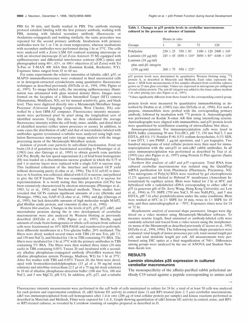
Table 1. Changes in p35 protein levels in cerebellar macroneuronscultured in the presence or absence of laminin
Groups
Hours in vitro
1 24 72 120
Polylysine 230 6 25 550 6 85 1100 6 120 2400 6 145Laminin (10 mg/ml) 245 6 45 1850 6 110* 3050 6 85* 4100 6 110*Laminin (10 mg/ml)
plus anti-b1 integrin(150 mg/ml) 260 6 75 950 6 125*
p35 protein levels were determined by quantitative Western blotting using 125Iprotein A, as described in Materials and Methods. Each value represents themean 6 SEM from measurements of five samples obtained from cerebellar culturesgrown on 25 mm glass coverslips. Values are expressed in micrograms per milligramof total cellular protein. The anti-b1 integrin was added to the tissue culture medium1 hr after plating (see also Pigino et al., 1997).*Values significantly different from those found in the corresponding control group.
3
Fluorescence intensity measurements were performed in the cell body of cells maintained in culture for 24 hr; a total of at least 50 cells was analyzedfor each protein and experimental condition. D, cdk5 histone H1 activity in control (lane 1) and RP1-treated (lane 2, 5 mM) cerebellar macroneurons.cdk5 was immunoprecipitated from cell homogenates after 1 d in vitro (200 mg of total cellular protein per sample) and kinase reactions performed asdescribed in Materials and Methods. Films were exposed for 1 d. E, Graph showing quantitation of cdk5 histone H1 activity in control, sense, and RP1-or RP2-treated cultures, as revealed by Cerenkow counting of samples prepared as described in D.
9860 J. Neurosci., December 1, 1998, 18(23):9858–9869 Paglini et al. • p35 Protein Function during Axonal Development

Figure 2. A, Effect of the antisense oligonucleotide RP1 (5 mM) on p35 protein levels as revealed by Western blot analysis of cell extracts obtained fromcultured cerebellar macroneurons growing on laminin and reacted with the C19 antibody diluted 1:200. Lane 1, Control nontreated, 24 hr after plating;lane 2, sense-treated, 24 hr after plating; lane 3, RP1-treated, 12 hr after plating; lane 4, RP1-treated, 24 hr after plating; lane 5, RP1-treated, 36 hr afterplating. Five micrograms of total cellular protein were added in each lane. B, Quantitative fluorescence measurements showing the effect of the antisenseoligonucleotides RP1 (5 or 2.5 mM) or RP2 (2.5 mm) on p35 immunofluorescence. Fluorescence intensity measurements were performed in the cell body4, 12, and 24 hr after plating; a total of 100 cells was measured for each time point and experimental condition. C, Quantitative fluorescencemeasurements showing the relative levels of tubulin, cdk5, p35, ERK1, MAP2, and tau in control and RP1-treated cells. Figure legend continues.
Paglini et al. • p35 Protein Function during Axonal Development J. Neurosci., December 1, 1998, 18(23):9858–9869 9861

residues 289–307 mapping at the C terminus of p35 is shown inFigure 1A. The antibody recognizes a single band of ;35 kDawhen whole-cell extracts of cultured cerebellar macroneuronsgrowing on polylysine are resolved in SDS-PAGE, blotted, andimmunostained with the anti-p35 antibody (Fig. 1A, lanes 1-3).The staining generated by this antibody is greatly inhibited byneutralization with purified peptide (Fig. 1A, lane 4). Quantita-tive Western blot analysis revealed that embryonic day 15 (E15)cerebellar cells express low levels of p35 at the time of plating andthat a significant increase occurs after the cells have developed inculture for .2 d. This increase in p35 protein levels closelyparallels the increase in cdk5 activity and in axonal length ob-served after the initial establishment of neuronal polarity incultured cerebellar macroneurons (Table 1; see also Pigino et al.,1997).
More importantly, our results show that when cerebellar ma-croneurons are cultured on a laminin-containing substrate, adramatic increase in p35 protein levels is detected (Fig. 1B, lanes1-6; Table 1). This phenomenon is also accompanied by anacceleration in the time course of p35 expression; thus, Westernblot analysis of whole-cell extracts obtained from neurons cul-tured in the presence of laminin for 4, 10, or 20 hr and probedwith the anti-p35 antibodies revealed a considerable and signifi-cant increase in the levels of p35 when compared with the onesdetected at equivalent time points in neurons maintained onpolylysine alone (Fig. 1B). The increase in p35 protein levelsdetected in cerebellar macroneurons growing on laminin is sig-nificantly inhibited when the cells are incubated with an affinity-purified rabbit polyclonal antibody against b-1 integrin (Table 1).
It is unlikely that the increase in p35 protein levels induced bylaminin is the result of an overall increase in protein synthesis,because no changes in the levels of cdk5 (Pigino et al., 1997),casein kinase II (Pigino et al., 1997), MAP1b (DiTella et al.,1996), MAP2 (Pigino et al., 1997), a-tubulin (Fig. 1B, lanes 1-6),or kinesin heavy chain (data not shown) are detected betweenneurons growing on laminin or polylysine alone. Northern blotanalysis provided additional support to these findings and re-vealed that laminin rapidly stimulates p35 mRNA expression incultured cerebellar macroneurons (Fig. 1C); as expected, thistreatment has no effect on cdk5 mRNA levels (Fig. 1D).
It has recently been shown that neurons not only express p35,but also a highly related isoform, designated as p39 (Tang et al.,1995). Therefore, it became of interest to determine whetherlaminin enhances the expression of p39. To test for this possibilitywe prepared a polyclonal antibody against p39 using as immuno-gen a peptide corresponding to amino acid residues 349–367mapping at the C terminus of the 39 kDa isoform; this region ofthe p39 molecule shows no significant homology with the corre-sponding one of p35. Figure 1E shows that the p39 polyclonalantibody recognizes a single band of ;40 kDa in Western blots ofwhole-brain homogenates. This analysis revealed that in the ce-rebral cortex or cerebellum the expression of the p39-immunoreactive protein species is lower at late embryonic andearly postnatal days but that it increases significantly afterward,reaching a peak during the second postnatal week, in which thehighest levels are detected (Fig. 1C, lanes 1–5). In addition,Western blots of cell extracts obtained from cultures of cerebellarmacroneurons revealed that these neurons express very low levelsof p39 at the time of plating but that a significant increase occursafter the cells have developed in vitro for .4 d (data not shown).More importantly, our results show that there are no significant
differences in the levels of p39 between neurons cultured onpolylysine alone or polylysine plus laminin (Fig. 1E, lanes 6–7).
Based on these observations it is likely that the increase in cdk5activity observed in neurons growing on laminin (Pigino et al.,1997) is associated with an enhanced and selective expression ofp35. However, other possibilities were also considered. For exam-ple, an enhancement of cdk5 activity may also result from areduced expression of the mitotic inhibitor p27, whose expressionincreases in parallel with terminal neuronal differentiation andthat is capable of suppressing cdk5 activity (Lee et al., 1996b). Wethink this is unlikely because no differences in p27 protein levelswere detected between neurons growing on laminin or polylysinealone (Fig. 1F, lanes 1–2).
Alternatively, and because laminin may stimulate cdk5 activityby enhancing the expression of other cdk5 activators, like p67(Shetty et al., 1994), we decided to examine the consequences ofp35 suppression by antisense oligonucleotide treatment on theactivity of cdk5 in cultured cerebellar macroneurons growing onlaminin. Two different nonoverlapping phosphorothioate anti-sense oligonucleotides were fabricated. The sequences selectedwere specific for p35, but not for p39. As controls, we alsoobtained sense oligonucleotide sequences corresponding to bothof the antisense sequences. The oligonucleotides were addeddirectly to the culture medium in the presence of Lipofectin (seeMaterials and Methods) and renewed every 6 hr. Figure 2A showsthat the addition of the p35 antisense oligonucleotide RP1 at adose of 5 mM, but not of the corresponding sense-strand oligonu-cleotide, to cells growing on laminin produces a significant re-duction in p35 protein levels as determined by Western blotanalysis of whole-cell homogenates reacted with either the rabbitantiserum C-19 or the N-20 antibody. Inhibition of p35 expressionbecomes evident after 16 hr of antisense treatment, and by 36 hronly trace amounts of protein are detected in the cell homoge-nates. In addition, to test for specificity, quantitative fluorescencemeasurements were performed, and the relative levels of p35,p39, cdk5, ERK1, and several cytoskeletal proteins were evalu-ated at different time points after the addition of the antisense orsense oligonucleotides. The results obtained, shown in Figure 2B,indicate that although both of the antisense sequences are effec-tive in reducing p35 protein levels in a dose-dependent manner,neither of the sense sequences detectably altered p35 proteinlevels. In addition, neither of the antisense sequences nor thesense sequences resulted in diminutions of the levels of cdk5, p39,ERK1, tubulin, MAP1b, MAP2, or tau protein levels (Fig. 2C).Having shown that the p35 antisense oligonucleotides effectivelyand selectively inhibit p35 expression, we decided to examinecdk5 activity in p35-suppressed cultured cerebellar macroneuronsmaintained on a laminin-containing substrate. For such a pur-pose, cdk5 was immunoprecipitated from sense- and antisense-treated cultures; the ability of the cdk5 immunoprecipitates tophosphorylate histone H1 was then evaluated. The results ob-tained showed high cdk5 histone H1 kinase activity in the immu-noprecipitates from sense-treated cultures and a significant de-crease of such an activity in the ones obtained from the antisense-treated cultures (Fig. 2D,E).
An additional possibility to consider is that the increase in p35protein levels and the associated cdk5-enhanced histone H1 ki-nase activity (Pigino et al., 1997) observed in neurons growing onlaminin are the result of an enhanced rate of neurite extensionand not a direct consequence of laminin activity. To distinguishbetween these possibilities, the following experiment was per-formed. In previous studies we have shown that the acute addition
9862 J. Neurosci., December 1, 1998, 18(23):9858–9869 Paglini et al. • p35 Protein Function during Axonal Development

of laminin to the tissue culture medium of neurons growing onpolylysine stimulates cdk5 activity and increases the rate of neu-rite extension (DiTella et al., 1996; Pigino et al., 1997). Thisenhanced neurite outgrowth response can be prevented if cellsare treated with a combination of antisense oligonucleotidesagainst the tau and MAP1b mRNAs (DiTella et al., 1996). Wetook advantage of this situation to test whether soluble lamininwas capable of increasing p35 expression in neurons with lowlevels of tau and MAP1b, as well as a with a reduced rate ofaxonal growth. The results obtained clearly indicate that lamininis capable of enhancing p35 expression independent of the mag-nitude of the neurite outgrowth response that the cells display(Table 2).
The subcellular localization of p35 in developingcerebellar macroneuronsPrevious studies have shown that in cultured cortical neurons(Nikolic et al., 1996) or cerebellar macroneurons (Pigino et al.,1997), as well as during in situ cerebellar development (Matsus-hita et al., 1995), cdk5 redistributes from the cell body towardaxonal growth cones during process formation. Although a sim-ilar phenomenon has been described for p35 in cultured corticalneurons (Nikolic et al., 1996), the situation appears to be differentin the case of in situ developing neurons because immunohisto-chemical studies have failed to detect significant levels of p35immunoreactivity within axons or growth cones; by contrast, highp35 immunolabeling was observed in cell bodies and dendrites ofadult neurons (Tomizawa et al., 1996). This apparent discrepancymay be related with the fact that no immunocytochemical studyhas examined the subcellular distribution of p35 in embryonicbrain (or cerebellar) tissue. Therefore, to clarify this point and
obtain further evidences about the participation of p35 duringlaminin-stimulated cdk5 activation, the subcellular localization ofp35 in cultured cerebellar macroneurons was analyzed by confo-cal microscopy; in addition, subcellular fractionation techniqueswere used to isolate growth cones from the embryonic cerebraland cerebellar cortex and to determine by Western blotting thepresence of the cdk5/p35 kinase.
In neurons growing on polylysine, light immunoreactivity forp35 is detected during the first 48 hr after plating; in these cells,most of the labeling is found within the cell body, including thenucleus, but absent from either minor processes or axons (Fig.3A–D). However, after 48 hr when most of the neurons havealready extended an axon of .120 mm, a significant change in theintensity of the labeling and in the distributional pattern of p35 isdetected in the majority of the cells. Thus, at this stage, an intensestaining of the cell body and axonal growth cones is observed withthe antibodies against p35 used at concentrations of 0.2–0.5mg/ml (Fig. 3E–G); at neuritic tips this immunolabeling is partic-ularly prominent within the peripheral regions of growth cones.At higher antibody concentrations (1–2 mg/ml) p35 is also de-tected in microspikes located within the distal third of growingaxons, as well as within the growth cones of minor processes, theneurites that will lately develop into dendrites (Caceres et al.,1991, 1992).
To test whether the localization of p35 at the periphery ofaxonal growth cones involves a plasma membrane association, amild extraction protocol using saponin was used. This procedureselectively removes cytosolic proteins but retains cytoskeletalmembrane interactions (Nakata and Hirokawa, 1987; Brandt etal., 1995; Pigino et al., 1997). In saponized cells, both p35 anti-bodies stain the actin-rich peripheral regions of growth cones, aswell as microspikes or filopodial extensions emerging from lamel-lipodial veils (Fig. 3H, I).
Western blot analysis of growth cone particles isolated bysubcellular fractionation according to the procedures describedby Pfenninger et al. (1983) (Lohse et al., 1996; see Materials andMethods) and probed with antibodies specific for cdk5 or p35,revealed the presence of the cdk5/p35 kinase in growth conesobtained from either the cerebral cortex or cerebellum (Fig. 4A);a very low signal for p39 was also detected in these preparations(Fig. 4A). To test for the purity of the GCP fraction we analyzedthe distribution of several well established growth cone markers,namely c-src (Helmke and Pfenninger, 1996) or bgc (Quiroga etal., 1995; Mascotti et al., 1997); both proteins were highly en-riched in the growth cone extracts containing cdk5 or p35 (Fig.4A); by contrast high molecular weight MAP2 was undetectablein these preparations (Fig. 4A). Moreover, to discard possiblecontaminations of GCPs with membranes of relatively low buoy-ant densities, we assayed GCPs for the presence of a well estab-lished Golgi marker, such as the protein designated as TGN-38(Horn and Banting, 1994). As shown in Figure 4A, this proteinwas completely absent from this preparation.
In a complementary series of experiments we used quanti-tative fluorescence techniques to measure the relative amountof p35 at neuritic tips of axons from neurons growing on eitherpolylysine or laminin. The results obtained, which are shown inFigure 4, B and C, indicate that laminin selectively and signif-icantly increases the relative amount of p35 associated with thesubcortical cytoskeleton of axonal growth cones. This analysisalso revealed that laminin not only increases the intensity ofp35 immunolabeling, but that it selectively accelerates the timecourse of appearance of p35 immunofluorescence at the distal
Table 2. Changes in p35 protein levels after the acute addition oflaminin to control and tau/MAP1b suppressed cerebellar macroneuronsgrowing on polylysine
Groups
Hours after the addition oflaminin to the culture medium
0 24
Non-treatedAxonal length 110 6 12 230 6 10p35 protein levels 720 6 80 1050 6 110
Non-treated plus laminin(20 mg/ml)Axonal length 115 6 10 360 6 24*p35 protein levels 660 6 70* 2300 6 120*
MAP1b and tau antisense-treatedplus laminin (20 mg/ml)Axonal length 65 6 5* 85 6 10*p35 protein levels 650 6 60 2300 6 85
All experiments started 24 hr after plating. Length values are expressed in microme-ters. Data represent the mean 6 SEM. A total of 100 cells was analyzed per timepoint and experimental condition. p35 protein levels were determined by quantita-tive Western blotting using 125I protein A, as described in Materials and Methods.Each value represents the mean 6 SEM from measurements of five samplesobtained from cerebellar cultures grown on 25 mm glass coverslips. Values areexpressed in micrograms per milligram of total cellular protein. The antisenseoligonucleotides were added to the culture medium 4 hr after plating and replen-ished every 12 hr until the end of the experiment. For suppressing MAP1b we usedthe antisense oligonucleotide designated As1 (see DiTella et al., 1992), whereas forsuppressing tau we used the antisense oligonucleotide RT11/14 (Caceres and Kosik,1990; Caceres et al., 1992; DiTella et al., 1996); both oligonucleotides were used atconcentrations of 50 mM.*Values significantly different from those found in the corresponding control group.
Paglini et al. • p35 Protein Function during Axonal Development J. Neurosci., December 1, 1998, 18(23):9858–9869 9863

end of growing axons. Under this experimental condition, itwas possible to detect high p35 immunolabeling within axonalgrowth cones, as soon as 4 – 6 hr after plating, when a signifi-cant proportion of cells have already developed an axon-likeneurite of .80 mm.
p35 suppression inhibits laminin-enhanced axonalelongation and MAP1b phosphorylation
In a previous study (Pigino et al., 1997) we have shown that inneurons growing on laminin cdk5 and mode I phosphorylated
Figure 3. Distribution and subcellular localization of p35 in cerebellar macroneurons growing on polylysine. A–D, Confocal micrographs showing thedistribution of tyrosinated a-tubulin (A, C) and p35 (B, D) in cerebellar macroneurons maintained in culture for 36 hr. Note than in both stage II (A,B) and in stage III (C, D) neurons, p35 is mainly localized to the cell body; there is no staining of minor processes or the axon. E, F, Confocal imagesshowing the distribution of tyrosinated a-tubulin (E) and p35 (F) at the distal end of a growing axon of a cerebellar macroneuron maintained in culturefor 48 hr. At this stage of neuritic development p35 immunolabeling is present in the cell body and within axonal growth cones. G, Red-green overlayof the images shown in E and F. H, I, High-power confocal micrographs showing the distribution of tyrosinated a-tubulin (H ) and p35 ( I ) at the distalend of a growing axon. Note that p35 immunolabeling is highly enriched at the growth cone periphery in regions almost devoid of microtubules. For thisexperiment the cells were extracted with 0.02% saponin before fixation. Scale bars, 10 mm.
9864 J. Neurosci., December 1, 1998, 18(23):9858–9869 Paglini et al. • p35 Protein Function during Axonal Development

MAP1b (e.g., phosphorylation at PDPK epitope; Ulloa et al.,1993a,b) increase significantly at the distal end of growing axonsin parallel with rapid axonal elongation. Besides, cdk5 suppres-sion significantly reduces laminin-enhanced axonal elongationand the mode I of MAP1b phosphorylation (Ulloa et al., 1993a,b;Pigino et al., 1997). Based on these observations we have pro-posed that at least one mechanism by which cdk5 may participatein axonal extension involves the regulation of MAP1b phosphor-ylation at the distal end of the growing axon. Therefore, to obtainfurther evidences about this proposal we decided to examine theconsequences of p35 suppression on process formation andMAP1b phosphorylation in cerebellar macroneurons growing ona laminin-containing substrate.
Under this condition p35 suppression significantly and selec-tively reduces laminin-stimulated axonal extension (Fig. 5A).This phenomenon becomes evident 20–24 hr after the addition ofthe antisense oligonucleotides, being highly coincident with thereduction in p35 protein levels (Fig. 5B). By contrast, no inhibi-tion of axonal elongation was observed when equivalent cultureswere treated with the corresponding sense oligonucleotides orwith a p39 antisense oligonucleotide (Fig. 5A,B). We also failedto detect an inhibition of axonal elongation when the RP1 or RP2oligonucleotides were added from the time of plating, and for aperiod of 24 hr, to cerebellar macroneurons growing on polyly-sine alone (data not shown).
Our observations (Fig. 5C,D) also show that a 48 hr treatmentof cerebellar macroneurons growing on a laminin-containingsubstrate with the RP1 antisense oligonucleotide (5 mM) signifi-cantly reduces the labeling generated by the mAb 150 that rec-ognizes a phosphorylated mode I epitope on the MAP1b mole-cule (Ulloa et al., 1993a,b; Pigino et al., 1997) while it increasesthe one observed with the rabbit antiserum 531, which recognizesa PDPK phosphorylatable MAP1b epitope when it is dephospho-rylated (Ulloa et al., 1993a,b; Pigino et al., 1997).
Because at axonal tips the cdk5/p35 complex is highly en-riched in the lamellipodial veil of growth cones, we decided toexamine whether dephosphorylated MAP1b was also presentin this subcellular compartment. In previous studies we haveshown that dephosphorylated MAP1b distributes throughoutthe cell, localizing in the cell body, minor neurites, and axonalprocesses; it should be noted that most of the dephosphory-lated MAP1b present in developing cerebellar macroneuronsis lost when cells are extracted with Triton X-100 under mi-crotubule stabilizing conditions before fixation (DiTella et al.,1996; Pigino et al., 1997). However, when the cells are ex-tracted with 0.02% saponin under conditions that preservecytoskeletal–membrane interactions (Nakata and Hirokawa,1987; Pigino et al., 1997) 531 immunolabeling is detectedthroughout the neurons including neuritic tips (Fig. 6). Highresolution fluorescence microscopy, digital image processing,and ratio image analysis of cells double labeled with the 531
4
cultured on polylysine (Ctrl ) or a laminin-containing substrate (Lam).Immunofluorescence was performed in cells fixed before (Ctrl GC/LamGC) or after saponin (Ctrl GC Sap/Lam GC Sap) or Triton X-100 (LamGC Triton) extraction performed under conditions that stabilize thecytoskeleton (see Materials and Methods). Note that in cells growing onlaminin a significant amount of p35 immunofluorescence remains associ-ated with the cytoskeleton after saponin extraction performed underconditions that preserve cytoskeletal–membrane interactions. A total of50 cells was measured per time point and experimental condition. Eachvalue represents the mean 6 SEM.
Figure 4. A, Western blots of total brain homogenates ( H ) and growthcone particles (GCP) reacted with antibodies against cdk5 (dilution 1:200,0.5 mg/ml), p35 (dilution 1:200, 0.5 mg/ml), p39 (dilution 1:50, 2 mg/ml),c-src (dilution 1:100, 1 mg/ml), MAP2 (dilution 1:500), and TGN38(dilution 1:200). The lanes reacted with antibodies against cdk5, p35,c-src, MAP2, and TGN38 were loaded with 10 mg of total cellular protein;the ones reacted with the p39 antibody were loaded with 75 mg of totalcellular protein. The total brain homogenate and the GCP were obtainedfrom E18 rat embryos as described in Materials and Methods. B, Averageintensity measurements of p35 immunofluorescence within the cell bodyof cerebellar macroneurons cultured on polylysine (Ctrl ) or on a laminin-containing substrate (Lam); immunofluorescence was performed in cellsfixed before extraction with detergents. A total of 50 cells was measuredper time point and experimental condition. Each value represents themean 6 SEM. C, Average intensity measurements of p35 immunofluo-rescence within growth cones (GC) of axons of cerebellar macroneurons
Paglini et al. • p35 Protein Function during Axonal Development J. Neurosci., December 1, 1998, 18(23):9858–9869 9865

antiserum and an mAb against tyrosinated a-tubulin clearlyrevealed the presence of dephosphorylated MAP1b within thelamellipodial veils of growth cones; confocal microscopy con-firmed these observations and also revealed the presence ofintense 531 immunolabeling in microspikes emerging from theperiphery of growth cones (Fig. 6 D, insert; H ).
DISCUSSION
p35 participation in axonal elongation
p35 is a neuron-specific regulatory subunit of cdk5 that is ex-pressed in postmitotic neurons but is absent in proliferatingneuronal progenitors (Lew et al., 1994; Tsai et al., 1994; Tomi-
Figure 5. A, Graph showing that suppression of p35, but not of p39, selectively inhibits axonal elongation in cerebellar macroneurons growing on alaminin-containing substrate. Open squares, Axonal length in sense-treated cultures; solid squares, idem but in RP1-treated cultures (5 mM); soliddiamonds, idem but in p39 antisense-treated cultures (5 mM); total length of minor processes per cell in p35 sense-treated cultures; idem but inRP1-treated cultures. A total of 100 cells was measured per time point and experimental condition. Each value represents the mean 6 SEM. B, Averageintensity measurements of p35 and p39 immunofluorescence in cerebellar macroneurons treated with p35 (RP1, 5 mM) or p39 (5 mM) antisenseoligonucleotides. Measurements were performed in the cell body, and a total of 50 cells were analyzed per time point and experimental condition. Eachvalue represents the mean 6 SEM. C, Average intensity measurements of PDPK-dephosphorylated MAP1b immunofluorescence in p35 sense- ( S) andantisense- (AS) treated cultures; cells were stained with the polyclonal antibody 531 (Ab 531). Measurements were performed in proximal (PAX ) anddistal (DAX ) axonal segments. Note that in the p35 antisense-treated neurons, 531 immunolabeling increases at the distal axonal segment. A total of50 cells was analyzed per time point and experimental condition; each value represents the mean 6 SEM. Oligonucleotides were used at a concentrationof 5 mM. D, Average intensity measurements of PDPK-phosphorylated MAP1b immunofluorescence in p35 sense- (S) and antisense- (AS) treatedcultures; cells were stained with the mAb 150. Measurements were performed in proximal (PAX ) and distal (DAX ) axonal segments. Note that in thep35 antisense-treated neurons mAb 150 immunolabeling decreases at the distal axonal segment; also note the absence of mAB 150 immunolabeling inproximal axonal segments of both sense- and antisense-treated cultures. Oligonucleotides were used at a concentration of 5 mM. A total of 50 cells wasanalyzed per time point and experimental condition; each value represents the mean 6 SEM.
9866 J. Neurosci., December 1, 1998, 18(23):9858–9869 Paglini et al. • p35 Protein Function during Axonal Development

zawa et al., 1996; Delalle et al., 1997). Based on the analysis of thespatiotemporal pattern of p35 mRNA distribution (Tsai et al.,1994; Delalle et al., 1997) and of the phenotype of mice lackingp35, a key role for the cdk5/p35 kinase in proper neuronalmigration has been established (Chae et al., 1997). Thus, exam-ination of mice lacking p35, and, thus, p35/cdk5 activity, hasrevealed severe cortical lamination defects, including a reversedpacking order of cortical neurons, such that earlier born neuronsreside in superficial layers and lately generated ones occupy deeplayers (Chae et al., 1997).
A second major proposed role for the cdk5/p35 kinase is theregulation of neuritic development (Nikolic et al., 1996; Delalle etal., 1997). In favor of this view, previous studies have establishedthe existence of a high degree of temporal correlation among p35mRNA expression, the induction of cdk5 activity, and the forma-tion of major axonal tracts (Tsai et al., 1993; Delalle et al., 1997;Tomizawa et al., 1997); besides, expression of a p35 antisenseconstruct in cultured cortical neurons reduces neurite outgrowth(Nikolic et al., 1996). Although the present results are consistentwith these observations, they suggest that the cdk5/p35 complex,rather than having an essential role in process formation (Nikolicet al., 1996), acts as a modulator of axonal elongation. A recentstudy showing that in immortalized hippocampal cells, p35 issufficient but not essential for inducing neurite outgrowth alsofavors this view (Xiong et al., 1997). In the case of cerebellarmacroneurons, we found a high degree of spatial and temporalcorrelation between the expression of the cdk5/p35 kinase andaxonal elongation when the neurons were maintained on alaminin-containing substrate; by contrast, such a correlation wasnot observed in neurons growing on polylysine alone in which theexpression of the cdk5/p35 kinase occurs after the initial exten-sion of neurites and the establishment of neuronal polarity (thisstudy; see also Pigino et al., 1997). Besides, and perhaps moreimportantly, two nonoverlapping antisense oligonucleotides thateffectively diminished the levels of p35 were capable of selectivelysuppressing laminin-enhanced axonal elongation, without alter-ing the normal extension of minor neurites or the ability ofcerebellar macroneurons to differentiate one of the minor pro-cesses into an axon-like neurite. As with any study involving theuse of antisense oligonucleotides, it was important to establishthat the observed effects were not related to a diminution in thehealth of the cultures. Several observations suggest that the p35antisense oligonucleotides specifically and selectively blocked theexpression of p35. First, sequence analysis of the regions of therat p35 mRNA selected for designing the antisense oligonucleo-tides revealed no significant homology with any other reportedsequence. Besides, none of the S-modified antisense oligonucleo-tides used in this study contains four contiguous guanosines resi-dues, which are believed to increase oligomer affinity to proteinsand, hence, generate nonspecific inhibitory effects (Wagner, 1995).Second, the antisense oligonucleotide treatment dramatically re-duced p35 protein levels without altering the levels of several otherproteins, including cdk5, p39, tubulin, MAP1b, MAP2, tau, andERK1. Third, the effects of the antisense oligonucleotides were
4
dephosphorylated MAP1b (F) within an axonal tip of a cerebellar ma-croneuron growing on polylysine and prepared for immunocytochemistryas described previously. G, Ratio image analysis clearly reveals the pres-ence of dephosphorylated MAP1b within the peripheral lamellipodial veilof axonal growth cones. H, Confocal image also showing the presence ofPDPK-dephosphorylated MAP1b at the periphery of axonal growthcones. Scale bars, 10 mm.
Figure 6. A, B, Double immunofluorescence micrographs showing thedistribution of tyrosinated a-tubulin (A) and PDPK-dephosphorylatedMAP1b in a stage II cerebellar macroneuron cultured on polylysine. Thecell was extracted with saponin before fixation under conditions thatpreserve cytoskeletal–membrane interactions. MAP1b was labeled withthe rabbit antiserum 531 used at a dilution of 1:500. For this experimentthe images were formed on the faceplate of an SIT camera and digitizeddirectly into the Metamorph Image Processor. Using image B as numer-ator and image A as denominator, we applied the ratio image menu of theprocessor to generate the image shown in C, which clearly reveals thepresence of dephosphorylated MAP1b within the lamellipodial veil ofgrowth cones. D, A confocal image of a stage II cerebellar macroneuronalso showing the presence of PDPK-dephosphorylated MAP1b within thelamellipodial veils of growth cones; the insert shows the presence of 531immunolabeling (dilution 1:500) within microspikes located at the periph-ery of lamellipodial veils. E, F, Double immunofluorescence micrographsshowing the distribution of tyrosinated a-tubulin (E) and PDPK-
Paglini et al. • p35 Protein Function during Axonal Development J. Neurosci., December 1, 1998, 18(23):9858–9869 9867

dose-dependent and not observed when the cells were treated withequivalent doses of the corresponding “sense” oligonucleotides.Finally, the antisense treatment was not effective in reducing ax-onal length when it was applied to neurons that express low levelsof p35 and, hence, display low cdk5 activity, e.g., neurons culturedon polylysine for up to 48 hr.
Our results also suggest that p35, and not the p39 isoform, isinvolved in regulating axonal extension in neurons growing on alaminin-containing substrate. Thus, no decreases in axonal lengthor in the time course of axonal formation were detected in cere-bellar macroneurons treated with a p39 antisense oligonucleotide.This may not be surprising because p39 is predominantly expressedin polarized neurons, and not during the initial stages of processformation, both in situ and in vitro (Cai et al., 1997; this study).Although this analysis suggests that p39 has no essential role inneurite outgrowth and axonal extension, it does not rule out thepossibility of p39 being involved in the maintenance of moremature neurites or in process formation in response to a differenttype of environmental cue. In this regard, it is worth noting that inimmortalized hippocampal pyramidal cells p39, rather than p35, isboth necessary and sufficient for promoting neurite outgrowth inresponse to basic fibroblast growth factor (Xiong et al., 1997).
Suppression of either cdk5 (Pigino et al., 1997) or p35 (thisstudy), but not of p39, significantly decreases the levels of PDPK-phosphorylated MAP1b. Because this phenomenon selectivelyoccurs at the distal end of growing axons, it is likely that thecdk5/p35 kinase regulates MAP1b phosphorylation within axonalgrowth cones. Thus, the time course of appearance of the cdk5/p35 kinase within growth cones is highly coincident with a de-crease of PDPK-dephosphorylated MAP1b and with an increasein PDPK-phosphorylated MAP1b. It is unlikely that the presenceof the cdk5/p35 kinase within axonal growth cones of culturedneurons (this study; see also Nikolic et al., 1996; Pigino et al.,1997) represent some peculiarity related with their in vitro devel-opment, because Western blot analysis of growth cone particlesclearly revealed the presence of both proteins in this compart-ment. Our results, and those of Nikolic et al. (1996), show thatwithin growth cones most of the cdk5/p35 kinase is associatedwith the subcortical cytoskeleton but not with microtubules. Thismay be important in terms of previous studies that have shownthat dephosphorylated MAP1b binds to microfilaments (Pedrottiand Islam, 1996) and not to microtubules (DiTella et al., 1996);besides, dephosphorylation kinetics suggests that the PDPK site,but not casein kinase II sites, negatively regulates the associationof MAP1b with F-actin (Pedrotti and Islam, 1996). In accordancewith that, we detected a significant association of PDPK-dephosphorylated MAP1b with the growth cone subcortical cy-toskeleton in neurons displaying low levels of p35 and, hence, ofcdk5 activity. Therefore, it is tempting to speculate that thepresence of an active cdk5/p35 complex within the growth conesubcortical cytoskeleton, as in the case of neurons growing onlaminin, will phosphorylate MAP1b, detaching it from microfila-ments and allowing its interaction with microtubules, an eventthat seems to be crucial for the participation of MAP1b in axonalelongation (DiTella et al., 1996: Pigino et al., 1997). However, itis worth noting that our results do not rule out the possibility ofcdk5 participating in axonal elongation by additional mechanisms(see Pigino et al., 1997 for a discussion of this issue).
Laminin regulation of p35 expressionThe present observations provide novel insights about the mech-anisms by which laminin may regulate axonal elongation. Specif-
ically, we show that laminin increases p35 mRNA and proteinlevels, a phenomenon that in turn enhances cdk5 activity, MAP1bphosphorylation, and axonal extension. The upregulation of p35protein levels in neurons growing on a laminin-containing sub-strate seems not to be related with an overall increase in proteinsynthesis, because no effects were observed in the levels of totaltubulin, MAP1b, MAP2, tau, cdk5, p39, and p27; nor it is theconsequence of an increased rate of growth, because laminin-stimulated p35 expression was also detected in neurons displayinga reduced rate of axonal elongation, such as in MAP1b- andtau-suppressed neurons. It is also unlikely that laminin increasescdk5 activity by stimulating the expression of other cdk5 activa-tors, because it was completely prevented in neurons treated withantisense oligonucleotides specific for the p35 mRNA. Besides,our results also suggest that laminin-stimulated cdk5 activity isnot the result of a reduced expression of a cdk5 inhibitor becauseno differences in the levels of p27 (Lee et al., 1996b) weredetected between neurons growing on polylysine or laminin.
Our results suggest that one mechanism by which laminin in-creases p35 protein levels may involve the stimulation of p35mRNA expression; this may not be an unusual phenomenon. Infact, a significant body of evidence exists showing that integrinactivation after ECM binding can regulate expression of manygenes (Ginsberg et al., 1995; Giancotti, 1997). For example, inanchorage-dependent cell lines, replating cells on fibronectin trig-gers expression of early immediate genes, such as c-fos and c-jun(Dike and Farmer, 1988; Dike and Ingber, 1996), whereas in ratkidney cells, cyclin A expression is specifically regulated by adhesion(Guadagno et al., 1993; see also Giancotti, 1997). In the specificcase of neurons, a recent study has shown that laminin stimulatesthe expression of two mitochondrial proteins during process out-growth (Weeks et al., 1997). Studies are in progress to determinethe mechanisms by which laminin increases p35 mRNA levels.
REFERENCESAvila J, Dominguez J, Diaz-Nido J (1994) Regulation of microtubule
dynamics by microtubule-associated protein expression and phosphor-ylation during neuronal development. Int J Dev Biol 38:13–25.
Black M, Slaughter T, Fisher I (1994) Microtubule-associated protein 1b(MAP1b) is concentrated in the distal region of growing axons. J Neu-rosci 14:857–870.
Bottenstein J, Sato G (1979) Growth of a rat neuroblastoma cell line in aserum-free supplemented medium. Proc Natl Acad Sci USA 81:5613–5617.
Brandt R, Leger J, Lee G (1995) Interaction of tau with the neuralplasma membrane mediated by tau’s amino-terminal projection do-main. J Cell Biol 131:1327–1340.
Brugg B, Reddy D, Matus A (1993) Attenuation of microtubule-associated protein 1B expression by antisense oligodeoxynucleotidesinhibits initiation of neurite outgrowth. Neuroscience 52:489–496.
Caceres A, Kosik K (1990) Inhibition of neurite polarity by antisenseoligonucleotides in primary cerebellar neurons. Nature 343:461–463.
Caceres A, Potrebic S, Kosik K (1991) The effect of tau antisense oligo-nucleotides in primary cerebellar neurons. J Neurosci 11:1515–1523.
Caceres A, Mautino J, Kosik K (1992) Suppression of MAP2 in culturedcerebellar macroneurons inhibits minor neurite formation. Neuron9:607–618.
Cai XH, Tomizawa K, Tang D, Lu YF, Moriwaki A, Tokuda M,Nagahata S, Hatase O, Matsui H (1997) Changes in the expression ofnovel Cdk5 activator messenger RNA (p39nck5ai mRNA) during ratbrain development. J Neurosci Res 28:355–360.
Carri N, Rubin K, Gullberg K, Ebendal T (1992) Neuritogenesis oncollagen substrates. Involvement of integrin-like matrix receptors inretinal fibre outgrowth collagen. Int J Dev Neurosci 10:395–403.
Chae T, Kwon Y, Bronson R, Dikkes P, Li E, Tsai L (1997) Mice lackingp35, a neuronal specific activator of cdk5, display cortical laminationdefects, seizures, and adult lethality. Neuron 18:29–42.
9868 J. Neurosci., December 1, 1998, 18(23):9858–9869 Paglini et al. • p35 Protein Function during Axonal Development

Delalle I, Bhide P, Caviness J, Tsai L (1997) Temporal and spatialpatterns of expression of p35, a regulatory subunit of cyclin-dependentkinase 5, in the nervous system of the mouse. J Neurocytol 26:283–296.
DiTella M, Feiguin F, Morfini G, Caceres A (1994) Microfilament asso-ciated growth cone component depends upon tau for its intracellularlocalization. Cell Motil Cytoskeleton 29:117–130.
DiTella M, Feiguin F, Carri N, Kosik K, Caceres A (1996) MAP1B/taufunctional redundancy during laminin-enhanced axonal growth. J CellSci 109:467–477.
Dike LE, Farmer S (1988) Cell adhesion induces expression of growth-associated genes in suspension-arrested fibroblasts. Proc Natl Acad SciUSA 85:6792–6795.
Dike LE, Ingber D (1996) Integrin-dependent induction of early re-sponse genes in capillary endothelial cells. J Cell Sci 109:2855–2863.
Drubin D, Feinstein S, Shooter E, Kirschner M (1985) Nerve growthfactor induced neurite outgrowth in PC12 cells involved the coordinateinduction of microtubule assembly and assembly promoting factors.J Cell Biol 101:1799–1807.
Edelman W, Zervas M, Costello P, Roback L, Fisher I, Hammarback J,Cowan N, Davies P, Wainer B, Kucherlapati R (1996) Neuronal ab-normalities in microtubule-associated protein 1B mutant mice. ProcNatl Acad Sci USA 93:1270–1275.
Esmaeli-Azad B, McCarty J, Feinstein SC (1994) Sense and antisensetransfection analysis of tau function: tau influences net microtubuleassembly, neurite outgrowth, and neurite stability. J Cell Sci107:869–879.
Ferreira A, Caceres A (1991) Estrogen-enhanced neurite growth: evi-dence for a selective induction of tau and stable microtubules. J Neu-rosci 13:3112–3123.
Ferreira A, Busciglio J, Caceres A (1989) Microtubule formation andneurite growth in cerebellar macroneurons which develop in vitro:evidence for the involvement of the microtubule-associated proteinsMAP1a, HMW-MAP2 and tau. Dev Brain Res 49:215–228.
Giancotti F (1997) Integrin signaling: specificity and control of cell sur-vival and cell cycle progression. Curr Opin Cell Biol 9:691–700.
Ginsberg M, Schwartz M, Schaller M (1995) Integrins: emerging para-digms of signal transduction. Annu Rev Cell Dev Biol 11:549–600.
Guadagno TM, Ohtsubo M, Roberts TM, Assoian RK (1993) A linkbetween cyclin A expression and adhesion-dependent cell cycle pro-gression. Science 362:1572–1575.
Harada A, Oguchi K, Okabe S, Kuno J, Terada S, Ohsima T, Sato-Yoshitake R, Takei Y, Noda T, Hirokawa N (1994) Altered microtu-bule organization in small caliber axons of mice lacking tau protein.Nature 369:488–491.
Helmke S, Pfenninger K (1996) Growth cone enrichment and cytoskel-etal association of non-receptor tyrosine kinases. Cell Motil Cytoskel-eton 30:194–207.
Hirokawa N (1994) Microtubule organization and dynamics dependenton microtubule-associated proteins. Curr Opin Cell Biol 6:74–81.
Horn M, Banting G (1994) Okaidaic acid lead to a fragmentation of thetrans-Golgi network and an increase in expression of TGN38 at the cellsurface. Biochem J 301:69–73.
Lee K, Rosales J, Tang D, Wang J (1996a) Interaction of cyclin-dependent kinase 5 (cdk5) and neuronal cdk5 activator in bovine brain.J Biol Chem 271:1538–1543.
Lee M, Nikolic M, Baptista C, Lai E, Tsai L, Massague J (1996b) Thebrain-specific activator p35 allows cdk5 to escape inhibition by p27Kip1in neurons. Proc Natl Acad Sci USA 93:3259–3263.
Lew J, Huang Q, Zhong Q, Winkfein R, Aebersold R, Hunt T, Wang J(1994) A brain-specific activator of cyclin-dependent kinase 5. Nature371:423–426.
Li HN, Quiroga S, Pfenninger KH (1992) Variable membrane glycop-roteins in different growth cone preparations. J Neurosci 12:2393–2402.
Lohse K, Helmke S, Wood M, Quiroga S, de la Houssaye B, Miller V,Negre-Aminou P, Pfenninger KH (1996) Axonal origin and purity ofgrowth cones isolated from fetal rat brain. Dev Brain Res 96:83–96.
Maccioni R, Cambiazo V (1995) Role of microtubule-associated pro-teins in the control of microtubule assembly. Physiol Rev 75:835–864.
Mascotti F, Caceres A, Pfenninger KH, Quiroga S (1997) Expressionand distribution of IGF-1 receptors containing a b-subunit variant (bgc)in developing neurons. J Neurosci 17:1447–1459.
Matsushita M, Matsui H, Itano T, Tomizawa K, Masaaki T, Suwaki H,Wang J, Hatase O (1995) Developmental changes in cyclin dependentkinase 5 subcellular localization in rat cerebellum. NeuroReport6:1267–1270.
Matus A (1988) Microtubule-associated proteins: their potential role indetermining neuronal morphology. Annu Rev Neurosci 11:29–44.
Mitchison T, Kirschner M (1988) Cytoskeletal dynamics and nervegrowth. Neuron 1:761–772.
Nakata T, Hirokawa N (1987) Cytoskeletal reorganization of humanplatelets after stimulation revealed by the quick-freeze deep-etch tech-nique. J Cell Biol 106:1771–1780.
Nikolic M, Dudek H, Kwon Y, Ramos Y, Tsai L (1996) The cdk5/p35kinase is essential for neurite outgrowth during neuronal differentia-tion. Genes Dev 10:816–825.
Pedrotti B, Islam K (1996) Dephosphorylated but not phosphorylatedmicrotubule-associated protein MAP1B binds to microfilaments. FEBSLett 388:131–133.
Pfenninger K, Ellis L, Johnson M, Friedman, L, Somlo S (1983) Nervegrowth cones isolated from fetal rat brain. I. Subcellular fractionationand characterization. Cell 33:573–584.
Pigino G, Paglini G, Ulloa L, Avila J, Caceres A (1997) Analysis of theexpression, distribution and function of cyclin dependent kinase 5(cdk5) in developing cerebellar macroneurons. J Cell Sci 110:257–270.
Quiroga S, Garofalo RS, Pfenninger KH (1995) Insulin-like growth fac-tor I receptors of fetal brain are enriched in nerve growth cones andcontain a b-subunit variant. Proc Natl Acad Sci USA 92:4309–4312.
Reichardt LF, Tomaselli K (1991) Extracellular matrix molecules andtheir receptors: functions in neural development. Annu Rev Neurosci14:531–570.
Rocha M, Avila J (1995) Characterization of microtubule-associatedprotein phosphoisoforms present in isolated growth cones. Dev BrainRes 89:47–55.
Shetty K, Kaech S, Link W, Jaffe H, Flores C, Wray S, Pant H, BeuhausenS (1995) Molecular characterization of a neuronal-specific proteinthat stimulates the activity of cdk5. J Neurochem 64:1988–1995.
Takei Y, Kondo S, Harada A, Inomata S, Noda T, Hirokawa N (1997)Delayed development of nervous system in mice homozygous for dis-rupted microtubule-associated protein 1B (MAP1B) gene. J Cell Biol137:1615–1626.
Tanaka E, Sabry J (1995) Making the connection: cytoskeletal rear-rangements during growth cone guidance. Cell 83:171–176.
Tang D, Young J, Lee K, Matsushita M, Matsuli H, Tomizawa K, HataseO, Wang J (1995) An isoform of the neuronal cyclin-dependent kinase5 (cdk5) activator. J Biol Chem 270:26897–26903.
Tomizawa K, Matsui H, Matsushita M, Lew J, Tokuda M, Itano T,Konishi R, Wang J, Hotase O (1996) Localization and developmentalchanges in the neuron-specific cyclin-dependent kinase 5 activator(p35ncka5) in the rat brain. Neuroscience 74:519–529.
Tsai L, Takahashi T, Caviness V, Harlow E (1993) Activity and expres-sion pattern of cyclin-dependent kinase 5 in the embryonic mousenervous system. Development 119:1029–1040.
Tsai L, Delalle L, Caviness V, Chae T, Harlow E (1994) p35 is aneural-specific regulatory subunit of cyclin-dependent kinase 5. Nature371:419–423.
Ulloa L, Avila J, Diaz-Nido J (1993a) Heterogeneity in the phosphory-lation of microtubule-associated protein MAP1B during rat brain de-velopment. J Neurochem 63:961–972.
Ulloa L, Diaz-Nido J, Avila J (1993b) Depletion of casein kinase II byantisense oligonucleotide prevents neuritogenesis in neuroblastomacells. EMBO J 12:1633–1640.
Ulloa L, Ibarrola N, Avila J, Diaz-Guerra F (1994) Microtubule-associated protein 1B (MAP1B) is present in glial cells phosphorylateddifferent than in neurons. Glia 10:266–275.
Wagner R (1995) The state of the art in antisense research. Nat Med1:1116–1118.
Weeks BS, Burbelo P, Jucker M, Weiner MA, Roque F, Kleinman HK(1997) Laminin stimulates expression of two mitochondrial proteinsduring neurite outgrowth. Int J Dev Neurosci 14:365–374.
Xiong W, Pestell R, Rosner M (1997) Role of cyclins in neuronal dif-ferentiation of immortalized hippocampal cells. Mol Cell Biol 17:6585–6597.
Paglini et al. • p35 Protein Function during Axonal Development J. Neurosci., December 1, 1998, 18(23):9858–9869 9869