Macrophage MicroRNA-155 Promotes Cardiac Hypertrophy and Failure
Upload
independentCategory
view
1download
0
Harald H.H.W. Schmidt, Gary K. Owens, J. David Lambeth and Kathy K. GriendlingDikalov, Alejandra San Martin, Alicia Lyle, David S. Weber, Daiana Weiss, W. Robert Taylor,
Anna Dikalova, Roza Clempus, Bernard Lassègue, Guangjie Cheng, James McCoy, SergeySmooth Muscle Hypertrophy in Transgenic Mice
Nox1 Overexpression Potentiates Angiotensin II-Induced Hypertension and Vascular
Print ISSN: 0009-7322. Online ISSN: 1524-4539 Copyright © 2005 American Heart Association, Inc. All rights reserved.
is published by the American Heart Association, 7272 Greenville Avenue, Dallas, TX 75231Circulation doi: 10.1161/CIRCULATIONAHA.105.538934
2005;112:2668-2676; originally published online October 17, 2005;Circulation.
http://circ.ahajournals.org/content/112/17/2668World Wide Web at:
The online version of this article, along with updated information and services, is located on the
http://circ.ahajournals.org/content/suppl/2005/07/21/CIRCULATIONAHA.105.538934.DC1.htmlData Supplement (unedited) at:
http://circ.ahajournals.org//subscriptions/
is online at: Circulation Information about subscribing to Subscriptions:
http://www.lww.com/reprints Information about reprints can be found online at: Reprints:
document. Permissions and Rights Question and Answer this process is available in the
click Request Permissions in the middle column of the Web page under Services. Further information aboutOffice. Once the online version of the published article for which permission is being requested is located,
can be obtained via RightsLink, a service of the Copyright Clearance Center, not the EditorialCirculationin Requests for permissions to reproduce figures, tables, or portions of articles originally publishedPermissions:
by guest on February 19, 2014http://circ.ahajournals.org/Downloaded from by guest on February 19, 2014http://circ.ahajournals.org/Downloaded from by guest on February 19, 2014http://circ.ahajournals.org/Downloaded from by guest on February 19, 2014http://circ.ahajournals.org/Downloaded from by guest on February 19, 2014http://circ.ahajournals.org/Downloaded from by guest on February 19, 2014http://circ.ahajournals.org/Downloaded from by guest on February 19, 2014http://circ.ahajournals.org/Downloaded from by guest on February 19, 2014http://circ.ahajournals.org/Downloaded from

Nox1 Overexpression Potentiates Angiotensin II–InducedHypertension and Vascular Smooth Muscle Hypertrophy
in Transgenic MiceAnna Dikalova, PhD; Roza Clempus, MD; Bernard Lassègue, PhD; Guangjie Cheng, PhD;
James McCoy, BS; Sergey Dikalov, PhD; Alejandra San Martin, PhD; Alicia Lyle, BS;David S. Weber, PhD; Daiana Weiss, MD; W. Robert Taylor, MD, PhD; Harald H.H.W. Schmidt, MD;
Gary K. Owens, PhD; J. David Lambeth, MD, PhD; Kathy K. Griendling, PhD
Background—Reactive oxygen species (ROS) have been implicated in the development of cardiovascular pathologies.NAD(P)H oxidases (Noxes) are major sources of reactive oxygen species in the vessel wall, but the importance ofindividual Nox homologues in specific layers of the vascular wall is unclear. Nox1 upregulation has been implicated incardiovascular pathologies such as hypertension and restenosis.
Methods and Results—To investigate the pathological role of Nox1 upregulation in vascular smooth muscle, transgenicmice overexpressing Nox1 in smooth muscle cells (TgSMCnox1) were created, and the impact of Nox1 upregulation on themedial hypertrophic response during angiotensin II (Ang II)–induced hypertension was studied. These mice haveincreased expression of Nox1 protein in the vasculature, which is accompanied by increased superoxide production.Infusion of Ang II (0.7 mg/kg per day) into these mice for 2 weeks led to a potentiation of superoxide productioncompared with similarly treated negative littermate controls. Systolic blood pressure and aortic hypertrophy were alsomarkedly greater in TgSMCnox1 mice than in their littermate controls. To confirm that this potentiation of vascularhypertrophy and hypertension was due to increased ROS formation, additional groups of mice were coinfused with theantioxidant Tempol. Tempol decreased the level of Ang II–induced aortic superoxide production and partially reversedthe hypertrophic and hypertensive responses in these animals.
Conclusions—These data indicate that smooth muscle–specific Nox1 overexpression augments the oxidative, pressor, andhypertrophic responses to Ang II, supporting the concept that medial Nox1 participates in the development ofcardiovascular pathologies. (Circulation. 2005;112:2668-2676.)
Key Words: angiotensin � hypertension � hypertrophy � muscle, smooth � free radicals
Reactive oxygen species (ROS) such as superoxide andhydrogen peroxide act as intercellular and intracellular
messengers that play physiological and pathophysiologicalroles in vascular biology.1,2 They have profound effects onvascular smooth muscle cell (VSMC) growth and migration,processes that are critically important in cardiovascular pa-thology and contribute to the vascular changes associatedwith hypertension, arteriosclerosis, and restenosis.3
A major source of ROS in vasculature is the NAD(P)Hoxidase family of enzymes that are distributed throughout thevessel wall.3 The VSMC NAD(P)H oxidases are structurallydistinct from the classic neutrophil oxidase. In large arteries,VSMCs lack gp91phox, the catalytic subunit that transferselectrons from NADPH to molecular oxygen to form super-
Editorial p 2585Clinical Perspective p 2676
oxide.4 Instead, these cells express the gp91phox homologuesNox1 and Nox4.4 Although both Nox1 and Nox4 are upregu-lated after angiotensin II (Ang II) infusion,5 the 2 enzymes aredifferentially regulated during restenosis.6 Furthermore,Nox1 and Nox4 are found in distinct intracellular compart-ments, suggesting that they serve different functions withinthe cell.7 The precise biological roles of specific Nox proteinsthus remain unclear.
A growing body of evidence suggests that Ang II mediatesits potent vasoconstrictor, mitogenic, and hypertrophic effectsat least in part through ROS.8–12 Not only is vascular
Received January 31, 2005; revision received April 26, 2005; accepted May 10, 2005.From the Division of Cardiology (A.D., R.C., B.L., S.D., A.S.M., A.L., D.S.W., D.W., W.R.T., K.K.G.) and Department of Pathology (G.C., J.M.,
J.D.L.), Emory University, Atlanta, Ga; Department of Pharmacology, Monash University, Clayton, Victoria, Australia (H.H.H.W.S.); and Departmentof Molecular Physiology and Biological Physics, University of Virginia School of Medicine, Charlottesville (G.K.O.).
The online-only Data Supplement, which contains additional information about Methods, can be found at http://circ.ahajournals.org/cgi/content/full/CIRCULATIONAHA.105.538934/DC1.
Correspondence to Kathy K. Griendling, Emory University, Division of Cardiology, 319 WMB, 1639 Pierce Dr, Atlanta, GA 30322. [email protected]
© 2005 American Heart Association, Inc.
Circulation is available at http://www.circulationaha.org DOI: 10.1161/CIRCULATIONAHA.105.538934
2668
Hypertension
by guest on February 19, 2014http://circ.ahajournals.org/Downloaded from

superoxide elevated after Ang II infusion,5,11–15 but treatmentwith antioxidants decreases Ang II–induced hypertension inrats and mice.16–19 Genetic overexpression of superoxidedismutase (SOD) also attenuates the rise in blood pressurestimulated by Ang II.8 One of the hallmarks of hypertensiondue to activation of the renin-angiotensin system is medialhypertrophy of large vessels.20 In vitro, Ang II–inducedVSMC hypertrophy requires ROS derived from NAD(P)Hoxidases, specifically Nox1.4,21,22 Aortic hypertrophy in re-sponse to Ang II infusion is impaired in gp91phox-deficientmice, but gp91phox is expressed only in the endothelium andadventitia and not in the aortic media.11 This raises thequestion of whether medial NAD(P)H oxidases regulatehypertrophy in vivo.
On the basis of these considerations, transgenic miceoverexpressing Nox1 in smooth muscle cells (SMCs) werecreated and used to investigate the impact of Nox1 upregu-lation, as occurs during hypertension and restenosis, on thepressor and medial hypertrophic response during Ang II–induced hypertension. We hypothesized that increased Nox1-derived ROS would exacerbate the increased wall thicknessthat occurs during hypertension and thus may also lead toelevations in blood pressure. We found that upregulation ofNox1 does indeed potentiate both the pressor and hypertro-phic responses to Ang II in a ROS-dependent manner,supporting the concept that medial Nox1 participates in thedevelopment of cardiovascular pathologies.
MethodsExtended methods are provided in the online-only Data Supplement.
ReagentsDNeasy Tissue and RNeasy kits were from Qiagen, Inc. Nytran Nmembranes were from Schleicher & Schuell. 1-Hydroxy-3-methoxycarbonyl-2,2,5,5-tetramethyl-pyrrolidine (CMH) was pur-chased from Alexis Corp. Dihydroethidium was purchased fromMolecular Probes. Anti-Nox1 rabbit polyclonal antibody was pre-pared as described.23 The gp91 phox antibody (clone 54.1) was a giftfrom Dr Mark Quinn.
AnimalsThe nox1 transgene was constructed in a specially designatedpBSCX1-LEL plasmid.24 The final construct consisted of the stronguniversal promoter designated CX1 upstream of green fluorescentprotein (GFP) cDNA with a stop codon flanked by loxP DNAcis-elements and followed by human nox1 cDNA (Figure 1A). CX1is a hybrid promoter consisting of a portion of the �-actin andcytomegalovirus enhancer sequences and was designed specificallyfor high-level ubiquitous expression in all mouse tissues and celltypes in vivo.24 Two founder lines of these transgenic mice (Tg1
nox1
and Tg2nox1) were generated in David Lambeth’s laboratory by the
Emory University transgenic mouse core facility.Mice containing the nox1 transgene were bred with mice express-
ing a cre transgene composed of cre recombinase cDNA under thecontrol of the smooth muscle myosin heavy chain-� promoter(SM-MHC).25 Both strains are on a C57Bl/6 background. In micepositive for both the nox1 and cre transgenes, Cre recombinase isexpressed only in smooth muscle and excises the floxed GFP cDNA,leaving nox1 cDNA under the control of the CX1 promoter.Consequently, the human nox1 transgene is expressed exclusively insmooth muscle (TgSMCnox1) (Figure 1A).
The presence of human nox1 and cre in mouse genomic DNA wasdetected with the use of conventional polymerase chain reaction(PCR) (Figure 1B) with the primers indicated in Table 1. The humannox1 primers do not detect the native murine gene because of thepresence of 1.8 kb of intervening intron sequences. Detection of thenox1 and cre transgenes was also performed by real-time PCR withthe use of genomic DNA from tail clips, as described in the
Figure 1. Creation of transgenic (Tg) micespecifically expressing human nox1 in SMCs(TgSMCnox1 mice). A, The human nox1 cDNAwas cloned into a construct containing theCX-1 promoter and a floxed enhanced GFPsequence (middle). GFP is present ubiqui-tously in these mice, but they do not expresshuman Nox1 because of the stop signalimmediately downstream of GFP. Thesemice were then crossed with mice carryingCre recombinase driven by the smooth mus-cle myosin heavy chain promoter (SM MHC)(top). In the resulting mice (bottom), GFP isexcised by recombination of the loxP sites,and nox1 expression is driven by CX-1 inSMCs only, resulting in tissue-specific over-expression of Nox1. B, Representative PCRof genomic DNA for each type of mouse.Tgnox1 mice carry human nox1 in theirgenome. TgSMCcre mice carry cre, whereasTgSMCnox1 mice carry both genes. WT micecarry neither human nox1 nor cre. C, South-ern blot analysis of genomic DNA digestedwith BamHI and probed with 32P-labeledhuman nox1 cDNA (top) and cre cDNA (bot-tom) confirms the presence of human nox1and cre in transgenic mice. Note that thenox1 band is more intense in Tg1
SMCnox1 thanin Tg2
SMCnox1 mice, indicating the presence ofa higher copy number of the transgene.
Dikalova et al Nox1 Increases Hypertension and Hypertrophy 2669
by guest on February 19, 2014http://circ.ahajournals.org/Downloaded from

online-only Data Supplement. Primer sequences are indicated inTable 1.
The presence of these transgenes in the breeding pairs wasconfirmed by Southern blot analysis in both transgenic lines(Tg1
SMCnox1 and Tg2SMCnox1) (Figure 1C). Transgenic mice heterozy-
gous for Nox1 overexpression in SMCs and their wild-type (WT)littermates were used in experiments. All mice used in this studywere between 6 and 7 months of age. All procedures were approvedby the Emory University Institutional Animal Care and UseCommittee.
Treatment GroupsMale TgSMCnox1 and WT mice were divided into 4 groups: control(saline infusion), Ang II, Tempol, and Tempol�Ang II. The micewere anesthetized with an intraperitoneal injection of 375 mg/kg2,2,2-tribromoethanol, and micro-osmotic pumps were implantedsubcutaneously in the midscapular region. Pumps delivered either0.9% saline or Ang II at a rate of 0.7 mg/kg per day. For the Tempolgroups, Tempol dissolved in saline was administered in separatemicro-osmotic pumps at a rate of 50 mg/kg per day.18 After 14 days,the animals were killed by CO2 inhalation, and their aortas wereharvested for study.
Cell CultureVSMCs were isolated from aortas of male TgSMCnox1 and WT mice bythe explant method with modifications.26 Cells were grown inDMEM containing 10% FBS, passaged by trypsinization, and usedfor experiments at passage 3.
Western BlottingAortas were harvested and cleaned of fat and connective tissue.Proteins from mouse aortas or cultured aortic SMCs from TgSMCnox1
and WT mice were extracted and analyzed by Western blotting asdescribed previously.7 After incubation with horseradish peroxidase–conjugated secondary antibody, proteins were detected by ECLchemiluminescence.
Immunofluorescent HistochemistrySingle-label fluorescent immunohistochemistry was performed onfrozen 5-�m OCT-embedded tissue sections as described previous-ly6 with the use of a rabbit polyclonal anti-nox1 antibody23 at 1:100dilution. Serial sections were treated with secondary antibodies aloneto control for nonspecific staining.
Real-Time Quantitative ReverseTranscriptase–PCRTotal RNA was purified from the indicated TgSMCnox1 and WT tissueswith the use of proteinase K and DNase I digestions and the RNeasykit (Qiagen). RNA from tissue and heterologous internal luciferasestandards were reverse transcribed with Superscript II enzyme(Invitrogen) with random primers. Message expression was quanti-fied with the use of the Lightcycler instrument (Roche) with SYBRgreen dye and specific human or mouse nox1 or mouse gp91phox,
nox4, or p22phox primers and normalized to luciferase and 18SrRNA.
Detection of Intracellular Superoxide WithHigh-Performance Liquid ChromatographyTo evaluate intracellular production of superoxide, we measured theformation of oxyethidium from DHE using high-performance liquidchromatography (HPLC) analysis as recently reported.27 For eachexperiment, three 2-mm aortic rings were incubated with 50 �mol/Ldihydroethidium in fresh Krebs/HEPES buffer and homogenized in300 �L methanol. Separation of ethidium, oxyethidium, and dihy-droethidium was performed with the use of an acetonitrile gradientand a C-18 reverse-phase column (Nucleosil 250-4.5 mm) on aBeckman HPLC System. Oxidized ethidium was expressed permilligram protein. In some samples, polyethylene glycol (PEG)–SOD (100 U/mL) was added 1 hour before addition of dihydro-ethidium. PEG-SOD inhibited the dihydroethidium signal by 60%.
Superoxide Detection by Electron Spin ResonanceAortas harvested as described above were cut into 2-mm rings, and3 of each were incubated for 40 minutes at 37°C in 1 mL ofKrebs/HEPES buffer (pH�7.4) containing 5 �mol/L DETC,50 mmol/L Desferal, and 0.5 mmol/L CMH. Rings were then frozenin liquid nitrogen, and electron spin resonance (ESR) spectra wererecorded with a Bruker EMX ESR spectrometer and a super-high Qmicrowave cavity. The ESR instrument settings were as follows:field sweep, 50 G; microwave frequency, 9.78 GHz; microwavepower, 20 mW; modulation amplitude, 5 G; conversion time, 327.68ms; time constant, 5242.88 ms; 512-point resolution and receivergain, 1�104. The amplitude of the signal was measured, and theconcentration of CM-radical was determined by calibration withstandard concentrations of CM-nitroxide. The portion of the signaldue to superoxide was determined by preincubation of duplicatesamples with PEG-SOD (100 U/mL) for 3 hours in Krebs/HEPESbuffer. PEG-SOD pretreatment inhibited 65% to 75% of CM-nitroxide formation. The formation of CM-nitroxide was normalizedto the dry weight of aorta rings.
Systolic Blood Pressure MeasurementSystolic blood pressure was measured with the use of tail-cuffplethysmography (Visitech Systems Inc). Blood pressure was mea-sured twice before the implantation of osmotic pumps and on days 3,7, 10, and 14 after pump placement. A set of 10 to 20 measurementswas obtained for each animal, and the mean blood pressure wascalculated. This noninvasive method of measuring blood pressurecorrelates well with intra-arterial measurements in normotensive andhypertensive mice.28
Assessment of HypertrophyAfter euthanasia, the heart and aorta were pressure-perfused at100 mm Hg with 0.9% sodium chloride solution, followed bypressure fixation with a 10% formalin solution. Aortas were embed-ded in paraffin, and three 5-�m cross sections were cut starting 6 mmfrom the aortic arch and stained with hematoxylin and eosin. Digital
TABLE 1. Genotyping Primers
Primerfor Primer Sequence 5� to 3�
ExpectedProduct, bp
Conventional PCR Nox1 Forward: GTGAGGATGTTTTCCAGTATGAAGReverse: TGTCAAAGTTTAATGCTGCATGACCA
308
Cre Forward: GAGTGATGAGGTTCGCAAGAReverse: GAACGCTAGAGCCTGTTTTG
357
Real-time quantitative PCR Nox1 Forward: TTCACCAATTCCCAGGATTGAAGTGGATGGTCReverse: GACCTGTCACGATGTCAGTGGCCTTGTCAA
379
Cre Forward: CGATGGATTTCCGTCTCTGGTGTAGCTGAReverse: CTTCCAGGGCGCGAGTTGATAGCTG
123
2670 Circulation October 25, 2005
by guest on February 19, 2014http://circ.ahajournals.org/Downloaded from

images were obtained with the use of a Zeiss Axioskop. To quantifywall thickness, radial lines were drawn to determine the distancefrom internal elastic lamina to the external lamina at a minimum of10 locations per aortic section, and a mean value was calculated. Todetermine cross-sectional wall area (CSWA), the perimeters of theinternal and external elastic laminas were traced. The area insideeach respective perimeter was determined, and the difference be-tween these areas was reported as the CSWA. All measurementswere completed with the use of NIH Image (version 1.62).
Statistical AnalysisData are shown as mean�SE. Statistical significance was assessedby ANOVA on untransformed data, followed by comparison ofgroup averages by contrast analysis, with the use of the Super-
ANOVA statistical program (Abacus Concepts). A probability value�0.05 was considered statistically significant.
ResultsCharacterization of TgSMCnox1 MiceAs shown in Figure 1, successful creation of TgSMCnox1 micewas confirmed by genetic methods. Overexpression of Nox1protein in vascular smooth muscle (VSM) was confirmed byWestern analysis of aortas (Figure 2A). Two specific bands(�55 and 75 kDa) were detected in TgSMCnox1 mice, consistentwith our previous measurements of Nox1 protein.4,7 Both bandswere increased upon Ang II infusion. Smooth muscle–specific
Figure 2. Increased expression of Nox1protein in TgSMCnox1 mice. A and C, Rep-resentative Western blots and mean datafor Nox1, Nox4, and gp91phox in aorticprotein extracts from untreated mice ormice treated with Ang II for 14 days, asindicated. Bands (lower, most prominentfor Nox1) were quantified by densitome-try and normalized to the cell cycle pro-tein CDK4. Values represent mean�SEMof 4 to 6 independent experiments. *Sig-nificant increase in Nox1 in Ang II– ver-sus saline-infused mice of the same ge-notype (P�0.01); #significant increase inNox1 expression in Tg1
SMCnox1 mice com-pared with WT (P�0.01). B, Immunofluo-rescent detection of Nox1 in WT (left)and Tg1
SMCnox1 (right) mice, showingincreased smooth muscle–specificexpression of Nox1 in Tg1
SMCnox1 animals.There is also endogenous Nox1 expres-sion in the fatty outer adventitia. Therewas no staining in the absence of pri-mary antibody (data not shown). Greenindicates autofluorescence of elasticlaminae; blue, DAPI staining for nuclei;and red, Nox1 antibody staining.
Dikalova et al Nox1 Increases Hypertension and Hypertrophy 2671
by guest on February 19, 2014http://circ.ahajournals.org/Downloaded from

expression of Nox1 was markedly enhanced, as determined byWestern analysis of cells cultured from TgSMCnox1 mouse aortas(data not shown) and immunofluorescence histochemistry(Figure 2B). Nox1 was detected in the fatty tissue surround-ing the outer adventitia in both WT and TgSMCnox1 mice,indicating that Nox1 is endogenously expressed in periadven-titial fat (Figure 2B).
Human nox1 expression was increased in other smoothmuscle–containing tissues such as colon and heart (contain-ing coronaries) but not in brain, liver, or spleen (Table 2),confirming specific targeting of gene overexpression. Expres-sion of this gene was accompanied by a statistically insignif-icant upregulation of gp91phox mRNA but had no effect onendogenous nox1, nox4, or p22phox mRNA (Table 3).Neither gp91phox nor Nox4 protein levels were altered inTgSMCnox1 mouse aortas (Figure 2C). Catalase and manganeseSOD were upregulated in these animals, whereas Cu-Zn SODwas unchanged, and extracellular SOD was decreased (Figure3). There was no obvious effect of smooth muscle–specificoverexpression of the nox1 gene on body weight (WT,29.6�0.8 g; TgSMCnox1, 28.5�0.8 g; n�20), vascular develop-ment, or other physical descriptors.
Superoxide Production in TgSMCnox1 MiceTo test the activity of the overexpressed Nox1 protein, weused 2 methods to measure its product, superoxide.Dihydroethidium-HPLC specifically detects intracellular su-peroxide, whereas CMH-ESR quantitatively detects bothintracellular and extracellular superoxide. Basal superoxidelevels were modestly elevated in aortas from TgSMCnox1 com-pared with WT aortas, especially intracellular levels, whichwere significantly different from control (P�0.05) (Figure 4).Infusion of an activator of Nox1, Ang II, increased superox-ide production in both transgenic and WT mice; however, thisincrease was dramatically enhanced in both lines of TgSMCnox1
compared with WT mice (Figure 4). The elevation of super-oxide production in response to Ang II was nearly completelyabolished in transgenic mice by coadministration of theantioxidant Tempol29,30 (Figure 4). Antioxidant treatmentwith Tempol alone did not affect baseline ROS production ineither WT or TgSMCnox mice.
Effect of Nox 1 Overexpression on Blood PressurePrevious work has shown that the increase of blood pressurein response to Ang II infusion in mice is partially mediated by
Figure 3. Compensatory changes in antioxidant enzyme expres-sion in TgSMCnox1 mice. Representative Western blots and meandata for catalase, Cu-ZnSOD, MnSOD, and extracellular SOD inaortic protein extracts are shown. Bands were quantified bydensitometry and normalized to the cell cycle protein CDK4.Values represent mean�SEM of 3 to 4 independent experi-ments. #Significant change in protein expression in Tg1
SMCnox1
mice compared with WT mice (P�0.05).
Figure 4. Aortic superoxide production in TgSMCnox1 and WTmice. Mice were infused with saline or Ang II (0.7 mg/kg perday) or coinfused with Tempol (50 mg/kg per day) for 14 days.Superoxide levels in the aorta rings were measured by 2 differ-ent methods. A, Oxyethidium formation to detect intracellularsuperoxide. B, ESR measurement of superoxide with CMH usedas a spin probe. Values represent mean�SEM for 6 animals pergroup. *Significant increase in superoxide level vs saline-infusedmice of the same genotype (P�0.05); #significant increase insuperoxide level vs WT mice (P�0.05); �significant decrease insuperoxide level vs Ang II alone (P�0.05).
TABLE 2. Expression of Human nox1 in Different Tissues of Transgenic Mice
Aorta Colon Heart Liver Brain Spleen
WT Not detected Not detected Not detected Not detected Not detected Not detected
Tg1SMCnox1 750�70 12�2 25�3 Not detected Not detected Not detected
Tg2SMCnox1 250�30 4.1�0.5 10�2 Not detected Not detected Not detected
Copy numbers are mean�SE per 106 copies of 18S cDNA.
2672 Circulation October 25, 2005
by guest on February 19, 2014http://circ.ahajournals.org/Downloaded from

NAD(P)H oxidase-derived ROS.13 We measured the effect ofsmooth muscle–specific Nox1 overexpression on systolicblood pressure using tail-cuff plethysmography. Basal bloodpressure was unchanged in TgSMCnox1 mice compared with thatof their WT littermates (Figure 5). As expected, Ang IIinfusion significantly increased blood pressure in bothTg1
SMCnox1 and WT mice. Similar to the effect of Nox1overexpression on superoxide production, the blood pressureresponse was significantly enhanced in Tg1
SMCnox1 mice com-pared with WT mice (day 14, 176�4 mm Hg in Tg1
SMCnox1
versus 154�3 mm Hg in WT). Similar results were foundwith the second line of transgenic mice (data not shown).
To determine whether the enhanced blood pressure re-sponse in Nox1-overexpressing mice is in fact due to theaccompanying increase in superoxide production, Tempoltreatment of each experimental group was performed. Coin-fusion of Tempol with Ang II significantly reduced bloodpressure in both Tg1
SMCnox1 and WT mice, but Tempol had noeffect on basal blood pressure in either group. This reductionwas more pronounced in transgenic mice. These observationssuggest that Nox1-derived ROS can also influence microvas-cular tone.
Effect of Nox 1 Overexpression on Ang II–InducedVascular HypertrophyOne consequence of, or perhaps contributing factor to,increased blood pressure is hypertrophy of large vessels.Although the ROS sensitivity of this response has beenestablished in vitro,21,22 the role of Nox1 in hypertrophy hasnot been studied in vivo. For this reason, we assessed aortichypertrophy on the 14th day of saline or Ang II infusion inTgSMCnox1 and WT mice. As shown in Figure 6A and quanti-fied in Figure 6B and 6C, the medial thickness and CSWA ofaortas from TgSMCnox1 mice were slightly but significantlygreater than those of WT mice. Ang II infusion significantly
increased aortic medial thickness and CSWA in control miceand in both lines of TgSMCnox1 mice. However, both Tg1
SMCnox1
and Tg2SMCnox1 mice developed a significantly greater hyper-
trophic response to Ang II compared with that in WT mice.Cotreatment of mice with Tempol attenuated Ang II–inducedhypertrophy in all mice, confirming that the observed effectswere associated with enhanced ROS production.
DiscussionIn this study we used transgenic technology to examine theeffect of upregulating a single NAD(P)H oxidase (Nox1) in asingle layer of the vessel wall (media) on responses associ-ated with hypertension. We found that upregulation of Nox1alone does not alter the basal vascular phenotype of theseanimals, but that on activation of this newly expressedenzyme with Ang II, VSM Nox1 exacerbates the hyperten-sive and hypertrophic responses of the vasculature. Theseresults suggest that smooth muscle–derived ROS can have aprofound effect on the development of hypertensive vasculardisease.
Both in vitro and in vivo studies have demonstrated a rolefor NAD(P)H oxidase–derived ROS in the abnormal cellularresponses leading to hypertensive vascular disease (for re-view, see Touyz31). With the relatively recent discovery ofmultiple gp91phox homologues,32 a new layer of complexityhas been added to the existing questions about how theseenzymes modulate vascular function. Ang II infusion upregu-lates multiple Nox family members (Nox1, Nox2, and Nox4),perhaps in different layers of the vessel wall,5,23,33 andalthough NAD(P)H oxidase activity has been shown tocontribute to hypertension,9 it is unclear which Nox enzymesmediate this effect. Landmesser et al13 demonstrated thatdeletion of p47phox significantly attenuated the rise in bloodpressure stimulated by Ang II. Additional studies by Li et al34
showed that the impairment of endothelium-dependent vaso-
TABLE 3. Expression of nox Subunit mRNAs in Aortas of Transgenic Mice
H nox1 M nox1 nox4 p22phox gp91phox
WT Not detected 1.6�0.2 526�50 111�6 2.2�0.8
Tg1SMCnox1 865�63 1.3�0.1 449�38 118�9 11�2
Copy numbers are mean�SE per 106 copies of 18S cDNA in 4 animals. H indicates human; M,mouse.
Figure 5. Enhanced hypertensive effect of AngII in TgSMCnox1 mice. TgSMCnox1 mice (filled sym-bols) and WT littermates (open symbols) wereinfused with saline or Ang II or coinfused withTempol, as in Figure 3. Blood pressure valuesrepresent mean�SEM for 12 animals pergroup. *Significant increase in blood pressurevs saline-infused mice of the same genotype(P�0.05); #significant increase in blood pres-sure vs WT mice (P�0.05); �significantdecrease in blood pressure vs Ang II alone(P�0.05).
Dikalova et al Nox1 Increases Hypertension and Hypertrophy 2673
by guest on February 19, 2014http://circ.ahajournals.org/Downloaded from

dilation induced by Ang II is abolished in p47phox�/� mice.These reports provide definitive proof that a member of theNAD(P)H oxidase family contributes to hypertension. How-ever, p47phox can regulate gp91phox, Nox1,35 and possiblyother Nox family members, so that these studies do not permitdetermination of which NAD(P)H oxidase is involved.
In the present study we took advantage of Cre/lox technol-ogy to generate mice in which Nox1 is specifically overex-pressed in VSM using a SM-MHC/Cre mouse previouslydeveloped and characterized by Regan et al.25 This permittedus not only to assess the role of Nox1 in vascular function butalso to dissect smooth muscle–specific responses from thoseof the endothelium and adventitia. As expected, human nox1mRNA was detected only in organs with smooth muscle–containing tissues, such as aorta and colon, but not in brain,spleen, or liver. However, there was an unexpectedly highexpression of nox1 mRNA in heart. This may be due to thefact that real-time PCR can detect very low levels of message,such as those located in the coronary arteries in a whole-heartpreparation. Alternatively, the heart is known to transientlyexpress other genes that are normally restricted to smoothmuscle.36,37 Indeed, Regan et al25 reported a small subpopu-lation of cells within the myocardium that activated theSM-MHC cre gene in their studies, although the precise
lineage of these cells was not ascertained. Two differentfounder lines of mice that express different levels of nox1mRNA had similar phenotypes with respect to superoxide,blood pressure, and vascular hypertrophy, with some dosageeffect (Figures 4 to 6 and Table 3). The fact that mice withrelatively low levels of Nox1 overexpression (Tg2
SMCnox1),similar to those observed in hypertension,5 also exhibitedenhanced hypertensive and hypertrophic responses to Ang IIinfusion mitigates concerns about nonphysiological overex-pression of this gene.
The functionality of Nox1 overexpression was confirmed bymeasuring its product, superoxide (Figure 4). Two differentmethods, HPLC analysis of oxyethidium and ESR, were used toverify our findings. Interestingly, TgSMCnox1 mice had only aminimal increase in aortic basal superoxide production. Becauseour measurements reflect the balance between superoxide pro-duction and removal, this relatively small effect may be due to acompensatory upregulation of antioxidant enzyme expression oractivity in these animals. Indeed, we observed increased expres-sion of both catalase and MnSOD in TgSMCnox1 mice. Alterna-tively, because Nox1 requires regulatory subunits, increasedexpression alone may not increase activity; an activating stimu-lus such as Ang II may be necessary as well. This latterinterpretation is supported by our observation that superoxide
Figure 6. Exacerbation of Ang II–inducedaortic vascular hypertrophy in TgSMCnox1
mice. Mice were infused with saline orAng II or coinfused with Tempol for 14days as described in the legend to Fig-ure 4. Aortic hypertrophy was quantifiedas described in the text. A, Representa-tive images of hematoxylin and eosin–stained aortic cross sections. B and C,Quantification of aortic hypertrophy. Val-ues are mean�SEM for medial thicknessor area of aortas from 6 mice. *Signifi-cant increase in wall thickness or area vssaline-infused mice of the same geno-type (P�0.05); #significant increase inwall thickness or area vs WT mice(P�0.05); �significant decrease in wallthickness or area vs Ang II alone(P�0.05).
2674 Circulation October 25, 2005
by guest on February 19, 2014http://circ.ahajournals.org/Downloaded from
production in response to Ang II is enhanced in TgSMCnox1
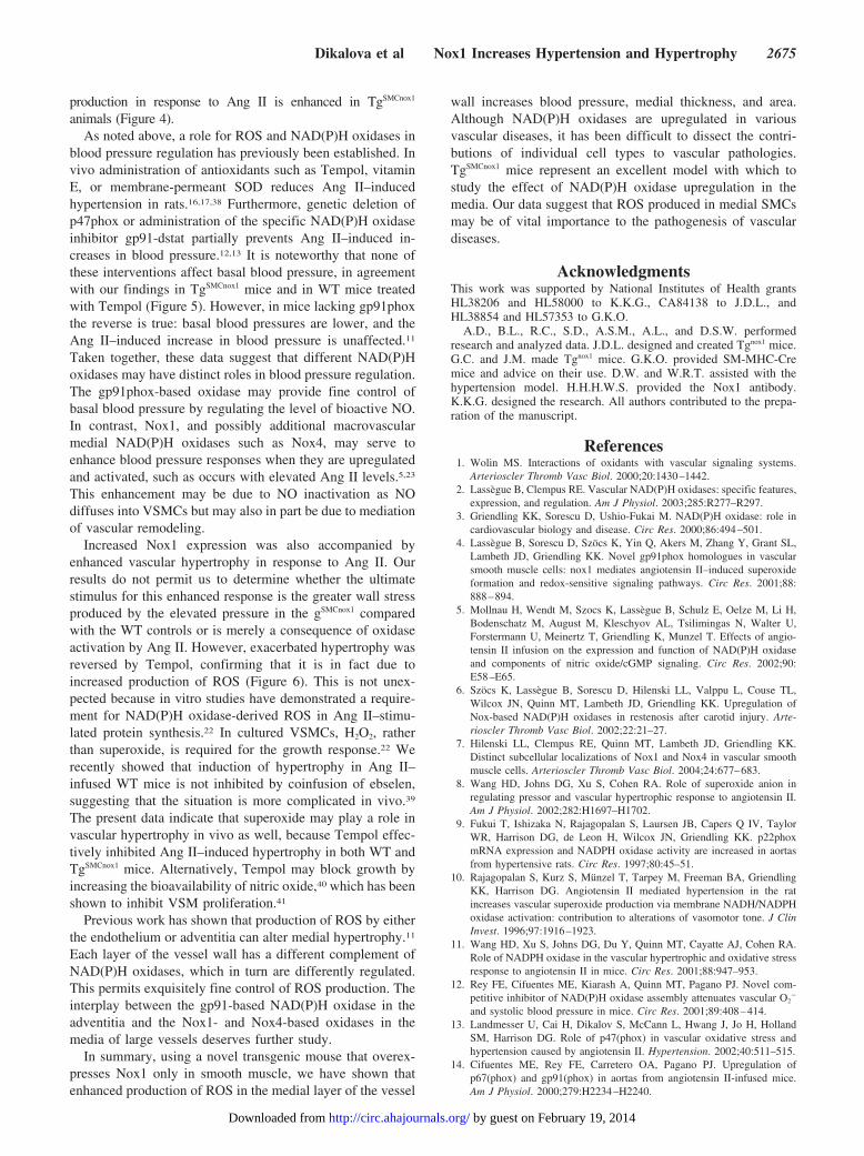
animals (Figure 4).As noted above, a role for ROS and NAD(P)H oxidases in
blood pressure regulation has previously been established. Invivo administration of antioxidants such as Tempol, vitaminE, or membrane-permeant SOD reduces Ang II–inducedhypertension in rats.16,17,38 Furthermore, genetic deletion ofp47phox or administration of the specific NAD(P)H oxidaseinhibitor gp91-dstat partially prevents Ang II–induced in-creases in blood pressure.12,13 It is noteworthy that none ofthese interventions affect basal blood pressure, in agreementwith our findings in TgSMCnox1 mice and in WT mice treatedwith Tempol (Figure 5). However, in mice lacking gp91phoxthe reverse is true: basal blood pressures are lower, and theAng II–induced increase in blood pressure is unaffected.11
Taken together, these data suggest that different NAD(P)Hoxidases may have distinct roles in blood pressure regulation.The gp91phox-based oxidase may provide fine control ofbasal blood pressure by regulating the level of bioactive NO.In contrast, Nox1, and possibly additional macrovascularmedial NAD(P)H oxidases such as Nox4, may serve toenhance blood pressure responses when they are upregulatedand activated, such as occurs with elevated Ang II levels.5,23
This enhancement may be due to NO inactivation as NOdiffuses into VSMCs but may also in part be due to mediationof vascular remodeling.
Increased Nox1 expression was also accompanied byenhanced vascular hypertrophy in response to Ang II. Ourresults do not permit us to determine whether the ultimatestimulus for this enhanced response is the greater wall stressproduced by the elevated pressure in the gSMCnox1 comparedwith the WT controls or is merely a consequence of oxidaseactivation by Ang II. However, exacerbated hypertrophy wasreversed by Tempol, confirming that it is in fact due toincreased production of ROS (Figure 6). This is not unex-pected because in vitro studies have demonstrated a require-ment for NAD(P)H oxidase-derived ROS in Ang II–stimu-lated protein synthesis.22 In cultured VSMCs, H2O2, ratherthan superoxide, is required for the growth response.22 Werecently showed that induction of hypertrophy in Ang II–infused WT mice is not inhibited by coinfusion of ebselen,suggesting that the situation is more complicated in vivo.39
The present data indicate that superoxide may play a role invascular hypertrophy in vivo as well, because Tempol effec-tively inhibited Ang II–induced hypertrophy in both WT andTgSMCnox1 mice. Alternatively, Tempol may block growth byincreasing the bioavailability of nitric oxide,40 which has beenshown to inhibit VSM proliferation.41
Previous work has shown that production of ROS by eitherthe endothelium or adventitia can alter medial hypertrophy.11
Each layer of the vessel wall has a different complement ofNAD(P)H oxidases, which in turn are differently regulated.This permits exquisitely fine control of ROS production. Theinterplay between the gp91-based NAD(P)H oxidase in theadventitia and the Nox1- and Nox4-based oxidases in themedia of large vessels deserves further study.
In summary, using a novel transgenic mouse that overex-presses Nox1 only in smooth muscle, we have shown thatenhanced production of ROS in the medial layer of the vessel
wall increases blood pressure, medial thickness, and area.Although NAD(P)H oxidases are upregulated in variousvascular diseases, it has been difficult to dissect the contri-butions of individual cell types to vascular pathologies.TgSMCnox1 mice represent an excellent model with which tostudy the effect of NAD(P)H oxidase upregulation in themedia. Our data suggest that ROS produced in medial SMCsmay be of vital importance to the pathogenesis of vasculardiseases.
AcknowledgmentsThis work was supported by National Institutes of Health grantsHL38206 and HL58000 to K.K.G., CA84138 to J.D.L., andHL38854 and HL57353 to G.K.O.
A.D., B.L., R.C., S.D., A.S.M., A.L., and D.S.W. performedresearch and analyzed data. J.D.L. designed and created Tgnox1 mice.G.C. and J.M. made Tgnox1 mice. G.K.O. provided SM-MHC-Cremice and advice on their use. D.W. and W.R.T. assisted with thehypertension model. H.H.H.W.S. provided the Nox1 antibody.K.K.G. designed the research. All authors contributed to the prepa-ration of the manuscript.
References1. Wolin MS. Interactions of oxidants with vascular signaling systems.
Arterioscler Thromb Vasc Biol. 2000;20:1430–1442.2. Lassègue B, Clempus RE. Vascular NAD(P)H oxidases: specific features,
expression, and regulation. Am J Physiol. 2003;285:R277–R297.3. Griendling KK, Sorescu D, Ushio-Fukai M. NAD(P)H oxidase: role in
cardiovascular biology and disease. Circ Res. 2000;86:494–501.4. Lassègue B, Sorescu D, Szöcs K, Yin Q, Akers M, Zhang Y, Grant SL,
Lambeth JD, Griendling KK. Novel gp91phox homologues in vascularsmooth muscle cells: nox1 mediates angiotensin II–induced superoxideformation and redox-sensitive signaling pathways. Circ Res. 2001;88:888–894.
5. Mollnau H, Wendt M, Szocs K, Lassegue B, Schulz E, Oelze M, Li H,Bodenschatz M, August M, Kleschyov AL, Tsilimingas N, Walter U,Forstermann U, Meinertz T, Griendling K, Munzel T. Effects of angio-tensin II infusion on the expression and function of NAD(P)H oxidaseand components of nitric oxide/cGMP signaling. Circ Res. 2002;90:E58–E65.
6. Szöcs K, Lassègue B, Sorescu D, Hilenski LL, Valppu L, Couse TL,Wilcox JN, Quinn MT, Lambeth JD, Griendling KK. Upregulation ofNox-based NAD(P)H oxidases in restenosis after carotid injury. Arte-rioscler Thromb Vasc Biol. 2002;22:21–27.
7. Hilenski LL, Clempus RE, Quinn MT, Lambeth JD, Griendling KK.Distinct subcellular localizations of Nox1 and Nox4 in vascular smoothmuscle cells. Arterioscler Thromb Vasc Biol. 2004;24:677–683.
8. Wang HD, Johns DG, Xu S, Cohen RA. Role of superoxide anion inregulating pressor and vascular hypertrophic response to angiotensin II.Am J Physiol. 2002;282:H1697–H1702.
9. Fukui T, Ishizaka N, Rajagopalan S, Laursen JB, Capers Q IV, TaylorWR, Harrison DG, de Leon H, Wilcox JN, Griendling KK. p22phoxmRNA expression and NADPH oxidase activity are increased in aortasfrom hypertensive rats. Circ Res. 1997;80:45–51.
10. Rajagopalan S, Kurz S, Münzel T, Tarpey M, Freeman BA, GriendlingKK, Harrison DG. Angiotensin II mediated hypertension in the ratincreases vascular superoxide production via membrane NADH/NADPHoxidase activation: contribution to alterations of vasomotor tone. J ClinInvest. 1996;97:1916–1923.
11. Wang HD, Xu S, Johns DG, Du Y, Quinn MT, Cayatte AJ, Cohen RA.Role of NADPH oxidase in the vascular hypertrophic and oxidative stressresponse to angiotensin II in mice. Circ Res. 2001;88:947–953.
12. Rey FE, Cifuentes ME, Kiarash A, Quinn MT, Pagano PJ. Novel com-petitive inhibitor of NAD(P)H oxidase assembly attenuates vascular O2
�
and systolic blood pressure in mice. Circ Res. 2001;89:408–414.13. Landmesser U, Cai H, Dikalov S, McCann L, Hwang J, Jo H, Holland
SM, Harrison DG. Role of p47(phox) in vascular oxidative stress andhypertension caused by angiotensin II. Hypertension. 2002;40:511–515.
14. Cifuentes ME, Rey FE, Carretero OA, Pagano PJ. Upregulation ofp67(phox) and gp91(phox) in aortas from angiotensin II-infused mice.Am J Physiol. 2000;279:H2234–H2240.
Dikalova et al Nox1 Increases Hypertension and Hypertrophy 2675
by guest on February 19, 2014http://circ.ahajournals.org/Downloaded from

15. Virdis A, Neves MF, Amiri F, Touyz RM, Schiffrin EL. Role ofNAD(P)H oxidase on vascular alterations in angiotensin II-infused mice.J Hypertens. 2004;22:535–542.
16. Nishiyama A, Fukui T, Fujisawa Y, Rahman M, Tian RX, Kimura S, AbeY. Systemic and regional hemodynamic responses to Tempol in angio-tensin II–infused hypertensive rats. Hypertension. 2001;37:77–83.
17. Bech-Laursen J, Rajagopalan S, Galis Z, Tarpey M, Freeman BA,Harrison DG. Role of superoxide in angiotensin II–induced but notcatecholamine-induced hypertension. Circulation. 1997;95:588–593.
18. Kawada N, Imai E, Karber A, Welch WJ, Wilcox CS. A mouse model ofangiotensin II slow pressor response: role of oxidative stress. J Am SocNephrol. 2002;13:2860–2868.
19. Schnackenberg CG, Wilcox CS. Two-week administration of Tempolattenuates both hypertension and renal excretion of 8-Iso prostaglandinf2alpha. Hypertension. 1999;33:424–428.
20. Owens GK, Rabinovitch PS, Schwartz SM. Smooth muscle cell hyper-trophy versus hyperplasia in hypertension. Proc Natl Acad Sci U S A.1981;78:7759–7763.
21. Griendling KK, Minieri CA, Ollerenshaw JD, Alexander RW. Angioten-sin II stimulates NADH and NADPH oxidase activity in cultured vascularsmooth muscle cells. Circ Res. 1994;74:1141–1148.
22. Zafari AM, Ushio-Fukai M, Akers M, Yin Q, Shah A, Harrison DG,Taylor WR, Griendling KK. Novel role of NADH/NADPH oxidase-derived hydrogen peroxide in angiotensin II–induced hypertrophy of ratvascular smooth muscle cells. Hypertension. 1998;32:488–495.
23. Wingler K, Wunsch S, Kreutz R, Rothermund L, Paul M, Schmidt HH.Upregulation of the vascular NAD(P)H-oxidase isoforms Nox1 and Nox4by the renin-angiotensin system in vitro and in vivo. Free Radic BiolMed. 2001;31:1456–1464.
24. Okabe M, Ikawa M, Kominami K, Nakanishi T, Nishimune Y. ‘Greenmice’ as a source of ubiquitous green cells. FEBS Lett. 1997;407:313–319.
25. Regan CP, Manabe I, Owens GK. Development of a smooth muscle-–targeted cre recombinase mouse reveals novel insights regarding smoothmuscle myosin heavy chain promoter regulation. Circ Res. 2000;87:363–369.
26. Velarde V, Ullian ME, Morinelli TA, Mayfield RK, Jaffa AA. Mech-anisms of MAPK activation by bradykinin in vascular smooth musclecells. Am J Physiol. 1999;277:C253–C261.
27. Fink B, Laude K, McCann L, Doughan A, Harrison D, Dikalov S.Detection of intracellular superoxide formation in endothelial cells andintact tissues using dihydroethidium and an HPLC-based assay.Am J Physiol. 2004;287:C895–C902.
28. Johns C, Gavras I, Handy DE, Salomao A, Gavras H. Models of exper-imental hypertension in mice. Hypertension. 1996;28:1064–1069.
29. Dobrian AD, Schriver SD, Prewitt RL. Role of angiotensin II and freeradicals in blood pressure regulation in a rat model of renal hypertension.Hypertension. 2001;38:361–366.
30. Beswick RA, Zhang H, Marable D, Catravas JD, Hill WD, Webb RC.Long-term antioxidant administration attenuates mineralocorticoid hyper-tension and renal inflammatory response. Hypertension. 2001;37:781–786.
31. Touyz RM. Reactive oxygen species, vascular oxidative stress, and redoxsignaling in hypertension: what is the clinical significance? Hypertension.2004;44:248–252.
32. Lambeth JD, Cheng G, Arnold R, Edens WA. Novel homologs ofgp91phox. Trends in Biochem Sci. 2000;25:459–461.
33. Pagano PJ, Clark JK, Cifuentes-Pagano ME, Clark SM, Callis GM, QuinnMT. Localization of a constitutively active, phagocyte-like NADPHoxidase in rabbit aortic adventitia: enhancement by angiotensin II. ProcNatl Acad Sci U S A. 1997;94:14438–14488.
34. Li JM, Wheatcroft S, Fan LM, Kearney MT, Shah AM. Opposing rolesof p47phox in basal versus angiotensin II–stimulated alterations in vascularO2
� production, vascular tone, and mitogen-activated protein kinase acti-vation. Circulation. 2004;109:1307–1313.
35. Takeya R, Ueno N, Kami K, Taura M, Kohjima M, Izaki T, Nunoi H,Sumimoto H. Novel human homologues of p47phox and p67phox par-ticipate in activation of superoxide-producing NADPH oxidases. J BiolChem. 2003;278:25234–25246.
36. Li L, Miano JM, Cserjesi P, Olson EN. SM22 alpha, a marker of adultsmooth muscle, is expressed in multiple myogenic lineages during embry-ogenesis. Circ Res. 1996;78:188–195.
37. McHugh KM. Molecular analysis of smooth muscle development in themouse. Dev Dyn. 1995;204:278–290.
38. Ortiz MC, Manriquez MC, Romero JC, Juncos LA. Antioxidants blockangiotensin II–induced increases in blood pressure and endothelin.Hypertension. 2001;38:655–659.
39. Weber DS, Rocic P, Mellis AM, Laude K, Lyle AN, Harrison DG,Griendling KK. Angiotensin II-induced hypertrophy is potentiated inmice overexpressing p22phox in vascular smooth muscle. Am J Physiol.2005;288:H37–H42.
40. Schnackenberg CG, Welch WJ, Wilcox CS. Normalization of bloodpressure and renal vascular resistance in SHR with a membrane-permeable superoxide dismutase mimetic: role of nitric oxide. Hyper-tension. 1998;32:59–64.
41. Garg UC, Hassid A. Nitric oxide generating vasodilators and 8-bromocyclic GMP inhibit mitogenesis and proliferation of cultured rat vascularsmooth muscle cells. J Clin Invest. 1989;83:1774–1777.
CLINICAL PERSPECTIVEThe role of reactive oxygen species (ROS) in vascular diseases such as hypertension and atherosclerosis has beencontroversial, in part because of the disappointingly negative results of the vitamin trials. However, the failure of thesetrials to improve cardiovascular end points does not negate the concept that oxidative stress plays a role in diseasepathogenesis. Such trials necessarily treat patients with established disease, irrespective of their oxidant status and need fortreatment. Vitamins target free radicals such as superoxide but have little effect on the equally potent ROS hydrogenperoxide. Practically speaking, human subjects cannot be used to investigate the cause-effect relationships between ROSand disease pathogenesis. In contrast, transgenic animals represent an ideal model to provide insight into the role of ROSin the development of vascular disease. The present study clearly demonstrates that chronic oxidative stress produced byupregulation of the NADPH oxidase Nox1 in transgenic mice aggravates vascular hypertrophy and hypertension after a2-week infusion of angiotensin II. These effects were reversed by the superoxide scavenger Tempol, confirming that theyare due to increased ROS. The results of this study suggest that rather than attempting to scavenge oxidative products oncethey are formed, future therapy for oxidant-related vascular diseases should be directed toward inhibiting sources of ROSgeneration, such as Nox1. This approach would have the advantage of specificity and the ability to prevent ROS-mediateddamage before it occurs.
2676 Circulation October 25, 2005
by guest on February 19, 2014http://circ.ahajournals.org/Downloaded from

1
On-Line Supplement
Nox1 overexpression potentiates angiotensin II-induced hypertension and vascular smoothmuscle hypertrophy in transgenic mice
Anna Dikalova,a Bernard Lassègue,a Roza Clempus,a Guangjie Cheng,b James McCoy,b SergeyDikalov,a Alejandra San Martin,a Alicia Lyle,a David S. Webera, Daiana Weiss,a W. Robert
Taylor,a Harald H. H. W. Schmidt,c Gary K. Owens,d J. David Lambeth,b Kathy K. Griendlinga,e
aDivision of Cardiology and bDepartment of Pathology, Emory University, Atlanta, GA,cDepartment of Pharmacology, Monash University, Victoria, Australia and dDepartment ofMolecular Physiology and Biological Physics, University of Virginia School of Medicine,
Charlottesville, VA
Materials and Methods
Reagents: All cell culture media and materials were purchased from the Gibco division ofInvitrogen (Carlsbad, CA). Collagenase type 2 was from Worthington (Feehold, NJ). DNeasyTissue and RNeasy Kits were from Qiagen, Inc. (Valencia, CA). Phenylmethylsulfonyl fluoride(PMSF) was from Roche (Indianapolis, USA). Nytran N membranes were from Schleicher &Schuell (Keene, NH). 1-Hydroxy-3-methoxycarbonyl-2,2,5,5-tetramethyl-pyrrolidine (CMH)was purchased from Alexis Corp. (San Diego, CA). Dihydroethidium was purchased fromMolecular Probes (Eugene, OR). Anti-Nox1 rabbit polyclonal antibody was prepared asdescribed.1 The other rabbit polyclonal antibodies were as follows: CDK4 from Santa CruzBiotechnology (Santa Cruz, CA), catalase from Athens Research and Technology (Athens, GA),Mn SOD and Cu/Zn SOD were from Stressgen Biotechnologies (Victoria, BC, Canada) and theec SOD antibody was described previously.2 The mouse gp91phox antibody (clone 54.1) was agift from Dr. Mark Quinn. Anti-Mouse HRP-conjugated antibodies were from Amersham(Piscataway, NJ), anti-rabbit HRP-conjugatged antibodies were from Bio Rad Laboratories(Hercules, CA). All other chemicals including 2,2,2-tribromoethanol (Avertin), angiotensin II andTempol were from Sigma-Aldrich (St. Louis, MO).
AnimalsThe nox1 transgene was constructed in a specially designated pBSCX1-LEL plasmid
vector.3 The final construct consisted of the strong universal promoter designated CX1, greenfluorescent protein (GFP) cDNA with a stop codon flanked by loxP DNA cis-elements, andhuman nox1 cDNA (Figure 1A). CX1 is a hybrid promoter consisting of a portion of the b-actinand CMV enhancer sequences and was designed specifically for high-level ubiquitousexpression in all mouse tissues and cell types in vivo.3 Two founder lines of these transgenicmice (Tg1
nox1 and Tg2nox1) were generated in David Lambeth’s laboratory by the Emory
University transgenic mouse core facility.To create transgenic mice in which Nox1 is specifically overexpressed in smooth muscle,
we employed a Cre/LoxP system. Mice containing the nox1 transgene were bred with transgenicmice expressing a cre transgene composed of Cre recombinase cDNA under the control of thesmooth muscle cell myosin-heavy-chain-a promoter.4 Both strains are on a C57Bl/6

2
background. In mice positive for both the nox1 transgene and the cre transgene, Crerecombinase is expressed only in smooth muscle and excises the floxed GFP cDNA, leavingnox1 cDNA under the control of the CX1 promoter. Consequently, the human nox1 transgene isexpressed exclusively in smooth muscle (TgSMCnox1)(Figure 1A).
The presence of human nox1 and cre in mouse genomic DNA was detected using aconventional PCR method (Fig. 1B). Primer sequences are indicated in Table 1. The humannox1 primers do not detect the native murine gene, because of the presence of 1.8 kb ofintervening intron sequences. Detection of the nox1 and cre transgenes was also performed byreal-time PCR using genomic DNA from tail clips. In this qualitative assay, the presence of eachtransgene is detected at the end of the amplification reaction by melting curve analysis in thepresence of SYBR green dye and comparison to positive and negative controls. Primer sequencesare indicated in Table 1. As in the conventional PCR method, native mouse nox1 is not detecteddue to the presence of introns between the primers.
The presence of these transgenes in the breeding pairs was confirmed by Southern blotanalysis in both transgenic lines (Tg1
SMCnox1 and Tg2SMCnox1) (Figure 1C). Transgenic mice
heterozygous for Nox1 overexpression in smooth muscle cells and their wild-type (WT)littermates were used in the experiments. All mice used in this study were on a C57BL/6background, and were between 6 and 7 months of age. All procedures were approved by theEmory University Institutional Animal Care and Use Committee and were in compliance withthe standards for the Care and Use of Laboratory Animals of the Institute of Laboratory AnimalResources, National Academy of Sciences, Bethesda, MD.
Southern blot analysis of genomic DNAGenomic DNA was extracted from tail clips and aortas using the DNeasy Tissue Kit
following the manufacturer’s recommendations. DNA was then digested with Bam HI overnight,separated on a 0.8% agarose gel, transferred onto a nylon membrane, and pre-hybridized for 2-3hours at 42°C in ULTRAhyb (Ambion, Austin, TX). Hybridization was performed overnightwith 32P-labeled cDNA probes designed to detect nox1 or cre. Final washing conditions were 20min at 55°C in 0.1X SSC and 1%SDS for nox1 and 4 x 20 min at 62°C in 0.2X SSC and 1%SDSfor cre. To prepare the cre probe, the pIC cre plasmid, obtained from the local transgenic corefacility, was digested with Sal I and Pst I. The excised 1032 bp insert, corresponding to the fullcoding region, was purified by agarose gel electrophoresis. The human nox1 probe comprises afragment of the coding region (nucleotides 347-1726 in accession number AF127763). It wasprepared f rom human nox1 cDNA by PCR using pr imers 5 ' -GTCAACATTGGCCTGTGCCCGAGCGTC-3' and5'-CTCCCAAAGGAGGTTTTCTGTTTCA-3'.The resulting PCR product (1.4 kb) was purified by agarose gel electrophoresis. Probes werelabeled with a random priming kit (Prime it II, Stratagene) using a-32P dCTP. Bands weredetected by autoradiography.
Treatment groupsTgSMCnox1 and WT mice were divided into four groups: Control (saline infusion), Ang II,
Tempol and Tempol + Ang II. The mice were anesthetized with an intraperitoneal injection of375 mg/kg 2,2,2-tribromoethanol, and micro-osmotic pumps were implanted subcutaneously inthe mid-scapular region. Pumps delivered either 0.9% saline or angiotensin II at a rate of 0.7mg/kg/day. For the Tempol groups, Tempol dissolved in 0.9% NaCl was administered inseparate micro-osmotic pumps at a rate of 50 mg/kg/day.5 After 14 days, the animals were killedby CO2 inhalation, and their aortas were harvested for study.

3
Cell cultureVSMCs were isolated from aortas of male TgSMCnox1 and WT mice by the explant method
with modifications.6 The mice were killed by CO2 inhalation, and aortas were removed, cutlongitudinally and cleaned of endothelium and adventitia in Dulbecco’s modified Eagle’smedium (DMEM) containing antibiotics. One-mm pieces of the aorta were incubated in filteredDMEM containing 1 mg/ml collagenase for two hours and placed in a culture dish. After 20minutes, DMEM containing 20% FBS was added. DMEM containing 10% FBS was used afterpassage 1. Cells were passaged by trypsinization and used for experiments at passage 3.
Western blottingMouse aortas or cultured aortic smooth muscle cells from TgSMCnox1 and WT mice were
homogenized and incubated for 30 min in 1% Triton X-100 lysis buffer (50 mmol/L HEPES, 50mmol/L NaCl, 5 mmol/L EDTA, 10 mmol/L sodium pyrophosphate, 50 mmol/L sodiumfluoride, 1 mmol/L sodium orthovanadate) containing fresh protease inhibitors (10 mg/mLleupeptin, 10 mg/mL aprotinin and 1 mmol/L PMSF). Samples were then centrifuged at 18,000 Xg for 10 minutes, and proteins in the supernatant were separated using 12% SDS-PAGE(for SODenzymes) or 9% SDS-PAGE( for Nox proteins and catalase), transferred to nitrocellulosemembranes, blocked, and incubated overnight at 4°C with the primary antibodies. Afterincubation with HRP-conjugated secondary antibodies, proteins were detected by ECLchemiluminescence.
Immunofluorescent histochemistrySingle-label fluorescent immunohistochemistry was performed on frozen 5-µm OCT-
embedded tissue sections as described previously7 using a rabbit polyclonal anti-nox1 antibody1
at 1:100 dilution. Serial sections were treated with secondary antibodies alone to control fornon-specific staining.
Real time quantitative RT-PCRSeveral tissues (liver, heart, spleen, brain, colon, and aorta) were harvested from WT and
TgSMCnox1 mice, and total RNA was isolated using the RNeasy Kit following the manufacturer’srecommendations for purification from tissue. Preparation of the RNA included digestion withproteinase K and DNase I to eliminate possible genomic DNA contamination that could result infalse positives in the PCR. Control samples were used without reverse transcriptase (RTnegative). cDNA was prepared from 5 micrograms of total RNA. Exogenous luciferase RNA(1x10⁸ copies) was added to each sample before reverse transcription to serve as a heterologousinternal control. Primer sequences used to detect luciferase by real-time PCR were 5’-GCTGCTGGTGCCAACCCTATTCTCCTT-3’ and5’-CGCGCAACTTTTTCGCGGTTGTTACTTGA-3’. To prepare luciferase RNA, the pGL3Basic reporter plasmid (Promega) was modified by insertion of a T7 promoter at the Xba I site,just 3' of the luciferase coding region. A fragment of the modified plasmid, including the 3' halfof luciferase and the T7 promoter was excised by digestion with Sap I, and gel purified. Thisfragment was used as a template for in vitro transcription with T7 polymerase. The RNA productwas digested with DNase I to remove the DNA template, purified using the RNeasy kit andquantified by UV spectrophotometry. The reverse transcription reaction was performed usingSuperscript II enzyme (Invitrogen) and random nanomer primers as described previously.8Message expression was quantified using the Lightcycler instrument (Roche) with SYBR green

4
dye and specific human or mouse nox1 primers and normalized to luciferase and 18S rRNA.Our human nox1 primers do not detect murine nox1 in conditions used for real-time PCR.
Detection of intracellular superoxide using high performance liquid chromatographyTo evaluate intracellular production of superoxide, we measured the formation of
oxyethidium from dihydroethidium (DHE) using high performance liquid chromatography(HPLC) analysis as recently reported.9 Aortas were carefully dissected and cleaned of fat andloose connective tissue. Each aorta was cut into 10-12 2-mm rings and placed in chilledKrebs/HEPES buffer, pH 7.4. For each experiment, 3 rings were incubated for 20 min at 37°C in1 mL of Krebs/HEPES-buffer containing 50 mmol/L DHE, washed twice, and incubated for afurther hour in fresh Krebs/HEPES buffer at 37°C. Aortic rings were then homogenized in 300mL methanol. Fifty mL of homogenate were taken for protein measurements, and the remainderwas filtered through a 0.22 mm syringe filter. Separation of ethidium, oxyethidium anddihydroethidium in the filtered samples was performed on a C-18 reverse phase column(Nucleosil 250-4.5 mm) using a Beckman HPLC System equipped with both UV andfluorescence detectors. The mobile phase contained 60% acetonitrile and 0.1% trifluoroacetic acid.Dihydroethidium, ethidium and oxyethidium were separated by a linear increase in acetonitrileconcentration from 37% to 47% in 23 min at a flow rate of 0.5 ml/min. Fluorescence detection at580 nm (emission) and 480 nm (excitation) was used to monitor oxyethidium production. UVabsorption at 355 nM was used for detection of dihydroethidium. Oxidized ethidium wasexpressed per mg protein. Protein concentration was measured by Bradford assay. In somestudies PEG-SOD (100 U/ml) was added one hour prior to addition of dihydroethidium. PEG-SOD inhibited the DHE signal by 60%.
Superoxide detection by electron spin resonance (ESR)Aortas harvested as described above were cut into 2-mm rings, and three of each were
incubated for 40 min at 37˚C in 1 ml of Krebs/HEPES buffer (pH=7.4) containing 5 mM DETC,50 mM desferal and 0.5 mM CMH. Rings were then frozen in liquid nitrogen, and ESR spectrawere recorded using a Bruker EMX ESR spectrometer and a super-high Q microwave cavity.The ESR instrument settings were as follows: field sweep, 50 G; microwave frequency, 9.78 GHz;microwave power, 20 mW; modulation amplitude, 5 G; conversion time, 327.68 ms; time constant,5242.88 ms; 512 points resolution and receiver gain, 1 x 10⁴. The amplitude of the signal wasmeasured and the concentration of CM-radical was determined by calibration with standardconcentrations of CM-nitroxide. The portion of the signal due to superoxide was determined bypreincubation of duplicate samples with PEG-SOD (100 U/ml) for 3 hours in Krebs/HEPESbuffer. PEG-SOD pretreatment inhibited 65-75% of CM-nitroxide formation.
After the ESR measurement, rings were transferred into Eppendorf tubes, dried for 48hours at 37˚C, and their dry weight was determined. The formation of CM-nitroxide wasnormalized to the dry weight of aorta rings.
Systolic blood pressure measurementSystolic blood pressure was measured using tail-cuff plethysmography (Visitech Systems
Inc., Apex, NC). Blood pressure was measured twice before the implantation of osmotic pumpsand on days 3, 7, 10, and 14 after pump placement. A set of 10-20 measurements was obtainedfor each animal, and the mean blood pressure was calculated. This noninvasive method of

5
measuring blood pressure correlates well with intraarterial measurements in normotensive andhypertensive mice.10
Assessment of HypertrophyFollowing euthanasia, the heart and aorta were pressure-perfused at 100 mm Hg with
0.9% sodium chloride solution, followed by pressure fixation with a 10% formalin solution.Aortas were embedded in paraffin, three 5-mm cross-sections were cut starting 6-mm from theaortic arch, and stained with hematoxylin and eosin. Digital images were obtained using a ZeissAxioskop. To quantify wall thickness, perpendicular lines were drawn to determine the distancefrom internal elastic lamina to the external lamina at a minimum of 10 locations per aorticsection and a mean value was calculated. All measurements were completed using NIH image(version 1.62).
StatisticsData are shown as mean ± SE. Statistical significance was assessed by analysis of
variance on untransformed data, followed by comparison of group averages by contrast analysis,using the SuperANOVA statistical program (Abacus Concepts; Berkeley, CA). A p value <0.05was considered to be statistically significant.
References
1. Wingler K, Wunsch S, Kreutz R, Rothermund L, Paul M, Schmidt HH. Upregulation ofthe vascular NAD(P)H-oxidase isoforms Nox1 and Nox4 by the renin-angiotensin systemin vitro and in vivo. Free Radic Biol Med. 2001;31:1456-1464.
2. Fukai T, Galis ZS, Meng XP, Parthasarathy S, Harrison DG. Vascular expression ofextracellular superoxide dismutase in atherosclerosis. J Clin Invest. 1998;101:2101-2111.
3. Okabe M, Ikawa M, Kominami K, Nakanishi T, Nishimune Y. 'Green mice' as a sourceof ubiquitous green cells. FEBS Lett. 1997;407:313-319.
4. Regan CP, Manabe I, Owens GK. Development of a smooth muscle-targeted crerecombinase mouse reveals novel insights regarding smooth muscle myosin heavy chainpromoter regulation. Circ Res. 2000;87:363-369.
5. Kawada N, Imai E, Karber A, Welch WJ, Wilcox CS. A mouse model of angiotensin IIslow pressor response: role of oxidative stress. J Am Soc Nephrol. 2002;13:2860-2868.
6. Velarde V, Ullian ME, Morinelli TA, Mayfield RK, Jaffa AA. Mechanisms of MAPKactivation by bradykinin in vascular smooth muscle cells. Am J Physiol. 1999;277:C253-261.
7. Szöcs K, Lassègue B, Sorescu D, Hilenski LL, Valppu L, Couse TL, Wilcox JN, QuinnMT, Lambeth JD, Griendling KK. Upregulation of Nox-based NAD(P)H oxidases inrestenosis after carotid injury. Arterioscler Thromb Vasc Biol. 2002;22:21-27.
8. Lassègue B, Sorescu D, Szöcs K, Yin Q, Akers M, Zhang Y, Grant SL, Lambeth JD,Griendling KK. Novel gp91phox homologues in vascular smooth muscle cells: nox1mediates angiotensin II-induced superoxide formation and redox-sensitive signalingpathways. Circ. Res. 2001;88:888-894.
9. Fink B, Laude K, McCann L, Doughan A, Harrison D, Dikalov S. Detection ofintracellular superoxide formation in endothelial cells and intact tissues using

6
dihydroethidium and an HPLC-based assay. Am J Physiol Cell Physiol. 2004;287:C895-902.
10. Johns C, Gavras I, Handy DE, Salomao A, Gavras H. Models of experimentalhypertension in mice. Hypertension. 1996;28:1064-1069.
Copyright © 2022 FDOKUMEN