Bond-valence methods for pKa prediction: critical reanalysis and a new approach
Upload
independentCategory
view
1download
0
Regulatory Peptides 177 (2012) 27–34
Contents lists available at SciVerse ScienceDirect
Regulatory Peptides
j ourna l homepage: www.e lsev ie r .com/ locate / regpep
Angiotensin-(3–4) counteracts the Angiotensin II inhibitory action on renalCa2+-ATPase through a cAMP/PKA pathway
Flavia Axelband a,b, Juliana Dias a,b, Filipe Miranda a,b, Fernanda M. Ferrão a,b, Rosana I. Reis c,Claudio M. Costa-Neto c, Lucienne S. Lara b,d, Adalberto Vieyra a,b,⁎a Instituto de Biofísica Carlos Chagas Filho, Universidade Federal do Rio de Janeiro, Rio de Janeiro, Brazilb Instituto Nacional de Ciência e Tecnologia em Biologia Estrutural e Bioimagem, Rio de Janeiro, Brazilc Departamento de Bioquímica e Imunologia, Faculdade de Medicina de Ribeirão Preto, Universidade de São Paulo, Ribeirão Preto, Brazild Instituto de Ciências Biomédicas, Universidade Federal do Rio de Janeiro, Rio de Janeiro, Brazil
Abbreviations: ACE, angiotensin converting enzyme; Aangiotensin-(1−7); Ang-(3−4), Val3−Tyr4; AT1R, type 1type 2 angiotensin II receptors; AT(1−7)R(MAS), angiotensitensin IV receptors; BSA, bovine serum albumin; CGP42[[(2S)-6-[[(2S)-5-[bis(azanyl)-methylideneamino]-2-(phpentanoyl]amino]-2-[[(2S)-3-(4-hydroxyphenyl)-2-(pyridinamino]-hexanoyl]-amino]-3-(1H-imidazol-5-yl)propanoyl]3-methyl-pentanoic acid; CHAPS, 3-[(3-cholamidopropylsulfonate; db-cAMP, dibutyril cyclic AMP; DMEM, DulbecHEPES, (4-(2-hydroxyethyl)-1-piperazineethanesulfonmethylxanthine; losartan, 2-Butyl-4-chloro-1-[[2′(nyl]-4-yl]methyl]-1H-imidazole-5-methanol; PKA, cykinase; PKAi, the PKA inhibitor peptide PKAi(5–24); PKC(6S)-1-[[4-(dimethylamino)-3-methylphenyl]methyl]-5tetrahydro-1H-imidazo[4,5-c]pyridine-6-carboxylic acidsulfate-polyacrylamide gel electrophoresis; TBS, TRIS-buffeacid; TRIS, 2-amino-2-hydroxymethyl-propane-1,3,diol.⁎ Corresponding author at: Carlos Chagas Filho Institute
of Rio de Janeiro, Carlos Chagas Av. 373, 21941–900 Rio de J6520; fax: +55 21 2280 8193.
E-mail address: [email protected] (A. Vieyra).
0167-0115/$ – see front matter © 2012 Elsevier B.V. Alldoi:10.1016/j.regpep.2012.04.004
a b s t r a c t
a r t i c l e i n f oArticle history:Received 6 December 2011Received in revised form 30 March 2012Accepted 23 April 2012Available online 3 May 2012
Keywords:Ang-(3−4)cAMPCyclic AMP-dependent protein kinasePlasma membrane Ca2+-ATPaseKidney cellsProximal tubule basolateral membranes
We recently demonstrated that Angiotensin-(3−4) [Ang-(3−4)], an Ang II-derived dipeptide, overcomes in-hibition of plasma membrane Ca2+-ATPase promoted by nanomolar concentrations of Ang II in basolateralmembranes of renal proximal tubule cells, with involvement of a so far unknown AT2R-dependent and NO-independent mechanism. The present study investigates the signaling pathway triggered by Ang-(3−4)that is responsible for counteracting the inhibitory effect of Ang II, and attempts to elucidate the functionalinteraction of the dipeptide with Ang II at the level of AT2R. Stimulation by cholera toxin of Gsα protein struc-turally linked to AT2R − as revealed by their co-immunoprecipitation − mimicked the effect of Ang-(3−4)on Ca2+-ATPase activity. Furthermore, addition of dibutyril-cAMP (db-cAMP) mimicked Ang-(3−4), where-as the specific PKA inhibitor, PKAi(5–24) peptide, suppressed the counter-regulatory effect of Ang-(3−4) andthe AT2R agonist, CGP42112A. Membrane-associated PKA activity was stimulated by Ang-(3−4) orCGP42112A to comparable levels as db-cAMP, and the Ang-(3–4) effect was abrogated by the AT2R antago-nist PD123319, whereas the AT1R antagonist Losartan had no effect. Ang-(3−4) stimulated PKA-mediatedphosphorylation of Ca2+-ATPase and activated PKA to comparable levels. Binding assays demonstratedthat Ang-(3−4) could not displace
3H-Ang II from HEK 293T cells expressing AT2R, but 10−10 mol/L Ang-
(3−4) resulted in the appearance of a probable higher-affinity site (picomolar range) for Ang II. The resultspresented herein demonstrate that Ang-(3−4), acting as an allosteric enhancer, suppresses Ang II-mediatedinhibition of Ca2+-ATPase through an AT2R/cAMP/PKA pathway, after inducing conformational changes inAT2R that results in generation of higher-affinity sites for Ang II.
© 2012 Elsevier B.V. All rights reserved.
ng II, angiotensin II; Ang-(1−7),angiotensin II receptors; AT2R,
n-(1−7) receptors; AT4R, angio-11A, (2S,3S)-2-[[(2S)-1-[(2S)-2-enylmethoxy-carbonylamino)-3-ylcarbonylamino)-propanoyl]pyrrolidin-2-yl]carbonylamino]-)dimethylammonio]-1-propane-co's modified Eagle's medium;ic acid); IBMX, 3-isobutyl-1-1H-tetrazol-5-yl)[1,1′-biphe-clic AMP-dependent protein, protein kinase C; PD123319,-(2,2-diphenylacetyl)-4,5,6,7-; SDS-PAGE, sodium dodecylred saline; TCA, trichloroacetic
of Biophysics, Federal Universityaneiro, Brazil. Tel.:+ 55 21 2562
rights reserved.
1. Introduction
Recently, new components of the renin angiotensin system (RAS)have been described including bioactive shorter peptides derivedfrom Angiotensin II (Ang II), such as Ang III, Ang IV and Ang-(1−7)(for review, see [1]). These metabolites exert their effects by inter-acting with the classic receptors described for Ang II, the AT1 andAT2 receptors (AT1R and AT2R), or acting through their own recep-tors, e.g. AT4R and AT(1−7)R (MAS), which are specific for Ang IVand Ang-(1−7), respectively [2,3]. Actions that are similar or oppo-site to those generated upon binding of Ang II to its receptors canbe observed, depending on the type of receptor being activated.
Recently, we found parallel pathways in the renal cortex that cangenerate a very short, potent Ang II-derived metabolite, Ang-(3−4) [4].Importantly, this Ang II-derived peptide displays antihypertensive ac-tions in vivo and in vitro [5,6]. Furthermore, we demonstrated [7] thatAng-(3−4) counteracts – with femtomolar affinity – the inhibition that10−10 mol/L Ang II promotes − via PKC − on Ca2+-ATPase resident in
28 F. Axelband et al. / Regulatory Peptides 177 (2012) 27–34
the basolateral membranes of kidney proximal tubule cells [8]. As thisATPase is responsible for the fine tuning of cytosolic Ca2+, and proximalreabsorption of fluid is modulated by the intracellular activity of thiscation [9], the influence of Ang-(3−4) on transepithelial Ca2+ fluxesemerges as a novel physiological process for controlling the volume ofbody fluid compartments, and consequently, of arterial pressure. Todate, the signaling cascade triggered by this potent dipeptide remainsunknown.
We have demonstrated that cAMP-dependent protein kinase(PKA) activates the plasma membrane Ca2+-ATPase in proximal tu-bules [10]. PKA activation antagonized the influence of PKC on renal(Na++K+)ATPase [11] and the recently cloned and purified [12]ouabain-insensitive Na+-ATPase [13] through interacting with path-ways that involve Ang II receptors. Therefore, we hypothesized thatAng-(3−4)-induced reactivation of the Ang II-inhibited renal Ca2+-ATPase involves triggering of the PKA-signaling pathways in proximaltubule cells.
The present work investigated whether Ang-(3−4) modifies Ang IIbinding at the level of AT2R in a renal cell line that only expresses thisclass of receptors, and if a PKA pathway downstream of AT2R andPKA-mediated phosphorylation of renal Ca2+-ATPase is associatedwith reactivation of the renal Ca2+-ATPase that is inhibited by physio-logical Ang II concentrations.
2. Experimental procedures
2.1. Animal care
Santa Inês sheep were raised on ranches in Jequié (BA, Brazil) andMontes Claros (MG, Brazil) that are under supervision of the BrazilianMinistry of Agriculture. The health of the animals was certified by alicensed veterinarian from an authorized slaughterhouse. Sheepwere used due to the fact that their kidneys produce abundant levelsof Ca2+-ATPase and because their kidneys are frequently discarded aswastage. This avoids use of experimental animals such as rats, whichwould be sacrificed for their kidneys only. This study was approvedby the local ethics committee for the use of animals in biomedicalassays (IBCCF No.104), which follows the recommendations of theNational Institute of Health (NIH) guide for the care and use of labo-ratory animals.
2.2. Materials
Human embryonic kidney cells (HEK 293T) were purchased fromAmerican Type Culture Collection.
3H-Ang II was obtained from GE
Healthcare. Unlabeled Ang II, the AT2R agonist CGP42112A, choleratoxin (CTX), dibutyril cyclic AMP (db-cAMP), histone H8 and the AT2Rantagonist PD123319 were bought from Sigma-Aldrich; the PKA inhib-itor peptide (PKAi) was purchased from Calbiochem. Ang-(3−4) wassynthesized by EZBiolab. Losartan was from Merck, Sharp & Dohme,and Protein A/G PLUS-agarose from Santa Cruz Biotechnology. The5f10 antibody and phospho-(Ser/Thr) PKA substrate antibodies werepurchased from Thermo Scientific and Cell Signaling Inc., respectively.The anti-AT2R (anti-goat and anti-rabbit) and anti-Gsα protein anti-bodies were obtained from Santa Cruz Biotechnology and Calbiochem,respectively.
32P-labeled orthophosphate (
32Pi) was purchased from the
Brazilian Institute of Energy and Nuclear Research, and used to prepare[γ-
32P]ATP according toMaia and coworkers [14]. The radioactivity recov-
ered in assays was quantified using a liquid scintillation counter.
2.3. Cell culture and transfections
HEK 293T cells were maintained in Dulbecco's modified Eagle'smedium (DMEM) (Invitrogen) supplemented with 10% fetal bovineserum, gentamycin sulfate (50 μg/ml) and glutamine (2 mmol/L) ina 5% CO2/air atmosphere. Transient transfections of the cells were
performed using the calcium phosphate precipitation method de-scribed by Gether and coworkers [15].
2.4. Binding assays
HEK 293T cells transfected with the AT2R expression plasmid weretransferred to 12-well culture plates (3×10⁵ cells/well) 24 h beforebinding assays were carried out. One day after plating, cells werewashed brieflywith cold 25 mmol/L Tris–HCl buffer (pH7.4) containing140 mmol/L NaCl, 5 mmol/L MgCl2 and 0.1% bovine serum albumin(BSA). Binding experiments were performed at 4 °C, and initiated byadding 4×10⁻¹² mol/L
30H-Ang II and increasing concentrations
(10−11 to 10−6 mol/L) of non-radioactive Ang II or Ang-(3−4) as com-petitors. In one series of experiments, bindingwas initiatedwith
3H-Ang
II 4×10⁻¹² mol/L and displacement with unlabeled Ang II was assayedin the presence of a fixed Ang-(3−4) concentration (10−10 mol/L). Thebinding buffer consisted of 25 mmol/L Tris–HCl (pH 7.4), 5 mmol/LMgCl2, 0.1% BSA and 100 μg/ml bacitracin. The competition profiles andIC50 values were calculated based on one- or two-site competitionprofile analysis tools using GraphPad Prism 4.0 software.
2.5. Preparation of basolateral membranes from kidney proximal tubulecells
Membranes were isolated from the outer cortex (cortex corticis) ofsheep kidneys [7]. More than 90% of the cell population in this regioncomprises proximal tubules [16]. The basolateral membrane-enrichedfraction was isolated and purified using the Percoll gradient method[10,17]. Controls for enrichment with basolateral membranes andfor minimal residual contamination with other intracellular mem-branes have been described elsewhere [17].
2.6. Plasma membrane Ca2+-ATPase activity assays
Ca2+-ATPase activity determinations and calculation of total CaCl2needed for the required free Ca2+ concentrationwere as described pre-viously [7]. Briefly, membrane suspensions (0.2 mg/ml in 250 mmol/Lsucrose) were preincubated for 30 min at 37 °C with 1 mmol/L ouabainand supplied with a basic reactionmedium containing (in mmol/L) bis-TRIS-propane buffer 50 (pH 9.0), MgCl2 5, NaN3 10, KCl 120, EGTA 0.2and CaCl2 0.27 (resulting in 20 μmol/L free Ca2+). Other additions areindicated in the corresponding figure legends. The plasma membraneCa2+-ATPase activity was calculated as the difference between the ac-tivities in the absence and presence of 2 mmol/L EGTA. The reactionwas started by adding 5 mmol/L [γ-
32P]ATP (specific activity ~1 Ci/mol),
and after 20 min at 37 °C, it was stopped by adding 2 vol of activatedcharcoal (in 0.1 mol/L HCl). The suspensions were centrifuged at18,000×g and aliquots of the clear supernatants were counted using aliquid scintillation counter. Specific additions of other reagents were de-tailed in the corresponding figures or figure legends.
2.7. Determination of cAMP-dependent protein kinase activity
The PKA activity was measured through the incorporation of theγ-phosphoryl group of [γ-
32P]ATP into histone in the absence or pres-
ence of PKAi, a specific PKA inhibitor [10,18]. The basolateral mem-branes (0.7 mg/ml in 0.1 ml final volume) were added to mediumcontaining 20 mmol/L HEPES-TRIS (pH 7.0), 4 mmol/L MgCl2,10 nmol/L 3-isobutyl-1-methylxanthine (IBMX) and 1.5 mg/ml his-tone H8, with or without 100 nmol/L PKAi. Other additions are indi-cated in the corresponding figure legends. After mixing, histonephosphorylation was initiated through the addition of 10 μmol/L [γ-32P]ATP (10 Ci/mmol), carried out at 37 °C for 10 min and arrested
by adding 0.1 ml trichloroacetic acid (TCA) (40%, w/v). After vigorousstirring, samples were filtered under vacuum (Millipore filters,0.45 μm pore diameter). The filters were washed with 8 ml 20% TCA
29F. Axelband et al. / Regulatory Peptides 177 (2012) 27–34
and 9 ml 0.1 mol/L phosphate buffer (pH 7.0) to remove unused [γ-32P]
ATP, and counted using a liquid scintillation counter.
2.8. Immunoprecipitation, SDS-PAGE and immunodetection of theAT2R/Gsα complex
Membranes (1 mg/ml) were solubilized in a sucrose solution con-taining 0.1% (w/v) 3-[(3-cholamidopropyl)dimethylammonio]-1-propanesulfonate (CHAPS) for 30 min at room temperature. Ten micro-liters of primary anti-goat polyclonal antibody of AT2R was mixed with1 mlmembrane suspension and incubated at 4 °C for 1 h, supplementedwith 20 μL of protein A/G PLUS-agarose and incubated overnight withgentle stirring. The samples were centrifuged at 1000 g for 5 min andthe supernatant retained as a control for the immunoprecipitation proce-dure. The pellet was washed three times with Tris-buffered saline (TBS)before being heated in a water-bath to 100 °C for 4 min with 40 μL SDS-PAGE sample buffer. After final centrifugation at 16,100 g for 2 min, thesupernatant was subjected to SDS-PAGE followed by Western blotting.
Sample proteins were separated using SDS-PAGE (10%) and trans-ferred to nitrocellulose membranes at 350 mA. The membranes wereincubated with 5% non-fat milk in TBS (pH 7.6) for 1 h to preventnon-specific binding and probed with rabbit polyclonal anti-Gsα anti-body (1:1000 dilution) for 1 h at room temperature under gentle stir-ring, washed three times with TBS containing 0.1% Tween 20 (TBST),exposed to the secondary antibody, washed again and visualizedusing ECL™. For a control of the efficiency of immunoprecipitation,AT2R was immunodetected using an anti-rabbit polyclonal antibody(1:500) after stripping of the nitrocellulose membrane. AT2R andGsα have a molecular mass close to that of the heavy chain of IgG.Therefore, the strategy was to immunoprecipitate the receptorsusing an anti-goat polyclonal antibody, followed by a rabbit Gsα orAT2R polyclonal antibody for the Western blotting, to minimize theappearance of unspecific bands. A negative control (Western blottingwithout membrane proteins) was used for precise localization of theIgG bands.
2.9. PKA-mediated phosphorylation of Ca2+-ATPase activity
Ca2+-ATPase was phosphorylated and immunoprecipitated as fol-lows: basolateral membranes (1 mg/ml) were incubated in a reactionmedium (final volume 1 ml) containing 50 mmol/L bis-TRIS-propanebuffer (pH 7.4), 0.5 mmol/L ouabain, 5 mmol/L MgCl2, 10 mmol/L NaF,CaEGTA buffer (60 μmol/L Ca2+) and 10 nmol/L IBMX, supplementedwith 10−10 mol/L Ang-(3−4), 10−7 mol/L db-cAMP and 10−7 mol/LPKAi in the combinations detailed in the corresponding figure legend.Phosphorylation was initiated by adding a mixture of 120 mM KCl and5 mmol/L unlabeled ATP. After 10 min at 37 °C, the reaction wasstopped using 0.1% (w/v) CHAPS. The tubes were supplemented with10 μL anti-Ca2+-ATPase antibody (5f10 anti-mouse antibody; 1:500 di-lution) and incubated for 1 h at 4 °C before 20 μL A/G PLUS-agarosewasadded. The mixtures were maintained under gentle agitation at 4 °Covernight, and centrifuged at 1000 g for 5 min at the same temperature.The sediments were washed three times using TBS, and the final pelletswere resuspended in 40 μL SDS-PAGE sample buffer before being heat-ed to 100 °C for 4 min. After centrifugation at 16,100 g for 2 min, the su-pernatant was subjected to SDS-PAGE followed by Western blotting.
Sample proteins were separated using SDS-PAGE (10%) and trans-ferred to nitrocellulose membranes at 350 mA. The membranes wereincubated with 5% non-fat milk in TBS (pH 7.6) for 1 h to preventnon-specific binding and probed with anti-Ca2+-ATPase antibody5f10 (1:500 dilution) overnight at 4 °C. The nitrocellulose membraneswerewashedfive times for 3 min using TBSwith 0.1% Tween 20, probedusing anti-mouse secondarymonoclonal antibody (1:2500 dilution) for1 h and developed using ECL™. The membranes were stripped using0.2 mol/L glycine (pH 2.2) for 1 h, incubated with polyclonal anti-rabbit phospho-(Ser/Thr) PKA substrate antibody (1:1000 dilution)
and treated as above, with the exception that a secondary anti-rabbitpolyclonal antibody was used (1:5000 dilution).
2.10. Statistical analysis
The data are presented as mean±SE. Differences were analyzedusing one-way ANOVA, followed by a Newman–Keuls post test. Dif-ferences were considered significant at pb0.05.
3. Results
3.1. Involvement of cAMP-mediated pathway in the recovery of the AngII-inhibited Ca2+-ATPase activity
During experiments where single concentrations of Ang-(3–4) andAng II were assayed to demonstrate counteracting effects on Ca2+-ATPase, the concentrations used were 10−14 mol/L and 10−10 mol/L,respectively. We crossed these concentrations with dose–response cur-ves obtained previously and demonstrated: (i) 10−14 mol/L Ang-(3–4)completely reactivated the Ca2+-ATPase inhibited by 10−10 mol/L AngII [7]; and that (ii) the maximal inhibition of Ca2+-ATPase is attainedusing 10−10 mol/L Ang II [8].
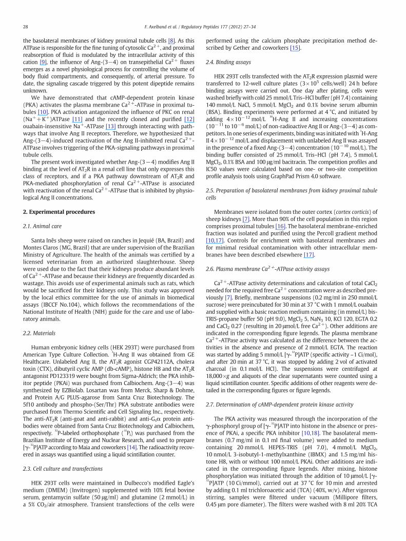
The experiments depicted in Fig. 1 investigated whether a cAMP-mediated pathway is involved in maintaining normal Ca2+-ATPase ac-tivity in the presence of an inhibitory concentration (10−10 mol/L) ofAng II. Fig. 1A demonstrates that stimulation of the membrane-associated adenylyl cyclase by CTX mimicked the counteracting effectof Ang-(3−4) on the inhibition of Ca2+-ATPase by Ang II, whereas thetoxin alone had no effect. In addition, Fig. 1B demonstrates that additionof db-cAMP abrogated the inhibition of the Ca2+ pump by Ang II, andhad no influence when used in isolation.
3.2. Gsα protein is associated with AT2R in basolateral membranes
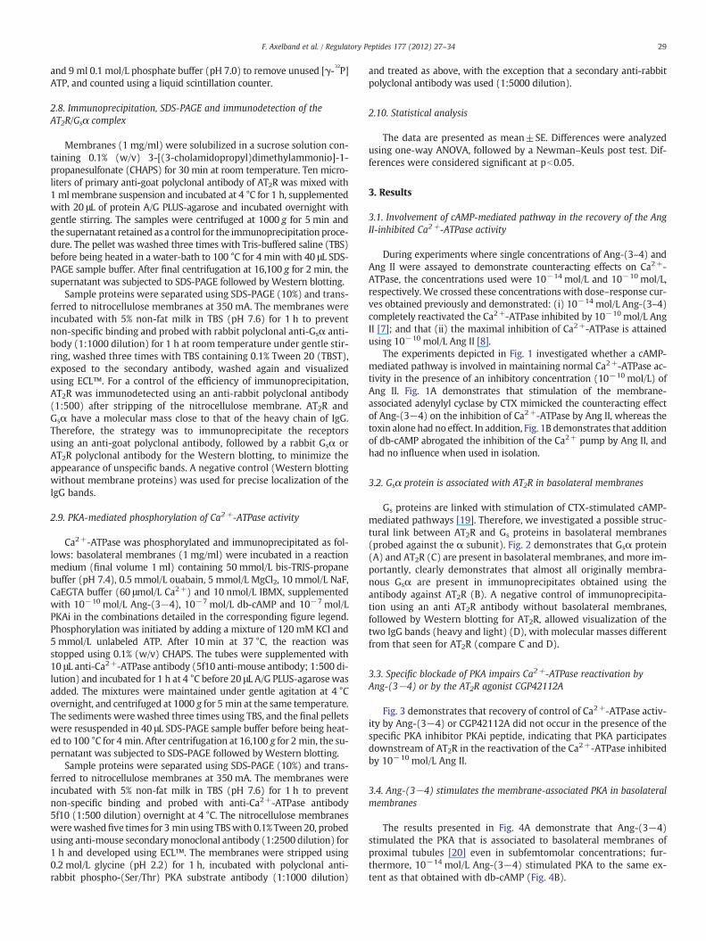
Gs proteins are linked with stimulation of CTX-stimulated cAMP-mediated pathways [19]. Therefore, we investigated a possible struc-tural link between AT2R and Gs proteins in basolateral membranes(probed against the α subunit). Fig. 2 demonstrates that Gsα protein(A) and AT2R (C) are present in basolateral membranes, andmore im-portantly, clearly demonstrates that almost all originally membra-nous Gsα are present in immunoprecipitates obtained using theantibody against AT2R (B). A negative control of immunoprecipita-tion using an anti AT2R antibody without basolateral membranes,followed by Western blotting for AT2R, allowed visualization of thetwo IgG bands (heavy and light) (D), with molecular masses differentfrom that seen for AT2R (compare C and D).
3.3. Specific blockade of PKA impairs Ca2+-ATPase reactivation byAng-(3−4) or by the AT2R agonist CGP42112A
Fig. 3 demonstrates that recovery of control of Ca2+-ATPase activ-ity by Ang-(3−4) or CGP42112A did not occur in the presence of thespecific PKA inhibitor PKAi peptide, indicating that PKA participatesdownstream of AT2R in the reactivation of the Ca2+-ATPase inhibitedby 10−10 mol/L Ang II.
3.4. Ang-(3−4) stimulates the membrane-associated PKA in basolateralmembranes
The results presented in Fig. 4A demonstrate that Ang-(3−4)stimulated the PKA that is associated to basolateral membranes ofproximal tubules [20] even in subfemtomolar concentrations; fur-thermore, 10−14 mol/L Ang-(3−4) stimulated PKA to the same ex-tent as that obtained with db-cAMP (Fig. 4B).
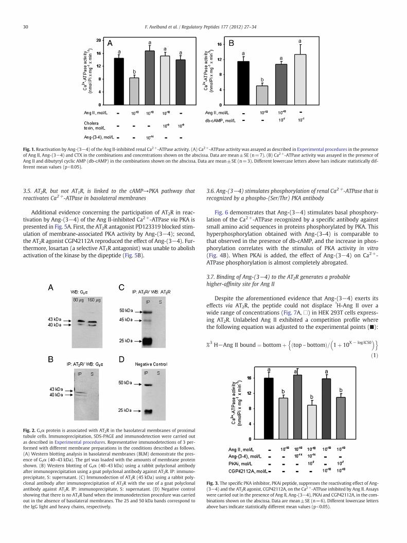
Fig. 1. Reactivation by Ang-(3−4) of the Ang II-inhibited renal Ca2+-ATPase activity. (A) Ca2+-ATPase activity was assayed as described in Experimental procedures in the presenceof Ang II, Ang-(3−4) and CTX in the combinations and concentrations shown on the abscissa. Data are mean±SE (n=7). (B) Ca2+-ATPase activity was assayed in the presence ofAng II and dibutyryl cyclic AMP (db-cAMP) in the combinations shown on the abscissa. Data are mean±SE (n=3). Different lowercase letters above bars indicate statistically dif-ferent mean values (pb0.05).
30 F. Axelband et al. / Regulatory Peptides 177 (2012) 27–34
3.5. AT2R, but not AT1R, is linked to the cAMP→PKA pathway thatreactivates Ca2+-ATPase in basolateral membranes
Additional evidence concerning the participation of AT2R in reac-tivation by Ang-(3−4) of the Ang II-inhibited Ca2+-ATPase via PKA ispresented in Fig. 5A. First, the AT2R antagonist PD123319 blocked stim-ulation of membrane-associated PKA activity by Ang-(3−4); second,the AT2R agonist CGP42112A reproduced the effect of Ang-(3−4). Fur-thermore, losartan (a selective AT1R antagonist) was unable to abolishactivation of the kinase by the dipeptide (Fig. 5B).
Fig. 2. Gsα protein is associated with AT2R in the basolateral membranes of proximaltubule cells. Immunoprecipitation, SDS-PAGE and immunodetection were carried outas described in Experimental procedures. Representative immunodetections of 3 per-formed with different membrane preparations in the conditions described as follows.(A) Western blotting analysis in basolateral membranes (BLM) demonstrate the pres-ence of Gsα (40–43 kDa). The gel was loaded with the amounts of membrane proteinshown. (B) Western blotting of Gsα (40–43 kDa) using a rabbit polyclonal antibodyafter immunoprecipitation using a goat polyclonal antibody against AT2R. IP: immuno-precipitate, S: supernatant. (C) Immunodection of AT2R (45 kDa) using a rabbit poly-clonal antibody after immunoprecipitation of AT2R with the use of a goat polyclonalantibody against AT2R. IP: immunoprecipitate, S: supernatant. (D) Negative controlshowing that there is no AT2R band when the immunodetection procedure was carriedout in the absence of basolateral membranes. The 25 and 50 kDa bands correspond tothe IgG light and heavy chains, respectively.
3.6. Ang-(3−4) stimulates phosphorylation of renal Ca2+-ATPase that isrecognized by a phospho-(Ser/Thr) PKA antibody
Fig. 6 demonstrates that Ang-(3−4) stimulates basal phosphory-lation of the Ca2+-ATPase recognized by a specific antibody againstsmall amino acid sequences in proteins phosphorylated by PKA. Thishyperphosphorylation obtained with Ang-(3–4) is comparable tothat observed in the presence of db-cAMP, and the increase in phos-phorylation correlates with the stimulus of PKA activity in vitro(Fig. 4B). When PKAi is added, the effect of Ang-(3−4) on Ca2+-ATPase phosphorylation is almost completely abrogated.
3.7. Binding of Ang-(3−4) to the AT2R generates a probablehigher-affinity site for Ang II
Despite the aforementioned evidence that Ang-(3−4) exerts itseffects via AT2R, the peptide could not displace
3H-Ang II over a
wide range of concentrations (Fig. 7A, □) in HEK 293T cells express-ing AT2R. Unlabeled Ang II exhibited a competition profile wherethe following equation was adjusted to the experimental points (■):
%3 H−Ang II bound ¼ bottomþ top – bottomð Þ= 1þ 10X − log IC50� �n o
ð1Þ
Fig. 3. The specific PKA inhibitor, PKAi peptide, suppresses the reactivating effect of Ang-(3−4) and the AT2R agonist, CGP42112A, on the Ca2+-ATPase inhibited by Ang II. Assayswere carried out in the presence of Ang II, Ang-(3−4), PKAi and CGP42112A, in the com-binations shown on the abscissa. Data are mean±SE (n=6). Different lowercase lettersabove bars indicate statistically different mean values (pb0.05).

Fig. 4. Ang-(3−4) stimulates the membrane-associated PKA. PKA activity in basolateral membranes was assayed in the presence of the Ang-(3−4) (A) and db-cAMP concentrations(B) shown on the abscissae. Different lowercase letters above bars indicate statistically different mean values (pb0.05). Data are mean±SE (n=7 and n=5 in A and B,respectively).
31F. Axelband et al. / Regulatory Peptides 177 (2012) 27–34
where “bottom” and “top” are the minimum and maximal values ofthe curve, “X” is the logarithm of the concentration of unlabeledAng II, and “IC50” is the concentration of unlabeled Ang II that is ef-fective for displacing 50% of the
3H-Ang II initially bound.
When competition binding assays for Ang II were performed in thepresence of a constant concentration (10−10 mol/L) of Ang-(3−4), weobserved the appearance of a probable higher-affinity site for Ang II,with an IC50 approximately four orders of magnitude lower than inthe absence of Ang-(3−4) (Fig. 7B). In this case, the competition profilewas obtained using an equation derived for two classes of sites:
%3H−Ang II bound ¼ bottomþ top – bottomð Þ×
�Fraction 1= 1þ 10X – log IC50 of fraction 1
� �h i
þ 1 – Fraction 1ð Þ= 1þ 10X – log IC50 of fraction 2� �h i�
ð2Þ
where “bottom”, “top” and “X” have the definitions outlined above.“Fraction 1” corresponds to parcel of receptors with high affinity forAng II, and “Fraction 2” corresponds to receptors (sites) with lower af-finity for Ang II. The IC50 values for these two classes of sites are repre-sented by Eq. (2).
The binding affinity data calculated from these equations are pres-ented in Table 1.
4. Discussion
Signaling pathways coupled to AT1R receptors are relatively welldocumented, whereas those linked to AT2R remain poorly understood
Fig. 5. PKA is functionally linked to AT2R in basolateral membranes. (A) PKA activity in baPD123319, and the AT2R agonist, CGP42112A, in the combinations shown. Data are mean±nations shown. Assays were performed in the presence of Ang-(3−4) and losartan in the coters above bars indicate statistically different mean values (pb0.05).
[21,22]. Herein, we present evidence that Ang-(3−4), in addition toits reported inhibition of the angiotensin converting enzyme (ACE)[5,23–25], can interact with AT2R coupled to Gs proteins in thebasolateral membranes of kidney proximal tubule cells. From a phys-iological point of view, the concentrations of Ang-(3−4) used duringthe present study are less than those present in human plasma andrenal tissue, where the dipeptide can accumulate [26,27]. Down-stream adenylyl cyclase and PKA activation occurs after interactionof Ang-(3−4) with AT2R/Gs and culminates in the reactivation of theneighboring plasma membrane Ca2+-ATPase inhibited by picomolarAng II concentrations. Most important in terms of direct evidence con-cerning the interaction of Ang-(3−4) with the membrane-associatedPKA is the stimulation – not previously described – of the kinase bysubfemtomolar Ang-(3−4) concentrations (see Fig. 4). The resultspresented in Fig. 6 demonstrating stimulation of the regulatory phos-phorylation of Ca2+-ATPase, together with those showing involvementof AT2R, provide further evidence that Ca2+-ATPase is the final target ofa reactivating pathway, AT2R→Gs→adenylyl cyclase→cAMP→PKA, inwhich Ang-(3−4) is a primary messenger.
Isoform 1 of Ca2+-ATPase, which predominates in renal proximaltubule cells and can be phosphorylated by PKA [10], possesses a spe-cific site for this kinase, localized downstream of the calmodulin bind-ing domain [28,29]. However, several isoforms of Ca2+-ATPase have athreonine residue target for PKC in the pump sequence correspondingto the calmodulin binding domain [29]. It may be speculated that theinhibitory PKC-mediated phosphorylation of the latter in the pres-ence of Ang II [8] is counteracted by the PKA-mediated stimulatoryphosphorylation of the former site elicited by Ang-(3−4) (Figs. 4Band 6). If this were the case, opposite phosphorylations elicitedby Ang II and Ang-(3−4) could act by modulating binding of the
solateral membranes was assayed in the presence of Ang-(3−4), the AT2R antagonist,SE (n=8). (B) PKA activity in the presence of Ang-(3−4) and losartan, in the combi-mbinations shown. Data are mean±SE (n=5). In both panels different lowercase let-
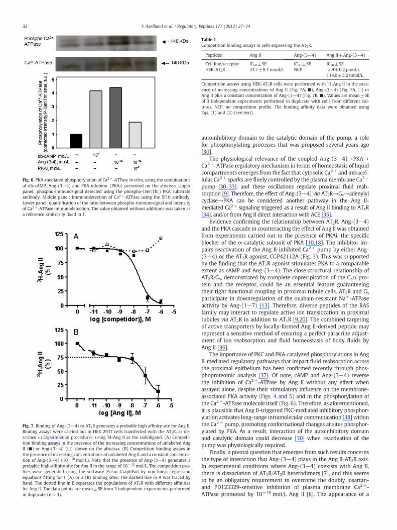
Fig. 6. PKA-mediated phosphorylation of Ca2+-ATPase in vitro, using the combinationsof db-cAMP, Ang-(3−4) and PKA inhibitor (PKAi) presented on the abscissa. Upperpanel: phospho-immunosignal detected using the phospho-(Ser/Thr) PKA substrateantibody. Middle panel: immunodetection of Ca2+-ATPase using the 5f10 antibody.Lower panel: quantification of the ratio between phospho-immunosignal and intensityof Ca2+-ATPase immunodetection. The value obtained without additions was taken asa reference arbitrarily fixed in 1.
Fig. 7. Binding of Ang-(3−4) to AT2R generates a probable high affinity site for Ang II.Binding assays were carried out in HEK 293T cells transfected with the AT2R, as de-scribed in Experimental procedures, using
3H-Ang II as the radioligand. (A) Competi-
tion binding assays in the presence of the increasing concentrations of unlabeled AngII (■) or Ang-(3−4) (□) shown on the abscissa. (B) Competition binding assays inthe presence of increasing concentrations of unlabeled Ang II and a constant concentra-tion of Ang-(3−4) (10−10 mol/L). Note that the presence of Ang-(3−4) generates aprobable high-affinity site for Ang II in the range of 10−12 mol/L. The competition pro-files were generated using the software Prism GraphPad by non-linear regressionequations fitting for 1 (A) or 2 (B) binding sites. The dashed line in A was traced byhand. The dotted line in B separates the populations of AT2R with different affinitiesfor Ang II. The data points are mean±SE from 3 independent experiments performedin duplicate (n=3).
Table 1Competition binding assays in cells expressing the AT2R.
Peptides Ang II Ang-(3−4) Ang II+Ang-(3−4)
Cell line/receptor IC50±SE IC50±SE IC50±SEHEK-AT2R 33.7±9.1 nmol/L NCP 2.9±0.2 pmol/L
114.0±5.2 nmol/L
Competition assays using HEK-AT2R cells were performed with3H-Ang II in the pres-
ence of increasing concentrations of Ang II (Fig. 7A, ■), Ang-(3−4) (Fig. 7A, □) orAng II plus a constant concentration of Ang-(3−4) (Fig. 7B, ■). Values are mean±SEof 3 independent experiments performed in duplicate with cells from different cul-tures. NCP, no competition profile. The binding affinity data were obtained usingEqs. (1) and (2) (see text).
32 F. Axelband et al. / Regulatory Peptides 177 (2012) 27–34
autoinhibitory domain to the catalytic domain of the pump, a rolefor phosphorylating processes that was proposed several years ago[30].
The physiological relevance of the coupled Ang-(3−4)→PKA→Ca2+-ATPase regulatory mechanism in terms of homeostasis of liquidcompartments emerges from the fact that cytosolic Ca2+ and intracel-lular Ca2+ sparks are finely controlled by the plasmamembrane Ca2+
pump [30–33], and these oscillations regulate proximal fluid reab-sorption [9]. Therefore, the effect of Ang-(3−4) via AT2R→Gs→adenylylcyclase→PKA can be considered another pathway in the Ang II-mediated Ca2+ signaling triggered as a result of Ang II binding to AT1R[34], and/or from Ang II direct interaction with ACE [35].
Evidence confirming the relationship between AT2R, Ang-(3−4)and the PKA cascade in counteracting the effect of Ang II was obtainedfrom experiments carried out in the presence of PKAi, the specificblocker of the α-catalytic subunit of PKA [10,18]. The inhibitor im-pairs reactivation of the Ang II-inhibited Ca2+ pump by either Ang-(3−4) or the AT2R agonist, CGP42112A (Fig. 3). This was supportedby the finding that the AT2R agonist stimulates PKA to a comparableextent as cAMP and Ang-(3−4). The close structural relationship ofAT2R/Gs, demonstrated by complete coprecipitation of the Gsα pro-tein and the receptor, could be an essential feature guaranteeingtheir tight functional coupling in proximal tubule cells. AT2R and Gs
participate in downregulation of the ouabain-resistant Na+-ATPaseactivity by Ang-(1−7) [13]. Therefore, diverse peptides of the RASfamily may interact to regulate active ion translocation in proximaltubules via AT2R in addition to AT1R [9,20]. The combined targetingof active transporters by locally-formed Ang II-derived peptide mayrepresent a sensitive method of ensuring a perfect paracrine adjust-ment of ion reabsorption and fluid homeostasis of body fluids byAng II [36].
The importance of PKC and PKA-catalyzed phosphorylations in AngII-mediated regulatory pathways that impact fluid reabsorption acrossthe proximal epithelium has been confirmed recently through phos-phoproteomic analysis [37]. Of note, cAMP and Ang-(3−4) reversethe inhibition of Ca2+-ATPase by Ang II without any effect whenassayed alone, despite their stimulatory influence on the membrane-associated PKA activity (Figs. 4 and 5) and in the phosphorylation ofthe Ca2+-ATPase molecule itself (Fig. 6). Therefore, as aforementioned,it is plausible that Ang II-triggered PKC-mediated inhibitory phosphor-ylation activates long-range intramolecular communication [38] withinthe Ca2+ pump, promoting conformational changes at sites phosphor-ylated by PKA. As a result, interaction of the autoinhibitory domainand catalytic domain could decrease [30] when reactivation of thepump was physiologically required.
Finally, a pivotal question that emerges from such results concernsthe type of interaction that Ang-(3−4) plays in the Ang II-AT2R axis.In experimental conditions where Ang-(3−4) coexists with Ang II,there is dissociation of AT1R/AT2R heterodimers [7], and this seemsto be an obligatory requirement to overcome the doubly losartan-and PD123329-sensitive inhibition of plasma membrane Ca2+-ATPase promoted by 10−10 mol/L Ang II [8]. The appearance of a
33F. Axelband et al. / Regulatory Peptides 177 (2012) 27–34
second binding site, with affinity for Ang II in the 10−12 mol/L range(IC50 2.9 pmol/L), i.e. four orders of magnitude higher than that ob-served without Ang-(3–4) (IC50 33.7 nmol/L), is accompanied of amoderate reduction in affinity of the latter site to 114.0 nmol/L(Table 1). Such alterations in affinity and in the competition profilefor Ang II are probably representative of a subpopulation of AT2Rwith altered conformation due to the allosteric interference of Ang-(3−4) binding, consequently responsible for complete reversing ofthe Ang II-induced inhibition of the Ca2+ pump [7]. Therefore, Ang-(3−4) would act as an allosteric modulator when bound to theAT2R. Therefore, Ang-(3−4) would not directly compete with AngII, but would be responsible for inducing conformational changes inthe receptors, resulting in modification of the Ang II binding profile.Allosteric modulators can modify affinity of the primary agonist forG protein-coupled receptors either when in their monomeric [39] ordimeric states [40]. Therefore, in native proximal tubule membranes,a similar behavior of conformational modification due to the allostericinterference of Ang-(3−4) could occur with AT2R/AT1R heterodimers[7]. We speculate that this is a key event linked to the counter-regulatory action of Ang-(3−4). This event would correspond tothe structural modifications induced by Ang-(3−4) in receptors thatfunctionally couple a primed, but silent, AT2R/Gs complex linked toa downstream PKA pathway.
5. Conclusions
In conclusion, this study has revealed new partners of AT2R inrenal cells: the components of the cAMP-stimulated kinase pathwayresident in basolateral membranes and Ang-(3−4), the latter actingas an allosteric modulator [40]. This expands our understanding con-cerning AT2R functionality and its association with other receptorsthat participate in renal ion and fluid handling.
Acknowledgments
The technical assistance of Glória Costa-Sarmento is acknowledged.Correction in the English presentation of this manuscript was done byBioMedES (UK), which is gratefully acknowledged. This work wassupported by grants from the Brazilian National Research Council(CNPq), the Carlos Chagas Filho Rio de Janeiro State Research Foundation(FAPERJ), the São Paulo Research Foundation (FAPESP), the BrazilianFederal Agency for Support and Evaluation of Graduate Education(CAPES), and the National Institutes of Science and Technology, Brazil.
References
[1] Kramkowski K, Mogielnicki A, Buczko W. The physiological significance of the al-ternative pathways of angiotensin II production. J Physiol Pharmacol 2006;57:529–39.
[2] Santos RA, Simões-e-Silva AC, Maric C, Silva DMR, Machado RP, Buhr I, Heringer-Walther S, Pinheiro SVB, Lopes MT, Bader M, Mendes EP, Lemos VS, Campagnole-Santos MJ, Schultheiss HP, Speth R, Walther T. Angiotensin-(1−7) is an endogenousligand for the G protein-coupled receptor MAS. Proc Natl Acad Sci U S A 2003;100:8258–63.
[3] Haulica I, Petrescu G, Slatineanu SM, Bild W, Mihaila CN, Ionita T. New bioactiveangiotensins formation pathways and functional involvements. Rom J InternMed 2004;42:27–40.
[4] Axelband F, Dias J, Miranda F, Ferrão FM, Barros NM, Carmona AK, Lara LS, VieyraA. A scrutiny of the biochemical pathways from Ang II to Ang-(3−4) in renal bas-olateral membranes. Regul Pept 2009;158:47–56.
[5] Saito Y, Wanezaki K, Kawato A, Imayasu S. Structure and activity of angiotensin Iconverting enzyme inhibitory peptides from sake and sake lees. Biosci BiotechnolBiochem 1994;58:1767–71.
[6] Matsufuji H, Matsui T, Ohshige S, Kawasaky T, Osajima K, Osajima Y. Antihyper-tensive effects of angiotensin fragments in SHR. Biosci Biotechnol Biochem1995;59:1398–401.
[7] Axelband F, Assunção-Miranda I, de Paula IR, Ferrão FM, Dias J, Miranda A,Miranda F, Lara LS, Vieyra A. Ang-(3−4) suppresses inhibition of renal plasmamembrane calcium pump by Ang II. Regul Pept 2009;155:81–90.
[8] Assunção-Miranda I, Guilherme AL, Reis-Silva C, Costa-Sarmento G, Oliveira MM,VieyraA. Protein kinase C-mediated inhibition of renal Ca2+-ATPase byphysiological
concentrations of angiotensin II is reversed by AT1- and AT2-receptor antagonists.Regul Pept 2005;127:151–7.
[9] Du Z, FergusonW,Wang T. Role of PKC and calcium in modulation of effects of an-giotensin II on sodium transport in proximal tubule. Am J Physiol 2003;284:F688–92.
[10] Cabral LM, Wengert M, da Ressurreição AA, Feres-Elias PH, Almeida FG, Vieyra A,Caruso-Neves C, Einicker-Lamas M. Ceramide is a potent activator of plasmamembrane Ca2+-ATPase from kidney-proximal tubule cells with protein kinaseA as an intermediate. J Biol Chem 2007;282:24599–606.
[11] Shahedi M, Laborde K, Bussières L, Dechaux M, Sachs C. Protein kinase C activationcauses inhibition of Na+/K+-ATPase activity in Madin-Darby canine kidney epi-thelial (MDCK) cells. Pfluegers Arch 1992;420:269–74.
[12] Rocafull MA, Romero FJ, Thomas LE, del Castillo JR. Isolation and cloning of the K+-independent, ouabain-insensitive Na+-ATPase. Biochim Biophys Acta 2011;1808:1684–700.
[13] Lara LS, Vives D, Correa JS, Cardozo FP,Marques-FernadesMF, Lopes AG, Caruso-NevesC. PKA-mediated effect of MAS receptor in counteracting angiotensin II-stimulatedrenal Na+-ATPase. Arch Biochem Biophys 2010;496:117–22.
[14] Maia JCC,Gomes SL, JulianiMH. Preparation of (gamma-32P) and (alpha-
32P)-nucleoside
triphosphates with high specific activity. Genes and antigenes of parasites, a labora-tory manual proceedings. Rio de Janeiro: Editora Fundação Oswaldo Cruz; 1983.p. 146–57.
[15] Gether U, Johansen TE, Schwartz TW. Chimeric NK1 (substance P)/NK3 (neurokininB) receptors — identification of domains determining the binding specificity oftachykinin. J Biol Chem 1993;268:7893–8.
[16] Whittembury G, Proverbio F. Twomodes of Na+ extrusion in cells from guinea pigkidney cortex slices. Pfluegers Arch 1970;316:1–25.
[17] Coka-Guevara S, Markus RP, Caruso-Neves C, Lopes AG, Vieyra A. Adenosine in-hibits the renal plasma-membrane (Ca2++Mg2+)-ATPase through a pathwaysensitive to cholera toxin and sphingosine. Eur J Biochem 1999;263:71–8.
[18] Valverde RH, Britto-Borges T, Lowe J, Einicker-Lamas M, Mintz E, Cuillel M, VieyraA. Two serine residues control sequential steps during catalysis of the yeast cop-per ATPase through different mechanisms that involve kinase-mediated phos-phorylations. J Biol Chem 2011;286:6879–89.
[19] Simonds WF. G protein regulation of adenylate cyclase. Trends Pharmacol Sci1999;20:66–73.
[20] Vieira-Filho LD, Lara LS, Silva PA, Santos FT, Luzardo R, Oliveira FS, Paixão AD,Vieyra A. Placental malnutrition changes the regulatory network of renal Na-ATPase in adult rat progeny: reprogramming by maternal α-tocopherol duringlactation. Arch Biochem Biophys 2011;505:91–7.
[21] Mogi M, Iwai M, Horiuchi M. New insights into the regulation of angiotensin re-ceptors. Curr Opin Nephrol Hypertens 2009;18:138–43.
[22] Rodrigues-Ferreira S, Nahmias C. An ATIPical family of angiotensin II AT2receptor-interacting proteins. Trends Endocrinol Metab 2010;21:684–90.
[23] Matsui T, Hayashi A, Tamaya K, Matsumoto K, Kawasaki T, Murakami K, Kimoto K.Depressor effect induced by dipeptide, Val-Tyr, in hypertensive transgenic mice isdue, in part, to the suppression of human circulating renin–angiotensin system.Clin Exp Pharmacol Physiol 2003;30:262–5.
[24] Pentzien AK, Meisel H. Transepithelial transport and stability in blood serum ofangiotensin-I-converting enzyme inhibitory dipeptides. Z Naturforsch C 2008;63:451–9.
[25] Wang Z, Zhang S, Jin H,WangW, Huo J, Zhou L,Wang Y, Feng F, Zhang L. Angiotensin-I-converting enzyme inhibitory peptides: chemical feature based pharmacophoregeneration. Eur J Med Chem 2011;46:3428–33.
[26] Matsui T, Tamaya K, Matsumoto K, Osajima Y, Uezono K, Kawasaki T. Plasma con-centrations of angiotensin metabolites in young male normotensive and mild hy-pertensive subjects. Hypertens Res 1999;22:273–7.
[27] Matsui T, ImamuraM, Oka H, Osajima K, Kimoto K, Kawasaki T, Matsumoto K. Tissuedistribution of antihypertensive dipeptide, Val-Tyr, after its single oral administra-tion to spontaneously hypertensive rats. J Pept Sci 2004;10:535–45.
[28] James PH, Pruschy M, Vorherr TE, Penniston JT, Carafoli E. Primary structure of thecAMP-dependent phosphorylation site of the plasma membrane calcium pump.Biochemistry 1989;28:4253–8.
[29] Di Leva F, Domi T, Fedrizzi L, Lim D, Carafoli E. The plasma membrane Ca2+
ATPase of animal cells: structure, function and regulation. Arch Biochem Biophys2008;476:65–74.
[30] Carafoli E. Biogenesis: plasma membrane calcium ATPase: 15 years of work on thepurified enzyme. FASEB J 1994;8:993–1002.
[31] Isshiki M, Ando J, Korenaga R, Kogo H, Fujimoto T, Fujita T, Kamiya A. EndothelialCa2+ waves preferentially originate at specific loci in caveolin-rich cell edges.Proc Natl Acad Sci U S A 1998;95:5009–14.
[32] Isshiki M, Anderson RGW. Calcium signal transduction from caveolae. Cell Calci-um 1999;26:201–8.
[33] Tortelote GG, Valverde RH, Lemos T, Guilherme A, Einicker-Lamas M, Vieyra A.The plasma membrane Ca2+ pump from proximal kidney tubules is exclusivelylocalized and active in caveolae. FEBS Lett 2004;576:31–5.
[34] Zhuo JL, Li XC. New insights and perspectives on intrarenal renin-angiotensin sys-tem: focus on intracrine/intracellular angiotensin II. Peptides 2011;32:1551–65.
[35] Guimarães PB, Alvarenga ÉC, Siqueira PD, Paredes-Gamero EJ, Sabatini RA,Morais RL, Reis RI, Santos EL, Teixeira LG, Casarini DE, Martin RP, ShimutaSI, Carmona AK, Nakaie CR, Jasiulionis MG, Ferreira AT, Pesquero JL, OliveiraSM, Bader M, Costa-Neto CM, Pesquero JB. Angiotensin II binding to angio-tensin I-converting enzyme triggers calcium signaling. Hypertension 2011;57:965–72.
[36] Navar LG, Nishiyama A. Why are angiotensin concentrations so high in the kid-ney? Curr Opin Nephrol Hypertens 2004;13:107–15.
34 F. Axelband et al. / Regulatory Peptides 177 (2012) 27–34
[37] Li XC, Zhuo JL. Phosphoproteomic analysis of AT1 receptor-mediated signaling re-sponses in proximal tubules of angiotensin II-induced hypertensive rats. KidneyInt 2011;80:620–32.
[38] Valverde RH, Morin I, Lowe J, Mintz E, Cuillel M, Vieyra A. Cyclic AMP-dependentprotein kinase controls energy interconversion during the catalytic cycle of theyeast copper-ATPase. FEBS Lett 2008;582:891–5.
[39] Wang L, Martin B, Brenneman R, Luttrell LM, Maudsley S. Allosteric modulators ofG protein-coupled receptors: future therapeutics for complex physiological disor-ders. J Pharmacol Exp Ther 2009;331:340–8.
[40] Schwartz TW, Holst B. Allosteric enhancers, allosteric agonists and ago-allostericmodulators: where do they bind and how do they act? Trends Pharmacol Sci2007;28:366–73.
Copyright © 2022 FDOKUMEN