A novel dominant COL11A1 mutation resulting in a severe skeletal dysplasia
Upload
udistritalCategory
view
0download
0
Kidney International, Vol. 59 (2001), pp. 2222–2232
Mycophenolate mofetil prevents salt-sensitive hypertensionresulting from angiotensin II exposure
BERNARDO RODRIGUEZ-ITURBE, HECTOR PONS, YASMIR QUIROZ, KATHERINE GORDON,JAIMAR RINCON, MARIBEL CHAVEZ, GUSTAVO PARRA, JAIME HERRERA-ACOSTA,DULCENOMBRE GOMEZ-GARRE, RAQUEL LARGO, JESUS EGIDO, and RICHARD J. JOHNSON
Renal Service and Department of Immunobiology (INBIOMED), Hospital Universitario, Maracaibo, Venezuela; Divisionof Nephrology, University of Washington Medical Center, Seattle, Washington, USA; Nephrology Department, InstitutoNacional de Cardiologıa, Mexico City, Mexico; and the Nephrology Laboratory, Fundacion Jimenez Dıaz, Madrid, Spain
Conclusions. SSHTN resulting from Ang II infusion is asso-Mycophenolate mofetil prevents salt-sensitive hypertensionciated with infiltration and activation of immune cells thatresulting from angiotensin II exposure.produce Ang II. MMF treatment reduces these features andBackground. Interstitial mononuclear cell infiltration is aprevents the development of SSHTN.feature of experimental models of salt-sensitive hypertension
(SSHTN). Since several products of these cells are capable ofmodifying local vascular reactivity and sodium reabsorption,we investigated whether mycophenolate mofetil (MMF), a drug
Lombardi et al have recently reported that pressorknown to inhibit infiltration and proliferation of immune cells,doses of angiotensin II (Ang II) administered for twowould modify the SSHTN induced by angiotensin II (Ang II)
infusion. weeks resulted in the subsequent development of salt-Methods. Sprague-Dawley rats received Ang II for two weeks sensitive hypertension (SSHTN) in the rat [1]. This inves-
using subcutaneous minipumps. A high-sodium (4% NaCl) diet tigation established an experimental model that permitswas started on the third week and was maintained until thethe analysis of the sequential structural changes in theeighth week. MMF (30 mg/kg, N 5 15), an immunosuppressive
drug, or vehicle (N 5 15) was given daily by gastric gavage kidney that are acutely induced by Ang II as well as withduring the initial three weeks. Sham-operated rats (N 5 9) the development of persistent SSHTN. The structuralwere used as controls. Body weight, blood pressure (tail-cuff changes present in the kidney consist of focal vascularplethysmography), and serum creatinine were determined
and tubulointerstitial damage with areas of tubular atro-weekly. Urinary malondialdehyde (MDA) excretion, renal his-phy and a reduction in peritubular capillaries, while intology, and immunohistology, including the presence of Ang II
and superoxide-producing cells, were analyzed at the end of contrast, the glomeruli were minimally abnormal. TheseAng II infusion and at eight weeks. findings supported the postulate that impairment of pres-
Results. MMF treatment did not modify hypertension in-sure-natriuresis response caused by tubulointerstitial in-duced during exogenous Ang II infusion, but prevented thejury could play a pivotal role in the development ofsubsequent SSHTN. Tubulointerstitial injury resulting from
Ang II infusion was significantly reduced by MMF treatment, hypertension in a variety of clinical conditions [2].as were proliferative activity, T-cell infiltration and activation Nevertheless, histologic findings in the studies of Lom-(interleukin-2 receptor expression), superoxide-producing cells, bardi et al as well as in a variety of other experimentaland urinary MDA excretion. Ang II-producing cells were pres-
and clinical conditions associated with SSHTN includeent in the renal tubulointerstitium of rats with SSHTN (60 6infiltration, at times prominent, of mononuclear cells in30 Ang II-positive cells/mm2 at 8 weeks) and were reduced
by two thirds in the MMF-treated group. Forty percent of tubulointerstitial areas of the kidney [1]. Examples oflymphocytes infiltrating the tubulointerstitium stained positive mononuclear cell infiltration in other experimental hy-for Ang II. The expression of Ang II receptors in the kidney
pertension models include the “vicious cycle” of two-was unmodified.kidney one-clip Goldblatt hypertension [3, 4], aging [5],cyclosporine nephropathy [6], and catecholamine infu-
Key words: immune system, renal interstitium, cell proliferation, im-sion [7]. An influx of immunocompetent cells may bemunosuppression, rat experimental model, blood pressure, mononu-
clear cell infiltrate. induced by chemokines and leukocyte adhesion proteinsexpressed by injured renal cells [reviewed in 8]. Infiltrat-
Received for publication July 7, 2000ing cells could then produce cytokines in situ, which couldand in revised form November 28, 2000
Accepted for publication January 8, 2001 modify local vascular reactivity [reviewed in 9] and pro-mote inflammation and scarring [reviewed in 10]. There- 2001 by the International Society of Nephrology
2222

Rodrıguez-Iturbe et al: Immunocompetent cells in hypertension 2223
fore, the mononuclear cellular infiltrate could be criti- In addition to angiotensin, the Ang II 1 MMF groupreceived MMF (CellCept; Roche Laboratories, Mont-cally involved in modifications of the pressure-natriuresis
and sodium reabsorption mechanisms and, thereby, in clair, NJ, USA) by gastric gavage, in doses of 30 mg ·kg · day21, during the angiotensin infusion phase andthe pathogenesis of SSHTN.
The present studies were designed to evaluate the role, the washout phase of the experiment. As described inprevious work, the MMF was suspended in 500 mL ofif any, of infiltrating immunocompetent cells that occur
in the kidney during the angiotensin exposure, on the water by vigorous agitation immediately before adminis-tration because the drug is insoluble in water [12]. Thesubsequent development of hypertension. For this pur-
pose, we treated rats with the immunosuppressive drug Ang II group received 500 mL water daily by gastricgavage during the angiotensin and washout phases. Themycophenolate mofetil (MMF) during the angiotensin
infusion phase of this experimental model. control group (N 5 9) consisted of rats in which thesame surgical procedure was performed (including bothMycophenolate mofetil is a widely used drug for the
treatment of transplant recipients. It reversibly inhibits the implantation and removal of the minipumps), butminipumps were not inserted.the enzyme inosine monophosphate dehydrogenase that
regulates de novo purine synthesis, and consequently is a All of the animals (Ang II group, Ang II 1 MMFgroup, and control group) had a similar dietary protocol,selective lymphocyte immunosuppressor [11]. Recently,
MMF has been shown to diminish the expression of in- consisting of standard rat chow with normal sodium con-tent (Ratarina Protinal Company, Valencia, Venezuela)tracellular adhesion molecules and the infiltration of im-
mune cells in the remnant kidney model [12] and in the during the initial three weeks of the study (angiotensinand washout periods) and a high-sodium (4% NaCl) dietatherosclerotic lesions in the rabbit aorta [13]. Further-
more, MMF treatment has antiproliferative effects in (Harlan Teklad, Madison, WI, USA) from weeks 4 to 8.Serum creatinine and urinary protein were determinedmesangial cells [14, 15] and tubulointerstitial cells [16],
and reduces collagen deposition [16]. Since the drug has by autoanalyzer methodology (Express Plus; Ciba Corn-ing, Essex, UK). Urinary malondialdehyde (MDA) wasrelatively minor side effects [17] and in contrast to cy-
closporine A and Tacrolimus is not nephrotoxic, it ap- determined in 24-hour urine samples obtained at the endof the angiotensin phase and at the end of the experiment.peared well suited for the purposes of this work. Our
results indicate that treatment with MMF prevents the Rats from the Ang II and Ang II 1 MMF groups weresacrificed under ether anesthesia by aortic desanguina-development of SSHTN in the Ang II model. This effect
was associated with a reduction of tubulointerstitial lym- tion after removal of the kidneys for histologic studies attwo time periods: at the end of the angiotensin administra-phocyte infiltration and Ang II-producing cells, a reduc-
tion in histologic damage and a reduction in superoxide tion and at the end of the experiment at the eighth week.producing cells and urinary lipid peroxidation products.
Blood pressure measurements
Systolic and mean arterial blood pressures were deter-METHODS
mined in conscious restrained rats by tail-cuff plethysmo-Experimental design graphy (IITC; Life Scientific Instruments, Woodland
Hills, CA, USA). Before the experiments were begun,The experimental protocol included three successivephases over an eight-week period: the angiotensin infu- rats were conditioned three to four times to the proce-
dure. Once the experiments were started, blood pres-sion phase (2 weeks of angiotensin administration), wash-out phase (5 to 7 days following angiotensin), and a high- sures were measured weekly. In each session, the blood
pressure recorded was the mean of three separate mea-salt diet phase (from the end of the washout phase untilthe 8th week). surements for each rat.
Male Sprague-Dawley rats (Instituto Venezolano deUrinary MDA determinationsInvestigaciones Cientıficas, Caracas, Venezuela) weighing
290 to 340 g were divided in three groups that received Thiobarbituric reactive substances were determinedby the method of Ohkawa, Ohishi, and Yagi [19] as de-Ang II infusion and MMF (Ang II 1 MMF group) or
vehicle (Ang II group) or were sham operated (control scribed previously [12]. Briefly, MDA was measured in24-hour urine samples centrifuged at 2000 3 g and pro-group). The rats of the Ang II 1 MMF group (N 5 15)
and the Ang II group (N 5 15) received 435 ng · kg · cessed the same day. A sample of 400 mL was added toa reaction mixture consisting of 200 mL of 8.1% sodiummin21 of Ang II (Sigma Chemical Co. St. Louis, MO,
USA) dissolved in Ringer’s lactate, for two weeks by dodecyl sulfate, 1.5 mL of 20% acetic acid (pH 3.5), 1.5mL of 0.5% thiobarbituric acid, and 400 mL of bidistilledosmotic minipumps (Alzet model 2002; Alza Corpora-
tion, Palo Alto, CA, USA) that were surgically placed water. The mixture was heated at 958C in a water bathfor 60 minutes, and 1 mL of water and 5 mL of n-butanol-subcutaneously and removed at the end of two weeks,
as described elsewhere [18]. pyridine were added; the mixture was agitated and cen-

Rodrıguez-Iturbe et al: Immunocompetent cells in hypertension2224
trifuged at 2000 3 g for 15 minutes. The absorbance of ing with goat anti-OPN (OP 199; gift from C. Giachelli,University of Washington, Seattle, WA, USA) and ED-1the upper organic layer was read at 532 nm (Shimatzu(Harlan Bioproducts, Indianapolis, IN, USA), was de-Spectrophotometer model UV21100S, Kyoto, Japan).scribed in previous work [1]. Proliferating cell nuclearMalonaldehyde bis-dimethyl acetal was used as the ex-antigen was identified with the appropriate antibodyternal standard.[monoclonal antibody (mAb) anti-PCNA clone PC10;
Histologic studies concentration 50 mg/mL; Zymed Laboratories, Inc. SanFrancisco, CA, USA] as described in a previous study [20].Coronal kidney sections were fixed in Methyl Carnoy
Superoxide production in renal cells was studied inand embedded in Paraplast Plus (Monoject, Sherwood8 mm cryostat sections by the cytochemical method ofMedical Scientific Division, St. Louis, MO, USA). TheBriggs et al [21]. Slides were incubated for 60 minutesremainder of the tissue was included in tissue-freez-at 378C in the following solution: 50 mL 0.05 mol/Ling medium (Triangle Biomedical Sciences, Durham,Tris-HCl buffer, 1 mL diaminobenzidine (DAB) stockNC, USA), frozen in dry ice and acetone, and stored atsolution (5 g DAB/132 mL Tris buffer 0.05 mol/L,2708C.pH 7.6), 250 mL 8% NiCl, 32.5 mL 10% NaN3, and 50Paraffin-embedded sections 4 mm thick were stainedmL 0.5 mol/L MnCl2. Sections were fixed with 10% for-with periodic acid-Schiff reagent (PAS) and hematoxylinmalin and counterstained with 1% methyl green.and eosin (HE). Tubulointerstitial injury was evaluated
Indirect immunofluorescence was used to identifyin the entire cortex and corticomedullary areas. As pre-T lymphocytes (mAb anti-rat CD5, clone MRCOX19;viously described [6, 7], tubulointerstitial injury was de-concentration 50 mg/mL; Biosource International, Cama-fined by any of the following findings: tubular dilation,rillo, CA, USA), interleukin-2 (IL-2) receptor (mAbatrophy or sloughing, cellular infiltration (identified byanti-CD25; concentration 100 mg/mL; Accurate Chemi-HE staining), interstitial widening, or basement mem-cal and Scientific Corporation, Westbury, NY, USA),brane thickening. The severity of the injury was ex-Ang II-producing cells [rabbit anti-Ang II (human) IgG,pressed as a semiquantitative score based on the exten-dilution 1:20; Peninsula Labs. Inc., CA, USA], and fi-sion of the finding: 0 5 no changes present; grade 1 5bronectin (mAb rabbit anti-rat fibronectin; dilution 1:50;
,10%; grade 2 5 10 to 25%; grade 3 5 25 to 50%; gradeCalBiochem Laboratories, La Jolla, CA, USA) as de-4 5 50 to 75%; and grade 5 5 75 to 100% [6].scribed previously [19]. The secondary antibody forComputer-assisted image analysis software was usedstaining CD5 and IL-2 receptor was FITC-conjugatedto study osteopontin (OPN; Optimas, version 6.2; Mediaaffinity-pure F(ab9)2 fragment rat anti-mouse IgG withCybernetics, Silver Springs, MD, USA) and fibronectinminimal cross-reactivity with rat protein (Accurate Chem-
expression (Sigma Scan Pro 5.0; SPSS Inc., Chicago, IL, ical and Scientific Corporation). The secondary antibodyUSA) in cortical tubulointerstitial areas. The digitized for fibronectin staining was donkey anti-rabbit FITCimages of OPN were acquired using a Leica DMRB (concentration 20 mg/mL; Accurate Chemical and Scien-microscope fitted with a microimage i308 low-light video tific Corporation).camera with a 1⁄2-inch HyperHAD sensor and a Flash- Double staining of Ang II-positive cells was done aspoint video framegrabber board (Bartels and Stout, described before [22]. Briefly, tissue sections were firstBellevue, WA, USA) as described before [1]. The fibro- incubated with either anti-CD5 or anti-ED-1 mAb atnectin studies were done on images acquired from a the concentration described earlier and then with rhoda-Zeiss Axioscope with a Kodak DC 120 digital megapixel mine-conjugated (TRIC) affinity-pure F(ab9)2 rat anti-camera. Results are given as the percentage of positive mouse IgG with minimal cross reactivity with rat proteinsarea for each biopsy (exclusive of glomeruli). (Accurate Chemical and Scientific Co.). Afterward, tis-
Cellular infiltration and proliferation (positive cells sues were incubated overnight with the anti-Ang II anti-with a given staining/mm2) were counted by two different body (discussed previously) and finally, with the second-observers using an ocular fitted with a grid in at least 20 ary fluorescein conjugated (DTAF) affinity-pure donkeymicroscopic fields (340 magnification) in cortical and anti-rabbit IgG antibody (dilution 1:50; Accurate Chemi-corticomedullay areas, and the mean value of the counts cal and Scientific Co.).was used in each biopsy. Glomerular cell counts were ex- Immunoperoxidase staining was used to localize angio-pressed as positive cells per glomerular cross section (gcs). tensin receptors using rabbit or goat anti-rat angiotensin
Histologic evaluation (individual cell counts and com- II type 1 and 2 (AT1 and AT2) antibodies (Santa Cruzputer-assisted image analysis) was done without previous Biotechnology, Santa Cruz, CA, USA) as described pre-knowledge of the source of the tissue. viously [23]. Briefly, AT1 receptors were studied in kidney
sections incubated with the corresponding antibody (dilu-Histologic technique and reagents tion 1:30) and then with a secondary antibody conjugated
Indirect immunoperoxidase methodology was used to with a peroxidase-labeled polymer (Envision System;Dako A/S, Carpinteria, CA, USA). Finally, the slides wereidentify OPN, macrophages, and proliferating cells. Stain-

Rodrıguez-Iturbe et al: Immunocompetent cells in hypertension 2225
Fig. 1. Angiotensin (Ang II) infusion induces salt-dependent hyperten-sion in untreated rats (j) but not in mycophenolate mofetil (MMF)-treated rats (h). Dotted lines correspond to the 95% CI in control
Fig. 2. Weight changes during the experiment. The values in untreated(sham-operated) rats. Data are mean 6 SD. *P , 0.05 and **P ,rats (j) and the values in MMF-treated rats (h) are not statistically0.01 vs. the values at the fourth week (end of the wash-out period afterdifferent at any time interval. The weight in both groups of experimentalangiotensin infusion). During angiotensin infusion and washout periods,rats diminished during the angiotensin infusion (period a: P , 0.001 vs.rats received a normal Na diet.each previous week) and increased afterward (period b: P , 0.001 vs.each previous week after week 2), without reaching the 95% CI of theweight in control rats. Data are mean 6 SD.
incubated with 3,39-diaminobenzidine (DAB; Sigma).AT2 localization was done by the avidin-biotin method-ology. Biotinylated mouse anti-goat IgG (Amersham, RESULTSArlington Heights, IL, USA) was used as a secondary Effects of MMF treatment on blood pressure, weight,antibody, and positive sites were also evidenced with and renal functionDAB. Control slides were treated with nonrelevant anti-
The effects of MMF on the systolic blood pressure aresera. Sections were counterstained with Mayer’s hema-shown in Figure 1. During the administration of Ang II,toxylin (Sigma Chemical Co.). Double immunostainingthe blood pressure increased in both experimentalwas also performed to determine whether lymphocytesgroups. In the first week, the blood pressure incrementwere expressing Ang II receptors. For this purpose, mAbwas more pronounced in the Ang II group (P , 0.01),mouse anti-rat CD5 and affinity-pure rabbit anti-Ang IIbut both groups had a similar blood pressure elevationtype 1 receptor (anti-AT1) and anti-Ang II type 2 receptorat the end of two weeks. In the washout period, the(anti-AT2) antisera (Alpha Diagnostics, San Antonio,blood pressure returned to baseline levels in both groups,TX, USA) were used as primary antibodies. Secondaryand after the fourth week, corresponding to one weekantibodies were rhodamine-conjugated rat anti-mousein the high-salt diet, the blood pressure progressivelyIgG and fluorescein-conjugated (DTAF) donkey anti-rose in the Ang II group. In contrast, the blood pressurerabbit IgG.in the Ang II 1 MMF group remained at the sameAll histologic and immunohistochemical micropho-baseline-washout levels, well within the 95% CI of thetography are collected in two plates (Figs. 9 and 10; Dis-control group (Fig. 1).cussion section).
As shown in Figure 2, the rats from both experimentalStatistical analysis groups lost weight during the Ang II infusions (P ,
0.001), and the difference in weight loss between theStatistical analyses were done with the help of a com-Ang II and the Ang II 1 MMF groups was not statisti-mercial statistical package (Instat GraphPadt, San Diego,cally significant. During the washout period, the bodyCA, USA). Comparisons between groups were done byweight started to increase in both experimental groups,one-way analysis of variance (ANOVA) followed by Tu-and this tendency continued throughout the high-saltkey-Kramer post-tests or by unpaired t tests using Welch’sdiet period (Fig. 2).correction when the differences in SD were significant.
Baseline serum creatinine was similar in the experi-Serial changes were evaluated with repeated-measuresmental groups (Ang II group, 0.33 6 0.08; Ang II 1ANOVA and Tukey-Kramer post-tests. Two-tailed PMMF, 0.33 6 0.05 mg/dL). At the end of angiotensinvalues ,0.05 were considered significant. Results are
given as mean 6 SD. infusion, the serum creatinine had doubled in the Ang

Rodrıguez-Iturbe et al: Immunocompetent cells in hypertension2226
Fig. 4. Tubulointerstitial proliferative activity (PCNA-positive cells)Fig. 3. Serum creatinine levels (mean 6 SD) increased during angio- is increased at the end of angiotensin infusion. The values in the Ang IItensin infusion and returned to normal during the period of high-salt group (j) are significantly higher than in the Ang II 1 MMF groupdiet. The Ang II group (j) and the Ang II 1 MMF group (h) are not ( ). Controls (C) are shown as open columns. Data are mean 6 SD.statistically different. Dotted lines indicate the 95% CI of the control rats. ***P , 0.001.
II and the Ang II 1 MMF groups and then returned to macrophages were less numerous in the MMF-treatedrats at the end of Ang II infusion (Ang II group, 41 6control levels in both experimental groups (Fig. 3).
Baseline levels of proteinuria (mg/24 hours) were 34 vs. Ang II 1 MMF group, 28 6 14) and at the endof the SSHTN period (Ang II group, 17 6 10 vs. Ang II 12.2 6 1.4 in the Ang II group, 2.2 6 0.7 in the Ang II 1
MMF group, and 2.6 6 1.0 in the control group. At the MMF group 5 10 6 5), but the difference observedbetween groups was not statistically significant. In con-end of eight weeks, the values in the Ang II group (5.7 6
1.5) and in Ang II 1 MMF group (4.7 6 1.6) were higher trast, the infiltration of T lymphocytes (CD5-positivecells/mm2 in the cortical tubulointerstitium) was more(P , 0.05) than the values in the control rats (2.3 6 1.1)
but were still within normal limits. prominent (P , 0.05) in the Ang II group than in theAng II 1 MMF group at the end of the Ang II phase
Histologic findings (2 weeks). At the end of the high-salt period (8th week),the lymphocyte infiltration had diminished, but theTubulointerstitial damage was evaluated after twoAng II group continued to have a more intense infiltra-weeks of Ang II. At this time, the PAS score (0 to 5)tion than the Ang II 1 MMF group (P , 0.05). Thesewas 2.9 6 0.7 in the Ang II group and 1.8 6 1.2 in thefindings are shown in Figure 5 and photomicrographs inAng II 1 MMF group (P , 0.05). Representative areasFigure 9E and F.of the histologic damage are shown in Figure 9A and B.
As shown in Figure 6, infiltrating T cells in the corticalCellular proliferation and infiltration tubulointerstitial areas expressing IL-2 receptor were ap-
proximately 18 times more numerous in the Ang II groupAt the end of angiotensin infusion, proliferating cellthan in the Ang II 1 MMF group at the end of the angio-nuclear antigen (PCNA)-positive cells were four timestensin phase. The findings were essentially the same atmore numerous in the Ang II group than in the Ang II 1the end of the salt-sensitive hypertensive phase (8 weeks;MMF group (P , 0.001). Six weeks after stopping theFig. 6). A representative example is shown in Figureangiotensin infusion (end of the high-salt diet period),10A. The number of cells expressing IL-2 receptor wasthe proliferative activity had returned to almost normalessentially unchanged at two weeks and at eight weeks(control) levels in both groups (Fig. 4). PCNA stainingin the corresponding experimental groups (Fig. 6).appeared to label infiltrating cells, while fibroblasts, resi-
dent glomerular cells, and tubular cells were negativeSuperoxide staining(Fig. 9C, D).
The macrophage infiltration (positive ED-1 cells/mm2 At the end of the angiotensin phase, the number ofsuperoxide-positive cells in glomeruli (positive cells/gcs)in cortical tubulointerstitial areas) was more intense at
the end of the Ang II infusion than at the end of the was not statistically different in the Ang II (0.42 6 0.22)and in the Ang II 1 MMF groups (0.24 6 0.21). However,experiment in both experimental groups. ED-1–positive

Rodrıguez-Iturbe et al: Immunocompetent cells in hypertension 2227
Fig. 7. Angiotensin-positive cells in tubulointerstitial areas were reducedFig. 5. Infiltrating lymphocytes resulting from angiotensin infusionby MMF treatment. Abbreviations are: (j) Ang II group; ( ) Ang II 1(Ang II group; j) are suppressed by MMF treatment (Ang II 1 MMFMMF group. There were no interstitial angiotensin-positive cells in thegroup; ). Control group (C) is shown as open column. Data arecontrol (C) group. Data are mean 6 SD; *P , 0.05; ***P , 0.0001.mean 6 SD; *P , 0.05.
Ang II 1 MMF group, 0.14 6 0.07 cells/gcs) and at theend of the SSHTN phase (Ang II group, 0.21 6 0.13cells/gcs; Ang II 1 MMF group, 0.20 6 0.21). Therewere essentially no angiotensin-positive cells in the inter-stitium of sham-operated rats. In contrast, tubulointersti-tial cells showing intense staining for Ang II were foundin both experimental groups. As shown in Figures 7and 10C and D, MMF treatment was associated with areduction of angiotensin-positive interstitial cells (cells/mm2) at the end of Ang II infusion (Ang II group, 45 66 vs. Ang II 1 MMF group, 21 6 3; P , 0.001) and atthe end of the salt-sensitive period at the eighth week(Ang II group, 65 6 31 vs. Ang II 1 MMF group, 20 621; P , 005).
Double-staining methodology demonstrated that 9 to10% of the intraglomerular lymphocytes stained positivefor Ang II at two and eight weeks. Intraglomerular ED11cells very seldom were positive for Ang II (,4%). Incontrast, at two weeks in the tubulointerstitial areas,
Fig. 6. Activated T cells expressing interleukin-2 (IL-2) receptor are 41 6 4.5% of the CD51 cells in the MMF-untreatedsuppressed by MMF treatment. Symbols are: (j) Ang II group; ( )group and 40 6 5.4% of the CD51 cells in the MMF-Ang II 1 MMF group; (h) controls. Data are mean 6 SD; **P , 0.01.treated group stained positive for Ang II. A similar pro-portion of infiltrating lymphocytes produced Ang II ateight weeks (41 6 4.6% of CD51 cells in the MMF-
within the cortical tubulointerstitial areas (excluding glo- untreated group and 42 6 6.3% of CD51 cells in themeruli), the group treated with MMF had significantly MMF-treated group). Figure 10 E and F demonstratefewer cells staining for intracellular superoxide (Ang II the double immunostaining. In tubulointerstitium, 8 togroup, 94 6 23 cells/mm2 vs. Ang II 1 MMF group, 24 6 10% of infiltrating macrophages stained positive for18 cells/mm2, P , 0.05). A representative example is Ang II at two and eight weeks in the MMF-treated andshown in Figure 10B. untreated groups.
Angiotensin II-producing cells Angiotensin receptorsIntraglomerular angiotensin positive cells were scarce Angiotensin II type 2 receptors were scarce but pres-
ent in vascular locations and rarely found in proximalat two weeks (Ang II group, 0.23 6 0.11 cells/gcs,

Rodrıguez-Iturbe et al: Immunocompetent cells in hypertension2228
group treated with MMF and the untreated group wassignificant (Ang II group, 351 6 56.8; Ang II 1 MMFgroup, 257 6 53.7 nmol/24 hours, P , 0.01). Lipid peroxi-dation products in the urine returned to basal levels atthe end of the high-salt period (8 weeks; Fig. 8).
DISCUSSION
Johnson and Schreiner have theorized that salt-sensi-tive essential hypertension could be the result of micro-vascular and tubulointerstitial damage that would inter-fere with the pressure-natriuresis physiologic mechanism[2]. In support of this hypothesis, they have presented
Fig. 8. Malondialdehyde (MDA) urinary excretion. Urinary MDA excre- evidence derived from experimental [1, 3–7], as well astion is increased after angiotensin infusion in both experimental groups
clinical conditions [24–28] in which focal or diffuse inter-in relationship to basal (pre-angiotensin) values, but MMF treatment re-stitial disease results in hypertension when the salt con-duced the urinary lipid peroxidation products. Symbols are: (j) Ang II
group; ( ) Ang II 1 MMF group. Data are mean 6 SD; **P , 0.01. tent of the diet is increased. Since the histologic appear-ance of all of the experimental models and clinicalconditions mentioned previously in this article includeinfiltration of mononuclear cells, we further hypothe-tubular cells. Rats in both experimental groups andsized that these cells could play a determinant role insham-operated rats had similar AT2-positive cells in thisthe interstitial injury and, thereby, in the pathogenesislocation (mean values in all groups, ,3 positive cells/of SSHTN.100 proximal tubular cells). AT1 receptors were found
We decided to evaluate the effect of MMF treatmentin glomeruli, proximal tubules (Fig. 10G), and the renalin the model of salt-sensitive hypertension induced byvasculature. The number of AT1-positive cells in glomer-Ang II infusion [1] because this drug has a variety ofuli (AT1 1 cells/gcs) was comparable in the Ang II grouppotentially beneficial actions in this model. Not only is(4.9 6 5.6) and in the Ang II 1 MMF group (3.3 6 2.9).MMF a selective lymphocyte immunosuppressive agentSimilarly, the number of proximal tubular cells express-[11], but it also inhibits proliferation of resident renaling AT1 receptors (AT1 1 cells/100 tubular cells) wascells [14, 15] and collagen deposition [16]. Furthermore,equivalent at two and eight weeks (sham, 18.8 6 3.4;MMF inhibits the production of lymphocyte and macro-Ang II group at 2 weeks, 19.3 6 17.8; Ang II 1 MMFphage-derived transforming growth factor-b, tumor ne-group at 2 weeks, 17.4 6 8.4; Ang II group at 8 weeks,crosis factor-a, interferon-g in specific experimental mod-19.3 6 18.6; Ang II 1 MMF group at 8 weeks, 17.5 6 7.1).els [29, 30] and reduces T helper cell-1 (Th1) [31] and Th2Mononuclear cells were negative for the AT1 and AT2[32] cytokine responses. The dose, route of administration,Ang II receptors by immunostaining, and similarly, byand preparation of the drug prior to use were similar todouble immunostaining, no Ang II receptors could beour previously reported experiments [12, 13].identified in CD5-positive lymphocytes.
Mycophenolate mofetil or vehicle was administeredOsteopontin and fibronectin expression during the period of Ang II infusion and during the wash-
out phase. MMF was not administered during the high-At the end of angiotensin infusion, OPN expressionsalt diet phase to eliminate the possibility of potential(percentage of cortex) was comparable in the Ang IIdirect effects of MMF on blood pressure. Hypertensiongroup (0.89 6 0.4) and in the Ang II 1 MMF groupwas initially less pronounced in the Ang II 1 MMF group(0.85 6 0.7). Fibronectin was expressed in 16.0 6 5.1%than in the Ang II group, but after two weeks of Ang IIof cortex in the Ang II group and in 11.7 6 6.9% in theinfusion, hypertension was similar in both groups, andAng II 1 MMF group (P 5 NS).during the washout period, the blood pressure returned
Urinary malondialdehyde excretion to normal in both groups (Fig. 1).The major finding in this study was that MMF treat-The urinary excretion of MDA increased significantly
ment effectively prevented the development of hyper-(P , 0.001) with the angiotensin infusion. Basal levelstension in the subsequent phase when rats were placedof urinary MDA excretion were 102 6 28.1 and 98 6on a high-salt diet. Whereas the Ang II group developed26.3 nmol/24 hours for the Ang II group and Ang II 1progressive hypertension (systolic blood pressure ofMMF groups, respectively. As shown in Figure 8, at the170 6 27 mm Hg at the 8th week; Fig. 1), the MMF-end of the angiotensin phase (2 weeks), the values oftreated group maintained the blood pressure within theurinary MDA were two to three times higher than the
basal levels. At this time, the difference between the 95% CI of the control group (Fig. 1).

Rodrıguez-Iturbe et al: Immunocompetent cells in hypertension 2229
Fig. 9. Structural changes, proliferative activity, and lymphocyte infiltration. (A) Structural changes (PAS staining) found at the end of angiotensininfusion (2 weeks) in the Ang II group present focal damage, including tubular dilation and sloughing (arrows) and areas of interstitial scleroticwidening (arrowheads). (B) A biopsy section from a rat from the Ang II 1 MMF group appears essentially normal. Proliferative activity as shownby PCNA-positive cells (anti-PCNA staining, avidin-biotin-peroxidase technique) at the end of angiotensin infusion is more pronounced in theAng II group (C ) than in the Ang II 1 MMF group (D); PCNA-positive cells are infiltrating cells, while the resident glomerular and tubular cellsare negative. T-lymphocyte infiltration (staining with anti-CD5 mAb) in the Ang II group (E ) is reduced by MMF treatment (F ).
Several mechanisms may be responsible for the pro- [38–47]. In fact, our double-staining studies demonstratethat approximately 40% of the lymphocytes and 10% oftection offered by MMF from post-Ang II SSHTN. First,
there is a reduction in the tubulointerstitial damage (PAS the macrophages in tubulointerstitium stain positive forAng II at two and eight weeks. Since the proportion ofscore) induced by Ang II infusion (Fig. 9A, B). Surpris-
ingly, OPN expression, a marker of Ang II-induced tubu- Ang II-positive lymphocytes and macrophages did notchange with MMF treatment, it is likely that the decreaselar injury [33], was not reduced by MMF treatment. How-
ever, this may reflect the fact that OPN is stimulated by in the number of Ang II-positive tubulointerstitial cellsobtained with MMF is related to the reduction of theischemia [34], and MMF may not block the acute vaso-
constrictive and hemodynamic effects of Ang II. The immune infiltrate. Suppression of renal Ang II is likelyassociated with reduction in the inflammatory reactivityreduction in tubulointerstitial damage may be a conse-
quence of suppression of the local inflammatory reaction and oxidative stress [48–50] as well as with improvementin the physiologic mechanisms for sodium excretion, in-fueled by the infiltration of immunocompetent cells. As
expected [11–13], MMF treatment was associated with cluding pressure natriuresis [51].The observed distribution of Ang II receptors in thea reduction in infiltration of lymphocytes (Figs. 5 and
9E, F), lymphocyte activation (IL-2 receptor expression; kidney is in agreement with the description of others[38]. We found no changes in the expression of Ang IIFigs. 6 and 10A), and to a lesser extent, macrophages. A
second mechanism is suggested by the novel observation receptors in the experimental groups with respect tocontrol (sham) values. Previous studies on the expressionthat Ang II is expressed in infiltrating mononuclear cells
located in the cortical and juxtamedullary tubulointersti- of AT1 receptors in the kidney after Ang II infusionshave produced conflicting results [37, 52–55]. Since AT1tial areas and that the number of these Ang II-positive
cells is significantly reduced by MMF treatment. receptors can be expressed by mononuclear cells [46,47, 56], we performed double-staining experiments thatThese findings are not totally unexpected, since in-
creased renal production of Ang II [35, 36] and angioten- failed to show their presence in the infiltrating lympho-cytes. The possibility that in Ang II-positive lympho-sin-converting enzyme expression [37] have been reported
in angiotensin infusion experiments, and several investi- cytes, antibody recognition of type 1 receptors wouldbe prevented by ligand (Ang II) fixation is consideredgations have shown that mononuclear cells may express
angiotensin-converting enzyme and produce Ang II unlikely because antibodies to type 1 receptors are made

Rodrıguez-Iturbe et al: Immunocompetent cells in hypertension2230
Fig. 10. Lymphocyte activation, superoxide-producing cells, Ang II-positive cells, and AT1 re-ceptor expression. Cells expressing IL-2 receptor demonstrated by indirect immunofluorescence(staining with anti-CD25) in the tubulointerstitium of a rat untreated with MMF (A). Superoxide-producing cells (arrows) shown with staining for intracellular superoxide in the Ang II group(B). Ang II-positive cells are increased in the renal interstitium of rats from the untreated AngII group (C ) in relationship to the MMF-treated rats (D). Double staining demonstrates twolymphocytes (E; TRIC staining, mAb anti-CD5) that also stain positive for Ang II (FITC staining,rabbit anti-Ang II IgG) indicated by arrows in (F ). Staining for AT1 receptor is shown in (G).
to a 10 amino acid peptide sequence near the N-terminus tensive response to a high-salt diet. In our rats, therewas no diarrhea, and the changes in weight were similarof rat Ang II, which is located at a site different from
the ligand binding site (personal communication; A. Ma- in the groups with and without MMF treatment, con-sisting of weight loss during Ang II infusion and subse-sarrat, Director of Alpha Diagnostic International, San
Antonio, TX, USA). quent weight gain during the high-salt period (Fig. 2).We did not measure food consumption; nevertheless, theAn additional potential mechanism for the protective
effects of MMF on the post-Ang II salt-sensitive hyper- weight loss during Ang II infusion was probably due toboth an increased metabolic rate and relative anorexiatension is the reduction in oxidative stress. A role for
oxidative stress in renal injury [reviewed in 57] as well resulting from severe hypertension and the subcutaneousAng II minipumps. During the period of high-salt diet,as in hypertension [58, 59] has been reported. In angio-
tensin-mediated hypertension, increased vascular super- when the rats were not receiving angiotensin or MMF,both groups gained weight in an almost similar fashionoxide production results from activation of the NADH/
NADPH oxidase system [50]. Whether resulting from without reaching the 95% CI of the body weight in thecontrol group (Fig. 2). Despite maintaining a similarits action as lymphocyte immunosuppressant or as a con-
sequence of a decrease in local Ang II, MMF reduced weight at every week in the high-salt period, hyperten-sion developed in the Ang II group, while the Ang II 1the number of superoxide-producing cells (Fig. 10B) and
the amount of MDA production (Fig. 8). MMF group remained normotensive (Fig. 1).Since infiltration of mononuclear cells in the kidneyOne concern of the MMF treatment is the potential
development of gastrointestinal side-effects. Diarrhea is a feature present in experimental [1, 6, 7, 60, 61] andgenetic models [62] of hypertension as well as in essentialand lack of food intake could result in loss of weight
and volume contraction that would minimize the hyper- hypertension in humans [27], it is attractive to consider a
Rodrıguez-Iturbe et al: Immunocompetent cells in hypertension 2231
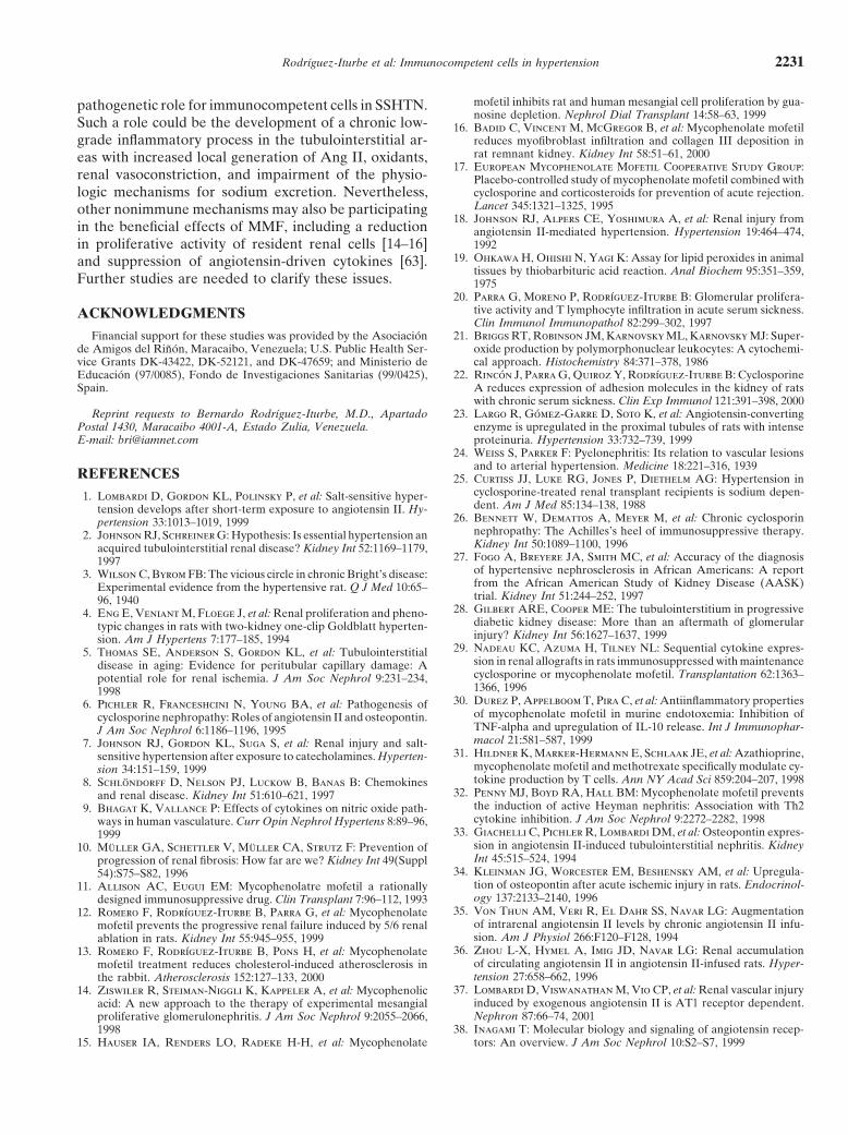
mofetil inhibits rat and human mesangial cell proliferation by gua-pathogenetic role for immunocompetent cells in SSHTN.nosine depletion. Nephrol Dial Transplant 14:58–63, 1999
Such a role could be the development of a chronic low- 16. Badid C, Vincent M, McGregor B, et al: Mycophenolate mofetilreduces myofibroblast infiltration and collagen III deposition ingrade inflammatory process in the tubulointerstitial ar-rat remnant kidney. Kidney Int 58:51–61, 2000eas with increased local generation of Ang II, oxidants,
17. European Mycophenolate Mofetil Cooperative Study Group:renal vasoconstriction, and impairment of the physio- Placebo-controlled study of mycophenolate mofetil combined with
cyclosporine and corticosteroids for prevention of acute rejection.logic mechanisms for sodium excretion. Nevertheless,Lancet 345:1321–1325, 1995other nonimmune mechanisms may also be participating
18. Johnson RJ, Alpers CE, Yoshimura A, et al: Renal injury fromin the beneficial effects of MMF, including a reduction angiotensin II-mediated hypertension. Hypertension 19:464–474,
1992in proliferative activity of resident renal cells [14–16]19. Ohkawa H, Ohishi N, Yagi K: Assay for lipid peroxides in animaland suppression of angiotensin-driven cytokines [63].
tissues by thiobarbituric acid reaction. Anal Biochem 95:351–359,Further studies are needed to clarify these issues. 1975
20. Parra G, Moreno P, Rodrıguez-Iturbe B: Glomerular prolifera-tive activity and T lymphocyte infiltration in acute serum sickness.ACKNOWLEDGMENTSClin Immunol Immunopathol 82:299–302, 1997
21. Briggs RT, Robinson JM, Karnovsky ML, Karnovsky MJ: Super-Financial support for these studies was provided by the Asociacionde Amigos del Rinon, Maracaibo, Venezuela; U.S. Public Health Ser- oxide production by polymorphonuclear leukocytes: A cytochemi-
cal approach. Histochemistry 84:371–378, 1986vice Grants DK-43422, DK-52121, and DK-47659; and Ministerio deEducacion (97/0085), Fondo de Investigaciones Sanitarias (99/0425), 22. Rincon J, Parra G, Quiroz Y, Rodrıguez-Iturbe B: Cyclosporine
A reduces expression of adhesion molecules in the kidney of ratsSpain.with chronic serum sickness. Clin Exp Immunol 121:391–398, 2000
23. Largo R, Gomez-Garre D, Soto K, et al: Angiotensin-convertingReprint requests to Bernardo Rodrıguez-Iturbe, M.D., ApartadoPostal 1430, Maracaibo 4001-A, Estado Zulia, Venezuela. enzyme is upregulated in the proximal tubules of rats with intense
proteinuria. Hypertension 33:732–739, 1999E-mail: [email protected]. Weiss S, Parker F: Pyelonephritis: Its relation to vascular lesions
and to arterial hypertension. Medicine 18:221–316, 1939REFERENCES 25. Curtiss JJ, Luke RG, Jones P, Diethelm AG: Hypertension in
cyclosporine-treated renal transplant recipients is sodium depen-1. Lombardi D, Gordon KL, Polinsky P, et al: Salt-sensitive hyper-dent. Am J Med 85:134–138, 1988tension develops after short-term exposure to angiotensin II. Hy-
26. Bennett W, Demattos A, Meyer M, et al: Chronic cyclosporinpertension 33:1013–1019, 1999nephropathy: The Achilles’s heel of immunosuppressive therapy.2. Johnson RJ, Schreiner G: Hypothesis: Is essential hypertension anKidney Int 50:1089–1100, 1996acquired tubulointerstitial renal disease? Kidney Int 52:1169–1179,
27. Fogo A, Breyere JA, Smith MC, et al: Accuracy of the diagnosis1997of hypertensive nephrosclerosis in African Americans: A report3. Wilson C, Byrom FB: The vicious circle in chronic Bright’s disease:from the African American Study of Kidney Disease (AASK)Experimental evidence from the hypertensive rat. Q J Med 10:65–trial. Kidney Int 51:244–252, 199796, 1940
28. Gilbert ARE, Cooper ME: The tubulointerstitium in progressive4. Eng E, Veniant M, Floege J, et al: Renal proliferation and pheno-diabetic kidney disease: More than an aftermath of glomerulartypic changes in rats with two-kidney one-clip Goldblatt hyperten-injury? Kidney Int 56:1627–1637, 1999sion. Am J Hypertens 7:177–185, 1994
29. Nadeau KC, Azuma H, Tilney NL: Sequential cytokine expres-5. Thomas SE, Anderson S, Gordon KL, et al: Tubulointerstitialsion in renal allografts in rats immunosuppressed with maintenancedisease in aging: Evidence for peritubular capillary damage: Acyclosporine or mycophenolate mofetil. Transplantation 62:1363–potential role for renal ischemia. J Am Soc Nephrol 9:231–234,1366, 19961998
30. Durez P, Appelboom T, Pira C, et al: Antiinflammatory properties6. Pichler R, Franceshcini N, Young BA, et al: Pathogenesis ofof mycophenolate mofetil in murine endotoxemia: Inhibition ofcyclosporine nephropathy: Roles of angiotensin II and osteopontin.TNF-alpha and upregulation of IL-10 release. Int J Immunophar-J Am Soc Nephrol 6:1186–1196, 1995macol 21:581–587, 19997. Johnson RJ, Gordon KL, Suga S, et al: Renal injury and salt-
31. Hildner K, Marker-Hermann E, Schlaak JE, et al: Azathioprine,sensitive hypertension after exposure to catecholamines. Hyperten-mycophenolate mofetil and methotrexate specifically modulate cy-sion 34:151–159, 1999tokine production by T cells. Ann NY Acad Sci 859:204–207, 19988. Schlondorff D, Nelson PJ, Luckow B, Banas B: Chemokines
32. Penny MJ, Boyd RA, Hall BM: Mycophenolate mofetil preventsand renal disease. Kidney Int 51:610–621, 1997the induction of active Heyman nephritis: Association with Th29. Bhagat K, Vallance P: Effects of cytokines on nitric oxide path-cytokine inhibition. J Am Soc Nephrol 9:2272–2282, 1998ways in human vasculature. Curr Opin Nephrol Hypertens 8:89–96,
33. Giachelli C, Pichler R, Lombardi DM, et al: Osteopontin expres-1999sion in angiotensin II-induced tubulointerstitial nephritis. Kidney10. Muller GA, Schettler V, Muller CA, Strutz F: Prevention ofInt 45:515–524, 1994progression of renal fibrosis: How far are we? Kidney Int 49(Suppl
34. Kleinman JG, Worcester EM, Beshensky AM, et al: Upregula-54):S75–S82, 1996tion of osteopontin after acute ischemic injury in rats. Endocrinol-11. Allison AC, Eugui EM: Mycophenolatre mofetil a rationallyogy 137:2133–2140, 1996designed immunosuppressive drug. Clin Transplant 7:96–112, 1993
35. Von Thun AM, Veri R, El Dahr SS, Navar LG: Augmentation12. Romero F, Rodrıguez-Iturbe B, Parra G, et al: Mycophenolateof intrarenal angiotensin II levels by chronic angiotensin II infu-mofetil prevents the progressive renal failure induced by 5/6 renalsion. Am J Physiol 266:F120–F128, 1994ablation in rats. Kidney Int 55:945–955, 1999
36. Zhou L-X, Hymel A, Imig JD, Navar LG: Renal accumulation13. Romero F, Rodrıguez-Iturbe B, Pons H, et al: Mycophenolateof circulating angiotensin II in angiotensin II-infused rats. Hyper-mofetil treatment reduces cholesterol-induced atherosclerosis intension 27:658–662, 1996the rabbit. Atherosclerosis 152:127–133, 2000
37. Lombardi D, Viswanathan M, Vio CP, et al: Renal vascular injury14. Ziswiler R, Steiman-Niggli K, Kappeler A, et al: Mycophenolicinduced by exogenous angiotensin II is AT1 receptor dependent.acid: A new approach to the therapy of experimental mesangialNephron 87:66–74, 2001proliferative glomerulonephritis. J Am Soc Nephrol 9:2055–2066,
38. Inagami T: Molecular biology and signaling of angiotensin recep-199815. Hauser IA, Renders LO, Radeke H-H, et al: Mycophenolate tors: An overview. J Am Soc Nephrol 10:S2–S7, 1999
Rodrıguez-Iturbe et al: Immunocompetent cells in hypertension2232
39. Weinstock J, Kassab J: Functional angiotensin II receptors on via membrane NADH/NADPH oxidase activation: Contributionto alterations of vasomotor tone. J Clin Invest 97:1916–1923, 1996macrophages from isolated liver granulomas of murine Schistoso-
51. Wang C-T, Chin SY, Navar GL: Impairment of pressure-natriure-miasis mansoni. J Immunol 132:2598–2601, 1984sis and renal autoregulation in ANG II-infused hypertensive rats.40. Weinstock J, Blum A: Granuloma macrophages in immune Schis-Am J Physiol 279:F325–F325, 2000tosomiasis mansoni generate components of the angiotensin sys-
52. Cheng HF, Becker BN, Burus KD, Harris RC: Angiotensin IItem. Cell Immunol 89:39–45, 1984upregulates type-1 angiotensin II receptors in renal proximal tu-41. Dezso B, Jacobsen J, Poulsen K: Evidence for the presence ofbule. J Clin Invest 95:2012–2019, 1995angiotensins in normal, unstimulated, alveolar macrophages and
53. Wang DH, Du Y, Yao A, Hu Z: Regulation of type 1 angiotensinmonocytes. J Hypertens 7:5–11, 1989II receptor and its subtype gene expression in kidney by sodium42. Silverstein E, Pertschuk L, Friedland J: Immunofluorescenceloading and angiotensin II infusion. J Hypertens 14:1409–1415, 1996localization of angiotensin converting enzyme in epithelioid and
54. Amiri F, Garcıa R: Differential regulation of renal glomerulargiant cells of sarcoidosis granulomas. Proc Natl Acad Sci USAand preglomerular vascular angiotensin II receptors. Am J Physiol76:6646–6648, 1979270:E810–E815, 199643. Rohrbach M, Conrad A: Comparison of the T lymphocyte-depen-
55. Harrison-Bernard LM, El-Dahr SS, O’Leary DF, Navar LG:dent of angiotensin-converting enzyme and leucine aminopepti-Regulation of angiotensin II type 1 receptor mRNA and proteindase in cultured human monocytes. Clin Exp Immunol 83:510–515,in angiotensin II-induced hypertension. Hypertension 33:340–346,1991199944. Ryu J, Vuk-Pavlovic Z, Rohrbach M: Monocyte heterogeneity
56. Tsutsumi K, Stromberg C, Saavedra J: Characterization of angio-in angiotensin-converting enzyme induction mediated by autolo-tensin II receptor subtypes in the rat spleen. Peptides 13:291–296,gous T lymphocytes. Clin Exp Immunol 88:288–294, 1992199245. Lazarus D, Aschoff J, Fanburg B, Lancillo J: Angiotensin
57. Ward RJ, Abiaka C, Peters TJ: Inflammation and tissue injury:converting enzyme (kininase II) mRNA production and enzymatic The world of free radicals. J Nephrol 7:89–95, 1994activity in human peripheral blood monocytes are induced by GM- 58. Swei A, Lacy F, Delano FA, Schmid-Schonbein GW: OxidativeCSF but not by other cytokines. Biochem Biophys Acta 1226:12–18, stress in the Dahl hypertensive rat. Hypertension 30:1628–1633,1994 1997
46. Costerousse O, Allegrini J, Lopez M, Alhenc-Gelas F: Angio- 59. Lacy F, O’Connor DT, Schmidt-Schonbein GW: Plasma hydro-tensin I-converting enzyme in human circulating mononuclear gen peroxide in hypertensives and normotensive subjects at geneticcells: Generic polymorphism of expression in T lymphocytes. Bio- risk of hypertension. J Hypertens 16:291–303, 1998chem J 290:33–44, 1993 60. Verhagen AMG, Rabelink TJ, Braam B, et al: Endothelin A
47. Nataraj C, Oliveiro MI, Mannon RB, et al: Angiotensin II regu- receptor blockade alleviates hypertension and renal lesions associ-lates cellular immune responses through a calcineurin-dependent ated with chronic nitric oxide synthase inhibition. J Am Soc Ne-pathway. J Clin Invest 104:1693–1701, 1999 phrol 9:755–762, 1998
48. Ruiz-Ortega M, Bustos C, Hernandez-Presa MA, et al: Angio- 61. Oliveira M, Antunes E, de Nucci G, et al: Chronic inhibitiontensin II participates in mononuclear cell recruitment in experi- of nitric oxide synthesis: A new model of arterial hypertension.mental immune complex nephritis through nuclear factor-kappa B Hypertension 20:298–303, 1992activation and monocyte chemoattractant protein-1 synthesis. 62. Abumiya T, Masuda J, Kawai J, et al: Heterogeneity in the appear-J Immunol 161:430–439, 1998 ance and distribution of macrophage subsets and their possible
49. Matsusaka T, Hymes J, Ichikawa I: Angiotensin in progressive involvement in hypertensive vascular lesions in rats. Lab Investrenal diseases: Theory and practice. J Am Soc Nephrol 7:2025–2043, 75:125–136, 19961996 63. Kranzhofer R, Browatzki M, Schmidt J, Kubler W: Angiotensin
50. Rajagopalan S, Kurz S, Munzel T, et al: Angiotensin II-mediated II activates the proinflammatory transcription factor nuclear factorkB in human monocytes. Biophys Res Commun 257:826–628, 1999hypertension in the rat increases vascular superoxide production
Copyright © 2022 FDOKUMEN