Structural analyses of the PKA RIIβ holoenzyme ... - PLOS
23
RESEARCH ARTICLE Structural analyses of the PKA RIIβ holoenzyme containing the oncogenic DnaJB1-PKAc fusion protein reveal protomer asymmetry and fusion-induced allosteric perturbations in fibrolamellar hepatocellular carcinoma Tsan-Wen Lu 1 , Phillip C. Aoto ID 2 , Jui-Hung Weng ID 2 , Cole Nielsen ID 2 , Jennifer N. Cash ID 3 , James Hall ID 2 , Ping Zhang 2¤ , Sanford M. Simon 4 , Michael A. Cianfrocco 3 *, Susan S. Taylor ID 1,2 * 1 Department of Chemistry and Biochemistry, University of California, San Diego, La Jolla, California, United States of America, 2 Department of Pharmacology, University of California, San Diego, La Jolla, California, United States of America, 3 Life Sciences Institute, Department of Biological Chemistry, University of Michigan, Ann Arbor, Michigan, United States of America, 4 Laboratory of Cellular Biophysics, The Rockefeller University, New York, New York, United States of America ¤ Current address: National Cancer Institute, National Institute of Health, Frederick, Maryland, United States of America * [email protected] (MAC); [email protected] (SST) Abstract When the J-domain of the heat shock protein DnaJB1 is fused to the catalytic (C) subunit of cAMP-dependent protein kinase (PKA), replacing exon 1, this fusion protein, J-C subunit (J- C), becomes the driver of fibrolamellar hepatocellular carcinoma (FL-HCC). Here, we use cryo-electron microscopy (cryo-EM) to characterize J-C bound to RIIβ, the major PKA regu- latory (R) subunit in liver, thus reporting the first cryo-EM structure of any PKA holoenzyme. We report several differences in both structure and dynamics that could not be captured by the conventional crystallography approaches used to obtain prior structures. Most striking is the asymmetry caused by the absence of the second cyclic nucleotide binding (CNB) domain and the J-domain in one of the RIIβ:J-C protomers. Using molecular dynamics (MD) simulations, we discovered that this asymmetry is already present in the wild-type (WT) RIIβ 2 C 2 but had been masked in the previous crystal structure. This asymmetry may link to the intrinsic allosteric regulation of all PKA holoenzymes and could also explain why most disease mutations in PKA regulatory subunits are dominant negative. The cryo-EM struc- ture, combined with small-angle X-ray scattering (SAXS), also allowed us to predict the gen- eral position of the Dimerization/Docking (D/D) domain, which is essential for localization and interacting with membrane-anchored A-Kinase-Anchoring Proteins (AKAPs). This posi- tion provides a multivalent mechanism for interaction of the RIIβ holoenzyme with mem- branes and would be perturbed in the oncogenic fusion protein. The J-domain also alters several biochemical properties of the RIIβ holoenzyme: It is easier to activate with cAMP, PLOS BIOLOGY PLOS Biology | https://doi.org/10.1371/journal.pbio.3001018 December 28, 2020 1 / 23 a1111111111 a1111111111 a1111111111 a1111111111 a1111111111 OPEN ACCESS Citation: Lu T-W, Aoto PC, Weng J-H, Nielsen C, Cash JN, Hall J, et al. (2020) Structural analyses of the PKA RIIβ holoenzyme containing the oncogenic DnaJB1-PKAc fusion protein reveal protomer asymmetry and fusion-induced allosteric perturbations in fibrolamellar hepatocellular carcinoma. PLoS Biol 18(12): e3001018. https:// doi.org/10.1371/journal.pbio.3001018 Academic Editor: Franca Fraternali, King’s College London, UNITED KINGDOM Received: May 12, 2020 Accepted: December 18, 2020 Published: December 28, 2020 Peer Review History: PLOS recognizes the benefits of transparency in the peer review process; therefore, we enable the publication of all of the content of peer review and author responses alongside final, published articles. The editorial history of this article is available here: https://doi.org/10.1371/journal.pbio.3001018 Copyright: This is an open access article, free of all copyright, and may be freely reproduced, distributed, transmitted, modified, built upon, or otherwise used by anyone for any lawful purpose. The work is made available under the Creative Commons CC0 public domain dedication.
-
Upload
khangminh22 -
Category
Documents
-
view
1 -
download
0
Transcript of Structural analyses of the PKA RIIβ holoenzyme ... - PLOS
RESEARCH ARTICLE
Structural analyses of the PKA RIIβholoenzyme containing the oncogenic
DnaJB1-PKAc fusion protein reveal protomer
asymmetry and fusion-induced allosteric
perturbations in fibrolamellar hepatocellular
carcinoma
Tsan-Wen Lu1, Phillip C. AotoID2, Jui-Hung WengID
2, Cole NielsenID2, Jennifer N. CashID
3,
James HallID2, Ping Zhang2¤, Sanford M. Simon4, Michael A. Cianfrocco3*, Susan
S. TaylorID1,2*
1 Department of Chemistry and Biochemistry, University of California, San Diego, La Jolla, California, United
States of America, 2 Department of Pharmacology, University of California, San Diego, La Jolla, California,
United States of America, 3 Life Sciences Institute, Department of Biological Chemistry, University of
Michigan, Ann Arbor, Michigan, United States of America, 4 Laboratory of Cellular Biophysics, The
Rockefeller University, New York, New York, United States of America
¤ Current address: National Cancer Institute, National Institute of Health, Frederick, Maryland, United States
of America
* [email protected] (MAC); [email protected] (SST)
Abstract
When the J-domain of the heat shock protein DnaJB1 is fused to the catalytic (C) subunit of
cAMP-dependent protein kinase (PKA), replacing exon 1, this fusion protein, J-C subunit (J-
C), becomes the driver of fibrolamellar hepatocellular carcinoma (FL-HCC). Here, we use
cryo-electron microscopy (cryo-EM) to characterize J-C bound to RIIβ, the major PKA regu-
latory (R) subunit in liver, thus reporting the first cryo-EM structure of any PKA holoenzyme.
We report several differences in both structure and dynamics that could not be captured by
the conventional crystallography approaches used to obtain prior structures. Most striking is
the asymmetry caused by the absence of the second cyclic nucleotide binding (CNB)
domain and the J-domain in one of the RIIβ:J-C protomers. Using molecular dynamics (MD)
simulations, we discovered that this asymmetry is already present in the wild-type (WT)
RIIβ2C2 but had been masked in the previous crystal structure. This asymmetry may link to
the intrinsic allosteric regulation of all PKA holoenzymes and could also explain why most
disease mutations in PKA regulatory subunits are dominant negative. The cryo-EM struc-
ture, combined with small-angle X-ray scattering (SAXS), also allowed us to predict the gen-
eral position of the Dimerization/Docking (D/D) domain, which is essential for localization
and interacting with membrane-anchored A-Kinase-Anchoring Proteins (AKAPs). This posi-
tion provides a multivalent mechanism for interaction of the RIIβ holoenzyme with mem-
branes and would be perturbed in the oncogenic fusion protein. The J-domain also alters
several biochemical properties of the RIIβ holoenzyme: It is easier to activate with cAMP,
PLOS BIOLOGY
PLOS Biology | https://doi.org/10.1371/journal.pbio.3001018 December 28, 2020 1 / 23
a1111111111
a1111111111
a1111111111
a1111111111
a1111111111
OPEN ACCESS
Citation: Lu T-W, Aoto PC, Weng J-H, Nielsen C,
Cash JN, Hall J, et al. (2020) Structural analyses of
the PKA RIIβ holoenzyme containing the oncogenic
DnaJB1-PKAc fusion protein reveal protomer
asymmetry and fusion-induced allosteric
perturbations in fibrolamellar hepatocellular
carcinoma. PLoS Biol 18(12): e3001018. https://
doi.org/10.1371/journal.pbio.3001018
Academic Editor: Franca Fraternali, King’s College
London, UNITED KINGDOM
Received: May 12, 2020
Accepted: December 18, 2020
Published: December 28, 2020
Peer Review History: PLOS recognizes the
benefits of transparency in the peer review
process; therefore, we enable the publication of
all of the content of peer review and author
responses alongside final, published articles. The
editorial history of this article is available here:
https://doi.org/10.1371/journal.pbio.3001018
Copyright: This is an open access article, free of all
copyright, and may be freely reproduced,
distributed, transmitted, modified, built upon, or
otherwise used by anyone for any lawful purpose.
The work is made available under the Creative
Commons CC0 public domain dedication.

and the cooperativity is reduced. These results provide new insights into how the finely
tuned allosteric PKA signaling network is disrupted by the oncogenic J-C subunit, ultimately
leading to the development of FL-HCC.
Introduction
Fibrolamellar hepatocellular carcinoma (FL-HCC) is a rare liver cancer that commonly occurs
in young adults with no chronic liver disease history. Recent studies identified a unique kinase
mutation, DnaJB1-PKAc (J-C), in most of patients. This mutation due to an approximately
400–kilobase pair deletion on chromosome 19 forms a chimeric transcript of the DNAJB1exon 1 fused with the PRKACA exons 2 to 10 [1]. The mutant is translated as a stable protein
where the J-domain (residues 1 to 69) of DnaJB1 is fused to the cAMP-dependent protein
kinase (PKA) catalytic (Cα or C) subunit (PKAc) (residues 15 to 336) (Fig 1A). This fusion
protein (J-C subunit) is uniquely expressed in tumor tissues, but not in adjacent normal tissues
in FL-HCC patients. Moreover, mouse models, generated using CRISPR-Cas9 gene modifica-
tion, confirmed that this chimeric protein, DnaJB1-PKAc, is oncogenic [2,3]. Structural stud-
ies of the J-C subunit (J-C) showed that its kinase core structure is nearly identical to the wild-
type (WT) PKAc but has 4 extra helices at the N-terminus adjacent to the A-helix, and both
proteins have similar biochemical properties [4].
PKA plays as a central role in cAMP-dependent signaling pathways, which regulate numer-
ous biological processes in mammalian cells [5]. The activity of PKA in cells is tightly regulated
by regulatory (R) subunits [5,6]. In its resting physiological state, PKA exists as an inactive
holoenzyme, which is composed of 2 C subunits and 1 R subunit dimer. The activity of PKA
can be stimulated by the elevated concentration of cAMP, through binding of cAMP to R sub-
units which unleashes the activity. Each of 4 functionally nonredundant R subunit isoforms
(RIα, RIβ, RIIα, and RIIβ) has distinct quaternary holoenzyme structures, cAMP sensitivity,
and cellular localization [7–11]. All 4 R subunit isoforms share a similar domain organization
which consists of an N-terminal Dimerization/Docking (D/D) domain followed by an intrinsi-
cally disordered linker that connects to 2 tandem cyclic nucleotide binding (CNB) domains.
Embedded within each linker is an inhibitor motif that resembles a peptide substrate and
docks into the active site cleft of the C subunit in the holoenzyme. RI subunits have a pseudo-
substrate inhibitor motif, while in RII subunits, this motif is a substrate that becomes phos-
phorylated. In each CNB domain, there is a conserved phosphate-binding cassette (PBC) that
directly binds cAMP. The PBC is the signature motif of the CNB domain and is critical for
cAMP activation.
Molecular dynamics (MD) simulations and nuclear magnetic resonance (NMR) data of the
chimeric J-C subunit and the holoenzyme formed with RIα revealed the unique dynamic and
flexible features of the J-domain [11,12]. Transcriptome research has also shown that the gene
expression and protein levels of the R subunits are affected in FL-HCC [13]. In normal liver
tissue, RIIβ is the predominate R subunit [14]. In tumors, however, RIα mRNA and protein
levels are up-regulated, while RIIβ mRNA and protein levels are decreased compared to nor-
mal liver tissue [13,15].
To provide mechanistic insight into the functional consequences of the DnaJB1-PKAc
fusion protein that might allow us to better understand how it is a driver of FL-HCC, we used
single-particle cryo-electron microscopy (cryo-EM) to determine a structure of the RIIβ holo-
enzyme formed with J-C. This is the first cryo-EM structure of any PKA holoenzyme and also
PLOS BIOLOGY DnaJB1-PKAc fusion and fibrolamellar hepatocellular carcinoma
PLOS Biology | https://doi.org/10.1371/journal.pbio.3001018 December 28, 2020 2 / 23
Data Availability Statement: All structure files are
available from the EMDB-PDB database (accession
numbers 6WJF and 6WJG).
Funding: The SAXS data collection was conducted
at the Advanced Light Source (ALS), a national
user facility operated by Lawrence Berkeley
National Laboratory on behalf of the Department of
Energy, Office of Basic Energy Sciences, also a
DOE Office of Science User Facility under contract
no. DE-AC02-05CH11231, through the Integrated
Diffraction Analysis Technologies (IDAT) program,
supported by DOE Office of Biological and
Environmental Research. Additional support comes
from the National Institute of Health project ALS-
ENABLE (P30 GM124169) and a High-End
Instrumentation Grant S10OD018483. The
computational work used the Extreme Science and
Engineering Discovery Environment (XSEDE)
SDSC at the Comet GPU through allocation TG-
MCB170143. This work was supported by Taiwan
MOE scholarship (T.-W.L.), Ruth L. Kirschstein
National Research Service Award NIH/NCI T32
CA009523 (P.C.A.), and NIH grants R01 GM34921
and R35 GM130389 (S.S.T.). The cryo-EM
structural analysis reported in this publication was
supported by the NIH under award number
S10OD020011. The funders had no role in study
design, data collection and analysis, decision to
publish, or preparation of the manuscript.
Competing interests: The authors have declared
that no competing interests exist.
Abbreviations: AKAP, A-Kinase-Anchoring Protein;
CNB, cyclic nucleotide binding; cryo-EM, cryo-
electron microscopy; D/D, Dimerization/Docking;
FL-HCC, fibrolamellar hepatocellular carcinoma;
GaMD, Gaussian accelerated MD; MD, molecular
dynamics; MTM, membrane-targeting motif; NMR,
nuclear magnetic resonance; PBC, phosphate-
binding cassette; PKA, cAMP-dependent protein
kinase; RMSF, root-mean-square fluctuation;
SAXS, small-angle X-ray scattering; WT, wild-type.

confirms the differences between the compact RIIβ holoenzyme and the more extended RIIαholoenzyme that was shown by negative staining EM [16]. The organization of the DnaJB1-P-
KAc RIIβ holoenzyme (RIIβ2J-C2) is similar to what was found earlier in the previously solved
crystal structure of the WT RIIβ holoenzyme (RIIβ2C2) [8]; the compact overall quaternary
organization was not altered by the addition of the J-domain, which is also resolved in our
structure. In contrast to the previous WT RIIβ holoenzyme crystal structure, where the D/D
domain was not visible, the general position of the D/D domain is seen in the cryo-EM struc-
ture and confirmed by small-angle X-ray scattering (SAXS). Both our MD simulation data and
the cryo-EM structure showed that the presence of the J-domain in this protein complex can
change the dynamic features of holoenzyme compared to the WT RIIβ holoenzyme, especially
the CNB-B domains, and both methods reveal an intrinsic asymmetry in the 2 RIIβ:J-C proto-
mers. MD simulations of the WT RIIβ holoenzyme also reveal a dramatic asymmetry in the
CNB-B domains that was hidden in the crystal structure. The asymmetry in the mutant holo-
enzyme is due to the flexibility in 1 protomer of the CNB-B domain in the RIIβ subunit and
the J-domain and the A-helix in the J-C subunit. In addition, the chimeric protein holoenzyme
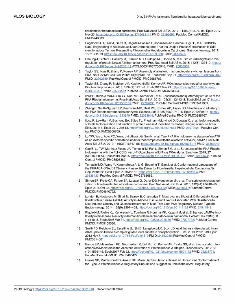
Fig 1. Cryo-EM structure of the RIIβ2J-C2 holoenzyme. (A) Domain diagram and color coding of RIIβ2J-C2 holoenzyme. (B, C)
Cryo-EM structure the RIIβ2J-C2 holoenzyme (PDB = 6WJG) at 6Å with C2 symmetry imposed. (C) The EM density of the RIIβ2J-C2
holoenzyme starts at Asp96J-C (equivalent to Asp41C in the WT C subunit) in the N-terminus of the J-C subunits and ends at
Tyr265RIIβ in the RIIβ subunit. The density for the A-helix, J-domain, and CNB-B domains are missing. (D, E) Structure of the
RIIβ2J-C2 holoenzyme structure (PDB = 6WJF) after classification reveals presence of ordered J-domain and CNB-B domain. (E) One
of the protomers of RIIβ2J-C2 holoenzyme has ordered J-domain and CNB-B domain, whereas J-domain, A-helix, and CNB-B
domain are flexible in the other protomer. CNB, cyclic nucleotide binding; cryo-EM, cryo-electron microscopy; PDB, Protein Data
Bank; WT, wild-type.
https://doi.org/10.1371/journal.pbio.3001018.g001
PLOS BIOLOGY DnaJB1-PKAc fusion and fibrolamellar hepatocellular carcinoma
PLOS Biology | https://doi.org/10.1371/journal.pbio.3001018 December 28, 2020 3 / 23

with RIIβ is activated more readily by cAMP than the WT RIIβ holoenzyme. Our studies of
the RIIβ2J-C2 structure, dynamics, and function demonstrate the power of combining crystal-
lography, MD simulations, and cryo-EM to elucidate the dynamic features of a holoenzyme
complex and also provide us with a better understanding of FL-HCC that hopefully can shed
light on new potential therapeutic strategies.
Results
Structural analysis of the DnaJB1-PKAc RIIβ holoenzyme
In the DnaJB1-PKAc chimera, the extra J-domain replaces exon 1 (residues 1 to 14) of WT C
subunit and forms 4 helices with the terminal helix being contiguous with the A-helix (Fig
1A). In order to confirm the architecture of the complex, we first performed negative stain sin-
gle-particle EM and single-particle analysis on the RIIβ2J-C2 holoenzyme (S1 Fig). Inspection
of both micrographs and 2D class averages indicated that the RIIβ2J-C2 holoenzyme possessed
a similar overall structure as the WT RIIβ holoenzyme forming a tetrameric complex with a
C2 axis of symmetry at the central hole (S1B and S1C Fig). Comparison of reprojections of the
WT RIIβ holoenzyme (PDB = 3TNP) with 2D averages further confirmed a similar architec-
ture (S1B Fig). These data allowed us to conclude that the mutant RIIβ2J-C2 holoenzyme is
similar to the WT RIIβ holoenzyme at low resolution.
To obtain higher-resolution information, we used cryo-EM to determine a structure of the
RIIβ2J-C2 holoenzyme. Due to a preferred orientation on the cryo-EM grid, data were col-
lected using a tilt angle of 40˚ (S2A and S2B Fig). After 2D classification and 3D reconstruc-
tion, we are able to get the structure to an average resolution of 6.2Å with C2 symmetry
imposed (Fig 1B and 1C, S2C and S2D Fig, and Table 1). The structures revealed clear density
in the core region of RIIβ2J-C2 holoenzyme, but density for the J-domain in the J-C subunits
as well as for the CNB-B domains in the RIIβ subunits was missing (Fig 1). In addition, the
density for the entire A-helix as well as the linker that wraps around the N-lobe that connects
the A-helix to the β-strand 1 was missing. It is flexible with a break at Asp96J-C (equivalent to
Asp41C in the WT C subunit) (Fig 1C). The density for the B/C/N-helix ends after the C-helix
in the CNB-A domain (Tyr265RIIβ), while density for the N-helix in the CNB-B domain is
missing (Fig 1C). This is the precise break between the 2 CNB domains. Presumably, these
unresolved or flexible domains are dynamic regions that form continuous states under cryo-
EM conditions.
We then further masked the core regions and focused on local areas for refinement. After
continued classification and refinement (S3A Fig), we were able to visualize 1 side of the J-
domain and CNB-B domain together with clear density for the B/C/N-helix (Fig 1D and 1E
and S3A and S3B Fig). The overall structure reveals distinct density for 1 complete RIIβ:J-C
protomer, especially the contiguous B/C/N-helix (B-helix, C-helix, and N-helix), the CNB-B
domain, and the J-domain (Fig 1E, top). In this protomer, the J-domain and CNB-B domain
are in close proximity (Fig 1E). In contrast, the J-domain and CNB-B domain as well as the A-
helix in the J-C subunit remain unresolved in the other protomer (Fig 1E, bottom). This asym-
metry could be an essential mechanistic feature of the RIIβ holoenzyme. This phenomenon
has not been observed previously and most likely cannot be trapped by conventional crystal-
lography. For example, in our previous crystal structure of the RIIβ holoenzyme, the tempera-
ture factors are high in the A-helix of the C subunits and in the CNB-B domains of RIIβsubunit (S4 Fig); however, any potential asymmetry is obscured, most likely by crystal packing
[8]. By using cryo-EM 3D reconstruction, we are able to observe this state that could represent
an important event in PKA activation.
PLOS BIOLOGY DnaJB1-PKAc fusion and fibrolamellar hepatocellular carcinoma
PLOS Biology | https://doi.org/10.1371/journal.pbio.3001018 December 28, 2020 4 / 23

Table 1. Cryo-EM data collection, refinement, and validation statistics.
Structure: RIIβ2J-C2
Data collection
Grids Gold UltrAuFoil 1.2/1.3
Vitrification method FEI Vitrobot
Microscope Titan Krios
Magnification 29,000×Voltage (kV) 300
Stage tilt (˚) 40
Detector K2 Summit
Recording mode Counting
Dose rate (e−/pix/sec) 7.789
Total electron exposure (e−/Å2) 77.9
Number of frames 100
Defocus range (μm) 1–3
Pixel size (Å) 1.0
Number of micrographs 1,129
Initial particle images (no.) 642,843
Data processing: C2 symmetry
Final particle images (no.) 69,605
Symmetry C2
Map resolution (Å) 6.2
Data processing: C1 symmetry
Final particle images (no.) 11,182
Symmetry C1
Map resolution (Å) 7.5
Refinement
Initial model used (PDB code) 3TNP, 4WB7
Symmetry C1 C2
PDB code 6WJF 6WJG
EMDB code EMD-21692 EMD-23693
Model resolution (Å)
FSC threshold
7.5
0.143
6.2
0.143
Map sharpening B factor (Å2) −500 −302
Model composition
Non-hydrogen atoms
Protein residues
Ligands
9,527
1,172
0
7,708
944
0
B factors (Å2)
Protein (min/max/mean)
Ligand
30/850/350
N/A
30/550/300
N/A
RMS deviations
Bond lengths (Å)
Bond angles (˚)
0.004
0.878
0.003
0.875
Validation
MolProbity score
Clash score
Rotamer outliers (%)
1.88
4.64
0
1.81
3.72
0
Ramachandran plot
Favored (%)
Allowed (%)
Disallowed (%)
85.65
14.35
0
85.36
14.64
0
cryo-EM, cryo-electron microscopy; EMDB, The Electron Microscopy Data Bank; FSC, fourier shell correlation;
PDB, Protein Data Bank; RMS, root-mean-square.
https://doi.org/10.1371/journal.pbio.3001018.t001
PLOS BIOLOGY DnaJB1-PKAc fusion and fibrolamellar hepatocellular carcinoma
PLOS Biology | https://doi.org/10.1371/journal.pbio.3001018 December 28, 2020 5 / 23

Molecular dynamics simulations reveal asymmetry in the RIIβholoenzymes
To further explore the dynamic features of the 2 RIIβ holoenzymes, we used MD simulations
and found surprisingly that RIIβ holoenzymes formed with WT C subunit and the J-C subunit
have very different domain dynamics. Three independent 500-ns MD simulations were carried
out to further explore these differences. The root-mean-square fluctuation (RMSF) analyses of
each holoenzyme demonstrated distinct backbone dynamics between WT and fusion holoen-
zymes. The first 14 residues of the C subunit, which are typically disordered in our crystal
structures and are replaced by the J-domain in the J-C fusion, are not included in these simula-
tions; this construct is referred to as C(Δ1–14). The last 23 residues of the RIIβ subunit as well
as the linker region and the D/D domain (residues 1 to 103) are also not included.
Wild-type RIIβ holoenzyme. In the simulations of the RIIβ2C(Δ1–14)2 holoenzyme, both
C subunits are stable and show low RMSF throughout the entire simulation (Fig 2A) as do
both CNB-A domains in the RIIβ subunit. In contrast, one of the CNB-B domains in the RIIβsubunit is highly flexible (Fig 2B). The flexible region begins approximately at residue Tyr265-RIIβ, which is at the junction of the C-helix in the CNB-A domain and the N-helix in the
CNB-B domain (Fig 2C and 2D). Besides the highly dynamic features of the CNB-B domains,
some local regions, such as the N-terminal linker and the β4-β5 loops in the CNB-B domains,
also showed especially high RMSF. In addition, 1 small dynamic region (residues 122 to 129)
was also seen in the linker that joins the inhibitor sequence to CNB-A (Fig 2B); this dynamic
segment wraps around the B/C/N-helix. These regions, as well as the carboxyl terminus 23 res-
idues, are both unresolved in the previous RIIβ2C2 crystal structure [8]. A deeper analysis of
our simulation results further confirmed the dynamic properties of these 2 regions. By overlay-
ing the conformational ensemble from the simulations, we can more clearly appreciate the dis-
tinct dynamic properties of the 2 CNB domains in the RIIβ2C2 holoenzyme (Fig 2E). One of
the protomers reveals a relatively stable CNB-B domain, while the other CNB-B domain is
highly flexible. In both protomers, Tyr265RIIβ serves as a pivot point; however, in one of the
protomers, the entire CNB-B domain, including the N-helix, is extremely flexible after Tyr265-RIIβ. The MD simulation data further confirm the dynamic regions that we observed in the
cryo-EM structure both being characterized by breaks at Tyr265RIIβ.
Tyr265RIIβ and the dynamic properties of the B/C/N-helix are important for RIIβ holoen-
zyme activation. The long B/C/N-helix is the signature feature of all PKA holoenzymes. Once
activated by cAMP, the B/C/N-helix divides into 3 segments (B-helix, C-helix, and N-helix)
(Fig 2F and 2G). Several studies have already pointed out the importance of the flexibility of
the B/C/N-helix in activation of the RIα holoenzyme [10,17,18]. Here, we show that the B/C/
N-helix is also very dynamic, but different, in the RIIβ holoenzyme. Each isoform has the same
hinge points, one is between the B-helix and the C-helix and the other is between the C-helix
and the N-helix. Most importantly, however, the major hinge points are different (S5A Fig).
The major hinge point in RIIβ, Tyr265RIIβ, is located at the junction of the C-helix and the N-
helix (Fig 2F), while Leu233RIα, between the B- and the C-helix, is the more prominent pivot
point for RIα (Fig 2G). To quantitate these differences, we measured the hinge angles of B-
helix/C-helix and C-helix/N-helix in the cAMP-bound form structures of both RIα and RIIβ(PDB = 1RGS and 1CX4, respectively). The angle between the B- and C-helix is larger in RIαthan in RIIβ; however, RIIβ has larger hinge angle between the C- and N-helix (Fig 2F and
2G), which is the precise junction between the 2 CNB domains.
In addition to differences in their dynamics, the B/C/N helix in RIα and RIIβ, which share
53% sequence identity, also have distinct helical, N-capping, and C-capping propensities
(S5B–S5D Fig). The B/C/N-helix of RIα has very high helical propensity with a local minimum
PLOS BIOLOGY DnaJB1-PKAc fusion and fibrolamellar hepatocellular carcinoma
PLOS Biology | https://doi.org/10.1371/journal.pbio.3001018 December 28, 2020 6 / 23

at Gly235RIα, which is near the primary hinge point (Leu233RIα) for the cAMP-bound RIα[10]. In RIIβ, the helical propensity of the B/C/N-helix is much lower compared to RIα, per-
haps allowing it to take advantage of the natural hinge point between the 2 CNB domains,
Tyr265RIIβ (Fig 2E and S5B Fig).
RIIβ2J-C2 holoenzyme. The RIIβ2J-C2 holoenzyme simulations showed different dynam-
ics compared to WT holoenzyme. Both the N- and C-lobes of J-C subunit are as stable as WT;
however, the extra J-domain in the J-C subunits shows much higher RMSF values than the
kinase portion of the fusion protein (Fig 3A). The addition of the J-domain in the J-C subunit
also has an effect on the dynamics of the RIIβ subunit. Unlike the CNB-B domains in the
RIIβ2C2 holoenzyme, both CNB-B domains in RIIβ2J-C2 holoenzyme remain strikingly more
stable during the simulation (Fig 3B and S6 Fig). The J-domain presumably can stabilize the
CNB-B domains either through spatial steric effects or direct interactions that are driven by
the close proximity. This close communication between the J-domain and the CNB-B domain
is captured in one of the protomers in our cryo-EM structure (Figs 1E and 3C). The close
Fig 2. MD simulations of the RIIβ2C2 holoenzyme reveal functional and isoform-specific dynamics. (A) Both N- and C-lobe of the
C subunit remain stable in the RMSF analysis of RIIβ2C2 holoenzyme. (B) CNB-B domain of the RIIβ subunit in one of the protomers
is flexible in the RMSF analysis in RIIβ2C2 holoenzyme. (C) Domain diagram and residues of the B/C/N-helix in RIα and RIIβ subunit.
(D) RIIβ subunit in the RIIβ2C2 holoenzyme crystal structure (PDB ID = 3TNP). (E) The overlaid of all states of each RIIβ subunit
protomer in the RIIβ2C2 holoenzyme from MD simulations. CNB domain in one of the protomers is more flexible than the other, and
both of the protomers have breakages at Tyr265. Residue Tyr265RIIβ was shown as pink ball. (F, G)The conformational change of RIIβ(F) and RIα (G) B/C/N-helices upon cAMP stimulation. The main pivot point of RIIβ B/C/N-helix is at Tyr265RIIβ (G), while the main
pivot point of RIα B/C/N-helix is at Leu233RIα (G). The data used to make these figures can be found in S1 Data. CNB, cyclic
nucleotide binding; MD, molecular dynamics; PDB, Protein Data Bank; RMSF, root-mean-square fluctuation.
https://doi.org/10.1371/journal.pbio.3001018.g002
PLOS BIOLOGY DnaJB1-PKAc fusion and fibrolamellar hepatocellular carcinoma
PLOS Biology | https://doi.org/10.1371/journal.pbio.3001018 December 28, 2020 7 / 23

proximity of the J-domain and the CNB-B domain suggests that their dynamic properties are
related; clearly, the CNB-B domain can sense the presence of the J-domain. The other feature
that is revealed by the cryo-EM structure is that the A-helix in 1 J-C subunit is also missing,
suggesting that the J-domain influences not only the CNB-B domain but also the A-helix that
it is directly fused to β-strand 1 in the N-lobe (Fig 3C).
We can also visualize the differences in dynamics of the CNB domains in the DnaJB1-PKAc
and WT RIIβ holoenzymes by measuring the polar vector between CNB-A and CNB-B
domains. The vector was chosen from Arg230RIIβ in the PBC of CNB-A domain to Arg359RIIβ
in the PBC of CNB-B domain (Fig 3D). The plots of φ, θ, and length of the vectors demon-
strated clearly the different dynamics between the 2 CNB domains in the DnaJB1-PKAc and in
WT PKAc RIIβ holoenzymes. In the RIIβ2C2 holoenzyme, the vector in one of the protomers
moves and fluctuates significantly; the distance between domains varies from 50 to 70Å with a
wide range of φ and θ angles movements (Fig 3E). The vector in the other protomer, however,
is relatively less dynamic in both the φ and/or θ axis and length. The asymmetric dynamics of
the CNB domains in each protomer of the RIIβ2C2 holoenzyme can also be observed here. In
contrast, the vectors in both protomers of RIIβ2J-C2 holoenzyme remain relatively more stable.
The plot of φ, θ, and length of the vectors in RIIβ2J-C2 holoenzyme showed less disperse angles
Fig 3. MD simulations of the RIIβ2J-C2 holoenzyme reveal distinct dynamics. (A) Both N- and C-lobe of J-C subunit remain stable
but J-domain is flexible in the RMSF analysis of the RIIβ2J-C2 holoenzyme. (B) CNB-B domain of the RIIβ subunit in both protomers
remain stable in the RMSF analysis in RIIβ2C2 holoenzyme. (C) J-domain and CNB-B domain of the RIIβ2J-C2 holoenzyme are in close
proximity. (D) The representation of polar coordinate vector from Arg230RIIβ to Arg359RIIβ. (E, F) The plots of φ, θ, and length of the
vectors from Arg230RIIβ to Arg359RIIβ in MD simulations of RIIβ2C2 (E) and RIIβ2J-C2 holoenzymes (F). The vector in one of the
protomers of the RIIβ2C2 holoenzyme moves and fluctuates significantly (E), whereas the vectors in both protomers of the RIIβ2J-C2
holoenzyme remain stable (F). The data used to make these figures can be found in S1 Data. CNB, cyclic nucleotide binding; MD,
molecular dynamics; RMSF, root-mean-square fluctuation.
https://doi.org/10.1371/journal.pbio.3001018.g003
PLOS BIOLOGY DnaJB1-PKAc fusion and fibrolamellar hepatocellular carcinoma
PLOS Biology | https://doi.org/10.1371/journal.pbio.3001018 December 28, 2020 8 / 23

and lengths movements (Fig 3F), and both vectors in the protomers populate a smaller φ, θ-
space with less diverse length fluctuations. Asymmetric dynamics is also present in the
RIIβ2J-C2 holoenzyme (Fig 3F). Together, this implies that the length of the N-terminus of the
C subunit, whether longer as in the J-C subunit or shorter as in the C subunit used here, may
have a differential impact on the observed asymmetric dynamics.
SAXS analysis and localization of the D/D domain
The solution structures of both WT and DnaJB1-PKAc RIIβ holoenzymes were also deter-
mined by SAXS coupled with size-exclusion chromatography to support our cryo-EM struc-
ture. Based on pair distance distribution functions P(r), both holoenzymes have similar
dimension (Dmax = 129.09Å for WT holoenzyme and Dmax = 128.63Å for DnaJB1-PKAc holo-
enzyme). The Dmax values are consistent with our cryo-EM structure, where the 3D organiza-
tion of RIIβ2C2 and RIIβ2J-C2 holoenzyme are similar (Fig 4A and 4B and Table 2). However,
the DnaJB1-PKAc holoenzyme has a larger Rg value than WT holoenzyme (Rg = 41.69Å for
Fig 4. SAXS analyses of RIIβ2C2 and RIIβ2J-C2 holoenzymes. (A, B) P(r) functions of RIIβ2C2 (A) and RIIβ2J-C2 (B) holoenzymes. (C, D) Scattering plots, atomic
structure, and ab initio models fittings of RIIβ2C2 holoenzyme (C) with χ2 = 1.2591 (atomic structure) and χ2 = 1.300 (ab initio) and RIIβ2J-C2 holoenzyme (D) with χ2 =
1.2900 (atomic structure) and χ2 = 1.152 (ab initio). (E, F) The SAXS atomic structure models of RIIβ2C2 (E) and RIIβ2J-C2 holoenzymes (F) overlay with ab initio
envelopes. The data used to make these figures can be found in S1 Data. CNB, cyclic nucleotide binding; SAXS, small-angle X-ray scattering.
https://doi.org/10.1371/journal.pbio.3001018.g004
Table 2. Rg and Dmax values of RIIβ2C2 and RIIβ2J-C2 holoenzymes from SAXS.
RIIβ2C2 RIIβ2J-C2
Rg (Å) from P(r) 41.69 ± 0.28 43.13 ± 0.41
Dmax (Å) from P(r) 129.09 128.63
Porod volume (Å3) from P(r) 290,000 347,000
Rg (Å) from Guinier 41.41 ± 0.49 43.29 ± 0.61
MW estimation� (kDa) 171 204
Theoretical MW (kDa) 173 187
�MW estimation was obtained by Porod volume/1.7.
MW, molecular weight; SAXS, small-angle X-ray scattering.
https://doi.org/10.1371/journal.pbio.3001018.t002
PLOS BIOLOGY DnaJB1-PKAc fusion and fibrolamellar hepatocellular carcinoma
PLOS Biology | https://doi.org/10.1371/journal.pbio.3001018 December 28, 2020 9 / 23

WT holoenzyme and Rg = 43.13Å for DnaJB1-PKAc holoenzyme). A similar trend of Rg values
can also be obtained from Guinier analyses (Rg = 41.41Å and Rg = 43.29Å for WT and
DnaJB1-PKAc holoenzyme, respectively) (S7A and S7B Fig and Table 2). The higher molecu-
lar weight of DnaJB1-PKAc can explain why RIIβ2J-C2 holoenzyme has a larger radius of
gyration.
The higher molecular weight and the presence of the J-domain were also reflected in the
larger Porod volumes of DnaJB1-PKAc holoenzyme (290,000Å3 for RIIβ2C2 versus 347000Å3
for RIIβ2J-C2) (Table 2). The molecular weight can be estimated by dividing the Porod volume
by 1.7 according to the method of Petoukhov and colleagues [19]. The estimated molecular
weight of RIIβ2C2 was obtained as 171 kDa in comparison to its theoretical molecular weight
of 173 kDa (Table 2). The estimated molecular weight of the RIIβ2J-C2 holoenzyme was 204
kDa, while its theoretical molecular weight is 186 kDa (Table 2). Considering that the flexible
J-domain enhances the domain dynamics of the holoenzyme, the result is that it delocalizes
over a larger volume [20]. This can explain why RIIβ2J-C2 holoenzyme has a larger deviation
between estimated and theoretical molecular weights than WT holoenzyme.
We further analyze Kratky plots of these 2 holoenzymes, which can provide a way to assess
the degree of flexibility within the scattering macromolecules. Kratky plot analyses of these 2
holoenzymes showed bell-shape peaks at low q; however, neither of them converges to the q-
axis at high q (S7C and S7D Fig), indicating that both of the complexes are multi-domains pro-
teins with flexible regions [20]. These results are consistent with our structures and MD simu-
lations where we identified several dynamic/flexible domains as well as a flexible linker.
To fit the SAXS data, we used the WT crystal structure as a starting model and further con-
sidered the dynamic properties of each domain. The missing linkers were built as flexible poly-
Gly chains, while the missing D/D domain was generated using homology models from an
online protein structure prediction software program, I-TASSER [21]. Both of our holoenzyme
models fit the experimental SAXS data well (χ2 = 1.2591 and χ2 = 1.2900 for WT and J-C holo-
enzyme, respectively) (Fig 4C and 4D and S7E and S7F Fig). In both of the holoenzyme struc-
tural models, the D/D domain of RIIβ is localized on the same face as the CNB-B domains and
the myristylation (Myr) sites of the C subunit (Fig 4E and 4F).
The ab initio models of WT and fusion holoenzymes were generated from the SAXS scatter-
ing data using the program DAMMIN [22], and these 2 models reveal good quality of fitting to
the experimental data with χ2 score 1.300 and 1.152 for WT and J-C holoenzyme, respectively.
Both atomic structure and ab initio approaches show complementary results that have similar
3D shapes (Fig 4C and 4D). Besides the 3D shapes, the dynamic features of the holoenzymes
were also observed from the ab initio models. Consistent with our cryo-EM structure and MD
simulation data, the CNB-B domains in the WT holoenzyme ab initio model are not fully visi-
ble suggesting that they are flexible, whereas the CNB-B domains remain clear in the J-C holo-
enzyme ab initio model as they are stabilized by the J-domains. Similar to the SAXS atomic
model, the WT holoenzyme ab initio model also reveals the general position of the D/D
domain which tethers at the same face as the CNB-B domains. The fact that the D/D domain is
missing in the J-C holoenzyme SAXS envelope suggests that the J-domain may also influence
D/D domain dynamics.
Although the position of the D/D domain cannot be identified unambiguously in our cryo-
EM structure due to its flexibility, the extra density in our structure nevertheless provides fur-
ther supporting evidence for the general position of the D/D domain (S8A and S8B Fig). The
first visible N-terminal residue in our RIIβ structure is Ile104RIIβ, which is located at the hole
formed by the 2 RIIβ:J-C protomers (S8A Fig). The extra density locates near the center of the
RIIβ holoenzyme, close to Ile104RIIβ and extends along the central hole to the same surface
where the CNB-B domains and J-domains are located (S8B Fig).
PLOS BIOLOGY DnaJB1-PKAc fusion and fibrolamellar hepatocellular carcinoma
PLOS Biology | https://doi.org/10.1371/journal.pbio.3001018 December 28, 2020 10 / 23

Altered biochemical function of the DnaJB1-PKAc RIIβ holoenzyme
We next asked whether the fusion protein affects not only RIIβ holoenzyme dynamics but also
its biochemical properties. Both our MD simulations and our cryo-EM structure suggest that 1
J-domain communicates with its adjacent CNB-B domain and interferes with the overall
dynamic properties of the holoenzyme (Fig 2B, 2F and 2G), In addition, several studies have
shown that the dynamic features of the CNB domains play a significant role in the allosteric
activation of the RIα holoenzyme by cAMP [23–27] and pathogenic mutations in RIα cluster
in the 2 CNB domains [28]. We thus compared cAMP activation of the holoenzyme formed
with J-C to holoenzyme formed with WT C subunit.
Holoenzyme formed with the fusion protein was easier to activate than the WT holoenzyme
(EC50 = 285nM for C subunit versus EC50 = 170nM for J-C subunit) (Fig 5C and Table 3). To
investigate the importance of the J-domain in the RIIβ holoenzyme activation by cAMP versus
the absence of the first 14 residues (exon 1), we engineered and expressed several deletion con-
structs (Fig 5A and 5B and S9 Fig). Each of the 4 extra helices in the J-domain was deleted
sequentially, generating J-C(Δ1–13), J-C(Δ1–38), J-C(Δ1–54), and C(Δ1–14) deletion mutants.
C(Δ1–14) is equivalent to J-C(Δ1–69) where the whole J-domain is deleted. All of these mutants
expressed as stable proteins and formed holoenzymes with RIIβ. The deletion mutants were all
easier to activate with cAMP, and all showed lower Hill coefficients compared to the WT RIIβholoenzyme (Fig 5C and Table 3). The mutant that simply lacked the first exon, C(Δ1–14),
which is the same as J-C(Δ1–69), also showed a reduced EC50, indicating that the presence of
the first exon (residues 1 to 14) is crucial for proper activation and allosteric regulation of the
RIIβ holoenzyme. Moreover, the Hill coefficient for cAMP activation actually showed negative
cooperativity once exon 1 was removed (Table 3). Deletion of the first 14 residues in the PKA C
subunit as well as the addition of the J-domain not only changed CNB-B domain dynamics but
also affected the finely tuned allosteric activation of the RIIβ holoenzyme by cAMP.
To investigate how exon 1 of the C subunit influence the asymmetric dynamics and cAMP
activation of the RIIβ holoenzyme, Gaussian accelerated MD (GaMD) simulations on RIIβ2C2,
RIIβ2J-C2, and RIIβ2C(Δ1–14)2 were carried out. Specifically, we ask whether it is the J-domain
or the absence of the residues 1 to 14 that account for the differences. Using the radius of
cAMP as a probe, we calculated the solvent-accessible surface area of the PBC pocket of the
CNB-B domains in each of the complexes. Both RIIβ2J-C2, and RIIβ2C(Δ1–14)2 complexes
have higher accessible surface area than the WT complex (Fig 5D), suggesting that it is easier
for cAMP to dock into the PBC pockets, which will facilitate activation. To further analyze the
cAMP accessibility in the PBC pocket, we measured the distance between the Cβ atom in
Ala360RIIβ and the Cγ atom in Leu351RIIβ (Fig 5E). This distance in the RIIβ holoenzyme is
6.9Å, while the PBC pocket is more closed (4.9Å) once it binds cAMP (Fig 5E). We then calcu-
lated the distance populations in our simulations. Consistent with our solvent-accessible sur-
face analysis, both the RIIβ2J-C2 and RIIβ2C(Δ1–14)2 complexes reveal more open states than
the WT holoenzyme (Fig 5F). In both analyses, there is a noticeable asymmetry between proto-
mers in the cAMP accessibility of the PBC pockets for the RIIβ2C(Δ1–14)2 and RIIβ2J-C2 holo-
enzymes, supporting our earlier observations of asymmetry. The full-length WT holoenzyme
exhibits milder asymmetry, consistent with our hypothesis that the protomers are tightly cou-
pled (Fig 5D and 5F and S10 Fig). These results may help to explain why the J-C holoenzyme
formed with RIIβ, as well as the 1–14 deletion mutant of WT C, is easier to activate with
cAMP, while also having reduced cooperativity as measured by the Hill coefficient, compared
to the WT RIIβ holoenzyme.
To further explore the effect of exon 1 deletion versus the J-domain fusion on the A-helix
stability, we analyzed the helical propensity of the A-helix in 3 different constructs, WT C-,
PLOS BIOLOGY DnaJB1-PKAc fusion and fibrolamellar hepatocellular carcinoma
PLOS Biology | https://doi.org/10.1371/journal.pbio.3001018 December 28, 2020 11 / 23

Fig 5. The RIIβ2J-C2 holoenzyme is easier to activate with cAMP than the RIIβ2C2 holoenzyme. (A) Structure of
J-C subunit (left) and C subunit (right). The junctions of 4 helices in J-domain are labeled as orange arrows, where the
first exon junction is labeled as blue arrow. (B) Coomassie blue staining SDS-PAGE of purified C-, J-C-, J-C(Δ1–13)-,
J-C(Δ1–38)-, J-C(Δ1–54)-, and C(Δ1–14) subunits. C(Δ1–14) subunit is equivalent to J-C(Δ1–69) subunit, whole J-
domain deletion. (C) The fusion protein RIIβ2J-C2 holoenzyme and its deletion mutants were easier to activate with
cAMP than the WT RIIβ2C2 holoenzyme. n = 2 biological replicate. All error bars represent SEM. (D) The CNB-B
domains in RIIβ2J-C2 and RIIβ2C(Δ1–14)2 holoenzymes have larger cAMP accessible surface area than RIIβ2C2
holoenzyme. (E) The distance between Cβ atom in Ala360RIIβ and Cγ atom in Leu351RIIβ in the CNB-B domain of
cAMP-bound (left, PDB = 1CX4) and holoenzyme (right, PDB = 3TNP) forms. (F) The distance population
probability of the PBC pocket in RIIβ2C2, RIIβ2J-C2, and RIIβ2C(Δ1–14)2 holoenzymes. The CNB-B domains in the
RIIβ2J-C2 and RIIβ2C(Δ1–14)2 holoenzymes are more prone to open than the RIIβ2C2 holoenzyme. Both of the
RIIβ2J-C2 and RIIβ2C(Δ1–14)2 holoenzymes reveal intrinsic asymmetry in the CNB-B domains. The data used to make
these figures can be found in S1 Data. CNB, cyclic nucleotide binding; PDB, Protein Data Bank; SEM, standard error
of the mean; WT, wild-type.
https://doi.org/10.1371/journal.pbio.3001018.g005
Table 3. EC50 and Hill coefficient of RIIβ2C2, RIIβ2J-C2 RIIβ2J-C(Δ1–13)2, RIIβ2J-C(Δ1–38)2, RIIβ2J-C(Δ1–54)2, and RIIβ2C(Δ1–14)2 holoenzymes from cAMP
activation curves.
C J-C J-C(Δ1–13) J-C(Δ1–39) J-C(Δ1–55) C(Δ1–14)
EC50 (nM) 285 ± 54 170 ± 32 144 ± 22 177 ± 31 141 ± 32 166 ± 38
HillSlope 0.949 ± 0.155 0.742 ± 0.091 0.749 ± 0.077 0.690 ± 0.074 0.644 ± 0.085 0.744 ± 0.119
https://doi.org/10.1371/journal.pbio.3001018.t003
PLOS BIOLOGY DnaJB1-PKAc fusion and fibrolamellar hepatocellular carcinoma
PLOS Biology | https://doi.org/10.1371/journal.pbio.3001018 December 28, 2020 12 / 23

J-C-, and C(Δ1–14) subunits (S11A Fig). The A-helix in the WT C subunit, which is part of the
N-terminal linker that flanks the N- and C-lobes of the kinase core and is flanked by intrinsi-
cally disorder regions, has a surprisingly high helical propensity. Either deleting exon 1 or fus-
ing with the J-domain decreases the predicted helical propensity (S11A Fig). Moreover, the
last residue in the exon 1, Ser14C, serves as a significant N-capping residue (S11B Fig).
Decreasing the A-helix stability and missing the N-capping residue, as demonstrated in our
computational and biochemical data, can influence the function and allosteric properties of
the entire RIIβ holoenzyme.
Discussion
We describe here a structure of the RIIβ2J-C2 holoenzyme solved by cryo-EM single-particle
3D reconstruction. Although the overall compact 3D organization of the 2 RIIβ:J-C protomers
in the holoenzyme structure is similar to the previous WT RIIβ holoenzyme crystal structure,
there are several differences in both the structure and dynamics in the DnaJB1-PKAc RIIβholoenzyme, which could result in the mis-regulation of PKA signaling. With cryo-EM, we are
now able to observe more details about the dynamic features of the RIIβ holoenzyme that
could not be captured by conventional X-ray crystallography including the general localization
of the D/D domain and the asymmetry of the CNB-B domains. Our structure together with
MD simulations and biochemical studies not only allows us to better understand how
DnaJB1-PKAc disrupts RIIβ holoenzyme function and dynamics, but also makes us appreciate
for the first time the intrinsic asymmetry of the RIIβ holoenzyme that is likely an inherent fea-
ture of the activation mechanism that was masked in the earlier crystal structure. Our cryo-
EM structure also confirms that the compact RIIβ holoenzyme is distinct from the extended
RIIα holoenzyme.
Structure and dynamics of WT and DnaJB1-PKAc RIIβ holoenzymes
The cryo-EM structure and MD simulation data both reveal new and previously unappreciated
features of the RIIβ holoenzyme. First is the asymmetry of the RIIβ holoenzyme, which is seen
with MD simulations in the WT holoenzyme and captured in the cryo-EM structure but hid-
den in the previous crystal structure. With MD simulations, we observed an intrinsic asymme-
try in the WT RIIβ holoenzyme where 1 protomer has a more flexible CNB-B domain than the
other. This same asymmetry is observed in the cryo-EM structure formed with the DnaJB1
fusion C subunit. The collective results suggest that the activation process most likely initiates
from 1 protomer and then passes to the other. Does it then eventually spread to the entire
holoenzyme, or does the intrinsic asymmetry simply involve toggling between the 2 protomers
without resulting in full dissociation of the C subunits? Is the asymmetric feature an intrinsic
part of the activation mechanism? How does the activation signal pass from 1 protomer to the
other? These are the major future challenges. The cryo-environment and particle orientation
problems of cryo-EM single-particle technique may limit us to visualize the full picture of pro-
tein dynamics; however, it still allowed us to capture an important conformational state of this
holoenzyme. The beauty of single-particle cryo-EM, unlike crystallography, is that one can
capture an ensemble of conformational states in a single experiment [29]; we anticipate that
other variations on the compact globular conformation described here will be embedded in
some of the other particles.
Second is the cross communication between the CNB-B domain with the J-Domain, which
is revealed in the cryo-EM structure. As seen in previous structures, the J-domain is very flexi-
ble [11, 2]; however, in the RIIβ holoenzyme, we see that the flexibility of the J-domain also
appear to influence the dynamic features of its adjacent CNB-B domain as well as the A-helix
PLOS BIOLOGY DnaJB1-PKAc fusion and fibrolamellar hepatocellular carcinoma
PLOS Biology | https://doi.org/10.1371/journal.pbio.3001018 December 28, 2020 13 / 23

that it is fused to. The J-domain can also further influence the dynamic features of the CNB-B
domains in the RIIβ holoenzyme, which we did not see in the RIα holoenzyme. Several studies
have demonstrated that the dynamic features of the CNB domains in RIα are essential for
PKA activation and allostery [25,26,30,31]. Here, for the first time, we are able to show that the
RIIβ holoenzyme also has flexible CNB-B domains, although it is different from RIα in terms
of its structure and allosteric regulation [32–34].
In summary, we show here that J-domain can influence the dynamic properties and the
symmetry of the CNB-B domains. Our MD simulation data clearly indicated that the J-domain
can stabilize the CNB-B domains, while the cryo-EM structure revealed how the J-domain and
the CNB-B domain interact and together control the A-helix. We believe that this effect can be
significant for stability, activation, cAMP binding, and/or interaction with other binding
partners.
D/D domain, myristylation site, and AKAP binding
Our cryo-EM model of the RIIβ holoenzyme, including the general position of the D/D
domain, is consistent with our earlier crystal structure and SAXS data as well as with recent
cross-linking data, and confirms that the compact RIIβ holoenzyme is quite different from the
RIIα holoenzyme. The general positioning of the D/D domain, based on the low resolution
cryo-EM density and SAXS, is consistent with the previous hypothesis that the unresolved N-
terminal regions of RIIβ thread through the central hole of the RIIβ holoenzyme positioning
the D/D domain close to the lower surface of RIIβ holoenzyme as seen in Fig 4 in close prox-
imity to the CNB-B domains, the J-domain, and the bottom surface of the C-lobe in the kinase
domain.
The D/D domain is essential for PKA localization. PKA holoenzymes, especially RII holo-
enzymes, typically bind with high affinity to an amphipathic helix that is embedded in scaffold
proteins referred to as A-Kinase-Anchoring Proteins (AKAPs) through their D/D domains
[35–37]. In this way, AKAPs bring the PKA holoenzyme to specific membrane locations in the
cell where it is in close proximity to dedicated substrates such as receptors, transporters, and
ion channels [5,38], and several lines of evidence have demonstrated that disrupting this inter-
action can affect not only PKA localization but also substrate specificity [38–43].
Our results suggest that the linker joining the D/D domain and the inhibitor site in RIIβweaves through the hole that is created by the 2 RIIβ:J-C protomers. This placement of the D/
D domain is consistent with cross-linking studies and different from the RIIα holoenzyme
which was shown by negative staining EM to be extended [16,44]. It is also consistent with our
earlier linker swap SAXS experiments showing that the motif that is responsible for the com-
pact structure of the RIIβ holoenzyme is embedded in the linker [45]. This model places the
D/D domain and the myristylation sites attached to the N-termini of the C subunits on the
same general surface, which allows them to form multivalent interactions with membranes
(Fig 6A). In solution, the myristyl groups in the RIIβ holoenzyme are solvent exposed and can
anchor to nanodiscs even in the absence of AKAPs [42,46], whereas the myristyl groups in the
RIα holoenzyme are embedded in the acyl pocket in the C-lobe of the C subunit and do not
contribute to membrane anchoring in the absence of an AKAP [46]. In contrast to RIα holoen-
zymes, most of the RII subunits are always localized close to membranes [5]. Therefore, having
the D/D domain in the RIIβ holoenzyme localized on the same face as the myristylation sites
provides a dual mechanism for forming multivalent interactions with the membrane. It is also
likely that anchoring the N-terminus to membranes could influence the conformation of the
missing 14 residues. This segment with its basic residues followed by a phosphorylation site is
in fact a classic myristylation motif, similar to the Src kinase, where basic residues also
PLOS BIOLOGY DnaJB1-PKAc fusion and fibrolamellar hepatocellular carcinoma
PLOS Biology | https://doi.org/10.1371/journal.pbio.3001018 December 28, 2020 14 / 23

contribute to membrane anchoring [47]. As we show here, ordering the first 14 residues in the
membrane-bound holoenzyme could easily affect the dynamic behavior of the entire WT holo-
enzyme. The hydrophobic carboxyl terminus of the RIIβ subunit, as well as its dynamic β4-β5
loop in the CNB-B domain, could also become ordered when the holoenzyme is anchored to
membranes. The highly basic β4-β5 loop in RIIβ is, in particular, in close proximity to the acidic
surface of the membrane (Fig 6A). In addition, AKAPs themselves typically have a membrane-
targeting motif (MTM), so many mechanisms could be used to stabilize interactions with mem-
branes (Fig 6A). In the J-C fusion protein, the myristylation site is lost which could also affect,
although not necessarily abolish, RIIβ holoenzyme binding to membranes in cells (Fig 6A).
Two helices drive the allosteric regulation of the PKA holoenzymes
There are 2 helices that drive the allosteric regulation of PKA, the B/C/N-helix in the R sub-
units and the A-helix in the C subunit (Fig 6B). Sequences difference in the B/C/N-helix, as
Fig 6. The structural, dynamic, and allosteric features of RIIβ2J-C2 holoenzymes. (A) The J-domain disrupts the
multivalent interaction, and it can also stabilize the CNB-B domain dynamic in RIIβ holoenzyme. Meanwhile,
RIIβ2J-C2 holoenzyme is easier to be activated by cAMP than RIIβ2C2 holoenzyme. (B) Two helices, B/C/N-helix and
A-helix, regulate the RIIβ holoenzyme allostery. (C) DnaJB1-PKAc impairs PKA signaling network in an isoform-
specific manner. CNB, cyclic nucleotide binding; PKA, cAMP-dependent protein kinase; PKI, protein kinase inhibitor.
https://doi.org/10.1371/journal.pbio.3001018.g006
PLOS BIOLOGY DnaJB1-PKAc fusion and fibrolamellar hepatocellular carcinoma
PLOS Biology | https://doi.org/10.1371/journal.pbio.3001018 December 28, 2020 15 / 23

well as differences in helical propensity and dynamics, between RI and RII reveal distinct allo-
steric networks between the 2 PKA isoforms (S12 Fig), and the hinge point position in the B/
C/N-helix can also clearly contribute in unique ways to this allosteric communication. Our
results indicate not only that the location of the major hinge point in the cAMP-bound confor-
mation is significant, but also suggests that the B/C/N-helix dynamics and the activation pro-
cesses are probably linked (S12 Fig).
While the dynamic B/C/N-helix of each R subunit is a dominant feature for cAMP-medi-
ated activation of each holoenzyme, the stable A-helix is a dominant allosteric feature of the C
subunit. The A-helix is embedded in the N-terminal tail (N-tail) (residues 1 to 39) that wraps
around both lobes of the kinase core (Fig 6B). This N-tail that precedes the kinase core and is
missing entirely in our cryo-EM structure is a key allosteric regulatory element [48]. Of partic-
ular importance is the hydrophobic motif (Trp30C and Phe26C or Trp85J-C and Phe81J-C) at
the end of A-helix, which in the WT C subunit, is wedged between the C-helix and the activa-
tion loop of the kinase core (Fig 6B)[48].
The importance of exon 1 and the A-helix in the PKA C subunit has been demonstrated
both biochemically and computationally [49,50]. Biochemically, we show that the deletion of
residues 1 to 14 (exon 1) in WT C subunit can introduce instability and can also affect holoen-
zyme function [49]. Our previous study also indicated that the J-C subunit is less thermostable
than WT C subunit [11,12]. Several posttranslational modification sites in the WT C subunit
are localized at the other end of the A-helix in exon 1, and these are missing in the fusion pro-
tein. These include the myristylation site at Gly1C, the phosphorylation at Ser10C, and deami-
dation at Asn2C, and all are thought to contribute to function [51–53]. How these residues are
ordered when the acyl group is anchored to a membrane and how this influences the structure
and function of the holoenzyme remains to be elucidated. Computationally, the community
map analysis based on the MD simulations shows that the DFG motif, which is crucial for
kinase activity, is in the same community with the A-helix [50]. This community also connects
to other substrate binding motifs, indicating these motifs are allosterically coupled [50]. It is
clear that the A-helix can regulate many other motifs in both the C and R subunits, so that
destabilizing the A-helix could have a major effect on holoenzyme function.
Visualizing allostery
By capturing full-length PKA holoenzymes in a crystal lattice, we were able to visualize the
striking symmetry of PKA as well as the allosteric cross talk between the 2 protomers, which is
an essential feature of PKA activation [7,8,10,11]. However, any differences in dynamics, in
particular of the CNB-B domains, are masked in these holoenzyme crystal structures most
likely by crystal packing. We show here that the asymmetry, although hidden in the RIIβ crys-
tal structure, can be seen with MD simulations and in the cryo-EM structure. In the crystal
structure, the enhanced dynamics and/or flexibility is reflected in high temperature factors for
the CNB-B domains and the A-helix relative to the CNB-A domain ant the rest of the C sub-
unit, but the asymmetry, which most likely is an integral part of the allosteric mechanism for
activation, is hidden. MD simulations allowed us to delve more deeply into the intrinsic asym-
metry that is embedded in the dynamic properties of each CNB domain. This loss of allosteric
communication between the 2 protomers is also reflected in the reduced Hill coefficient for
cAMP activation. With single-particle cryo-EM reconstruction, however, we can directly
observe the asymmetry of the 2 protomers. The combination of crystallography, MD simula-
tions, and cryo-EM is thus a powerful way to explore the different states; 1 approach alone is
not sufficient.
PLOS BIOLOGY DnaJB1-PKAc fusion and fibrolamellar hepatocellular carcinoma
PLOS Biology | https://doi.org/10.1371/journal.pbio.3001018 December 28, 2020 16 / 23

DnaJB1-PKAc disrupts PKA function in an isoform-specific manner
PKA-dependent signaling is finely regulated, which is demonstrated best in RIα where there
are many mutations that are mostly clustered in the 2 CNB domains [28]. Those in the CNB-A
domain, for example, lead to Carney complex disease and create holoenzymes that are easier
to activate with cAMP, while those in the CNB-B domain lead to acrodysostosis and are more
difficult to activate [28]. The differences are not off/on, but simply reflect a slightly different
balance which is sufficient to cause distinct disease phenotypes.
Although the DnaJB1-PKAc fusion protein is the driver of FL-HCC, our structures of the
RIα holoenzyme and the RIIβ holoenzyme with J-C subunit do not reveal a clear mechanism
for the pathogenesis of the mutation; instead, we showed that the regulation of each holoen-
zyme is disrupted. The RIIβ holoenzyme formed with the fusion protein is easier to activate
with cAMP than WT holoenzyme. This is in contrast to RIα where the PKAc and DnaJB1-P-
KAc RIα holoenzymes show similar cAMP activation [11]; however, DnaJB1-PKAc can dis-
rupt RIα localization and cAMP signaling compartmentation [54]. Each of these PKA
signaling networks is highly regulated, and perturbation of that fine tuning can have profound
consequences. In contrast to mutations in the R subunits, the results of a mutation or fusion in
the C subunit will be complex and multivalent, and every holoenzyme will be affected as well
as interactions with PKI (Fig 6C). In the liver where regulation of metabolism is linked so
closely to PKA-mediated gene transcription, the effects of this mutation will be complex.
Based on our work here, coupled with previous studies of the PKI complex and the RIα holo-
enzyme, it is clear that the fine tuning and allosteric regulation of each complex will be altered
as well as the expression levels of each PKA regulatory subunit; the entire PKA signaling net-
work will be disrupted.
Supporting information
S1 Fig. Negative stain EM confirms that RIIβ2J-C2 and WT RIIβ holoenzymes have similar
architectures. (A) Representative micrograph for negatively stained RIIβ2J-C2 holoenzyme.
Example particles are shown in red boxes. (B) 2D class averages of RIIβ2J-C2 shown alongside
projections of WT RIIβ holoenzyme crystal structure. The WT RIIβ2C2 crystal structure was
filtered to 20Å. (C) Model and negatively stained EM density of RIIβ2J-C2 holoenzyme. EM,
electron microscopy; WT, wild-type.
(TIF)
S2 Fig. Cryo-EM structure of C2 symmetric RIIβ2J-C2 holoenzyme. (A) Representative
tilted micrograph with the right half of the image showing picked particles. (B) Local CTF plot
for micrograph in (A). Image generated using Appion and Gctf. (C) Representative 2D class
averages. (D) 3D FSC curve. The data used to make this figure can be found in S1 Data. cryo-
EM, cryo-electron microscopy; CTF, contrast transfer function; FSC, fourier shell correlation.
(TIF)
S3 Fig. 3D classification of RIIβ2J-C2 holoenzyme. (A) 3D classification. (B) Local density
map of RIIβ2J-C2 holoenzyme.
(TIF)
S4 Fig. Temperature factor of RIIβ2C2 holoenzyme (PDB = 3TNP). Both the A-helix and the
CNB-B domain reveal high temperature factor. PDB, Protein Data Bank.
(TIF)
S5 Fig. The comparison of RIα and RIIβ B/C/N-helices. (A) The B/C/N-helices of RIα and
RIIβ have different hinge angles. (B) The B/C/N-helix of RIα have high helical propensity with
PLOS BIOLOGY DnaJB1-PKAc fusion and fibrolamellar hepatocellular carcinoma
PLOS Biology | https://doi.org/10.1371/journal.pbio.3001018 December 28, 2020 17 / 23

a local minimum at Gly235RIα. In the RIIβ, helical propensity of B/C/N-helix is much lower
with a break point at Tyr265RIIβ. (C) N-capping analysis of B/C/N-helix in the RIα and RIIβ.
(D) C-capping analysis of B/C/N-helix in the RIα and RIIβ. The data used to make these fig-
ures can be found in S1 Data.
(TIF)
S6 Fig. The overlaid of all states of each RIIβ subunit protomer in RIIβ2J-C2 holoenzyme
from MD simulations. Both CNB domains show similar dynamics and have breakages at
Tyr265. Residue Tyr265RIIβ was shown as pink ball. CNB, cyclic nucleotide binding; MD,
molecular dynamics.
(TIF)
S7 Fig. SAXS analyses of RIIβ2C2 and RIIβ2J-C2 holoenzymes. (A, B) Guiner plots of
RIIβ2C2 (A) and RIIβ2J-C2 holoenzymes (B). (C, D) Kratky plots of RIIβ2C2 (C) and RIIβ2J-C2
(D) holoenzymes both show bell-shape peaks at low q and not converging to the q-axis at high
q. (E, F) Scattering plots at low q and the model fittings of RIIβ2C2 (E) and RIIβ2J-C2 (F) holo-
enzymes. The data used to make these figures can be found in S1 Data. SAXS, small-angle X-
ray scattering.
(TIF)
S8 Fig. Cryo-EM structure of RIIβ2J-C2 holoenzyme reveals the general position of D/D
domain. (A) The extra density near the residue Ile104RIIβ locates at the central hole of
RIIβ2J-C2 holoenzyme. Residues Ile104RIIβ were labeled as yellow balls. (B) The extra density
extends along the central hole to the same face as the CNB-B domains and J-domains. Residues
Ile104RIIβ were labeled as yellow balls. CNB, cyclic nucleotide binding; cryo-EM, cryo-electron
microscopy; D/D, Dimerization/Docking.
(TIF)
S9 Fig. Full coomassie blue staining SDS-PAGE of purified C-, J-C-, J-C(Δ1–13)-, J-C(Δ1–
38)-, J-C(Δ1–54)-, and J-C(Δ1–69) subunits. J-C(Δ1–69) subunit is equivalent to C(Δ1–14)
subunit.
(TIF)
S10 Fig. Displacement vector between CNB-A (Arg230RIIβ) and CNB-B (Arg359RIIβ)
domains. Each pair of panels shows the 2 protomers (left and right) in the holoenzyme. Plots
show θ(x), φ(y), and (d) distance in color. Each row is an independent simulation. Upper left
panels are conventional MD, as shown in Fig 3 of the main text, and other panels are from
GaMD. Full-length C (C-gamd) shows on average a muted asymmetry between protomer dis-
placement vectors. Asymmetry (Δθ,Δφ,Δd) is measured as the average per frame difference of
θ, φ, and d between protomers, calculated as DX ¼ 100 �
PN
i;jðXi=Xi � Xj=XjÞ
N , where i and j are pro-
tomers at matching frames. The data used to make these figures can be found in S1 Data.
CNB, cyclic nucleotide binding; GaMD, Gaussian accelerated MD; MD, molecular dynamics.
(TIF)
S11 Fig. Helical propensity analyses of C subunit, J-C subunit, and C(1–14Δ). (A) Either
fusing with J-domain or just simply deletion of first exon reduces the helical propensity of A-
helix. (B) Capping propensity analysis of the A-helix. Ser14C reveals a strong N-capping pro-
pensity. The data used to make these figures can be found in S1 Data.
(TIF)
S12 Fig. Isoform-specific PKA structures and B/C/N-helix dynamics. PKA RIα and RIIβundergo different conformational changes between holoenzyme form and cAMP-bound form.
PLOS BIOLOGY DnaJB1-PKAc fusion and fibrolamellar hepatocellular carcinoma
PLOS Biology | https://doi.org/10.1371/journal.pbio.3001018 December 28, 2020 18 / 23

PKA, cAMP-dependent protein kinase.
(TIF)
S1 Methods. Materials and methods.
(DOCX)
S1 Raw Images. Full coomassie blue staining SDS-PAGE of purified C-, J-C-, J-C(Δ1–13)-,
J-C(Δ1–38)-, J-C(Δ1–54)-, and J-C(Δ1–69) subunits. J-C(Δ1–69) subunit is equivalent to C
(Δ1–14) subunit.
(PDF)
S1 Data.
(XLSX)
Acknowledgments
We thank all the members in Taylor and Cianfrocco laboratories. We also thank all the staffs/
scientists at ALS for the help with beam access and data collection/analysis.
Author Contributions
Conceptualization: Tsan-Wen Lu, Michael A. Cianfrocco, Susan S. Taylor.
Data curation: Tsan-Wen Lu, Phillip C. Aoto, Jui-Hung Weng, Cole Nielsen, James Hall, Ping
Zhang, Michael A. Cianfrocco.
Formal analysis: Tsan-Wen Lu, Phillip C. Aoto, Jui-Hung Weng, Cole Nielsen, Michael A.
Cianfrocco, Susan S. Taylor.
Funding acquisition: Michael A. Cianfrocco, Susan S. Taylor.
Investigation: Tsan-Wen Lu, Michael A. Cianfrocco, Susan S. Taylor.
Methodology: Tsan-Wen Lu, Phillip C. Aoto.
Project administration: Tsan-Wen Lu.
Resources: Sanford M. Simon.
Supervision: Michael A. Cianfrocco, Susan S. Taylor.
Validation: Tsan-Wen Lu, Phillip C. Aoto, Jui-Hung Weng, Jennifer N. Cash, Michael A.
Cianfrocco.
Visualization: Tsan-Wen Lu, Phillip C. Aoto, Jui-Hung Weng, Ping Zhang, Michael A. Cian-
frocco, Susan S. Taylor.
Writing – original draft: Tsan-Wen Lu, Michael A. Cianfrocco, Susan S. Taylor.
Writing – review & editing: Tsan-Wen Lu, Phillip C. Aoto, Jennifer N. Cash, Sanford M.
Simon, Michael A. Cianfrocco, Susan S. Taylor.
References1. Honeyman JN, Simon EP, Robine N, Chiaroni-Clarke R, Darcy DG, Lim IIP, et al. Detection of a recur-
rent DNAJB1-PRKACA chimeric transcript in fibrolamellar hepatocellular carcinoma. Science. 2014;
343(6174):1010–4. Epub 2014 Mar 1. https://doi.org/10.1126/science.1249484 PMID: 24578576;
PubMed Central PMCID: PMC4286414.
2. Kastenhuber ER, Lalazar G, Houlihan SL, Tschaharganeh DF, Baslan T, Chen CC, et al. DNAJB1-
PRKACA fusion kinase interacts with beta-catenin and the liver regenerative response to drive
PLOS BIOLOGY DnaJB1-PKAc fusion and fibrolamellar hepatocellular carcinoma
PLOS Biology | https://doi.org/10.1371/journal.pbio.3001018 December 28, 2020 19 / 23
fibrolamellar hepatocellular carcinoma. Proc Natl Acad Sci U S A. 2017; 114(50):13076–84. Epub 2017
Nov 23. https://doi.org/10.1073/pnas.1716483114 PMID: 29162699; PubMed Central PMCID:
PMC5740683.
3. Engelholm LH, Riaz A, Serra D, Dagnæs-Hansen F, Johansen JV, Santoni-Rugiu E, et al. CRISPR/
Cas9 Engineering of Adult Mouse Liver Demonstrates That the Dnajb1-Prkaca Gene Fusion Is Suffi-
cient to Induce Tumors Resembling Fibrolamellar Hepatocellular Carcinoma. Gastroenterology. 2017;
153:1662–73. https://doi.org/10.1053/j.gastro.2017.09.008 PMID: 28923495
4. Cheung J, Ginter C, Cassidy M, Franklin MC, Rudolph MJ, Robine N, et al. Structural insights into mis-
regulation of protein kinase A in human tumors. Proc Natl Acad Sci U S A. 2015; 112(5):1374–9. https://
doi.org/10.1073/pnas.1424206112 WOS:000349087700046. PMID: 25605907
5. Taylor SS, Ilouz R, Zhang P, Kornev AP. Assembly of allosteric macromolecular switches: lessons from
PKA. Nat Rev Mol Cell Biol. 2012; 13(10):646–58. Epub 2012 Sep 21. https://doi.org/10.1038/nrm3432
PMID: 22992589; PubMed Central PMCID: PMC3985763.
6. Taylor SS, Zhang P, Steichen JM, Keshwani MM, Kornev AP. PKA: lessons learned after twenty years.
Biochim Biophys Acta. 2013; 1834(7):1271–8. Epub 2013 Mar 29. https://doi.org/10.1016/j.bbapap.
2013.03.007 PMID: 23535202; PubMed Central PMCID: PMC3763834.
7. Ilouz R, Bubis J, Wu J, Yim YY, Deal MS, Kornev AP, et al. Localization and quaternary structure of the
PKA RIbeta holoenzyme. Proc Natl Acad Sci U S A. 2012; 109(31):12443–8. Epub 2012 Jul 17. https://
doi.org/10.1073/pnas.1209538109 PMID: 22797896; PubMed Central PMCID: PMC3411989.
8. Zhang P, Smith-Nguyen EV, Keshwani MM, Deal MS, Kornev AP, Taylor SS. Structure and allostery of
the PKA RIIbeta tetrameric holoenzyme. Science. 2012; 335(6069):712–6. Epub 2012 Feb 11. https://
doi.org/10.1126/science.1213979 PMID: 22323819; PubMed Central PMCID: PMC3985767.
9. Ilouz R, Lev-Ram V, Bushong EA, Stiles TL, Friedmann-Morvinski D, Douglas C, et al. Isoform-specific
subcellular localization and function of protein kinase A identified by mosaic imaging of mouse brain.
Elife. 2017; 6. Epub 2017 Jan 13. https://doi.org/10.7554/eLife.17681 PMID: 28079521; PubMed Cen-
tral PMCID: PMC5300705.
10. Lu TW, Wu J, Aoto PC, Weng JH, Ahuja LG, Sun N, et al. Two PKA RIα holoenzyme states define ATP
as an isoform-specific orthosteric inhibitor that competes with the allosteric activator, cAMP. Proc Natl
Acad Sci U S A. 2019; 116(33):16347–56. https://doi.org/10.1073/pnas.1906036116 PMID: 31363049
11. Cao B, Lu TW, Martinez Fiesco JA, Tomasini M, Fan L, Simon SM, et al. Structures of the PKA RIalpha
Holoenzyme with the FLHCC Driver J-PKAcalpha or Wild-Type PKAcalpha. Structure. 2019; 27
(5):816–28.e4. Epub 2019 Mar 25. https://doi.org/10.1016/j.str.2019.03.001 PMID: 30905674; PubMed
Central PMCID: PMC6506387.
12. Tomasini MD, Wang Y, Karamafrooz A, Li G, Beuming T, Gao J, et al. Conformational Landscape of
the PRKACA-DNAJB1 Chimeric Kinase, the Driver for Fibrolamellar Hepatocellular Carcinoma. Sci
Rep. 2018; 8(1):720. Epub 2018 Jan 18. https://doi.org/10.1038/s41598-017-18956-w PMID:
29335433; PubMed Central PMCID: PMC5768683.
13. Simon EP, Freije CA, Farber BA, Lalazar G, Darcy DG, Honeyman JN, et al. Transcriptomic characteri-
zation of fibrolamellar hepatocellular carcinoma. Proc Natl Acad Sci U S A. 2015; 112(44):E5916–25.
Epub 2015 Oct 23. https://doi.org/10.1073/pnas.1424894112 PMID: 26489647; PubMed Central
PMCID: PMC4640752.
14. London E, Nesterova M, Sinaii N, Szarek E, Chanturiya T, Mastroyannis SA, et al. Differentially Regu-
lated Protein Kinase A (PKA) Activity in Adipose Tissue and Liver Is Associated With Resistance to
Diet-Induced Obesity and Glucose Intolerance in Mice That Lack PKA Regulatory Subunit Type IIα.
Endocrinology. 2014; 155(9):3397–408. https://doi.org/10.1210/en.2014-1122 PMID: 24914943
15. Riggle KM, Riehle KJ, Kenerson HL, Turnham R, Homma MK, Kazami M, et al. Enhanced cAMP-stimu-
lated protein kinase A activity in human fibrolamellar hepatocellular carcinoma. Pediatr Res. 2016; 80
(1):110–8. Epub 2016 Mar 31. https://doi.org/10.1038/pr.2016.36 PMID: 27027723; PubMed Central
PMCID: PMC5105330.
16. Smith FD, Reichow SL, Esseltine JL, Shi D, Langeberg LK, Scott JD, et al. Intrinsic disorder within an
AKAP-protein kinase A complex guides local substrate phosphorylation. Elife. 2013; 2:e01319. Epub
2013 Nov 7. https://doi.org/10.7554/eLife.01319 PMID: 24192038; PubMed Central PMCID:
PMC3814001.
17. Barros EP, Malmstrom RD, Nourbakhsh K, Del Rio JC, Kornev AP, Taylor SS, et al. Electrostatic Inter-
actions as Mediators in the Allosteric Activation of Protein Kinase A RIalpha. Biochemistry. 2017; 56
(10):1536–45. Epub 2017 Feb 22. https://doi.org/10.1021/acs.biochem.6b01152 PMID: 28221775;
PubMed Central PMCID: PMC5495472.
18. Hirakis SP, Malmstrom RD, Amaro RE. Molecular Simulations Reveal an Unresolved Conformation of
the Type IA Protein Kinase A Regulatory Subunit and Suggest Its Role in the cAMP Regulatory
PLOS BIOLOGY DnaJB1-PKAc fusion and fibrolamellar hepatocellular carcinoma
PLOS Biology | https://doi.org/10.1371/journal.pbio.3001018 December 28, 2020 20 / 23

Mechanism. Biochemistry. 2017; 56(30):3885–8. Epub 2017 Jul 1. https://doi.org/10.1021/acs.
biochem.7b00461 PMID: 28661131; PubMed Central PMCID: PMC5751417.
19. Petoukhov MV, Franke D, Shkumatov AV, Tria G, Kikhney AG, Gajda M, et al. New developments in
the ATSAS program package for small-angle scattering data analysis. J Appl Crystallogr. 2012; 45(Pt
2):342–50. Epub 2012 Apr 1. https://doi.org/10.1107/S0021889812007662 PMID: 25484842; PubMed
Central PMCID: PMC4233345.
20. Rambo RP, Tainer JA. Characterizing flexible and intrinsically unstructured biological macromolecules
by SAS using the Porod-Debye law. Biopolymers. 2011; 95(8):559–71. Epub 2011 Apr 22. https://doi.
org/10.1002/bip.21638 PMID: 21509745; PubMed Central PMCID: PMC3103662.
21. Yang J, Zhang Y. I-TASSER server: new development for protein structure and function predictions.
Nucleic Acids Res. 2015; 43(W1):W174–81. Epub 2015 Apr 18. https://doi.org/10.1093/nar/gkv342
PMID: 25883148; PubMed Central PMCID: PMC4489253.
22. Hura GL, Menon AL, Hammel M, Rambo RP, Poole FL II, Tsutakawa SE, et al. Robust, high-throughput
solution structural analyses by small angle X-ray scattering (SAXS). Nat Methods. 2009; 6(8):606–12.
Epub 2009 Jul 22. https://doi.org/10.1038/nmeth.1353 PMID: 19620974; PubMed Central PMCID:
PMC3094553.
23. Kornev AP, Taylor SS, Ten Eyck LF. A generalized allosteric mechanism for cis-regulated cyclic nucleo-
tide binding domains. PLoS Comput Biol. 2008; 4(4):e1000056. Epub 2008 Apr 12. https://doi.org/10.
1371/journal.pcbi.1000056 PMID: 18404204; PubMed Central PMCID: PMC2275311.
24. Boras BW, Kornev A, Taylor SS, McCulloch AD. Using Markov state models to develop a mechanistic
understanding of protein kinase A regulatory subunit RIalpha activation in response to cAMP binding. J
Biol Chem. 2014; 289(43):30040–51. Epub 2014 Sep 10. https://doi.org/10.1074/jbc.M114.568907
PMID: 25202018; PubMed Central PMCID: PMC4208011.
25. England JP, Hao Y, Bai L, Glick V, Hodges HC, Taylor SS, et al. Switching of the folding-energy land-
scape governs the allosteric activation of protein kinase A. Proc Natl Acad Sci U S A. 2018; 115(32):
E7478–E85. Epub 2018 Jul 25. https://doi.org/10.1073/pnas.1802510115 PMID: 30038016; PubMed
Central PMCID: PMC6094112.
26. Akimoto M, McNicholl ET, Ramkissoon A, Moleschi K, Taylor SS, Melacini G. Mapping the Free Energy
Landscape of PKA Inhibition and Activation: A Double-Conformational Selection Model for the Tandem
cAMP-Binding Domains of PKA RIalpha. PLoS Biol. 2015; 13(11):e1002305. Epub 2015 Dec 1. https://
doi.org/10.1371/journal.pbio.1002305 PMID: 26618408; PubMed Central PMCID: PMC4664472.
27. Byun JA, Akimoto M, VanSchouwen B, Lazarou TS, Taylor SS, Melacini G. Allosteric pluripotency as
revealed by protein kinase A. Sci Adv. 2020; 6(25):eabb1250. Epub 2020 Jul 1. https://doi.org/10.1126/
sciadv.abb1250 PMID: 32596469; PubMed Central PMCID: PMC7304965.
28. Bruystens JG, Wu J, Fortezzo A, Del Rio J, Nielsen C, Blumenthal DK, et al. Structure of a PKA RIalpha
Recurrent Acrodysostosis Mutant Explains Defective cAMP-Dependent Activation. J Mol Biol. 2016;
428(24 Pt B):4890–904. Epub 2016 Nov 9. https://doi.org/10.1016/j.jmb.2016.10.033 PMID: 27825928;
PubMed Central PMCID: PMC5149412.
29. Frank J. New Opportunities Created by Single-Particle Cryo-EM: The Mapping of Conformational
Space. Biochemistry. 2018; 57(6):888. Epub 2018 Jan 26. https://doi.org/10.1021/acs.biochem.
8b00064 PMID: 29368918; PubMed Central PMCID: PMC5926531.
30. McNicholl ET, Das R, SilDas S, Taylor SS, Melacini G. Communication between tandem cAMP binding
domains in the regulatory subunit of protein kinase A-Ialpha as revealed by domain-silencing mutations.
J Biol Chem. 2010; 285(20):15523–37. Epub 2010 Mar 6. https://doi.org/10.1074/jbc.M110.105783
PMID: 20202931; PubMed Central PMCID: PMC2865341.
31. Hao Y, England JP, Bellucci L, Paci E, Hodges HC, Taylor SS, et al. Activation of PKA via asymmetric
allosteric coupling of structurally conserved cyclic nucleotide binding domains. Nat Commun. 2019; 10
(1):3984. Epub 2019 Sep 6. https://doi.org/10.1038/s41467-019-11930-2 PMID: 31484930; PubMed
Central PMCID: PMC6726620.
32. Su Y, Dostmann WR, Herberg FW, Durick K, Xuong NH, Ten Eyck L, et al. Regulatory subunit of protein
kinase A: structure of deletion mutant with cAMP binding domains. Science. 1995; 269(5225):807–13.
Epub 1995 Aug 11. https://doi.org/10.1126/science.7638597 PMID: 7638597.
33. Zawadzki KM, Taylor SS. cAMP-dependent protein kinase regulatory subunit type IIbeta: active site
mutations define an isoform-specific network for allosteric signaling by cAMP. J Biol Chem. 2004; 279
(8):7029–36. Epub 2003 Nov 20. https://doi.org/10.1074/jbc.M310804200 PMID: 14625280.
34. Diller TC, Madhusudan, Xuong NH, Taylor SS. Molecular basis for regulatory subunit diversity in cAMP-
dependent protein kinase: crystal structure of the type II beta regulatory subunit. Structure. 2001; 9
(1):73–82. Epub 2001 May 9. https://doi.org/10.1016/s0969-2126(00)00556-6 PMID: 11342137.
PLOS BIOLOGY DnaJB1-PKAc fusion and fibrolamellar hepatocellular carcinoma
PLOS Biology | https://doi.org/10.1371/journal.pbio.3001018 December 28, 2020 21 / 23

35. Gold MG, Lygren B, Dokurno P, Hoshi N, McConnachie G, Tasken K, et al. Molecular basis of AKAP
specificity for PKA regulatory subunits. Mol Cell. 2006; 24(3):383–95. Epub 2006 Nov 4. https://doi.org/
10.1016/j.molcel.2006.09.006 PMID: 17081989.
36. Kinderman FS, Kim C, von Daake S, Ma YL, Pham BQ, Spraggon G, et al. A dynamic mechanism for
AKAP binding to RII isoforms of cAMP-dependent protein kinase. Mol Cell. 2006; 24(3):397–408.
https://doi.org/10.1016/j.molcel.2006.09.015 WOS:000241869500007. PMID: 17081990
37. Sarma GN, Kinderman FS, Kim C, von Daake S, Chen L, Wang BC, et al. Structure of D-AKAP2:PKA
RI complex: insights into AKAP specificity and selectivity. Structure. 2010; 18(2):155–66. Epub 2010
Feb 18. https://doi.org/10.1016/j.str.2009.12.012 PMID: 20159461; PubMed Central PMCID:
PMC3090270.
38. Fraser ID, Scott JD. Modulation of ion channels: a “current” view of AKAPs. Neuron. 1999; 23(3):423–6.
Epub 1999 Aug 5. https://doi.org/10.1016/s0896-6273(00)80795-3 PMID: 10433254.
39. Banky P, Huang LJ, Taylor SS. Dimerization/docking domain of the type Ialpha regulatory subunit of
cAMP-dependent protein kinase. Requirements for dimerization and docking are distinct but overlap-
ping. J Biol Chem. 1998; 273(52):35048–55. Epub 1998 Dec 18. https://doi.org/10.1074/jbc.273.52.
35048 PMID: 9857038.
40. Day ME, Gaietta GM, Sastri M, Koller A, Mackey MR, Scott JD, et al. Isoform-specific targeting of PKA
to multivesicular bodies. J Cell Biol. 2011; 193(2):347–63. Epub 2011 Apr 20. https://doi.org/10.1083/
jcb.201010034 PMID: 21502359; PubMed Central PMCID: PMC3080257.
41. Wang Y, Ho TG, Bertinetti D, Neddermann M, Franz E, Mo GC, et al. Isoform-selective disruption of
AKAP-localized PKA using hydrocarbon stapled peptides. ACS Chem Biol. 2014; 9(3):635–42. Epub
2014 Jan 16. https://doi.org/10.1021/cb400900r PMID: 24422448; PubMed Central PMCID:
PMC3985448.
42. Tillo SE, Xiong WH, Takahashi M, Miao S, Andrade AL, Fortin DA, et al. Liberated PKA Catalytic Sub-
units Associate with the Membrane via Myristoylation to Preferentially Phosphorylate Membrane Sub-
strates. Cell Rep. 2017; 19(3):617–29. Epub 2017 Apr 20. https://doi.org/10.1016/j.celrep.2017.03.070
PMID: 28423323; PubMed Central PMCID: PMC5481286.
43. Patel HH, Hamuro LL, Chun BJ, Kawaraguchi Y, Quick A, Rebolledo B, et al. Disruption of protein
kinase A localization using a trans-activator of transcription (TAT)-conjugated A-kinase-anchoring pep-
tide reduces cardiac function. J Biol Chem. 2010; 285(36):27632–40. Epub 2010 Jun 29. https://doi.org/
10.1074/jbc.M110.146589 PMID: 20581396; PubMed Central PMCID: PMC2934631.
44. Walker-Gray R, Stengel F, Gold MG. Mechanisms for restraining cAMP-dependent protein kinase
revealed by subunit quantitation and cross-linking approaches. Proc Natl Acad Sci U S A. 2017; 114
(39):10414–9. Epub 2017 Sep 13. https://doi.org/10.1073/pnas.1701782114 PMID: 28893983;
PubMed Central PMCID: PMC5625894.
45. Vigil D, Blumenthal DK, Taylor SS, Trewhella J. Solution scattering reveals large differences in the
global structures of type II protein kinase A isoforms. J Mol Biol. 2006; 357(3):880–9. Epub 2006 Feb 8.
https://doi.org/10.1016/j.jmb.2006.01.006 PMID: 16460759.
46. Zhang P, Ye F, Bastidas AC, Kornev AP, Wu J, Ginsberg MH, et al. An Isoform-Specific Myristylation
Switch Targets Type II PKA Holoenzymes to Membranes. Structure. 2015; 23(9):1563–72. Epub 2015
Aug 19. https://doi.org/10.1016/j.str.2015.07.007 PMID: 26278174; PubMed Central PMCID:
PMC4558360.
47. Sigal CT, Zhou W, Buser CA, McLaughlin S, Resh MD. Amino-terminal basic residues of Src mediate
membrane binding through electrostatic interaction with acidic phospholipids. Proc Natl Acad Sci U S A.
1994; 91(25):12253–7. Epub 1994 Dec 6. https://doi.org/10.1073/pnas.91.25.12253 PMID: 7527558;
PubMed Central PMCID: PMC45415.
48. Herberg FW, Dostmann WRG, Zorn M, Davis SJ, Taylor SS. Crosstalk between Domains in the Regula-
tory Subunit of Camp-Dependent Protein-Kinase—Influence of Amino-Terminus on Camp Binding and
Holoenzyme Formation. Biochemistry. 1994; 33(23):7485–94. https://doi.org/10.1021/bi00189a057
WOS:A1994NT32800057. PMID: 8003514
49. Herberg FW, Zimmermann B, Mcglone M, Taylor SS. Importance of the A-helix of the catalytic subunit
of cAMP-dependent protein kinase for stability and for orienting subdomains at the cleft interface. Pro-
tein Sci. 1997; 6(3):569–79. https://doi.org/10.1002/pro.5560060306 PMID: 9070439
50. McClendon CL, Kornev AP, Gilson MK, Taylor SS. Dynamic architecture of a protein kinase. Proc Natl
Acad Sci U S A. 2014; 111(43):E4623–31. Epub 2014 Oct 17. https://doi.org/10.1073/pnas.
1418402111 PMID: 25319261; PubMed Central PMCID: PMC4217441.
51. Gaffarogullari EC, Masterson LR, Metcalfe EE, Traaseth NJ, Balatri E, Musa MM, et al. A myristoyl/
phosphoserine switch controls cAMP-dependent protein kinase association to membranes. J Mol Biol.
2011; 411(4):823–36. Epub 2011 Jul 12. https://doi.org/10.1016/j.jmb.2011.06.034 PMID: 21740913;
PubMed Central PMCID: PMC3487414.
PLOS BIOLOGY DnaJB1-PKAc fusion and fibrolamellar hepatocellular carcinoma
PLOS Biology | https://doi.org/10.1371/journal.pbio.3001018 December 28, 2020 22 / 23

52. Jedrzejewski PT, Girod A, Tholey A, Konig N, Thullner S, Kinzel V, et al. A conserved deamidation site
at Asn 2 in the catalytic subunit of mammalian cAMP-dependent protein kinase detected by capillary
LC-MS and tandem mass spectrometry. Protein Sci. 1998; 7(2):457–69. Epub 1998 Apr 1. https://doi.
org/10.1002/pro.5560070227 PMID: 9521123; PubMed Central PMCID: PMC2143929.
53. Pepperkok R, Hotz-Wagenblatt A, Konig N, Girod A, Bossemeyer D, Kinzel V. Intracellular distribution
of mammalian protein kinase A catalytic subunit altered by conserved Asn2 deamidation. J Cell Biol.
2000; 148(4):715–26. Epub 2000 Feb 23. https://doi.org/10.1083/jcb.148.4.715 PMID: 10684253;
PubMed Central PMCID: PMC2169370.
54. Zhang JZ, Lu TW, Stolerman LM, Tenner B, Yang JR, Zhang JF, et al. Phase Separation of a PKA Reg-
ulatory Subunit Controls cAMP Compartmentation and Oncogenic Signaling. Cell. 2020. Epub 2020
Aug 28. https://doi.org/10.1016/j.cell.2020.07.043 PMID: 32846158.
PLOS BIOLOGY DnaJB1-PKAc fusion and fibrolamellar hepatocellular carcinoma
PLOS Biology | https://doi.org/10.1371/journal.pbio.3001018 December 28, 2020 23 / 23