Potential genetic risk factors in angiotensin-converting enzyme-inhibitor-induced angio-oedema
Upload
univ-lille1Category
view
1download
0
Ž .Molecular Brain Research 49 1997 229–237
Research report
Biochemical identification and ganglionic localization of leechangiotensin-converting enzymes
Franck Vandenbulcke a, Virginie Laurent a, Martine Verger-Bocquet a, George B. Stefano a,b,Michel Salzet a,b,)
a Centre de Biologie Cellulaire, Laboratoire de Phylogenie Moleculaire des Annelides, EA DRED 1027, Groupe de Neuroendocrinologie des Hirudinees,´ ´ ´ ´SN3, UniÕersite des Sciences et Technologies de Lille, F-59655 VilleneuÕe d’Ascq Cedex, France´ ´
b Multidisciplinary Center for the Study of Aging, Old Westbury Neuroscience Research Institute, State UniÕersity of New York, College at Old Westbury,Old Westbury, NY 11568-0210, USA
Accepted 8 April 1997
Abstract
We demonstrate the presence of a membrane and soluble form of leech Theromyzon tessulatum angiotensin-converting enzymeŽ .ACE . Four steps in the purification of this enzyme include gel-permeation, captopril-sepharose affinity and anion-exchange chromatog-
Ž .raphy followed by a reverse-phase HPLC. The peptidyl dipeptidases of f120 and f100 kDa are glycosylated enzymes hydrolysingthe Phe8-His9 bond of angiotensin I, exhibiting the same specific activity and K whereas the soluble ACE exhibits a higher catalyticm
efficiency. This hydrolysis is inhibited by the ACE-specific antagonist captopril. Western blot analysis of a polyclonal antiserum raisedagainst the first 11 amino-acid residues of the membrane ACE and the N-terminal sequence of the soluble molecule also demonstrates thepresence of two ACE enzymes. Anti-ACE immunocytochemistry also supports the presence of two forms of ACE. This material is foundin neurons and glia. We demonstrate for the first time the cellular localization and biochemical characterization of ACEs in the centralnervous system of an invertebrate. Thus, the leech brain may represent a simple model for the study of these enzymes. q 1997 ElsevierScience B.V.
Keywords: Angiotensin-converting enzyme; Leech; Multicellular cooperation; Renin–angiotensin system
1. Introduction
ŽThe angiotensin-converting enzyme dipeptidyl.carboxypeptidase I, kininase II; ACE; EC 3.4.15.1 is a
Ž .key enzyme of the renin–angiotensin system RAS whichleads to the formation of the potent vasopressor an-
Ž .giotensin II AII in mammals. This is performed by thecleavage of the His-Leu bond from the C-terminus of the
Ž . w xinactive decapeptide angiotensin I AI 5 . ACE alsow xinactivates the vasopressor peptide bradykinin 5 . Mam-
Ž .Abbreviations: ACE, angiotensin-converting enzyme EC 3.4.15.1 ;ACELA, angiotensin-converting-like enzyme assay; AIHA, angiotensin Ihydrolysis assay; AI, angiotensin I; AII, angiotensin II; DIA, dot-im-munobinding assay; FPLC, fast-protein liquid chromatography; HPLC,high-performance liquid chromatography; NEP, neuropeptide-degrading
Ž .endopeptidase EC 3.4.24.11 ; TBS, Tris-HCl buffer saline.) Ž .Corresponding author. Fax: q33 03 2004-1130; E-mail:
malian ACE exists in two distinct forms, a somatic ACEŽ . w x ŽM s170–190 kDa 22 and a testicular enzyme Mr r
. w xsf100 kDa 2,5 . The cloning and characterization ofw xhuman somatic ACE cDNA 22 demonstrates that it con-
sists of two highly homologous domains with two putativew xactive sites. In contrast, cDNAs for human 2 or mouse
w x14 testicular ACE appear to have a single active siteexhibiting an identical sequence to the C-domain of thecorresponding somatic ACE. The two forms of mammalianACE are encoded in a single copy of the ACE gene andtranscription of the testicular enzyme is under the control
w xof an intragenic testis-specific promoter 14 . Both ACEisoforms hydrolyze AI, bradykinin and other natural pep-
w xtide substrates including substance P and enkephalins 5 .Recently, in invertebrates, in the house fly Musca do-
mestica, a peptidyl dipeptidase, which has very similarenzymatic properties to mammalian ACE, has been identi-
w xfied 9 . The other invertebrate ACE that has been studied
0169-328Xr97r$17.00 q 1997 Elsevier Science B.V. All rights reserved.Ž .PII S0169-328X 97 00146-0

( )F. Vandenbulcke et al.rMolecular Brain Research 49 1997 229–237230
in detail is the leech enzyme. A membranar leech ACEwith a M of f120 kDa, hydrolysing the Phe8-His9 bondr
of the angiotensin I, has been purified and the first 23w xamino acids sequenced 11,12 . Moreover, an ACE-like
activity was also detected in the soluble fraction, suggest-ing the existence of a second leech ACE that is a solubleprotein.
The leech brain, especially those of Theromyzon tessu-latum, is well-known. The biochemical and morphologicalcharacteristics of numerous neurons and glial cells arewell-established. The supraesophageal ganglion of thisleech is made up of f500 cells distributed in six pairs ofcompartments, three anteriors and three posteriors. Throughneurohormones, this ganglion controls important functions:gametogenesis, chromatic adaptation, oxygen consumption
w xand osmoregulation 25 . Then, the central nervous systemŽ .CNS of this invertebrate constitutes a particularly favor-able model for the study and the understanding of themulticellular cooperation involved in the regulation of therenin–angiotensin system and the non-renin–angiotensinsystem.
In the present study, we focused our attention on thesupraesophageal ganglion and the neuroheamal area, aregion where a lot of neuropeptides might be secreted inthe dorsal vessel. The aim of this work was first, toidentify the soluble form of the leech ACE and second, toprecise the distribution of the ACE-like immunoreactivitywithin the leech brain.
2. Materials and methods
2.1. Materials
2.1.1. ChemicalsCaptopril, angiotensin I, angiotensin II, 3,3X-diamino-
benzidine-tetrahydrochloride, hydrogen peroxide were ob-tained from Sigma. The peroxidase-conjugated goat anti-rabbit IgG was from Pasteur Diagnostics. All organicsolvents were HPLC grade and purchased from Merck.Deionized water was obtained from a Milli-Q systemŽ .Millipore .
2.1.2. AnimalsLeeches of the species T. tessulatum were reared under
w xlaboratory conditions according to Malecha et al. 15 .
2.1.3. AntiseraThe polyclonal antiserum against the N-terminal side
Ž . Ž .GLDPELSPGCF of the leech ACE anti-ACE was gen-Žerated in rabbits using synthetic peptide Neosystem,
.France coupled to human serum albumin via glutaralde-Ž .hyde. In enzyme-linked immunosorbent assays ELISA ,
cross-reaction of the antiserum was 20% for rabbit lungŽ .ACE Sigma .
2.2. Methods
2.2.1. MicroscopyFor light microscopy, anterior parts of T. tessulatum
were fixed overnight at 48C in Bouin-Hollande fixativeŽ .q10% HgCl saturated solution . They were then embed-2
ded in paraffin and serially sectioned at 7 mm. Afterremoval of paraffin with toluene, the sections were succes-sively treated with the anti-ACE diluted 1 : 200 and withgoat anti-rabbit IgG conjugated to horseradish peroxidase
w xas described elsewhere 19 .For electron microscopy, anterior and lateral compart-
ments of the brain were fixed for 2 h at 48C in a mixture of4% paraformaldehyde, 0.2% picric acid and 0.1% glu-taraldehyde in 0.1 M phosphate buffer. The tissues werepost-fixed in 1% OsO4 5 min and were dehydrated before
Ž .embedding in LR white TAAB . Immunostaining wasperformed directly on ultrathin sections of brains collected
Ž .on nickel grids and treated as follows: 1 10% H O , 82 2Ž . Ž .min; 2 distilled water, 10 min; 3 Coons buffer pH 7.4
Ž .containing 1% normal goat serum NGS and 1% bovineŽ . Ž .serum albumine BSA , 10 min; 4 anti-ACE diluted
1 : 800 in Coons buffer containing 1% NGS and 1% BSA,Ž .48 h, 48C; 5 Tris 0.1 M pH 7.4 containing 0.5 M NaCl,
Ž .1% BSA and 1% NGS, 2=10 min; 6 10 nM colloidalŽ .gold-labeled goat anti-rabbit IgG Janssen, Belgium di-
Ž .luted 1 : 50 in the precedent buffer, 1.5 h; and finally 7Ž . Ž .buffer 2=10 min and distilled water 2=10 min . The
sections were then stained 10 min with uranyl acetate andthen examined with a Jeol JEM 100 CX electron micro-scope.
Controls involved omission of primary antiserum, re-placement of primary antibody by non-immune rabbitserum and the pre-absorption of the antiserum overnight at
Ž48C with the homologous antigen synthetic leech ACEfragments, Neosystems, France at a concentration of 500
.mgrml of pure antiserum , were performed at the opticaland the ultrastructural levels.
2.2.2. ImmunoassaysŽ .Dot-immunobinding assay DIA and ELISA based onw xthe protocols of Salzet et al. 20,21 were used to follow
the ACE-like immunoreactivity during the purification pro-cedures.
2.3. Biochemistry
2.3.1. Protein extractionAfter anesthesia of the animals in 0.01% chloretone,
head parts of T. tessulatum were excised, immediatelyfrozen in liquid nitrogen and stored at y708C. 500 head
Ž . Ž . w xparts 5 g were placed in 25 ml 1 gr5 ml TBSTris-HCl pH 8.4 20 mM containing 200 mM NaCl and
Žhomogenized at 48C with a polytron 5=15-s bursts on. Žsetting 9 . After centrifugation 30 min at 12 000 rpm on a
.Sigma 2K15 rotor at 48C , the pellet was re-extracted

( )F. Vandenbulcke et al.rMolecular Brain Research 49 1997 229–237 231
6-fold with the same amount of saline solution. The com-bined supernatants were centrifuged at 48C, 2-fold 1 h at
Ž .10 000 rpm on a Beckman centrifuge JA-20 rotor . Theclarified supernatant was filtered on nitrocellulose mem-
Ž .branes 0.45 mm pore size, Millipore . Protein concentra-w xtions were determined with the Bradford procedure 3
using g globulin as a standard.
2.3.2. Protein purificationThe clarified supernatant was applied onto a column of
Ž .Superdex G75 16r60, Pharmacia , pre-equilibrated withTBS at a flow rate of 1 mlrmin and eluted with the samebuffer. The column effluent was monitored by absorbanceat 280 nm. All column fractions were assayed for ACE-likeactivity. A pool of active fractions was then loaded onto a
Ž .captopril-sepharose affinity column 20 ml bed volumeequilibrated in 10 mM Tris-300 mM-NaCl, at a flow rateof 10 mlrh. Once the sample is applied, the affinitycolumn is then washed with 10 mM Tris-300 mM-KCl andthe elution performed with 10 mM Tris-300 mM-KClr100mM ZnCl pH 8. The eluted fractions were then concen-2
trated to a final volume of 5 ml. Additionally, the pool ofactive fractions were also loaded onto a Mono Q columnŽ .HR 5r5, Pharmacia , equilibrated in TBS. The columnwas washed with the same buffer and eluted with a lineargradient of NaCl of 0 to 0.5 M for 30 min and finally at1.5 M during 5 min at a flow rate of 1 mlrmin. Activefractions from the Mono Q are then separated on a C3
Ž .Ultrapore column 75=4.6 mm, Beckman , equilibratedin water. Elution was performed with a discontinuouslinear gradient of acetonitrilerwater over 0–100% for 30min at a flow rate of 1 mlrmin. The column effluent wasmonitored by absorbance at 226 nm. The acetonitrile wasquickly removed from the eluted fractions by freeze-dry-
Ž .ing, using a vacuum centrifuge Savant . Fractions wereadjusted to a pH of 8.4 by adding TBS and the ACE-likeenzyme was detected by the ACE assays. All HPLCpurifications were performed with a Beckman Gold HPLCsystem equipped with a diode array.
2.3.3. Protein characterizationElectrophoresis was performed on native 5–25% poly-
Ž .acrylamide gradient gels PAGE as described by Laemmliw x8 . The Western blot procedure has been described in
w xdetail elsewhere 20 . Automated Edman degradation ofthe purified peptide and detection of phenylthiohydantoin
Ž .PTH-Xaa derivatives were performed on a pulse liquidŽ .automatic sequencer Applied Biosystems, model 470 .
2.4. Determination of the ACE-like actiÕity
2.4.1. ImmunoassayŽ .The ACE assay AIHA, angiotensin I hydrolysis assay
w xwas performed according to Pre and Bladier 17 on frac-tions obtained at each step of purification by DIA andELISA of AII in vitro. The assay was performed in thefollowing incubation buffer: 100 ml sample, 100 ml TBSpH 8.4 containing 4 mM 2.3 dimercaptopropanol, 3.2 mM8-hydroxyquinoline sulfate and 10 mM AI. After a 90-minincubation at 378C, the reaction was stopped by placingincubation vessels on ice. Blanks did not contain AI.
2.4.2. HPLC ACE-like enzyme actiÕity determinationTo determine the enzyme activity, active fractions con-
taining the purified ACE-like enzyme were incubated withŽ .10 mM synthetic peptides AI and with or without 10 mMŽof specific ACE inhibitor captopril, according Ondetti et
w x.al. 16 in TBS, in a final volume of 100 ml. The reactionwas terminated by the addition of 30% trifluoroacetic acidŽ . Žvrv . Samples were spun at high speed 10 000=g;
.Sigma 2K15 for 15 min. Supernatants were diluted byŽ .adding 100 ml of 0.1% vrv trifluoroacetic acid before
Ž .analysis on a C 125=4.6 mm, Waters reverse-phase8w xHPLC column according to Welling et al. 26 . The molar
ratio of DRVYIHPF and HL was calculated from dataw xgiven by Stephenson and Kenny 24 on the quantification
of peptides by HPLC.
2.5. Determination of kinetics of degradation
Kinetic parameters were determined from the regressionw xline fitted to the data plotted as 1rV vs. 1r S . Correla-
w xtion coefficients were )0.99 13 . Values for K werecat
calculated assuming molecular mass of 100 and 120 kDafor the pure ACEs.
3. Results
3.1. Biochemistry
The isolation of the membrane form of leech ACE hasw xbeen described in detail elsewhere 11,12 . Isolation of the
Table 1Purification of the leech ACE-like enzyme-soluble form
Ž . Ž . Ž .Step Total protein mg Sp. activity Urmg Recovery % Fold
1 Protein-extract 35 0.005 100 1Ž .2 Gel-permeation FPLC 4.8 0.25 78 50
3 Captopril-sepharose affinity column 0.60 1.52 35 3044 Mono Q 0.52 1.8 28 3605 Reverse-phase HPLC 0.44 2.05 25 410
y1 Ž .Experimental details are indicated in Section 2. FPLC, fast-protein liquid chromatography; Sp. activit, specific activity in nmol AII min U mgproteiny1.

( )F. Vandenbulcke et al.rMolecular Brain Research 49 1997 229–237232
Fig. 1. Electrophoresis analysis of the leech ACE. a: electrophoresisanalysis of the ACE-soluble form at the final step of purification. b:Western blot analysis of the soluble form with a-ACE directed against theN-terminal sequence of the membrane ACE form. c: periodic acid schifftreatment of the membranar and soluble ACE forms. Arrows at the leftindicate the positions of the two ACE forms.
Žsoluble ACE after a TBS extraction gel-permeation chro-matography on a fast protein liquid chromatography sys-
Ž . .tem FPLC with a Superdex G75 column revealed asingle active zone in AIHA at a retention time of 41–49
Ž .min Table 1 . Further purification of proteins contained inthis zone by separating them on a captopril affinity columnand then by anion-exchange chromatography on a FPLCMono Q column revealed positive material in the voidvolume of AIHA. The active fractions were applied to aC3 reverse-phase HPLC column with a linear gradient ofacetonitrilerwater. Positive activity with the AIHA was
Ž .resolved at a retention time of 5.62 min data not shown .A molecular mass of f100 kDa was estimated for this
Ž .material Fig. 1, lane a .Western blot analysis with the anti-leech ACE raised
against the N-terminal sequence of the membrane ACEwas undertaken on the purified enzyme. As shown in Fig.1, lane b, this protein is recognized by the antiserum.Moreover, the N-terminal sequence of the purified enzymeŽ .first 20 amino-acid residues; Table 2 is identical to theone previously obtained after solubilization of the leech
w xhead membranes 11 . Furthermore, periodic acid treatmentŽ .Schiff reaction of the two enzymes confirms that they are
Ž .both glycosylated Fig. 1, lane c .The specific activity of the soluble ACE on AI hydroly-
sis is similar to that found in leech membranes, i.e. 1.75"
0.27 vs. 1.5"0.45 nmol of AIIrmg of protein. Moreover,Ž .their K values are also similar 750 vs. 830 mM . Bym
contrast, the K rK , corresponding to the catalytic effi-cat m
Table 2Automated Edman degradation of 100 pmol of the leech ACE-solubleform, with a repetitive yield of 93%
AA pmol
Ž .Gly G 68.03Ž .Leu L 63.44Ž .Asp D 49.49Ž .Pro P 58.52Ž .Glu E 79.22Ž .Leu L 69.77Ž .Ser S 49.23Ž .Pro P 45.28Ž .Gly G 48.41
XaaŽ .Phe F 34.43Ž .Ser S 13.86Ž .Ala A 29Ž .Asp D 48.94Ž .Glu E 30.83Ž .Ala A 46.46Ž .Gly G 53.45Ž .Ala A 44.67Ž .Gln Q 27.06Ž .Leu L 28.29Ž .Phe F 28.55
AA, amino-acid residues; Xaa, non-identified phenylthiohydantoinderivative.
ciency of the enzyme is higher in the case of the hy-Ž y1 y1.drophilic ACE-like enzyme 346 vs. 153 s mM .
Captopril blocks both enzymes with a similar efficiencyŽ .IC s230"65 vs. 175"45 nM, respectively . The two50
y ŽACE forms need Cl for their catalytic action optimal.concentration was 0.3 M NaCl .
3.2. Immunocytochemistry
Positive ACE immunoreactivity is found in the leechŽCNS supra-and subesophageal ganglia, segmental ganglia
.of ventral nervous cord . Immunostaining is found onnerve cell bodies, fibers and on some glial cells. Positivenerve cell bodies are located in the different compartments
Ž . Žof the supra- Fig. 2.1 and subesophageal ganglia Fig..2.2 . Immunoreactive fibers were distributed throughout
Ž .the dorsal commissure Fig. 2.1 . Thus, the neurohaemalarea also contains ACE-immunoreactive fibers. A few glialcells located in the ganglionic compartments were also
Ž .immunostained Fig. 2.5 . It is possible to identify glial
Fig. 2. ACE immunoreactivity in CNS of T. tessulatum. 1: section through supraesophageal ganglion showing immunostaining with ACE antiserum inŽ . Ž . Ž .neurons of compartments 4,5 4,5 and in fibers of dorsal commissure dc . Immunoreactive fibers are observed in neurohaemal area arrow ; dv, dorsal
Ž .vessel. 2: immunopositive neurons to ACE antiserum detected in lateral compartments lcp of subesophageal ganglion. dc, dorsal commissure. 3,4:Ž . Ž .adjacent sections through posterior compartments of supraesophageal ganglion treated with a-ACE pre-adsorbed Fig. 4 or not Fig. 3 with the
Ž .homologous antigen. Pre-adsorption of the antiserum with synthetic ACE abolished the immunostaining arrows . 5: immunostaining with ACE antiserumŽ . Ž Ž .. Ž .in glial cells arrows of a lateral compartment compartment 7 7 and of an anterior nerve an ; dc, dorsal commissure. 6,7: adjacent sections through
Ž . Ž . Ž .lateral compartments lcp of subesophageal ganglion incubated with either a-ACE Fig. 6 or a-ACE pre-adsorbed with synthetic ACE Fig. 7 . TheŽ . Ž . Ž .ACE-staining capacity is abolished after adsorption with ACE arrows . vc, ventral commissure. Bars40 mm for 1,2 , s25 mm for 3,4,6,7 , and s30
Ž .mm for 5 .

( )F. Vandenbulcke et al.rMolecular Brain Research 49 1997 229–237 233

( )F. Vandenbulcke et al.rMolecular Brain Research 49 1997 229–237234
cells because of their specific aspect and in particular byŽ .the presence of glial processes Fig. 2.5 . Pre-adsorption of
anti-ACE with the homologous antigen abolished the posi-
tive staining, reflecting the specificity of the immuno-Ž .staining Fig. 2.3–4, 6–7 .
ACE immunoreactivity, visualized by gold-labeling, was
( )F. Vandenbulcke et al.rMolecular Brain Research 49 1997 229–237 235
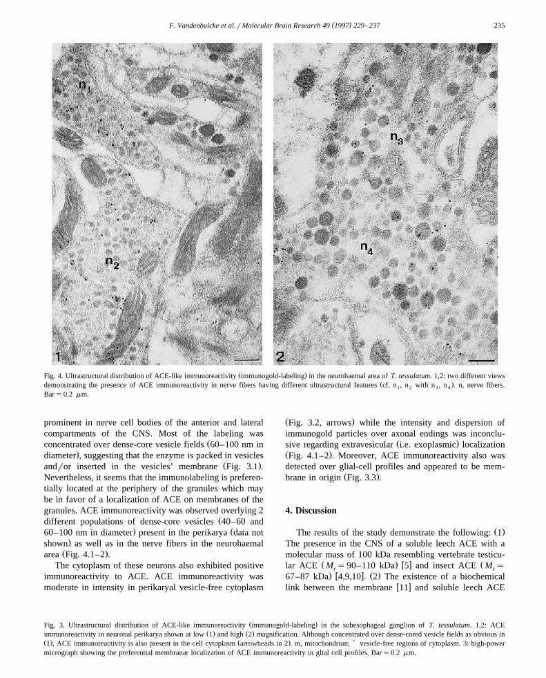
Ž .Fig. 4. Ultrastructural distribution of ACE-like immunoreactivity immunogold-labeling in the neurohaemal area of T. tessulatum. 1,2: two different viewsŽ .demonstrating the presence of ACE immunoreactivity in nerve fibers having different ultrastructural features cf. n , n with n , n . n, nerve fibers.1 2 3 4
Bars0.2 mm.
prominent in nerve cell bodies of the anterior and lateralcompartments of the CNS. Most of the labeling was
Žconcentrated over dense-core vesicle fields 60–100 nm in.diameter , suggesting that the enzyme is packed in vesicles
Ž .andror inserted in the vesicles’ membrane Fig. 3.1 .Nevertheless, it seems that the immunolabeling is preferen-tially located at the periphery of the granules which maybe in favor of a localization of ACE on membranes of thegranules. ACE immunoreactivity was observed overlying 2
Ždifferent populations of dense-core vesicles 40–60 and. Ž60–100 nm in diameter present in the perikarya data not
.shown as well as in the nerve fibers in the neurohaemalŽ .area Fig. 4.1–2 .
The cytoplasm of these neurons also exhibited positiveimmunoreactivity to ACE. ACE immunoreactivity wasmoderate in intensity in perikaryal vesicle-free cytoplasm
Ž .Fig. 3.2, arrows while the intensity and dispersion ofimmunogold particles over axonal endings was inconclu-
Ž .sive regarding extravesicular i.e. exoplasmic localizationŽ .Fig. 4.1–2 . Moreover, ACE immunoreactivity also wasdetected over glial-cell profiles and appeared to be mem-
Ž .brane in origin Fig. 3.3 .
4. Discussion
Ž .The results of the study demonstrate the following: 1The presence in the CNS of a soluble leech ACE with amolecular mass of 100 kDa resembling vertebrate testicu-
Ž . w x Žlar ACE M s90–110 kDa 5 and insect ACE M sr r. w x Ž .67–87 kDa 4,9,10 . 2 The existence of a biochemical
w xlink between the membrane 11 and soluble leech ACE
Ž .Fig. 3. Ultrastructural distribution of ACE-like immunoreactivity immunogold-labeling in the subesophageal ganglion of T. tessulatum. 1,2: ACEŽ . Ž .immunoreactivity in neuronal perikarya shown at low 1 and high 2 magnification. Although concentrated over dense-cored vesicle fields as obvious in
Ž . Ž . )1 , ACE immunoreactivity is also present in the cell cytoplasm arrowheads in 2 . m, mitochondrion; vesicle-free regions of cytoplasm. 3: high-powermicrograph showing the preferential membranar localization of ACE immunoreactivity in glial cell profiles. Bars0.2 mm.

( )F. Vandenbulcke et al.rMolecular Brain Research 49 1997 229–237236
demonstrate by Western blot analysis and periodic schifftreatment experiences and automated Edman degradation.Ž .3 The two ACE possess the same N-terminus, reinforcingthe hypothesis that both are, respectively, the membraneand the soluble forms of ACE. In fact, ACE forms belongto the type-I class of integral transmembrane domain witha 17-amino acid hydrophobic anchor located 30 amino-acid
w x Ž .residues from cytosolic C-terminus 5 . 4 The immunocy-tochemical studies demonstrate the presence of ACE im-munoreactivity in neurons and glial cells. Taken together,the study demonstrates that invertebrate ACE resemblesvertebrate ACE. Additionally, its localization suggests thatboth neurons and glial cells are involved with processingsimilar signal molecules.
Recently, in invertebrates, in M. domestica, a peptidyldipeptidase which has similar enzymatic properties to
w xmammalian ACE has been described 9 . This enzyme hasan apparent M of f80 kDa, thus, resembling the single-r
w xdomain enzyme 9 . A more hydrophilic ACE with a M ofr
67 kDa has been purified in M. domestica using a lisino-w xpril-sepharose affinity column 10 . This enzyme removed
C-terminal dipeptides from angiotensin I, bradykinin,Leu-enkephalin and Met-enkephalin as well as functioningas an endopeptidase by hydrolysing dipeptideamides fromLeu-enkephalinamide and Met-enkephalinamide and then adipeptideamide and tripeptideamide from substance P.Musca ACE also displays properties of both N- and C-do-
w xmains of human ACE 10 . More recently, cDNA of insectACE has been cloned from Drosophila melanogaster em-bryo cDNA library that codes for a single-domain 615amino-acid protein with high levels of similarity to both
w xN- and C-domains of mammalian ACE 4 .The leech has also provided information on invertebrate
ACE. We purified ACE from leech head membranesw x11,12 . The first 23 amino-acid residues of the N-terminalpart of the purified leech ACE revealed f87% sequenceidentity with the N-terminal sequence of the plasma serum
w x Ž .guinea-pig ACE 11 . This ACE of f120 kDa hydroly-ses at pH 8.4 and 378C, the Phe8-His9 bond of theangiotensin I with a high catalytic activity, i.e. K s830m
mM and K rK s153 sy1 mMy1. This zinc metal-cat m
lopeptidase also attacks peptides having in their sequenceeither a Gly-His or a Gly-Phe or a Phe-His bonds, e.g.
Ž y1 y1.enkephalins K rK s12 s mM or bradykinincat mŽ y1 y1. w xK rK s2200 s mM 13 . This data are consis-cat m
tent with an ACE-like activity implicated in metabolism ofangiotensins and bradykinin in leeches as in insects andmammals. Our results are in agreement with the findingthat this is a single-domain mammalian-like ACE.
Immunocytochemical reactivity reveals that this mate-rial is found on neurons and glial cells. These findingssuggest that invertebrate neurons and glial cells may com-municate via the renin–angiotensin system. Regarding thepositive immunostaining of neurons and glial cells, atpresent, it is not possible to determine if 2 differentmolecular forms are present or if different forms exist in
the same cell type. Moreover, the observation of ACEimmunoreactivity in nerve fibers in a neurohaemal area isin agreement with the known function of ACE in insects asa secreted enzyme.
In comparing this information to that found in verte-brates, we note that in higher animals there are two
w xisoforms of ACE derived from a single gene 7,14 . Insomatic tissues, ACE is expressed as a glycoprotein com-posed of a single polypeptide chain with an apparentmolecular mass of 170 kDa, containing two large homolo-gous domains, called the N- and C-domains, with each
w xdomain bearing an active catalytic site 22 . In male germi-nal cells, ACE is synthetized as a lower molecular mass
Ž .form 110 kDa , containing only the active C-domain ofw xendothelial ACE 22 . Recently, it has been demonstrated
in vertebrates that an EDTA-sensitive protease associatedw xto membrane preparations 18 releases a hydrophilic form
of ACE from cell surfaces by cleavage of the ACE proteinw xclose to its membrane anchor 1 which then creates a
soluble form of ACE. This may also occur in invertebrates.In vertebrates, the solubilized ACE circulates in many
w xbody fluids 6 , suggesting an immunoregulatory function.In regard to the N-terminal sequence of both forms ofleech ACE and their recognition by anti-ACE antiserum,we suggest that the membrane form may be anchored byits C-terminus. Insect ACE displays properties of both N-and C-domains of human ACE, indicating a high degree of
w xconservation during evolution 9,10 . In leeches, bothmembrane and soluble ACE forms display similar activitytowards AI, except for the catalytic efficiency which ishigher for the hydrophilic form.
In summary, comparison of the core regions of the twomammalian ACE domains with the single-domain insectACE shows three significant events in evolutionary termsw x4 . The divergence of the higher insect and mammal
Ž .lineages usually put at 550 million years ago , the radia-Ž .tion of the modern mammals 65 million years ago and
the gene duplication event that led from a single-domainACE-like protein to the mammalian two-domain ACE.However, it is possible to say that the duplication probablyoccurred in the deuterostome lineage, as seen by thegreater similarity between the N- and the C-domains thanbetween the N- and the C-domains of vertebrate and insectACE. This study also provides additional evidence that the
Ž .mammalian-like peptides i.e. enkephalins, etc. found inin v e r te b ra te s a ls o h a v e m a m m a lia n - l ik eprocessingrterminating enzymes demonstrating that these
w xprocesses first evolved in simpler animals 23 .
Acknowledgements
The studies were in part supported by NIH GrantsŽ .MHrDA 17138 and DA 09010 to G.B.S. and NIH
Ž .Fogarty Grant INT 00045 to G.B.S. and M.S. . We areindebted to Annie Desmons of the Laboratoire de

( )F. Vandenbulcke et al.rMolecular Brain Research 49 1997 229–237 237
Phylogenie Moleculaire des Annelides, EA DRED 1027,´ ´ ´for her excellent technical assistance.
References
w x1 V. Beldent, A. Michaud, C. Bonnefoy, M.-T. Chauvet, P. Corvol,Cell surface localization of proteolysis of human endothelial an-
Ž .giotensin I converting enzyme, J. Biol. Chem. 270 1995 28962–28969.
w x2 K.E. Bernstein, B.M. Martin, E.A. Bernstein, J. Linton, G. Stricker,The isolation of angiotensin converting enzyme cDNA, J. Biol.
Ž .Chem. 263 1988 11021–11024.w x3 M.M.A. Bradford, Rapid and sensitive method for the quantification
of microgram quantities of protein utilizing the principle of proteinŽ .dying, Anal. Biochem. 72 1976 248–254.
w x4 M.J. Cornell, T.A. Williams, N.S. Lamango, D. Coates, P. Corvol,F. Soubrier, J. Hoheisel, H. Lehrach, I. Isaac, Cloning and expres-sion of an evolutionary conserved single-domain angiotensin-con-verting enzyme from Drosophila melanogaster, J. Biol. Chem. 271Ž .1996 13613–13619.
w x5 P. Corvol, T.A. Williams, F. Soubrier, Peptidyl dipeptidase A:Ž .angiotensin I-converting enzyme, in: A.J. Barrett Ed. , Methods in
Enzymology, vol. 248, Academic Press, 1995, pp. 283–305.w x6 N.M. Hooper, A.J. Turner, Isolation of two differentially glycosy-
Žlated forms of peptidyl-dipeptidase A angiotensin converting en-.zyme from pig brain: a re-evaluation of their role in neuropeptide
Ž .metabolism, Biochem. J. 241 1987 625–633.w x7 T.E. Howard, S.-Y. Shai, K.G. Langford, B.M. Martin, K.E. Bern-
stein, Transcription of testicular angiotensin-converting enzymeŽ .ACE is initiated within the 12th intron of the somatic ACE gene,
Ž .Mol. Cell. Biol. 10 1990 4294–4302.w x8 U.K. Laemmli, Cleavage of structural proteins during the assembly
Ž .of the head of bateriophage T4, Nature 227 1970 680–685.w x9 N.S. Lamango, R.E. Isaac, Identification and properties of a peptidyl
dipeptidase in the housefly Musca domestica, that resembles mam-Ž .malian angiotensin-converting enzyme, Biochem. J. 299 1994
651–657.w x10 N.S. Lamango, M. Sajid, R.E. Isaac, The endopeptidase activity and
the activation by Cly of angiotensin converting enzyme is evolu-tionary conserved: purification and properties of an angiotensin-con-verting enzyme from the housefly, Musca domestica, Biochem. J.
Ž .314 1996 639–646.w x11 V. Laurent, M. Salzet, A comparison of the N-terminal sequence of
leech angiotensin converting-like enzyme with forms of vertebrateŽ .angiotensin converting enzyme, Neurosci. Lett. 198 1995 60–64.
w x12 V. Laurent, M. Salzet, Identification and properties of an an-giotensin-converting enzyme in the leech Theromyzon tessulatum,
Ž . Ž .Peptides 17 5 1996 737–745.w x13 V. Laurent, M. Salzet, Metabolism of enkephalins in head mem-
branes of the leech Theromyzon tessulatum by peptidases, Regul.Ž .Peptides 65 1996 123–131.
w x14 A.L. Lattion, F. Soubrier, J. Allegrini, C. Hubert, P. Corvol, F.Alhenc-Gelas, The testicular transcript of the angiotensin I-convert-ing enzyme encodes for the ancestral, non-duplicated form of the
Ž .enzyme, FEBS Lett. 252 1989 99–104.w x15 J. Malecha, M. Verger-Bocquet, A. Lepretre, G. Tramu, Mise en
evidence et evolution, au cours du cycle biologique, de neurones´ ´producteurs d’une substance apparentee a la motiline porcine dans le´ `ganglion supraœsophagien de la sangsue Theromyzon tessulatum,
Ž .Can. J. Zool. 67 1989 636–640.w x16 M.A. Ondetti, D.W. Cushman, Enzymes of the renin–angiotensin
Ž .system and their inhibitors, Annu. Rev. Biochem. 51 1982 283–308.
w x17 J. Pre, D. Bladier, A rapid and sensitive spectrophotometric methodfor routine determination of serum angiotensin I converting enzyme,
Ž .IRCS Med. Sci. 11 1983 220–221.w x18 R. Ramchandran, I. Sen, Cleavage processing of angiotensin-con-
verting enzyme by a membrane-associated metalloprotease, Bio-Ž .chemistry 34 1995 12645–12652.
w x19 M. Salzet, M. Verger-Bocquet, C. Wattez, J. Malecha, Evidence forangiotensin-like molecules in the central nervous system of the leech
Ž .Theromyzon tessulatum O.F.M. . A possible diuretic effect, Comp.Ž .Biochem. Physiol. 101A 1992 83–90.
w x20 M. Salzet, C. Wattez, J.-L. Baert, J. Malecha, Biochemical evidenceof angiotensin II-like peptides and proteins in the brain of the
Ž .rhynchobdellid leech Theromyzon tessulatum, Brain Res. 631 1993247–255.
w x21 M. Salzet, C. Wattez, M.C. Slomianny, B. Leu, K.J. Siegert, ELISA´¨for oxytocin. Highly sensitive tests for the titration of an oxytocin-likesubstance in the leech Erpobdella octoculata, Comp. Biochem.
Ž .Physiol. 102C 1992 483–487.w x22 F. Soubrier, F. Alhenc-Gelas, C. Hubert, J. Allegrini, M. John, G.
Tregear, P. Corvol, Two putative active centers in human an-giotensin I-converting enzyme revealed by molecular cloning, Proc.
Ž .Natl. Acad. Sci. USA 85 1988 9386–9390.w x23 G.B. Stefano, B. Scharrer, E.M. Smith, T.K. Hughes Jr., H.I.
Magazine, T.V. Bilfinger, A.R. Hartman, G.L. Fricchione, Y. Liu,M.H. Makman, Opioid and opiate immunoregulatory processes, Crit.
Ž .Rev. Immunol. 16 1996 109–144.w x24 S.L. Stephenson, J.A. Kenny, Metabolism of neuropeptides. Hydrol-
ysis of angiotensins, bradykinin, substance P and oxytocin by pigŽ .kidney microvillar membranes, Biochem. J. 241 1987 237–247.
w x25 M. Verger-Bocquet, C. Wattez, M. Salzet, J. Malecha, Immunocyto-´chemical identification of peptidergic neurons in compartment 4 ofthe supraesophageal ganglion of the leech Theromyzon tessulatumŽ . Ž .O.F.M. , Can. J. Zool. 70 1991 856–865.
w x26 G.W. Welling, A.J. Scheffer, S. Wellig-Wester, Determination ofenzyme activity by high-performance liquid chromatography, J.
Ž .Chromatogr. B 659 1994 209–225.
Copyright © 2022 FDOKUMEN