Leech Neuroimmune Signaling
12
13 The Brain and Host Defense Copyright © 2009 by Academic Press. Inc. All rights of reproduction in any form reserved. 2010 BERCZI 978-0-44-453544-3 00002 Leech Neuroimmune Signaling Aurelie Tasiemski and Michel Salzet Laboratoire de Neuroimmunologie des Annélides, Université des Sciences et technologies de Lille, France c0002 c0002 Chapter 2 1 INTRODUCTION In the past, several laboratories have found monoaminer- gic, opioid, opiate and endocannabinoid signal molecules in invertebrates [1–4]. It comes as no surprise to find these important communication molecules throughout the animal kingdom, and some in the plant kingdom as well, indicat- ing that they may have their origins very far back in evolu- tion, and share a common ancestor [5]. This chapter will focus on invertebrate opioid, opiate and endocannabinoid physiology, which demonstrates that neuroimmune com- munication occurs in specific invertebrates. We will spe- cifically concentrate on leeches. 2 NEUROIMMUNE MODULATION BY OPIOIDS The presence of biologically active neuropeptides in inver- tebrates, comparable to those of vertebrates, has been known for a considerable period of time [6–9]. However, detailed information on one specific class of these pep- tides, the endogenous opioids, has been almost exclusively confined to the mammalian nervous system, although there have been several reports documenting the occur- rence of endogenous opioid peptides in submammalian vertebrates [10–13]. With regard to invertebrates, the opioid peptides Met- and Leu-enkephalin as well as Met-enkephalin-Arg-Phe were isolated and sequenced from mollusk neural tissues [11, 14], arthropods [15, 16] and annelids [17]. Their precursor, the proenkephalin-like protein, was identified in leech immunocytes [18]. Leech proenkephalin dem- onstrates amino acid sequence similarity with vertebrate proenkephalin (amphibian 26.2 percent). The proenkepha- lins contain Met- and Leu-enkephalin in a ratio of 1 : 2 in the leech. They also possess Met-enkephalin-Arg-Gly-Leu and S0010 S0010 p0010 p0010 S0020 S0020 p0020 p0020 p0030 p0030 Met-enkephalin-Arg-Phe that are flanked by dibasic amino acid residues, demonstrating cleavage sites for subtilisin prohormone convertase enzymes (SPC) recently cloned in leeches (Table 2.1). Furthermore, using both sequence comparison and a specific antiserum raised against bovine proenkephalin A (209–237), peptide B was identified in leech proenkephalin and shown to exhibit a high antimicro- bial activity [19]. These results further support the hypoth- esis that these molecules (peptide B as well as enkelytin, a truncated form of peptide B) first evolved in simpler ani- mals. Similarly, recently a neuropeptide like precursor, con- taining several copies of YGGYG or YGGYGRG peptides resembling enkephalins (YGGFL, YGGFM, YGGFMRG) have recently been discovered in both C. elegans and D. melanogaster genomes [20]. Interestingly, one of these TABLE 2.1 Sequence similarity of leech SPC1 and SPC2 Protein Homology (%) SPC1 Mouse Human Aplysia Hydra PROHORMONE-CONVERTASE 1 PROHORMONE-CONVERTASE 1 PROHORMONE-CONVERTASE 1 PROHORMONE-CONVERTASE 1 68 67 60 58 SPC2 Lymnae Aplysia Mouse Human Xenopus Drosophila C. elegans PROHORMONE-CONVERTASE 2 PROHORMONE-CONVERTASE 2 PROHORMONE-CONVERTASE 2 PROHORMONE-CONVERTASE 2 PROHORMONE-CONVERTASE 2 PROHORMONE-CONVERTASE 2 PROHORMONE-CONVERTASE 2 70 70 67 67 65 62 62 t0010 t0010 CH002.indd 13 CH002.indd 13 10/7/2009 11:29:27 AM 10/7/2009 11:29:27 AM
-
Upload
univ-lille1 -
Category
Documents
-
view
1 -
download
0
Transcript of Leech Neuroimmune Signaling

13The Brain and Host DefenseCopyright © 2009 by Academic Press. Inc. All rights of reproduction in any form reserved.2010
BERCZI 978-0-44-453544-3 00002
Leech Neuroimmune Signaling
Aurelie Tasiemski and Michel Salzet Laboratoire de Neuroimmunologie des Ann é lides, Universit é des Sciences et technologies de Lille, France
c0002 c0002
Chapter 2
1 INTRODUCTION
In the past, several laboratories have found monoaminer-gic, opioid, opiate and endocannabinoid signal molecules in invertebrates [1 – 4] . It comes as no surprise to find these important communication molecules throughout the animal kingdom, and some in the plant kingdom as well, indicat-ing that they may have their origins very far back in evolu-tion, and share a common ancestor [5] . This chapter will focus on invertebrate opioid, opiate and endocannabinoid physiology, which demonstrates that neuroimmune com-munication occurs in specific invertebrates. We will spe-cifically concentrate on leeches.
2 NEUROIMMUNE MODULATION BY OPIOIDS
The presence of biologically active neuropeptides in inver-tebrates, comparable to those of vertebrates, has been known for a considerable period of time [6 – 9] . However, detailed information on one specific class of these pep-tides, the endogenous opioids, has been almost exclusively confined to the mammalian nervous system, although there have been several reports documenting the occur-rence of endogenous opioid peptides in submammalian vertebrates [10 – 13] .
With regard to invertebrates, the opioid peptides Met- and Leu-enkephalin as well as Met-enkephalin-Arg-Phe were isolated and sequenced from mollusk neural tissues [11, 14] , arthropods [15, 16] and annelids [17] . Their precursor, the proenkephalin-like protein, was identified in leech immunocytes [18] . Leech proenkephalin dem-onstrates amino acid sequence similarity with vertebrate proenkephalin (amphibian 26.2 percent). The proenkepha-lins contain Met- and Leu-enkephalin in a ratio of 1 : 2 in the leech. They also possess Met-enkephalin-Arg-Gly-Leu and
S0010 S0010
p0010 p0010
S0020 S0020
p0020 p0020
p0030 p0030
Met-enkephalin-Arg-Phe that are flanked by dibasic amino acid residues, demonstrating cleavage sites for subtilisin prohormone convertase enzymes (SPC) recently cloned in leeches ( Table 2.1 ). Furthermore, using both sequence comparison and a specific antiserum raised against bovine proenkephalin A (209 – 237), peptide B was identified in leech proenkephalin and shown to exhibit a high antimicro-bial activity [19] . These results further support the hypoth-esis that these molecules (peptide B as well as enkelytin, a truncated form of peptide B) first evolved in simpler ani-mals. Similarly, recently a neuropeptide like precursor, con-taining several copies of YGGYG or YGGYGRG peptides resembling enkephalins (YGGFL, YGGFM, YGGFMRG) have recently been discovered in both C. elegans and D. melanogaster genomes [20] . Interestingly, one of these
TABLE 2.1 Sequence similarity of leech SPC1 and SPC2
Protein Homology (%)
SPC1
Mouse Human Aplysia Hydra
PROHORMONE-CONVERTASE 1 PROHORMONE-CONVERTASE 1 PROHORMONE-CONVERTASE 1 PROHORMONE-CONVERTASE 1
68 67 60 58
SPC2
Lymnae Aplysia Mouse Human Xenopus Drosophila C. elegans
PROHORMONE-CONVERTASE 2 PROHORMONE-CONVERTASE 2 PROHORMONE-CONVERTASE 2 PROHORMONE-CONVERTASE 2 PROHORMONE-CONVERTASE 2 PROHORMONE-CONVERTASE 2 PROHORMONE-CONVERTASE 2
70 70 67 67 65 62 62
t0010 t0010
CH002.indd 13CH002.indd 13 10/7/2009 11:29:27 AM10/7/2009 11:29:27 AM
SECTION | II Development and Function of the Neuroimmune System14
BERCZI 978-0-44-453544-3 00002
molecules (nlp 31) was chemically synthesized and appeared to exert antifungal properties [20] .
Given the presence of opioid peptides in Lophotrochozoans, the presence of specific receptors was investigated in the same tissues. This was suggested by binding studies performed with Mytilus immunocyte mem-brane suspensions. The effects of pre-incubation of the immunocyte membranes with increasing concentrations of [D-Ala 2 ,Leu 5 , Cys 6 ] enkephalin (DALCE), a non-equilibrium delta opioid antagonist, on recovery of the delta bind-ing sites of 3 H[D-Pen 2 , D-Pen 5 ] enkephalin ( 3 H-DPDPE), 3 H-deltorphin I and 3-[D-Ala 2 , Met 5 ]enkephalimide ( 3 H-DAMA) in Mytilus revealed two delta-type binding sites [21] . DALCE pre-treatment resulted in a marked, concen-tration-dependent decrease in recovery of 3 H-DPDPE-bind-ing sites, whereas it only slightly modified the recovery of 3 H-deltorphin I-binding sites and 3 H-DAMA sites [21] . The ability of a variety of other opioids to displace specifi-cally bound 3 H-DAMA was investigated in another experi-ment [21] . The opioid peptides did so in the following decreasing order: deltorphin I � DAMA � Met-enkepha-lin � DADLE � DPDPE. By contrast, the mu and kappa ligands DAGO and dynorphin 1 – 17 were quite weak. Naltrindole was found to be more potent than naloxone in displacing 3 H-DAMA. Thus, as in human granulocytes, invertebrate immunocytes provide additional evidence that a special delta subtype of opioid receptor for immunoreg-ulating Met-enkephalin exists in invertebrates and can be classified as a subtype, δ 2 , of the classical delta recep-tor δ 1 [21] . Similar data were obtained in leeches [18] . In fact, in cold saturation experiments, where a set amount (0.2 nM) of radioligand ( 3 H-DAMA) is added to each tube and the concentration of the ligand increased by adding progressively larger amounts of unlabeled ligand, a single high-affinity binding site was revealed for leech immu-nocyte and ganglion membrane homogenates with K d s of 1.4 � 0.3 nM and 1.0 � 0.2 nM, and B max s of 385 � 23 and 252 � 17 fmol/mg protein, respectively. Pre-treatment of leech immunocyte membranes with increasing con-centrations of DALCE significantly blocked 3 H-DPDPE binding to delta opioid sites [18] . By contrast, the same treatment of membranes with DALCE did not affect the binding of 3 H-DAMA. In summary, both neural and immune tissues in leeches, as in mussels, express opioid receptor subtypes, demonstrating their potential to respond to, as well as interact with, these chemical messengers.
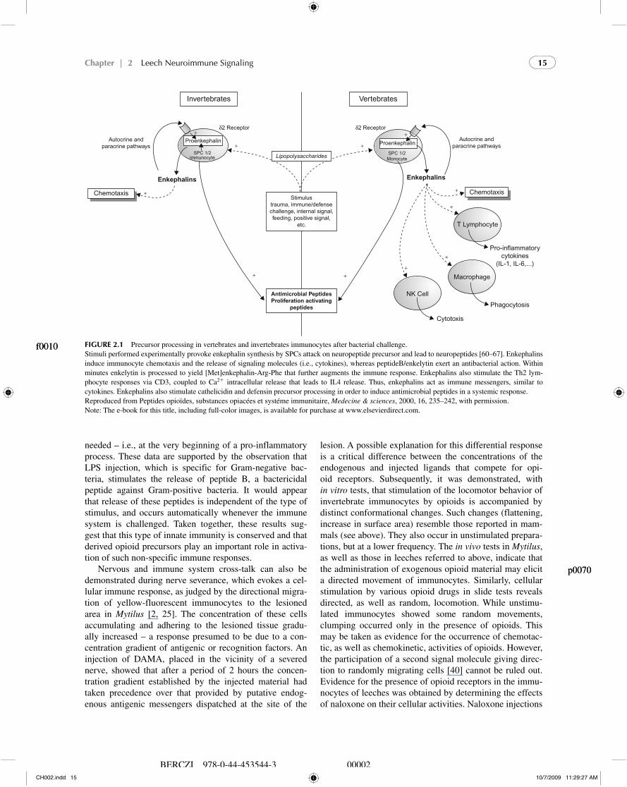
In vertebrates, chemotactic effects of endogenous opioid peptides on human polymorphonuclear leukocytes, mono-cytes and lymphocytes have been demonstrated ( Figure 2.1 ) [22 – 24] . Moreover, opioids, by stereoselective mecha-nisms, are involved in invertebrate autoimmunoregulatory processes. Interestingly, a subpopulation of granulocytes and immunocytes from Mytilus edulis and leeches has the ability to respond to low opioid concentrations by adher-ing and clumping [25, 26] . The adherence-promoting role
p0050 p0050
of DAMA (D-Ala 2 -Met 5 -enkephalin), and its blockage by naloxone, in a dose – response manner, was clearly evident. By contrast, exogenous Met-enkephalin at the same low concentration of DAMA did not increase cellular adher-ence above control levels, due to the presence of proteolytic enzymes in the hemolymph [27] . Indeed, subsequent stud-ies demonstrated that neutral endopeptidase 24.11 (CD10, “ enkephalinase ” ) was present on both human and inverte-brate immunocytes [27] , where it serves to modulate neu-ropeptide activation of the respective cells [28, 29] .
Proenkephalin processing is also involved in immune response modulation. It has recently been demonstrated in man and leeches that immune signaling or alerting may lead to enhanced proenkephalin proteolytic processing in immunocytes, with release of opioid peptides and the antimicrobial peptide enkelytin [19, 30 – 32] through SPC activation [33, 34] ( Figure 2.1 ). In this scenario, the opi-oid peptides would stimulate immunocyte chemotaxis and phagocytosis as well as the secretion of classical cytokines [35, 36] . During this process the liberated enkelytin/peptide B would simultaneously attack bacteria, thereby allowing time for the immune stimulating capabilities of opioid pep-tides (i.e., chemokine induction) to manifest itself [35, 36] . This hypothesis is further supported by the presence of specific Met-enkephalin receptors on both vertebrate and invertebrate immunocytes, as noted earlier [19] , and the presence of mRNA encoding for proenkephalin as well as chromogranins in immunocytes [37, 38] . Intra-peritoneal injection of lipopolysaccharides leads to SPC3 activation with peptide B/enkelytin and opioid release in rat spleen [34] . Furthermore, SPC3 knockdown mice died sooner after LPS injection than control animals, reflecting the important role of SPC3 in the innate immune response (unpublished data) and the expression of an endocrine phenotype in the immune cells [36] . Moreover, important to our understanding of the significance of this system in diverse organisms is the fact that surgical trauma and elec-trical shocks directed to neural tissues cause, as with LPS, an increase in circulating levels of peptide B and Met-enkephalin. Previously, Stefano and colleagues demon-strated that the electrical shock protocol represents a stress process in Mytilus which activates immunocytes via the secretion of Met-enkephalin [39] . Peptide B can also be added to this stress response. The co-processing and lib-eration of peptide B and Met-enkephalin represent a uni-fied neuroimmune protective response to immediate threat to the organism, regardless of the form that the stimulation takes. Clearly, threatening stimuli may be accompanied by bacteria: thus, to avoid the complication of a bacterial infection, peptide B is released either for specific bacte-rial assaults or as a precaution, whereas Met-enkephalin stimulates or activates immunocytes during the initial stages of the immune response ( Figure 2.1 ). With this in mind, the unified neuroimmune response would provide a highly beneficial survival strategy at a time when it is most
CH002.indd 14CH002.indd 14 10/7/2009 11:29:27 AM10/7/2009 11:29:27 AM
Chapter | 2 Leech Neuroimmune Signaling 15
BERCZI 978-0-44-453544-3 00002
VertebratesInvertebrates
δ2 Receptor δ2 Receptor
Proenkephalin Proenkephalin
Lipopolysaccharides
Stimulustrauma, immune/defensechallenge, internal signal,feeding, positive signal,
etc.
Antimicrobial Peptides
Proliferation activating
peptides
Autocrine andparacrine pathways
Autocrine andparacrine pathways
Chemotaxis Chemotaxis
T Lymphocyte
Pro-inflammatorycytokines
(IL-1, IL-6,...)
Macrophage
Phagocytosis
Cytotoxis
NK Cell
Enkephalins Enkephalins
SPC 1/2immunocyte
SPC 1/2Monocyte
+ +
+
+
+
+
+++
+
+
FIGURE 2.1 Precursor processing in vertebrates and invertebrates immunocytes after bacterial challenge. Stimuli performed experimentally provoke enkephalin synthesis by SPCs attack on neuropeptide precursor and lead to neuropeptides [60 – 67] . Enkephalins induce immunocyte chemotaxis and the release of signaling molecules (i.e., cytokines), whereas peptideB/enkelytin exert an antibacterial action. Within minutes enkelytin is processed to yield [Met]enkephalin-Arg-Phe that further augments the immune response. Enkephalins also stimulate the Th2 lym-phocyte responses via CD3, coupled to Ca 2 � intracellular release that leads to IL4 release. Thus, enkephalins act as immune messengers, similar to cytokines. Enkephalins also stimulate cathelicidin and defensin precursor processing in order to induce antimicrobial peptides in a systemic response. Reproduced from Peptides opio ï des, substances opiac é es et syst é me immunitaire, Medecine & sciences , 2000, 16, 235 – 242, with permission. Note: The e-book for this title, including full-color images, is available for purchase at www.elsevierdirect.com .
f0010 f0010
needed – i.e., at the very beginning of a pro-inflammatory process. These data are supported by the observation that LPS injection, which is specific for Gram-negative bac-teria, stimulates the release of peptide B, a bactericidal peptide against Gram-positive bacteria. It would appear that release of these peptides is independent of the type of stimulus, and occurs automatically whenever the immune system is challenged. Taken together, these results sug-gest that this type of innate immunity is conserved and that derived opioid precursors play an important role in activa-tion of such non-specific immune responses.
Nervous and immune system cross-talk can also be demonstrated during nerve severance, which evokes a cel-lular immune response, as judged by the directional migra-tion of yellow-fluorescent immunocytes to the lesioned area in Mytilus [2, 25] . The concentration of these cells accumulating and adhering to the lesioned tissue gradu-ally increased – a response presumed to be due to a con-centration gradient of antigenic or recognition factors. An injection of DAMA, placed in the vicinity of a severed nerve, showed that after a period of 2 hours the concen-tration gradient established by the injected material had taken precedence over that provided by putative endog-enous antigenic messengers dispatched at the site of the
p0070 p0070
lesion. A possible explanation for this differential response is a critical difference between the concentrations of the endogenous and injected ligands that compete for opi-oid receptors. Subsequently, it was demonstrated, with in vitro tests, that stimulation of the locomotor behavior of invertebrate immunocytes by opioids is accompanied by distinct conformational changes. Such changes (flattening, increase in surface area) resemble those reported in mam-mals (see above). They also occur in unstimulated prepara-tions, but at a lower frequency. The in vivo tests in Mytilus , as well as those in leeches referred to above, indicate that the administration of exogenous opioid material may elicit a directed movement of immunocytes. Similarly, cellular stimulation by various opioid drugs in slide tests reveals directed, as well as random, locomotion. While unstimu-lated immunocytes showed some random movements, clumping occurred only in the presence of opioids. This may be taken as evidence for the occurrence of chemotac-tic, as well as chemokinetic, activities of opioids. However, the participation of a second signal molecule giving direc-tion to randomly migrating cells [40] cannot be ruled out. Evidence for the presence of opioid receptors in the immu-nocytes of leeches was obtained by determining the effects of naloxone on their cellular activities. Naloxone injections
CH002.indd 15CH002.indd 15 10/7/2009 11:29:27 AM10/7/2009 11:29:27 AM
SECTION | II Development and Function of the Neuroimmune System16
BERCZI 978-0-44-453544-3 00002
into sites of nerve severance of leeches, as noted above, counteracted the cellular immune reaction observed in the absence of this drug [2, 25] . Thus, immunocytes contain-ing opioid peptides have the capability of responding to them as well.
3 NEUROIMMUNE MODULATION BY OPIATES
With regard to invertebrates, the presence of an opiate receptor mechanism in the CNS of the marine mollusk Mytilus edulis was first suggested by a rise in ganglionic dopamine levels following intracardiac administration of exogenous Met- and Leu-enkephalin, an effect reversible by naloxone [1, 41 – 46] . The first actual demonstration of high-affinity opiate binding sites in an invertebrate gan-glion was accomplished by Stefano and colleagues [47] in Mytilus edulis . The biochemical characteristics of this system, analyzed in detail by Kream and colleagues [48] , have been found to parallel those of mammalian systems. The opiate receptor is distinguished from classical neuro-nal opioid receptor subtypes on the basis of pharmacologi-cal properties as revealed by radioligand competition and by functional studies, as well as by biochemical proper-ties. This receptor is opiate alkaloid selective and opioid peptide insensitive, and is named μ 3 [49] . 6-Glucuronide, not the 3-glucuronide metabolite of morphine, binds to the μ 3 receptor in invertebrates [49] . As with classical opioid receptors [50] , μ 3 is linked to trimeric G proteins that, in turn, have the capability to modulate Ca 2 � and K � chan-nels, adenylyl cyclase, and probably other signal transduc-tion systems [51] , and is coupled to intracellular calcium transients [52] , supporting a classical μ signaling pattern. Supporting these biochemical and pharmacological stud-ies involved with the demonstration of opiate receptors in Mytilus was the molecular cloning of a human μ receptor [53] . A transcript for the μ receptor was amplified from RNA extracted from nervous ganglia. Sequence analysis of the PCR product demonstrates that this fragment exhibits 95 percent sequence identity with the human brain μ opi-ate receptor [53] . Furthermore, exposing pedal ganglia to both interleukin 1- α and - β at 30 and 50 ng/ml for 24 hours resulted in a 47 percent and a 60 percent increase in the band density, respectively [54] . Twenty-four-hour treat-ment of pedal ganglia with morphine (300 nM) diminished the μ 3 receptor transcript to almost zero, confirming that this opiate receptor μ 3 is specific for morphine and is cou-pled to NO signaling [54] .
The demonstration of endogenous opiates (e.g., mor-phine, codeine) in various vertebrate tissues, including the nervous system [55 – 65] , is quite important for establishing the significance of the μ 3 opiate receptor subtype noted ear-lier. There is a body of evidence indicating that opiate alka-loids such as morphine, morphine 3- and 6-glucuronide, as
S0030 S0030
p0080 p0080
well as the morphine putative precursor molecules (thebaine, salutaridine, norcocolarine, reticuline, tetrahydropapoverine (THP) and codeine) exist in vertebrates [55 – 65] . In inver-tebrates, specifically Mytilus edulis , the presence of mor-phine, morphine 6-glucuronide, morphine 3-glucuronide, codeine, THP and reticuline have also been reported [66, 67] . Besides these biochemical studies, immunocytochemi-cal localization of a morphine-like material was reported in neural and immune tissues as well as in invertebrate tissues [55 – 67] .
In leeches [68] , the amount of morphine-like sub-stance quantified using biochemical techniques is similar that detected using RIA assay, ca . 3 μ pmol/mg. This evi-dence was substantiated by nitric oxide release experi-ments performed with the 8.64-min collected substance. Morphine-like substance is also able to stimulate nitric oxide release via constitutive NO synthetase, as does the morphine standard, and these effects are antagonized by naloxone, confirming the identity of the purified sub-stance. After exposing immunocytes to vehicle, the shape factor was present on 11 percent of activated cells, exhib-iting an amoeboid conformation of 0.48 � 0.1 SEM. After exposing immunocytes to the freeze-dried material obtained by combining all the material in a 30- μ l volume, of which 10 μ l was added to a 10- μ l volume of immuno-cytes, the immunocytes were only 3.1 � 1.1 percent active while exhibiting a shape factor of 6.7 � 0.5; P � 0.05. Naloxone at 10 � 7 M blocked the action of the extracted morphine-like material (spontaneous activation was 9.5 � 2.6 percent when naloxone was pre-administered to these cells followed by the extracted material). Moreover, leech ganglia morphine-like substance is able to block cAMP level through NO release acting on the adenylate cyclase. By contrast, in animals injected with LPS (1 μ g/ml) in a 100- μ l volume of leech saline, the morphine levels after 30 hours increased from 2.4 � 1.1 pmol/mg SEM to 78 � 12.3 pmol/gm. The increase in endogenous morphine levels was both time- and concentration-dependent.
Similarly , reports demonstrate that the ganglionic micro-glia (brain immune cells) can become active after ganglionic excision [69] and egress from the tissue through the nerves that are severed. Additionally, leeches possess endothelial nitric oxide synthase (eNOS) processes in their nervous and immune tissues [70] . Morphine’s actions in these diverse tissues complement what is known about NO-mediated immune and vascular functions, namely that NO can downregulate them from an excitatory state or prevent the excitatory state from occurring [71 – 73] . Morphine down-regulates invertebrate immunocytes, causing active motile amoeboid cells to become round and immobile [74, 75] . It also diminishes invertebrate microglial activation and egress from ganglia maintained in vitro [39] ( Figure 2.2 ).
Taken together, morphine inhibits invertebrate immu-nocytes. However, it must be emphasized that our obser-vations in this regard are ongoing. This is important
CH002.indd 16CH002.indd 16 10/7/2009 11:29:28 AM10/7/2009 11:29:28 AM

Chapter | 2 Leech Neuroimmune Signaling 17
BERCZI 978-0-44-453544-3 00002
Invertebrates
ChemotaxisChemotaxis
Vertebrates
NK Cell
T Cells
B Cell
Macrophages
POMC
MonocytePOMC
Immunocyte
AnandamideMorphine
Autocrine andparacrine pathways
Pro-informatorycytokines
(TNF, IL-1)
Autocrine and paracrinepathways
FAAH
FAAHμ3-receptor/CBRμ3—receptor/CBR
ACTH, α-MSH
ACTH, α-MSH
+
+
+
+
−
−
−
Lipopolysaccharides
Pro-inflammatorycytokines
(IL-1, IL-6,...)
Cytolysis,Th1-cell activity
Cytolysis
Antibodyformation
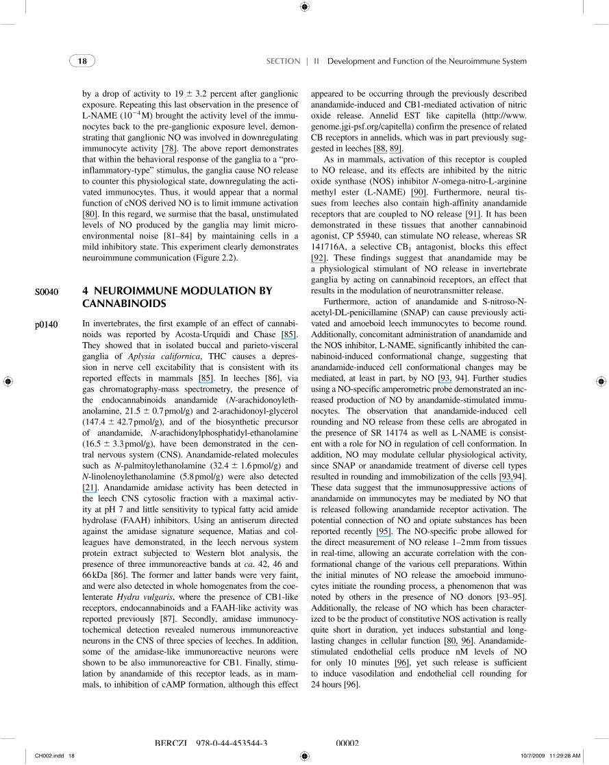
FIGURE 2.2 Comparison between immune modulation in vertebrates and in mammals: implication of opioid/POMC-derived peptides. In mammals, THC and anandamide inhibit T lymphocyte proliferation and Th1 activity. They also stimulate IgE but inhibit IgG production. THC and anandamide block cytolysis and phagocytosis of natural killer cells and macrophages, respectively (88). Moreover, both monocytes and invertebrate immunocytes contain POMC, prohormone convertase genes, and cannabinoid receptors. After either cognitive stress or pathogen infections, and through autocrine, paracrine or endocrine pathways, these cascade events lead to ACTH and α MSH release in both animal kingdoms. These peptides, like THC and anandamide, are known to inhibit T cell proliferation, IgG production, macrophage phagocytosis, and NK cell-mediated cytolysis. Hence, these substances inhibit in synergy the immune response. IL, interleukin; TNF α , tumor necrosis factor- α . Reproduced from Peptides opio ï des, substances opiac é es et syst é me immunitaire, Medecine & sciences , 2000, 16, 235 – 242, with permission. Note: The e-book for this title, including full-color images, is available for purchase at www.elsevierdirect.com .
f0020 f0020
because, following the inhibitory action, the cells rebound into excitation as noted earlier [75 – 77] . In Mytilus , pedal ganglia excised and maintained in culture for up to 2 hours release NO at low levels [78] . The range can vary between 0 and 1.1 nM [79] . Invertebrate immunocytes, which also release NO [75 – 79] , produce it within the same concentration range. Thus, it was of interest to determine if immunocytes added to cultured ganglia would enhance basal/constitutive ganglionic NO production since immune cells (e.g., microglia and immunocytes) have been found and identified in mollusk ganglia [73 – 79] . In this regard, non-stimulated immunocytes do not significantly stimulate ganglionic NO release when incubated with pedal ganglia [79] . However, ganglia exposed to activated immunocytes (pre-exposed to an efficacious concentration of interleukin 1 β ) [80] for 30 minutes before being added to the gangli-onic incubation medium) release significant levels of NO above basal levels after 30 minutes of incubation [78] . In these experiments, 91 � 2.5 percent of the non-stimulated
immunocytes exhibited form factors in the 0.72 – 0.89 range (sampled prior to ganglionic addition), whereas 62 � 10.3 percent of the interleukin 1 β stimulated immunocytes formed factors in the 0.39 – 0.49 percent range, having an amoeboid shape, as well as being motile ( n � 5; compar-ison of the percentage activated by a one-tailed student’s t-test revealed a P � 0.005). Addition of the nitric oxide synthase (NOS) inhibitor, L- NAME (10 � 4 M) inhibited basal ganglionic NO release, which was also initiated by exposing the ganglia to activated immunocytes, substanti-ating the identity of the material being monitored [78] .
Given the above results, it was of interest to determine the activity state of immunocytes following their incu-bation with the non-exposed and activated immunocyte exposed ganglia [78] . Non-activated immunocytes, follow-ing ganglionic exposure, exhibited activity levels in the 13 percent range, representing a non-significant increase [78] . Immunocytes exposed to interleukin 1 β had a 65 percent activity level at the beginning of the experiment, followed
CH002.indd 17CH002.indd 17 10/7/2009 11:29:28 AM10/7/2009 11:29:28 AM
SECTION | II Development and Function of the Neuroimmune System18
BERCZI 978-0-44-453544-3 00002
by a drop of activity to 19 � 3.2 percent after ganglionic exposure. Repeating this last observation in the presence of L-NAME (10 � 4 M) brought the activity level of the immu-nocytes back to the pre-ganglionic exposure level, demon-strating that ganglionic NO was involved in downregulating immunocyte activity [78] . The above report demonstrates that within the behavioral response of the ganglia to a “ pro-inflammatory-type ” stimulus, the ganglia cause NO release to counter this physiological state, downregulating the acti-vated immunocytes. Thus, it would appear that a normal function of cNOS derived NO is to limit immune activation [80] . In this regard, we surmise that the basal, unstimulated levels of NO produced by the ganglia may limit micro-environmental noise [81 – 84] by maintaining cells in a mild inhibitory state. This experiment clearly demonstrates neuroimmune communication ( Figure 2.2 ).
4 NEUROIMMUNE MODULATION BY CANNABINOIDS
In invertebrates, the first example of an effect of cannabi-noids was reported by Acosta-Urquidi and Chase [85] . They showed that in isolated buccal and parieto-visceral ganglia of Aplysia californica , THC causes a depres-sion in nerve cell excitability that is consistent with its reported effects in mammals [85] . In leeches [86] , via gas chromatography-mass spectrometry, the presence of the endocannabinoids anandamide ( N -arachidonoyleth-anolamine, 21.5 � 0.7 pmol/g) and 2-arachidonoyl-glycerol (147.4 � 42.7 pmol/g), and of the biosynthetic precursor of anandamide, N -arachidonylphosphatidyl-ethanolamine (16.5 � 3.3 pmol/g), have been demonstrated in the cen-tral nervous system (CNS). Anandamide-related molecules such as N -palmitoylethanolamine (32.4 � 1.6 pmol/g) and N -linolenoylethanolamine (5.8 pmol/g) were also detected [21] . Anandamide amidase activity has been detected in the leech CNS cytosolic fraction with a maximal activ-ity at pH 7 and little sensitivity to typical fatty acid amide hydrolase (FAAH) inhibitors. Using an antiserum directed against the amidase signature sequence, Matias and col-leagues have demonstrated, in the leech nervous system protein extract subjected to Western blot analysis, the presence of three immunoreactive bands at ca . 42, 46 and 66 kDa [86] . The former and latter bands were very faint, and were also detected in whole homogenates from the coe-lenterate Hydra vulgaris , where the presence of CB1-like receptors, endocannabinoids and a FAAH-like activity was reported previously [87] . Secondly, amidase immunocy-tochemical detection revealed numerous immunoreactive neurons in the CNS of three species of leeches. In addition, some of the amidase-like immunoreactive neurons were shown to be also immunoreactive for CB1. Finally, stimu-lation by anandamide of this receptor leads, as in mam-mals, to inhibition of cAMP formation, although this effect
S0040 S0040
p0140 p0140
appeared to be occurring through the previously described anandamide-induced and CB1-mediated activation of nitric oxide release. Annelid EST like capitella ( http://www.genome.jgi-psf.org/capitella ) confirm the presence of related CB receptors in annelids, which was in part previously sug-gested in leeches [88, 89] .
As in mammals, activation of this receptor is coupled to NO release, and its effects are inhibited by the nitric oxide synthase (NOS) inhibitor N -omega-nitro-L-arginine methyl ester (L-NAME) [90] . Furthermore, neural tis-sues from leeches also contain high-affinity anandamide receptors that are coupled to NO release [91] . It has been demonstrated in these tissues that another cannabinoid agonist, CP 55940, can stimulate NO release, whereas SR 141716A, a selective CB 1 antagonist, blocks this effect [92] . These findings suggest that anandamide may be a physiological stimulant of NO release in invertebrate ganglia by acting on cannabinoid receptors, an effect that results in the modulation of neurotransmitter release.
Furthermore , action of anandamide and S-nitroso-N-acetyl-DL-penicillamine (SNAP) can cause previously acti-vated and amoeboid leech immunocytes to become round. Additionally, concomitant administration of anandamide and the NOS inhibitor, L-NAME, significantly inhibited the can-nabinoid-induced conformational change, suggesting that anandamide-induced cell conformational changes may be mediated, at least in part, by NO [93, 94] . Further studies using a NO-specific amperometric probe demonstrated an inc-reased production of NO by anandamide-stimulated immu-nocytes. The observation that anandamide-induced cell rounding and NO release from these cells are abrogated in the presence of SR 14174 as well as L-NAME is consist-ent with a role for NO in regulation of cell conformation. In addition, NO may modulate cellular physiological activity, since SNAP or anandamide treatment of diverse cell types resulted in rounding and immobilization of the cells [93,94] . These data suggest that the immunosuppressive actions of anandamide on immunocytes may be mediated by NO that is released following anandamide receptor activation. The potential connection of NO and opiate substances has been reported recently [95] . The NO-specific probe allowed for the direct measurement of NO release 1 – 2 mm from tissues in real-time, allowing an accurate correlation with the con-formational change of the various cell preparations. Within the initial minutes of NO release the amoeboid immuno-cytes initiate the rounding process, a phenomenon that was noted by others in the presence of NO donors [93 – 95] . Additionally, the release of NO which has been character-ized to be the product of constitutive NOS activation is really quite short in duration, yet induces substantial and long-lasting changes in cellular function [80, 96] . Anandamide-stimulated endothelial cells produce nM levels of NO for only 10 minutes [96] , yet such release is sufficient to induce vasodilation and endothelial cell rounding for 24 hours [96] .
CH002.indd 18CH002.indd 18 10/7/2009 11:29:28 AM10/7/2009 11:29:28 AM
Chapter | 2 Leech Neuroimmune Signaling 19
BERCZI 978-0-44-453544-3 00002
In regard to the possible molecular mechanism involved with the immobilization and rounding of immunocytes, it can be speculated that NO is extremely labile and reacts rapidly (in milliseconds) with proteins and molecular oxy-gen. Once inside the cytosol it activates guanyl cyclase, increasing the levels of cGMP, which triggers a reduction in the intracellular calcium concentration by enhancing its cellular extrusion and intracellular sequestration [97] . This mechanism can account for the inhibition of spe-cific immunocyte adherence, aggregation and chemotaxis, as well as the immunocyte rounding effect. Moreover, in order for leukocytes to spread (become amoeboid) intra-cellular free calcium must be present at relatively high levels [98] . Clearly, once the intracellular calcium level is reduced and adherence is blocked, the cell will assume a rounded conformation due to a lack of contact with a sur-face. Indeed, given the large number of intracellular mech-anisms that can be influenced by NO, any combination of its actions could also inhibit select immunocyte, micro-glia and endothelial cell behaviors. These actions include autoribosylation of glyceraldehyde-3-phosphate dehydro-genase, inhibition of mitochondrial Fe-S enzymes, and alteration of cellular adhesion proteins [96 – 98] . Thus, the molecular actions of NO on cellular function may explain the rounding action observed and quantified in this report. Moreover, ananadamide, like morphine, inhibits cytokines and neurotransmitters though adenylate cyclase inhibi-tion, demonstrating a feedback control of the inflammatory response in leech central nervous system [22, 93] . We have also demonstrated similar effects of morphine and anan-damide on ACTH levels [23] ; this phenomenon is recep-tor-dependent. They increased proteolytic processing of both the ACTH precursor and the ACTH peptide through nitric oxide release from leech immunocytes ( Figure 2.2 ). The significance of these results in regard to invertebrate immunocytes is quite consequential in that they also have the ability to produce NO. Furthermore, there is a growing literature demonstrating the presence of immunoreactive material resembling NOS in invertebrate cells. The appli-cation of NO via NO donors has also been demonstrated to have intracellular actions as well as to influence inverte-brate immunocyte behavior [95] .
Taken together, these data suggest that functional cou-pling of NO to anandamide processes evolved early in evolution. Thus, coupled to the present findings the ability of anandamide to induce NO production in evolutionarily diverse organisms, suggests the conservation of a highly significant process that regulates cellular activation.
5 BRAIN INNATE IMMUNE RESPONSE
An important property of leeches is their capacity to regen-erate neurites and synaptic connections in the adult CNS. Neurites that have been damaged or severed can sprout,
S0050 S0050
p0190 p0190
establish de novo growth cones, and extend and reconnect specifically with normal targets [22] . Possible explana-tions for this useful attribute include a continued presence of embryonic factors that are required for neuronal growth and maturation, along with the ability to induce expres-sion or repression of critical factors in response to signals released by the damaged tissues. Initial molecular analy-ses of changes in gene expression modulation provoked by damage are considered below. Another aspect that may be important is that leech central neurons continue to expand their central and peripheral dendritic and terminal arbors throughout the life of the animal, suggesting the possibil-ity that the machinery for growth and addition of synaptic coupling may never be turned down or off completely in this invertebrate group. In mammals, by contrast, not only are many embryonic growth-promoting molecules and their receptors apparently no longer present in the adult, but also the adult CNS produces various growth-inhibiting molecules that were not present in the embryo or neonate [24] . Several recent reports, reviewed below, continue to examine important aspects of neural regeneration in the adult leech. Neuronal regeneration leading to full recovery of normal function requires not just reconnection to targets but also re-establishment of complex behaviors mediated by the regenerated parts [99] .
Early stages of leech CNS regeneration follow-ing a mechanical lesion are characterized by two events that appear to be crucial for successful repair: one is the increased activity of epithelial nitric oxide synthase (NOS) in the area of the lesion and the generation and diffusion of nitric oxide (NO), and the second is the induced migration of microglia towards, and their accumulation at, the injury site [100, 101] . Further analyses of these phenomena have been reported in the past few years, and are summarized below [102 – 104] . To obtain a more accurate measurement of the dynamics of NO generated following a lesion, an indirect approach to measuring NO levels (the standard cit-rulline assay) was first done and showed that NO is gener-ated within 30 minutes after the nerve cord has been injured [105] . Second, to obtain higher temporal and spatial resolu-tion, a polarographic NO-selective microsensor coupled to terahertz measurement has recently been used [106] . With this probe and the spectroscopy technology, we were able to detect, immediately following nerve crush, a signifi-cant efflux of NO from the lesion site. This efflux peaked within minutes after the crush, and then decreased rapidly exponentially (time constant � 120 s) to a steady low value which was known to follow injury, from previous meas-urements. To assay directly for a role of NO on microglial accumulation at the injury site, Chen et al . [107] modulated NO levels in several ways. As demonstrated by NOS immu-noreactivity, a large increase in NOS occurs at the crush site within 5 minutes of injury, and this high level persists for at least 24 hours. Microglial accumulation at the lesion, however, is not detectable at 5 minutes, but is quite strong
CH002.indd 19CH002.indd 19 10/7/2009 11:29:28 AM10/7/2009 11:29:28 AM
SECTION | II Development and Function of the Neuroimmune System20
BERCZI 978-0-44-453544-3 00002
after a few hours and peaks at � 24 hours. Inhibition of NO synthesis by the prior application of the NOS inhibitor L-NAME effectively blocks microglial accumulation, while the presence of its inactive enantiomer D-NAME has lit-tle or no effect. Interestingly, increasing NO levels with the NO donor spermine NOate (SPNO) also inhibits accu-mulation of microglia at the crush, but not in the presence of the NO scavenger cPTIO. Examination of microglial kinetics in living nerve cords shows that the effect of SPNO application occurs by the reduction of average microglial migratory speeds, even to no movement. Thus, NO is clearly implicated as a modulator of microglial movement, and indeed appears to function as a stop signal at high levels, leading to the higher density of these cells at the injury site.
Lipidomics study by mass spectrometry (Arafah, sub-mitted) confirms the involvement of eicosannoid in the microglia migration. However, this regenerative process is even more impressive upon a controlled bacterial infec-tion, suggesting that induction of regeneration of normal CNS is linked to danger signaling through innate immune response [108] Recent evidence has shown that micro-bial components differentially induce the transcription, by microglial cells, of antimicrobial peptide genes, the products of which accumulate rapidly at sites in the CNS undergoing regeneration following axotomy [108] . Using a preparation of leech CNS depleted of microglial cells, we also demonstrated the production of antimicrobial peptides by neurons. Interestingly, in addition to exerting antibacte-rial properties, both peptides act as promoters of the regen-erative process of axotomized leech CNS [108] . These data are the first reporting the neuronal synthesis of antimicro-bial peptides and their participation in both innate immune response and CNS regeneration.
6 CONCLUSIONS
In summary, innate immune response effectors as well as opioid and opiate immune processes appear to have had an earlier start in evolution than formerly realized. Additionally, given the presence of the components of these signaling systems (i.e., receptors), stereospecificity may be the actual “ glue ” maintaining these systems dur-ing evolution. The high opiate alkaloid selectivity of the μ 3 opiate receptor subtype reinforces a role for and the pres-ence of endogenous morphine. In regard to their immu-nomodulation, it appears that the opiate alkaloids inhibit whereas opioid peptides tend to stimulate invertebrate and vertebrate immune cells, including pro-inflammatory cytokine production, operating in an antagonistic manner unlike their analogous analgesic actions. At the level of the nervous system, innate immune response is implicated in brain defense but also in regeneration. Thus, we are left with the conclusion that many of these signal molecules and their functions had their origins in “ simple ” animals.
S0060 S0060
p0220 p0220
ACKNOWLEDGEMENTS
This work was supported in part by the MNERT and the CNRS.
REFERENCES
1. Stefano GB . Comparative aspects of opioid – dopamine interaction . Cell Mol Neurobiol 1982 ; 2 : 167 – 78 .
2. Stefano GB , Salzet M . Invertebrate opioid precursors: evolutionary conservation and the significance of enzymatic processing . Intl Rev Cytol 1999 ; 187 : 261 – 86 .
3. Salzet M , Stefano GB . The endocannabinoid system in inver-tebrates. Prostaglandins, leukotriene and essens . Fatty Acids 2001 ; 66 : 353 – 61 .
4. Stefano GB , Cadet P , Rialas CM , Mantione K , Casares F , Goumon Y , Zhu W . Invertebrate opiate immune and neural signaling . Adv Exp Med Biol. 2003 ; 521 : 126 – 47 .
5. Stefano GB . Conformational matching: a possible evolutionary force in the evolvement of signal systems . In: Stefano GB , editor. CRC handbook of comparative opioid and related neuropeptide Mechanisms . Boca Raton, FL : CRC Press Inc ; 1986 . p. 271 – 77 .
6. Frontali N , Gainer H . Peptides in invertebrate nervous system . In: Gainer H , editor. Peptides in neurobiology . New York, NY : Plenum Press ; 1977 . p. 259 – 71 .
7. Haynes LW . Peptide neuroregulation in invertebrates . Prog Neurobiol 1980 ; 15 : 205 – 23 .
8. Scharrer B . The neurosecretory neuron in neuroendocrine regulatory mechanisms . Am Zool 1967 ; 7 : 161 – 68 .
9. Scharrer B . Peptidergic neuron: Facts and trends . Gen Comp Endocrinol 1978 ; 34 : 50 – 62 .
10. Audigier Y , Deprat AM , Cros J . Comparative study of opiate and enkephalin receptors on lower vertebrates . Comp Biochem Physiol 1980 ; 67 : 191 – 94 .
11. Leung MK , Stefano GB . Isolation and identification of enkephalin in pedal ganglia of Mytilus edulis (mollusca) . Proc Natl Acad Sci USA 1984 ; 81 : 955 – 58 .
12. Leung MK , Stefano GB . Isolation-identification of opioids in inver-tebrates . In: Stefano GB , editor. CRC handbook of comparative opioid and related neuropeptide Mechanisms . Boca Raton, FL : CRC Press, Inc ; 1986 . p. 41 – 48 .
13. Leung MK , Mixon B , Kuruvilla S , Stefano GB . Evidence for the presence of enkephalin precursor in pedal ganglia of Mytilus edulis . In: Boer HH , Geraerts WPM , Joosse J , editors Neurobiology: mol-luscan models . Amsterdam : North Holland Publishing Company ; 1987 . p. 215 – 18 .
14. Ewandinger NM , Ridgway RL , Syed NI , Lukowiak K , Bulloch AGM . Identification and localization of a (Met5)-enkephalin-like peptide in the mollusc, Lymnaea stagnalis . Brain Res 1996 ; 737 : 1 – 15 .
15. Luschen W , Buck F , Willig A , Jaros PP . Isolation, sequence analy-sis, and physiological properties of enkephalins in the nervous tis-sue of the shore crab Carcinus maenas L . Proc Natl Acad Sci USA 1991 ; 88 : 8671 – 75 .
16. Rothe H , Luschen W , Asken A , Willig A , Jaros PP . Purified crusta-cean enkephalins inhibits release of hyperglycemic hormone in the crab Carcinus maenas L . Comp Biochem Physiol 1991 ; 99C : 57 – 62 .
17. Salzet M , Bulet P , Verger-Bocquet M , Malecha J . Isolation and structural characterization of enkephalin-related peptides in the
S0070 S0070
p0230 p0230
CH002.indd 20CH002.indd 20 10/7/2009 11:29:28 AM10/7/2009 11:29:28 AM
Chapter | 2 Leech Neuroimmune Signaling 21
BERCZI 978-0-44-453544-3 00002
brain of the Rhynchobdellid leech Theromyzon tessulatum . FEBS Lett. 1995 ; 357 : 187 – 91 .
18. Salzet M , Stefano GB . Invertebrate proenkephalin: delta opi-oid binding sites in leech ganglia and immunocytes . Brain Res 1997 ; 768 : 224 – 32 .
19. Tasiemski A , Verger-Bocquet M , Cadet M , et al . Proenkephalin A-derived peptides in invertebrate innate immune processes . Brain Res Mol Brain Res 2000 ; 76 : 237 – 52 .
20. Couillault C , Pujol N , Reboul J , Sabatier L , Guichou JF , Kohara Y , Ewbank JJ . TLR-independent control of innate immunity in Caenorhabditis elegans by the TIR domain adaptor protein TIR-1, an ortholog of human SARM . Nat Immunol. 2004 ; 5 ( 5 ) : 471 – 82 .
21. Stefano GB , Melchiorri P , Negri L , Hughes TK , Scharrer B . (D-Ala2)-Deltorphin I binding and pharmacological evidence for a special subtype of delta opioid receptor on human and invertebrate immune cells . Proc Natl Acad Sci USA 1992 ; 89 : 9316 – 20 .
22. Stefano GB , Leung MK , Zhao X , Scharrer B . Evidence for the involvement of opioid neuropeptides in the adherence and migration of immunocompetent invertebrate hemocytes . Proc Natl Acad Sci USA 1989 ; 86 : 626 – 30 .
23. Stefano GB , Cadet P , Scharrer B . Stimulatory effects of opioid neu-ropeptides on locomotory activity and conformational changes in invertebrate and human immunocytes: evidence for a subtype of delta receptor . Proc Natl Acad Sci USA 1989 ; 86 : 6307 – 11 .
24. Stefano GB . Role of opioid neuropeptides in immunoregulation . Prog Neurobiol 1989 ; 33 : 149 – 59 .
25. Shipp MA , Stefano GB , D’Adamio L , et al . Downregulation of enkephalin-mediated inflammatory response by CD10/neutral endopeptidase 24.11 . Nature 1990 ; 347 : 394 – 96 .
26. Shipp MA , Stefano GB , Switzer SN , Griffin JD , Reinherz E . CD10 (CALLA)/neutral endopeptidase 24.11 modulates inflammatory peptide-induced changes in neutrophil morphology, migration, and adhesion proteins and is itself regulated by neutrophil activation . Blood 1991 ; 78 : 1834 – 41 .
27. Stefano GB , Salzet B , Fricchione GL . Enkelytin, and opioid peptide association in invertebrates and vertebrates: immune activation and pain . Immunol Today 1998 ; 19 : 265 – 68 .
28. Tasiemski A , Verger-Bocquet M , Cadet M , et al . Proenkephalin A-derived peptides in invertebrate innate immune processes . Brain Res Mol Brain Res 2000 ; 76 : 237 – 52 .
29. Tasiemski A , Hammad H , Vandenbulcke F , Breton C , Bilfinger TV , Pestel J , Salzet M . Presence of chromogranin derived antimi-crobial peptides in plasma during coronary artery bypass sur-gery and evidence of an immune origin of these peptides . Blood 2002 ; 100 : 553 – 59 .
30. Salzet M , Vieau D , Day R . Cross-talk between nervous and immune systems through the animal kingdom : focus on opioids . Trends in Neurosci. 2000 ; 23 : 550 – 55 .
31. Lansac G , Dong W , Dubois C , Ben Larbi N , Carlos A , Fournier I , Salzet M , Day R . Lipopolysaccharide-mediated regulation of pro-protein convertases in the rat spleen: Induction of neuroendocrine-associated convertases and neuropeptides and their potential role in antibacterial activity . J. Exp. Med 2009 . (in press) .
32. Salzet M . Neuroimmunology of opioids: from invertebrates to human . Neuroendocrinol. Lett. 2002 ; 22 : 451 – 57 .
33. Day R , Salzet M . The neuroendocrine phenotype, cellular plasticity, and the search for genetic switches: redefining the diffuse neuroen-docrine system . Neuroendocrinol. Lett. 2002 ; 23 : 447 – 51 .
34. Salzet M , Stefano GB . Chromogranin A-derived like substance in leeches . Neuroendocrinol. Lett. 2003 ; 24 ( 3-4 ) : 227 – 32 .
35. Salzet M . Cellular localization of a chromogranin-B like derived peptides in leeches . Neuroendocrinol. Lett. 2003 ; 23 : 209 – 12 .
36. Stefano GB . CRC handbook of comparative opioid and related neu-ropeptide mechanisms . Boca Raton, FL : CRC Press Inc ; 1986 (I) 287 pp; (II) 289 pp .
37. Hughes TK , Smith EM , Cadet P , et al . Interaction of immunoactive monokines (IL-1 and TNF) in the bivalve mollusc Mytilus edulis . Proc Natl Acad Sci USA 1990 ; 87 : 4426 – 29 .
38. Rozsa KS , Hiripi L , Stefano GB . Pharmacological and biochemical properties of opiate receptors in molluscs . In: Wollemann M , editor. Symposium on biogenic amines and peptide receptors : Hungarian Press ; 1979 .
39. Rozsa KS , Hiripi L , Stefano GB . Pharmacological and biochemi-cal properties of opiate receptors in the brain of mollusks . In: Vizi ES , Wollemann M , editors Aminergic and peptidergic receptors . London : Pergamon Press ; 1979 . p. 115 – 31 .
40. Stefano GB , Catapane EJ . Enkephalin increases dopamine levels in the CNS of a marine mollusc . Life Sci 1979 ; 24 : 1617 – 22 .
41. Stefano GB , Hiripi L . Methionine-enkephalin, and morphine alter monoamine and cyclic nucleotide levels in the cerebral ganglia of the freshwater bivalve Anodonta cygnea . Life Sci 1979 ; 25 : 391 – 98 .
42. Stefano GB , Vadesaz I , Hiripi L . Naloxone selectively blocks dopamine response of BR-type neurone in Helix pomatia . Experientia 1979 ; 35 : 1337 – 38 .
43. Stefano GB , Zukin RS , Kream RM . Evidence for the presynaptic localization of a high affinity opiate binding site on dopamine neu-rons in the pedal ganglia of Mytilus edulis . J Pharmacol Exp Ther 1982 ; 222 : 759 – 64 .
44. Stefano GB , Kream RM , Zukin RS . Demonstration of stereospecific opiate binding in the nervous tissue of the marine mollusc Mytilus edulis . Brain Res 1980 ; 181 : 445 – 50 .
45. Kream RM , Zukin RS , Stefano GB . Demonstration of two classes of opiate binding sites in the nervous tissue of the marine mollusc Mytilus edulis . Positive homotropic cooperativity of lower affinity binding sites . J Biol Chem 1980 ; 255 : 9218 – 24 .
46. Stefano GB , Digenis A , Spector S , et al . Opiate like substances in an invertebrate, a novel opiate receptor on invertebrate and human immunocytes, and a role in immunosuppression . Proc Natl Acad Sci USA 1993 ; 90 : 11099 – 103 .
47. Cruciani RA , Dvorkin B , Klinger HP , Makman MH . Presence in neuroblastoma cells of a m3 receptor with selectivity for opi-ate alkaloids but without affinity for opioid peptides . Brain Res 1994 ; 667 : 229 – 37 .
48. Dobrenis K , Makman MH , Stefano GB . Occurrence of the opiate alkaloid-selective m3 receptor in mammalian microglia, astrocytes and kupffer cells . Brain Res 1995 ; 686 : 239 – 48 .
49. Stefano GB , Hartman A , Bilfinger TV , et al . Presence of the mu3 opiate receptor in endothelial cells: Coupling to nitric oxide produc-tion and vasodilation . J Biol Chem 1995 ; 270 : 30290 – 93 .
50. Cadet P , Mantione KJ , Stefano GB . Molecular identifica-tion and functional expression of mu3, a novel alternatively spliced variant of the human mu opiate receptor gene . J Immunol 2003 ; 170 : 5118 – 23 .
51. Cadet P , Stefano GB . Mytilus edulis pedal ganglia express μ opiate receptor transcripts exhibiting high sequence identity with human neuronal μ 1 . Mol Brain Res 1999 ; 74 : 242 – 46 .
52. Gintzler AR , Levy A , Spector S . Antibodies as a means of isolat-ing and characterizing biologically active substances: Presence of a non-peptide morphine-like compound in the central nervous system . Proc Natl Acad Sci USA 1976 ; 73 : 2132 – 36 .
CH002.indd 21CH002.indd 21 10/7/2009 11:29:29 AM10/7/2009 11:29:29 AM

SECTION | II Development and Function of the Neuroimmune System22
BERCZI 978-0-44-453544-3 00002
53. Goldstein A , Barrett RW , James IF , et al . Morphine and other opiates from beef brain and adrenal . Proc Natl Acad Sci USA 1985 ; 82 : 5203 – 7 .
54. Cardinale GJ , Donnerer J , Finck AD , Kantrowitz JD , Oka K , Spector S . Morphine and codeine are endogenous components of human cerebrospinal fluid . Life Sci 1987 ; 40 : 301 – 6 .
55. Donnerer J , Cardinale G , Coffey J , Lisek CA , Jardine I , Spector S . Chemical characterization and regulation of endogenous morphine and codeine in the rat . J Pharmacol Exp Ther 1987 ; 242 : 583 – 87 .
56. Donnerer J , Oka K , Brossi A , Rice KC , Spector S . Presence and for-mation of codeine and morphine in the rat . Proc Natl Acad Sci USA 1986 ; 83 : 4566 – 67 .
57. Oka K , Kantrowitz JD , Spector S . Isolation of morphine from toad skin . Proc Natl Acad Sci USA 1985 ; 82 : 1852 – 54 .
58. Stefano GB , Scharrer B . Endogenous morphine and related opi-ates, a new class of chemical messengers . Adv Neuroimmunol 1994 ; 4 : 57 – 68 .
59. Lee SC , Spector S . DON’T USE Changes in endogenous mor-phine and codeine contents in the fasting rat . J. Pharm Exp Ther 1991 ; 257 : 647 – 52 .
60. Epple A , Nibbio B , Spector S , Brinn J . Endogenous codeine: auto-crine regulator of catecholamine release from chromaffin cells . Life Sci 1994 ; 54 : 695 – 702 .
61. Zhu W , Ma Y , Cadet P , et al . Presence of reticuline in rat brain: a pathway for morphine biosynthesis . Mol Brain Res 2003 ; 117 : 83 – 90 .
62. Goumon Y , Casares F , Zhu W , Stefano GB . The presence of mor-phine in ganglioic tissues of Modiolus deminissus : A highly sensi-tive method of quantitation for morphine and its derivatives . Mol Brain Res 2001 ; 86 : 184 – 88 .
63. Stefano GB , Goumon Y , Casares F , et al . Endogenous morphine . Trends Neurosci 2000 ; 9 : 436 – 42 .
64. Zhu W , Baggerman G , Goumon Y , Casares F , Brownwell B , Stefano GB . Presence of morphine and morphine-6-glucuronide in the marine mollusk Mytilus edulis ganglia determined by GC/MS and Q-TOF-MS. Starvation increases opiate alkaloid levels . Mol Brain Res 2001 ; 88 : 155 – 60 .
65. Laurent V , Salzet B , Verger-Bocquet M , Bernet F , Salzet M . Morphine-like substance in leech ganglia. Evidence and immune modulation . Eur J Biochem. 2000 ; 267 : 2354 – 61 .
66. Liu Y , Shenouda D , Bilfinger TV , Stefano ML . Magazine HI, Stefano GB. Morphine stimulates nitric oxide release from inverte-brate microglia . Brain Res 1996 ; 722 : 125 – 31 .
67. Magazine HI , Liu Y , Bilfinger TV , Fricchione GL , Stefano GB . Morphine-induced, conformational changes in human monocytes, granulocytes, and endothelial cells and in invertebrate immu-nocytes and microglia are mediated by nitric oxide . J Immunol 1996 ; 156 : 4845 – 50 .
68. Rojavin M , Szabo I , Bussiere JL , Rogers TJ , Adler MW , Eisenstein TK . Morphine treatment in vitro or in vivo decreases phagocytic functions of murine macrophages . Life Sci 1993 ; 53 : 997 – 1006 .
69. Rojavin M , Szabo I , Bussiere JL , Rogers TJ , Adler MW , Eisenstein TK . Morphine treatment in vitro or in vivo decreases phagocytic functions of murine macrophages . Life Sci 1993 ; 53 : 997 – 1006 .
70. Stefano GB , Liu Y . Opiate antagonism of opioid actions on immu-nocyte activation and nitric oxide release . Anim Biol 1996 ; 1 : 11 – 16 .
71. Mattocks DW , Salzet M , Salzet B , Stefano GB . Anandamide-induced conformational changes in leech and mussel immunocytes are mediated by nitric oxide . Anim Biol 1997 ; 6 : 73 – 77 .
72. Stefano GB , Salzet M , Bilfinger TV . Long-term exposure of human blood vessels to HIV gp120, morphine and anandamide increases
endothelial adhesion of monocytes: Uncoupling of Nitric Oxide . J Cardiovasc Pharmacol 1998 ; 31 : 862 – 82 .
73. Stefano GB , Salzet M , Rialas C , Mattocks DW , Fimiani C , Bilfinger TV . Macrophage behavior associated with acute and chronic expo-sure to HIV GP120, morphine and anandamide: endothelial implica-tions . Intl J Cardiol 1998 ; 64 : S3 – S13 .
74. Magazine HI , Chang J , Goumon Y , Stefano GB . Rebound from nitric oxide inhibition triggers enhanced monocyte activation and chemotaxis . J Immunol 2000 ; 165 : 102 – 7 .
75. Stefano GB , Mantione K , Jones D , Zhu W , Casares F , Cadet P . Immunocytes modulate ganglionic nitric oxide release which later affects their activity level . Neuroendocrinology Letters 2004 ; 25 : 57 – 61 .
76. Liu Y , Casares F , Stefano GB . D2 opioid receptor mediates immu-nocyte activation . Chinese J Neuroimmunol Neurol 1996 ; 3 : 69 – 72 .
77. Hughes TKJ , Smith EM , Barnett JA , Charles R , Stefano GB . LPS stimulated invertebrate hemocytes: a role for immunoreactive TNF and IL-1 . Dev Comp Immunol 1991 ; 15 : 117 – 22 .
78. Stefano GB , Salzet M . Magazine HI. Cyclic nitric oxide release by human granulocytes, and invertebrate ganglia and immunocytes: nano-technological enhancement of amperometric nitric oxide deter-mination . Medical Sci Mon 2002 : 8 .
79. Stefano GB , Goumon Y , Bilfinger TV , Welters I , Cadet P . Basal nitric oxide limits immune, nervous and cardiovascular excitation: Human endothelia express a mu opiate receptor . Prog Neurobiol 2000 ; 60 : 531 – 44 .
80. Stefano GB , Benz D . Nitric oxide modulates cell shape . Curr Opin Eur Med 2002 ; 3 : 32 – 39 .
81. Nieto-Fernandez FE , Mattocks DW , Cavani F , Salzet M , Stefano GB . Morphine coupling to invertebrate immunocyte nitric oxide release is dependent on intracellular calcium transients . Comp Biochem Physiol 1999 ; 123 : 295 – 99 .
82. Acosta-Urquidi J , Chase R . The effects of delta9-tetrahydrocan-nabinol on action potentials in the mollusc Aplysia . Can J Physiol Pharmacol 1975 ; 53 : 793 – 98 .
83. Matias I , Bisogno T , Melck D , Vandenbulcke F , Verger-Bocquet M , De Petrocellis L , Sergheraert C , Breton C . Di MarzoV., Salzet M. Evidence for a cannabinoid system in the central nervous system of the leech Hirudo medicinalis . Mol. Brain Res. 2001 ; 87 : 145 – 59 .
84. De Petrocellis L , Melck D , Bisogno T , Milone A , Di Marzo V . Finding of the endocannabinoid signalling system in Hydra, a very primitive organism: possible role in the feeding response . Neuroscience 1999 ; 92 : 377 – 87 .
85. Stefano GB , Salzet B , Salzet M . Identification and characteriza-tion of the leech CNS cannabinoid receptor: coupling to nitric oxide release . Brain Res. 1997 ; 753 : 219 – 24 .
86. Munro S , Thomas KL , Abu-Shaar M . Molecular characterization of a peripheral receptor for cannabinoids . Nature 1993 ; 365 : 61 – 65 .
87. Stefano GB , Liu Y , Goligorsky MS . Cannabinoid receptors are cou-pled to nitric oxide release in invertebrate immunocytes, microglia, and human monocytes . J Biol Chem 1996 ; 271 : 19238 – 42 .
88. Stefano GB , Bilfinger TV , Rialas CM , Deutsch DG . 2-arachidonyl-glycerol stimulates nitric oxide release from human immune and vas-cular tissues and invertebrate immunocytes by cannabinoid receptor 1 . Pharmacol Res 2000 ; 42 : 317 – 22 .
89. Di Marzo V , Melck D , Bisogno T , De Petrocellis Y . Endocannabinoids : endogenous cannabinoid receptor ligands with neuromodulatory action . Trends Neurosci. 1998 ; 21 : 521 – 28 .
90. Stefano GB , Rialas CM , Deutsch DG , Salzet M . Anandamide ami-dase inhibition enhances anandamide-stimulated nitric oxide release in invertebrate neural tissues . Brain Res. 1998 ; 793 : 341 – 45 .
CH002.indd 22CH002.indd 22 10/7/2009 11:29:29 AM10/7/2009 11:29:29 AM

Chapter | 2 Leech Neuroimmune Signaling 23
BERCZI 978-0-44-453544-3 00002
91. Stefano GB , Liu Y , Goligorsky MS . Cannabinoid receptors are coupled to nitric oxide release in invertebrate immunocytes, micro-glia, and human monocytes . J Biol Chem 1996 ; 271 : 19238 – 42 .
92. Mattocks D , Salzet M , Salzet B , Stefano GB . Anandamide-induced conformational changes in leech and mussel immunocytes are mediated by nitric oxide . Animal Biol. 1997 ; 6 : 73 – 77 .
93. Stefano GB . Autoimmunovascular regulation: morphine and anandamide stimualted nitric oxide release . J Neuroimmunol. 1999 ; 83 : 70 – 80 .
94. Fimiani C , Mattocks DW , Cavani F , et al . Morphine and anan-damide stimulate intracellular calcium transients in human arte-rial endothelial endothelial cells: coupling to nitric oxide release . Cellular Signaling 1999 ; 11 : 189 – 93 .
95. Salzet M , Breton C , Bisogno T , Di Marzo V . Comparative biol-ogy of the endocannabinoid system: Possible role in the immune response . Eur. J. Biochem. 2000 ; 267 : 4917 – 27 .
96. Salzet M , Stefano GB . Cannabinoid system in invertebrates . Prostaglandins Leukotrienes Essential Fatty Acids 2002 ; 66 : 353 – 61 .
97. Salzet M , Cocquerelle C , Verger-Bocquet M , et al . Leech immu-nocytes contain proopiomelanocortin: nitric oxide mediates hemol-ymph POMC processing . J Immunol 1997 ; 159 : 5400 – 11 .
98. Nicholls JG , Hernandez UG . Growth, and synapse formation by identified leech neurones in culture: a review . Q J Exp Physiol 1989 ; 74 ( 7 ) : 965 – 73 .
99. Koeberle PD , Bahr M . Growth, and guidance cues for regenerating axons: where have they gone? J Neurobiol 2004 ; 59 ( 1 ) : 162 – 80 .
100. Burrell BD , Sahley CL , Muller KJ . Progressive recovery of learn-ing during regeneration of a single synapse in the medicinal leech . J Comp Neurol 2003 ; 457 ( 1 ) : 67 – 74 .
101. McGlade-McCulloh E , Morrissey AM , Norona F , Muller KJ . Individual microglia move rapidly and directly to nerve lesions in the leech central nervous system . Proc Natl Acad Sci USA 1989 ; 86 ( 3 ) : 1093 – 97 .
102. Shafer OT , Chen A , Kumar SM , Muller KJ , Sahley CL . Injury-induced expression of endothelial nitric oxide synthase by glial and microglial cells in the leech central nervous system within minutes after injury . Proc Biol Sci 1998 ; 265 ( 1411 ) : 2171 – 75 .
103. Duan Y , Panoff J , Burrell BD , Sahley CL , Muller KJ . Repair and regeneration of functional synaptic connections: cellular and molecular interactions in the leech . Cell Mol Neurobiol. 2005 ; 25 ( 2 ) : 441 – 50 .
104. Duan Y , Haugabook SJ , Sahley CL , Muller KJ . Methylene blue blocks cGMP production and disrupts directed migra-tion of microglia to nerve lesions in the leech CNS . J Neurobiol. 2003 ; 57 ( 2 ) : 183 – 92 .
105. Kumar SM , Porterfield DM , Muller KJ , Smith PJS , Sahley CL . Nerve injury induces a rapid efflux of nitric oxide (NO) detected with a novel NO microsensor . J Neurosci. 2001 ; 21 ( 1 ) : 215 – 20 .
106. Abbas A , Dargent T , Croix D , Salzet M , Bocquet B . Ex-vivo, detection of neural events using THz BioMEMS . Med. Sci. Monitor 2009 . in press .
107. Chen A , Kumar SM , Sahley CL , Muller KJ . Nitric oxide influences injury-induced microglial migration and accumulation in the leech CNS . J Neurosci 2000 ; 20 ( 3 ) : 1036 – 43 .
108. Schikorski D , Cuvillier Hot V , Leippe M , Macagno E , Salzet M , Tasiemski A . The medicinal leech as a model for studying the antimicrobial response of the central nervous system . J. Immunol 2008 ; 181 ( 2 ) : 1083 – 95 .
CH002.indd 23CH002.indd 23 10/7/2009 11:29:29 AM10/7/2009 11:29:29 AM

CH002.indd 24CH002.indd 24 10/7/2009 11:29:29 AM10/7/2009 11:29:29 AM