UKnowledge Gαq-ASSOCIATED SIGNALING PROMOTES ...
170
University of Kentucky University of Kentucky UKnowledge UKnowledge Theses and Dissertations--Psychology Psychology 2015 Gαq-ASSOCIATED SIGNALING PROMOTES NEUROADAPTATION G q-ASSOCIATED SIGNALING PROMOTES NEUROADAPTATION TO ETHANOL AND WITHDRAWAL-ASSOCIATED HIPPOCAMPAL TO ETHANOL AND WITHDRAWAL-ASSOCIATED HIPPOCAMPAL DAMAGE DAMAGE Anna R. Reynolds Univerity of Kentucky, [email protected] Right click to open a feedback form in a new tab to let us know how this document benefits you. Right click to open a feedback form in a new tab to let us know how this document benefits you. Recommended Citation Recommended Citation Reynolds, Anna R., "Gαq-ASSOCIATED SIGNALING PROMOTES NEUROADAPTATION TO ETHANOL AND WITHDRAWAL-ASSOCIATED HIPPOCAMPAL DAMAGE" (2015). Theses and Dissertations--Psychology. 74. https://uknowledge.uky.edu/psychology_etds/74 This Doctoral Dissertation is brought to you for free and open access by the Psychology at UKnowledge. It has been accepted for inclusion in Theses and Dissertations--Psychology by an authorized administrator of UKnowledge. For more information, please contact [email protected].
-
Upload
khangminh22 -
Category
Documents
-
view
1 -
download
0
Transcript of UKnowledge Gαq-ASSOCIATED SIGNALING PROMOTES ...

University of Kentucky University of Kentucky
UKnowledge UKnowledge
Theses and Dissertations--Psychology Psychology
2015
Gαq-ASSOCIATED SIGNALING PROMOTES NEUROADAPTATION G q-ASSOCIATED SIGNALING PROMOTES NEUROADAPTATION
TO ETHANOL AND WITHDRAWAL-ASSOCIATED HIPPOCAMPAL TO ETHANOL AND WITHDRAWAL-ASSOCIATED HIPPOCAMPAL
DAMAGE DAMAGE
Anna R. Reynolds Univerity of Kentucky, [email protected]
Right click to open a feedback form in a new tab to let us know how this document benefits you. Right click to open a feedback form in a new tab to let us know how this document benefits you.
Recommended Citation Recommended Citation Reynolds, Anna R., "Gαq-ASSOCIATED SIGNALING PROMOTES NEUROADAPTATION TO ETHANOL AND WITHDRAWAL-ASSOCIATED HIPPOCAMPAL DAMAGE" (2015). Theses and Dissertations--Psychology. 74. https://uknowledge.uky.edu/psychology_etds/74
This Doctoral Dissertation is brought to you for free and open access by the Psychology at UKnowledge. It has been accepted for inclusion in Theses and Dissertations--Psychology by an authorized administrator of UKnowledge. For more information, please contact [email protected].

STUDENT AGREEMENT: STUDENT AGREEMENT:
I represent that my thesis or dissertation and abstract are my original work. Proper attribution
has been given to all outside sources. I understand that I am solely responsible for obtaining
any needed copyright permissions. I have obtained needed written permission statement(s)
from the owner(s) of each third-party copyrighted matter to be included in my work, allowing
electronic distribution (if such use is not permitted by the fair use doctrine) which will be
submitted to UKnowledge as Additional File.
I hereby grant to The University of Kentucky and its agents the irrevocable, non-exclusive, and
royalty-free license to archive and make accessible my work in whole or in part in all forms of
media, now or hereafter known. I agree that the document mentioned above may be made
available immediately for worldwide access unless an embargo applies.
I retain all other ownership rights to the copyright of my work. I also retain the right to use in
future works (such as articles or books) all or part of my work. I understand that I am free to
register the copyright to my work.
REVIEW, APPROVAL AND ACCEPTANCE REVIEW, APPROVAL AND ACCEPTANCE
The document mentioned above has been reviewed and accepted by the student’s advisor, on
behalf of the advisory committee, and by the Director of Graduate Studies (DGS), on behalf of
the program; we verify that this is the final, approved version of the student’s thesis including all
changes required by the advisory committee. The undersigned agree to abide by the statements
above.
Anna R. Reynolds, Student
Dr. Mark A. Prendergast, Major Professor
Dr. David E. Berry, Director of Graduate Studies

Gαq-ASSOCIATED SIGNALING PROMOTES NEUROADAPTATION TO
ETHANOL AND WITHDRAWAL-ASSOCIATED HIPPOCAMPAL DAMAGE
DISSERTATION
Dissertation submitted in partial fulfillment of the requirements for the degree of Doctor of Philosophy in the
College of Arts and Sciences at the University of Kentucky
By Anna R. Reynolds
Lexington, Kentucky
Director: Dr. Mark A. Prendergast, Professor of Psychology
Lexington, Kentucky
2015
Copyright ©Anna R. Reynolds 2015

ABSTRACT OF DISSERTATION
Gαq-ASSOCIATED SIGNALING PROMOTES NEUROADAPTATION TO ETHANOL AND WITHDRAWAL-ASSOCIATED HIPPOCAMPAL DAMAGE
Prolonged, heavy consumption of alcohol produces marked neuroadaptations in
excitatory neurotransmission. These effects are accelerated following patterns of intermittent heavy drinking wherein periods of heavy consumption are followed by periods of abstinence. Studies have shown that neuroadaptive changes in the glutamatergic N-methyl-D-aspartate (NMDA) receptor produces excitotoxicity during periods of withdrawal; however, upstream targets were not adequately characterized. The present studies sought to identify these targets by assessing the role of group 1 metabotropic glutamate receptors (mGluR) and intracellular calcium in promoting cytotoxicity of hippocampal cell layers in vitro. It was hypothesized that ethanol-induced activity of mGluR1-and-5 contributes to hippocampal cytotoxicity and promotes the behavioral effects of withdrawal in vivo. In order to identify and test this theory, rat hippocampal explants were co-exposed to chronic intermittent ethanol exposure with or without the addition of a group 1 mGluR antagonist to assess cytotoxicity in neuronal cell types. In a second study, adult male rodents were co-exposed to chronic intermittent ethanol exposure with or without the addition of an mGluR5 antagonist to assess the role of these receptors in the development of dependence as reflected in withdrawal behaviors. Together, these studies help to identify and screen toxicity of putative pharmacotherapies for the treatment of ethanol dependence in the clinical population. KEYWORDS: ethanol dependence, metabotropic glutamate receptors,
intracellular calcium, signal transduction
_ Anna R. Reynolds____________ Student’s Signature
__7/5/15____________________ Date

Gαq-ASSOCIATED SIGNALING PROMOTES NEUROADAPTATION TO ETHANOL AND WITHDRAWAL-ASSOCIATED HIPPOCAMPAL DAMAGE
By
Anna R. Reynolds
__Mark A. Prendergast____________
Director of Dissertation
____David Berry_________________
Director of Graduate Studies
_______7/05/15__________________
Date

For Holden Robert

iii
ACKNOWLEDGEMENTS
The following dissertation, while an individual work, benefited from the insights
and direction of several people. First, my Dissertation Chair, Dr. Mark Prendergast,
exemplifies the high quality scholarship to which I aspire. Next, I wish to thank the
complete Dissertation Committee and outside reader respectively: Dr. Craig Rush, Dr.
Michael Bardo, Dr. Nada Porter, and Dr. Richard Grondin. Each individual provided
insights that guided and challenged my thinking, substantially improving the finished
product.
This research is supported by Grant AA013388 from the National Institute on
Alcohol and Alcoholism (NIAAA) awarded to MAP and DA035200 from the National
Institute on Drug Abuse (NIDA).

iv
Table of Contents ACKNOWLEDGEMENTS ............................................................................................................ iii LIST OF FIGURES ......................................................................................................................... vi CHAPTER ONE: General Introduction ........................................................................................... 1
1.1. Background and Significance ................................................................................................ 1 1.2. Alcohol Withdrawal: Effects on Brain and Behavior ............................................................ 4
CHAPTER TWO: Study One: Protein Kinase Activation and Cytotoxicity of Intermittent Ethanol in Vitro .............................................................................................................................................. 8
2.1. Introduction ........................................................................................................................... 8 2.2. Experimental Rationale ......................................................................................................... 9 2.3. Methods ............................................................................................................................... 10 2.4. Results ................................................................................................................................. 14 2.5. Discussion ............................................................................................................................ 15
CHAPTER THREE: Study Two: Effects of Chronic, Intermittent Ethanol on Withdrawal Behavior and Working Memory in Vivo ........................................................................................ 28
3.1. Introduction ......................................................................................................................... 28 3.2. Experimental Rationale ....................................................................................................... 30 3.3. Methods ............................................................................................................................... 31 3.4. Results ................................................................................................................................. 35 3.5. Discussion ............................................................................................................................ 38
CHAPTER FOUR: Study Three: Influence of mGluR1/5-containing Receptors and Intracellular Calcium in Development of Ethanol Dependence in Vitro ............................................................ 59
4.1. Introduction ......................................................................................................................... 59 4.2. Experimental Rationale ....................................................................................................... 62 4.3. Methods ............................................................................................................................... 62 4.4. Results ................................................................................................................................. 66 4.5. Discussion ............................................................................................................................ 69
CHAPTER FIVE: Influence of mGluR5-containing Receptors in the Development of Ethanol Dependence In Vivo ........................................................................................................................ 97
5.1. Introduction ......................................................................................................................... 97 5.2. Experimental Rationale ....................................................................................................... 98 5.3. Methods ............................................................................................................................... 98

v
5.4. Results ............................................................................................................................... 102 5.5. Discussion .......................................................................................................................... 103
Chapter Six: A General Discussion and Future Pharmacotherapies for Treatment Alcohol Use Disorders ....................................................................................................................................... 110
Anticonvulsants ........................................................................................................................ 111 Antidepressants ......................................................................................................................... 112 Antipsychotics .......................................................................................................................... 113 Opioid Receptor Antagonist Nalmefelene ................................................................................ 114 Summary ................................................................................................................................... 114
Literature Cited ............................................................................................................................. 117 Curriculum vitae ........................................................................................................................... 152

vi
LIST OF FIGURES
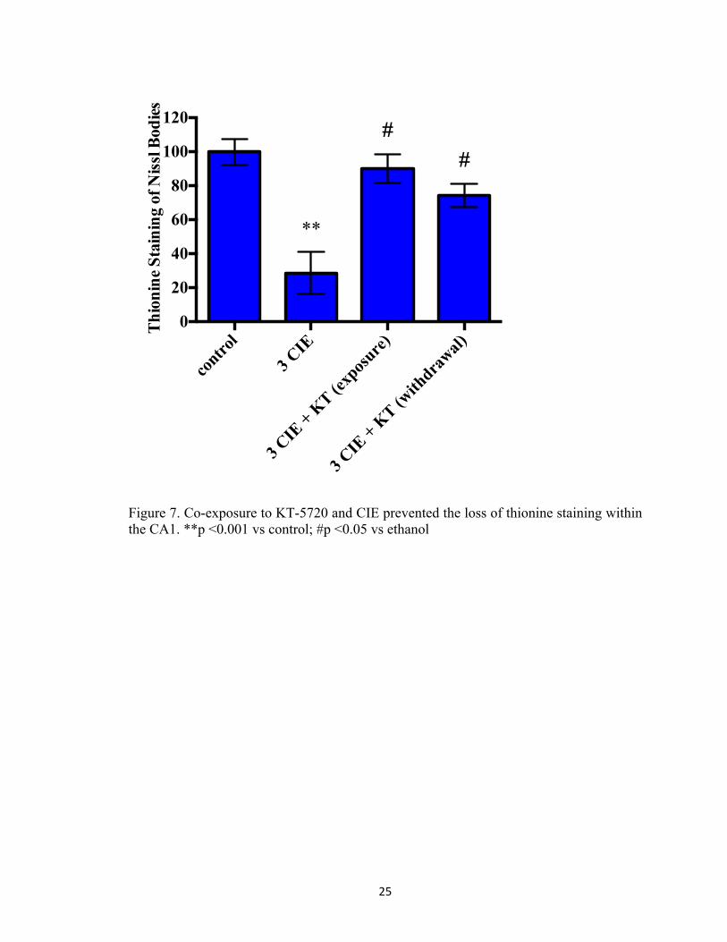
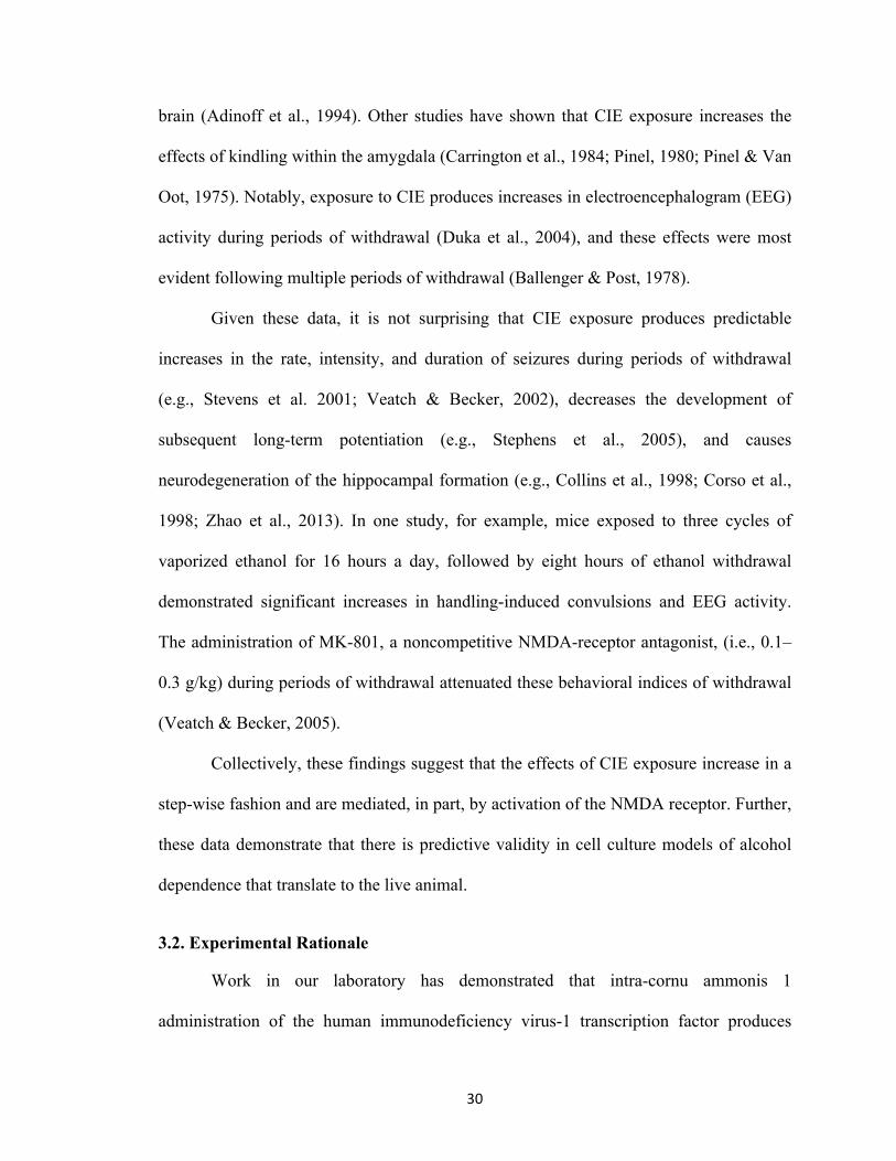
Figure 1. Alcohol abuse and misuse is a widespread phenomenon in the United States (SAMSHA, 2014). According to the National Survey on Drug Use and Health, an estimated 60 millions report current binge alcohol use. ................................................................................................................. 7 Figure 2. Experimental rationale for assessing mechanisms of damage associated with multiple withdrawals in organotypic hippocampal slice cultures. ................................................................ 20 Figure 3. Rat hippocampal explants were exposed to ethanol (50 mM) for five days in vitro, followed by a 24-hour period of withdrawal and repeated three times. KT-5720, a protein kinase inhibitor was applied to ethanol-enriched medium or ethanol-free medium to assess the influence of protein kinase A activity in promoting NMDA-receptor-mediated cytotoxicity produced by CIE. ................................................................................................................................................. 21 Figure 4. Co-exposure to KT-5720 (1 µM) and ethanol prevented the loss of NeuN immunoreactivity within the CA1 region produced by CIE. **p <0.001 vs control; #p <0.05 vs ethanol ............................................................................................................................................ 22 Figure 5. Co-exposure to KT-5720 and CIE attenuated the loss of NeuN immunofluorescence within the CA3 region. *p <0.05 vs control; **p <0.001 vs control .............................................. 23 Figure 6. Co-exposure to KT-5720 and CIE prevented the loss of NeuN immunofluorescence within the dentate gyrus. *p <0.05 vs control; **p <0.001 vs control; #p <0.05 vs ethanol .......... 24 Figure 7. Co-exposure to KT-5720 and CIE prevented the loss of thionine staining within the CA1. **p <0.001 vs control; #p <0.05 vs ethanol .......................................................................... 25 Figure 8. Co-exposure to KT-5720 and CIE attenuated the loss of thionine staining within the CA3. **p <0.001 vs control ............................................................................................................ 26 Figure 9. Co-exposure to KT-5720 and CIE prevented the loss of thionine staining within the dentate gyrus. **p <0.001 vs control; #p <0.05 vs ethanol ............................................................ 27 Figure 11. Representative experimental timelines showing that subjects were exposed to ethanol (4 g/kg) twice daily for five days, followed by two days of withdrawal, and repeated either a total of two times (i.e., two CIE) or three times (i.e., three CIE). .......................................................... 42 Figure 12. Withdrawal behavior was rated on a 10-point scale modified from prior reports in our laboratory (Sharrett-Field et al., 2013). Behaviors ranged from mild (e.g., hypoactivity, rigidity, aggression, and stereotypy) to more moderate (e.g., dystonic gait, retropulsion, splayed paws, and tremor) to severe (e.g., vocalizations and seizure). ........................................................................ 43 Figure 13. Changes in body weight in subjects exposed to ethanol (n=5) or an isocaloric diet (n=6) for two cycles of CIE. X-axis: days in Week One and Week Two. Data points show mean body weight in grams for subjects exposed to ethanol (blue bar) or an isocaloric diet (gray bar). Two asterisks indicate a significant day-by-treatment interaction. ................................................ 44 Figure 14. Changes in food consumption in subjects exposed to ethanol (n=5) or an isocaloric diet (n=6) for two cycles of CIE. X-axis: week. Data points show mean food consumption in grams for subjects exposed to ethanol (blue bar) or an isocaloric diet (gray bar). Two asterisks indicate a significant day-by-treatment interaction. ........................................................................................ 45

vii
Figure 15. Changes in body weight in subjects exposed to ethanol (n=5) or an isocaloric diet (n=6) for three cycles of CIE. X-axis: days in Week One (left), Week Two (middle), and Week Three (right). Data points show mean body weight in grams for subjects exposed to ethanol (blue bar) or an isocaloric diet (gray bar). Two asterisks indicate a significant day-by-treatment interaction. ...................................................................................................................................... 46 Figure 16. Changes in food consumption in subjects exposed to ethanol (n=5) or an isocaloric diet (n=6) for three cycles of CIE. X-axis: week. Data points show mean food consumption in grams for subjects exposed to ethanol (filled circle) or an isocaloric diet (empty circle). Two asterisks indicate a significant day-by-treatment interaction. ....................................................................... 47 Figure 17. Behavioral effects of withdrawal in subjects exposed to ethanol (n=5) or an isocaloric diet (n=6) for two cycles of CIE. X-axis: day. Data points show mean scores in withdrawal behavior for subjects exposed to ethanol (blue bar) or an isocaloric diet (gray bar). An asterisk indicates that is a significant difference between ethanol and control subjects. ............................ 48 Figure 18. Behavioral effects of withdrawal in subjects exposed to ethanol (n=5) or an isocaloric diet (n=6) for three cycles of CIE. X-axis: day. Data points show mean scores in withdrawal behavior for subjects exposed to ethanol (blue bar) or an isocaloric diet (gray bar). Two asterisks indicate a significant interaction between day and treatment. ........................................................ 49 Figure 19. Cognitive performance was assessed in subjects exposed to ethanol (n=5; filled circle) or an isocaloric diet (n=6; filled square) for two cycles of CIE. X-axis: day. Data points show mean scores in latency to platform. One asterisk indicates that there is a significant difference between acquisition days. ............................................................................................................... 50 Figure 20. Cognitive performance was assessed in subjects exposed to ethanol (n=5; filled circle) or an isocaloric diet (n=6; filled square) for two cycles of CIE. X-axis: day. Data points show mean scores in distance travelled. One asterisk indicates that there is a significant difference between acquisition days. ............................................................................................................... 51 Figure 21. Cognitive performance was assessed in subjects exposed to ethanol (n=5; filled circle) or an isocaloric diet (n=6; filled square) for two cycles of CIE. X-axis: day. Data points show mean scores in velocity. Two asterisks indicates that there is a significant difference between treatment groups. ............................................................................................................................ 52 Figure 22. Working memory assessment in subjects exposed to ethanol (n=5) or an isocaloric diet (n=6) for two cycles of CIE. X-axis: day. Data points show mean scores in platform crosses during the probe trial for subjects exposed to ethanol (filled circle) or an isocaloric diet (filled square). ........................................................................................................................................... 53 Figure 23. Cognitive performance was assessed in subjects exposed to ethanol (n=5; filled circle) or an isocaloric diet (n=6; filled square) for three cycles of CIE. X-axis: day. Data points show mean scores in latency to platform. One asterisk indicates that there is a significant difference between acquisition days. ............................................................................................................... 54 Figure 24. Cognitive performance was assessed in subjects exposed to ethanol (n=5; filled circle) or an isocaloric diet (n=6; filled square) for three cycles of CIE. X-axis: day. Data points show mean scores in distance travelled. One asterisk indicates that there is a significant difference between acquisition days. ............................................................................................................... 55 Figure 25. Cognitive performance was assessed in subjects exposed to ethanol (n=5; filled circle) or an isocaloric diet (n=6; filled square) for three cycles of CIE. X-axis: day. Data points show
viii
mean scores in velocity. Two asterisks indicate that there is a significant difference between groups. ............................................................................................................................................ 56 Figure 26. Peak BELs in subjects exposed to ethanol (n=5) for three cycles of CIE. X-axis: day. Data points show mean levels determined at 90 minutes post ethanol administration on Week One, Week Two, and Week Three. ................................................................................................ 57 Figure 27. Representative heat map images depicting distance travelled in Morris Water Maze decreased significantly on Day Five (bottom image), as compared to Day One (top image). ....... 58 Figure 28. Pictorial representation of the hypothesis that ethanol stimulates group 1 mGluR promoting release of intracellular calcium from IP3-mediated stores. Calcium also stimulates the adenylate cyclase signal transduction pathway that can produce coordinated phosphorylation of NMDA receptors (i.e., GluN1 and GluN2B) by PKA and PKC and subsequent trafficking of these receptor complexes from extrasynaptic to synaptic sites. During withdrawal, excessive calcium influx through these synaptic receptors activates phospholipases, endonucleases, and proteases to produce cytotoxicity and cell death. ........................................................................... 78 Figure 29. Rat hippocampal explants were exposed to 50 mM ethanol for five days followed by 24-hours of withdrawal with the cycle repeated three times (i.e., 3 CIE) with or without the addition of one of five antagonists. ................................................................................................ 79 Figure 30. Effects of noncompetitive mGluR1 antagonist CPCCOEt (0.5–3 µM) on NeuN immunofluorescence in the pyramidal CA1 cell layer of the rat hippocampus. Data are presented as percent control of the mean +/- the SEM. ** = statistical significance (p<0.001) compared to control hippocampi. ........................................................................................................................ 80 Figure 31. Effects of noncompetitive mGluR1 antagonist CPCCOEt (0.5–3 µM) on NeuN immunofluorescence in the pyramidal CA3 cell layer of the rat hippocampus. Data are presented as percent control of the mean +/- the SEM. * = statistical significance (p <0.05) compared to control hippocampi. ** = statistical significance (p<0.001) compared to control hippocampi. # = statistical significance (p <0.05) compared to ethanol hippocampi. .............................................. 81 Figure 32. Effects of noncompetitive mGluR1 antagonist CPCCOEt (0.5–3 µM) on NeuN immunofluorescence in the granule cell layer of the dentate gyrus. Data are presented as percent control of the mean +/- the SEM. * = statistical significance (p <0.05) compared to control hippocampi. ** = statistical significance (p<0.001) compared to control hippocampi. # = statistical significance (p <0.05) compared to ethanol hippocampi. .............................................. 82 Figure 33. Representative images of hippocampi exposed to ethanol-naïve media (control) or ethanol media (50 mM) or hippocampi co-exposed to 3.0 µM CPCCOEt and ethanol (50 mM). 83 Figure 34. Effects of mGluR5 antagonist SIB-1893 on NeuN immunofluorescence in the primary cell layer of the CA1. Data are presented as percent control of the mean +/- the SEM. * = statistical significance (p <0.05) compared to control hippocampi. ** = statistical significance (p <0.001) compared to control hippocampi. # = statistical significance (p <0.05) compared to ethanol hippocampi. ........................................................................................................................ 84 Figure 35. Effects of mGluR5 antagonist SIB-1893 on NeuN immunofluorescence in the primary cell layer of the CA3. Data are presented as percent control of the mean +/- the SEM. ** = statistical significance (p <0.001) compared to control hippocampi. ............................................. 85 Figure 36. Effects of mGluR5 antagonist SIB-1893 on NeuN immunofluorescence in the granule cell layer of the dentate gyrus. Data are presented as percent control of the mean +/- the SEM. **

ix
= statistical significance (p <0.001) compared to control hippocampi. # = statistical significance (p <0.05) compared to ethanol hippocampi. ................................................................................... 86 Figure 37. Representative images of hippocampi exposed to ethanol-naïve media (control) or ethanol media (50 mM) or hippocampi co-exposed to 20 µM SIB-1897 and ethanol (50 mM). ... 87 Figure 38. Effects of inhibitor of IP3-mediated calcium release (0.5 µM) on NeuN immunofluorescence in the pyramidal CA1 cell layer of the rat hippocampus. Data are presented as percent control of the mean +/- the SEM. * = statistical significance (p <0.05) compared to control hippocampi. ** = statistical significance (p <0.001) compared to control hippocampi. # = statistical significance (p <0.05) compared to ethanol hippocampi. .............................................. 88 Figure 39. Effects of inhibitor of IP3-mediated calcium release (0.5 µM) on NeuN immunofluorescence in the pyramidal CA3 cell layer of the rat hippocampus. Data are presented as percent control of the mean +/- the SEM. * = statistical significance (p <0.05) compared to control hippocampi. ** = statistical significance (p <0.001) compared to control hippocampi. # = statistical significance (p <0.05) compared to ethanol hippocampi. .............................................. 89 Figure 40. Effects of inhibitor of IP3-mediated calcium release (0.5 µM) on NeuN immunofluorescence in the granule cell layer of the dentate gyrus. Data are presented as percent control of the mean +/- the SEM. * = statistical significance (p <0.05) compared to control hippocampi. ** = statistical significance (p <0.001) compared to control hippocampi. # = statistical significance (p <0.05) compared to ethanol hippocampi. .............................................. 90 Figure 41. Effects of sigma-1 receptor antagonist BD-1047 (20 and 80 µM) on NeuN immunofluorescence in the CA1 hippocampal cell layer. Data are presented as percent control of the mean +/- the SEM. ** = statistical significance (p <0.001) compared to control hippocampi. # = statistical significance (p <0.05) compared to ethanol hippocampi. ........................................... 91 Figure 42. Effects of sigma-1 receptor antagonist BD-1047 (20 and 80 µM) on NeuN immunofluorescence in the CA3 hippocampal cell layer. Data are presented as percent control of the mean +/- the SEM. ** = statistical significance (p <0.001) compared to control hippocampi. # = statistical significance (p <0.05) compared to ethanol hippocampi. ........................................... 92 Figure 43. Effects of sigma-1 receptor antagonist BD-1047 (20 and 80 µM) on NeuN immunofluorescence in the dentate gyrus hippocampal cell layer. Data are presented as percent control of the mean +/- the SEM. ** = statistical significance (p <0.001) compared to control hippocampi. # = statistical significance (p <0.05) compared to ethanol hippocampi. ................... 93 Figure 44. Effects of ryanodine receptor antagonist dantrolene (5 µM) on NeuN immunofluorescence in the CA1 hippocampal cell layer. Data are presented as percent control of the mean +/- the SEM. ** = statistical significance (p <0.001) compared to control hippocampi.94 Figure 45. Effects of ryanodine receptor antagonist dantrolene (5 µM) on NeuN immunofluorescence in the CA3 hippocampal cell layer. Data are presented as percent control of the mean +/- the SEM. ** = statistical significance (p <0.001) compared to control hippocampi.95 Figure 46. Effects of ryanodine receptor antagonist dantrolene (5 µM) on NeuN immunofluorescence in the dentate gyrus hippocampal cell layer. Data are presented as percent control of the mean +/- the SEM. ** = statistical significance (p <0.001) compared to control hippocampi. .................................................................................................................................... 96 Figure 47. Representative experimental timelines depicting subjects were exposed to ethanol (4 g/kg) with or without the addition of MPEP (3 mg/kg) twice daily for five days followed by two days of withdrawal and repeated three times (i.e., 3 CIE). ........................................................... 106

x
Figure 48. Changes in body weight in subjects exposed to ethanol or an isocaloric diet with or without the addition of mGluR5 antagonist MPEP (3 mg/kg) for three cycles of CIE. X-axis: days in Week One, Week Two, and Week Three. Data points show mean body weight in grams. Two asterisks indicate that there is a significant day-by-treatment interaction. N=7 for control subjects; N=8 for control with MPEP; N=6 for ethanol subjects; N=8 for subjects administered ethanol and MPEP ............................................................................................................................................ 107 Figure 49. Behavioral effects of withdrawal in subjects exposed to ethanol or an isocaloric diet with or without the administration of MPEP for three cycles of CIE. X-axis: day. Data points show mean scores in withdrawal behavior observed during the third consecutive withdrawal from CIE. Two asterisks indicate that there is a significant diet-by-drug interaction. A pound sign indicates that there is a main effect of MPEP on withdrawal behavior in ethanol-dependent rats. N=7 for control subjects; N=8 for control with MPEP; N=6 for ethanol subjects; N=8 for subjects administered ethanol and MPEP. .................................................................................................. 108 Figure 50. Peak BELs in ethanol-dependent subjects on Weeks One, Two, and Three of the CIE treatment regimen. X-axis: Week. Data points show average BELs determined at 90 minutes post ethanol administration on Day Two of Weeks One, Two, and Three. Two asterisks indicate that there is a significant main effect of Week. A pound sign indicates that there is a main effect of MPEP on BELs in ethanol-dependent rats. N=4 for ethanol subjects; N=5 for ethanol with MPEP. ...................................................................................................................................................... 109

1
CHAPTER ONE: General Introduction
1.1. Background and Significance
Alcohol misuse and abuse is a widespread problem in the United States of
America. According to the National Survey on Drug Use and Health, just over half of the
population (52.1%), or an estimated 135.5 million individuals over the aged of 12, can be
characterized as being a current consumer of alcohol (Substance Abuse and Mental
Health Services Administration [SAMHSA], 2014). Of this number, nearly 60 million
individuals can be characterized as binge drinkers and 16 million individuals as heavy
drinkers (SAMHSA, 2014; Figure 1). Binge drinking is currently defined as an event in
which one’s blood ethanol level exceeds 0.08% (National Institute on Alcohol Abuse and
Alcoholism, 2004). Binge drinking as separate from alcohol use disorders is a quite
recent issue. The first volume of the Oxford English Dictionary (letters A and B) was
published in 1888 and defined “binge” as a verb meaning “to fawn; to cringe.” This
would indicate that such drinking practices were not considered a noteworthy issue or fell
under the catch-all of “alcoholism.” However, in a 1933 supplement, the dictionary
included in the definition a noun meaning “a heavy drinking bout,” as well as another
verb meaning “to drink heavily, ‘soak’” (see Crabbe et al., 2011 for an elegant review).
The American Psychological Association (APA) previously defined binge
drinking as the consumption of four or five drinks in one occasion (for females and
males, respectively; DSM-IV). Given that alcoholic beverages vary in the amount of
alcohol in a drink, this definition could be misinterpreted. It does not allow for
quantification via a biological dose of alcohol consumed, and while it does account for
other factors that could influence the effects of alcohol (e.g., gender), it does not account
for others (e.g., body mass index). The APA updated terminology in 2013 in the fifth

2
edition of the Diagnostic and Statistical Manual of Mental Disorders (DSM-V) to
combine the terms “alcohol abuse” and “alcohol dependence” under a more
encompassing definition of “alcohol use disorder.” This term allows for identification
without resorting to alcohol units consumed (APA, 2013). In order to be characterized
with an alcohol use disorder, an individual’s alcohol-related behaviors should include at
least two of the following eleven possible symptoms: consuming larger amounts than
intended, unsuccessful efforts to control drinking, significant amount of time spent on
alcohol-related activities, craving, failure to fulfill obligations, continued use despite
negative consequences, failure to participate in social activities, recurrent use in the
presence of hazardous domains, health conditions are exacerbated by alcohol use,
tolerance, and presence of withdrawal symptoms (APA, 2013).
These updated criterion provided by the APA also characterize the severity of
alcohol use disorders into one of three categories: mild (i.e., presence of two or three
symptoms), moderate (i.e., presence of four or five symptoms), and severe (i.e., presence
of six or more symptoms (APA, 2013). The National Institute on Alcohol Abuse and
Alcoholism (NIAAA) has a more detailed definition of binge drinking as a pattern (i.e.,
repetitive consumption) in which the total amount of alcohol in the blood (i.e., blood
alcohol concentration) exceeds 0.08% (i.e., 80 mg%; NIAAA, 2004). This definition
allows for a clearly defined quantification of binge drinking so as to more prudently
assess consequences of this pattern of consumption; it also provides a foundation for
physiological investigations on the subject (for a review, see Crabbe et al., 2011).
While the definition of binge or heavy drinking as a pathology continues to be
debated, the SAMHSA 2014 survey clearly notes a self-identified desire for intervention

3
with an estimated 18 million Americans reporting wanting treatment for an alcohol use
disorder in 2012 (alcohol abuse and dependence on alcohol). The survey also notes that a
very small percentage of individuals (1.5 million) actually received treatment at a
treatment facility (SAMHSA, 2014). Currently, there are four medications approved by
the United States Food and Drug Administration (FDA) for the treatment of alcohol
dependence: oral naltrexone, extended-release naltrexone, disulfiram, and acamprosate.
In a nine-month randomized trial, abstinence was higher for disulfiram (250 mg/day) than
for topiramate (150 mg/day), an anticonvulsant medication currently being assessed for
treatment of alcohol dependence (De Sousa et al., 2008). Findings from another trial
indicate that topiramate (25–300 mg/day) is more efficacious in promoting abstinence
than naltrexone (50 mg/day) or acamprosate (333 mg/day) (Narayana et al., 2008), while
other studies indicate that topiramate (50–400 mg/day) and naltrexone (50 mg/day) are
similarly effective for the treatment of alcohol dependence (Flórez et al., 2008; Flórez et
al., 2011). Given the fact that only a small percentage of individuals who are unhappy
with their drinking actually receive help, and that efficacy for relapse prevention outside
of the clinic is only modestly effective, pharmaceutical interventions are needed to
address this gap in care, as they may prove to be effectively both in terms of time and
economics of treatment because alcohol abuse and misuse is a widespread phenomenon.
Despite many preventative efforts (e.g., community-based services), the estimated
economic burden in the United States for excessive alcohol use, including binge drinking,
heavy drinking, underage consumption, and consumption by pregnant women was
estimated at $223.5 billion (e.g., Bouchery et al., 2006; Center for Disease Control and
Prevention, 2012). This figure includes costs for acute injuries, chronic health problems,

4
property damage, fetal alcohol syndrome, and loss of productivity. Notably, binge
drinking is responsible for the majority of these costs (76.1%). The American Journal of
Preventative Medicine suggests that responsible individuals and their families cover an
estimated 42% of the total costs associated with the excessive use of alcohol and the
United States government carries an approximate burden of 42% of these costs (Sacks et
al., 2013). Given that these figures are an approximation, the remainder of costs is not yet
accounted for. In the state of Kentucky, for example, the governmental economic burden
for alcohol-related damage and disease is an estimated $0.86 per drink (Sacks et al.,
2013). Precise economic costs are difficult to figure for a drug that is responsible for not
only individual physical damage but also has a ripple effect of damage to both self and
others physically and psychologically while often causing material damage as well;
however, within the context of these studies, it can be stated that the abuse of alcohol is a
widespread problem in the United States and the costs associated with its damage (both
direct and collateral) are significant. Future efforts to control this problem, then, need to
face it on a variety of fronts, both social and medical, including the development of
putative pharmacotherapies for the treatment of alcohol use disorders.
1.2. Alcohol Withdrawal: Effects on Brain and Behavior
Prolonged, excessive alcohol consumption (i.e., 4 or 5 drinks on one occasion) is
known to promote brain atrophy. The effects include significant decreases in the number
of cortical neurons (Harper, 1987), widening of ventricles (Bergman et al., 1980; Carlen
et al., 1978; Ron, 1977), cortical degeneration (Epstein, 1977), and widening of sulci and
fissures (Bergman et al., 1980). In one study, males with a history of alcohol use
disorders displayed significant decreases of anterior hippocampal volume (Sullivan et al.,

5
1995). In another study, individuals with a history of withdrawal-induced seizure activity
from alcohol dependence demonstrated significant decreases in the volume of temporal
matter when compared with individuals who had not experienced a seizure during
detoxification from alcohol (Sullivan et al., 1996). However, it has also been shown that
decreases in the volume of white matter recover quite significantly following protracted
abstinence (Pfefferbaum et al., 1995). Thus, given proper treatment, the effects of alcohol
use disorders may be diminished.
Alcohol use disorders have also been shown to produce neurocognitive
consequences, and low levels of blood flow in the brain have been associated with
decreases in cognitive function. For example, significant relationships between scores on
neuropsychological assessments and density of gray matter (e.g., hippocampus, cortices,
and cerebellum) have been reported in alcohol-dependent participants (Chanraud et al.,
2007). In another study, alcohol-dependent female participants displayed decreases in
verbal and nonverbal working memory, gait, and balance (Sullivan & Marsh, 2003).
Other studies have demonstrated decreases in assessments measuring visuospatial
abilities, psychomotor speed, gait, and balance (for reviews, see Kleinknecht &
Goldstein, 1972; Luhar et al., 2013; Sullivan et al., 2000).
Studies employing retrospective analyses of in-patient chart records of adults
suggest that multiple withdrawals, or periods of abstinence, are associated with increases
in rate, intensity, and duration of withdrawal-induced seizures. Gross and colleagues
(1972) found a significant relationship between seizures experienced during acute
withdrawal and re-admittance for acute withdrawal. Ballenger and Post (1978)
demonstrated a significant relationship between years of abuse, amount of alcohol
6
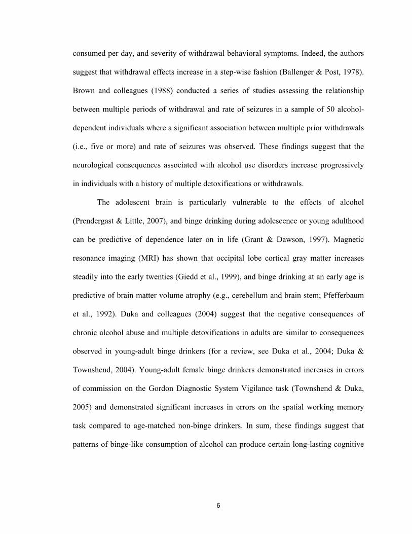
consumed per day, and severity of withdrawal behavioral symptoms. Indeed, the authors
suggest that withdrawal effects increase in a step-wise fashion (Ballenger & Post, 1978).
Brown and colleagues (1988) conducted a series of studies assessing the relationship
between multiple periods of withdrawal and rate of seizures in a sample of 50 alcohol-
dependent individuals where a significant association between multiple prior withdrawals
(i.e., five or more) and rate of seizures was observed. These findings suggest that the
neurological consequences associated with alcohol use disorders increase progressively
in individuals with a history of multiple detoxifications or withdrawals.
The adolescent brain is particularly vulnerable to the effects of alcohol
(Prendergast & Little, 2007), and binge drinking during adolescence or young adulthood
can be predictive of dependence later on in life (Grant & Dawson, 1997). Magnetic
resonance imaging (MRI) has shown that occipital lobe cortical gray matter increases
steadily into the early twenties (Giedd et al., 1999), and binge drinking at an early age is
predictive of brain matter volume atrophy (e.g., cerebellum and brain stem; Pfefferbaum
et al., 1992). Duka and colleagues (2004) suggest that the negative consequences of
chronic alcohol abuse and multiple detoxifications in adults are similar to consequences
observed in young-adult binge drinkers (for a review, see Duka et al., 2004; Duka &
Townshend, 2004). Young-adult female binge drinkers demonstrated increases in errors
of commission on the Gordon Diagnostic System Vigilance task (Townshend & Duka,
2005) and demonstrated significant increases in errors on the spatial working memory
task compared to age-matched non-binge drinkers. In sum, these findings suggest that
patterns of binge-like consumption of alcohol can produce certain long-lasting cognitive

7
effects in young-adult social drinkers that are similar to those observed in chronic
alcohol-dependent adults.
Collectively, these studies demonstrate that patterns of binge-like alcohol
consumption and multiple detoxifications or withdrawals, predict worse neurologic
outcomes in the clinical population. The purpose of the present report is to identify
potential mechanisms associated with promoting these consequences using rodent models
of chronic, intermittent ethanol exposure (i.e., CIE) in vitro and in vivo.
Current alcohol use
~136 million
Binge alcohol use
~ 60 million
Heavy alcohol use
~ 16 million
5/10
2.3/10
0.8/10
Figure 1. Alcohol abuse and misuse is a widespread phenomenon in the United States (SAMSHA, 2014). According to the National Survey on Drug Use and Health, an estimated 60 millions report current binge alcohol use.

8
CHAPTER TWO: Study One: Protein Kinase Activation and Cytotoxicity of Intermittent Ethanol in Vitro
2.1. Introduction
Goddard and colleagues (1969) conducted a seminal series of studies employing
electroencephalogram (EEG) techniques to assess the effects of repeated, subthreshold
stimulations to areas of the limbic system on epileptic-like convulsions in rodents
(Goddard et al., 1969). In these studies, delivery of initial electrical stimulations did not
produce significant alterations in EEG activity or epileptic-like behavioral activity. But
significant spikes in EEG activity and convulsions were observed following repeated,
daily, subthreshold stimulation of areas of the limbic system. The authors concluded that
spikes in EEG activity and epileptic-like activity were “kindled” in areas of the limbic
system. These effects have since been observed in many different types of preclinical
laboratory animals (Epsztein et al., 2008; Loscher et al., 1998; Sutala et al., 1988).
Other studies have also examined the effects of kindling in other areas of the
brain, such as the amygdala. Indeed, it is suggested that repeated electrical stimulation to
the amygdala can produce subsequent yet spontaneous spikes in EEG activity in the
hippocampus, as well as seizure activity (McNamara et al., 1988). These findings are
particularly interesting in light of the notion that the dentate gyrus, the primary granule
cell layer of the hippocampus, is known to demonstrate a unique reorganization after
kindling in these areas. For example, McNamara (1988) provided evidence that the
sprouting of axons produced by neurons in the granule cell layer and mossy fiber tract of
the pyramidal cell layer of the CA3 is associated with decreases in the seizure threshold.
Notably, although neurons in the granule cell layer die following electrical stimulation to

9
the hippocampus, these effects are followed by subsequent neuronal proliferation (Parent
et al., 1997). These studies are the foundation for the hypothesis that multiple periods of
withdrawal could serve as a kindling stimulus in the development of cytotoxicity
following chronic, intermittent ethanol (CIE) exposure in vitro.
2.2. Experimental Rationale
Prior work has suggested that multiple cycles of CIE produces consistent and
significant decreases of neuron-specific nuclear protein (NeuN) and thionine staining of
Nissl bodies in each of the primary cell layers of the hippocampal formation (i.e., the
granule cell layer of the dentate gyrus and the pyramidal cell layers of the CA1 and CA3;
Reynolds et al., 2015). These effects were prevented following exposure to the NMDA-
receptor antagonist (2R)-amino-5-phosphonovaleric acid (APV) at a 40µM concentration.
These effects highlight what is classically referred to as “NMDA-receptor-mediated
excitotoxicity” (Reynolds et al., 2015).
Excitotoxicity is conceptualized as the overactivation or overstimulation of amino
acid receptor complexes producing subsequent neuronal cell death (Olney et al., 1986).
Choi (1987, 1992) identified the NMDA receptor as the likely candidate for producing
these excitotoxic effects in vitro and described this event as being mediated by an
excessive influx of extracellular calcium and the subsequent activation of phospholipases,
endonucleases, and proteases. This possibility is consistent with the work of others who
have shown that chronic ethanol exposure increases calcium influx through the NMDA
receptor (DeWitte et al., 2003), increases the sensitivity of the NMDA receptor (Lovinger
et al., 1993), increases production of NMDA-receptor complexes (Floyd et al., 2003), and

10
increases synaptic clustering of the NMDA receptor at the synapse (Carpenter-Hyland et
al., 2004).
Other studies have shown neuroadaptive changes in second messenger systems
using a CIE treatment regimen in cortical neurons of 75 mM ethanol for 14 hours
followed by 10 hours of withdrawal that is repeated a total of five times and terminated
by either a two- or five-day period of withdrawal (i.e., Qiang et al., 2007). Western blot
and immunoblot analyses have shown that CIE produced selective increases in GluN1
and GluN2B subunit expression on the surface membrane (Qiang et al., 2007). However,
exposure to KT-5720 (i.e., 1 µM)—a protein kinase A (PKA) inhibitor—prevented
increases in GluN1 and partially prevented increases in GluN2B expression (Qiang et al.,
2007). These data are consistent with studies conducted by Carpenter-Hyland and
colleagues (2004) suggesting changes in the NMDA-receptor synaptic clustering is
dependent on the activity of PKA. These findings suggest a role for PKA signaling in the
overactivation of the NMDA receptor and subsequent cytotoxicity; however, this
relationship is not yet fully understood. The purpose of the present studies, therefore, is to
examine the distinct role that PKA-dependent NMDA-receptor activation has in
promoting hippocampal cytotoxicity produced by CIE in vitro.
2.3. Methods
Organotypic hippocampal slice culture preparation. Whole brains from eight-day-old
Sprague-Dawley rats (Harlan Laboratories; Indianapolis, IN) were aseptically removed
and placed in culture dishes containing frozen dissecting medium composed of Minimum
Essential Medium (MEM; Invitrogen, Carlsbad, CA), 25 mM HEPES (Sigma, St. Louis,
MO), and 50 µM streptomycin/penicillin (Invitrogen). Bilateral hippocampi were
11
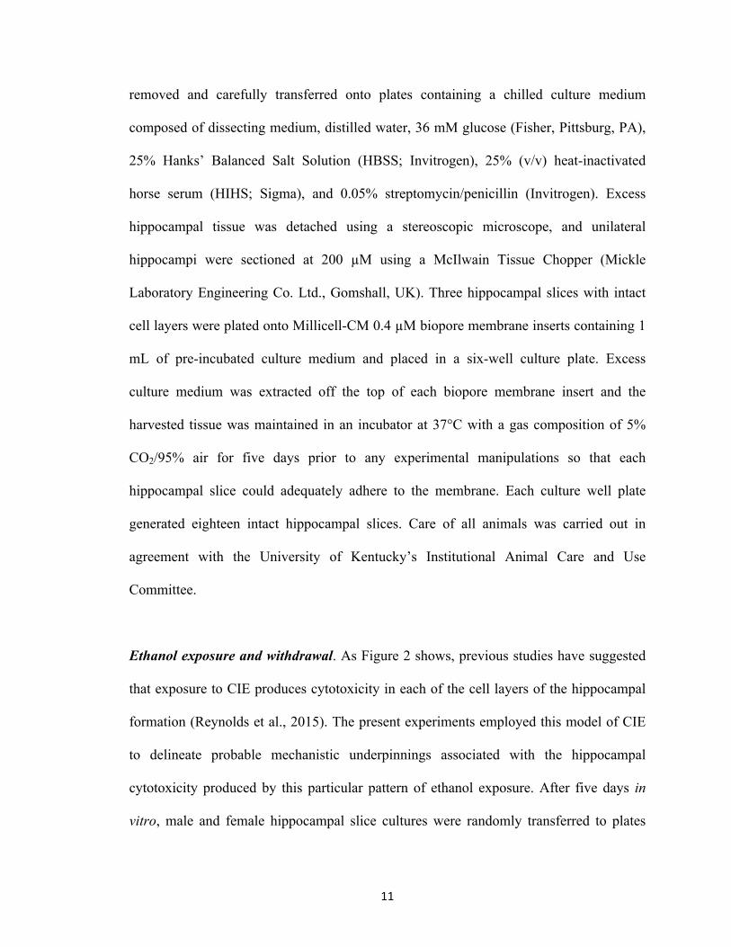
removed and carefully transferred onto plates containing a chilled culture medium
composed of dissecting medium, distilled water, 36 mM glucose (Fisher, Pittsburg, PA),
25% Hanks’ Balanced Salt Solution (HBSS; Invitrogen), 25% (v/v) heat-inactivated
horse serum (HIHS; Sigma), and 0.05% streptomycin/penicillin (Invitrogen). Excess
hippocampal tissue was detached using a stereoscopic microscope, and unilateral
hippocampi were sectioned at 200 µM using a McIlwain Tissue Chopper (Mickle
Laboratory Engineering Co. Ltd., Gomshall, UK). Three hippocampal slices with intact
cell layers were plated onto Millicell-CM 0.4 µM biopore membrane inserts containing 1
mL of pre-incubated culture medium and placed in a six-well culture plate. Excess
culture medium was extracted off the top of each biopore membrane insert and the
harvested tissue was maintained in an incubator at 37°C with a gas composition of 5%
CO2/95% air for five days prior to any experimental manipulations so that each
hippocampal slice could adequately adhere to the membrane. Each culture well plate
generated eighteen intact hippocampal slices. Care of all animals was carried out in
agreement with the University of Kentucky’s Institutional Animal Care and Use
Committee.
Ethanol exposure and withdrawal. As Figure 2 shows, previous studies have suggested
that exposure to CIE produces cytotoxicity in each of the cell layers of the hippocampal
formation (Reynolds et al., 2015). The present experiments employed this model of CIE
to delineate probable mechanistic underpinnings associated with the hippocampal
cytotoxicity produced by this particular pattern of ethanol exposure. After five days in
vitro, male and female hippocampal slice cultures were randomly transferred to plates

12
containing either 1 ml of the culture medium (control) or medium containing a binge-like
ethanol concentration (i.e., 0 and 50 mM) for five days with or without the addition of
PKA inhibitor KT-5720 (1µM). At 11 days in vitro, cultures were removed from their
three respective treatment groups and transferred to plates containing 1 ml of fresh
ethanol-naïve culture medium for a 24-hour ethanol withdrawal period. These treatments
were repeated a total of three times in consecutive order. Subsets of hippocampi were
exposed to the CIE treatment regimen described above, but exposure to KT-5720
occurred only during withdrawal and not during ethanol exposure. During each five-day
exposure period, ethanol and control-treated cultures were maintained inside Ziploc bags
filled with 5% CO2/95% air and water bath solutions containing either distilled water (50
ml) for control plates or distilled water (50 ml) containing ethanol (50 mM) for ethanol-
treated plates. On in vitro day 23, cultures were fixed for immunohistochemistry and
histology. This CIE treatment regimen is shown in Figure 3.
Immunohistochemistry and histology. Cultures were fixed by placing 1 ml of 10%
formalin solution on the top and bottom of each well for 30 minutes before being washed
twice with phosphate buffered saline (PBS) and stored at 4°C until
immunohistochemistry was initiated. NeuN (Fox-3) is found in nearly all post-mitotic
neurons (Kim et al., 2009; Mullen et al., 1992), and thus cultures were labeled with NeuN
to assess cytotoxicity in each primary cell layer of the hippocampal formation: pyramidal
cell layers of the cornu ammonis (CA1 and CA3) and granule cell layer of the dentate
gyrus. Fixed inserts were transferred to a plate containing 1 ml of permeabilization
(wash) buffer (200 ml PBS [Invitrogen], 200 µL Triton X-100 [Sigma], 0.010 mg Bovine

13
Serum [Sigma]) with 1 ml of buffer added to the top of each well for 45 minutes to
permeate cell membranes. Tissue was then incubated with the primary monoclonal
antibody mouse anti-NeuN (1:200; Sigma) for 24 hours. Inserts were then washed with
PBS and incubated for 24 hours with goat anti-mouse fluorescein isothiocyanate (FITC;
1:200; Sigma). Histological staining using thionine was conducted to confirm
immunohistochemical findings. Thionine is a monochromatic dye known to bind to Nissl
substance(s) located on cytoplasmic RNA (Kadar et al., 2009) and DNA content of all
cell nuclei (Scott & Willett, 1996). Following immunohistochemistry, tissues were
exposed to a 0.2% thionine stock solution for five minutes followed by a two-minute
dehydration period with 70% ethanol before being washed twice and imaged. Cultures
were imaged with SPOT software 4.0.2 (advanced version) for Windows (W. Nuhsbahm
Inc.; McHenry, IL, USA) through a 5x objective with a Leica DMIRB microscope (W.
Nuhsbahm Inc.; McHenry, IL, USA) connected to a computer and captured with a SPOT
7.2 color mosaic camera (W. Nuhsburg). For immunohistochemical studies, FITC
fluorescence was detected with a band-pass filter at 495 nm (520 nm emission).
Statistical analyses. Statistical analyses were conducted to assess the influence of PKA in
the development of ethanol dependence. Effects were considered significant at p<0.05.
Study One was conducted two times using two different rat litters. All
immunohistochemical and histological data were converted to percent control and then
combined for ease of data interpretation. A one-way analysis of variance (ANOVA) was
conducted for all in vitro techniques proposed with treatment as the factor for each
hippocampal cell layer (i.e., CA1, CA3, and dentate gyrus). This statistical strategy is

14
based on results of preliminary data suggesting that sex is not a factor influencing the
effects produced by repeated binge-like ethanol. Post-hoc comparisons were conducted if
a significant effect of treatment was detected using a protracted Fisher’s Least Significant
Difference (LSD). These planned comparisons were used to make pairwise comparisons
between means. For graphical representation of thionine data, mean data from each
condition were converted using the formula ([x-100]-100*[-1]) so as to express data on
the same scale used for the immunohistochemical data. N=25–47 for
immunohistochemical data and 20–24 for histological data per primary cell layer of the
hippocampal formation.
2.4. Results
Study One examined the influence of PKA on the development of ethanol
dependence in rat hippocampal explants exposed to a PKA inhibitor either during ethanol
exposure or during withdrawal. Hippocampi exposed to three cycles of CIE demonstrated
a 19% decrease of NeuN immunoreactivity in the CA1 (F[5,228] = 6.26, p<0.001; Figure
4), a 31% decrease of NeuN immunoreactivity in the CA3 (F[5,228] = 11.46, p<0.001;
Figure 5), and a 25% decrease of NeuN immunoreactivity in the dentate gyrus (F[5,228]
= 10.82, p<0.001; Figure 6). Exposure to KT-5720 (1 µM) during periods of withdrawal
did not prevent this loss of NeuN immunoreactivity in any hippocampal subregion
(Figures 4, 5, and 6), while exposure to KT-5720 (1 µM) during periods of ethanol
exposure significantly attenuated this effect in the CA1 (p<0.001; Figure 4), CA3 (p <
0.05; Figure 5), and dentate gyrus (p<0.05; Figure 6).
Thionine staining of slices yielded similar findings, as exposure to KT-5720 (1
µM) significantly attenuated the losses of thionine staining in the CA1 (F[3, 88] = 12.06,

15
p<0.001; Figure 7), CA3 (F[3, 88] = 86.82, p<0.001; Figure 8), and dentate gyrus (F[3,
88] = 7.91, p<0.001; Figure 9). Notably, exposure to KT-5720 (1 µM) during periods of
withdrawal modestly attenuated the loss of thionine staining (p<0.05; Figure 7) in the
CA1; however, this effect was not observed in the CA3 or dentate gyrus. Moreover,
exposure to KT-5720 in ethanol-naïve slices produced significant decreases of NeuN
immunoreactivity (p<0.05, data not shown) and thionine staining (p<0.05, data not
shown) as compared to control values in the CA3. This effect was not demonstrated in
the CA1 or dentate gyrus subregions of the hippocampal formation. Representative
images are found in Figure 10.
2.5. Discussion
The present studies found that multiple cycles of CIE produced cytotoxicity in
hippocampal slice cultures, as reflected by significant decreases of NeuN
immunoreactivity and thionine staining. These findings are not unexpected, as we had
previously found that exposure to 50 mM ethanol for five days in vitro followed by a
single ethanol withdrawal period did not result in significant decreases of NeuN
immunoreactivity or thionine staining in any hippocampal subregion (Reynolds et al.,
2015). Other studies have also found that exposure to ethanol (50 mM) followed by a
single period of withdrawal did not produce excitotoxicity in vitro (Butler et al., 2009;
Self et al., 2005), but chronic exposure to this concentration of ethanol produced a
heightened sensitivity of hippocampal glutamatergic receptors systems to agonists (Self
et al., 2004). In a recent study, it was found that exposure to three cycles of CIE exposure
in hippocampal slices produced consistent and significant decreases of NeuN
immunoreactivity and thionine staining in the pyramidal cell layers of the CA1 and CA3

16
and the granule cell layer of the dentate gyrus. These data are consistent with findings in
which exposure to CIE produced deficits, such as cytotoxicity in cortical neurons (Nagy
& Laszlo, 2002) and increased seizure susceptibility (Kokka et al., 1993), EEG activity
(Veatch & González, 1996), and hippocampal neurodegeneration in vivo (Collins et al.,
1998; Zhao et al., 2013). The present findings expand on this literature by characterizing
the distinct roles of ethanol exposure and PKA on hippocampal injury produced by CIE
exposure. In a previous study, exposure to 50 mM ethanol for 18 consecutive days (in the
absence of any withdrawal) did not produce significant loss of NeuN immunoreactivity or
thionine staining in any hippocampal subregion (Reynolds et al., 2015), but NMDA
receptors were overactivated during periods of withdrawal (Reynolds et al., 2015). The
present findings extend upon these results by demonstrating that neuroadaptations in the
NMDA receptor, which likely confer sensitivity to cytotoxicity, are mediated, in part, via
PKA prior to withdrawal. This finding suggests the importance of PKA in promoting
neuroadaptations in NMDA-receptor activity following CIE exposure (i.e., 50 mM).
In general, CIE exposure produced a more modest reduction of NeuN
immunofluorescence than thionine staining. While the reasons for these effects are
unknown, it is likely that they reflect inherent differences regarding the neuronal
selectivity of these immunohistochemical and histological markers (Kadar et al., 2009;
Mullen et al., 1992; Scott & Willett, 1996; Wolf et al., 1996). The more significant
decreases in thionine observed in the current study could reflect a loss of neuronal and
non-neuronal cell types. For example, prior work has shown that a single exposure to
ethanol withdrawal produces significant decreases of glial fibrillary acidic protein
immunofluorescence in the rat hippocampus (Wilkins et al., 2006), and astrocytes are

17
known to express each of the known NMDA-receptor subunits and are relatively
vulnerable to classic NMDA-receptor-mediated excitotoxicity (Lee et al. 2010).
Therefore, the present findings suggest that both neurons and glia (i.e., astrocytes) may
demonstrate sensitivity to NMDA-receptor-dependent cytotoxicity following CIE.
Notably, these cytotoxic effects are ubiquitous in nature, as they can be observed in each
examined cell layer of the hippocampal formation (i.e., CA1, CA3, and dentate gyrus).
Indeed, prior studies have demonstrated that the pyramidal cell layer of the CA1 is
selectively vulnerable to the excitotoxic effects of ethanol withdrawal (Prendergast et al.
2004). It is, therefore, likely that the CIE exposure treatment regimen employed in the
present study produced a greater extent of withdrawal-related cytotoxicity via spreading
cytotoxicity of the pyramidal cell layer to the granule cell layer. This notion is supported
by the work of Guiterrez and Heinemann (1999) which demonstrated de novo sprouting
of mossy fiber tracts from granule cells to pyramidal cells.
The role of PKA in the overactivation of NMDA receptors. NMDA receptors are
heteromeric ionotropic glutamatergic receptors comprised of an obligatory NR1 and
combination of NR2 (A–D) subunits (Dingledine et al., 1999; for a review, see Paoletti &
Neyton, 2007). During withdrawal from chronic ethanol exposure, activation of these
receptors can lead to excessive calcium (Ca2+) influx and subsequent cell death via
activation of phospholipases, endonucleases, and proteases (i.e., excitotoxicity; Choi,
1992). Following CIE exposure, neuroadaptative changes in NMDA receptors can further
potentiate Ca2+-mediated excitotoxicity. In one study, for example, exposure to CIE
produced increases in NMDA-receptor-mediated excitatory post-synaptic potentials

18
(EPSPs) in the pyramidal cell layer of the CA1 of the rat hippocampus (Nelson et al,
2005).
Ethanol exposure indirectly enhances NMDA-receptor function through PKA-
dependent phosphorylation (Carpenter-Hyland et al., 2004) and the subsequent
trafficking of NMDA receptors from the endoplasmic reticulum to the synapse (Mu et al.,
2003). The reasons for these effects are associated with cAMP-response element-binding
protein (CREB) activation and subsequent increases in gene expression of the GluN2B
subunit (Rani et al., 2005). Previous studies examining the modulatory effects of ethanol
on GABA suggest it has a role in the release of intracellular Ca2+ in the activation of
PKA (Kelm et al., 2008). These alterations in PKA activity are associated with decreases
in the sedative effects of ethanol and voluntary ethanol consumption (Thiele et al., 2000;
Wand et al., 2001). The present studies examined the functional role of PKA-dependent
overactivation of NMDA receptors. Co-exposure to ethanol with the PKA inhibitor KT-
5720 for three cycles of CIE significantly attenuated the loss of mature neurons. By
contrast, exposure to KT-5720 during periods of EWD did not attenuate these effects.
These findings suggest that the CIE regimen produces increases in PKA activity, which
promotes the overactivation of NMDA receptors during periods of withdrawal.
The selectivity of protein kinase inhibitors is debatable. Given that there are an
estimated 500 protein kinases encoded in the human genome and that they each are
belong to one super-family, it is no surprise that protein kinase inhibitors may target more
than one protein kinase. In one study, for example, specificities of 60 compounds were
assessed against a panel of 70–80 protein kinases (Bain et al., 2007). While this study
made recommendations for specific protein kinase inhibitors that target specific kinases,

19
such as mitogen-activated protein kinase (MAPK) (e.g., BIRB 0796) and
phosphoinositide-3 kinases (e.g., PPI and Src inhibitor-1 collectively), recommendations
to specifically target other kinases (e.g., PKA) were not made. However, other studies
have employed KT-5720 in experimental procedures to specifically identify the role of
PKA in various physiological processes (e.g., Almami et al., 2014; Clarysse et al., 2014;
Rodriguez-Duran & Escobar, 2014; Sun et al., 2014; Xin et al., 2014). Together, these
studies suggest that conclusions pertaining to the use of KT-5720 should be drawn
cautiously.
Collectively, the present study and prior work suggest that the concomitant
neuroadaptations in protein kinase activation (e.g., PKA) and increased function of
NMDA receptors are likely associated with both the behavioral and neurodegenerative
effects observed following multiple bouts of heavy ethanol consumption (Duka et al.,
2003; Duka et al., 2004; Sullivan et al., 1996) and in the development of tolerance.

20
Figure 2. Experimental rationale for assessing mechanisms of damage associated with multiple withdrawals in organotypic hippocampal slice cultures.

21
Week 3Week 1 Week 2
ETHANOL (50 MM) +/
KT-5720
WITHDRAWAL +/KT-5720
5 days in vitro
1 day in vitro
ETHANOL (50 MM) +/
KT-5720
ETHANOL (50 MM) +/
KT-5720
WITHDRAWAL +/KT-5720
WITHDRAWAL +/KT-5720
Figure 3. Rat hippocampal explants were exposed to ethanol (50 mM) for five days in vitro, followed by a 24-hour period of withdrawal and repeated three times. KT-5720, a protein kinase inhibitor was applied to ethanol-enriched medium or ethanol-free medium to assess the influence of protein kinase A activity in promoting NMDA-receptor-mediated cytotoxicity produced by CIE.

22
contro
l
contro
l + K
T (exp
osure)
contro
l + K
T (with
drawal)
3 CIE
3 CIE
+ KT (e
xpos
ure)
3 CIE
+ KT (w
ithdra
wal)50
60
70
80
90
100
110
Neu
N I
mm
unof
lour
esce
nce
****
#
Figure 4. Co-exposure to KT-5720 (1 µM) and ethanol prevented the loss of NeuN immunoreactivity within the CA1 region produced by CIE. **p <0.001 vs control; #p <0.05 vs ethanol

23
contro
l
contro
l + K
T (exp
osure)
contro
l + K
T (with
drawal)
3 CIE
3 CIE
+ KT (e
xpos
ure)
3 CIE
+ KT (w
ithdra
wal)50
60
70
80
90
100
110
Neu
N I
mm
unof
lour
esce
nce
**** **
*
Figure 5. Co-exposure to KT-5720 and CIE attenuated the loss of NeuN immunofluorescence within the CA3 region. *p <0.05 vs control; **p <0.001 vs control

24
contro
l
contro
l + K
T (exp
osure)
contro
l + K
T (with
drawal)
3 CIE
3 CIE
+ KT (e
xpos
ure)
3 CIE
+ KT (w
ithdra
wal)50
60
70
80
90
100
110
Neu
N I
mm
unof
lour
esce
nce
**
#*
**
*
Figure 6. Co-exposure to KT-5720 and CIE prevented the loss of NeuN immunofluorescence within the dentate gyrus. *p <0.05 vs control; **p <0.001 vs control; #p <0.05 vs ethanol
25
contro
l
3 CIE
3 CIE
+ KT (e
xpos
ure)
3 CIE
+ KT (w
ithdra
wal)0
20
40
60
80
100
120
Thi
onin
e St
aini
ng o
f Nis
sl B
odie
s
**
##
Figure 7. Co-exposure to KT-5720 and CIE prevented the loss of thionine staining within the CA1. **p <0.001 vs control; #p <0.05 vs ethanol

26
contro
l
3 CIE
3 CIE
+ KT (e
xpos
ure)
3 CIE
+ KT (w
ithdra
wal)0
20
40
60
80
100
120
Thi
onin
e St
aini
ng o
f Nis
sl B
odie
s
****
Figure 8. Co-exposure to KT-5720 and CIE attenuated the loss of thionine staining within the CA3. **p <0.001 vs control

27
contro
l
3 CIE
3 CIE
+ KT (e
xpos
ure)
3 CIE
+ KT (w
ithdra
wal)0
20
40
60
80
100
120
Thi
onin
e St
aini
ng o
f Nis
sl B
odie
s
****
#
Figure 9. Co-exposure to KT-5720 and CIE prevented the loss of thionine staining within the dentate gyrus. **p <0.001 vs control; #p <0.05 vs ethanol
Figure 10. Representative images of NeuN immunoreactivity and thionine staining in hippocampal slices exposed to ethanol (three CIE), co-exposure to ethanol (three CIE and KT-5720 [1 µM]), or ethanol-naïve control media.
Control 3 CIE 3 CIE + KT-5720 (exposure)
NeuN
Thionine

28
CHAPTER THREE: Study Two: Effects of Chronic, Intermittent Ethanol on Withdrawal Behavior and Working Memory in Vivo
3.1. Introduction
Neuroadaptations and cognitive impairment associated with multiple alcohol
detoxifications that are observed in humans (Duka et al., 2003) may also be demonstrated
in rodent models (Stephens & Duka, 2008). This means that rodents can be used for
preclinical assessment to assess metabolic and/or physical tolerance to ethanol after
short-term exposure to ethanol (Self et al., 2009). Rodents are particularly valuable in
these studies as they quickly develop effects from ethanol. In previous preclinical studies,
repeated administration of high doses of ethanol have been used to evoke physical
dependence in rodents. Factors considered when evaluating the effects of ethanol
dependence in these studies typically include duration and dose (Majchrowicz, 1975;
Sircar & Sircar, 2005; Wallgren et al., 1973). In one study, for example, a four-day,
binge-like exposure to high doses of ethanol produced rapid intoxication. This was
followed by repeated administration of high doses of ethanol for four consecutive days
creating ethanol dependence rather rapidly (Majchrowicz, 1975). Once dependence was
developed and blood ethanol levels declined, behavioral effects of withdrawal were
evident. These effects included, but were not limited to, hypoactivity, tremors, wet dog
shakes, spastic rigidity, and convulsions (Majchrowicz, 1975). Becker and colleagues
(2000) concluded that the behavioral effects of withdrawal could be classified into one of
three categories: 1) overactivation of the autonomic nervous system (e.g., tachycardia,
increased blood pressure, sweating, and vomiting); 2) CNS hyperexcitability (e.g.,

29
anxiety and seizures); and 3) disturbances related to sensation and perception (e.g.,
hallucinations and delirium).
Work in our laboratory observed cognitive impairments and withdrawal
behavioral effects in rodents exposed to ethanol three times per day (3–5 g/kg) for four
days (Self et al., 2009). Rodents in this study rapidly developed metabolic tolerance to
ethanol, which was reflected in significant decreases in peak blood ethanol levels from
~187.69 mg/dl (Day Two) to ~100 mg/dl (Day Four; Self et al., 2009). Long-term ethanol
administration can also be used to examine dependence in rodents. In one study, for
example, ethanol administration (~6%) for 14 consecutive days produced significant
increases in NMDA GluN2A and GluN2B subunits in the rodent hippocampus (Devaud
& Alele, 2004). In another study, rodents exposed to ethanol (~6%) for a prolonged
period of time (13 months) followed by a six-week period of withdrawal demonstrated
significant decreases in spatial working memory compared to dependent rodents that did
not experience this period of withdrawal (Alele & Devaud, 2005). In sum, these studies
demonstrated that both short-term and long-term administration of ethanol withdrawal
may be used to evoke physical dependence, tolerance, and behavioral effects of
withdrawal in preclinical laboratory animals.
Other studies have assessed mechanisms involved in the associated damage of
multiple withdrawals, as rodents are known to display predictable increases in the
severity of withdrawal symptoms in a manner similar to the clinical population (Stephens
et al., 2001; Stephens et al., 2005). Studies using rodents have shown that CIE exposure
impairs metabolic processes (Clemmensen et al., 1988), creates kindling of the cortices
(McCown & Breese, 1990), and causes hyperexcitability of the limbic structures of the
30
brain (Adinoff et al., 1994). Other studies have shown that CIE exposure increases the
effects of kindling within the amygdala (Carrington et al., 1984; Pinel, 1980; Pinel & Van
Oot, 1975). Notably, exposure to CIE produces increases in electroencephalogram (EEG)
activity during periods of withdrawal (Duka et al., 2004), and these effects were most
evident following multiple periods of withdrawal (Ballenger & Post, 1978).
Given these data, it is not surprising that CIE exposure produces predictable
increases in the rate, intensity, and duration of seizures during periods of withdrawal
(e.g., Stevens et al. 2001; Veatch & Becker, 2002), decreases the development of
subsequent long-term potentiation (e.g., Stephens et al., 2005), and causes
neurodegeneration of the hippocampal formation (e.g., Collins et al., 1998; Corso et al.,
1998; Zhao et al., 2013). In one study, for example, mice exposed to three cycles of
vaporized ethanol for 16 hours a day, followed by eight hours of ethanol withdrawal
demonstrated significant increases in handling-induced convulsions and EEG activity.
The administration of MK-801, a noncompetitive NMDA-receptor antagonist, (i.e., 0.1–
0.3 g/kg) during periods of withdrawal attenuated these behavioral indices of withdrawal
(Veatch & Becker, 2005).
Collectively, these findings suggest that the effects of CIE exposure increase in a
step-wise fashion and are mediated, in part, by activation of the NMDA receptor. Further,
these data demonstrate that there is predictive validity in cell culture models of alcohol
dependence that translate to the live animal.
3.2. Experimental Rationale
Work in our laboratory has demonstrated that intra-cornu ammonis 1
administration of the human immunodeficiency virus-1 transcription factor produces

31
activation of NMDA receptors during withdrawal and subsequent persisting spatial
learning deficits in rodents (Self et al., 2009). These effects were prevented following the
administration of MK-801, an NMDA-receptor antagonist. Other studies have shown that
a CIE treatment regimen activates microglia and produces spatial learning and memory
deficits associated with neurodegeneration of limbic structures (Zhao et al., 2013). These
findings are consistent with the cognitive consequences readily observed in the clinical
population following prolonged, alcohol abuse (Kril et al., 1997; Sullivan et al., 2000;
Sullivan et al., 2005). The present study was designed to assess the effects of CIE
exposure on withdrawal behavior and working memory in a live animal model of ethanol
dependence.
3.3. Methods
Subjects. Twenty-two adult, male Sprague-Dawley rats (i.e., 300–325 grams; Harlan
Laboratories, Indianapolis, IN) were housed individually and allowed to acclimate to the
animal colony for two days following arrival. Subjects were then handled for two minutes
per day for three consecutive days prior to experimentation. Subjects were allowed ad
libitum access to food and water throughout the entire duration of the experiment. Food
weight data were collected at 0800 hour once per week. Each animal was weighed at
0800 hour on days Monday through Friday, prior to ethanol administration. A mortality
rate of 5% occurred to complications with the gavage procedure.
Ethanol administration and withdrawal. Previous studies conducted in our laboratory
have shown that CIE exposure produces hippocampal cytotoxicity in cell cultures
(Reynolds et al., 2015). The present study sought to extend these in vitro findings to

32
assess the effects of CIE on withdrawal behavior and working memory. In these studies,
subjects were administered ethanol (4 g/kg) via intragastric gavage twice daily at 0800
and 1600 for five consecutive days followed by a two-day period of withdrawal; this was
repeated either a total of two (Figure 11) or three times (Figure 11). One day after the last
period of withdrawal, cognitive function was assessed for one week in the Morris Water
Maze. This study aimed to achieve peak blood ethanol levels of approximately 200
mg/dl. The justification for the desired peak blood ethanol levels achieved of 200 mg/dl
is based on a previous study that suggested ethanol-induced sedation occurs between 340
and 190 mg/dl, but ataxia occurs between 570 and 250 mg/dl in the rat (Majchrowicz,
1975). The effective dose (i.e., ED50) for hypnotic effects of ethanol are approximately
1.80 g/kg in the rodent (Garfield & Bukusoglu, 1996). Experimental timelines for Study
Two are depicted in Figure 11.
Behavioral effects of withdrawal. Subjects exposed to CIE and an isocaloric diet were
observed and rated for behavioral effects of withdrawal. In this portion of the study,
subjects were administered ethanol for five consecutive days, followed by two days of
withdrawal; this was repeated a total of two times (i.e., two cycles of CIE) or three times
(i.e., three cycles of CIE). During each period of withdrawal, subjects were monitored 16
hours after the last ethanol administration (i.e., 0900 hour) for behavioral effects of
withdrawal. Assessment of withdrawal behavior occurred in a square Plexiglas chamber
for two minutes. During this time, two experimenters who were blinded to experimental
conditions rated the physical effects of withdrawal using a modified behavioral scale that
has been used in other studies (Majchrowicz, 1975; Self et al., 2009). Severity of

33
withdrawal symptoms was assessed using a 10-point discrete scale adapted from previous
studies (e.g., Majchrowicz, 1975; Self et al., 2009). These effects were rigidity, tremor,
stereotypy, retropulsion, dystonic gait, hypoactivity, aggression, splayed paws,
vocalization, and seizure (Figure 12).
Morris Water Maze. Two days following CIE administration, subjects were tested for
five consecutive days in the Morris Water Maze to assess cognitive function. This
protocol was published in a previous study by our laboratory (Self et al., 2009) and by
others (Morris, 1984). The apparatus was an open field pool 157.48 cm in diameter and
34.29 cm in height that was divided into five quadrants (platform, N, S, E, and W). The
removable platform (10.16 cm in diameter) was consistently positioned 1 cm below the
surface of the water and 30 cm from the pool wall in the northeast quadrant. Water was
maintained at between 19°C and 21°C and dyed black with non-toxic black powdered
paint so that the platform was hidden to sight. Each day of testing consisted of four 60-
second trials in which the animal located the platform via navigation through the pool. In
the event that the animal located the platform during the trial, they were allowed to
briefly rest on the platform for 15 seconds before being returned to their home cage for
five minutes. In the event that the animal did not navigate to the platform during the 60-
second trial, they were gently guided to the platform and allowed to rest for 15 seconds
before being returned to their home cage for five minutes. On Day Five, animals
completed a probe trial in which the platform was removed and the animal had 30
seconds to navigate the pool. Following this probe test, animals were returned to their
home cages. During each of the trials, a video monitor analyzer (EZ Mapper; AccuScan

34
Instrument, Inc) was used to assess 1) latency to platform (seconds), 2) distance travelled
(centimeters), and 3) velocity (centimeters per second).
Analysis of blood ethanol levels. In order to assess blood ethanol levels (BELs),
approximately 200 µL of tail blood was collected in two Fisherbrand heparinized micro-
hematocrit capillary tubes (Fisher Scientific) on Day Four of Weeks One, Two, and
Three. Next, samples were centrifuged for four minutes using an Analox benchtop
centrifuge (Analox Instruments) with blood plasma collected and placed into a 0.65 mL
Costar microcentrifuge tube (Fisher Scientific). Samples were stored at -80°C until
further analyses of BELs using an Analox AM1 apparatus. This device contains a Clark-
type amperometric oxygen electrode to directly measure levels of molecular oxygen.
When molecular oxygen is present, the enzyme alcohol oxidase is oxidized into two
byproducts: acetaldehyde and hydrogen peroxide. This device, therefore, indirectly
measures BELs, as oxygen consumption is directly proportional to ethanol concentrations
in each blood plasma sample.
Statistical analyses. Statistical analyses were conducted to assess the effects of CIE on
body weight, food consumption, and behavioral effects of withdrawal, as well as on
cognitive performance and BELs. Effects were considered significant at p<0.05. Body
weight data were analyzed by a two-factor repeated-measure ANOVA with day and
treatment as factors. Food consumption data were analyzed by a two-factor repeated-
measure ANOVA with week and treatment as factors. Behavioral effects of withdrawal
were analyzed by a two-factor repeated-measure ANOVA with day and treatment as
factors. In the Morris Water Maze, 1) latency (seconds), 2) distance (centimeters), and 3)

35
velocity (centimeters per second) were analyzed using separate two-factor repeated-
measure ANOVAs with day and treatment. Probe trial data from the Morris Water Maze
was analyzed by a one-factor ANOVA treatment (control and ethanol). BELs were
analyzed using a two-factor repeated-measure ANOVA with day and treatment (control
and ethanol) as factors. Planned comparisons were conducted if a significant effect of day
or treatment or an interaction between these two factors was detected using Tukey’s
Honestly Significant Difference (HSD). These planned comparisons were used to make
conservative pairwise comparisons between means.
3.4. Results
Body weight and food consumption. ANOVA revealed a significant interaction of day
and treatment (F[1,109] = 10.325, p<0.001) in subjects exposed to two cycles of CIE
(n=5) or an isocaloric diet (n=6). Figure 13 shows that significant decreases in body
weight (grams) were detected in subjects that were administered ethanol starting on Day
Three of the CIE treatment regimen. These decreases in body weight were accompanied
by decreases in food consumption, as ANOVA revealed a significant interaction of week
and treatment (F[1,21] = 36.834, p<0.001) in subjects exposed to two cycles of CIE.
Figure 14 shows that while there was no main effect of week in subjects administered a
control diet, subjects in the ethanol condition showed increases in food consumption on
the second week, as compared to the first week (F[1,21] = 65.799, p<0.001). Further, as
Figure 14 shows, subjects administered ethanol consumed less food in Week One
(F[1,109] = 30.300, p<0.001) and Week Two (p< 0.05; Tukey’s HSD) compared to
subjects that received an isocaloric diet.

36
ANOVA also revealed a significant interaction of day and treatment (F[1,164] =
7.272, p<0.001) in subjects exposed to three cycles of CIE (n=5) or an isocaloric diet
(n=6). Figure 15 shows that significant decreases in body weight (grams) were detected
in subjects that were administered ethanol starting on Day One of the CIE treatment
regimen. These decreases in body weight were accompanied by decreases in food
consumption, as ANOVA revealed a significant interaction of week and treatment
(F[1,21] = 5.590, p<0.05) in subjects exposed to three cycles of CIE. Figure 16 shows
that while there was no significant effect of treatment on food consumption for Week
One (p> 0.05; Tukey’s HSD), subjects administered ethanol demonstrated significant
decreases in food consumption compared to subjects that received an isocaloric diet in
Week Two (p< 0.001; Tukey’s HSD) and Week Three (p< 0.001; Tukey’s HSD).
Behavioral effects of withdrawal. We assessed the influence of CIE on the development
of dependence, as reflected by behavioral effects of withdrawal. ANOVA revealed a
significant main effect of treatment (F[1,21] = 8.578, p<0.05). Figure 17 shows that
subjects administered ethanol had significant increases in behavioral effects of
withdrawal compared to subjects that received an isocaloric diet. There was not a
significant main effect of day in subjects exposed to two cycles of CIE.
By contrast, ANOVA revealed a significant interaction of day and treatment
(F[1,164] = 17.430, p<0.001) in subjects exposed to three cycles of CIE (n=5) or an
isocaloric diet (n=6). Figure 18 shows that significant increases were detected on Day
Two and Day Three of withdrawal in subjects that received ethanol administration. This

37
figure also shows that the most robust increases in withdrawal behavior occurred on Day
Three of withdrawal (p<0.05; Tukey’s HSD) in ethanol-dependent subjects.
Morris Water Maze. ANOVA revealed a significant main effect of day on latency
(F[1,36] = 45.267, p<0.001) and distance (F[1,36] = 47.682, p<0.001) in subjects
exposed to two cycles of CIE (n=5) or an isocaloric diet (n=6). Figures 19 and 20 show
that latency to platform and distance travelled were significantly decreased as a function
of day in subjects that received ethanol and an isocaloric diet, respectively. ANOVA also
revealed a significant interaction of day and treatment on velocity (F[1,36] = 2.706,
p<0.05). Figure 21 shows that ethanol administration significantly decreased velocity on
Day Two (p<0.05; Tukey’s HSD), Day Four (p<0.05; Tukey’s HSD), and Day Five
(p<0.05; Tukey’s HSD). This effect was not observed on Day One or Day Three (p>0.05;
Tukey’s HSD). There were no significant effects of platform crosses at the 15-second
interval or total platform crosses during the probe trial (Figure 22).
ANOVA revealed a significant main effect of day on latency (F[1,36] = 34.951,
p<0.001) and distance (F[1,36] = 135.627, p<0.001) in subjects exposed to three cycles
of CIE (n=5) or an isocaloric diet (n=6). Figures 23 and 24 show that latency to platform
and distance travelled were significantly decreased as a function of day in subjects that
were administered ethanol and an isocaloric diet, respectively. By contrast, ANOVA
revealed a significant interaction of day and treatment on velocity (F[1,36] = 2.020,
p<0.05). Figure 25 shows that ethanol administration significantly increased velocity on
Day Four (p<0.05; Tukey’s HSD). This figure also shows that these increases were not

38
observed on Days One, Two, Three, or Five (p>0.05; Tukey’s HSD) in subjects exposed
to three cycles of CIE.
Blood ethanol levels. ANOVA revealed significant main effects of day (F[1,12] =
25.352, p<0.05) and treatment (F[1,12] = 6.287, p<0.05) on BELs in subjects exposed to
three cycles of CIE (n=5). Figure 26 shows that peak BELs achieved were less than 250
mg/dl or slightly less than three times the legal driving limit.
3.5. Discussion
In the present report, subjects administered CIE demonstrated significant
decreases in body weight, as compared to subjects administered a control diet. Given that
these decreases in body weight were accompanied by decreases in food consumption, the
sedative effects of binge-like ethanol administration likely produced these effects. These
findings are not unexpected and are consistent with previous findings in which binge-like
ethanol exposure results in weight loss in preclinical laboratory animals (Broadwater et
al., 2011A; Broadwater et al., 2011B; Matthews et al., 2008; Roberto et al., 2010).
The present study demonstrates that exposure to CIE produces physical
withdrawal. These findings are consistent with prior work demonstrating prototypical
withdrawal-like behaviors (for reviews, see Becker, 2012 & 2013; Botia et al., 2015;
N’Gouemo et al., 2015; Pérez & DeBiasi, 2015; Van Skike et al., 2015). In one study, for
example, adult male rodents administered binge-like ethanol had seizures during
withdrawal following a startle stimulus (N’Gouemo et al., 2015). The present findings
also demonstrate that severity of withdrawal is significantly increased on the third
consecutive withdrawal as compared to prior withdrawals. These findings are consistent

39
with prior studies suggesting that CIE exposure produces a progressive intensification in
the physical manifestation of withdrawal in rodents (Veatch & Becker, 2002). Notably,
other studies have suggested that a history of binge-like ethanol intake does not alter
seizure susceptibility (Cox et al., 2013). The lack of agreement between studies may be a
reflection of the level of intoxication and/or the route of administration. With this said,
the present findings are consistent with data derived from studies that indicated that
previous periods of detoxification predict worse neurologic outcomes (Lechtenberg &
Worner, 1991, 1992; for a review, see Duka et al., 2004). Gross and colleagues (1972),
for example, found that individuals with five prior detoxifications were more likely to
experience a seizure during subsequent detoxification (Gross et al., 1972). Collectively,
these findings suggest that rodent models of CIE exposure have been productive in
illustrating that the severity of withdrawal symptoms increases in a predictable and step-
wise fashion.
In the present study, moderate severity of withdrawal symptoms could reflect
more moderate BELs that resulted from lower ethanol doses. Peak BELs remained below
230 mg/dl at each time point examined at 90 minutes following a 4 g/kg dose. By
contrast, Majchrowicz (1975) employed a model of binge-like ethanol administration in
which ethanol was administered in fractional doses of 9–15 g/kg/day. While ethanol-
induced sedation was achieved throughout the entire intubation period, withdrawal
symptoms were present only when levels of ethanol in the blood were lower than 100
mg/dl. Majchrowicz (1975) noted that mortality occurred during both periods of
intoxication and periods of withdrawal, and other studies have modified Majchrowicz’s
binge model in order to minimize mortality rates (e.g., Vetreno & Crews, 2015). In a

40
more recent study, BELs significantly correlated with withdrawal severity in adolescent
and adult rats (Morris et al., 2010).
In the present study, we also examined the effects of CIE exposure on cognitive
performance in the Morris Water Maze, a test developed by Morris (1981) who suggested
that rodents use proximal cues and distal cues to navigate through their environment. The
current study employed both distal and proximal cues within the Morris Water Maze:
local stimuli (e.g., shapes on each wall of testing room) were present during each trial and
testing period and the platform remained hidden in the same location (i.e., northwest
quadrant). Another study conducted by Morris (1984) demonstrated that tasks in the
water maze are dependent on higher cortical brain structures, such as the hippocampus,
and that while rodents with a hippocampal lesion can navigate to a visible platform using
proximal cues, they cannot navigate to a hidden platform using distal cues. This
conclusion is consistent with a study by Watson and Lashly (1915) who described
inherent differences between proximal and distal spatial localization (quoted in Rudy et
al., 1987, pp. 239–240):
In “proximal” orientations, a goal object is visible or audible, or detectable
by smell and so can be approached from a distance. Learning is straight
forward as it requires no more than the operation of a motor “taxis”
system toward a significant conditional or unconditional cue. In “distal”
orientations, however, the goal object is invisible, inaudible, and cannot be
detected by smell. No “local” cues, where “local” implies cues that are
spatially concurrent with the goal object, are evadable to guide the animal

41
to the goal. Thus any “directionality” behavior can only be achieved by
learning about the spatial location of the goal relative to the distal cues.
In the present study, we found that latency to platform (i.e., measured in seconds) and
distance travelled (i.e., measured in centimeters) were significantly decreased as a
function of day in subjects that were administered either an ethanol or an isocaloric diet
for two or three cycles of CIE exposure. Given that the Morris Water Maze is a task used
to examine spatial reference memory, typically defined as a hippocampal-dependent
function (Buzsáki & Moser, 2013; Foster et al., 2012; Moser et al., 1995; Vorhees &
Williams, 2006), the present findings suggest that CIE exposure does not interfere with a
rodent’s ability to use spatial localization to navigate through its environment. Reversal
learning in the Morris Water Maze is thought to assess more subtle differences (Vorhees
& Williams, 2006) and tap into other processes, such as executive function (D’Hooge &
De Deyn, 2001; Robbins & Arnsten, 2009). In one study, for example, no differences
between experimental treatment groups were identified in a cohort of middle-age rats that
were taught to locate a hidden platform (Latimer et al., 2014); however, when a spatial
reversal task was employed (i.e., task required subjects to locate a hidden platform in a
new location on one day and remember the location after several days), significant
differences among treatment groups were detected (Latimer et al., 2014). While future
studies could assess more subtle differences in executive function using the reversal
learning task employed by Porter and colleagues (Latimer et al., 2014), the findings here
suggest that physical dependence can be achieved by employing a model of CIE exposure
and that spatial navigation remains intact in adult rodents affected by this dependence.

42
WATER MAZE
2 CIE
3 CIE
WATER MAZE
5 DAYS
Figure 11. Representative experimental timelines showing that subjects were exposed to ethanol (4 g/kg) twice daily for five days, followed by two days of withdrawal, and repeated either a total of two times (i.e., two CIE) or three times (i.e., three CIE).

43
Hypoactivity
Rigidity
Aggression
Stereotypy
SEVEREVocalization
Seizure
MODERATEDystonic gait
Retropulsion
Splayed paws
tremor
MILD
Figure 12. Withdrawal behavior was rated on a 10-point scale modified from prior reports in our laboratory (Sharrett-Field et al., 2013). Behaviors ranged from mild (e.g., hypoactivity, rigidity, aggression, and stereotypy) to more moderate (e.g., dystonic gait, retropulsion, splayed paws, and tremor) to severe (e.g., vocalizations and seizure).

44
Week
1
Week
2300
350
400G
ram
s
Body Weight Data2 CIEControl
**
Figure 13. Changes in body weight in subjects exposed to ethanol (n=5) or an isocaloric diet (n=6) for two cycles of CIE. X-axis: days in Week One and Week Two. Data points show mean body weight in grams for subjects exposed to ethanol (blue bar) or an isocaloric diet (gray bar). Two asterisks indicate a significant day-by-treatment interaction.

45
Week
1
Week
20
20
40
60
80
100G
ram
s
Food Consumption Data
2 CIEControl
**
Figure 14. Changes in food consumption in subjects exposed to ethanol (n=5) or an isocaloric diet (n=6) for two cycles of CIE. X-axis: week. Data points show mean food consumption in grams for subjects exposed to ethanol (blue bar) or an isocaloric diet (gray bar). Two asterisks indicate a significant day-by-treatment interaction.

46
Week
1
Week
2
Week
3300
350
400G
ram
s
Body Weight Data
**
3 CIEControl
**
**
Figure 15. Changes in body weight in subjects exposed to ethanol (n=5) or an isocaloric diet (n=6) for three cycles of CIE. X-axis: days in Week One (left), Week Two (middle), and Week Three (right). Data points show mean body weight in grams for subjects exposed to ethanol (blue bar) or an isocaloric diet (gray bar). Two asterisks indicate a significant day-by-treatment interaction.

47
Week
1
Week
2
Week
30
40
80
120
160G
ram
sFood Consumption Data
**
3 CIEControl
**
Figure 16. Changes in food consumption in subjects exposed to ethanol (n=5) or an isocaloric diet (n=6) for three cycles of CIE. X-axis: week. Data points show mean food consumption in grams for subjects exposed to ethanol (filled circle) or an isocaloric diet (empty circle). Two asterisks indicate a significant day-by-treatment interaction.
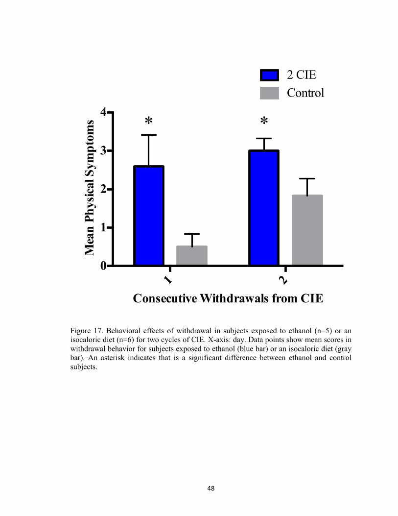
48
1 20
1
2
3
4
Consecutive Withdrawals from CIE
Mea
n Ph
ysic
al S
ympt
oms
2 CIEControl
* *
Figure 17. Behavioral effects of withdrawal in subjects exposed to ethanol (n=5) or an isocaloric diet (n=6) for two cycles of CIE. X-axis: day. Data points show mean scores in withdrawal behavior for subjects exposed to ethanol (blue bar) or an isocaloric diet (gray bar). An asterisk indicates that is a significant difference between ethanol and control subjects.
49
1 2 30
2
4
6
Consecutive Withdrawals from CIE
Mea
n Ph
ysic
al S
ympt
oms
3 CIEControl
**
**
Figure 18. Behavioral effects of withdrawal in subjects exposed to ethanol (n=5) or an isocaloric diet (n=6) for three cycles of CIE. X-axis: day. Data points show mean scores in withdrawal behavior for subjects exposed to ethanol (blue bar) or an isocaloric diet (gray bar). Two asterisks indicate a significant interaction between day and treatment.

50
1 2 3 4 50
20
40
60
Acquisition Day
Lat
ency
to P
latf
orm
(sec
onds
)
Morris Water Maze
2 CIEControl
*
Figure 19. Cognitive performance was assessed in subjects exposed to ethanol (n=5; filled circle) or an isocaloric diet (n=6; filled square) for two cycles of CIE. X-axis: day. Data points show mean scores in latency to platform. One asterisk indicates that there is a significant difference between acquisition days.

51
1 2 3 4 50
600
1200
1800
Acquisition Day
Dis
tanc
e T
rave
lled
(cm
)
Morris Water Maze
2 CIEControl
*
Figure 20. Cognitive performance was assessed in subjects exposed to ethanol (n=5; filled circle) or an isocaloric diet (n=6; filled square) for two cycles of CIE. X-axis: day. Data points show mean scores in distance travelled. One asterisk indicates that there is a significant difference between acquisition days.

52
1 2 3 4 50
10
20
30
40
Acquisition Day
Vel
ocity
(cm
per
sec
ond)
Morris Water Maze
2 CIEControl
**** **
Figure 21. Cognitive performance was assessed in subjects exposed to ethanol (n=5; filled circle) or an isocaloric diet (n=6; filled square) for two cycles of CIE. X-axis: day. Data points show mean scores in velocity. Two asterisks indicates that there is a significant difference between treatment groups.

53
15 se
conds
total
0
2
4
6Pr
obe
Tri
als
(sec
onds
)
Morris Water Maze
2 CIEControl
Figure 22. Working memory assessment in subjects exposed to ethanol (n=5) or an isocaloric diet (n=6) for two cycles of CIE. X-axis: day. Data points show mean scores in platform crosses during the probe trial for subjects exposed to ethanol (filled circle) or an isocaloric diet (filled square).

54
1 2 3 4 50
20
40
60
Acquisition Day
Lat
ency
to P
latf
orm
(sec
onds
)
Morris Water Maze
3 CIEControl
*
Figure 23. Cognitive performance was assessed in subjects exposed to ethanol (n=5; filled circle) or an isocaloric diet (n=6; filled square) for three cycles of CIE. X-axis: day. Data points show mean scores in latency to platform. One asterisk indicates that there is a significant difference between acquisition days.

55
1 2 3 4 50
600
1200
1800
Acquisition Day
Dis
tanc
e T
rave
lled
(cm
)
Morris Water Maze
3 CIEControl
*
Figure 24. Cognitive performance was assessed in subjects exposed to ethanol (n=5; filled circle) or an isocaloric diet (n=6; filled square) for three cycles of CIE. X-axis: day. Data points show mean scores in distance travelled. One asterisk indicates that there is a significant difference between acquisition days.

56
1 2 3 4 50
10
20
30
40
Acquisition Day
Vel
ocity
(cm
per
sec
ond)
Morris Water Maze
3 CIEControl
**
Figure 25. Cognitive performance was assessed in subjects exposed to ethanol (n=5; filled circle) or an isocaloric diet (n=6; filled square) for three cycles of CIE. X-axis: day. Data points show mean scores in velocity. Two asterisks indicate that there is a significant difference between groups.

57
100
150
200
250
mg/
dl
Peak Blood Ethanol Levels
Figure 26. Peak BELs in subjects exposed to ethanol (n=5) for three cycles of CIE. X-axis: day. Data points show mean levels determined at 90 minutes post ethanol administration on Week One, Week Two, and Week Three.

58
Figure 27. Representative heat map images depicting distance travelled in Morris Water Maze decreased significantly on Day Five (bottom image), as compared to Day One (top image).

59
CHAPTER FOUR: Study Three: Influence of mGluR1/5-containing Receptors and Intracellular Calcium in Development of Ethanol Dependence in Vitro
4.1. Introduction
Group 1 metabotropic glutamate receptors. Glutamate is the primary excitatory
neurotransmitter in the mammalian central nervous system. There are two distinct classes
of glutamate receptors: ionotropic glutamate receptors (e.g., N-methyl-D-aspartate
[NMDA] receptors) and metabotropic glutamate receptors (i.e., mGluRs). While
ionotropic glutamate-type receptors were first characterized as containing ligand-gated
ion channels (Cotman et al., 1989), mGluRs were later identified as large guanine
nucleotide-binding protein (G-protein)-coupled receptors involved in the modulation of
excitatory neurotransmission (Nakanishi, 1994). Studies using molecular cloning and
cross-hybridization techniques have identified eight known subtypes of mGluRs
classified into three groups based on sequence homology, signal transduction, and
pharmacological action (Nakanishi, 1994; for a review, see Conn & Pin, 1997). Group 1
mGluRs (i.e., mGluR1 and mGluR5) are Gαq-linked to phospholipase C and can
stimulate phosphoinositide hydrolysis (Abe et al., 1992; Houamed et al., 1991; Pin et al.,
1992). These receptors are known to be involved in a myriad of biological processes,
including regulation of second messengers (Miller et al., 1995; Schoepp & Conn, 1993;
Schoepp et al., 1994), ion channels, such as potassium channels (Charpak et al., 1990;
Guérineau et al., 1994), and neuronal excitability (Davies et al., 1995; Gereau & Conn,
1995). These diverse effects are likely mediated in part via the wide distribution of group
1 mGluRs throughout the brain and spinal cord (Baude et al., 1993; Fotuhi et al., 1993;
Martin et al., 1992; Petralia, 1997).

60
Group 1 mGluRs interact closely with intracellular scaffolding proteins, such as
Homer proteins. A study using immunofluorescent techniques found that Homer1b
retains group 1 mGluRs at the endoplasmic reticulum when these proteins are
coexpressed (Roche et al., 1989). Ango et al. (2000) found that the mGluR5 is found in
dendrites and axons when cotransfected with Homer1a whereas these receptors are
concentrated at cell bodies when transfected alone in cerebellar granule cells. Further,
immunocytochemical and western blot analyses show that Homer proteins can actually
bind and activate group 1 mGluRs independent of agonist application (Ango et al., 2001;
for a review, see Spooren et al., 2001).
Group 1 mGluRs are also known to be modulated by extracellular agonists, which
may indirectly affect function of these receptors. Cozzoli and colleagues (2009) found
that binge-like ethanol administration produced significant increases in Homer2a/b
expression as well as Homer2-phosphatidylinositol 3-kinase (PI3K) signaling in the
nucleus accumbens. These effects were attenuated with administration of mGluR5
antagonist MPEP and PI3K antagonist Wortmannin. Notably, pretreatment with mGluR5
antagonist MPEP dose-dependently reduced binge ethanol consumption while mGluR1
antagonist (hydroxyimino)cyclopropa[b]chromen-1a-carboxylate ethyl ester (CPCCOEt)
did not alter ethanol self-administration. Collectively, these studies suggest that Gαq-
coupled group 1 mGluRs influence the voluntary intake of ethanol in rodents. However,
the functional influence of group 1 mGluRs in promoting the development of ethanol
dependence is not fully understood. Further, the role of activated mGluR1-containing
receptors and mGluR5-containing receptors is not fully characterized with regards to
ethanol withdrawal-induced cytotoxicity. Thus, the present study examined the functional

61
relationship between ethanol-associated activation of group 1 mGluRs and subsequent
and persisting withdrawal-induced NMDA-receptor-dependent cytotoxicity following
CIE. This study closely followed a model from an earlier study (Reynolds et al., 2015).
Inositol-triphosphate-mediated release of intracellular calcium. Calcium is a ubiquitous
intracellular signal known to control a cadre of cellular processes (e.g., neurotransmitter
release and synaptic plasticity) and is also modulated by ethanol (Berridge et al., 2000).
The primary activator of intracellular calcium is calcium itself (i.e., calcium induced
calcium release); however, inositol triphosphate (IP3) can also engage the IP3 receptors
to release calcium from internal stores (Verkhratsky, 2002), and exposure to ethanol can
release intracellular calcium from receptor complexes located at the endoplasmic
reticulum, such as the IP3 receptor (Daniell & Harris, 1989). Other studies suggest that
exposure to ethanol produces presynaptic release of calcium from internal stores prior to
γ-Aminobutyric acid (GABA) neurotransmission in cerebellar neurons (Kelm et al.,
2007) and that GABAergic neurotransmission is dependent on the release of intracellular
calcium in dopaminergic neurons (Thiele et al., 2009).
During periods of withdrawal from chronic ethanol exposure, excessive
extracellular calcium influx via the NMDA receptor can produce excitotoxicity
(Prendergast et al., 2004) and neurodegeneration (Lau & Tymianski, 2010). The mGluR5
is also implicated in the neurotoxic effects of withdrawal. In one study, for example,
exposure to SIB-1893, the competitive mGluR5 antagonist, attenuated cytotoxicity
produced by withdrawal in cultured organotypic hippocampal slices (Harris et al., 2003).
The inhibition of intracellular calcium mobilization via antagonists for the IP3, sigma-1

62
receptor, and ryanodine receptors can attenuate excitotoxicity, and in some cases, provide
neuroprotection in various models of neurodegenerative disorders, such as HIV-1 Tat
(Haughey et al., 2001) and stroke (Shen et al., 2008). Previous studies examining the
modulatory effects of ethanol on GABA implicate a role for the release of intracellular
calcium in the activation of PKA (Kelm et al., 2008). These alterations in PKA activity
are associated with decreases in the sedative effects of ethanol and voluntary ethanol
consumption (Thiele et al., 2000; Wand et al., 2001).
4.2. Experimental Rationale
These findings suggest a role for mGluR5 in the behavioral effects of ethanol and,
thus, in the development of acute alcohol dependence. They also suggest a potential role
for intracellular calcium-initiating alterations in the NMDA receptor that potentiate
cytotoxicity during ethanol withdrawal. A linking of these mechanisms and the primary
hypothesis tested is shown in Figure 28.
4.3. Methods
Organotypic hippocampal slice culture preparation. Whole brains from eight-day-old
Sprague-Dawley rats (Harlan Laboratories; Indianapolis, IN) were aseptically removed
and placed in culture dishes containing frozen dissecting medium composed of Minimum
Essential Medium (MEM; Invitrogen, Carlsbad, CA), 25 mM HEPES (Sigma, St. Louis,
MO), and 50 µM streptomycin/penicillin (Invitrogen). Bilateral hippocampi were then
removed and carefully transferred onto plates containing chilled culture medium
composed of dissecting medium, distilled water, 36 mM glucose (Fisher, Pittsburg, PA),
25% Hanks’ Balanced Salt Solution (HBSS; Invitrogen), 25% (v/v) heat-inactivated
horse serum (HIHS; Sigma), and 0.05% streptomycin/penicillin (Invitrogen). Excess

63
hippocampal tissue was carefully detached using a stereoscopic microscope and unilateral
hippocampi and then sectioned at 200 µM using a McIlwain Tissue Chopper (Mickle
Laboratory Engineering Co. Ltd., Gomshall, UK). Three hippocampal slices with intact
cell layers were plated onto Millicell-CM 0.4 µM biopore membrane inserts containing 1
mL of pre-incubated culture medium and placed in a six-well culture plate. Excess
culture medium was extracted off the top of each biopore membrane insert and the
harvested tissue were maintained in an incubator at 37°C with a gas composition of 5%
CO2/95% air for five days prior to any experimental manipulations so that each
hippocampal slice could adequately adhere to the membrane. Each culture well plate
generated eighteen intact hippocampal slices. Care of all animals was carried out in
agreement with the University of Kentucky’s Institutional Animal Care and Use
Committee.
Ethanol exposure and withdrawal. A previous study found that exposure to CIE
produced NMDA-receptor-mediated hippocampal cytotoxicity (Reynolds et al., 2015).
This study suggested that these effects are produced by the activation of these receptor
complexes during periods of withdrawal, but the neuroadaptive changes that take place
prior to withdrawal are not yet fully understood. The present study employed this model
of CIE exposure to delineate probable mechanistic underpinnings associated with the
development of ethanol dependence. The timeline of the study is detailed in Figure 29.
After five days in vitro, male and female hippocampal slice cultures were
randomly transferred to plates containing either 1 ml of the culture medium (control) or
medium containing a binge-like ethanol concentration (i.e., 0 and 50 mM) for five days

64
with or without the addition of 1) mGluR1 antagonist (CPCCOEt; 0.5–3 µM); 2)
mGluR5 antagonist (SIB-1893; 20–200 µM); 3) Xestospongin C (0–1 µM), a membrane-
permeant inhibitor of IP3-mediated calcium release; 4) BD1047 (0–100 µM), a selective
antagonist at the sigma-1 receptor; or 5) dantrolene (0–30 µM), a ryanodine receptor
antagonist (Kim et al., 2010; Tarragon et al., 2012; Taylor & Tovey, 2010; Wang et al.,
2004). At 11 days in vitro, cultures were removed from their respective treatment groups
and transferred to plates containing 1 ml of fresh ethanol-naïve culture medium for a 24-
hour ethanol withdrawal period. These treatments were repeated a total of three times in
consecutive order. During each five-day exposure period, ethanol and control-treated
cultures were maintained inside Ziploc bags filled with 5%CO2/95% air and water bath
solutions containing either distilled water (50 ml) for control plates or distilled water (50
ml) containing ethanol (50 mM) for ethanol-treated plates. At 23 days in vitro, cultures
were fixed for immunohistochemistry.
Immunohistochemistry. Cultures were fixed by placing 1 ml of 10% formalin solution
on the top and bottom of each well for 30 minutes, washing twice with phosphate
buffered saline (PBS), and storing at 4°C until immunohistochemistry was initiated.
NeuN (Fox-3) is found in nearly all post-mitotic neurons (Kim et al., 2009; Mullen et al.,
1992), and thus these cultures were labeled with NeuN to assess cytotoxicity in each
primary cell layer of the hippocampal formation—pyramidal cell layers of the cornu
ammonis (CA1 and CA3) and granule cell layer of the dentate gyrus. Fixed inserts were
transferred to a plate containing 1 ml of permeabilization (wash) buffer (200 ml PBS
[Invitrogen], 200 µL Triton X-100 [Sigma], 0.010 mg Bovine Serum [Sigma]), with 1 ml

65
of buffer added to the top of each well for 45 minutes to permeate cell membranes.
Tissue was then incubated with the primary monoclonal antibody mouse anti-NeuN
(1:200; Sigma) for 24 hours. Inserts were then washed with PBS and incubated for 24
hours with goat anti-mouse fluorescein isothiocyanate (FITC; 1:200; Sigma). Cultures
were imaged with SPOT software 4.0.2 (advanced version) for Windows (W. Nuhsbahm
Inc.; McHenry, IL, USA) through a 5x objective with a Leica DMIRB microscope (W.
Nuhsbahm Inc.; McHenry, IL, USA) connected to a computer and captured with a SPOT
7.2 color mosaic camera (W. Nuhsbahm). In these immunohistochemical studies, FITC
fluorescence was detected with a band-pass filter at 495 nm (520 nm emission).
Statistical analyses. Statistical analyses were conducted to assess the influence of group
1 mGluRs and intracellular calcium in the development of ethanol dependence. Effects
were considered significant at p<0.05. This study was conducted two times using two
different rat litters. All immunohistochemical data were converted to percent control and
then combined for ease of data interpretation. A one-way ANOVA was conducted for all
in vitro techniques with treatment as the factor for each hippocampal cell layer (i.e., CA1,
CA3, and dentate gyrus). This statistical strategy was based on the results of preliminary
data suggesting that sex is not a factor influencing the effects produced by repeated
binge-like ethanol. Planned comparisons were conducted if a significant effect of
treatment was to make pairwise comparisons between means detected using Fisher’s
LSD.
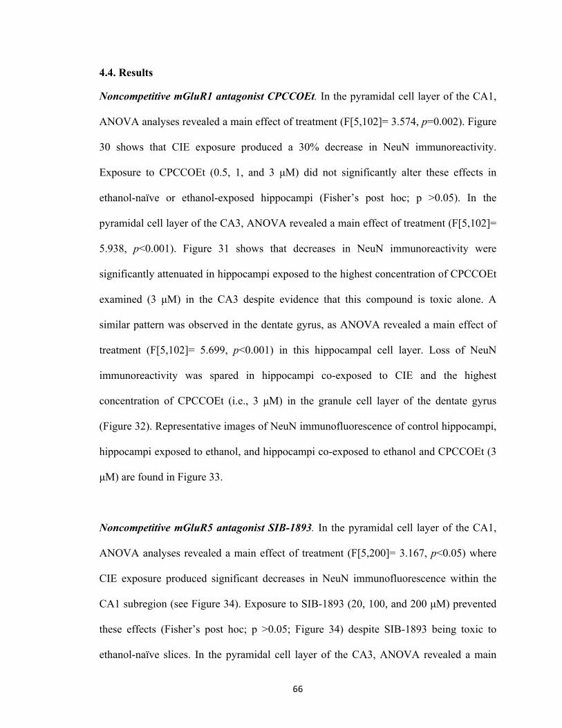
66
4.4. Results
Noncompetitive mGluR1 antagonist CPCCOEt. In the pyramidal cell layer of the CA1,
ANOVA analyses revealed a main effect of treatment (F[5,102]= 3.574, p=0.002). Figure
30 shows that CIE exposure produced a 30% decrease in NeuN immunoreactivity.
Exposure to CPCCOEt (0.5, 1, and 3 µM) did not significantly alter these effects in
ethanol-naïve or ethanol-exposed hippocampi (Fisher’s post hoc; p >0.05). In the
pyramidal cell layer of the CA3, ANOVA revealed a main effect of treatment (F[5,102]=
5.938, p<0.001). Figure 31 shows that decreases in NeuN immunoreactivity were
significantly attenuated in hippocampi exposed to the highest concentration of CPCCOEt
examined (3 µM) in the CA3 despite evidence that this compound is toxic alone. A
similar pattern was observed in the dentate gyrus, as ANOVA revealed a main effect of
treatment (F[5,102]= 5.699, p<0.001) in this hippocampal cell layer. Loss of NeuN
immunoreactivity was spared in hippocampi co-exposed to CIE and the highest
concentration of CPCCOEt (i.e., 3 µM) in the granule cell layer of the dentate gyrus
(Figure 32). Representative images of NeuN immunofluorescence of control hippocampi,
hippocampi exposed to ethanol, and hippocampi co-exposed to ethanol and CPCCOEt (3
µM) are found in Figure 33.
Noncompetitive mGluR5 antagonist SIB-1893. In the pyramidal cell layer of the CA1,
ANOVA analyses revealed a main effect of treatment (F[5,200]= 3.167, p<0.05) where
CIE exposure produced significant decreases in NeuN immunofluorescence within the
CA1 subregion (see Figure 34). Exposure to SIB-1893 (20, 100, and 200 µM) prevented
these effects (Fisher’s post hoc; p >0.05; Figure 34) despite SIB-1893 being toxic to
ethanol-naïve slices. In the pyramidal cell layer of the CA3, ANOVA revealed a main

67
effect of treatment (F[5,102]= 3.385, p<0.05). Figure 35 shows that decreases in NeuN
immunoreactivity were significantly attenuated in hippocampi exposed to the lowest
concentration of SIB-1893 (20 µM) examined in the CA3. A similar pattern was observed
in the dentate gyrus, as ANOVA revealed a main effect of treatment (F[7,201]=6.831,
p<0.001) in this hippocampal cell layer. Loss of NeuN immunoreactivity was spared in
hippocampi co-exposed to binge-like ethanol and 20 µM of SIB-1893 in the granule cell
layer of the dentate gyrus (Figure 36). Representative images are found in Figure 37.
Inhibitor of IP3-mediated calcium release Xestospongin C. In the pyramidal cell layer
of the CA1, ANOVA analyses revealed a main effect of treatment (F[3,133]= 21.910,
p<0.001) where CIE produced significant decreases in NeuN immunofluorescence within
the CA1 subregion (see Figure 38). Exposure to Xestospongin C (0.5 µM) prevented
these effects (Fisher’s post hoc; p >0.05; Figure 38) despite Xestospongin C being
neuroprotective to ethanol-naïve slices. In the pyramidal cell layer of the CA3, ANOVA
revealed a main effect of treatment (F[3,133]= 10.1325, p<0.001). Figure 39 shows that
decreases in NeuN immunoreactivity were significantly attenuated in hippocampi
exposed to Xestospongin C (0.5 µM) examined in the CA3 despite Xestospongin C being
neuroprotective to ethanol-naïve slices. A similar pattern was observed in the dentate
gyrus, as ANOVA revealed a main effect of treatment (F[3,133]=14.891, p<0.001) in this
hippocampal cell layer. Loss of NeuN immunoreactivity was spared in hippocampi co-
exposed to CIE and 0.5 µM of Xestospongin C in the granule cell layer of the dentate
gyrus despite Xestospongin C being neuroprotective to ethanol-naïve slices (Figure 40).

68
Sigma-1 receptor antagonist BD-1047. In the pyramidal cell layer of the CA1, ANOVA
analyses revealed a main effect of treatment (F[5,91]= 5.848, p<0.001) where CIE
produced significant decreases in NeuN immunofluorescence within the CA1 subregion
(see Figure 41). Exposure to BD-1047 (20 µM) prevented these effects (Fisher’s post
hoc; p >0.001; Figure 41) despite BD-1047 (20 µM) being neuroprotective to ethanol-
naïve slices. In the pyramidal cell layer of the CA3, ANOVA revealed a main effect of
treatment (F[5,91]= 6.023, p<0.001). Figure 42 shows that decreases in NeuN
immunoreactivity were significantly attenuated in hippocampi exposed to BD-1047 (20
µM) examined in the CA3 despite BD-1047 being neuroprotective to ethanol-naïve
slices. A similar pattern was observed in the dentate gyrus, as ANOVA revealed a main
effect of treatment (F[5,91]=6.381, p<0.001) in this hippocampal cell layer. Loss of
NeuN immunoreactivity was spared in hippocampi co-exposed to CIE and 20 µM of BD-
1047 in the granule cell layer of the dentate gyrus despite BD-1047 being neuroprotective
to ethanol-naïve slices (Figure 43).
Ryanodine receptor antagonist dantrolene. In the pyramidal cell layer of the CA1,
ANOVA analyses revealed a main effect of treatment (F[3,91]= 5.848, p<0.001) where
CIE exposure produced significant decreases in NeuN immunofluorescence within the
CA1 subregion (see Figure 44). Exposure to dantrolene (5 µM) did not prevent these
effects (Fisher’s post hoc; p >0.001; Figure 44) despite dantrolene (5 µM) being toxic to
ethanol-naïve slices. In the pyramidal cell layer of the CA3, ANOVA revealed a main
effect of treatment (F[3,91]= 6.023, p<0.001). Figure 45 shows that decreases in NeuN
immunoreactivity were significantly attenuated in hippocampi exposed to dantrolene (5
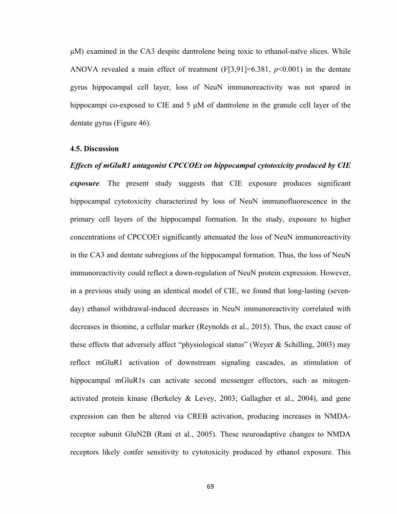
69
µM) examined in the CA3 despite dantrolene being toxic to ethanol-naïve slices. While
ANOVA revealed a main effect of treatment (F[3,91]=6.381, p<0.001) in the dentate
gyrus hippocampal cell layer, loss of NeuN immunoreactivity was not spared in
hippocampi co-exposed to CIE and 5 µM of dantrolene in the granule cell layer of the
dentate gyrus (Figure 46).
4.5. Discussion
Effects of mGluR1 antagonist CPCCOEt on hippocampal cytotoxicity produced by CIE
exposure. The present study suggests that CIE exposure produces significant
hippocampal cytotoxicity characterized by loss of NeuN immunofluorescence in the
primary cell layers of the hippocampal formation. In the study, exposure to higher
concentrations of CPCCOEt significantly attenuated the loss of NeuN immunoreactivity
in the CA3 and dentate subregions of the hippocampal formation. Thus, the loss of NeuN
immunoreactivity could reflect a down-regulation of NeuN protein expression. However,
in a previous study using an identical model of CIE, we found that long-lasting (seven-
day) ethanol withdrawal-induced decreases in NeuN immunoreactivity correlated with
decreases in thionine, a cellular marker (Reynolds et al., 2015). Thus, the exact cause of
these effects that adversely affect “physiological status” (Weyer & Schilling, 2003) may
reflect mGluR1 activation of downstream signaling cascades, as stimulation of
hippocampal mGluR1s can activate second messenger effectors, such as mitogen-
activated protein kinase (Berkeley & Levey, 2003; Gallagher et al., 2004), and gene
expression can then be altered via CREB activation, producing increases in NMDA-
receptor subunit GluN2B (Rani et al., 2005). These neuroadaptive changes to NMDA
receptors likely confer sensitivity to cytotoxicity produced by ethanol exposure. This

70
interpretation is supported by the work of Ticku and colleagues (Qiang et al., 2007) who
demonstrated the upregulation of GluN1 and GluN2B subunits in cortical neurons
exposed to binge-like ethanol in vitro (Qiang et al., 2007). Further, earlier work
suggested that this model of ethanol dependence reflects increases in NMDA-receptor
activation upon withdrawal, and thus the activity of group 1 mGluRs may contribute to
this specific neuroadaptation to ethanol (Reynolds et al., 2015).
It has been shown that mGluR1s are differentially expressed in neurons
throughout various areas of the brain and spinal cord (Shigemoto et al., 1993). In the
hippocampus, for example, levels of mGluR1 expression are highest in the granule cell
layer of the dentate gyrus and pyramidal cell layer of the CA3 (Davies et al., 1995; Miller
et al., 1995; Petralia et al., 1997). In the present study, mGluR1 antagonist CPCCOEt did
not alter NeuN immunofluorescence in the pyramidal CA1 cell layer of the hippocampus,
which is consistent with a previous study that found that immunolabeling of mGluR1s
was not detectable in the pyramidal cell layer of the CA1 in the rat hippocampus
(Shigemoto et al., 1993). In sum, the present findings extend upon prior work suggesting
a region-specific functional role for the mGluR1 in the development of hippocampal
cytotoxicity within the CA3 and dentate gyrus.
Effects of mGluR5 antagonist SIB-1893 on hippocampal cytotoxicity produced by CIE
exposure. Noncompetitive mGluR5 antagonists (e.g., SIB-1893) bind to the 7-
transmembrane domain and alter conformational changes without affecting the
extracellular ligand binding site (for a review, see Spooren et al., 2001). Other studies
have suggested that mGluR5 antagonists are neuroprotective in various models of

71
neurodegeneration (Battalglia et al., 2002; Chapman et al., 2000; Harris et al., 2003). In
the present study, exposure to noncompetitive mGluR5 antagonist SIB-1893 prevented
hippocampal cytotoxicity produced by CIE exposure. Other studies have shown that
acute exposure to SIB-1893 prevented ethanol withdrawal-induced loss of propidium
iodide, a marker of cytotoxicity, in the CA1 subregion of the mature hippocampus (Harris
et al., 2003). The mGluR5 is, therefore, important for both ethanol intoxication and
withdrawal. An earlier in vivo study found that the mGluR5 antagonist MPEP mitigated
increases in Homer2 signaling that were produced by binge-like ethanol exposure and
ethanol-induced plasticity (Cozzoli et al., 2009), and other studies have shown that MPEP
dose-dependently decreased operant ethanol self-administration and GluN1 and GluN2
mRNA expression in the cingulate cortex while CPCCOEt did not alter voluntary ethanol
self-administration (Hodge et al., 2006). Other studies have shown the efficacy of MPEP
in attenuating ethanol reinstatement elicited by drug-associated cues (Bäckström et al.,
2004). Blednov and Harris (2008), for example, demonstrated that MPEP (10 mg/kg)
attenuates ethanol withdrawal behavior in rodents if administered prior to ethanol
administration but not following.
It is notable that in the current study mGluR5 antagonist SIB-1893 exposure in
the presence of ethanol prevented the hippocampal NeuN loss produced by binge-like
ethanol. The reasons for the neuroprotective effects of SIB-1893 are likely associated
with the effects of mGluR5s on NMDA-receptor activity in the modulation of
glutamatergic tone. For example, NMDA-receptor activity is influenced by the activation
of the mGluR5 in the hippocampus via second messenger effector protein kinase C in
vitro (Chen et al., 2011). Other studies have suggested that Ca2+/calmodulin-dependent

72
protein kinase IIα (CaMKIIα) binds to the intracellular C terminus of the mGluR5 to
produce a release of calcium from intracellular stores and that subsequent
phosphorylation of adjacent GluN2Bs can potentiate NMDA-receptor activity (Jin et al.,
2013). Together, these studies suggest that the neuroprotective effects of mGluR5
antagonist SIB-1893 include the modulation of glutamatergic tone via downstream
effects at the NMDA receptor.
Effects of blocking IP3-mediated release of intracellular calcium on hippocampal
cytotoxicity. Calcium is a ubiquitous intracellular signal responsible for controlling
numerous cellular processes. At rest, cells have a basal calcium concentration of 100 nM.
When activated, cells have a calcium concentration of 1000 nM (for a review, see
Berridge et al., 2003). Internal stores of calcium are held within membrane systems of the
endoplasmic reticulum (ER) or sarcoplasmic reticulum of muscle cells (Berridge, 1993;
Clapham, 1995). Release of calcium from these internal stores is controlled by various
channels, such as the inositol-1,4,5-trisphosphate (IP3) receptor and ryanodine receptor
families (Clapper et al., 1987). The present study examined the efficacy of Xestospongin
(i.e., C), a macrocyclic bis-1-oxaquinolizidines characterized as a potent blocker of IP3-
mediated calcium release on the vesicles of the ER membrane (Gafni et al., 1997),
against toxicity produced by CIE. We found that application of Xestospongin C for three
consecutive cycles of CIE actually increased levels of NeuN immunofluorescence in the
rat hippocampus. The reasons for these effects are unknown, but likely reflect inherent
characteristics of the slice culture preparations, such as damage to the ER, as IP3
channels are regulated by several factors and cytosolic actions of calcium can either be

73
stimulatory or inhibitory (for a review see Berridge et al., 2003; Verkhratsky, 2002).
Therefore, the present findings suggest a functional implication of blocking IP3-mediated
calcium release in organotypic hippocampal slice cultures so as to preserve calcium
homeostasis. These data are similar to a previous study conduced in our laboratory
demonstrating the efficacy of acute Xestospongin application during withdrawal from
methamphetamine (Smith et al., 2008).
Given that there is a 10-fold increase of IP3 receptors in the cerebellum as
compared to the hippocampus (Smith, 1987) and a robust shrinking of the cerebellum
observed in alcohol-dependent individuals (Chanraud et al., 2007; Sullivan et al., 2000;
for a review, see Rosenbloom & Pfefferbaum 2008), future studies could assess the
influence of these receptor complexes in other areas of the brain implicated in having
neurological consequences in individuals with an alcohol use disorder, such as the
cerebellum.
Collectively, these findings suggest that while ethanol application mediates a
release of intracellular calcium in the hippocampus (Daniell & Harris; 1989, Mironov &
Hermann, 1995) that may reflect a decrease of IP3 mRNA (Rodríguez et al., 1996;
Simonyi et al., 1996), preparation of organotypic hippocampal slices may also inherently
cause calcium dysregulation from the ER.
Effects of sigma-1 receptor antagonism on the hippocampal cytotoxicity produced by
CIE. The sigma-1 receptor is characterized as a calcium-sensitive, ligand-regulated
chaperone of the ER. Under basal conditions, the sigma-1 receptor is located on the
ER/mitochondrial membrane interface. Here the sigma-1 receptor forms a

74
macromolecular complex with glucose regulated protein (i.e., BiP). Calcium depletion of
the ER (via ligand activation) initiates dissociation of these two chaperone proteins.
Unbound sigma-1 receptors can then bind to IP3 (type 3) receptors, preventing
degradation by proteasomes. The dissociation of the sigma-1 receptor from BiP also
allows for the sigma-1 receptor to stabilize aggregated IP3 receptors on the
ER/mitochondria interface membrane and translocation of IP3 receptors to ER periphery
following prolonged calcium depletion (i.e., ER stress). Notably, the unbound sigma-1
receptor can potentiate prolonged IP3 receptor-mediated Ca2+ signaling to the
mitochondria for checks and balances of ER calcium homeostasis (Hayashi & Su, 2007;
for a review, see Hayashi et al., 2009). In the present study, we assessed the influence of
the chaperone sigma-1 protein in promoting cytotoxic effects of CIE using a sigma-1
receptor antagonist. We found that blocking this receptor prevented the loss of NeuN
immunofluorescence produced by three consecutive cycles of CIE. We also found that
chronic blockade of the sigma-1 receptor significantly increased levels of NeuN
immunofluorescence in ethanol-naïve hippocampi in a manner similar to Xestospongin
C.
Given that the sigma-1 receptor is highly concentrated in the hippocampus
(Alonso et al., 2000), it is not surprising that the sigma-1 receptor is known to alleviate
alterations in learning and memory produced by exogenous substances (Maurice & Su,
2009; Meunier et al., 2006). In one study, for example, hippocampal neurons from
rodents exposed to CIE followed by a period of withdrawal demonstrated an inhibited
EPSP-to-spike coupling potentiation curve in the pyramidal cell layer of the CA1 (Sabeti
et al., 2011). Exposure to a selective sigma-1 receptor antagonist BD1047 (1 µM)

75
resulted in a return to normal EPSP spike potentiation. In vivo, sigma-1 receptor
antagonism is known to attenuate cognitive deficits produced by CIE (10 mg/kg BD
1047; Meunier et al., 2006) and reduce ethanol consumption (1 mL/kg NE-100; Sabino et
al., 2009). Western blot analyses found that CIE produced an upregulation of sigma-1
receptor protein expression in the hippocampus (Sabeti et al., 2011). Notably, these
effects were regulated via a sigma-1 receptor antagonist (10 mg/kg BD 1047). Functional
polymorphisms in the sigma-1 receptor gene were also associated with a propensity for
alcoholism in a case-controlled study (Miyatake et al., 2004). In sum, these studies
suggest that targeting the sigma-1 receptor may have clinical implications for behavioral
effects associated with prolonged ethanol consumption.
The sigma-1 receptor is also known to be involved in mediating NMDA
glutamate-type receptor activity in the hippocampal formation. For example, in one
study, exposure to the high affinity sigma-1 receptor agonist ditolylguanidine (DGT)
produced increases in NMDA-mediated neuronal firing in the pyramidal cell layer of the
hippocampus (Monnet et al., 1990). In another study, exposure to sigma-1 receptor
antagonist haloperidol produced suppression of these agonist-dependent responses to
NMDA (Monnet et al., 1992). It is known that withdrawal can produce CNS
hyperexcitability and/or seizure activity following prolonged exposure to ethanol
(Prendergast et al., 2004; Puz & Stokes, 2005; for a review, see Prendergast &
Mulholland, 2012) and that these effects are augmented following exposure to multiple
withdrawals in rodent models of CIE (Stevens et al., 2001; Veatch & Becker, 2002, 2005)
and in the clinical population (Ballenger & Post, 1978; Shaw et al., 1998; Wojnar et al.,
1999). Further, neuroadaptations in NMDAR function and/or expression are implicated in

76
potentiating excitotoxicity during periods of withdrawal from CIE exposure (Carpenter-
Hyland et al., 2004; Rani et al., 2005; Qiang et al., 2007). Thus, in addition to these
NMDA-receptor-dependent neuroadaptations, the sigma-1 receptor may modulate these
effects. In one study, exposure to ifenprodil, the selective NMDA-receptor antagonist for
the NR2B complex, was found to be efficacious in alleviating seizures during periods of
withdrawal in rodents (Malinowska et al., 1999). This is notable, as in addition to affinity
for NMDA receptors, ifenprodil also has a high affinity for sigma-1 receptors (Hanner et
al., 1996; for a review, see Debonnel & de Montigny, 1996). Collectively, these studies
suggest that blocking the sigma-1 receptor attenuates classic excitotoxicity in vitro.
Future studies could assess the efficacy of blocking this chaperone protein during
withdrawal from CIE, or drug-taking behaviors (e.g., ethanol self-administration and
psychomotor stimulants), in vivo.
Effects of ryanodine receptor antagonist dantrolene on the hippocampal cytotoxicity
produced by CIE. In the present study, we found that blocking the ryanodine receptor
with dantrolene was not effective in attenuating hippocampal cytotoxicity when co-
applied with ethanol for three consecutive cycles of CIE. The reasons for these effects are
unknown but could reflect the relatively low concentrations of dantrolene employed.
Future studies could use higher concentrations of dantrolene in organotypic hippocampal
slice culture preparations. Given previous studies that have shown that while the NMDA
receptors can trigger calcium release from the ryanodine receptor (Isokawa & Bradley,
2006) and ethanol application inhibits NMDA receptors (Lovinger et al., 1989), future

77
studies could assess the effects of blunting ryanodine receptor activation during periods
of withdrawal from CIE in vitro and in vivo.
Using this study’s model, it has been shown that ethanol withdrawal-induced
hippocampal cytotoxicity is NMDA-receptor dependent (Reynolds et al., 2015), and the
present findings extend upon the current literature by characterizing the distinct roles that
mGluR1- and mGluR5-containing receptors have in promoting the development of
ethanol dependence prior to NMDA-receptor dependent withdrawal-associated
cytotoxicity. Collectively, these findings suggest that the concomitant neuroadaptations
in group 1 mGluRs and NMDA receptors are likely associated with both the behavioral
and neurodegenerative effects observed following multiple bouts of heavy ethanol
consumption (Duka et al., 2003; Duka et al., 2004; Sullivan et al., 1996) and in the
development of dependence. Group 1 mGluRs—in particular the mGluR5—may,
therefore, be a therapeutic target for treatment of alcohol use disorders. In vivo
examination of this notion is discussed in Chapter 5.

78
IP3R; IP3 receptor Sigma-1R; sigma-1 receptor
calcium RyR; ryanodine receptor
Gq Signalling and Pharmacological Targets of Reynolds Dissertation Studies (underlined) mGluR 1
and 5 Gq PLC
IP3 DAG
Stores
Sigma-1R RyR
IP3R
AC
cAMP
PIP2
GluN1/2B
(+)
GluN1/2B
PKC PKA
ethanol
Figure 28. Pictorial representation of the hypothesis that ethanol stimulates group 1 mGluR promoting release of intracellular calcium from IP3-mediated stores. Calcium also stimulates the adenylate cyclase signal transduction pathway that can produce coordinated phosphorylation of NMDA receptors (i.e., GluN1 and GluN2B) by PKA and PKC and subsequent trafficking of these receptor complexes from extrasynaptic to synaptic sites. During withdrawal, excessive calcium influx through these synaptic receptors activates phospholipases, endonucleases, and proteases to produce cytotoxicity and cell death.

79
CPCCOEt
MGLUR1 MGLUR5 IP3R SIGMA-1R RYR
SIB-1897 xesto c BD-1047 dantrolene
3 CIEDrugs used in presence of ethanol
Figure 29. Rat hippocampal explants were exposed to 50 mM ethanol for five days followed by 24-hours of withdrawal with the cycle repeated three times (i.e., 3 CIE) with or without the addition of one of five antagonists.

80
CTRL
CTRL + 0.5 C
P
CTRL + 1.0 C
P
CTRL + 3.0 C
P
3 CIE
3 CIE
+ 0.5 C
P
3 CIE
+ 1.0 C
P
3 CIE
+ 3.0 C
P60
80
100
120
Neu
N I
mm
unof
lour
esce
nce
** **
Figure 30. Effects of noncompetitive mGluR1 antagonist CPCCOEt (0.5–3 µM) on NeuN immunofluorescence in the pyramidal CA1 cell layer of the rat hippocampus. Data are presented as percent control of the mean +/- the SEM. ** = statistical significance (p<0.001) compared to control hippocampi.
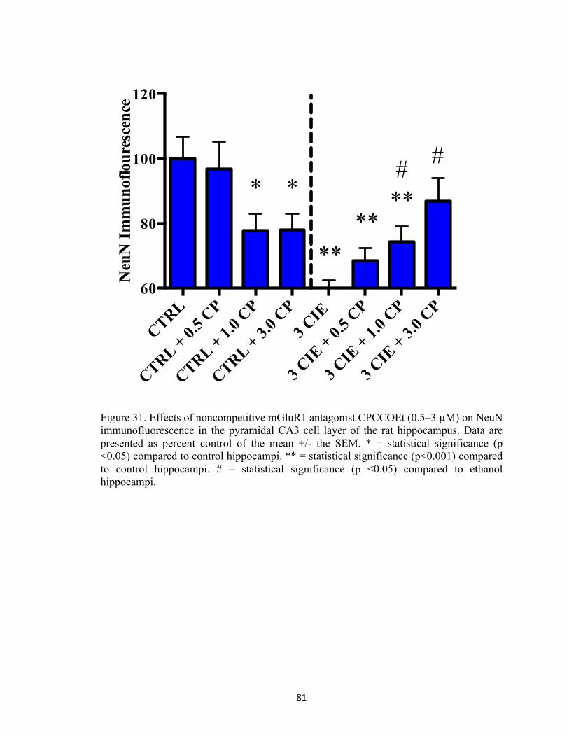
81
CTRL
CTRL + 0.5 C
P
CTRL + 1.0 C
P
CTRL + 3.0 C
P
3 CIE
3 CIE
+ 0.5 C
P
3 CIE
+ 1.0 C
P
3 CIE
+ 3.0 C
P60
80
100
120
Neu
N I
mm
unof
lour
esce
nce
********
##
Figure 31. Effects of noncompetitive mGluR1 antagonist CPCCOEt (0.5–3 µM) on NeuN immunofluorescence in the pyramidal CA3 cell layer of the rat hippocampus. Data are presented as percent control of the mean +/- the SEM. * = statistical significance (p <0.05) compared to control hippocampi. ** = statistical significance (p<0.001) compared to control hippocampi. # = statistical significance (p <0.05) compared to ethanol hippocampi.

82
CTRL
CTRL + 0.5 C
P
CTRL + 1.0 C
P
CTRL + 3.0 C
P
3 CIE
3 CIE
+ 0.5 C
P
3 CIE
+ 1.0 C
P
3 CIE
+ 3.0 C
P60
80
100
120
Neu
N I
mm
unof
lour
esce
nce
****
***#
**
Figure 32. Effects of noncompetitive mGluR1 antagonist CPCCOEt (0.5–3 µM) on NeuN immunofluorescence in the granule cell layer of the dentate gyrus. Data are presented as percent control of the mean +/- the SEM. * = statistical significance (p <0.05) compared to control hippocampi. ** = statistical significance (p<0.001) compared to control hippocampi. # = statistical significance (p <0.05) compared to ethanol hippocampi.

83
Figure 33. Representative images of hippocampi exposed to ethanol-naïve media (control) or ethanol media (50 mM) or hippocampi co-exposed to 3.0 µM CPCCOEt and ethanol (50 mM).

84
CTRL
CTRL + 20 SIB
CTRL + 100 S
IB
CTRL + 200 S
IB3 C
IE
3 CIE
+ 20 SIB
3 CIE
+ 100 S
IB
3 CIE
+ 200 S
IB60
80
100
120
Neu
N I
mm
unof
lour
esce
nce
**
#
*# #
Figure 34. Effects of mGluR5 antagonist SIB-1893 on NeuN immunofluorescence in the primary cell layer of the CA1. Data are presented as percent control of the mean +/- the SEM. * = statistical significance (p <0.05) compared to control hippocampi. ** = statistical significance (p <0.001) compared to control hippocampi. # = statistical significance (p <0.05) compared to ethanol hippocampi.

85
CTRL
CTRL + 20 SIB
CTRL + 100 S
IB
CTRL + 200 S
IB3 C
IE
3 CIE
+ 20 SIB
3 CIE
+ 100 S
IB
3 CIE
+ 200 S
IB60
80
100
120
Neu
N I
mm
unof
lour
esce
nce
** ****** **
Figure 35. Effects of mGluR5 antagonist SIB-1893 on NeuN immunofluorescence in the primary cell layer of the CA3. Data are presented as percent control of the mean +/- the SEM. ** = statistical significance (p <0.001) compared to control hippocampi.

86
CTRL
CTRL + 20 SIB
CTRL + 100 S
IB
CTRL + 200 S
IB3 C
IE
3 CIE
+ 20 SIB
3 CIE
+ 100 S
IB
3 CIE
+ 200 S
IB60
80
100
120
Neu
N I
mm
unof
lour
esce
nce
******** **
#
Figure 36. Effects of mGluR5 antagonist SIB-1893 on NeuN immunofluorescence in the granule cell layer of the dentate gyrus. Data are presented as percent control of the mean +/- the SEM. ** = statistical significance (p <0.001) compared to control hippocampi. # = statistical significance (p <0.05) compared to ethanol hippocampi.

87
Figure 37. Representative images of hippocampi exposed to ethanol-naïve media (control) or ethanol media (50 mM) or hippocampi co-exposed to 20 µM SIB-1897 and ethanol (50 mM).

88
CTRL
CTRL + 0.5 X
C3 C
IE
3 CIE
+ 0.5 X
C60
80
100
120N
euN
Im
mun
oflo
ures
cenc
e
**
#*
Figure 38. Effects of inhibitor of IP3-mediated calcium release (0.5 µM) on NeuN immunofluorescence in the pyramidal CA1 cell layer of the rat hippocampus. Data are presented as percent control of the mean +/- the SEM. * = statistical significance (p <0.05) compared to control hippocampi. ** = statistical significance (p <0.001) compared to control hippocampi. # = statistical significance (p <0.05) compared to ethanol hippocampi.

89
CTRL
CTRL + 0.5 X
C3 C
IE
3 CIE
+ 0.5 X
C60
80
100
120
140
Neu
N I
mm
unof
lour
esce
nce
*
#*
Figure 39. Effects of inhibitor of IP3-mediated calcium release (0.5 µM) on NeuN immunofluorescence in the pyramidal CA3 cell layer of the rat hippocampus. Data are presented as percent control of the mean +/- the SEM. * = statistical significance (p <0.05) compared to control hippocampi. ** = statistical significance (p <0.001) compared to control hippocampi. # = statistical significance (p <0.05) compared to ethanol hippocampi.

90
CTRL
CTRL + 0.5 X
C3 C
IE
3 CIE
+ 0.5 X
C60
80
100
120
140
Neu
N I
mm
unof
lour
esce
nce
*
#***
Figure 40. Effects of inhibitor of IP3-mediated calcium release (0.5 µM) on NeuN immunofluorescence in the granule cell layer of the dentate gyrus. Data are presented as percent control of the mean +/- the SEM. * = statistical significance (p <0.05) compared to control hippocampi. ** = statistical significance (p <0.001) compared to control hippocampi. # = statistical significance (p <0.05) compared to ethanol hippocampi.
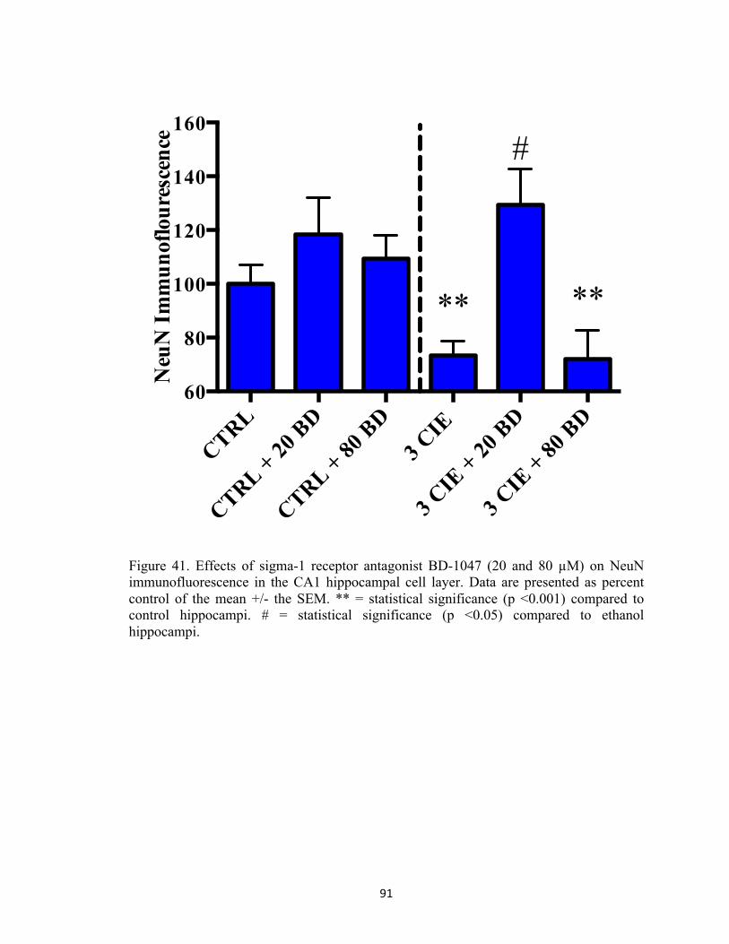
91
CTRL
CTRL + 20 B
D
CTRL + 80 B
D3 C
IE
3 CIE
+ 20 B
D
3 CIE
+ 80 B
D60
80
100
120
140
160
Neu
N I
mm
unof
lour
esce
nce
**
#
**
Figure 41. Effects of sigma-1 receptor antagonist BD-1047 (20 and 80 µM) on NeuN immunofluorescence in the CA1 hippocampal cell layer. Data are presented as percent control of the mean +/- the SEM. ** = statistical significance (p <0.001) compared to control hippocampi. # = statistical significance (p <0.05) compared to ethanol hippocampi.

92
CTRL
CTRL + 20 B
D
CTRL + 80 B
D3 C
IE
3 CIE
+ 20 B
D
3 CIE
+ 80 B
D
100
150
200N
euN
Im
mun
oflo
ures
cenc
e (%
con
trol
)
**
#
**
**
Figure 42. Effects of sigma-1 receptor antagonist BD-1047 (20 and 80 µM) on NeuN immunofluorescence in the CA3 hippocampal cell layer. Data are presented as percent control of the mean +/- the SEM. ** = statistical significance (p <0.001) compared to control hippocampi. # = statistical significance (p <0.05) compared to ethanol hippocampi.

93
CTRL
CTRL + 20 B
D
CTRL + 80 B
D3 C
IE
3 CIE
+ 20 B
D
3 CIE
+ 80 B
D
100
150
200
Neu
N I
mm
unof
lour
esce
nce
**
#
**
** **
Figure 43. Effects of sigma-1 receptor antagonist BD-1047 (20 and 80 µM) on NeuN immunofluorescence in the dentate gyrus hippocampal cell layer. Data are presented as percent control of the mean +/- the SEM. ** = statistical significance (p <0.001) compared to control hippocampi. # = statistical significance (p <0.05) compared to ethanol hippocampi.

94
CTRL
CTRL + 5 DT
3 CIE
3 CIE
+ 5 DT
60
80
100
120
Neu
N I
mm
unof
lour
esce
nce
**** **
Figure 44. Effects of ryanodine receptor antagonist dantrolene (5 µM) on NeuN immunofluorescence in the CA1 hippocampal cell layer. Data are presented as percent control of the mean +/- the SEM. ** = statistical significance (p <0.001) compared to control hippocampi.

95
CTRL
CTRL + 5 DT
3 CIE
3 CIE
+ 5 DT
60
80
100
120
Neu
N I
mm
unof
lour
esce
nce
****
Figure 45. Effects of ryanodine receptor antagonist dantrolene (5 µM) on NeuN immunofluorescence in the CA3 hippocampal cell layer. Data are presented as percent control of the mean +/- the SEM. ** = statistical significance (p <0.001) compared to control hippocampi.

96
CTRL
CTRL + 5 DT
3 CIE
3 CIE
+ 5 DT
60
80
100
120
Neu
N I
mm
unof
lour
esce
nce
** ****
Figure 46. Effects of ryanodine receptor antagonist dantrolene (5 µM) on NeuN immunofluorescence in the dentate gyrus hippocampal cell layer. Data are presented as percent control of the mean +/- the SEM. ** = statistical significance (p <0.001) compared to control hippocampi.

97
CHAPTER FIVE: Influence of mGluR5-containing Receptors in the Development of Ethanol Dependence In Vivo
5.1. Introduction
Rodent models of CIE have been productive in that they display parallel
behavioral and neurodegenerative effects to those observed in the clinical population.
These effects include increases in duration of seizures during withdrawal from ethanol
(Stephens et al., 2001; Veatch & Becker, 2002), cognitive deficits (Badanich et al., 2011;
Duka et al., 2003; Zhao et al., 2013), and hippocampal neurodegeneration (Collins et al.,
1998; Corso et al., 1998; Zhao et al., 2013). Notably, rodents exposed to a CIE regimen
displayed significant increases in handling-induced convulsions and spontaneous
electroencephalogram activity (Veatch & Becker, 2005), and these effects were reduced
following the administration of noncompetitive NMDA-receptor antagonist MK-801
during periods of ethanol withdrawal. Other studies have found that protein kinase A and
C-dependent increase in GluN1 and GluN2B surface expression in cortical neurons
exposed to chronic and intermittent ethanol (Qiang et al., 2007), and that the NMDA-
receptor containing GluN1/2N2B subunits confer sensitivity to excitotoxicity (e.g., via
increased channel opening) (Ran et al., 2003; for a review, see Prendergast &
Mulholland, 2012). It has also been noted that trafficking of these receptors from the ER
to the synapse can occur (Mu et al., 2003), further potentiating cell death.
Chronic exposure to ethanol also modulates Gq-coupled group 1 mGluR, which
can stimulate IP3-mediated release of intracellular calcium (Conn et al., 1997) or activate
downstream second messenger signaling pathways (Olive et al., 2009) in rodents and in
the clinical population (e.g., Lominac et al., 2006). As an example, in vivo

98
experimentation has shown binge-like ethanol consumption produced increased signaling
of the mGluR5, and the administration of an mGluR5 antagonist significantly attenuated
binge-like ethanol consumption in this rodent model of scheduled, high-alcohol
consumption (Cozzoli et al., 2009). In another study, administration of an mGluR5
antagonist mitigated withdrawal manifestations and the sedative-like behavioral effects of
ethanol if the mGluR5 antagonist was injected prior to, but not following, ethanol
exposure (Blendov et al., 2008). Notably, human studies have proposed a link between
genetic alterations in the mGluR5 and a propensity for alcohol abuse (Schumann et al.,
2008).As noted in Chapter 4, blocking mGluR1-and-5 containing receptors attenuates the
damage to hippocampal neuronal integrity produced by CIE in vitro.
5.2. Experimental Rationale
Recent efforts have been made to understand the adverse behavioral and
neurodegenerative effects that are produced by CIE exposure. The mechanistic
underpinnings associated with these effects, however, are not fully characterized. The
model of CIE exposure proposed in these studies mimics patterns of binge-like
consumption using a binge-like ethanol concentration (i.e., 50 mM[~230 mg/dl]) that can
be readily achieved in the clinical population. The use of an in vivo model of CIE
exposure allowed for an examination of the role of the mGluR5 in the development of
dependence.
5.3. Methods
Subjects. Thirty-two adult, male Sprague-Dawley rats (i.e., weighed in at 300–325 grams
upon arrival; Harlan Laboratories, Indianapolis, IN) were housed individually and
allowed to acclimate to the animal colony for two days following arrival. Subjects were

99
then handled for two minutes per day for three consecutive days prior to experimentation.
Subjects were allowed ad libitum access to food and water throughout the entire duration
of the experiment. Food weight data were collected at 0800 hour each week. Each animal
was weighed at 0800 hour on days Monday through Friday, prior to ethanol
administration. A mortality rate of 5% was observed due to complications with the
intragastric gavage procedure.
Ethanol exposure and withdrawal. The experiments included in this study were designed
to extend the in vitro findings described previously to a live animal model of CIE. In
these studies, subjects were administered ethanol (4 g/kg) via intragastric gavage twice
daily at 0800 and 1600 for five consecutive days with or without the addition of 3 mg/kg
2-Methyl-6-(phenylethynyl)pyridine (MPEP). MPEP is a group 1 metabotrophic
glutamate receptor antagonist that has been used previously as a neuroprotective
compound in several models of drug and alcohol dependence, as well as in several
models of neurodegenerative disorders. Given that nasogastric gavage administration of
MPEP (10 mg/kg) for one month reduced Parkinsonian symptoms in non-human
primates (Morin et al., 2013), we chose to deliver this compound to subjects orally.
During periods of withdrawal, subjects received no experimental manipulations; this was
repeated for three consecutive cycles. The experimental timeline for this study is depicted
in Figure 47.
Withdrawal behavior. Following each five-day ethanol administration period, subjects
were not administered ethanol for two consecutive days (i.e., ethanol withdrawal). During

100
the third period of withdrawal, subjects were monitored for 16 hours after the last ethanol
administration (i.e., 0900 hours) for physiological manifestations of withdrawal. The
rationale for the timing of these behavioral manifestations is based on the study described
in Chapter 3 suggesting robust withdrawal behaviors are observed on the third period of
withdrawal. Assessment of withdrawal behavior occurred in a square Plexiglas chamber
for two minutes. During this time, an experimenter blinded to experimental conditions
rated physical effects of withdrawal using a modified behavioral scale that was described
in detail previously in our laboratory (Majchrowicz, 1975; Self et al., 2009) using a 10-
point discrete scale (i.e., all or nothing). This scale includes the following behaviors:
rigidity, tremor, stereotypy, retropulsion, dystonic gait, hypoactivity, aggression, splayed
paws, vocalization, and seizure.
Analysis of blood ethanol levels. In order to assess BELs, approximately 200 µL of tail
blood was collected into two Fisherbrand heparinized micro-hematocrit capillary tubes
(Fisher Scientific) on Day Two of Weeks One, Two, and Three. Next, samples were
centrifuged for four minutes using an Analox benchtop centrifuge (Analox Instruments)
with blood plasma collected and placed into a .65 mL Costar microcentrifuge tube (Fisher
Scientific). Samples were stored at -20°C until further analyses of BELs using an ethanol
assay kit (i.e., colorimetric; Abcam [Cambridge, United Kingdom]) for Week 1 and the
Analox for Weeks 2 and 3 (please see Chapter 3 for a detailed description of Analox
methodology). With regards to the ethanol ELISA assay, alcohol oxidase oxidizes
ethanol to generate hydrogen peroxide that reacts with the probe included in the assay to
generate color. All standards, background wells, and sample wells were run in duplicate

101
per manufacturer’s instructions, and 10 µL of blood plasma were incubated at room
temperature for 60 minutes with reaction enzymes in a 96-well plate (Corning, New
York). Optical density was measured using a Beckman Coulter DTX 880 Multimodal
Detector (Lagerhausstrasse, Austria) using Beckman Coulter Multimode Detection
Software (v.20.0.12). Absorbance was detected at 595 nm for standards, background
wells, and sample wells. Mean absorbance for each value was determined and then
averaged so as to yield one measurement for each sample. Mean absorbance of the blank
(i.e., standard with 0 nmol/well ethanol) was subtracted from each standard and sample
value to yield the corrected absorbance value, and a standard curve was generated based
on values of standards. Concentrations of ethanol in samples were then determined via
sample amount from standard curve (i.e., Sa; nmol) divided by the sample volume added
into the sample well (i.e., Sv; µL) using this formula: Sa/Sv.
Statistical analyses. Statistical analyses were conducted to assess the effects of CIE on
body weight, behavioral effects of withdrawal, and BELs. Effects were considered
significant at p<0.05. Body weight data were analyzed by a two-factor repeated-measure
ANOVA with day and treatment (control and ethanol) as factors. Behavioral effects of
withdrawal and BELs were analyzed by a two-factor repeated-measure ANOVA with
diet and drug as factors. Planned comparisons were conducted if a significant effect of
day or treatment or an interaction of these two factors was detected using Bonferroni.
These planned comparisons were used to make conservative pairwise comparisons
between means.

102
5.4. Results
Body weight. ANOVA revealed a significant interaction of day and diet (F[3,50] = 12.69,
p<0.001) in subjects exposed to three cycles of CIE or an isocaloric diet with or without
the addition of mGluR5 antagonist MPEP. Figure 48 shows that significant decreases in
body weight (grams) were detected in subjects that were administered ethanol compared
to subjects administered an isocaloric diet. This figure also shows that MPEP
administration did not have an effect on body weight in subjects exposed to CIE or an
isocaloric diet.
Behavioral effects of withdrawal. Subjects were administered CIE (or an isocaloric
control diet) for three consecutive weeks with or without the addition of an oral and
selective mGluR5 antagonist MPEP (3 mg/kg) to evaluate the influence of this particular
receptor subtype in the development of ethanol dependence. ANOVA revealed a
significant diet-by-drug interaction (F[1,28] = 44.87, p<0.001). Figure 49 shows that
subjects administered ethanol had significant increases in behavioral effects of
withdrawal compared to subjects that received an isocaloric diet. This figure also shows
that oral administration of mGluR5 antagonist MPEP significantly attenuated the physical
manifestations of withdrawal in ethanol-dependent rats.
Blood ethanol levels. ANOVA revealed a significant main effect of drug (F[1,7] = 11.77,
p<0.05) on BELs in ethanol-dependent subjects. Figure 50 shows that MPEP
administration modestly reduced BELs from during Weeks 1, 2, and 3 of the CIE
treatment regimen. . ANOVA also revealed a significant main effect of week (F[1,14] =
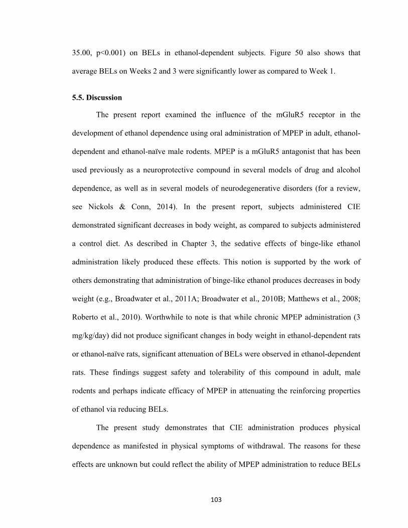
103
35.00, p<0.001) on BELs in ethanol-dependent subjects. Figure 50 also shows that
average BELs on Weeks 2 and 3 were significantly lower as compared to Week 1.
5.5. Discussion
The present report examined the influence of the mGluR5 receptor in the
development of ethanol dependence using oral administration of MPEP in adult, ethanol-
dependent and ethanol-naïve male rodents. MPEP is a mGluR5 antagonist that has been
used previously as a neuroprotective compound in several models of drug and alcohol
dependence, as well as in several models of neurodegenerative disorders (for a review,
see Nickols & Conn, 2014). In the present report, subjects administered CIE
demonstrated significant decreases in body weight, as compared to subjects administered
a control diet. As described in Chapter 3, the sedative effects of binge-like ethanol
administration likely produced these effects. This notion is supported by the work of
others demonstrating that administration of binge-like ethanol produces decreases in body
weight (e.g., Broadwater et al., 2011A; Broadwater et al., 2010B; Matthews et al., 2008;
Roberto et al., 2010). Worthwhile to note is that while chronic MPEP administration (3
mg/kg/day) did not produce significant changes in body weight in ethanol-dependent rats
or ethanol-naïve rats, significant attenuation of BELs were observed in ethanol-dependent
rats. These findings suggest safety and tolerability of this compound in adult, male
rodents and perhaps indicate efficacy of MPEP in attenuating the reinforcing properties
of ethanol via reducing BELs.
The present study demonstrates that CIE administration produces physical
dependence as manifested in physical symptoms of withdrawal. The reasons for these
effects are unknown but could reflect the ability of MPEP administration to reduce BELs

104
during Weeks 1, 2, and 3 of the first week of the CIE treatment regimen. Perhaps the
ability of MPEP to reduce withdrawal behavior in ethanol-dependent rats is a function of
pharmacokinetic considerations, rather than pharmacokinetic considerations.
Nonetheless, in general, the most consistent features of withdrawal-like behavior in the
current study were retropulsion (e.g., backing into a corner), dystonic gait, extreme tail
rigidity, and severe head tremors. While no seizures were observed in the current report,
withdrawal behaviors related to motor abnormalities were more consistent across
subjects. For example, moderate to severe head tremors were observed in nearly all
ethanol-dependent subjects. These findings are consistent with the work of others who
unequivocally demonstrate hallmark characteristics of central nervous system
hyperexcitability during periods of withdrawal from binge-like ethanol exposure (for
reviews, see Becker, 2012, 2013; Botia et al., 2015; N’Gouemo et al., 2015; Pérez &
DeBiasi, 2015; Van Skike et al., 2015).
Notably, chronic and oral administration of mGluR5 antagonist MPEP
significantly attenuated the behavioral effects of withdrawal in the present report, such as
sparing the extreme tail rigidity observed during the third consecutive period of
withdrawal in ethanol-dependent subjects administered CIE. These findings are
consistent with prior work demonstrating the efficacy of MPEP against
neurodegenerative diseases. In one study, for example, nasogastric gavage administration
of MPEP (10 mg/kg) for one month reduced Parkinsonian symptoms in non-human
primates (Morin et al., 2013). Indeed, this prior study shows that MPEP is orally
available and is consistent with the findings of the current study with regards to the
bioavailability of MPEP. It is notable that MPEP spared motor dystonias (e.g., dystonic

105
gait and retropulsion) in the current report during withdrawal from CIE in ethanol-
dependent rodents. These findings are consistent with the work of others who have
demonstrated the efficacy of MPEP for motor abnormalities known to be produced by the
benchmark treatment of Parkinson disease via L-3,4-dihydroxyphenylalanine (l-DOPA)
administration (Morin et al., 2013A), as well as the efficacy of chronic MPEP
administration to attenuate neurodegeneration and mitochondrial dysfunction produced
by 6-hydroxydopamine (6-OHDA) exposure in the nigrostriatum in rodents (Ferringo et
al., 2015). Others have shown that an “effective” dose of MPEP (i.e., 3 mg/kg/day)
reduced cognitive impairment produced by dopaminergic striatal lesioning in rodents
(DeLonibus et al., 2009). The reasons for these effects are unknown, but could include
the ability of MPEP to normalize basal glutamate levels in areas of the limbic system,
such as the basal ganglia (Morin et al., 2013B).
Cozzoli and colleagues (2009) found that binge-like ethanol administration
produced significant increases in Homer2a/b expression as well as Homer2-
phosphatidylinositol 3-kinase (PI3K) signaling in the nucleus accumbens. These effects
were significantly blocked with MPEP. In the same study, pretreatment with mGluR5
antagonist MPEP dose-dependently reduced binge ethanol consumption in rodents
(Cozzoli et al., 2009). Others have shown that MPEP administration (10 mg/kg)
attenuated ethanol withdrawal anxiety-like behaviors (Kumar et al., 2013). Other studies
have shown a reduction in ethanol reinstatement following MPEP administration elicited
by drug-associated cues (Bäckström et al., 2004), as well as attenuation of ethanol
withdrawal-elicited behaviors in rodents (Blendov & Harris, 2008).

106
Previous studies have suggested that MPEP administration attenuates the
reinforcing properties of others drugs of abuse, such as psychomotor stimulants. For
example, MPEP administration was found to reduce cocaine self-administration (Kenny
et al., 2005) and cocaine reinstatement (Kumaresan et al., 2009) in rodents and non-
human primates (Lee et al., 2005; Platt et al., 2008). Similarly, MPEP has been shown to
attenuate the reinforcing properties of nicotine (e.g., DeSousa & Markou, 2011), as well
as ketamine and heroin (Van der Kam et al, 2007). Collectively, these studies
demonstrate the safety and efficacy of MPEP in various models of disease, such as
neurodegeneration and addiction.
Ethanol 4g/kg/twice per day + 3 mg/kg MPEP
Withdrawal behavior
D2
D6BEL
D2 D2
Ethanol 4g/kg/twice per day + 3 mg/kg MPEP
Ethanol 4g/kg/twice per day + 3 mg/kg MPEP
BEL BEL
Figure 47. Representative experimental timelines depicting subjects were exposed to ethanol (4 g/kg) with or without the addition of MPEP (3 mg/kg) twice daily for five days followed by two days of withdrawal and repeated three times (i.e., 3 CIE).

107
Week
1
Week
2
Week
3300
350
400
450M
ean
Bod
y W
eigh
t (gr
ams)
**
ControlControl + MPEP
****
3 CIE3 CIE + MPEP
Figure 48. Changes in body weight in subjects exposed to ethanol or an isocaloric diet with or without the addition of mGluR5 antagonist MPEP (3 mg/kg) for three cycles of CIE. X-axis: days in Week One, Week Two, and Week Three. Data points show mean body weight in grams. Two asterisks indicate that there is a significant day-by-treatment interaction. N=7 for control subjects; N=8 for control with MPEP; N=6 for ethanol subjects; N=8 for subjects administered ethanol and MPEP
108
contro
l
contro
l + M
PEP (3 m
g/kg)
3 CIE
3 CIE
+ MPEP (3
mg/k
g)0
2
4
6
8M
ean
Phys
ical
Sym
ptom
sT
hird
Con
secu
tive
With
draw
al **
#
Figure 49. Behavioral effects of withdrawal in subjects exposed to ethanol or an isocaloric diet with or without the administration of MPEP for three cycles of CIE. X-axis: day. Data points show mean scores in withdrawal behavior observed during the third consecutive withdrawal from CIE. Two asterisks indicate that there is a significant diet-by-drug interaction. A pound sign indicates that there is a main effect of MPEP on withdrawal behavior in ethanol-dependent rats. N=7 for control subjects; N=8 for control with MPEP; N=6 for ethanol subjects; N=8 for subjects administered ethanol and MPEP.

109
Week
1
Week
2
Week
30
50
100
150
200
250
300B
lood
Eth
anol
Lev
ls (m
g/dl
)
#
*
#
Ethanol Ethanol + MPEP
#
*
Figure 50. Peak BELs in ethanol-dependent subjects on Weeks One, Two, and Three of the CIE treatment regimen. X-axis: Week. Data points show average BELs determined at 90 minutes post ethanol administration on Day Two of Weeks One, Two, and Three. Two asterisks indicate that there is a significant main effect of Week. A pound sign indicates that there is a main effect of MPEP on BELs in ethanol-dependent rats. N=4 for ethanol subjects; N=5 for ethanol with MPEP.

110
Chapter Six: A General Discussion and Future Pharmacotherapies for Treatment Alcohol Use Disorders
Alcohol use disorders are a widespread public health problem in the Unites States
(SAMSHA, 2014). While approximately 18 million individuals self-report the desire to
seek treatment for an alcohol use disorder (e.g., alcohol abuse and dependence on
alcohol), less than 10% of these individuals reported obtaining receiving formal treatment
at a facility (SAMHSA, 2014). Currently, there are four medications approved by the
United States FDA for the treatment of alcohol dependence: oral naltrexone, extended-
release naltrexone, disulfiram, and acamprosate (Gastfriend, 2014; Hartung et al., 2014;
for a review see Kraemer, 2014; Kufahl et al., 2014). Indeed these drugs have been
moderately effective in the treatment of alcohol use disorders (Blodgett et al., 2014;
Guglielmo et al., 2015; Jonas et al., 2014; Liang & Olsen, 2014). While the reasons for
the limited efficacy of current medications indicated for treatment of alcohol use
disorders are unknown, it is likely that many individuals desire to control consumption
rather than abstain completely. Recent research efforts, therefore, have been aimed at
identifying and developing putative pharmacotherapies for the treatment of alcohol use
disorders so as to address this gap in care by so as to tailor medications development
toward the needs of the individual. Putative pharmacotherapies for the treatment of
alcohol use disorders will be discussed below, followed by closing remarks of this
dissertation.

111
Anticonvulsants
Anticonvulsant medications (e.g., topiramate, gabapentin, and baclofen) are a
potential candidate class of compounds for the treatment of alcohol use disorders, due in
part, to enhancement of inhibitory neurotransmission and perhaps their limited abuse
potential. In general, many of these studies compared candidate compounds to
medications with an indication for treatment of alcohol use disorders so as to have a solid
reference point (e.g., disulfiram, naltrexone, and acamprosate). In one study, topiramate
(0–300 mg/day) reduced the number of self-assessed heavy drinking days (Johnson et al.,
2007). In another study, for example, topiramate (25–300 mg/day) promoted abstinence
in alcohol-dependent individuals as compared to naltrexone (50 mg/day) or acamprosate
(333 mg/day) (Narayana et al., 2015). Consistent with this notion, several randomized
clinical trials suggest moderate efficacy of topiramate (25–300 mg) for treatment of
alcohol use disorders (Baltieri et al., 2008; Fernandez Miranda et al., 2007; Johnson et al.,
2003; Johnson et al., 2004; Ma et al., 2006; Rubio et al., 2009). Yet in another study,
rates of abstinence were higher for disulfiram (250 mg/day) than for topiramate (150
mg/day) (De Sousa et al., 2008) while others have found similar efficacy between
topiramate (50–400 mg/day) and naltrexone (50 mg/day) (Flórez et al., 2008; Flórez et
al., 2011). In general, the efficacy of topiramate for the treatment of alcohol use disorders
is comparable to those marketed for this indication.
Gabapentin, another anticonvulsant medication, has been more recently assessed
for the treatment of alcohol use disorders. In one study, for example, gabapentin (900 and
1800 mg/day) promoted abstinence in individuals with an alcohol use disorder (Mason et
al., 2014). In two other studies, however, gabapentin (100-1200 mg/day) was not
efficacious in protracted abstinence (Arias et al., 2010; Anton et al., 2009; Furieri &

112
Nakamura-Palacious, 2007). Zonisamide (50–300 mg/day), another anticonvulsant drug,
significantly attenuated subjective effects measures of “craving”, a subjective feeling
thought to precede relapse (Rubio et al., 2010) however these effects are modest. Other
anticonvulsant medications (e.g., oxcarbazepine and tiagabine) are thought to not be
effective in promoting abstinence (Croissant et al., 2006 and Paparrigopoulos et al.,
2010). Interestingly, baclofen, a medication indicated for treatment of skeletal muscle
spasms, reduced amount of alcohol consumed (Flannery et al., 2004), as well as
attenuated rates of relapse (Addolorato et al., 2002; Addolorato et al., 2011). Yet in
another study, baclofen administration was not efficacious for treatment of alcohol use
disorders (Garbutt et al., 2010).
Collectively, these studies demonstrate that there is modest efficacy of
anticonvulsants for the treatment of alcohol use disorders in a clinical setting. Perhaps the
efficacy of these medications that mediate GABAergic tone could be examined for the
treatment of acute but severe alcohol withdrawal in combination with benzodiazepines in
patients so at to prevent known neurocognitive complications and cross-dependence on
benzodiazepines.
Antidepressants
Antidepressants (e.g., sertraline, gabapentin, and baclofen) are a potential
candidate class of compounds for the treatment of alcohol use disorders, due in part, to
high rates of comorbid alcohol use disorders and psychological disorders. In one series if
studies, sertraline (200 mg/day) promoted protracted abstinence (Pettinati et al., 2001;
Pettinati et al., 2004). In another series of studies, sertraline (50–200 mg/day) decreased
drinking in late-onset individuals with an alcohol use disorder, but actually increased

113
drinking in those individuals with an early-onset of an alcohol use disorder (Kranzler et
al., 2011; Kranzler et al., 2012). Similar findings with regards to time of onset have been
observed with ondansetron, a serotonin 5-HT3 receptor antagonist and antiemetic
(Kranzler et al., 2003; Roache et al., 2008) or in individuals with altered function of
serotonin transport (Johnson et al., 2011). Other candidate medications with current
indications for depression (e.g., fluvoxamine and escitalopram) were not shown to be
efficacious for treatment of alcohol use disorders when administered alone (Chick et al.,
2004; Stella et al., 2008) while metadoxine (3000 mg/day), a 5HT2B antagonist, was
efficacious in promoting relapse in one study (Guerrini et al., 2006).
Collectively, these findings suggest that there are individual differences (e.g.,
early- versus late-onset and polymorphisms in serotonin transport) in the efficacy of
antidepressants for the treatment of alcohol use disorders. Drug combination therapies
have been more effective than monotherapy with these serotonergic agents. Perhaps
screening of patients for polymorphisms in serotonergic transporter could yield insight
into the most effective medication for the treatment-seeking individual.
Antipsychotics
Antipsychotic medications (e.g., aripiprazole, flupenthixol, olanzapine, tiapride,
lisuride and amisulpride) recently examined for therapeutic potential in the treatment of
alcohol use disorders are not effective (Anton et al., 2008; Bender et al., 2007; Guardia et
al., 2004; Marra et al., 2002; Schmidt et al., 2002; Weisbeck et al., 2001; Weisbeck et al.,
2003). The reasons for these effects are unknown, but could reflect adverse side effects of
antipsychotic medications.

114
Opioid Receptor Antagonist Nalmefelene
European countries have recently approved nalmefelene, a partial kappa receptor
agonist, for the treatment of alcohol dependence in patients who seek treatment for
controlled consumption. While results from several large-scale randomized clinical trials
suggest nalmefelene (20–40mg/day), in combination with cognitive therapy, significantly
reduces drinking (Mann et al., 2013; Gual et al., 2013; Van de Brink et al., 2013), further
examination of this medication is warranted before any definitive conclusions can be
drawn.
Collectively, these large-scale clinical trials demonstrate that there is an
unequivocal need to identify and develop putative pharmacotherapies for the treatment of
alcohol use disorders. Drug combination therapies might be most effective for treatment
of alcohol use disorders, particularly in individuals with altered serotonergic transport
function, co-morbid psychological disorders, or individuals whom seek treatment to
control drinking rather than abstain completely.
Summary
Alcohol-related disorders arise from a variety of factors (e.g., early- and late-
onset, genetics, and comorbidities with other psychological disorders) and are
complicated by corresponding factors (e.g., physiological and psychological). Further, the
patient desire to seek treatment adds a final complication (e.g., full abstinence or
controlled consumption). Pharmacotherapy alone will not answer the large-scale problem
of alcohol use disorders in the United States but has the potential to reduce rates of
consumption and relapse in individuals seeking treatment. Patterns of intermittent
consumption (e.g., multiple withdrawals and binge drinking) are commonly demonstrated

115
by dependent individuals, and neurological, behavioral, cognitive, and effects of
sensitization or kindling to multiple withdrawals occurs in humans and rodent models of
CIE exposure. The studies in this dissertation sought to add to the field by identifying
underlying mechanisms associated with hippocampal cytotoxicity in vitro that contribute
to the behavioral effects of withdrawal in vivo.
The studies in this dissertation found that neuroadaptations in protein kinase
activity occur in the presence of binge-like ethanol exposure prior to the onset of
withdrawal in the rat hippocampus. This protein kinase activation in response to ethanol
application contributes to neuroadaptations in glutamatergic tone, promoting cytotoxicity
of hippocampal pyramidal and granule cell layers. Next, this in vitro model of CIE
exposure was translated into a model of CIE exposure in vivo so as to examine the effects
of presumed hippocampal excitotoxicity on cognitive performance in the Morris Water
Maze. While physical dependence was achieved in subjects administered CIE (i.e.,
confirmed by a significant presence of physical withdrawal characteristics), spatial
learning and memory remained intact in ethanol-dependent rats. It is reasonable to
conclude, therefore, that the specific task used for cognitive assessment was not sensitive
enough to the relatively modest hippocampal cytotoxicity reliably detected in our in vitro
assay. Future studies should implement a reversal task in the Morris Water Maze to
detect more subtle differences in reference memory before definitive conclusions can be
made. A series of antagonist studies that assessed a specific signal transduction cascade
(e.g., G-protein group 1 mGluRs and release of intracellular calcium from the ER)
identified that Gαq proteins (i.e., mGluR1-and-5-containing receptors) are involved in the
development of dependence in vitro and in vivo. For example, selective mGluR1 and

116
mGluR5 antagonist exposure in the presence of ethanol restored hippocampal neuronal
integrity in vitro. The oral administration mGluR5 antagonist in the presence of ethanol
limited the severity of withdrawal from CIE administration in vivo. In general, these
findings suggest that organotypic hippocampal slice cultures are a good model for initial
high-throughput screening of compounds and the identification of mechanisms involved
in the development of ethanol dependence. Specifically, these data suggest that blocking
group 1 mGluRs is effective in reducing the rate in which development of dependence
occurs in rodents. However, this effect could be due to pharmacokinetic interactions due
to decreases of BELs as compared to a pharmacodynamics effect. These data also suggest
that concentration of these metabotropic glutamate receptors localized in the
hippocampus mediate the cytotoxic effects of CIE exposure that contribute to the
behavioral effects of ethanol withdrawal.
From these results, putative pharmacotherapies that modulate glutamatergic tone
may be more thoroughly evaluated with regards to treatment of alcohol use disorders. In
particular, pharmacological manipulations of group 1 mGluRs in the presence of binge-
like ethanol warrant further study. These understandings may contribute to political,
social, and individual success by providing more effective options for the treatment of
alcohol use disorders in the clinical population.

117
Literature Cited
Abe, T., Sugihara, H., Nawa, H., Shigemoto, R., Mizuno, N., Nakanishi, S., 1992.
Molecular characterization of a novel metabotropic glutamate receptor mGluR5
coupled to inositol phosphate/Ca2+ signal transduction. J. Biol. Chem. 267,
13361–8.
Addolorato, G., Caputo, F., Capristo, E., Domenicali, M., Bernardi, M., Janiri, L.,
Agabio, R., Colombo, G., Gessa, G.L., Gasbarrini, G.,. Baclofen efficacy in
reducing alcohol craving and intake: a preliminary double-blind randomized
controlled study. Alcohol Alcohol 37, 504–8.
Addolorato, G., Leggio, L., Ferrulli, A., Cardone, S., Bedogni, G., Caputo, F., Gasbarrini,
G., Landolfi, R.,. Dose-response effect of baclofen in reducing daily alcohol
intake in alcohol dependence: secondary analysis of a randomized, double-blind,
placebo-controlled trial. Alcohol Alcohol 46, 312–7.
Adinoff, B., 1994. Double-blind study of alprazolam, diazepam, clonidine, and placebo in
the alcohol withdrawal syndrome: preliminary findings. Alcohol. Clin. Exp. Res.
18, 873–8.
Alele, P.E., Devaud, L.L., 2005. Differential adaptations in GABAergic and
glutamatergic systems during ethanol withdrawal in male and female rats.
Alcohol. Clin. Exp. Res. 29, 1027–34.

118
Alonso, G., Phan, V., Guillemain, I., Saunier, M., Legrand, A., Anoal, M., Maurice, T.,
2000. Immunocytochemical localization of the sigma(1) receptor in the adult rat
central nervous system. Neuroscience 97, 155–70.
American Psychiatric Association. (2000). Diagnostic and statistical manual of mental
disorders (Revised 4th ed.). Washington, DC.
American Psychiatric Association. (2013). Diagnostic and
statistical manual of mental disorders (5th ed.). Washington, DC:
Author.
Anton, R.F., Kranzler, H., Breder, C., Marcus, R.N., Carson, W.H., Han, J., 2008. A
randomized, multicenter, double-blind, placebo-controlled study of the efficacy
and safety of aripiprazole for the treatment of alcohol dependence. J. Clin.
Psychopharmacol. 28, 5–12.
Anton, R.F., Myrick, H., Baros, A.M., Latham, P.K., Randall, P.K., Wright, T.M.,
Stewart, S.H., Waid, R., Malcolm, R., 2009. Efficacy of a combination of
flumazenil and gabapentin in the treatment of alcohol dependence: relationship to
alcohol withdrawal symptoms. J. Clin. Psychopharmacol. 29, 334–42.
Arias, A.J., Feinn, R., Oncken, C., Covault, J., Kranzler, H.R., 2010. Placebo-controlled
trial of zonisamide for the treatment of alcohol dependence. J. Clin.
Psychopharmacol. 30, 318–22.

119
Bäckström, P., Bachteler, D., Koch, S., Hyytiä, P., Spanagel, R., 2004. mGluR5
antagonist MPEP reduces ethanol-seeking and relapse behavior.
Neuropsychopharmacology 29, 921–8.
Badanich, K.A., Becker, H.C., Woodward, J.J., 2011. Effects of chronic intermittent
ethanol exposure on orbitofrontal and medial prefrontal cortex-dependent
behaviors in mice. Behav. Neurosci. 125, 879–91.
Ballenger, J.C., Post, R.M., 1978. Kindling as a model for alcohol withdrawal syndromes.
Br. J. Psychiatry 133, 1–14.
Baltieri, D.A., Daró, F.R., Ribeiro, P.L., de Andrade, A.G., 2008. Comparing topiramate
with naltrexone in the treatment of alcohol dependence. Addiction 103, 2035–44.
Battaglia, G., Fornai, F., Busceti, C.L., Aloisi, G., Cerrito, F., De Blasi, A., Melchiorri,
D., Nicoletti, F., 2002. Selective blockade of mGlu5 metabotropic glutamate
receptors is protective against methamphetamine neurotoxicity. J. Neurosci. 22,
2135–41.
Baude, A., Nusser, Z., Roberts, J.D., Mulvihill, E., McIlhinney, R.A., Somogyi, P., 1993.
The metabotropic glutamate receptor (mGluR1 alpha) is concentrated at
perisynaptic membrane of neuronal subpopulations as detected by immunogold
reaction. Neuron 11, 771–87.
Becker, H.C., 2000. Animal models of alcohol withdrawal. Alcohol Res. Health 24, 105–
13.

120
Becker, H.C., 2012. Effects of alcohol dependence and withdrawal on stress
responsiveness and alcohol consumption. Alcohol Res. 34, 448–58.
Becker, H.C., 2013. Animal models of excessive alcohol consumption in rodents. Curr.
Top. Behav. Neurosci. 13, 355–77.
Bender, S., Scherbaum, N., Soyka, M., Rüther, E., Mann, K., Gastpar, M., 2007. The
efficacy of the dopamine D2/D3 antagonist tiapride in maintaining abstinence: a
randomized, double-blind, placebo-controlled trial in 299 alcohol-dependent
patients. Int. J. Neuropsychopharmacol. 10, 653–60.
Bergman, H., Borg, S., Hindmarsh, T., Ideström, C.M., Mützell, S., 1980. Computed-
tomography of the brain and neuropsychological assessement of alcoholic
patients. Adv. Exp. Med. Biol. 126, 771–86.
Berkeley, J.L., Levey, A.I., 2003. Cell-specific extracellular signal-regulated kinase
activation by multiple G protein-coupled receptor families in hippocampus. Mol.
Pharmacol. 63, 128–35.
Berridge, M.J., 1993. Cell signalling. A tale of two messengers. Nature 365, 388–9.
Berridge, M.J., Bootman, M.D., Roderick, H.L., 2003. Calcium signalling: dynamics,
homeostasis and remodelling. Nat. Rev. Mol. Cell Biol. 4, 517–29.
Berridge, M.J., Lipp, P., Bootman, M.D., 2000. The versatility and universality of
calcium signalling. Nat. Rev. Mol. Cell Biol. 1, 11–21.

121
Blednov, Y.A., Harris, R.A., 2008. Metabotropic glutamate receptor 5 (mGluR5)
regulation of ethanol sedation, dependence and consumption: relationship to
acamprosate actions. Int. J. Neuropsychopharmacol. 11, 775–93.
Blodgett, J.C., Del Re, A.C., Maisel, N.C., Finney, J.W., 2014. A meta-analysis of
topiramate’s effects for individuals with alcohol use disorders. Alcohol. Clin.
Exp. Res. 38, 1481–8.
Botia, B., Legastelois, R., Houchi, H., Naassila, M., 2015. Basal anxiety negatively
correlates with vulnerability to ethanol-induced behavioral sensitization in
DBA/2J mice: modulation by diazepam. Alcohol. Clin. Exp. Res. 39, 45–54.
Bouchery, E.E., Harwood, H.J., Sacks, J.J., Simon, C.J., Brewer, R.D., 2011. Economic
costs of excessive alcohol consumption in the U.S., 2006. Am. J. Prev. Med. 41,
516–24.
Broadwater, M., Varlinskaya, E.I., Spear, L.P., 2011 (A). Different chronic ethanol
exposure regimens in adolescent and adult male rats: effects on tolerance to
ethanol-induced motor impairment. Behav. Brain Res. 225, 358–62.
Broadwater, M., Varlinskaya, E.I., Spear, L.P., 2011 (B). Chronic intermittent ethanol
exposure in early adolescent and adult male rats: effects on tolerance, social
behavior, and ethanol intake. Alcohol. Clin. Exp. Res. 35, 1392–403.
Brown, M.E., Anton, R.F., Malcolm, R., Ballenger, J.C., 1988. Alcohol detoxification
and withdrawal seizures: clinical support for a kindling hypothesis. Biol.
Psychiatry 23, 507–14.

122
Buzsáki, G., Moser, E.I., 2013. Memory, navigation and theta rhythm in the
hippocampal-entorhinal system. Nat. Neurosci. 16, 130–8.
Carlen, P.L., Wortzman, G., Holgate, R.C., Wilkinson, D.A., Rankin, J.C., 1978.
Reversible cerebral atrophy in recently abstinent chronic alcoholics measured by
computed tomography scans. Science 200, 1076–8.
Carpenter-Hyland, E.P., Woodward, J.J., Chandler, L.J., 2004. Chronic ethanol induces
synaptic but not extrasynaptic targeting of NMDA receptors. J. Neurosci. 24,
7859–68.
Carrington, C.D., Ellinwood, E.H., Krishnan, R.R., 1984. Effects of single and repeated
alcohol withdrawal on kindling. Biol. Psychiatry 19, 525–37.
Center for Disease Control (CDC). Behavioral Risk Surveillance System.
www.cdc.gov/brfss/ (August 13, 2012).
Chanraud, S., Martelli, C., Delain, F., Kostogianni, N., Douaud, G., Aubin, H.-J.,
Reynaud, M., Martinot, J.-L., 2007. Brain morphometry and cognitive
performance in detoxified alcohol-dependents with preserved psychosocial
functioning. Neuropsychopharmacology 32, 429–38.
Chapman, A.G., Nanan, K., Williams, M., Meldrum, B.S., 2000. Anticonvulsant activity
of two metabotropic glutamate group I antagonists selective for the mGlu5
receptor: 2-methyl-6-(phenylethynyl)-pyridine (MPEP), and (E)-6-methyl-2-
styryl-pyridine (SIB 1893). Neuropharmacology 39, 1567–74.

123
Charpak, S., Gähwiler, B.H., Do, K.Q., Knöpfel, T., 1990. Potassium conductances in
hippocampal neurons blocked by excitatory amino-acid transmitters. Nature 347,
765–7.
Chen, T., Fei, F., Jiang, X., Zhang, L., Qu, Y., Huo, K., Fei, Z., 2012. Down-regulation of
Homer1b/c attenuates glutamate-mediated excitotoxicity through endoplasmic
reticulum and mitochondria pathways in rat cortical neurons. Free Radic. Biol.
Med. 52, 208–17.
Chick, J., Aschauer, H., Hornik, K., 2004. Efficacy of fluvoxamine in preventing relapse
in alcohol dependence: a one-year, double-blind, placebo-controlled multicentre
study with analysis by typology. Drug Alcohol Depend. 74, 61–70.
Collins, M.A., Zou, J.Y., Neafsey, E.J., 1998. Brain damage due to episodic alcohol
exposure in vivo and in vitro: furosemide neuroprotection implicates edema-based
mechanism. FASEB J. 12, 221–30.
Conn, P.J., Pin, J.P., 1997. Pharmacology and functions of metabotropic glutamate
receptors. Annu. Rev. Pharmacol. Toxicol. 37, 205–37.
Corso, T.D., Mostafa, H.M., Collins, M.A., Neafsey, E.J., 1998. Brain neuronal
degeneration caused by episodic alcohol intoxication in rats: effects of
nimodipine, 6,7-dinitro-quinoxaline-2,3-dione, and MK-801. Alcohol. Clin. Exp.
Res. 22, 217–24.

124
Cotman, C.W., Geddes, J.W., Bridges, R.J., Monaghan, D.T.,. N-methyl-D-aspartate
receptors and Alzheimer’s disease. Neurobiol. Aging 10, 603–5; discussion 618–
20.
Cozzoli, D.K., Goulding, S.P., Zhang, P.W., Xiao, B., Hu, J.-H., Ary, A.W., Obara, I.,
Rahn, A., Abou-Ziab, H., Tyrrel, B., Marini, C., Yoneyama, N., Metten, P.,
Snelling, C., Dehoff, M.H., Crabbe, J.C., Finn, D.A., Klugmann, M., Worley,
P.F., Szumlinski, K.K., 2009. Binge drinking upregulates accumbens mGluR5-
Homer2-PI3K signaling: functional implications for alcoholism. J. Neurosci. 29,
8655–68.
Cox, B.R., Olney, J.J., Lowery-Gionta, E.G., Sprow, G.M., Rinker, J.A., Navarro, M.,
Kash, T.L., Thiele, T.E., 2013. Repeated cycles of binge-like ethanol (EtOH)-
drinking in male C57BL/6J mice augments subsequent voluntary EtOH intake but
not other dependence-like phenotypes. Alcohol. Clin. Exp. Res. 37, 1688–95.
Clapper, D.L., Walseth, T.F., Dargie, P.J., Lee, H.C., 1987. Pyridine nucleotide
metabolites stimulate calcium release from sea urchin egg microsomes
desensitized to inositol trisphosphate. J. Biol. Chem. 262, 9561–8.
Clapham, D.E., 1995. Intracellular calcium. Replenishing the stores. Nature 375, 634–5.
Clemmesen, L., Ingvar, M., Hemmingsen, R., Bolwig, T.G., 1988. Local cerebral glucose
consumption during ethanol withdrawal in the rat: effects of single and multiple
episodes and previous convulsive seizures. Brain Res. 453, 204–14.

125
Crabbe, J.C., Harris, R.A., Koob, G.F., 2011. Preclinical studies of alcohol binge
drinking. Ann. N. Y. Acad. Sci. 1216, 24–40.
Croissant, B., Diehl, A., Klein, O., Zambrano, S., Nakovics, H., Heinz, A., Mann, K.,
2006. A pilot study of oxcarbazepine versus acamprosate in alcohol-dependent
patients. Alcohol. Clin. Exp. Res. 30, 630–5.
Daniell, L.C., Harris, R.A., 1989. Ethanol and inositol 1,4,5-trisphosphate release
calcium from separate stores of brain microsomes. J. Pharmacol. Exp. Ther. 250,
875–81.
Davies, S.P., Helps, N.R., Cohen, P.T., Hardie, D.G., 1995. 5’-AMP inhibits
dephosphorylation, as well as promoting phosphorylation, of the AMP-activated
protein kinase. Studies using bacterially expressed human protein phosphatase-2C
alpha and native bovine protein phosphatase-2AC. FEBS Lett. 377, 421–5.
Debonnel, G., de Montigny, C., 1996. Modulation of NMDA and dopaminergic
neurotransmissions by sigma ligands: possible implications for the treatment of
psychiatric disorders. Life Sci. 58, 721–34.
Devaud, L.L., Alele, P., 2004. Differential effects of chronic ethanol administration and
withdrawal on gamma-aminobutyric acid type A and NMDA-receptor subunit
proteins in male and female rat brain. Alcohol. Clin. Exp. Res. 28, 957–65.
D’Hooge, R., De Deyn, P.P., 2001. Applications of the Morris water maze in the study of
learning and memory. Brain Res. Brain Res. Rev. 36, 60–90.

126
De Leonibus, E., Managò, F., Giordani, F., Petrosino, F., Lopez, S., Oliverio, A.,
Amalric, M., Mele, A., 2009. Metabotropic glutamate receptors 5 blockade
reverses spatial memory deficits in a mouse model of Parkinson’s disease.
Neuropsychopharmacology 34, 729–38.
De Sousa, A.A., De Sousa, J., Kapoor, H., 2008. An open randomized trial comparing
disulfiram and topiramate in the treatment of alcohol dependence. J. Subst. Abuse
Treat. 34, 460–3.
D’Souza, M.S., Markou, A., 2011. Metabotropic glutamate receptor 5 antagonist 2-
methyl-6-(phenylethynyl)pyridine (MPEP) microinfusions into the nucleus
accumbens shell or ventral tegmental area attenuate the reinforcing effects of
nicotine in rats. Neuropharmacology 61, 1399–405.
Duka, T., Townshend, J.M., Collier, K., Stephens, D.N., 2003. Impairment in cognitive
functions after multiple detoxifications in alcoholic inpatients. Alcohol. Clin. Exp.
Res. 27, 1563–72.
Duka, T., Townshend, J.M., 2004. The priming effect of alcohol pre-load on attentional
bias to alcohol-related stimuli. Psychopharmacology (Berl). 176, 353–61.
Duka, T., Gentry, J., Malcolm, R., Ripley, T.L., Borlikova, G., Stephens, D.N., Veatch,
L.M., Becker, H.C., Crews, F.T., 2004. Consequences of multiple withdrawals
from alcohol. Alcohol. Clin. Exp. Res. 28, 233–46.
Epstein, P.S., Pisani, V.D., Fawcett, J.A., 1977. Alcoholism and cerebral atrophy.
Alcohol. Clin. Exp. Res. 1, 61–5.

127
Ferrigno, A., Vairetti, M., Ambrosi, G., Rizzo, V., Richelmi, P., Blandini, F., Armentero,
M.-T., 2015. Selective blockade of mGlu5 metabotropic glutamate receptors is
protective against hepatic mitochondrial dysfunction in 6-OHDA lesioned
Parkinsonian rats. Clin. Exp. Pharmacol. Physiol.
Fernandez Miranda, J.J., Marina González, P.A., Montes Pérez, M., Díaz González, T.,
Gutierrez Cienfuegos, E., Antuña Díaz, M.J., Bobes García, J.,. Topiramate as
add-on therapy in non-respondent alcohol dependant patients: a 12 month follow-
up study. Actas españolas Psiquiatr. 35, 236–42.
Flannery, B.A., Garbutt, J.C., Cody, M.W., Renn, W., Grace, K., Osborne, M., Crosby,
K., Morreale, M., Trivette, A., 2004. Baclofen for alcohol dependence: a
preliminary open-label study. Alcohol. Clin. Exp. Res. 28, 1517–23.
Flórez, G., García-Portilla, P., Alvarez, S., Saiz, P.A., Nogueiras, L., Bobes, J., 2008.
Using topiramate or naltrexone for the treatment of alcohol-dependent patients.
Alcohol. Clin. Exp. Res. 32, 1251–9.
Flórez, G., Saiz, P.A., García-Portilla, P., Alvarez, S., Nogueiras, L., Bobes, J., 2011.
Topiramate for the treatment of alcohol dependence: comparison with naltrexone.
Eur. Addict. Res. 17, 29–36.
Foster, T.C., Defazio, R.A., Bizoń, J.L., 2012. Characterizing cognitive aging of spatial
and contextual memory in animal models. Front. Aging Neurosci. 4, 12.
Fotuhi, M., Sharp, A.H., Glatt, C.E., Hwang, P.M., von Krosigk, M., Snyder, S.H.,
Dawson, T.M., 1993. Differential localization of phosphoinositide-linked

128
metabotropic glutamate receptor (mGluR1) and the inositol 1,4,5-trisphosphate
receptor in rat brain. J. Neurosci. 13, 2001–12.
Furieri, F.A., Nakamura-Palacios, E.M., 2007. Gabapentin reduces alcohol consumption
and craving: a randomized, double-blind, placebo-controlled trial. J. Clin.
Psychiatry 68, 1691–700.
Gafni, J., Munsch, J.A., Lam, T.H., Catlin, M.C., Costa, L.G., Molinski, T.F., Pessah,
I.N., 1997. Xestospongins: potent membrane permeable blockers of the inositol
1,4,5-trisphosphate receptor. Neuron 19, 723–33.
Gallagher, S.M., Daly, C.A., Bear, M.F., Huber, K.M., 2004. Extracellular signal-
regulated protein kinase activation is required for metabotropic glutamate
receptor-dependent long-term depression in hippocampal area CA1. J. Neurosci.
24, 4859–64.
Garbutt, J.C., Kampov-Polevoy, A.B., Gallop, R., Kalka-Juhl, L., Flannery, B.A., 2010.
Efficacy and safety of baclofen for alcohol dependence: a randomized, double-
blind, placebo-controlled trial. Alcohol. Clin. Exp. Res. 34, 1849–57.
Garfield, J.M., Bukusoglu, C., 1996. Propofol and ethanol produce additive hypnotic and
anesthetic effects in the mouse. Anesth. Analg. 83, 156–61.
Gastfriend, D.R., 2014. A pharmaceutical industry perspective on the economics of
treatments for alcohol and opioid use disorders. Ann. N. Y. Acad. Sci. 1327, 112–
30.

129
Gereau, R.W., Conn, P.J., 1995. Multiple presynaptic metabotropic glutamate receptors
modulate excitatory and inhibitory synaptic transmission in hippocampal area
CA1. J. Neurosci. 15, 6879–89.
Guérineau, N.C., Gähwiler, B.H., Gerber, U., 1994. Reduction of resting K+ current by
metabotropic glutamate and muscarinic receptors in rat CA3 cells: mediation by
G-proteins. J. Physiol. 474, 27–33.
Giedd, J.N., Blumenthal, J., Jeffries, N.O., Castellanos, F.X., Liu, H., Zijdenbos, A.,
Paus, T., Evans, A.C., Rapoport, J.L., 1999. Brain development during childhood
and adolescence: a longitudinal MRI study. Nat. Neurosci. 2, 861–3.
Grant, B.F., Dawson, D.A., 1997. Age at onset of alcohol use and its association with
DSM-IV alcohol abuse and dependence: results from the National Longitudinal
Alcohol Epidemiologic Survey. J. Subst. Abuse 9, 103–10.
Gross, M.M., Rosenblatt, S.M., Malenowski, B., Broman, M., Lewis, E., 1972.
Classification of alcohol withdrawal syndromes. Q. J. Stud. Alcohol 33, 400–7.
Gual, A., He, Y., Torup, L., van den Brink, W., Mann, K., 2013. A randomised, double-
blind, placebo-controlled, efficacy study of nalmefene, as-needed use, in patients
with alcohol dependence. Eur. Neuropsychopharmacol. 23, 1432–42.
Guardia, J., Segura, L., Gonzalvo, B., Iglesias, L., Roncero, C., Cardús, M., Casas, M.,
2004. A double-blind, placebo-controlled study of olanzapine in the treatment of
alcohol-dependence disorder. Alcohol. Clin. Exp. Res. 28, 736–45.

130
Guerrini, I., Gentili, C., Nelli, G., Guazzelli, M., 2006. A follow up study on the efficacy
of metadoxine in the treatment of alcohol dependence. Subst. Abuse Treat. Prev.
Policy 1, 35.
Guglielmo, R., Martinotti, G., Quatrale, M., Ioime, L., Kadilli, I., Di Nicola, M., Janiri,
L., 2015. Topiramate in Alcohol Use Disorders: Review and Update. CNS Drugs.
Hanner, M., Moebius, F.F., Flandorfer, A., Knaus, H.G., Striessnig, J., Kempner, E.,
Glossmann, H., 1996. Purification, molecular cloning, and expression of the
mammalian sigma1-binding site. Proc. Natl. Acad. Sci. U. S. A. 93, 8072–7.
Harper, C., Kril, J., Daly, J., 1987. Are we drinking our neurones away? Br. Med. J.
(Clin. Res. Ed). 294, 534–6.
Harris, B.R., Gibson, D.A., Prendergast, M.A., Blanchard, J.A., Holley, R.C., Hart, S.R.,
Scotland, R.L., Foster, T.C., Pedigo, N.W., Littleton, J.M., 2003. The
neurotoxicity induced by ethanol withdrawal in mature organotypic hippocampal
slices might involve cross-talk between metabotropic glutamate type 5 receptors
and N-methyl-D-aspartate receptors. Alcohol. Clin. Exp. Res. 27, 1724–35.
Hartung, D.M., McCarty, D., Fu, R., Wiest, K., Chalk, M., Gastfriend, D.R., 2014.
Extended-release naltrexone for alcohol and opioid dependence: a meta-analysis
of healthcare utilization studies. J. Subst. Abuse Treat. 47, 113–21.
Haughey, N.J., Nath, A., Mattson, M.P., Slevin, J.T., Geiger, J.D., 2001. HIV-1 Tat
through phosphorylation of NMDA receptors potentiates glutamate excitotoxicity.
J. Neurochem. 78, 457–67.

131
Hayashi, T., Rizzuto, R., Hajnoczky, G., Su, T.-P., 2009. MAM: more than just a
housekeeper. Trends Cell Biol. 19, 81–8.
Hayashi, T., Su, T.-P., 2007. Sigma-1 receptor chaperones at the ER-mitochondrion
interface regulate Ca(2+) signaling and cell survival. Cell 131, 596–610.
Hodge, C.W., Miles, M.F., Sharko, A.C., Stevenson, R.A., Hillmann, J.R., Lepoutre, V.,
Besheer, J., Schroeder, J.P., 2006. The mGluR5 antagonist MPEP selectively
inhibits the onset and maintenance of ethanol self-administration in C57BL/6J
mice. Psychopharmacology (Berl). 183, 429–38.
Houamed, K.M., Kuijper, J.L., Gilbert, T.L., Haldeman, B.A., O’Hara, P.J., Mulvihill,
E.R., Almers, W., Hagen, F.S., 1991. Cloning, expression, and gene structure of a
G protein-coupled glutamate receptor from rat brain. Science 252, 1318–21.
Isokawa, M., Alger, B.E., 2006. Ryanodine receptor regulates endogenous cannabinoid
mobilization in the hippocampus. J. Neurophysiol. 95, 3001–11.
Jin, D.-Z., Guo, M.-L., Xue, B., Mao, L.-M., Wang, J.Q., 2013. Differential regulation of
CaMKIIα interactions with mGluR5 and NMDA receptors by Ca(2+) in neurons.
J. Neurochem. 127, 620–31.
Johnson, B.A., Ait-Daoud, N., Bowden, C.L., DiClemente, C.C., Roache, J.D., Lawson,
K., Javors, M.A., Ma, J.Z., 2003. Oral topiramate for treatment of alcohol
dependence: a randomised controlled trial. Lancet 361, 1677–85.
Johnson, B.A., Ait-Daoud, N., Seneviratne, C., Roache, J.D., Javors, M.A., Wang, X.-Q.,
Liu, L., Penberthy, J.K., DiClemente, C.C., Li, M.D., 2011. Pharmacogenetic
132
approach at the serotonin transporter gene as a method of reducing the severity of
alcohol drinking. Am. J. Psychiatry 168, 265–75.
Johnson, B.A., Rosenthal, N., Capece, J.A., Wiegand, F., Mao, L., Beyers, K., McKay,
A., Ait-Daoud, N., Anton, R.F., Ciraulo, D.A., Kranzler, H.R., Mann, K.,
O’Malley, S.S., Swift, R.M., 2007. Topiramate for treating alcohol dependence: a
randomized controlled trial. JAMA 298, 1641–51.
Johnson, B.A., Swift, R.M., Ait-Daoud, N., DiClemente, C.C., Javors, M.A., Malcolm,
R.J., 2004. Development of novel pharmacotherapies for the treatment of alcohol
dependence: focus on antiepileptics. Alcohol. Clin. Exp. Res. 28, 295–301.
Jonas, D.E., Feltner, C., Garbutt, J.C., 2014. Medications for alcohol use disorders--reply.
JAMA 312, 1351.
Kelm, M.K., Criswell, H.E., Breese, G.R., 2007. Calcium release from presynaptic
internal stores is required for ethanol to increase spontaneous gamma-
aminobutyric acid release onto cerebellum Purkinje neurons. J. Pharmacol. Exp.
Ther. 323, 356–64.
Kelm, M.K., Criswell, H.E., Breese, G.R., 2008. The role of protein kinase A in the
ethanol-induced increase in spontaneous GABA release onto cerebellar Purkinje
neurons. J. Neurophysiol. 100, 3417–28.
Kenny, P.J., Boutrel, B., Gasparini, F., Koob, G.F., Markou, A., 2005. Metabotropic
glutamate 5 receptor blockade may attenuate cocaine self-administration by

133
decreasing brain reward function in rats. Psychopharmacology (Berl). 179, 247–
54.
Kim, K.K., Adelstein, R.S., Kawamoto, S., 2009. Identification of neuronal nuclei
(NeuN) as Fox-3, a new member of the Fox-1 gene family of splicing factors. J.
Biol. Chem. 284, 31052–61.
Kim, F.J., Kovalyshyn, I., Burgman, M., Neilan, C., Chien, C.-C., Pasternak, G.W., 2010.
Sigma 1 receptor modulation of G-protein-coupled receptor signaling:
potentiation of opioid transduction independent from receptor binding. Mol.
Pharmacol. 77, 695–703.
Kleinknecht, R.A., Goldstein, S.G., 1972. Neuropsychological deficits associated with
alcoholism. A review and discussion. Q. J. Stud. Alcohol 33, 999–1019.
Kraemer, K.L., 2014. Review: In alcohol use disorders, oral naltrexone, 50 mg/d, or
acamprosate reduces return to drinking. Ann. Intern. Med. 161, JC7.
Kranzler, H.R., Armeli, S., Tennen, H., 2012. Post-treatment outcomes in a double-blind,
randomized trial of sertraline for alcohol dependence. Alcohol. Clin. Exp. Res.
36, 739–44.
Kranzler, H.R., Feinn, R., Armeli, S., Tennen, H., 2012. Comparison of alcoholism
subtypes as moderators of the response to sertraline treatment. Alcohol. Clin. Exp.
Res. 36, 509–16.

134
Kranzler, H.R., Pierucci-Lagha, A., Feinn, R., Hernandez-Avila, C., 2003. Effects of
ondansetron in early- versus late-onset alcoholics: a prospective, open-label study.
Alcohol. Clin. Exp. Res. 27, 1150–5.
Kril, J.J., Halliday, G.M., Svoboda, M.D., Cartwright, H., 1997. The cerebral cortex is
damaged in chronic alcoholics. Neuroscience 79, 983–98.
Kufahl, P.R., Watterson, L.R., Olive, M.F., 2014. The development of acamprosate as a
treatment against alcohol relapse. Expert Opin. Drug Discov. 9, 1355–69.
Kumar, J., Hapidin, H., Bee, Y.-T.G., Ismail, Z., 2013. Effects of the mGluR5 antagonist
MPEP on ethanol withdrawal induced anxiety-like syndrome in rats. Behav. Brain
Funct. 9, 43.
Kumaresan, V., Yuan, M., Yee, J., Famous, K.R., Anderson, S.M., Schmidt, H.D., Pierce,
R.C., 2009. Metabotropic glutamate receptor 5 (mGluR5) antagonists attenuate
cocaine priming- and cue-induced reinstatement of cocaine seeking. Behav. Brain
Res. 202, 238–44.
Latimer, C.S., Brewer, L.D., Searcy, J.L., Chen, K.-C., Popović, J., Kraner, S.D.,
Thibault, O., Blalock, E.M., Landfield, P.W., Porter, N.M., 2014. Vitamin D
prevents cognitive decline and enhances hippocampal synaptic function in aging
rats. Proc. Natl. Acad. Sci. U. S. A. 111, E4359–66.
Lau, A., Tymianski, M., 2010. Glutamate receptors, neurotoxicity and neurodegeneration.
Pflügers Arch. 460, 525–42.

135
Lee, B., Platt, D.M., Rowlett, J.K., Adewale, A.S., Spealman, R.D., 2005. Attenuation of
behavioral effects of cocaine by the Metabotropic Glutamate Receptor 5
Antagonist 2-Methyl-6-(phenylethynyl)-pyridine in squirrel monkeys: comparison
with dizocilpine. J. Pharmacol. Exp. Ther. 312, 1232–40.
Lechtenberg, R., Worner, T.M., 1991. Relative kindling effect of detoxification and non-
detoxification admissions in alcoholics. Alcohol Alcohol 26, 221–5.
Lechtenberg, R., Worner, T.M., 1992. Seizure incidence enhancement with increasing
alcohol intake. Ann. N. Y. Acad. Sci. 654, 474–6.
Liang, J., Olsen, R.W., 2014. Alcohol use disorders and current pharmacological
therapies: the role of GABA(A) receptors. Acta Pharmacol. Sin. 35, 981–93.
Lominac, K.D., Kapasova, Z., Hannun, R.A., Patterson, C., Middaugh, L.D., Szumlinski,
K.K., 2006. Behavioral and neurochemical interactions between Group 1 mGluR
antagonists and ethanol: potential insight into their anti-addictive properties. Drug
Alcohol Depend. 85, 142–56.
Lovinger, D.M., White, G., Weight, F.F., 1989. Ethanol inhibits NMDA-activated ion
current in hippocampal neurons. Science 243, 1721–4.
Luhar, R.B., Sawyer, K.S., Gravitz, Z., Ruiz, S.M., Oscar-Berman, M., 2013. Brain
volumes and neuropsychological performance are related to current smoking and
alcoholism history. Neuropsychiatr. Dis. Treat. 9, 1767–84.
Ma, J.Z., Ait-Daoud, N., Johnson, B.A., 2006. Topiramate reduces the harm of excessive
drinking: implications for public health and primary care. Addiction 101, 1561–8.

136
Majchrowicz, E., 1975. Induction of physical dependence upon ethanol and the
associated behavioral changes in rats. Psychopharmacologia 43, 245–54.
Malinowska, B., Napiórkowska-Pawlak, D., Pawlak, R., Buczko, W., Göthert, M., 1999.
Ifenprodil influences changes in mouse behaviour related to acute and chronic
ethanol administration. Eur. J. Pharmacol. 377, 13–9.
Mann, K., Bladström, A., Torup, L., Gual, A., van den Brink, W., 2013. Extending the
treatment options in alcohol dependence: a randomized controlled study of as-
needed nalmefene. Biol. Psychiatry 73, 706–13.
Marra, D., Warot, D., Berlin, I., Hispard, E., Notides, C., Tilikete, S., Payan, C., Lépine,
J.-P., Dally, S., Aubin, H.-J., 2002. Amisulpride does not prevent relapse in
primary alcohol dependence: results of a pilot randomized, placebo-controlled
trial. Alcohol. Clin. Exp. Res. 26, 1545–52.
Martin, F.C., Charles, A.C., Sanderson, M.J., Merrill, J.E., 1992. Substance P stimulates
IL-1 production by astrocytes via intracellular calcium. Brain Res. 599, 13–8.
Mason, B.J., Quello, S., Goodell, V., Shadan, F., Kyle, M., Begovic, A., 2014.
Gabapentin Treatment for Alcohol Dependence. JAMA Intern. Med. 174, 70.
Matthews, D.B., Tinsley, K.L., Diaz-Granados, J.L., Tokunaga, S., Silvers, J.M., 2008.
Chronic intermittent exposure to ethanol during adolescence produces tolerance to
the hypnotic effects of ethanol in male rats: a dose-dependent analysis. Alcohol
42, 617–21.

137
Maurice, T., Su, T.-P., 2009. The pharmacology of sigma-1 receptors. Pharmacol. Ther.
124, 195–206.
Meunier, J., Demeilliers, B., Célérier, A., Maurice, T., 2006. Compensatory effect by
sigma1 (sigma1) receptor stimulation during alcohol withdrawal in mice
performing an object recognition task. Behav. Brain Res. 166, 166–76.
McCown, T.J., Breese, G.R., 1990. Multiple withdrawals from chronic ethanol “kindles”
inferior collicular seizure activity: evidence for kindling of seizures associated
with alcoholism. Alcohol. Clin. Exp. Res. 14, 394–9.
Miller, S., Romano, C., Cotman, C.W., 1995. Growth factor upregulation of a
phosphoinositide-coupled metabotropic glutamate receptor in cortical astrocytes.
J. Neurosci. 15, 6103–9.
Mironov, S.L., 1995. Plasmalemmal and intracellular Ca2+ pumps as main determinants
of slow Ca2+ buffering in rat hippocampal neurones. Neuropharmacology 34,
1123–32.
Miyatake, R., Furukawa, A., Matsushita, S., Higuchi, S., Suwaki, H., 2004. Functional
polymorphisms in the sigma1 receptor gene associated with alcoholism. Biol.
Psychiatry 55, 85–90.
Monnet, F.P., Debonnel, G., Fournier, A., de Montigny, C., 1992. Neuropeptide Y
potentiates the N-methyl-D-aspartate response in the CA3 dorsal hippocampus. II.
Involvement of a subtype of sigma receptor. J. Pharmacol. Exp. Ther. 263, 1219–
25.

138
Monnet, F.P., Debonnel, G., Junien, J.L., De Montigny, C., 1990. N-methyl-D-aspartate-
induced neuronal activation is selectively modulated by sigma receptors. Eur. J.
Pharmacol. 179, 441–5.
Morin, N., Grégoire, L., Morissette, M., Desrayaud, S., Gomez-Mancilla, B., Gasparini,
F., Di Paolo, T., 2013. MPEP, an mGlu5 receptor antagonist, reduces the
development of L-DOPA-induced motor complications in de novo Parkinsonian
monkeys: biochemical correlates. Neuropharmacology 66, 355–64.
Morin, N., Morissette, M., Grégoire, L., Gomez-Mancilla, B., Gasparini, F., Di Paolo, T.,
2013. Chronic treatment with MPEP, an mGlu5 receptor antagonist, normalizes
basal ganglia glutamate neurotransmission in L-DOPA-treated Parkinsonian
monkeys. Neuropharmacology 73, 216–31.
Morris, R. Spatial localization does not require the presence of local cues. Learning and
Motivation. 1981;12:239–260.
Morris, R., 1984. Developments of a water-maze procedure for studying spatial learning
in the rat. J. Neurosci. Methods 11, 47–60.
Morris, S.A., Kelso, M.L., Liput, D.J., Marshall, S.A., Nixon, K., 2010. Similar
withdrawal severity in adolescents and adults in a rat model of alcohol
dependence. Alcohol 44, 89–98.
Moser, M.B., Moser, E.I., Forrest, E., Andersen, P., Morris, R.G., 1995. Spatial learning
with a minislab in the dorsal hippocampus. Proc. Natl. Acad. Sci. U. S. A. 92,
9697–701.

139
Mu, Y., Otsuka, T., Horton, A.C., Scott, D.B., Ehlers, M.D., 2003. Activity-dependent
mRNA splicing controls ER export and synaptic delivery of NMDA receptors.
Neuron 40, 581–94.
Mullen, R.J., Buck, C.R., Smith, A.M., 1992. NeuN, a neuronal specific nuclear protein
in vertebrates. Development 116, 201–11.
Nakanishi, S., Masu, M., Bessho, Y., Nakajima, Y., Hayashi, Y., Shigemoto, R., 1994.
Molecular diversity of glutamate receptors and their physiological functions. EXS
71, 71–80.
N’Gouemo, P., Akinfiresoye, L.R., Allard, J.S., Lovinger, D.M., 2015. Alcohol
Withdrawal-Induced Seizure Susceptibility is Associated with an Upregulation of
CaV1.3 Channels in the Rat Inferior Colliculus. Int. J. Neuropsychopharmacol.
18.
Narayana, P., Gupta, A., Sharma, P., 2008. Use of anti-craving agents in soldiers with
alcohol dependence syndrome. Med. J. Armed Forces India 64, 320–324.
NIAAA Council Approves Definition of Binge Drinking. NIAAA Newsletter [Winter
2004]. Vol. 3. Bethesda, MD: DHHS-NIH; 2004. Ref Type: Newspaper.
Nickols, H.H., Conn, P.J., 2014. Development of allosteric modulators of GPCRs for
treatment of CNS disorders. Neurobiol. Dis. 61, 55–71.
Olive, M.F., 2009. Metabotropic glutamate receptor ligands as potential therapeutics for
addiction. Curr. Drug Abuse Rev. 2, 83–98.

140
Paparrigopoulos, T., Tzavellas, E., Karaiskos, D., Malitas, P., Liappas, I., 2010. An open
pilot study of tiagabine in alcohol dependence: tolerability and clinical effects. J.
Psychopharmacol. 24, 1375–80.
Petralia, R.S., Wang, Y.X., Singh, S., Wu, C., Shi, L., Wei, J., Wenthold, R.J., 1997. A
monoclonal antibody shows discrete cellular and subcellular localizations of
mGluR1 alpha metabotropic glutamate receptors. J. Chem. Neuroanat. 13, 77–93.
Pettinati, H.M., Dundon, W., Lipkin, C.,. Gender differences in response to sertraline
pharmacotherapy in Type A alcohol dependence. Am. J. Addict. 13, 236–47.
Pettinati, H.M., Volpicelli, J.R., Luck, G., Kranzler, H.R., Rukstalis, M.R., Cnaan, A.,
2001. Double-blind clinical trial of sertraline treatment for alcohol dependence. J.
Clin. Psychopharmacol. 21, 143–53.
Pérez, E.E., De Biasi, M., 2015. Assessment of affective and somatic signs of ethanol
withdrawal in C57BL/6J mice using a short-term ethanol treatment. Alcohol 49,
237–43.
Pfefferbaum, A., Sullivan, E. V, Mathalon, D.H., Shear, P.K., Rosenbloom, M.J., Lim,
K.O., 1995. Longitudinal changes in magnetic resonance imaging brain volumes
in abstinent and relapsed alcoholics. Alcohol. Clin. Exp. Res. 19, 1177–91.
Pfefferbaum, A., Lim, K.O., Zipursky, R.B., Mathalon, D.H., Rosenbloom, M.J., Lane,
B., Ha, C.N., Sullivan, E. V, 1992. Brain gray and white matter volume loss
accelerates with aging in chronic alcoholics: a quantitative MRI study. Alcohol.
Clin. Exp. Res. 16, 1078–89.

141
Pin, J.P., Waeber, C., Prezeau, L., Bockaert, J., Heinemann, S.F., 1992. Alternative
splicing generates metabotropic glutamate receptors inducing different patterns of
calcium release in Xenopus oocytes. Proc. Natl. Acad. Sci. U. S. A. 89, 10331–5.
Pinel, J.P., 1980. Alcohol withdrawal seizures: implications of kindling. Pharmacol.
Biochem. Behav. 13 Suppl 1, 225–31.
Pinel, J.P., Van Oot, P.H., 1975. Generality of the kindling phenomenon: some clinical
implications. Can. J. Neurol. Sci. 2, 467–75.
Prendergast, M.A., Harris, B.R., Mullholland, P.J., Blanchard, J.A., Gibson, D.A.,
Holley, R.C., Littleton, J.M., 2004. Hippocampal CA1 region neurodegeneration
produced by ethanol withdrawal requires activation of intrinsic polysynaptic
hippocampal pathways and function of N-methyl-D-aspartate receptors.
Neuroscience 124, 869–77.
Prendergast, M.A., Little, H.J., 2007. Adolescence, glucocorticoids and alcohol.
Pharmacol. Biochem. Behav. 86, 234–45.
Prendergast, M.A., Mulholland, P.J., 2012. Glucocorticoid and polyamine interactions in
the plasticity of glutamatergic synapses that contribute to ethanol-associated
dependence and neuronal injury. Addict. Biol. 17, 209–23.
Puz, C.A., Stokes, S.J., 2005. Alcohol withdrawal syndrome: assessment and treatment
with the use of the Clinical Institute Withdrawal Assessment for Alcohol-revised.
Crit. Care Nurs. Clin. North Am. 17, 297–304.

142
Qiang, M., Denny, A.D., Ticku, M.K., 2007. Chronic intermittent ethanol treatment
selectively alters N-methyl-D-aspartate receptor subunit surface expression in
cultured cortical neurons. Mol. Pharmacol. 72, 95–102.
Ran, I., Miura, R.M., Puil, E., 2003. Spermine modulates neuronal excitability and
NMDA receptors in juvenile gerbil auditory thalamus. Hear. Res. 176, 65–79.
Rani, C.S.S., Qiang, M., Ticku, M.K., 2005. Potential role of cAMP response element-
binding protein in ethanol-induced N-methyl-D-aspartate receptor 2B subunit
gene transcription in fetal mouse cortical cells. Mol. Pharmacol. 67, 2126–36.
Reynolds, A.R., Berry, J.N., Sharrett-Field, L., Prendergast, M.A., 2015. Ethanol
withdrawal is required to produce persisting N-methyl-D-aspartate receptor-
dependent hippocampal cytotoxicity during chronic intermittent ethanol exposure.
Alcohol 49, 219–27.
Roache, J.D., Wang, Y., Ait-Daoud, N., Johnson, B.A., 2008. Prediction of serotonergic
treatment efficacy using age of onset and Type A/B typologies of alcoholism.
Alcohol. Clin. Exp. Res. 32, 1502–12.
Roberto, M., Cruz, M.T., Gilpin, N.W., Sabino, V., Schweitzer, P., Bajo, M., Cottone, P.,
Madamba, S.G., Stouffer, D.G., Zorrilla, E.P., Koob, G.F., Siggins, G.R., Parsons,
L.H., 2010. Corticotropin releasing factor-induced amygdala gamma-
aminobutyric acid release plays a key role in alcohol dependence. Biol. Psychiatry
67, 831–9.

143
Robbins, T.W., Arnsten, A.F.T., 2009. The neuropsychopharmacology of fronto-
executive function: monoaminergic modulation. Annu. Rev. Neurosci. 32, 267–
87.
Roche, K.W., Tu, J.C., Petralia, R.S., Xiao, B., Wenthold, R.J., Worley, P.F., 1999.
Homer 1b regulates the trafficking of group I metabotropic glutamate receptors. J.
Biol. Chem. 274, 25953–7.
Rodríguez, F.D., Lundqvist, C., Alling, C., Gustavsson, L., 1996. Ethanol and
phosphatidylethanol reduce the binding of [3H]inositol 1,4,5-trisphosphate to rat
cerebellar membranes. Alcohol Alcohol 31, 453–61.
Ron, M.A., 1977. Brain damage in chronic alcoholism: a neuropathological,
neuroradiological and psychological review. Psychol. Med. 7, 103–12.
Rosenbloom, M.J., Pfefferbaum, A., 2008. Magnetic resonance imaging of the living
brain: evidence for brain degeneration among alcoholics and recovery with
abstinence. Alcohol Res. Health 31, 362–76.
Rubio, G., López-Muñoz, F., Ferre, F., Martínez-Gras, I., Ponce, G., Pascual, J.M.,
Jiménez-Arriero, M.A., Alamo, C.,. Effects of zonisamide in the treatment of
alcohol dependence. Clin. Neuropharmacol. 33, 250–3.
Rubio, G., Martínez-Gras, I., Manzanares, J., 2009. Modulation of impulsivity by
topiramate: implications for the treatment of alcohol dependence. J. Clin.
Psychopharmacol. 29, 584–9.
144
Sabeti, J., 2011. Ethanol exposure in early adolescence inhibits intrinsic neuronal
plasticity via sigma-1 receptor activation in hippocampal CA1 neurons. Alcohol.
Clin. Exp. Res. 35, 885–904.
Sabino, V., Cottone, P., Zhao, Y., Steardo, L., Koob, G.F., Zorrilla, E.P., 2009. Selective
reduction of alcohol drinking in Sardinian alcohol-preferring rats by a sigma-1
receptor antagonist. Psychopharmacology (Berl). 205, 327–35.
Sacks, J.J., Roeber, J., Bouchery, E.E., Gonzales, K., Chaloupka, F.J., Brewer, R.D.,
2013. State costs of excessive alcohol consumption, 2006. Am. J. Prev. Med. 45,
474–85.
Schmidt, L.G., Smolka, M.N.,. Results from two pharmacotherapy trials show alcoholic
smokers were more severely alcohol dependent but less prone to relapse than
alcoholic non-smokers. Alcohol Alcohol 42, 241–6.
Self, R.L., Smith, K.J., Butler, T.R., Pauly, J.R., Prendergast, M.A., 2009. Intra-cornu
ammonis 1 administration of the human immunodeficiency virus-1 protein trans-
activator of transcription exacerbates the ethanol withdrawal syndrome in rodents
and activates N-methyl-D-aspartate glutamate receptors to produce persisting
spatial. Neuroscience 163, 868–76.
Schumann, G., Johann, M., Frank, J., Preuss, U., Dahmen, N., Laucht, M., Rietschel, M.,
Rujescu, D., Lourdusamy, A., Clarke, T.-K., Krause, K., Dyer, A., Depner, M.,
Wellek, S., Treutlein, J., Szegedi, A., Giegling, I., Cichon, S., Blomeyer, D.,
Heinz, A., Heath, S., Lathrop, M., Wodarz, N., Soyka, M., Spanagel, R., Mann,
K., 2008. Systematic analysis of glutamatergic neurotransmission genes in alcohol

145
dependence and adolescent risky drinking behavior. Arch. Gen. Psychiatry 65,
826–38.
Shaw, G.K., Waller, S., Latham, C.J., Dunn, G., Thomson, A.D.,. The detoxification
experience of alcoholic in-patients and predictors of outcome. Alcohol Alcohol
33, 291–303.
Shen, Y.-C., Wang, Y.-H., Chou, Y.-C., Liou, K.-T., Yen, J.-C., Wang, W.-Y., Liao, J.-
F., 2008. Dimemorfan protects rats against ischemic stroke through activation of
sigma-1 receptor-mediated mechanisms by decreasing glutamate accumulation. J.
Neurochem. 104, 558–72.
Shigemoto, R., Nomura, S., Ohishi, H., Sugihara, H., Nakanishi, S., Mizuno, N., 1993.
Immunohistochemical localization of a metabotropic glutamate receptor,
mGluR5, in the rat brain. Neurosci. Lett. 163, 53–7.
Schoepp, D.D., Conn, P.J., 1993. Metabotropic glutamate receptors in brain function and
pathology. Trends Pharmacol. Sci. 14, 13–20.
Schoepp, D.D., Goldsworthy, J., Johnson, B.G., Salhoff, C.R., Baker, S.R., 1994. 3,5-
dihydroxyphenylglycine is a highly selective agonist for phosphoinositide-linked
metabotropic glutamate receptors in the rat hippocampus. J. Neurochem. 63, 769–
72.
Simonyi, A., Zhang, J.P., Sun, A.Y., Sun, G.Y., 1996. Chronic ethanol on mRNA levels
of IP3R1, IP3 3-kinase and mGluR1 in mouse Purkinje neurons. Neuroreport 7,
2115–8.
146
Sircar, R., Sircar, D., 2005. Adolescent rats exposed to repeated ethanol treatment show
lingering behavioral impairments. Alcohol. Clin. Exp. Res. 29, 1402–10.
Smith, T.L., 1987. Chronic ethanol consumption reduces [3H]inositol (1,4,5)
trisphosphate specific binding in mouse cerebellar membrane fragments. Life Sci.
41, 2863–8.
Smith, K.J., Butler, T.R., Self, R.L., Braden, B.B., Prendergast, M.A., 2008. Potentiation
of N-methyl-D-aspartate receptor-mediated neuronal injury during
methamphetamine withdrawal in vitro requires co-activation of IP3 receptors.
Brain Res. 1187, 67–73.
Spooren, W.P., Gasparini, F., Salt, T.E., Kuhn, R., 2001. Novel allosteric antagonists
shed light on mglu(5) receptors and CNS disorders. Trends Pharmacol. Sci. 22,
331–7.
Stella, L., Addolorato, G., Rinaldi, B., Capuano, A., Berrino, L., Rossi, F., Maione, S.,
2008. An open randomized study of the treatment of escitalopram alone and
combined with gamma-hydroxybutyric acid and naltrexone in alcoholic patients.
Pharmacol. Res. 57, 312–7.
Stephens, D.N., Brown, G., Duka, T., Ripley, T.L., 2001. Impaired fear conditioning but
enhanced seizure sensitivity in rats given repeated experience of withdrawal from
alcohol. Eur. J. Neurosci. 14, 2023–31.

147
Stephens, D.N., Duka, T., 2008. Review. Cognitive and emotional consequences of binge
drinking: role of amygdala and prefrontal cortex. Philos. Trans. R. Soc. Lond. B.
Biol. Sci. 363, 3169–79.
Substance Abuse and Mental Health Services Administration, Results from the 2013
National Survey on Drug Use and Health: Summary of National Findings,
NSDUH Series H-48, HHS Publication No. (SMA) 14-4863. Rockville, MD:
Substance Abuse and Mental Health Services Administration, 2014.
Sullivan, E. V, Pfefferbaum, A., 2005. Neurocircuitry in alcoholism: a substrate of
disruption and repair. Psychopharmacology (Berl). 180, 583–94.
Sullivan, E. V, Marsh, L., Mathalon, D.H., Lim, K.O., Pfefferbaum, A., 1995. Anterior
hippocampal volume deficits in nonamnesic, aging chronic alcoholics. Alcohol.
Clin. Exp. Res. 19, 110–22.
Sullivan, E. V, Marsh, L., Mathalon, D.H., Lim, K.O., Pfefferbaum, A., 1996.
Relationship between alcohol withdrawal seizures and temporal lobe white matter
volume deficits. Alcohol. Clin. Exp. Res. 20, 348–54.
Sullivan, E. V, Rosenbloom, M.J., Pfefferbaum, A., 2000. Pattern of motor and cognitive
deficits in detoxified alcoholic men. Alcohol. Clin. Exp. Res. 24, 611–21.
Sullivan, E. V, Marsh, L., 2003. Hippocampal volume deficits in alcoholic Korsakoff’s
syndrome. Neurology 61, 1716–9.

148
Tarragon, E., Baliño, P., Aragon, C.M.G., 2012. Dantrolene blockade of ryanodine
receptor impairs ethanol-induced behavioral stimulation, ethanol intake and loss
of righting reflex. Behav. Brain Res. 233, 554–62.
Taylor, C.W., Tovey, S.C., 2010. IP(3) receptors: toward understanding their activation.
Cold Spring Harb. Perspect. Biol. 2, a004010.
The Compact Edition of the Oxford English Dictionary. Oxford University Press;
Oxford, UK: 1971.
Townshend, J.M., Duka, T., 2005. Binge drinking, cognitive performance and mood in a
population of young social drinkers. Alcohol. Clin. Exp. Res. 29, 317–25.
Thiele, T.R., Faumont, S., Lockery, S.R., 2009. The neural network for chemotaxis to
tastants in Caenorhabditis elegans is specialized for temporal differentiation. J.
Neurosci. 29, 11904–11.
Van den Brink, W., Aubin, H.-J., Bladström, A., Torup, L., Gual, A., Mann, K.,. Efficacy
of as-needed nalmefene in alcohol-dependent patients with at least a high drinking
risk level: results from a subgroup analysis of two randomized controlled 6-month
studies. Alcohol Alcohol 48, 570–8.
Van der Kam, E.L., de Vry, J., Tzschentke, T.M., 2007. Effect of 2-methyl-6-
(phenylethynyl) pyridine on intravenous self-administration of ketamine and
heroin in the rat. Behav. Pharmacol. 18, 717–24.

149
Van Skike, C.E., Diaz-Granados, J.L., Matthews, D.B., 2015. Chronic intermittent
ethanol exposure produces persistent anxiety in adolescent and adult rats.
Alcohol. Clin. Exp. Res. 39, 262–71.
Veatch, L.M., Becker, H.C., 2002. Electrographic and behavioral indices of ethanol
withdrawal sensitization. Brain Res. 946, 272–82.
Veatch, L.M., Becker, H.C., 2005. Lorazepam and MK-801 effects on behavioral and
electrographic indices of alcohol withdrawal sensitization. Brain Res. 1065, 92–
106.
Verkhratsky, A. The endoplasmic reticulum and neuronal calcium signalling. Cell
Calcium 32, 393–404.
Vetreno, R.P., Crews, F.T., 2015. Binge ethanol exposure during adolescence leads to a
persistent loss of neurogenesis in the dorsal and ventral hippocampus that is
associated with impaired adult cognitive functioning. Front. Neurosci. 9, 35.
Vorhees, C. V, Williams, M.T., 2006. Morris water maze: procedures for assessing
spatial and related forms of learning and memory. Nat. Protoc. 1, 848–58.
Wallgren, H., Kosunen, A.L., Ahtee, L., 1973. Technique for producing an alcohol
withdrawal syndrome in rats. Isr. J. Med. Sci. 9 Suppl, 63–71.
Wand, G., Levine, M., Zweifel, L., Schwindinger, W., Abel, T., 2001. The cAMP-protein
kinase A signal transduction pathway modulates ethanol consumption and
sedative effects of ethanol. J. Neurosci. 21, 5297–303.

150
Wang, R., Bolstad, J., Kong, H., Zhang, L., Brown, C., Chen, S.R.W., 2004. The
predicted TM10 transmembrane sequence of the cardiac Ca2+ release channel
(ryanodine receptor) is crucial for channel activation and gating. J. Biol. Chem.
279, 3635–42.
Watson, J.B., Lashley, K.S., 1915. Homing and related activitites of birds. Carnegie Inst.
Washingt. 7.
Weyer, A., Schilling, K., 2003. Developmental and cell type-specific expression of the
neuronal marker NeuN in the murine cerebellum. J. Neurosci. Res. 73, 400–9.
Wiesbeck, G.A., Weijers, H.G., Lesch, O.M., Glaser, T., Toennes, P.J., Boening, J.,.
Flupenthixol decanoate and relapse prevention in alcoholics: results from a
placebo-controlled study. Alcohol Alcohol 36, 329–34.
Wiesbeck, G.A., Weijers, H.-G., Wodarz, N., Lesch, O.M., Glaser, T., Boening, J., 2003.
Gender-related differences in pharmacological relapse prevention with
flupenthixol decanoate in detoxified alcoholics. Arch. Womens. Ment. Health 6,
259–62.
Wojnar, M., Bizoń, Z., Wasilewski, D., 1999. Assessment of the role of kindling in the
pathogenesis of alcohol withdrawal seizures and delirium tremens. Alcohol. Clin.
Exp. Res. 23, 204–8.
Zhao, Y.-N., Wang, F., Fan, Y.-X., Ping, G.-F., Yang, J.-Y., Wu, C.-F., 2013. Activated
microglia are implicated in cognitive deficits, neuronal death, and successful
recovery following intermittent ethanol exposure. Behav. Brain Res. 236, 270–82.

151

152
ANNA R. REYNOLDS Curriculum vitae
June 2015
________________________________________________________________________
EDUCATION
2013 MS, Department of Psychology, University of Kentucky
2009 BA, Department of Psychology, University of Kentucky
RESEARCH EXPERIENCE
2010-present Neuropharmacology of drugs of abuse in vitro and in vivo, Psychology Department and Spinal Cord and Brain Injury Research Center (Dr. Mark Prendergast), University of Kentucky
2009–2010 Studies of vertebrate genome evolution, Department of Biology and Spinal Cord and Brain Injury Research Center (Dr. Randall Voss), University of Kentucky
2008–2009 Human psychopharmacology of drugs of abuse (Senior Honors thesis under the supervision of Dr. Craig Rush), Laboratory of Human Behavioral Pharmacology, Department of Psychology and Behavioral Sciences, University of Kentucky
GRANTS AND FELLOWSHIPS 2014–present National Institute on Drug Abuse (NIDA) T32–DA035200, University
of Kentucky (Dr. Craig Rush)
2013–2014 National Institute on Drug Abuse (NIDA) T32–DA16176, University of Kentucky (Dr. Linda Dwoskin)
PUBLICATIONS Refereed Journals Articles * denotes corresponding author

153
Reynolds AR, Saunders MS, Brewton HW, Winchester SW, Elgumati IE, & Prendergast MA. Acute oral administration of the novel, competitive and selective glucocorticoid receptor antagonist ORG 34517 reduces the severity of ethanol withdrawal and related hypothalamic- pituitary-adrenal axis activation (In press: Drug and Alcohol Dependence).
Wang X, Reynolds AR, Elshahawi SI, Shaaban KA, Ponomareva LV, Saunders MA, Elgumati IS, Zhang Y, Copley GC, Hower JC, Sunkara M, Morris AJ, Kharel MK, Van Lanen SG, Prendergast MA, Thorson JS (2015). Terfestatins B and C, New p-Terphenyl Glycosides Produced by Streptomyces sp. RM-5-8. Organic Letters.
*Reynolds AR, Berry JB, Sharrett-Field LJ & Prendergast MA. (2015). Ethanol withdrawal is required to produce persisting N-methyl-D-aspartate receptor-dependent hippocampal cytotoxicity during chronic intermittent ethanol exposure. Alcohol.
Reynolds AR, Bolin BL, Stoops WW & Rush CR. (2013). Laboratory methods for assessing abuse potential in humans II: Relationship between drug discrimination and ratings of subjective effects. Behavioural Pharmacology. 24(5–6):523–32.
Bolin BL, Reynolds AR, Stoops WW & Rush CR. (2013). Laboratory methods for assessing abuse potential in humans I: Relationship between drug self-administration and ratings of subjective effects. Behavioural Pharmacology. 24(5–6): 533–42.
Sharrett-Field LJ, Butler TR, Berry JN, Reynolds AR & Prendergast MA (2013). Mifepristone pre-treatment reduces ethanol withdrawal severity in vivo. Alcoholism, Clinical and Experimental Research. 37(8):1417–23.
Sharrett-Field LJ, Butler TR, Reynolds AR, Berry JN & Prendergast MA (2013). Sex differences in neuroadaptation to alcohol and withdrawal neurotoxicity. Pflügers Archiv – European Journal of Physiology. 456(5):643–54.
Voss SR, Kump DK, Putta S, Pauly N, Reynolds AR, Henry RJ, Basa S, Walker JA & Smith JJ (2011). Origin of amphibian and avian chromosomes by fission, fusion, and retention of ancestral chromosomes. Genome Research. 21(8):1306–12.
Submitted Manuscripts Van Skike CE, Casey EM, Maggio SE, Reynolds AR, Bardo MT, Dwoskin LP,
Prendergast MA, Nixon, K. Critical Needs in Medications Development for Cessation of Alcohol and Nicotine Polysubstance Abuse (Submitted to Progress in Neuropsychopharmacology and Biological Psychiatry).

154
Berry JN, Sharrett-Field LJ, Reynolds AR & Prendergast MA. Corticosterone enhances N-methyl-D-aspartate receptor signaling to promote ventral tegmental area neurotoxicity in the reconstituted mesolimbic dopamine pathway (Submitted to Synapse).
Manuscripts in Preparation
Prendergast MA, Butler TR, Smith KJ, Little HJ, & Reynolds AR. The novel, competitive glucocorticoid receptor antagonist ORG34517 reduces the severity of withdrawal from repeated binge-like ethanol administration (In preparation for Alcoholism: Clinical and Experimental Research).
Berry JN, Butler TR, Sharrett-Field LJ, Reynolds AR & Prendergast MA. Exposure to corticosterone during ethanol exposure and withdrawal augments the loss of synaptophysin immunoreactivity in a NR2B–dependent manner (In preparation for Alcoholism: Clinical and Experimental Research).
Poster Presentations
Reynolds AR, Berry JN, Sharrett-Field LJ & Prendergast MA (2014). Development of ethanol dependence requires activation of MGlua 1-and-5 containing receptors. Bluegrass Chapter of SfN Neuroscience Day, Lexington, KY.
Reynolds AR, Berry JN, Sharrett-Field LJ, Saunders MA & Prendergast MA (2014). Ethanol exposure stimulates the mGluR5 receptor prior to withdrawal to promote cytotoxicity of chronic, intermittent ethanol. Abstract submitted for poster presentation, Research Society on Alcoholism, Bellevue, Washington.
Reynolds AR, Berry JN, Sharrett-Field LJ & Prendergast MA (2013). Chronic intermittent t-ethanol exposure produces a loss if mature neurons in hippocampal slice cultures. Abstract for poster presentation, Research Society on Alcoholism, Orlando, FL; Bluegrass Chapter of SfN Neuroscience Day, Lexington, KY.
Berry JN, Sharrett-Field LJ, Reynolds AR & Prendergast MA (2013). Co-exposure to corticosterone and ethanol augments the loss of synaptophysin in a NR2B-dependent manner. Abstract for poster presentation, Research Society on Alcoholism, Orlando, FL.
Sharrett-Field LJ, Berry JN, Reynolds AR & Prendergast MA (2013). Startle reactivity predicts and is altered by intermittent access to ethanol in the outbread long evans

155
rat. Abstract for poster presentation, Research Society on Alcoholism, Orlando, FL.
Berry JN, Sharrett-Field LJ, Reynolds AR & Prendergast MA (2012). Chronic corticosterone sensitizes the mesolimbic dopamine reward pathway to excitatory NMDA receptor activity. Abstract for poster presentation, Society for Neuroscience, New Orleans, LA.
Sharrett-Field LJ, Butler TR, Berry JN, Reynolds AR & Prendergast MA (2012). Short term ethanol exposure induces calpain dependent α–spectrin proteolysis and neurodegeneration in the hippocampus. Abstract for poster presentation, Research Society on Alcoholism, San Francisco, CA.
Berry JN, Sharrett-Field LJ, Butler TR, Reynolds AR & Prendergast MA (2011). Time–dependence of cysteine protease activation following excitotoxic hippocampal injury. Abstract for poster presentation, Society for Neuroscience, Washington, D.C.
TEACHING EXPERIENCE
2014 Co-Instructor, Psychology 459, “Neuropharmacology: Drugs and Behavior,” Psychology Department, University of Kentucky
2013 Guest Lecture, Psychology 311, “Sleep and Consciousness,” Psychology Department, Eastern Kentucky University
2012 Guest Lecture, Psychology 496, “Endocannabinoids and Drugs of Abuse,” Psychology Department, University of Kentucky
2011 Guest Lecture, Psychology 215 “Research Methods,” Psychology Department, University of Kentucky
2010–2012 Teaching Assistant, Psychology Department, University of Kentucky
LEADERSHIP
2014–present Graduate Student Executive Committee, Experimental Psychology Representative, Department of Psychology, University of Kentucky
2014 Undergraduate Student Research Mentor, Summer Training in Alcohol Research, University of Kentucky

156
2013–present Undergraduate Student Research Mentor for Psychology and Biology Majors, University of Kentucky
2013–present Community Outreach, Brain Awareness for local Elementary Schools
2013–2014 Executive Committee, Graduate Student Representative, Bluegrass Society for Neuroscience
2011 Seminar Coordinator, Behavioral Neuroscience and Pharmacology, University of Kentucky
PROFESSIONAL MEMBERSHIPS/AFFILIATIONS
2011–present Research Society on Alcoholism
2010–present Bluegrass Chapter of the Society for Neuroscience
2008 Psi Chi National Honors Society
2005 Phi Eta Sigma Honors Society