NPM-ALK oncogenic kinase promotes cell-cycle progression through activation of JNK/cJun signaling in...
54
doi:10.1182/blood-2006-11-059451 Prepublished online April 6, 2007; Claret and George Z Rassidakis Vasiliki Leventaki, Elias Drakos, L. Jeffrey Medeiros, Megan S Lim, Kojo S Elenitoba-Johnson, Francois X activation of JNK/cJun signaling in anaplastic large cell lymphoma NPM-ALK oncogenic kinase promotes cell cycle progression through (1930 articles) Signal Transduction (795 articles) Oncogenes and Tumor Suppressors (4217 articles) Neoplasia (231 articles) Cell Cycle Articles on similar topics can be found in the following Blood collections http://bloodjournal.hematologylibrary.org/site/misc/rights.xhtml#repub_requests Information about reproducing this article in parts or in its entirety may be found online at: http://bloodjournal.hematologylibrary.org/site/misc/rights.xhtml#reprints Information about ordering reprints may be found online at: http://bloodjournal.hematologylibrary.org/site/subscriptions/index.xhtml Information about subscriptions and ASH membership may be found online at: digital object identifier (DOIs) and date of initial publication. the indexed by PubMed from initial publication. Citations to Advance online articles must include final publication). Advance online articles are citable and establish publication priority; they are appeared in the paper journal (edited, typeset versions may be posted when available prior to Advance online articles have been peer reviewed and accepted for publication but have not yet Copyright 2011 by The American Society of Hematology; all rights reserved. 20036. the American Society of Hematology, 2021 L St, NW, Suite 900, Washington DC Blood (print ISSN 0006-4971, online ISSN 1528-0020), is published weekly by For personal use only. by guest on May 29, 2013. bloodjournal.hematologylibrary.org From
-
Upload
independent -
Category
Documents
-
view
2 -
download
0
Transcript of NPM-ALK oncogenic kinase promotes cell-cycle progression through activation of JNK/cJun signaling in...

doi:10.1182/blood-2006-11-059451Prepublished online April 6, 2007;
Claret and George Z RassidakisVasiliki Leventaki, Elias Drakos, L. Jeffrey Medeiros, Megan S Lim, Kojo S Elenitoba-Johnson, Francois X activation of JNK/cJun signaling in anaplastic large cell lymphomaNPM-ALK oncogenic kinase promotes cell cycle progression through
(1930 articles)Signal Transduction � (795 articles)Oncogenes and Tumor Suppressors �
(4217 articles)Neoplasia � (231 articles)Cell Cycle �
Articles on similar topics can be found in the following Blood collections
http://bloodjournal.hematologylibrary.org/site/misc/rights.xhtml#repub_requestsInformation about reproducing this article in parts or in its entirety may be found online at:
http://bloodjournal.hematologylibrary.org/site/misc/rights.xhtml#reprintsInformation about ordering reprints may be found online at:
http://bloodjournal.hematologylibrary.org/site/subscriptions/index.xhtmlInformation about subscriptions and ASH membership may be found online at:
digital object identifier (DOIs) and date of initial publication. theindexed by PubMed from initial publication. Citations to Advance online articles must include
final publication). Advance online articles are citable and establish publication priority; they areappeared in the paper journal (edited, typeset versions may be posted when available prior to Advance online articles have been peer reviewed and accepted for publication but have not yet
Copyright 2011 by The American Society of Hematology; all rights reserved.20036.the American Society of Hematology, 2021 L St, NW, Suite 900, Washington DC Blood (print ISSN 0006-4971, online ISSN 1528-0020), is published weekly by
For personal use only. by guest on May 29, 2013. bloodjournal.hematologylibrary.orgFrom

NPM-ALK Oncogenic Kinase Promotes Cell Cycle Progression Through
Activation of JNK/cJun Signaling in Anaplastic Large Cell Lymphoma
Vasiliki Leventaki,1 Elias Drakos,
1 L. Jeffrey Medeiros,
1 Megan S. Lim,3 Kojo S.
Elenitoba-Johnson,3 Francois X. Claret,2 and George Z. Rassidakis1
1Department of Hematopathology and 2Department of Molecular Therapeutics,
The University of Texas M.D. Anderson Cancer Center, Houston, TX; 3Department of Pathology, University of Michigan, Ann Arbor, MI.
Running title: JNK/cJun Activation by NPM-ALK
Keywords: NPM-ALK; cJun; JNK; Anaplastic Large Cell Lymphoma; Cell cycle
Address for Correspondence
George Z. Rassidakis, M.D., Ph.D.
Department of Hematopathology, Unit 54,
The University of Texas M.D. Anderson Cancer Center,
1515 Holcombe Blvd, Houston, Texas 77030
Phone: 713-745-2535; Fax: 713-792-7273
E-mail: [email protected]
Blood First Edition Paper, prepublished online April 6, 2007; DOI 10.1182/blood-2006-11-059451
Copyright © 2007 American Society of Hematology
For personal use only. by guest on May 29, 2013. bloodjournal.hematologylibrary.orgFrom

2
ABSTRACT
Anaplastic large cell lymphoma (ALCL) frequently carries the t(2;5)(p23;q35)
resulting in aberrant expression of nucleophosmin-anaplastic lymphoma kinase
(NPM-ALK). We show that, in 293T and Jurkat cells, forced expression of active
NPM-ALK, but not kinase-dead mutant NPM-ALK (K210R), induced JNK and
cJun phosphorylation, and this was linked to a dramatic increase in AP-1
transcriptional activity. Conversely, inhibition of ALK activity in NPM-ALK+ ALCL
cells resulted in a concentration-dependent de-phosphorylation of JNK and cJun
and decreased AP-1 DNA-binding. Additionally, JNK physically binds NPM-ALK
and is highly activated in cultured and primary NPM-ALK+ ALCL cells. cJun
phosphorylation in NPM-ALK+ ALCL cells is mediated by JNKs as shown by
selective knocking down of JNK1 and JNK2 genes using siRNA. Inhibition of JNK
activity using SP600125 decreased cJun phosphorylation and AP-1
transcriptional activity and this was associated with decreased cell proliferation
and G2/M cell cycle arrest in a dose-dependent manner. Silencing of cJun gene
by siRNA led to decreased S-phase fraction of cell cycle associated with up-
regulation of p21 and down-regulation of cyclin D3 and cyclin A. Taken together,
these findings reveal a novel function of NPM-ALK, phosphorylation and
activation of JNK and cJun, which may contribute to uncontrolled cell cycle
progression and oncogenesis.
For personal use only. by guest on May 29, 2013. bloodjournal.hematologylibrary.orgFrom

3
INTRODUCTION
Anaplastic large cell lymphoma (ALCL),1 a distinct type of non-Hodgkin
lymphoma of T/null cell lineage, is frequently associated with the t(2;5)(p23;q35)
resulting in aberrant expression of the NPM-ALK oncogenic kinase.2 The
oncogenic potential of NPM-ALK has been shown in vitro and in mouse models.
NPM-ALK can transform rodent fibroblasts and bone marrow cells3,4 and induces
lymphoid malignancy in mice transplanted with bone marrow cells expressing
NPM-ALK-retroviral vectors.5 Furthermore, NPM-ALK transgenic mice develop T-
cell lymphomas and plasma cell neoplasms.6 Although the mechanisms of NPM-
ALK-mediated oncogenesis are not completely understood, previous studies
have shown that NPM-ALK activates known oncogenic pathways including Shc,
IRS-1, phospholipase C-γ (PLCγ), the signal transducer and activator of
transcription 3 (STAT3) and STAT5, phosphatidylinositol 3-kinase (PI3K)/AKT
and its downstream effectors FOXO3a and mTOR, and MEK/ERK.3,4,7-15
The activator protein 1 (AP-1) transcription factor is a dimeric complex that
contains members of the Jun (cJun, JunD, and JunB), Fos (c-Fos, FosB, Fra1,
Fra2), Maf and ATF families.16 AP-1 transcription factors belong to the basic
leucine-zipper group of DNA binding proteins and form homo- or heterodimers
that bind to DNA recognition elements, known as TPA responsive elements
(TREs) and cAMP responsive elements (CREs), thus inducing the transcription
of numerous target genes. AP-1 regulates a wide range of cellular processes
including cell proliferation, death, survival and differentiation.17 cJun,18 the best
characterized member of the AP-1 family, positively regulates cell proliferation.19
For personal use only. by guest on May 29, 2013. bloodjournal.hematologylibrary.orgFrom

4
The growth promoting activity of cJun is mediated through transcriptional
activation of cell cycle regulators such as cyclins (cyclin D1, cyclin D3, cyclin A,
and cyclin E), as well as suppression of p53 and cyclin-dependent kinase (CDK)
inhibitors such as p21Cip1 (p21), p19ARF and p16Ink4. 20-22
cJun is positively regulated at the transcription level by its own gene
product, through a positive feedback loop.23 It is well-established that
phosphorylation of cJun is essential for stimulation of its transcriptional
activity.16,24,25 Members of the mitogen activated protein kinase (MAPK) family
have been implicated in cJun phosphorylation and regulation. JNKs (cJun N-
terminal kinase), so far, are the only protein kinases that phosphorylate the N-
terminal sites of cJun at Ser63 and Ser73 residues. ERKs (extracellular signal-
regulated kinase) regulate cJun activity through phosphorylation of one of the
inhibitory sites located in the C-terminal DNA binding site.26 Phosphorylation of
N-terminal sites by JNKs stimulates cJun transcriptional ability by facilitating
recruitment of CBP co-activator.27
The role of AP-1 transcription factors in lymphomagenesis is currently
under investigation. Previous studies have shown that two members of the Jun
family, cJun and JunB, are expressed at a high level in CD30-positive
lymphomas including ALK+ ALCL.28-30 However, the potential role of cJun in
ALCL oncogenesis and the molecular mechanisms regulating its activity are
unknown.
This study uncovers a novel function of NPM-ALK oncogenic kinase that
may contribute to uncontrolled cell cycle progression and oncogenesis in ALCL.
For personal use only. by guest on May 29, 2013. bloodjournal.hematologylibrary.orgFrom

5
We show that NPM-ALK is capable of phosphorylating and activating JNK, which
in turn phosphorylates and activates cJun. This JNK/cJun axis substantially
enhances cJun transcriptional activity and appears to confer strong growth-
promoting and cell cycle regulatory signals in ALCL cells through down-
regulation of a known transcriptional target of cJun, the cyclin-dependent kinase
(CDK) inhibitor p21. These data suggest that modulation of JNK and cJun activity
represents a novel target for therapy in patients with NPM-ALK+ ALCL.
MATERIAL AND METHODS
Cell lines and Reagents
Four cell lines including two NPM-ALK+ ALCL, Karpas 299 (a gift of Dr. M.
Kadin, Boston, USA), SU-DHL1, (purchased from DSMZ, Braunschweig,
Germany); a T-cell acute lymphoblastic leukemia (T-ALL), Jurkat; and a human
embryonic kidney (HEK), 293T (purchased from ATCC, Rockville, MD) were
used. Karpas 299, SU-DHL1 and Jurkat cells were maintained in RPMI 1640
medium supplemented with 10% fetal calf serum (FBS) (Invitrogen Corp., Grand
Island, NY). HEK 293T cells were grown in DMEM supplemented with 10% FBS.
All cell lines were incubated at 37°C in a humidified atmosphere containing 5%
CO2.
Karpas 299 and SU-DHL1 (each 0.5x10⁶ cells/ml) cells were treated with
inhibitors of JNK (SP60025, Alexis Biochemicals, San Diego, CA), ERK1/2
(U0126, Upstate, Lake Placid, NY) and ALK (WHI-P154, Calbiochem San Diego,
CA)31,32 activity using the indicated concentrations and time-points. Cell viability
For personal use only. by guest on May 29, 2013. bloodjournal.hematologylibrary.orgFrom

6
was assessed using trypan blue exclusion assay in triplicate; the mean
percentage of viable cells was calculated.
Plasmids and Transfections
The NPM-ALK gene was obtained from Dr. S. Morris (St. Jude Children’s
Research Hospital, Memphis, TN). Following amplification with polymerase chain
reaction (PCR), the gene was inserted into the pcDNA3.1/V5/HIS/TOPO-TA
vector (Invitrogen Life Technologies, Carlsbad, CA, USA) and into the pENTR/D-
TOPO vector. A control pcDNA3.1/V5/HIS/TOPO-TA vector containing the β-
galactosidase (lacZ) gene was included in the TOPI clone kit. A mutant PCR
fragment (K210R) was obtained by amplification using a forward primer that
spans a unique BspE1 site (ALK-K210R_F1,
GTGTCCGGAATGCCCAACGACCCAAGCCCCCTGCAAGTG
GCTGTGAGGACGCTGCCTG) and includes the A629G mutation and a
reverse primer that spans a unique Sac1 site (ALK-K210R_R1,
CGCCATGAGCTCCAGCAGGATGAACC). The fragment was amplified using
Pfu Turbo DNA polymerase with 2.5mM MgCl2, digested with BspE1 and Sac1
and cloned into a similarly digested NPM-ALK construct replacing the wild type
sequence with mutant. The wild type and mutant constructs were transformed
into One Shot TOP10 cell, single colonies were isolated, DNA was purified and
sequence was verified. Expression clones were generated using the LR Clonase
recombination reaction between the entry vector and the pcDNA-DEST40
For personal use only. by guest on May 29, 2013. bloodjournal.hematologylibrary.orgFrom

7
Gateaway vector (Invitrogen). Plasmid extraction was performed using the
EndoFree Plasmid Maxi kit (Qiagen, Inc. Valencia. CA, USA).
HEK 293T cells were stably transfected with empty vector (pDest40),
functional NPM-ALK or kinase-dead mutant NPM/ALK (K210R) constructs using
lipofectamine 2000 (Invitrogen). Jurkat cells were grown to mid-log phase, and
were transiently transfected (2x10⁶ cells) with 50µg of the empty vector, active
NPM-ALK or mutant NPM-ALK plasmids using Nucleofector reagents (Amaxa
Biosystems, Gaithersburg, MD). Cells were collected at 48h and whole lysates
were analyzed by Western blotting.
Electrophoretic Mobility Shift Assay (EMSA)
Nuclear extracts were prepared using the NE-PERTM kit (Pierce,
Rockford, IL). The double-stranded oligonucleotides used included 5’-
CGCTTGATGACTCAGCCGGAA-3’ which contains a consensus (underlined) or
mutant AP-1 binding site (Santa Cruz Biotechnology, Inc, Santa Cruz, CA) and
were radioactively end-labeled with [γ-32P] ATP (MP Biomedicals, LLC, Solon,
OH). For the binding reaction performed at 37 °C for 30 min, 4 µl of 32 P-labeled
oligonucleotide (400.000 cpm) and 10 µg nuclear extract were incubated in a
total volume of 20 µl in the presence of 20mM HEPES pH 7.9, 0.4Mm EDTA,
0.4Mm DTT, 5% glycerol, and 2µg poly (dI-dC). After addition of 4µl of 6X DNA
loading buffer the samples were resolved in 6% acrylamide gel, pre-run for 1
hour at 100 Volts, in 0.5XTBE at 100 Volts. For gel shift assays, 1µg of 10X
concentrated anti-cJun antibody (Santa Cruz Biotechnology) was added to the
For personal use only. by guest on May 29, 2013. bloodjournal.hematologylibrary.orgFrom

8
reaction mixture prior to the addition of the 32P-labeled oligonucleotide and
incubated overnight at 4°C.
Kinase Assays
Cells were treated with SP60025 at the indicated concentrations. After 48
hours, cell lysates (200 µg of protein) were incubated with immobilized GST-cJun
(1-79 amino acids), which precipitates and also serves as a substrate for JNK.
Following incubation at 4°C overnight, agarose beads were resuspended in
kinase reaction buffer (25mM Tris pH 7.5, 5mM β-glycerophosphate, 2mM DTT,
0.1mM Na3VO4, 10mM MgCl2) in the presence of 200µM ATP for 30 min at 30°C
with gently mixing every 10 min. cJun phosphorylation was then detected by
Western blotting using a phospho-cJun antibody. Gel loading was normalized by
immunoblotting with anti-GST antibody (Santa Cruz Biotechnology).
Luciferase Reporter assay
Luciferase assay was performed in triplicate using Promega’s Dual-
Luciferase Reporter (DRL) Assay System (Promega, Madison, WI). Jurkat cells
were transfected with 50µg of empty vector, or NPM-ALK or mutant NPM-ALK
(K210R) plasmids, along with 5µg of the AP-1 promoter reporter construct
containing three AP-1 binding sites upstream of the luciferase gene. At 48 hours
following transfection, luciferase assay was performed. Similarly, luciferase
reporter assay was performed in NPM-ALK+ ALCL cells first transfected with 5µg
of the AP-1 promoter reporter construct and then treated (at 24 hours) with 20µM
For personal use only. by guest on May 29, 2013. bloodjournal.hematologylibrary.orgFrom

9
of SP600125. For normalizing control, 5ng of plasmid encoding the Renilla
luciferase gene was co-transfected into each well in these experiments.
Proliferation Assay (MTS)
Proliferation of viable cells was assessed by incubating 100µl from each
sample with 20µl of tetrazolium compound [3-(4,5-dimethylthiazol-2-yl)-5-(3-
carboxymethoxyphenyl)-2-(4-sulfophenyl)-2H- tetrazolium, MTS] in a 96-well
plate. Absorbance at 490nm, which is proportional to the number of viable cells,
was measured using the CellTiter 96® AQueous cell proliferation assay (Promega)
and µQuant spectrophotometer (BIO-TEK Instruments Inc., Winooski, VT).
Multiple readings were obtained to ensure that the colorimetric reaction had
reached its endpoint. All treatments were performed in triplicate.
Apoptosis and Cell Cycle Analysis
Apoptosis was analyzed using annexin-V binding and propidium iodide
(PI) uptake assay (BD Biosciences Pharmingen, San Diego, CA) and flow
cytometry as described previously.14 To analyze the cell cycle, 0.5x10⁶ cells were
treated at the indicated concentrations of SP600125 or siRNA for 48 hours. Cell
cycle analysis was performed using PI staining and flow cytometry. The S-phase
fraction of the cell cycle was assessed by a colorimetric BrdU incorporation
assay as described elsewhere.14 All experiments were performed at least twice.
For personal use only. by guest on May 29, 2013. bloodjournal.hematologylibrary.orgFrom

10
Reverse Transcriptase-Polymerase Chain Reaction (RT-PCR)
Total RNA was extracted using the RNeasy Protect Mini Kit (Qiagen, Inc.).
To obtain complementary DNA (cDNA) for RT-PCR, 5µg of total RNA was
reverse transcribed at 42° C for 50 min in the presence of 0.5 µg of Oligo (Td)
12-18 primer, 10mM dNTP mix, 1x RT buffer and 50 Units of SuperScript II RT,
all provided in the SuperScript First-Strand Synthesis System for RT-PCR
(Invitrogen). For RT-PCR amplification of the JNK1 or JNK2 gene product, the
following primers were used: JNK1: 5’-CA GAA GCA AGC GTG ACA-3’ (forward)
and 5’-CTG GCC AGA CCG AAG TCA-3’ (reverse); JNK2: 5’-ATA TTG CAG
GAC GCT GCA- 3’ (forward) and 5’- CTT CAG GGT GCA GTC TGA- 3’
(reverse). The PCR program included initial DNA denaturation at 95OC (4 min),
followed by 35 cycles of 95OC (30 sec), 56OC (30 sec) and 72OC (1 min), and
lastly extension at 72OC (5 min). The presence of PCR products was tested using
1.5% agarose gels, UV light and the Alpha-Imager system (Alpha Innotech
Corporation, San Leandro, CA)
Western Blot Analysis
Western Blot analysis was performed using standard techniques as
described.14 The following antibodies were used: cJun, Ser63p-cJun, Ser73p-cJun,
JNK, p-JNK, JNK2 (Cell Signaling Technology, Beverly, MA); p21
(DakoCytomation, Carpinteria, CA), JNK1, cyclin A, under-phosphorylated Rb
(BD Pharmigen), and cyclin D3 (Novocastra, Newcastle Upon Tyne, UK). β-actin
For personal use only. by guest on May 29, 2013. bloodjournal.hematologylibrary.orgFrom

11
(Sigma, St Louis, MO) served as a control for protein load and integrity in all
immunoblots.
Co-immunoprecipitation Studies
Cell lysates from Karpas 299 and SU-DHL1 cell lines were
immunoprecipitated with anti-JNK1 (BD Bioscience), anti-JNK1/2, anti-JNK2 (Cell
Signaling Technology), anti-ALK (Zymed) or isotope IgG1 control
(DakoCytomation) antibodies. Briefly, antibodies were cross-linked to
Dynabeads-Protein G (Invitrogen) and incubated with 600µg of protein overnight
at 4°C according to manufacturer’s protocol. Immunocomplexes were then
washed with IP buffer (10mM, Tris-HCL Ph 7.8, 1mM EDTA, 150 mM NaCl, 1
mM NaF, 0.5% NP40, 0.5% glucopyranoside, 1µg/ml aprotinin, and 0.5 mM
PMSF) and proteins were eluted by boiling at 95°C for 5min in 1X loading buffer,
and then analyzed by Western blotting as described elsewhere.12 The co-
immunoprecipitation experiments were performed twice.
Knock-down of cJun and JNK by siRNA
The sequences of cJun, JNK1 and JNK2 siRNAs and a negative control
siRNA were purchased from Ambion Inc (Austin, TX) and were as follows: cJun:
GGAAGCUGGAGAGAAUCGCtt (sense) and CGAUUCUCUCCAGCUUCCtt
(antisense); JNK1: GGACUUACGUUGAAAACAGtt (sense) and
CUGUUUUCAACGUAAGUCCtt (antisense); JNK2:
GCUCUGCGUCACCCAUACAtt (sense) and UGUAUGGGUGACGCAGAGCtt
For personal use only. by guest on May 29, 2013. bloodjournal.hematologylibrary.orgFrom

12
(antisense). Transient transfection of Karpas 299 and SU-DHL1 cells was
performed using the Nucleofector reagents (Amaxa Biosystems). Approximately
2X10⁶ cells were transfected with 0, 20 and 40µg siRNA using 100µl of Kit T-
solution (Amaxa Biosystems) according to the manufacturer’s protocol. At 48
hours cJun, JNK1 and JNK2 protein levels were analyzed by Western blotting to
confirm adequate silencing of the genes.
Tissue Microarray and Immunohistochemical Methods
ALK+ ALCL tumor specimens from 22 previously untreated patients with
ALK+ ALCL were analyzed. All tumors were diagnosed at The University of
Texas M.D. Anderson Cancer Center according to the WHO classification.1
Construction of the tissue microarray containing triplicate cores from each tumor
and the immunohistochemical methods have been described elsewhere.29,33 The
antibodies used were specific for cJun (Cell Signaling Technology, dilution
1:100), Ser63p-cJun and Ser73p-cJun (Cell Signaling Technology, dilution 1:50) and
p-JNK (Cell Signaling Technology, dilution 1:50). Antigen retrieval was performed
by heating the slides in antigen retrieval solution (DakoCytomation) using a
household steamer for 40 min. Only nuclear staining of tumor cells for p-JNK,
Ser73p-cJun or cJun was considered positive, irrespective of intensity. The
percentage of c-Jun or Ser73p-cJun positive tumor cells was determined by
counting at least 500 neoplastic cells in each tumor. In addition, double
immunostaining for p-JNK and CD30 was performed using the
For personal use only. by guest on May 29, 2013. bloodjournal.hematologylibrary.orgFrom

13
peroxidase/alkaline phosphatase-based EnVision Doublestain system
(DakoCytomation) according to the manufacturer’s protocol.
Statistical Analysis
The association between the percentage of cJun or Ser73p-cJun positive
tumor cells (continuous variables) and JNK activation (categorical variable) was
analyzed using the non-parametrical Mann-Whitney test. p<0.05 was considered
statistically significant. All statistical calculations were carried out using the
Statview program (Abacus Concepts, Inc.; Berkeley, CA).
RESULTS
JNK and cJun are Highly Activated in NPM-ALK+ ALCL Cell Lines and
Tumors
We first examined the activation status of JNK in cultured and primary
NPM-ALK+ ALCL cells. As shown in Figure 1A, JNK isoforms are expressed in
both NPM-ALK+ ALCL cell lines tested. These results were confirmed by
immunohistochemistry performed in Karpas 299 and SU-DHL1 cell-blocks
(Figure 1B). Next, we assessed for expression and activation of cJun, a substrate
of JNK, in these cell lines. High levels of total cJun were found by Western Blot
analysis in Karpas 299 and SU-DHL1, confirming previously published data.28
We further assessed for cJun phosphorylation in these cell lines using antibodies
specific for Ser73p-cJun and Ser63p-cJun. cJun was highly phosphorylated at
For personal use only. by guest on May 29, 2013. bloodjournal.hematologylibrary.orgFrom

14
serine-73 residue in both NPM-ALK+ ALCL cell lines tested (Figure 1A).
Immunohistochemistry performed in cell-blocks confirmed that total cJun is
expressed in the nucleus at high levels and is predominantly phosphorylated at
serine 73 residue in NPM-ALK+ ALCL cells (Figure 1B). In addition, EMSA
revealed super shift of protein-DNA complex in SU-DHL1 cells pre-incubated with
an antibody specific for cJun, indicating that cJun is present in the AP-1
complexes in NPM-ALK+ ALCL (Figure 1C).
We also assessed the activation status of JNK and cJun (Ser73p-cJun) in
ALK+ ALCL tumors by immunohistochemistry using tissue microarrays. Using a
10% cutoff for positivity, p-JNK was expressed in 15 (68%) of 22 ALCL tumors
(Figure 2A). Ser73p-cJun expression was found in all ALK+ ALCL tumors with 30%
to 100% of tumor cells being positive. In contrast, Ser63p-cJun was detected
generally in a small percentage of tumor cells (<5%). Of note, JNK activation
significantly correlated with cJun expression in ALK+ ALCL tumors. The median
percentage of cJun+ tumor cells was 56% in the group of p-JNK+ tumors as
compared with 8% in the group of p-JNK- tumors (p=0.018, Mann-Whitney test,
Figure 2B).
Taken together, these results suggest that JNK and cJun are highly
activated in ALCL cell lines and primary tumor cells.
NPM-ALK Induces JNK and cJun Activation
To test our hypothesis that NPM-ALK is capable of activating JNK, HEK
293T cells were stably transfected with empty vector (pDest40), active NPM-
For personal use only. by guest on May 29, 2013. bloodjournal.hematologylibrary.orgFrom

15
ALK, and a mutant (kinase-dead) NPM-ALK (K210R) construct. Immunoblots
showed that stable expression of functional NPM-ALK in HEK 293T cells resulted
in phosphorylation of JNK, followed by activation of cJun at serine 73. Expression
of kinase-dead mutant NPM-ALK (K210R) was unable to induce activation of
JNK or cJun in this in vitro system (Figure 3A). To further confirm these data, a
human T-ALL cell line, Jurkat, was transiently transfected with the same NPM-
ALK expression plasmids and empty vector. Western blot analysis demonstrated
that transient expression of fully functional NPM-ALK in Jurkat cells resulted in
substantial increase of p-JNK levels, which was associated with increased serine
73-phosphorylation of cJun. Again, expression of a kinase-dead mutant NPM-
ALK (K210R) could not induce phosphorylation/activation of JNK and cJun in this
system (Figure 3B). We also found that kinase activity of NPM-ALK is not
affected by JNK activity, as phosphorylation of ALK indicating activation does not
change after silencing JNK1 or JNK2 genes (Suppl. Figure S1).
We next investigated whether NPM-ALK is capable of inducing AP-1
transcriptional activity as a result of JNK/cJun activation. Jurkat cells were
transiently transfected with a luciferase reporter gene under control of a promoter
that contains three successive AP-1 specific binding sites (3xAP-Luc) together
with an empty vector (pDest40), functional NPM-ALK or kinase-dead mutant
NPM-ALK (K210R). These experiments were performed with or without co-
transfection of a full-length cJun expression plasmid (pHA-cJun). At 48 hours
following transfection, a dramatic increase in relative luciferase units was
observed in cells expressing functional NPM-ALK but not in cells expressing the
For personal use only. by guest on May 29, 2013. bloodjournal.hematologylibrary.orgFrom

16
mutant NPM-ALK (Figure 3C). The transcriptional activity was enhanced in
Jurkat cells by co-expression of full-length cJun with functional NPM-ALK (Figure
3C, left panel).
To further demonstrate the biologic link between NPM-ALK and JNK, co-
immunoprecipitation studies were performed, which revealed that NPM-ALK
physically interacts with JNK1 in NPM-ALK+ ALCL cells. Reverse co-
immunoprecipitation experiments confirmed the physical interaction between
NPM-ALK and JNK (Figure 3D). It is likely that NPM-ALK also interacts with other
MAP kinases that operate upstream of JNKs.34 Next, NPM-ALK+ ALCL cells
were treated with the inhibitor WHI-P154 previously shown to inhibit ALK
enzymatic activity directly or indirectly.31,32 At 24 hours following treatment,
decreased ALK phosphorylation was observed at the concentration of 2.5 µM or
higher, which was linked to decreased phosphorylation of JNK (Figure 3E).
Inhibition ALK enzymatic activity dramatically decreased AP-1 DNA binding
activity as shown by EMSA using consensus AP-1 oligonucleotides, suggesting
that AP-1 activity in NPM-ALK+ ALCL cells is largely dependent on ALK-
mediated activation of JNK/cJun (Figure 3F).
Serine 73 cJun Phosphorylation Is Mediated by JNKs in NPM-ALK+ ALCL
Cells
Transcriptional activity of cJun is stimulated by phosphorylation of serine
residues in the N-terminal region. Members of the MAPK family such as JNKs
and ERKs have been shown to be involved in cJun phosphorylation and
For personal use only. by guest on May 29, 2013. bloodjournal.hematologylibrary.orgFrom

17
transcriptional activation. To address the question whether JNK phosphorylates
cJun at the N-terminal site in NPM-ALK+ ALCL, experiments were performed
using a JNK inhibitor, SP600125,35 and specific JNK1/2 siRNA to selectively
silence JNK1 or JNK2. As shown in Figure 4A, treatment of NPM-ALK+ ALCL
cells with SP600125 for 48 hours resulted in almost complete inhibition of serine
73 phosphorylation of cJun at a concentration of 20µm/mL. Total cJun levels
were also decreased to a lesser extent in a dose dependent manner confirming
an autoregulatory mechanism of cJun/AP-1 on its own promoter. When the
inhibitor was used at high concentrations, partial inhibition of constitutively active
phosphrylation of JNK was observed consistent with previously published data in
other cell types 35,36 (Figure 4A). Similar results were obtained at different time
points including 24 and 36 hours following treatment (not shown). By contrast,
under the same experimental conditions, treatment of ALCL cells with increasing
concentrations of the MEK1/2 inhibitor U0126 effectively inhibited ERK1/2
phosphorylation but did not affect cJun phosphorylation (Figure 4B). These
results confirm that neither ERK1 nor ERK2 phosphorylate cJun at its N-terminal
site to promote its transcriptional activity in NPM-ALK+ ALCL cells.
Using an in vitro kinase assay with cJun-GST as a substrate, JNK activity
was assessed after treatment of NPM-ALK+ ALCL cells with increasing
concentrations of SP600125. At 24 hours after incubation, inhibition of JNK
activity resulted in decreased cJun phosphorylation, further supporting the critical
role of JNK in cJun activation in NPM-ALK+ ALCL cells (Figure 4C). In addition,
For personal use only. by guest on May 29, 2013. bloodjournal.hematologylibrary.orgFrom

18
inhibition of JNK activity with SP600125 reduced substantially AP-1
transcriptional activity assessed by a luciferase reporter assay (Figure 4D).
To further confirm that cJun phosphorylation is dependent on JNKs, JNK1
or JNK2 gene expression was selectively inhibited in NPM-ALK+ ALCL cell lines
using specific siRNA. Adequate knocking down of JNK1 or JNK2 was confirmed
by RT-PCR and Western blot analysis (Figure 4E). Transient transfection of
NPM-ALK+ ALCL cells with JNK1 or JNK2 siRNA resulted in decreased cJun
phosphorylation linked to decreased total cJun expression (Figures 4E).
Silencing of JNK1 or JNK2 genes also resulted in a concentration-dependent up-
regulation of p21, a transcriptional target of cJun (Figure 4F).
Inhibition of JNK Activity Induces Cell Cycle Arrest in NPM-ALK+ ALCL
Cells
As shown in Figure 5A, inhibition of JNK activity in ALCL cells treated with
SP600125, resulted in decreased cell growth in a dose dependent manner. This
effect of JNK inhibition was mostly attributed to cell cycle arrest. Cell cycle
analysis demonstrated a dramatic decrease in the S-phase fraction from 23.4%
in untreated cells to 4.4% in cells treated with 20µM of SP600125. These results
were confirmed by BrdU incorporation studies using a colorimetric method
(Figure 5B). Cell cycle arrest was also associated with a more than 3-fold
increase of the G2/M fraction, from 21.5% in untreated cells up to 70% in cells
treated with 20µM of SP600125. (Figure 5B). In addition to cell cycle changes, a
For personal use only. by guest on May 29, 2013. bloodjournal.hematologylibrary.orgFrom

19
15% increase in apoptosis was also observed following treatment of NPM-ALK+
ALCL cells with SP600125 (Suppl. Figure 2A).
To investigate the mechanism underlying cell cycle arrest, we next
performed Western blot analysis to assess the levels of cell cycle regulators.
p21, a known transcriptional target of cJun, was shown to be upregulated
following inhibition of cJun phosphorylation, contributing to cell cycle arrest.
There was a decrease in the expression level of cyclin A in a dose dependent
manner, while a slight decrease in cyclin D3 levels was observed. Also, cell cycle
arrest was associated with downregulation of the underphosphorylated form of
the Rb protein (Figure 5C). Similar changes in cell cycle progression were
observed after silencing of JNK1 or JNK2 gene products using specific siRNA,
with up-regulation of p21, the latter being more prominent after silencing JNK2
gene (Figure 4F). Knocking down JNK1 or JNK2 genes also resulted in
increased apoptosis (Suppl. Figure 2B).
These results suggest that, in NPM-ALK+ ALCL, JNK activation promotes
cell cycle progression through regulation of cJun and its transcriptional target,
p21.
Silencing of cJun Gene Induces Cell Cycle Arrest Through Up-regulation of
p21
To investigate the significance of cJun over-expression in the growth of
NPM-ALK+ ALCL cells lines, we performed in vitro studies using specific siRNA
to knock down cJun gene. As shown in Figure 6A, transient transfection with
For personal use only. by guest on May 29, 2013. bloodjournal.hematologylibrary.orgFrom

20
cJun siRNA efficiently silenced cJun gene expression. Silencing of cJun gene
was associated with a significant decrease of AP-1 binding activity as assessed
by EMSA, which was more prominent in SU-DHL1 than Karpas 299 cells (Figure
6B).
Incubation of NPM-ALK+ ALCL cells with cJun siRNA resulted in
decreased cell growth in a concentration-dependent manner (Figure 6C). As
observed with the JNK inhibitor, there was a significant decrease in the S-phase
of Karpas 299 and SU-DHL1 cells transiently transfected with cJun siRNA
compared with control cells (Figure 6D).
Western blot analysis demonstrated that silencing of cJun gene was
associated with increased levels of p21, and downregulation of cyclin A (Figure
6E). These results suggest that cJun promotes cell cycle progression in ALCL,
predominantly by regulating the G2/M checkpoint. In addition, cyclin D3 levels
were decreased after knocking down cJun.
For personal use only. by guest on May 29, 2013. bloodjournal.hematologylibrary.orgFrom

21
DISCUSSION
Accumulating evidence over the past few years suggests that NPM-ALK
fusion kinase mediates its effects, at least in part, through activation of oncogenic
pathways, such as JAK/STAT, PI3K/AKT/mTOR, Ras/MEK/ERK and perhaps
other mechanisms that ultimately lead to apoptosis inhibition or uncontrolled cell
cycle progression.3,4,7-15 As ALCL tumor cells are highly proliferating,37
deregulation of mechanisms controlling cell cycle progression are likely to
contribute to its pathogenesis. Supportive of this hypothesis are previous studies
that have shown aberrant expression or inactivation of cell cycle regulatory
proteins in ALCL tumors, including p27, Rb, and cyclin D3.38-40 In this study, we
hypothesized that NPM-ALK phosphorylates/activates JNK, which, in turn,
activates cJun via phosporylation of serine residues at its transactivation domain.
cJun activation may result in increased cJun transcriptional activity on target cell
cycle regulatory genes.
cJun transcription factor, a well-characterized component of the homo–
or heterodimeric AP-1 complex, has been recognized as a positive regulator of
cell growth. Previous studies have shown that mice lacking cJun die at mid
gestational age with impaired hepatogenesis,19 while cJun-deficient mouse
embryonic fibroblasts have marked proliferation defects and undergo premature
senescence in vitro.41,42 In addition to those physiologic functions in normal
development and cell proliferation, cJun has been implicated in the
transformation of mammalian cell lines 43 and the development of skin and liver
tumors in mice.44,45 Phosphorylation of cJun at serine 63 and serine 73 residues
For personal use only. by guest on May 29, 2013. bloodjournal.hematologylibrary.orgFrom

22
of the N-terminal domain by JNKs 46 is critical for stimulation of cJun activity and
oncogenic transformation.24,43,47 In turn, JNKs can be activated through
phosphorylation by two upstream MAPK kinases (MKKs), MKK4 and MKK7.
Using vectors expressing functional and inactive (kinase-dead) NPM-ALK
in two different cell systems (HEK 293T and Jurkat), we show here for first time
that NPM-ALK oncogenic kinase is capable of phosphorylating/activating JNK,
which phosphorylates its substrate cJun at the critical serine-73 residue. As a
result, cJun becomes activated and up-regulated at the transcriptional level via a
well-established auto-regulatory positive feedback loop that ultimately leads to
increased cJun/AP-1 DNA binding and transcriptional activity. Conversely,
inhibition of ALK activity in NPM-ALK+ ALCL cells decreases JNK
phosphorylation and AP-1 DNA binding activity in a concentration-dependent
manner. cJun is phosphorylated at serine 73 by JNKs in NPM-ALK+ ALCL cells,
as pharmacologic inhibition of JNK activity or silencing JNK1 or JNK2 genes de-
phosphorylated cJun. Interestingly, in a previous study, transgenic expression of
NPM-ALK under the control of a Vav promoter produced murine lymphomas with
hyperactive JNK by up to 30-fold as compared with sIgM-stimulated primary B-
cells, suggesting a critical role of JNK activation in NPM-ALK-mediated
lymphomagenesis in vivo.48 Inhibition of other members of the MAPK family
involved in cJun phosphorylation, such as ERK1 and ERK2,49 did not affect Ser73p-
cJun levels in NPM-ALK+ ALCL cells in our study, in accordance with published
data in B-cell lymphoma cells.36
For personal use only. by guest on May 29, 2013. bloodjournal.hematologylibrary.orgFrom

23
Consistent with our in vitro data showing NPM-ALK-mediated activation of
JNK signaling, high expression levels of p-JNK and Ser73p-cJun are found in
tumor cells of patients with NPM-ALK+ ALCL. Mathas and colleagues previously
have reported high AP-1 activity and frequent overexpression of cJun protein in
Hodgkin lymphoma and ALCL cells.28 In a recent study, using a large number
(n=332) of lymphoproliferative disorders including Hodgkin lymphomas, and B- or
T-cell non-Hodgkin lymphomas of various histologic types, we showed that cJun
expression and phosphorylation were restricted to CD30+ tumors.30 However,
the percentage of Ser73p-cJun+ and total cJun+ tumor cells varied remarkably
among different histologic types. For instance, cJun expression and
phosphorylation levels were significantly higher in ALK+ than ALK- ALCL,30
further supporting the data in this study and suggesting that activation of JNK
signaling is largely dependent on NPM-ALK activity in ALCL.
Other chromosomal translocations of certain leukemia types, such as
chronic myelogenous leukemia or acute myeloid leukemia, also have been
shown to induce cell proliferation and oncogenic transformation through up-
regulation of cJun. For instance, BCR-ABL, AML1/Evi-1, or AML1-ETO fusion
proteins are capable of activating JNK signaling and enhance AP-1 activity.50-54
In addition, recent studies provide evidence that JNK signaling is activated and
promotes cell proliferation in non-small cell lung carcinoma and glioblastoma
cells, further supporting the emerging role of JNKs in tumorigenesis .55,56
Furthermore, we investigated the biologic effects of constitutive activation
of JNK and cJun in NPM-ALK+ ALCL. Inhibition of JNK activity using a specific
For personal use only. by guest on May 29, 2013. bloodjournal.hematologylibrary.orgFrom

24
JNK inhibitor, SP00125, or knocking down JNK1 or JNK2 genes by selective
siRNAs, de-phosphorylated cJun and arrested NPM-ALK+ ALCL cells at G2/M
phase. The latter effect can be attributed to p21 up-regulation, a transcriptional
target of cJun. It has been shown previously that JNK/cJun signaling is required
to promote proliferation in primary murine embryonic fibroblasts.42,57 Additionally,
treatment of human B lymphoma cells with SP600125 predominantly led to G2/M
growth arrest with fewer apoptotic cells (cells in subG1 phase) in a recent
report.36 Similar results have been reported for multiple myeloma,
erythroleukemia, and breast cancer cells.58,59 cJun can stimulate cell cycle
progression through the G1 phase of the cell cycle by a mechanism that involves
direct transcriptional control of the cyclin D1 gene.20,22 Furthermore, cJun also
decreases the transcription of negative regulators of cell-cycle progression, such
as the tumor suppressor p53 and the CDK inhibitor p21.17 We show that, in NPM-
ALK+ ALCL cells, the growth promoting activity of JNK/cJun signaling is mostly
mediated through regulation of molecules controlling cell cycle progression at the
G2/M checkpoint.
To further study the biologic effects of cJun overexpression in NPM-ALK+
ALCL, cJun gene was selectively silenced using specific siRNA. Of note, silencing
of c-Jun gene significantly reduced AP-1 DNA binding as assessed by EMSA,
suggesting that c-Jun is largely present in the AP-1 complexes in NPM-ALK+
ALCL. However, it is likely that other AP-1 transcription factors such as JunB or
members of the Fos family 28,60 may contribute to AP-1 activity in NPM-ALK+
ALCL cells, as indicated by the partial inhibition of AP-1 DNA binding in Karpas
For personal use only. by guest on May 29, 2013. bloodjournal.hematologylibrary.orgFrom

25
299 cells after knocking down cJun. Silencing of cJun led to a concentration-
dependent decrease of cell growth associated with a similar decrease in the S-
phase fraction of cell cycle. Again, cell cycle arrest was linked to p21 up-
regulation, and additionally, down-regulation of cyclin A that operates at the G2/M
checkpoint. Interestingly, cJun silencing also down-regulated cyclin D3, a known
AP-1 target gene,61 which was previously shown to be expressed at higher mRNA
and protein levels in ALK+ than ALK- ALCL.40,62
In summary, our data reveal a novel function of NPM-ALK oncogenic
kinase, activation of JNK and its downstream signaling, which appears to
critically contribute to cell proliferation in NPM-ALK+ ALCL through regulation of
cell cycle progression. Our results suggest that pharmacologic inhibition of
JNK/cJun activity or targeting JNK1/2 and cJun genes by gene therapy
approaches represent new investigational treatment options for patients with
NPM-ALK+ ALCL.
For personal use only. by guest on May 29, 2013. bloodjournal.hematologylibrary.orgFrom

26
ACKNOWLEDGMENTS
V. Leventaki performed experiments and contributed to the writing of the
manuscript; E. Drakos performed experiments; L.J. Medeiros, M.S. Lim, K.S.
Elenitoba-Johnson, and F.X. Claret contributed vital reagents and contributed to
the writing of the manuscript; G.Z. Rassidakis designed experiments, analyzed
the data and contributed to the writing of the manuscript.
For personal use only. by guest on May 29, 2013. bloodjournal.hematologylibrary.orgFrom

27
REFERENCES
1. Delsol G, Ralfkiaer E, Stein H, Wright D, Jaffe ES. Anaplastic large cell
lymphoma. In: Jaffe ES, Harris NL, Stein H, Vardiman JW, eds. World Health
Organization classification of tumours Pathology and genetics of tumors of
haematopoietic and lymphoid tissues. Lyon, France: IARC Press; 2001:230-235.
2. Morris SW, Kirstein MN, Valentine MB, Dittmer KG, Shapiro DN, Saltman
DL, Look AT. Fusion of a kinase gene, ALK, to a nucleolar protein gene, NPM, in
non-Hodgkin's lymphoma. Science. 1994;263:1281-1284.
3. Bai RY, Dieter P, Peschel C, Morris SW, Duyster J. Nucleophosmin-
anaplastic lymphoma kinase of large-cell anaplastic lymphoma is a constitutively
active tyrosine kinase that utilizes phospholipase C-gamma to mediate its
mitogenicity. Mol Cell Biol. 1998;18:6951-6961.
4. Bai RY, Ouyang T, Miething C, Morris SW, Peschel C, Duyster J.
Nucleophosmin-anaplastic lymphoma kinase associated with anaplastic large-
cell lymphoma activates the phosphatidylinositol 3-kinase/Akt antiapoptotic
signaling pathway. Blood. 2000;96:4319-4327.
5. Kuefer MU, Look AT, Pulford K, Behm FG, Pattengale PK, Mason DY,
Morris SW. Retrovirus-mediated gene transfer of NPM-ALK causes lymphoid
malignancy in mice. Blood. 1997;90:2901-2910.
6. Chiarle R, Gong JZ, Guasparri I, Pesci A, Cai J, Liu J, Simmons WJ, Dhall
G, Howes J, Piva R, Inghirami G. NPM-ALK transgenic mice spontaneously
develop T-cell lymphomas and plasma cell tumors. Blood. 2003;101:1919-1927.
For personal use only. by guest on May 29, 2013. bloodjournal.hematologylibrary.orgFrom

28
7. Fujimoto J, Shiota M, Iwahara T, Seki N, Satoh H, Mori S, Yamamoto T.
Characterization of the transforming activity of p80, a hyperphosphorylated
protein in a Ki-1 lymphoma cell line with chromosomal translocation t(2; 5). Proc
Natl Acad Sci U S A. 1996;93:4181-4186.
8. Zhang Q, Raghunath PN, Xue L, Majewski M, Carpentieri DF, Odum N,
Morris S, Skorski T, Wasik MA. Multilevel dysregulation of STAT3 activation in
anaplastic lymphoma kinase-positive T/null-cell lymphoma. J Immunol.
2002;168:466-474.
9. Zamo A, Chiarle R, Piva R, Howes J, Fan Y, Chilosi M, Levy DE,
Inghirami G. Anaplastic lymphoma kinase (ALK) activates Stat3 and protects
hematopoietic cells from cell death. Oncogene. 2002;21:1038-1047.
10. Chiarle R, Simmons WJ, Cai H, Dhall G, Zamo A, Raz R, Karras JG, Levy
DE, Inghirami G. Stat3 is required for ALK-mediated lymphomagenesis and
provides a possible therapeutic target. Nat Med. 2005;11:623-629.
11. Slupianek A, Nieborowska-Skorska M, Hoser G, Morrione A, Majewski M,
Xue L, Morris SW, Wasik MA, Skorski T. Role of phosphatidylinositol 3-kinase-
Akt pathway in nucleophosmin/anaplastic lymphoma kinase-mediated
lymphomagenesis. Cancer Res. 2001;61:2194-2199.
12. Rassidakis GZ, Feretzaki M, Atwell C, Grammatikakis I, Lin Q, Lai R,
Claret FX, Medeiros LJ, Amin HM. Inhibition of Akt increases p27Kip1 levels and
induces cell cycle arrest in anaplastic large cell lymphoma. Blood. 2005;105:827-
829.
For personal use only. by guest on May 29, 2013. bloodjournal.hematologylibrary.orgFrom

29
13. Gu TL, Tothova Z, Scheijen B, Griffin JD, Gilliland DG, Sternberg DW.
NPM-ALK fusion kinase of anaplastic large-cell lymphoma regulates survival and
proliferative signaling through modulation of FOXO3a. Blood. 2004;103:4622-
4629.
14. Vega F, Medeiros LJ, Leventaki V, Atwell C, Cho-Vega JH, Tian L, Claret
FX, Rassidakis GZ. Activation of mammalian target of rapamycin signaling
pathway contributes to tumor cell survival in anaplastic lymphoma kinase-positive
anaplastic large cell lymphoma. Cancer Res. 2006;66:6589-6597.
15. Marzec M, Kasprzycka M, Liu X, Raghunath PN, Wlodarski P, Wasik MA.
Oncogenic tyrosine kinase NPM/ALK induces activation of the MEK/ERK
signaling pathway independently of c-Raf. Oncogene. 2007;26:813-821.
16. Eferl R, Wagner EF. AP-1: a double-edged sword in tumorigenesis. Nat
Rev Cancer. 2003;3:859-868.
17. Shaulian E, Karin M. AP-1 as a regulator of cell life and death. Nat Cell
Biol. 2002;4:E131-136.
18. Angel P, Allegretto EA, Okino ST, Hattori K, Boyle WJ, Hunter T, Karin M.
Oncogene jun encodes a sequence-specific trans-activator similar to AP-1.
Nature. 1988;332:166-171.
19. Jochum W, Passegue E, Wagner EF. AP-1 in mouse development and
tumorigenesis. Oncogene. 2001;20:2401-2412.
20. Shaulian E, Karin M. AP-1 in cell proliferation and survival. Oncogene.
2001;20:2390-2400.
For personal use only. by guest on May 29, 2013. bloodjournal.hematologylibrary.orgFrom
30
21. Bakiri L, Lallemand D, Bossy-Wetzel E, Yaniv M. Cell cycle-dependent
variations in c-Jun and JunB phosphorylation: a role in the control of cyclin D1
expression. Embo J. 2000;19:2056-2068.
22. Wisdom R, Johnson RS, Moore C. c-Jun regulates cell cycle progression
and apoptosis by distinct mechanisms. Embo J. 1999;18:188-197.
23. Angel P, Hattori K, Smeal T, Karin M. The jun proto-oncogene is positively
autoregulated by its product, Jun/AP-1. Cell. 1988;55:875-885.
24. Behrens A, Sibilia M, Wagner EF. Amino-terminal phosphorylation of c-
Jun regulates stress-induced apoptosis and cellular proliferation. Nat Genet.
1999;21:326-329.
25. Vogt PK, Bader AG. Jun: stealth, stability, and transformation. Mol Cell.
2005;19:432-433.
26. Minden A, Lin A, McMahon M, Lange-Carter C, Derijard B, Davis RJ,
Johnson GL, Karin M. Differential activation of ERK and JNK mitogen-activated
protein kinases by Raf-1 and MEKK. Science. 1994;266:1719-1723.
27. Arias J, Alberts AS, Brindle P, Claret FX, Smeal T, Karin M, Feramisco J,
Montminy M. Activation of cAMP and mitogen responsive genes relies on a
common nuclear factor. Nature. 1994;370:226-229.
28. Mathas S, Hinz M, Anagnostopoulos I, Krappmann D, Lietz A, Jundt F,
Bommert K, Mechta-Grigoriou F, Stein H, Dorken B, Scheidereit C. Aberrantly
expressed c-Jun and JunB are a hallmark of Hodgkin lymphoma cells, stimulate
proliferation and synergize with NF-kappa B. Embo J. 2002;21:4104-4113.
For personal use only. by guest on May 29, 2013. bloodjournal.hematologylibrary.orgFrom

31
29. Rassidakis GZ, Thomaides A, Atwell C, Ford R, Jones D, Claret FX,
Medeiros LJ. JunB expression is a common feature of CD30+ lymphomas and
lymphomatoid papulosis. Mod Pathol. 2005;18:1365-1370.
30. Drakos E, Leventaki V, Schlette EJ, Jones D, Lin P, Medeiros LJ,
Rassidakis GZ. c-Jun Expression and Activation Are Restricted to CD30+
Lymphoproliferative Disorders. Am J Surg Pathol. 2007; 31: 447-453.
31. Marzec M, Kasprzycka M, Ptasznik A, Wlodarski P, Zhang Q, Odum N,
Wasik MA. Inhibition of ALK enzymatic activity in T-cell lymphoma cells induces
apoptosis and suppresses proliferation and STAT3 phosphorylation
independently of Jak3. Lab Invest. 2005;85:1544-1554.
32. Amin HM, Medeiros LJ, Ma Y, Feretzaki M, Das P, Leventaki V,
Rassidakis GZ, O'Connor SL, McDonnell TJ, Lai R. Inhibition of JAK3 induces
apoptosis and decreases anaplastic lymphoma kinase activity in anaplastic large
cell lymphoma. Oncogene. 2003;22:5399-5407.
33. Rassidakis GZ, Jones D, Thomaides A, Sen F, Lai R, Cabanillas F,
McDonnell TJ, Medeiros LJ. Apoptotic rate in peripheral T-cell lymphomas: a
study using a tissue microarray with validation on full tissue sections. Am J Clin
Pathol. 2002;118:328-334.
34. Crockett DK, Lin Z, Elenitoba-Johnson KS, Lim MS. Identification of NPM-
ALK interacting proteins by tandem mass spectrometry. Oncogene.
2004;23:2617-2629.
35. Bennett BL, Sasaki DT, Murray BW, O'Leary EC, Sakata ST, Xu W,
Leisten JC, Motiwala A, Pierce S, Satoh Y, Bhagwat SS, Manning AM, Anderson
For personal use only. by guest on May 29, 2013. bloodjournal.hematologylibrary.orgFrom

32
DW. SP600125, an anthrapyrazolone inhibitor of Jun N-terminal kinase. Proc
Natl Acad Sci U S A. 2001;98:13681-13686.
36. Gururajan M, Chui R, Karuppannan AK, Ke J, Jennings CD, Bondada S.
c-Jun N-terminal kinase (JNK) is required for survival and proliferation of B-
lymphoma cells. Blood. 2005;106:1382-1391.
37. Rassidakis GZ, Sarris AH, Herling M, Ford RJ, Cabanillas F, McDonnell
TJ, Medeiros LJ. Differential expression of BCL-2 family proteins in ALK-positive
and ALK-negative anaplastic large cell lymphoma of T/null-cell lineage. Am J
Pathol. 2001;159:527-535.
38. Rassidakis GZ, Claret FX, Lai R, Zhang Q, Sarris AH, McDonnell TJ,
Medeiros LJ. Expression of p27(Kip1) and c-Jun activation binding protein 1 are
inversely correlated in systemic anaplastic large cell lymphoma. Clin Cancer Res.
2003;9:1121-1128.
39. Rassidakis GZ, Lai R, Herling M, Cromwell C, Schmitt-Graeff A, Medeiros
LJ. Retinoblastoma protein is frequently absent or phosphorylated in anaplastic
large-cell lymphoma. Am J Pathol. 2004;164:2259-2267.
40. Dalton RR, Rassidakis GZ, Atwell C, Wang S, Oyarzo MP, Medeiros LJ.
Differential expression of cyclin D3 in ALK+ and ALK- anaplastic large cell
lymphoma. Hum Pathol. 2005;36:806-811.
41. Johnson RS, van Lingen B, Papaioannou VE, Spiegelman BM. A null
mutation at the c-jun locus causes embryonic lethality and retarded cell growth in
culture. Genes Dev. 1993;7:1309-1317.
For personal use only. by guest on May 29, 2013. bloodjournal.hematologylibrary.orgFrom

33
42. Schreiber M, Kolbus A, Piu F, Szabowski A, Mohle-Steinlein U, Tian J,
Karin M, Angel P, Wagner EF. Control of cell cycle progression by c-Jun is p53
dependent. Genes Dev. 1999;13:607-619.
43. Smith LM, Wise SC, Hendricks DT, Sabichi AL, Bos T, Reddy P, Brown
PH, Birrer MJ. cJun overexpression in MCF-7 breast cancer cells produces a
tumorigenic, invasive and hormone resistant phenotype. Oncogene.
1999;18:6063-6070.
44. Zenz R, Scheuch H, Martin P, Frank C, Eferl R, Kenner L, Sibilia M,
Wagner EF. c-Jun regulates eyelid closure and skin tumor development through
EGFR signaling. Dev Cell. 2003;4:879-889.
45. Eferl R, Ricci R, Kenner L, Zenz R, David JP, Rath M, Wagner EF. Liver
tumor development. c-Jun antagonizes the proapoptotic activity of p53. Cell.
2003;112:181-192.
46. Derijard B, Hibi M, Wu IH, Barrett T, Su B, Deng T, Karin M, Davis RJ.
JNK1: a protein kinase stimulated by UV light and Ha-Ras that binds and
phosphorylates the c-Jun activation domain. Cell. 1994;76:1025-1037.
47. Binetruy B, Smeal T, Karin M. Ha-Ras augments c-Jun activity and
stimulates phosphorylation of its activation domain. Nature. 1991;351:122-127.
48. Turner SD, Tooze R, Maclennan K, Alexander DR. Vav-promoter
regulated oncogenic fusion protein NPM-ALK in transgenic mice causes B-cell
lymphomas with hyperactive Jun kinase. Oncogene. 2003;22:7750-7761.
For personal use only. by guest on May 29, 2013. bloodjournal.hematologylibrary.orgFrom

34
49. Morton S, Davis RJ, McLaren A, Cohen P. A reinvestigation of the
multisite phosphorylation of the transcription factor c-Jun. Embo J. 2003;22:3876-
3886.
50. Raitano AB, Halpern JR, Hambuch TM, Sawyers CL. The Bcr-Abl
leukemia oncogene activates Jun kinase and requires Jun for transformation.
Proc Natl Acad Sci U S A. 1995;92:11746-11750.
51. Tanaka T, Mitani K, Kurokawa M, Ogawa S, Tanaka K, Nishida J, Yazaki
Y, Shibata Y, Hirai H. Dual functions of the AML1/Evi-1 chimeric protein in the
mechanism of leukemogenesis in t(3;21) leukemias. Mol Cell Biol. 1995;15:2383-
2392.
52. Burgess GS, Williamson EA, Cripe LD, Litz-Jackson S, Bhatt JA, Stanley
K, Stewart MJ, Kraft AS, Nakshatri H, Boswell HS. Regulation of the c-jun gene
in p210 BCR-ABL transformed cells corresponds with activity of JNK, the c-jun N-
terminal kinase. Blood. 1998;92:2450-2460.
53. Frank RC, Sun X, Berguido FJ, Jakubowiak A, Nimer SD. The t(8;21)
fusion protein, AML1/ETO, transforms NIH3T3 cells and activates AP-1.
Oncogene. 1999;18:1701-1710.
54. Elsasser A, Franzen M, Kohlmann A, Weisser M, Schnittger S, Schoch C,
Reddy VA, Burel S, Zhang DE, Ueffing M, Tenen DG, Hiddemann W, Behre G.
The fusion protein AML1-ETO in acute myeloid leukemia with translocation
t(8;21) induces c-jun protein expression via the proximal AP-1 site of the c-jun
promoter in an indirect, JNK-dependent manner. Oncogene. 2003;22:5646-5657.
For personal use only. by guest on May 29, 2013. bloodjournal.hematologylibrary.orgFrom

35
55. Khatlani TS, Wislez M, Sun M, Srinivas H, Iwanaga K, Ma L, Hanna AE,
Liu D, Girard L, Kim YH, Pollack JR, Minna JD, Wistuba, II, Kurie JM. c-Jun N-
terminal kinase is activated in non-small-cell lung cancer and promotes
neoplastic transformation in human bronchial epithelial cells. Oncogene. 2006.
56. Cui J, Han SY, Wang C, Su W, Harshyne L, Holgado-Madruga M, Wong
AJ. c-Jun NH(2)-terminal kinase 2alpha2 promotes the tumorigenicity of human
glioblastoma cells. Cancer Res. 2006;66:10024-10031.
57. Wada T, Joza N, Cheng HY, Sasaki T, Kozieradzki I, Bachmaier K,
Katada T, Schreiber M, Wagner EF, Nishina H, Penninger JM. MKK7 couples
stress signalling to G2/M cell-cycle progression and cellular senescence. Nat Cell
Biol. 2004;6:215-226.
58. Hideshima T, Hayashi T, Chauhan D, Akiyama M, Richardson P,
Anderson K. Biologic sequelae of c-Jun NH(2)-terminal kinase (JNK) activation in
multiple myeloma cell lines. Oncogene. 2003;22:8797-8801.
59. Mingo-Sion AM, Marietta PM, Koller E, Wolf DM, Van Den Berg CL.
Inhibition of JNK reduces G2/M transit independent of p53, leading to
endoreduplication, decreased proliferation, and apoptosis in breast cancer cells.
Oncogene. 2004;23:596-604.
60. Watanabe M, Sasaki M, Itoh K, Higashihara M, Umezawa K, Kadin ME,
Abraham LJ, Watanabe T, Horie R. JunB induced by constitutive CD30-
extracellular signal-regulated kinase 1/2 mitogen-activated protein kinase
signaling activates the CD30 promoter in anaplastic large cell lymphoma and
reed-sternberg cells of Hodgkin lymphoma. Cancer Res. 2005;65:7628-7634.
For personal use only. by guest on May 29, 2013. bloodjournal.hematologylibrary.orgFrom

36
61. Brooks AR, Shiffman D, Chan CS, Brooks EE, Milner PG. Functional
analysis of the human cyclin D2 and cyclin D3 promoters. J Biol Chem.
1996;271:9090-9099.
62. Thompson MA, Stumph J, Henrickson SE, Rosenwald A, Wang Q, Olson
S, Brandt SJ, Roberts J, Zhang X, Shyr Y, Kinney MC. Differential gene
expression in anaplastic lymphoma kinase-positive and anaplastic lymphoma
kinase-negative anaplastic large cell lymphomas. Hum Pathol. 2005;36:494-504.
For personal use only. by guest on May 29, 2013. bloodjournal.hematologylibrary.orgFrom

37
FIGURE LEGENDS
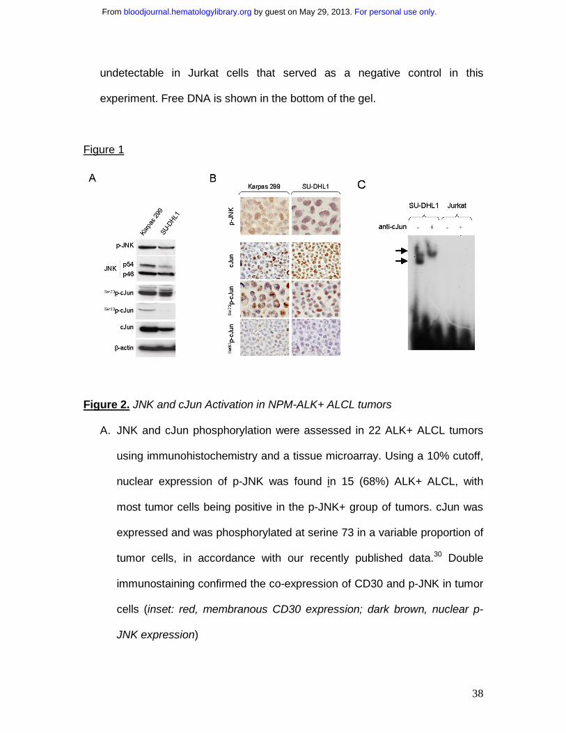
Figure 1. Expression of activated JNK and cJun proteins and AP-1 DNA Binding
Activity in NPM-ALK+ ALCL cells
A. Two NPM-ALK+ ALCL cell lines, Karpas 299 and SU-DHL1, were
examined by immunoblot analysis using antibodies specific for JNK, p-JNK,
cJun and its phosphorylated forms, Ser73p-cJun and Ser63p-cJun. JNK1 and
JNK2 are expressed as isoforms of 46kD and 54 kD, respectively, generated
by alternatively splicing. Both cell lines were found to express high levels of
JNK and cJun, as well as their phosphorylated forms with Ser73p-cJun being
more prominent. Two Hodgkin lymphoma cell lines, L-428 and L-1236, served
as positive controls and REH cells, derived from a patient with pre-B-ALL,
served as a negative control, respectively, for expression of cJun and p-cJun
(data not shown).
B. Immunochistochemistry was performed on formalin-fixed, paraffin-
embedded Karpas 299 and SU-DHL1 cell blocks using the same antibodies
for detection of cJun and their phosphorylated/activated forms. Both ALK+
ALCL cells revealed nuclear staining for cJun, Ser73p-cJun and Ser63p-cJun.
C. AP-1 DNA binding activity was assessed in SU-DHL1 and Jurkat by
electrophoretic mobility shift assay (EMSA) using the double stranded
consensus oligonucleotides with AP-1 specific binding. Super shift of protein-
DNA complex (arrow) was detected in SU-DHL1 cells when nuclear extracts
were preincubated with (+) antibody against cJun, indicating the AP-1
complex in ALK+ALCL cells contains cJun. AP-1 DNA binding activity was
For personal use only. by guest on May 29, 2013. bloodjournal.hematologylibrary.orgFrom
38
undetectable in Jurkat cells that served as a negative control in this
experiment. Free DNA is shown in the bottom of the gel.
Figure 1
Figure 2. JNK and cJun Activation in NPM-ALK+ ALCL tumors
A. JNK and cJun phosphorylation were assessed in 22 ALK+ ALCL tumors
using immunohistochemistry and a tissue microarray. Using a 10% cutoff,
nuclear expression of p-JNK was found in 15 (68%) ALK+ ALCL, with
most tumor cells being positive in the p-JNK+ group of tumors. cJun was
expressed and was phosphorylated at serine 73 in a variable proportion of
tumor cells, in accordance with our recently published data.30 Double
immunostaining confirmed the co-expression of CD30 and p-JNK in tumor
cells (inset: red, membranous CD30 expression; dark brown, nuclear p-
JNK expression)
For personal use only. by guest on May 29, 2013. bloodjournal.hematologylibrary.orgFrom

39
B. The median percentage of cJun+ tumor cells in ALK+ ALCL was
significantly higher in tumors expressing p-JNK than in tumors negative for
p-JNK (Box and Whisker blot).
Figure 2
For personal use only. by guest on May 29, 2013. bloodjournal.hematologylibrary.orgFrom

40
Figure 3. NPM-ALK activates JNK and cJun and Induces AP-1 transcriptional
activity
A. HEK 293T cells were stably transfected with expression plasmids
including empty vector (pDest40), active NPM-ALK, or mutant NPM-ALK
(K210R) with a kinase-dead domain. Expression of NPM-ALK, in cells
transfected with functional or kinase-dead mutant NPM-ALK, was confirmed
by Western Blot analysis using the ALK1 antibody. ALK activity in cells
transfected with the functional NPM-ALK construct was confirmed using an
antibody specific for phosphorylated ALK. Stable expression of functional
NPM-ALK in HEK 293T cells resulted in JNK phosphorylation/activation,
which was associated with phosphorylation of cJun at serine 73 and an
increase in total c-Jun levels that is attributable to positive autoregulation of
cJun transcription. Total JNK1 expression served as a protein loading control
in this experiment.
B. Jurkat cells were transiently transfected with 50µg of empty vector
(pDest40), active NPM-ALK, or mutant, kinase-dead NPM-ALK (K210R) and
whole-cell lysates were prepared at 48 hours after transfection. Immunoblots
showed that transient expression of the functional NPM-ALK in Jurkat cells
resulted in a substantial increase of p-JNK, which was associated with
increased phosphorylation of cJun.
C. To study the NPM-ALK-induced AP-1 transcriptional activity, Jurkat cells
were transiently transfected with a luciferase reporter gene under the control
of a promoter that contains three successive AP-1 specific binding sites
For personal use only. by guest on May 29, 2013. bloodjournal.hematologylibrary.orgFrom

41
(3xAP-Luc) together with an empty expression vector or expression plasmids
encoding for the functional or kinase-dead mutant NPM-ALK. Two sets of
experiments were performed with or without co-transfection of a full-length
cJun expression plasmid (pHA-cJun). After 48 hours the cells were collected
to determine luciferase activity. The results showed an increase in relative
luciferase units in cells expressing the functional NPM-ALK indicating an
increase in AP-1 transcriptional activity. The transcriptional activity was
enhanced in Jurkat cells by co-expression of full-length cJun (right panel).
The fold activation compared with the basal activity of the AP-1 promoter
sites in cells transfected with the empty vector, which was set to 1. All
measurements were performed in triplicate.
D. Co-immunoprecipitaion studies were performed in SU-DHL1 and Karpas
299 cells. Whole-cell lysates were first immunoprecipitated with JNK1, JNK2
or control IgG1 antibodies and then immunoblotted using specific ALK
antibody. Conversely, whole-cell lysates were also immunoprecipitated with
ALK or control IgG1 antibodies and then immunoblotted using a JNK1/2
antibody that detects both JNK1 and JNK2. The same membrane was also
probed with ALK antibody. The upper two arrows show a specific band at
80kDa (NPM-ALK) indicating that NPM-ALK physically interacts with JNK1
and JNK2 in ALK+ALCL cells. The lower arrow indicates detection of JNK1/2
by Western blot analysis after inverse co-immunoprecipitation that further
confirmed the physical interaction between NPM-ALK and JNKs.
Immunoglobulin heavy chain (HC) served as a loading control.
For personal use only. by guest on May 29, 2013. bloodjournal.hematologylibrary.orgFrom

42
E. SU-DHL1 and Karpas 299 cells were treated with the inhibitor WHI-P154
at concentrations (0, 0.5, 2.5, 5 or 10 µM), previously shown to inhibit JAK3
and ALK enzymatic activity. Whole-cell lysates were prepared at 24 hours
following treatment. Immunoblots demonstrate that ALK phosphorylation is
decreased at a concentration of 2.5 µm, and correlates with decreased
phosphorylation (activation) of JNK.
F. Inhibition of ALK enzymatic activity in NPM-ALK+ ALCL cells resulted in
decreased AP-1 DNA binding activity in a concentration-dependent manner
as shown by EMSA and autoradiography (black arrow). SU-DHL1 and
Karpas 299 cells were treated with the inhibitor WHI-P154 at concentrations
of 0, 5 or 10 µM. Following incubation for 24 hours, nuclear extracts were
prepared and assessed by EMSA using double stranded consensus AP-1
oligonucleotide. Free DNA (free probe, FP) is shown in the bottom of the gel.
For personal use only. by guest on May 29, 2013. bloodjournal.hematologylibrary.orgFrom

43
Figure 3
For personal use only. by guest on May 29, 2013. bloodjournal.hematologylibrary.orgFrom

44
Figure 4. cJun phosphorylation is mediated by the JNK group of MAPKs in NPM-
ALK+ ALCL cells.
A. SU-DHL1 cells were treated with the specific JNK inhibitor, SP600125, at
concentrations of 0, 10, 20 or 40 µM. Whole-cell lysates were prepared at 48
hours and immunoblots showed that inhibition of JNK activity resulted in
decreased Ser73p-cJun levels in a dose-dependent manner. This decrease is
detectable at a concentration of 10µM, but it is more prominent at a
concentration of 20µM. A decrease in cJun protein level is also observed due
to an auto-regulatory mechanism of cJun on its own transcription.
B. Treatment of SU-DHL1 cells with U0126, a specific inhibitor of the MEK1/2
group of MKK, was performed at concentrations of 0, 10, and 20 µM.
Following incubation for 48 hours, whole-cell lystates were prepared and
immunoblots showed decreased ERK1/2 phosphorylation indicating sufficient
ERK inactivation. However, this did not result in decreased cJun
phosphorylation. These results suggest that JNKs, but not ERK, mediate cJun
activation in NPM-ALK+ ALCL.
C. JNK activity was assessed using an in vitro kinase assay using GST-cJun
as a substrate. SU-DHL1 cells were treated with the specific JNK inhibitor,
SP600125, at concentrations of 0, 20, and 40 µM. Following 24 hours
incubation, cells were collected and whole-cell lysates were prepared and
incubated with the GST-cJun substrate. Immunoblots showed that inhibition
of JNK activity is associated with decreased cJun phosphorylation in vitro.
For personal use only. by guest on May 29, 2013. bloodjournal.hematologylibrary.orgFrom

45
D. SU-DHL1 cells were transfected with AP-1-Luc reporter plasmid and,
following incubation for 24 hours, were treated with the specific JNK inhibitor
SP600125 at concentrations of 0 and 20 µM. After 24 hours the cells were
collected to determine luciferase activity. The graph shows a substantial
decrease in AP-1 promoter activity after JNK inhibition.
E. To further investigate the role of JNK1/2 isoforms in cJun activation, SU-
DHL1 cells were transiently transfected with 20 µg of siRNA selectively
targeting JNK1 or JNK2 gene products, and endogenous JNK1/JNK2
expression levels were confirmed by reverse transcriptace-PCR (RT-PCR),
as well as by Western blot analysis. Cells were harvested at 48 hours after
transfection and mRNA and whole lysates were prepared. RT-PCR and
immunoblots confirmed JNK1 or JNK2 silencing. cJun phosphorylation was
decreased after knocking down JNK1 or JNK2 in immunoblots.
F. SU-DHL1 cells were transiently transfected with 0, 10 and 20 µg of JNK1
or JNK2 or control siRNA, and whole cell lysates were prepared at 48 hours
after transfection. Immunoblots demonstrate up-regulation of p21 protein
levels in a concentration-dependent manner.
For personal use only. by guest on May 29, 2013. bloodjournal.hematologylibrary.orgFrom

46
Figure 4
For personal use only. by guest on May 29, 2013. bloodjournal.hematologylibrary.orgFrom

47
Figure 5. Inhibition of JNK activity reduces cell growth due to G2/M cell cycle
arrest in NPM-ALK+ ALCL cells.
a. SU-DHL1 cells were treated with increasing concentrations (0, 10, 20 µM)
of SP600125. Inhibition of JNK activity resulted in decreased cell viability and
proliferation of viable cells as assessed by trypan blue exclusion (left panel)
and MTS assays (right panel), respectively. Data presented as the mean ±
SD in triplicate measurements.
b. The decrease in cell growth of SU-DHL1 cells, following treatment with the
inhibitor SP600125 for 48 hours, was associated with cell cycle arrest. The S-
phase of the cell cycle was assessed by BrdU incorporation (left panel) and
measured by a colorimetric assay. Results were expressed as percentage of
cells being in the S-phase of cell cycle (mean±SD of triplicate measurements)
compared with untreated cells. Cell cycle analysis, assessed by propidium
iodide staining and flow cytometry (right panel), revealed a dramatic decrease
in the S-phase fraction associated with a more than 3-fold increase of the
G2/M fraction after treatment of SU-DHL1 cells with 20 µM SP600125.
c. Western blot analysis following treatment of SU-DHL1 cells with
SP600125 at the same time point (48 hours), showed increased levels of the
CDK inhibitor p21, a known cell cycle regulator for transition to S and through
G2 phase. Also, increased levels of underphosphorylated retinoblastoma
protein and decreased levels of cyclin A were observed in a dose dependent
manner.
For personal use only. by guest on May 29, 2013. bloodjournal.hematologylibrary.orgFrom

48
Figure 5
For personal use only. by guest on May 29, 2013. bloodjournal.hematologylibrary.orgFrom

49
Figure 6. Inhibition of cJun expression reduces cell growth in ALK+ALCL cells
A. NPM-ALK+ ALCL cells were transiently transfected with 10 µg or 20 µg
(SU-DHL1) and 20 µg or 40 µg (Karpas 299) cJun or control siRNA, and
whole-cell lysates from both cell lines were prepared at 48h after transfection.
Western blot analysis showed that endogenous cJun was almost completely
silenced when 40µg siRNA was used. As expected, decreased levels of
Ser73p-cJun were also observed.
B. Silencing of cJun gene expression in NPM-ALK+ ALCL cells resulted in
decreased AP-1 DNA binding activity as shown by EMSA and
autoradiography (first row, black arrow). SU-DHL1 and Karpas 299 cells were
transiently transfected with 20µg c-Jun siRNA. Following incubation for 48
hours, nuclear extracts were prepared and assessed by EMSA using double
stranded consensus AP-1 oligonucleotide. Mutant AP-1 oligonucleotide
served as a negative control (second row). Free probe (FB, DNA
oligonucletides) is shown in the bottom panel.
C. Selective silencing of cJun gene expression resulted in decreased cell
viability (upper panel) and a more prominent decrease in proliferation of
viable cells (lower panel) as assessed by trypan blue exclusion and MTS
assays, respectively. The data presented are the mean±SD in triplicate
measurements.
D. Reduced cell proliferation in NPM-ALK+ ALCL cells is attributable to
cell cycle arrest, as cJun silencing also resulted in decreased S-phase
For personal use only. by guest on May 29, 2013. bloodjournal.hematologylibrary.orgFrom

50
fraction evaluated by BrdU incorporation studies. Results have been
normalized to those of control siRNA samples.
E. Western blot analysis following transient transfection of NPM-ALK+
ALCL cells with specific cJun siRNA showed up-regulation of p21 and
underphosphorylated Rb in a concentration-dependent fashion, as well as
down-regulation of cyclin A and cyclin D3.
For personal use only. by guest on May 29, 2013. bloodjournal.hematologylibrary.orgFrom

51
Figure 6
For personal use only. by guest on May 29, 2013. bloodjournal.hematologylibrary.orgFrom

52
Figure 7. NPM-ALK promotes cell cycle progression through activation of
JNK/cJun signaling in NPM-ALK+ ALCL
ALCL frequently carries the chromosomal translocation t(2;5)(p23;q35),
resulting in aberrant expression of nucleophosmin-anaplastic lymphoma kinase
(NPM-ALK). NPM-ALK has been shown to be oncogenic through activation of a
number of cell signaling pathways. This study reveals another oncogenic pathway,
JNK/cJun, that is constitutively activated by the NPM-ALK fusion kinase. The cJun
N-terminal kinases (JNKs), members of the mitogen-activated protein kinase
(MAPK) superfamily, have been shown to play a role in cell proliferation and
transformation. Activation of JNK, through phosphorylation by two distinct MAPK
kinases (MKKs), MKK4 and MKK7, mediates posphorylation of cJun, the best
characterized transcription factor of the activator-protein 1 (AP-1) family.
Phosphorylation of cJun by JNK is essential for stimulation of its transcriptional
activity and its growth promoting effects. The latter can be mediated through
regulation of a number of cell cycle controlling genes including the cyclin-
dependent kinase inhibitor p21, which operates at both the G1/S and G2/M
checkpoints. Taken together, our data suggest that the NPM-ALK-induced
activation of JNK/cJun signaling may lead to uncontrolled cell cycle progression
and cell proliferation, thus contributing to oncogenesis of NPM-ALK+ ALCL.
For personal use only. by guest on May 29, 2013. bloodjournal.hematologylibrary.orgFrom

53
Figure 7
For personal use only. by guest on May 29, 2013. bloodjournal.hematologylibrary.orgFrom