positive Anaplastic Large-cell Lymphoma - ERA
354
Delineation of Molecular Mechanisms Underlying the Pathobiology of ALK- positive Anaplastic Large-cell Lymphoma by Chengsheng Wu A thesis submitted in partial fulfillment of the requirements for the degree of Doctor of Philosophy Medical Sciences - Laboratory Medicine and Pathology University of Alberta © Chengsheng Wu, 2017
-
Upload
khangminh22 -
Category
Documents
-
view
0 -
download
0
Transcript of positive Anaplastic Large-cell Lymphoma - ERA
Delineation of Molecular Mechanisms
Underlying the Pathobiology of ALK-
positive Anaplastic Large-cell Lymphoma
by
Chengsheng Wu
A thesis submitted in partial fulfillment of the requirements for the degree of
Doctor of Philosophy
Medical Sciences - Laboratory Medicine and Pathology
University of Alberta
© Chengsheng Wu, 2017

ii
Abstract
ALK-positive anaplastic large-cell lymphoma (ALK+ALCL) is a rare type non-Hodgkin
lymphoma of null/T cell original, preferentially occurs in children and young adults.
Approximately 85% of ALK+ALCL patients carry the gene translocation t(2;5)(p23;q35),
which results in the generation of the chimeric protein ― NPM-ALK, a key oncogenic
driver of this disease. NPM-ALK interacts and activates a wide range of molecules,
including STAT3, ERK1/2, and PI3K, thus triggering the cell proliferation and
anti-apoptotic effects. In this thesis, I further explored the molecular mechanisms of the
pathobiology of ALK+ALCL from different perspectives, and hypothesized that the
pathobiology of ALK+ALCL, such as tumorigenecity, chemoresistance and cancer
stemness, can be attributed to novel NPM-ALK―regulated biochemical defects as well
as signaling pathways that are not directly linked to NPM-ALK.
STAT1 is generally considered as a tumor suppressor and reported to antagonize
STAT3 transcriptional activity in some cell models. However, the biological function of
STAT1 has not been studied in ALK+ALCL. This study firstly reported that STAT1
expression is decreased in ALK+ALCL cell lines and patient samples; and NPM-ALK is
directly responsible for the downregulation of STAT1, as it promotes STAT1
phosphorylation at Y701 and, thereby, downregulates STAT1 in a STAT3-dependent
proteasome pathway. Furthermore, results showed that STAT1, if overexpressed to a
iii
relatively high level, functions as a potent tumor suppressor in ALK+ALCL by
attenuating STAT3 transcriptional activity and inducing the expression of IFNγ which
further activates the STAT1 signaling.
The Lai lab previously unearthed two distinct cell populations in ALK+ALCL cell lines
that are differentially responding to a Sox2 reporter, with cells responsive to the reporter
(RR) being more tumorigenic and chemoresistant than cells unresponsive to the
reporter (RU). Although Sox2 is implicated in the RU/RR dichotomy, the expression level
of Sox2 is not different between RU and RR cells, suggesting the involvement of other
factor(s). This study reported that MYC is one of the key factors in the RU/RR dichotomy,
as it is highly expressed in RR cells as compared to RU cells. The high level of MYC was
firstly reported to promote Sox2 DNA binding and its transcriptional activity in RR cells.
More evidence suggested that it is the highly active Wnt/β-catenin pathway in RR cells
that confers to the high expression of MYC. The transcriptionally active Sox2 in RR cells
in return upregulates the Wnt/β-catenin pathway, which thereafter promotes the
expression of MYC, thus forming a positive forward loop. In conclusion, the positive
forward loop involving the Wnt/β-catenin/MYC/Sox2 axis defines a highly tumorigenic
small cell population in ALK+ALCL.
The molecular mechanisms underlining tumor plasticity, especially in hematological
malignance, is not fully understood. This study reported that H2O2, a potent oxidative

iv
stress inducer, can convert a fraction of RU cells derived from ALK+ALCL cells to RR
cells (converted RR cells), supporting the existence of tumor plasticity in hematological
malignancy. The converted RR cells have adopted the RR cells’ phenotypes ( including
chemoresistance to doxorubicin, a widely used chemotherapeutic drug for ALK+ALCL
patients, clonogenicity and sphere-forming ability) and biochemical features (the
increased expression of Wnt/β-catenin/MYC and Sox2 downstream targets). Similar
biological changes were observed in RR cells upon oxidative challenge. Furthermore,
more evidence showed that the activated Wnt/β-catenin/MYC/Sox2 axis upon oxidative
stress is required for the RU to RR cells conversion since pharmacological inhibition of
β-catenin/MYC or siRNA knockdown of Sox2 significantly abrogated the conversion. In
conclusion, this study has demonstrated a novel experimental model in which
acquisition of tumorigenicity and cancer stem-like features can be induced by oxidative
stress in ALK+ALCL, a hematologic malignancy, through the activation of
Wnt/β-catenin/MYC/Sox2 axis.
Overall, characterization of these molecular mechanisms underlying the tumorigenesis
of ALK+ALCL has furthered the understanding of the pathobiology of this disease and
also provided potential therapeutic targets for ALK+ALCL patients that are less
responsive or resistance to conventional chemotherapy.

v
Preface
This thesis represents collaborative work, led by Dr. Raymond Lai at the University of
Alberta. The patient samples were obtained and diagnosed at Cross Cancer Institute,
University of Alberta, Edmonton, Alberta, Canada, and the use of these patient samples
for research has received research ethics approval at Feb 17th of 2016, by the Human
Research Ethics Board at the University of Alberta (Study title: Study of biology of ALK
in human ALK+ cancers, with approval number: Pro00062737). The animal studies in
this thesis have also received ethical approval by the Animal Care and Use Committee
(ACUC) at Dec 10th of 2015 (Study title: Mouse models for development of antitumor
therapies, approval number: AUP00000782).
Chapter 2 of this thesis has been published as:
Wu C, Molavi O, Zhang H, Gupta N, Alshareef A, Bone K, Gopal K, Wu F, Lewis J,
Douglas D, Kneteman N, and Lai R. STAT1 is phosphorylated and down-regulated by
the oncogenic tyrosine kinase NPM-ALK in ALK-positive anaplastic large-cell lymphoma.
Blood. 2015.126:336-345. I was first author of this paper. I prepared the first draft and
revisions based on the suggestions and comments of the co-authors. I designed and
performed most of the experiments described herein, except for the following: O.M.
performed some studies, data shown in Figure 2.1A and Figure 2.4F-G; H.Z., N.G., A.A.,
and F.W. performed portions of the experiments and provided technical support and
intellectual input. K.B. provided SupM2 and Karpas 299 cell lines that were stably
transfected with Tet on system. N.G., K.G., J.L, D.D., and N.K. were responsible for the
animal study design, conducting euthanization of the SCID mouse and data analysis.
R.L. provided numerous comments and final review of the manuscript before it was
submitted for publication.
vi
Chapter 3 of this thesis has been published as:
Wu C, Zhang H, Gupta N, Alshareef A, Wang Q, Huang Y, Lewis JT, Douglas DN,
Kneteman NM, and Lai R. A positive feedback involving the Wnt/β-catenin/MYC/Sox2
axis defines a highly tumorigenic cell subpopulation in ALK-positive anaplastic large-cell
lymphoma. Journal of Hematology and Oncology, 2016.9(1):120. I was first author of
this paper. I prepared the first draft and revisions based on the suggestions and
comments of the co-authors. I designed and performed most of the experiments
described herein, except for the following: N.G. performed the ChIP-qPCR experiment
and data analysis, shown in Figure 3.4E; A.A. performed the Wnt signaling pathway
PCR array and data analysis, shown in Figure 3.6; H.Z., Q.W. and Y.H. have contribution
to the Figure 3.3C and Figure 3.11A. H.Z., N.G. and A.A. also provided significant
intellectual input. J.L., D.D., and N.K. were responsible for the animal study design,
conducting euthanization of the SCID mouse and data analysis. R.L. provided numerous
comments and final review of the manuscript before it was submitted for publication.
Chapter 4 of this thesis has been prepared for submission as:
Wu C, Gupta N, Zhang H, Huang Y, and Lai R. Oxidative stress promotes the
tumorigenicity in ALK-positive anaplastic large-cell lymphoma by activating the
Wnt/β-catenin/MYC/Sox2 axis. In preparation. I was first author of this paper, I prepared
the first draft and revisions based on the suggestions and comments of the co-authors. I
designed and performed all the experiments in this study. N.G., H.Z., and Y.H.
contributed to Figure 4.3A, provided technical assistance and intellectual input. R.L.
provided numerous comments and final review of the manuscript.

vii
Acknowledgements
I wish to express my sincere gratitude to the following individuals without whom this
thesis would not be possible.
Foremost, my sincerest thanks to my supervisor, Dr. Raymond Lai, for his scientific
training and guidance throughout the PhD program. He taught me how to critical think
and also, most importantly, ignited my passion for science. Without his supervision, I
would not have gotten this work done or have current achievements.
I would like to thank my committee members Dr. Yangxin Fu and Dr. Robert Ingham for
their constant mentorship, support and comments throughout my whole doctoral
program, as well as their suggestions with respect to the content of this thesis. Here I
would like again to express my sincere thanks to Dr. Robert Ingham for his kindness to
allow me to use the electroporator in his lab in the past 4 years.
I would like to thank Dr. Monika Keelan, who is in charge of the graduate studies
program in the Department of Laboratory Medicine and Pathology, for her assistance
and guidance throughout my PhD study. I also would like to express my sincere thanks
to Ms. Cheryl Titus, the graduate program advisor, for her kindness and constant
assistance in my whole PhD study.
I would also like to thank Dr. Roger Leng and Dr. Ing Swie Goping for serving as
examiners for my candidacy examination. I appreciate their time, questions and
comments for my study. I also would like to thank Dr. Jelena Holovati for chairing my
candidacy examination and her support during the examination. I would also like to

viii
appreciate Dr. Suzanne Kresta for her time to attend my candidacy examination and her
supports to me as well.
I would like to thank Dr. Roger Leng and Dr. Shirin Bonni for serving as external
examiners for my final PhD defense. I do appreciate your time and valuable comments. I
also would like to thank Dr. Jelena Holovati for chairing my final PhD defense. I really do
appreciate your time and support.
I would like to thank all present and past members of Dr.Lai’s lab, who gave me their
support and help throughout my PhD study. Specifically, I would like to give thanks to Dr.
Hai-feng Zhang, Mr. Alshareef Abdulraheem, Dr. Gupta Nidhi, Mr.Yung-Hsing Huang,
Dr. Fang Wu, and Dr. Peng Wang for their constant help and invaluable friendship.
Thanks to Ms. Yuen Morrissey as well for her help in my PhD study.
I would like to acknowledge the technical assistance of Jingzhou Huang and Dr.
Xue-Jun Sun in the Flow Cytometry lab at the Department of Experimental Oncology,
Cross Cancer Institute, University of Alberta.
I would like to express my sincere gratitude to the Chinese Scholar Council (CSC) for
supporting me with a graduate scholarship for the past 4 years. Without the generous
support from CSC, I do not even have any chance to stand here. I would like to thank
the Canadian Institutes of Health Research (CIHR) for funding the research projects.
Last but not the least; I would like to give my sincere thanks and gratitude to my family
and my wife Lois Luo for all their endless loves and supports. Without your selfless loves
and support, I would never have reached this stage.

ix
Table of contents
Title Page………………………………………………………………………………………....i
Abstract…………………………………………………………………………………………..ii
Preface…………………………………………………………………………………………...v
Acknowledgments……………………………………………………………………….…….vii
Table of Contents………………………………………………………………………............ix
List of Abbreviations…………………………………………………….……..…………….xviii
List of Tables…………………………………...………………………………..…………...xxiv
List of Figures……………………………………………………………...……..................xxv
Bibliography…………………………………………………………………………………...xvii
CHAPTER 1: General Introduction…………………………………………………………1
1.1 Introduction……………………………………………………………………… 2
1.2 ALK-positive anaplastic Large Cell Lymphoma ………………………..............3
1.2.1 Morphology ………………………………………………………. …….…3
1.2.2 Immunophenotype………………………………………………………...6
1.2.3 Survival analysis……………………………………………………….…7
1.2.4 Genotype…………………………………………………………….…....8
1.2.4.1 ALK………………………………………………………………...……..8
1.2.4.2 NPM………………………………………………………………….….10
1.2.4.3 NPM-ALK ………………………………………………………...….…11
x
1.2.4.4 Other ALK fusion proteins…………………………………………….12
1.2.5 NPM-ALK-mediated transformation…..…………….…………….…..….15
1.2.6 NPM-ALK ꟷ interacting substrates and activated signaling
pathways……………………………………………………………………..…....16
1.2.6.1 JAK/STATs………………………………………………………….…18
A) JAKs and STAT3 in ALK+ALCL……………….………….…………18
B) STAT1………………………………………………………………….22
1.2.6.2 PLC-γ……………………………………………….………….............26
1.2.6.3 PI3K/AKT……………………………………………………...............26
1.2.6.4 RAS/MEK/ERK……………………………………….……………..…29
1.2.6.5 mTOR……………………………………………………………….…..30
1.2.7 Therapeutic strategy for ALK+ALCL patients...……………………….…30
1.3 Wnt/β-catenin……………………………………………………………………...32
1.3.1 Wnt/β-catenin signaling…………………………………………………….32
1.3.2 Wnt/β-catenin in cancer and ALK+ALCL……………………..................34
1.4 Cancer Stem Cells (CSCs)………………………………………………………35
1.4.1 Identification and isolation of CSCs……………………………………….35
1.4.2 Molecular features of CSCs………………………………………………..37
1.4.3 Signaling pathways in CSCs………………………………………………38
1.4.4 CSCs in ALK+ALCL ………………………………………………………..39
1.5 Inducible pluripotent stem cells (iPS) factors…………………………………40
xi
1.5.1 Sox2…………………………………………………………………….……41
1.5.1.1 Sox2 in cancer……………………………………………………….…41
1.5.1.2 Post-translational modification of Sox2……………….....................43
1.5.2 MYC in cancer………………………………………………………………44
1.6 Reactive oxygen species (ROS) in cancer………………………..................48
1.7 Thesis overview…………………………………………………………….…….52
1.7.1 Rationale……………………………………………………………….……52
1.7.2 Objectives………………………………………………………..................54
1.8 References……………………………………………………………………….55
CHAPTER 2: STAT1 is phosphorylated and downregulated by the oncogenic
tyrosine kinase NPM-ALK in ALK-positive anaplastic large-cell
lymphoma……………………………………………………………………………….........94
2.1 Introduction……………………………………………………………………….95
2.2 Methods and materials……………………………………………………........97
2.2.1 Primary tumors and cell lines……………….…………………................97
2.2.2 Chemical treatments………………………………………………………..98
2.2.3 Immunohitochemistry………………………………………………………99
2.2.4 Short interfering RNA and transfections………………………………….99
2.2.5 Plasmid constructs and transfection…………………………………….100
2.2.6 Generation of Tet-on inducible stable cell lines………………………...100
xii
2.2.7 Cell-cycle assay………………………………….………………………..101
2.2.8 Western blotting and co-immunoprecipitation………………………….102
2.2.9 Methylcellulose colony formation assay………………………………..104
2.2.10 Dual luciferase assay…………………..……………………………….104
2.2.11 STAT3 DNA probe binding assay………………………….................105
2.2.12 Trypan blue exclusion assay and MTS assay………………………..106
2.2.13 RNA extraction, cDNA synthesis, and quantitative reverse
transcriptase PCR (quantitative RT-PCR)…………………………………….106
2.2.14 SCID mouse xenograft studies………………………………………...107
2.2.15 Statistical analysis……….………………………………………………108
2.3 Results…………………………………………………………………..………108
2.3.1 STAT1 is expressed at a low level in ALK+ALCL tumors and cell
lines………………………………………………………………………………………...108
2.3.2 The ubiquitin-proteasome pathway is involved in the downregulation of
STAT1 in ALK+ALCL cells……………………………………...………………………..110
2.3.3 NPM-ALK promotes STAT1 phosphorylation at Y701 and downregulates
STAT1….…………..……………….…………………...…………………………………113
2.3.4 STAT3 is necessary for the downregulation of STAT1 mediated by
NPM-ALK……………………………………………………………………………….….116
2.3.5 STAT1 signaling is functionally intact in ALK+ALCL…………………...117
2.3.6 The STAT1-IFNγ positive feedback loop………………………………..124
xiii
2.3.7 STAT1C is a tumor suppressor in ALK+ALCL in vitro and in vivo…125
2.3.8 STAT1C significantly decreases STAT3 transcriptional activity ……...127
2.3.9 siRNA knockdown of STAT1 confers resistance to STAT3
inhibition-induced cell death……………………………………………………………..129
2.4 Discussion………………………………………………………………………..131
2.5 References……………………………………………………………………….136
CHAPTER 3: A positive feedback involving the Wnt/β-catenin/MYC/Sxo2 axis
defines a highly tumorigenic cell subpopulation in ALK-positive anaplastic
large-cell lymphoma………….……………………………………………………………145
3.1 Introduction………….……………………………………………………………146
3.2 Methods and materials………….……………………………………………….148
3.2.1 Primary tumors, cell lines and treatments………………………………..148
3.2.2 Short interfering RNA and transfections…………………….…………….149
3.2.3 RNA extraction, cDNA synthesis, quantitative reverse transcriptase PCR
(quantitative RT-PCR) and chromatin-immnoprecipitation PCR……..………………..149
3.2.4 Immunohistochemistry and immunofluorescence studies……………..152
3.2.5 Plasmid constructs and transfection……………………………………...153
3.2.6 Western blotting………………………………………………………...…..153
3.2.7 Luciferase assay …………………………………………………………...154
3.2.8 Transwell assay …………………………………………………….….......154
xiv
3.2.9 Cell-cycle and MTS assay………………………………………….……...155
3.2.10 SRR2 probe binding assay……………………………………………….155
3.2.11 Nuclear cytoplasm fractionation assay………………………………….156
3.2.12 Methylcellulose colony formation assay………………………………...156
3.2.13 SCID mouse xenograft studies…………………………………………..156
3.2.14 Side population assay………………………………………...………….157
3.2.15 Statistical analysis………………………………………………………...157
3.3 Results……………………………………………………………………….......158
3.3.1 The identification of MYC as a key regulator of the RU/RR
dichotomy………………………………………………………………………………..158
3.3.2 MYC promotes Sox2-SRR2 binding and the transcriptional activity of
Sox2………………………………………………………………………………….......166
3.3.3 The high level of MYC in RR cells is attributed to the Wnt/β-catenin
pathway…………………………………………………………………………………..171
3.3.4 The positive regulatory loop involving Sox2, Wnt/β-catenin and MYC in
RR cells……………………………………………………………..............................180
3.3.5 MYC is heterogeneously expressed in primary tumor samples, and it
co-localizes with active β-catenin……………………………………………...…......182
3.3.6 RU cells stably transfected with MYC are biochemically and
phenotypically similar to RR cells……………………………………………………...185
3.3.7 Side population cells are not detectable in RU and RR cells and ABCG2 is
xv
not significantly differentially expressed in mRNA level between RU and RR
cells………………………..………………………………………………………….187
3.4 Discussion………………………………………………………………………..190
3.5 References……………………………………………………………………….197
CHAPTER 4: Oxidative stress promotes the tumorigenicity in ALK-positive
anaplastic large-cell lymphoma by activating the Wnt/β-catenin/MYC/Sox2
axis……………………………………………………………………………………….…...204
4.1 Introduction…………………………………………………………………...…205
4.2 Methods and materials…….………………………………………………….…207
4.2.1 Cell lines and chemicals………………………….………………….…….207
4.2.2 H2O2, NAC, 10074-G5 and quercetin treatment……………….………..207
4.2.3 Luciferase assay and flow cytometry………………………………….….208
4.2.4 Trypan blue exclusion and MTS assay……………………………….…..209
4.2.5 Short interfering RNA and transfections……………………………….…209
4.2.6 RNA extraction, cDNA synthesis, and quantitative reverse transcriptase
PCR (quantitative RT-PCR)……………………………………………………………209
4.2.7 Western blotting…………………………………………………………….210
4.2.8 Nuclear cytoplasm fractionation assay…………………………………...210
4.2.9 SRR2 probe binding assay……………………………………….………..210
4.2.10 Methylcellulose colony formation assay………………………………...210
xvi
4.2.11 Limiting dilution assay……..……………………………………………...211
4.2.12 Statistical analysis………………………..………….…………………....211
4.3 Results………………………………………….………………………………...211
4.3.1 Oxidative stress induces a conversion from RU to RR cells….………..211
4.3.2 The converted RR cells share the similar biological functions with RR
cells…………………………………………………………………...………………….218
4.3.3 The converted RR cells share the similar biochemical characteristics with
RR cells………………………………………………………..……………..………….222
4.3.4 Inhibition of β-catenin/MYC or siRNA knockdown of Sox2 dramatically
abrogates the conversion of RU cells to RR cells induced by oxidative stress…...224
4.3.5 ERK1/2 is activated in both RU and RR cells upon H2O2
re-challenge……………………………………………………………………………...226
4.3.6 STAT1 is activated in both RU and RR cells upon oxidative stress......228
4.4 Discussion………………………………………………………………………..230
4.5 References……………………………………………………………………….236
CHAPTER 5: General Discussion and Conclusions……………………………........244
5.1 Thesis overview…………………………………………………………….......245
5.2 STAT1 in ALK+ALCL……………………………………………………………246
5.3 The RU/RR dichotomy in ALK+ALCL…………………………………….......248
5.4 Oxidative stress-induced RU/RR conversion in ALK+ALCL………………..251
xvii
5.5 STAT1 and the RU/RR dichotomy….…………………………………………253
5.6 Conclusions and future directions…………..………………………………...254
5.7 References…………………………………………………………..................256
Bibliography…..…………………………………………………………………………….257
xviii
List of Abbreviations
1O2 – singlet oxygen
ABC transporters − ATP-binding cassette transporters
ALCL− anaplastic large-cell lymphoma
ALDH − Aldehyde dehydrogenase
ALK – anaplastic lymphoma kinase
AML − acute myeloid leukemia
AP-1 − Activator Protein-1
APC − adenomatous polyposis coli
ATM – ataxia telangiectasia mutated
BAD – Bcl2-associated death promoter
Bcl-2 – B-cell lymphoma-2
Bcl-XL – B-cell lymphoma-extra large
BCR-ABL – breakpoint cluster region- Abelson
bHLH − helix-loop-helix
BRCA1 – breast cancer susceptibility 1
CA-CTNNB1 – constitutive catenin beta 1
CD – cytoplasmic tyrosine kinase domain
CDC25A − cell division cycle 25 homolog A
Cdk2 − cyclin-dependent kinase 2
ChIP – chromatin-immunoprecipitation

xix
CHOP − Cyclophosphamide, hydroxydaunorubicin (doxorubicin), oncovin (vincristine),
and prednisone
CK1α − casein kinase 1α
CML − chronic myeloid leukemia
CRC − colon-rectal cancer
CSC − cancer stem cells
DAG − diacylglycerol
DMSO – dimethyl sulfoxide
DNMTs − DNA methyltransferases
DVl − disheveled
ECD – extracellular domain
eIF2A − eukaryotic translation initiation factor 2A
EMA – epithelial membrane antigen
EMT − epithelia-mesenchymal transition
ER – endoplasmic reticulum
ER – estrogen receptor
ERK − extracellular signal-related kinase
ETS-1 – E26 transformation-specific-1
EV – empty vector
FOXO3a − forkhead family of transcription factors 3a
FZD − Frizzled
xx
GAS − IFN--activated-site
GSK3β − glycogen synthase kinase-3β
H2O2 – hydrogen peroxide
HGFR −Hepatocyte growth factor receptor
HHAT − Hedgehog acyltransferase
HMG − high mobility group DNA binding domain
HOXA5 – homeobox A5
IFN − interferon
IFNAR1/2 − IFN alpha receptor
IGF – insulin growth factor
IP3 − inositol triphosphate
iPS − pluripotent stem cells
IRF-1 − Interferon regulatory factor -1
ISGF3 − IFN-stimulated gene factor 3
ISRE − IFN-stimulated response element
IκB − inhibitors of κB
JAK − Janus activated kinase
Jeb − Jelly belly
LRP5/6 – Low density lipoprotein receptor-related protein 5/6
MAPK- Mitogen-activated protein kinase
Max − MYC-associated factor X
xxi
Mcl-1 − myeloid cell leukemia sequence 1
MEFs − mouse embryonic fibroblasts
mESCs − mouse embryonic stem cells
miRNA − microRNA
MK − midkine
mTOR − mammalian target of rapamycin
MTS−3-(4,5-dimethylthiazol-2-yl)-5-(3-carboxymethoxyphenyl)-2-(4-sulfophenyl)-2H-tetr
azolium
MYC: V-myc avian myelocytomatosis viral oncogene homolog
NAC − N-acetyl-L-cysteine
NADPH – nicotinamide adenine dinucleotide phosphate
NF-κB − nuclear factor kappa-light-chain-enhancer of activated B cells
NHL − non-Hodgkin lymphoma
NKLAM − Natural Killer Lytic-Associated Molecule
NKLAM – natural killer lytic-associated molecule
NPM – nucleophosmin
Nrf2 – nuclear factor erythroid 2-related factor 2
NSCLC − non-small-cell lung cancer
O2- − superoxide anions
O3 – ozone
OH- − hydrogen radical
xxii
PI3K − phosphatidylinositol 3 kinase
PIP2 − phosphatidylinositol
PKC − protein kinase C
PLC-γ− phospholipase C γ
PTEN − phosphatase and tensin homolog
PTN − pleiotrophin
qRT-PCR – quantitative reverse transcriptase-polymerase chain reaction
ROS – reactive oxygen species
ROS1 − c-ros oncogene 1
RPS6 − ribosomal protein S6
RR – reporter responsive
RTK – receptor tyrosine kinase
RU – reporter unresponsive
SCID – severe combined immune deficiency
SHH − Sonic Hedgehog
SHP1 − Src homology region 2 domain-containing phosphatase-1
shRNA – small hairpin RNA
siRNA − small interfering RNA
Smurf1 – smad ubiquitination regulation factor 1
Sox2 – sex determining region Y- box 2
SRR2 – Sox2 regulatory region 2

xxiii
STAT − signal transducer and activator of transcription
STAT1C – constitutively active STAT1
TCF/LEF − T-cell factor/lymphoid enhancer factor
TICs – tumor-initiating cells
TM – transmembrane domain
TNF – tumor necrosis factor
TPM4 − tropomyosin
TSCs − tumor stem cells
TYK2 − tyrosine kinase 2
VEGF – vascular endothelia growth factor
WHO – World Health Organization

xxiv
List of Tables
Table 1.1 Chromosomal translocations involving ALK in ALK+ALCL and other
malignancies…………………………………………………………………………………...14
Table 3.1 Primers used for quantitative RT-PCR…………………………………………151
Table 3.2 The top putative factors that are predicated to bind to SRR2 sequence by
JASPAR motif matches analysis at P<0.001……………………………………………...160
Table 4.1 Primers used for quantitative RT-PCR………………………………………209
xxv
List of Figures
Figure 1.1 The common pattern of ALK+ALCL……………………………………………...4
Figure 1.2 Variant morphological patterns in ALK+ALCL……………………………….…..5
Figure 1.3 Immunohistochemical stainings of CD30 and ALK in ALCL……………….......7
Figure 1.4 The functional domains of ALK, NPM, and NPM-ALK………………………10
Figure 1.5 Representative signaling pathways activated by NPM-ALK……………….…17
Figure 1.6 NPM-ALK activates STATs signaling pathway in ALK+ALCl…………………21
Figure 1.7 Signal transduction of the IFN/STATs……………………………….................23
Figure 1.8 NPM-ALK activates the PI3K/AKT pathway in ALK+ALCL…………………...28
Figure 1.9 Overview of the Wnt/β-catenin pathway………………………………….........33
Figure 1.10 The biological processes regulated by MYC in cancer cells………………..47
Figure 1.11 Phosphorylation and stabilization of MYC by ERK and GSK3β…………….48
Figure 1.12 Determination of cellular redox status by a balance between levels of ROS
inducers and ROS scavengers………………………………………………………………51
Figure 2.1 Expression of STAT1 in ALK+ALCL cell lines and patient samples………..109
Figure 2.2 The ubiquitin-proteasome pathway is involved in the downregulation of
STAT1 in ALK+ALCL cells…………………………………………………………………...111
Figure 2.3 NPM-ALK promotes STAT1 phosphorylation at Y701 and downregulates
STAT1……………………………………………………………………………………........114
Figure 2.4 STAT1 signaling is functionally intact in ALK+ALCL……………………........118
xxvi
Figure 2.5 STAT1 provides tumor suppressor function in ALK+ALCL in vitro………….122
Figure 2.6 Overexpression of STAT1 induces the expression of IFNγ in ALK+ALCL and
suppresses the tumor cell growth in vivo………………………………………………….124
Figure 2.7 STAT1C significantly decreases STAT3 transcriptional activity…………….128
Figure 2.8 siRNA knockdown of STAT1 confers resistance to STAT3 inhibition-induced
cell death……………………………………………………. ……………………………….130
Figure 2.9 Schematic model of STAT1 in ALK+ ALCL………………………..………….136
Figure 3.1 Knockdown of Sox2 by siRNA significantly downregulates the SRR2
luciferase activity in RR cells………………………………………………………………..159
Figure 3.2 RR cells express a substantially higher level of MYC than RU cells…........161
Figure 3.3 The high MYC expression contributes to the RR phenotype……………….162
Figure 3.4 MYC promotes the SRR2 probe binding and the transcriptional activity of
Sox2…………………………………………………………………………………………...168
Figure 3.5 NPM-ALK/STAT3 is not differentially activated or expressed between RU and
RR cells……………………………………………………………………………………….172
Figure 3.6 The Wnt signaling is more active in SupM2-RR cells in comparison to
SupM2-RU cells……………………………………………………………………………...174
Figure 3.7 The Wnt/β-catenin pathway contributes to the high MYC expression in RR
cells…………………………………………………………………………………..............175
Figure 3.8 RU cells transfected with the constitutively active CTNNB1 (CA-CTNNB1)
acquire the RR phenotype…………………………………………………………………..179
xxvii
Figure 3.9 The positive regulatory loop of Sox2–Wnt/β-catenin–MYC in RR cells.......181
Figure 3.10 MYC is heterogeneously expressed and its expression is co-localized with
active β-catenin in ALK+ALCL tumor cells………………………………………………...184
Figure 3.11 RU cells stably transfected with MYC are biochemically and phenotypically
similar with RR cells…………………………………………………………………………186
Figure 3.12 Identification of side populations cells by Hoechst-efflux assay and the
expression of ABC transporters in RU and RR cells derived from ALK+ALCL………..189
Figure 4.1 Oxidative challenge induces the conversion of RU to RR cells…………….213
Figure 4.2 RU and RR cells upon H2O2 re-challenge treatment are more
doxorubicin-resistant and clonogenic, and have captured enhanced sphere-forming
ability as compared to cells without treatment………………………...……...................221
Figure 4.3 The converted RR cells induced by oxidative stress share the biochemical
features with RR cells……………….............................................................................223
Figure 4.4 Pharmacological inhibition of MYC or β-catenin, or siRNA knockdown of Sox2
in RU cells significantly abrogates the conversion of RU cells to RR cells…………….225
Figure 4.5 Inhibition of ERK1/2 activity .in RR cells dramatically decreases the
expression level of MYC and Sox2, as well as the SRR2 luciferase activity…………..227
Figure 4.6 STAT1 is activated in RU and RR cells upon oxidative stress………….229

1
CHAPTER 1
General Introduction

2
1.1 Introduction
Anaplastic large-cell lymphoma (ALCL), first described by Stein and colleagues in 1985,
is a rare type of peripheral null or T-cell non-Hodgkin lymphoma (NHL), consisting of
approximately 2-3% of all lymphoid malignancies, ~3% of all adult NHLs, and ~10-20%
of childhood lymphomas (1, 2). The subtype of this lymphoma cells strongly express an
antigen named CD30, a member of the tumor necrosis factor receptor family (3, 4).
CD30 was initially detected and recognized by antibody generated in Kiel, West
Germany, and therefore termed as Ki-1 (3). In the early 1990s, Morris et al found that a
group of ALCL cases carry a recurrent chromosome translocation involving the
anaplastic lymphoma kinase (ALK) gene on chromosome 2p23 and the nucleophosmin
(NPM) gene on chromosome 5q35, and this chromosome translocation results in the
generation of a fusion protein − NPM-ALK (5). In 2008, the World Health Organization
(WHO) classified ALCL into 3 entities including primary cutaneous ALCL, ALK-negative
ALCL (ALK-ALCL) and ALK-positive ALCL (ALK+ALCL) (6). Primary cutaneous ALCL
preferentially presents in the skin; while ALK- and ALK+ ALCL usually present in
systemic forms (7), therefore, they are also classified as systemic ALCL. Primary
systemic ALCL preferentially occurs in childhood and young adults, and accounts for
approximately 40% of pediatric patients with NHL (7). However, primary systemic ALCL
only accounts for less than 5% of adult patients with NHL, with male predominance (7).
Patients diagnosed with systemic ALCL are often found in an advanced stage of
disease; specifically, involvements of lymph nodes and multiple extranodal sites are
usually presented in these patients (1).

3
1.2 ALK-positive anaplastic large-cell lymphoma
1.2.1 Morphology
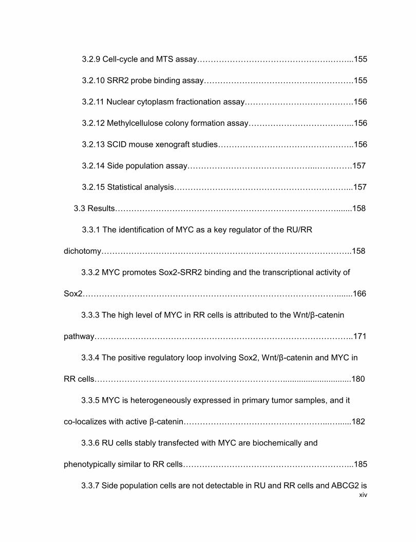
The WHO classification has described at least 5 histologic variants or patterns of
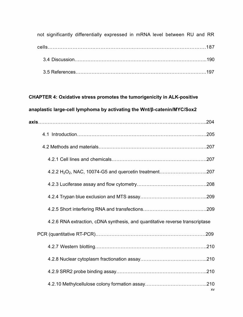
ALK+ALCL cases, including common pattern, lymphohisocytic variant, the small-cell
variant, Hodgkin-like, and a “composite” type containing more than one variant (6).
The common pattern constitutes of ~70% of all ALK+ALCL cases (7), and it refers to
that a small number of tumor cells typically infiltrate the lymphatic sinusoids and these
tumor cells usually present a pattern of sheet-like in the lymph node (Figure 1.1) (6). In
some cases, immunohistochemistry stainings, such as anti-ALK staining, are required
to recognize the tumor cells, as a relatively small number of tumor cells are infiltrated in
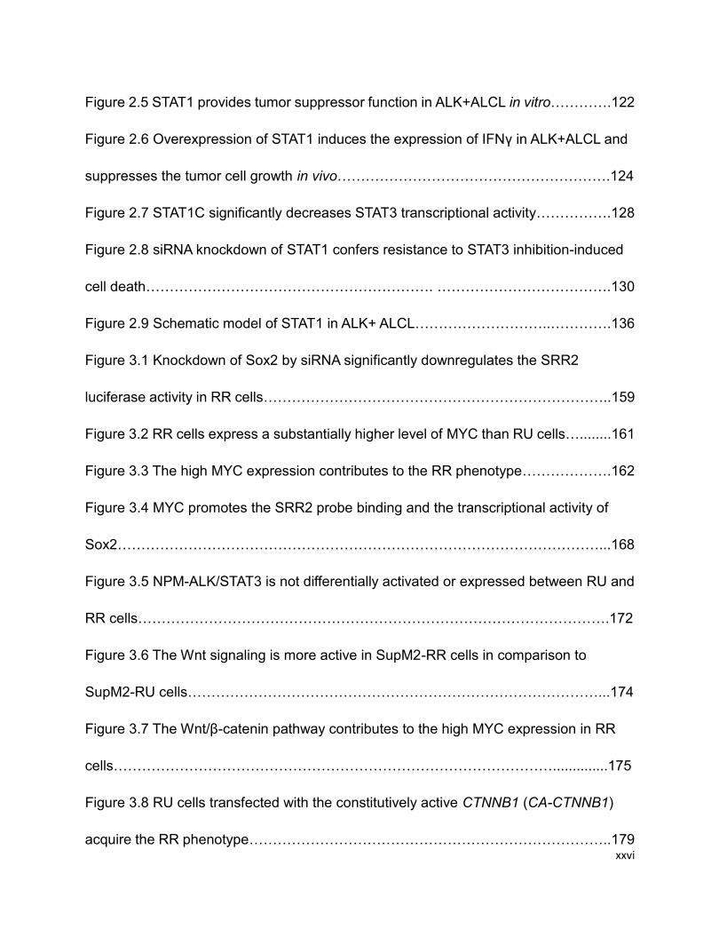
the lymph node (6). Approximately 10% of all ALK+ALCL cases are lymphohistiocytic
variant in which the infiltrated reactive histiocytes can be easily observed (Figure 1.2A-
B) (8). Sometimes, the ALK+ALCL tumor cells are difficult to find because of
erythrophagocytosis in situ (9). The small-cell variant consists of ~5-10% of all cases
and it, namely, presents the smaller size of tumor cells, in which the perivascular
clustering can be occasionally observed (Figure 1.2C-D) (10). A Hodgkin-like pattern,
representing ~1-3% of all cases, resembles the nodular sclerosis variant of classical
Hodgkin lymphoma with a polymorphous cellular background (Figure 1.2E-F) (11, 12).
However, not all cases of ALK+ALCL present only one of above patterns (13), in other
words, multiple patterns are always observed in a single case. A “composite” type
containing more than one variant can be seen in a small number of cases (13). Note
that the histologic pattern may vary in sequential biopsies of ALK+ALCL tumors from

4
the same patient (14), indicating the existence of multiple histologic patterns in a single
case.
Nevertheless, the representative ALK+ALCL cells or hallmark cells can be identified in
all patterns of cases (3). The hallmark cells are featured with irregular large size of cell,
with “horse or kidney-shaped” nucleus and abundant cytoplasm, along with an evidently
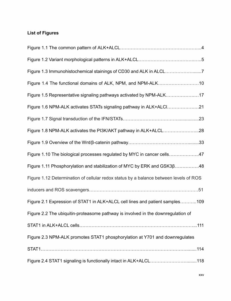
central Golgi apparatus (Figure 1.1C and 1.2B) (14)
Figure 1.1 The common pattern of ALK+ALCL. A, B) The lymph node architecture is
effaced by sheets of neoplastic lymphoid cell (A:H&E, X100; B:H&E, X400). C, D) Two
high-power images taken at the same magnification from the same case demonstrate a
wide spectrum of cytologic features and cell sizes, including characteristic hallmark
cells (C:H&E, X1000) and very large, sometimes mutinucleat cells (D:H&E, X1000).
5
Reprinted with consent from “Xing X and Feldman AL. Anaplastic Large Cell
Lymphoma: ALK Positive, ALK Negative, and Primary Cutaneous. Adv Anat Pathol.
2015; 22(1):29-49.” Licence number: 3976891276365.
Figure 1.2 Variant morphologic patterns in ALK+ALCL. In the lymphohistiocytic
pattern, the tumor cells mix with histiocytes and small lymphocytes (A: H&E, X400);
hallmark cells can be seen (B: H&E, X1000). The small-cell pattern contains
predominantly small-sized to medium-sized cells, often with pale cytoplasm (C: H&E,
400), but hallmark cells also can be found, often adjacent to blood vessels (D: H&E,
1000). The Hodgkin-like pattern shows the architectural features of the nodular
sclerosis type of classical Hodgkin lymphoma (E: X40), although the neoplastic cells
within the nodules typically resemble hallmark cells more than classic Reed-Sternberg
cells (F: X1000).

6
Reprinted with consent from “Xing X and Feldman AL. Anaplastic Large Cell
Lymphoma: ALK Positive, ALK Negative, and Primary Cutaneous. Adv Anat Pathol.
2015; 22(1):29-49.” Licence number: 3976891276365.
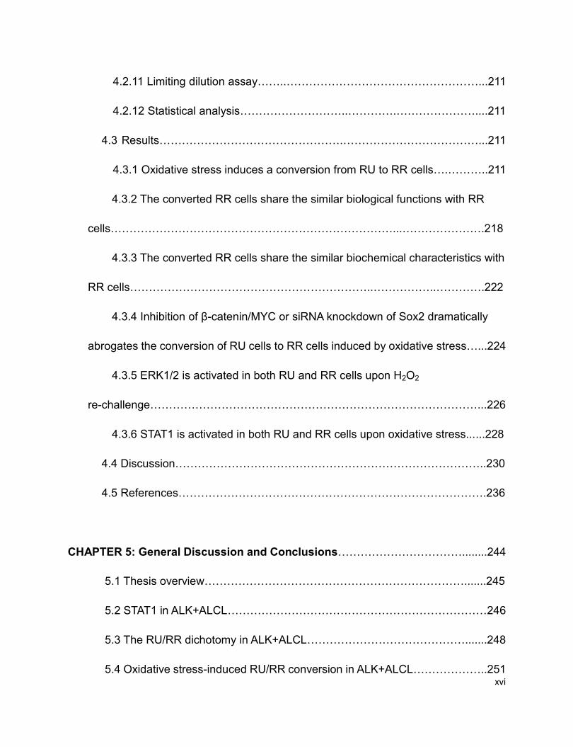
1.2.2 Immunophenotype
Nearly all ALK+ALCL cases exhibit the positive staining of CD30 on the cell surface
and within the Golgi area (Figure 1.3) (9, 10). CD30, as a member of the tumor
necrosis factor (TNF) receptor superfamily, is generally expressed on activated
lymphocyte cells (15). Most of ALK+ALCL cases also present the positive staining of
epithelial membrane antigen (EMA, or Mucin 1) (16), which is a glycoprotein generally
expressed on the surface of epithelia cells in the lungs, intestines, etc (17). Besides,
the cytotoxic cell antigen T-cell restricted intracellular antigen 1 (TIA-1), granzyme B
and perforin are also detectable in most of ALK+ALCL cases (18-21).
As a type of T-cell lymphoma, most of ALK+ALCL cells have gene rearrangement of T-
cell receptor (TCR) and the expressions of one or more T-cell/nature killer cells
antigens (20, 22). For instance, CD2 and CD4 are widely expressed on ALK+ALCL
cells, and CD8 is also detectable in some cases (20). However, ALK+ALCL cells are
deficient in normal T-cell signaling pathways because of lack of or decreased
expressions of T-cell antigens including the pan T-cell marker CD3 and the αβ T-cell
receptor complex (22). An early study revealed that the expression of CD3 was lost in
23 of 24 ALK+ALCL cases (22), and the expressions of CD5 and CD7 are always
undetectable on ALK+ALCL cells (23). While some ALK+ALCL cells are diagnosed as
“Null” cell type, as they lack T-cell phenotype (13). Due to the lack of both T-cell and B-
7
cell surface markers, the “Null” cell type ALK+ALCL cells are thereby deficient in some
specific T or B-cell signaling pathways (13).
The most evident immunophenotype for ALK+ALCL is the expression of chimeric ALK
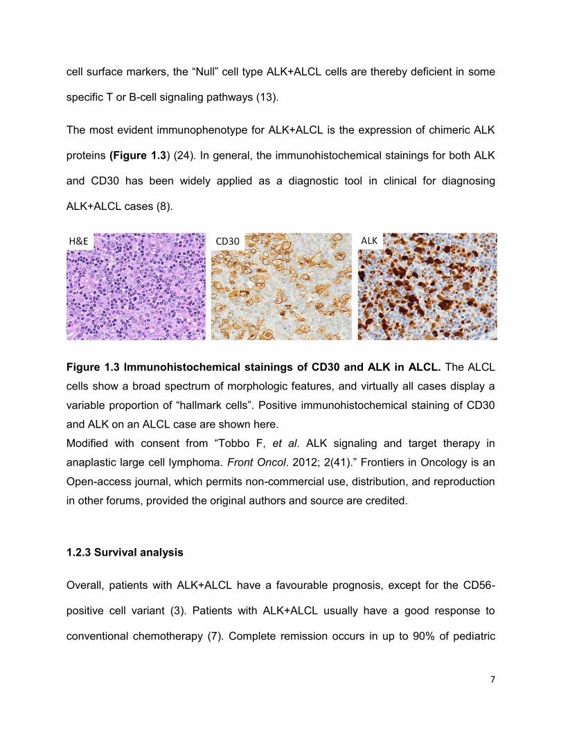
proteins (Figure 1.3) (24). In general, the immunohistochemical stainings for both ALK
and CD30 has been widely applied as a diagnostic tool in clinical for diagnosing
ALK+ALCL cases (8).
Figure 1.3 Immunohistochemical stainings of CD30 and ALK in ALCL. The ALCL
cells show a broad spectrum of morphologic features, and virtually all cases display a
variable proportion of “hallmark cells”. Positive immunohistochemical staining of CD30
and ALK on an ALCL case are shown here.
Modified with consent from “Tobbo F, et al. ALK signaling and target therapy in
anaplastic large cell lymphoma. Front Oncol. 2012; 2(41).” Frontiers in Oncology is an
Open-access journal, which permits non-commercial use, distribution, and reproduction
in other forums, provided the original authors and source are credited.
1.2.3 Survival analysis
Overall, patients with ALK+ALCL have a favourable prognosis, except for the CD56-
positive cell variant (3). Patients with ALK+ALCL usually have a good response to
conventional chemotherapy (7). Complete remission occurs in up to 90% of pediatric
8
patients upon conventional chemotherapy (8). The 3-year and 5-year disease-free
survival for pediatric patients range from 60% to 85%, and the 5-year overall survival
for young adult patients is ~85% with conventional chemotherapy (3). A recent clinical
study has indicated that patients with ALK+ALCL have a significantly higher 5-year
overall survival rate than ALK-ALCL patients (70% verse 49%) (3, 25). Of note, no
significant difference is observed when comparing the 5-year survival rates between
ALK+ALCL and ALK-ALCL patients of over 40 years old (2), this indicates that the
younger ages of patients might help explain the favourable diagnosis of ALK+ALCL
versus ALK-ALCL (3).
1.2.4 Genotype
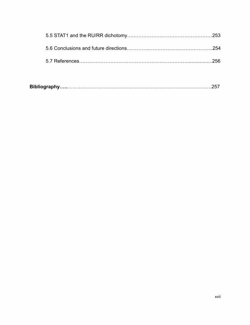
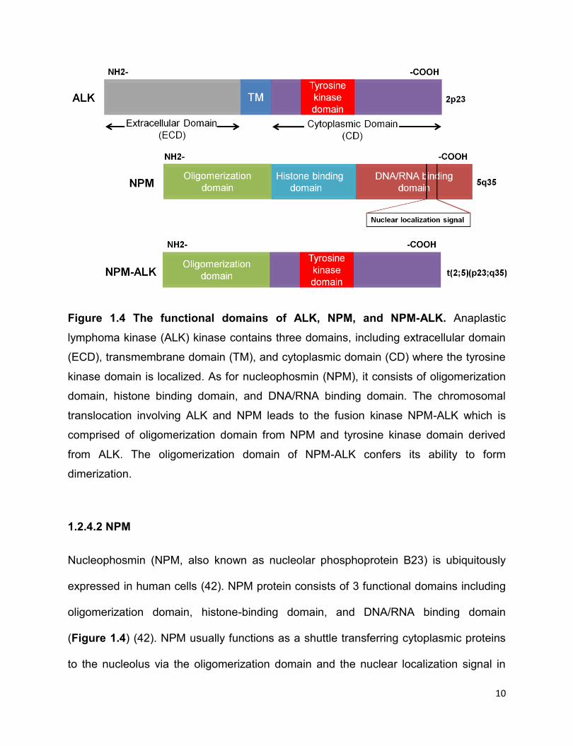
1.2.4.1 ALK
ALK is a receptor tyrosine kinase (RTK), belonging to the superfamily of insulin growth
factor (IGF) receptor (26). ALK contains 3 domains including an extracellular domain
(ECD) for ligand binding, a transmembrane domain (TM), and a cytoplasmic tyrosine
kinase domain (CD) (Figure 1.4) (26).
The ligands of ALK signaling in vertebrates remain poorly studied (27). In D
melanogaster, Jelly belly (Jeb) protein has been shown as a ligand and activator of
ALK (28). The small heparin-binding growth factors pleiotrophin (PTN) and midkine (MK)
have been found to activate the ALK downstream targets by physically binding the
extracellular domain of ALK in some cell models (29, 30). However, PTN or MK, as
ligand and activator of ALK, is controversial due to lack of reproducibility (31, 32). More
9
recently, Murray et al reported that heparin specifically binds to ALK and induces ALK
dimerization and activation in the neuroblastoma cell line ‒ NB1 independent of PTN or
MK (33). Two independent research groups have recently identified that augmentor-α
(also known as FAM150B) specifically binds to the extracellular domain of ALK and
robustly stimulates ALK signaling (34, 35). However, the two groups had conflicting
conclusion on augmentor-β (also named FAM150A) being as a potent ALK ligand.
Guan et al reported that augmentor-β activates ALK signaling as potently as
augmentor-α (34), but Andrey et al suggested that augmentor-β only weakly binds to
and activates ALK (35).
The expression of ALK is typically restricted to the cells of neural system, such as the
thalamus, mid-brain, olfactory bulb and ganglia, thus it is considered that ALK may play
key roles in the development of neural system (36). ALK gene-double knockout mice
exhibit abnormal behaviours (37), thus supporting the idea that ALK is involved in the
development of mammalian brain system, despite its exact physiological roles being
unidentified (37). Intriguingly, the aberrant expression of ALK has been detected in
certain types of cancer, such as neuroblastoma (38) and glioblastoma (39), which are
both of neural origin. Further studies have demonstrated that the aberrant ALK
expression, either by gene amplification or mutation, is implicated in the pathogenesis
of these types of cancer (40, 41). Nevertheless, the oncogenic role of ALK remains
poorly understood, since the majority of studies on the oncogenic role of ALK has been
performed in ALK+ALCL cells in which ALK mostly exists as a NPM-ALK fusion protein
(26).
10
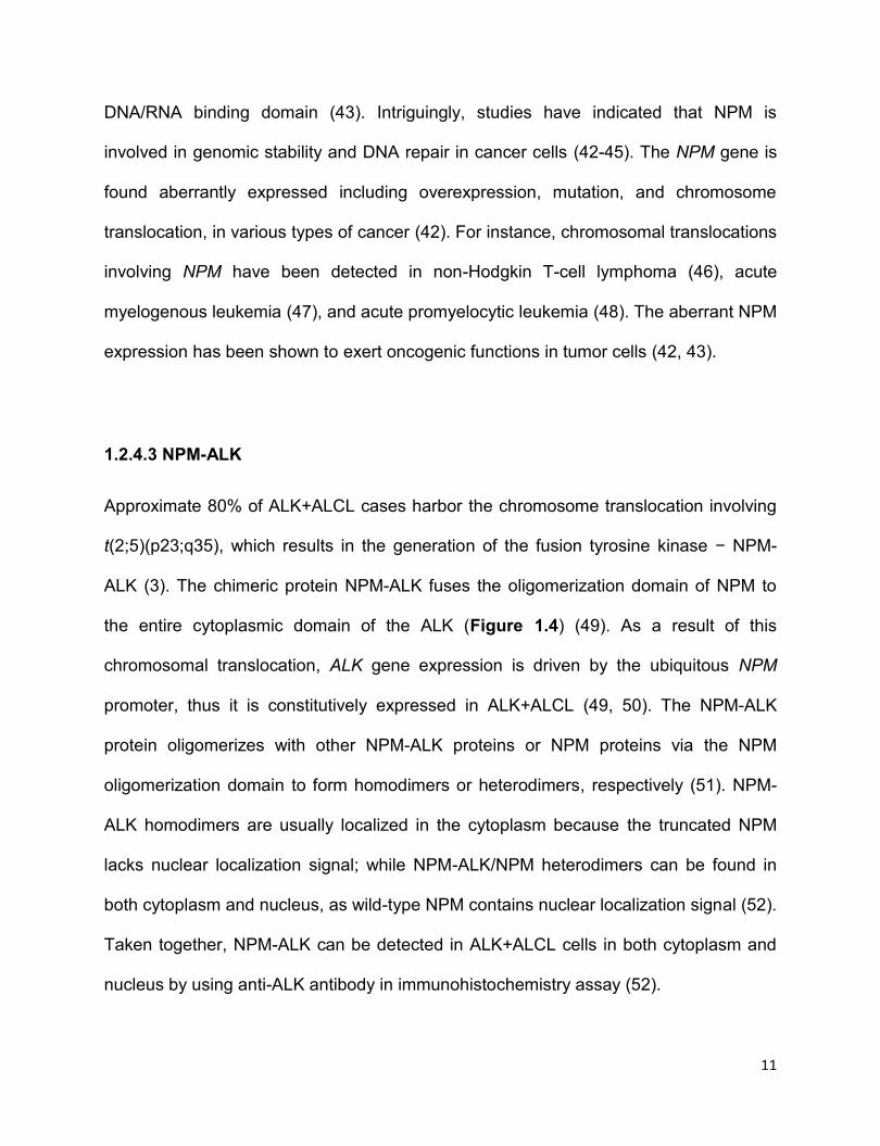
Figure 1.4 The functional domains of ALK, NPM, and NPM-ALK. Anaplastic
lymphoma kinase (ALK) kinase contains three domains, including extracellular domain
(ECD), transmembrane domain (TM), and cytoplasmic domain (CD) where the tyrosine
kinase domain is localized. As for nucleophosmin (NPM), it consists of oligomerization
domain, histone binding domain, and DNA/RNA binding domain. The chromosomal
translocation involving ALK and NPM leads to the fusion kinase NPM-ALK which is
comprised of oligomerization domain from NPM and tyrosine kinase domain derived
from ALK. The oligomerization domain of NPM-ALK confers its ability to form
dimerization.
1.2.4.2 NPM
Nucleophosmin (NPM, also known as nucleolar phosphoprotein B23) is ubiquitously
expressed in human cells (42). NPM protein consists of 3 functional domains including
oligomerization domain, histone-binding domain, and DNA/RNA binding domain
(Figure 1.4) (42). NPM usually functions as a shuttle transferring cytoplasmic proteins
to the nucleolus via the oligomerization domain and the nuclear localization signal in
11
DNA/RNA binding domain (43). Intriguingly, studies have indicated that NPM is
involved in genomic stability and DNA repair in cancer cells (42-45). The NPM gene is
found aberrantly expressed including overexpression, mutation, and chromosome
translocation, in various types of cancer (42). For instance, chromosomal translocations
involving NPM have been detected in non-Hodgkin T-cell lymphoma (46), acute
myelogenous leukemia (47), and acute promyelocytic leukemia (48). The aberrant NPM
expression has been shown to exert oncogenic functions in tumor cells (42, 43).
1.2.4.3 NPM-ALK
Approximate 80% of ALK+ALCL cases harbor the chromosome translocation involving
t(2;5)(p23;q35), which results in the generation of the fusion tyrosine kinase − NPM-
ALK (3). The chimeric protein NPM-ALK fuses the oligomerization domain of NPM to
the entire cytoplasmic domain of the ALK (Figure 1.4) (49). As a result of this
chromosomal translocation, ALK gene expression is driven by the ubiquitous NPM
promoter, thus it is constitutively expressed in ALK+ALCL (49, 50). The NPM-ALK
protein oligomerizes with other NPM-ALK proteins or NPM proteins via the NPM
oligomerization domain to form homodimers or heterodimers, respectively (51). NPM-
ALK homodimers are usually localized in the cytoplasm because the truncated NPM
lacks nuclear localization signal; while NPM-ALK/NPM heterodimers can be found in
both cytoplasm and nucleus, as wild-type NPM contains nuclear localization signal (52).
Taken together, NPM-ALK can be detected in ALK+ALCL cells in both cytoplasm and
nucleus by using anti-ALK antibody in immunohistochemistry assay (52).
12
The dimerization of NPM-ALK results in the autophosphorylation and constitutive
activation of ALK kinase in the absence of ligands, and subsequently, NPM-ALK exerts
as a potent oncogenic tyrosine kinase by interacting and activating a wide range of
proteins or signaling pathways, such as Janus activated kinase/signal transducer and
activator of transcription (JAK/STAT) (53), phosphatidylinositol 3 kinase (PI3K)/AKT
(54), RAS/ERK kinase (MEK)/extracellular signal-related kinase (ERK) (51, 55, 56), all
of which are related with important biological processes such as cell proliferation,
survival, invasiveness, etc (51, 55, 56). The oncogenic role of NPM-ALK in vitro and in
vivo has been well documented in a number of studies using cell lines and transgenic
mouse models (50).
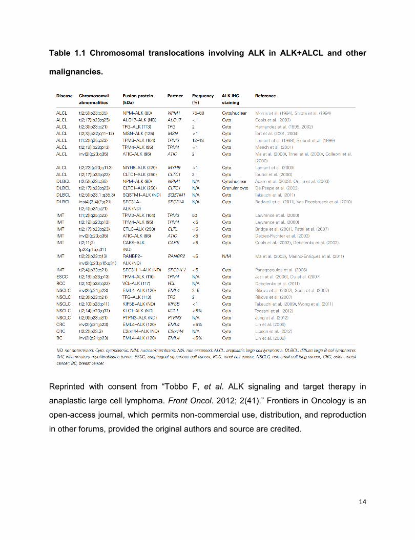
1.2.4.4 Other ALK fusion proteins
Other than NPM-ALK fusion protein, numerous other ALK fusion proteins have been
detected in ALK+ALCL (Table 1) (57). All these ALK fusion proteins, including NPM-
ALK, share common characteristics, as demonstrated below: 1) The ALK partner gene
drives ALK gene expression (3, 26, 52). 2) The ALK partner protein contains
oligomerization domain through which ALK fusion protein dimerizes with each other (3,
26, 52). 3) The ALK fusion proteins can be automatically phosphorylated and
constitutively activated by themselves (3, 26, 52). 4) The ALK partner protein
determines the subcellular localization of the ALK fusion protein (3, 26, 52). In the case
of NPM-ALK, as mentioned above, it can be detected in both cytoplasm and nucleus
(55). To be specific, it is the NPM-ALK/NPM heterodimers that are localized in
cytoplasm and nucleus (52). As for most of other types of ALK fusion proteins, they are
13
only detectable in cytoplasm (26, 57). This differential subcellular localization of NPM-
ALK and other ALK fusion proteins can be ascribed to the wild-type NPM which
contains nuclear localization signals to assist in shuttling the NPM-ALK/NPM
heterodimers to the nucleus (8, 57). However, other truncated ALK partner proteins
lack of this nuclear localization signal; therefore, these ALK fusion proteins cannot
migrate to the nucleus (8, 57). For instance, as TPM4 lacks nuclear localization signal,
TPM4-ALK homodimers and TPM4/TPM4-ALK heterodimers are both localized in
cytoplasm (Table 1) (57).
Notably, chromosomal translocations involving ALK are detected not only in ALK+ALCL
but also in other types of malignancies, such as non-small-cell lung cancer (NSCLC)
(58) and colorectal cancer (CRC) (59) (Table 1.1).
14
Table 1.1 Chromosomal translocations involving ALK in ALK+ALCL and other
malignancies.
Reprinted with consent from “Tobbo F, et al. ALK signaling and target therapy in
anaplastic large cell lymphoma. Front Oncol. 2012; 2(41).” Frontiers in Oncology is an
open-access journal, which permits non-commercial use, distribution, and reproduction
in other forums, provided the original authors and source are credited.
15
1.2.5 NPM-ALKꟷmediated transformation
ALK+ALCL cells are highly ALK-addicted (56). Since ~85% of ALK+ALCL cases
express NPM-ALK chimeric kinase, NPM-ALK is the most well studied ALK fusion
kinase in ALK+ALCL (3). The oncogenic potential of NPM-ALK fusion kinase has been
well established both in vitro and in vivo (50). The transformative ability of NPM-ALK
was first validated in mice transplanted with NPM-ALK−transduced bone marrow
progenitors (60). In a recent study, Zhang et al showed that NPM-ALK is able to
transform human CD4+ T lymphocytes, and the transformed T cells were
morphologically and immunophenotypically similar to patient-derived ALK+ALCL cells
(61). These transformed T cells implanted into immunodeficient mice developed tumors
that are indistinguishable from human ALK+ALCL tumors (61). However, it is still
controversial in terms of the cell lineage of NPM-ALK−induced hematopoietic
lymphoma in NPM-ALK transgenic mice (50). Some studies have revealed that NPM-
ALK transgenic mice develop T-cell lymphoma (60-63); while evidence from other
studies have supported that NPM-ALK transgenic mice develop B-cell lineage
lymphoma (60, 64-66), even when NPM-ALK expression was driven by a T-cell specific
promoter (63, 65). Of note, Malcolm et al recently reported that CD4-driven NPM-ALK
transgenic mice with RAG2-/- develops peripheral T cell lymphoma where the
malignance arises in early thymic precursors, and more importantly, these murine
tumors histologically resemble human ALK+ALCL (67). In this study, the authors also
reported that NPM-ALK signaling mimics T-cell receptor (TCR) β signaling and induces
the maturation of thymic T cell lymphomas (67). This study has provided a novel insight
16
on how NPM-ALK initiates peripheral T cell lymphoma in a mouse transgenic model
that histologically resembles human ALK+ALCL.
Given that NPM-ALK transgenic mice do not develop classic ALK+ALCL tumors, it is
reasonable to speculate that other factors, in addition to NPM-ALK, are required to fully
drive ALK+ALCL pathogenesis (50). This notion has also been supported by the
observation that a relatively high frequency of NPM-ALK transcripts can be detected in
non-malignant cells (68).
1.2.6 NPM-ALKꟷinteracting substrates and activated oncogenic signaling pathways
A few studies have revealed a number of substrates of NPM-ALK by mass
spectrometry-based proteomic approach. An study published in 2004 suggested that a
total of 46 proteins were identified as substrates of NPM-ALK by co-
immunoprecipitation with anti-ALK antibody, followed by electrospray ionization and
tandem mass spectrometry, and these proteins include Jak2, Jak3, Stat3, PI3K, some
adaptor molecules (such as Rho-GTPase activating protein), heat shock proteins (such
as Hsp60 precursor), and phosphatases (protein phosphatase 2 subunit) (69). Voena
et al used tandem mass spectrometry and found that NPM-ALK directly interacts and
phosphorylates Shp2, also named protein-tyrosine phosphatase 2C (PTP-2C), in the
tyrosine residues Y542 and Y580, thus promoting ALK+ALCL cell proliferation (70).
More importantly, the authors unearthed that only the active form of NPM-ALK, but not
the kinase dead NPM-ALKK210R, is able to bind and activate Shp2 (70). In parallel with
this study, another group reported that NPM-ALK interacts and phosphorylates
17
proteinassociated splicing factor (PSF) at Tyr293, resulting in its subcellular
delocalization and dysfunction (71). In addition, the interaction between NPM-ALK and
PSF depends on the active ALK kinase domain, as PSF does not bind kinase-dead
NPM-ALK (71). More recent studies using advanced tandem mass spectrometry have
revealed novel substrates of NPM-ALK, such as DNA mismatch repair protein MSH2
(72), and Wiskott-Aldrich syndrome protein (WASp), thus contributing to the
tumorigenesis of ALK+ALCL cells (73).
In general, NPM-ALK, along with other ALK fusion kinases, exerts its oncogenic roles
mainly by directly or indirectly interacting and activating a number of molecules involved
in multiple oncogenic signaling pathways (56), including JAK/STATs (53), PI3K/AKT
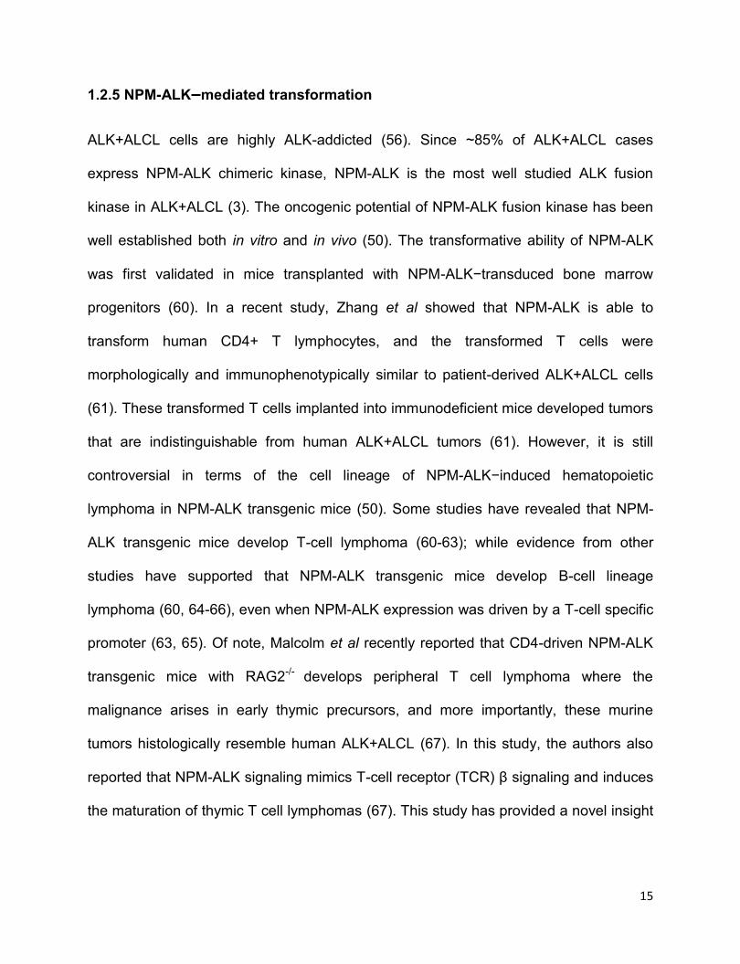
(54), RAS/MEK/ERK (74), and phospholipase C-γ (PLC-γ) (75) (Figure 1.5).

18
Figure 1.5 Representative signaling pathways activated by NPM-ALK. In ALK+
anaplastic large-cell lymphoma (ALK+ALCL), NPM-ALK interacts and activates many
essential adaptors involved in multiple signaling pathways, including JAK/STATs, PI3K,
RAS/MEK/ERK and PLC-γ. Four representative signaling pathways are shown here.
NPM-ALK: Nucleophosphomin−anaplastic lymphoma kinase; STATs: Signal transducer
and activator of transcription; PI3K: phosphatidylinositol 3 kinase; MAPK: Mitogen-
activated protein kinase; ERK: extracellular signal-related kinase; PLC-γ:
phospholipase C γ. JAK3: Janus kinase 3; Bcl2: B-cell lymphoma 2; Mcl1: Myeloid cell
lymphoma 1; BAD: Bcl-2-associated death promoter; mTOR: mammalian target of
rapamycin; FOXO3a: Forkhead box protein O 3a; DAG: diacylglycerol; PKC: protein
kinase C.
1.2.6.1 JAK/STATs
A) JAKs and STAT3 in ALK+ALCL
The JAK/STAT pathway is one of the most studied pathways that are activated by
NPM-ALK in ALK+ALCL (55). NPM-ALK fusion tyrosine kinase interacts and
phosphorylates STAT3 at tyrosine 705 (pSTAT3Y705) (76), a critical residue for STAT3
activation and dimerization (77). Then the phosphorylated STAT3 forms homo or
heterodimers with phosphorylated STAT3 or other family members of STATs, followed
by translocation to the nucleus to execute as a transcription factor regulating a wide
range of genes involved in cell proliferation, survival, and anti-apoptosis, such as MYC,
CCNDs, BCL2, and Survivin (78). Indeed, high level of pSTAT3Y705 and the nuclear
localized pSTAT3Y705 are always detected in ALK+ALCL primary tumor cells and cell
lines (76, 79). Multiple factors contribute to the constitutive activation of STAT3 in
ALK+ALCL (56). For instance, Han et al reported that JAK3, which can be activated by
cellular cytokines such as interleukin 2 (IL2), IL4, IL9, IL13, IL15, and IL21 (80-85),
19
phosphorylates STAT3 at tyrosine 705 in ALK+ALCL (86). In keeping with this, two
other studies have demonstrated that ALK+ALCL cells express IL9 and IL21, both of
which are JAK3 activators (82, 85), to activate the JAK3/STAT3 axis (84, 87). In
addition, JAK3 is directly phosphorylated and activated by NPM-ALK in ALK+ALCL (26).
The significance of JAK3 for STAT3 activity is underscored by the finding that
pharmaceutical inhibitor or siRNA knockdown of JAK3 downregulates the pSTAT3Y705
level in ALK+ALCL (86, 88). Amin et al used co-immunoprecipitation assay and found
that NPM-ALK, STAT3 and JAK3 physically interact with each other in ALK+ALCL cells,
indirectly suggesting the close interaction between them (88). Further studies have
demonstrated that the level of active JAK3 strongly correlates with the level of NPM-
ALK and pSTAT3Y705 in ALK+ALCL primary tumors (86). Intriguingly, one study
reported that NPM-ALK induces the expression of IL22 and its receptor IL22R in
ALK+ALCL, thus forming an autocrine stimulatory loop for JAK3/STAT3 activation (89).
STAT3 is a well-known potent oncogenic protein (90). Aberrant expression or activation
of STAT3 has been widely observed in solid human tumors and haematological
malignancies, including ALK+ALCL (91-95). STAT3 is known to be a key mediator of
the NPM-ALKꟷinduced transformation in ALK+ALCL (91). Inhibition of STAT3 activity
by STAT3 dominant negative construct (96) or pharmaceutical reagent (97) induces
significant cell-cycle arrest and cell apoptosis in ALK+ALCL cell lines. One study
showed that siRNA knockdown of STAT3 in SupM2 cells, an ALK+ALCL cell line,
modulates the expression of around 1500 genes, with approximately 900 of them
downregulated (98). In addition, STAT3 transcriptionally induces the expression of DNA
methyltransferases 1 (DNMT1) which thereafter physically interacts with STAT3 and
20
histone deacetylase 1 (HDAC-1) and binds to the promoter of the tumor-suppressor
gene ‒ Src homology region 2 domain-containing phosphatase-1 (SHP1), leading to
the epigenetically silence of SHP1 (99, 100). SHP1 is known as a tyrosine phosphatase
that physically interacts with NPM-ALK, JAK3 and STAT3 in ALK+ALCL, subsequently
resulting in the dephosphorylation of both NPM-ALK and JAK3/STAT3 and degradation
of NPM-ALK and JAK3 in a proteasomal pathway (101, 102). Similarly, STAT5A is also
epigenetically silenced by STAT3−induced DNA methylation (103). As a member of the
STAT family, STAT5A functions as a negative regulator of NPM-ALK expression in
ALK+ALCL, evidenced by that restoration of STAT5A expression transcriptionally
downregulates the expression of NPM-ALK and decreases the level of pSTAT3Y705
(103). In contrast, STAT5B functions as an oncogenic protein to promote cell
proliferation and growth upon NPM-ALK phosphorylation and activation (104). The
relationship between NPM-ALK, JAKs, STAT3, STAT5A and STAT5B has been briefly
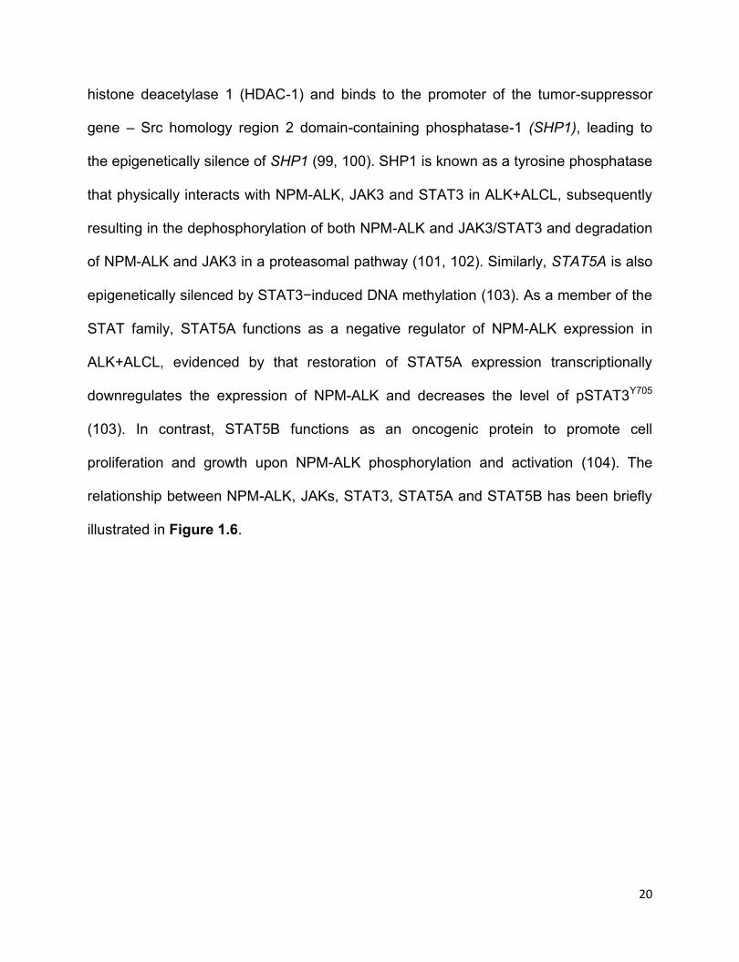
illustrated in Figure 1.6.
21
Figure 1.6 NPM-ALK activates STATs signaling pathway in ALK+ALCL. Signal
transducer and activator of transcription 3 (STAT3) can be directly activated by NPM-
ALK, or indirectly through Janus kinase 3 (JAK3). Besides, ALK+ALCL cells express
interleukin 9 (IL9), IL21 and IL22, which are JAKs’ activator, to activate STAT3 as well.
The activated STAT3 signaling potently promotes cell proliferation and growth by
regulating a number of genes, such as Survivin, B-cell lymphoma 2 (BCL2) and BCL-XL.
Src homology region 2 domain-containing phosphatase-1 (SHP1) and STAT5A are
reported to be methylated and silenced in ALK+ALCL because of STAT3. If their
expressions are restored, both SHP1 and STAT5A act as negative regulators of NPM-
ALK/STAT3 axis by transcriptionally downregulating the expression of NPM-ALK and
decreasing the level of pSTAT3Y705. In addition, STAT5B, another family member of
STATs, is also phosphorylated and activated by NPM-ALK, thus promoting cell growth
and proliferation.
22
B) STAT1
IFNs/STAT1 signaling
STAT1 is the first-discovered member of STATs family (90). Induction of STAT1
activation occurs through cytokines, growth factors, and hormones (105). STAT1 is
well-known as the main mediator of both type I interferon (IFNα/β) and type II
interferon (IFNγ) signaling pathway in immune responses, thus it is regarded as an
important component in immune system (106, 107). Upon type I IFN binding with
cellular receptor IFN alpha receptor (IFNAR1/2), the endogenous kinase JAK1 and
tyrosine kinase 2 (TYK2) are recruited to the cytoplasmic domain of IFNAR1/2,
followed by phosphorylation and activation; and the activated JAK1 and TYK2
reversely phosphorylate IFNAR1/2 (105, 106). The phosphorylated IFNAR1/2 thus
create docking sites for STAT1 and STAT2, another member of STATs family, and
then the phosphorylated JAK1 and TYK2 that bound to the cytoplasmic domain of
IFNAR1/2 phosphorylate STAT1 and STAT2, then the phosphorylated STAT1 and
STAT2 form homodimers or heterodimers and subsequently migrate to the nucleus to
regulate a number of IFN-related genes (Figure 1.7) (105, 106). Specifically,
STAT1/STAT2 heterodimers or STAT1 homodimers bind to the IFN-stimulated
response element (ISRE) promoter sequence or the IFN--activated-site (GAS)
promoter sequences, respectively, to stimulate IFN-stimulated gene transcription (105,
106). As for type II IFN signaling pathway, the binding of IFNγ and its receptor IFNγ
receptor 1/2 (IFNGR1/2) phosphorylates and activates JAK1 and JAK2, following by
the phosphorylation of IFNGR1/2 (105, 106). The phosphorylated IFNGR1/2 thus
create docking sites for STAT1 which subsequently is phosphorylated by JAK1 and
23
JAK2, and consequently the phosphorylated STAT1 homodimerizes with itself and
migrate to the nucleus to bind to GAS promoter sequence to function as transcription
factors (Figure 1.7) (106).
Figure 1.7 Signal transduction of the IFN/STATs. Binding of type I and type II IFNs
to their respective receptors (IFNAR1/2 for type I and IFNGR1/2 for type II) leads to the
activations of Janus kinases 1 (JAK1) and tyrosine kinase 2 (TYK2) for IFNAR1/2, and
JAK1 and JAK2 for IFNGR1/2. The activated JAKs and TYK2 subsequently
phosphorylate the respective receptors, which thereby create docking sites for signal
transducer and activator of transcription 1 (STAT1) or STAT2. STAT1 and STAT2 are
phosphorylated on tyrosine residues by JAKs and TYK2 once they bound to the
docking sites of the IFNs receptors. Phosphorylated STATs form STAT1 homodimers
and STAT1/STAT2 heterodimers which move to the nucleus to function as transcription
factors. STAT1 homodimers bind to IFN--activated-site (GAS) promoter sequences and
24
STAT1/STAT2 heterodimers bind to the IFN-stimulated response element (ISRE)
promoter sequence, respectively, to stimulate IFN-stimulated gene transcription.
STAT1 is degraded in ubiquitin-dependent proteasome pathway
Ubiquitin-dependent proteasome pathway is an important biological process that
regulates the degradation of cellular proteins by ubiquitinating substrate proteins,
followed by protein degradation in 26S multisubunit proteasome system (108). This
biological process requires the involvement of a sequential of ubiquitin-related enzymes
including ubiquitin-activating enzymes (E1), ubiquitin-conjugating enzymes (E2), and
ubiquitin ligases (E3) (108). In brief, the E1 enzyme activates ubiquitin, which is
subsequently transferred to ubiquitin-conjugating enzymes E2; then the ubiquitin is
transferred to the substrate protein with the assist of specific E3 ligase (109).
Consequently, the ubiquitin-tagged substrate protein is degraded in the 26S
proteasome (109). Mounting studies have suggested that STAT1 is degraded in
ubiquitin-dependent proteasome pathway (110-115). In a study published in 2008, the
authors reported that ERK1/2 phosphorylates STAT1 at serine 727 and downregulates
its expression in ubiquitin-dependent proteasome pathway in mouse embryonic
fibroblasts model, in which the F-box E3 ligase βTRCP promotes STAT1 ubuiquitination
(112). In a recent study, an E3 ligase smad ubiquitination regulation factor 1 (Smurf1) is
reported to ubiquitinate and degrade STAT1 in human embryonic kidney 293 cells
(113). More recently, one study revealed that pSTAT1Y701, the active form of STAT1, is
the primary form of ubiquitinated STAT1 induced by IFNγ in 293T cells, and it is rapidly
degraded in K48-linked ubiquitin-proteasome pathway (111). Moreover, the authors of
25
the study also demonstrated that the deubiquitinase USP2a interacts with pSTAT1Y701
in the nucleus and abrogates the ubiquitination and degradation of pSTAT1Y701 (111).
Surprisingly, Lawrence and Kornbluth recently reported that an E3 ubiquitin ligase ꟷ
Natural Killer Lytic-Associated Molecule (NKLAM) ubiquitinates STAT1, but positively
regulates STAT1 transcriptional activity and its DNA binding ability in mice macrophage
cells (110).
The biological function of STAT1
STAT1 has been reported to be a tumor suppressor by transcriptionally regulating the
expression of a host of pro-apoptotic and anti-proliferative genes, such as Interferon
response factor -1 (IRF-1) (116), caspases (117), members of the death receptor family
(118), BCL-XL and BCL-2 (119, 120). A number of studies have suggested that STAT1
antagonizes the transcriptional activity of the oncogenic protein STAT3 in some models
(90, 120-122). The tumor suppressor function of STAT1 is also indicated in an in vivo
study showing STAT1-/- double knockout mice are more vulnerable to virus and
bacterial infections and also prone to develop tumors upon exposure to cancer-inducing
chemicals (123, 124). STAT1-deficient female mice also spontaneously develop
mammary adenocarcinomas (125). In keeping with its tumor suppressor function,
STAT1 expression is frequently absent or decreased in various types of cancer,
including oral squamous cell carcinoma (126), melanoma (127, 128), cutaneous T-cell
lymphoma (129), breast cancer (130) and esophagus squamous cancer (131, 132).
How STAT1 is downregulated in cancer remains poorly understood. There is one
relevant report, in which STAT1 gene promoter was found methylated in human head

26
and neck squamous cancer (133). Nevertheless, a few studies have concluded that
STAT1 is a tumor promoter in some cancer models, such as breast cancer (134), T-cell
acute lymphoblastic leukemia (135) and late-stage melanoma (136). These
contradictory findings indicate the dilemma functions of STAT1 in cancer in a cellular
context-dependent manner (90).
1.2.6.2 PLC-γ
PLC-γ is an enzyme that cleaves phospholipids (137). The activation of PLC-γ by cell
receptor tyrosine kinases (RTKs) results in the hydrolysis of phosphatidylinositol (PIP2)
into two secondary messages, inositol triphosphate (IP3) and diacylglycerol (DAG)
(137). IP3 stimulates the release of Ca2+ from the endoplasmic reticulum, and DAG
interacts and activates the serine/threonine protein kinase C (PKC) (137, 138). NPM-
ALK tyrosine residue 664 (Y664) directly interacts with PLC-γ and leads to the
activation of PLC-γ (75). The significance of PLC-γ to the NPM-ALK−induced
transformation is underscored by the finding that mutant NPM-ALKY664F cannot bind or
activate PLC-γ and demonstrated attenuated transforming ability in Ba/F3 and Rat-1
cells, which can be partially rescued by the overexpression of PLC-γ (75).
1.2.6.3 PI3K/AKT
NPM-ALK also activates the PI3K/AKT pathway in ALK+ALCL (54). Activation of the
PI3K/AKT pathway promotes cell growth of ALK+ALCL cells (54, 139). Inhibition of
PI3K/AKT pathway by either pharmacologic inhibitors, or dominant-negative PI3K, or
27
AKT mutants suppress cell growth or induce cell apoptosis in multiple types of cells
transfected with NPM-ALK (54, 139).
PI3K contains two subunits including a catalytic subunit p110 and a regulatory subunit
p85 (140, 141). NPM-ALK fusion kinase binds to p85 and phosphorylates PI3K, which
subsequently phosphorylates and activates the serine/threonine kinase AKT (also
known as protein kinase B) at threonine 308 (54, 56). However, the maximal activation
of AKT requires additional phosphorylation at serine 473 (141). The fully activated AKT
regulates diverse cellular processes, such as protein synthesis, cell anti-
apoptosis/survival, cell growth and proliferation, cell metabolism, and cell
migration/invasion (141, 142). Specifically, activated AKT suppresses the pro-apoptotic
protein Bcl2-associaed death promoter (BAD), and thus leads to the increase of anti-
apoptotic protein Bcl-XL (143). On the other hand, activated AKT promotes cell survival
by increasing the activity of nuclear factor kappa-light-chain-enhancer of activated B
cells (NF-κB) and promoting the degradation of inhibitors of κB (IκB), which sequesters
the nuclear translocation of NF-κB (144). The activated PI3K/AKTꟷpromoted cell
growth of ALK+ALCL is also partially dependent on the activated Sonic Hedgehog
(SHH) pathway, which is downstream of AKT signaling (145, 146). In addition, activated
AKT suppresses cell apoptosis by inhibiting the activity of caspas 9 and decreasing the
expression of FAS ligand (147, 148). NPM-ALK−induced activation of PI3K/AKT
pathway phosphorylates Forkhead family of transcription factors 3a (FOXO3a) and
prevents its nuclear translocation, consequently leading to the degradation of FOXO3a
in a proteasome pathway (149-151). The degraded FOXO3a links to the
downregulation of the negative regulator of cell cycle progression p27Kip1 and Bim 1, as
28
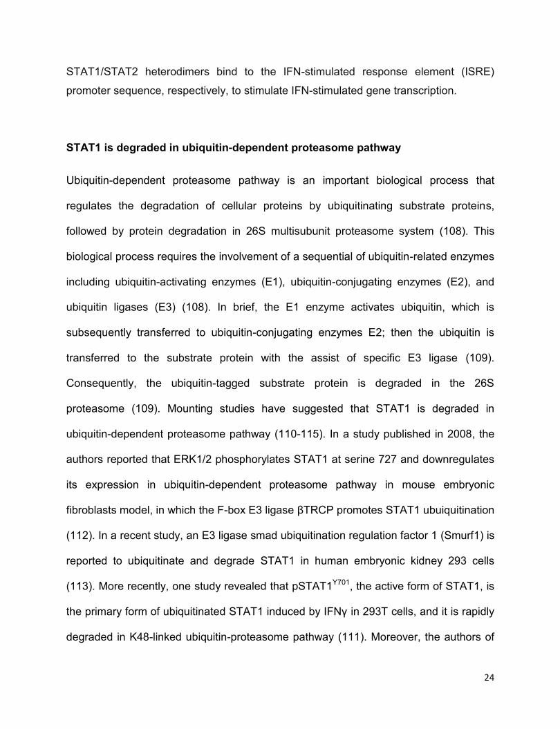
well as the upregulation of cyclin D2 (150, 151). A brief interaction between NPM-ALK
and PI3K/AKT has been illustrated in Figure 1.8.
Figure 1.8 NPM-ALK activates the PI3K/AKT pathway in ALK+ALCL. NPM-ALK
phosphorylates and activates PI3K at p85 subunit, which subsequently activates the
serine/threonine kinase AKT. The activated AKT phosphorylates Forkhead family of
transcription factors 3a (FOXO3a) and promotes FOXO3a degradation in a proteasome
pathway. Phosphorylation of the FOXO3a sequesters its nuclear translocaton to
upregulate the negative regulator of cell cycle progression p27Kip1 and Bim1 and
downregulate cyclin D2. Activated AKT also promotes the activity of nuclear factor-κB
(NF- κB) by degradating inhibitor of κB (IκB) in a proteasomal pathway. IκB inhibits the
activity of NF- κB by preventing the nucleus translocation of NF- κB. The
phosphorylated AKT also suppresses the activity of caspas 9 and the expression of Fas
ligands (FAS-L), as well as the activity of Bcl 2-associaed death promoter (BAD) which

29
suppresses the expression of anti-apoptotic protein Bcl-XL. In addition, AKT signalling
also promotes the expression of sonic hedgehog (SHH), which also contributes to the
cell proliferation of ALK+ALCL.
1.2.6.4 RAS/MEK/ERK
The serine/threonine kinase ERK1/2−mediated pathway is known to promote cell
growth, survival, differentiation, and migration (152). ERK1/2 can be activated by many
growth factors and some kinases through the activation of the Ras GTPase following by
the phosphorylation and activation of Raf1 (153). While the NPM-ALK‒induced
activation of MEK/ERK appears to be independent of Raf-1, as inhibition of Raf-1 with
pharmacological inhibitors or siRNA knockdown of Raf-1 have no effects on ERK1/2
phosphorylation induced by NPM-ALK (154). The activated RAS/MEK/ERK pathway
can be found in both ALK+ALCL cell lines and primary tumor samples (154). Inhibition
of ERK1/2 by MEK1/2 inhibitor U0126 or siRNA knockdown of ERK1 suppresses cell
growth and induces cell apoptosis in ALK+ALCL cell lines, supporting the role of the
ERK1/2 pathway in promoting the cell growth of ALK+ALCL cells (154). NPM-
ALKꟷmediated activation of ERK1/2 activates E26 transformation-specific-1 (ETS-1) in
ALK+ALCL (155). Interestingly, the transcriptional active ETS-1 enhances the gene
promoter activation of JunB, which is reported to promote CD30 expression; and the
upregulated expression of CD30 in turn activates ERK1/2 in ALK+ALCL, thus creating a
positive forward loop in ALK+ALCL (155, 156).

30
1.2.6.5 mTOR
In ALK+ALCL, the serine/threonine kinase mTOR is also activated by NPM-ALK
mediated by PI3K/AKT and RAS/MEK/ERK (155, 157). The biological significance of
mTOR is underlined by the fact that inhibition of mTOR by pharmaceutical inhibitor
rapamycin induced significant cell apoptosis in ALK+ALCL cell lines (158). mTOR
regulates a number of genes associated with protein translation, cell proliferation, and
survival (159, 160). For instance, mTOR phosphorylates and activates p70S6K (also
known as ribosomal protein S6 kinase beta-1), which thereafter phosphorylates and
activates ribosomal protein S6 (RPS6) to regulate protein synthesis and enhance cell
growth (161).
1.2.7 Therapeutic strategy for ALK+ALCL patients
As indicated above, ALK+ALCL generally have a better prognosis in comparison to
ALK-ALCL (7). Cyclophosphamide, hydroxydaunorubicin (doxorubicin), oncovin
(vincristine), and prednisone (CHOP)ꟷbased therapeutic regimen is the standard
treatment for aggressive lymphomas, including ALK+ALCL (1). Brentuximab vedotin
has also showed encouraging clinical outcomes when it was applied to treat CD30+
peripheral T-cell lymphomas (including ALK+ALCL), either as a follow-up treatment with
CHOP or a combination with CHP (due to the overlapped neurotoxicity between
vincristine and Brentuximab vedotin) as the front-line treatment (1, 162). CHOP-based
standard treatment induces complete remission in most of ALK+ALCL patients, but up
to 40% of patients still develop tumor relapses and chemoresistance (55). Patients with

31
relapsed or refractory disease usually benefit from high-dose of chemotherapy or
autologous stem cell transplantation (8).
Although stem cell transplantation is an effective therapeutic regimen for relapsed
ALK+ALCL patients, novel therapy based on the pathobiology of ALK+ALCL is
warranted (55). As ALK+ALCL cells are very much NPM-ALK addicted, this chimeric
protein has become an obvious and easy therapeutic target for this disease, supported
by a number of in vitro and in vivo studies (50, 163-165). As the discovery of full length,
translocated, or mutated ALK contributing to the tumorigenesis of neuroblastoma or
non-small-cell lung cancer, a number of small molecular inhibitors targeting ALK have
been developed (166-168). For instance, crizotinib is a small molecule inhibitor
developed for targeting ALK, c-Met/Hepatocyte growth factor receptor (HGFR) and c-
ros oncogene 1 (ROS1), and it specifically binds within the ATP-binding pocket of
target kinases (168). In 2011, the U.S. Food and Drug Administration (FDA) approved
crizotinib (PF-02341066; Trade name: Xalkori, Pfizer) for the treatment of late-stage
ALK-positive non-small-cell lung cancers (169). Currently, crizotinib is also undergoing
clinical trials for treating patients with ALK+ALCL and neuroblastoma (170, 171).
Results from a small number of clinical trials have shown the effectiveness of crizitinib
in pediatric ALK+ALCL patients and the relapsed/refractory patients (170). Despite the
success of crizotinib, crizotinib-resistance still could happen in ALK+ALCL patients and
other ALK-positive malignancies (172, 173). The novel therapeutic strategies
overcoming the potential crizotinib-resistance to this type of malignancy generally
involve targeting NPM-ALK downstream molecules in combination with ALK inhibitors
(1, 8, 174).
32
1.3 Wnt/β-catenin
1.3.1 Wnt/β-catenin signaling
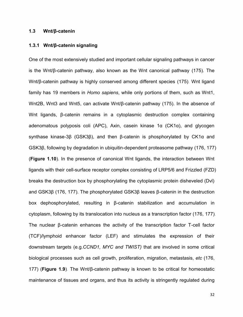
One of the most extensively studied and important cellular signaling pathways in cancer
is the Wnt/β-catenin pathway, also known as the Wnt canonical pathway (175). The
Wnt/β-catenin pathway is highly conserved among different species (175). Wnt ligand
family has 19 members in Homo sapiens, while only portions of them, such as Wnt1,
Wnt2B, Wnt3 and Wnt5, can activate Wnt/β-catenin pathway (175). In the absence of
Wnt ligands, β-catenin remains in a cytoplasmic destruction complex containing
adenomatous polyposis coli (APC), Axin, casein kinase 1α (CK1α), and glycogen
synthase kinase-3β (GSK3β), and then β-catenin is phosphorylated by CK1α and
GSK3β, following by degradation in ubiquitin-dependent proteasome pathway (176, 177)
(Figure 1.10). In the presence of canonical Wnt ligands, the interaction between Wnt
ligands with their cell-surface receptor complex consisting of LRP5/6 and Frizzled (FZD)
breaks the destruction box by phosphorylating the cytoplasmic protein disheveled (Dvl)
and GSK3β (176, 177). The phosphorylated GSK3β leaves β-catenin in the destruction
box dephosphorylated, resulting in β-catenin stabilization and accumulation in
cytoplasm, following by its translocation into nucleus as a transcription factor (176, 177).
The nuclear β-catenin enhances the activity of the transcription factor T-cell factor
(TCF)/lymphoid enhancer factor (LEF) and stimulates the expression of their
downstream targets (e.g.CCND1, MYC and TWIST) that are involved in some critical
biological processes such as cell growth, proliferation, migration, metastasis, etc (176,
177) (Figure 1.9). The Wnt/β-catenin pathway is known to be critical for homeostatic
maintenance of tissues and organs, and thus its activity is stringently regulated during
33
normal development (178). Deregulation of the Wnt/β-catenin pathway is implicated in
various diseases, including cancers (178).
Figure 1.9 Overview of the Wnt/β-catenin pathway. In the absence of Wnt ligands,
β-catenin remains in the destruction complex containing adenomatous polyposis coli
(APC), Axin, casein kinase 1α (CK1α), and glycogen synthase kinase-3β (GSK3β),
where it is phosphorylated by CK1α and GSK3β, following by degradation in ubiquitin-
dependent proteasome pathway (Left panel). In the presence of Wnt ligands, the
interaction between Wnt ligands with their cell-surface receptor complex consisting of
LRP5/6 and Frizzled (FZD) interrupt the destruction box by phosphorylating disheveled
(Dvl) and GSK3β, and inhibiting GSK3βꟷmediated phosphorylation of β-catenin. The
dephosphorylation of β-catenin leads to its stabilization and accumulation and
translocation into nucleus where it exerts as a transcription factor. The nuclear β-
catenin cooperates with the transcription factor T-cell factor (TCF)/lymphoid enhancer
factor (LEF) to promote the expression of their downstream targets, including CCND1
and MYC (Right panel).
34
1.3.2 Wnt/β-catenin in cancer and ALK+ALCL
Upregulated Wnt/β-catenin activity has been widely observed in human cancers (177).
For example, β-catenin is deregulated in 21% of cases of primary cutaneous B-cell
lymphomas and 42% of cases of cutaneous T-cell lymphoma (179). The constitutive
activation of the Wnt/β-catenin pathway is also reported in mantle cell lymphoma and
ALK+ALCL (180-182). Lai lab previously described that β-catenin is constitutively active
in ALK+ALCL cells, and knockdown of this pathway by β-catenin siRNA transfection
results in significant cell death in ALK+ALCL cell lines (182). Strikingly, Lai group also
found that siRNA knockdown of β-catenin dramatically decreases the levels of
pSTAT3Y705 and STAT3, representing yet another mechanism by which STAT3 in
ALK+ALCL (182). Of note, unlike the relationship between Break Point Cluster-Abelson
(BCR-ABL) tyrosine kinase and β-catenin in chronic myeloid leukemia (CML), NPM-
ALK does not regulate the protein level, nuclear localization or tyrosine phosphorylation
of β-catenin but the transcriptional activity of Wnt/β-catenin in ALK+ALCL (182, 183). In
normal cells, β-catenin is phosphorylated at multiple serine/threonine residues by
GSK3β and therefore downregulated in ubiquitin-proteasome pathway (175). One
recent report suggested that NPM-ALK phosphorylates GSK3β on Serine 9 through the
PI3K/AKT pathway (184). Considering the negative role of active GSK3β on the protein
level of β-catenin, this report correlates well with the findings of constitutively active β-
catenin in ALK+ALCL cells (168, 170).
35
1.4 Cancer stem cells (CSCs)
The CSCs are a small fraction of cells within tumor bulks that have the ability to initiate
the whole tumor bulks and are also capable to self-renew to maintain the small
population (185). This small cell population is also called tumor-initiating cells (TICs) or
tumor stem cells (TSCs) (186). Like normal stem cells, CSCs are not only capable to
self-renew to maintain the CSCs population, but also able to differentiate into non-
CSCs, resulting in the heterogeneity of tumor cells (186, 187). It is widely accepted that
CSCs are responsible for chemoresistance and tumor relapses in cancer patients, as it
is believed that conventional chemotherapy only eliminates the major population of
tumor cells but leave CSCs untouched (188).
1.4.1 Identification and Isolation of CSCs
CSCs population was firstly described in human acute myeloid leukemia (AML) in 1997
(189). Since then, the CSCs have been identified in various types of cancers including
breast cancer, colon cancer, prostate, esophagus squamous cancer, lung cancer (185).
Up to date, multiple assays and approaches have been developed to identify the CSCs
population, such as side population assay (using Hoechst 33343 dye efflux assay),
specific cell surface markers detection, tumorsphere formation assay and serial
transplantation assay (188).
The side population assay is a flow cytometric approach based on the dye efflux
properties of ATP-binding cassette (ABC) transporters (190). This assay has been
performed in multiple studies to identify the CSC population derived from a number of
36
cancers including ovarian cancer (191, 192), breast cancer (193, 194), head and neck
cancer (195), renal cell carcinoma (196), as well as ALK+ALCL (197). Nevertheless,
there are two major limitations of this assay (198). Firstly, the side population cells
contain both CSCs and non-CSCs, and they do not represent all CSCs (198). Secondly,
Hoechst 33342, used in this assay for isolating side population cells, cannot be applied
to many types of cells because of its cellular toxicity (198). Furthermore, the side
population assay has also received many criticisms because of its relatively high
technical challenges, poor consistency and low reproducibility (190). As for specific cell
surface markers’ staining, it has also been widely deployed to identify the CSC
population. For instance, CD44High/CD24Low and Aldehyde dehydrogenaseHigh (ALDHhigh)
have been considered as stem cell markers for both breast cancer and esophagus
squamous cells (199-203). The CD133 has also been reported to be a CSCs marker for
both colon cancer and brain cancer (204, 205). Like side population cells, stem cell
markers positive cells do not represent all the CSCs, which also include non-CSCs. As
for tumorsphere formation assay, it is a valuable tool to identify CSCs in vitro, as cells
with high ability of self-renewal and differentiation are expected to grow into colonies in
serum-free medium or in soft agar/methylcellulose medium (188). However, this in vitro
assay has many limitations. For instance, this assay only measures ex vivo cell
proliferation but not tumor propagating ability in vivo (188). Therefore, in vivo study is
always required to confirm the results of this in vitro assay (188). Last but not the least,
serial transplantation assay is regarded as the gold standard assay to identify CSCs
because this assay fulfills two criteria of CSCs including the ability to self-renew and
initiate the tumor bulks (188). This assay has been widely used in a number of studies
of identifying CSCs (185, 186, 188, 197, 206). Briefly, in this assay, tumor cells are

37
transplanted into the immunodeficient mice, then the xenograft tumors formed by the
tumor cells are isolated, diluted and implanted into the new immunodeficient mice, if
applicable, the newly formed xenograft tumors are again isolated, diluted and implanted
into new immunodeficient mice to test the tumor cells’ self-renewal and tumor-initiating
ability.
1.4.2 Molecular features of CSCs
Mounting studies have suggested that CSCs carry elevated expression of some
embryonic stem cells markers (e.g. MYC, Sox2, and Nanog) in comparison to tumor
bulk cells (186). Two recent reports have showed that the tomaxifen-resistant triple-
negative and estrogen receptor-positive (ER+) breast cancer cell lines have enriched
breast CSCs which are characterized by elevated expression of Nanog, Oct3/4, and
Sox2, as compared to their major cell populations (207, 208). MYC is also found highly
expressed in glioma stem cells and CD34+ AML cells (considered as leukemia stem
cells), as compared to their non-stem cells counterparts (209, 210). Importantly, the
high level of MYC expression is critical for maintaining the CSC population, as inhibition
of MYC in glioma stem cells and leukemia stem cells significantly decreases the CSC
populations (209, 210). In addition to these embryonic stem cell markers, CSCs also
overexpress ABC transporters, a cell surface protein family responsible for drug
transportations. Among the family of ABC transporters, ABCG2 is the one that has
been widely studied, and it is also the one that mainly confers to the side population cell
phenotype by pumping out Hoechst 33342 dye efflux (198). Enriched expression of
ABCG2 has been reported in a number of CSCs from various types of cancer, such as
38
lung cancer (211), hepatocellular carcinoma (212), retinoblastoma (213), and
ALK+ALCL (197). Of note, ABCG2 may even serve as a potential biomarker for CSCs
in some types of tumors (198).
1.4.3 Signaling pathways in CSCs
Accumulated evidence have demonstrated that certain signaling pathways (e.g. Notch,
SHH, Wnt/β-catenin and fibroblast growth factor-2), that are known to regulate the
differentiation and self-renewal of normal embryonic stem cells (214), are specially
more active in CSCs (186). These embryonic signaling pathways contribute to the self-
renewal and tumor-initiating ability in CSCs (185, 186, 215). For instance, Jang et al
reported that breast CSCs have enhanced Wnt/β-catenin activity, and inhibition of
Wnt/β-catenin signaling suppresses breast cancer cells growth, tumorigenicity,
metastasis and the expression of CSCs-related genes (216). The Wnt/β-catenin
signaling is also found more active in leukemia-initiating cells (217, 218), and depletion
of β-catenin results in impaired leukemogenesis in a BCR-ABL−induced CML mouse
model (219, 220). Lai lab also recently reported that the PI3K/AKT/MYC pathway is
elevated in cancer stem-like cells of esophagus squamous cancer cell lines,
pharmacological inhibition of PI3K/AKT/MYC dramatically attenuates the cancer
stemness features including chemoresistance and tumor formation ability (221). In a
recent study, Justilien et al showed that the SHH pathway is elevated by
phosphorylated Sox2 at threonine 118 in lung cancer stem-like cells, and the SHH
pathway is essential for maintaining the CSC phenotype, as inhibition of this pathway
diminishes the CSC phenotype (222).

39
1.4.4 CSCs in ALK+ALCL
NHL is a heterogeneous malignance compromising more than 40 different entities
classified by various parameters including morphology and immunophenotype (223).
Most of NHLs are believed to originate from mature peripheral lymphocytes, such as
many B-cell NHLs arise from germinal center cells (223, 224). However, evidence
shows that T-cell lymphoblastic lymphoma cells originate from immature precursor
lymphocytes in the thymus (223). Three recent studies have unearthed that ALK+ALCL
tumor cells may originate from early thymic progenitor cells (67, 197, 225). Moti et al
has reported that ALK+ anaplastic large-cell lymphoma-propagating cells isolated by
side population assay share gene expression signatures with early thymic progenitors,
indicating these tumor-propagating cells being of a primitive origin (197). The same
research group further reported that the murine peripheral T cell lymphoma in CD4-
driven NPM-ALK transgenic mouse arises in early thymocytes (67). In addition,
Hassler et al recently compared the DNA methylation patterns of ALK+ALCL cells with
T cells of different differentiation stages and found that the DNA methylation patterns of
ALK+ALCL highly resemble progenitor thymic T cells, suggesting that ALK+ALCL
occurs in progenitor thymic T cells rather than mature T cells (225). Notably, there is a
single study to date that has unearthed the biology of CSCs in ALK+ALCL (197). In this
report, Moti et al declared that they were able to use side population technique to find
CSCs in ALK+ALCL cell lines and patient samples, and these CSCs not only give rise
to the bulk population but also maintain the growth of entire cell population (197).
Biochemically, ALK+ALCL CSCs are featured with relatively high expression of some
embryonic stem cell or CSC markers including Sox2, Oct4, Klf4, and ABCG2, as
40
compared to the main cell population (197). Of note, these CSC cells also harbor
elevated expression of NPM-ALK, and these CSCs cells show sensitivity to crizotinib
due to the high level of NPM-ALK in this subset of cells, indicating that NPM-ALK may
be a promising therapeutic target in these CSCs (197).
1.5 Inducible pluripotent stem cells (iPS) factors
The iPS factors are those transcription factors being crucial for maintaining and
promoting cell pluripotency (226). The biological significance of these factors including
Oct4, MYC, Sox2, and Klf4 is highlighted by their ability to reprogramming fully
differentiated somatic cells to iPS cells in certain conditions (227, 228). The
expressions of these iPS factors usually are restricted to embryonic stem cells and
some somatic stem cells (228). However, recent studies have reported that these iPS
factors also have been detected in various types of cancer cells (229). Emerging
evidence has suggested that these iPS factors contribute to the tumor cell growth and
progression (230). For example, Klf4 is shown aberrantly expressed in pancreatic
cancer cell lines and primary tumor samples (231), and it promotes cell-cycle
progression by downregulating cell-cycle inhibitors p27Kip1 and p21CIP1 (231). For
another instance, Sox2 and MYC are both aberrantly expressed in breast cancer cell
lines and patients samples (232, 233), and the two proteins orchestrate to promote the
tumorigenesis and cancer stemness of breast cancer cells (207, 232). Of the four iPS
factors, Sox2 and MYC will be focused and discussed in my thesis.
41
1.5.1 Sox2
The Sox (Sex determining region Y-Box) family members are a group of transcription
factors containing a highly conserved DNA binding domain, namely, High Mobility
Group DNA binding domain (HMG box domain) (234). The Sox family proteins are
known to be involved in the regulation of embryonic development and determination of
cell fate (234, 235). Sox2, as one of the Sox family members, is one of the four master
iPS factors involved in reprogramming fibroblasts to iPS cells (227). In embryonic stem
cells, Sox2 orchestrates with other factors, particularly Oct3/4, to maintain the cell
pluripotency and self-renewal ability (236). Further studies have shown that Sox2
transcriptionally regulated genes often share a common DNA element Sox2-Oct cis-
regulatory element where Sox2 and Oct3/4 can both bind synergistically, indicating
these two factors cooperatively regulating target genes(236).
1.5.1.1 Sox2 in cancer
Although Sox2 expression is strictly existed in embryonic stem cells and some somatic
stem cells, it is aberrantly expressed in various types of tumors, including breast cancer
(232, 237), colon cancer (237), small-cell-lung cancer (238), and ALK+ALCL (239). The
biological significance of Sox2 in cancer has been underlined by the fact that Sox2
regulates tumor metastasis (240), migration (241), and CSC populations in various
cancer models (207, 232). Sox2 expression in some cancers is correlated with cancer
stemness properties, such as chemoresistance (207), tumor initiation (242) and self-
renewal ability (242). Indeed, as mentioned above, Sox2 expression is found enriched

42
in CSCs derived from multiple types of cancer cells, and knockdown of Sox2 using
siRNA results in sensitization of cells to chemotherapy and the decreased CSCs
population (242-244).
Sox2 has been shown to facilitate the G1/S transition and upregulate the cyclin D1
expression in breast cancer cells (245). In addition, Sox2 is reported to co-operate with
β-catenin to promote Sox2 downstream targets expression, thus promoting cell
proliferation and tumorigenesis (245). Besides, Sox2 is found to promote the
expression of pluripotency-promoting signaling pathways or stem cell markers or
proteins, such as Wnt/β-catenin, Notch, AKT/mTOR, and Hedgehog, as well as CD44,
ALDH1 and MYC in some cancer cell models (222, 241, 246-249). Intriguingly, there is
one study showing that Sox2 upregulates the Wnt/β-catenin pathway, which in turn
upregulates Sox2 expression, thus forming a positive forward loop in breast cancer
cells (207). Moreover, Sox2 is also reported to promote cell migration and invasion by
promoting the epithelia-mesenchymal transition (EMT) through upregulating Slug
expression and the Wnt/β-catenin pathway (250, 251). Our current understanding of
Sox2 biological function in cancer is mostly based on the studies performed in solid
tumor models, while the biological role of Sox2 in haematological malignance remains
elusive (252). Lai lab previously determined that Sox2 is aberrantly expressed in
ALK+ALCL, in which NPM-ALK/STAT3 axis is the main driver of Sox2 expression (239).
Interestingly, Lai lab also revealed that Sox2 transcriptional activity is heterogeneous in
ALK+ALCL cell lines (239); this phenomenon is not restricted to ALK+ALCL cells, as it
is also observed in ER+ breast cancer cells (253).
43
1.5.1.2 Post-translational modifications of Sox2
The post-translational modifications of Sox2 and their regulations are poorly understood
either in embryonic stem cells or cancer cells (235). Most of the studies on the Sox2
post-translational regulation are performed in mouse embryonic stem cells (mESCs)
(254). In mESCs, AKT1 is reported to phosphorylate Sox2 at threonine 118 (T118) and
stabilize it, and thereafter promote its transcriptional activity (255). The biological
significance of phosphorylation of Sox2 at T118 is underscored by that mouse
embryonic fibroblasts (MEFs) with phosphorylated Sox2 T118 enabling more efficient
induction of iPS cells (255). In concert with this report, Fang et al reported the same
findings in another study (256). Interestingly, Fang et al also reported that Sox2 protein
and its transcriptional activity are regulated by a balanced methylation and
phosphorylation switch in ESCs (256). Specifically, the authors suggested that Set7
monomethylates Sox2 at lysine 119 (K119), resulting in the decreased Sox2
transcriptional activity and degradation of Sox2 in ubiquitin-dependent proteasome
pathway; on the other hand, AKT1 phosphorylates Sox2 at theonine118 (T118) and
stabilizes Sox2 protein by antagonizing K119me by Set7 (256). Furthermore, the
increased Set7 expression correlates with decreased Sox2 expression and appropriate
differentiation in their cultured embryoid bodies (EBs), which are in parallel with early
embryonic developments (256). In addition, Ouyang et al reported that cyclin-
dependent kinase 2 (Cdk2) physically interacts and phosphorylates Sox2 at two other
phosphorylation sites, Ser 39 and Ser 253, both of which are required for establishing
the pluripotent state but not for maintenance of ESC. Notably, a recent study has
demonstrated that Sox2 transcriptional activity can be positively regulated by O-GlcNAc

44
(257). In this study, Sox2 is found to be O-GluNAclated during reprogramming in
mESCs, and Sox2 loses O-GlcNAcylation at Ser 248 upon induction of differentiation in
mESCs (257). In the same study, authors also provided evidence that MEFs with an O-
GlcNAc-deficient Sox2 (S248A) show increased somatic cell reprogramming efficiency
(257).
However, post-translational regulation of Sox2 in human cancer remains poorly studied.
Only two studies of Sox2 phosphorylation in cancer have been reported to date (222,
258). In one literature, the authors have demonstrated that protein kinase Cι (PKCι)
phosphorylates Sox2 at T118, which then promotes the SHH signaling pathway by
upregulating the SHH ligands, thus maintaining a cancer stem-like phenotype in lung
squamous carcinoma cancer stem-like cell (222). In the other study, Sox2 is
phosphorylated by aurora kinase A, which is a crucial process for maintaining the stem
cell-like cell population in ovarian teratocarcinoma cell line PA-1 (258). Overall, our
understanding of post-translational regulation of Sox2 in cancer is very limited.
1.5.2 MYC in cancer
MYC family of protein comprises three members, c-MYC (also known as MYC), L-MYC,
and N-MYC, and they all share the basic helix-loop-helix (bHLH) domain (259). The
biological role of L-MYC is poorly understood (260). As for N-MYC, its expression is
tissue specific and can be found in some mouse tissues of an early stage of
development, such as kidney and forebrain (261). The overexpression of N-MYC is
observed in some cancer, such as neuroblastoma and human small-cell lung cancer
45
(262, 263), and the high expression of N-MYC in cancer is correlated with worse clinical
outcome (262-264). MYC, as a proto-oncogene, is generally expressed in a broad
spectrum of tissues in both newborn and adult mice (259). As a transcription factor,
MYC usually heterodimerizes with MYC-associated factor X (Max) and regulates their
downstream targets (259). Like Sox2, MYC is also one of the four iPS factors which are
indispensable for reprogramming and induction of iPSs (228). The biological
significance of MYC has been highlighted by the fact that ~15% of human genes are
the downstream targets of MYC (265).
It is necessary to stress that the expression of MYC is generally low in adult tissues
(259). Due to the critical biological functions of MYC, the expression of MYC in normal
circumstances is stringently regulated (266). In general, the half-life of MYC in normal
cells is approximately 20 to 30 minutes (267). Deregulation of MYC expression lead to
various disorders, including cancer (268). The overexpression of MYC is indeed
observed in a wide range of (~70%) human cancers (268). As indicated above, the
relatively high level of MYC is observed in CSCs derived from multiple types of cancer
cells, in comparison with bulk tumor cells (209, 221, 266, 269).
Mounting studies have indicated that the overexpressed MYC in cancer cells plays
pivotal roles in various biological processes, including cell cycle, cell growth, apoptosis,
energy metabolism, chromatin modification, angiogenesis, genome stability, etc (Figure
1.10) (268-270). For instance, Perna et al used chromatin immunoprecipitation (ChIP)-
Seq and mRNA profiling in mouse fibroblasts cells with MYC overexpression and
identified close to 300 MYC-dependent serum responses genes that are involved in a
variety of biological processes including nucleotide biosynthesis, ribosome biogenesis,
46
DNA replication and RNA processing (271). Another interesting study published
recently has shown that MYC induces the expression of PD-L1 and CD47, two immune
checkpoint proteins on the tumor cell surface, thereby assisting tumor cells evading
immune surveillance (272). A number of studies have concluded that inhibition of MYC
can lead to tumor regression and cell differentiation through both tumor cell-intrinsic
and host-dependent manner (273).
It has been revealed that multiple factors account for the upregulated expression of
MYC in cancer cells (268). For example, chromosomal translocations involving MYC
and BCL2 or immunoglobulin heavy chain locus Ig H have been detected in diffuse
large B-cell lymphoma and Burkitt’s lymphoma (274-276). MYC gene amplification has
been frequently found in many human malignancies including breast cancer, lung
cancer, and ovarian cancer (233, 277-279). In addition, a number of oncogenic
signaling pathways contribute to the upregulation of MYC mRNA or stabilization of
MYC protein, such as JAK/STAT (280), Wnt/β-catenin (281), Notch (282), MEK/ERK
(283), and PI3K/AKT (284). For example, ERK1/2 kinase is frequently activated in
human cancer (285, 286), and it is reported that ERK phosphorylates MYC at serine 62
(p-MYCS62), which is an active form of MYC (287, 288). The p-MYCS62 is subsequently
phosphorylated at another residue threonine 58 (T58) by GSK3β, which results in the
binding of p-MYCS62/S58 with the ubiquitin ligase Fbw7-SCF, followed by the degradation
of MYC protein in proteasome pathway (Figure 1.11) (289, 290). The ratio of MYC
phosphorylation at S62 and T58 determinates the protein level and transcriptional
activity of MYC in cancer (283, 288) .
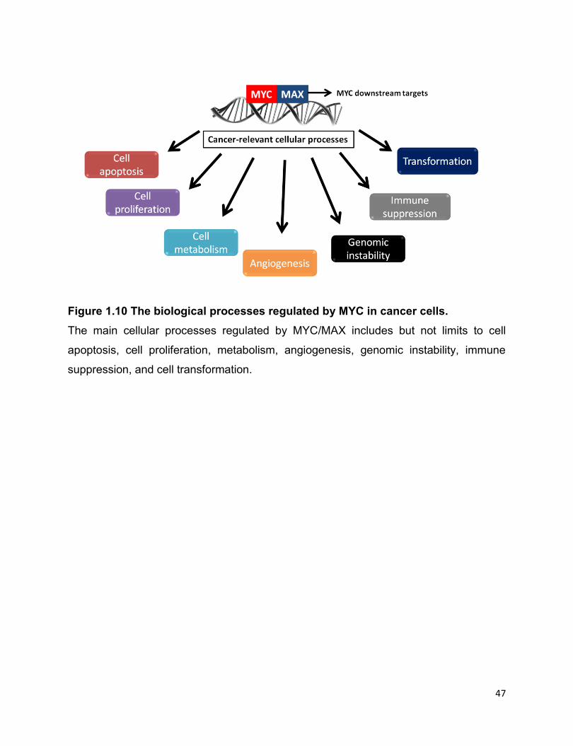
47
Figure 1.10 The biological processes regulated by MYC in cancer cells.
The main cellular processes regulated by MYC/MAX includes but not limits to cell
apoptosis, cell proliferation, metabolism, angiogenesis, genomic instability, immune
suppression, and cell transformation.

48
Figure 1.11 Phosphorylation and stabilization of MYC by ERK and GSK3β.
Extracellular signal-regulated kinase (ERK) phosphorylates MYC at Serine 62 (p-
MYCS62) and stabilizes MYC protein, which then heterodimerizes with MYC-associated
factor X (MAX) and acts as transcription factor. The p-MYCS62 can also be further
phosphorylated by glycogen synthase kinase 3β (GSK3β) at Threonine 58 (p-
MYCS62/T58). Upon binding of p-MYCS62/T58 with the ubiquitin ligase Fbw7-SCF, MYC
protein is subsequently ubiquitinated and degraded in proteasome pathway.
This figure has been modified from “Chang F, et al. Signaling transduction mediated by
the Ras/Raf/MEK/ERK pathway from cytokine receptors to transcription factors:
potential targeting for therapeutic interventions. Leukemia. 2013(7):1263-1293.”
1.6 Reactive oxygen species (ROS) in cancer
Reactive species, mainly consisting of ROS, are by-products generated by cell
respiration and metabolism that mostly happen in the mitochondria (291, 292).
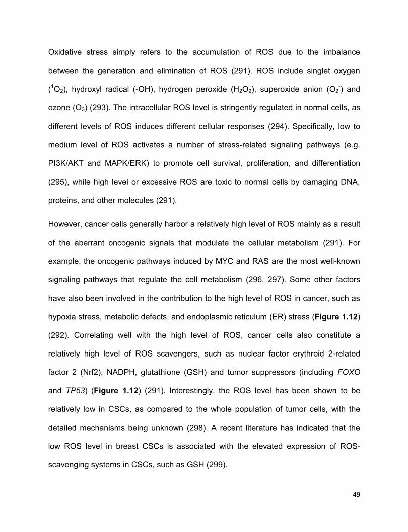
49
Oxidative stress simply refers to the accumulation of ROS due to the imbalance
between the generation and elimination of ROS (291). ROS include singlet oxygen
(1O2), hydroxyl radical (-OH), hydrogen peroxide (H2O2), superoxide anion (O2-) and
ozone (O3) (293). The intracellular ROS level is stringently regulated in normal cells, as
different levels of ROS induces different cellular responses (294). Specifically, low to
medium level of ROS activates a number of stress-related signaling pathways (e.g.
PI3K/AKT and MAPK/ERK) to promote cell survival, proliferation, and differentiation
(295), while high level or excessive ROS are toxic to normal cells by damaging DNA,
proteins, and other molecules (291).
However, cancer cells generally harbor a relatively high level of ROS mainly as a result
of the aberrant oncogenic signals that modulate the cellular metabolism (291). For
example, the oncogenic pathways induced by MYC and RAS are the most well-known
signaling pathways that regulate the cell metabolism (296, 297). Some other factors
have also been involved in the contribution to the high level of ROS in cancer, such as
hypoxia stress, metabolic defects, and endoplasmic reticulum (ER) stress (Figure 1.12)
(292). Correlating well with the high level of ROS, cancer cells also constitute a
relatively high level of ROS scavengers, such as nuclear factor erythroid 2-related
factor 2 (Nrf2), NADPH, glutathione (GSH) and tumor suppressors (including FOXO
and TP53) (Figure 1.12) (291). Interestingly, the ROS level has been shown to be
relatively low in CSCs, as compared to the whole population of tumor cells, with the
detailed mechanisms being unknown (298). A recent literature has indicated that the
low ROS level in breast CSCs is associated with the elevated expression of ROS-
scavenging systems in CSCs, such as GSH (299).
50
ROS of low to medium level in cancer cells has been reported to promote tumor cells
growth, proliferation and metastasis (292). Mahalingaiah et al reported that ER+ breast
cancer cell line MCF-7 cultured with chronic low doses of H2O2 has exhibited enhanced
tumorigenicity and chemoresistance, with upregulated gene expressions that are
related with pro-metastasis, such as vascular endothelia growth factor (VEGF) and
CD44 (300). It has been reported that low to medium level of ROS promotes
tumorigenesis by interacting and activating a number of oncogenic proteins or signaling
pathways in cancer cells, including MAPK/ERK, Wnt/β-catenin, PI3K/AKT, STAT3 (294),
all of which are related to tumor cell growth and proliferation (152, 291, 292, 294, 301,
302). One study revealed that ROS maintains the phosphorylation and oncogenic
activity of NPM-ALK kinase in ALK+ALCL cells, and pharmacological inhibition of ROS
generation downregulates the expression level of phosphorylated NPM-ALK, as well as
pSTAT3Y705, pERK1/2T202/Y204 and pAKTS473, all of which are downstream targets of
NPM-ALK (303).
51
Figure 1.12 Determination of cellular redox status by a balance between levels of
ROS inducers and ROS scavengers. The production of reactive oxygen species
(ROS) can be induced by hypoxia, metabolic defects, endoplasmic reticulum (ER)
stress and oncogenes. Conversely, ROS are eliminated by the activation of the
transcription factor nuclear factor erythroid 2-related factor 2 (NRF2), the production of
glutathione and nicotinamide adenine dinucleotide phosphate (NADPH), the activity of
tumour suppressors (such as breast cancer susceptibility 1 (BRCA1), p53,
phosphatase and tensin homolog (PTEN) and ataxia telangiectasia mutated (ATM) and
the action of dietary antioxidants.
Reprinted with permission from “Gorrini C, et al. Modulation of oxidative stress as an
anticancer strategy. Nat Rev Drug Discov. 2013. 12(12):931-947.” Copyright©2013
Nature Publishing Group. License number: 3976920022852
52
1.7 Thesis overview
1.7.1 Rationale
ALK+ALCL is an aggressive null/T-cell lymphoma most of which carries the oncogenic
fusion tyrosine kinase NPM-ALK (8). It is well-known that NPM-ALK is the key
oncogenic driver of this malignance by interacting and activating a number of molecular
adaptors involved in multiple oncogenic signaling pathways, such as JAK/STATs and
PI3K/AKT(55). Of these signaling pathways, NPM-ALK/STAT3 pathway is the one of
the most important pathways (91). It is reported that NPM-ALK/STAT3 not only
promotes the genes expression related with anti-apoptosis and cell proliferation but
also suppresses the expression of tumor suppressors or negative regulators of NPM-
ALK or STAT3 activity, such as Shp1 and STAT5A (45). In general, despite the fact that
ALK+ALCL is a favourable and curable disease and the pathobiology of ALK+ALCL
has been widely studied in the past 20 years (1), there is still a large portion (up to
~40%) of patients that develop either chemoresistance or tumor relapses after first-line
chemotherapy and these relapsed tumors always present in a more aggressive form
(55). Therefore, further understandings of the pathobiology of ALK+ALCL are still
warranted for designing novel therapeutic strategies. The scope of this thesis is to
further understand the oncogenic tyrosine kinase NPM-ALK and the pathobiology of
ALK+ALCL, and to focus on the tumor suppressor STAT1 and a highly tumorigenic cell
subpopulation derived from ALK+ALCL cells. In general, I will test the hypothesize that
the pathobiology of ALK+ALCL, such as tumorigenecity, chemoresistance and cancer
stemness, can be attributed to novel NPM-ALKꟷregulated biochemical defects as well
as signaling pathways that are not directly linked to NPM-ALK.

53
STAT1, a member of STATs family, is generally considered as a tumor suppressor in
various types of cancer (90). The decreased STAT1 expression has been observed in a
wide range of cancer (304). STAT1 is reported to suppress STAT3 transcriptional
activity in some cell models, and vice versa (90). Based on the facts that STAT3 is
constitutively active in ALK+ALCL and the reciprocal repression between STAT1 and
STAT3 activity in some cancer models(90, 91), I hypothesize that STAT1 signaling is
deficient in ALK+ALC; if this is the case, STAT1 functions as a tumor suppressor and
suppresses STAT3 activity in ALK+ALCL if it is overexpressed to a relatively high level.
Sox2 is one of the four iPS factors, and its biological significance in cancer cells has
been documented in a recent review (252). The Lai lab previous revealed two
phenotypically distinct cell population in ALK+ALCL cell lines based on their differential
response to a Sox2 reproterꟷSRR2 (239). Cells responsive to SRR2 (RR) are more
tumorigenic and chemoresistant than cells unresponsive to the reporter (RU) (239).
Nevertheless, the expression level of Sox2 in RU and RR cells is almost identical,
indicating other factors being involved in this context (239). MYC is another iPS factor,
which is also aberrantly expressed in ALK+ALCL (305). The reciprocal regulation of
MYC and Sox2 expression has been identified in breast cancer model (243). More
importantly, the preliminary unpublished data indicated that MYC is a putative factor
binding SRR2 reporter. In brief, here I hypothesize that MYC is one of the key factors
regulating the RU/RR dichotomy, and potentially regulates Sox2 transcriptional activity.
Oxidative stress has been shown to induce the conversion of RU cells to RR cells
derived from ER+ breast cancer cells and esophagus squamous cancer cells,
presenting a biological process of tumor plasticity (221, 306). However, tumor plasticity

54
is poorly understood and studied in haematological malignancies, including ALK+ALCL.
Using RU/RR cells model derived from ALK+ALCL, I hypothesize that RU cells can be
induced to convert to RR cells by oxidative stress.
1.7.2 Objectives
In Chapter 2, my objective is to evaluate the expression level of STAT1 in ALK+ALCL
cell lines and patient samples. If STAT1 is indeed downregulated in this type of
malignancy, then mechanisms behind this will be investigated. Next, two questions will
be explored, including whether STAT1 signaling pathway is functionally intact and
whether STAT1 is a potent tumor suppressor if its expression is restored in ALK+ALCL
cells. Lastly, my objective involves the study of whether STAT1 indeed suppresses
STAT3 transcriptional activity in ALK+ALCL.
In Chapter 3, my main objective is to delineate the key regulator(s) involved in RU/RR
dichotomy and the biochemical features of RR cells. In addition, the regulation between
this key regulator(s) and Sox2 will be investigated; furthermore, how this regulator(s) is
differentially expressed or regulated between RU and RR cells will be explored as well.
Lastly, whether the RR cells, according to the distinctive biochemical features of RR
cells, are existed in ALK+ALCL patient samples will be evaluated.
In Chapter 4, my objective is study “tumor plasticity” using RU/RR model derived from
ALK+ALCL. The question for me is whether RU can be induced to convert to RR cells
upon oxidative stress. If oxidative challenge can convert RU cells to RR cells, whether
these converted RR cells share similar biological and biochemical features with native
55
RR cells will be also determined. Furthermore, the molecular mechanisms underlining
this phenomenon will be investigated.
1.8 References
1. Hapgood G, Savage KJ. The biology and management of systemic anaplastic
large cell lymphoma. Blood. 2015;126(1):17-25.
2. Mora J, Filippa DA, Thaler HT, Polyak T, Cranor ML, Wollner N. Large cell non-
Hodgkin lymphoma of childhood: Analysis of 78 consecutive patients enrolled in 2
consecutive protocols at the Memorial Sloan-Kettering Cancer Center. Cancer.
2000;88(1):186-97.
3. Kinney MC, Higgins RA, Medina EA. Anaplastic large cell lymphoma: twenty-five
years of discovery. Archives of pathology & laboratory medicine. 2011;135(1):19-43.
4. Rizzardi GP, Tambussi G, Barcellini W, Capiluppi B, Clerici E, Maestra LL, et al.
Soluble CD30, tumour necrosis factor (TNF)-alpha, and TNF receptors in primary HIV-1
infection: relationship with HIV-1, RNA, clinical outcome and early antiviral therapy.
Journal of biological regulators and homeostatic agents. 1997;11(1-2):43-9.
5. Morris SW, Kirstein MN, Valentine MB, Dittmer KG, Shapiro DN, Saltman DL, et
al. Fusion of a kinase gene, ALK, to a nucleolar protein gene, NPM, in non-Hodgkin's
lymphoma. Science. 1994;263(5151):1281-4.
6. Xing X, Feldman AL. Anaplastic large cell lymphomas: ALK positive, ALK
negative, and primary cutaneous. Advances in anatomic pathology. 2015;22(1):29-49.
7. Piccaluga PP, Gazzola A, Mannu C, Agostinelli C, Bacci F, Sabattini E, et al.
Pathobiology of anaplastic large cell lymphoma. Adv Hematol. 2010:345053.

56
8. Ferreri AJ, Govi S, Pileri SA, Savage KJ. Anaplastic large cell lymphoma, ALK-
positive. Crit Rev Oncol Hematol. 2012;83(2):293-302.
9. Pileri S, Falini B, Delsol G, Stein H, Baglioni P, Poggi S, et al. Lymphohistiocytic
T-cell lymphoma (anaplastic large cell lymphoma CD30+/Ki-1 + with a high content of
reactive histiocytes). Histopathology. 1990;16(4):383-91.
10. Kinney MC, Collins RD, Greer JP, Whitlock JA, Sioutos N, Kadin ME. A small-
cell-predominant variant of primary Ki-1 (CD30)+ T-cell lymphoma. The American
journal of surgical pathology. 1993;17(9):859-68.
11. Nakamura S, Shiota M, Nakagawa A, Yatabe Y, Kojima M, Motoori T, et al.
Anaplastic large cell lymphoma: a distinct molecular pathologic entity: a reappraisal
with special reference to p80(NPM/ALK) expression. The American journal of surgical
pathology. 1997;21(12):1420-32.
12. Vassallo J, Lamant L, Brugieres L, Gaillard F, Campo E, Brousset P, et al. ALK-
positive anaplastic large cell lymphoma mimicking nodular sclerosis Hodgkin's
lymphoma: report of 10 cases. The American journal of surgical pathology.
2006;30(2):223-9.
13. Medeiros LJ, Elenitoba-Johnson KS. Anaplastic Large Cell Lymphoma.
American journal of clinical pathology. 2007;127(5):707-22.
14. Benharroch D, Meguerian-Bedoyan Z, Lamant L, Amin C, Brugieres L, Terrier-
Lacombe MJ, et al. ALK-positive lymphoma: a single disease with a broad spectrum of
morphology. Blood. 1998;91(6):2076-84.
15. Alzona M, Jack HM, Fisher RI, Ellis TM. CD30 defines a subset of activated
human T cells that produce IFN-gamma and IL-5 and exhibit enhanced B cell helper
activity. J Immunol. 1994;153(7):2861-7.

57
16. ten Berge RL, Snijdewint FG, von Mensdorff-Pouilly S, Poort-Keesom RJ,
Oudejans JJ, Meijer JW, et al. MUC1 (EMA) is preferentially expressed by ALK positive
anaplastic large cell lymphoma, in the normally glycosylated or only partly
hypoglycosylated form. Journal of clinical pathology. 2001;54(12):933-9.
17. Nath S, Mukherjee P. MUC1: a multifaceted oncoprotein with a key role in
cancer progression. Trends Mol Med. 2014;20(6):332-42.
18. Felgar RE, Salhany KE, Macon WR, Pietra GG, Kinney MC. The expression of
TIA-1+ cytolytic-type granules and other cytolytic lymphocyte-associated markers in
CD30+ anaplastic large cell lymphomas (ALCL): correlation with morphology,
immunophenotype, ultrastructure, and clinical features. Human pathology.
1999;30(2):228-36.
19. Krenacs L, Wellmann A, Sorbara L, Himmelmann AW, Bagdi E, Jaffe ES, et al.
Cytotoxic cell antigen expression in anaplastic large cell lymphomas of T- and null-cell
type and Hodgkin's disease: evidence for distinct cellular origin. Blood. 1997;89(3):980-
9.
20. Foss HD, Anagnostopoulos I, Araujo I, Assaf C, Demel G, Kummer JA, et al.
Anaplastic large-cell lymphomas of T-cell and null-cell phenotype express cytotoxic
molecules. Blood. 1996;88(10):4005-11.
21. Foss HD, Demel G, Anagnostopoulos I, Araujo I, Hummel M, Stein H. Uniform
expression of cytotoxic molecules in anaplastic large cell lymphoma of null/T cell
phenotype and in cell lines derived from anaplastic large cell lymphoma. Pathobiology.
1997;65(2):83-90.

58
22. Bonzheim I, Geissinger E, Roth S, Zettl A, Marx A, Rosenwald A, et al.
Anaplastic large cell lymphomas lack the expression of T-cell receptor molecules or
molecules of proximal T-cell receptor signaling. Blood. 2004;104(10):3358-60.
23. Kesler MV, Paranjape GS, Asplund SL, McKenna RW, Jamal S, Kroft SH.
Anaplastic large cell lymphoma: a flow cytometric analysis of 29 cases. American
journal of clinical pathology. 2007;128(2):314-22.
24. ten Berge RL, Oudejans JJ, Ossenkoppele GJ, Pulford K, Willemze R, Falini B,
et al. ALK expression in extranodal anaplastic large cell lymphoma favours systemic
disease with (primary) nodal involvement and a good prognosis and occurs before
dissemination. Journal of clinical pathology. 2000;53(6):445-50.
25. Vose J, Armitage J, Weisenburger D, International TCLP. International
peripheral T-cell and natural killer/T-cell lymphoma study: pathology findings and
clinical outcomes. Journal of clinical oncology : official journal of the American Society
of Clinical Oncology. 2008;26(25):4124-30.
26. Chiarle R, Voena C, Ambrogio C, Piva R, Inghirami G. The anaplastic lymphoma
kinase in the pathogenesis of cancer. Nature reviews Cancer. 2008;8(1):11-23.
27. Wellstein A. ALK receptor activation, ligands and therapeutic targeting in
glioblastoma and in other cancers. Frontiers in oncology. 2012;2:192.
28. Lee HH, Norris A, Weiss JB, Frasch M. Jelly belly protein activates the receptor
tyrosine kinase Alk to specify visceral muscle pioneers. Nature. 2003;425(6957):507-12.
29. Stoica GE, Kuo A, Powers C, Bowden ET, Sale EB, Riegel AT, et al. Midkine
binds to anaplastic lymphoma kinase (ALK) and acts as a growth factor for different cell
types. The Journal of biological chemistry. 2002;277(39):35990-8.

59
30. Bowden ET, Stoica GE, Wellstein A. Anti-apoptotic signaling of pleiotrophin
through its receptor, anaplastic lymphoma kinase. The Journal of biological chemistry.
2002;277(39):35862-8.
31. Moog-Lutz C, Degoutin J, Gouzi JY, Frobert Y, Brunet-de Carvalho N, Bureau J,
et al. Activation and inhibition of anaplastic lymphoma kinase receptor tyrosine kinase
by monoclonal antibodies and absence of agonist activity of pleiotrophin. The Journal of
biological chemistry. 2005;280(28):26039-48.
32. Mathivet T, Mazot P, Vigny M. In contrast to agonist monoclonal antibodies, both
C-terminal truncated form and full length form of Pleiotrophin failed to activate
vertebrate ALK (anaplastic lymphoma kinase)? Cellular signalling. 2007;19(12):2434-43.
33. Murray PB, Lax I, Reshetnyak A, Ligon GF, Lillquist JS, Natoli EJ, Jr., et al.
Heparin is an activating ligand of the orphan receptor tyrosine kinase ALK. Sci Signal.
2015;8(360):ra6.
34. Guan J, Umapathy G, Yamazaki Y, Wolfstetter G, Mendoza P, Pfeifer K, et al.
FAM150A and FAM150B are activating ligands for anaplastic lymphoma kinase. eLife.
2015;4:e09811.
35. Reshetnyak AV, Murray PB, Shi X, Mo ES, Mohanty J, Tome F, et al. Augmentor
alpha and beta (FAM150) are ligands of the receptor tyrosine kinases ALK and LTK:
Hierarchy and specificity of ligand-receptor interactions. Proceedings of the National
Academy of Sciences of the United States of America. 2015;112(52):15862-7.
36. Iwahara T, Fujimoto J, Wen D, Cupples R, Bucay N, Arakawa T, et al. Molecular
characterization of ALK, a receptor tyrosine kinase expressed specifically in the
nervous system. Oncogene. 1997;14(4):439-49.

60
37. Bilsland JG, Wheeldon A, Mead A, Znamenskiy P, Almond S, Waters KA, et al.
Behavioral and neurochemical alterations in mice deficient in anaplastic lymphoma
kinase suggest therapeutic potential for psychiatric indications.
Neuropsychopharmacology. 2008;33(3):685-700.
38. Lamant L, Pulford K, Bischof D, Morris SW, Mason DY, Delsol G, et al.
Expression of the ALK tyrosine kinase gene in neuroblastoma. Am J Pathol.
2000;156(5):1711-21.
39. Koyama-Nasu R, Haruta R, Nasu-Nishimura Y, Taniue K, Katou Y, Shirahige K,
et al. The pleiotrophin-ALK axis is required for tumorigenicity of glioblastoma stem cells.
Oncogene. 2014;33(17):2236-44.
40. Chen Y, Takita J, Choi YL, Kato M, Ohira M, Sanada M, et al. Oncogenic
mutations of ALK kinase in neuroblastoma. Nature. 2008;455(7215):971-4.
41. Wallace GCt, Dixon-Mah YN, Vandergrift WA, 3rd, Ray SK, Haar CP, Mittendorf
AM, et al. Targeting oncogenic ALK and MET: a promising therapeutic strategy for
glioblastoma. Metab Brain Dis. 2013;28(3):355-66.
42. Grisendi S, Mecucci C, Falini B, Pandolfi PP. Nucleophosmin and cancer. Nature
reviews Cancer. 2006;6(7):493-505.
43. Box JK, Paquet N, Adams MN, Boucher D, Bolderson E, O'Byrne KJ, et al.
Nucleophosmin: from structure and function to disease development. BMC molecular
biology. 2016;17(1):19.
44. Colombo E, Bonetti P, Lazzerini Denchi E, Martinelli P, Zamponi R, Marine JC,
et al. Nucleophosmin is required for DNA integrity and p19Arf protein stability.
Molecular and cellular biology. 2005;25(20):8874-86.

61
45. Lin CY, Tan BC, Liu H, Shih CJ, Chien KY, Lin CL, et al. Dephosphorylation of
nucleophosmin by PP1beta facilitates pRB binding and consequent E2F1-dependent
DNA repair. Molecular biology of the cell. 2010;21(24):4409-17.
46. Greenland C, Dastugue N, Touriol C, Lamant L, Delsol G, Brousset P.
Anaplastic large cell lymphoma with the t(2;5)(p23;q35) NPM/ALK chromosomal
translocation and duplication of the short arm of the non-translocated chromosome 2
involving the full length of the ALK gene. Journal of clinical pathology. 2001;54(2):152-4.
47. Falini B, Mecucci C, Tiacci E, Alcalay M, Rosati R, Pasqualucci L, et al.
Cytoplasmic nucleophosmin in acute myelogenous leukemia with a normal karyotype.
The New England journal of medicine. 2005;352(3):254-66.
48. Gogineni SK, Shah HO, Chester M, Lin JH, Garrison M, Alidina A, et al. Variant
complex translocations involving chromosomes 1, 9, 9, 15 and 17 in acute
promyelocytic leukemia without RAR alpha/PML gene fusion rearrangement. Leukemia.
1997;11(4):514-8.
49. Drexler HG, Gignac SM, von Wasielewski R, Werner M, Dirks WG. Pathobiology
of NPM-ALK and variant fusion genes in anaplastic large cell lymphoma and other
lymphomas. Leukemia. 2000;14(9):1533-59.
50. Giuriato S, Turner SD. Twenty years of modelling NPM-ALK-induced
lymphomagenesis. Frontiers in bioscience. 2015;7:236-47.
51. Lai R, Ingham RJ. The pathobiology of the oncogenic tyrosine kinase NPM-ALK:
a brief update. Therapeutic advances in hematology. 2013;4(2):119-31.
52. Hallberg B, Palmer RH. Mechanistic insight into ALK receptor tyrosine kinase in
human cancer biology. Nature reviews Cancer. 2013;13(10):685-700.
62
53. Zhang Q, Raghunath PN, Xue L, Majewski M, Carpentieri DF, Odum N, et al.
Multilevel dysregulation of STAT3 activation in anaplastic lymphoma kinase-positive
T/null-cell lymphoma. J Immunol. 2002;168(1):466-74.
54. Slupianek A, Nieborowska-Skorska M, Hoser G, Morrione A, Majewski M, Xue L,
et al. Role of phosphatidylinositol 3-kinase-Akt pathway in nucleophosmin/anaplastic
lymphoma kinase-mediated lymphomagenesis. Cancer research. 2001;61(5):2194-9.
55. Amin HM, Lai R. Pathobiology of ALK+ anaplastic large-cell lymphoma. Blood.
2007;110(7):2259-67.
56. Pearson JD, Lee JK, Bacani JT, Lai R, Ingham RJ. NPM-ALK: The Prototypic
Member of a Family of Oncogenic Fusion Tyrosine Kinases. J Signal Transduct.
2012;2012:123253.
57. Tabbo F, Barreca A, Piva R, Inghirami G. ALK Signaling and Target Therapy in
Anaplastic Large Cell Lymphoma. Frontiers in oncology. 2012;2:41.
58. Klempner SJ, Cohen DW, Costa DB. ALK translocation in non-small cell lung
cancer with adenocarcinoma and squamous cell carcinoma markers. Journal of
thoracic oncology : official publication of the International Association for the Study of
Lung Cancer. 2011;6(8):1439-40.
59. Aisner DL, Nguyen TT, Paskulin DD, Le AT, Haney J, Schulte N, et al. ROS1
and ALK fusions in colorectal cancer, with evidence of intratumoral heterogeneity for
molecular drivers. Molecular cancer research : MCR. 2014;12(1):111-8.
60. Lange K, Uckert W, Blankenstein T, Nadrowitz R, Bittner C, Renauld JC, et al.
Overexpression of NPM-ALK induces different types of malignant lymphomas in IL-9
transgenic mice. Oncogene. 2003;22(4):517-27.
63
61. Zhang Q, Wei F, Wang HY, Liu X, Roy D, Xiong QB, et al. The potent oncogene
NPM-ALK mediates malignant transformation of normal human CD4(+) T lymphocytes.
The American journal of pathology. 2013;183(6):1971-80.
62. Jager R, Hahne J, Jacob A, Egert A, Schenkel J, Wernert N, et al. Mice
transgenic for NPM-ALK develop non-Hodgkin lymphomas. Anticancer research.
2005;25(5):3191-6.
63. Chiarle R, Gong JZ, Guasparri I, Pesci A, Cai J, Liu J, et al. NPM-ALK
transgenic mice spontaneously develop T-cell lymphomas and plasma cell tumors.
Blood. 2003;101(5):1919-27.
64. Turner SD, Tooze R, Maclennan K, Alexander DR. Vav-promoter regulated
oncogenic fusion protein NPM-ALK in transgenic mice causes B-cell lymphomas with
hyperactive Jun kinase. Oncogene. 2003;22(49):7750-61.
65. Turner SD, Merz H, Yeung D, Alexander DR. CD2 promoter regulated
nucleophosmin-anaplastic lymphoma kinase in transgenic mice causes B lymphoid
malignancy. Anticancer research. 2006;26(5A):3275-9.
66. Giuriato S, Foisseau M, Dejean E, Felsher DW, Al Saati T, Demur C, et al.
Conditional TPM3-ALK and NPM-ALK transgenic mice develop reversible ALK-positive
early B-cell lymphoma/leukemia. Blood. 2010;115(20):4061-70.
67. Malcolm TI, Villarese P, Fairbairn CJ, Lamant L, Trinquand A, Hook CE, et al.
Anaplastic large cell lymphoma arises in thymocytes and requires transient TCR
expression for thymic egress. Nature communications. 2016;7:10087.
68. Maes B, Vanhentenrijk V, Wlodarska I, Cools J, Peeters B, Marynen P, et al. The
NPM-ALK and the ATIC-ALK fusion genes can be detected in non-neoplastic cells. The
American journal of pathology. 2001;158(6):2185-93.
64
69. Crockett DK, Lin Z, Elenitoba-Johnson KS, Lim MS. Identification of NPM-ALK
interacting proteins by tandem mass spectrometry. Oncogene. 2004;23(15):2617-29.
70. Voena C, Conte C, Ambrogio C, Boeri Erba E, Boccalatte F, Mohammed S, et al.
The tyrosine phosphatase Shp2 interacts with NPM-ALK and regulates anaplastic
lymphoma cell growth and migration. Cancer research. 2007;67(9):4278-86.
71. Galietta A, Gunby RH, Redaelli S, Stano P, Carniti C, Bachi A, et al. NPM/ALK
binds and phosphorylates the RNA/DNA-binding protein PSF in anaplastic large-cell
lymphoma. Blood. 2007;110(7):2600-9.
72. Wu F, Wang P, Young LC, Lai R, Li L. Proteome-wide identification of novel
binding partners to the oncogenic fusion gene protein, NPM-ALK, using tandem affinity
purification and mass spectrometry. The American journal of pathology.
2009;174(2):361-70.
73. Murga-Zamalloa CA, Mendoza-Reinoso V, Sahasrabuddhe AA, Rolland D,
Hwang SR, McDonnell SR, et al. NPM-ALK phosphorylates WASp Y102 and
contributes to oncogenesis of anaplastic large cell lymphoma. Oncogene. 2016.
74. Marzec M, Kasprzycka M, Liu X, El-Salem M, Halasa K, Raghunath PN, et al.
Oncogenic tyrosine kinase NPM/ALK induces activation of the rapamycin-sensitive
mTOR signaling pathway. Oncogene. 2007;26(38):5606-14.
75. Bai RY, Dieter P, Peschel C, Morris SW, Duyster J. Nucleophosmin-anaplastic
lymphoma kinase of large-cell anaplastic lymphoma is a constitutively active tyrosine
kinase that utilizes phospholipase C-gamma to mediate its mitogenicity. Molecular and
cellular biology. 1998;18(12):6951-61.
65
76. Zamo A, Chiarle R, Piva R, Howes J, Fan Y, Chilosi M, et al. Anaplastic
lymphoma kinase (ALK) activates Stat3 and protects hematopoietic cells from cell
death. Oncogene. 2002;21(7):1038-47.
77. Yu H, Lee H, Herrmann A, Buettner R, Jove R. Revisiting STAT3 signalling in
cancer: new and unexpected biological functions. Nature reviews Cancer.
2014;14(11):736-46.
78. Johnston PA, Grandis JR. STAT3 signaling: anticancer strategies and
challenges. Molecular interventions. 2011;11(1):18-26.
79. Khoury JD, Medeiros LJ, Rassidakis GZ, Yared MA, Tsioli P, Leventaki V, et al.
Differential expression and clinical significance of tyrosine-phosphorylated STAT3 in
ALK+ and ALK- anaplastic large cell lymphoma. Clinical cancer research : an official
journal of the American Association for Cancer Research. 2003;9(10 Pt 1):3692-9.
80. Kirken RA, Rui H, Malabarba MG, Howard OM, Kawamura M, O'Shea JJ, et al.
Activation of JAK3, but not JAK1, is critical for IL-2-induced proliferation and STAT5
recruitment by a COOH-terminal region of the IL-2 receptor beta-chain. Cytokine.
1995;7(7):689-700.
81. Fenghao X, Saxon A, Nguyen A, Ke Z, Diaz-Sanchez D, Nel A. Interleukin 4
activates a signal transducer and activator of transcription (Stat) protein which interacts
with an interferon-gamma activation site-like sequence upstream of the I epsilon exon
in a human B cell line. Evidence for the involvement of Janus kinase 3 and interleukin-4
Stat. The Journal of clinical investigation. 1995;96(2):907-14.
82. Malabarba MG, Rui H, Deutsch HH, Chung J, Kalthoff FS, Farrar WL, et al.
Interleukin-13 is a potent activator of JAK3 and STAT6 in cells expressing interleukin-2

66
receptor-gamma and interleukin-4 receptor-alpha. The Biochemical journal. 1996;319
( Pt 3):865-72.
83. Ortmann RA, Cheng T, Visconti R, Frucht DM, O'Shea JJ. Janus kinases and
signal transducers and activators of transcription: their roles in cytokine signaling,
development and immunoregulation. Arthritis research. 2000;2(1):16-32.
84. Dien Bard J, Gelebart P, Anand M, Zak Z, Hegazy SA, Amin HM, et al. IL-21
contributes to JAK3/STAT3 activation and promotes cell growth in ALK-positive
anaplastic large cell lymphoma. The American journal of pathology. 2009;175(2):825-
34.
85. Demoulin JB, Uyttenhove C, Van Roost E, DeLestre B, Donckers D, Van Snick J,
et al. A single tyrosine of the interleukin-9 (IL-9) receptor is required for STAT activation,
antiapoptotic activity, and growth regulation by IL-9. Molecular and cellular biology.
1996;16(9):4710-6.
86. Amin HM, Medeiros LJ, Ma Y, Feretzaki M, Das P, Leventaki V, et al. Inhibition
of JAK3 induces apoptosis and decreases anaplastic lymphoma kinase activity in
anaplastic large cell lymphoma. Oncogene. 2003;22(35):5399-407.
87. Qiu L, Lai R, Lin Q, Lau E, Thomazy DM, Calame D, et al. Autocrine release of
interleukin-9 promotes Jak3-dependent survival of ALK+ anaplastic large-cell
lymphoma cells. Blood. 2006;108(7):2407-15.
88. Amin HM, Lin Q, Lai R. Jak3 contributes to the activation of ALK and Stat3 in
ALK(+) anaplastic large cell lymphoma. Laboratory investigation; a journal of technical
methods and pathology. 2006;86(4):417-9; author reply 20-1.
67
89. Bard JD, Gelebart P, Anand M, Amin HM, Lai R. Aberrant expression of IL-22
receptor 1 and autocrine IL-22 stimulation contribute to tumorigenicity in ALK+
anaplastic large cell lymphoma. Leukemia. 2008;22(8):1595-603.
90. Avalle L, Pensa S, Regis G, Novelli F, Poli V. STAT1 and STAT3 in
tumorigenesis: A matter of balance. Jak-Stat. 2012;1(2):65-72.
91. Chiarle R, Simmons WJ, Cai H, Dhall G, Zamo A, Raz R, et al. Stat3 is required
for ALK-mediated lymphomagenesis and provides a possible therapeutic target. Nature
medicine. 2005;11(6):623-9.
92. Carpenter RL, Lo HW. STAT3 Target Genes Relevant to Human Cancers.
Cancers. 2014;6(2):897-925.
93. Bowman T, Garcia R, Turkson J, Jove R. STATs in oncogenesis. Oncogene.
2000;19(21):2474-88.
94. Buettner R, Mora LB, Jove R. Activated STAT signaling in human tumors
provides novel molecular targets for therapeutic intervention. Clinical cancer research :
an official journal of the American Association for Cancer Research. 2002;8(4):945-54.
95. Thomas SJ, Snowden JA, Zeidler MP, Danson SJ. The role of JAK/STAT
signalling in the pathogenesis, prognosis and treatment of solid tumours. British journal
of cancer. 2015;113(3):365-71.
96. Amin HM, McDonnell TJ, Ma Y, Lin Q, Fujio Y, Kunisada K, et al. Selective
inhibition of STAT3 induces apoptosis and G(1) cell cycle arrest in ALK-positive
anaplastic large cell lymphoma. Oncogene. 2004;23(32):5426-34.
97. Wu C, Molavi O, Zhang H, Gupta N, Alshareef A, Bone KM, et al. STAT1 is
phosphorylated and downregulated by the oncogenic tyrosine kinase NPM-ALK in ALK-
positive anaplastic large-cell lymphoma. Blood. 2015;126(3):336-45.
68
98. Piva R, Agnelli L, Pellegrino E, Todoerti K, Grosso V, Tamagno I, et al. Gene
expression profiling uncovers molecular classifiers for the recognition of anaplastic
large-cell lymphoma within peripheral T-cell neoplasms. Journal of clinical oncology :
official journal of the American Society of Clinical Oncology. 2010;28(9):1583-90.
99. Zhang Q, Wang HY, Marzec M, Raghunath PN, Nagasawa T, Wasik MA.
STAT3- and DNA methyltransferase 1-mediated epigenetic silencing of SHP-1 tyrosine
phosphatase tumor suppressor gene in malignant T lymphocytes. Proceedings of the
National Academy of Sciences of the United States of America. 2005;102(19):6948-53.
100. Zhang Q, Wang HY, Woetmann A, Raghunath PN, Odum N, Wasik MA. STAT3
induces transcription of the DNA methyltransferase 1 gene (DNMT1) in malignant T
lymphocytes. Blood. 2006;108(3):1058-64.
101. Honorat JF, Ragab A, Lamant L, Delsol G, Ragab-Thomas J. SHP1 tyrosine
phosphatase negatively regulates NPM-ALK tyrosine kinase signaling. Blood.
2006;107(10):4130-8.
102. Han Y, Amin HM, Franko B, Frantz C, Shi X, Lai R. Loss of SHP1 enhances
JAK3/STAT3 signaling and decreases proteosome degradation of JAK3 and NPM-ALK
in ALK+ anaplastic large-cell lymphoma. Blood. 2006;108(8):2796-803.
103. Zhang Q, Wang HY, Liu X, Wasik MA. STAT5A is epigenetically silenced by the
tyrosine kinase NPM1-ALK and acts as a tumor suppressor by reciprocally inhibiting
NPM1-ALK expression. Nature medicine. 2007;13(11):1341-8.
104. Nieborowska-Skorska M, Slupianek A, Xue L, Zhang Q, Raghunath PN, Hoser G,
et al. Role of signal transducer and activator of transcription 5 in nucleophosmin/
anaplastic lymphoma kinase-mediated malignant transformation of lymphoid cells.
Cancer research. 2001;61(17):6517-23.
69
105. Kim HS, Lee MS. STAT1 as a key modulator of cell death. Cellular signalling.
2007;19(3):454-65.
106. Decker T, Muller M, Stockinger S. The yin and yang of type I interferon activity in
bacterial infection. Nature reviews Immunology. 2005;5(9):675-87.
107. Ramana CV, Chatterjee-Kishore M, Nguyen H, Stark GR. Complex roles of Stat1
in regulating gene expression. Oncogene. 2000;19(21):2619-27.
108. Zetter BR, Mangold U. Ubiquitin-independent degradation and its implication in
cancer. Future oncology. 2005;1(5):567-70.
109. Chen D, Dou QP. The ubiquitin-proteasome system as a prospective molecular
target for cancer treatment and prevention. Current protein & peptide science.
2010;11(6):459-70.
110. Lawrence DW, Kornbluth J. E3 ubiquitin ligase NKLAM ubiquitinates STAT1 and
positively regulates STAT1-mediated transcriptional activity. Cellular signalling.
2016;28(12):1833-41.
111. Ren Y, Zhao P, Liu J, Yuan Y, Cheng Q, Zuo Y, et al. Deubiquitinase USP2a
Sustains Interferons Antiviral Activity by Restricting Ubiquitination of Activated STAT1 in
the Nucleus. PLoS pathogens. 2016;12(7):e1005764.
112. Soond SM, Townsend PA, Barry SP, Knight RA, Latchman DS, Stephanou A.
ERK and the F-box protein betaTRCP target STAT1 for degradation. The Journal of
biological chemistry. 2008;283(23):16077-83.
113. Yuan C, Qi J, Zhao X, Gao C. Smurf1 protein negatively regulates interferon-
gamma signaling through promoting STAT1 protein ubiquitination and degradation. The
Journal of biological chemistry. 2012;287(21):17006-15.

70
114. Gao C, Mi Z, Guo H, Kuo PC. Osteopontin regulates ubiquitin-dependent
degradation of Stat1 in murine mammary epithelial tumor cells. Neoplasia.
2007;9(9):699-706.
115. Kim TK, Maniatis T. Regulation of interferon-gamma-activated STAT1 by the
ubiquitin-proteasome pathway. Science. 1996;273(5282):1717-9.
116. Wang Y, Ren Z, Tao D, Tilwalli S, Goswami R, Balabanov R. STAT1/IRF-1
signaling pathway mediates the injurious effect of interferon-gamma on oligodendrocyte
progenitor cells. Glia. 2010;58(2):195-208.
117. Sironi JJ, Ouchi T. STAT1-induced apoptosis is mediated by caspases 2, 3, and
7. The Journal of biological chemistry. 2004;279(6):4066-74.
118. Choi EA, Lei H, Maron DJ, Wilson JM, Barsoum J, Fraker DL, et al. Stat1-
dependent induction of tumor necrosis factor-related apoptosis-inducing ligand and the
cell-surface death signaling pathway by interferon beta in human cancer cells. Cancer
research. 2003;63(17):5299-307.
119. Ning Y, Riggins RB, Mulla JE, Chung H, Zwart A, Clarke R. IFNgamma restores
breast cancer sensitivity to fulvestrant by regulating STAT1, IFN regulatory factor 1, NF-
kappaB, BCL2 family members, and signaling to caspase-dependent apoptosis.
Molecular cancer therapeutics. 2010;9(5):1274-85.
120. Stephanou A, Brar BK, Knight RA, Latchman DS. Opposing actions of STAT-1
and STAT-3 on the Bcl-2 and Bcl-x promoters. Cell death and differentiation.
2000;7(3):329-30.
121. Hong F, Jaruga B, Kim WH, Radaeva S, El-Assal ON, Tian Z, et al. Opposing
roles of STAT1 and STAT3 in T cell–mediated hepatitis: regulation by SOCS. Journal of
Clinical Investigation. 2002;110(10):1503-13.
71
122. Dimberg LY, Dimberg A, Ivarsson K, Fryknas M, Rickardson L, Tobin G, et al.
Stat1 activation attenuates IL-6 induced Stat3 activity but does not alter apoptosis
sensitivity in multiple myeloma. BMC cancer. 2012;12:318.
123. Meraz MA, White JM, Sheehan KC, Bach EA, Rodig SJ, Dighe AS, et al.
Targeted disruption of the Stat1 gene in mice reveals unexpected physiologic specificity
in the JAK-STAT signaling pathway. Cell. 1996;84(3):431-42.
124. Kaplan DH, Shankaran V, Dighe AS, Stockert E, Aguet M, Old LJ, et al.
Demonstration of an interferon gamma-dependent tumor surveillance system in
immunocompetent mice. Proceedings of the National Academy of Sciences of the
United States of America. 1998;95(13):7556-61.
125. Chan SR, Vermi W, Luo J, Lucini L, Rickert C, Fowler AM, et al. STAT1-deficient
mice spontaneously develop estrogen receptor alpha-positive luminal mammary
carcinomas. Breast cancer research : BCR. 2012;14(1):R16.
126. Laimer K, Spizzo G, Obrist P, Gastl G, Brunhuber T, Schafer G, et al. STAT1
activation in squamous cell cancer of the oral cavity: a potential predictive marker of
response to adjuvant chemotherapy. Cancer. 2007;110(2):326-33.
127. Wong LH, Krauer KG, Hatzinisiriou I, Estcourt MJ, Hersey P, Tam ND, et al.
Interferon-resistant human melanoma cells are deficient in ISGF3 components, STAT1,
STAT2, and p48-ISGF3gamma. The Journal of biological chemistry.
1997;272(45):28779-85.
128. Kovarik J, Boudny V, Kocak I, Lauerova L, Fait V, Vagundova M. Malignant
melanoma associates with deficient IFN-induced STAT 1 phosphorylation. International
journal of molecular medicine. 2003;12(3):335-40.
72
129. Sun WH, Pabon C, Alsayed Y, Huang PP, Jandeska S, Uddin S, et al.
Interferon-alpha resistance in a cutaneous T-cell lymphoma cell line is associated with
lack of STAT1 expression. Blood. 1998;91(2):570-6.
130. Widschwendter A, Tonko-Geymayer S, Welte T, Daxenbichler G, Marth C,
Doppler W. Prognostic significance of signal transducer and activator of transcription 1
activation in breast cancer. Clinical cancer research : an official journal of the American
Association for Cancer Research. 2002;8(10):3065-74.
131. Zhang Y, Molavi O, Su M, Lai R. The clinical and biological significance of
STAT1 in esophageal squamous cell carcinoma. BMC cancer. 2014;14:791.
132. Zhang Y, Zhang Y, Yun H, Lai R, Su M. Correlation of STAT1 with apoptosis and
cell-cycle markers in esophageal squamous cell carcinoma. PloS one.
2014;9(12):e113928.
133. Xi S, Dyer KF, Kimak M, Zhang Q, Gooding WE, Chaillet JR, et al. Decreased
STAT1 expression by promoter methylation in squamous cell carcinogenesis. Journal
of the National Cancer Institute. 2006;98(3):181-9.
134. Khodarev N, Ahmad R, Rajabi H, Pitroda S, Kufe T, McClary C, et al.
Cooperativity of the MUC1 oncoprotein and STAT1 pathway in poor prognosis human
breast cancer. Oncogene. 2010;29(6):920-9.
135. Sanda T, Tyner JW, Gutierrez A, Ngo VN, Glover J, Chang BH, et al. TYK2-
STAT1-BCL2 pathway dependence in T-cell acute lymphoblastic leukemia. Cancer
discovery. 2013;3(5):564-77.
136. Schultz J, Koczan D, Schmitz U, Ibrahim SM, Pilch D, Landsberg J, et al. Tumor-
promoting role of signal transducer and activator of transcription (Stat)1 in late-stage
melanoma growth. Clinical & experimental metastasis. 2010;27(3):133-40.
73
137. Schlessinger J. Phospholipase Cgamma activation and phosphoinositide
hydrolysis are essential for embryonal development. Proceedings of the National
Academy of Sciences of the United States of America. 1997;94(7):2798-9.
138. Park JB, Lee CS, Jang JH, Ghim J, Kim YJ, You S, et al. Phospholipase
signalling networks in cancer. Nature reviews Cancer. 2012;12(11):782-92.
139. Vega F, Medeiros LJ, Leventaki V, Atwell C, Cho-Vega JH, Tian L, et al.
Activation of mammalian target of rapamycin signaling pathway contributes to tumor
cell survival in anaplastic lymphoma kinase-positive anaplastic large cell lymphoma.
Cancer research. 2006;66(13):6589-97.
140. Vanhaesebroeck B, Stephens L, Hawkins P. PI3K signalling: the path to
discovery and understanding. Nature reviews Molecular cell biology. 2012;13(3):195-
203.
141. Hemmings BA, Restuccia DF. PI3K-PKB/Akt pathway. Cold Spring Harbor
perspectives in biology. 2012;4(9):a011189.
142. Song G, Ouyang G, Bao S. The activation of Akt/PKB signaling pathway and cell
survival. Journal of cellular and molecular medicine. 2005;9(1):59-71.
143. Datta SR, Dudek H, Tao X, Masters S, Fu H, Gotoh Y, et al. Akt phosphorylation
of BAD couples survival signals to the cell-intrinsic death machinery. Cell.
1997;91(2):231-41.
144. Romashkova JA, Makarov SS. NF-kappaB is a target of AKT in anti-apoptotic
PDGF signalling. Nature. 1999;401(6748):86-90.
145. Riobo NA, Lu K, Ai X, Haines GM, Emerson CP, Jr. Phosphoinositide 3-kinase
and Akt are essential for Sonic Hedgehog signaling. Proceedings of the National
Academy of Sciences of the United States of America. 2006;103(12):4505-10.
74
146. Singh RR, Cho-Vega JH, Davuluri Y, Ma S, Kasbidi F, Milito C, et al. Sonic
hedgehog signaling pathway is activated in ALK-positive anaplastic large cell
lymphoma. Cancer research. 2009;69(6):2550-8.
147. Wang SJ, Omori N, Li F, Jin G, Zhang WR, Hamakawa Y, et al. Potentiation of
Akt and suppression of caspase-9 activations by electroacupuncture after transient
middle cerebral artery occlusion in rats. Neuroscience letters. 2002;331(2):115-8.
148. Suhara T, Kim HS, Kirshenbaum LA, Walsh K. Suppression of Akt signaling
induces Fas ligand expression: involvement of caspase and Jun kinase activation in
Akt-mediated Fas ligand regulation. Molecular and cellular biology. 2002;22(2):680-91.
149. Kawano T, Morioka M, Yano S, Hamada J, Ushio Y, Miyamoto E, et al.
Decreased akt activity is associated with activation of forkhead transcription factor after
transient forebrain ischemia in gerbil hippocampus. Journal of cerebral blood flow and
metabolism : official journal of the International Society of Cerebral Blood Flow and
Metabolism. 2002;22(8):926-34.
150. Santo EE, Stroeken P, Sluis PV, Koster J, Versteeg R, Westerhout EM. FOXO3a
is a major target of inactivation by PI3K/AKT signaling in aggressive neuroblastoma.
Cancer research. 2013;73(7):2189-98.
151. Das TP, Suman S, Alatassi H, Ankem MK, Damodaran C. Inhibition of AKT
promotes FOXO3a-dependent apoptosis in prostate cancer. Cell death & disease.
2016;7:e2111.
152. McCubrey JA, Steelman LS, Chappell WH, Abrams SL, Wong EW, Chang F, et
al. Roles of the Raf/MEK/ERK pathway in cell growth, malignant transformation and
drug resistance. Biochimica et biophysica acta. 2007;1773(8):1263-84.

75
153. Chang F, Steelman LS, Lee JT, Shelton JG, Navolanic PM, Blalock WL, et al.
Signal transduction mediated by the Ras/Raf/MEK/ERK pathway from cytokine
receptors to transcription factors: potential targeting for therapeutic intervention.
Leukemia. 2003;17(7):1263-93.
154. Marzec M, Kasprzycka M, Liu X, Raghunath PN, Wlodarski P, Wasik MA.
Oncogenic tyrosine kinase NPM/ALK induces activation of the MEK/ERK signaling
pathway independently of c-Raf. Oncogene. 2007;26(6):813-21.
155. Watanabe M, Itoh K, Togano T, Kadin ME, Watanabe T, Higashihara M, et al.
Ets-1 activates overexpression of JunB and CD30 in Hodgkin's lymphoma and
anaplastic large-cell lymphoma. The American journal of pathology. 2012;180(2):831-8.
156. Watanabe M, Sasaki M, Itoh K, Higashihara M, Umezawa K, Kadin ME, et al.
JunB induced by constitutive CD30-extracellular signal-regulated kinase 1/2 mitogen-
activated protein kinase signaling activates the CD30 promoter in anaplastic large cell
lymphoma and reed-sternberg cells of Hodgkin lymphoma. Cancer research.
2005;65(17):7628-34.
157. Staber PB, Vesely P, Haq N, Ott RG, Funato K, Bambach I, et al. The
oncoprotein NPM-ALK of anaplastic large-cell lymphoma induces JUNB transcription
via ERK1/2 and JunB translation via mTOR signaling. Blood. 2007;110(9):3374-83.
158. Gao J, Yin M, Zhu Y, Gu L, Zhang Y, Li Q, et al. Prognostic significance and
therapeutic potential of the activation of anaplastic lymphoma kinase/protein kinase
B/mammalian target of rapamycin signaling pathway in anaplastic large cell lymphoma.
BMC cancer. 2013;13:471.
76
159. Hansel DE, Platt E, Orloff M, Harwalker J, Sethu S, Hicks JL, et al. Mammalian
target of rapamycin (mTOR) regulates cellular proliferation and tumor growth in
urothelial carcinoma. The American journal of pathology. 2010;176(6):3062-72.
160. Laplante M, Sabatini DM. mTOR signaling in growth control and disease. Cell.
2012;149(2):274-93.
161. Babchia N, Calipel A, Mouriaux F, Faussat AM, Mascarelli F. The PI3K/Akt and
mTOR/P70S6K signaling pathways in human uveal melanoma cells: interaction with B-
Raf/ERK. Investigative ophthalmology & visual science. 2010;51(1):421-9.
162. Fanale MA, Horwitz SM, Forero-Torres A, Bartlett NL, Advani RH, Pro B, et al.
Brentuximab vedotin in the front-line treatment of patients with CD30+ peripheral T-cell
lymphomas: results of a phase I study. Journal of clinical oncology : official journal of
the American Society of Clinical Oncology. 2014;32(28):3137-43.
163. George SK, Vishwamitra D, Manshouri R, Shi P, Amin HM. The ALK inhibitor
ASP3026 eradicates NPM-ALK(+) T-cell anaplastic large-cell lymphoma in vitro and in
a systemic xenograft lymphoma model. Oncotarget. 2014;5(14):5750-63.
164. Galkin AV, Melnick JS, Kim S, Hood TL, Li N, Li L, et al. Identification of NVP-
TAE684, a potent, selective, and efficacious inhibitor of NPM-ALK. Proceedings of the
National Academy of Sciences of the United States of America. 2007;104(1):270-5.
165. Kothari S, Ud-Din N, Lisi M, Coyle T. Crizotinib in anaplastic lymphoma kinase-
positive anaplastic large cell lymphoma in the setting of renal insufficiency: a case
report. Journal of medical case reports. 2016;10:176.
166. Wu J, Savooji J, Liu D. Second- and third-generation ALK inhibitors for non-small
cell lung cancer. Journal of hematology & oncology. 2016;9:19.
77
167. Sullivan I, Planchard D. ALK inhibitors in non-small cell lung cancer: the latest
evidence and developments. Therapeutic advances in medical oncology. 2016;8(1):32-
47.
168. Farina F, Gambacorti-Passerini C. ALK inhibitors for clinical use in cancer
therapy. Front Biosci (Elite Ed). 2016;8:46-60.
169. Kazandjian D, Blumenthal GM, Chen HY, He K, Patel M, Justice R, et al. FDA
approval summary: crizotinib for the treatment of metastatic non-small cell lung cancer
with anaplastic lymphoma kinase rearrangements. The oncologist. 2014;19(10):e5-11.
170. Mosse YP, Lim MS, Voss SD, Wilner K, Ruffner K, Laliberte J, et al. Safety and
activity of crizotinib for paediatric patients with refractory solid tumours or anaplastic
large-cell lymphoma: a Children's Oncology Group phase 1 consortium study. The
Lancet Oncology. 2013;14(6):472-80.
171. Ou SH, Bartlett CH, Mino-Kenudson M, Cui J, Iafrate AJ. Crizotinib for the
treatment of ALK-rearranged non-small cell lung cancer: a success story to usher in the
second decade of molecular targeted therapy in oncology. The oncologist.
2012;17(11):1351-75.
172. Zdzalik D, Dymek B, Grygielewicz P, Gunerka P, Bujak A, Lamparska-Przybysz
M, et al. Activating mutations in ALK kinase domain confer resistance to structurally
unrelated ALK inhibitors in NPM-ALK-positive anaplastic large-cell lymphoma. Journal
of cancer research and clinical oncology. 2014;140(4):589-98.
173. Ceccon M, Mologni L, Bisson W, Scapozza L, Gambacorti-Passerini C.
Crizotinib-resistant NPM-ALK mutants confer differential sensitivity to unrelated Alk
inhibitors. Molecular cancer research : MCR. 2013;11(2):122-32.
78
174. Merkel O, Hamacher F, Sifft E, Kenner L, Greil R. Novel therapeutic options in
anaplastic large cell lymphoma: molecular targets and immunological tools. Molecular
cancer therapeutics. 2011;10(7):1127-36.
175. MacDonald BT, Tamai K, He X. Wnt/beta-catenin signaling: components,
mechanisms, and diseases. Developmental cell. 2009;17(1):9-26.
176. Anastas JN, Moon RT. WNT signalling pathways as therapeutic targets in cancer.
Nature reviews Cancer. 2013;13(1):11-26.
177. Duchartre Y, Kim YM, Kahn M. The Wnt signaling pathway in cancer. Critical
reviews in oncology/hematology. 2016;99:141-9.
178. Moon RT, Kohn AD, De Ferrari GV, Kaykas A. WNT and beta-catenin signalling:
diseases and therapies. Nature reviews Genetics. 2004;5(9):691-701.
179. Bellei B, Pacchiarotti A, Perez M, Faraggiana T. Frequent beta-catenin
overexpression without exon 3 mutation in cutaneous lymphomas. Modern pathology :
an official journal of the United States and Canadian Academy of Pathology, Inc.
2004;17(10):1275-81.
180. Gelebart P, Anand M, Armanious H, Peters AC, Dien Bard J, Amin HM, et al.
Constitutive activation of the Wnt canonical pathway in mantle cell lymphoma. Blood.
2008;112(13):5171-9.
181. Hegazy SA, Alshareef A, Gelebart P, Anand M, Armanious H, Ingham RJ, et al.
Disheveled proteins promote cell growth and tumorigenicity in ALK-positive anaplastic
large cell lymphoma. Cellular signalling. 2013;25(1):295-307.
182. Anand M, Lai R, Gelebart P. beta-catenin is constitutively active and increases
STAT3 expression/activation in anaplastic lymphoma kinase-positive anaplastic large
cell lymphoma. Haematologica. 2011;96(2):253-61.

79
183. Coluccia AM, Vacca A, Dunach M, Mologni L, Redaelli S, Bustos VH, et al. Bcr-
Abl stabilizes beta-catenin in chronic myeloid leukemia through its tyrosine
phosphorylation. The EMBO journal. 2007;26(5):1456-66.
184. McDonnell SR, Hwang SR, Basrur V, Conlon KP, Fermin D, Wey E, et al. NPM-
ALK signals through glycogen synthase kinase 3beta to promote oncogenesis.
Oncogene. 2012;31(32):3733-40.
185. O'Brien CA, Kreso A, Dick JE. Cancer stem cells in solid tumors: an overview.
Seminars in radiation oncology. 2009;19(2):71-7.
186. O'Brien CA, Kreso A, Jamieson CH. Cancer stem cells and self-renewal. Clinical
cancer research : an official journal of the American Association for Cancer Research.
2010;16(12):3113-20.
187. Hadjimichael C, Chanoumidou K, Papadopoulou N, Arampatzi P,
Papamatheakis J, Kretsovali A. Common stemness regulators of embryonic and cancer
stem cells. World journal of stem cells. 2015;7(9):1150-84.
188. Han L, Shi S, Gong T, Zhang Z, Sun X. Cancer stem cells: therapeutic
implications and perspectives in cancer therapy. Acta Pharmaceutica Sinica B.
2013;3(2):65-75.
189. Bonnet D, Dick JE. Human acute myeloid leukemia is organized as a hierarchy
that originates from a primitive hematopoietic cell. Nature medicine. 1997;3(7):730-7.
190. Golebiewska A, Brons NH, Bjerkvig R, Niclou SP. Critical appraisal of the side
population assay in stem cell and cancer stem cell research. Cell stem cell.
2011;8(2):136-47.

80
191. Boesch M, Zeimet AG, Reimer D, Schmidt S, Gastl G, Parson W, et al. The side
population of ovarian cancer cells defines a heterogeneous compartment exhibiting
stem cell characteristics. Oncotarget. 2014;5(16):7027-39.
192. Yasuda K, Torigoe T, Morita R, Kuroda T, Takahashi A, Matsuzaki J, et al.
Ovarian cancer stem cells are enriched in side population and aldehyde
dehydrogenase bright overlapping population. PloS one. 2013;8(8):e68187.
193. Christgen M, Ballmaier M, Lehmann U, Kreipe H. Detection of putative cancer
stem cells of the side population phenotype in human tumor cell cultures. Methods in
molecular biology. 2012;878:201-15.
194. Britton KM, Kirby JA, Lennard TW, Meeson AP. Cancer stem cells and side
population cells in breast cancer and metastasis. Cancers. 2011;3(2):2106-30.
195. Tabor MH, Clay MR, Owen JH, Bradford CR, Carey TE, Wolf GT, et al. Head
and neck cancer stem cells: the side population. The Laryngoscope. 2011;121(3):527-
33.
196. Huang B, Huang YJ, Yao ZJ, Chen X, Guo SJ, Mao XP, et al. Cancer stem cell-
like side population cells in clear cell renal cell carcinoma cell line 769P. PloS one.
2013;8(7):e68293.
197. Moti N, Malcolm T, Hamoudi R, Mian S, Garland G, Hook CE, et al. Anaplastic
large cell lymphoma-propagating cells are detectable by side population analysis and
possess an expression profile reflective of a primitive origin. Oncogene.
2015;34(14):1843-52.
198. Ding XW, Wu JH, Jiang CP. ABCG2: a potential marker of stem cells and novel
target in stem cell and cancer therapy. Life sciences. 2010;86(17-18):631-7.
81
199. Fujiwara D, Kato K, Nohara S, Iwanuma Y, Kajiyama Y. The usefulness of three-
dimensional cell culture in induction of cancer stem cells from esophageal squamous
cell carcinoma cell lines. Biochemical and biophysical research communications.
2013;434(4):773-8.
200. Yang L, Ren Y, Yu X, Qian F, Bian BS, Xiao HL, et al. ALDH1A1 defines
invasive cancer stem-like cells and predicts poor prognosis in patients with esophageal
squamous cell carcinoma. Modern pathology : an official journal of the United States
and Canadian Academy of Pathology, Inc. 2014;27(5):775-83.
201. Zhao JS, Li WJ, Ge D, Zhang PJ, Li JJ, Lu CL, et al. Tumor initiating cells in
esophageal squamous cell carcinomas express high levels of CD44. PloS one.
2011;6(6):e21419.
202. de Beca FF, Caetano P, Gerhard R, Alvarenga CA, Gomes M, Paredes J, et al.
Cancer stem cells markers CD44, CD24 and ALDH1 in breast cancer special
histological types. Journal of clinical pathology. 2013;66(3):187-91.
203. Ricardo S, Vieira AF, Gerhard R, Leitao D, Pinto R, Cameselle-Teijeiro JF, et al.
Breast cancer stem cell markers CD44, CD24 and ALDH1: expression distribution
within intrinsic molecular subtype. Journal of clinical pathology. 2011;64(11):937-46.
204. Wang C, Xie J, Guo J, Manning HC, Gore JC, Guo N. Evaluation of CD44 and
CD133 as cancer stem cell markers for colorectal cancer. Oncology reports.
2012;28(4):1301-8.
205. Singh SK, Hawkins C, Clarke ID, Squire JA, Bayani J, Hide T, et al. Identification
of human brain tumour initiating cells. Nature. 2004;432(7015):396-401.
206. McCauley HA, Guasch G. Serial orthotopic transplantation of epithelial tumors in
single-cell suspension. Methods in molecular biology. 2013;1035:231-45.
82
207. Piva M, Domenici G, Iriondo O, Rabano M, Simoes BM, Comaills V, et al. Sox2
promotes tamoxifen resistance in breast cancer cells. EMBO molecular medicine.
2014;6(1):66-79.
208. Arif K, Hussain I, Rea C, El-Sheemy M. The role of Nanog expression in
tamoxifen-resistant breast cancer cells. OncoTargets and therapy. 2015;8:1327-34.
209. Wang J, Wang H, Li Z, Wu Q, Lathia JD, McLendon RE, et al. c-Myc is required
for maintenance of glioma cancer stem cells. PloS one. 2008;3(11):e3769.
210. Zhang Y, Chen HX, Zhou SY, Wang SX, Zheng K, Xu DD, et al. Sp1 and c-Myc
modulate drug resistance of leukemia stem cells by regulating survivin expression
through the ERK-MSK MAPK signaling pathway. Molecular cancer. 2015;14:56.
211. Ho MM, Ng AV, Lam S, Hung JY. Side population in human lung cancer cell
lines and tumors is enriched with stem-like cancer cells. Cancer research.
2007;67(10):4827-33.
212. Shi GM, Xu Y, Fan J, Zhou J, Yang XR, Qiu SJ, et al. Identification of side
population cells in human hepatocellular carcinoma cell lines with stepwise metastatic
potentials. Journal of cancer research and clinical oncology. 2008;134(11):1155-63.
213. Seigel GM, Campbell LM, Narayan M, Gonzalez-Fernandez F. Cancer stem cell
characteristics in retinoblastoma. Molecular vision. 2005;11:729-37.
214. Blank U, Karlsson G, Karlsson S. Signaling pathways governing stem-cell fate.
Blood. 2008;111(2):492-503.
215. Cabrera MC, Hollingsworth RE, Hurt EM. Cancer stem cell plasticity and tumor
hierarchy. World journal of stem cells. 2015;7(1):27-36.
83
216. Jang GB, Kim JY, Cho SD, Park KS, Jung JY, Lee HY, et al. Blockade of
Wnt/beta-catenin signaling suppresses breast cancer metastasis by inhibiting CSC-like
phenotype. Scientific reports. 2015;5:12465.
217. Yeung J, Esposito MT, Gandillet A, Zeisig BB, Griessinger E, Bonnet D, et al.
beta-Catenin mediates the establishment and drug resistance of MLL leukemic stem
cells. Cancer cell. 2010;18(6):606-18.
218. Luis TC, Naber BA, Roozen PP, Brugman MH, de Haas EF, Ghazvini M, et al.
Canonical wnt signaling regulates hematopoiesis in a dosage-dependent fashion. Cell
stem cell. 2011;9(4):345-56.
219. Zhao C, Blum J, Chen A, Kwon HY, Jung SH, Cook JM, et al. Loss of beta-
catenin impairs the renewal of normal and CML stem cells in vivo. Cancer cell.
2007;12(6):528-41.
220. Hu Y, Chen Y, Douglas L, Li S. beta-Catenin is essential for survival of leukemic
stem cells insensitive to kinase inhibition in mice with BCR-ABL-induced chronic
myeloid leukemia. Leukemia. 2009;23(1):109-16.
221. Zhang HF, Wu C, Alshareef A, Gupta N, Zhao Q, Xu XE, et al. The PI3K/AKT/c-
MYC axis promotes the acquisition of cancer stem-like features in esophageal
squamous cell carcinoma. Stem Cells. 2016.
222. Justilien V, Walsh MP, Ali SA, Thompson EA, Murray NR, Fields AP. The PRKCI
and SOX2 oncogenes are coamplified and cooperate to activate Hedgehog signaling in
lung squamous cell carcinoma. Cancer cell. 2014;25(2):139-51.
223. Merkel O, Kenner L, Turner SD. Stem cell hunt in NHL. Oncoscience.
2015;2(10):809-10.
84
224. Basso K, Dalla-Favera R. Germinal centres and B cell lymphomagenesis. Nature
reviews Immunology. 2015;15(3):172-84.
225. Hassler MR, Pulverer W, Lakshminarasimhan R, Redl E, Hacker J, Garland GD,
et al. Insights into the Pathogenesis of Anaplastic Large-Cell Lymphoma through
Genome-wide DNA Methylation Profiling. Cell reports. 2016;17(2):596-608.
226. Nishikawa S, Goldstein RA, Nierras CR. The promise of human induced
pluripotent stem cells for research and therapy. Nature reviews Molecular cell biology.
2008;9(9):725-9.
227. Karagiannis P, Eto K. Ten years of induced pluripotency: from basic
mechanisms to therapeutic applications. Development. 2016;143(12):2039-43.
228. Zhao R, Daley GQ. From fibroblasts to iPS cells: induced pluripotency by defined
factors. Journal of cellular biochemistry. 2008;105(4):949-55.
229. Kim J, Orkin SH. Embryonic stem cell-specific signatures in cancer: insights into
genomic regulatory networks and implications for medicine. Genome Med.
2011;3(11):75.
230. Amaya CN, Bryan BA. Enrichment of the embryonic stem cell reprogramming
factors Oct4, Nanog, Myc, and Sox2 in benign and malignant vascular tumors. BMC
Clin Pathol. 2015;15:18.
231. Wei D, Kanai M, Jia Z, Le X, Xie K. Kruppel-like factor 4 induces p27Kip1
expression in and suppresses the growth and metastasis of human pancreatic cancer
cells. Cancer research. 2008;68(12):4631-9.
232. Leis O, Eguiara A, Lopez-Arribillaga E, Alberdi MJ, Hernandez-Garcia S,
Elorriaga K, et al. Sox2 expression in breast tumours and activation in breast cancer
stem cells. Oncogene. 2012;31(11):1354-65.

85
233. Singhi AD, Cimino-Mathews A, Jenkins RB, Lan F, Fink SR, Nassar H, et al.
MYC gene amplification is often acquired in lethal distant breast cancer metastases of
unamplified primary tumors. Modern pathology : an official journal of the United States
and Canadian Academy of Pathology, Inc. 2012;25(3):378-87.
234. Sarkar A, Hochedlinger K. The sox family of transcription factors: versatile
regulators of stem and progenitor cell fate. Cell stem cell. 2013;12(1):15-30.
235. Kamachi Y, Kondoh H. Sox proteins: regulators of cell fate specification and
differentiation. Development. 2013;140(20):4129-44.
236. Rizzino A. Concise review: The Sox2-Oct4 connection: critical players in a much
larger interdependent network integrated at multiple levels. Stem cells.
2013;31(6):1033-9.
237. Neumann J, Bahr F, Horst D, Kriegl L, Engel J, Luque RM, et al. SOX2
expression correlates with lymph-node metastases and distant spread in right-sided
colon cancer. BMC cancer. 2011;11:518.
238. Gordon A, Conlon C, Collin J, Peto T, Gray D, Hands L, et al. An eight year
experience of conservative management for aortic graft sepsis. European journal of
vascular surgery. 1994;8(5):611-6.
239. Gelebart P, Hegazy SA, Wang P, Bone KM, Anand M, Sharon D, et al. Aberrant
expression and biological significance of Sox2, an embryonic stem cell transcriptional
factor, in ALK-positive anaplastic large cell lymphoma. Blood Cancer J. 2012;2:e82.
240. Lu YX, Yuan L, Xue XL, Zhou M, Liu Y, Zhang C, et al. Regulation of colorectal
carcinoma stemness, growth, and metastasis by an miR-200c-Sox2-negative feedback
loop mechanism. Clinical cancer research : an official journal of the American
Association for Cancer Research. 2014;20(10):2631-42.

86
241. Han X, Fang X, Lou X, Hua D, Ding W, Foltz G, et al. Silencing SOX2 induced
mesenchymal-epithelial transition and its expression predicts liver and lymph node
metastasis of CRC patients. PloS one. 2012;7(8):e41335.
242. Santini R, Pietrobono S, Pandolfi S, Montagnani V, D'Amico M, Penachioni JY,
et al. SOX2 regulates self-renewal and tumorigenicity of human melanoma-initiating
cells. Oncogene. 2014;33(38):4697-708.
243. Zhao D, Pan C, Sun J, Gilbert C, Drews-Elger K, Azzam DJ, et al. VEGF drives
cancer-initiating stem cells through VEGFR-2/Stat3 signaling to upregulate Myc and
Sox2. Oncogene. 2015;34(24):3107-19.
244. Basu-Roy U, Bayin NS, Rattanakorn K, Han E, Placantonakis DG, Mansukhani
A, et al. Sox2 antagonizes the Hippo pathway to maintain stemness in cancer cells.
Nature communications. 2015;6:6411.
245. Chen Y, Shi L, Zhang L, Li R, Liang J, Yu W, et al. The molecular mechanism
governing the oncogenic potential of SOX2 in breast cancer. The Journal of biological
chemistry. 2008;283(26):17969-78.
246. Chen S, Xu Y, Chen Y, Li X, Mou W, Wang L, et al. SOX2 gene regulates the
transcriptional network of oncogenes and affects tumorigenesis of human lung cancer
cells. PloS one. 2012;7(5):e36326.
247. Gen Y, Yasui K, Nishikawa T, Yoshikawa T. SOX2 promotes tumor growth of
esophageal squamous cell carcinoma through the AKT/mammalian target of rapamycin
complex 1 signaling pathway. Cancer science. 2013;104(7):810-6.
248. Tian Y, Jia X, Wang S, Li Y, Zhao P, Cai D, et al. SOX2 oncogenes amplified
and operate to activate AKT signaling in gastric cancer and predict immunotherapy
responsiveness. Journal of cancer research and clinical oncology. 2014;140(7):1117-24.
87
249. Herreros-Villanueva M, Zhang JS, Koenig A, Abel EV, Smyrk TC, Bamlet WR, et
al. SOX2 promotes dedifferentiation and imparts stem cell-like features to pancreatic
cancer cells. Oncogenesis. 2013;2:e61.
250. Yang N, Hui L, Wang Y, Yang H, Jiang X. Overexpression of SOX2 promotes
migration, invasion, and epithelial-mesenchymal transition through the Wnt/beta-
catenin pathway in laryngeal cancer Hep-2 cells. Tumour biology : the journal of the
International Society for Oncodevelopmental Biology and Medicine. 2014;35(8):7965-
73.
251. Gao H, Teng C, Huang W, Peng J, Wang C. SOX2 Promotes the Epithelial to
Mesenchymal Transition of Esophageal Squamous Cells by Modulating Slug
Expression through the Activation of STAT3/HIF-alpha Signaling. International journal
of molecular sciences. 2015;16(9):21643-57.
252. Weina K, Utikal J. SOX2 and cancer: current research and its implications in the
clinic. Clinical and translational medicine. 2014;3:19.
253. Wu F, Zhang J, Wang P, Ye X, Jung K, Bone KM, et al. Identification of two
novel phenotypically distinct breast cancer cell subsets based on Sox2 transcription
activity. Cellular signalling. 2012;24(11):1989-98.
254. Zhang S, Cui W. Sox2, a key factor in the regulation of pluripotency and neural
differentiation. World journal of stem cells. 2014;6(3):305-11.
255. Jeong CH, Cho YY, Kim MO, Kim SH, Cho EJ, Lee SY, et al. Phosphorylation of
Sox2 cooperates in reprogramming to pluripotent stem cells. Stem cells.
2010;28(12):2141-50.
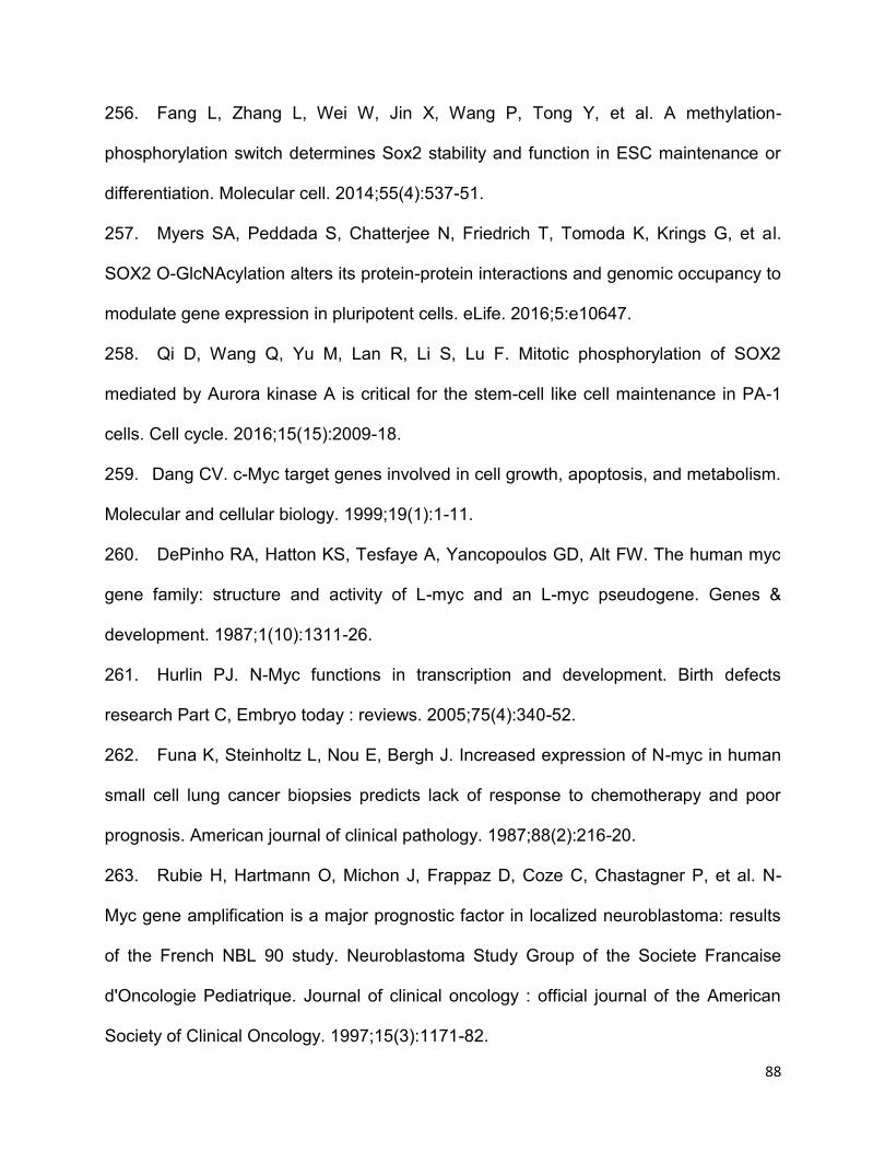
88
256. Fang L, Zhang L, Wei W, Jin X, Wang P, Tong Y, et al. A methylation-
phosphorylation switch determines Sox2 stability and function in ESC maintenance or
differentiation. Molecular cell. 2014;55(4):537-51.
257. Myers SA, Peddada S, Chatterjee N, Friedrich T, Tomoda K, Krings G, et al.
SOX2 O-GlcNAcylation alters its protein-protein interactions and genomic occupancy to
modulate gene expression in pluripotent cells. eLife. 2016;5:e10647.
258. Qi D, Wang Q, Yu M, Lan R, Li S, Lu F. Mitotic phosphorylation of SOX2
mediated by Aurora kinase A is critical for the stem-cell like cell maintenance in PA-1
cells. Cell cycle. 2016;15(15):2009-18.
259. Dang CV. c-Myc target genes involved in cell growth, apoptosis, and metabolism.
Molecular and cellular biology. 1999;19(1):1-11.
260. DePinho RA, Hatton KS, Tesfaye A, Yancopoulos GD, Alt FW. The human myc
gene family: structure and activity of L-myc and an L-myc pseudogene. Genes &
development. 1987;1(10):1311-26.
261. Hurlin PJ. N-Myc functions in transcription and development. Birth defects
research Part C, Embryo today : reviews. 2005;75(4):340-52.
262. Funa K, Steinholtz L, Nou E, Bergh J. Increased expression of N-myc in human
small cell lung cancer biopsies predicts lack of response to chemotherapy and poor
prognosis. American journal of clinical pathology. 1987;88(2):216-20.
263. Rubie H, Hartmann O, Michon J, Frappaz D, Coze C, Chastagner P, et al. N-
Myc gene amplification is a major prognostic factor in localized neuroblastoma: results
of the French NBL 90 study. Neuroblastoma Study Group of the Societe Francaise
d'Oncologie Pediatrique. Journal of clinical oncology : official journal of the American
Society of Clinical Oncology. 1997;15(3):1171-82.
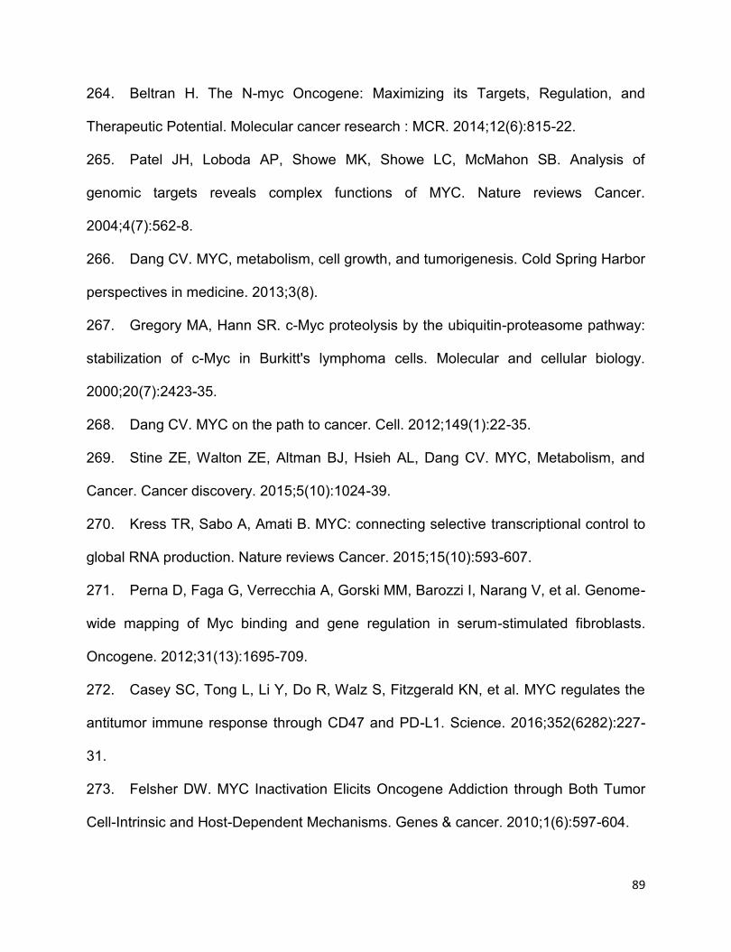
89
264. Beltran H. The N-myc Oncogene: Maximizing its Targets, Regulation, and
Therapeutic Potential. Molecular cancer research : MCR. 2014;12(6):815-22.
265. Patel JH, Loboda AP, Showe MK, Showe LC, McMahon SB. Analysis of
genomic targets reveals complex functions of MYC. Nature reviews Cancer.
2004;4(7):562-8.
266. Dang CV. MYC, metabolism, cell growth, and tumorigenesis. Cold Spring Harbor
perspectives in medicine. 2013;3(8).
267. Gregory MA, Hann SR. c-Myc proteolysis by the ubiquitin-proteasome pathway:
stabilization of c-Myc in Burkitt's lymphoma cells. Molecular and cellular biology.
2000;20(7):2423-35.
268. Dang CV. MYC on the path to cancer. Cell. 2012;149(1):22-35.
269. Stine ZE, Walton ZE, Altman BJ, Hsieh AL, Dang CV. MYC, Metabolism, and
Cancer. Cancer discovery. 2015;5(10):1024-39.
270. Kress TR, Sabo A, Amati B. MYC: connecting selective transcriptional control to
global RNA production. Nature reviews Cancer. 2015;15(10):593-607.
271. Perna D, Faga G, Verrecchia A, Gorski MM, Barozzi I, Narang V, et al. Genome-
wide mapping of Myc binding and gene regulation in serum-stimulated fibroblasts.
Oncogene. 2012;31(13):1695-709.
272. Casey SC, Tong L, Li Y, Do R, Walz S, Fitzgerald KN, et al. MYC regulates the
antitumor immune response through CD47 and PD-L1. Science. 2016;352(6282):227-
31.
273. Felsher DW. MYC Inactivation Elicits Oncogene Addiction through Both Tumor
Cell-Intrinsic and Host-Dependent Mechanisms. Genes & cancer. 2010;1(6):597-604.
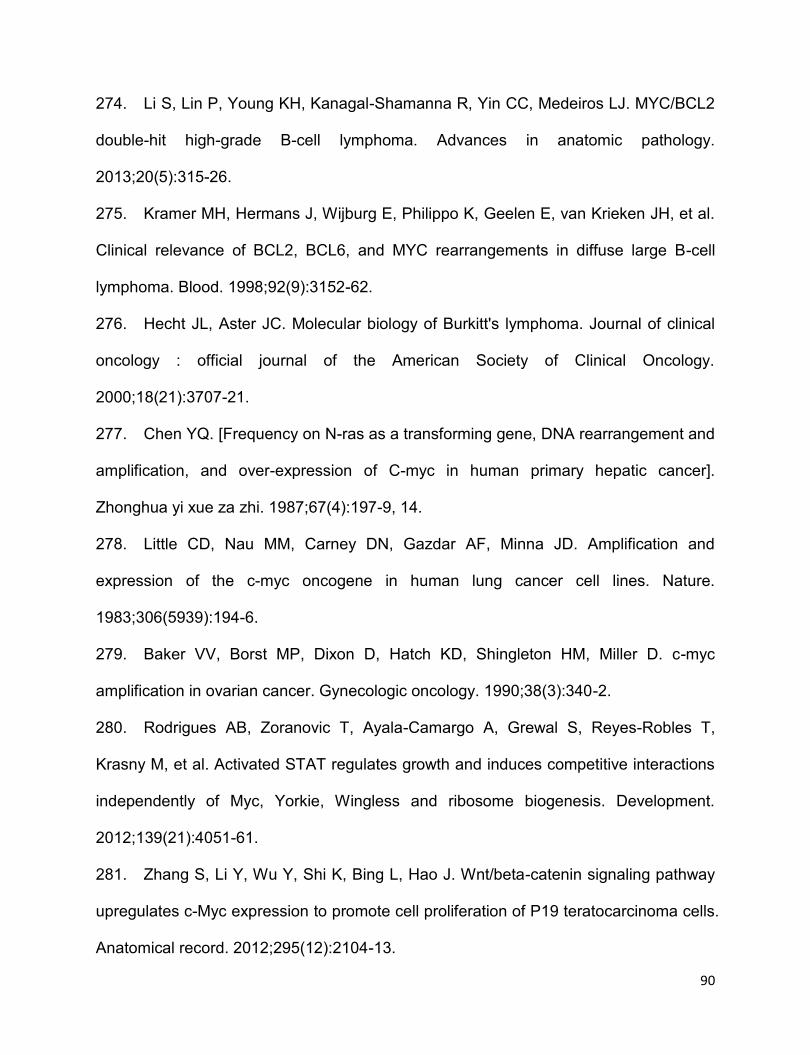
90
274. Li S, Lin P, Young KH, Kanagal-Shamanna R, Yin CC, Medeiros LJ. MYC/BCL2
double-hit high-grade B-cell lymphoma. Advances in anatomic pathology.
2013;20(5):315-26.
275. Kramer MH, Hermans J, Wijburg E, Philippo K, Geelen E, van Krieken JH, et al.
Clinical relevance of BCL2, BCL6, and MYC rearrangements in diffuse large B-cell
lymphoma. Blood. 1998;92(9):3152-62.
276. Hecht JL, Aster JC. Molecular biology of Burkitt's lymphoma. Journal of clinical
oncology : official journal of the American Society of Clinical Oncology.
2000;18(21):3707-21.
277. Chen YQ. [Frequency on N-ras as a transforming gene, DNA rearrangement and
amplification, and over-expression of C-myc in human primary hepatic cancer].
Zhonghua yi xue za zhi. 1987;67(4):197-9, 14.
278. Little CD, Nau MM, Carney DN, Gazdar AF, Minna JD. Amplification and
expression of the c-myc oncogene in human lung cancer cell lines. Nature.
1983;306(5939):194-6.
279. Baker VV, Borst MP, Dixon D, Hatch KD, Shingleton HM, Miller D. c-myc
amplification in ovarian cancer. Gynecologic oncology. 1990;38(3):340-2.
280. Rodrigues AB, Zoranovic T, Ayala-Camargo A, Grewal S, Reyes-Robles T,
Krasny M, et al. Activated STAT regulates growth and induces competitive interactions
independently of Myc, Yorkie, Wingless and ribosome biogenesis. Development.
2012;139(21):4051-61.
281. Zhang S, Li Y, Wu Y, Shi K, Bing L, Hao J. Wnt/beta-catenin signaling pathway
upregulates c-Myc expression to promote cell proliferation of P19 teratocarcinoma cells.
Anatomical record. 2012;295(12):2104-13.
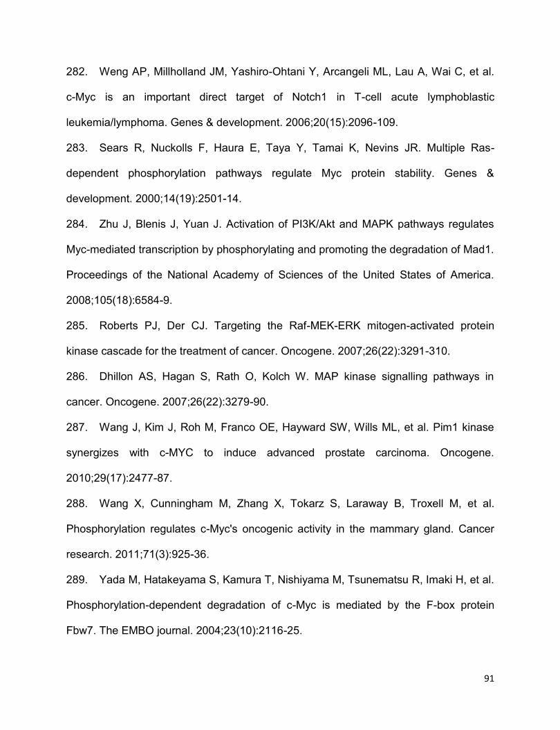
91
282. Weng AP, Millholland JM, Yashiro-Ohtani Y, Arcangeli ML, Lau A, Wai C, et al.
c-Myc is an important direct target of Notch1 in T-cell acute lymphoblastic
leukemia/lymphoma. Genes & development. 2006;20(15):2096-109.
283. Sears R, Nuckolls F, Haura E, Taya Y, Tamai K, Nevins JR. Multiple Ras-
dependent phosphorylation pathways regulate Myc protein stability. Genes &
development. 2000;14(19):2501-14.
284. Zhu J, Blenis J, Yuan J. Activation of PI3K/Akt and MAPK pathways regulates
Myc-mediated transcription by phosphorylating and promoting the degradation of Mad1.
Proceedings of the National Academy of Sciences of the United States of America.
2008;105(18):6584-9.
285. Roberts PJ, Der CJ. Targeting the Raf-MEK-ERK mitogen-activated protein
kinase cascade for the treatment of cancer. Oncogene. 2007;26(22):3291-310.
286. Dhillon AS, Hagan S, Rath O, Kolch W. MAP kinase signalling pathways in
cancer. Oncogene. 2007;26(22):3279-90.
287. Wang J, Kim J, Roh M, Franco OE, Hayward SW, Wills ML, et al. Pim1 kinase
synergizes with c-MYC to induce advanced prostate carcinoma. Oncogene.
2010;29(17):2477-87.
288. Wang X, Cunningham M, Zhang X, Tokarz S, Laraway B, Troxell M, et al.
Phosphorylation regulates c-Myc's oncogenic activity in the mammary gland. Cancer
research. 2011;71(3):925-36.
289. Yada M, Hatakeyama S, Kamura T, Nishiyama M, Tsunematsu R, Imaki H, et al.
Phosphorylation-dependent degradation of c-Myc is mediated by the F-box protein
Fbw7. The EMBO journal. 2004;23(10):2116-25.
92
290. Farrell AS, Sears RC. MYC degradation. Cold Spring Harbor perspectives in
medicine. 2014;4(3).
291. Gorrini C, Harris IS, Mak TW. Modulation of oxidative stress as an anticancer
strategy. Nature reviews Drug discovery. 2013;12(12):931-47.
292. Sosa V, Moline T, Somoza R, Paciucci R, Kondoh H, ME LL. Oxidative stress
and cancer: an overview. Ageing research reviews. 2013;12(1):376-90.
293. Simic MG, Bergtold DS, Karam LR. Generation of oxy radicals in biosystems.
Mutation research. 1989;214(1):3-12.
294. Klaunig JE, Kamendulis LM, Hocevar BA. Oxidative stress and oxidative
damage in carcinogenesis. Toxicologic pathology. 2010;38(1):96-109.
295. Janssen-Heininger YM, Mossman BT, Heintz NH, Forman HJ, Kalyanaraman B,
Finkel T, et al. Redox-based regulation of signal transduction: principles, pitfalls, and
promises. Free radical biology & medicine. 2008;45(1):1-17.
296. Miller DM, Thomas SD, Islam A, Muench D, Sedoris K. c-Myc and cancer
metabolism. Clinical cancer research : an official journal of the American Association for
Cancer Research. 2012;18(20):5546-53.
297. Kim MH, Kim H. Oncogenes and tumor suppressors regulate glutamine
metabolism in cancer cells. Journal of cancer prevention. 2013;18(3):221-6.
298. Dayem AA, Choi HY, Kim JH, Cho SG. Role of oxidative stress in stem, cancer,
and cancer stem cells. Cancers. 2010;2(2):859-84.
299. Diehn M, Cho RW, Lobo NA, Kalisky T, Dorie MJ, Kulp AN, et al. Association of
reactive oxygen species levels and radioresistance in cancer stem cells. Nature.
2009;458(7239):780-3.
93
300. Mahalingaiah PK, Singh KP. Chronic oxidative stress increases growth and
tumorigenic potential of MCF-7 breast cancer cells. PloS one. 2014;9(1):e87371.
301. Reuter S, Gupta SC, Chaturvedi MM, Aggarwal BB. Oxidative stress,
inflammation, and cancer: how are they linked? Free radical biology & medicine.
2010;49(11):1603-16.
302. Waris G, Ahsan H. Reactive oxygen species: role in the development of cancer
and various chronic conditions. Journal of carcinogenesis. 2006;5:14.
303. Thornber K, Colomba A, Ceccato L, Delsol G, Payrastre B, Gaits-Iacovoni F.
Reactive oxygen species and lipoxygenases regulate the oncogenicity of NPM-ALK-
positive anaplastic large cell lymphomas. Oncogene. 2009;28(29):2690-6.
304. Meissl K, Macho-Maschler S, Muller M, Strobl B. The good and the bad faces of
STAT1 in solid tumours. Cytokine. 2015.
305. Weilemann A, Grau M, Erdmann T, Merkel O, Sobhiafshar U, Anagnostopoulos I,
et al. Essential role of IRF4 and MYC signaling for survival of anaplastic large cell
lymphoma. Blood. 2015;125(1):124-32.
306. Gopal K, Gupta N, Zhang H, Alshareef A, Alqahtani H, Bigras G, et al. Oxidative
stress induces the acquisition of cancer stem-like phenotype in breast cancer
detectable by using a Sox2 regulatory region-2 (SRR2) reporter. Oncotarget.
2016;7(3):3111-27.

94
CHAPTER 2
STAT1 is phosphorylated and downregulated by the oncogenic tyrosine kinase
NPM-ALK in ALK-positive anaplastic large-cell lymphoma1
1This chapter has been modified from a previous publication: Wu C, Molavi O, Zhang H, Gupta N, Alshareef A, Bone K.M, Gopal K, Wu F, Lewis J.T,
Douglas D.N, Kneteman N.M, and Lai R. STAT1 is phosphorylated and downregulated
by the oncogenic tyrosine kinase NPM-ALK in ALK-positive anaplastic large-cell
lymphoma. Blood. 2015; 126(3):336-345. The content of this publication has been
included in this thesis with permission from the publisher (License number:
3981420253343).
I prepared the first draft and revisions based on the suggestions and comments of the
co-authors. I designed and performed most of the experiments described herein,
except for the following: O.M. performed some studies, data shown in Figure 2.1A and
Figure 2.4F-G; H.Z., N.G., A.A., and F.W. performed portions of the experiments and
provided technical support and intellectual input. K.B. provided the SupM2 and Karpas
299 cell lines stably transfected with Tet on system. N.G., K.G., J.L., D. D., and N.K.
were responsible for the animal study design, conducting euthanization of the SCID
mouse and data analysis. R.L. provided numerous comments and final review of the
manuscript before it was submitted for publication.
95
2.1 Introduction
Signal transducer and activator of transcription 1 (STAT1), a member of the STAT
family of transcription factors, is a major mediator of γ-interferon (IFNү) signaling (1).
Subjection of IFNү leads to phosphorylation of STAT1 at tyrosine 701, which is known
as an active form of STAT1, and phosphorylated STAT1 at tyrosine 701 subsequently
form STAT1 homo/heterodimers with other STAT family members, followed by
migration into nucleus to execute their transcriptional functions (2). STAT1 is also
reported to carry tumor suppressor functions (1-3). In comparison, STAT3, another
STAT family member, is oncogenic when inappropriately activated (4, 5). Correlating
with their opposing roles, STAT1 and STAT3 are known to have functional interactions.
For instance, STAT1 and STAT3 can form heterodimers (4, 6-8), and the expression of
gene targets regulated by STAT1 and STAT3 is dependent on the relative proportions
of STAT1 homodimers, STAT3 homodimers and the STAT1/STAT3 heterodimers (9,
10). This concept is well exemplified by the observation that treatment of multiple
myeloma cell lines with α-interferon triggers a shift from STAT3 homodimers to STAT1
homodimers and STAT3/STAT1 heterodimers, and this biochemical change correlates
with a switch from cell proliferation to apoptosis (10). Several other studies also have
shown that STAT1 and STAT3 can directly antagonize each other functionally (11-15).
It has been reported that activated STAT3 suppresses the DNA-binding of STAT1
homodimers in myeloid cells although it does not affect STAT1 tyrosine
phosphorylation or its nuclear translocation (11). In contrast, IFNγ is reported to
selectively inhibit STAT3-dependent IL-6 signaling in human endothelial cells by
increasing the expression of SOCS3, which inhibits STAT3 signaling (14).
96
STAT1 expression is reported to be downregulated in various types of cancer, including
oral squamous cell carcinoma (16), melanoma (17, 18), cutaneous T-cell lymphoma
(19), breast cancer (20) and esophagus squamous cancer (21, 22). How STAT1 is
downregulated in cancer remains poorly studied. However, a number of studies have
indicated that STAT1 is degraded in ubiquitin-dependent proteasome pathway in some
other cell models (23-28). For instance, Soond et al reported that ERK1/2
phosphorylates STAT1 at serine 727 (p-STAT1S727, an active form of STAT1) and
promotes its degradation in proteasome pathway in E3 ligase βTRCP dependent
manner in mouse fibroblast cells (25). A more recent study revealed that IFNs induces
phosphorylation and ubiquitination of activated STAT1 (p-STAT1Y705, or pSTAT1), thus
promoting its degradation in proteasome pathway in human embryonic kidney 293T
cells (24). Intriguingly, a recent study showed that NKLAM, as an E3 ubiquitin ligase,
ubiquitinates STAT1 but positively regulates STAT1 transcriptional activity and its DNA
binding ability in mice macrophage cells (23).
ALK-positive anaplastic large-cell lymphoma (ALK+ALCL) is a specific type of non-
Hodgkin lymphoma of mature T-cell lineage (29). The characteristic chromosomal
translocation, t(2;5)(p23;q35), is observed in 80% of ALK+ALCL tumors, and this
aberrancy results in the generation of the oncogenic fusion protein, NPM-ALK (29, 30).
NPM-ALK interacts and activates a host of cellular signaling proteins, such as JAK,
STAT3 and PI3K, thereby deregulating these pathways and leading to increased cell
proliferation and survival (29, 30). The pivotal role of STAT3 signaling in mediating the
oncogenic effects of NPM-ALK has been particularly highlighted (29). As STAT1 can
97
inhibit STAT3, as discussed above, we hypothesize that the STAT1 expression and/or
signaling is defective in ALK+ALCL, leaving the STAT3 activation unchecked. The
expression and functional status of STAT1 in ALK+ALCL have never been examined.
In this study, the expression and the functional status of STAT1 in ALK+ALCL cells
were evaluated. The results indicated that NPM-ALK directly contributes to the
relatively low expression of STAT1 in ALK+ALCL tumors and this process is
proteasomal- and STAT3-dependent. The data from this study also demonstrated that
STAT1, if it is overexpressed to a higher expression level, exerts tumor suppressor
functions.
2.2 Methods and materials
2.2.1 Primary tumors and cell lines
All primary tumors were diagnosed at the Cross Cancer Institute (Edmonton, Alberta,
Canada) and the diagnostic criteria were based on those described in the WHO
Classification Scheme. The use of these patient samples for research has received
research ethics approval at Feb 17th of 2016, by the Human Research Ethics Board at
the University of Alberta, with approval number: Pro00062737. Karpas 299, established
from blast cells in the peripheral blood of a 25-year-old white man diagnosed with
ALK+ALCL, was a gift from Dr. Marshal Kadin (Boston, MA). SupM2 cell line,
established from the cerebrospinal fluid of a 5-year-old-girl with refractory ALK+ALCL,
and SU-DHL1 cell line, established from peritoneal effusion of a 10-year-old boy
diagnosed with ALK+ALCL, were both purchased from Deutche Sammlung von
Mikrooorganismen und Zellkulturen GmbH, DSMZ, Germany. All three ALK+ALCL cell

98
lines express CD30 and carry chromosome translocation t(2;5)(p23;q35), resulting in
the generation of NPM-ALK fusion gene. All three cell lines were grown and expanded
in RPMI 1640 (Invitrogen, Life Technologies, Grand Island, NY) supplemented with 10%
fetal bovine serum (FBS, Invitrogen) in 5% CO2 atmosphere at 37°C. GP293 cell line,
an advanced version of human embryonic kidney cells 293, was grown in the DMEM
medium supplemented with 10% FBS (Invitrogen) in 5% CO2 atmosphere at 37°C.
2.2.2 Chemical treatments
MG132, cycloheximide, IFNγ, crizotinib, Stattic, U0126, and 5-aza-2’-deoxycytidine
were all purchased from Sigma Aldrich (St Louis, MO). All above reagents were
dissolved in Dimethyl Sulfoxide (DMSO) (Sigma Aldrich). 0.1µM and 0.5 µM 5 aza-2’-
deoxycytidine were used in this study to evaluate whether STAT1 gene is methylated in
SupM2 cells. 0.5 mM, 1 mM and 2 mM U0126 were used to treat SupM2 cells for 5
hours to assess the relationship between STAT1 and MEK/ERK pathway. 5µM MG132
and 100 µg/mL cycloheximide were subjected to SupM2 and Karpas 299 cells for 2
hours and 6 hours, respectively, to assess the half-life of STAT1. Various doses of
crizotinib ranging from 0 to 50 nM were applied to study the relationship between NPM-
ALK and STAT1 in ALK+ALCL cells. Various doses (ranging from 0 to 50 ng/mL) of
IFNγ were utilized to activate STAT1 signaling in ALK+ALCL cells in a time-course
experiment. Stattic (ranging from 0 to 5 nM) was used to inhibit STAT3 activity in this
study.
99
2.2.3 Immunohistochemistry
Immunohistochemistry was performed using standard technique as previously
described (31). In brief, formalin-fixed, paraffin-embedded tissue sections (4 µM
thickness) were deparaffinized and hydrated. Heat-induced epitope retrieval was
executed by using citrate buffer (pH=6) in a pressure cooker, followed by incubation of
tissue sections with 3% H2O2 for 10 minutes to block endogenous peroxidase activity at
room temperature. The tissue sections were incubated with anti-STAT1 antibody (1:500)
(42H3, Cell Signaling Technology Inc; Danvers, MA) overnight in a humidified chamber
in 4°C cold room. The next day, tissue sections, after 3 X PBS washes, were
subsequently incubated with anti-Rabbit IgG (EnVison; Dako, Santa Clara, CA) for 40
minutes at room temperature. Next, tissue sections were incubated with 3,3’-
diaminobenzidine/H2O2 (Dako) for color development and counterstained with
hematoxylin for 1 minute. Pictures were taken on a Zeiss Axioscope 2 micoscope (Carl
Zeiss, Heidelberg, Germany).
2.2.4 Short interfering RNA and transfections
siRNA (SMART pool) for STAT1, STAT3 and scrambled siRNA (SMART pool) were
purchased from Dharmacon (Lafayette, CO). Individual STAT1 siRNA 1 and 2 were
also purchased from Dharmacon. All siRNAs were diluted with ddH2O and stocked with
the concentration of 20 µM. Transfection of GP293 cells with siRNAs (final
concentration: 200 nM) were performed using lipofectamine 2000 reagents (Invitrogen).
In brief, 1 million GP293 cells seeded in 10 cm-diameter cell culture plate (CorningTM,
ThermoFisher Scientific Canada) were transfected with 200 nM siRNA using
lipofectamine 2000 reagent (Invitrogen). Transient transfections of 5 million of
100
ALK+ALCL cells with siRNAs (final concentration: 600 nM) were performed using BTX
Electro Square Porator ECM830 (225 V, 8.5 milliseconds, 3 pulses) (Harvard
Apparatus; Holliston MA). The efficiency of target gene inhibition was assessed using
Western blotting.
2.2.5 Plasmid constructs and transfections
pRK5-HA-Ubiquitin-WT was a gift from Ted Dawson (Addgene plasmid #17608)
(Cambridge, MA). STAT1C- flag in pcDNA3.1 was a gift from Dr. OUCHI (19). Transient
transfections of Karpas 299 and SupM2 cells (5 million cells) with 10 μg plasmid were
performed using the BTX Electro Square Porator ECM830 (225 V, 8.5 milliseconds, 3
pulses). NPM-ALK in pcDNA3.1 was generated by the Lai lab (32). Transient
transfections of GP293 cells (1 million cells) with 2 μg of either NPM-ALK or pcDNA3.1,
or GP293 cells co-transfected with both NPM-ALK and pcDNA3.1-STAT1-flag
(Addgene) or STAT1Y701F-flag (Addgene) were performed using lipofectamine 2000
reagents (Invitrogen). In brief, 1 million cells seeded in 10 cm-diameter cell culture plate
(CorningTM, ThermoFisher Scientific Canada) were transfected with 2 µg plasmid using
lipofectamine 2000 reagents (Invitrogen).
2.2.6 Generation of Tet on inducible stable cell lines
To generate the STAT1C-inducible stable cell lines, two ALK+ALCL cell lines, SupM2
and Karpas 299, were infected with retroviral supernatant generated by transfecting
Phoenix packaging cells with the pRetro-X Tet on Advanced plasmid (Clontech,
Mountain View, CA) using lipofectamine 2000 (Invitrogen) following the manufacturer’s
suggested protocol. These cells were maintained in 200 μg/mL G418 (Invitrogen), 100
101
units/mL penicillin and 100 μg/mL streptomycin (Invitrogen) for two weeks to select for
stable cells. STAT1C cDNA was cloned by GenScript (Piscataway, NJ) into a retroviral
vector pRetro-Tight-Pur (Clontech) under the control of a doxycycline-inducible
promoter, and viral supernatant was, used to transduce Tet on SupM2 and Karpas 299
cells and selected by puromycin up to 2 μg/mL (Invitrogen) for 1 month. Empty vector
was also transduced in these cells as controls. The expression of phosphorylated
STAT1Y701 and STAT1 were confirmed by treating the cells in culture with doxycycline
(Sigma Aldrich) at different concentrations for 48 hours in RPMI 1640 supplemented
with 10% Tet system-approved FBS (Clontech).
2.2.7 Cell-cycle assay
To evaluate cell-cycle and apoptosis in the ALK+ALCL cell lines, the cells were stained
with propidium iodide, a late marker of cell apoptosis, and the cell-cycle analysis was
performed by flow cytometry (BD FACS Calibur, Flow Cytometry Lab, Department of
Experimental Oncology, Cross Cancer Institute, University of Alberta, Canada). In brief,
following treatment, cells were fixed with ice-cold 70% ethanol for 24 hours. Cells were
then washed gently with cold 1 X PBS three times, following by suspension with 50 µl
of (100 µg/mL stock) RNase and 200 µl of (50 µg/mL stock) propidium iodide in dark at
37 °C for 15 minutes. Then cells were processed for flow cytometry (BD FACS Calibur)
to measure the forward scatter and side scatter to identify single cells. Data acquisition
was gated using pulse width versus pulse area to exclude apoptotic cells and cell
doublets, and the cell-cycle phase distribution was determined using the CellQuest
program (20,000 events were counted). The experiments were performed in triplicate.

102
2.2.8 Western blotting and co-immunoprecipitation
Western blotting was performed using standard techniques. Briefly, the cells were
washed with cold 1 x PBS (pH7.0) and lysed in 1 x RIPA lysis and extraction buffer
(ThermoFisher Scientific Canada) containing protease inhibitor cocktail (Sigma Aldrich)
and phosphatase inhibitor cocktail (Sigma Aldrich). After incubation on ice for 30
minutes, the cell lysates were subjected to centrifuge at 14,000 g for 15 minutes at 4°C,
and the supernatants were collected. The protein concentration was assessed using
BCA protein assay kit (Bio-Rad, Hercules, CA, USA). The protein lysates were then
heated with 4 X loading buffer (240 mM Tris-HCL pH 6.8, 40% glycerol, 8% SDS, 0.04%
bromophenol blue, 5% β-mercaptoethanol) at 100°C for 5 minutes. 12% or 10% SDS-
polyacrylamide gel (SDS-PAGE) was used. After electrophoresis and transfer to
nitrocellulose membranes (Bio-Rad) by electroblotting, the membranes were probed
with primary antibodies (1:1000) (overnight, 4°C) and secondary antibodies (1:1000) (1
hour, room temperature), followed by the enhanced chemiluminescene (ECL) Western
blotting detection system (Bio-Rad).
Co-immunoprecipitation was performed and described briefly as below. Cells were
harvested and washed with cold 1X PBS for 2 times, then were lysated with CelLytic M
buffer (Sigma Aldrich) containing protease inhibitor cocktails (P8340, Sigma Aldrich)
and phosphatase inhibitor cocktails (P5726, Sigma Aldrich). After incubation on ice for
30 minutes, the cell lysates were proceeded to centrifuge at 14,000 X g for 15 minutes
at 4°C. The supernatants (protein lysates) were collected and quantitated by using
PierceTM BCA protein Assay Kit (ThermoFisher Scientific Canada). In principle, in this
protein quantification assay, protein lysates can reduce Cu2+ to Cu1+ in an alkaline
103
medium, and bicinchoninic acid (BCA) subsequently reacts with Cu1+, forming a water
soluble complex that exhibits strong absorbance at 562 nm. Then 500 µg protein
lysates were incubated with 3 µg primary antibody and rotated at 4 °C for overnight.
Next day, the 1 X PBS-washed protein A Plus/Protein G agarose (ThermoFisher
Scientific Canada) were added to the protein lysates and rotated at 4°C for additional 3
hours. Last, the protein lysates were centrifuged at 8000 X g for 3 minutes and the
agaroses were kept and washed gently with cold 1 X PBS for three times.
Subsequently, the agaroses were diluted in 50 µl 4 X loading buffer (40% glycerol, 240
mM Tris-HCl pH 6.0, 8% SDS, 0.04% bromophenol blue, and 5% β-mercaptoethanol)
and heated for 5 minutes at 100 °C, followed by centrifuge at 14000 X g for 15 minutes.
The supernatant were collected and thereafter were proceed to SDS-PAGE.
Antibodies against for phosphorylated STAT1Y701 (58D6), STAT1 (D1K9Y),
phosphorylated ALKY1604 (#3341), ALK (C26G7), phosphorylated STAT3Y705 (D3A7),
IRF-1 (D5E4), Cleaved PARP (D64E10), T-bet (D6N8N), Bcl-2 (D55G8), Caspas 3
(8G10), PARP (46D11), IRF-7 (D2A1J), Survivin (71G4B7) were purchased from Cell
Signaling Technology (Danvers, MA). Antibodies against β-actin (C4), γ-tubulin (C20),
HA (Y11), Ub (FL-76), STAT3 (H-190), SOCS-1 (H-93) were purchased from Santa
Cruz Biotechnology (Santa Cruz, CA). Anti-flag (clone M2) antibody was purchased
from Sigma Aldrich. The above antibodies were diluted by 1000 folds with TBS buffer,
0.5% Tween 20 (Sigma Aldrich) and 5% BSA (Sigma Aldrich) for use. Secondary
antibodies anti-Rabbit IgG (1:2000) and anti-Mouse IgG (1:2000) were purchased from
Cell Signaling Technology.
104
The densitometry value of Western blotting bands was analyzed with Image J software
(Bethesda, WA).
2.2.9 Methylcellulose colony formation assay
Methylcellulose-based media was purchased from R&D systems Inc (Minneapolis, MN),
and the methylcellulose colony formation assay was performed as below. Briefly, 1000
cells were seeded in each well of 6-well plate with 4 mL methylcellulose-based media
containing 1.2% methylcellulose agarose, 30% FBS, 1% bovine serum albumin, 0.1%
penicillin/streptomycin (Thermo Fisher Scientific), and 0.1% β-mercaptoethanol. The
colonies with more than 40 μm in diameter were counted after 10-14 days of culture.
2.2.10 Dual luciferase assay
To assess the transcriptional activity of STAT3 affected by STAT1C in ALK+ALCL, the
dual luciferase assay kit (Dual-GloTM Luciferase Assay System, Promega, Madision, WI)
was used. Tet on SupM2-STAT1C cells were transiently transfected with 1 μg of pRL
Renilla Luciferase Reporter Vector (Promega) and 10 μg of pLUCTSK3 (Promega),
using the BTX Electro Square Porator ECM830 (225 V, 8.5 million seconds, 3 pulses).
Cells exposed with different doses of doxycycline were harvested at 24 hours post
transfection, and the firefly and Renilla luciferases were measured sequentially from a
single sample according to the manufacturer’s instructions. In brief, firstly, the firefly
luciferase reporter was measured by adding luciferase assay reagent II (provided in the
kit) in the cell pellets after 3 X cold 1 X PBS wash to generate a luminescent signaling
(last for ~1 minute), after the reaction is quenched, the Renilla luciferase reaction was
initiated by adding Stop & Glo® Reagent (provided in the kit), which produced stabilized
105
signal from the Renilla luciferase. The luciferase values were read by FLUOstar Omega
microplate reader (BMG LabTech, ThermoFisher Scientific Canada). This experiment
was performed in triplicate.
2.2.11 STAT3 DNA probe binding assay
Tet on SupM2-STAT1C cells were induced with 0, 20, 200 ng/mL doxycycline for 48
hours, then the cells were harvested and washed with cold PBS twice, following by
cytoplasmic and nuclear fractionation using the Pierce NE-PER kit (ThermoFisher
Scientific Canada). This kit can efficiently solubilize and separate cytoplasmic and
nuclear proteins into fractions. In brief, CERI and CER II buffer provided in the kit were
used to lysate the cells to extract cytoplasmic fraction, and NER buffer in the kit was
utilized to extract nuclear fraction. 300 ng nuclear protein was incubated with 3 pmol of
either mutant or wild-type biotin-labeled STAT3 probe (constructed by IDT, Edmonton,
Alberta, Canada) for 0.5 hour by rotating at room temperature. Streptadvidin agarose
beads (75 μl, ThermoFisher Scientific Canada) were added to each sample, and the
samples were rotated on UltraRockerTM Rocking Platform (Bio-Rad) by overnight at
4°C.The next day, the samples were washed with cold 1 X PBS three times, and
protein was eluted at 100°C in 4 X protein loading buffer and loaded on SDS-PAGE
gels.
Wild-type STAT3 probe: 5’-GATCTAGGAATTCCCAGAAGG-3’;
Mutant STAT3 probe: 5’-GATCTAGGAACCTTTGACGGG-3’.
The underlined bolded sequence is STAT3 consensus DNA binding sequence.
106
2.2.12 Trypan blue exclusion assay and MTS assay
Cell viability was assessed by trypan blue exclusion assay. Trypan blue was purchased
from Thermofisher Scientific Canada. The viable cells were calculated as the total
number of cells within the grids on the hemacytometer. Viable cells do not take up
trypan blue but non-viable cells do. The CellTiter 96® AQueous One Solution Cell
Proliferation Assay kit was purchased from Promega (Madison, WI) for MTS assay to
assess the cell growth. This assay was performed by adding 20 µl of 3-(4,5-
dimethylthiazol-2-yl)-5-(3-carboxymethoxyphenyl)-2-(4-sulfophenyl)-2H-tetrazolium
(MTS) reagent provided in the kit directly to the wells containing live cells in 96-well
plate (Corning), with incubating time ranging from 1 to 5 hours. Then the absorption
values were read by FLUOstar Omega microplate reader (BMG LabTech,
ThermoFisher Scientific Canada).
2.2.13 RNA extraction, cDNA synthesis, and quantitative reverse transcriptase
PCR (quantitative RT-PCR)
Total RNA extraction was performed with the Qiagen RNeasy Kit (Qiagen, Toronto,
Ontario, Canada) according to the manufacturer’s protocol. In brief, cells after 1 X cold
PBS three times wash and centrifuge (300 X g) were lysed and then homogenized.
Ethanol was added to the cell lysate, which subsequently were loaded onto the RNeasy
silica membrane (provided in the kit). RNA can bind to the membrane, but all other
contaminants were washed away. 1 g of RNA was reverse-transcribed using
Superscript II (Invitrogen) to generate cDNA. 1 L of the resulting cDNA mixture was
added to the Power SYBR Green PCR Master Mix (Invitrogen) and amplified with gene
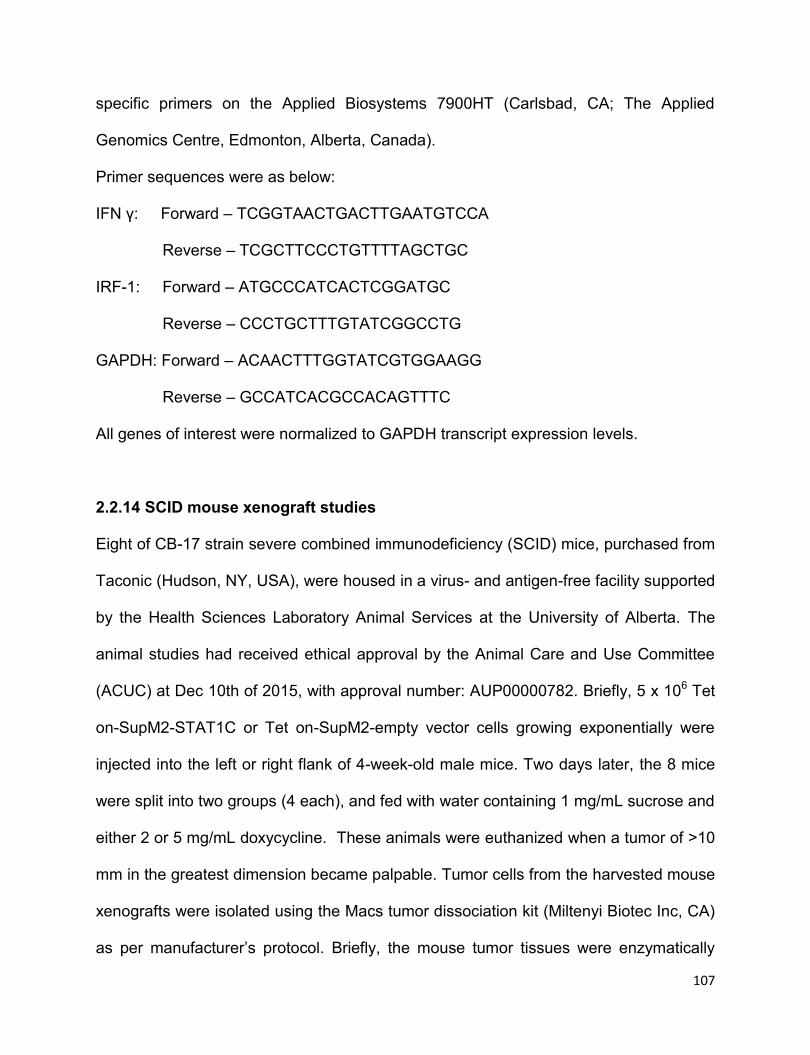
107
specific primers on the Applied Biosystems 7900HT (Carlsbad, CA; The Applied
Genomics Centre, Edmonton, Alberta, Canada).
Primer sequences were as below:
IFN γ: Forward – TCGGTAACTGACTTGAATGTCCA
Reverse – TCGCTTCCCTGTTTTAGCTGC
IRF-1: Forward – ATGCCCATCACTCGGATGC
Reverse – CCCTGCTTTGTATCGGCCTG
GAPDH: Forward – ACAACTTTGGTATCGTGGAAGG
Reverse – GCCATCACGCCACAGTTTC
All genes of interest were normalized to GAPDH transcript expression levels.
2.2.14 SCID mouse xenograft studies
Eight of CB-17 strain severe combined immunodeficiency (SCID) mice, purchased from
Taconic (Hudson, NY, USA), were housed in a virus- and antigen-free facility supported
by the Health Sciences Laboratory Animal Services at the University of Alberta. The
animal studies had received ethical approval by the Animal Care and Use Committee
(ACUC) at Dec 10th of 2015, with approval number: AUP00000782. Briefly, 5 x 10⁶ Tet
on-SupM2-STAT1C or Tet on-SupM2-empty vector cells growing exponentially were
injected into the left or right flank of 4-week-old male mice. Two days later, the 8 mice
were split into two groups (4 each), and fed with water containing 1 mg/mL sucrose and
either 2 or 5 mg/mL doxycycline. These animals were euthanized when a tumor of >10
mm in the greatest dimension became palpable. Tumor cells from the harvested mouse
xenografts were isolated using the Macs tumor dissociation kit (Miltenyi Biotec Inc, CA)
as per manufacturer’s protocol. Briefly, the mouse tumor tissues were enzymatically
108
digested using the Enzyme mix provided in the kit, and the gentleMACSTM Dissociators
(provided in the kit) were applied for the mechanical dissociation steps. After tumor
dissociation, the tumor samples were filtered to single-cell suspension, following by
Western blotting analysis.
2.2.15 Statistical analysis
Data were expressed as mean standard deviation. Half maximal inhibitory
concentration (IC50) was calculated by Graphpad Prism (La Jolla, CA). Significance
was determined by using Student’s t-test and Fisher exact test. Statistical significance
is denoted by * (P<0.05) and **(P<0.01).
2.3 Results
2.3.1 STAT1 is expressed at a low level in ALK+ALCL tumors and cell lines
Western blotting was employed to evaluate the expression level of STAT1 in
ALK+ALCL cells. As compared to peripheral blood T-cells from healthy donors, the total
STAT1 levels in all 3 frozen tumors were low (Figure 2.1A). When the same Western
blot was probed with an antibody reactive with pSTAT1, 2 of 3 cases showed a
relatively low level of pSTAT1. On the right panel, all three ALK+ALCL cell lines
expressed a relatively low level of total STAT1. Again, 2 of 3 cell lines showed a low
pSTAT1 expression level.
Then immunohistochemistry was employed to evaluate the expression of STAT1 in
formalin-fixed/paraffin-embedded tissues. As shown in Figure 2.1B, STAT1 was

109
strongly expressed by the tonsillar interfollicular T-cells (left panel, white arrow) but only
weakly by germinal center B-cells (left panel, black arrow). All ALK+ALCL tumors
examined (n=7) consistently showed no/weak STAT1 expression relative to the
surrounding benign lymphocytes and macrophages (middle panel). In contrast, all 5
ALK-negative ALCL (ALK-ALCL) tumors showed a relatively high level of STAT1
expression (right panel). This difference in STAT1 expression between ALK+ALCL and
ALK-ALCL tumors is statistically significant (P=0.0013, Fisher exact test).
Figure 2.1 Expression of STAT1 in ALK+ALCL cell lines and patient samples.
A) Western blotting studies revealed a relatively low expression level of STAT1 in 3
ALK+ALCL tumors and 3 cell lines, compared to that of normal peripheral blood T-cells.
Densitometry values shown were normalized to the β-actin bands. Most cases also had
a low expression level of pSTAT1. B) Immunohistochemical study results showed the
expression of STAT1 in paraffin-embedded tissues derived from a representative
110
benign tonsil (left, 100X magnification) a representative ALK+ALCL tumor (middle,
400X magnification) and a representative ALK-ALCL tumor (right, 200X magnification).
In benign tonsils, the interfollicular T-cells (white arrow) were strongly STAT1-positive,
whereas the germinal center B-cells (black arrow) were STAT1-weak. In the ALK+ALCL
tumor, the infiltrating T-cells were strongly positive for STAT1 whereas the large
lymphoma cells (red arrow) were virtually negative for STAT1. In the ALK-ALCL tumor,
the large lymphoma cells (blue arrow) were strongly positive for STAT1.
2.3.2 The ubiquitin-proteasome pathway is involved in the downregulation of
STAT1 in ALK+ALCL cells
Some literature suggested that several possible mechanisms of STAT1 downregulation.
First, the STAT1 gene promoter has been reported to be methylated and silenced in
head and neck squamous carcinoma (33). Thus, SupM2 cells were subjected with 5-
aza-2’-deoxycytidine up to 7 days; however, no restoration of STAT1 was found
(Figure 2.2A). Second, another literature suggested that the MEK signaling pathway
can downregulate STAT1 in mouse embryonic fibroblasts (25); however, the addition of
a MEK inhibitor (U0126) to SupM2 cells also did not appreciably change the STAT1
level detected by Western blotting (Figure 2.2B). Lastly, in the same study, the authors
revealed that both STAT1 and pSTAT1 are degraded via the ubiquitin-proteasome
pathway after oxidative stress in mouse embryonic fibroblasts (25). Thus, we subjected
SupM2 and Karpas 299 cells to MG132, a proteasome inhibitor. As shown in Figure
2.2C-D, both STAT1 and pSTAT1 levels increased in a dose/time-dependent manner.
Furthermore, the amount of ubiquitinated STAT1 increased with MG132 treatment in
the same time frame (Figure 2.2E-F). As shown in Figure 2.2G-H, in the presence of
111
cycloheximide(CHX), MG132 treatment was found to dramatically prolong the half-life
of STAT1 in SupM2 and Karpas 299 cells.
Figure 2.2 The ubiquitin-proteasome pathway is involved in the downregulation
of STAT1 in ALK+ALCL cells. A) SupM2 cells were subjected with 0.1 μM, 5 μM 5-
aza-2’-deoxycytidine (5-aza-dC) up to 7 days, and the expression of STAT1 was not
restored. B) SupM2 cells were subjected with various doses of MEK inhibitor U0126 for
5 hours, STAT1 expression was not altered in this experiment. C) SupM2 and Karpas
299 cells were treated with varying doses of MG132 for 5 hours. pSTAT1 and STAT1
were upregulated in response to MG132 in a dose dependent manner. D) A time
course experiment in which SupM2 and Karpas 299 cells were treated with 5 μM of
MG132 was performed; pSTAT1 and STAT1 were upregulated in a time-dependent
manner.
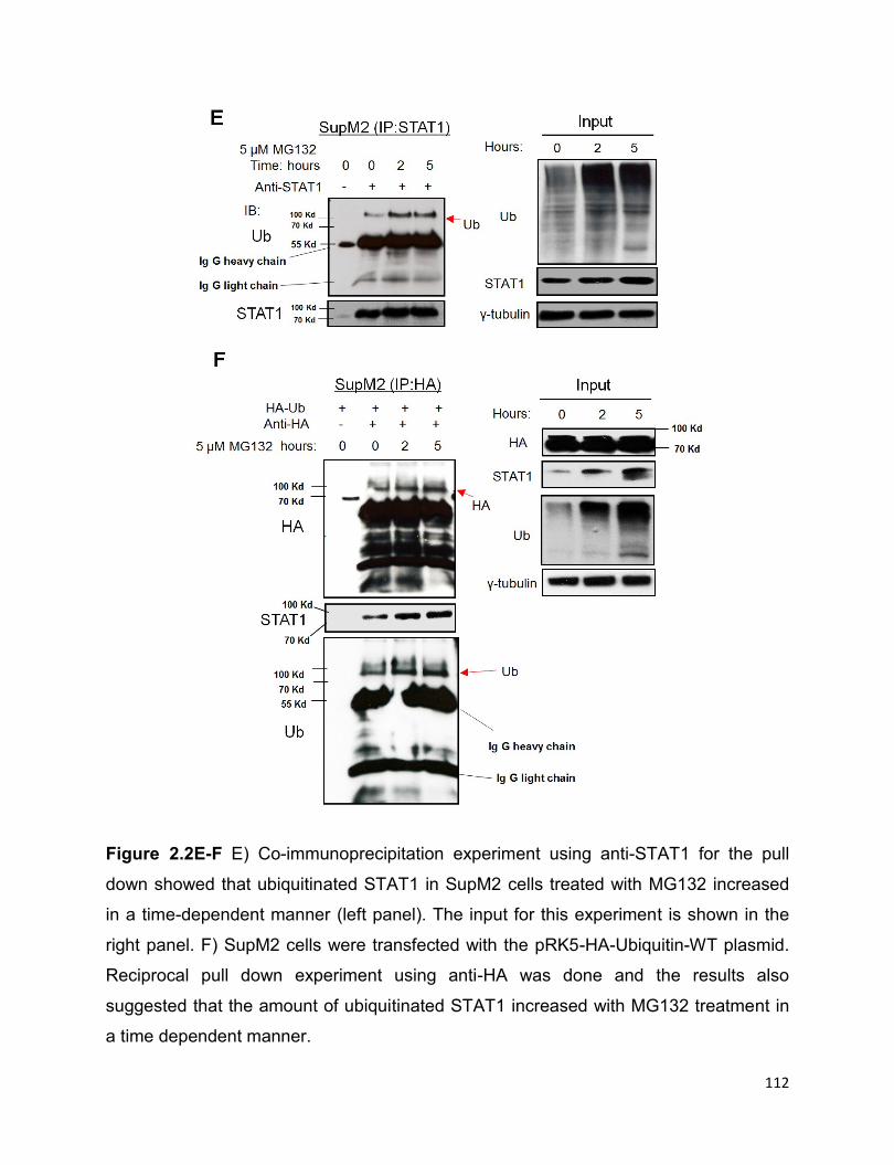
112
Figure 2.2E-F E) Co-immunoprecipitation experiment using anti-STAT1 for the pull
down showed that ubiquitinated STAT1 in SupM2 cells treated with MG132 increased
in a time-dependent manner (left panel). The input for this experiment is shown in the
right panel. F) SupM2 cells were transfected with the pRK5-HA-Ubiquitin-WT plasmid.
Reciprocal pull down experiment using anti-HA was done and the results also
suggested that the amount of ubiquitinated STAT1 increased with MG132 treatment in
a time dependent manner.

113
Figure 2.2G-H G, H) SupM2 and Karpas 299 cells were treated with or without 5μM
MG132 in the presence of 100 μg/mL cycloheximide (CHX) up to 2 or 6 hours. The
results from both cell lines are graphically illustrated at the right panel. The half-life of
STAT1 in SupM2 and Karpas 299 are 1.5 hours and 3 hours, respectively. MG132
potently extended the half-life of STAT1 in the two cell lines. Image J software was
used to analyze the densitometry value of Western blots bands. Results shown were
representative of three independent experiments.
2.3.3 NPM-ALK promotes STAT1 phosphorylation at Y701 and downregulates
STAT1
The NPM-ALK/STAT3 axis is a critical oncogenic driver in ALK+ALCL (29). Thus, we
asked if this axis contributes to the low expression of STAT1. As shown in Figure 2.3A,
with crizotinib-induced ALK inhibition, pSTAT1 decreased while total STAT1 increased
in a dose-dependent manner in both cell lines. As shown in Figure 2.3B, crizotinib also
induced time-dependent changes in pSTAT1 and total STAT1; importantly, the levels of
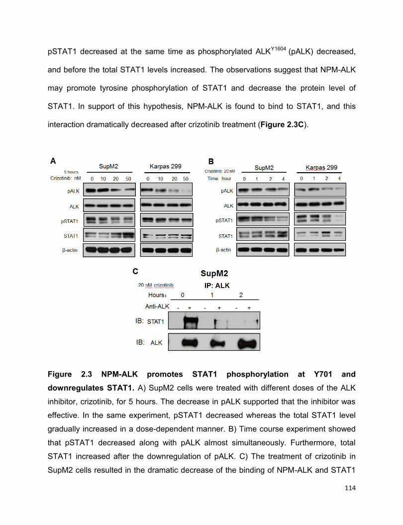
114
pSTAT1 decreased at the same time as phosphorylated ALKY1604 (pALK) decreased,
and before the total STAT1 levels increased. The observations suggest that NPM-ALK
may promote tyrosine phosphorylation of STAT1 and decrease the protein level of
STAT1. In support of this hypothesis, NPM-ALK is found to bind to STAT1, and this
interaction dramatically decreased after crizotinib treatment (Figure 2.3C).
Figure 2.3 NPM-ALK promotes STAT1 phosphorylation at Y701 and
downregulates STAT1. A) SupM2 cells were treated with different doses of the ALK
inhibitor, crizotinib, for 5 hours. The decrease in pALK supported that the inhibitor was
effective. In the same experiment, pSTAT1 decreased whereas the total STAT1 level
gradually increased in a dose-dependent manner. B) Time course experiment showed
that pSTAT1 decreased along with pALK almost simultaneously. Furthermore, total
STAT1 increased after the downregulation of pALK. C) The treatment of crizotinib in
SupM2 cells resulted in the dramatic decrease of the binding of NPM-ALK and STAT1
115
(i.e. at the time point of 1 hour and 2 hour), assessed by co-immunoprecipitation assay.
Results shown were representative of 3 independent experiments. Results shown
were representative of three independent experiments.
Furthermore, transient transfection of NPM-ALK into GP293 cells resulted in a dramatic
increase in pSTAT1, and this change was accompanied by a reciprocal decrease in
total STAT1 (Figure 2.3D). As shown in Figure 2.3E, the effect on total STAT1 due to
enforced NPM-ALK expression in GP293 cells was dependent on the proteasomal
degradation pathway, since MG132 almost completely abrogated this effect.
Then we questioned if the phosphorylation of STAT1 at its tyrosine residue 701 is
necessary for its downregulation. Thus, GP293 cells were co-transfected NPM-ALK
with wild-type STAT1 or mutant STAT1Y701F. As shown in Figure 2.3F, NPM-ALK
effectively phosphorylated STAT1 when cells were co-transfected with wild-type STAT1.
There was only a relatively small increase in pSTAT1 in cells co-transfected with
STAT1Y701F, suggesting the tyrosine residue 701 is the predominant tyrosine
phosphorylation site of STAT1 for NPM-ALK. After 24 hours of gene transfection, cells
transfected with wild-type STAT1 expressed only a small amount of total STAT1 as
compared to that in cells transfected with STAT1Y701F. Taken together, the data
suggested that NPM-ALK promotes STAT1 phosphorylation at predominantly the Y701
residue, and by doing so, promotes its degradation.
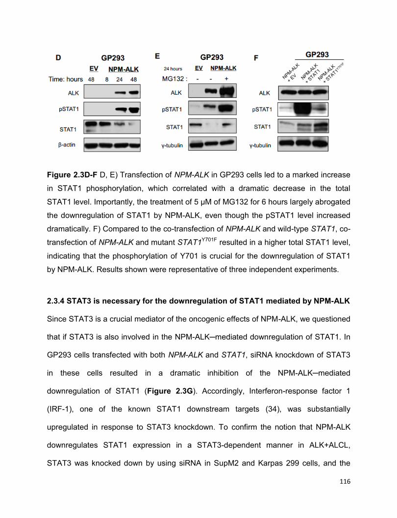
116
Figure 2.3D-F D, E) Transfection of NPM-ALK in GP293 cells led to a marked increase
in STAT1 phosphorylation, which correlated with a dramatic decrease in the total
STAT1 level. Importantly, the treatment of 5 μM of MG132 for 6 hours largely abrogated
the downregulation of STAT1 by NPM-ALK, even though the pSTAT1 level increased
dramatically. F) Compared to the co-transfection of NPM-ALK and wild-type STAT1, co-
transfection of NPM-ALK and mutant STAT1Y701F resulted in a higher total STAT1 level,
indicating that the phosphorylation of Y701 is crucial for the downregulation of STAT1
by NPM-ALK. Results shown were representative of three independent experiments.
2.3.4 STAT3 is necessary for the downregulation of STAT1 mediated by NPM-ALK
Since STAT3 is a crucial mediator of the oncogenic effects of NPM-ALK, we questioned
that if STAT3 is also involved in the NPM-ALK─mediated downregulation of STAT1. In
GP293 cells transfected with both NPM-ALK and STAT1, siRNA knockdown of STAT3
in these cells resulted in a dramatic inhibition of the NPM-ALK─mediated
downregulation of STAT1 (Figure 2.3G). Accordingly, Interferon-response factor 1
(IRF-1), one of the known STAT1 downstream targets (34), was substantially
upregulated in response to STAT3 knockdown. To confirm the notion that NPM-ALK
downregulates STAT1 expression in a STAT3-dependent manner in ALK+ALCL,
STAT3 was knocked down by using siRNA in SupM2 and Karpas 299 cells, and the

117
results suggested that a readily detectable upregulation of STAT1 in both cell lines was
observed after STAT3 knockdown (Figure 2.3H).
Figure 2.3G-H G) With the siRNA knockdown of STAT3, the NPM-ALK─mediated
downregulation of STAT1 was largely abrogated. Correlating with this finding, IRF-1 (a
STAT1 downstream target) was higher when STAT3 was silenced in presence of NPM-
ALK. H) Time course experiment of siRNA knockdown of STAT3 in the two ALK+ALCL
cell lines correlated with the reciprocal increase in the total STAT1 level. Results shown
were representative of three independent experiments.
2.3.5 STAT1 signaling is functionally intact in ALK+ALCL
We then asked if the STAT1 signaling pathway in ALK+ALCL is functionally intact. As
shown in Figure 2.4A, STAT1 was rapidly phosphorylated in ALK+ALCL cells in
response to IFNγ. The protein level of IRF-1 was expectedly upregulated. Furthermore,
at 24 hours, both cell lines showed evidence of apoptosis, as evidenced by the
expression of cleaved PARP detectable by Western blotting and the significant
increase in the sub G0/G1 phase detectable by flow cytometry (Figure 2.4B and 2.4C).
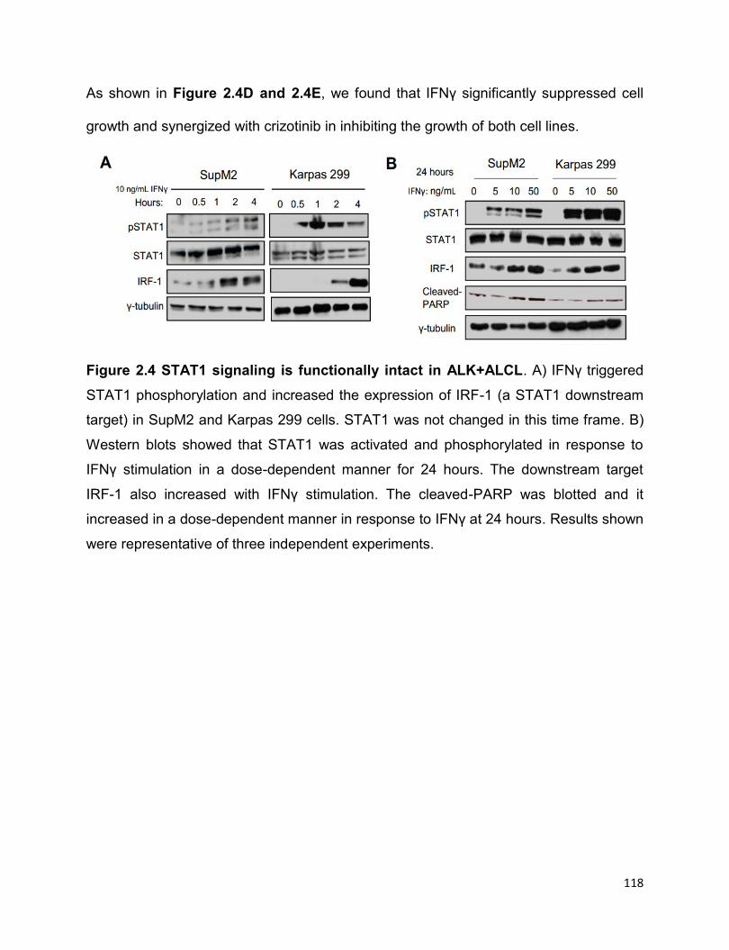
118
As shown in Figure 2.4D and 2.4E, we found that IFNγ significantly suppressed cell
growth and synergized with crizotinib in inhibiting the growth of both cell lines.
Figure 2.4 STAT1 signaling is functionally intact in ALK+ALCL. A) IFNγ triggered
STAT1 phosphorylation and increased the expression of IRF-1 (a STAT1 downstream
target) in SupM2 and Karpas 299 cells. STAT1 was not changed in this time frame. B)
Western blots showed that STAT1 was activated and phosphorylated in response to
IFNү stimulation in a dose-dependent manner for 24 hours. The downstream target
IRF-1 also increased with IFNү stimulation. The cleaved-PARP was blotted and it
increased in a dose-dependent manner in response to IFNү at 24 hours. Results shown
were representative of three independent experiments.

119
Figure 2.4C-D C) Cell-cycle analysis suggested that the percent of cells in Sub G0/G1
significantly increased upon the treatment of various doses of IFNү for 24 hours in
Karpas 299 cells. D) The trypan blue exclusion assay suggested that IFNү significantly
decreased cell growth in both ALK+ALCL cell lines in a dose-dependent manner.
Statistical significance is denoted by * (P<0.05) and ** (P<0.01). Results shown were
representative of triplicate experiments.
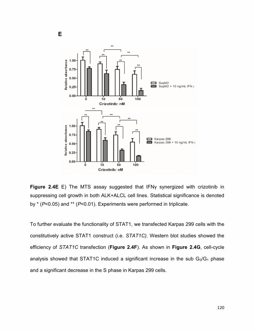
120
Figure 2.4E E) The MTS assay suggested that IFNү synergized with crizotinib in
suppressing cell growth in both ALK+ALCL cell lines. Statistical significance is denoted
by * (P<0.05) and ** (P<0.01). Experiments were performed in triplicate.
To further evaluate the functionality of STAT1, we transfected Karpas 299 cells with the
constitutively active STAT1 construct (i.e. STAT1C). Western blot studies showed the
efficiency of STAT1C transfection (Figure 2.4F). As shown in Figure 2.4G, cell-cycle
analysis showed that STAT1C induced a significant increase in the sub G0/G1 phase
and a significant decrease in the S phase in Karpas 299 cells.

121
Figure 2.4F-G F, G) Karpas 299 cells transfected with STAT1C-flag showed a
significant accumulation of the sub G0/G1 fraction. Statistical significance was
calculated by Student’s t-test where *P<0.05, **P<0.01.
To further examine the biological significance of STAT1 in ALK+ALCL, we established
two conditional STAT1C-stably transfected ALK+ALCL cell lines where the expression
of STAT1C can be induced in a dose-dependent manner with the addition of
doxycycline (i.e. Dox), labeled Tet on-SupM2-STAT1C and Tet on-Karpas 299-STAT1C.
The Dox-dose dependent expression of STAT1C in these two cell lines was confirmed
by Western blotting (Figure 2.5A). Using Tet on-SupM2-STAT1C cells, we found a
positive correlation between STAT1 and that of IRF-7 and T-bet, two STAT1
downstream targets (35, 36). SOCS-1, a negative regulator of the STAT1 signaling
pathway (37), was also upregulated. In contrast, the protein levels of STAT3 and
phosphorylated STAT3Y705 (pSTAT3) did not change appreciably. Interestingly, two
STAT3 target genes, Bcl-2 and Survivin (38, 39), were downregulated in a dose
dependent manner (Figure 2.5A). As shown in Figure 2.5B-C, the induced expression
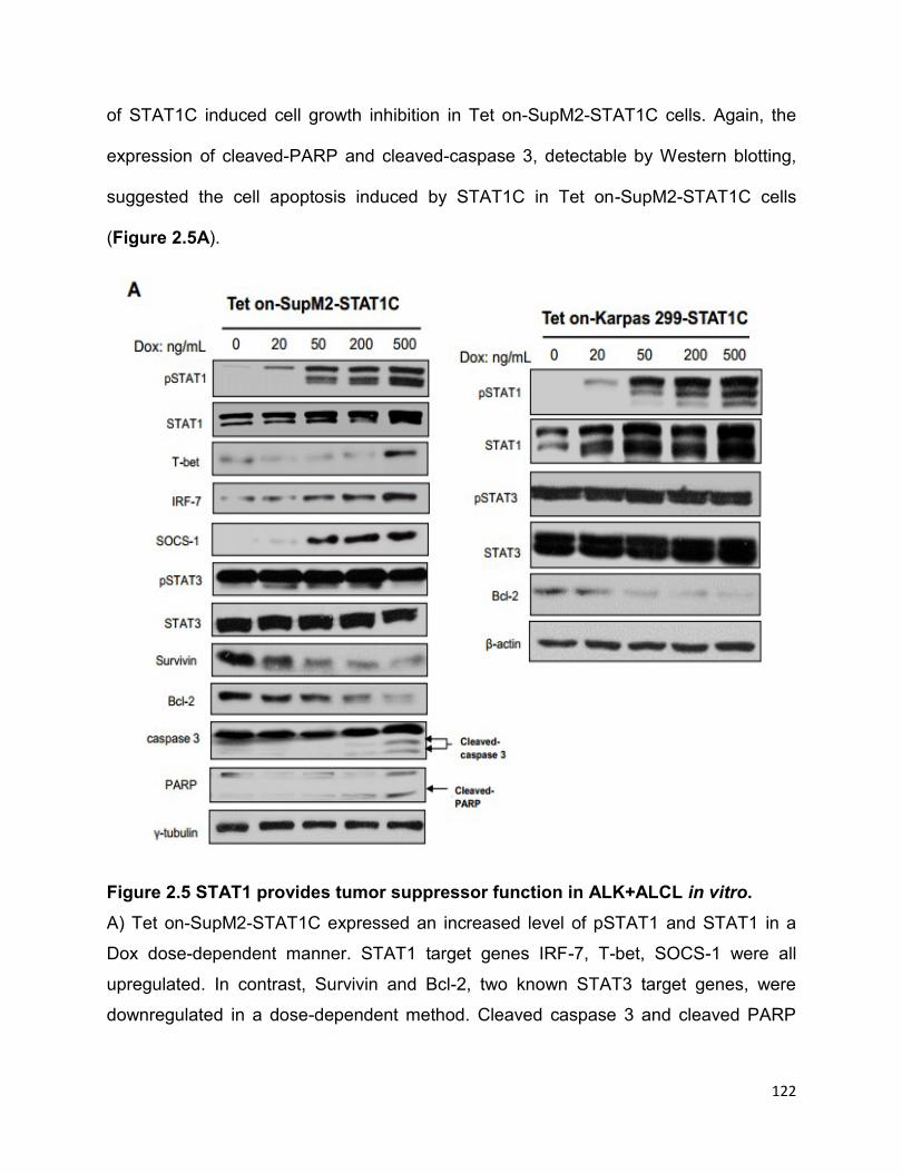
122
of STAT1C induced cell growth inhibition in Tet on-SupM2-STAT1C cells. Again, the
expression of cleaved-PARP and cleaved-caspase 3, detectable by Western blotting,
suggested the cell apoptosis induced by STAT1C in Tet on-SupM2-STAT1C cells
(Figure 2.5A).
Figure 2.5 STAT1 provides tumor suppressor function in ALK+ALCL in vitro.
A) Tet on-SupM2-STAT1C expressed an increased level of pSTAT1 and STAT1 in a
Dox dose-dependent manner. STAT1 target genes IRF-7, T-bet, SOCS-1 were all
upregulated. In contrast, Survivin and Bcl-2, two known STAT3 target genes, were
downregulated in a dose-dependent method. Cleaved caspase 3 and cleaved PARP
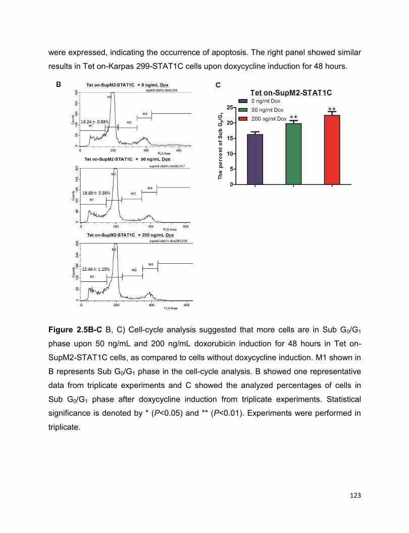
123
were expressed, indicating the occurrence of apoptosis. The right panel showed similar
results in Tet on-Karpas 299-STAT1C cells upon doxycycline induction for 48 hours.
Figure 2.5B-C B, C) Cell-cycle analysis suggested that more cells are in Sub G0/G1
phase upon 50 ng/mL and 200 ng/mL doxorubicin induction for 48 hours in Tet on-
SupM2-STAT1C cells, as compared to cells without doxycycline induction. M1 shown in
B represents Sub G0/G1 phase in the cell-cycle analysis. B showed one representative
data from triplicate experiments and C showed the analyzed percentages of cells in
Sub G0/G1 phase after doxycycline induction from triplicate experiments. Statistical
significance is denoted by * (P<0.05) and ** (P<0.01). Experiments were performed in
triplicate.
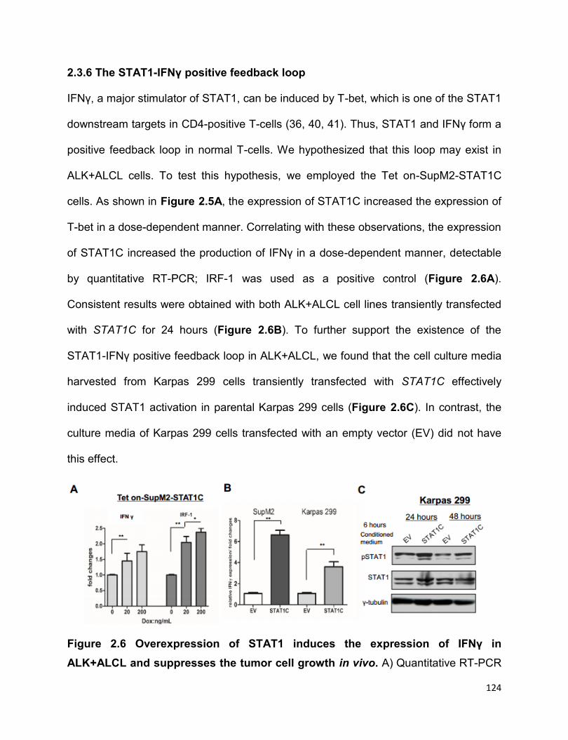
124
2.3.6 The STAT1-IFNγ positive feedback loop
IFNγ, a major stimulator of STAT1, can be induced by T-bet, which is one of the STAT1
downstream targets in CD4-positive T-cells (36, 40, 41). Thus, STAT1 and IFNγ form a
positive feedback loop in normal T-cells. We hypothesized that this loop may exist in
ALK+ALCL cells. To test this hypothesis, we employed the Tet on-SupM2-STAT1C
cells. As shown in Figure 2.5A, the expression of STAT1C increased the expression of
T-bet in a dose-dependent manner. Correlating with these observations, the expression
of STAT1C increased the production of IFNγ in a dose-dependent manner, detectable
by quantitative RT-PCR; IRF-1 was used as a positive control (Figure 2.6A).
Consistent results were obtained with both ALK+ALCL cell lines transiently transfected
with STAT1C for 24 hours (Figure 2.6B). To further support the existence of the
STAT1-IFNγ positive feedback loop in ALK+ALCL, we found that the cell culture media
harvested from Karpas 299 cells transiently transfected with STAT1C effectively
induced STAT1 activation in parental Karpas 299 cells (Figure 2.6C). In contrast, the
culture media of Karpas 299 cells transfected with an empty vector (EV) did not have
this effect.
Figure 2.6 Overexpression of STAT1 induces the expression of IFNγ in
ALK+ALCL and suppresses the tumor cell growth in vivo. A) Quantitative RT-PCR
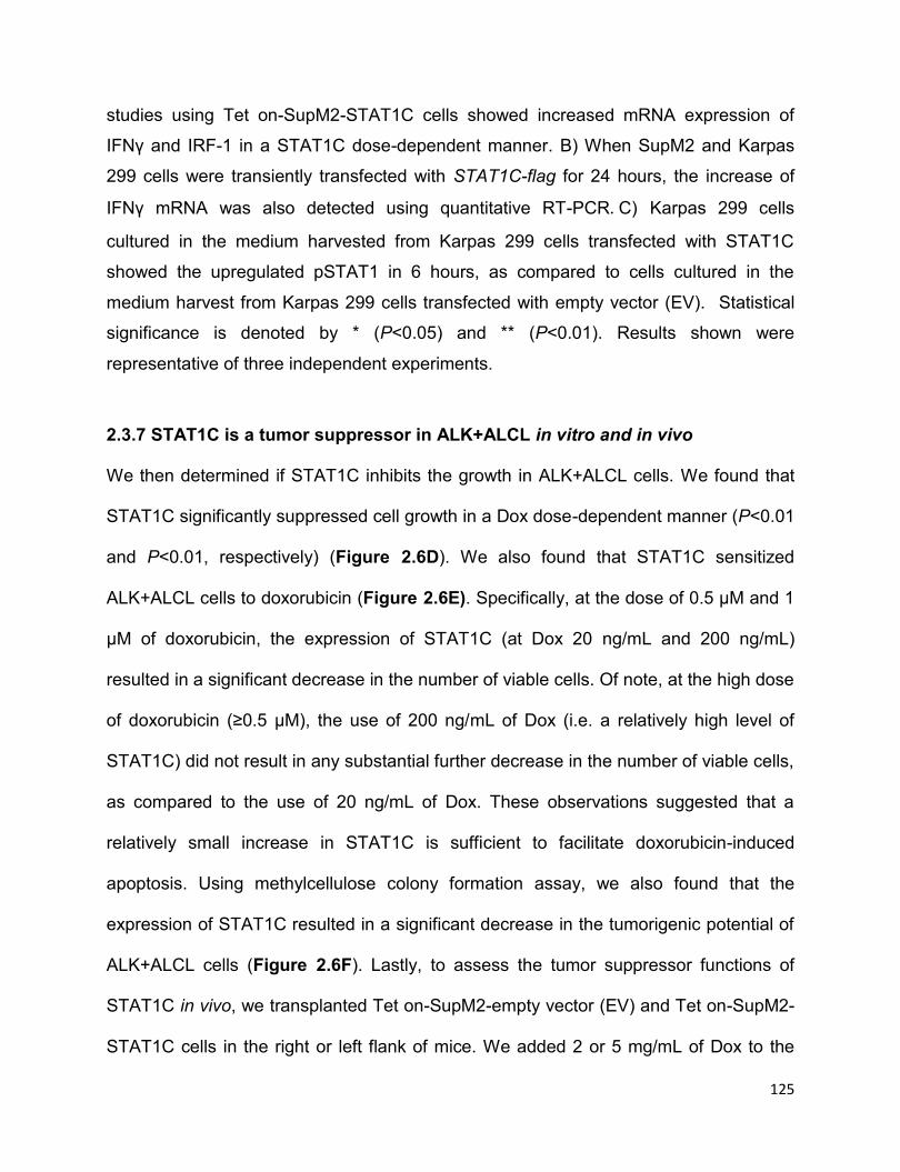
125
studies using Tet on-SupM2-STAT1C cells showed increased mRNA expression of
IFNγ and IRF-1 in a STAT1C dose-dependent manner. B) When SupM2 and Karpas
299 cells were transiently transfected with STAT1C-flag for 24 hours, the increase of
IFNγ mRNA was also detected using quantitative RT-PCR.C) Karpas 299 cells
cultured in the medium harvested from Karpas 299 cells transfected with STAT1C
showed the upregulated pSTAT1 in 6 hours, as compared to cells cultured in the
medium harvest from Karpas 299 cells transfected with empty vector (EV). Statistical
significance is denoted by * (P<0.05) and ** (P<0.01). Results shown were
representative of three independent experiments.
2.3.7 STAT1C is a tumor suppressor in ALK+ALCL in vitro and in vivo
We then determined if STAT1C inhibits the growth in ALK+ALCL cells. We found that
STAT1C significantly suppressed cell growth in a Dox dose-dependent manner (P<0.01
and P<0.01, respectively) (Figure 2.6D). We also found that STAT1C sensitized
ALK+ALCL cells to doxorubicin (Figure 2.6E). Specifically, at the dose of 0.5 µM and 1
µM of doxorubicin, the expression of STAT1C (at Dox 20 ng/mL and 200 ng/mL)
resulted in a significant decrease in the number of viable cells. Of note, at the high dose
of doxorubicin (≥0.5 µM), the use of 200 ng/mL of Dox (i.e. a relatively high level of
STAT1C) did not result in any substantial further decrease in the number of viable cells,
as compared to the use of 20 ng/mL of Dox. These observations suggested that a
relatively small increase in STAT1C is sufficient to facilitate doxorubicin-induced
apoptosis. Using methylcellulose colony formation assay, we also found that the
expression of STAT1C resulted in a significant decrease in the tumorigenic potential of
ALK+ALCL cells (Figure 2.6F). Lastly, to assess the tumor suppressor functions of
STAT1C in vivo, we transplanted Tet on-SupM2-empty vector (EV) and Tet on-SupM2-
STAT1C cells in the right or left flank of mice. We added 2 or 5 mg/mL of Dox to the

126
drinking water to induce STAT1C expression (Figure 2.6G). Tet on-SupM2-STAT1C
cells showed significantly smaller tumor sizes than the empty vector group (Figure
2.6H) (P<0.01 for both). The tumors of the two groups were illustrated in Figure 2.6I,
and the STAT1 protein levels in four representative tumors from mice #1 and mice #6
were analysed using Western blotting, as shown in Figure 2.6J.
Figure 2.6D-E D) Using Tet on-SupM2-STAT1C cells, we found a significant decrease
in the number of viable cells with increasing Dox. E) Using Tet on-SupM2-STAT1C cells,
we found that STAT1C sensitizes cells to doxorubicin-induced cell growth inhibition at
0.5 and 1.0 µM of doxorubicin. F) Colony formation significantly decreased with
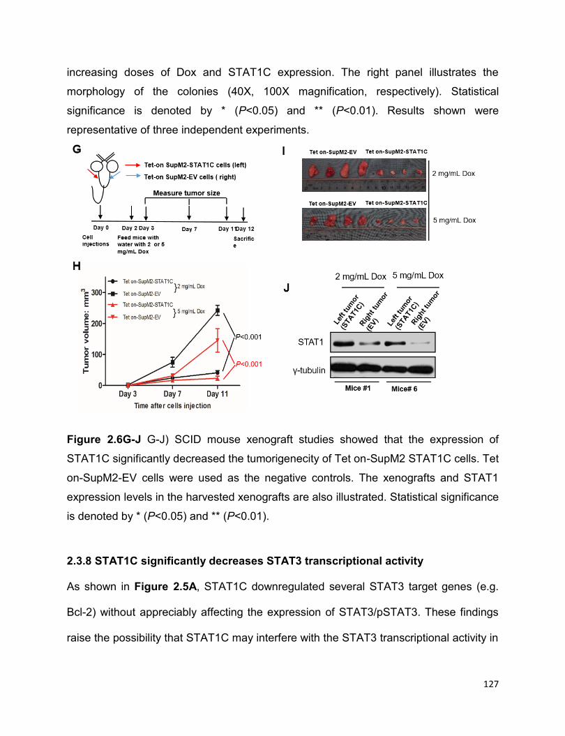
127
increasing doses of Dox and STAT1C expression. The right panel illustrates the
morphology of the colonies (40X, 100X magnification, respectively). Statistical
significance is denoted by * (P<0.05) and ** (P<0.01). Results shown were
representative of three independent experiments.
Figure 2.6G-J G-J) SCID mouse xenograft studies showed that the expression of
STAT1C significantly decreased the tumorigenecity of Tet on-SupM2 STAT1C cells. Tet
on-SupM2-EV cells were used as the negative controls. The xenografts and STAT1
expression levels in the harvested xenografts are also illustrated. Statistical significance
is denoted by * (P<0.05) and ** (P<0.01).
2.3.8 STAT1C significantly decreases STAT3 transcriptional activity
As shown in Figure 2.5A, STAT1C downregulated several STAT3 target genes (e.g.
Bcl-2) without appreciably affecting the expression of STAT3/pSTAT3. These findings
raise the possibility that STAT1C may interfere with the STAT3 transcriptional activity in
128
ALK+ALCL cells. As shown in Figure 2.7A, increasing levels of STAT1C in the Tet on-
SupM2-STAT1C cells resulted in a dose-dependent decrease in the STAT3
transcriptional activity, as measured by dual luciferase assay. Using the same cell line,
we found that STAT1C inhibited STAT3 binding to its consensus DNA sequence in a
dose-dependent manner (Figure 2.7B). In addition, using co-immunopreciptation assay
and the Tet on-SupM2-STAT1C cells, we found that expression of STAT1C increased
the formation of STAT1/STAT3 heterodimers, presumably at the expense of STAT3
homodimers, as the total STAT3 protein level did not change appreciably in this
experiment (Figure 2.7C).
Figure 2.7 STAT1C significantly decreases STAT3 transcriptional activity.
A) STAT3 luciferase reporter assay showed STAT1C significantly decreased STAT3
transcriptional activity in a Dox-dose dependent manner. Treatment of 200 ng/mL of
Dox in Tet on-SupM2- EV cells showed low-dose Dox has unappreciable effects on
STAT3 transcriptional activity. B) STAT3 consensus DNA probe binding assay showed

129
STAT1C sequestered STAT3 DNA binding ability in a Dox-dose dependent manner,
and the Western blotting in the right panel showed the input of cell nuclear lysates.
Histone Deacetylase 1(HDAC-1) was used as loading control for nuclear lysates. C)
Co-immunoprecipitation assay showed induction of STAT1C expression leads to more
STAT1/STAT3 heterodimers formations, at the expense of STAT3 homodimers. The
right panel showed the input. Statistical significance is denoted by * (P<0.05) and **
(P<0.01). Results shown were representative of three independent experiments.
2.3.9 siRNA knockdown of STAT1 confers resistance to STAT3 inhibition-induced
cell death
We investigated if STAT1 plays a role in STAT3 inhibition-induced apoptosis in
ALK+ALCL, a well-published phenomenon (42). As shown in Figure 2.8A, treatment of
SupM2 with Stattic, a widely used STAT3 inhibitor (43), dramatically decreased
pSTAT3 level. Transfection of STAT1 siRNA significantly decreased the inhibitory effect
of Stattic, and the IC50 increased significantly compared to SupM2 cells transfected
with scramble siRNA (Figure 2.8B). These findings suggest that STAT3 inhibition-
induced apoptosis is partly STAT1-dependent. In parallel with these findings, co-
transfection of STAT1 siRNA and STAT3 siRNA in SupM2 cells led to decreased cell
killing, as compared to transfection of STAT3 siRNA alone (Figure 2.8C). Western blots
shown in Figure 2.8C also showed the efficiency of siRNAs knockdown in SupM2 cells.
Of note, transfection of STAT3 siRNA substantially increased the STAT1 total protein
level, and this result was consistent to that shown in Figure 2.3H.

130
Figure 2.8 siRNA knockdown of STAT1 confers resistance to STAT3 inhibition-
induced cell death. A) SupM2 were exposed to varying doses of Stattic for 4 hours,
Western blotting showed the decrease of pSTAT3 in a dose-dependent method. B)
SupM2 cells transfected with two species of STAT1 siRNA showed significantly higher
IC50 to Stattic compared to cells transfected with scramble siRNA transfection.
Western blotting studies showed the siRNA knockdown efficiency, which was higher
with STAT1 siRNA 1 compared to STAT1 siRNA 2. The densitometry values shown
were normalized to the band of γ-tubulin. C) Transfection of STAT1 siRNA significantly
reduced the suppression of cell growth induced by STAT3 siRNA in SupM2 cells. The
Western blotting results showed the efficiency of siRNA knockdowns. Image J software
was used to analyze the densitometry value of Western blots bands. Statistical
significance is denoted by * (P<0.05) and ** (P<0.01). Results shown were
representative of 3 independent experiments.
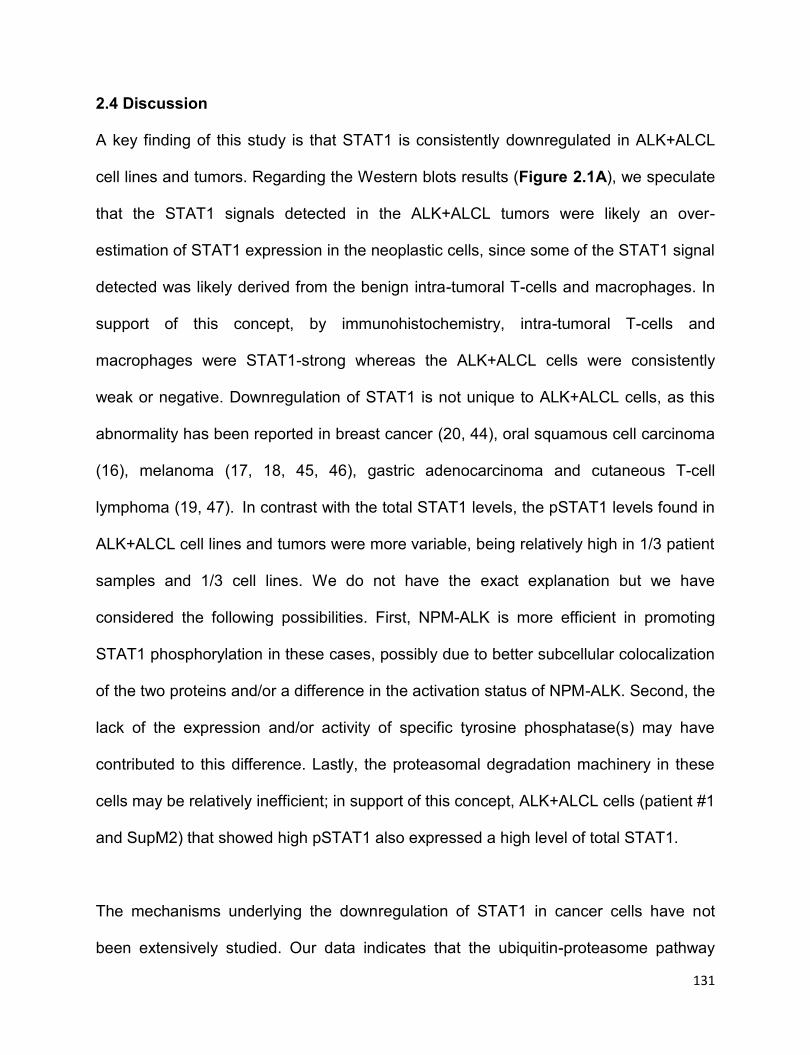
131
2.4 Discussion
A key finding of this study is that STAT1 is consistently downregulated in ALK+ALCL
cell lines and tumors. Regarding the Western blots results (Figure 2.1A), we speculate
that the STAT1 signals detected in the ALK+ALCL tumors were likely an over-
estimation of STAT1 expression in the neoplastic cells, since some of the STAT1 signal
detected was likely derived from the benign intra-tumoral T-cells and macrophages. In
support of this concept, by immunohistochemistry, intra-tumoral T-cells and
macrophages were STAT1-strong whereas the ALK+ALCL cells were consistently
weak or negative. Downregulation of STAT1 is not unique to ALK+ALCL cells, as this
abnormality has been reported in breast cancer (20, 44), oral squamous cell carcinoma
(16), melanoma (17, 18, 45, 46), gastric adenocarcinoma and cutaneous T-cell
lymphoma (19, 47). In contrast with the total STAT1 levels, the pSTAT1 levels found in
ALK+ALCL cell lines and tumors were more variable, being relatively high in 1/3 patient
samples and 1/3 cell lines. We do not have the exact explanation but we have
considered the following possibilities. First, NPM-ALK is more efficient in promoting
STAT1 phosphorylation in these cases, possibly due to better subcellular colocalization
of the two proteins and/or a difference in the activation status of NPM-ALK. Second, the
lack of the expression and/or activity of specific tyrosine phosphatase(s) may have
contributed to this difference. Lastly, the proteasomal degradation machinery in these
cells may be relatively inefficient; in support of this concept, ALK+ALCL cells (patient #1
and SupM2) that showed high pSTAT1 also expressed a high level of total STAT1.
The mechanisms underlying the downregulation of STAT1 in cancer cells have not
been extensively studied. Our data indicates that the ubiquitin-proteasome pathway
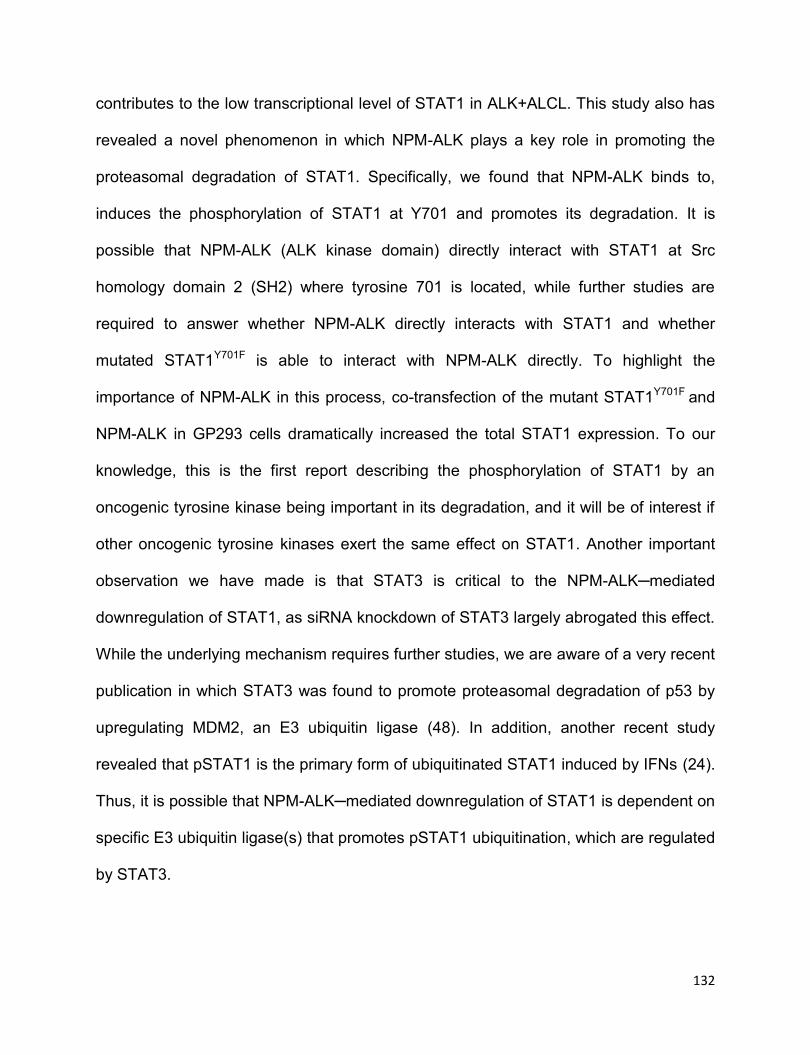
132
contributes to the low transcriptional level of STAT1 in ALK+ALCL. This study also has
revealed a novel phenomenon in which NPM-ALK plays a key role in promoting the
proteasomal degradation of STAT1. Specifically, we found that NPM-ALK binds to,
induces the phosphorylation of STAT1 at Y701 and promotes its degradation. It is
possible that NPM-ALK (ALK kinase domain) directly interact with STAT1 at Src
homology domain 2 (SH2) where tyrosine 701 is located, while further studies are
required to answer whether NPM-ALK directly interacts with STAT1 and whether
mutated STAT1Y701F is able to interact with NPM-ALK directly. To highlight the
importance of NPM-ALK in this process, co-transfection of the mutant STAT1Y701F and
NPM-ALK in GP293 cells dramatically increased the total STAT1 expression. To our
knowledge, this is the first report describing the phosphorylation of STAT1 by an
oncogenic tyrosine kinase being important in its degradation, and it will be of interest if
other oncogenic tyrosine kinases exert the same effect on STAT1. Another important
observation we have made is that STAT3 is critical to the NPM-ALK─mediated
downregulation of STAT1, as siRNA knockdown of STAT3 largely abrogated this effect.
While the underlying mechanism requires further studies, we are aware of a very recent
publication in which STAT3 was found to promote proteasomal degradation of p53 by
upregulating MDM2, an E3 ubiquitin ligase (48). In addition, another recent study
revealed that pSTAT1 is the primary form of ubiquitinated STAT1 induced by IFNs (24).
Thus, it is possible that NPM-ALK─mediated downregulation of STAT1 is dependent on
specific E3 ubiquitin ligase(s) that promotes pSTAT1 ubiquitination, which are regulated
by STAT3.
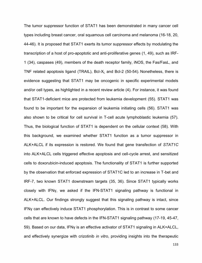
133
The tumor suppressor function of STAT1 has been demonstrated in many cancer cell
types including breast cancer, oral squamous cell carcinoma and melanoma (16-18, 20,
44-46). It is proposed that STAT1 exerts its tumor suppressor effects by modulating the
transcription of a host of pro-apoptotic and anti-proliferative genes (1, 49), such as IRF-
1 (34), caspases (49), members of the death receptor family, iNOS, the Fas/FasL, and
TNF related apoptosis ligand (TRAIL), Bcl-XL and Bcl-2 (50-54). Nonetheless, there is
evidence suggesting that STAT1 may be oncogenic in specific experimental models
and/or cell types, as highlighted in a recent review article (4). For instance, it was found
that STAT1-deficient mice are protected from leukemia development (55). STAT1 was
found to be important for the expansion of leukemia initiating cells (56). STAT1 was
also shown to be critical for cell survival in T-cell acute lymphoblastic leukemia (57).
Thus, the biological function of STAT1 is dependent on the cellular context (58). With
this background, we examined whether STAT1 function as a tumor suppressor in
ALK+ALCL if its expression is restored. We found that gene transfection of STAT1C
into ALK+ALCL cells triggered effective apoptosis and cell-cycle arrest, and sensitized
cells to doxorubicin-induced apoptosis. The functionality of STAT1 is further supported
by the observation that enforced expression of STAT1C led to an increase in T-bet and
IRF-7, two known STAT1 downstream targets (35, 36). Since STAT1 typically works
closely with IFNү, we asked if the IFN-STAT1 signaling pathway is functional in
ALK+ALCL. Our findings strongly suggest that this signaling pathway is intact, since
IFNγ can effectively induce STAT1 phosphorylation. This is in contrast to some cancer
cells that are known to have defects in the IFN-STAT1 signaling pathway (17-19, 45-47,
59). Based on our data, IFNγ is an effective activator of STAT1 signaling in ALK+ALCL,
and effectively synergize with crizotinib in vitro, providing insights into the therapeutic
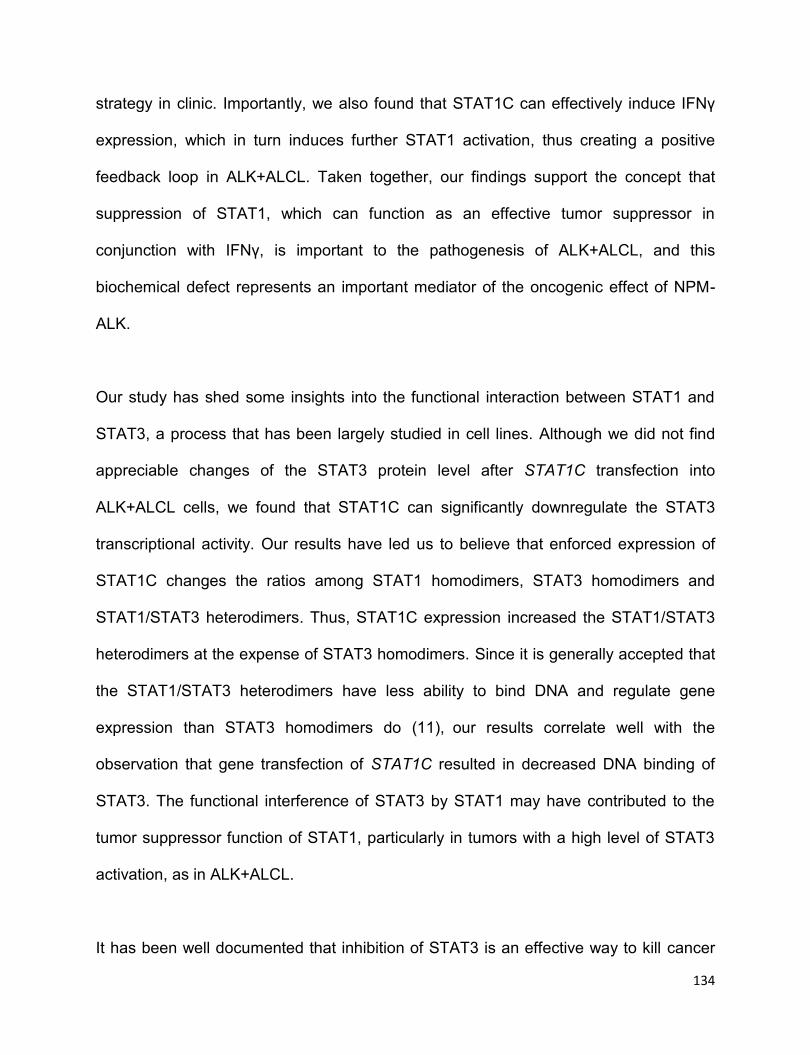
134
strategy in clinic. Importantly, we also found that STAT1C can effectively induce IFNγ
expression, which in turn induces further STAT1 activation, thus creating a positive
feedback loop in ALK+ALCL. Taken together, our findings support the concept that
suppression of STAT1, which can function as an effective tumor suppressor in
conjunction with IFNү, is important to the pathogenesis of ALK+ALCL, and this
biochemical defect represents an important mediator of the oncogenic effect of NPM-
ALK.
Our study has shed some insights into the functional interaction between STAT1 and
STAT3, a process that has been largely studied in cell lines. Although we did not find
appreciable changes of the STAT3 protein level after STAT1C transfection into
ALK+ALCL cells, we found that STAT1C can significantly downregulate the STAT3
transcriptional activity. Our results have led us to believe that enforced expression of
STAT1C changes the ratios among STAT1 homodimers, STAT3 homodimers and
STAT1/STAT3 heterodimers. Thus, STAT1C expression increased the STAT1/STAT3
heterodimers at the expense of STAT3 homodimers. Since it is generally accepted that
the STAT1/STAT3 heterodimers have less ability to bind DNA and regulate gene
expression than STAT3 homodimers do (11), our results correlate well with the
observation that gene transfection of STAT1C resulted in decreased DNA binding of
STAT3. The functional interference of STAT3 by STAT1 may have contributed to the
tumor suppressor function of STAT1, particularly in tumors with a high level of STAT3
activation, as in ALK+ALCL.
It has been well documented that inhibition of STAT3 is an effective way to kill cancer
135
cells that express constitutively activated STAT3 (60), including ALK+ALCL cells (42).
Based on our results presented here, one may speculate that apoptosis induced by
STAT3 inhibition in ALK+ALCL may be dependent on STAT1. Specifically, a relatively
high level of STAT1 expression in ALK+ALCL will lower the threshold of apoptosis due
to STAT3 inhibition, whereas siRNA knockdown of STAT1 will provide resistance of
apoptosis induced by STAT3 inhibition. In keeping with this concept, we found that
siRNA knockdown of STAT1 in ALK+ALCL indeed led to increased resistance to Stattic,
a well-studied STAT3 inhibitor (43). These findings also further highlight the biological
importance of the functional interactions between STAT1 and STAT3 in dictating the
cell fate of cancer cells. In brief, the schematic working model of STAT1 in ALK+ALCL
has been demonstrated in Figure 2.9.
To conclude, we report that STAT1 is consistently downregulated in ALK+ALCL. In
view of the observations that the STAT1 signaling is intact and STAT1 (if sufficiently
expressed) exerts potent tumor suppressor effect, we conclude that the downregulation
of STAT1 by NPM-ALK represents a novel mechanism by which this oncogenic tyrosine
kinase promotes tumorigenesis. Our results have also shed important insight into the
functional interaction between STAT1 and STAT3 in cancer cells, and the biological
significance of this interaction in dictating the cell fate. We believe that enhancing the
STAT1 signaling might be an attractive therapeutic strategy in treating cancer,
especially when STAT3 is constitutively active.

136
Figure 2.9 Schematic model of STAT1 in ALK+ ALCL.
NPM-ALK promotes STAT1 phosphorylation at Y701, and thereby, downregulates
STAT1 in a STAT3-dependent proteasome pathway. If STAT1 expression is restored to
a certain high level in ALK+ALCL, it is sufficient to induce cell growth inhibition and cell
apoptosis by suppressing STAT3 transcriptional activity and inducing T-bet expression
which stimulates the transcription of IFNγ, thus resulting in a positive forward loop for
STAT1 activation.
STAT: Signal transducer and activator of transcription; IRF: interferon response factor;
Bcl-2: B-cell lymphoma 2; IFNγ: interferon gamma; JAK: Janus kinase; T-bet: T-box
transcription factor TBX21.
2.5 References
1. Kim HS, Lee MS. STAT1 as a key modulator of cell death. Cellular signalling.
2007;19(3):454-65.

137
2. Schroder K, Hertzog PJ, Ravasi T, Hume DA. Interferon-gamma: an overview of
signals, mechanisms and functions. Journal of leukocyte biology. 2004;75(2):163-89.
3. Koromilas AE, Sexl V. The tumor suppressor function of STAT1 in breast cancer.
Jak-Stat. 2013;2(2):e23353.
4. Avalle L, Pensa S, Regis G, Novelli F, Poli V. STAT1 and STAT3 in
tumorigenesis: A matter of balance. Jak-Stat. 2012;1(2):65-72.
5. Bowman T, Garcia R, Turkson J, Jove R. STATs in oncogenesis. Oncogene.
2000;19(21):2474-88.
6. Guzzo C, Che Mat NF, Gee K. Interleukin-27 induces a STAT1/3- and NF-
kappaB-dependent proinflammatory cytokine profile in human monocytes. The Journal
of biological chemistry. 2010;285(32):24404-11.
7. Sato T, Selleri C, Young NS, Maciejewski JP. Inhibition of interferon regulatory
factor-1 expression results in predominance of cell growth stimulatory effects of
interferon-gamma due to phosphorylation of Stat1 and Stat3. Blood. 1997;90(12):4749-
58.
8. Stancato LF, David M, Carter-Su C, Larner AC, Pratt WB. Preassociation of
STAT1 with STAT2 and STAT3 in separate signalling complexes prior to cytokine
stimulation. The Journal of biological chemistry. 1996;271(8):4134-7.
9. Regis G, Pensa S, Boselli D, Novelli F, Poli V. Ups and downs: the
STAT1:STAT3 seesaw of Interferon and gp130 receptor signalling. Semin Cell Dev Biol.
2008;19(4):351-9.
10. Thyrell L, Arulampalam V, Hjortsberg L, Farnebo M, Grander D, Pokrovskaja
Tamm K. Interferon alpha induces cell death through interference with interleukin 6

138
signaling and inhibition of STAT3 activity. Experimental cell research.
2007;313(19):4015-24.
11. Ho HH, Ivashkiv LB. Role of STAT3 in type I interferon responses. Negative
regulation of STAT1-dependent inflammatory gene activation. The Journal of biological
chemistry. 2006;281(20):14111-8.
12. Shen Y, Devgan G, Darnell JE, Jr., Bromberg JF. Constitutively activated Stat3
protects fibroblasts from serum withdrawal and UV-induced apoptosis and antagonizes
the proapoptotic effects of activated Stat1. Proceedings of the National Academy of
Sciences of the United States of America. 2001;98(4):1543-8.
13. Kortylewski M, Kujawski M, Wang T, Wei S, Zhang S, Pilon-Thomas S, et al.
Inhibiting Stat3 signaling in the hematopoietic system elicits multicomponent antitumor
immunity. Nature medicine. 2005;11(12):1314-21.
14. Bluyssen HA, Rastmanesh MM, Tilburgs C, Jie K, Wesseling S, Goumans MJ, et
al. IFN gamma-dependent SOCS3 expression inhibits IL-6-induced STAT3
phosphorylation and differentially affects IL-6 mediated transcriptional responses in
endothelial cells. American journal of physiology Cell physiology. 2010;299(2):C354-62.
15. Dimberg LY, Dimberg A, Ivarsson K, Fryknas M, Rickardson L, Tobin G, et al.
Stat1 activation attenuates IL-6 induced Stat3 activity but does not alter apoptosis
sensitivity in multiple myeloma. BMC cancer. 2012;12:318.
16. Laimer K, Spizzo G, Obrist P, Gastl G, Brunhuber T, Schafer G, et al. STAT1
activation in squamous cell cancer of the oral cavity: a potential predictive marker of
response to adjuvant chemotherapy. Cancer. 2007;110(2):326-33.
17. Wong LH, Krauer KG, Hatzinisiriou I, Estcourt MJ, Hersey P, Tam ND, et al.
Interferon-resistant human melanoma cells are deficient in ISGF3 components, STAT1,

139
STAT2, and p48-ISGF3gamma. The Journal of biological chemistry.
1997;272(45):28779-85.
18. Kovarik J, Boudny V, Kocak I, Lauerova L, Fait V, Vagundova M. Malignant
melanoma associates with deficient IFN-induced STAT 1 phosphorylation. International
journal of molecular medicine. 2003;12(3):335-40.
19. Sun WH, Pabon C, Alsayed Y, Huang PP, Jandeska S, Uddin S, et al.
Interferon-alpha resistance in a cutaneous T-cell lymphoma cell line is associated with
lack of STAT1 expression. Blood. 1998;91(2):570-6.
20. Widschwendter A, Tonko-Geymayer S, Welte T, Daxenbichler G, Marth C,
Doppler W. Prognostic significance of signal transducer and activator of transcription 1
activation in breast cancer. Clinical cancer research : an official journal of the American
Association for Cancer Research. 2002;8(10):3065-74.
21. Zhang Y, Molavi O, Su M, Lai R. The clinical and biological significance of
STAT1 in esophageal squamous cell carcinoma. BMC cancer. 2014;14:791.
22. Zhang Y, Zhang Y, Yun H, Lai R, Su M. Correlation of STAT1 with apoptosis and
cell-cycle markers in esophageal squamous cell carcinoma. PloS one.
2014;9(12):e113928.
23. Lawrence DW, Kornbluth J. E3 ubiquitin ligase NKLAM ubiquitinates STAT1 and
positively regulates STAT1-mediated transcriptional activity. Cellular signalling.
2016;28(12):1833-41.
24. Ren Y, Zhao P, Liu J, Yuan Y, Cheng Q, Zuo Y, et al. Deubiquitinase USP2a
Sustains Interferons Antiviral Activity by Restricting Ubiquitination of Activated STAT1 in
the Nucleus. PLoS pathogens. 2016;12(7):e1005764.

140
25. Soond SM, Townsend PA, Barry SP, Knight RA, Latchman DS, Stephanou A.
ERK and the F-box protein betaTRCP target STAT1 for degradation. The Journal of
biological chemistry. 2008;283(23):16077-83.
26. Yuan C, Qi J, Zhao X, Gao C. Smurf1 protein negatively regulates interferon-
gamma signaling through promoting STAT1 protein ubiquitination and degradation. The
Journal of biological chemistry. 2012;287(21):17006-15.
27. Gao C, Mi Z, Guo H, Kuo PC. Osteopontin regulates ubiquitin-dependent
degradation of Stat1 in murine mammary epithelial tumor cells. Neoplasia.
2007;9(9):699-706.
28. Kim TK, Maniatis T. Regulation of interferon-gamma-activated STAT1 by the
ubiquitin-proteasome pathway. Science. 1996;273(5282):1717-9.
29. Amin HM, Lai R. Pathobiology of ALK+ anaplastic large-cell lymphoma. Blood.
2007;110(7):2259-67.
30. Chiarle R, Voena C, Ambrogio C, Piva R, Inghirami G. The anaplastic lymphoma
kinase in the pathogenesis of cancer. Nature reviews Cancer. 2008;8(1):11-23.
31. Gelebart P, Anand M, Armanious H, Peters AC, Dien Bard J, Amin HM, et al.
Constitutive activation of the Wnt canonical pathway in mantle cell lymphoma. Blood.
2008;112(13):5171-9.
32. Wu F, Wang P, Young LC, Lai R, Li L. Proteome-wide identification of novel
binding partners to the oncogenic fusion gene protein, NPM-ALK, using tandem affinity
purification and mass spectrometry. The American journal of pathology.
2009;174(2):361-70.

141
33. Xi S, Dyer KF, Kimak M, Zhang Q, Gooding WE, Chaillet JR, et al. Decreased
STAT1 expression by promoter methylation in squamous cell carcinogenesis. Journal
of the National Cancer Institute. 2006;98(3):181-9.
34. Fulda S, Debatin KM. IFNgamma sensitizes for apoptosis by upregulating
caspase-8 expression through the Stat1 pathway. Oncogene. 2002;21(15):2295-308.
35. Wang F, Gao X, Barrett JW, Shao Q, Bartee E, Mohamed MR, et al. RIG-I
mediates the co-induction of tumor necrosis factor and type I interferon elicited by
myxoma virus in primary human macrophages. PLoS pathogens. 2008;4(7):e1000099.
36. Afkarian M, Sedy JR, Yang J, Jacobson NG, Cereb N, Yang SY, et al. T-bet is a
STAT1-induced regulator of IL-12R expression in naive CD4+ T cells. Nat Immunol.
2002;3(6):549-57.
37. Dai X, Sayama K, Yamasaki K, Tohyama M, Shirakata Y, Hanakawa Y, et al.
SOCS1-negative feedback of STAT1 activation is a key pathway in the dsRNA-induced
innate immune response of human keratinocytes. The Journal of investigative
dermatology. 2006;126(7):1574-81.
38. Bhattacharya S, Ray RM, Johnson LR. STAT3-mediated transcription of Bcl-2,
Mcl-1 and c-IAP2 prevents apoptosis in polyamine-depleted cells. The Biochemical
journal. 2005;392(Pt 2):335-44.
39. Gritsko T, Williams A, Turkson J, Kaneko S, Bowman T, Huang M, et al.
Persistent activation of stat3 signaling induces survivin gene expression and confers
resistance to apoptosis in human breast cancer cells. Clinical cancer research : an
official journal of the American Association for Cancer Research. 2006;12(1):11-9.
40. Lighvani AA, Frucht DM, Jankovic D, Yamane H, Aliberti J, Hissong BD, et al. T-
bet is rapidly induced by interferon-gamma in lymphoid and myeloid cells. Proceedings
142
of the National Academy of Sciences of the United States of America.
2001;98(26):15137-42.
41. Szabo SJ, Kim ST, Costa GL, Zhang X, Fathman CG, Glimcher LH. A novel
transcription factor, T-bet, directs Th1 lineage commitment. Cell. 2000;100(6):655-69.
42. Amin HM, McDonnell TJ, Ma Y, Lin Q, Fujio Y, Kunisada K, et al. Selective
inhibition of STAT3 induces apoptosis and G(1) cell cycle arrest in ALK-positive
anaplastic large cell lymphoma. Oncogene. 2004;23(32):5426-34.
43. Schust J, Sperl B, Hollis A, Mayer TU, Berg T. Stattic: a small-molecule inhibitor
of STAT3 activation and dimerization. Chemistry & biology. 2006;13(11):1235-42.
44. Chan SR, Vermi W, Luo J, Lucini L, Rickert C, Fowler AM, et al. STAT1-deficient
mice spontaneously develop estrogen receptor alpha-positive luminal mammary
carcinomas. Breast cancer research : BCR. 2012;14(1):R16.
45. Pansky A, Hildebrand P, Fasler-Kan E, Baselgia L, Ketterer S, Beglinger C, et al.
Defective Jak-STAT signal transduction pathway in melanoma cells resistant to growth
inhibition by interferon-alpha. International journal of cancer. 2000;85(5):720-5.
46. Boudny V, Dusek L, Adamkova L, Chumchalova J, Kocak I, Fait V, et al. Lack of
STAT 1 phosphorylation at TYR 701 by IFNgamma correlates with disease outcome in
melanoma patients. Neoplasma. 2005;52(4):330-7.
47. Abril E, Real LM, Serrano A, Jimenez P, Garcia A, Canton J, et al.
Unresponsiveness to interferon associated with STAT1 protein deficiency in a gastric
adenocarcinoma cell line. Cancer immunology, immunotherapy : CII. 1998;47(2):113-
20.
143
48. Yu H, Yue X, Zhao Y, Li X, Wu L, Zhang C, et al. LIF negatively regulates
tumour-suppressor p53 through Stat3/ID1/MDM2 in colorectal cancers. Nature
communications. 2014;5:5218.
49. Sironi JJ, Ouchi T. STAT1-induced apoptosis is mediated by caspases 2, 3, and
7. The Journal of biological chemistry. 2004;279(6):4066-74.
50. Miura Y, Tsujioka T, Nishimura Y, Sakaguchi H, Maeda M, Hayashi H, et al.
TRAIL expression up-regulated by interferon-gamma via phosphorylation of STAT1
induces myeloma cell death. Anticancer research. 2006;26(6B):4115-24.
51. Choi EA, Lei H, Maron DJ, Wilson JM, Barsoum J, Fraker DL, et al. Stat1-
dependent induction of tumor necrosis factor-related apoptosis-inducing ligand and the
cell-surface death signaling pathway by interferon beta in human cancer cells. Cancer
research. 2003;63(17):5299-307.
52. Ning Y, Riggins RB, Mulla JE, Chung H, Zwart A, Clarke R. IFNgamma restores
breast cancer sensitivity to fulvestrant by regulating STAT1, IFN regulatory factor 1, NF-
kappaB, BCL2 family members, and signaling to caspase-dependent apoptosis.
Molecular cancer therapeutics. 2010;9(5):1274-85.
53. Bernabei P, Coccia EM, Rigamonti L, Bosticardo M, Forni G, Pestka S, et al.
Interferon-gamma receptor 2 expression as the deciding factor in human T, B, and
myeloid cell proliferation or death. Journal of leukocyte biology. 2001;70(6):950-60.
54. Stephanou A, Brar BK, Knight RA, Latchman DS. Opposing actions of STAT-1
and STAT-3 on the Bcl-2 and Bcl-x promoters. Cell death and differentiation.
2000;7(3):329-30.
55. Kovacic B, Stoiber D, Moriggl R, Weisz E, Ott RG, Kreibich R, et al. STAT1 acts
as a tumor promoter for leukemia development. Cancer cell. 2006;10(1):77-87.
144
56. Heuser M, Sly LM, Argiropoulos B, Kuchenbauer F, Lai C, Weng A, et al.
Modeling the functional heterogeneity of leukemia stem cells: role of STAT5 in
leukemia stem cell self-renewal. Blood. 2009;114(19):3983-93.
57. Sanda T, Tyner JW, Gutierrez A, Ngo VN, Glover J, Chang BH, et al. TYK2-
STAT1-BCL2 pathway dependence in T-cell acute lymphoblastic leukemia. Cancer
discovery. 2013;3(5):564-77.
58. Wang H, Yang Y, Sharma N, Tarasova NI, Timofeeva OA, Winkler-Pickett RT, et
al. STAT1 activation regulates proliferation and differentiation of renal progenitors.
Cellular signalling. 2010;22(11):1717-26.
59. Dunn GP, Sheehan KC, Old LJ, Schreiber RD. IFN unresponsiveness in LNCaP
cells due to the lack of JAK1 gene expression. Cancer research. 2005;65(8):3447-53.
60. Sellier H, Rebillard A, Guette C, Barre B, Coqueret O. How should we define
STAT3 as an oncogene and as a potential target for therapy? Jak-Stat.
2013;2(3):e24716.
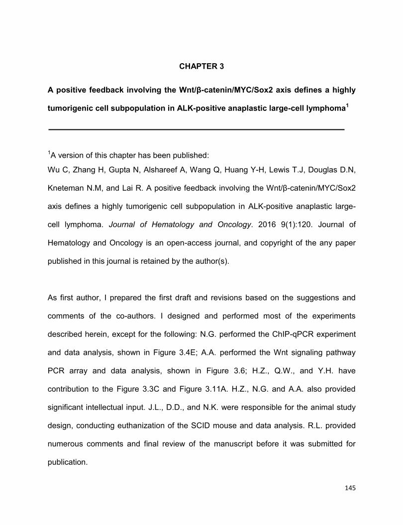
145
CHAPTER 3
A positive feedback involving the Wnt/β-catenin/MYC/Sox2 axis defines a highly
tumorigenic cell subpopulation in ALK-positive anaplastic large-cell lymphoma1
1A version of this chapter has been published:
Wu C, Zhang H, Gupta N, Alshareef A, Wang Q, Huang Y-H, Lewis T.J, Douglas D.N,
Kneteman N.M, and Lai R. A positive feedback involving the Wnt/β-catenin/MYC/Sox2
axis defines a highly tumorigenic cell subpopulation in ALK-positive anaplastic large-
cell lymphoma. Journal of Hematology and Oncology. 2016 9(1):120. Journal of
Hematology and Oncology is an open-access journal, and copyright of the any paper
published in this journal is retained by the author(s).
As first author, I prepared the first draft and revisions based on the suggestions and
comments of the co-authors. I designed and performed most of the experiments
described herein, except for the following: N.G. performed the ChIP-qPCR experiment
and data analysis, shown in Figure 3.4E; A.A. performed the Wnt signaling pathway
PCR array and data analysis, shown in Figure 3.6; H.Z., Q.W., and Y.H. have
contribution to the Figure 3.3C and Figure 3.11A. H.Z., N.G. and A.A. also provided
significant intellectual input. J.L., D.D., and N.K. were responsible for the animal study
design, conducting euthanization of the SCID mouse and data analysis. R.L. provided
numerous comments and final review of the manuscript before it was submitted for
publication.

146
3.1 Introduction
ALK-positive anaplastic large-cell lymphoma (ALK+ALCL) is a specific type of non-
Hodgkin lymphoma of null/T-cell lineage, and these tumors occur most frequently in
young adults and children (1, 2). Approximately 80% of ALK+ALCL patients carry the
chromosomal translocation, t(2;5)(p23;q35), which results in the generation of the
abnormal fusion protein NPM-ALK (1, 2). NPM-ALK has been extensively studied and
shown to be the major oncogenic driver in these tumors. By virtue of its constitutively
active tyrosine kinase activity, NPM-ALK drives oncogenesis primarily by binding to and
phosphorylating a host of signaling proteins, such as STAT3 and PI3K, thereby
deregulating these signaling pathways (2). As a result of these biochemical aberrancies,
NPM-ALK promotes unregulated cell growth and survival, silences the expression of
tumor suppressors and deregulates cellular metabolism (2). From the clinical
perspective, ALK+ALCL tumors are clinically aggressive (1). Although complete
remission can be induced in most pediatric ALK+ALCL patients after conventional
combination chemotherapy, chemoresistance and disease relapses occur in a
substantial proportion of adult patients (2). The biological basis of chemoresistance in
ALK+ALCL patients is incompletely understood, but a recent report (3) describing the
existence of cancer stem-like cells (CSCs) in ALK+ALCL raises the possibility that
these cells may be implicated, similar to how CSCs might contribute to
chemoresistance and cancer relapses in other cancer models (4, 5).
Sox2, one of the four master transcription factors involved in re-programming
fibroblasts to inducible pluripotent stem cells, is normally expressed only in embryonic
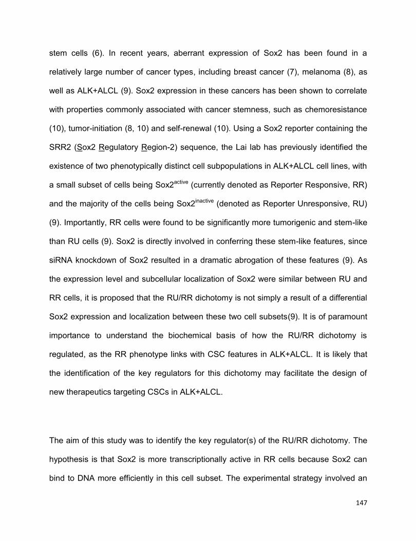
147
stem cells (6). In recent years, aberrant expression of Sox2 has been found in a
relatively large number of cancer types, including breast cancer (7), melanoma (8), as
well as ALK+ALCL (9). Sox2 expression in these cancers has been shown to correlate
with properties commonly associated with cancer stemness, such as chemoresistance
(10), tumor-initiation (8, 10) and self-renewal (10). Using a Sox2 reporter containing the
SRR2 (Sox2 Regulatory Region-2) sequence, the Lai lab has previously identified the
existence of two phenotypically distinct cell subpopulations in ALK+ALCL cell lines, with
a small subset of cells being Sox2active (currently denoted as Reporter Responsive, RR)
and the majority of the cells being Sox2inactive (denoted as Reporter Unresponsive, RU)
(9). Importantly, RR cells were found to be significantly more tumorigenic and stem-like
than RU cells (9). Sox2 is directly involved in conferring these stem-like features, since
siRNA knockdown of Sox2 resulted in a dramatic abrogation of these features (9). As
the expression level and subcellular localization of Sox2 were similar between RU and
RR cells, it is proposed that the RU/RR dichotomy is not simply a result of a differential
Sox2 expression and localization between these two cell subsets(9). It is of paramount
importance to understand the biochemical basis of how the RU/RR dichotomy is
regulated, as the RR phenotype links with CSC features in ALK+ALCL. It is likely that
the identification of the key regulators for this dichotomy may facilitate the design of
new therapeutics targeting CSCs in ALK+ALCL.
The aim of this study was to identify the key regulator(s) of the RU/RR dichotomy. The
hypothesis is that Sox2 is more transcriptionally active in RR cells because Sox2 can
bind to DNA more efficiently in this cell subset. The experimental strategy involved an
148
analysis of the SRR2 sequence, in an attempt to identify potential transcription factor(s)
that regulate the DNA binding of Sox2. A positive feedback loop involving the Wnt/β-
catenin/MYC/Sox2 axis was identified to define a highly tumorigenic and
chemoresistant cell subset in ALK+ALCL.
3.2 Methods and materials
3.2.1 Primary tumors, cell lines and treatments
All primary tumors were diagnosed at the Cross Cancer Institute (Edmonton, Alberta,
Canada) and the diagnostic criteria were based on those described in the WHO
Classification Scheme. The use of these patient samples for research had received
research ethics approval at Feb 17th of 2016, by the Human Research Ethics Board at
the University of Alberta, with approval number: Pro00062737. The details of the cell
lines used in this study were described in section 2.2.1 of Chapter 2. SupM2 and
Karpas 299 cell lines were grown and expanded in RPMI 1640 medium (Invitrogen, Life
Technologies, Grand Island, NY), supplemented with 10% fetal bovine serum (FBS,
Invitrogen) and 1% penicillin streptomycin (Thermo Fisher Scientific) in 5% CO2
atmosphere at 37°C. All cell lines that were stably transfected with Sox2 reporter were
grown and expanded in RPMI 1640 (Invitrogen) supplemented with 10% FBS
(Invitrogen), 1% penicillin streptomycin (ThermoFisher Scientific Canada) and 200
μg/mL puromycin dihydrochloride (Sigma Aldrich, St Louis, MO) in 5% CO2 atmosphere
at 37°C. 10074-G5, quercetin, doxorubicin, crizotinib, Stattic, iodonitrotetrazolium
chloride and G418 were all purchased from Sigma Aldrich. All of the treatments were
149
performed following the manufacturer’s instructions. In brief, 10074-G5, quercetin,
doxorubicin, crizotinib and Stattic were also dissolved in DMSO (Sigma Aldrich). RU or
RR cells were subjected with 10 μM 10074-G5, 50 µM quercetin, 100 nM crizotinib, or
10 nM Stattic for 24 hours, cells with DMSO treatment were included as control. The
MYC protein level and Sox2 reporter luciferase activity were analyzed after these
treatments. RU and RR cells were subjected with various doses of 10074-G5 or
doxorubicin for 48 or 72 hours to assess the cell viability after treatments. 5 µM 10074-
G5 and 20 ng/mL doxorubicin were combined for the drug combination treatment (48
hours treatment).
3.2.2 Short interfering RNA and transfections
SMARTpool short interfering RNAs (siRNAs) for MYC, β-catenin, Sox2 and scrambled
siRNA were purchased from Dharmacon (Lafayette, CO). All siRNAs were diluted to a
stock concentration of 20 µM. Transient transfections of ALK+ALCL cells with siRNAs
(final concentration: 600 nM) were performed as previously described in section 2.2.4
of Chapter 2.
3.2.3 RNA extraction, cDNA synthesis, quantitative reverse transcriptase PCR
(quantitative RT-PCR) and chromatin-immunoprecipitation PCR
Total RNA extraction was performed with the Qiagen RNeasy Kit (Qiagen, Toronto,
Ontario, Canada) according to the manufacturer’s protocol. This assay was performed
as previously described in section 2.2.13 of Chapter 2. Regular quantitative RT-PCR

150
assay was described in section 2.2.13 of Chapter 2. MAGnifyTM ChIP kit was
purchased from ThermoFisher Scientific Canada for Chromatin-immunoprecipitation
PCR experiment. The procedures were following the manufacture’s protocol. In brief,
cells were firstly treated with formaldehyde to generate protein-protein and protein-DNA
crosslinks within chromatin complex, and then the cells were lysed. The cell chromatin
were released from the nuclei and sheared by sonication to reduce the DNA fragment
size to 200-500 bp for analysis. The DNA fragments of interest were immunoprecipitate
with Sox2 antibody (D6D9, Cell Signaling Technology), and the DNA fragments were
utilized for quantitative RT-PCR.
Primers for SRR2 probe for ChIP-PCR experiment are shown as below:
Forward primer: 5’- ACATTGTACTGGGAAGGGACA-3’,
Reverse primer: 5’- AGCAAGAACTGGCGAATGTG-3’.
All genes of interest were normalized to GAPDH transcript expression levels.
All other primer sequences were listed as below:
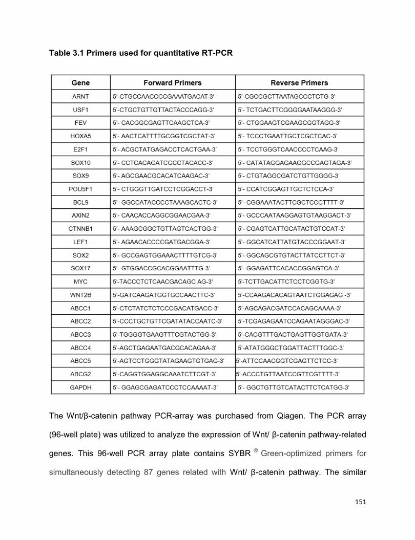
151
Table 3.1 Primers used for quantitative RT-PCR
The Wnt/β-catenin pathway PCR-array was purchased from Qiagen. The PCR array
(96-well plate) was utilized to analyze the expression of Wnt/ β-catenin pathway-related
genes. This 96-well PCR array plate contains SYBR ® Green-optimized primers for
simultaneously detecting 87 genes related with Wnt/ β-catenin pathway. The similar

152
experimental process was followed as regular quantitative RT-PCR (see section 2.2.13
of Chapter 2).
3.2.4 Immunohistochemistry and immunofluorescence studies
The MYC antibody (Y69) and active β-catenin antibody (8E7) were purchased from
Abcam (Cambridge, MA) and Millipore (Billerica, MA), respectively. 1:100 dilution of
MYC antibody was used in the immunohistochemistry assay. 1:200 dilution of MYC
antibody and 1:200 dilution of active β-catenin antibody were used in
immunofluorescence double staining. The procedures of Immunohistochemistry were
described in the previous publication and section 2.2.3 of Chapter 2 (11). For the
immunofluorescence assay, the procedures were described as below. Briefly, formalin-
fixed, paraffin-embedded tissue sections were deparafinized and hydrated. Heat-
induced epitope retrieval was performed using citrate buffer (pH=6.0) and a pressure
cooker using microwave. Tissue sections were then permeabilized for 10 min with 0.2%
Triton X-100 in 1x PBS containing 10mM HEPES (ThermoFisher Scientific Canada)
and 3% BSA (Sigma Aldrich), followed by the block with 1x PBS containing 10mM
HEPES (ThermoFisher Scientific Canada) and 3% BSA (Sigma Aldrich) for 1 hour. The
tissue sections were incubated with the primary antibodies, anti-active β-catenin and
MYC diluted in 1 X PBS with 10 mM HEPES and 1% BSA overnight in 4 °C. In the
second day, after three times of washes with 1 x PBS (30 minutes in total), tissue
sections were incubated with secondary antibodies (Alexa Fluor 594 goat anti-rabbit
antibody and Alexa Fluor 488 goat anti-mouse antibody, Invitrogen, Burlington, CA),
diluted in 1x PBS, 1:300 for 1 hour. After wash in 1 X PBS, tissues were incubated in 1
153
µg/mL Hoechst 33342 (Sigma Aldrich, B2261) for 10 minutes, followed by washes in 1
X PBS and mounted with Mounting Medium (Dako, Mississauga, ON, Canada). Cells
were visualized with a Zeiss LSM510 confocal microscope (Carl Zeiss, Heidelberg,
Germany) at the Core Cell Imaging Facility, Cross Cancer Institute, University of
Alberta, Edmonton, Canada.
3.2.5 Plasmid constructs and transfection
pcDNA3.3-MYC was a gift from Derrick Rossi (Addgene plasmid # 26818) and pcDNA-
delta N89 Beta-catenin was a gift from Eric Fearon (Addgene plasmid # 19288), Myc-
Mad-HA plasmid was a gift from Bert Vogelstein (Addgene plasmid #16557). pcDNA
empty vector was purchased from Addgene (Cambridge, MA). pcDNA-Flag-Sox2
plasmid was generated by the Lai lab. The procedures for transfection were described
in section 2.2.5 of Chapter 2. The stable cell lines were established by the following
steps. 10 million of RU cells originated from SupM2 were transfected with 20 µg
pcDNA3.3-MYC or pcDNA, and then cultured for 3 weeks in selection medium with
increasing concentration of G418 up to 200 μg/mL. RR cells derived from SupM2 that
was stably transfected with pcDNA were also established following the same procedure.
3.2.6 Western blotting
Western blotting studies were performed as described in section 2.2.8 of Chapter 2.
Antibodies reactive to phosphorylated MYCS62 (E1J4K), MYC (D84C12), Sox2 (D6D9),
β-catenin (D10A8), phosphorylated GSK3βS9 (D85E12), LEF1(C18A7), γ-tubulin
154
antibody (#5886) and Histone deacetylase 1 (HDAC-1) antibody (#2062) were
purchased from Cell Signaling Technology (Danvers, MA), α-tubulin antibody (TU-02)
and β-actin antibody (sc-130300) were purchased from Santa Cruz (Dallas, Texas),
antibody reactive to active β-catenin (8E7) was purchased from Merck Millipore
(Toronto, ON, Canada). Secondary antibodies anti-Rabbit IgG (1:2000) and anti-Mouse
IgG (1:2000) were purchased from Cell Signaling Technology.
3.2.7 Luciferase assay
The luciferase assay kit was purchased from Promega (Madison, WI), the procedure for
luciferase assay was performed following the manufacture’s protocol. Briefly, cell
pellets after cold 1 X PBS wash for three times were lysed by 100 µl 1 X lysis buffer (5
X lysis buffer provided in kit were diluted by double distilled water), then the cell lysates
were processed for assess protein concentrations and luciferase values. The luciferase
values were read by FLUOstar Omega microplate reader (BMG LabTech,
ThermoFisher Scientific Canada), and normalized to their protein concentrations. The
luciferase activity of RU cells was normalized to 1 when compared to it in RR cells in
this study. Each experiment was performed in triplicate.
3.2.8 Transwell assay
The 6-well plates of polyester transwell permeable supports with 0.4µm pore size were
purchased from Corning Inc (Toronto, ON, Canada). Briefly, 0.5 million of RU cells and
the same number of RR cells, diluted RR cells (diluted with RU cells ) or parental

155
SupM2 cells were seeded in the upper chamber and lower chamber, respectively, and
cultured for 72 hours. The same number of RU cells co-cultured with RU cells in the
lower chamber was included as control group. Then the luciferase assay and Western
blotting studies were performed.
3.2.9 Cell-cycle and MTS assay
The experimental procedures were described in section 2.2.7 and section 2.2.12 of
Chapter 2.
3.2.10 SRR2 probe binding assay
Cells were harvested and washed with cold 1x PBS twice, following by cytoplasmic and
nuclear fractionation using the Pierce NE-PER kit (ThermoFisher Scientific Canada).
300 μg nuclear proteins were incubated with or without 3 pmol of biotin-labeled SRR2
probe (constructed by IDT, Edmonton, Alberta, Canada) for 0.5 hour by rotating at
room temperature. Streptadvidin agarose beads (75 μl, TheromoFisher Scientific
Canada) were added to each sample, and the samples were rotated by overnight at
4°C. The next day, the samples were washed with cold 1 X PBS three times, and
protein was eluted at 100°C in 4 X protein loading buffer (40% glycerol, 240 mM Tris-
HCl pH 6.0, 8% SDS, 0.04% bromophenol blue, and 5% β-mercaptoethanol) for 5
minutes, and Western blotting studies were performed. The sequence of the SRR2
probe: ‘5-AAGAATTTCCCGGGCTCGGGCAGCCATTGTGATGCATATAGGATTATTC
ACGTGGTAATG-3′

156
The underlined sequence is SRR2 consensus sequence.
3.2.11 Nuclear cytoplasm fractionation assay
The nuclear-cytoplasmic fractionation kit was purchased from ThermoFisher Scientific.
The experiment was performed as described in section 2.2.11 of Chapter 2.
3.2.12 Methylcellulose colony formation assay
Methylcellulose-based media was purchased from R&D systems Inc, and the
methylcellulose colony formation assay was performed as previously described in
section 2.2.14 of Chapter 2. After 10 days of culture, the colonies were stained with
iodonitrotetrazolium chloride overnight and images were acquired using Alphalmager
HP (ThermoFisher Scientific Canada).
3.2.13 SCID mouse xenograft studies
Twelve CB-17 strain SCID male mice, purchased from Taconic (Hudson, NY), were
housed in a virus- and antigen-free facility supported by the Health Sciences
Laboratory Animal Services at the University of Alberta and were cared for in
accordance with the Canadian Council on Animal Care guidelines. The animal studies
had received ethical approval by the Animal Care and Use Committee (ACUC) at Dec
10th of 2015, with approval number: AUP00000782. Briefly, 2 million cells of SupM2-
RU-EV, SupM2-RU-MYC and SupM2-RR-EV growing exponentially were injected into
157
both flanks of 4-week-old mice, four mice each group. The tumor sizes were measured
twice every week. These animals were euthanized when a tumor reached 10 mm in the
greatest dimension.
3.2.14 Side population assay
The side population assay was performed as previously described by Moti et al (3). In
brief, 1 X 10⁶ cells/mL were washed and resuspended in prewarmed 1 X PBS
containing 2% FBS and 10 mM HEPES (Life Technology). Samples were incubated
with 5 μg/mL Hoechst 33342 (Sigma Aldrich) with or without 100 μM Verapamil (Sigma
Aldrich) for 90 minutes at 37°C in a water bath in the dark. After incubation, the
samples were incubated on ice for 10 minutes and washed with cold 1X PBS, following
by counterstaining with 2 μg/mL of propidium iodide (Sigma Aldrich) before flow
cytometric analysis.
3.2.15 Statistical analysis
Data were expressed as mean standard deviation. The statistical analysis was
performed using Graphpad Prism 5 (La Jolla, CA), and the significance of two
independent groups of samples was determined using Student’s t-test or Chi-square
test. Statistical significance is denoted by * (P<0.05) and ** (P<0.01).
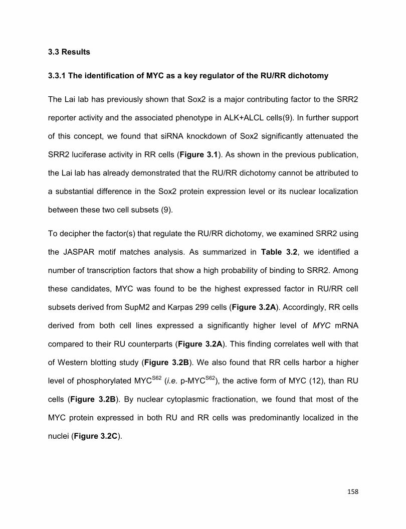
158
3.3 Results
3.3.1 The identification of MYC as a key regulator of the RU/RR dichotomy
The Lai lab has previously shown that Sox2 is a major contributing factor to the SRR2
reporter activity and the associated phenotype in ALK+ALCL cells(9). In further support
of this concept, we found that siRNA knockdown of Sox2 significantly attenuated the
SRR2 luciferase activity in RR cells (Figure 3.1). As shown in the previous publication,
the Lai lab has already demonstrated that the RU/RR dichotomy cannot be attributed to
a substantial difference in the Sox2 protein expression level or its nuclear localization
between these two cell subsets (9).
To decipher the factor(s) that regulate the RU/RR dichotomy, we examined SRR2 using
the JASPAR motif matches analysis. As summarized in Table 3.2, we identified a
number of transcription factors that show a high probability of binding to SRR2. Among
these candidates, MYC was found to be the highest expressed factor in RU/RR cell
subsets derived from SupM2 and Karpas 299 cells (Figure 3.2A). Accordingly, RR cells
derived from both cell lines expressed a significantly higher level of MYC mRNA
compared to their RU counterparts (Figure 3.2A). This finding correlates well with that
of Western blotting study (Figure 3.2B). We also found that RR cells harbor a higher
level of phosphorylated MYCS62 (i.e. p-MYCS62), the active form of MYC (12), than RU
cells (Figure 3.2B). By nuclear cytoplasmic fractionation, we found that most of the
MYC protein expressed in both RU and RR cells was predominantly localized in the
nuclei (Figure 3.2C).

159
Figure 3.1 Knockdown of Sox2 by siRNA significantly downregulates the SRR2
luciferase activity in RR cells. A) The SRR2 luciferase activity in RR cells derived
from SupM2 and Karpas 299 with scrambled siRNA (scr siRNA) or Sox2 siRNA
transfection. The Western blots on the right panel showed the knockdown efficiency of
Sox2 protein expression. Statistical significance is denoted by * (P<0.05) and **
(P<0.01). Results shown were representative of three independent experiments.

160
Table 3.2 The top putative factors that are predicated to bind to SRR2 sequence
by JASPAR motif matches analysis at P<0.001.
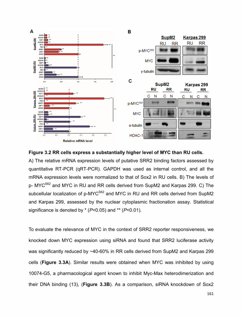
161
Figure 3.2 RR cells express a substantially higher level of MYC than RU cells.
A) The relative mRNA expression levels of putative SRR2 binding factors assessed by
quantitative RT-PCR (qRT-PCR). GAPDH was used as internal control, and all the
mRNA expression levels were normalized to that of Sox2 in RU cells. B) The levels of
p- MYCS62 and MYC in RU and RR cells derived from SupM2 and Karpas 299. C) The
subcellular localization of p-MYCS62 and MYC in RU and RR cells derived from SupM2
and Karpas 299, assessed by the nuclear cytoplasmic fractionation assay. Statistical
significance is denoted by * (P<0.05) and ** (P<0.01).
To evaluate the relevance of MYC in the context of SRR2 reporter responsiveness, we
knocked down MYC expression using siRNA and found that SRR2 luciferase activity
was significantly reduced by ~40-60% in RR cells derived from SupM2 and Karpas 299
cells (Figure 3.3A). Similar results were obtained when MYC was inhibited by using
10074-G5, a pharmacological agent known to inhibit Myc-Max heterodimerization and
their DNA binding (13), (Figure 3.3B). As a comparison, siRNA knockdown of Sox2

162
resulted in a similar reduction in SRR2 luciferase activity (Figure 3.1). Correlating with
these findings, transfection of MYC into RU derived from the two cell lines resulted in a
significant increase in SRR2 luciferase activity, even though the level remained to be
substantially lower than that of RR cells (Figure 3.3C-D). As expected, transfection of
MYC into RR cells from both cell lines also led to a significantly increased SRR2
reporter activity (Figure 3.3C-D). Taken together, these findings suggest that MYC is a
key regulator of the SRR2 reporter activity.
Figure 3.3 The high MYC expression contributes to the RR phenotype. A) The
SRR2 luciferase activity in RR cells derived from SupM2 and Karpas 299 cells with scr
siRNA or MYC siRNA transfection. The Western blots below showed the knockdown
efficiency of MYC. B) The SRR2 luciferase activity in RR cells with the treatment of 10
μM 10074-G5 for 24 hours. Cells with DMSO treatment were used as a negative control.
Statistical significance is denoted by * (P<0.05) and ** (P<0.01). Results shown were
representative of three independent experiments.
163
Figure 3.3C-D C) RU and RR cells derived from both cell lines were transiently
transfected with pcDNA3.3-MYC (i.e. MYC). pcDNA empty vector (EV) was included as
a negative control. The Western blots showed the MYC transfection efficiency. D) The
SRR2 luciferase activity in RU and RR cells with either EV or MYC transient
transfection. Statistical significance is denoted by * (P<0.05) and ** (P<0.01). Results
shown were representative of three independent experiments.
We then asked if inhibition of MYC in RR cells also decrease the clonogenicity and
chemoresistance that are associated with the RR phenotype. As shown in Figure
3.3E-G, pharmacologic inhibition of MYC using 10074-G5 in RR cells resulted in a
significant decrease in methylcellulose colony formation and sensitization of these cells
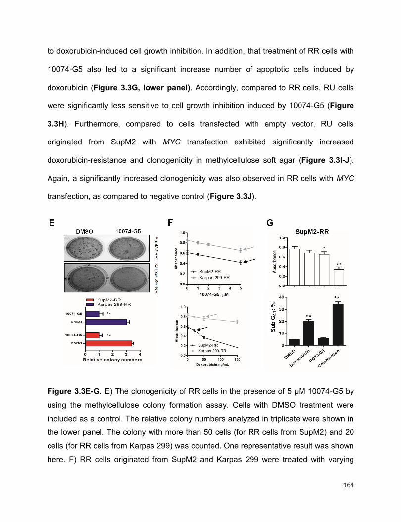
164
to doxorubicin-induced cell growth inhibition. In addition, that treatment of RR cells with
10074-G5 also led to a significant increase number of apoptotic cells induced by
doxorubicin (Figure 3.3G, lower panel). Accordingly, compared to RR cells, RU cells
were significantly less sensitive to cell growth inhibition induced by 10074-G5 (Figure
3.3H). Furthermore, compared to cells transfected with empty vector, RU cells
originated from SupM2 with MYC transfection exhibited significantly increased
doxorubicin-resistance and clonogenicity in methylcellulose soft agar (Figure 3.3I-J).
Again, a significantly increased clonogenicity was also observed in RR cells with MYC
transfection, as compared to negative control (Figure 3.3J).
Figure 3.3E-G. E) The clonogenicity of RR cells in the presence of 5 µM 10074-G5 by
using the methylcellulose colony formation assay. Cells with DMSO treatment were
included as a control. The relative colony numbers analyzed in triplicate were shown in
the lower panel. The colony with more than 50 cells (for RR cells from SupM2) and 20
cells (for RR cells from Karpas 299) was counted. One representative result was shown
here. F) RR cells originated from SupM2 and Karpas 299 were treated with varying
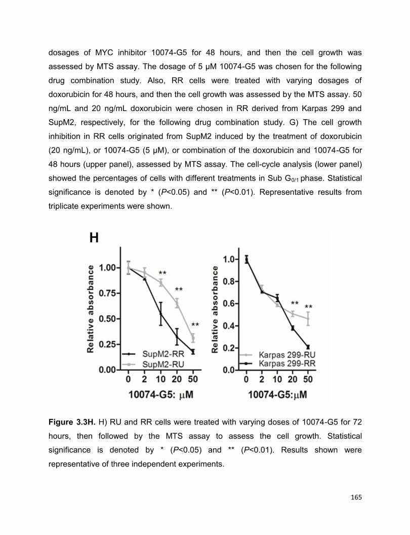
165
dosages of MYC inhibitor 10074-G5 for 48 hours, and then the cell growth was
assessed by MTS assay. The dosage of 5 µM 10074-G5 was chosen for the following
drug combination study. Also, RR cells were treated with varying dosages of
doxorubicin for 48 hours, and then the cell growth was assessed by the MTS assay. 50
ng/mL and 20 ng/mL doxorubicin were chosen in RR derived from Karpas 299 and
SupM2, respectively, for the following drug combination study. G) The cell growth
inhibition in RR cells originated from SupM2 induced by the treatment of doxorubicin
(20 ng/mL), or 10074-G5 (5 µM), or combination of the doxorubicin and 10074-G5 for
48 hours (upper panel), assessed by MTS assay. The cell-cycle analysis (lower panel)
showed the percentages of cells with different treatments in Sub G0/1 phase. Statistical
significance is denoted by * (P<0.05) and ** (P<0.01). Representative results from
triplicate experiments were shown.
Figure 3.3H. H) RU and RR cells were treated with varying doses of 10074-G5 for 72
hours, then followed by the MTS assay to assess the cell growth. Statistical
significance is denoted by * (P<0.05) and ** (P<0.01). Results shown were
representative of three independent experiments.

166
Figure 3.3I-J I) The cell growth inhibition induced by varying doses of doxorubicin for
48 hours in RU cells from SupM2 cells that were transiently transfected with either EV
or MYC, assessed by the MTS assay. J) The clonogenicity of RU and RR cells from
SupM2 with either EV or MYC transient transfection, assessed by the methylcellulose
colony formation assay. The relative colony numbers analyzed in triplicate were shown
in the lower panel. The colony with more than 50 cells was counted. Statistical
significance is denoted by * (P<0.05) and ** (P<0.01). Representative results from
triplicate experiments were shown.
3.3.2 MYC promotes Sox2-SRR2 binding and the transcriptional activity of Sox2
The observation that both RU and RR cell subsets express a similar level of Sox2
protein raised the possibility that MYC upregulates SRR2 activity by increasing Sox2-
SRR2 binding and Sox2 transcriptional activity. In support of this concept, when we
performed pull-down assay using a biotin-labeled SRR2 probe, we found abundant
167
MYC-SRR2 binding in RR cells but not RU cells (Figure 3.4A); a similar pattern of the
Sox2-SRR2 interaction was also found in RU and RR cells (Figure 3.4A). Furthermore,
enforced expression of MYC in RU cells led to substantially more Sox2 pulled down by
the SRR2 probe, while the total Sox2 protein level only faintly increased in this
experiment (Figure 3.4B). Correlating with this increased Sox2-SRR2 binding, the
mRNA levels of several genes including WNT2B, CTNNB1 and BCL9 were significantly
increased (Figure 3.4C), all of which were shown to be Sox2 downstream targets in RR
cells (see section 4). Additionally, 10074-G5 treatment of RR cells from SupM2 cells
resulted in a rapid and dramatic decrease in Sox2-SRR2 binding (Figure 3.4D), with
the total protein levels of MYC and Sox2 being unaffected in this timeframe (i.e., 4
hours). Sox2-SRR2 binding also significantly decreased at 4 hours upon 10074-G5
treatment when we employed chromatin immunoprecipitation-qPCR (Figure 3.4E).
Correlating with this decreased Sox2-SRR2 binding, the mRNA levels of two Sox2
downstream targets including CTNNB1 and BCL9 were also significantly
downregulated in RR cells upon MYC inhibition by using 10074-G5 at 8 hours, with the
Sox2 protein level not appreciably altered in this time point (Figure 3.4F).
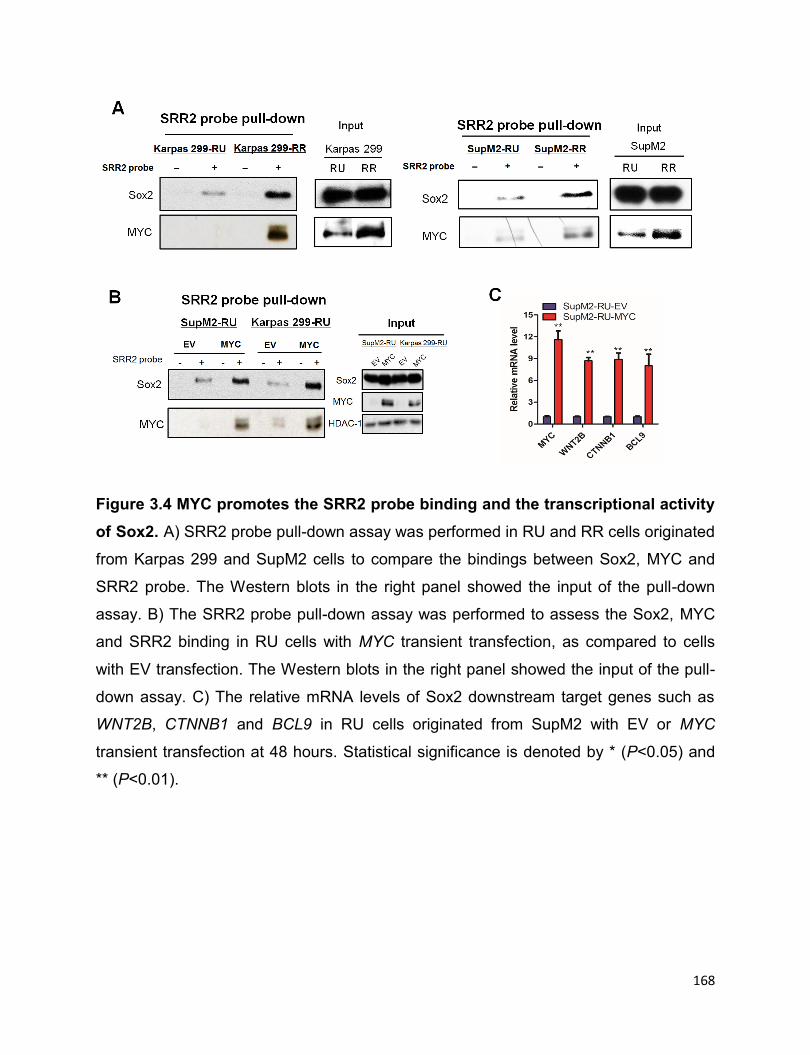
168
Figure 3.4 MYC promotes the SRR2 probe binding and the transcriptional activity
of Sox2. A) SRR2 probe pull-down assay was performed in RU and RR cells originated
from Karpas 299 and SupM2 cells to compare the bindings between Sox2, MYC and
SRR2 probe. The Western blots in the right panel showed the input of the pull-down
assay. B) The SRR2 probe pull-down assay was performed to assess the Sox2, MYC
and SRR2 binding in RU cells with MYC transient transfection, as compared to cells
with EV transfection. The Western blots in the right panel showed the input of the pull-
down assay. C) The relative mRNA levels of Sox2 downstream target genes such as
WNT2B, CTNNB1 and BCL9 in RU cells originated from SupM2 with EV or MYC
transient transfection at 48 hours. Statistical significance is denoted by * (P<0.05) and
** (P<0.01).
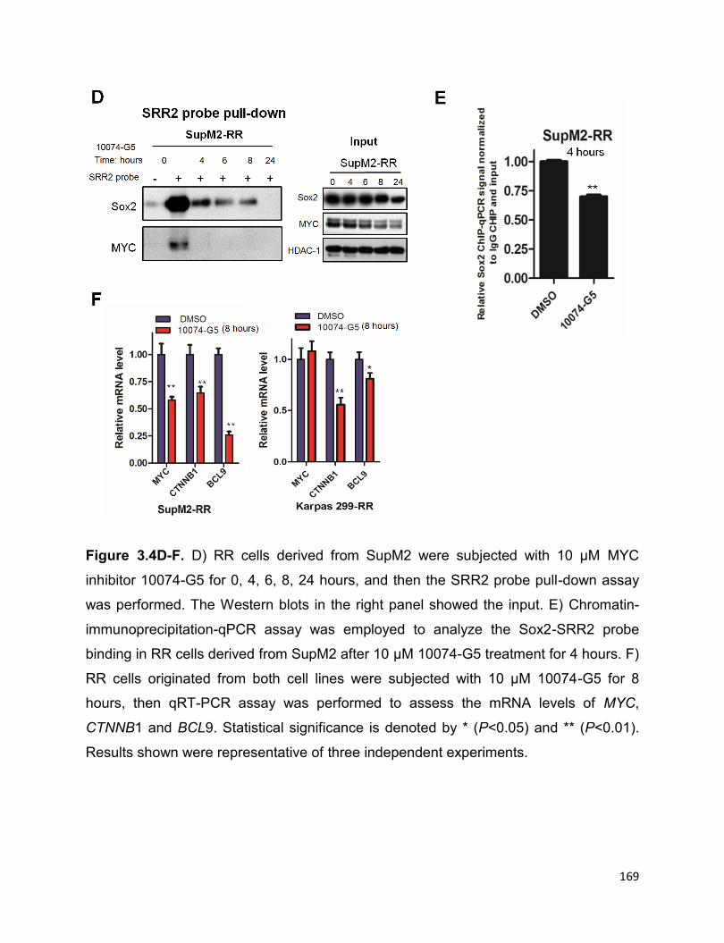
169
Figure 3.4D-F. D) RR cells derived from SupM2 were subjected with 10 µM MYC
inhibitor 10074-G5 for 0, 4, 6, 8, 24 hours, and then the SRR2 probe pull-down assay
was performed. The Western blots in the right panel showed the input. E) Chromatin-
immunoprecipitation-qPCR assay was employed to analyze the Sox2-SRR2 probe
binding in RR cells derived from SupM2 after 10 μM 10074-G5 treatment for 4 hours. F)
RR cells originated from both cell lines were subjected with 10 µM 10074-G5 for 8
hours, then qRT-PCR assay was performed to assess the mRNA levels of MYC,
CTNNB1 and BCL9. Statistical significance is denoted by * (P<0.05) and ** (P<0.01).
Results shown were representative of three independent experiments.

170
We found evidence that Sox2 alone is not sufficient to regulate Sox2-SRR2 binding and
the SRR2 activity in RU cells. As shown in the left panel of Figure 3.4G, We
transfected SOX2-FLAG in these two cell subsets derived from SupM2, and it is evident
that Sox2 overexpression did not significantly increase SRR2 luciferase activity in RU
cells; in contrast, the same experimental manipulation led to a significant increase in
the reporter activity in RR cells. Accordingly, Sox2-flag overexpression did not
appreciably increase Sox2-SRR2 binding in RU cells, but a substantial increase of
Sox2-SRR2 binding was observed in RR cells (Figure 3.4H).
Figure 3.4G-H G) The SRR2 luciferase activity in RU and RR cells originated from
SupM2 with EV or pcDNA-SOX2-FLAG (i.e.SOX2-FLAG) transfection at 48 hours. The
Western blots showed the transfection efficiency of SOX2-FLAG. H) The Sox2-SRR2

171
binding ability in RU and RR cells originated from SupM2 with EV or SOX2-FLAG
transfection at 48 hours. Statistical significance is denoted by * (P<0.05) and **
(P<0.01).
3.3.3 The high level of MYC in RR cells is attributed to the Wnt/β-catenin pathway
To explain why MYC is preferentially expressed at a high level in RR cells, We first
evaluated the activation status of NPM-ALK/STAT3 axis between these two cell
subsets. Consistent with the Lai lab’s previous studies (9), the expression and
activation levels of NPM-ALK and STAT3 were similar between RU and RR cells
(Figure 3.5A). Pharmacologic inhibition of NPM-ALK (crizotinib) or STAT3 (stattic)
dramatically decreased the expression of both Sox2 and MYC in RU and RR cells
equally well, and these findings correlated with a reduction of the SRR2 luciferase
activity by ~50-70% in both cell subsets (Figure 3.5B-C). Based on these findings, it is
evident that, while the NPM-ALK/STAT3 axis contributes to a basal expression level of
MYC, it does not explain the differential MYC expression between RU and RR cells.

172
Figure 3.5 NPM-ALK/STAT3 is not differentially activated or expressed between
RU and RR cells. A) The protein levels of pALK, ALK, pSTAT3, and STAT3 in RU and
RR cells derived from SupM2 and Karpas 299. B-C) RR and RU cells were treated with
either DMSO, or 100 nM ALK inhibitor crizotinib, or 10 nM STAT3 inhibitor stattic for 24
hours. The Western blots were employed to assess the expression/activation levels of
NPM-ALK, STAT3, MYC and Sox2 in both RU and RR cells. The SRR2 luciferase
activity in RU and RR cells was also evaluated by the luciferase assay. Statistical
significance is denoted by * (P<0.05) and ** (P<0.01). Results shown were
representative of three independent experiments.
We then asked if the Wnt/β-catenin pathway is a contributing factor, as this pathway is
known to upregulate MYC in other cancer cell types (14-16). Firstly, we performed the
Wnt signaling pathway PCR array to compare RU and RR cells derived from SupM2.
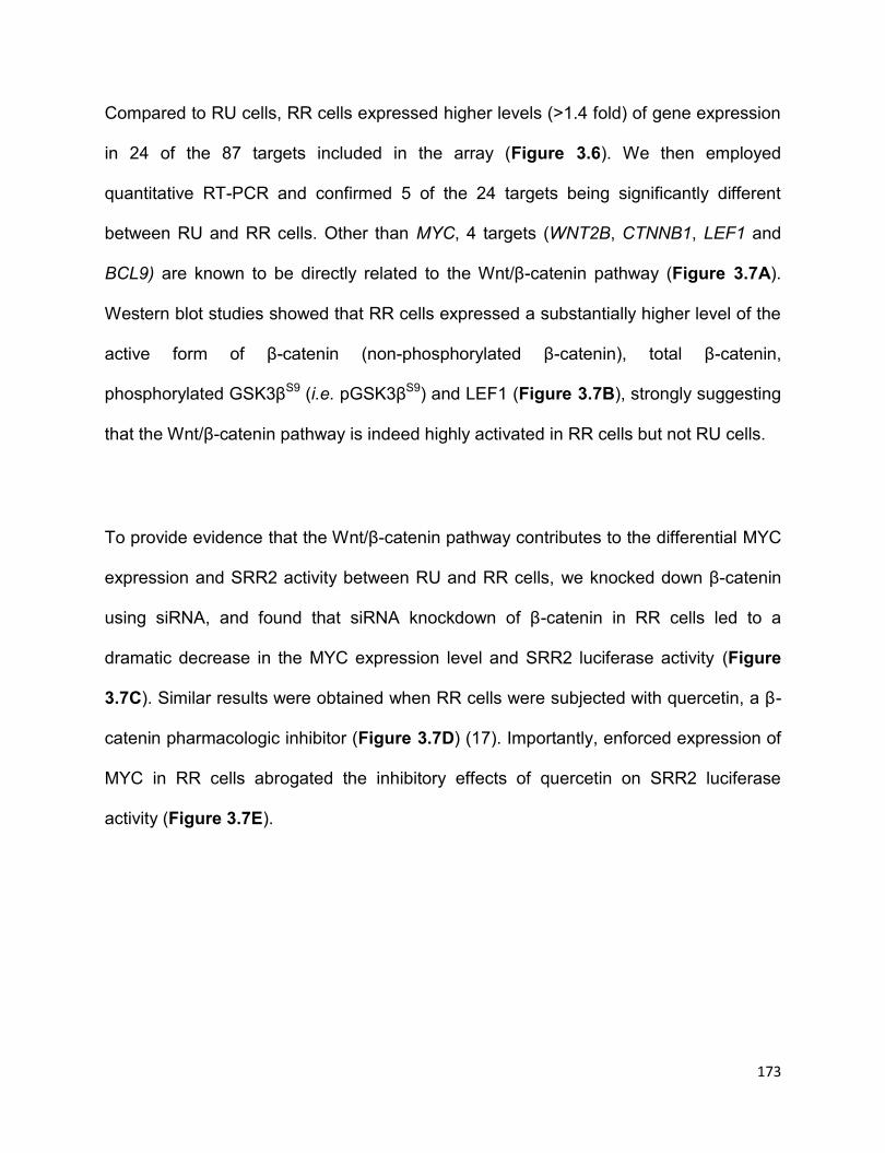
173
Compared to RU cells, RR cells expressed higher levels (>1.4 fold) of gene expression
in 24 of the 87 targets included in the array (Figure 3.6). We then employed
quantitative RT-PCR and confirmed 5 of the 24 targets being significantly different
between RU and RR cells. Other than MYC, 4 targets (WNT2B, CTNNB1, LEF1 and
BCL9) are known to be directly related to the Wnt/β-catenin pathway (Figure 3.7A).
Western blot studies showed that RR cells expressed a substantially higher level of the
active form of β-catenin (non-phosphorylated β-catenin), total β-catenin,
phosphorylated GSK3βS9 (i.e. pGSK3βS9) and LEF1 (Figure 3.7B), strongly suggesting
that the Wnt/β-catenin pathway is indeed highly activated in RR cells but not RU cells.
To provide evidence that the Wnt/β-catenin pathway contributes to the differential MYC
expression and SRR2 activity between RU and RR cells, we knocked down β-catenin
using siRNA, and found that siRNA knockdown of β-catenin in RR cells led to a
dramatic decrease in the MYC expression level and SRR2 luciferase activity (Figure
3.7C). Similar results were obtained when RR cells were subjected with quercetin, a β-
catenin pharmacologic inhibitor (Figure 3.7D) (17). Importantly, enforced expression of
MYC in RR cells abrogated the inhibitory effects of quercetin on SRR2 luciferase
activity (Figure 3.7E).
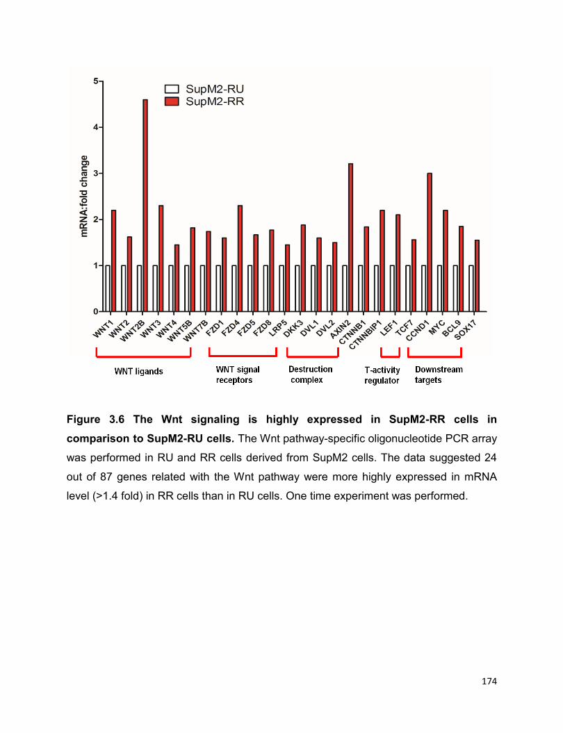
174
Figure 3.6 The Wnt signaling is highly expressed in SupM2-RR cells in
comparison to SupM2-RU cells. The Wnt pathway-specific oligonucleotide PCR array
was performed in RU and RR cells derived from SupM2 cells. The data suggested 24
out of 87 genes related with the Wnt pathway were more highly expressed in mRNA
level (>1.4 fold) in RR cells than in RU cells. One time experiment was performed.
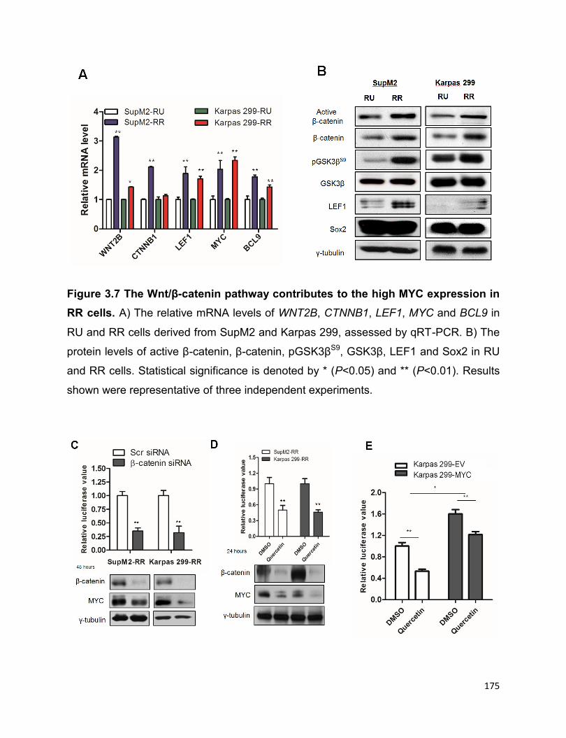
175
Figure 3.7 The Wnt/β-catenin pathway contributes to the high MYC expression in
RR cells. A) The relative mRNA levels of WNT2B, CTNNB1, LEF1, MYC and BCL9 in
RU and RR cells derived from SupM2 and Karpas 299, assessed by qRT-PCR. B) The
protein levels of active β-catenin, β-catenin, pGSK3βS9, GSK3β, LEF1 and Sox2 in RU
and RR cells. Statistical significance is denoted by * (P<0.05) and ** (P<0.01). Results
shown were representative of three independent experiments.
176
Figure 3.7C-E C) The SRR2 luciferase activity in RR cells with scr siRNA or β-catenin
siRNA transfection at 48 hours. The Western blots below showed the knockdown
efficiency of β-catenin and MYC. D) The SRR2 luciferase activity in RR cells treated
with DMSO or 50 µM quercetin, a pharmacological β-catenin inhibitor for 24 hours. The
Western blots below showed the knockdown efficiency of β-catenin and MYC. E) The
SRR2 luciferase activity in RR cells derived from Karpas 299 with EV or c-MYC
transfection in the presence of 50 µM quercetin for 24 hours; cells with DMSO
treatment were included as a negative control. The SRR2 luciferase activity decreased
by ~50% in RR cells from Karpas 299 with EV transfection upon quercetin treatment,
whereas it only decreased by ~25% in cells with c-MYC transfection. The MYC
transfection efficiency was validated in Figure 3.3C. Statistical significance is denoted
by * (P<0.05) and ** (P<0.01). Representative results from three independent
experiments were shown.
Since RR cells expressed more ligands for the Wnt/β-catenin pathway (such as Wnt2B)
than RU cells, we asked if soluble factors produced by RR cells can increase MYC
expression and SRR2 activity in RU cells. To test this, we used the transwell co-culture
system that is illustrated in Figure 3.7F. As shown in Figure 3.7G, we found that the
SRR2 luciferase activity in RU cells was significantly increased after 72 hours of co-
culture with RR cells. By Western blotting studies (Figure 3.7H), we confirmed that the
Wnt/β-catenin pathway in RU cells was upregulated after 72 hours of co-culture with
RR cells, as evidenced by the increased protein expressions of active β-catenin, β-
catenin, and LEF1. Accordingly, MYC was also upregulated. However, we did not
observe any significant changes of luciferase activity or MYC expression in RU cells
when co-cultured with either diluted RR cells or parental SupM2 cells (Figure 3.7I).
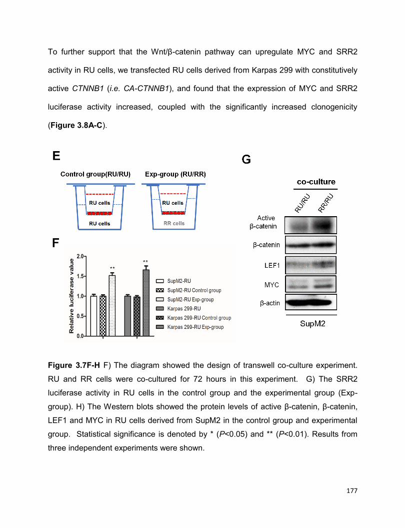
177
To further support that the Wnt/β-catenin pathway can upregulate MYC and SRR2
activity in RU cells, we transfected RU cells derived from Karpas 299 with constitutively
active CTNNB1 (i.e. CA-CTNNB1), and found that the expression of MYC and SRR2
luciferase activity increased, coupled with the significantly increased clonogenicity
(Figure 3.8A-C).
Figure 3.7F-H F) The diagram showed the design of transwell co-culture experiment.
RU and RR cells were co-cultured for 72 hours in this experiment. G) The SRR2
luciferase activity in RU cells in the control group and the experimental group (Exp-
group). H) The Western blots showed the protein levels of active β-catenin, β-catenin,
LEF1 and MYC in RU cells derived from SupM2 in the control group and experimental
group. Statistical significance is denoted by * (P<0.05) and ** (P<0.01). Results from
three independent experiments were shown.
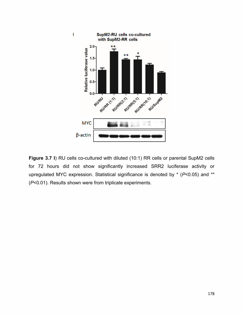
178
Figure 3.7 I) RU cells co-cultured with diluted (10:1) RR cells or parental SupM2 cells
for 72 hours did not show significantly increased SRR2 luciferase activity or
upregulated MYC expression. Statistical significance is denoted by * (P<0.05) and **
(P<0.01). Results shown were from triplicate experiments.
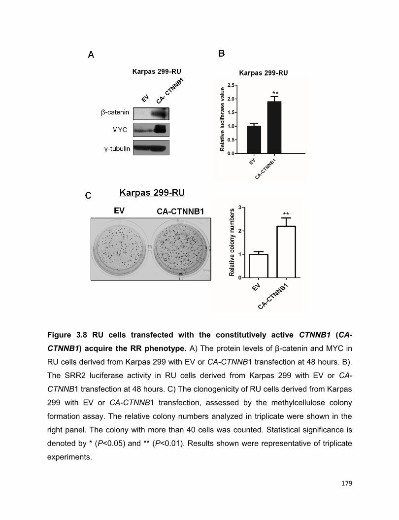
179
Figure 3.8 RU cells transfected with the constitutively active CTNNB1 (CA-
CTNNB1) acquire the RR phenotype. A) The protein levels of β-catenin and MYC in
RU cells derived from Karpas 299 with EV or CA-CTNNB1 transfection at 48 hours. B).
The SRR2 luciferase activity in RU cells derived from Karpas 299 with EV or CA-
CTNNB1 transfection at 48 hours. C) The clonogenicity of RU cells derived from Karpas
299 with EV or CA-CTNNB1 transfection, assessed by the methylcellulose colony
formation assay. The relative colony numbers analyzed in triplicate were shown in the
right panel. The colony with more than 40 cells was counted. Statistical significance is
denoted by * (P<0.05) and ** (P<0.01). Results shown were representative of triplicate
experiments.

180
3.3.4 The positive regulatory loop involving Sox2, Wnt/β-catenin and MYC in RR
cells
In view of several recent publications reporting that Sox2 can activate the Wnt/β-
catenin pathway in a number of cell types (10, 18, 19), we asked if Sox2 also can exert
similar effects in ALK+ALCL. As shown in Figure 3.9A, siRNA knockdown of Sox2 in
RR cells significantly decreased the transcript levels of SOX2, WNT2B, CTNNB1, MYC
and BCL9. Furthermore, the protein levels of p-MYCS62 and MYC were dramatically
decreased in RR cells upon Sox2 siRNA knockdown (Figure 3.9B). Correlating with the
fact that Sox2 is relatively transcriptionally quiescent in RU cells, the expressions of p-
MYCS62 and MYC in these cells did not change appreciably in response to Sox2
knockdown (Figure 3.9B). Taken together, these findings support the existence of a
positive feedback loop involving Wnt/β-catenin, MYC and Sox2 in RR cells. In other
words, in RR cells, the high level of MYC promotes the transcriptional activity of Sox2,
which in turn activates the Wnt/β-catenin pathway and sustains a high level of MYC
expression. By contrast, in RU cells, Sox2 does not effectively activate the Wnt/β-
catenin pathway due to the relatively low level of MYC; in the absence of active Wnt/β-
catenin pathway, MYC remains to be lowly expressed. A model summarizing this
concept is illustrated in Figure 3.9C.

181
Figure 3.9 The positive regulatory loop of Sox2–Wnt/β-catenin–MYC in RR cells.
A) qRT-PCR assay was performed to analyze the relative mRNA levels of WNT2B,
CTNNB1, MYC and BCL9 in RR cells derived from SupM2 and Karpas 299 cells with
scr siRNA or Sox2 siRNA transfection at 48 hours. B) The protein levels of Sox2, p-
MYCS62 and MYC in RR cells with scr siRNA or Sox2 siRNA transfection at 48 hours.
Results shown are representative of three independent experiments.
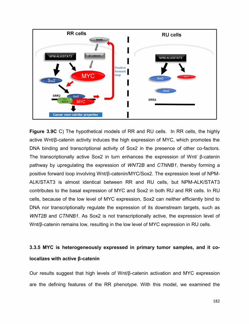
182
Figure 3.9C C) The hypothetical models of RR and RU cells. In RR cells, the highly
active Wnt/β-catenin activity induces the high expression of MYC, which promotes the
DNA binding and transcriptional activity of Sox2 in the presence of other co-factors.
The transcriptionally active Sox2 in turn enhances the expression of Wnt/ β-catenin
pathway by upregulating the expression of WNT2B and CTNNB1, thereby forming a
positive forward loop involving Wnt/β-catenin/MYC/Sox2. The expression level of NPM-
ALK/STAT3 is almost identical between RR and RU cells, but NPM-ALK/STAT3
contributes to the basal expression of MYC and Sox2 in both RU and RR cells. In RU
cells, because of the low level of MYC expression, Sox2 can neither efficiently bind to
DNA nor transcriptionally regulate the expression of its downstream targets, such as
WNT2B and CTNNB1. As Sox2 is not transcriptionally active, the expression level of
Wnt/β-catenin remains low, resulting in the low level of MYC expression in RU cells.
3.3.5 MYC is heterogeneously expressed in primary tumor samples, and it co-
localizes with active β-catenin
Our results suggest that high levels of Wnt/β-catenin activation and MYC expression
are the defining features of the RR phenotype. With this model, we examined the

183
expression of MYC and active β-catenin (as a surrogate marker of Wnt activation) using
double immunofluorescence staining analyzed by using confocal microscopy. After
evaluating tumor cells (10 random fields) derived from 3 cases, we found a highly
significant correlation between the expression of MYC and active β-catenin (P<0.0001)
(Figure 3.10A and the table below, 400X). We also performed immunohistochemistry
to study MYC expression in 7 additional cases of formalin-fixed/paraffin-embedded
ALK+ALCL tumors. As illustrated in Figure 3.10B, MYC is heterogeneously expressed
in tumor cells and two representative fields from a case were shown here (400X). MYC
expression was restricted to a small subset (~30%) of neoplastic cells; benign
lymphocytes and fibroblasts were negative.
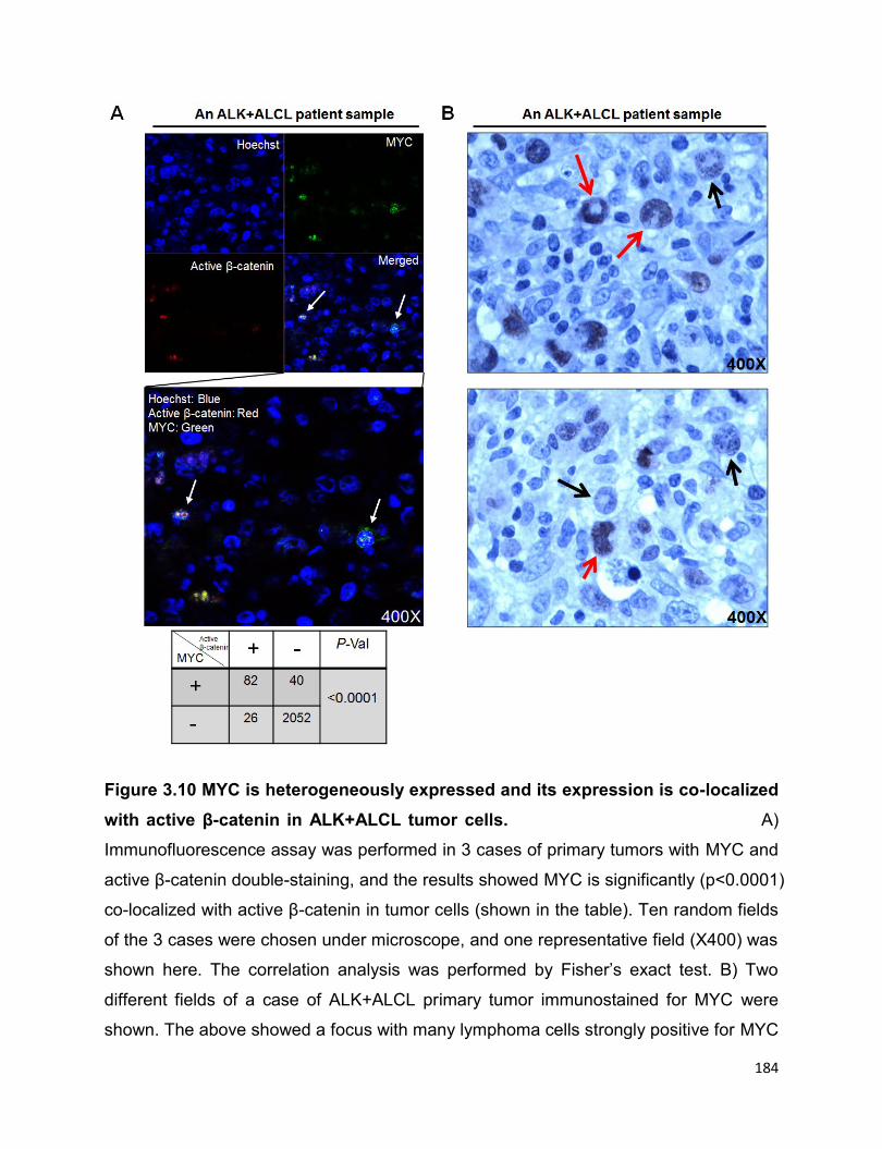
184
Figure 3.10 MYC is heterogeneously expressed and its expression is co-localized
with active β-catenin in ALK+ALCL tumor cells. A)
Immunofluorescence assay was performed in 3 cases of primary tumors with MYC and
active β-catenin double-staining, and the results showed MYC is significantly (p<0.0001)
co-localized with active β-catenin in tumor cells (shown in the table). Ten random fields
of the 3 cases were chosen under microscope, and one representative field (X400) was
shown here. The correlation analysis was performed by Fisher’s exact test. B) Two
different fields of a case of ALK+ALCL primary tumor immunostained for MYC were
shown. The above showed a focus with many lymphoma cells strongly positive for MYC
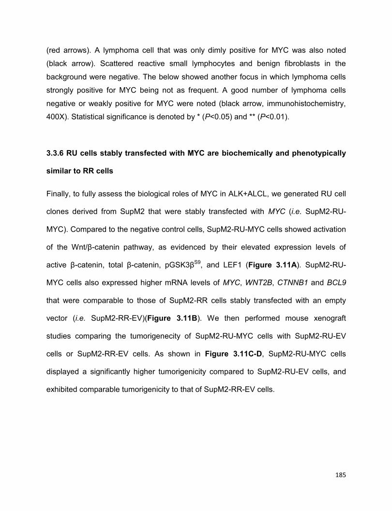
185
(red arrows). A lymphoma cell that was only dimly positive for MYC was also noted
(black arrow). Scattered reactive small lymphocytes and benign fibroblasts in the
background were negative. The below showed another focus in which lymphoma cells
strongly positive for MYC being not as frequent. A good number of lymphoma cells
negative or weakly positive for MYC were noted (black arrow, immunohistochemistry,
400X). Statistical significance is denoted by * (P<0.05) and ** (P<0.01).
3.3.6 RU cells stably transfected with MYC are biochemically and phenotypically
similar to RR cells
Finally, to fully assess the biological roles of MYC in ALK+ALCL, we generated RU cell
clones derived from SupM2 that were stably transfected with MYC (i.e. SupM2-RU-
MYC). Compared to the negative control cells, SupM2-RU-MYC cells showed activation
of the Wnt/β-catenin pathway, as evidenced by their elevated expression levels of
active β-catenin, total β-catenin, pGSK3βS9, and LEF1 (Figure 3.11A). SupM2-RU-
MYC cells also expressed higher mRNA levels of MYC, WNT2B, CTNNB1 and BCL9
that were comparable to those of SupM2-RR cells stably transfected with an empty
vector (i.e. SupM2-RR-EV)(Figure 3.11B). We then performed mouse xenograft
studies comparing the tumorigenecity of SupM2-RU-MYC cells with SupM2-RU-EV
cells or SupM2-RR-EV cells. As shown in Figure 3.11C-D, SupM2-RU-MYC cells
displayed a significantly higher tumorigenicity compared to SupM2-RU-EV cells, and
exhibited comparable tumorigenicity to that of SupM2-RR-EV cells.
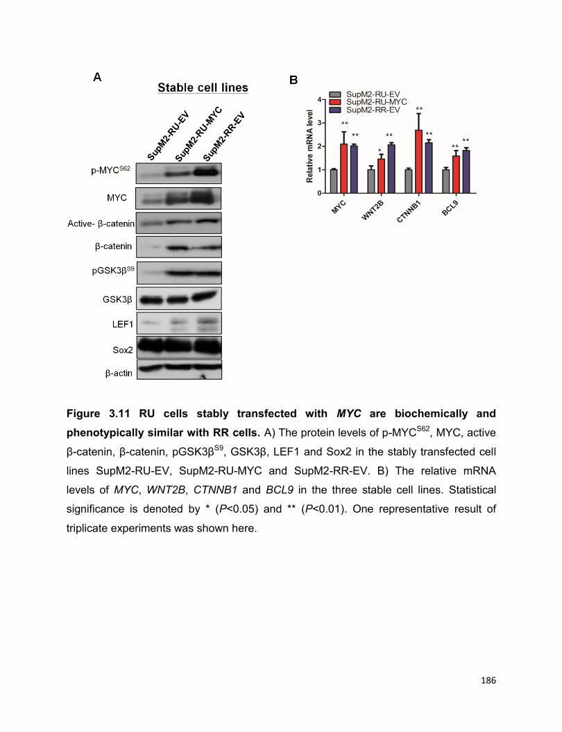
186
Figure 3.11 RU cells stably transfected with MYC are biochemically and
phenotypically similar with RR cells. A) The protein levels of p-MYCS62, MYC, active
β-catenin, β-catenin, pGSK3βS9, GSK3β, LEF1 and Sox2 in the stably transfected cell
lines SupM2-RU-EV, SupM2-RU-MYC and SupM2-RR-EV. B) The relative mRNA
levels of MYC, WNT2B, CTNNB1 and BCL9 in the three stable cell lines. Statistical
significance is denoted by * (P<0.05) and ** (P<0.01). One representative result of
triplicate experiments was shown here.
187
Figure 3.11C-D C) The diagram showed the design of mice xenograft study. D) The
tumor growth rates of the three stable cell lines in the mouse xenograft study. The right
panel showed the representative tumors from the three groups at the termination point.
Statistical significance is denoted by * (P<0.05) and ** (P<0.01).
3.3.7 Side population cells are not detectable in RU and RR cells and ABCG2 is
not significantly differentially expressed in mRNA level between RU and RR cells.
We realized that Moti et al recently reported that side population cells are detectable in
ALK+ALCL cell lines and patient samples using the Hoechst-efflux assay and the

188
authors of this study also provided evidence that these side population cells are tumor-
propagating cells (3). Therefore, we tested whether side population cells can be also
detected in ALK+ALCL cell lines in our hands, and if it is the case, whether RR cells
have enriched side population cells in comparison to RU cells. However, we were
unable to detect the side population cells in our hands, either in parental SupM2 and
Karpas 299 cell lines or RU and RR cells derived from these two cell lines (Figure
3.12A-B). In the meanwhile, Moti et al declared that the gene expression level of ATP-
binding cassette-G2 (ABCG2), a drug cell transporter (20), is higher in the side
population cells as compared to the main cell population (3). We then asked whether
ABCG2 is also highly expressed in RR cells as compared to RU cells. To this end, we
performed quantitative RT-PCR to detect the mRNA level of ABCG2 and also included
some other ABC family transporters, including ABCC1, ABCC2, ABCC3, ABCC4,
ABCC5, and ABCC6. As shown in Figure 3.12C, except ABCC4, all other ABC
transporters were not significantly differentially expressed in mRNA level between RU
and RR cells derived from SupM2.

189
Figure 3.12 Identification of side population cells by Hoechst-efflux assay and the
expression of ABC transporters in RU and RR cells derived from ALK+ALCL. A-B)
Side population cells were not observed in parental Karpas 299 and SupM2 cells in our
hands, as well as in RU and RR cells derived from parental SupM2 cells. C) qRT-PCR
assay was employed to compare the relative mRNA levels of ABC transporters,
including ABCC1, ABCC2, ABCC3, ABCC4, ABCC5, ABCC6 and ABCG2, between RU
and RR cells derived from SupM2 cells. Statistical significance is denoted by * (P<0.05)
and ** (P<0.01). Results shown are representative of three independent experiments.
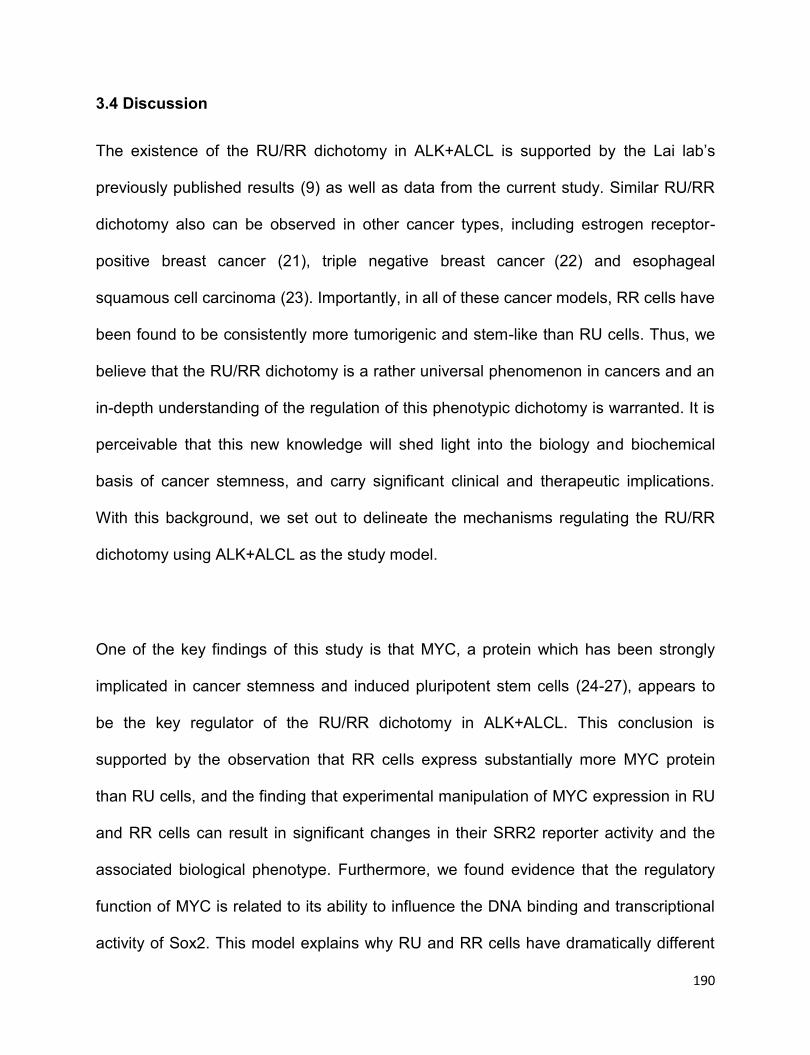
190
3.4 Discussion
The existence of the RU/RR dichotomy in ALK+ALCL is supported by the Lai lab’s
previously published results (9) as well as data from the current study. Similar RU/RR
dichotomy also can be observed in other cancer types, including estrogen receptor-
positive breast cancer (21), triple negative breast cancer (22) and esophageal
squamous cell carcinoma (23). Importantly, in all of these cancer models, RR cells have
been found to be consistently more tumorigenic and stem-like than RU cells. Thus, we
believe that the RU/RR dichotomy is a rather universal phenomenon in cancers and an
in-depth understanding of the regulation of this phenotypic dichotomy is warranted. It is
perceivable that this new knowledge will shed light into the biology and biochemical
basis of cancer stemness, and carry significant clinical and therapeutic implications.
With this background, we set out to delineate the mechanisms regulating the RU/RR
dichotomy using ALK+ALCL as the study model.
One of the key findings of this study is that MYC, a protein which has been strongly
implicated in cancer stemness and induced pluripotent stem cells (24-27), appears to
be the key regulator of the RU/RR dichotomy in ALK+ALCL. This conclusion is
supported by the observation that RR cells express substantially more MYC protein
than RU cells, and the finding that experimental manipulation of MYC expression in RU
and RR cells can result in significant changes in their SRR2 reporter activity and the
associated biological phenotype. Furthermore, we found evidence that the regulatory
function of MYC is related to its ability to influence the DNA binding and transcriptional
activity of Sox2. This model explains why RU and RR cells have dramatically different
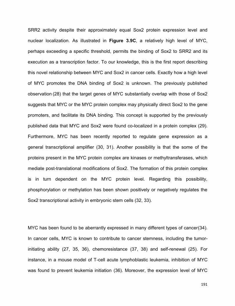
191
SRR2 activity despite their approximately equal Sox2 protein expression level and
nuclear localization. As illustrated in Figure 3.9C, a relatively high level of MYC,
perhaps exceeding a specific threshold, permits the binding of Sox2 to SRR2 and its
execution as a transcription factor. To our knowledge, this is the first report describing
this novel relationship between MYC and Sox2 in cancer cells. Exactly how a high level
of MYC promotes the DNA binding of Sox2 is unknown. The previously published
observation (28) that the target genes of MYC substantially overlap with those of Sox2
suggests that MYC or the MYC protein complex may physically direct Sox2 to the gene
promoters, and facilitate its DNA binding. This concept is supported by the previously
published data that MYC and Sox2 were found co-localized in a protein complex (29).
Furthermore, MYC has been recently reported to regulate gene expression as a
general transcriptional amplifier (30, 31). Another possibility is that the some of the
proteins present in the MYC protein complex are kinases or methyltransferases, which
mediate post-translational modifications of Sox2. The formation of this protein complex
is in turn dependent on the MYC protein level. Regarding this possibility,
phosphorylation or methylation has been shown positively or negatively regulates the
Sox2 transcriptional activity in embryonic stem cells (32, 33).
MYC has been found to be aberrantly expressed in many different types of cancer(34).
In cancer cells, MYC is known to contribute to cancer stemness, including the tumor-
initiating ability (27, 35, 36), chemoresistance (37, 38) and self-renewal (25). For
instance, in a mouse model of T-cell acute lymphoblastic leukemia, inhibition of MYC
was found to prevent leukemia initiation (36). Moreover, the expression level of MYC
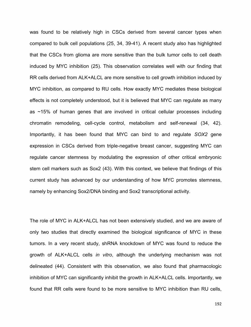
192
was found to be relatively high in CSCs derived from several cancer types when
compared to bulk cell populations (25, 34, 39-41). A recent study also has highlighted
that the CSCs from glioma are more sensitive than the bulk tumor cells to cell death
induced by MYC inhibition (25). This observation correlates well with our finding that
RR cells derived from ALK+ALCL are more sensitive to cell growth inhibition induced by
MYC inhibition, as compared to RU cells. How exactly MYC mediates these biological
effects is not completely understood, but it is believed that MYC can regulate as many
as ~15% of human genes that are involved in critical cellular processes including
chromatin remodeling, cell-cycle control, metabolism and self-renewal (34, 42).
Importantly, it has been found that MYC can bind to and regulate SOX2 gene
expression in CSCs derived from triple-negative breast cancer, suggesting MYC can
regulate cancer stemness by modulating the expression of other critical embryonic
stem cell markers such as Sox2 (43). With this context, we believe that findings of this
current study has advanced by our understanding of how MYC promotes stemness,
namely by enhancing Sox2/DNA binding and Sox2 transcriptional activity.
The role of MYC in ALK+ALCL has not been extensively studied, and we are aware of
only two studies that directly examined the biological significance of MYC in these
tumors. In a very recent study, shRNA knockdown of MYC was found to reduce the
growth of ALK+ALCL cells in vitro, although the underlying mechanism was not
delineated (44). Consistent with this observation, we also found that pharmacologic
inhibition of MYC can significantly inhibit the growth in ALK+ALCL cells. Importantly, we
found that RR cells were found to be more sensitive to MYC inhibition than RU cells,
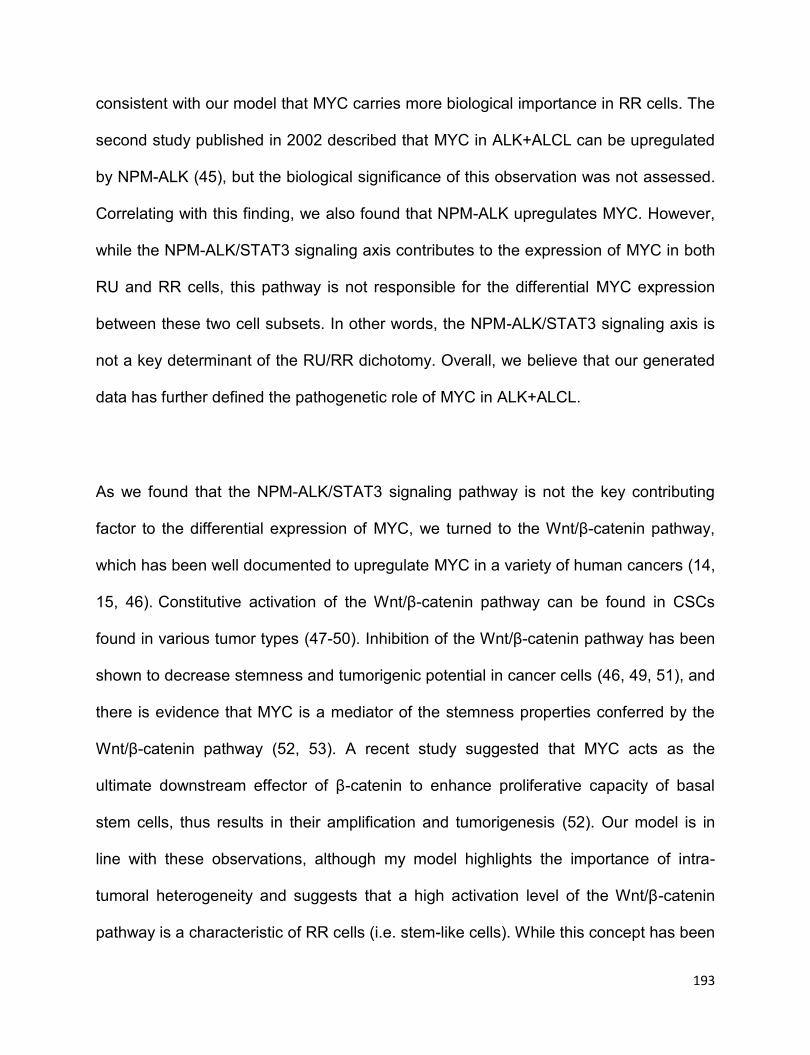
193
consistent with our model that MYC carries more biological importance in RR cells. The
second study published in 2002 described that MYC in ALK+ALCL can be upregulated
by NPM-ALK (45), but the biological significance of this observation was not assessed.
Correlating with this finding, we also found that NPM-ALK upregulates MYC. However,
while the NPM-ALK/STAT3 signaling axis contributes to the expression of MYC in both
RU and RR cells, this pathway is not responsible for the differential MYC expression
between these two cell subsets. In other words, the NPM-ALK/STAT3 signaling axis is
not a key determinant of the RU/RR dichotomy. Overall, we believe that our generated
data has further defined the pathogenetic role of MYC in ALK+ALCL.
As we found that the NPM-ALK/STAT3 signaling pathway is not the key contributing
factor to the differential expression of MYC, we turned to the Wnt/β-catenin pathway,
which has been well documented to upregulate MYC in a variety of human cancers (14,
15, 46). Constitutive activation of the Wnt/β-catenin pathway can be found in CSCs
found in various tumor types (47-50). Inhibition of the Wnt/β-catenin pathway has been
shown to decrease stemness and tumorigenic potential in cancer cells (46, 49, 51), and
there is evidence that MYC is a mediator of the stemness properties conferred by the
Wnt/β-catenin pathway (52, 53). A recent study suggested that MYC acts as the
ultimate downstream effector of β-catenin to enhance proliferative capacity of basal
stem cells, thus results in their amplification and tumorigenesis (52). Our model is in
line with these observations, although my model highlights the importance of intra-
tumoral heterogeneity and suggests that a high activation level of the Wnt/β-catenin
pathway is a characteristic of RR cells (i.e. stem-like cells). While this concept has been

194
brought up in a previously published paper (48), our data has provided the mechanistic
explanation as to how the Wnt/β-catenin pathway promotes stemness for the first time.
Specifically, a high level of Wnt/β-catenin activity promotes a relatively high level of
MYC expression, which permits Sox2 to exert its transcriptional activity. Furthermore,
based on our observations that Sox2 upregulates a number of the Wnt/β-catenin
pathway ligands as well as β-catenin, we have demonstrated, for the first time, a
positive feedback loop involving Wnt/β-catenin, MYC and Sox2, and my hypothetical
model has been illustrated in Figure 3.9C. Our data supports a model in which this
positive feedback loop is the defining feature of RR cells in ALK+ALCL.
Our results have provided further support for the concept of tumor plasticity, since RU
cells can be converted to RR cells by manipulating the Wnt/β-catenin/ MYC axis. Tumor
plasticity also has been illustrated by a recent study in which non-stem cells from lung
cancer were converted to CSCs when the Wnt/β-catenin pathway is activated by
hepatocyte growth factor (48). The Lai lab also recently published that RU cells derived
from esophageal squamous cell carcinoma (23) and estrogen receptor-positive breast
cancer cell lines, as well as primary breast tumors cells (54), can convert to RR cells
upon oxidative stress. Findings from the current study has highlighted the importance of
the Wnt/β-catenin/ MYC axis in the context of tumor plasticity.
Results from our immunofluorescence staining/confocal microscopy have provided
further evidence to support the existence of the positive feedback loop involving Wnt/β-
195
catenin in a small cell subset of ALK+ALCL. Thus, MYC significantly co-localizes with
active β-catenin in approximately 4% of cells. Interestingly, we found that cells co-
expressing these two markers were scattered throughout the tumors, with no good
evidence of clustering. This is a rather unexpected observation, since CSCs identified
by using immunohistochemistry or immunofluorescence staining published in a very
small number of studies have been shown to cluster (i.e. approximately 10 cells per
cluster) (48, 55-57). Based on these observations, it has been speculated that CSCs
thrive in specific microenvironment or ‘niche’. In view of the relatively small number of
studies of the in-situ localization of CSCs, we believe that one cannot come to any
specific generalization. Nonetheless, it may be speculated that CSCs in hematologic
neoplasms (such as ALK+ALCL) may have different biological behaviors from those in
solid tumors, which were the study subjects of most publications reporting the in-situ
localization of CSCs. Regarding our immunohistochemical studies, we would like to
point out that, while we found only ~30% of tumor cells being labeled with MYC, two
previous publications showed that MYC immunohistochemical reactivity is detectable in
the majority of tumor cells in ALK+ALCL(44, 45). This discrepancy may be due to the
use of different MYC antibodies and/or immunostaining protocols. In our experience, a
substantially higher number of MYC-positive cells can be obtained if higher
concentration of anti-MYC antibody is used. The concentration of anti-MYC antibody
we chose was based on the observation that this antibody concentration is optimal in
revealing the intra-tumoral heterogeneity of MYC expression.

196
In light of a recent publication that a tumor-propagating population can be identified and
isolated in ALK+ALCL cell lines and tumors using the hoechst-efflux assay (3), we
asked if RR cells identified using our study model are enriched in the tumor-
propagating population identified using the hoechst-efflux assay. In our hands, we was
unable to identify this small cell population in Karpas 299 and SupM2 cells, including
RU and RR cells derived from SupM2 (Figure 3.12A-B), despite our attempt to follow
the experimental protocol described in the publication (3). This discrepancy may be
attributed to various technical reasons and/or biological differences between
ALK+ALCL cell clones. As the expression level of NPM-ALK and ABCG2 were reported
to be expressed at a higher level in the tumor-propagating population (3), we asked if
RR cells also carry these characteristics, while we found that RR and RU cells did not
differ in the expression of the NPM-ALK or ABCG2 in mRNA level (Figure 3.5A and
Figure 3.12C). Some other ABC family transporters, such as ABCC1 and ABCC2, were
also not significantly different in mRNA level between RU and RR cells (Figure 3.12C).
To conclude, we report that MYC is the key regulator of the RU/RR dichotomy in
ALK+ALCL. High level of MYC promotes the DNA binding ability and transcriptional
activity of Sox2. Our study has highlighted the importance of the Wnt/β-catenin pathway
in contributing to the high MYC protein expression in RR cells. The existence of a
positive feedback loop involving the Wnt/β-catenin/MYC/Sox2 axis defines a small cell
subset in ALK+ALCL that are characterized by high tumorigenecity and
chemoresistance.

197
3.5 References
1. Kinney MC, Higgins RA, Medina EA. Anaplastic large cell lymphoma: twenty-five
years of discovery. Archives of pathology & laboratory medicine. 2011;135(1):19-43.
2. Amin HM, Lai R. Pathobiology of ALK+ anaplastic large-cell lymphoma. Blood.
2007;110(7):2259-67.
3. Moti N, Malcolm T, Hamoudi R, Mian S, Garland G, Hook CE, et al. Anaplastic
large cell lymphoma-propagating cells are detectable by side population analysis and
possess an expression profile reflective of a primitive origin. Oncogene.
2015;34(14):1843-52.
4. Marusyk A, Almendro V, Polyak K. Intra-tumour heterogeneity: a looking glass
for cancer? Nature reviews Cancer. 2012;12(5):323-34.
5. Wernig M, Meissner A, Foreman R, Brambrink T, Ku M, Hochedlinger K, et al. In
vitro reprogramming of fibroblasts into a pluripotent ES-cell-like state. Nature.
2007;448(7151):318-24.
6. Chen Y, Shi L, Zhang L, Li R, Liang J, Yu W, et al. The molecular mechanism
governing the oncogenic potential of SOX2 in breast cancer. The Journal of biological
chemistry. 2008;283(26):17969-78.
7. Leis O, Eguiara A, Lopez-Arribillaga E, Alberdi MJ, Hernandez-Garcia S,
Elorriaga K, et al. Sox2 expression in breast tumours and activation in breast cancer
stem cells. Oncogene. 2012;31(11):1354-65.
8. Santini R, Pietrobono S, Pandolfi S, Montagnani V, D'Amico M, Penachioni JY,
et al. SOX2 regulates self-renewal and tumorigenicity of human melanoma-initiating
cells. Oncogene. 2014;33(38):4697-708.

198
9. Gelebart P, Hegazy SA, Wang P, Bone KM, Anand M, Sharon D, et al. Aberrant
expression and biological significance of Sox2, an embryonic stem cell transcriptional
factor, in ALK-positive anaplastic large cell lymphoma. Blood Cancer J. 2012;2:e82.
10. Piva M, Domenici G, Iriondo O, Rabano M, Simoes BM, Comaills V, et al. Sox2
promotes tamoxifen resistance in breast cancer cells. EMBO molecular medicine.
2014;6(1):66-79.
11. Wu C, Molavi O, Zhang H, Gupta N, Alshareef A, Bone KM, et al. STAT1 is
phosphorylated and downregulated by the oncogenic tyrosine kinase NPM-ALK in ALK-
positive anaplastic large-cell lymphoma. Blood. 2015;126(3):336-45.
12. Marampon F, Ciccarelli C, Zani BM. Down-regulation of c-Myc following
MEK/ERK inhibition halts the expression of malignant phenotype in rhabdomyosarcoma
and in non muscle-derived human tumors. Molecular cancer. 2006;5:31.
13. Wang H, Chauhan J, Hu A, Pendleton K, Yap JL, Sabato PE, et al. Disruption of
Myc-Max heterodimerization with improved cell-penetrating analogs of the small
molecule 10074-G5. Oncotarget. 2013;4(6):936-47.
14. Juan J, Muraguchi T, Iezza G, Sears RC, McMahon M. Diminished WNT ->
beta-catenin -> c-MYC signaling is a barrier for malignant progression of BRAFV600E-
induced lung tumors. Genes & development. 2014;28(6):561-75.
15. Zhang S, Li Y, Wu Y, Shi K, Bing L, Hao J. Wnt/beta-catenin signaling pathway
upregulates c-Myc expression to promote cell proliferation of P19 teratocarcinoma cells.
Anatomical record. 2012;295(12):2104-13.
16. You Z, Saims D, Chen S, Zhang Z, Guttridge DC, Guan KL, et al. Wnt signaling
promotes oncogenic transformation by inhibiting c-Myc-induced apoptosis. The Journal
of cell biology. 2002;157(3):429-40.
199
17. Gelebart P, Anand M, Armanious H, Peters AC, Dien Bard J, Amin HM, et al.
Constitutive activation of the Wnt canonical pathway in mantle cell lymphoma. Blood.
2008;112(13):5171-9.
18. Li X, Xu Y, Chen Y, Chen S, Jia X, Sun T, et al. SOX2 promotes tumor
metastasis by stimulating epithelial-to-mesenchymal transition via regulation of
WNT/beta-catenin signal network. Cancer letters. 2013;336(2):379-89.
19. Chen S, Xu Y, Chen Y, Li X, Mou W, Wang L, et al. SOX2 gene regulates the
transcriptional network of oncogenes and affects tumorigenesis of human lung cancer
cells. PloS one. 2012;7(5):e36326.
20. Dean M. ABC transporters, drug resistance, and cancer stem cells. J Mammary
Gland Biol Neoplasia. 2009;14(1):3-9.
21. Wu F, Zhang J, Wang P, Ye X, Jung K, Bone KM, et al. Identification of two
novel phenotypically distinct breast cancer cell subsets based on Sox2 transcription
activity. Cellular signalling. 2012;24(11):1989-98.
22. Jung K, Gupta N, Wang P, Lewis JT, Gopal K, Wu F, et al. Triple negative breast
cancers comprise a highly tumorigenic cell subpopulation detectable by its high
responsiveness to a Sox2 regulatory region 2 (SRR2) reporter. Oncotarget.
2015;6(12):10366-73.
23. Zhang HF, Wu C, Alshareef A, Gupta N, Zhao Q, Xu XE, et al. The PI3K/AKT/c-
MYC axis promotes the acquisition of cancer stem-like features in esophageal
squamous cell carcinoma. Stem cells. 2016.
24. Wilson A, Murphy MJ, Oskarsson T, Kaloulis K, Bettess MD, Oser GM, et al. c-
Myc controls the balance between hematopoietic stem cell self-renewal and
differentiation. Genes & development. 2004;18(22):2747-63.
200
25. Morrissey PA, Quinn PB, Sheehy PJ. Newer aspects of micronutrients in chronic
disease: vitamin E. The Proceedings of the Nutrition Society. 1994;53(3):571-82.
26. Satoh Y, Matsumura I, Tanaka H, Ezoe S, Sugahara H, Mizuki M, et al. Roles for
c-Myc in self-renewal of hematopoietic stem cells. The Journal of biological chemistry.
2004;279(24):24986-93.
27. Gabay M, Li Y, Felsher DW. MYC activation is a hallmark of cancer initiation and
maintenance. Cold Spring Harbor perspectives in medicine. 2014;4(6).
28. Kwan KY, Shen J, Corey DP. C-MYC transcriptionally amplifies SOX2 target
genes to regulate self-renewal in multipotent otic progenitor cells. Stem cell reports.
2015;4(1):47-60.
29. Yang Z, Augustin J, Hu J, Jiang H. Physical Interactions and Functional
Coordination between the Core Subunits of Set1/Mll Complexes and the
Reprogramming Factors. PloS one. 2015;10(12):e0145336.
30. Nie Z, Hu G, Wei G, Cui K, Yamane A, Resch W, et al. c-Myc is a universal
amplifier of expressed genes in lymphocytes and embryonic stem cells. Cell.
2012;151(1):68-79.
31. Lin CY, Loven J, Rahl PB, Paranal RM, Burge CB, Bradner JE, et al.
Transcriptional amplification in tumor cells with elevated c-Myc. Cell. 2012;151(1):56-67.
32. Fang L, Zhang L, Wei W, Jin X, Wang P, Tong Y, et al. A methylation-
phosphorylation switch determines Sox2 stability and function in ESC maintenance or
differentiation. Molecular cell. 2014;55(4):537-51.
33. Jeong CH, Cho YY, Kim MO, Kim SH, Cho EJ, Lee SY, et al. Phosphorylation of
Sox2 cooperates in reprogramming to pluripotent stem cells. Stem cells.
2010;28(12):2141-50.
201
34. Dang CV. MYC on the path to cancer. Cell. 2012;149(1):22-35.
35. Pan XN, Chen JJ, Wang LX, Xiao RZ, Liu LL, Fang ZG, et al. Inhibition of c-Myc
overcomes cytotoxic drug resistance in acute myeloid leukemia cells by promoting
differentiation. PloS one. 2014;9(8):e105381.
36. Roderick JE, Tesell J, Shultz LD, Brehm MA, Greiner DL, Harris MH, et al. c-Myc
inhibition prevents leukemia initiation in mice and impairs the growth of relapsed and
induction failure pediatric T-ALL cells. Blood. 2014;123(7):1040-50.
37. Shen A, Wang L, Huang M, Sun J, Chen Y, Shen YY, et al. c-Myc alterations
confer therapeutic response and acquired resistance to c-Met inhibitors in MET-
addicted cancers. Cancer research. 2015;75(21):4548-59.
38. Zhang Y, Chen HX, Zhou SY, Wang SX, Zheng K, Xu DD, et al. Sp1 and c-Myc
modulate drug resistance of leukemia stem cells by regulating survivin expression
through the ERK-MSK MAPK signaling pathway. Molecular cancer. 2015;14:56.
39. Salcido CD, Larochelle A, Taylor BJ, Dunbar CE, Varticovski L. Molecular
characterisation of side population cells with cancer stem cell-like characteristics in
small-cell lung cancer. British journal of cancer. 2010;102(11):1636-44.
40. Walter D, Satheesha S, Albrecht P, Bornhauser BC, D'Alessandro V, Oesch SM,
et al. CD133 positive embryonal rhabdomyosarcoma stem-like cell population is
enriched in rhabdospheres. PloS one. 2011;6(5):e19506.
41. Akita H, Marquardt JU, Durkin ME, Kitade M, Seo D, Conner EA, et al. MYC
activates stem-like cell potential in hepatocarcinoma by a p53-dependent mechanism.
Cancer research. 2014;74(20):5903-13.
42. Pelengaris S, Khan M, Evan G. c-MYC: more than just a matter of life and death.
Nature reviews Cancer. 2002;2(10):764-76.
202
43. Zhao D, Pan C, Sun J, Gilbert C, Drews-Elger K, Azzam DJ, et al. VEGF drives
cancer-initiating stem cells through VEGFR-2/Stat3 signaling to upregulate Myc and
Sox2. Oncogene. 2015;34(24):3107-19.
44. Weilemann A, Grau M, Erdmann T, Merkel O, Sobhiafshar U, Anagnostopoulos I,
et al. Essential role of IRF4 and MYC signaling for survival of anaplastic large cell
lymphoma. Blood. 2015;125(1):124-32.
45. Raetz EA, Perkins SL, Carlson MA, Schooler KP, Carroll WL, Virshup DM. The
nucleophosmin-anaplastic lymphoma kinase fusion protein induces c-Myc expression in
pediatric anaplastic large cell lymphomas. The American journal of pathology.
2002;161(3):875-83.
46. Jang GB, Hong IS, Kim RJ, Lee SY, Park SJ, Lee ES, et al. Wnt/beta-Catenin
Small-Molecule Inhibitor CWP232228 Preferentially Inhibits the Growth of Breast
Cancer Stem-like Cells. Cancer research. 2015;75(8):1691-702.
47. Fodde R, Brabletz T. Wnt/beta-catenin signaling in cancer stemness and
malignant behavior. Current opinion in cell biology. 2007;19(2):150-8.
48. Vermeulen L, De Sousa EMF, van der Heijden M, Cameron K, de Jong JH,
Borovski T, et al. Wnt activity defines colon cancer stem cells and is regulated by the
microenvironment. Nature cell biology. 2010;12(5):468-76.
49. Giambra V, Jenkins CE, Lam SH, Hoofd C, Belmonte M, Wang X, et al.
Leukemia stem cells in T-ALL require active Hif1alpha and Wnt signaling. Blood.
2015;125(25):3917-27.
50. Takahashi-Yanaga F, Kahn M. Targeting Wnt signaling: can we safely eradicate
cancer stem cells? Clinical cancer research : an official journal of the American
Association for Cancer Research. 2010;16(12):3153-62.

203
51. Jang GB, Kim JY, Cho SD, Park KS, Jung JY, Lee HY, et al. Blockade of
Wnt/beta-catenin signaling suppresses breast cancer metastasis by inhibiting CSC-like
phenotype. Scientific reports. 2015;5:12465.
52. Moumen M, Chiche A, Decraene C, Petit V, Gandarillas A, Deugnier MA, et al.
Myc is required for beta-catenin-mediated mammary stem cell amplification and
tumorigenesis. Molecular cancer. 2013;12(1):132.
53. Sansom OJ, Meniel VS, Muncan V, Phesse TJ, Wilkins JA, Reed KR, et al. Myc
deletion rescues Apc deficiency in the small intestine. Nature. 2007;446(7136):676-9.
54. Gopal K, Gupta N, Zhang H, Alshareef A, Alqahtani H, Bigras G, et al. Oxidative
stress induces the acquisition of cancer stem-like phenotype in breast cancer
detectable by using a Sox2 regulatory region-2 (SRR2) reporter. Oncotarget.
2016;7(3):3111-27.
55. Li L, Neaves WB. Normal stem cells and cancer stem cells: the niche matters.
Cancer research. 2006;66(9):4553-7.
56. Borovski T, De Sousa EMF, Vermeulen L, Medema JP. Cancer stem cell niche:
the place to be. Cancer research. 2011;71(3):634-9.
57. Oskarsson T, Batlle E, Massague J. Metastatic stem cells: sources, niches, and
vital pathways. Cell stem cell. 2014;14(3):306-21.

204
CHAPTER 4
Oxidative stress promotes the tumorigenicity in ALK-positive anaplastic large-
cell lymphoma by activating the Wnt/β-catenin/MYC/Sox2 axis1
1This chapter has been modified from a manuscript in preparation for submission for publication: Chengsheng Wu, Nidhi Gupta, Hai-feng Zhang, Yung-Hsing Huang, and Raymond Lai.
Oxidative stress promotes the tumorigenicity in ALK-positive anaplastic large-cell
lymphoma by activating the Wnt/β-catenin/MYC/Sox2 axis. In preparation.
I prepared the first draft and revisions based on the suggestions and comments of the
co-authors. I designed and performed all the experiments in this study. N.G., H.Z., and
Y.H. contributed to Figure 4.3A, provided technical assistance and intellectual input.
R.L. provided numerous comments and final review of the manuscript.
205
4.1 Introduction
ALK-positive anaplastic large-cell lymphoma (ALK+ALCL) is a rare subtype of non-
Hodgkin lymphoma of null or T-cell lineage, which preferentially occurs in children and
young adults (1). Gene translocation involving ALK and NPM has been detected in
approximately 80% of ALK+ALCL cases (2), and the translocation results in the
generation of fusion tyrosine kinase NPM-ALK, the key oncogenic driver of this
neoplasm (3). The oncogenic potential of NPM-ALK has been well underscored by its
interaction and activation of a number of molecular proteins involved in multiple
signaling pathways, such as JAK/STATs, MAPK/ERK and PI3K/AKT(3). Among these
oncogenic signaling pathways, NPM-ALK/STAT3 is one of the most critical mediators of
NPM-ALK-induced transformation, by upregulating the gene expression related with
anti-apoptosis and cell growth, including BCL2, Survivin, and MYC (1, 4, 5); and
downregulating the negative regulators of NPM-ALK/STAT3 activity, such as Shp1,
STAT5A and STAT1 (3, 6-8).
Intra-tumor heterogeneity accounts for the diversity of tumor cells within a tumor and
this concept has evolved in the past decade to phenotypical, functional and genetical
heterogeneity (9-11). The existence of intra-tumor heterogeneity can be explained by
two hypothetical models including the clone evolution and cancer stem cell (CSC)
models (11). Briefly, the clone evolution model proposes that tumor progression is an
evolutionary process for a single cell clone with acquisition of multiple genetic
mutations and also a selection process of more aggressive subclones (9). While the
CSC model raises that only a small fraction of tumor cells have the capability to initiate
the whole tumor bulks and the ability to self-renewal to maintain this small fraction (9,
206
11). With these two models, it is well recognized that cancer cells contain a hierarchy of
CSCs and non-CSCs (10, 11). More interestingly, recent evidence shows that non-
CSCs are able to shift to CSCs under certain circumstances and vice versa (9-11). This
phenomenon indicates a biological process named CSC plasticity or “acquired cancer
stemness” (12). For instance, estrogen receptor-positive (ER+) breast cancer cell line
MCF-7 acquires tumorigenic potential and chemoresistance upon the treatment of
chronic oxidative stress (13). Hypoxia stress also enriches the CSC population in
CD133+ glioma stem cells (14). CSC plasticity has been postulated to be a major
mechanism to explain how cancer cells develop chemoresistance, and ultimately, to
contribute to cancer treatment failure (15-17). However, to our knowledge, the concept
of CSC plasticity remains poorly understood in haematological malignancy, including
ALK+ALCL. Thus, we believe an in-depth understanding of CSC plasticity in
ALK+ALCL is warranted.
The Lai lab previously identified two distinct cell subpopulations derived from ER+
breast cancer cells (18), esophagus squamous cancer cells (19), and ALK+ALCL cell
lines based on their differential responses to a Sox2 reporter ꟷ SRR2 carrying GFP and
luciferase (20). The reporter responsive (RR) cells are consistently more
chemoresistant and tumorigenic than the reporter unresponsive (RU) cells. Our
previous study suggested that the positive forward loop involving the Wnt/β-catenin-
MYC/Sox2 axis is the defining feature of RR cells derived from ALK+ALCL (21).
Although the Lai lab has recently showed that RU cells derived from ER+ breast cancer
cells and esophagus squamous cancer cells are able to acquire stemness features
upon oxidative stress (19, 22), we do not know whether similar findings can be
207
observed in ALK+ALCL, since ALK+ALCL is a haematological neoplasm that is very
much distinct from solid tumor models, such as cell models of breast cancer and
esophagus squamous cancer. In this study, we showed that RU cells can be induced to
RR cells (i.e. converted RR cells) upon oxidative challenge. These converted RR cells
share the similar biological and biochemical characteristics with native RR cells.
Furthermore, we found that the activated Wnt/β-catenin/MYC/Sox2 axis upon oxidative
challenge is responsible for the conversion of RU cells to RR cells, as pharmacological
inhibition of β-catenin or MYC or siRNA knockdown of Sox2 in RU cells significantly
abrogates this biological process.
4.2 Methods and materials
4.2.1 Cell lines and chemicals
The cell lines and the condition of cell culture were described in section 2.2.1 of
Chapter 2 and section 3.2.1 of Chapter 3. 10074-G5, U0126, doxorubicin, doxycycline,
iodonitrotetrazolium chloride, N-acetylcysteine (NAC), and quercetin were all purchased
from Sigma Aldrich. Hydrogen peroxide (H2O2) was purchased from ThermoFisher
Scientific Canada. U0126, doxorubicin and doxycycline treatments were described in
section 3.2.1 of Chapter 3 and section 2.2.6 of Chapter 2.
4.2.2 H2O2 re-challenge and chemical treatment
Approximately 1 x 10⁷ of cells was started with H2O2 re-challenge experiment in this
study. Briefly, cells were subjected with various doses of H2O2 ranging from 0 to 1 mM

208
for 3 days, and then cells were changed with fresh culture medium containing various
doses of H2O2 and continuously cultured up to day 5, followed by the luciferase reporter
assay and flow cytometry assay to assess luciferase activity and GFP expression. Cell
viability was also assessed from day 1 to day 3. For the treatment of NAC, cells were
subjected with 0.3 mM H2O2 in the presence of various doses of NAC ranging from 0 to
20 mM for 48 hours, and then the luciferase reporter assay and flow cytometry assay
were executed to evaluate the SRR2 luciferase activity and GFP expression. For the
treatment of 10074-G5 or quercetin, cells were subjected with 5 μM 10074-G5 or 50 µM
quercetin from day 4 to day 5 (24 hours) in the H2O2 re-challenge experiment, the
luciferase reporter assay was then followed to evaluate the SRR2 reporter activity.
4.2.3 Luciferase assay and flow cytometry
The luciferase assay kit was purchased from Promega (Madison, WI), and luciferase
activity was measured as described previous in section 3.2.7 of Chapter 3. The flow
cytometry analyses for assessing GFP expression were performed as below. Cells after
treatment were washed with cold 1x PBS three times and were resuspended in 1 X
cold PBS before flow cytometry (BD Bioscience). 20,000 live cells were acquired for
each run. All the flow cytometry data were analyzed using the FCS Express 5 software
from De Novo Software (Glendale, CA).
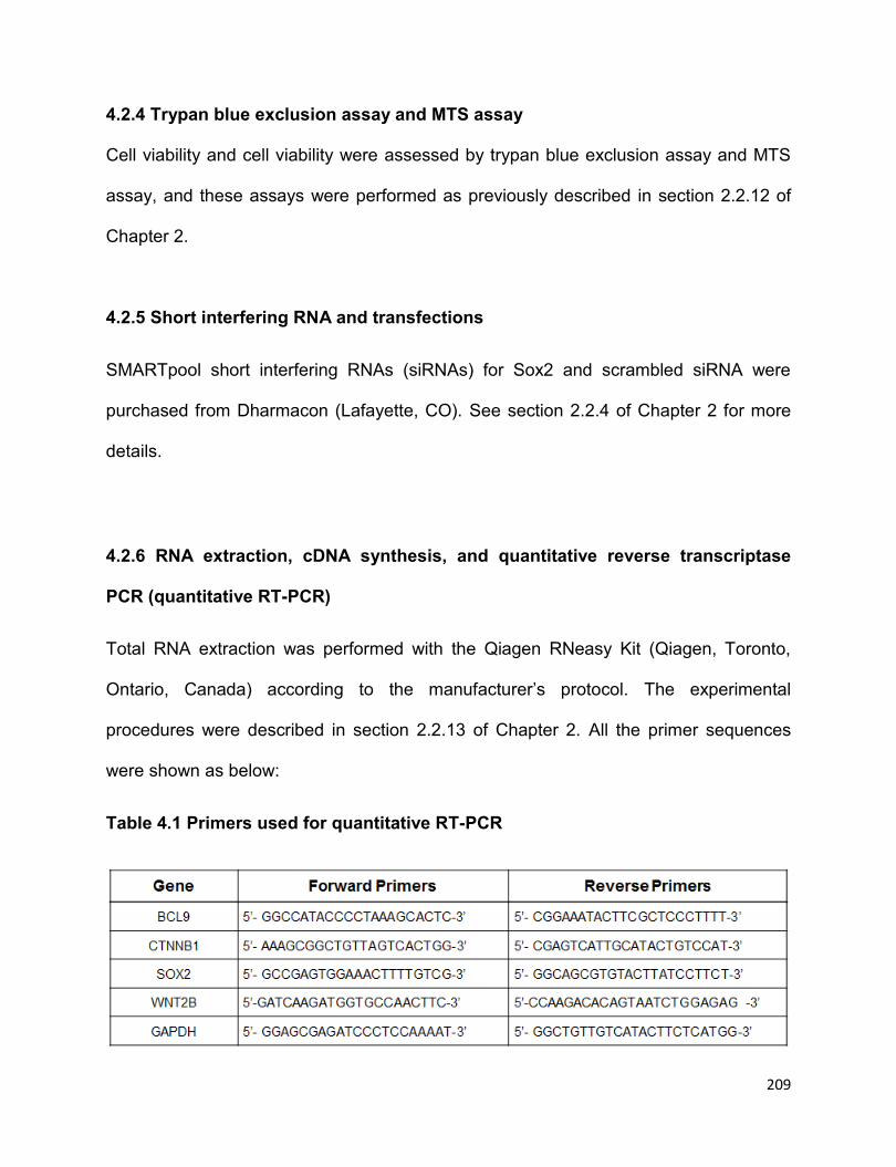
209
4.2.4 Trypan blue exclusion assay and MTS assay
Cell viability and cell viability were assessed by trypan blue exclusion assay and MTS
assay, and these assays were performed as previously described in section 2.2.12 of
Chapter 2.
4.2.5 Short interfering RNA and transfections
SMARTpool short interfering RNAs (siRNAs) for Sox2 and scrambled siRNA were
purchased from Dharmacon (Lafayette, CO). See section 2.2.4 of Chapter 2 for more
details.
4.2.6 RNA extraction, cDNA synthesis, and quantitative reverse transcriptase
PCR (quantitative RT-PCR)
Total RNA extraction was performed with the Qiagen RNeasy Kit (Qiagen, Toronto,
Ontario, Canada) according to the manufacturer’s protocol. The experimental
procedures were described in section 2.2.13 of Chapter 2. All the primer sequences
were shown as below:
Table 4.1 Primers used for quantitative RT-PCR
210
4.2.7 Western blotting
Western blotting studies were performed as described as previously in section 2.2.8 of
Chapter 2.
Antibodies reactive to phosphorylated MYCS62 (E1J4K), MYC (D84C12), Sox2 (D6D9),
and β-catenin (D10A8) were purchased from Cell Signaling Technology (Danvers, MA),
β-actin antibody (sc-130300) was purchased from Santa Cruz (Dallas, Texas), and
antibody reactive to active β-catenin (8E7) was purchased from Merck Millipore
(Toronto, ON, Canada). Secondary antibodies anti-Rabbit IgG (1:2000) and anti-Mouse
IgG (1:2000) were purchased from Cell Signaling Technology.
4.2.8 Nuclear cytoplasm fractionation assay
The nuclear-cytoplasmic fractionation kit was purchased from ThermoFisher Scientific
Canada. The experiment was performed as described in section 2.2.11 of Chapter 2.
4.2.9 SRR2 probe binding assay
SRR2 probe binding assay was performed as previously described in section 3.2.10 of
Chapter 3.
4.2.10 Methylcellulose colony formation assay
The methylcellulose colony formation assay was performed as previously described in
section 2.2.9 of Chapter 2 and section 3.2.12 of Chapter 3.
211
4.2.11 Limiting dilution assay
Cells with or without H2O2 re-challenge treatment were seeded in RPMI1640 media
(Invitrogen) supplemented with 20% fetal bovine serum (Invitrogen), 1% penicillin
streptomycin (ThermoFisher Scientific) and 200 μg/mL puromycin dihydrochloride
(Sigma Aldrich) in 96-well low adherent plate (Coring) at 10 limiting dilutions ranging
from 1 to 1000 cells, and each dilution has triplicate wells. After 7 days, cells in each
well were stained with iodonitrotetrazolium chloride (Sigma Aldrich) for 24 hours and
images were acquired using Alphalmager HP (ThermoFisher Scientific Canada). The
number of visible spheres was counted by eye.
4.2.12 Statistical analysis
Data were expressed as mean standard deviation. The statistical analysis was
performed using Graphpad Prism 5 (La Jolla, CA), and the significance of two
independent groups of samples was determined using Student’s t-test. Statistical
significance is denoted by * (P<0.05) and ** (P<0.01).
4.3 Results
4.3.1 Oxidative stress induces a conversion from RU to RR cells
Using relatively low concentrations of H2O2, an agent known to potently induce
oxidative stress and the generation of reactive oxygen species (ROS) (23), we
previously successfully converted small subsets of RU cells derived from esophageal
212
squamous cell carcinoma cells and ER+ breast cancer cells into RR cells (19, 22). We
asked if a similar phenomenon can occur in hematopoietic cancers such as ALK+ALCL.
Two ALK+ALCL cell lines, namely SupM2 and Karpas 299, were used for this
experiment.
As shown in Figure 4.1A, upon treatment of various doses of H2O2 for 3 days, RU cells
from both cell lines that harbored ~5% GFP-positive cells exhibited significantly
increased GFP-positive cells. As high doses of H2O2 are expectedly cytotoxic, we
assessed the number of viable cells at different doses of H2O2 from day 1 to day 3. As
shown in Figure 4.1B, we found that H2O2 retarded the cell growth in a dose-
dependent manner. Taking the cell growth inhibition and percentages of GFP-positive
cells induced by various doses of H2O2 into consideration, we decided to choose 0.3
mM and 0.5 mM H2O2 for RU cells derived from SupM2 and Karpas 299, respectively,
to induce a sufficient percentage of GFP-positive cells, or say converted RR cells. We
also found that a re-challenge with H2O2 on day 3 significantly increased the
percentages of converted RR cells in both cell lines. As shown in Figure 4.1C-D, ~80%
of RU cells of SupM2 remaining in the tissue culture were GFP-positive 2 days after the
re-challenge. Similarly, ~30% of RU cells derived from Karpas 299 became converted
RR cells (Figure 4.1D). This H2O2 re-challenge strategy was used to generate
converted RR cells for the remainder of the study. Consistent to the results of GFP
expression, the luciferase activity in RU cells derived from both cell lines also
significantly increased upon H2O2 re-challenge (Figure 4.1E). Notably, the RR cells
derived from SupM2 also significantly acquired the SRR2 activity based on the

213
readouts of luciferase activity and GFP expression upon H2O2 re-challenge (Figure
4.1E-F).
To confirm that the RU to RR cells conversion induced by H2O2 was indeed dependent
on oxidative stress, we attempted to block this process using N-acetyl-L-cysteine (NAC),
a pharmacologic agent known to minimize cellular oxidative stress (24). As shown in
Figure 4.1G-H, in a dose-dependent manner, NAC significantly lowered the number of
H2O2-induced GFP-positive cells as well as luciferase activity upon treatment of 0.3 mM
H2O2 for 2 days.
Figure 4.1 Oxidative challenge induces the conversion of RU to RR cells. A) RU
cells derived from SupM2 and Karpas 299 cells were subjected with various doses of
H2O2 for up to 3 days. The percentages of GFP, one of two surrogate markers of
SRR2 activity, were assessed by flow cytometry in both RU cells at day 0, day 1, day 2
and day 3. Statistical significance is denoted by * (P<0.05) and ** (P<0.01). Results
shown were representative of three independent experiments.

214
Figure 4.1B B) The cell numbers of RU cells derived from SupM2 and Karpas 299
upon various doses of H2O2 treatment were counted by trypan blue exclusion assay
from day 0 to day 3. Results from triplicate experiments were shown.

215
Figure 4.1C-D C) the schematic experimental model of 0.3 mM H2O2 re-challenge
treatment in RU cells derived from SupM2. D) The percentages of GFP-positive cells
and the expression of GFP in RU cells derived from SupM2 and Karpas 299 cells upon
0.3 mM or 0.5 mM H2O2 re-challenge treatment. Statistical significance is denoted by *
(P<0.05) and ** (P<0.01). Results shown were representative of three independent
experiments.

216
Figure 4.1E-F E) the luciferase activity in RU and RR cells derived from SupM2 and
Karpas 299 at day 5 upon 0.3 mM and 0.5 mM H2O2 re-challenge treatment. F) the
GFP expression in RU and RR cells derived from SupM2 with and without H2O2 re-
challenge. Statistical significance is denoted by * (P<0.05) and ** (P<0.01). Results
shown were representative of three independent experiments.
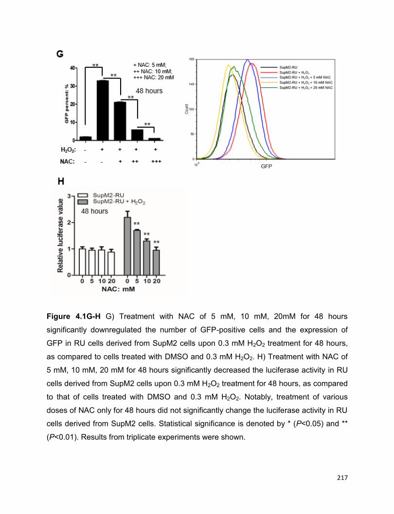
217
Figure 4.1G-H G) Treatment with NAC of 5 mM, 10 mM, 20mM for 48 hours
significantly downregulated the number of GFP-positive cells and the expression of
GFP in RU cells derived from SupM2 cells upon 0.3 mM H2O2 treatment for 48 hours,
as compared to cells treated with DMSO and 0.3 mM H2O2. H) Treatment with NAC of
5 mM, 10 mM, 20 mM for 48 hours significantly decreased the luciferase activity in RU
cells derived from SupM2 cells upon 0.3 mM H2O2 treatment for 48 hours, as compared
to that of cells treated with DMSO and 0.3 mM H2O2. Notably, treatment of various
doses of NAC only for 48 hours did not significantly change the luciferase activity in RU
cells derived from SupM2 cells. Statistical significance is denoted by * (P<0.05) and **
(P<0.01). Results from triplicate experiments were shown.

218
4.3.2 The converted RR cells share the similar biological functions with RR cells
Then we asked whether converted RR cells are phenotypically similar with RR cells.
Specifically, we were interested in whether converted RR cells showed increased
clonogenicity and chemoresistance, as seen in native RR cells. Thus, we subjected
native RU and converted RR cells from SupM2 to various doses of doxorubicin, a
chemotherapy drug used as front-line chemotherapy for ALK+ALCL patients (1). As
shown in Figure 4.2A, converted RR cells were found to be significantly more
doxorubicin-resistant than native RU cells. Furthermore, the converted RR cells even
demonstrated more doxorubicin-resistant than native RR cells at the high dose of
doxorubicin (i.e. 200 ng/mL) (Figure 4.2A).
Next, we tested the clonogenicity of converted RR cells using methylcellulose colony
formation assay. As shown in Figure 4.2B, converted RR cells derived from SupM2
formed significantly more colonies than native RU cells in the presence or absence of
50 ng/mL doxorubicin. In addition, when compared to RR cells without H2O2 re-
challenge in the presence of 50 ng/mL doxorubicin, RR cells derived from SupM2 after
H2O2 re-challenge formed significantly more number of colony in the presence of
doxorubicin, despite that no significant changes in the colony numbers was observed
between the groups of RR cells with and without H2O2 re-challenge (Figure 4.2B).
Then we tested the sphere-forming abilities of RU cells, converted RR cells and
nativel RR cells derived from Karpas 299 using the limiting dilution assay. As shown in
Figure 4.2C, we seeded a serial diluted cells ranging from 1000 cells to 1 cell in each

219
well of 96-well plate, and observed that a low number of RU and RR cells upon H2O2 re-
challenge (i.e. 125 cells for RU, 63 or 32 cells for RR) indeed formed significantly more
spheres, as compared to their native cells (Figure 4.2C-D).
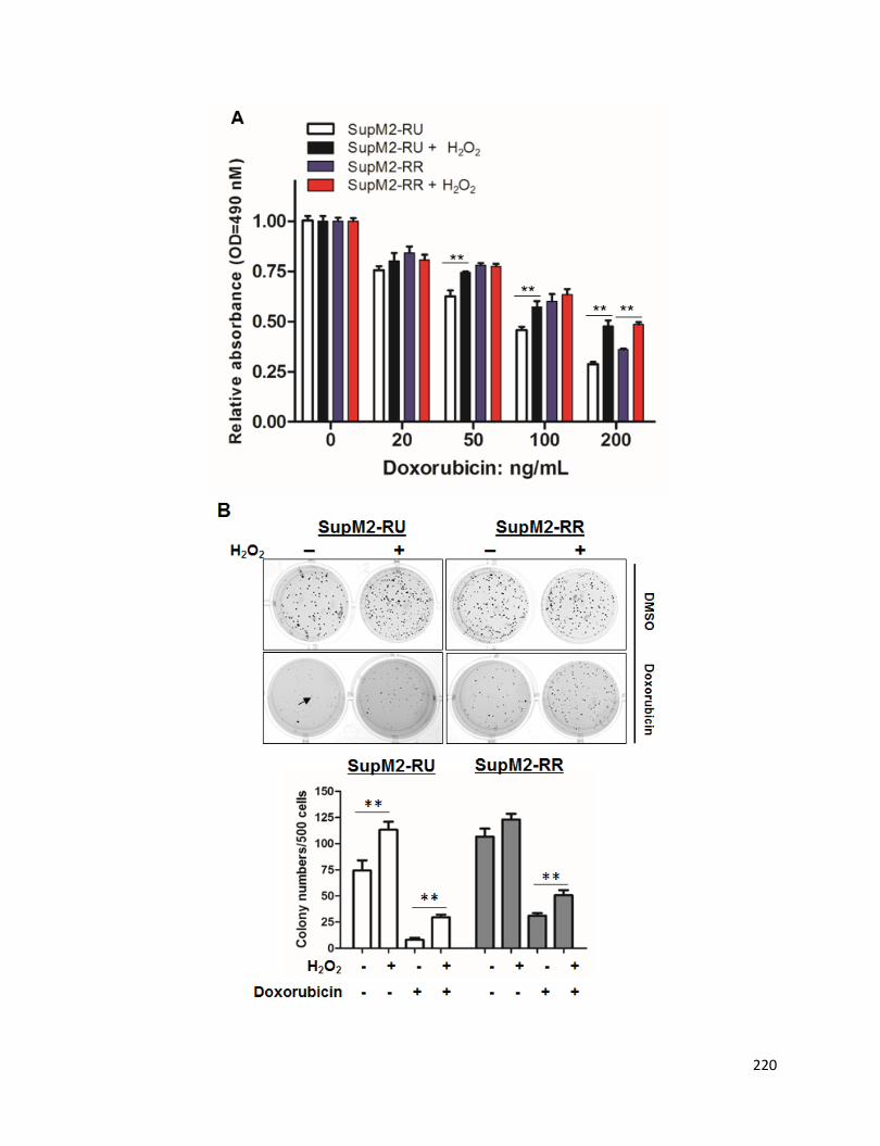
220
221
Figure 4.2 RU and RR cells upon H2O2 re-challenge treatment are more
doxorubicin-resistant and clonogenic, and have captured enhanced sphere-
forming ability as compared to cells without treatment. A) RU and RR cells from
SupM2 cells were re-challenged with 0.3 mM H2O2 for 5 days. Then the cells were
subjected with various doses of doxorubicin for 48 hours. Cells without H2O2 treatment
were included as comparison. B) RU and RR cells derived from SupM2 cells were re-
challenged with 0.3 mM H2O2 for 5 days. Then the cells were preceded for
methylcellulose colony formation assay in the presence/absence of 50 ng/mL
doxorubicin, and cells without H2O2 re-challenge were included as comparison. The
experiments were repeated in triplicate, and one representative result was shown here.
The colonies larger than the one pointed by the bold black arrow were counted only.
The graph below demonstrated the number of colonies in the above experimental
groups. C-D) The serial diluted RU and RR cells derived from Karpas 299 were seeded
in 96-well plate. After 8 days, the number of spheres was counted in the highlighted
wells circulated by red broken lines. Results suggested that low number of RU and RR
cells (125 cells and 63 cells, respectively) upon H2O2 re-challenge formed significant
more spheres than their native cells. This experiment was repeated in triplicate,
statistical significance is denoted by * (P<0.05) and ** (P<0.01).

222
4.3.3 The converted RR cells share the similar biochemical characteristics with
RR cells
Our previous study suggested that the highly active Wnt/β-catenin/MYC/Sox2 axis
defines the RR cells (21). Thus, we asked if converted RR cells showed activation of
this signaling axis. As shown in Figure 4.3A, converted RR cells derived from SupM2
cells showed upregulated expression of active β-catenin, the total protein level of β-
catenin as well as p-MYCS62/MYC, as compared to native RU cells. Although the total
MYC is not appreciably altered in RR cells upon H2O2 re-challenge, p-MYCS62, the
active form of MYC (25), markedly increased (Figure 4.3A). We also observed that the
expression level and activity of NPM-ALK and STAT3 were not appreciably changed in
this experiment (Figure 4.3A).
Moreover, using quantitative RT-PCR (qRT-PCR), we also were able to demonstrate
that Sox2 transcriptional activity was upregulated in converted RR cells; as shown in
Figure 4.3B, the expression of three known Sox2 downstream targets including
WNT2B, CTNNB1 and BCL9 were significantly higher in converted RR cells in mRNA
level, in comparison to their native RU cells (Figure 4.3B). Similar results were
observed in RR cells after oxidative re-challenge (Figure 4.3B).
To substantiate these data, we performed the Sox2-SRR2 probe binding assay to
investigate whether the converted RR cells indeed have increased Sox2-DNA binding.
As shown Figure 4.3C, more Sox2 was pulled down by the biotin-labeled SRR2 probe
in the converted RR cells than RU cells; and a visible increase of Sox2-SRR2 binding
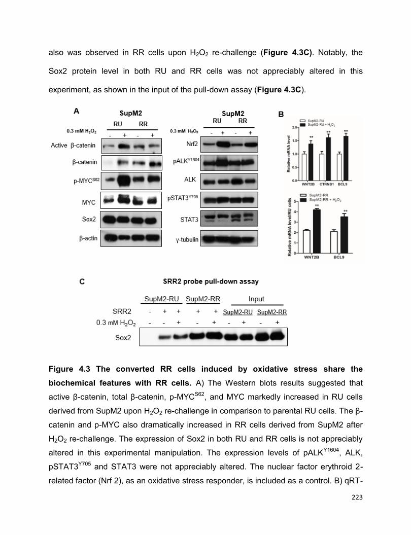
223
also was observed in RR cells upon H2O2 re-challenge (Figure 4.3C). Notably, the
Sox2 protein level in both RU and RR cells was not appreciably altered in this
experiment, as shown in the input of the pull-down assay (Figure 4.3C).
Figure 4.3 The converted RR cells induced by oxidative stress share the
biochemical features with RR cells. A) The Western blots results suggested that
active β-catenin, total β-catenin, p-MYCS62, and MYC markedly increased in RU cells
derived from SupM2 upon H2O2 re-challenge in comparison to parental RU cells. The β-
catenin and p-MYC also dramatically increased in RR cells derived from SupM2 after
H2O2 re-challenge. The expression of Sox2 in both RU and RR cells is not appreciably
altered in this experimental manipulation. The expression levels of pALKY1604, ALK,
pSTAT3Y705 and STAT3 were not appreciably altered. The nuclear factor erythroid 2-
related factor (Nrf 2), as an oxidative stress responder, is included as a control. B) qRT-
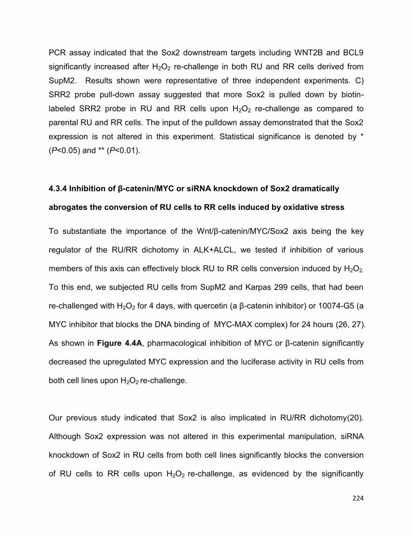
224
PCR assay indicated that the Sox2 downstream targets including WNT2B and BCL9
significantly increased after H2O2 re-challenge in both RU and RR cells derived from
SupM2. Results shown were representative of three independent experiments. C)
SRR2 probe pull-down assay suggested that more Sox2 is pulled down by biotin-
labeled SRR2 probe in RU and RR cells upon H2O2 re-challenge as compared to
parental RU and RR cells. The input of the pulldown assay demonstrated that the Sox2
expression is not altered in this experiment. Statistical significance is denoted by *
(P<0.05) and ** (P<0.01).
4.3.4 Inhibition of β-catenin/MYC or siRNA knockdown of Sox2 dramatically
abrogates the conversion of RU cells to RR cells induced by oxidative stress
To substantiate the importance of the Wnt/β-catenin/MYC/Sox2 axis being the key
regulator of the RU/RR dichotomy in ALK+ALCL, we tested if inhibition of various
members of this axis can effectively block RU to RR cells conversion induced by H2O2.
To this end, we subjected RU cells from SupM2 and Karpas 299 cells, that had been
re-challenged with H2O2 for 4 days, with quercetin (a β-catenin inhibitor) or 10074-G5 (a
MYC inhibitor that blocks the DNA binding of MYC-MAX complex) for 24 hours (26, 27).
As shown in Figure 4.4A, pharmacological inhibition of MYC or β-catenin significantly
decreased the upregulated MYC expression and the luciferase activity in RU cells from
both cell lines upon H2O2 re-challenge.
Our previous study indicated that Sox2 is also implicated in RU/RR dichotomy(20).
Although Sox2 expression was not altered in this experimental manipulation, siRNA
knockdown of Sox2 in RU cells from both cell lines significantly blocks the conversion
of RU cells to RR cells upon H2O2 re-challenge, as evidenced by the significantly
225
downregulated SRR2 luciferase (Figure 4.4B). The Western blots in the lower panel
suggested the siRNA knockdown efficiency of Sox2 (Figure 4.4B).
Figure 4.4 Pharmacological inhibition of MYC or β-catenin, or siRNA knockdown
of Sox2 in RU cells significantly abrogates the conversion of RU cells to RR cells.
A) Pharmacological inhibition of MYC or β-catenin by using 10074-G5 or quercetin for
24 hours decrease the upregulated expression of MYC and the SRR2 luciferase activity
induced by H2O2 re-challenge. B) Sox2 knockdown by siRNA in RU cells significantly
downregulated the SRR2 luciferase activity in comparison to cells transfected with
scrambled siRNA in the presence of H2O2 re-challenge. Statistical significance is
denoted by * (P<0.05) and ** (P<0.01). Results shown were representative of triplicate
experiments.
226
4.3.5 ERK1/2 is activated in both RU and RR cells upon H2O2 re-challenge
Our previous publication and our current study both indicated that ERK1/2 is
preferentially activated in RR cells, instead of RU cells (Figure 4.5A) (20). ERK1/2 is
reported to be activated by oxidative stress in multiple cell models, and the activated
ERK1/2 is well-known as a positive regulator of MYC protein stability and its
transcriptional activity (28-31). Indeed, we observed that ERK1/2 is activated upon
H2O2 re-challenge in both RU and RR cells derived from SupM2 (Figure 4.5B).
Furthermore, we found that blockage of ERK1/2 activity by MEK inhibitor U0126 for 24
hours in RR cells derived from SupM2 and Karpas 299 dramatically reduced the protein
level of p-MYCS62, MYC and Sox2, along with the significantly decreased SRR2
luciferase activity (Figure 4.5C-D), indicating that active ERK1/2 is involved in the
RU/RR dichotomy.

227
Figure 4.5 Inhibition of ERK1/2 activity in RR cells dramatically decreases the
expression level of MYC and Sox2, as well as the SRR2 luciferase activity. A) The
pERK1/2T202/Y204 (pERK1/2) is highly expressed in RR cells in comparison to RU cells. B)
ERK1/2 is activated in both RU and RR cells upon H2O2 re-challenge. C-D) Inhibition of
ERK1/2 activity by MEK inhibitor 10 nM U0126 for 24 hours dramatically decreased the
protein levels of p-MYCS62, MYC and Sox2 in RR cells, as well as the SRR2 luciferase
activity. Results shown were representative of three independent experiments.
228
4.3.6 STAT1 is activated in both RU and RR cells upon oxidative stress
Our previous study indicated that STAT1 is a potent tumor suppressor in ALK+ALCL (6).
Then we asked whether STAT1 is differentially activated or expressed in RU and RR
cells, thus partially contributing to the RU/RR dichotomy. However, we did not find any
appreciable difference in the protein level of STAT1 between RU and RR cells (Figure
4.6A-B), but the level of pSTAT1 presented inconsistent patterns between RU and RR
cells derived from SupM2 and Karpas 299 (Figure 4.6A-B).
Then we tested if STAT1 is activated in RU and RR cells after oxidative stress. Firstly,
we subjected RR and RU cells derived from SupM2 with various doses of H2O2 for 6
hours, and observed that STAT1 was activated in a H2O2 dose-dependent manner, with
the total STAT1 protein being not altered in this time frame (Figure 4.6A). The protein
levels of p-MYCS62 and MYC decreased in both RU and RR cells in response to the
high doses of H2O2 (Figure 4.6A). In addition, STAT1 was also found activated in both
RU and RR cells derived from SupM2 in H2O2 re-challenge experiment (Figure 4.6C).
Since STAT1 has been reported to suppress the expression of MYC in some cell
models (32, 33), we asked if it is the same case in ALK+ALCL. Using Tet on-SupM2-
STAT1C cells where constitutively active STAT1 (STAT1C) can be induced by the
addition of doxycycline, we observed that STAT1C indeed downregulates MYC
expression (Figure 4.6D). Whereas, an in-depth study is required to further understand
the biological role of active STAT1 in RU to RR cells conversion induced by oxidative
stress.

229
Figure 4.6 STAT1 is activated in RU and RR cells upon oxidative stress. A-B)
STAT1 is not differentially expressed between RU and RR cells, the expression of
activated STAT1 (pSTAT1) is not consistently higher in RU cells derived from SupM2
and Karpas 299 cell lines. In A, STAT1 was also activated in a H2O2-dose dependent
manner at 6 hours in both RU and RR cells. MYC and p-MYCS62 were downregulated in
RU and RR cells treated with higher doses of H2O2, but were upregulated in RU cells
when treated with relatively low doses of H2O2 (such as 0.2 mM and 0.5 mM of H2O2).
C) STAT1 was also activated in both RU and RR cells derived from SupM2 in H2O2 re-
challenge experiment. D) In Tet on-SupM2-STAT1C inducible system, induction of
STAT1C downregulated the expression of MYC at 72 hours.
230
4.4 Discussion
Oxidative stress occurs when the generation of various ROS overwhelms the intrinsic
cellular anti-oxidant defense (34, 35). ROS includes superoxide anions (O2-), hydrogen
radical (OH-), hydrogen peroxide (H2O2) and ozone (O3) (35). ROS, normally generated
as by-products of cell respiration, can serve as cellular messengers in redox signaling
(36). The intracellular ROS level is stringently regulated in normal cells, as different
levels of ROS induces different cellular responses (36). Specifically, low to medium
level of ROS activates a number of stress-related signalling pathways (e.g. PI3K/AKT
and MAPK/ERK) to promote cell survival, proliferation and differentiation (37), and high
level or excessive ROS are cytotoxic to normal cells by oxidizing or damaging cellular
DNA, proteins, and other macromolecules (38). However, cancer cells usually
constitute relatively high level of ROS due to the endoplasmic reticulum (ER) stress and
abberant signaling pathways that are related with cellular metabolism (38).
Accompanyed with the high level of ROS, cancer cells also contain relatively high level
of ROS scavengers, such as nuclear factor erythroid 2- related factor 2 (Nrf2) and
glutathione (GSH) (38). Some stuides have suggested that ROS in cancer prmotes
tumorigenesis (36, 39). For instance, chronic oxidative stress has been shown to
enhance the tumorigenic potential and chemoresistance of ER+ breast cancer cell line
MCF-7 by upregulating a number of pro-metastatic genes including vascular endothelia
growth factor (VEGF) and CD44(13). With this background, we set to further study the
biology of RU/RR cells, with the question whether RU cells can acquire tumorigenic
potential and behave like RR cells upon oxidative stress.

231
One of the key findings of this study is that RU cells derived from ALK+ALCL can be
induced to convert to RR cells upon oxidative challenge. Since our previous
publications and current study indicated that RR cells are more clonogenic/tumorigenic,
chemoresistant, and enhanced sphere-forming ability than RU cells (20, 21), the finding
in this study represents a biological process of CSC plasticity or “acquired cancer
stemness”, a concept that has been well-documented in a number of studies (9-11, 40).
In fact, oxidative stress or other adversities has been shown related with CSC plasticity
or “acquired cancer stemness” in some studies (10, 40-42). For example, Saijo et al
reported that oxidaitve stress can induce the dedifferentiation of lung cancer non-CSCs
into CSCs under oxidative stress through activating the transcription factor Homeobox
A5 (HOXA5) and upregulating Sox2 expression (43). Hypoxia stress, an inducer of
endogenous oxidative stress (36), also promotes the self-renewal capacity and
enriches the CD133+ glioma stem cells population through activating PI3K/AKT and
ERK1/2 pathways, whereas inhibition of PI3K/AKT or ERK1/2 significantly abrogates
the CD133 expansion induced by hypoxia challenge (14). To our knowledge, the
concept of CSC plasticity or “acquired cancer stemness” were mostly investigated in
solid cancer models, studies in haematological malignancies are extremely few. We
believe that data from this study supports the existence and molecular mechanisms of
tumor plasticity induced by oxidative stress in haematological neoplasm ꟷ ALK+ALCL.
To the best of our knowledge, the study models for CSC plasticity or “acquired cancer
stemness” are extremely few, and most of the studies of “acquired cancer stemness”
have focused on either the changes of expression levels of stem cell surface markers

232
(e.g. CD133 and CD44) or the percentages of side population cells, a small fraction of
cells that are believed to be CSCs (22, 40, 41, 44-47). With literature search, we only
found one recent publication in which the authors used a colorectal cancer cell model
which harbors Wnt reporter with GFP as single readout (47). Specifically, the authors
found that the Wnt reporter unresponsive colorectal cancer cells are able to convert to
reporter responsive cells, which carry more stem-like features, upon the activation of
Wnt pathway by hepatocyte growth factor (HGF) (47). We also believe the RU/RR
model presented in our study is also a good tool for studying CSC plasticity, as the
dynamic conversion of RU to RR cells can be simply detected by analyzing the
expression of GFP and luciferase. In brief, we believe our RU/RR model is an easy and
effective tool for studying CSC plasticity or “acquired cancer stemness”.
While the phenomenon that RU cells convert to RR cells is not restricted to ALK+ALCL
cell lines. Our lab recently have published that RU cells derived from two types of solid
cancers, including ER+ breast cancer cell lines and esophagus squamous cancer cells,
can also be induced to convert to RR cells upon oxidative challenge (22, 48). Gopal et
al reported that Sox2 is implicated in the RU to RR cells conversion in ER+ breast
cancer cells, but the detailed molecular mechanisms were not investigated (22). As for
esophagus squamous cancer model, the activated PI3K/AKT/MYC is responsible for
the RU to RR cells conversion induced by oxidative stress(48). Unlike ER+ breast
cancer and the esophagus squamous cancer models, in the current study we found
that it is the active Wnt/β-catenin/MYC/Sox2 axis that contributes to the RU to RR cells
conversion in ALK+ALCL cell model. These studies have indicated that RU to RR cells
233
conversion is a universal phenomenon, but the underlying molecular mechanisms
occur in a cellular context-dependent manner.
It has been previously revealed that oxidative stress (e.g.H2O2) can activate a number
of molecules and cellular signaling pathways (e.g. PI3K/AKT, MAPK/ERK, Wnt/β-
catenin, and STAT3) (35), of which Wnt/β-catenin is one of the most well-known
pathways regulating cell pluripotency and differentiation (12). Funato et al reported that
ROS activates the Wnt/β-catenin pathway by stabilizing β-catenin and interrupting the
interaction between dishevelled (Dvl) and nucleoredoxin (NRX), a strong inhibitor of
Wnt/β-catenin signaling (49). Keeping in line with this report, we also observed the
stabilized β-catenin protein in the converted RR cells, as well as the RR cells upon
H2O2 re-challenge. The stabilized β-catenin protein subsequently upregulates its
downstream targets, including MYC. Our previous study suggested that the high level of
MYC promotes Sox2 DNA binding and its transcriptional activity, and the
transcriptionally active Sox2 in turn further potentiate the expression/activation of
Wnt/β-catenin pathway (21). Indeed, in this study we found the potentiated Sox2-DNA
binding and upregulated expression of Sox2 downstream targets, such as CTNNB1
and WNT2B, in the converted RR cells, as compared to native RU cells. Results from
current study further supported our RR cells model in which Wnt/β-catenin/MYC axis
drives Sox2 transcriptional activity, which thereafter potentiates the Wnt/β-catenin
activity, inducing a positive forward loop (21). Nevertheless, oxidative stress has also
been shown to be critical for maintaining the active status of NPM-ALK kinase in
ALK+ALCL (50), as inhibition of ROS generation by using a pharmacological

234
inhibitorꟷnordihydroguaiaretic acid (NDGA) led to the decreased activation of NPM-
ALK, as well as its downstream molecules, including STAT3, AKT and ERK1/2 (50). In
our study, the activation status/expression level of NPM-ALK and STAT3 in both RU
and RR cells derived from SupM2 show slight or no changes upon H2O2 re-challenge
(Figure 4.3A). This conflicting result may be caused by different experimental protocols
applied. Specifically, we created a chronic oxidative circumstance by using a low dose
of H2O2 (0.3 mM) in our experimental model, while the authors of the report discussed
above transiently subjected ALK+ALCL cells with high dose (1 mM) of H2O2 (for 1 hour)
(50). Of note, we did find the robustly activated ERK1/2 after H2O2 re-challenge in both
RU and RR cells (Figure 4.3A). Given that pERK1/2 are known to positively regulate
the phosphorylation and stabilization of MYC protein (31, 51), it is highly likely that
pERK1/2 are involved in the RU to RR cells conversion induced by oxidative stress in
this context. In addition to this, our preliminary data also suggested that ERK1/2 is
preferentially activated in RR cells to RU cells, and inhibition of ERK1/2 activity by MEK
inhibitor U0126 in RR cells dramatically decreased the expression levels of MYC and
Sox2, along with the significantly decreased SRR2 luciferase activity (Figure 4.5A-C).
Nevertheless, further studies are necessary to unearth the biological roles of active
ERK1/2 in the RU to RR cells conversion in ALK+ALCL.
Our data also suggested that pharmacological inhibition of MYC or its upregulator ꟷ β-
catenin can significantly abrogate the RU to RR cells conversion induced by oxidative
stress, furthering highlighting the biological significance of β-catenin and MYC in our
RR cells model. In addition, although Sox2 expression is not appreciably altered in RU

235
cells derived from SupM2 upon H2O2 re-challenge, siRNA knockdown of Sox2
significantly abolished the RU to RR cells conversion. Correlating well with this data, we
found that Sox2 in the converted RR cells indeed bind DNA more efficiently and has
increased transcriptional activity, as compared to native RU cells. Regarding the
transcriptional activity of Sox2 in our experimental model, there is another possibility
which is Sox2 being more phosphorylated or only phosphorylated in RR cells and the
converted RR cells, instead of RU cells; because phosphorylation of Sox2 is reported to
be essential for Sox2 transcriptional activity and stemness features in mouse
embryonic stem cells (mESCs) and CSCs (52-54). For instance, Sox2 phosphorylated
by AKT1 at threonine 118 (T118) is essential for Sox2 protein stabilization and its
transcriptional activity to promote the pluripotent state of mESCs, while mESCs
harboring mutated Sox2T118A has significantly attenuated pluripotency, as compared to
cells transfected with wild-type Sox2 (52, 54). In cancer stem-like lung squamous
carcinoma cells, protein kinase C ι (PKCι) has been reported to interact directly and
phosphorylate Sox2 at Threonine 118 which subsequently enhances the hedgehog
pathway to maintain a cancer stem-like phenotype (53). However, due to the lack of
commercial antibody against human phosphorylated Sox2, phosphorylation of Sox2 in
human cancer is extremely poorly understood. It will be of interest and value for an in-
depth understanding of the cell biology of RU, RR, and the converted RR cells by
investigating the phosphorylation status of Sox2 in these cells.
In addition, STAT1 is reported to be a tumor suppressor in ALK+ALCL (6). We were
curious whether STAT1 is involved in RU/RR dichotomy or the RU to RR cells
conversion induced by oxidative stress. However, data from this study suggested that

236
STAT1 is not consistently preferentially activated in either RU or RR cells from the two
cell lines we utilized in our study, indicating that the activated STAT1 might not be an
important regulator of the RU/RR dichotomy. Nevertheless, we observed that STAT1 is
activated in RU and RR cells derived from SupM2 with H2O2 transient exposure and re-
challenge treatment (Figure 4.6A and 4.6C), in keeping with a previous report which
showed that oxidative stress activates STAT1 in rat basilar arteries (55). The activated
STAT1 has been shown to suppress MYC expression in some cell models (32, 33). In
line with this, we also observed that STAT1 downregulates MYC expression in
ALK+ALCL cell lines using Tet on-inducible system (Figure 4.6D). Of note, MYC
expression is upregulated in both RU and RR cells upon H2O2 re-challenge, despite
that STAT1 is also activated in this biological process. This may be explained by that
oxidative stress-induced STAT1 activation is not sufficient to antagonize the oxidative
stress-activated Wnt/β-catenin pathway that upregulates MYC expression in our cell
models. Nevertheless, it will be of interest to investigate the biological role of STAT1 in
the RU to RR cells conversion induced by oxidative stress.
In summary, our study has demonstrated a novel experimental model in which acquired
tumorigenic potential upon oxidative stress was shown in ALK+ALCL, and highlighted
the significance of the Wnt/β-catenin/MYC/Sox2 axis in this biological process.
4.5 References
1. Ferreri AJ, Govi S, Pileri SA, Savage KJ. Anaplastic large cell lymphoma, ALK-
positive. Crit Rev Oncol Hematol. 2012;83(2):293-302.

237
2. Piccaluga PP, Gazzola A, Mannu C, Agostinelli C, Bacci F, Sabattini E, et al.
Pathobiology of anaplastic large cell lymphoma. Adv Hematol. 2010:345053.
3. Amin HM, Lai R. Pathobiology of ALK+ anaplastic large-cell lymphoma. Blood.
2007;110(7):2259-67.
4. Amin HM, McDonnell TJ, Ma Y, Lin Q, Fujio Y, Kunisada K, et al. Selective
inhibition of STAT3 induces apoptosis and G(1) cell cycle arrest in ALK-positive
anaplastic large cell lymphoma. Oncogene. 2004;23(32):5426-34.
5. Raetz EA, Perkins SL, Carlson MA, Schooler KP, Carroll WL, Virshup DM. The
nucleophosmin-anaplastic lymphoma kinase fusion protein induces c-Myc expression in
pediatric anaplastic large cell lymphomas. The American journal of pathology.
2002;161(3):875-83.
6. Wu C, Molavi O, Zhang H, Gupta N, Alshareef A, Bone KM, et al. STAT1 is
phosphorylated and downregulated by the oncogenic tyrosine kinase NPM-ALK in ALK-
positive anaplastic large-cell lymphoma. Blood. 2015;126(3):336-45.
7. Han Y, Amin HM, Frantz C, Franko B, Lee J, Lin Q, et al. Restoration of shp1
expression by 5-AZA-2'-deoxycytidine is associated with downregulation of
JAK3/STAT3 signaling in ALK-positive anaplastic large cell lymphoma. Leukemia.
2006;20(9):1602-9.
8. Zhang Q, Wang HY, Liu X, Wasik MA. STAT5A is epigenetically silenced by the
tyrosine kinase NPM1-ALK and acts as a tumor suppressor by reciprocally inhibiting
NPM1-ALK expression. Nature medicine. 2007;13(11):1341-8.
9. Cabrera MC, Hollingsworth RE, Hurt EM. Cancer stem cell plasticity and tumor
hierarchy. World J Stem Cells. 2015;7(1):27-36.
238
10. Tang DG. Understanding cancer stem cell heterogeneity and plasticity. Cell
research. 2012;22(3):457-72.
11. Meacham CE, Morrison SJ. Tumour heterogeneity and cancer cell plasticity.
Nature. 2013;501(7467):328-37.
12. Oren O, Smith BD. Eliminating Cancer Stem Cells by Targeting Embryonic
Signaling Pathways. Stem cell reviews. 2016.
13. Mahalingaiah PK, Singh KP. Chronic oxidative stress increases growth and
tumorigenic potential of MCF-7 breast cancer cells. PloS one. 2014;9(1):e87371.
14. Soeda A, Park M, Lee D, Mintz A, Androutsellis-Theotokis A, McKay RD, et al.
Hypoxia promotes expansion of the CD133-positive glioma stem cells through
activation of HIF-1alpha. Oncogene. 2009;28(45):3949-59.
15. Kemper K, de Goeje PL, Peeper DS, van Amerongen R. Phenotype switching:
tumor cell plasticity as a resistance mechanism and target for therapy. Cancer research.
2014;74(21):5937-41.
16. Chen W, Dong J, Haiech J, Kilhoffer MC, Zeniou M. Cancer Stem Cell
Quiescence and Plasticity as Major Challenges in Cancer Therapy. Stem Cells Int.
2016;2016:1740936.
17. Doherty MR, Smigiel JM, Junk DJ, Jackson MW. Cancer Stem Cell Plasticity
Drives Therapeutic Resistance. Cancers. 2016;8(1).
18. Wu F, Zhang J, Wang P, Ye X, Jung K, Bone KM, et al. Identification of two
novel phenotypically distinct breast cancer cell subsets based on Sox2 transcription
activity. Cellular signalling. 2012;24(11):1989-98.

239
19. Zhang HF, Wu C, Alshareef A, Gupta N, Zhao Q, Xu XE, et al. The PI3K/AKT/c-
MYC axis promotes the acquisition of cancer stem-like features in esophageal
squamous cell carcinoma. Stem cells. 2016.
20. Gelebart P, Hegazy SA, Wang P, Bone KM, Anand M, Sharon D, et al. Aberrant
expression and biological significance of Sox2, an embryonic stem cell transcriptional
factor, in ALK-positive anaplastic large cell lymphoma. Blood Cancer J. 2012;2:e82.
21. Wu C, Zhang HF, Gupta N, Alshareef A, Wang Q, Huang YH, et al. A positive
feedback loop involving the Wnt/beta-catenin/MYC/Sox2 axis defines a highly
tumorigenic cell subpopulation in ALK-positive anaplastic large cell lymphoma. Journal
of hematology & oncology. 2016;9(1):120.
22. Gopal K, Gupta N, Zhang H, Alshareef A, Alqahtani H, Bigras G, et al. Oxidative
stress induces the acquisition of cancer stem-like phenotype in breast cancer
detectable by using a Sox2 regulatory region-2 (SRR2) reporter. Oncotarget.
2016;7(3):3111-27.
23. Zmijewski JW, Banerjee S, Bae H, Friggeri A, Lazarowski ER, Abraham E.
Exposure to hydrogen peroxide induces oxidation and activation of AMP-activated
protein kinase. The Journal of biological chemistry. 2010;285(43):33154-64.
24. Reliene R, Fischer E, Schiestl RH. Effect of N-acetyl cysteine on oxidative DNA
damage and the frequency of DNA deletions in atm-deficient mice. Cancer research.
2004;64(15):5148-53.
25. Marampon F, Ciccarelli C, Zani BM. Down-regulation of c-Myc following
MEK/ERK inhibition halts the expression of malignant phenotype in rhabdomyosarcoma
and in non muscle-derived human tumors. Molecular cancer. 2006;5:31.

240
26. Wang H, Chauhan J, Hu A, Pendleton K, Yap JL, Sabato PE, et al. Disruption of
Myc-Max heterodimerization with improved cell-penetrating analogs of the small
molecule 10074-G5. Oncotarget. 2013;4(6):936-47.
27. Zhang J, Gill AJ, Issacs JD, Atmore B, Johns A, Delbridge LW, et al. The
Wnt/beta-catenin pathway drives increased cyclin D1 levels in lymph node metastasis
in papillary thyroid cancer. Human pathology. 2012;43(7):1044-50.
28. Gaitanaki C, Konstantina S, Chrysa S, Beis I. Oxidative stress stimulates
multiple MAPK signalling pathways and phosphorylation of the small HSP27 in the
perfused amphibian heart. J Exp Biol. 2003;206(Pt 16):2759-69.
29. Zhang L, Jope RS. Oxidative stress differentially modulates phosphorylation of
ERK, p38 and CREB induced by NGF or EGF in PC12 cells. Neurobiol Aging.
1999;20(3):271-8.
30. Tsai WB, Aiba I, Long Y, Lin HK, Feun L, Savaraj N, et al. Activation of
Ras/PI3K/ERK pathway induces c-Myc stabilization to upregulate argininosuccinate
synthetase, leading to arginine deiminase resistance in melanoma cells. Cancer
research. 2012;72(10):2622-33.
31. Sears R, Nuckolls F, Haura E, Taya Y, Tamai K, Nevins JR. Multiple Ras-
dependent phosphorylation pathways regulate Myc protein stability. Genes &
development. 2000;14(19):2501-14.
32. Dimberg A, Karlberg I, Nilsson K, Oberg F. Ser727/Tyr701-phosphorylated Stat1
is required for the regulation of c-Myc, cyclins, and p27Kip1 associated with ATRA-
induced G0/G1 arrest of U-937 cells. Blood. 2003;102(1):254-61.
241
33. Ramana CV, Grammatikakis N, Chernov M, Nguyen H, Goh KC, Williams BR, et
al. Regulation of c-myc expression by IFN-gamma through Stat1-dependent and -
independent pathways. The EMBO journal. 2000;19(2):263-72.
34. Reuter S, Gupta SC, Chaturvedi MM, Aggarwal BB. Oxidative stress,
inflammation, and cancer: how are they linked? Free radical biology & medicine.
2010;49(11):1603-16.
35. Sosa V, Moline T, Somoza R, Paciucci R, Kondoh H, ME LL. Oxidative stress
and cancer: an overview. Ageing research reviews. 2013;12(1):376-90.
36. Klaunig JE, Kamendulis LM, Hocevar BA. Oxidative stress and oxidative
damage in carcinogenesis. Toxicologic pathology. 2010;38(1):96-109.
37. Janssen-Heininger YM, Mossman BT, Heintz NH, Forman HJ, Kalyanaraman B,
Finkel T, et al. Redox-based regulation of signal transduction: principles, pitfalls, and
promises. Free radical biology & medicine. 2008;45(1):1-17.
38. Gorrini C, Harris IS, Mak TW. Modulation of oxidative stress as an anticancer
strategy. Nature reviews Drug discovery. 2013;12(12):931-47.
39. Waris G, Ahsan H. Reactive oxygen species: role in the development of cancer
and various chronic conditions. Journal of carcinogenesis. 2006;5:14.
40. Molina JR, Hayashi Y, Stephens C, Georgescu MM. Invasive glioblastoma cells
acquire stemness and increased Akt activation. Neoplasia. 2010;12(6):453-63.
41. El Khoury F, Corcos L, Durand S, Simon B, Le Jossic-Corcos C. Acquisition of
anticancer drug resistance is partially associated with cancer stemness in human colon
cancer cells. International journal of oncology. 2016.
242
42. Zhou M, Hou Y, Yang G, Zhang H, Tu G, Du YE, et al. LncRNA-Hh Strengthen
Cancer Stem Cells Generation in Twist-Positive Breast Cancer via Activation of
Hedgehog Signaling Pathway. Stem cells. 2016;34(1):55-66.
43. Saijo H, Hirohashi Y, Torigoe T, Horibe R, Takaya A, Murai A, et al. Plasticity of
lung cancer stem-like cells is regulated by the transcription factor HOXA5 that is
induced by oxidative stress. Oncotarget. 2016.
44. Das B, Tsuchida R, Malkin D, Koren G, Baruchel S, Yeger H. Hypoxia enhances
tumor stemness by increasing the invasive and tumorigenic side population fraction.
Stem cells. 2008;26(7):1818-30.
45. Hu X, Ghisolfi L, Keates AC, Zhang J, Xiang S, Lee DK, et al. Induction of
cancer cell stemness by chemotherapy. Cell cycle. 2012;11(14):2691-8.
46. Ghisolfi L, Keates AC, Hu X, Lee DK, Li CJ. Ionizing radiation induces stemness
in cancer cells. PloS one. 2012;7(8):e43628.
47. Vermeulen L, De Sousa EMF, van der Heijden M, Cameron K, de Jong JH,
Borovski T, et al. Wnt activity defines colon cancer stem cells and is regulated by the
microenvironment. Nature cell biology. 2010;12(5):468-76.
48. Zhang HF, Wu C, Alshareef A, Gupta N, Zhao Q, Xu XE, et al. The PI3K/AKT/c-
MYC Axis Promotes the Acquisition of Cancer Stem-Like Features in Esophageal
Squamous Cell Carcinoma. Stem cells. 2016;34(8):2040-51.
49. Funato Y, Michiue T, Asashima M, Miki H. The thioredoxin-related redox-
regulating protein nucleoredoxin inhibits Wnt-beta-catenin signalling through
dishevelled. Nature cell biology. 2006;8(5):501-8.
243
50. Thornber K, Colomba A, Ceccato L, Delsol G, Payrastre B, Gaits-Iacovoni F.
Reactive oxygen species and lipoxygenases regulate the oncogenicity of NPM-ALK-
positive anaplastic large cell lymphomas. Oncogene. 2009;28(29):2690-6.
51. Sears R, Leone G, DeGregori J, Nevins JR. Ras enhances Myc protein stability.
Molecular cell. 1999;3(2):169-79.
52. Fang L, Zhang L, Wei W, Jin X, Wang P, Tong Y, et al. A methylation-
phosphorylation switch determines Sox2 stability and function in ESC maintenance or
differentiation. Molecular cell. 2014;55(4):537-51.
53. Justilien V, Walsh MP, Ali SA, Thompson EA, Murray NR, Fields AP. The PRKCI
and SOX2 oncogenes are coamplified and cooperate to activate Hedgehog signaling in
lung squamous cell carcinoma. Cancer cell. 2014;25(2):139-51.
54. Jeong CH, Cho YY, Kim MO, Kim SH, Cho EJ, Lee SY, et al. Phosphorylation of
Sox2 cooperates in reprogramming to pluripotent stem cells. Stem cells.
2010;28(12):2141-50.
55. Osuka K, Watanabe Y, Usuda N, Atsuzawa K, Wakabayashi T, Takayasu M.
Oxidative stress activates STAT1 in basilar arteries after subarachnoid hemorrhage.
Brain research. 2010;1332:12-9.

244
CHAPTER 5
General Discussion and Conclusions

245
5.1 Thesis overview
In this thesis, I have further investigated the molecular mechanisms underlining the
pathobiology of ALK+ALCL by demonstrating deregulated proteins or signaling
pathways directly or indirectly related with NPM-ALK which is the key oncogenic driver
in this malignance (1). In Chapter 2, I presented that the expression of STAT1 is
decreased in ALK+ALCL because of NPM-ALK that induces the phosphorylation of
STAT1 at Y701 and thereby promotes STAT1 degradation in a STAT3-dependent
proteasome manner (2). In Chapter 3, I presented the molecular mechanisms
underlining the dichotomy of two distinct cell populations derived from ALK+ALCL cell
lines, namely RU and RR cells, based on their differential responsiveness to a Sox2
reporter ꟷ SRR2. In this study, I found that NPM-ALK/STAT3 is involved in the RU/RR
dichotomy, as NPM-ALK/STAT3 provides the basal expression levels of MYC and Sox2
in both RU and RR cells. However, the NPM-ALK/STAT3 axis alone does not account
for the distinct biological and biochemical differences between RU and RR cells,
because RU and RR cells exhibit a similar expression/activation of NPM-ALK/STAT3
(3). Briefly, in this study I found that the positive loop involving Wnt/β-
catenin/MYC/Sox2 axis defines the biochemical features of RR cells that carry high
tumorigenicity and chemoresistance. In Chapter 4, I showed that RU cells derived from
ALK+ALCL can be induced to convert to RR cells by oxidative stress, a biological
process known as “acquired tumorigenic potential” or tumor plasticity. In this study, I
found that the converted RR cells share similar biological functions and biochemical
features with native RR cells; moreover, increased expression/activity of Wnt/β-
246
catenin/MYC/Sox2 axis is required for the RU to RR cells conversion induced by
oxidative stress.
5.2 STAT1 in ALK+ALCL
STAT1 has been widely studied in a number of cancer models, including breast cancer
(4-6), esophagus squamous cancer (7, 8), melanoma (9, 10), and T-cell leukemia (11,
12). Intriguingly, STAT1 serves as either a tumor suppressor or a tumor promoter in
different types of cancer or even in the same type of cancer of different disease stages,
indicating the importance of cellular context in determining the biological roles of
STAT1 in cancer (13-17).
The downregulation of STAT1 expression in cancer cells has been reported in a few
studies (7, 8, 18, 19); however, how STAT1 expression is decreased in cancer remains
poorly studied. In this study, using immunohistochemical staining, I demonstrated that
STAT1 expression is decreased/lost in 7/7 ALK+ALCL primary tumor samples by using
immunohistochemical staining, while STAT1 expression is relatively high in 5/5 ALK-
ALCL (2). One of the limitations of this study is the very small number of primary patient
samples used. It would be more convincing to include a large cohort of primary tumor
cases in this study, and it is also of interest to study whether the expression level of
STAT1 correlates with disease progression in ALK+ALCL patients. Regarding this, the
Lai lab previously published that the high expression of STAT1 correlates with
favourable prognosis in esophagus squamous carcinoma patients (n=131) (7).
247
Surprisingly, NPM-ALK is directly implicated in the downregulation of STAT1 in
ALK+ALCL, as it promotes the phosphorylation of STAT1 at Y701, an active form of
STAT1, and facilitates its degradation in ubiquitin-proteasome pathway (2). A recent
study has indicated that phosphorylation of STAT1 at Y701, being the major form of
ubiquitinated STAT1 induced by IFNs, is rapidly degraded in ubiquitin-proteasome
pathway in 293T cells (20). My data also suggests that STAT3 is required in this
biological process, despite the underlining mechanism being not investigated (2). There
is evidence showing that the E3 ubiquitin ligase ꟷ MDM2 is the mediator of STAT3 in
downregulating the tumor suppressor p53 in ubiquitin-dependent proteasome pathway
in colorectal cancer and prostate cancer (21, 22). Identification of the mediator of NPM-
ALK/STAT3ꟷinduced STAT1 downregulation in ALK+ALCL would provide more details
for the in-depth understanding of STAT3/STAT1 interaction.
I have also shown that STAT1 functions as a potent tumor suppressor, if its
overexpressed to a relatively high level, in ALK+ALCL in vitro and in vivo (2).
Consistent with other reports, activation of STAT1 attenuates the STAT3 transcriptional
activity by decreasing STAT3 DNA binding/downstream targets and increasing the ratio
of STAT1/STAT3 heterodimers (23). Of note, in my study, it would be helpful to further
understand the biological functions of STAT1 in ALK+ALCL by identifying the gene
expression profiles upon STAT1 overexpression.
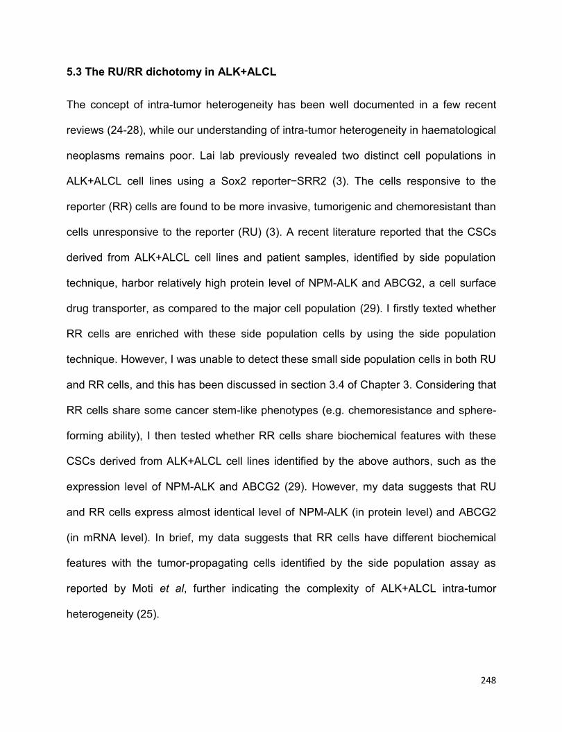
248
5.3 The RU/RR dichotomy in ALK+ALCL
The concept of intra-tumor heterogeneity has been well documented in a few recent
reviews (24-28), while our understanding of intra-tumor heterogeneity in haematological
neoplasms remains poor. Lai lab previously revealed two distinct cell populations in
ALK+ALCL cell lines using a Sox2 reporter−SRR2 (3). The cells responsive to the
reporter (RR) cells are found to be more invasive, tumorigenic and chemoresistant than
cells unresponsive to the reporter (RU) (3). A recent literature reported that the CSCs
derived from ALK+ALCL cell lines and patient samples, identified by side population
technique, harbor relatively high protein level of NPM-ALK and ABCG2, a cell surface
drug transporter, as compared to the major cell population (29). I firstly texted whether
RR cells are enriched with these side population cells by using the side population
technique. However, I was unable to detect these small side population cells in both RU
and RR cells, and this has been discussed in section 3.4 of Chapter 3. Considering that
RR cells share some cancer stem-like phenotypes (e.g. chemoresistance and sphere-
forming ability), I then tested whether RR cells share biochemical features with these
CSCs derived from ALK+ALCL cell lines identified by the above authors, such as the
expression level of NPM-ALK and ABCG2 (29). However, my data suggests that RU
and RR cells express almost identical level of NPM-ALK (in protein level) and ABCG2
(in mRNA level). In brief, my data suggests that RR cells have different biochemical
features with the tumor-propagating cells identified by the side population assay as
reported by Moti et al, further indicating the complexity of ALK+ALCL intra-tumor
heterogeneity (25).

249
My study presented in Chapter 3 unearths that MYC is a key player in the RU/RR
dichotomy. Specifically, as a result of the highly active Wnt/β-catenin pathway, RR cells
express a higher level of MYC than RU cells, and the high level of MYC promotes Sox2
DNA binding and its transcriptional activity. The transcriptionally active Sox2 enhances
the activation of Wnt/β-catenin pathway, which thereafter increases the expression of
MYC, thus forming a positive forward loop. My data also suggests that modulation of
the Wnt/β-catenin pathway can influence SRR2 reporter responsiveness in RU cells, as
evidenced by that in the transwell experiment RU cells can convert to “RR-like” cells
when co-cultured with RR cells which express a relative higher level of Wnt ligands,
such as Wnt2B. Regarding the transwell experiment presented in Chapter 3, one may
ask why RU cells are not able to convert to RR cells in the cultured ALK+ALCL cells
before sorting RU and RR cells. I do consider that RR cells only account for a small
percentage of whole cell populations, and the cytokines secreted by RR cells (e.g.
Wnt2B) are much diluted in the cultured medium, thus the conversion of RU cells to
“RR-like” induced by these cytokines is inappreciable. This notion has been supported
by my result that RU cells are not able to convert to RR cells when co-cultured with
parental ALK+ALCL cells or diluted RR cells (e.g. The dilution of RR/RU=1:10).
Regarding the phosphorylation status of Sox2 in RU and RR cells, I speculate that
Sox2 is only phosphorylated or more Sox2 is phosphorylated in RR cells in comparison
to RU cells, as Sox2 phosphorylation is known to be essential for maintaining stemness
in embryonic stem cells and CSCs (30-33). Sox2 phosphorylation in cancer cells is
poorly studied because of the lack of commercial and reliable antibody against human
phosphorylated Sox2. The Lai lab previously used mass spectrometry assay and

250
identified two potential phosphorylation sites of Sox2, Threonine 116 and Threonine
118, which only exist in RR cells derived from ER+ breast cancer cell line MCF-7 (Wu F,
personal communication, Sept, 2012). It would be of interest to perform the similar
study in RU and RR cells derived from ALK+ALCL, and further studies are needed to
investigate the phosphorylation status and residues of Sox2 in the RU/RR dichotomy
for an in-depth understanding of the biology of RU and RR cells.
In this study, I also observed that RU cells with overexpressed MYC are not exactly RR
cells, despite that RU cells with MYC overexpression have demonstrated as
comparable chemoresistance and tumorigenicity as RR cells do. As shown in Chapter
3, RU cells stably transfected with MYC still have significantly lower SRR2 reporter
activity than RR cells. This indirectly suggests that additional factors are required in RU
cells to fully drive SRR2 reporter responsiveness.
Another interesting observation in my experience is that RR cells automatically lose
SRR2 reporter activity to become “RU-like” cells in the long term cell culture condition.
Specifically, in regular cell culture condition, RR cells derived from SupM2 and Karpas
299 with initial purity of >95% GFP-positive cells, would gradually lose GFP-positive
cells and eventually reach a stable state containing ~40% and ~10% GFP-positive cells,
respectively, in 20 weeks. In contrast, RU cells, the GFP-negative cells, always remain
in a relatively stable state. This phenomenon reveals that RR cells, a more tumorigenic
and cancer stem-like cell population, would automatically differentiate to a less
tumorigenic or cancer stem-like state, supporting the CSC model in which CSCs are
capable to differentiate into non-CSCs (25). Of note, in my study, I cannot name RR
cells as CSC cells because of the lack of serial transplantation assay, which is the gold

251
standard assay for identifying CSCs (34). Therefore, to compare the tumor-initiating
ability of RU and RR cells, the serial transplantation assay should be performed in
immunodeficient mice, which is also my future study plan.
5.4 Oxidative stress-induced tumorigenic potential in ALK+ALCL
“Acquired cancer stemness” or CSC plasticity is a popular concept that has been raised
in recent years (25, 27, 35, 36). It is believed that the CSC phenotype is a dynamic
state that can be acquired by non-stem cells if appropriate stimuli and
microenvironment are present (9). For instance, it has been observed that a subset of
transformed human mammary epithelial cells spontaneously de-differentiate into stem-
like cells both in vitro and in vivo (37). In another study, fludarabine was used to induce
the non-side population cells derived from chronic lymphocytic leukemia to become
side population cells which are shown to be more chemoresistant (38). CSC plasticity
has been postulated to be a major mechanism to explain how cancer cells develop
resistance to chemotherapy, and ultimately, to contribute to cancer treatment failure (36,
39, 40).
However, due to the lack of good cell models, the exact molecular mechanisms
underlying “acquired cancer stemness” or CSC plasticity are not well-understood,
especially in haematological malignancies. I believe the RU/RR cell model is a good
tool for studying tumor plasticity. In Chapter 4, I showed that RU cells can be induced to
convert to RR cells upon oxidative stress. Further evidence showed that the converted
RR cells share similar biological functions and biochemical features with native RR cells.

252
Although oxidative stress-induced “acquisition of stemness” has been recently reported
in ER+ breast cancer cell lines, patient samples, and esophagus squamous cancer
cells, the molecular mechanisms underlined were completely distinct (41, 42). In my
model, I found the active Wnt/β-catenin/MYC/Sox2 axis is responsible for the RU/RR
conversion induced by oxidative stress.
However, I also observed the ERK1/2 is highly active in both RU and RR cells upon
H2O2 re-challenge. Although the biological role of active ERK1/2 in the RU to RR cells
conversion induced by oxidative stress is not investigated in my study, it is likely that
active ERK1/2 partially contributes to this conversion, as active ERK1/2 is shown to
phosphorylate and stabilize MYC in various cell models (43-46). My data also suggests
that ERK1/2 is preferentially activated in RR cells and inhibition of ERK1/2 activity by
using MEK inhibitor U0126 in RR cells dramatically decreased the MYC protein level
and the SRR2 luciferase activity, suggesting the involvement of active ERK1/2 in the
RU/RR dichotomy (Figure 4.5). Further studies are required to understand the
biological roles of active ERK1/2 in the RU to RR cells conversion.
Another interesting question in this study is that whether Sox2 is phosphorylated in the
converted RR cells, and what kinase(s) is involved here to interact and phosphorylate
Sox2. As indicated in a number of reports, phosphorylated Sox2 is implicated in
maintaining stemness in embryonic stem cells and CSCs (30-33). To my knowledge,
there is no literature so far reporting the relationship of phosphorylated Sox2 and
oxidative stress in either ESCs or cancer cells models. It will be of interest to delineate
the phosphorylation status of Sox2 in the converted RR cells induced by oxidative
stress, with comparison to RU and native RR cells.

253
In addition to oxidative stress, hypoxia stress also appears to be a good tool to study
tumor plasticity using RU/RR cells model. Hypoxia stress, a specific tumor
microenvironment caused by low oxygen supplements (47), has been reported to
induce cancer stemness in a wide range type of human cancer and this notion has
been well reviewed in some recent literature (47-50). Whether hypoxia challenge also
induces RU to RR cells conversion needs further investigation.
5.5 STAT1 and the RU/RR dichotomy
As shown in Chapter 2, STAT1 is a potent tumor suppressor in ALK+ALCL.
Overexpression of STAT1 induces cell cycle-arrest and cell apoptosis at least by
downregulating STAT3 downstream targets, including Survivin, Bcl2 and MYC (Figure
2.5 and Figure 4.6C). Initially I also tested whether STAT1 is differentially expressed or
activated in RU and RR cells, thus partially contributing to the RU/RR dichotomy.
However, the Western blotting results suggested that RU and RR cells derived from
SupM2 and Karaps 299 express similar levels of total STAT1, with inconsistent patterns
of active STAT1 level between the two cell subsets (Figure 4.6A-B).This result
indicates that the RU/RR dichotomy is not attributed to the different level of active
STAT1 in the two cell subsets. Notably, the biological function of STAT1 is studied in
ALK+ALCL cells as a whole cell population in Chapter 2. It is reasonable to propose
that overexpression of STAT1 may not be able to induce as sufficient cell apoptosis or
cell cycle-arrest in RR cells as it does in RU cells, because RR cells carry more cancer
stem-like features including chemoresistance and sphere-forming ability. To support
this, further studies are needed to compare the sensitivity of RU and RR cells to STAT1
254
overexpression-induced cell apoptosis or cell cycle-arrest. Of note, whether
overexpression of STAT1 can diminish the biological or biochemical features of RR
cells is another interesting question. To my knowledge, the biological role of STAT1 in
cancer stem cells has not been investigated yet.
In addition, I observed that STAT1 is activated in both RU and RR cells derived from
SupM2 upon H2O2 re-challenge (Figure 4.6C). The active STAT1 has been shown to
suppress MYC expression in some cell models (51, 52). In keeping with this notion, I
also found that constitutively active STAT1 downregulates MYC expression in Tet on-
inducible SupM2-STAT1C cells (Figure 4.6D). However, the active STAT1 stimulated
by oxidative re-challenge was not able to downregulate MYC expression in RU cells
upon H2O2 re-challenge, this is probably because of the oxidative stress-activated
Wnt/β-catenin pathway which in contrast promotes MYC expression and counteracts
the activated STAT1 pathway.
5.6 Conclusions and future directions
Overall, my study, firstly, has revealed a novel mechanism of downregulation of STAT1
by NPM-ALK in STAT3-dependent proteasome pathway, representing a novel
mechanism by which NPM-ALK exerts its oncogenic role in ALK+ALCL. It is of
paramount interest to study whether other tyrosine kinases have similar effects on
STAT1 in other types of cancer models. STAT1 functions as a potent tumor suppressor
in ALK+ALCL if it is overexpressed to a relatively high level. The interplay between
STAT1 and STAT3 has also been well demonstrated in my study. In brief, this study
255
has revealed that STAT1 and STAT3 act as Ying and Yang in oncogenesis in
ALK+ALCL cell model, providing insight into the functionally interaction of
STAT1/STAT3 in cancer. My study has also shred important insight on that activating
STAT1 signaling would be an attractive therapeutic strategy in treating ALK+ALCL,
even potentially other types of cancer where STAT3 is constitutively activated.
Secondly, my study has unearthed the biochemical features of a cancer stem-like
subpopulation in ALK+ALCL. Specifically, the positive forward loop involving Wnt/β-
catenin/MYC/Sox2 axis defines this cancer stem-like cell subpopulation. This study has
revealed promising therapeutic targets for these cancer stem-like cells in ALK+ALCL by
targeting Wnt/β-catenin/MYC/Sox2 axis. Specifically, doxorubicin, adjuvanted with
Wnt/β-catenin inhibitor or MYC inhibitor, can be potentially applied to ALK+ALCL
patients who develop chemoresistance and tumor relapses. Lastly, oxidative stress has
been shown to potentiate the tumorigenicity of ALK+ALCL, in which the active Wnt/β-
catenin/MYC/Sox2 axis is required. This study has supported the notion of CSC
plasticity or “acquired cancer stemness” in haematopoietic cancer. My study has
provided evidence that “acquired cancer stemness” can be achieved in haematopoietic
cancer model by oxidative stress. The biological process of “acquired cancer stemness”
can be easily studied by using the RU/RR cells model, as the SRR2 reporter
responsiveness is a useful approach to identify and quantify cancer stemness. I believe
the RU/RR cells model is a good model that can be applied to other types of cancer for
identifying cancer stem-like cell populations or studying “acquired cancer stemness”.
The future study involves evaluation of the expression of STAT1 and MYC being as
prognostic markers for ALK+ALCL disease. Furthermore, in vivo studies are warranted
256
to investigate the feasibility of the novel therapeutic targets, including activation of
STAT1 and targeting Wnt/β-catenin/MYC/Sox2 axis, in mouse models mimicking
human ALK+ALCL disease or patient-derived xenograft model.
In general, I believe the studies from this thesis have furthered the understanding of the
pathobiology of ALK+ALCL cells and shed light on the complexity of intra-tumor
heterogeneity and tumor plasticity in this haematological malignancy.
5.7 References
1. Kinney MC, Higgins RA, Medina EA. Anaplastic large cell lymphoma: twenty-five
years of discovery. Archives of pathology & laboratory medicine. 2011;135(1):19-43.
2. Wu C, Molavi O, Zhang H, Gupta N, Alshareef A, Bone KM, et al. STAT1 is
phosphorylated and downregulated by the oncogenic tyrosine kinase NPM-ALK in ALK-
positive anaplastic large-cell lymphoma. Blood. 2015;126(3):336-45.
3. Gelebart P, Hegazy SA, Wang P, Bone KM, Anand M, Sharon D, et al. Aberrant
expression and biological significance of Sox2, an embryonic stem cell transcriptional
factor, in ALK-positive anaplastic large cell lymphoma. Blood Cancer J. 2012;2:e82.
4. Koromilas AE, Sexl V. The tumor suppressor function of STAT1 in breast cancer.
Jak-Stat. 2013;2(2):e23353.
5. Khodarev N, Ahmad R, Rajabi H, Pitroda S, Kufe T, McClary C, et al.
Cooperativity of the MUC1 oncoprotein and STAT1 pathway in poor prognosis human
breast cancer. Oncogene. 2010;29(6):920-9.
257
6. Han W, Carpenter RL, Cao X, Lo HW. STAT1 gene expression is enhanced by
nuclear EGFR and HER2 via cooperation with STAT3. Mol Carcinog. 2013;52(12):959-
69.
7. Zhang Y, Molavi O, Su M, Lai R. The clinical and biological significance of
STAT1 in esophageal squamous cell carcinoma. BMC cancer. 2014;14:791.
8. Kaganoi J, Watanabe G, Okabe M, Nagatani S, Kawabe A, Shimada Y, et al.
STAT1 activation-induced apoptosis of esophageal squamous cell carcinoma cells in
vivo. Annals of surgical oncology. 2007;14(4):1405-15.
9. Kortylewski M, Komyod W, Kauffmann ME, Bosserhoff A, Heinrich PC,
Behrmann I. Interferon-gamma-mediated growth regulation of melanoma cells:
involvement of STAT1-dependent and STAT1-independent signals. The Journal of
investigative dermatology. 2004;122(2):414-22.
10. Schmitt MJ, Philippidou D, Reinsbach SE, Margue C, Wienecke-Baldacchino A,
Nashan D, et al. Interferon-gamma-induced activation of Signal Transducer and
Activator of Transcription 1 (STAT1) up-regulates the tumor suppressing microRNA-29
family in melanoma cells. Cell communication and signaling : CCS. 2012;10(1):41.
11. Sanda T, Tyner JW, Gutierrez A, Ngo VN, Glover J, Chang BH, et al. TYK2-
STAT1-BCL2 pathway dependence in T-cell acute lymphoblastic leukemia. Cancer
discovery. 2013;3(5):564-77.
12. Battle TE, Frank DA. STAT1 mediates differentiation of chronic lymphocytic
leukemia cells in response to Bryostatin 1. Blood. 2003;102(8):3016-24.
13. Meissl K, Macho-Maschler S, Muller M, Strobl B. The good and the bad faces of
STAT1 in solid tumours. Cytokine. 2015.

258
14. Kovacic B, Stoiber D, Moriggl R, Weisz E, Ott RG, Kreibich R, et al. STAT1 acts
as a tumor promoter for leukemia development. Cancer cell. 2006;10(1):77-87.
15. Avalle L, Pensa S, Regis G, Novelli F, Poli V. STAT1 and STAT3 in
tumorigenesis: A matter of balance. Jak-Stat. 2012;1(2):65-72.
16. Schultz J, Koczan D, Schmitz U, Ibrahim SM, Pilch D, Landsberg J, et al. Tumor-
promoting role of signal transducer and activator of transcription (Stat)1 in late-stage
melanoma growth. Clinical & experimental metastasis. 2010;27(3):133-40.
17. Regis G, Pensa S, Boselli D, Novelli F, Poli V. Ups and downs: the
STAT1:STAT3 seesaw of Interferon and gp130 receptor signalling. Semin Cell Dev Biol.
2008;19(4):351-9.
18. Xi S, Dyer KF, Kimak M, Zhang Q, Gooding WE, Chaillet JR, et al. Decreased
STAT1 expression by promoter methylation in squamous cell carcinogenesis. Journal
of the National Cancer Institute. 2006;98(3):181-9.
19. Sun WH, Pabon C, Alsayed Y, Huang PP, Jandeska S, Uddin S, et al.
Interferon-alpha resistance in a cutaneous T-cell lymphoma cell line is associated with
lack of STAT1 expression. Blood. 1998;91(2):570-6.
20. Ren Y, Zhao P, Liu J, Yuan Y, Cheng Q, Zuo Y, et al. Deubiquitinase USP2a
Sustains Interferons Antiviral Activity by Restricting Ubiquitination of Activated STAT1 in
the Nucleus. PLoS pathogens. 2016;12(7):e1005764.
21. Yu H, Yue X, Zhao Y, Li X, Wu L, Zhang C, et al. LIF negatively regulates
tumour-suppressor p53 through Stat3/ID1/MDM2 in colorectal cancers. Nature
communications. 2014;5:5218.
259
22. Pencik J, Schlederer M, Gruber W, Unger C, Walker SM, Chalaris A, et al.
STAT3 regulated ARF expression suppresses prostate cancer metastasis. Nature
communications. 2015;6:7736.
23. Dimberg LY, Dimberg A, Ivarsson K, Fryknas M, Rickardson L, Tobin G, et al.
Stat1 activation attenuates IL-6 induced Stat3 activity but does not alter apoptosis
sensitivity in multiple myeloma. BMC cancer. 2012;12:318.
24. Fisher R, Pusztai L, Swanton C. Cancer heterogeneity: implications for targeted
therapeutics. British journal of cancer. 2013;108(3):479-85.
25. Cabrera MC, Hollingsworth RE, Hurt EM. Cancer stem cell plasticity and tumor
hierarchy. World journal of stem cells. 2015;7(1):27-36.
26. Visvader JE. Cells of origin in cancer. Nature. 2011;469(7330):314-22.
27. Meacham CE, Morrison SJ. Tumour heterogeneity and cancer cell plasticity.
Nature. 2013;501(7467):328-37.
28. Tang DG. Understanding cancer stem cell heterogeneity and plasticity. Cell
research. 2012;22(3):457-72.
29. Moti N, Malcolm T, Hamoudi R, Mian S, Garland G, Hook CE, et al. Anaplastic
large cell lymphoma-propagating cells are detectable by side population analysis and
possess an expression profile reflective of a primitive origin. Oncogene.
2015;34(14):1843-52.
30. Ouyang J, Yu W, Liu J, Zhang N, Florens L, Chen J, et al. Cyclin-dependent
kinase-mediated Sox2 phosphorylation enhances the ability of Sox2 to establish the
pluripotent state. The Journal of biological chemistry. 2015;290(37):22782-94.
260
31. Fang L, Zhang L, Wei W, Jin X, Wang P, Tong Y, et al. A methylation-
phosphorylation switch determines Sox2 stability and function in ESC maintenance or
differentiation. Molecular cell. 2014;55(4):537-51.
32. Justilien V, Walsh MP, Ali SA, Thompson EA, Murray NR, Fields AP. The PRKCI
and SOX2 oncogenes are coamplified and cooperate to activate Hedgehog signaling in
lung squamous cell carcinoma. Cancer cell. 2014;25(2):139-51.
33. Jeong CH, Cho YY, Kim MO, Kim SH, Cho EJ, Lee SY, et al. Phosphorylation of
Sox2 cooperates in reprogramming to pluripotent stem cells. Stem cells.
2010;28(12):2141-50.
34. Han L, Shi S, Gong T, Zhang Z, Sun X. Cancer stem cells: therapeutic
implications and perspectives in cancer therapy. Acta Pharmaceutica Sinica B.
2013;3(2):65-75.
35. Vicente-Duenas C, Gutierrez de Diego J, Rodriguez FD, Jimenez R, Cobaleda C.
The role of cellular plasticity in cancer development. Curr Med Chem.
2009;16(28):3676-85.
36. Chen W, Dong J, Haiech J, Kilhoffer MC, Zeniou M. Cancer Stem Cell
Quiescence and Plasticity as Major Challenges in Cancer Therapy. Stem Cells Int.
2016;2016:1740936.
37. Chaffer CL, Brueckmann I, Scheel C, Kaestli AJ, Wiggins PA, Rodrigues LO, et
al. Normal and neoplastic nonstem cells can spontaneously convert to a stem-like state.
Proceedings of the National Academy of Sciences of the United States of America.
2011;108(19):7950-5.
261
38. Gross E, L'Faqihi-Olive FE, Ysebaert L, Brassac M, Struski S, Kheirallah S, et al.
B-chronic lymphocytic leukemia chemoresistance involves innate and acquired
leukemic side population cells. Leukemia. 2010;24(11):1885-92.
39. Kemper K, de Goeje PL, Peeper DS, van Amerongen R. Phenotype switching:
tumor cell plasticity as a resistance mechanism and target for therapy. Cancer research.
2014;74(21):5937-41.
40. Doherty MR, Smigiel JM, Junk DJ, Jackson MW. Cancer Stem Cell Plasticity
Drives Therapeutic Resistance. Cancers. 2016;8(1).
41. Gopal K, Gupta N, Zhang H, Alshareef A, Alqahtani H, Bigras G, et al. Oxidative
stress induces the acquisition of cancer stem-like phenotype in breast cancer
detectable by using a Sox2 regulatory region-2 (SRR2) reporter. Oncotarget.
2016;7(3):3111-27.
42. Zhang HF, Wu C, Alshareef A, Gupta N, Zhao Q, Xu XE, et al. The PI3K/AKT/c-
MYC axis promotes the acquisition of cancer stem-like features in esophageal
squamous cell carcinoma. Stem cells. 2016.
43. Wang Z, Ge L, Wang M, Carr BI. Phosphorylation regulates Myc expression via
prolonged activation of the mitogen-activated protein kinase pathway. Journal of
cellular physiology. 2006;208(1):133-40.
44. Tsai WB, Aiba I, Long Y, Lin HK, Feun L, Savaraj N, et al. Activation of
Ras/PI3K/ERK pathway induces c-Myc stabilization to upregulate argininosuccinate
synthetase, leading to arginine deiminase resistance in melanoma cells. Cancer
research. 2012;72(10):2622-33.
262
45. Sears R, Nuckolls F, Haura E, Taya Y, Tamai K, Nevins JR. Multiple Ras-
dependent phosphorylation pathways regulate Myc protein stability. Genes &
development. 2000;14(19):2501-14.
46. Marampon F, Ciccarelli C, Zani BM. Down-regulation of c-Myc following
MEK/ERK inhibition halts the expression of malignant phenotype in rhabdomyosarcoma
and in non muscle-derived human tumors. Molecular cancer. 2006;5:31.
47. Yun Z, Lin Q. Hypoxia and regulation of cancer cell stemness. Advances in
experimental medicine and biology. 2014;772:41-53.
48. Carnero A, Lleonart M. The hypoxic microenvironment: A determinant of cancer
stem cell evolution. BioEssays : news and reviews in molecular, cellular and
developmental biology. 2016;38 Suppl 1:S65-74.
49. Plaks V, Kong N, Werb Z. The cancer stem cell niche: how essential is the niche
in regulating stemness of tumor cells? Cell stem cell. 2015;16(3):225-38.
50. Keith B, Simon MC. Hypoxia-inducible factors, stem cells, and cancer. Cell.
2007;129(3):465-72.
51. Dimberg A, Karlberg I, Nilsson K, Oberg F. Ser727/Tyr701-phosphorylated Stat1
is required for the regulation of c-Myc, cyclins, and p27Kip1 associated with ATRA-
induced G0/G1 arrest of U-937 cells. Blood. 2003;102(1):254-61.
52. Ramana CV, Grammatikakis N, Chernov M, Nguyen H, Goh KC, Williams BR, et
al. Regulation of c-myc expression by IFN-gamma through Stat1-dependent and -
independent pathways. The EMBO journal. 2000;19(2):263-72.

263
Bibliography
Abril E, Real LM, Serrano A, Jimenez P, Garcia A, Canton J, et al.
Unresponsiveness to interferon associated with STAT1 protein deficiency in a
gastric adenocarcinoma cell line. Cancer immunology, immunotherapy : CII.
1998;47(2):113-20.
Afkarian M, Sedy JR, Yang J, Jacobson NG, Cereb N, Yang SY, et al. T-bet is a
STAT1-induced regulator of IL-12R expression in naive CD4+ T cells. Nat
Immunol. 2002;3(6):549-57.
Aisner DL, Nguyen TT, Paskulin DD, Le AT, Haney J, Schulte N, et al. ROS1 and
ALK fusions in colorectal cancer, with evidence of intratumoral heterogeneity for
molecular drivers. Molecular cancer research : MCR. 2014;12(1):111-8.
Akita H, Marquardt JU, Durkin ME, Kitade M, Seo D, Conner EA, et al. MYC
activates stem-like cell potential in hepatocarcinoma by a p53-dependent
mechanism. Cancer research. 2014;74(20):5903-13.
Alzona M, Jack HM, Fisher RI, Ellis TM. CD30 defines a subset of activated
human T cells that produce IFN-gamma and IL-5 and exhibit enhanced B cell
helper activity. J Immunol. 1994;153(7):2861-7.
Amaya CN, Bryan BA. Enrichment of the embryonic stem cell reprogramming
factors Oct4, Nanog, Myc, and Sox2 in benign and malignant vascular tumors.
BMC Clin Pathol. 2015;15:18.

264
Amin HM, Lai R. Pathobiology of ALK+ anaplastic large-cell lymphoma. Blood.
2007;110(7):2259-67.
Amin HM, Lin Q, Lai R. Jak3 contributes to the activation of ALK and Stat3 in
ALK(+) anaplastic large cell lymphoma. Laboratory investigation; a journal of
technical methods and pathology. 2006;86(4):417-9; author reply 20-1.
Amin HM, McDonnell TJ, Ma Y, Lin Q, Fujio Y, Kunisada K, et al. Selective
inhibition of STAT3 induces apoptosis and G(1) cell cycle arrest in ALK-positive
anaplastic large cell lymphoma. Oncogene. 2004;23(32):5426-34.
Amin HM, McDonnell TJ, Ma Y, Lin Q, Fujio Y, Kunisada K, et al. Selective
inhibition of STAT3 induces apoptosis and G(1) cell cycle arrest in ALK-positive
anaplastic large cell lymphoma. Oncogene. 2004;23(32):5426-34.
Amin HM, Medeiros LJ, Ma Y, Feretzaki M, Das P, Leventaki V, et al. Inhibition of
JAK3 induces apoptosis and decreases anaplastic lymphoma kinase activity in
anaplastic large cell lymphoma. Oncogene. 2003;22(35):5399-407.
Anand M, Lai R, Gelebart P. beta-catenin is constitutively active and increases
STAT3 expression/activation in anaplastic lymphoma kinase-positive anaplastic
large cell lymphoma. Haematologica. 2011;96(2):253-61.
Anastas JN, Moon RT. WNT signalling pathways as therapeutic targets in
cancer. Nature reviews Cancer. 2013;13(1):11-26.
Arif K, Hussain I, Rea C, El-Sheemy M. The role of Nanog expression in

265
tamoxifen-resistant breast cancer cells. OncoTargets and therapy. 2015;8:1327-
34.
Avalle L, Pensa S, Regis G, Novelli F, Poli V. STAT1 and STAT3 in
tumorigenesis: A matter of balance. Jak-Stat. 2012;1(2):65-72.
Babchia N, Calipel A, Mouriaux F, Faussat AM, Mascarelli F. The PI3K/Akt and
mTOR/P70S6K signaling pathways in human uveal melanoma cells: interaction
with B-Raf/ERK. Investigative ophthalmology & visual science. 2010;51(1):421-9.
Bai RY, Dieter P, Peschel C, Morris SW, Duyster J. Nucleophosmin-anaplastic
lymphoma kinase of large-cell anaplastic lymphoma is a constitutively active
tyrosine kinase that utilizes phospholipase C-gamma to mediate its mitogenicity.
Molecular and cellular biology. 1998;18(12):6951-61.
Baker VV, Borst MP, Dixon D, Hatch KD, Shingleton HM, Miller D. c-myc
amplification in ovarian cancer. Gynecologic oncology. 1990;38(3):340-2.
Bard JD, Gelebart P, Anand M, Amin HM, Lai R. Aberrant expression of IL-22
receptor 1 and autocrine IL-22 stimulation contribute to tumorigenicity in ALK+
anaplastic large cell lymphoma. Leukemia. 2008;22(8):1595-603.
Basso K, Dalla-Favera R. Germinal centres and B cell lymphomagenesis. Nature
reviews Immunology. 2015;15(3):172-84.
Basu-Roy U, Bayin NS, Rattanakorn K, Han E, Placantonakis DG, Mansukhani
A, et al. Sox2 antagonizes the Hippo pathway to maintain stemness in cancer
266
cells. Nature communications. 2015;6:6411.
Battle TE, Frank DA. STAT1 mediates differentiation of chronic lymphocytic
leukemia cells in response to Bryostatin 1. Blood. 2003;102(8):3016-24.
Bellei B, Pacchiarotti A, Perez M, Faraggiana T. Frequent beta-catenin
overexpression without exon 3 mutation in cutaneous lymphomas. Modern
pathology : an official journal of the United States and Canadian Academy of
Pathology, Inc. 2004;17(10):1275-81.
Beltran H. The N-myc Oncogene: Maximizing its Targets, Regulation, and
Therapeutic Potential. Molecular cancer research : MCR. 2014;12(6):815-22.
Benharroch D, Meguerian-Bedoyan Z, Lamant L, Amin C, Brugieres L, Terrier-
Lacombe MJ, et al. ALK-positive lymphoma: a single disease with a broad
spectrum of morphology. Blood. 1998;91(6):2076-84.
Bernabei P, Coccia EM, Rigamonti L, Bosticardo M, Forni G, Pestka S, et al.
Interferon-gamma receptor 2 expression as the deciding factor in human T, B,
and myeloid cell proliferation or death. Journal of leukocyte biology.
2001;70(6):950-60.
Bhattacharya S, Ray RM, Johnson LR. STAT3-mediated transcription of Bcl-2,
Mcl-1 and c-IAP2 prevents apoptosis in polyamine-depleted cells. The
Biochemical journal. 2005;392(Pt 2):335-44.
Bilsland JG, Wheeldon A, Mead A, Znamenskiy P, Almond S, Waters KA, et al.

267
Behavioral and neurochemical alterations in mice deficient in anaplastic
lymphoma kinase suggest therapeutic potential for psychiatric indications.
Neuropsychopharmacology. 2008;33(3):685-700.
Blank U, Karlsson G, Karlsson S. Signaling pathways governing stem-cell fate.
Blood. 2008;111(2):492-503.
Bluyssen HA, Rastmanesh MM, Tilburgs C, Jie K, Wesseling S, Goumans MJ, et
al. IFN gamma-dependent SOCS3 expression inhibits IL-6-induced STAT3
phosphorylation and differentially affects IL-6 mediated transcriptional responses
in endothelial cells. American journal of physiology Cell physiology.
2010;299(2):C354-62.
Boesch M, Zeimet AG, Reimer D, Schmidt S, Gastl G, Parson W, et al. The side
population of ovarian cancer cells defines a heterogeneous compartment
exhibiting stem cell characteristics. Oncotarget. 2014;5(16):7027-39.
Bonnet D, Dick JE. Human acute myeloid leukemia is organized as a hierarchy
that originates from a primitive hematopoietic cell. Nature medicine.
1997;3(7):730-7.
Bonzheim I, Geissinger E, Roth S, Zettl A, Marx A, Rosenwald A, et al.
Anaplastic large cell lymphomas lack the expression of T-cell receptor molecules
or molecules of proximal T-cell receptor signaling. Blood. 2004;104(10):3358-60.
Borovski T, De Sousa EMF, Vermeulen L, Medema JP. Cancer stem cell niche:

268
the place to be. Cancer research. 2011;71(3):634-9.
Boudny V, Dusek L, Adamkova L, Chumchalova J, Kocak I, Fait V, et al. Lack of
STAT 1 phosphorylation at TYR 701 by IFNgamma correlates with disease
outcome in melanoma patients. Neoplasma. 2005;52(4):330-7.
Bowden ET, Stoica GE, Wellstein A. Anti-apoptotic signaling of pleiotrophin
through its receptor, anaplastic lymphoma kinase. The Journal of biological
chemistry. 2002;277(39):35862-8.
Bowman T, Garcia R, Turkson J, Jove R. STATs in oncogenesis. Oncogene.
2000;19(21):2474-88.
Bowman T, Garcia R, Turkson J, Jove R. STATs in oncogenesis. Oncogene.
2000;19(21):2474-88.
Box JK, Paquet N, Adams MN, Boucher D, Bolderson E, O'Byrne KJ, et al.
Nucleophosmin: from structure and function to disease development. BMC
molecular biology. 2016;17(1):19.
Britton KM, Kirby JA, Lennard TW, Meeson AP. Cancer stem cells and side
population cells in breast cancer and metastasis. Cancers. 2011;3(2):2106-30.
Buettner R, Mora LB, Jove R. Activated STAT signaling in human tumors
provides novel molecular targets for therapeutic intervention. Clinical cancer
research : an official journal of the American Association for Cancer Research.
2002;8(4):945-54.
269
Cabrera MC, Hollingsworth RE, Hurt EM. Cancer stem cell plasticity and tumor
hierarchy. World journal of stem cells. 2015;7(1):27-36.
Carnero A, Lleonart M. The hypoxic microenvironment: A determinant of cancer
stem cell evolution. BioEssays : news and reviews in molecular, cellular and
developmental biology. 2016;38 Suppl 1:S65-74.
Carpenter RL, Lo HW. STAT3 Target Genes Relevant to Human Cancers.
Cancers. 2014;6(2):897-925.
Casey SC, Tong L, Li Y, Do R, Walz S, Fitzgerald KN, et al. MYC regulates the
antitumor immune response through CD47 and PD-L1. Science.
2016;352(6282):227-31.
Ceccon M, Mologni L, Bisson W, Scapozza L, Gambacorti-Passerini C.
Crizotinib-resistant NPM-ALK mutants confer differential sensitivity to unrelated
Alk inhibitors. Molecular cancer research : MCR. 2013;11(2):122-32.
Chaffer CL, Brueckmann I, Scheel C, Kaestli AJ, Wiggins PA, Rodrigues LO, et
al. Normal and neoplastic nonstem cells can spontaneously convert to a stem-
like state. Proceedings of the National Academy of Sciences of the United States
of America. 2011;108(19):7950-5.
Chan SR, Vermi W, Luo J, Lucini L, Rickert C, Fowler AM, et al. STAT1-deficient
mice spontaneously develop estrogen receptor alpha-positive luminal mammary
carcinomas. Breast cancer research : BCR. 2012;14(1):R16.

270
Chang F, Steelman LS, Lee JT, Shelton JG, Navolanic PM, Blalock WL, et al.
Signal transduction mediated by the Ras/Raf/MEK/ERK pathway from cytokine
receptors to transcription factors: potential targeting for therapeutic intervention.
Leukemia. 2003;17(7):1263-93.
Chen D, Dou QP. The ubiquitin-proteasome system as a prospective molecular
target for cancer treatment and prevention. Current protein & peptide science.
2010;11(6):459-70.
Chen S, Xu Y, Chen Y, Li X, Mou W, Wang L, et al. SOX2 gene regulates the
transcriptional network of oncogenes and affects tumorigenesis of human lung
cancer cells. PloS one. 2012;7(5):e36326.
Chen S, Xu Y, Chen Y, Li X, Mou W, Wang L, et al. SOX2 gene regulates the
transcriptional network of oncogenes and affects tumorigenesis of human lung
cancer cells. PloS one. 2012;7(5):e36326.
Chen W, Dong J, Haiech J, Kilhoffer MC, Zeniou M. Cancer Stem Cell
Quiescence and Plasticity as Major Challenges in Cancer Therapy. Stem Cells
Int. 2016;2016:1740936.
Chen Y, Shi L, Zhang L, Li R, Liang J, Yu W, et al. The molecular mechanism
governing the oncogenic potential of SOX2 in breast cancer. The Journal of
biological chemistry. 2008;283(26):17969-78.
Chen Y, Takita J, Choi YL, Kato M, Ohira M, Sanada M, et al. Oncogenic

271
mutations of ALK kinase in neuroblastoma. Nature. 2008;455(7215):971-4.
Chen YQ. [Frequency on N-ras as a transforming gene, DNA rearrangement and
amplification, and over-expression of C-myc in human primary hepatic cancer].
Zhonghua yi xue za zhi. 1987;67(4):197-9, 14.
Chiarle R, Gong JZ, Guasparri I, Pesci A, Cai J, Liu J, et al. NPM-ALK transgenic
mice spontaneously develop T-cell lymphomas and plasma cell tumors. Blood.
2003;101(5):1919-27.
Chiarle R, Simmons WJ, Cai H, Dhall G, Zamo A, Raz R, et al. Stat3 is required
for ALK-mediated lymphomagenesis and provides a possible therapeutic target.
Nature medicine. 2005;11(6):623-9.
Chiarle R, Voena C, Ambrogio C, Piva R, Inghirami G. The anaplastic lymphoma
kinase in the pathogenesis of cancer. Nature reviews Cancer. 2008;8(1):11-23.
Choi EA, Lei H, Maron DJ, Wilson JM, Barsoum J, Fraker DL, et al. Stat1-
dependent induction of tumor necrosis factor-related apoptosis-inducing ligand
and the cell-surface death signaling pathway by interferon beta in human cancer
cells. Cancer research. 2003;63(17):5299-307.
Christgen M, Ballmaier M, Lehmann U, Kreipe H. Detection of putative cancer
stem cells of the side population phenotype in human tumor cell cultures.
Methods in molecular biology. 2012;878:201-15.
Colombo E, Bonetti P, Lazzerini Denchi E, Martinelli P, Zamponi R, Marine JC, et
272
al. Nucleophosmin is required for DNA integrity and p19Arf protein stability.
Molecular and cellular biology. 2005;25(20):8874-86.
Coluccia AM, Vacca A, Dunach M, Mologni L, Redaelli S, Bustos VH, et al. Bcr-
Abl stabilizes beta-catenin in chronic myeloid leukemia through its tyrosine
phosphorylation. The EMBO journal. 2007;26(5):1456-66.
Crockett DK, Lin Z, Elenitoba-Johnson KS, Lim MS. Identification of NPM-ALK
interacting proteins by tandem mass spectrometry. Oncogene.
2004;23(15):2617-29.
Dai X, Sayama K, Yamasaki K, Tohyama M, Shirakata Y, Hanakawa Y, et al.
SOCS1-negative feedback of STAT1 activation is a key pathway in the dsRNA-
induced innate immune response of human keratinocytes. The Journal of
investigative dermatology. 2006;126(7):1574-81.
Dang CV. c-Myc target genes involved in cell growth, apoptosis, and metabolism.
Molecular and cellular biology. 1999;19(1):1-11.
Dang CV. MYC on the path to cancer. Cell. 2012;149(1):22-35.
Dang CV. MYC, metabolism, cell growth, and tumorigenesis. Cold Spring Harbor
perspectives in medicine. 2013;3(8).
Das B, Tsuchida R, Malkin D, Koren G, Baruchel S, Yeger H. Hypoxia enhances
tumor stemness by increasing the invasive and tumorigenic side population
fraction. Stem cells. 2008;26(7):1818-30.
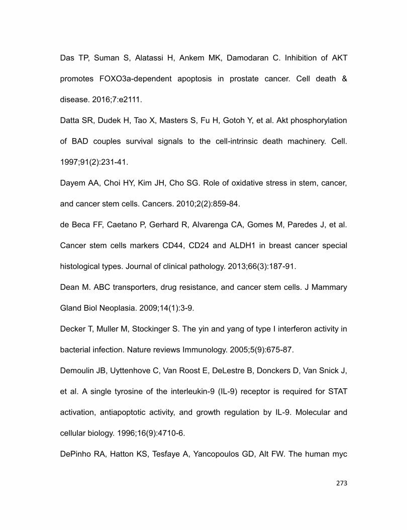
273
Das TP, Suman S, Alatassi H, Ankem MK, Damodaran C. Inhibition of AKT
promotes FOXO3a-dependent apoptosis in prostate cancer. Cell death &
disease. 2016;7:e2111.
Datta SR, Dudek H, Tao X, Masters S, Fu H, Gotoh Y, et al. Akt phosphorylation
of BAD couples survival signals to the cell-intrinsic death machinery. Cell.
1997;91(2):231-41.
Dayem AA, Choi HY, Kim JH, Cho SG. Role of oxidative stress in stem, cancer,
and cancer stem cells. Cancers. 2010;2(2):859-84.
de Beca FF, Caetano P, Gerhard R, Alvarenga CA, Gomes M, Paredes J, et al.
Cancer stem cells markers CD44, CD24 and ALDH1 in breast cancer special
histological types. Journal of clinical pathology. 2013;66(3):187-91.
Dean M. ABC transporters, drug resistance, and cancer stem cells. J Mammary
Gland Biol Neoplasia. 2009;14(1):3-9.
Decker T, Muller M, Stockinger S. The yin and yang of type I interferon activity in
bacterial infection. Nature reviews Immunology. 2005;5(9):675-87.
Demoulin JB, Uyttenhove C, Van Roost E, DeLestre B, Donckers D, Van Snick J,
et al. A single tyrosine of the interleukin-9 (IL-9) receptor is required for STAT
activation, antiapoptotic activity, and growth regulation by IL-9. Molecular and
cellular biology. 1996;16(9):4710-6.
DePinho RA, Hatton KS, Tesfaye A, Yancopoulos GD, Alt FW. The human myc
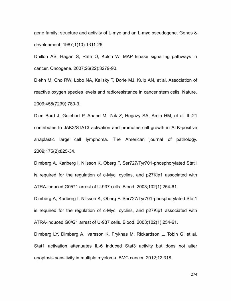
274
gene family: structure and activity of L-myc and an L-myc pseudogene. Genes &
development. 1987;1(10):1311-26.
Dhillon AS, Hagan S, Rath O, Kolch W. MAP kinase signalling pathways in
cancer. Oncogene. 2007;26(22):3279-90.
Diehn M, Cho RW, Lobo NA, Kalisky T, Dorie MJ, Kulp AN, et al. Association of
reactive oxygen species levels and radioresistance in cancer stem cells. Nature.
2009;458(7239):780-3.
Dien Bard J, Gelebart P, Anand M, Zak Z, Hegazy SA, Amin HM, et al. IL-21
contributes to JAK3/STAT3 activation and promotes cell growth in ALK-positive
anaplastic large cell lymphoma. The American journal of pathology.
2009;175(2):825-34.
Dimberg A, Karlberg I, Nilsson K, Oberg F. Ser727/Tyr701-phosphorylated Stat1
is required for the regulation of c-Myc, cyclins, and p27Kip1 associated with
ATRA-induced G0/G1 arrest of U-937 cells. Blood. 2003;102(1):254-61.
Dimberg A, Karlberg I, Nilsson K, Oberg F. Ser727/Tyr701-phosphorylated Stat1
is required for the regulation of c-Myc, cyclins, and p27Kip1 associated with
ATRA-induced G0/G1 arrest of U-937 cells. Blood. 2003;102(1):254-61.
Dimberg LY, Dimberg A, Ivarsson K, Fryknas M, Rickardson L, Tobin G, et al.
Stat1 activation attenuates IL-6 induced Stat3 activity but does not alter
apoptosis sensitivity in multiple myeloma. BMC cancer. 2012;12:318.
275
Ding XW, Wu JH, Jiang CP. ABCG2: a potential marker of stem cells and novel
target in stem cell and cancer therapy. Life sciences. 2010;86(17-18):631-7.
Doherty MR, Smigiel JM, Junk DJ, Jackson MW. Cancer Stem Cell Plasticity
Drives Therapeutic Resistance. Cancers. 2016;8(1).
Doherty MR, Smigiel JM, Junk DJ, Jackson MW. Cancer Stem Cell Plasticity
Drives Therapeutic Resistance. Cancers. 2016;8(1).
Drexler HG, Gignac SM, von Wasielewski R, Werner M, Dirks WG. Pathobiology
of NPM-ALK and variant fusion genes in anaplastic large cell lymphoma and
other lymphomas. Leukemia. 2000;14(9):1533-59.
Duchartre Y, Kim YM, Kahn M. The Wnt signaling pathway in cancer. Critical
reviews in oncology/hematology. 2016;99:141-9.
Dunn GP, Sheehan KC, Old LJ, Schreiber RD. IFN unresponsiveness in LNCaP
cells due to the lack of JAK1 gene expression. Cancer research.
2005;65(8):3447-53.
El Khoury F, Corcos L, Durand S, Simon B, Le Jossic-Corcos C. Acquisition of
anticancer drug resistance is partially associated with cancer stemness in human
colon cancer cells. International journal of oncology. 2016.
Falini B, Mecucci C, Tiacci E, Alcalay M, Rosati R, Pasqualucci L, et al.
Cytoplasmic nucleophosmin in acute myelogenous leukemia with a normal
karyotype. The New England journal of medicine. 2005;352(3):254-66.

276
Fanale MA, Horwitz SM, Forero-Torres A, Bartlett NL, Advani RH, Pro B, et al.
Brentuximab vedotin in the front-line treatment of patients with CD30+ peripheral
T-cell lymphomas: results of a phase I study. Journal of clinical oncology : official
journal of the American Society of Clinical Oncology. 2014;32(28):3137-43.
Fang L, Zhang L, Wei W, Jin X, Wang P, Tong Y, et al. A methylation-
phosphorylation switch determines Sox2 stability and function in ESC
maintenance or differentiation. Molecular cell. 2014;55(4):537-51.
Farina F, Gambacorti-Passerini C. ALK inhibitors for clinical use in cancer
therapy. Front Biosci (Elite Ed). 2016;8:46-60.
Farrell AS, Sears RC. MYC degradation. Cold Spring Harbor perspectives in
medicine. 2014;4(3).
Felgar RE, Salhany KE, Macon WR, Pietra GG, Kinney MC. The expression of
TIA-1+ cytolytic-type granules and other cytolytic lymphocyte-associated
markers in CD30+ anaplastic large cell lymphomas (ALCL): correlation with
morphology, immunophenotype, ultrastructure, and clinical features. Human
pathology. 1999;30(2):228-36.
Felsher DW. MYC Inactivation Elicits Oncogene Addiction through Both Tumor
Cell-Intrinsic and Host-Dependent Mechanisms. Genes & cancer. 2010;1(6):597-
604.
Fenghao X, Saxon A, Nguyen A, Ke Z, Diaz-Sanchez D, Nel A. Interleukin 4
277
activates a signal transducer and activator of transcription (Stat) protein which
interacts with an interferon-gamma activation site-like sequence upstream of the
I epsilon exon in a human B cell line. Evidence for the involvement of Janus
kinase 3 and interleukin-4 Stat. The Journal of clinical investigation.
1995;96(2):907-14.
Ferreri AJ, Govi S, Pileri SA, Savage KJ. Anaplastic large cell lymphoma, ALK-
positive. Crit Rev Oncol Hematol. 2012;83(2):293-302.
Fisher R, Pusztai L, Swanton C. Cancer heterogeneity: implications for targeted
therapeutics. British journal of cancer. 2013;108(3):479-85.
Fodde R, Brabletz T. Wnt/beta-catenin signaling in cancer stemness and
malignant behavior. Current opinion in cell biology. 2007;19(2):150-8.
Foss HD, Anagnostopoulos I, Araujo I, Assaf C, Demel G, Kummer JA, et al.
Anaplastic large-cell lymphomas of T-cell and null-cell phenotype express
cytotoxic molecules. Blood. 1996;88(10):4005-11.
Foss HD, Demel G, Anagnostopoulos I, Araujo I, Hummel M, Stein H. Uniform
expression of cytotoxic molecules in anaplastic large cell lymphoma of null/T cell
phenotype and in cell lines derived from anaplastic large cell lymphoma.
Pathobiology. 1997;65(2):83-90.
Fujiwara D, Kato K, Nohara S, Iwanuma Y, Kajiyama Y. The usefulness of three-
dimensional cell culture in induction of cancer stem cells from esophageal

278
squamous cell carcinoma cell lines. Biochemical and biophysical research
communications. 2013;434(4):773-8.
Fulda S, Debatin KM. IFNgamma sensitizes for apoptosis by upregulating
caspase-8 expression through the Stat1 pathway. Oncogene. 2002;21(15):2295-
308.
Funa K, Steinholtz L, Nou E, Bergh J. Increased expression of N-myc in human
small cell lung cancer biopsies predicts lack of response to chemotherapy and
poor prognosis. American journal of clinical pathology. 1987;88(2):216-20.
Funato Y, Michiue T, Asashima M, Miki H. The thioredoxin-related redox-
regulating protein nucleoredoxin inhibits Wnt-beta-catenin signalling through
dishevelled. Nature cell biology. 2006;8(5):501-8.
Gabay M, Li Y, Felsher DW. MYC activation is a hallmark of cancer initiation and
maintenance. Cold Spring Harbor perspectives in medicine. 2014;4(6).
Gaitanaki C, Konstantina S, Chrysa S, Beis I. Oxidative stress stimulates
multiple MAPK signalling pathways and phosphorylation of the small HSP27 in
the perfused amphibian heart. J Exp Biol. 2003;206(Pt 16):2759-69.
Galietta A, Gunby RH, Redaelli S, Stano P, Carniti C, Bachi A, et al. NPM/ALK
binds and phosphorylates the RNA/DNA-binding protein PSF in anaplastic large-
cell lymphoma. Blood. 2007;110(7):2600-9.
Galkin AV, Melnick JS, Kim S, Hood TL, Li N, Li L, et al. Identification of NVP-

279
TAE684, a potent, selective, and efficacious inhibitor of NPM-ALK. Proceedings
of the National Academy of Sciences of the United States of America.
2007;104(1):270-5.
Gao C, Mi Z, Guo H, Kuo PC. Osteopontin regulates ubiquitin-dependent
degradation of Stat1 in murine mammary epithelial tumor cells. Neoplasia.
2007;9(9):699-706.
Gao H, Teng C, Huang W, Peng J, Wang C. SOX2 Promotes the Epithelial to
Mesenchymal Transition of Esophageal Squamous Cells by Modulating Slug
Expression through the Activation of STAT3/HIF-alpha Signaling. International
journal of molecular sciences. 2015;16(9):21643-57.
Gao J, Yin M, Zhu Y, Gu L, Zhang Y, Li Q, et al. Prognostic significance and
therapeutic potential of the activation of anaplastic lymphoma kinase/protein
kinase B/mammalian target of rapamycin signaling pathway in anaplastic large
cell lymphoma. BMC cancer. 2013;13:471.
Gelebart P, Anand M, Armanious H, Peters AC, Dien Bard J, Amin HM, et al.
Constitutive activation of the Wnt canonical pathway in mantle cell lymphoma.
Blood. 2008;112(13):5171-9.
Gelebart P, Hegazy SA, Wang P, Bone KM, Anand M, Sharon D, et al. Aberrant
expression and biological significance of Sox2, an embryonic stem cell
transcriptional factor, in ALK-positive anaplastic large cell lymphoma. Blood
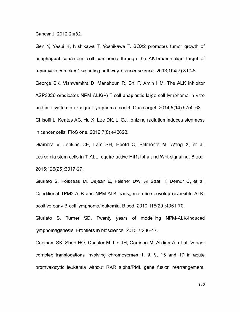
280
Cancer J. 2012;2:e82.
Gen Y, Yasui K, Nishikawa T, Yoshikawa T. SOX2 promotes tumor growth of
esophageal squamous cell carcinoma through the AKT/mammalian target of
rapamycin complex 1 signaling pathway. Cancer science. 2013;104(7):810-6.
George SK, Vishwamitra D, Manshouri R, Shi P, Amin HM. The ALK inhibitor
ASP3026 eradicates NPM-ALK(+) T-cell anaplastic large-cell lymphoma in vitro
and in a systemic xenograft lymphoma model. Oncotarget. 2014;5(14):5750-63.
Ghisolfi L, Keates AC, Hu X, Lee DK, Li CJ. Ionizing radiation induces stemness
in cancer cells. PloS one. 2012;7(8):e43628.
Giambra V, Jenkins CE, Lam SH, Hoofd C, Belmonte M, Wang X, et al.
Leukemia stem cells in T-ALL require active Hif1alpha and Wnt signaling. Blood.
2015;125(25):3917-27.
Giuriato S, Foisseau M, Dejean E, Felsher DW, Al Saati T, Demur C, et al.
Conditional TPM3-ALK and NPM-ALK transgenic mice develop reversible ALK-
positive early B-cell lymphoma/leukemia. Blood. 2010;115(20):4061-70.
Giuriato S, Turner SD. Twenty years of modelling NPM-ALK-induced
lymphomagenesis. Frontiers in bioscience. 2015;7:236-47.
Gogineni SK, Shah HO, Chester M, Lin JH, Garrison M, Alidina A, et al. Variant
complex translocations involving chromosomes 1, 9, 9, 15 and 17 in acute
promyelocytic leukemia without RAR alpha/PML gene fusion rearrangement.

281
Leukemia. 1997;11(4):514-8.
Golebiewska A, Brons NH, Bjerkvig R, Niclou SP. Critical appraisal of the side
population assay in stem cell and cancer stem cell research. Cell stem cell.
2011;8(2):136-47.
Gopal K, Gupta N, Zhang H, Alshareef A, Alqahtani H, Bigras G, et al. Oxidative
stress induces the acquisition of cancer stem-like phenotype in breast cancer
detectable by using a Sox2 regulatory region-2 (SRR2) reporter. Oncotarget.
2016;7(3):3111-27.
Gordon A, Conlon C, Collin J, Peto T, Gray D, Hands L, et al. An eight year
experience of conservative management for aortic graft sepsis. European journal
of vascular surgery. 1994;8(5):611-6.
Gorrini C, Harris IS, Mak TW. Modulation of oxidative stress as an anticancer
strategy. Nature reviews Drug discovery. 2013;12(12):931-47.
Gorrini C, Harris IS, Mak TW. Modulation of oxidative stress as an anticancer
strategy. Nature reviews Drug discovery. 2013;12(12):931-47.
Greenland C, Dastugue N, Touriol C, Lamant L, Delsol G, Brousset P. Anaplastic
large cell lymphoma with the t(2;5)(p23;q35) NPM/ALK chromosomal
translocation and duplication of the short arm of the non-translocated
chromosome 2 involving the full length of the ALK gene. Journal of clinical
pathology. 2001;54(2):152-4.
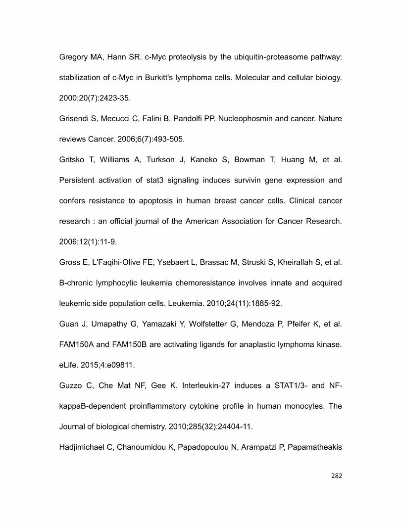
282
Gregory MA, Hann SR. c-Myc proteolysis by the ubiquitin-proteasome pathway:
stabilization of c-Myc in Burkitt's lymphoma cells. Molecular and cellular biology.
2000;20(7):2423-35.
Grisendi S, Mecucci C, Falini B, Pandolfi PP. Nucleophosmin and cancer. Nature
reviews Cancer. 2006;6(7):493-505.
Gritsko T, Williams A, Turkson J, Kaneko S, Bowman T, Huang M, et al.
Persistent activation of stat3 signaling induces survivin gene expression and
confers resistance to apoptosis in human breast cancer cells. Clinical cancer
research : an official journal of the American Association for Cancer Research.
2006;12(1):11-9.
Gross E, L'Faqihi-Olive FE, Ysebaert L, Brassac M, Struski S, Kheirallah S, et al.
B-chronic lymphocytic leukemia chemoresistance involves innate and acquired
leukemic side population cells. Leukemia. 2010;24(11):1885-92.
Guan J, Umapathy G, Yamazaki Y, Wolfstetter G, Mendoza P, Pfeifer K, et al.
FAM150A and FAM150B are activating ligands for anaplastic lymphoma kinase.
eLife. 2015;4:e09811.
Guzzo C, Che Mat NF, Gee K. Interleukin-27 induces a STAT1/3- and NF-
kappaB-dependent proinflammatory cytokine profile in human monocytes. The
Journal of biological chemistry. 2010;285(32):24404-11.
Hadjimichael C, Chanoumidou K, Papadopoulou N, Arampatzi P, Papamatheakis
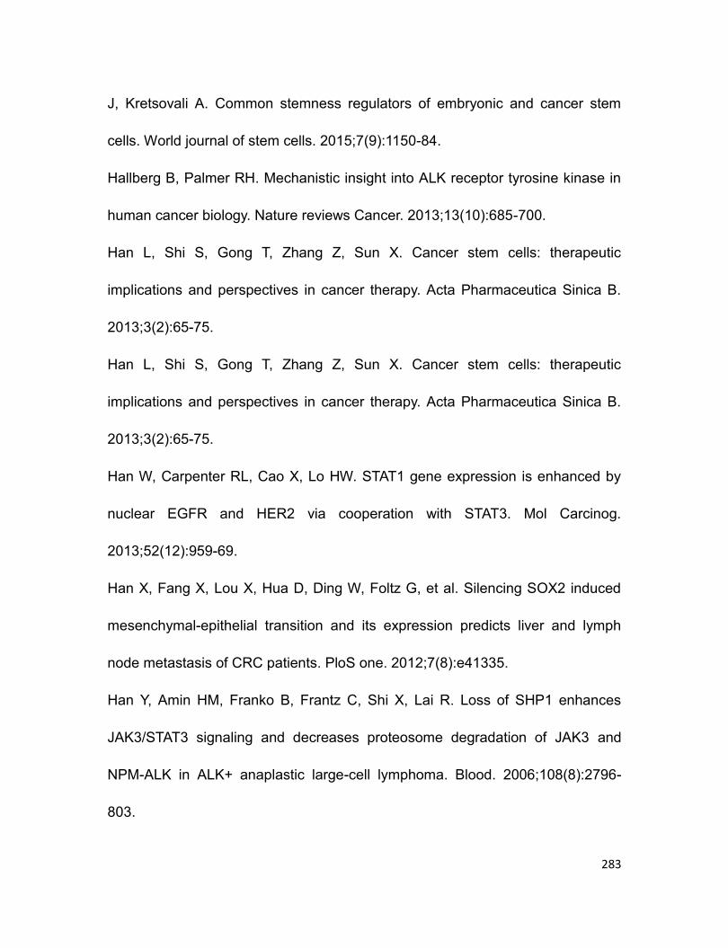
283
J, Kretsovali A. Common stemness regulators of embryonic and cancer stem
cells. World journal of stem cells. 2015;7(9):1150-84.
Hallberg B, Palmer RH. Mechanistic insight into ALK receptor tyrosine kinase in
human cancer biology. Nature reviews Cancer. 2013;13(10):685-700.
Han L, Shi S, Gong T, Zhang Z, Sun X. Cancer stem cells: therapeutic
implications and perspectives in cancer therapy. Acta Pharmaceutica Sinica B.
2013;3(2):65-75.
Han L, Shi S, Gong T, Zhang Z, Sun X. Cancer stem cells: therapeutic
implications and perspectives in cancer therapy. Acta Pharmaceutica Sinica B.
2013;3(2):65-75.
Han W, Carpenter RL, Cao X, Lo HW. STAT1 gene expression is enhanced by
nuclear EGFR and HER2 via cooperation with STAT3. Mol Carcinog.
2013;52(12):959-69.
Han X, Fang X, Lou X, Hua D, Ding W, Foltz G, et al. Silencing SOX2 induced
mesenchymal-epithelial transition and its expression predicts liver and lymph
node metastasis of CRC patients. PloS one. 2012;7(8):e41335.
Han Y, Amin HM, Franko B, Frantz C, Shi X, Lai R. Loss of SHP1 enhances
JAK3/STAT3 signaling and decreases proteosome degradation of JAK3 and
NPM-ALK in ALK+ anaplastic large-cell lymphoma. Blood. 2006;108(8):2796-
803.
284
Han Y, Amin HM, Frantz C, Franko B, Lee J, Lin Q, et al. Restoration of shp1
expression by 5-AZA-2'-deoxycytidine is associated with downregulation of
JAK3/STAT3 signaling in ALK-positive anaplastic large cell lymphoma.
Leukemia. 2006;20(9):1602-9.
Hansel DE, Platt E, Orloff M, Harwalker J, Sethu S, Hicks JL, et al. Mammalian
target of rapamycin (mTOR) regulates cellular proliferation and tumor growth in
urothelial carcinoma. The American journal of pathology. 2010;176(6):3062-72.
Hapgood G, Savage KJ. The biology and management of systemic anaplastic
large cell lymphoma. Blood. 2015;126(1):17-25.
Hassler MR, Pulverer W, Lakshminarasimhan R, Redl E, Hacker J, Garland GD,
et al. Insights into the Pathogenesis of Anaplastic Large-Cell Lymphoma through
Genome-wide DNA Methylation Profiling. Cell reports. 2016;17(2):596-608.
Hecht JL, Aster JC. Molecular biology of Burkitt's lymphoma. Journal of clinical
oncology : official journal of the American Society of Clinical Oncology.
2000;18(21):3707-21.
Hegazy SA, Alshareef A, Gelebart P, Anand M, Armanious H, Ingham RJ, et al.
Disheveled proteins promote cell growth and tumorigenicity in ALK-positive
anaplastic large cell lymphoma. Cellular signalling. 2013;25(1):295-307.
Hemmings BA, Restuccia DF. PI3K-PKB/Akt pathway. Cold Spring Harbor
perspectives in biology. 2012;4(9):a011189.
285
Herreros-Villanueva M, Zhang JS, Koenig A, Abel EV, Smyrk TC, Bamlet WR, et
al. SOX2 promotes dedifferentiation and imparts stem cell-like features to
pancreatic cancer cells. Oncogenesis. 2013;2:e61.
Heuser M, Sly LM, Argiropoulos B, Kuchenbauer F, Lai C, Weng A, et al.
Modeling the functional heterogeneity of leukemia stem cells: role of STAT5 in
leukemia stem cell self-renewal. Blood. 2009;114(19):3983-93.
Ho HH, Ivashkiv LB. Role of STAT3 in type I interferon responses. Negative
regulation of STAT1-dependent inflammatory gene activation. The Journal of
biological chemistry. 2006;281(20):14111-8.
Ho MM, Ng AV, Lam S, Hung JY. Side population in human lung cancer cell lines
and tumors is enriched with stem-like cancer cells. Cancer research.
2007;67(10):4827-33.
Hong F, Jaruga B, Kim WH, Radaeva S, El-Assal ON, Tian Z, et al. Opposing
roles of STAT1 and STAT3 in T cell–mediated hepatitis: regulation by SOCS.
Journal of Clinical Investigation. 2002;110(10):1503-13.
Honorat JF, Ragab A, Lamant L, Delsol G, Ragab-Thomas J. SHP1 tyrosine
phosphatase negatively regulates NPM-ALK tyrosine kinase signaling. Blood.
2006;107(10):4130-8.
Hu X, Ghisolfi L, Keates AC, Zhang J, Xiang S, Lee DK, et al. Induction of cancer
cell stemness by chemotherapy. Cell cycle. 2012;11(14):2691-8.
286
Hu Y, Chen Y, Douglas L, Li S. beta-Catenin is essential for survival of leukemic
stem cells insensitive to kinase inhibition in mice with BCR-ABL-induced chronic
myeloid leukemia. Leukemia. 2009;23(1):109-16.
Huang B, Huang YJ, Yao ZJ, Chen X, Guo SJ, Mao XP, et al. Cancer stem cell-
like side population cells in clear cell renal cell carcinoma cell line 769P. PloS
one. 2013;8(7):e68293.
Hurlin PJ. N-Myc functions in transcription and development. Birth defects
research Part C, Embryo today : reviews. 2005;75(4):340-52.
Jager R, Hahne J, Jacob A, Egert A, Schenkel J, Wernert N, et al. Mice
transgenic for NPM-ALK develop non-Hodgkin lymphomas. Anticancer research.
2005;25(5):3191-6.
Jang GB, Hong IS, Kim RJ, Lee SY, Park SJ, Lee ES, et al. Wnt/beta-Catenin
Small-Molecule Inhibitor CWP232228 Preferentially Inhibits the Growth of Breast
Cancer Stem-like Cells. Cancer research. 2015;75(8):1691-702.
Jang GB, Kim JY, Cho SD, Park KS, Jung JY, Lee HY, et al. Blockade of
Wnt/beta-catenin signaling suppresses breast cancer metastasis by inhibiting
CSC-like phenotype. Scientific reports. 2015;5:12465.
Jang GB, Kim JY, Cho SD, Park KS, Jung JY, Lee HY, et al. Blockade of
Wnt/beta-catenin signaling suppresses breast cancer metastasis by inhibiting
CSC-like phenotype. Scientific reports. 2015;5:12465.

287
Janssen-Heininger YM, Mossman BT, Heintz NH, Forman HJ, Kalyanaraman B,
Finkel T, et al. Redox-based regulation of signal transduction: principles, pitfalls,
and promises. Free radical biology & medicine. 2008;45(1):1-17.
Jeong CH, Cho YY, Kim MO, Kim SH, Cho EJ, Lee SY, et al. Phosphorylation of
Sox2 cooperates in reprogramming to pluripotent stem cells. Stem cells.
2010;28(12):2141-50.
Johnston PA, Grandis JR. STAT3 signaling: anticancer strategies and
challenges. Molecular interventions. 2011;11(1):18-26.
Juan J, Muraguchi T, Iezza G, Sears RC, McMahon M. Diminished WNT -> beta-
catenin -> c-MYC signaling is a barrier for malignant progression of
BRAFV600E-induced lung tumors. Genes & development. 2014;28(6):561-75.
Jung K, Gupta N, Wang P, Lewis JT, Gopal K, Wu F, et al. Triple negative breast
cancers comprise a highly tumorigenic cell subpopulation detectable by its high
responsiveness to a Sox2 regulatory region 2 (SRR2) reporter. Oncotarget.
2015;6(12):10366-73.
Justilien V, Walsh MP, Ali SA, Thompson EA, Murray NR, Fields AP. The PRKCI
and SOX2 oncogenes are coamplified and cooperate to activate Hedgehog
signaling in lung squamous cell carcinoma. Cancer cell. 2014;25(2):139-51.
Kaganoi J, Watanabe G, Okabe M, Nagatani S, Kawabe A, Shimada Y, et al.
STAT1 activation-induced apoptosis of esophageal squamous cell carcinoma
288
cells in vivo. Annals of surgical oncology. 2007;14(4):1405-15.
Kamachi Y, Kondoh H. Sox proteins: regulators of cell fate specification and
differentiation. Development. 2013;140(20):4129-44.
Kaplan DH, Shankaran V, Dighe AS, Stockert E, Aguet M, Old LJ, et al.
Demonstration of an interferon gamma-dependent tumor surveillance system in
immunocompetent mice. Proceedings of the National Academy of Sciences of
the United States of America. 1998;95(13):7556-61.
Karagiannis P, Eto K. Ten years of induced pluripotency: from basic mechanisms
to therapeutic applications. Development. 2016;143(12):2039-43.
Kawano T, Morioka M, Yano S, Hamada J, Ushio Y, Miyamoto E, et al.
Decreased akt activity is associated with activation of forkhead transcription
factor after transient forebrain ischemia in gerbil hippocampus. Journal of
cerebral blood flow and metabolism: official journal of the International Society of
Cerebral Blood Flow and Metabolism. 2002;22(8):926-34.
Kazandjian D, Blumenthal GM, Chen HY, He K, Patel M, Justice R, et al. FDA
approval summary: crizotinib for the treatment of metastatic non-small cell lung
cancer with anaplastic lymphoma kinase rearrangements. The oncologist.
2014;19(10):e5-11.
Keith B, Simon MC. Hypoxia-inducible factors, stem cells, and cancer. Cell.
2007;129(3):465-72.
289
Kemper K, de Goeje PL, Peeper DS, van Amerongen R. Phenotype switching:
tumor cell plasticity as a resistance mechanism and target for therapy. Cancer
research. 2014;74(21):5937-41.
Kesler MV, Paranjape GS, Asplund SL, McKenna RW, Jamal S, Kroft SH.
Anaplastic large cell lymphoma: a flow cytometric analysis of 29 cases. American
journal of clinical pathology. 2007;128(2):314-22.
Khodarev N, Ahmad R, Rajabi H, Pitroda S, Kufe T, McClary C, et al.
Cooperativity of the MUC1 oncoprotein and STAT1 pathway in poor prognosis
human breast cancer. Oncogene. 2010;29(6):920-9.
Khoury JD, Medeiros LJ, Rassidakis GZ, Yared MA, Tsioli P, Leventaki V, et al.
Differential expression and clinical significance of tyrosine-phosphorylated
STAT3 in ALK+ and ALK- anaplastic large cell lymphoma. Clinical cancer
research. 2003; 9(10 Pt 1):3692-9.
Kim HS, Lee MS. STAT1 as a key modulator of cell death. Cellular signalling.
2007;19(3):454-65.
Kim J, Orkin SH. Embryonic stem cell-specific signatures in cancer: insights into
genomic regulatory networks and implications for medicine. Genome Med.
2011;3(11):75.
Kim MH, Kim H. Oncogenes and tumor suppressors regulate glutamine
metabolism in cancer cells. Journal of cancer prevention. 2013;18(3):221-6.

290
Kim TK, Maniatis T. Regulation of interferon-gamma-activated STAT1 by the
ubiquitin-proteasome pathway. Science. 1996;273(5282):1717-9.
Kinney MC, Collins RD, Greer JP, Whitlock JA, Sioutos N, Kadin ME. A small-
cell-predominant variant of primary Ki-1 (CD30)+ T-cell lymphoma. The
American journal of surgical pathology. 1993;17(9):859-68.
Kinney MC, Higgins RA, Medina EA. Anaplastic large cell lymphoma: twenty-five
years of discovery. Archives of pathology & laboratory medicine. 2011;135(1):19-
43.
Kirken RA, Rui H, Malabarba MG, Howard OM, Kawamura M, O'Shea JJ, et al.
Activation of JAK3, but not JAK1, is critical for IL-2-induced proliferation and
STAT5 recruitment by a COOH-terminal region of the IL-2 receptor beta-chain.
Cytokine. 1995;7(7):689-700.
Klaunig JE, Kamendulis LM, Hocevar BA. Oxidative stress and oxidative
damage in carcinogenesis. Toxicologic pathology. 2010;38(1):96-109.
Klempner SJ, Cohen DW, Costa DB. ALK translocation in non-small cell lung
cancer with adenocarcinoma and squamous cell carcinoma markers. Journal of
thoracic oncology : official publication of the International Association for the
Study of Lung Cancer. 2011;6(8):1439-40.
Koromilas AE, Sexl V. The tumor suppressor function of STAT1 in breast cancer.
Jak-Stat. 2013;2(2):e23353.

291
Kortylewski M, Komyod W, Kauffmann ME, Bosserhoff A, Heinrich PC,
Behrmann I. Interferon-gamma-mediated growth regulation of melanoma cells:
involvement of STAT1-dependent and STAT1-independent signals. The Journal
of investigative dermatology. 2004;122(2):414-22.
Kortylewski M, Kujawski M, Wang T, Wei S, Zhang S, Pilon-Thomas S, et al.
Inhibiting Stat3 signaling in the hematopoietic system elicits multicomponent
antitumor immunity. Nature medicine. 2005;11(12):1314-21.
Kothari S, Ud-Din N, Lisi M, Coyle T. Crizotinib in anaplastic lymphoma kinase-
positive anaplastic large cell lymphoma in the setting of renal insufficiency: a
case report. Journal of medical case reports. 2016;10:176.
Kovacic B, Stoiber D, Moriggl R, Weisz E, Ott RG, Kreibich R, et al. STAT1 acts
as a tumor promoter for leukemia development. Cancer cell. 2006;10(1):77-87.
Kovarik J, Boudny V, Kocak I, Lauerova L, Fait V, Vagundova M. Malignant
melanoma associates with deficient IFN-induced STAT 1 phosphorylation.
International journal of molecular medicine. 2003;12(3):335-40.
Koyama-Nasu R, Haruta R, Nasu-Nishimura Y, Taniue K, Katou Y, Shirahige K,
et al. The pleiotrophin-ALK axis is required for tumorigenicity of glioblastoma
stem cells. Oncogene. 2014;33(17):2236-44.
Kramer MH, Hermans J, Wijburg E, Philippo K, Geelen E, van Krieken JH, et al.
Clinical relevance of BCL2, BCL6, and MYC rearrangements in diffuse large B-
292
cell lymphoma. Blood. 1998;92(9):3152-62.
Krenacs L, Wellmann A, Sorbara L, Himmelmann AW, Bagdi E, Jaffe ES, et al.
Cytotoxic cell antigen expression in anaplastic large cell lymphomas of T- and
null-cell type and Hodgkin's disease: evidence for distinct cellular origin. Blood.
1997;89(3):980-9.
Kress TR, Sabo A, Amati B. MYC: connecting selective transcriptional control to
global RNA production. Nature reviews Cancer. 2015;15(10):593-607.
Kwan KY, Shen J, Corey DP. C-MYC transcriptionally amplifies SOX2 target
genes to regulate self-renewal in multipotent otic progenitor cells. Stem cell
reports. 2015;4(1):47-60.
Lai R, Ingham RJ. The pathobiology of the oncogenic tyrosine kinase NPM-ALK:
a brief update. Therapeutic advances in hematology. 2013;4(2):119-31.
Laimer K, Spizzo G, Obrist P, Gastl G, Brunhuber T, Schafer G, et al. STAT1
activation in squamous cell cancer of the oral cavity: a potential predictive
marker of response to adjuvant chemotherapy. Cancer. 2007;110(2):326-33.
Lamant L, Pulford K, Bischof D, Morris SW, Mason DY, Delsol G, et al.
Expression of the ALK tyrosine kinase gene in neuroblastoma. Am J Pathol.
2000;156(5):1711-21.
Lange K, Uckert W, Blankenstein T, Nadrowitz R, Bittner C, Renauld JC, et al.
Overexpression of NPM-ALK induces different types of malignant lymphomas in
293
IL-9 transgenic mice. Oncogene. 2003;22(4):517-27.
Laplante M, Sabatini DM. mTOR signaling in growth control and disease. Cell.
2012;149(2):274-93.
Lawrence DW, Kornbluth J. E3 ubiquitin ligase NKLAM ubiquitinates STAT1 and
positively regulates STAT1-mediated transcriptional activity. Cellular signalling.
2016;28(12):1833-41.
Lee HH, Norris A, Weiss JB, Frasch M. Jelly belly protein activates the receptor
tyrosine kinase Alk to specify visceral muscle pioneers. Nature.
2003;425(6957):507-12.
Leis O, Eguiara A, Lopez-Arribillaga E, Alberdi MJ, Hernandez-Garcia S,
Elorriaga K, et al. Sox2 expression in breast tumours and activation in breast
cancer stem cells. Oncogene. 2012;31(11):1354-65.
Li L, Neaves WB. Normal stem cells and cancer stem cells: the niche matters.
Cancer research. 2006;66(9):4553-7.
Li S, Lin P, Young KH, Kanagal-Shamanna R, Yin CC, Medeiros LJ. MYC/BCL2
double-hit high-grade B-cell lymphoma. Advances in anatomic pathology.
2013;20(5):315-26.
Li X, Xu Y, Chen Y, Chen S, Jia X, Sun T, et al. SOX2 promotes tumor metastasis
by stimulating epithelial-to-mesenchymal transition via regulation of WNT/beta-
catenin signal network. Cancer letters. 2013;336(2):379-89.

294
Lighvani AA, Frucht DM, Jankovic D, Yamane H, Aliberti J, Hissong BD, et al. T-
bet is rapidly induced by interferon-gamma in lymphoid and myeloid cells.
Proceedings of the National Academy of Sciences of the United States of
America. 2001;98(26):15137-42.
Lin CY, Loven J, Rahl PB, Paranal RM, Burge CB, Bradner JE, et al.
Transcriptional amplification in tumor cells with elevated c-Myc. Cell.
2012;151(1):56-67.
Lin CY, Tan BC, Liu H, Shih CJ, Chien KY, Lin CL, et al. Dephosphorylation of
nucleophosmin by PP1beta facilitates pRB binding and consequent E2F1-
dependent DNA repair. Molecular biology of the cell. 2010;21(24):4409-17.
Little CD, Nau MM, Carney DN, Gazdar AF, Minna JD. Amplification and
expression of the c-myc oncogene in human lung cancer cell lines. Nature.
1983;306(5939):194-6.
Lu YX, Yuan L, Xue XL, Zhou M, Liu Y, Zhang C, et al. Regulation of colorectal
carcinoma stemness, growth, and metastasis by an miR-200c-Sox2-negative
feedback loop mechanism. Clinical cancer research : an official journal of the
American Association for Cancer Research. 2014;20(10):2631-42.
Luis TC, Naber BA, Roozen PP, Brugman MH, de Haas EF, Ghazvini M, et al.
Canonical wnt signaling regulates hematopoiesis in a dosage-dependent
fashion. Cell stem cell. 2011;9(4):345-56.

295
MacDonald BT, Tamai K, He X. Wnt/beta-catenin signaling: components,
mechanisms, and diseases. Developmental cell. 2009;17(1):9-26.
Maes B, Vanhentenrijk V, Wlodarska I, Cools J, Peeters B, Marynen P, et al. The
NPM-ALK and the ATIC-ALK fusion genes can be detected in non-neoplastic
cells. The American journal of pathology. 2001;158(6):2185-93.
Mahalingaiah PK, Singh KP. Chronic oxidative stress increases growth and
tumorigenic potential of MCF-7 breast cancer cells. PloS one. 2014;9(1):e87371.
Malabarba MG, Rui H, Deutsch HH, Chung J, Kalthoff FS, Farrar WL, et al.
Interleukin-13 is a potent activator of JAK3 and STAT6 in cells expressing
interleukin-2 receptor-gamma and interleukin-4 receptor-alpha. The Biochemical
journal. 1996;319 ( Pt 3):865-72.
Malcolm TI, Villarese P, Fairbairn CJ, Lamant L, Trinquand A, Hook CE, et al.
Anaplastic large cell lymphoma arises in thymocytes and requires transient TCR
expression for thymic egress. Nature communications. 2016;7:10087.
Marampon F, Ciccarelli C, Zani BM. Down-regulation of c-Myc following
MEK/ERK inhibition halts the expression of malignant phenotype in
rhabdomyosarcoma and in non-muscle-derived human tumors. Molecular
cancer. 2006;5:31.
Marusyk A, Almendro V, Polyak K. Intra-tumour heterogeneity: a looking glass for
cancer? Nature reviews Cancer. 2012;12(5):323-34.
296
Marzec M, Kasprzycka M, Liu X, Raghunath PN, Wlodarski P, Wasik MA.
Oncogenic tyrosine kinase NPM/ALK induces activation of the MEK/ERK
signaling pathway independently of c-Raf. Oncogene. 2007;26(6):813-21.
Mathivet T, Mazot P, Vigny M. In contrast to agonist monoclonal antibodies, both
C-terminal truncated form and full length form of Pleiotrophin failed to activate
vertebrate ALK (anaplastic lymphoma kinase)? Cellular signalling.
2007;19(12):2434-43.
McCauley HA, Guasch G. Serial orthotopic transplantation of epithelial tumors in
single-cell suspension. Methods in molecular biology. 2013;1035:231-45.
McCubrey JA, Steelman LS, Chappell WH, Abrams SL, Wong EW, Chang F, et
al. Roles of the Raf/MEK/ERK pathway in cell growth, malignant transformation
and drug resistance. Biochimica et biophysica acta. 2007;1773(8):1263-84.
McDonnell SR, Hwang SR, Basrur V, Conlon KP, Fermin D, Wey E, et al. NPM-
ALK signals through glycogen synthase kinase 3beta to promote oncogenesis.
Oncogene. 2012;31(32):3733-40.
Meacham CE, Morrison SJ. Tumour heterogeneity and cancer cell plasticity.
Nature. 2013;501(7467):328-37.
Medeiros LJ, Elenitoba-Johnson KS. Anaplastic Large Cell Lymphoma. American
journal of clinical pathology. 2007;127(5):707-22.
Meissl K, Macho-Maschler S, Muller M, Strobl B. The good and the bad faces of

297
STAT1 in solid tumours. Cytokine. 2015.
Meraz MA, White JM, Sheehan KC, Bach EA, Rodig SJ, Dighe AS, et al.
Targeted disruption of the Stat1 gene in mice reveals unexpected physiologic
specificity in the JAK-STAT signaling pathway. Cell. 1996;84(3):431-42.
Merkel O, Hamacher F, Sifft E, Kenner L, Greil R. Novel therapeutic options in
anaplastic large cell lymphoma: molecular targets and immunological tools.
Molecular cancer therapeutics. 2011;10(7):1127-36.
Merkel O, Kenner L, Turner SD. Stem cell hunt in NHL. Oncoscience.
2015;2(10):809-10.
Miller DM, Thomas SD, Islam A, Muench D, Sedoris K. c-Myc and cancer
metabolism. Clinical cancer research : an official journal of the American
Association for Cancer Research. 2012;18(20):5546-53.
Miura Y, Tsujioka T, Nishimura Y, Sakaguchi H, Maeda M, Hayashi H, et al.
TRAIL expression up-regulated by interferon-gamma via phosphorylation of
STAT1 induces myeloma cell death. Anticancer research. 2006;26(6B):4115-24.
Molina JR, Hayashi Y, Stephens C, Georgescu MM. Invasive glioblastoma cells
acquire stemness and increased Akt activation. Neoplasia. 2010;12(6):453-63.
Moog-Lutz C, Degoutin J, Gouzi JY, Frobert Y, Brunet-de Carvalho N, Bureau J,
et al. Activation and inhibition of anaplastic lymphoma kinase receptor tyrosine
kinase by monoclonal antibodies and absence of agonist activity of pleiotrophin.
298
The Journal of biological chemistry. 2005;280(28):26039-48.
Moon RT, Kohn AD, De Ferrari GV, Kaykas A. WNT and beta-catenin signalling:
diseases and therapies. Nature reviews Genetics. 2004;5(9):691-701.
Mora J, Filippa DA, Thaler HT, Polyak T, Cranor ML, Wollner N. Large cell non-
Hodgkin lymphoma of childhood: Analysis of 78 consecutive patients enrolled in
2 consecutive protocols at the Memorial Sloan-Kettering Cancer Center. Cancer.
2000;88(1):186-97.
Morris SW, Kirstein MN, Valentine MB, Dittmer KG, Shapiro DN, Saltman DL, et
al. Fusion of a kinase gene, ALK, to a nucleolar protein gene, NPM, in non-
Hodgkin's lymphoma. Science. 1994;263(5151):1281-4.
Morrissey PA, Quinn PB, Sheehy PJ. Newer aspects of micronutrients in chronic
disease: vitamin E. The Proceedings of the Nutrition Society. 1994;53(3):571-82.
Mosse YP, Lim MS, Voss SD, Wilner K, Ruffner K, Laliberte J, et al. Safety and
activity of crizotinib for paediatric patients with refractory solid tumours or
anaplastic large-cell lymphoma: a Children's Oncology Group phase 1
consortium study. The Lancet Oncology. 2013;14(6):472-80.
Moti N, Malcolm T, Hamoudi R, Mian S, Garland G, Hook CE, et al. Anaplastic
large cell lymphoma-propagating cells are detectable by side population analysis
and possess an expression profile reflective of a primitive origin. Oncogene.
2015;34(14):1843-52.

299
Moumen M, Chiche A, Decraene C, Petit V, Gandarillas A, Deugnier MA, et al.
Myc is required for beta-catenin-mediated mammary stem cell amplification and
tumorigenesis. Molecular cancer. 2013;12(1):132.
Murga-Zamalloa CA, Mendoza-Reinoso V, Sahasrabuddhe AA, Rolland D,
Hwang SR, McDonnell SR, et al. NPM-ALK phosphorylates WASp Y102 and
contributes to oncogenesis of anaplastic large cell lymphoma. Oncogene. 2016.
Murray PB, Lax I, Reshetnyak A, Ligon GF, Lillquist JS, Natoli EJ, Jr., et al.
Heparin is an activating ligand of the orphan receptor tyrosine kinase ALK. Sci
Signal. 2015;8(360):ra6.
Myers SA, Peddada S, Chatterjee N, Friedrich T, Tomoda K, Krings G, et al.
SOX2 O-GlcNAcylation alters its protein-protein interactions and genomic
occupancy to modulate gene expression in pluripotent cells. eLife.
2016;5:e10647.
Nakamura S, Shiota M, Nakagawa A, Yatabe Y, Kojima M, Motoori T, et al.
Anaplastic large cell lymphoma: a distinct molecular pathologic entity: a
reappraisal with special reference to p80(NPM/ALK) expression. The American
journal of surgical pathology. 1997;21(12):1420-32.
Nath S, Mukherjee P. MUC1: a multifaceted oncoprotein with a key role in cancer
progression. Trends Mol Med. 2014;20(6):332-42.
Neumann J, Bahr F, Horst D, Kriegl L, Engel J, Luque RM, et al. SOX2

300
expression correlates with lymph-node metastases and distant spread in right-
sided colon cancer. BMC cancer. 2011;11:518.
Nie Z, Hu G, Wei G, Cui K, Yamane A, Resch W, et al. c-Myc is a universal
amplifier of expressed genes in lymphocytes and embryonic stem cells. Cell.
2012;151(1):68-79.
Nieborowska-Skorska M, Slupianek A, Xue L, Zhang Q, Raghunath PN, Hoser
G, et al. Role of signal transducer and activator of transcription 5 in
nucleophosmin/ anaplastic lymphoma kinase-mediated malignant transformation
of lymphoid cells. Cancer research. 2001;61(17):6517-23.
Ning Y, Riggins RB, Mulla JE, Chung H, Zwart A, Clarke R. IFNgamma restores
breast cancer sensitivity to fulvestrant by regulating STAT1, IFN regulatory factor
1, NF-kappaB, BCL2 family members, and signaling to caspase-dependent
apoptosis. Molecular cancer therapeutics. 2010;9(5):1274-85.
Nishikawa S, Goldstein RA, Nierras CR. The promise of human induced
pluripotent stem cells for research and therapy. Nature reviews Molecular cell
biology. 2008;9(9):725-9.
O'Brien CA, Kreso A, Dick JE. Cancer stem cells in solid tumors: an overview.
Seminars in radiation oncology. 2009;19(2):71-7.
O'Brien CA, Kreso A, Jamieson CH. Cancer stem cells and self-renewal. Clinical
cancer research : an official journal of the American Association for Cancer
301
Research. 2010;16(12):3113-20.
Oren O, Smith BD. Eliminating Cancer Stem Cells by Targeting Embryonic
Signaling Pathways. Stem cell reviews. 2016.
Ortmann RA, Cheng T, Visconti R, Frucht DM, O'Shea JJ. Janus kinases and
signal transducers and activators of transcription: their roles in cytokine
signaling, development and immunoregulation. Arthritis research. 2000;2(1):16-
32.
Oskarsson T, Batlle E, Massague J. Metastatic stem cells: sources, niches, and
vital pathways. Cell stem cell. 2014;14(3):306-21.
Osuka K, Watanabe Y, Usuda N, Atsuzawa K, Wakabayashi T, Takayasu M.
Oxidative stress activates STAT1 in basilar arteries after subarachnoid
hemorrhage. Brain research. 2010;1332:12-9.
Ou SH, Bartlett CH, Mino-Kenudson M, Cui J, Iafrate AJ. Crizotinib for the
treatment of ALK-rearranged non-small cell lung cancer: a success story to
usher in the second decade of molecular targeted therapy in oncology. The
oncologist. 2012;17(11):1351-75.
Ouyang J, Yu W, Liu J, Zhang N, Florens L, Chen J, et al. Cyclin-dependent
kinase-mediated Sox2 phosphorylation enhances the ability of Sox2 to establish
the pluripotent state. The Journal of biological chemistry. 2015;290(37):22782-
94.
302
Pan XN, Chen JJ, Wang LX, Xiao RZ, Liu LL, Fang ZG, et al. Inhibition of c-Myc
overcomes cytotoxic drug resistance in acute myeloid leukemia cells by
promoting differentiation. PloS one. 2014;9(8):e105381.
Pansky A, Hildebrand P, Fasler-Kan E, Baselgia L, Ketterer S, Beglinger C, et al.
Defective Jak-STAT signal transduction pathway in melanoma cells resistant to
growth inhibition by interferon-alpha. International journal of cancer.
2000;85(5):720-5.
Park JB, Lee CS, Jang JH, Ghim J, Kim YJ, You S, et al. Phospholipase
signalling networks in cancer. Nature reviews Cancer. 2012;12(11):782-92.
Patel JH, Loboda AP, Showe MK, Showe LC, McMahon SB. Analysis of genomic
targets reveals complex functions of MYC. Nature reviews Cancer.
2004;4(7):562-8.
Pearson JD, Lee JK, Bacani JT, Lai R, Ingham RJ. NPM-ALK: The Prototypic
Member of a Family of Oncogenic Fusion Tyrosine Kinases. J Signal Transduct.
2012;2012:123253.
Pelengaris S, Khan M, Evan G. c-MYC: more than just a matter of life and death.
Nature reviews Cancer. 2002;2(10):764-76.
Pencik J, Schlederer M, Gruber W, Unger C, Walker SM, Chalaris A, et al.
STAT3 regulated ARF expression suppresses prostate cancer metastasis.
Nature communications. 2015;6:7736.
303
Perna D, Faga G, Verrecchia A, Gorski MM, Barozzi I, Narang V, et al. Genome-
wide mapping of Myc binding and gene regulation in serum-stimulated
fibroblasts. Oncogene. 2012;31(13):1695-709.
Piccaluga PP, Gazzola A, Mannu C, Agostinelli C, Bacci F, Sabattini E, et al.
Pathobiology of anaplastic large cell lymphoma. Adv Hematol. 2010:345053.
Pileri S, Falini B, Delsol G, Stein H, Baglioni P, Poggi S, et al. Lymphohistiocytic
T-cell lymphoma (anaplastic large cell lymphoma CD30+/Ki-1 + with a high
content of reactive histiocytes). Histopathology. 1990;16(4):383-91.
Piva M, Domenici G, Iriondo O, Rabano M, Simoes BM, Comaills V, et al. Sox2
promotes tamoxifen resistance in breast cancer cells. EMBO molecular
medicine. 2014;6(1):66-79.
Piva R, Agnelli L, Pellegrino E, Todoerti K, Grosso V, Tamagno I, et al. Gene
expression profiling uncovers molecular classifiers for the recognition of
anaplastic large-cell lymphoma within peripheral T-cell neoplasms. Journal of
clinical oncology : official journal of the American Society of Clinical Oncology.
2010;28(9):1583-90.
Plaks V, Kong N, Werb Z. The cancer stem cell niche: how essential is the niche
in regulating stemness of tumor cells? Cell stem cell. 2015;16(3):225-38.
Qi D, Wang Q, Yu M, Lan R, Li S, Lu F. Mitotic phosphorylation of SOX2
mediated by Aurora kinase A is critical for the stem-cell like cell maintenance in

304
PA-1 cells. Cell cycle. 2016;15(15):2009-18.
Qiu L, Lai R, Lin Q, Lau E, Thomazy DM, Calame D, et al. Autocrine release of
interleukin-9 promotes Jak3-dependent survival of ALK+ anaplastic large-cell
lymphoma cells. Blood. 2006;108(7):2407-15.
Raetz EA, Perkins SL, Carlson MA, Schooler KP, Carroll WL, Virshup DM. The
nucleophosmin-anaplastic lymphoma kinase fusion protein induces c-Myc
expression in pediatric anaplastic large cell lymphomas. The American journal of
pathology. 2002;161(3):875-83.
Ramana CV, Chatterjee-Kishore M, Nguyen H, Stark GR. Complex roles of Stat1
in regulating gene expression. Oncogene. 2000;19(21):2619-27.
Ramana CV, Grammatikakis N, Chernov M, Nguyen H, Goh KC, Williams BR, et
al. Regulation of c-myc expression by IFN-gamma through Stat1-dependent and
-independent pathways. The EMBO journal. 2000;19(2):263-72.
Regis G, Pensa S, Boselli D, Novelli F, Poli V. Ups and downs: the STAT1:STAT3
seesaw of Interferon and gp130 receptor signalling. Semin Cell Dev Biol.
2008;19(4):351-9.
Reliene R, Fischer E, Schiestl RH. Effect of N-acetyl cysteine on oxidative DNA
damage and the frequency of DNA deletions in atm-deficient mice. Cancer
research. 2004;64(15):5148-53.
Ren Y, Zhao P, Liu J, Yuan Y, Cheng Q, Zuo Y, et al. Deubiquitinase USP2a
305
Sustains Interferons Antiviral Activity by Restricting Ubiquitination of Activated
STAT1 in the Nucleus. PLoS pathogens. 2016;12(7):e1005764.
Reshetnyak AV, Murray PB, Shi X, Mo ES, Mohanty J, Tome F, et al. Augmentor
alpha and beta (FAM150) are ligands of the receptor tyrosine kinases ALK and
LTK: Hierarchy and specificity of ligand-receptor interactions. Proceedings of the
National Academy of Sciences of the United States of America.
2015;112(52):15862-7.
Reuter S, Gupta SC, Chaturvedi MM, Aggarwal BB. Oxidative stress,
inflammation, and cancer: how are they linked? Free radical biology & medicine.
2010;49(11):1603-16.
Ricardo S, Vieira AF, Gerhard R, Leitao D, Pinto R, Cameselle-Teijeiro JF, et al.
Breast cancer stem cell markers CD44, CD24 and ALDH1: expression
distribution within intrinsic molecular subtype. Journal of clinical pathology.
2011;64(11):937-46.
Riobo NA, Lu K, Ai X, Haines GM, Emerson CP, Jr. Phosphoinositide 3-kinase
and Akt are essential for Sonic Hedgehog signaling. Proceedings of the National
Academy of Sciences of the United States of America. 2006;103(12):4505-10.
Rizzardi GP, Tambussi G, Barcellini W, Capiluppi B, Clerici E, Maestra LL, et al.
Soluble CD30, tumour necrosis factor (TNF)-alpha, and TNF receptors in
primary HIV-1 infection: relationship with HIV-1, RNA, clinical outcome and early

306
antiviral therapy. Journal of biological regulators and homeostatic agents.
1997;11(1-2):43-9.
Rizzino A. Concise review: The Sox2-Oct4 connection: critical players in a much
larger interdependent network integrated at multiple levels. Stem cells.
2013;31(6):1033-9.
Roberts PJ, Der CJ. Targeting the Raf-MEK-ERK mitogen-activated protein
kinase cascade for the treatment of cancer. Oncogene. 2007;26(22):3291-310.
Roderick JE, Tesell J, Shultz LD, Brehm MA, Greiner DL, Harris MH, et al. c-Myc
inhibition prevents leukemia initiation in mice and impairs the growth of relapsed
and induction failure pediatric T-ALL cells. Blood. 2014;123(7):1040-50.
Rodrigues AB, Zoranovic T, Ayala-Camargo A, Grewal S, Reyes-Robles T,
Krasny M, et al. Activated STAT regulates growth and induces competitive
interactions independently of Myc, Yorkie, Wingless and ribosome biogenesis.
Development. 2012;139(21):4051-61.
Romashkova JA, Makarov SS. NF-kappaB is a target of AKT in anti-apoptotic
PDGF signalling. Nature. 1999;401(6748):86-90.
Rubie H, Hartmann O, Michon J, Frappaz D, Coze C, Chastagner P, et al. N-Myc
gene amplification is a major prognostic factor in localized neuroblastoma:
results of the French NBL 90 study. Neuroblastoma Study Group of the Societe
Francaise d'Oncologie Pediatrique. Journal of clinical oncology : official journal of

307
the American Society of Clinical Oncology. 1997;15(3):1171-82.
Saijo H, Hirohashi Y, Torigoe T, Horibe R, Takaya A, Murai A, et al. Plasticity of
lung cancer stem-like cells is regulated by the transcription factor HOXA5 that is
induced by oxidative stress. Oncotarget. 2016.
Salcido CD, Larochelle A, Taylor BJ, Dunbar CE, Varticovski L. Molecular
characterisation of side population cells with cancer stem cell-like characteristics
in small-cell lung cancer. British journal of cancer. 2010;102(11):1636-44.
Sanda T, Tyner JW, Gutierrez A, Ngo VN, Glover J, Chang BH, et al. TYK2-
STAT1-BCL2 pathway dependence in T-cell acute lymphoblastic leukemia.
Cancer discovery. 2013;3(5):564-77.
Sansom OJ, Meniel VS, Muncan V, Phesse TJ, Wilkins JA, Reed KR, et al. Myc
deletion rescues Apc deficiency in the small intestine. Nature.
2007;446(7136):676-9.
Santini R, Pietrobono S, Pandolfi S, Montagnani V, D'Amico M, Penachioni JY, et
al. SOX2 regulates self-renewal and tumorigenicity of human melanoma-
initiating cells. Oncogene. 2014;33(38):4697-708.
Santo EE, Stroeken P, Sluis PV, Koster J, Versteeg R, Westerhout EM. FOXO3a
is a major target of inactivation by PI3K/AKT signaling in aggressive
neuroblastoma. Cancer research. 2013;73(7):2189-98.
Sarkar A, Hochedlinger K. The sox family of transcription factors: versatile
308
regulators of stem and progenitor cell fate. Cell stem cell. 2013;12(1):15-30.
Sato T, Selleri C, Young NS, Maciejewski JP. Inhibition of interferon regulatory
factor-1 expression results in predominance of cell growth stimulatory effects of
interferon-gamma due to phosphorylation of Stat1 and Stat3. Blood.
1997;90(12):4749-58.
Satoh Y, Matsumura I, Tanaka H, Ezoe S, Sugahara H, Mizuki M, et al. Roles for
c-Myc in self-renewal of hematopoietic stem cells. The Journal of biological
chemistry. 2004;279(24):24986-93.
Schlessinger J. Phospholipase Cgamma activation and phosphoinositide
hydrolysis are essential for embryonal development. Proceedings of the National
Academy of Sciences of the United States of America. 1997;94(7):2798-9.
Schmitt MJ, Philippidou D, Reinsbach SE, Margue C, Wienecke-Baldacchino A,
Nashan D, et al. Interferon-gamma-induced activation of Signal Transducer and
Activator of Transcription 1 (STAT1) up-regulates the tumor suppressing
microRNA-29 family in melanoma cells. Cell communication and signaling :
CCS. 2012;10(1):41.
Schroder K, Hertzog PJ, Ravasi T, Hume DA. Interferon-gamma: an overview of
signals, mechanisms and functions. Journal of leukocyte biology.
2004;75(2):163-89.
Schultz J, Koczan D, Schmitz U, Ibrahim SM, Pilch D, Landsberg J, et al. Tumor-

309
promoting role of signal transducer and activator of transcription (Stat)1 in late-
stage melanoma growth. Clinical & experimental metastasis. 2010;27(3):133-40.
Schust J, Sperl B, Hollis A, Mayer TU, Berg T. Stattic: a small-molecule inhibitor
of STAT3 activation and dimerization. Chemistry & biology. 2006;13(11):1235-42.
Sears R, Leone G, DeGregori J, Nevins JR. Ras enhances Myc protein stability.
Molecular cell. 1999;3(2):169-79.
Sears R, Nuckolls F, Haura E, Taya Y, Tamai K, Nevins JR. Multiple Ras-
dependent phosphorylation pathways regulate Myc protein stability. Genes &
development. 2000;14(19):2501-14.
Seigel GM, Campbell LM, Narayan M, Gonzalez-Fernandez F. Cancer stem cell
characteristics in retinoblastoma. Molecular vision. 2005;11:729-37.
Sellier H, Rebillard A, Guette C, Barre B, Coqueret O. How should we define
STAT3 as an oncogene and as a potential target for therapy? Jak-Stat.
2013;2(3):e24716.
Shen A, Wang L, Huang M, Sun J, Chen Y, Shen YY, et al. c-Myc alterations
confer therapeutic response and acquired resistance to c-Met inhibitors in MET-
addicted cancers. Cancer research. 2015;75(21):4548-59.
Shen Y, Devgan G, Darnell JE, Jr., Bromberg JF. Constitutively activated Stat3
protects fibroblasts from serum withdrawal and UV-induced apoptosis and
antagonizes the proapoptotic effects of activated Stat1. Proceedings of the
310
National Academy of Sciences of the United States of America. 2001;98(4):1543-
8.
Shi GM, Xu Y, Fan J, Zhou J, Yang XR, Qiu SJ, et al. Identification of side
population cells in human hepatocellular carcinoma cell lines with stepwise
metastatic potentials. Journal of cancer research and clinical oncology.
2008;134(11):1155-63.
Simic MG, Bergtold DS, Karam LR. Generation of oxy radicals in biosystems.
Mutation research. 1989;214(1):3-12.
Singh RR, Cho-Vega JH, Davuluri Y, Ma S, Kasbidi F, Milito C, et al. Sonic
hedgehog signaling pathway is activated in ALK-positive anaplastic large cell
lymphoma. Cancer research. 2009;69(6):2550-8.
Singh SK, Hawkins C, Clarke ID, Squire JA, Bayani J, Hide T, et al. Identification
of human brain tumour initiating cells. Nature. 2004;432(7015):396-401.
Singhi AD, Cimino-Mathews A, Jenkins RB, Lan F, Fink SR, Nassar H, et al.
MYC gene amplification is often acquired in lethal distant breast cancer
metastases of unamplified primary tumors. Modern pathology : an official journal
of the United States and Canadian Academy of Pathology, Inc. 2012;25(3):378-
87.
Sironi JJ, Ouchi T. STAT1-induced apoptosis is mediated by caspases 2, 3, and
7. The Journal of biological chemistry. 2004;279(6):4066-74.
311
Slupianek A, Nieborowska-Skorska M, Hoser G, Morrione A, Majewski M, Xue L,
et al. Role of phosphatidylinositol 3-kinase-Akt pathway in
nucleophosmin/anaplastic lymphoma kinase-mediated lymphomagenesis.
Cancer research. 2001;61(5):2194-9.
Soeda A, Park M, Lee D, Mintz A, Androutsellis-Theotokis A, McKay RD, et al.
Hypoxia promotes expansion of the CD133-positive glioma stem cells through
activation of HIF-1alpha. Oncogene. 2009;28(45):3949-59.
Song G, Ouyang G, Bao S. The activation of Akt/PKB signaling pathway and cell
survival. Journal of cellular and molecular medicine. 2005;9(1):59-71.
Soond SM, Townsend PA, Barry SP, Knight RA, Latchman DS, Stephanou A.
ERK and the F-box protein betaTRCP target STAT1 for degradation. The Journal
of biological chemistry. 2008;283(23):16077-83.
Sosa V, Moline T, Somoza R, Paciucci R, Kondoh H, ME LL. Oxidative stress
and cancer: an overview. Ageing research reviews. 2013;12(1):376-90.
Staber PB, Vesely P, Haq N, Ott RG, Funato K, Bambach I, et al. The
oncoprotein NPM-ALK of anaplastic large-cell lymphoma induces JUNB
transcription via ERK1/2 and JunB translation via mTOR signaling. Blood.
2007;110(9):3374-83.
Stancato LF, David M, Carter-Su C, Larner AC, Pratt WB. Preassociation of
STAT1 with STAT2 and STAT3 in separate signalling complexes prior to cytokine

312
stimulation. The Journal of biological chemistry. 1996;271(8):4134-7.
Stephanou A, Brar BK, Knight RA, Latchman DS. Opposing actions of STAT-1
and STAT-3 on the Bcl-2 and Bcl-x promoters. Cell death and differentiation.
2000;7(3):329-30.
Stine ZE, Walton ZE, Altman BJ, Hsieh AL, Dang CV. MYC, Metabolism, and
Cancer. Cancer discovery. 2015;5(10):1024-39.
Stoica GE, Kuo A, Powers C, Bowden ET, Sale EB, Riegel AT, et al. Midkine
binds to anaplastic lymphoma kinase (ALK) and acts as a growth factor for
different cell types. The Journal of biological chemistry. 2002;277(39):35990-8.
Suhara T, Kim HS, Kirshenbaum LA, Walsh K. Suppression of Akt signaling
induces Fas ligand expression: involvement of caspase and Jun kinase
activation in Akt-mediated Fas ligand regulation. Molecular and cellular biology.
2002;22(2):680-91.
Sullivan I, Planchard D. ALK inhibitors in non-small cell lung cancer: the latest
evidence and developments. Therapeutic advances in medical oncology.
2016;8(1):32-47.
Sun WH, Pabon C, Alsayed Y, Huang PP, Jandeska S, Uddin S, et al. Interferon-
alpha resistance in a cutaneous T-cell lymphoma cell line is associated with lack
of STAT1 expression. Blood. 1998;91(2):570-6.
Szabo SJ, Kim ST, Costa GL, Zhang X, Fathman CG, Glimcher LH. A novel

313
transcription factor, T-bet, directs Th1 lineage commitment. Cell.
2000;100(6):655-69.
Tabbo F, Barreca A, Piva R, Inghirami G. ALK Signaling and Target Therapy in
Anaplastic Large Cell Lymphoma. Frontiers in oncology. 2012;2:41.
Tabor MH, Clay MR, Owen JH, Bradford CR, Carey TE, Wolf GT, et al. Head and
neck cancer stem cells: the side population. The Laryngoscope.
2011;121(3):527-33.
Takahashi-Yanaga F, Kahn M. Targeting Wnt signaling: can we safely eradicate
cancer stem cells? Clinical cancer research : an official journal of the American
Association for Cancer Research. 2010;16(12):3153-62.
Tang DG. Understanding cancer stem cell heterogeneity and plasticity. Cell
research. 2012;22(3):457-72.
ten Berge RL, Oudejans JJ, Ossenkoppele GJ, Pulford K, Willemze R, Falini B,
et al. ALK expression in extranodal anaplastic large cell lymphoma favours
systemic disease with (primary) nodal involvement and a good prognosis and
occurs before dissemination. Journal of clinical pathology. 2000;53(6):445-50.
ten Berge RL, Snijdewint FG, von Mensdorff-Pouilly S, Poort-Keesom RJ,
Oudejans JJ, Meijer JW, et al. MUC1 (EMA) is preferentially expressed by ALK
positive anaplastic large cell lymphoma, in the normally glycosylated or only
partly hypoglycosylated form. Journal of clinical pathology. 2001;54(12):933-9.
314
Thomas SJ, Snowden JA, Zeidler MP, Danson SJ. The role of JAK/STAT
signalling in the pathogenesis, prognosis and treatment of solid tumours. British
journal of cancer. 2015;113(3):365-71.
Thornber K, Colomba A, Ceccato L, Delsol G, Payrastre B, Gaits-Iacovoni F.
Reactive oxygen species and lipoxygenases regulate the oncogenicity of NPM-
ALK-positive anaplastic large cell lymphomas. Oncogene. 2009;28(29):2690-6.
Thyrell L, Arulampalam V, Hjortsberg L, Farnebo M, Grander D, Pokrovskaja
Tamm K. Interferon alpha induces cell death through interference with interleukin
6 signaling and inhibition of STAT3 activity. Experimental cell research.
2007;313(19):4015-24.
Tian Y, Jia X, Wang S, Li Y, Zhao P, Cai D, et al. SOX2 oncogenes amplified and
operate to activate AKT signaling in gastric cancer and predict immunotherapy
responsiveness. Journal of cancer research and clinical oncology.
2014;140(7):1117-24.
Tsai WB, Aiba I, Long Y, Lin HK, Feun L, Savaraj N, et al. Activation of
Ras/PI3K/ERK pathway induces c-Myc stabilization to upregulate
argininosuccinate synthetase, leading to arginine deiminase resistance in
melanoma cells. Cancer research. 2012;72(10):2622-33.
Tsai WB, Aiba I, Long Y, Lin HK, Feun L, Savaraj N, et al. Activation of
Ras/PI3K/ERK pathway induces c-Myc stabilization to upregulate
315
argininosuccinate synthetase, leading to arginine deiminase resistance in
melanoma cells. Cancer research. 2012;72(10):2622-33.
Turner SD, Merz H, Yeung D, Alexander DR. CD2 promoter regulated
nucleophosmin-anaplastic lymphoma kinase in transgenic mice causes B
lymphoid malignancy. Anticancer research. 2006;26(5A):3275-9.
Turner SD, Tooze R, Maclennan K, Alexander DR. Vav-promoter regulated
oncogenic fusion protein NPM-ALK in transgenic mice causes B-cell lymphomas
with hyperactive Jun kinase. Oncogene. 2003;22(49):7750-61.
Vanhaesebroeck B, Stephens L, Hawkins P. PI3K signalling: the path to
discovery and understanding. Nature reviews Molecular cell biology.
2012;13(3):195-203.
Vassallo J, Lamant L, Brugieres L, Gaillard F, Campo E, Brousset P, et al. ALK-
positive anaplastic large cell lymphoma mimicking nodular sclerosis Hodgkin's
lymphoma: report of 10 cases. The American journal of surgical pathology.
2006;30(2):223-9.
Vega F, Medeiros LJ, Leventaki V, Atwell C, Cho-Vega JH, Tian L, et al.
Activation of mammalian target of rapamycin signaling pathway contributes to
tumor cell survival in anaplastic lymphoma kinase-positive anaplastic large cell
lymphoma. Cancer research. 2006;66(13):6589-97.
Vermeulen L, De Sousa EMF, van der Heijden M, Cameron K, de Jong JH,

316
Borovski T, et al. Wnt activity defines colon cancer stem cells and is regulated by
the microenvironment. Nature cell biology. 2010;12(5):468-76.
Vicente-Duenas C, Gutierrez de Diego J, Rodriguez FD, Jimenez R, Cobaleda
C. The role of cellular plasticity in cancer development. Curr Med Chem.
2009;16(28):3676-85.
Visvader JE. Cells of origin in cancer. Nature. 2011;469(7330):314-22.
Voena C, Conte C, Ambrogio C, Boeri Erba E, Boccalatte F, Mohammed S, et al.
The tyrosine phosphatase Shp2 interacts with NPM-ALK and regulates
anaplastic lymphoma cell growth and migration. Cancer research.
2007;67(9):4278-86.
Vose J, Armitage J, Weisenburger D, International TCLP. International peripheral
T-cell and natural killer/T-cell lymphoma study: pathology findings and clinical
outcomes. Journal of clinical oncology : official journal of the American Society of
Clinical Oncology. 2008;26(25):4124-30.
Wahara T, Fujimoto J, Wen D, Cupples R, Bucay N, Arakawa T, et al. Molecular
characterization of ALK, a receptor tyrosine kinase expressed specifically in the
nervous system. Oncogene. 1997;14(4):439-49.
Wallace GCt, Dixon-Mah YN, Vandergrift WA, 3rd, Ray SK, Haar CP, Mittendorf
AM, et al. Targeting oncogenic ALK and MET: a promising therapeutic strategy
for glioblastoma. Metab Brain Dis. 2013;28(3):355-66.

317
Walter D, Satheesha S, Albrecht P, Bornhauser BC, D'Alessandro V, Oesch SM,
et al. CD133 positive embryonal rhabdomyosarcoma stem-like cell population is
enriched in rhabdospheres. PloS one. 2011;6(5):e19506.
Wang C, Xie J, Guo J, Manning HC, Gore JC, Guo N. Evaluation of CD44 and
CD133 as cancer stem cell markers for colorectal cancer. Oncology reports.
2012;28(4):1301-8.
Wang F, Gao X, Barrett JW, Shao Q, Bartee E, Mohamed MR, et al. RIG-I
mediates the co-induction of tumor necrosis factor and type I interferon elicited
by myxoma virus in primary human macrophages. PLoS pathogens.
2008;4(7):e1000099.
Wang H, Chauhan J, Hu A, Pendleton K, Yap JL, Sabato PE, et al. Disruption of
Myc-Max heterodimerization with improved cell-penetrating analogs of the small
molecule 10074-G5. Oncotarget. 2013;4(6):936-47.
Wang H, Yang Y, Sharma N, Tarasova NI, Timofeeva OA, Winkler-Pickett RT, et
al. STAT1 activation regulates proliferation and differentiation of renal
progenitors. Cellular signalling. 2010;22(11):1717-26.
Wang J, Kim J, Roh M, Franco OE, Hayward SW, Wills ML, et al. Pim1 kinase
synergizes with c-MYC to induce advanced prostate carcinoma. Oncogene.
2010;29(17):2477-87.
Wang J, Wang H, Li Z, Wu Q, Lathia JD, McLendon RE, et al. c-Myc is required
318
for maintenance of glioma cancer stem cells. PloS one. 2008;3(11):e3769.
Wang SJ, Omori N, Li F, Jin G, Zhang WR, Hamakawa Y, et al. Potentiation of
Akt and suppression of caspase-9 activations by electroacupuncture after
transient middle cerebral artery occlusion in rats. Neuroscience letters.
2002;331(2):115-8.
Wang X, Cunningham M, Zhang X, Tokarz S, Laraway B, Troxell M, et al.
Phosphorylation regulates c-Myc's oncogenic activity in the mammary gland.
Cancer research. 2011;71(3):925-36.
Wang Y, Ren Z, Tao D, Tilwalli S, Goswami R, Balabanov R. STAT1/IRF-1
signaling pathway mediates the injurious effect of interferon-gamma on
oligodendrocyte progenitor cells. Glia. 2010;58(2):195-208.
Wang Z, Ge L, Wang M, Carr BI. Phosphorylation regulates Myc expression via
prolonged activation of the mitogen-activated protein kinase pathway. Journal of
cellular physiology. 2006;208(1):133-40.
Waris G, Ahsan H. Reactive oxygen species: role in the development of cancer
and various chronic conditions. Journal of carcinogenesis. 2006;5:14.
Waris G, Ahsan H. Reactive oxygen species: role in the development of cancer
and various chronic conditions. Journal of carcinogenesis. 2006;5:14.
Watanabe M, Itoh K, Togano T, Kadin ME, Watanabe T, Higashihara M, et al.
Ets-1 activates overexpression of JunB and CD30 in Hodgkin's lymphoma and
319
anaplastic large-cell lymphoma. The American journal of pathology.
2012;180(2):831-8.
Watanabe M, Sasaki M, Itoh K, Higashihara M, Umezawa K, Kadin ME, et al.
JunB induced by constitutive CD30-extracellular signal-regulated kinase 1/2
mitogen-activated protein kinase signaling activates the CD30 promoter in
anaplastic large cell lymphoma and reed-sternberg cells of Hodgkin lymphoma.
Cancer research. 2005;65(17):7628-34.
Wei D, Kanai M, Jia Z, Le X, Xie K. Kruppel-like factor 4 induces p27Kip1
expression in and suppresses the growth and metastasis of human pancreatic
cancer cells. Cancer research. 2008;68(12):4631-9.
Weilemann A, Grau M, Erdmann T, Merkel O, Sobhiafshar U, Anagnostopoulos I,
et al. Essential role of IRF4 and MYC signaling for survival of anaplastic large
cell lymphoma. Blood. 2015;125(1):124-32.
Weina K, Utikal J. SOX2 and cancer: current research and its implications in the
clinic. Clinical and translational medicine. 2014;3:19.
Wellstein A. ALK receptor activation, ligands and therapeutic targeting in
glioblastoma and in other cancers. Frontiers in oncology. 2012;2:192.
Weng AP, Millholland JM, Yashiro-Ohtani Y, Arcangeli ML, Lau A, Wai C, et al. c-
Myc is an important direct target of Notch1 in T-cell acute lymphoblastic
leukemia/lymphoma. Genes & development. 2006;20(15):2096-109.

320
Wernig M, Meissner A, Foreman R, Brambrink T, Ku M, Hochedlinger K, et al. In
vitro reprogramming of fibroblasts into a pluripotent ES-cell-like state. Nature.
2007;448(7151):318-24.
Widschwendter A, Tonko-Geymayer S, Welte T, Daxenbichler G, Marth C,
Doppler W. Prognostic significance of signal transducer and activator of
transcription 1 activation in breast cancer. Clinical cancer research : an official
journal of the American Association for Cancer Research. 2002;8(10):3065-74.
Wilson A, Murphy MJ, Oskarsson T, Kaloulis K, Bettess MD, Oser GM, et al. c-
Myc controls the balance between hematopoietic stem cell self-renewal and
differentiation. Genes & development. 2004;18(22):2747-63.
Wong LH, Krauer KG, Hatzinisiriou I, Estcourt MJ, Hersey P, Tam ND, et al.
Interferon-resistant human melanoma cells are deficient in ISGF3 components,
STAT1, STAT2, and p48-ISGF3gamma. The Journal of biological chemistry.
1997;272(45):28779-85.
Wu C, Molavi O, Zhang H, Gupta N, Alshareef A, Bone KM, et al. STAT1 is
phosphorylated and downregulated by the oncogenic tyrosine kinase NPM-ALK
in ALK-positive anaplastic large-cell lymphoma. Blood. 2015;126(3):336-45.
Wu C, Zhang HF, Gupta N, Alshareef A, Wang Q, Huang YH, et al. A positive
feedback loop involving the Wnt/beta-catenin/MYC/Sox2 axis defines a highly
tumorigenic cell subpopulation in ALK-positive anaplastic large cell lymphoma.

321
Journal of hematology & oncology. 2016;9(1):120.
Wu F, Wang P, Young LC, Lai R, Li L. Proteome-wide identification of novel
binding partners to the oncogenic fusion gene protein, NPM-ALK, using tandem
affinity purification and mass spectrometry. The American journal of pathology.
2009;174(2):361-70.
Wu F, Zhang J, Wang P, Ye X, Jung K, Bone KM, et al. Identification of two novel
phenotypically distinct breast cancer cell subsets based on Sox2 transcription
activity. Cellular signalling. 2012;24(11):1989-98.
Wu J, Savooji J, Liu D. Second- and third-generation ALK inhibitors for non-small
cell lung cancer. Journal of hematology & oncology. 2016;9:19.
Xi S, Dyer KF, Kimak M, Zhang Q, Gooding WE, Chaillet JR, et al. Decreased
STAT1 expression by promoter methylation in squamous cell carcinogenesis.
Journal of the National Cancer Institute. 2006;98(3):181-9.
Xi S, Dyer KF, Kimak M, Zhang Q, Gooding WE, Chaillet JR, et al. Decreased
STAT1 expression by promoter methylation in squamous cell carcinogenesis.
Journal of the National Cancer Institute. 2006;98(3):181-9.
Xi S, Dyer KF, Kimak M, Zhang Q, Gooding WE, Chaillet JR, et al. Decreased
STAT1 expression by promoter methylation in squamous cell carcinogenesis.
Journal of the National Cancer Institute. 2006;98(3):181-9.
Xing X, Feldman AL. Anaplastic large cell lymphomas: ALK positive, ALK

322
negative, and primary cutaneous. Advances in anatomic pathology.
2015;22(1):29-49.
Yada M, Hatakeyama S, Kamura T, Nishiyama M, Tsunematsu R, Imaki H, et al.
Phosphorylation-dependent degradation of c-Myc is mediated by the F-box
protein Fbw7. The EMBO journal. 2004;23(10):2116-25.
Yang L, Ren Y, Yu X, Qian F, Bian BS, Xiao HL, et al. ALDH1A1 defines invasive
cancer stem-like cells and predicts poor prognosis in patients with esophageal
squamous cell carcinoma. Modern pathology : an official journal of the United
States and Canadian Academy of Pathology, Inc. 2014;27(5):775-83.
Yang N, Hui L, Wang Y, Yang H, Jiang X. Overexpression of SOX2 promotes
migration, invasion, and epithelial-mesenchymal transition through the Wnt/beta-
catenin pathway in laryngeal cancer Hep-2 cells. Tumour biology : the journal of
the International Society for Oncodevelopmental Biology and Medicine.
2014;35(8):7965-73.
Yang Z, Augustin J, Hu J, Jiang H. Physical Interactions and Functional
Coordination between the Core Subunits of Set1/Mll Complexes and the
Reprogramming Factors. PloS one. 2015;10(12):e0145336.
Yasuda K, Torigoe T, Morita R, Kuroda T, Takahashi A, Matsuzaki J, et al.
Ovarian cancer stem cells are enriched in side population and aldehyde
dehydrogenase bright overlapping population. PloS one. 2013;8(8):e68187.
323
Yeung J, Esposito MT, Gandillet A, Zeisig BB, Griessinger E, Bonnet D, et al.
beta-Catenin mediates the establishment and drug resistance of MLL leukemic
stem cells. Cancer cell. 2010;18(6):606-18.
You Z, Saims D, Chen S, Zhang Z, Guttridge DC, Guan KL, et al. Wnt signaling
promotes oncogenic transformation by inhibiting c-Myc-induced apoptosis. The
Journal of cell biology. 2002;157(3):429-40.
Yu H, Lee H, Herrmann A, Buettner R, Jove R. Revisiting STAT3 signalling in
cancer: new and unexpected biological functions. Nature reviews Cancer.
2014;14(11):736-46.
Yu H, Yue X, Zhao Y, Li X, Wu L, Zhang C, et al. LIF negatively regulates
tumour-suppressor p53 through Stat3/ID1/MDM2 in colorectal cancers. Nature
communications. 2014;5:5218.
Yuan C, Qi J, Zhao X, Gao C. Smurf1 protein negatively regulates interferon-
gamma signaling through promoting STAT1 protein ubiquitination and
degradation. The Journal of biological chemistry. 2012;287(21):17006-15.
Yun Z, Lin Q. Hypoxia and regulation of cancer cell stemness. Advances in
experimental medicine and biology. 2014;772:41-53.
Zamo A, Chiarle R, Piva R, Howes J, Fan Y, Chilosi M, et al. Anaplastic
lymphoma kinase (ALK) activates Stat3 and protects hematopoietic cells from
cell death. Oncogene. 2002;21(7):1038-47.
324
Zdzalik D, Dymek B, Grygielewicz P, Gunerka P, Bujak A, Lamparska-Przybysz
M, et al. Activating mutations in ALK kinase domain confer resistance to
structurally unrelated ALK inhibitors in NPM-ALK-positive anaplastic large-cell
lymphoma. Journal of cancer research and clinical oncology. 2014;140(4):589-
98.
Zetter BR, Mangold U. Ubiquitin-independent degradation and its implication in
cancer. Future oncology. 2005;1(5):567-70.
Zhang HF, Wu C, Alshareef A, Gupta N, Zhao Q, Xu XE, et al. The PI3K/AKT/c-
MYC Axis Promotes the Acquisition of Cancer Stem-Like Features in
Esophageal Squamous Cell Carcinoma. Stem cells. 2016;34(8):2040-51.
Zhang J, Gill AJ, Issacs JD, Atmore B, Johns A, Delbridge LW, et al. The
Wnt/beta-catenin pathway drives increased cyclin D1 levels in lymph node
metastasis in papillary thyroid cancer. Human pathology. 2012;43(7):1044-50.
Zhang L, Jope RS. Oxidative stress differentially modulates phosphorylation of
ERK, p38 and CREB induced by NGF or EGF in PC12 cells. Neurobiol Aging.
1999;20(3):271-8.
Zhang Q, Raghunath PN, Xue L, Majewski M, Carpentieri DF, Odum N, et al.
Multilevel dysregulation of STAT3 activation in anaplastic lymphoma kinase-
positive T/null-cell lymphoma. J Immunol. 2002;168(1):466-74.
Zhang Q, Wang HY, Liu X, Wasik MA. STAT5A is epigenetically silenced by the
325
tyrosine kinase NPM1-ALK and acts as a tumor suppressor by reciprocally
inhibiting NPM1-ALK expression. Nature medicine. 2007;13(11):1341-8.
Zhang Q, Wang HY, Marzec M, Raghunath PN, Nagasawa T, Wasik MA. STAT3-
and DNA methyltransferase 1-mediated epigenetic silencing of SHP-1 tyrosine
phosphatase tumor suppressor gene in malignant T lymphocytes. Proceedings
of the National Academy of Sciences of the United States of America.
2005;102(19):6948-53.
Zhang Q, Wang HY, Woetmann A, Raghunath PN, Odum N, Wasik MA. STAT3
induces transcription of the DNA methyltransferase 1 gene (DNMT1) in
malignant T lymphocytes. Blood. 2006;108(3):1058-64.
Zhang Q, Wei F, Wang HY, Liu X, Roy D, Xiong QB, et al. The potent oncogene
NPM-ALK mediates malignant transformation of normal human CD4(+) T
lymphocytes. The American journal of pathology. 2013;183(6):1971-80.
Zhang S, Cui W. Sox2, a key factor in the regulation of pluripotency and neural
differentiation. World journal of stem cells. 2014;6(3):305-11.
Zhang S, Li Y, Wu Y, Shi K, Bing L, Hao J. Wnt/beta-catenin signaling pathway
upregulates c-Myc expression to promote cell proliferation of P19
teratocarcinoma cells. Anatomical record. 2012;295(12):2104-13.
Zhang Y, Chen HX, Zhou SY, Wang SX, Zheng K, Xu DD, et al. Sp1 and c-Myc
modulate drug resistance of leukemia stem cells by regulating survivin
326
expression through the ERK-MSK MAPK signaling pathway. Molecular cancer.
2015;14:56.
Zhang Y, Chen HX, Zhou SY, Wang SX, Zheng K, Xu DD, et al. Sp1 and c-Myc
modulate drug resistance of leukemia stem cells by regulating survivin
expression through the ERK-MSK MAPK signaling pathway. Molecular cancer.
2015;14:56.
Zhang Y, Molavi O, Su M, Lai R. The clinical and biological significance of STAT1
in esophageal squamous cell carcinoma. BMC cancer. 2014;14:791.
Zhang Y, Zhang Y, Yun H, Lai R, Su M. Correlation of STAT1 with apoptosis and
cell-cycle markers in esophageal squamous cell carcinoma. PloS one.
2014;9(12):e113928.
Zhao C, Blum J, Chen A, Kwon HY, Jung SH, Cook JM, et al. Loss of beta-
catenin impairs the renewal of normal and CML stem cells in vivo. Cancer cell.
2007;12(6):528-41.
Zhao D, Pan C, Sun J, Gilbert C, Drews-Elger K, Azzam DJ, et al. VEGF drives
cancer-initiating stem cells through VEGFR-2/Stat3 signaling to upregulate Myc
and Sox2. Oncogene. 2015;34(24):3107-19.
Zhao JS, Li WJ, Ge D, Zhang PJ, Li JJ, Lu CL, et al. Tumor initiating cells in
esophageal squamous cell carcinomas express high levels of CD44. PloS one.
2011;6(6):e21419.

327
Zhao R, Daley GQ. From fibroblasts to iPS cells: induced pluripotency by defined
factors. Journal of cellular biochemistry. 2008;105(4):949-55.
Zhou M, Hou Y, Yang G, Zhang H, Tu G, Du YE, et al. LncRNA-Hh Strengthen
Cancer Stem Cells Generation in Twist-Positive Breast Cancer via Activation of
Hedgehog Signaling Pathway. Stem cells. 2016;34(1):55-66.
Zhu J, Blenis J, Yuan J. Activation of PI3K/Akt and MAPK pathways regulates
Myc-mediated transcription by phosphorylating and promoting the degradation of
Mad1. Proceedings of the National Academy of Sciences of the United States of
America. 2008;105(18):6584-9.
Zmijewski JW, Banerjee S, Bae H, Friggeri A, Lazarowski ER, Abraham E.
Exposure to hydrogen peroxide induces oxidation and activation of AMP-
activated protein kinase. The Journal of biological chemistry.
2010;285(43):33154-64.