UKnowledge - University of Kentucky
174
University of Kentucky University of Kentucky UKnowledge UKnowledge Theses and Dissertations--Plant and Soil Sciences Plant and Soil Sciences 2017 TRANSCRIPTIONAL AND POST-TRANSLATIONAL REGULATION TRANSCRIPTIONAL AND POST-TRANSLATIONAL REGULATION OF TERPENOID INDOLE ALKALOID BIOSYNTHESIS IN OF TERPENOID INDOLE ALKALOID BIOSYNTHESIS IN CATHARANTHUS ROSEUS Priyanka Paul University of Kentucky, [email protected] Author ORCID Identifier: https://orcid.org/0000-0001-8530-7877 Digital Object Identifier: https://doi.org/10.13023/ETD.2017.403 Right click to open a feedback form in a new tab to let us know how this document benefits you. Right click to open a feedback form in a new tab to let us know how this document benefits you. Recommended Citation Recommended Citation Paul, Priyanka, "TRANSCRIPTIONAL AND POST-TRANSLATIONAL REGULATION OF TERPENOID INDOLE ALKALOID BIOSYNTHESIS IN CATHARANTHUS ROSEUS" (2017). Theses and Dissertations--Plant and Soil Sciences. 94. https://uknowledge.uky.edu/pss_etds/94 This Doctoral Dissertation is brought to you for free and open access by the Plant and Soil Sciences at UKnowledge. It has been accepted for inclusion in Theses and Dissertations--Plant and Soil Sciences by an authorized administrator of UKnowledge. For more information, please contact [email protected].
-
Upload
khangminh22 -
Category
Documents
-
view
0 -
download
0
Transcript of UKnowledge - University of Kentucky

University of Kentucky University of Kentucky
UKnowledge UKnowledge
Theses and Dissertations--Plant and Soil Sciences Plant and Soil Sciences
2017
TRANSCRIPTIONAL AND POST-TRANSLATIONAL REGULATION TRANSCRIPTIONAL AND POST-TRANSLATIONAL REGULATION
OF TERPENOID INDOLE ALKALOID BIOSYNTHESIS IN OF TERPENOID INDOLE ALKALOID BIOSYNTHESIS IN
CATHARANTHUS ROSEUS
Priyanka Paul University of Kentucky, [email protected] Author ORCID Identifier:
https://orcid.org/0000-0001-8530-7877 Digital Object Identifier: https://doi.org/10.13023/ETD.2017.403
Right click to open a feedback form in a new tab to let us know how this document benefits you. Right click to open a feedback form in a new tab to let us know how this document benefits you.
Recommended Citation Recommended Citation Paul, Priyanka, "TRANSCRIPTIONAL AND POST-TRANSLATIONAL REGULATION OF TERPENOID INDOLE ALKALOID BIOSYNTHESIS IN CATHARANTHUS ROSEUS" (2017). Theses and Dissertations--Plant and Soil Sciences. 94. https://uknowledge.uky.edu/pss_etds/94
This Doctoral Dissertation is brought to you for free and open access by the Plant and Soil Sciences at UKnowledge. It has been accepted for inclusion in Theses and Dissertations--Plant and Soil Sciences by an authorized administrator of UKnowledge. For more information, please contact [email protected].

STUDENT AGREEMENT: STUDENT AGREEMENT:
I represent that my thesis or dissertation and abstract are my original work. Proper attribution
has been given to all outside sources. I understand that I am solely responsible for obtaining
any needed copyright permissions. I have obtained needed written permission statement(s)
from the owner(s) of each third-party copyrighted matter to be included in my work, allowing
electronic distribution (if such use is not permitted by the fair use doctrine) which will be
submitted to UKnowledge as Additional File.
I hereby grant to The University of Kentucky and its agents the irrevocable, non-exclusive, and
royalty-free license to archive and make accessible my work in whole or in part in all forms of
media, now or hereafter known. I agree that the document mentioned above may be made
available immediately for worldwide access unless an embargo applies.
I retain all other ownership rights to the copyright of my work. I also retain the right to use in
future works (such as articles or books) all or part of my work. I understand that I am free to
register the copyright to my work.
REVIEW, APPROVAL AND ACCEPTANCE REVIEW, APPROVAL AND ACCEPTANCE
The document mentioned above has been reviewed and accepted by the student’s advisor, on
behalf of the advisory committee, and by the Director of Graduate Studies (DGS), on behalf of
the program; we verify that this is the final, approved version of the student’s thesis including all
changes required by the advisory committee. The undersigned agree to abide by the statements
above.
Priyanka Paul, Student
Dr. Ling Yuan, Major Professor
Dr. Mark Coyne, Director of Graduate Studies
TRANSCRIPTIONAL AND POST-TRANSLATIONAL REGULATION OF TERPENOID INDOLE ALKALOID BIOSYNTHESIS IN CATHARANTHUS ROSEUS
____________________________________________________
DISSERTATION ____________________________________________________
Copyright © Priyanka Paul 2017
A Dissertation submitted in partial fulfillment of the requirements for the degree of Doctor of Philosophy in the
College of Agriculture, Food and Environment at the University of Kentucky
By
Priyanka Paul
Director: Dr. Ling Yuan, Professor of Department of Plant and Soil Sciences
Lexington, Kentucky
2017

ABSTRACT OF DISSERTATION TRANSCRIPTIONAL AND POST-TRANSLATIONAL REGULATION OF TERPENOID INDOLE ALKALOID BIOSYNTHESIS IN CATHARANTHUS ROSEUS Catharanthus roseus (Madagascar periwinkle) is the exclusive source of an array of terpenoid indole alkaloids (TIAs) that are used in the treatments of hypertension and certain types of cancer. TIA biosynthesis is under stringent spatiotemporal control and is induced by jasmonate (JA) and fungal elicitors. Tryptamine, derived from the indole branch, and secologanin from the iridoid branch are condensed to form the first TIA, strictosidine. Biosynthesis of TIA is regulated at the transcriptional level and several transcription factors (TFs) regulating the expression of genes encoding key enzymes in the pathway have been isolated and characterized. The JA-responsive APETALA2/ETHYLENE RESPONSE FACTOR (AP2/ERF), ORCA3, and the basic helix-loop-helix (bHLH) factor, CrMYC2, are the key activators of the TIA biosynthesis. Recently, two other TFs, the bHLH IRIDOID SYNTHESIS 1 (BIS1) and BIS2 were also identified as regulators of TIA pathway. Analysis of C. roseus genome sequence has revealed that ORCA3 forms a physical cluster with two uncharacterized AP2/ERFs, ORCA4 and ORCA5. In plants, physically linked clusters of TFs are less characterized. Moreover, the regulation of TF clusters is relatively unexplored. My research uncovered that the ORCA gene cluster is differentially regulated. ORCA4 and ORCA5, while functionally overlapping with ORCA3, regulate an additional set of TIA pathway genes. ORCA4 or ORCA5 overexpression has resulted in significant increase of TIA accumulation in C. roseus hairy roots. In addition, ORCA5 directly regulates the expression of ORCA4 and indirectly regulates ORCA3, likely via unknown factor(s). Interestingly, ORCA5 also activates the expression of ZCT3, a negative regulator of the TIA pathway. In addition CrMYC2 is capable of activating ORCA3 and co-regulating pathway genes concomitantly with ORCA3.
Several lines of evidence suggest that, in addition to the transcriptional control, biosynthesis of TIAs is also controlled at the posttranslational level, such as protein phosphorylation. Available literature indicates that a mitogen-activated protein kinase (MAPK) cascade is involved in this process. Analysis of C. roseus MAP kinome, identified two independent MAPK cascades regulating the indole and iridoid branches of the TIA pathway. We showed that the ORCA cluster and CrMYC2 act downstream of a MAP kinase cascade consisting of CrMAPKK1, CrMAPK3 and CrMAPK6.

Overexpression of CrMAPKK1 in C. roseus hairy roots upregulates TIA pathway genes expressions and boosts TIA accumulation. The other cascade, consisting of CrMAPKK6 and CrMAPK13, mostly regulates the iridoid branch of the TIA pathway. Overexpression of CrMAPK13 in C. roseus hairy roots significantly upregulates iridoid pathway genes and boosts tabersonine accumulation. Moreover, we recently identified the third MAPK cascade, consisting of CrMAPKK1 and CrMAPK20, that negatively regulates the indole branch of the TIA pathway. Overexpression of CrMAPK20 in C. roseus hairy roots represses the genes regulated by CrMYC2-ORCAs and reduces catharanthine accumulation. These findings significantly advance our understanding of transcriptional and post-translational regulatory mechanisms that govern TIA biosynthesis in C. roseus. KEY WORDS: AP2/ERF gene cluster, Catharanthus roseus, MAP kinome, terpenoid indole alkaloids, transcriptional and post-translational regulation.
Priyanka Paul 08-28-17

TRANSCRIPTIONAL AND POST-TRANSLATIONAL REGULATION OF TERPENOID INDOLE ALKALOID BIOSYNTHESIS IN CATHARANTHUS ROSEUS
By
Priyanka Paul
Dr. Ling Yuan Director of Dissertation Dr. Mark Coyne Director of Graduate Studies 08-28-17

To my beloved husband Sanjay
A man with love and support

iii
ACKNOWLEDGMENTS
I am grateful to so many people for their love and support over the past years. I would
first like to express my sincere gratitude to my advisor Prof. Ling Yuan for the
continuous support of my Ph.D. study and related research, for his patience, motivation,
and immense knowledge. His guidance helped me in all the time of research and writing
of this thesis. I could not have imagined having a better advisor and mentor for my Ph.D.
study.
Besides my advisor, I would like to thank the rest of my Ph.D. committee: Prof. Arthur
Hunt, Dr. Sharyn Perry, and Dr. Xuguo Zhou not only for their insightful comments and
encouragement, but also for the hard questions which gave me incentive me to widen my
research and expand my perspectives. I would also like to thank Prof. Subba Reddy Palli
who served as my outside examiner.
The members of Prof. Yuan lab are a great group of people and I have enjoyed working
with them for the past several years. It is just like my second home. I would like to thank
Dr. Sitakanta Pattanaik who, with great patience in every situation, trained me in my
research and with his great kindness he is always with me in every situation. I thank him
for correcting and proofreading this dissertation. Without his precious support it would
not have been possible to conduct this research. I also thank Kathy Shen who proofread
and corrected the language of my research papers.
I sincerely thank Dr. Baruvana Patra and Dr. Sanjay Singh for their great help in
conducting my research. The many discussions with Dr. Baruvana Patra, Dr. Sanjay
Kumar Singh, Dr. Jayadri Shekhar Ghosh and Dr. Yongliang Liu were not only helpful
but provided much encouragement. I sincerely thank Dr. Craig Schluttenhofer for
proofreading this dissertation.
I thank all my lab mates and friends, Dr. Craig Schluttenhofer and Dr. Xueyi Sui for the
stimulating discussions and for all the fun we have had in the last four years. I am also

iv
grateful to the visiting scholar Guiping Ren, Xiaoyu Liu and former lab members Dr.
Yongmei Wu, Dr. Chunmei Zhong, Dr. Tiandai Huang, Dr. Shiyuan Deng and Dr.
Yuxiang Wu for a congenial environment.
Additionally, many other people from the KTRDC and UK Plant and Soil Sciences
Department also deserve thanks, ranging from other graduate students to postdocs to
technicians and professors.
My dream of doing a Ph.D. would not have been possible without the constant love,
support, and encouragement from my life partner and lab mate Dr. Sanjay Singh. I
especially thank my parents Barun Paul and Minati Paul, my sister Riyanka Paul as one
of my best friends in life, and my in-laws Surendar Singh and Munera Devi who have
continually endured in their support of my endeavors. I want to thank my little princess
Ellora who is only 23 months old and giving all her loves to me. And I give thanks to the
almighty God for the blessings and graces!

v
TABLE OF CONTENTS
ACKNOWLEDGMENTS……………………………………………………... iii TABLE OF CONTENTS.……………………………………………………... v LIST OF TABLES…………………………………………………………….. ix LIST OF FIGURES………………………………………………………...….. x Chapter 1: Literature Review
1.1 Introduction……………………………………………..……………... 1
1.2 TIA biosynthesis pathway…………………………………………..…. 2
1.2.1 The MEP pathway………………..……………………………… 3
1.2.2 The iridoid pathway…………………………………………… 3 1.2.3 The shikimate and indole pathways……………...……………. 6 1.2.4 The late TIA pathway….………………………………………. 7
1.3 TIA biosynthesis in roots……………………………………………… 10 1.4 Regulation of TIA biosynthesis in C. roseus…………………...……... 10
1.4.1 Transcriptional Regulation of TIA biosynthesis in C. roseus…………………………………………………….........
11
1.4.2 Post-translational Regulation of TIA biosynthesis in C. roseus ………………………………………………………...
16
1.5 Metabolic engineering by assembling different branches of TIA pathway…………..…………………………………………………………
17
1.6 Conclusion…………………………………………………………...… 18 1.7 Outline of the dissertation 19
Chapter 2: A differentially regulated AP2/ERF transcription factor gene cluster modulate terpenoid indole alkaloid biosynthesis in Catharanthus roseus
2.1 Introduction….………………………………………………………... 21 2.2 Materials and Methods………………………………………….…….. 24
2.2.1 Plant materials…………………………………..……………….. 24 2.2.2 RNA isolation and cDNA synthesis……………………………... 24 2.2.3 Multiple sequence alignment, phylogeneticand co-expression analysis………........................................................................................
24
2.2.4 Quantitative RT-PCR………………………………………….… 25 2.2.5 Sub-cellular localization…………………………………………. 26 2.2.6 Protoplast isolation and electroporation…………………………. 26 2.2.7 Recombinant protein production and EMSA.…………………… 27 2.2.8 Construction of plant expression vectors and generation of hairy roots.……………………………………………….…………………...
27
2.2.9 Alkaloid extraction and analysis…………………………………. 28 2.3 Results and Discussion………………………………………………... 28
2.3.1 Two AP2/ERF genes form a cluster with ORCA3……………..... 28 2.3.2 ORCA3, ORCA4, and ORCA5 are preferentially induced in roots by MeJA…………………………………..……………………..............
31
2.3.3 ORCA4 and 5 localized to nucleus.………………………..……... 32 2.3.4 ORCAs are transcriptional activators…………………….………. 32

vi
2.3.5 ORCA4 and 5 activate multiple TIA pathway genes……………. 33
2.3.6 ORCA4 and 5 bind to the JRE of the STR promoter……………. 36 2.3.7 Ectopic expression of ORCA4 or ORCA5 activates key TIA pathway genes and boosts metabolite accumulation in C. roseus hairy roots…………………………………………………………………….
37
2.3.8 ORCA3/4/5 are differentially regulated…………………………. 43 2.3.9 ORCA TF cluster differentially activate TIA pathway genes………………………………………………….
45
2.3.10 CrMYC2 co-regulates TIA pathway genes with ORCA3 by direct binding to a T/G-box…………………………………………….
46
2.3.11 ORCA5 activates the ORCA4 but not ORCA3………………... 49 2.3.12 ORCA3 and 5 bind to the ORCA4 promoter…………………... 50 2.3.13 ORCA5 activates the expression of ZCT3 in C. roseus hairy roots and tobacco cells…………............................................................
52
2.3.14 C. roseus ORCAs and tobacco NIC2-locus ERFs are functionally interchangeable…………………………………………...
52
2.3.15 AP2/ERF gene cluster regulation in other plants………………. 54 2.4 Conclusion………………………….…………………………….......... 55
Chapter 3: A MAP kinase cascade acts upstream of an AP2/ERF gene cluster to regulate the terpenoid indole alkaloid (TIA) biosynthesis in Catharanthus roseus
3.1 Introduction….…………………………………………………............ 58 3.2 Materials and Methods………………………………………….……... 60
3.2.1 Plant materials…………………………………..………………… 60 3.2.2 RNA isolation and cDNA synthesis…………………………….... 60 3.2.3 Multiple sequence alignment phylogenetic analysis………........... 60 3.2.4 Identification, classification and co-expression of C. roseus protein kinases……………………………………………………..……
60
3.2.5 Quantitative RT-PCR…………………………………………….. 61 3.2.6 Sub-cellular localization………………………………………….. 62 3.2.7 Protoplast isolation and electroporation………………………….. 62 3.2.8 Construction of plant expression vectors and generation of hairy roots……………………………………………………………………..
63
3.2.9 Recombinant protein production and in vitro phosphorylation assay………………………………………………………………….…
63
3.2.10 Alkaloid extraction and analysis………………………………... 64 3.3 Results and Discussion………………………………………………… 64
3.3.1 Analysis of C. roseus kinome identifies MAP kinases potentially involved in TIA pathway regulation…………………………………..
64
3.3.2 CrMAPKK1 is MeJA responsive, nucleus-localized, and autophosphorylated……………………………………………………..
68
3.3.3 CrMAPKK1 interacts with CrMAPK3 and CrMAPK6 in plant cells……………………………………………………………………..
69
3.3.4 The N-terminal kinase interaction motif is crucial for CrMAPKK1 function……………………………………………….…..
72
3.3.5 CrMAPK3 and CrMAPKK1 significantly enhance the

vii
transactivation of TIA pathway gene promoters by ORCAs and CrMYC2………………………………………………………………...
73
3.3.6 Ectopic expression of CrMAPKK1 activates key TIA pathway genes and boosts alkaloid accumulation in C. roseus hairy roots……....
77
3.4 Conclusion…………………………………………………………....... 80 Chapter 4: Genome wide identification of Catharanthus roseus MAP kinome and functional characterization of two MAP kinases involved in TIA regulation
4.1 Introduction……………………………………………………………. 82 4.2 Materials and Methods…………………………………………….…... 87
4.2.1 Plant materials…………………….…………………………….... 87 4.2.2 RNA isolation and cDNA synthesis……………………………… 87 4.2.3 Multiple sequence alignment and phylogenetic analysis…………. 87 4.2.4 Identification of MAPK cascade genes in C. roseus..…………..... 88 4.2.5 Quantitative RT-PCR (qRT-PCR)………………………………... 88 4.2.6 Protoplast isolation and electroporation………………………….. 90 4.2.7 Yeast Two-hybrid Assay…………………………………………. 90 4.2.8 Construction of plant expression vectors and generation of hairy roots……………………………………………………………………..
91
4.2.9 Alkaloid extraction and analysis………………………………….. 91 4.3 Results and Discussion……………………………………………….... 91
4.3.1 Identification of the MAP1K, MAP2K, MAP3K and MAP4K families in C. roseus…………………………………………………….
91
4.3.2 Phylogenetic relationship and conserved domain analysis……….. 96 4.3.3 Expression profiles of CroMAP kinase cascade genes in different tissues………………………………………………………....................
100
4.3.4 Multiple sequence alignment and motif analysis of CroMAP kinase cascade components………………………………………….….
101
4.3.5 Identification of CroMAPK genes co-expressed with iridoid pathway genes…………………………………………………………..
106
4.3.6 CrMAPK13 significantly enhances the transactivation of iridoid pathway gene promoters by BIS1…………………………………….....
107
4.3.7 CrMAPK13 likely phosphorylates the amino acids S134 and T113 in BIS1………………………………………………………….....
108
4.3.8 CrMAPK13 interacts with BIS1 and CrMAPKK6 in tobacco cells.…………………….........................................................................
110
4.3.9 CrMAPK13 interacts with CrMAPKK6 in yeast cells………….... 112 4.3.10 Ectopic expression of CrMAPK13 activates key iridoid pathway genes and boosts alkaloid accumulation in C. roseus hairy roots……………………………………………………………………..
113
4.3.11 Spatial expression of CroMAPK genes potentially involved in regulation of indole branch of TIA pathway…………………………....
116
4.3.12 CrMAPK20 significantly represses the transactivation potential of ORCAs on STR promoter in protoplasts……………………………
117
4.3.13 CrMAPK20 interacts with ORCAs in plant cells………………. 118 4.3.14 MAPK20 interacts with ORCAs and CrMAPKK1 in yeast

viii
cells…………………………………………………………………….. 121 4.3.15 Ectopic expression of CrMAPK20 represses key indole pathway genes and reduces TIA accumulation in C. roseus hairy roots……………………………………………………………………..
122
4.3.16 CrMAPK20 interacts with CrMAPK3 in yeast cells……………. 124 4.4 Conclusion…………………………………………………………....... 125
Chapter 5: Summary and Future directions…………………………………….. 129 APPENDIX-A: LIST OF ABBREVIATIONS………………………………..... 134 References………………………………………………………………….….... 136 Vita…………………………………………………………………………..….. 156

ix
LIST OF TABLES
Table 2.1 Oligonucleotides used in this study………………………………….. 26 Table 2.2 Position and sequences of GC-rich motifs in promoters of AP2/ERFs in C. roseus, tobacco, tomato and potato…………………………………………
55
Table 3.1 Oligonucleotides used in this study………………………………….. 62 Table 4.1 Oligonucleotides used in this study…………………………………... 89 Table 4.2 List of MAP1Ks identified in C. roseus…………………………….... 92 Table 4.3 List of MAP2Ks identified in C. roseus……………………………… 93 Table 4.4 List of MAP3Ks identified in C. roseus…………………………….... 93 Table 4.5 List of MAP4Ks identified in C. roseus…………………………….... 95

x
LIST OF FIGURES
Figure 1.1 Schematic diagrams of the C. roseus TIA biosynthetic pathway….. 2 Figure 1.2 The iridoid pathway………………………………….…..………… 5 Figure 1.3 The shikimate and indole pathways…………………………..……. 7 Figure 1.4 The late TIA pathway………..…………...……………………...… 9 Figure 2.1 Phylogenetic analysis of AP2/ERF families and Amino acid sequence alignment of ORCA3, 4, and 5……………………..………………..
30
Figure 2.2 MeJA induction of ORCA3, ORCA4, and ORCA5……...…………. 31 Figure 2.3 Nuclear localization and transactivation assays of ORCA4 and ORCA5 in tobacco protoplasts.……………...…………………….…………..
32
Figure 2.4 Co-expression of ORCA4 and ORCA5 with TIA regulatory and structural genes and transactivation of key TIA pathway gene promoters by ORCA3/4/5 in tobacco protoplasts……………..………………………..…….
35
Figure 2.5 Binding of ORCA3/4/5 to the JRE of the STR promoter…….…..… 37 Figure 2.6 PCR confirmation of the transgenic status of C. roseus hairy root lines overexpressing ORCA4 or CrMYC2. ……………………….……….…...
39
Figure 2.7 Increase of key TIA pathway gene expression and alkaloid accumulation in ORCA4-overexpression C. roseus hairy roots.…………...…..
40
Figure 2.8 PCR confirmation of the transgenic status of C. roseus hairy root lines overexpressing ORCA5………………………………………..…………
41
Figure 2.9 Increase of key TIA pathway gene expression and alkaloid accumulation of C. roseus hairy root lines overexpressing ORCA5…………..
42
Figure 2.10 Differential regulation of the ORCA gene cluster…………..……. 44 Figure 2.11 Transactivation of LAMT and SLS gene promoters by ORCA3/4/5 in tobacco protoplasts…………………………..……………………….…….
46
Figure 2.12 Schematic diagrams of three TIA pathway promoters regulated by CrMYC2 and ORCAs………………………….…………………………..
48
Figure 2.13 CrMYC2 co-regulation of key TIA pathway genes with ORCA3 via binding to T/G-box…………………………….…………………………..
48
Figure 2.14 ORCA5 activates ORCA4 by directly binding to the GC-rich sequence of the promoter and auto-regulates itself…………………………….
51
Figure 2.15 ORCA5 activates the ZCT3 promoter and ORCA and NIC2 locus are functionally interchangeable…………….…………………………………
53
Figure 2.16 A simplified model depicting the transcriptional regulation of the ORCA cluster and TIA pathway genes…………………………………...……
57
Figure 3.1 Gene number distributions of MAP kinases (MAPK) in various plants and spatial expression analysis of the C. roseus MAPK cascade components……………………………………………………………………..
65
Figure 3.2 Multiple sequence alignment, phylogenetic analysis, nuclear localization, and bacterial expression-purification of CrMAPKK1……………
67
Figure 3.3 Co-expression of CrMAPKK1 with TIA regulatory and structural genes……………………………………………………………………………
68
Figure 3.4 MeJA induction and autophosphorylation of CrMAPKK1 and its interaction with CrMPK3 and 6………………………………………….…….
71
Figure 3.5 Sequence alignments of CrMAPK6 and AtMAPK6………………. 72

xi
Figure 3.6 Importance of the KIM motif for CrMAPKK1 function and phosphorylation of ORCA3 by CrMAPK3……………………..…..…………
75
Figure 3.7 Enhancement of the transactivation activity of ORCAs and CrMYC2 on TIA pathway gene promoters by CrMAPKK1…………………..
76
Figure 3.8 PCR confirmation of the transgenic status of CrMAPKK1-overexpressing Catharanthus hairy root lines…………………………………
78
Figure 3.9 Increase of TIA pathway gene expression and alkaloid accumulation in Catharanthus hairy roots overexpressing CrMAPKK1.…….
79
Figure 3.10 A simplified model depicting the transcriptional and posttranscriptional regulation of the ORCA cluster and TIA pathway genes….
80
Figure 4.1 Distribution of MAPK cascade kinase genes across the plant kingdom………………………………………………………………………...
96
Figure 4.2 Phylogenetic relationships, expression profiles, and protein structures of MAP1Ks in C. roseus…………………………………………….
98
Figure 4.3 Phylogenetic relationships, expression profiles, and protein structures of MAP2Ks in C. roseus……………………………………..……..
98
Figure 4.4 Phylogenetic relationships, expression profiles, and protein structures of MAP3Ks in C. roseus………………………………..…….…….
99
Figure 4.5 Phylogenetic relationships, expression profiles, and protein structures of MAP4Ks in C. roseus…………………………………………….
100
Figure 4.6 Multiple sequence alignments of the CroMAPK cascade proteins in C. roseus……………………………………………………..……………...
103
Figure 4.7 Co-expression analyses of CroMAPK cascade genes with iridoid pathway regulatory and structural genes………………………………………
106
Figure 4.8 Sequence alignments of CrMAPK13 and AtMAPK13……………. 107 Figure 4.9 Enhancement of the transactivation activity of BIS1 on TIA pathway gene promoters by CrMAPK13…………………………….………...
108
Figure 4.10 CrMAPK13 likely phosphorylates BIS1…………………………. 110 Figure 4.11 Interaction of CrMAPK13 with CrMPKK6 and BIS1……………. 111 Figure 4.12 Physical interaction between CrMAPK13 and CrMAPKK6 in yeast cells……………………………………………………………………...
112
Figure 4.13 PCR confirmation of the transgenic status of CrMAPK13-overexpressing C. roseus hairy root lines……………………………………..
114
Figure 4.14 Increase of iridoid pathway gene expression and TIA accumulation in C. roseus hairy roots overexpressing CrMAPK13………………………………………...
115
Figure 4.15 Spatial expression analysis of the C. roseus MAPK cascade components and repression of CrMAPK20 by MeJA treatment……………….
117
Figure 4.16 Co-expression of CrMAPK20 with TIA pathway genes and repression of the transactivation activity of ORCAs by CrMAPK20, phosphorylation of ORCA3 by CrMAPK20 and interaction of CrMAPK20 with ORCAs……………………………………………………………………
119
Figure 4.17 Physical interaction of CrMAPK20 with the CrMAPKK1 and ORCAs detected in yeast two-hybrid assay……………………………………
121
Figure 4.18 Repression of TIA pathway gene expression and alkaloid accumulation in C. roseus hairy roots overexpressing CrMAPK20…………...
123

xii
Figure 4.19 CrMAPK20 interacts with CrMAPK3 in yeast cell……………… 125 Figure 4.20 A simplified model depicting the posttranslational regulation of the ORCA gene cluster and TIA pathway genes………………………….…...
127
Figure 5.1. Multiple sequence alignment of CrPP2C1 with AtPP2C1………... 132 Figure 5.2 CrPP2C1 significantly reduces the transactivation potential of ORCA3 on STR promoter……………………………………………………..
132

1
Chapter 1
Literature Review
1.1 Introduction
Plants produce thousands of specialized metabolites (a.k.a. natural products or secondary
metabolites), such as phenols, terpenes and alkaloids. Alkaloids are large and structurally
diverse group of plant specialized metabolites which are present in approximately 20% of
plant species (Facchini and De Luca, 2008). The unique feature of alkaloids is the
presence of a nitrogen atom within a heterocyclic ring (Ziegler and Facchini, 2008).
Alkaloids protect plants against microbial, herbivore attack and/or UV irradiation. Many
of these alkaloids are also beneficial to human health. Due to their low abundance in
plants, many research projects have been developed to understand the molecular
mechanism regulating these complex biosynthetic pathways. Further complicating the
production, these molecules are usually synthesized in a plant-, an organ- or even cell-
specific manner (St-Pierre et al., 2013).
Terpenoid indole alkaloids
Terpenoid indole alkaloids (TIAs) are one of the largest and most distinct groups of plant
natural products with over 3000 special known structures found mostly in the
Apocynaceae, Rubiaceae, Nyssaceae and Loganiaceae families (Facchini and De Luca,
2008). Catharanthus roseus (L.) G. Don., also known as Madagascar periwinkle of the
family Apocynaceae, order Gentaniales (van der Heijden et al., 2004), produces more
than 130 TIAs and several of them are used as pharmaceuticals, such as the anticancer
drugs (vincristine and vinblastine), and antihypertensive agents (ajmalicine and
serpentine). The biosynthesis of TIAs is under stringent spatiotemporal control and is
induced by the phytohormone, jasmonate (JA), and fungal elicitors (Menke et al., 1999a).
Despite the importance of TIAs to human health, the molecular mechanism of regulation
of TIA biosynthesis is not well understood. The lack of appropriate genetic tools and
highly complex nature of the biochemical pathway are the major bottlenecks that limit
progress.

2
1.2 TIA biosynthesis pathway
The TIA biosynthesis pathway in C. roseus can be divided into five stages: (i) the MEP
(2-C-methyl-D-erythritol-4-phosphate) pathway that provides the isoprenoid subunit
isopentenyl diphosphate (IPP), (ii) the iridoid pathway that produces secologanin from
IPP and provides the terpene moiety of TIAs, (iii) the shikimate and indole pathways
provide the indole moiety, tryptamine, (iv) which undergoes a condensation reaction with
the iridoid, secologanin to form strictosidine, the principal precursor of all TIAs; the
strictosidine thereafter undergoes deglycosylation giving rise to many unstable
intermediates for biosynthesis of various classes of TIAs and (v) the late pathway leads to
the formation of crucial TIA precursor, vindoline. The biochemical coupling of vindoline
and catharanthine results in the dimeric TIAs, such as vinblastine and vincristine (Figure
1.1) (Paul et al., 2017).
Figure 1.1 Schematic diagrams of the C. roseus TIA biosynthetic pathway.
Asα, anthranilate synthase; TDC, tryptophan decarboxylase; DXS, 1-deoxy-5-xylulose-5-
phosphate synthase; DXR, 1-deoxy-5-xylulose-5-phosphate reductase; CMS, 4-cytidine
3
5'-diphospho-2-C-methyl-D-erythritol synthase; CMK, 4-cytidyl-diphospho-2-C-methyl-
D-erythritol kinase; MECS, 4-cytidyl-diphospho-2-C-methyl-D-erythritol synthase; HDS,
(E)-4-hydroxy-3-methyl-but-2-enyl pyrophosphate synthase; HDR, 1-hydroxy-2-methyl-
2-(E)-butenyl 4-diphosphate reductase; GPPS, geranyl diphosphate synthase; GES,
geraniol synthase; CPR, cytochrome P450 reductase; G10H, geraniol 10-hydroxylase;
10HGO, 10-hydroxygeraniol oxidoreductase; 7DLGT, 7-deoxyloganetic acid glucosyl
transferase; 7DLH, 7-deoxyloganic acid hydroxylase; IS, iridoid synthase; LAMT,
loganic acid O-methyltransferase; SLS, secologanin synthase; STR, strictosidine
synthase; SGD, strictosidine b-glucosidase; T16H, tabersonine-16-hydroxylase; 16OMT,
16-hydroxytabersonine-O-methyltransferase; NMT, N-methyltransferase; NMT; T3O,
tabersonine 3-oxygenase; T3R, tabersonine 3-reductase D4H, desacetoxyvindoline-4-
hydroxylase; DAT, deacetylvindoline-4-O-acetyltransferase; PRX, peroxidases; G3P,
glyceraldehyde 3-phosphate; MEP, 2-methyl-d-erythritol 4-phosphate; IPP, isopentenyl
diphosphate; GPP, geranyl diphosphate.
1.2.1 The MEP pathway
In plants there are two separate routes for IPP biosynthesis, the plastidic MEP pathway
and the cytosolic mevalonate (MVA) pathway (Lichtenthaler, 1999). Early feeding
studies supported the role of the MEP pathway in providing IPP to TIA biosynthesis
(Contin et al., 1998; Lichtenthaler, 1999). A number of MEP pathway enzymes have
been cloned and characterized from C. roseus, such as deoxyxylulose 5-phosphate
synthase (DXS) (Chahed et al., 2000), deoxyxylulose 5-reductase (DXR) (Veau et al.,
2000), methylerythritol 2,4-diphosphate synthase (MECS) (Veau et al., 2000) and
hydroxymethylbutenyldiphosphate synthase (HDS) (Oudin et al., 2007). The final
product of the MEP pathway is IPP, which is converted to geraniol in the internal
phloem-associated parenchyma (IPAP) cells of C. roseus leaves (Simkin et al., 2013).
1.2.2 The iridoid pathway
In C. roseus, the first step in iridoid biosynthesis is the oxidation of geraniol to 10-
hydroxygeraniol, catalyzed by a cytochrome P450 monoxygenase (CYP76B6), geraniol
10-hydroxylase (G10H, a.k.a. G8O) (Collu et al., 2001). G10H interacts with a

4
membrane-bound cytochrome P450 reductase (CPR) (Meijer et al., 1993), which requires
the cofactors FMN, FAD and NADPH for electrons transfer from NADPH to the
cytochrome P450 monoxygenase. Further oxidation of 10-hydroxygeraniol to 10-
oxogeraniol is catalyzed by 10-hydroxygeraniol oxidoreductase (10HGO, a.k.a. 8HGO)
in both Rauvolfia serpentina (Ikeda et al., 1991) and in Nepeta racemosa (Hallahan et al.,
1995). Two separate dehydrogenases, isolated from C. roseus, are probably involved in
this conversion process. One C. roseus geraniol dehydrogenase like gene product
catalyzed the conversion of 10-hydroxygeraniol to 10-oxogeranial and the minor products
10-oxogeraniol plus 10-hydroxygeranial in the presence of NADP (Keat et al., 2000;
Krithika et al., 2015). Another dehydrogenase with a distinctive amino acid sequence
partly converts 10-hydroxy-geraniol to 10-hydroxy-geranial, and 10-oxogeraniol and 10-
oxogeranial in the presence of NAD in C. roseus (Miettinen et al., 2014). Interestingly,
when incubated in the presence of iridoid synthase (IS) both proteins were capable to
produce iridoidials and related nepetalactol (Miettinen et al., 2014; Krithika et al., 2015).
IS which converts 10-oxogeranial into iridodial, was recently cloned and functionally
characterized (Geu-Flores et al., 2012; Miettinen et al., 2014; Krithika et al., 2015). IS
enzyme is the first member of the progesterone 5b-reductase (P5bR) family shown to
enantio-selectively convert progesterone to a cardenolide biosynthesis intermediate,
called 5b-pregnane-3, 20-dione in C. roseus (Munkert et al., 2015). Members of this gene
family are found in a number of plant species where they are involved in ambiguous
biological roles, including wound responses (Yang et al., 1997), leaf vascular strand
formation and patterning (Jun et al., 2002), and also participate in undefined biochemical
pathways. In C. roseus six members of this family including IS were cloned and
functionally characterized (Munkert et al., 2015). Further oxidation of
iridodial/nepetalactol by the 7-deoxyloganetic acid synthase (7-DLS) gene product yields
deoxyloganetic acid (Salim et al., 2013; Miettinen et al., 2014). This cytochrome P450
catalyzes a 3-step oxidation to produce 7-deoxyloganetic acid, which is glucosylated to
form deoxyloganic acid. Three different iridoid glucosyltransferases, UGT6, UGT7 and
UGT8 are isolated from C. roseus. Because CrUGT8 (UDP-glucose iridoid
glucosyltransferase/7-deoxyloganetic acid glucosyltransferase; 7-DLGT) has highest
catalytic efficiency and high substrate specificity, it is considered to be the most likely

5
enzyme to perform this reaction in vivo (Asada et al., 2013). Deoxyloganic acid
undergoes subsequent hydroxylation to loganic acid by the enzyme 7-deoxyloganic acid
hydroxylase (7-DLH) (Salim et al., 2013). Loganic acid is then methylated by loganic
acid methyl transferase (LAMT) to loganin (Murata et al., 2008). Cleavage of the
cyclopentane ring by secologanin synthase (SLS) yields the iridoid secologanin (Irmler et
al., 2000; Yamamoto et al., 2004) (Figure 1.2). All steps including the glycosylation and
succeeding hydroxylation are localized in the IPAP cells while LAMT and SLS are
localized to epidermal cells (Irmler et al., 2000; Burlat et al., 2004; Murata et al., 2008;
Geu-Flores et al., 2012; Asada et al., 2013; Salim et al., 2014). These findings suggest
the involvement of transporter(s) that may shuttle loganic acid from IPAP cells to the
epidermal cells to produce secologanin. This assumption is validated by recently
identified nitrate-peptide family (NPF) transporters, including CrNPF2.4, CrNPF2.5 and
CrNPF2.6 in C. roseus. All three transporters were able to transport the iridoid
glucosides 7-deoxyloganic acid, loganic acid, loganin and secologanin into Xenopus
oocytes. Moreover, it is hypothesized that these three transporters are responsible for
transporting multiple intermediates of the biosynthetic pathways that are present in both
IPAP and epidermis cells (Larsen et al., 2017).
Figure 1.2 The iridoid pathway. CPR, cytochrome P450 reductase; G10H, geraniol-10-

6
hydroxylase; 10HGO, 10-hydroxygeraniol oxidoreductase; IS, iridoid synthase; 7DLS, 7-
deoxyloganetic acid synthase; UGT8, iridoid glucosyltransferase; DL7H, deoxyloganic
acid 7-hydroxylase; LAMT, loganic acid methyl transferase; SLS, secologanin synthase.
1.2.3 The shikimate and indole pathways
The indole moiety of TIAs is derived from tryptamine, which is produced from the
decarboxylation of tryptophan by a pyridoxal-phosphate dependent enzyme tryptophan
decarboxylase (TDC) (De Luca et al., 1989). The coupling between tryptamine and
secologanin involves a stereo-selective Pictet–Spengler reaction catalyzed by
strictosidine synthase (STR) to form the central precursor of TIAs, strictosidine
(McKnight et al., 1990; McKnight et al., 1991). The glucose moiety of strictosidine is
then removed by strictosidine ß-D-glucosidase (SGD) resulting in a series of putative
unstable hemiacetal intermediates by uncharacterized enzymes to form the distinctive
classes of TIAs (Figure 1.3). The ring arrangements after the formation of these
intermediates are usually species-specific (Zhu et al., 1990; Szabó, 2008). These distinct
metabolites are produced in specific plant families (e.g. Apocynaceae, Rubiaceae and
Loganiaceae), and each member produces a distinct set of compounds that provides
variable biological function (Szabó, 2008). In C. roseus, varied arrangements of
strictosidine aglycone produce three major classes of TIAs, such as corynanthe, iboga and
aspidosperma (Qureshi and Scott, 1968). The biochemical reactions and genes involved
in many of these main rearrangements remain to be characterized. The downstream steps
such as synthesis of ajmalicine from strictosidine aglycone, have been poorly
characterized. Carbinolamine, derived from strictosidine aglycone, functions as an
intermediate to yield cathenamine, which is then reduced by cathenamine reductase (CR)
to form ajmalicine. Two different CRs have been identified in C. roseus cell cultures (El-
Sayed and Verpoorte, 2007). Cathenamine is reduced to ajmalicine and 19-epiajmalicine
by one CR, whereas the other converts the iminium form of cathenamine into
tetrahydroalstonine by tetrahydroalstonine synthase (THAS) (Hemscheidt and Zenk,
1985). Ajmalicine is converted into serpentine by a C. roseus vacuolar peroxidase (POD)
(Blom et al., 1991).

7
In situ hybridization and immunological studies revealed that TDC and STR transcripts
are localized to the epidermis of leaves, stems, flower-buds and cortical cells as well as
protoderm of the apical meristems in root tips (St-Pierre et al., 1999). RT-PCR of laser-
capture micro-dissected cells further supported that TDC, STR and SGD were particularly
expressed in the epidermis of C. roseus (Murata et al., 2008). Furthermore, most of the
TIA biosynthesis occurs in leaf epidermis (Thamm et al., 2016). Therefore, leaf
epidermal cells are considered to be the primary active sites for TIA biosynthesis.
Recently a tonoplast localized NPF transporter, CrNPF2.9 has been characterized from C.
roseus that exports strictosidine from the vacuole into the cytosol (Payne et al., 2017).
This finding underscores the importance of intracellular transport of TIA intermediates in
C. roseus.
Figure 1.3 The shikimate and indole pathways. TDC, tryptophan decarboxylase; STR,
strictosidine synthase; SGD, strictosidine b-glucosidase.
1.2.4 The late TIA pathway
The late pathway leads to the formation of crucial TIA precursors for bis-indole alkaloids.
Vindoline is one of the monomers of the bisindole alkaloid vinblastine and is derived

8
from tabersonine by seven enzymatic steps. The first step comprises hydroxylation of
tabersonine by tabersonine 16-hydroxylase 1 (T16H1; CYP71D12) (St-Pierre and De
Luca, 1995; Schröder et al., 1999). T16H1 was first cloned from cell cultures. Later, it
was reported that the enzyme occurs only in undifferentiated cells and in flowers.
However, recently a gene encoding T16H2 (CYP71D351) has been identified and is
shown to be involved in vindoline biosynthesis (Besseau et al., 2013). Subsequently, the
hydroxyl group of 16-hydroxytabersonine is O-methylated by 16-hydroxytabersonine-16-
O-methyltransferase (16-OMT) resulting 16-methoxytabersonine (Fahn et al., 1985;
Levac et al., 2008). The 16-methoxytabersonine is then oxidized by a cytochrome P450,
T3O (CYP71D1V2) at C3 position (Kellner et al., 2015; Qu et al., 2015) and then
reduced by the cinnamyl alcohol dehydrogenase-like enzyme T3R to produce 3-hydroxy-
16-methoxy-2,3-dihydrotabersonine (Qu et al., 2015). It was found that in the absence of
T3R, the T3O produces a number of 2,3-epoxide and derivatives, which are no longer the
substrates for T3R. Therefore, these two steps are likely to be catalyzed in a concerted
manner. Next, the enzyme N-methyltransferase (NMT) (Liscombe et al., 2010) catalyzes
n-methylation of 16-methoxy-2,3-dihydro-3-hydroxytabersonine to produce
desacetoxyvindoline, which is then hydroxylated by the desacetoxyvindoline 4-
hydroxylase (D4H) (De Carolis et al., 1990; De Carolis and De Luca, 1993; Vazquez-
Flota et al., 1997) to form deacetylvindoline. The final step is the acetylation of
deacetylvindoline by deacetylvindoline O-acetyltransferase (DAT) to yield vindoline (De
Luca and Cutler, 1987; St‐Pierre et al., 1998) (Figure 1.4). The last two steps in
vindoline biosynthesis are light regulated and occur only in differentiated plant materials.
Light is necessary for activation of D4H and DAT transcription. Localization of gene
expression to idioblast and laticifer cells in the leaves implies that desacetoxyvindoline
transport from leaf epidermis to the laticifer and idioblast cells is essential for completion
of vindoline pathway (Vazquez-Flota et al., 1997; St-Pierre et al., 1999). The
involvement of three different cell types (IPAP cells, leaf epidermis and
idioblast/laticifers), the aerial tissue-specific expression of the vindoline biosynthetic
pathway genes, and light regulation of the last two steps provide possible reasons for the
failure of cell cultures to accumulate vindoline.
9
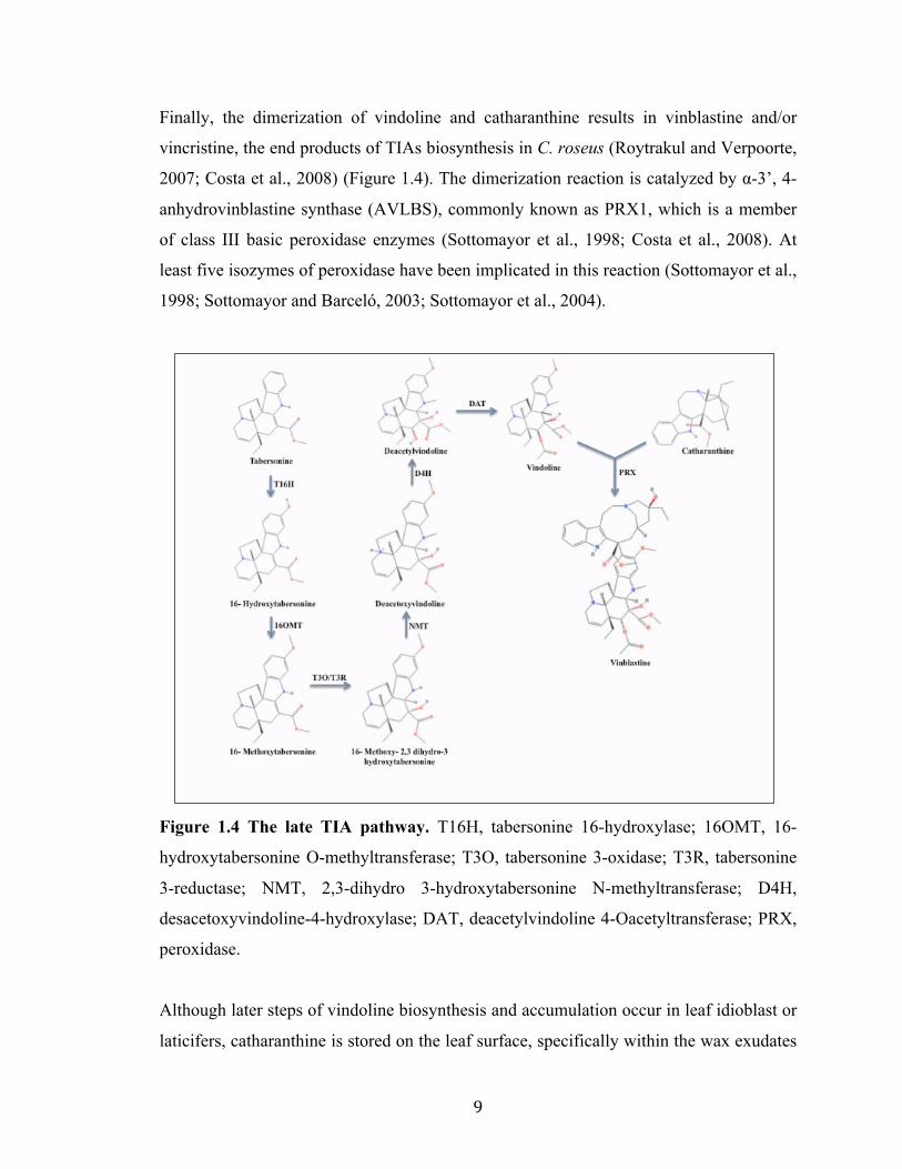
Finally, the dimerization of vindoline and catharanthine results in vinblastine and/or
vincristine, the end products of TIAs biosynthesis in C. roseus (Roytrakul and Verpoorte,
2007; Costa et al., 2008) (Figure 1.4). The dimerization reaction is catalyzed by α-3’, 4-
anhydrovinblastine synthase (AVLBS), commonly known as PRX1, which is a member
of class III basic peroxidase enzymes (Sottomayor et al., 1998; Costa et al., 2008). At
least five isozymes of peroxidase have been implicated in this reaction (Sottomayor et al.,
1998; Sottomayor and Barceló, 2003; Sottomayor et al., 2004).
Figure 1.4 The late TIA pathway. T16H, tabersonine 16-hydroxylase; 16OMT, 16-
hydroxytabersonine O-methyltransferase; T3O, tabersonine 3-oxidase; T3R, tabersonine
3-reductase; NMT, 2,3-dihydro 3-hydroxytabersonine N-methyltransferase; D4H,
desacetoxyvindoline-4-hydroxylase; DAT, deacetylvindoline 4-Oacetyltransferase; PRX,
peroxidase.
Although later steps of vindoline biosynthesis and accumulation occur in leaf idioblast or
laticifers, catharanthine is stored on the leaf surface, specifically within the wax exudates

10
(Roepke et al., 2010). The spatial separation of two monomeric precursors, i.e. vindoline
and catharanthine provides an explanation for low levels of dimeric TIAs in C. roseus. A
TIA transporter, CrTPT2 belonging to the ATP-binding cassette (ABC) family has been
cloned and characterized in C. roseus (Yu and De Luca, 2013). CrTPT2 is preferentially
expressed in leaf epidermal cells and yeast cells expressing CrTPT2 export catharanthine
into the media. In planta suppression of CrTPT2 by VIGS results in a decrease
catharanthine transport to the leaf surface as well as an increase of dimeric TIAs in the
plant (Yu and De Luca, 2013). Though the reasons for the spatial separation of
catharanthine and vindoline in C. roseus leaves is unclear, it has been hypothesized that
leaf damage triggered by herbivory allows the two monomers to come together leading to
the formation of the dimeric TIAs. These dimeric TIAs act as inhibitors against the
herbivores to protect the plants (Roepke et al., 2010).
1.3 TIA biosynthesis in roots
In C. roseus, the TIAs profile in roots is different from that found in aerial parts. Though
both ajmalicine and catharanthine are found in roots, vindoline is only accumulated in the
aerial parts (van der Heijden et al., 2004). In roots, tabersonine can be converted into
lochnericine or 19-hydroxytabersonine catalyzed by P450-dependent tabersonine-6,7-
epoxidase and tabersonine 19-hydroxylase (T19H), respectively (Laflamme et al., 2001;
Rodriguez et al., 2003). Both lochnericine and 19-hydroxytabersonine are proposed as
intermediates in synthesis of 19-O-acetylhörhammericine and the reaction is catalyzed by
minovincinine 19-O-acetyl transferase (MAT) (Giddings et al., 2011). However, the
conversions of tabersonine are less studied in roots compared to leaves.
1.4 Regulation of TIA biosynthesis in C. roseus
The biosynthesis of TIAs is highly complex and under stringent spatio-temporal control
(Thamm et al., 2016). Here, I discussed our current knowledge on the regulation of TIA
biosynthesis, including transcriptional and posttranslational regulation, in C. roseus.
1.4.1 Transcriptional regulation of TIA biosynthesis in C. roseus
During the past decades significant progress has been made in isolating and

11
characterizing genes encoding key biosynthetic enzymes in the TIA pathway (Asada et
al., 2013; Besseau et al., 2013; Qu et al., 2015). However, transcription factors (TFs)
regulating the TIA pathway are relatively less characterized. Currently, TFs regulating
the TIA pathway belong to two major families such as APETALA2/ETHYLENE
RESPONSE FACTORS (AP2/ERFs; ORCA2/3) (Menke et al., 1999b; van der Fits and
Memelink, 2000), and basic helix-loop-helix factors (bHLH; CrMYC2, BIS1/2) (Zhang
et al., 2011; Van Moerkercke et al., 2015; Van Moerkercke et al., 2016). Additionally,
WRKY family TF (CrWRKY1) (Suttipanta et al., 2011), C2H2 zinc fingers TFs
(ZCT1/2/3) (Pauw et al., 2004; Rizvi et al., 2016), the basic leucine zipper (bZIP) factors
(GBF1/2) (Sibéril et al., 2001), and a MYB-like factor (BPF1) (van der Fits et al., 2000),
also regulate TIA biosynthesis.
Previous studies revealed that phytohormone, jasmonic acid (JA) and its methyl
derivatives, called methyl jasmonates (MeJA), are major inducers of biosynthesis of
various natural products, such as artemisinin (Shen et al., 2016), taxol (Mirjalili and
Linden, 1996), and nicotine (Shoji et al., 2000). MeJA and fungal elicitors are also
known to induce expression of the majority of structural and regulatory genes in the TIA
pathway (Menke et al., 1999a). JA-responsive elements (JRE) are often found in the
promoters of TIA pathway genes and regulators including STR (Menke et al., 1999b),
TDC (Ouwerkerk and Memelink, 1999) CPR (Cardoso et al., 1997) and ORCA3 (Zhang
et al., 2011). The JRE of STR promoter contains a core GCC-box elements known to
bound by ORCA2/3 (Menke et al., 1999b). The ORCA3 JRE contains a T/G-box
(AACGTG) and an AT-rich sequence (Vom Endt et al., 2007). Generally, T/G-boxes are
located in numerous promoters of stress- and light- induced genes in plants and are
known binding sites for bHLH and bZIP family TFs.
AP2/ERF transcription factors: The AP2/ERF is a large TF family and is unique to
plants. AP2/ERFs are characterized by the presence of ∼60 conserved amino acids long
DNA binding AP2 domain (Ohme-Takagi and Shinshi, 1995). Based on the number of
AP2 domains and the presence of additional domains, including B3 DNA binding domain,
AP2/ERF family can be classified into five subfamilies, such as AP2, ERF, DREB, RAV,

12
and the fifth (members not classified to the other four groups). Among them the ERF
subfamily constitutes the largest group (Sakuma et al., 2002) and have been involved in
responses to ethylene and different abiotic stresses (van der Fits and Memelink, 2000;
Mizoi et al., 2012). In addition, previous studies have shown that AP2/ERFs are key
regulators of biosynthesis of a number of plant specialized metabolites including nicotine,
artemisinin and steroidal glycoalkaloid (SGA) biosynthesis in tobacco (Nicotiana
tabacum), sweet wormwood/annual wormwood (Artemisia annua L.), tomato (Solanum
lycopersicum) and potato (S. tuberosum), respectively (Shoji et al., 2010; Yu et al., 2012;
Cárdenas et al., 2016; Thagun et al., 2016). The AP2/ERF-domain factors, ORCA1 and
ORCA2 were the first TFs isolated from C. roseus. The JRE element in the STR promoter
was used in a yeast one-hybrid (Y1H) assay to identify these two AP2/ERF TFs (Menke
et al., 1999b). Unlike ORCA1, ORCA2 is a JA-responsive transcriptional activator,
which binds to the JRE of the STR promoter (Menke et al., 1999b). Later, another
AP2/ERF, ORCA3 was identified in C. roseus using transfer DNA (T-DNA) activation
tagging (van der Fits and Memelink, 2000). ORCA3 has been shown to induce the
expression of a number of TIA biosynthetic genes, including ANTHRANILATE
SYNTHASE α (AS α), TDC, DXS, CPR, STR and D4H, but not IS and G10H (van der
Fits and Memelink, 2000; Van Moerkercke et al., 2015). ORCA3-overexpressing cell
suspension cultures accumulate strictosidine, ajmalicine and an unidentified lochnericine-
type TIAs, only when the precursors loganin and tryptamine are added to the medium.
This is possibly due to the fact that most of the iridoid pathway genes are not regulated
by ORCA3 (van der Fits and Memelink, 2000). Recently, the C. roseus genome-sequence
analysis has revealed two more AP2/ERF genes, which are physically clustered with
ORCA3 at the same genomic scaffold (Kellner et al., 2015). But experimental evidence is
lacking on their biological functions.
Basic leucine zipper (bZIP) factors: The bZIPs are a large family of TFs reported in all
eukaryotes. The bZIPs bind DNA as homo- or hetero-dimers and have a characteristic
highly conserved bZIP domain that is ~40- to 80-amino acids long which consists of two
motifs; a basic region that binds DNA, and a leucine-zipper domain which is involved in
protein–protein interaction (Ellenberger et al.; Vinson et al., 1989; Vinson et al., 2006).

13
In plants, bZIP factors are involved in numerous biological processes, including growth
and development, light signaling, pathogen defense and secondary metabolism (Jakoby et
al., 2002; Alves et al., 2013; An et al., 2017). The bZIP TFs are known to bind T/G-box
motifs in the target promoters to regulate their expression. The C. roseus STR and TDC
promoters contain G-box (CACGTG) and T/G-box (AACGTG) elements, respectively
(Menke et al., 1999b). Two bZIP factors, CrGBF1 and CrGBF2 have been identified by
Y1H screening using the G-box of STR promoter as bait (Sibéril et al., 2001). Both GBFs
were shown to bind the G-box in STR promoter. However, they have weak affinity to the
T/G-box in the TDC promoter. Moreover, CrGBFs repressed STR expression in plant
cells suggesting that they act as repressors of TIA biosynthesis genes (Sibéril et al., 2001).
Zinc finger C. roseus transcription factors (ZCTs): Three zinc finger C. roseus
transcription factors (ZCTs) have been identified by a Y1H screening using the elicitor-
responsive DB region of the TDC promoter (Pauw et al., 2004). Sequence analyses
revealed that the ZCTs contain a LxLxL type repressor motif. The ZCTs bind to the RV
fragment of the STR promoter and the DB region of the TDC promoter in vitro in a zinc-
dependent manner. Moreover, ZCTs repressed the expression of TDC and STR in C.
roseus cells (Pauw et al., 2004). The repressor motif in ZCTs is most likely responsible
for the repressor activity of these proteins. The similarities in expression profile and
binding affinity suggests that ZCTs are functionally redundant. Like ORCA3, ZCTs were
also induced by MeJA and yeast elicitor. The binding site for ZCT proteins in the STR
promoter overlaps yet distinct from that of ORCA3. Both ORCA3 and ZCTs bind to the
DB region in the TDC promoter (Pauw et al., 2004). To determine the exact nature of
interaction and/or competition between ZCTs and ORCA3, more detailed studies need to
be performed.
AT-hook proteins: The bipartite JRE in the ORCA3 promoter comprises an AT-rich
quantitative sequence responsible for high-level expression, and a qualitative sequence,
T/G-box (AACGTG), that acts as an on/off switch (Vom Endt et al., 2007). Five AT-
hook proteins have been isolated in C. roseus using the JRE of the ORCA3 promoter in a
Y1H screening. The AT-hook TFs bound to the AT-rich quantitative sequence in the

14
ORCA3 promoter to activate its expression. These observations suggested that AT-hook
proteins co-regulate the expression of ORCA3 along with other regulators and act as
transcriptional enhancers (Vom Endt et al., 2007). The JRE of STR promoter contains a
GCC-box like element that is bound by ORCA3; however, a GCC-box is not present in
the ORCA3 JRE, suggesting that ORCA3 is not regulated by an AP2/ERF TFs (Vom
Endt et al., 2007).
bHLH transcription factors: The bHLH factors, one of the largest families of TFs in
plants, are involved in numerous developmental and metabolic processes, including
pathogen defense, plant growth and development, phytohormone signaling and
biosynthesis of specialized metabolites in plants (MacAlister and Bergmann, 2011; Patra
et al., 2013b; Song et al., 2013; Li and Sack, 2014; Zhou and Memelink, 2016). The
bHLH domain is comprising of ~60 amino acids, with the N-terminal ~15–20 basic
amino acids involved in DNA binding and the HLH region which forms the homo- or
heterodimeric complexes (Atchley and Fitch, 1997). The bHLH TFs characteristically
bind to the E-box sequences (CANNTG), including the G-box (CACGTG) in the
promoter of their target genes with some variation in binding specificity (Carretero-
Paulet et al., 2010; Fernández-Calvo et al., 2011). The bHLH TFs are divided into 12
sub-groups (Heim et al., 2003) and the bHLH TF CrMYC2, belongs sub-group IIIe, was
isolated by Y1H screen using the G-box of the STR promoter in C. roseus (Zhang et al.,
2011). CrMYC2 acts upstream of ORCA3 and regulates its expression by directly
binding to the T/G-box motif in the JRE of the ORCA3 promoter (Zhang et al., 2011).
Similar T/G-box motif was also found in the TDC promoter; however, whether CrMYC2
co-regulates TDC along with ORCA3 remains to be tested. RNAi-mediated suppression
of CrMYC2 in C. roseus cell cultures repressed JA-dependent induction of ORCA2/3,
suggesting CrMYC2 activates ORCA3 expression directly in response to JAs. In addition,
RNAi-mediated suppression of CrMYC2 reduced alkaloid accumulation in C. roseus cells
(Zhang et al., 2011).
While considered to be a master regulator, ORCA3 alone is not sufficient to activate the
entire TIA pathway. ORCA2 and ORCA3 probably regulate TIA biosynthesis in

15
combination with additional TFs (van der Fits and Memelink, 2000). This hypothesis is
validated by the recently identified and characterized sub-group IVa bHLH TFs, bHLH
IRIDOID SYNTHESIS 1 (BIS1) and BIS2 (Van Moerkercke et al., 2015; Van
Moerkercke et al., 2016). These two TFs act in a complementary manner with ORCA3 to
regulate the iridoid pathway genes. The expressions of both BIS1/2 are induced by MeJA,
and are specifically expressed in IPAP cells where iridoid biosynthesis takes place (Van
Moerkercke et al., 2015; Van Moerkercke et al., 2016). Moreover, the over-expression of
BIS1 induced MIA production in C. roseus cell cultures without precursor feeding or JA
induction (Van Moerkercke et al., 2015)
Box P-binding factors (BPF-1): In the STR promoter a region present upstream of the
G-box and the JRE, known as the BA region, is responsive to yeast elicitor but not to
MeJA (van der Fits et al., 2000). Y1H screening using this region identified the parsley
box P-binding factor (BPF-1) that contains a MYB-like domain. It has been demonstrated
that BPF-1 binds specifically to the BA fragment of STR promoter at two different sites
(van der Fits et al., 2000). The expression of BPF-1 is induced by yeast elicitor but not by
MeJA (van der Fits et al., 2000). These findings suggests that TIA biosynthesis involves
a JA-dependent pathway regulated by ORCAs/bHLHs and a JA-independent pathway via
BPF-1 (Thamm et al., 2016).
WRKY transcription factors: WRKY TFs are primarily plant-specific and known to
bind the W-boxes (TTGACC/T) elements in target promoters (Yang et al., 2013). WRKY
TFs have emerged as key regulators of specialized metabolite biosynthesis in a number of
plant species (Schluttenhofer and Yuan, 2015), including benzylisoquinoline alkaloids in
Coptis japonica (Kato et al., 2007), artemisinin in Artemisia annua (Ma et al., 2009),
triterpenoid withanolide in Withania somnifera (Singh et al., 2017) and camalexin in
Arabidopsis (Arabidopsis thaliana) (Ma et al., 2009). W-box elements are common in
TIA biosynthetic gene promoters, suggesting the potential role of WRKY proteins in the
regulation of TIA biosynthesis (Suttipanta et al., 2011). A group III WRKY TF,
CrWRKY1 was identified and functionally characterized in C. roseus (Suttipanta et al.,
2011). Ectopic expression of CrWRKY1 induced the accumulation of serpentine in hairy

16
roots. CrWRKY1 expression was mostly observed in roots, less in fruits and mature
leaves and was induced by MeJA. In addition, CrWRKY1 bound to W-box in the TDC
promoter and regulate its expression. As CrWRKY1 is not an activator it needs other co-
activators to induce expression of TDC in vivo (Suttipanta et al., 2011). Analysis of the
CrWRKY1 promoter revealed that it contains several E-boxes, MYB recognition motifs
and JA-responsive as-1 motifs (TGACG); however, W-box motifs are not found in the
CrWRKY1 promoter. These findings suggest that TFs belonging to MYB, bHLH and
TGA families are potentially involved in the regulation of CrWRKY1 (Yang et al., 2013).
Additional MeJA-responsive WRKYs have also been implicated in regulation of TIA
biosynthesis (Schluttenhofer et al., 2014).
1.4.2 Post-translational regulation of TIA biosynthesis in C. roseus
Accumulating evidence suggest that besides transcriptional regulation, metabolic
pathways are also regulated by posttranscriptional and post-translational mechanisms
(Maier et al., 2013; Patra et al., 2013b; Shen et al., 2017). Post-translational regulatory
mechanisms, including phosphorylation of TFs, have been studied in nicotine and
camalexin biosynthesis in tobacco and Arabidopsis, respectively (Xu et al., 2008; De
Boer et al., 2011; Mao et al., 2011) and protein kinases (PKs) involved in these processes
have been identified. In plants, the PK gene family is considered to be one of the largest
and most highly conserved gene families. PKs phosphorylate proteins leading to the
functional changes and are involved in approximately all biological processes, including
plant growth, development, and biotic and abiotic stress responses. The most common
protein kinase-mediated phosphorylation involves the mitogen-activated protein kinase
(MAPK) cascade, normally consisting of three sequentially activated kinases, MAPK
kinase kinase (MAPKKK)-MAPK kinase (MAPKK)-MAPK, encoded by multiple genes
(Smékalová et al., 2014). In MAPK cascade, MAPKs phosphorylate specific substrates,
including TFs and enzymes leading to the specific cellular responses.
During the past few years genome sequencing of different plant species has made it
possible to identify and characterize members of the MAPK cascades in a number of
plants. To date, MAPK cascade components have been reported from a wide range of

17
plant species, including Arabidopsis (Ichimura et al., 2002), rice (Hamel et al., 2006; Rao
et al., 2009), maize (Kong et al., 2013b; Liu et al., 2013), tomato (Kong et al., 2012; Wu
et al., 2014), Brachypodium distachyon (Chen et al., 2012), Capsicum annuum (Liu et al.,
2014), and cucumber (Wang et al., 2015). In C. roseus, JA-induced expression of TIA
pathway gene transcripts are reduced significantly in the presence of protein kinase
inhibitors but are not inhibited by the protein synthesis inhibitor, cycloheximide. This
observation indicates the possible role of protein phosphorylation in the JA signal
transduction (Menke et al., 1999a). Moreover, an undefined posttranslational
modification of ORCA3 protein is required for its transcriptional activity, and
phosphorylation by a protein kinase has been hypothesized in previous reports (Menke et
al., 1999a; van der Fits and Memelink, 2000). In C. roseus a JA-inducible MAPK
CrMAPK3, has been reported previously and proposed to play a role in abiotic stress
signal transduction and regulation of TIA biosynthesis. Transient over-expression of
CrMAPK3 showed increased expression of TIA biosynthesis pathway genes (STR, TDC,
D4H and DAT) and accumulation of TIAs in C. roseus leaf tissue. (Raina et al., 2012).
1.5 Metabolic engineering by assembling different branches of TIA pathway
Recently, significant progress has been made in the molecular and biochemical
characterization of genes encoding key enzymes in the iridoid branch of TIA pathway,
including 10HGO, IS, 7DLS, DLGT, and DL7H (Geu-Flores et al., 2012; Asada et al.,
2013; Besseau et al., 2013; Salim et al., 2013; Miettinen et al., 2014; Salim et al., 2014)
which are required for sequential conversion of geraniol to secologanin. Importantly,
strictosidine was successfully produced de novo in a yeast strain by reconstruction of 14
known TIA pathway genes. The engineered yeast strain accumulates greater than 0.5
mg/L strictosidine (Brown et al., 2015). This approach provides a basis for better
engineering the production of strictosidine and serves as a platform for producing any
number of complex TIA derivatives. Likewise, the complete pathway from IPP to
strictosidine has also been successfully reconstructed in Nicotiana benthamiana by agro-
infiltration (Miettinen et al., 2014). The molecular and biochemical characterization of
T3O and T3R concluded the 7-step tabersonine to vindoline pathway (Qu et al., 2015).
The reconstruction of this pathway in yeast led to the accumulation of vindorosine and

18
vindoline from tabersonine as well as of vindoline from 16-methoxytabersonine has also
been demonstrated (Qu et al., 2015). Therefore, both yeast strains and the transient
expression in N. benthamiana, can be further developed and optimized for targeted
metabolic engineering. These two biotechnological advances lay the foundation for
producing complex TIAs in heterologous systems in future. These approaches could be
used for the synthesis of anticancer drugs vinblastine and additional medicinal TIAs.
1.6 Conclusion
In recent years, an advance in numerous next-generation sequencing (NGS) techniques,
such as RNA-seq has enabled the scientific community to generate large-scale
transcriptome data from a wide range of plant tissues. These transcriptomic resources
have been used to identify genes for the missing steps in complex metabolic pathways,
including iridoid biosynthesis and the conversion of tabersonine to vindoline in C. roseus.
Successful reconstructions of the strictosidine biosynthesis pathway in both yeast and N.
benthamiana, and the 7-step conversion of tabersonine to vindoline in yeast strain, are the
major breakthroughs in TIA pathway. These findings may help in large-scale production
of valuable TIAs. While remarkable progress had been made in the last few years in
isolating and characterizing genes encoding key enzymes in TIA pathway, the regulation
of TIA biosynthesis is relatively less understood. This is probably due to the lack of
appropriate genetic tools and highly complex nature of the biochemical pathway.
Understanding the molecular mechanism of TIA regulation at both transcriptional and
posttranslational level will enhance our ability to engineer the biochemical pathway to
over-produce these valuable compounds in plants.

19
1.7 Outline of the dissertation
Catharanthus roseus (Madagascar periwinkle) produces numerous bioactive TIAs,
including the chemotherapeutics, vinblastine and vincristine. However, the transcriptional
and posttranslational regulation of TIA biosynthesis is not fully understood.
Therefore, the major objectives of my research are
Ø to isolate and characterize TFs regulating the TIA pathway,
Ø to elucidate the molecular mechanism underlying TIA regulation,
Ø to identify all the C. roseus MAP kinases (MAP Kinome), and
Ø to isolate and characterize candidate MAPKs involved in TIA regulation.
Chapter two focuses on functional characterization of an AP2/ERF TF gene cluster that
modulates TIA biosynthesis in C. roseus. ORCA3, a JA responsive AP2/ERF TF, and its
regulator, CrMYC2, a bHLH TF play key roles in TIA biosynthesis. ORCA3 forms a
physical cluster with two functionally uncharacterized AP2/ERFs, ORCA4 and ORCA5.
My research revealed that (i) the ORCA gene cluster is differentially regulated and (ii)
ORCA4 and ORCA5 while functionally overlapping with ORCA3, regulates an
additional set of TIA genes. Unlike ORCA3, ORCA4 or ORCA5 overexpression resulted
in significant increase of TIA accumulation in C. roseus hairy roots. (iii) ORCA5 directly
regulates the expression of ORCA4 and indirectly regulates ORCA3, likely via an
unknown factor(s). Moreover, ORCA5 shows auto-regulation. (iv) ORCA5 also activates
the expression of ZCT3, a negative regulator of the TIA pathway. In addition, (v)
CrMYC2 is capable of activating ORCA3 as well as co-regulating TIA pathway genes
concurrently with ORCA3.
Part of the content of this chapter is already published
Paul, P., Singh, S. K., Patra, B., Sui, X., Pattanaik, S., & Yuan, L. (2017). A differentially regulated AP2/ERF transcription factor gene cluster acts downstream of a MAP kinase cascade to modulate terpenoid indole alkaloid biosynthesis in Catharanthus roseus. New Phytologist 213:1107-1123.

20
Chapter three My research revealed that the ORCA gene cluster and CrMYC2 act
downstream of a MAP kinase cascade that comprises a MAPK kinase, CrMAPKK1,
CrMAPK3 and CrMAPK6. Here I describe the identification and characterization of
CrMAPKK1 that is involved in TIA pathway regulation. Overexpression of CrMAPKK1
in C. roseus hairy roots up regulated TIA pathways genes and boosts TIA accumulation.
The content of this chapter is already published
Paul, P., Singh, S. K., Patra, B., Sui, X., Pattanaik, S., & Yuan, L. (2017). A differentially regulated AP2/ERF transcription factor gene cluster acts downstream of a MAP kinase cascade to modulate terpenoid indole alkaloid biosynthesis in Catharanthus roseus. New Phytologist 213:1107-1123.
Chapter four describes the analysis of C. roseus MAP kinome as well as isolation and
characterization of two previously uncharacterized MAPKs, MAPK20 and MAPK13 that
regulate the TIA biosynthesis in C. roseus. Overexpression of CrMAPK13 in C. roseus
hairy roots upregulated the BIS-regulated iridoid pathway genes and induced tabersonine
accumulation. Overexpression of CrMAPK20 in C. roseus hairy roots repressed the
MYC2-ORCA regulated TIA pathway genes and reduced catharanthine accumulation.
Chapter five summarizes the overall findings of this research project and possible future
directions of TIA pathway regulation.
Copyright © Priyanka Paul 2017
21
Chapter 2
A differentially regulated AP2/ERF transcription factor gene cluster modulate
terpenoid indole alkaloid biosynthesis in Catharanthus roseus
2.1 Introduction
Terpenoid indole alkaloids (TIAs) are a group of diverse plant natural products.
Catharanthus roseus (Madagascar periwinkle) produces over a hundred TIAs, including
the anticancer drugs, vinblastine and vincristine (Kellner et al., 2015). TIAs are
condensed products of tryptamine, derived from the shikimate pathway, and secologanin,
from the terpenoid pathway (Figure 1.1). In recent years, significant progress has been
made in isolating and characterizing genes encoding key biosynthetic enzymes in the TIA
pathway (Asada et al., 2013; Besseau et al., 2013; Qu et al., 2015). By comparison,
transcription factors (TFs) regulating the TIA pathway are less characterized. TFs
belonging to the families of AP2/ERFs (Menke et al., 1999b; van der Fits and Memelink,
2000), bHLH (Zhang et al., 2011; Van Moerkercke et al., 2015; Van Moerkercke et al.,
2016), WRKY(Suttipanta et al., 2011), and C2H2 zinc fingers (Pauw et al., 2004; Rizvi et
al., 2016), have been shown to be regulators of the TIA pathway. In addition, the bZIP
factors, GBF1 and GBF2 (Sibéril et al., 2001), and a MYB-like factor, BPF1(van der Fits
et al., 2000), also influence TIA biosynthesis.
Jasmonic acid (JA) and its methyl derivatives, methyl jasmonates (MeJA), are major
inducers of biosynthesis of many natural products, including taxol (Mirjalili and Linden,
1996), artemisinin (Shen et al., 2016), and nicotine (Shoji et al., 2000). JA and fungal
elicitors also induce expression of the majority of regulatory and structural genes in the
TIA pathway (Menke et al., 1999b). JA responsive elements (JRE) are frequently found
in the promoters of TIA pathway genes.
In C. roseus, among the first isolated TFs are AP2/ERF-domain type factors, ORCA1 and
ORCA2 (Menke et al., 1999b). ORCA2 is a JA-responsive transcriptional activator that

22
binds to JRE of the STR promoter (Menke et al., 1999b). Later, another AP2/ERF,
ORCA3 was identified using T-DNA activation tagging and shown to induce the
expression of numerous TIA biosynthetic genes in the indole and vindoline branches,
including ASα, TDC, DXS, CPR, STR, and D4H, but not those in the iridoid branch, such
as IS and G10H (van der Fits and Memelink, 2000; Van Moerkercke et al., 2015).
Although considered to be a master regulator, ORCA3 alone is insufficient to activate the
complete TIA pathway. ORCA2 and ORCA3 likely regulate TIA biosynthesis
combinatorically with other TFs (van der Fits and Memelink, 2000). This assumption is
supported by the recent identification and characterization of two bHLH TFs, BIS1 and
BIS2, which acts in a complementary manner with ORCA3 to regulate structural genes in
the iridoid branch (Van Moerkercke et al., 2015; Van Moerkercke et al., 2016). The
recently sequenced C. roseus genome has revealed that two AP2/ERF genes, CroERF90
and CroERF92, are physically clustered with ORCA3 at the same genomic scaffold, and
thus proposed to be involved in regulation of the TIA pathway in concert with ORCA3
(Kellner et al., 2015). However, experimental evidence is lacking on their biological
functions.
Gene clusters have been identified in a number of plant species including maize (Frey et
al., 1997), rice (Shimura et al., 2007), oat (Qi et al., 2004), Arabidopsis (Field et al.,
2011), opium poppy (Winzer et al., 2012), and tomato (Itkin et al., 2013). Most of the
plant gene clusters identified thus far comprise non-homologous structural genes
encoding enzymes involved in specialized metabolite biosynthesis (Nutzmann et al.,
2016). Unlike the biosynthetic gene cluster, the clustered TFs consist of homologous
genes that have most likely arisen due to tandem duplication events. To date, physically
clustered genes encoding TFs have been identified in a limited number of plant species,
including Arabidopsis, tobacco tomato and potato (Duarte et al., 2006; Shoji et al., 2010;
Cárdenas et al., 2016). The NIC2 locus of Nicotiana tabacum, which comprises at least
seven AP2/ERFs are close homologs of ORCAs in C. roseus. The NIC2 locus ERFs
regulates the expression of structural genes in nicotine biosynthetic pathway and nicotine
accumulation in tobacco. The nic2 mutant has a low nicotine phenotype (Shoji et al.,
2010). Overexpression of the NIC2 locus ERFs, e.g. ERF189 and ERF221/ORC1

23
significantly induced nicotine biosynthesis in tobacco (Shoji et al., 2010; De Boer et al.,
2011). Recently, an AP2/ERF, GLYCOALKALOID METABOLISM 9 (GAME9) has
been characterized from tomato and potato for its role in steroidal glycoalkaloids (SGAs)
and upstream isoprenoid biosynthesis. Genome sequence analysis revealed that GAME9
is a member of an AP2/ERF TF gene cluster in both tomato and potato. GAME9
knockdown and overexpression in tomato and potato affects the expression of SGAs and
upstream mevalonate pathway genes, such as the cholesterol biosynthesis gene STEROL
SIDE CHAIN REDUCTASE 2 (SSR2) (Cárdenas et al., 2016). However, the regulatory
processes that govern the functions of TF clusters are unexplored.
The bHLH factor, CrMYC2, activates ORCA3, which in turn induces the expression of
several TIA biosynthetic genes (Zhang et al., 2011). In tobacco, MYC2 regulates the
AP2/ERFs within the NIC2 locus, and also co-regulates several nicotine pathway genes
with the AP2/ERFs by direct binding to the pathway gene promoters (De Boer et al.,
2011; Shoji and Hashimoto, 2011). Whether CrMYC2 also co-regulates the same TIA
pathway genes with ORCA3 remains to be investigated. Such possible dual regulation of
pathway genes underlines a layered, complex regulatory network for fine-tuning gene
expression.
To date, our knowledge on the transcriptional regulation of clustered TFs is limited. In
this study, we cloned and characterized ORCA4 (CroERF90) and ORCA5 (CroERF92),
the two members of the ORCA gene cluster. We provide experimental evidence that the
ORCA gene cluster is differentially regulated. ORCA4- but not ORCA3- is directly
regulated by ORCA5. ORCA5 exhibits auto-regulation. Additionally, ORCA5 directly
activates the expression of a negative regulator, ZCT3. While ORCA4 and ORCA5
functionally overlap with ORCA3, they modulate an additional set of TIA genes.
Interestingly, unlike ORCA3 and 4, ORCA5 significantly activates other TIA pathway
genes, such as LAMT and SLS. ORCA4 or ORCA5 overexpression resulted in dramatic
increase of TIA accumulation in C. roseus hairy roots. In addition, CrMYC2 activates
ORCA3 and co-regulates the TDC and CPR promoters concomitantly with ORCA3.

24
2.2 Materials and Methods
2.2.1 Plant materials
Catharanthus roseus (L.) G. Don var. ‘Little Bright Eye’ was used for gene expression
and cloning, and generation of transgenic hairy roots. Nicotiana tabacum var. Xanthi cell
line was used for protoplast-based transient expression assays.
2.2.2 RNA isolation and cDNA synthesis
Ten-day-old C. roseus seedlings treated with 100 µM methyl jasmonate (MeJA) for 2 h
were used for total RNA isolation and cDNA synthesis as described previously
(Suttipanta et al., 2007). ORCA4 and ORCA5 were PCR amplified from cDNA using
gene-specific primers (Table 2.1), cloned into the pGEM-T Easy vector (Promega), and
sequenced.
2.2.3 Multiple sequence alignment, phylogenetic and co-expression analysis
Alignment of deduced amino acid sequences was performed using the ClustalW program
(Larkin et al., 2007) with the default parameters. Phylogenetic trees were constructed and
visualized using the neighbor-joining method through MEGA version 5.2 software
(Tamura et al., 2011). The statistical reliability of individual nodes of the newly
constructed tree was assessed by bootstrap analyses with 1,000 replications.
In order to analyze the expression patterns of C. roseus TIA pathway genes and TFs,
transcriptome data for five different tissues (flower, mature leaf, immature leaf, stem and
root) were obtained from the Sequence Read Archive (SRA) database at the National
Center for Biotechnology Information (accession number SRA030483). Raw reads were
processed and reads per kilobase of transcript per million mapped reads (RPKM) value
was calculated as described before (Singh et al., 2015). Pairwise Pearson correlation
coefficient for each transcript was calculated using RPKM. Matrix distances for
expression heatmap were computed over Pearson correlations of gene expression values
(RPKM) by HEATMAP.2 function of GGPLOT (v.3.0.1) Bioconductor package in R
(v3.2.2) (Wickham, 2009).

25
2.2.4 Quantitative RT-PCR
Quantitative real-time PCR (qRT-PCR) was performed as described previously
(Suttipanta et al., 2011). The primers used in qRT-PCR are listed in Table 2.1. In
addition to the C. roseus 40S Ribosomal Protein S9 (RPS9) gene, Elongation Factor 1∞
(EF1∞) was used as a second internal control (Liscombe et al., 2010). All PCRs were
performed in triplicate and repeated at least twice.
Table 2.1 Oligonucleotides used in this study
RPS9 5’-GAGGGCCAAAACAAACTTA-3’ 5’-CCCTTATGTGCCTTTGCCTA-3’ EF1œ 5’-TACTGTCCCTGTTGGTCGTG-3’ 5’-AAGAGCTTCGTGGTGCATCT-3’ ASa GCGAACATTTGCAGATCCAT GGCCGATTTGTTATTGTTCC TDC 5’-ATCCGATCAAACCCATACCA-3’ 5’-CGTCATCCTCGACCATTTTT-3’ G10H 5’-TTATTCGGATTCTGCCAAGG-3’ 5’-TCCCCAAAGTGAATCGTCAT-3’ CPR 5’-TGGCAGAAAAGGCTTCTGAT-3’ 5’-CTCAGCCTGTGTGCTATCCA-3’ SLS 5’-GTTCCTTCTCACCGGAGTTG-3’ 5’-CCCATTTGGTCAACATGTCA-3’ STR 5’-ACCATTGTGTGGGAGGACAT-3’ 5’-ATTTGAATGGCACTCCTTGC-3’ LAMT 5’- AGCCAAGGCAGTGATTGTTG-3’ 5’-CTGCAATGCGGAAAGGTTTG-3’ IS 5’-CCACATGATTCGCCTTTTACCG-3’ 5’- AAACCCGAAAACCAGAGCTG-3’
ORCA3 5’-CGGGATCCGAAATACAGAAA-3’ 5’-GCCCTTATACCGGTTCCAAT-3’ ORCA4 5’-ATAGTAGTACTGCCGCCGAAAG-3’ 5’-ATCTCCGCCGCAAATTTTCC-3’ ORCA5 5’-TCTTCCAACGGAGGTTAACGG-3’ 5’-AATGTTGTCTCCAGGGCTTG-3’ ZCT1 5’-AGCCGAAAACTCATGCTTGT-3’ 5’-CGCCTTTGCAACAGGTTTAT-3’ ZCT2 5’-CGTCAATTTCCATCGTTTCA-3’ 5’-CCGATAGCGAATTCAAGTCC-3’ ZCT3 5’-GACAAGCTTTGGGAGGACAC-3’ 5’-GGCAAGGCAGGTAAGTTCAA-3’ ORCA4 5’-gcgagatct
ATGCTTGATGAAATTACTATTT-3’ 5’-atggagctc CATCTTATTCACCTTTTGATTA-3’
ORCA5 5’-gcggatcc ATGGAGTTTTCAGCTACTAATT-3’
5’-atgctgcag TTACAATATTGTCTCCTGTTTCA-3’
ZCT3 5’- gcggatcc ATGGCACTTGAAGCTTTGAATTC-3’
5’- arggatcc CTAATTAATTTGATGGTTTTCAAC-3’
ORCA3- Promoter
5’- gcgaattc CGGAGAGTA AAAACTATACTAAATG-3’
5’- atgaagctt AGAAACTGATT TTTTATGAAGTTTT-3’
ORCA4- Promoter
5’-gcgaattc CTCGTACACTATATTTATGTTATG-3’
5’-agaagctt GCTTTTCCTTAGAGAGATTGATTG-3’
ORCA5- Promoter
5’- gcgaattc TCGGATTGTCTATAACCCTTTGTG-3’
5’-agaagctt ATCAAGA CGGGAAAGATCAGATTC-3’
ZCT3- Promoter
5’- gcgaattc ATGAGTGCGAACTTGATAGTCG-3’
5’-atgaagctt AGCAGAAGAATTTGAAGATTGAAT-3’
STR-Probe
5’-ACATCACTCTTAGACCGCCTTCT T TGAAAGTGATTTCCCTTGGACCTT-3’
5’-AAGGTCCAAGGGAAATCACTTTCA AAGAAGGCGGTCTAAGAGTGATGT-3’

26
Table 2.1 Oligonucleotides used in this study (continued.)
ORCA4- Probe
5’-CCTTCATAGCCCGCCCAATTGG TAA ACGTGCCACCAACCTCC-3’
5’-GGAGGTTGGTGGCACGT TTA CCAATTGGGCGGGCTATGAAGG-3’
rolB 5’CTTATGACAAACTCATAGATAAAAGGTT-3’
5’- TCGTAACTATCCAACTCACA TCAC-3’
rolC 5’-CAACCTGTTTCCTACTTTGTTAAC-3’
5’- AAACAAGTGACACACTCAGCTTC-3’
virC 5’-TTTTGCTCCTTCAAGGGAGGTGCC-3’
5’-GGCTTCGCCAACCAATTTG GAGAT-3’
GUS 5’-ATGGTAGATCTGAGGGTAAA TTTC-3’
5’- GCTAGCTTGTTTGCCTCCCTG-3’
ntpII 5’-ATGGGGATTGAACAAGATGGA-3’ 5’-TCAGAAGAACTCGTCAAGAAG -3’
2.2.5 Sub-cellular localization
For sub-cellular localization, the full-length cDNA of ORCA4 or 5 was fused to the N-
terminus of the enhanced GFP (eGFP) in a pBS plasmid under CaMV3S promoter and
rbcS terminator to generate pORCA4/5-eGFP. A pBlueScript (pBS) plasmid containing
the only eGFP was used as a control. The plasmids containing either eGFP or ORCA4/5-
eGFP were individually electroporated into tobacco protoplasts as described previously
(Pattanaik et al., 2010a) and visualized after 20h incubation in the dark under a
fluorescent microscope (Eclipse TE200, Nikon, Japan) (Pattanaik et al., 2010b).
2.2.6 Protoplast isolation and electroporation
For transient protoplast assays, the reporter plasmids were generated by cloning STR,
TDC, CPR, LAMT, SLS, ZCT3, ORCA3, ORCA4 or ORCA5 promoters in a modified pUC
vector containing a firefly luciferase (LUC) and rbcS terminator. The effector plasmids
were constructed by cloning ORCA3/4/5, and CrMYC2 into a modified pBS vector under
the control of the CaMV35S promoter and rbcS teminator. For transactivation assay,
ORCA3, ORCA4 and ORCA5 were fused to the GAL4 DNA binding domain (GAL4-
DBD) in a pBS plasmid containing mirabilis mosaic virus (MMV) promoter and rbcS
terminator. The reporter plasmid used in the assay contains firefly LUC driven by a
minimal CaMV35S promoter with five tandem repeats of GAL4 Response Elements (5X
GALRE), and rbcS terminator fused. Protoplast isolation from N. tabacum cell suspension
cultures and electroporation with plasmid DNA were performed as described previously
(Pattanaik et al., 2010b). The reporter, effector, and internal control plasmids were

27
activities in transfected protoplasts were measured as described previously (Suttipanta et
al., 2007).
2.2.7 Recombinant protein production and EMSA
The ORCA3, 4 and 5 genes were cloned into the pGEX 4T-1 vector (GE Healthcare
Biosciences, Pittsburgh, PA, USA) to generate GST-fusion proteins. The constructs were
verified by DNA sequencing and transformed into Escherichia coli BL21 cells containing
pRIL (Agilent, Santa Clara, CA, USA). Protein expression was induced by adding
isopropyl ß-D-thiogalactopyranoside (IPTG) to a final concentration of 0.1mM to the cell
cultures at A600 ~ 0.8 and induced for 2 h at 37°C. The cells were harvested by
centrifugation and lysed using CelLytic B (Sigma, USA) according to the manufacturer’s
instructions. The GST fusion proteins were bound to Glutathione Sepharose 4B columns
(Amersham) and eluted by using 10mM reduced glutathione in 50mM Tris–HCl (pH 8.0)
buffer. For EMSA, biotin-labeled DNA probes were synthesized by Integrated DNA
Technologies (IDT) and annealed to produce double-stranded probes. Complementary
DNA probes were designed to include the jasmonate-responsive elements (JRE) of STR
promoter (GenBank accession no. Y10182) (Menke et al., 1999a) and putative GCC-
element of the ORCA4 promoter. EMSA experiment was carried out using light shift
chemiluminescent EMSA kit (Thermo Fisher Scientific). For the binding reactions 40
fmole of DS DNA was incubated with purified protein for 60 mins at room temperature.
The protein-DNA binding specificity was further confirmed by performing competition
experiment, where 1000 fold excess amount of cold probe (without Biotin label) was
added to the binding reactions. The DNA-protein complexes were resolved by
electrophoresis on 6% non-denaturing polyacrylamide gels and then transferred to
BiodyneB modified membrane (0.45 mm; Pierce). The band shifts were detected by a
chemiluminescent nucleic acid detection module (Pierce) and exposed to x-ray films.
2.2.8 Construction of plant expression vectors and generation of hairy roots
For plant transformation ORCA4, ORCA5 and CrMYC2 cDNAs were PCR-amplified and
cloned into a modified pCAMBIA2301 vector containing the CaMV35S promoter and the
rbcS terminator (Pattanaik et al., 2010b). The pCAMBIA2301 vector was used as an

28
empty vector (EV) control. The plasmids were mobilized into Agrobacterium rhizogenes
R1000. Transformation of C. roseus seedlings and generation of hairy roots were
performed using the protocol described previously (Suttipanta et al., 2011). Transgenic
status of the hairy root lines was verified by PCR amplification of rolB, rolC, virC, nptII
and GUS genes. Independent hairy root lines were selected for further analysis.
2.2.9 Alkaloid extraction and analysis
To extract alkaloids, transgenic hairy roots were frozen in liquid nitrogen and ground to
powder. Samples were extracted in methanol (1:100 w/v) twice for 24 h on a shaker.
Pooled extracts were then dried via a rotary evaporator and diluted in methanol 10 µL/mg
of the initial sample. The samples were then analyzed using high-performance liquid
chromatography (HPLC), followed by electrospray-injection in a tandem mass
spectrometry, as previously described (Suttipanta et al., 2011). The known alkaloid
standards were run to identify elution times and mass fragments.
2.3 Results and Discussion
2.3.1 Two AP2/ERF genes form a cluster with ORCA3
AP2/ERFs have emerged as one of the major regulators of specialized metabolite
biosynthesis in plants (Shoji et al., 2010; Yu et al., 2012). We identified 109 AP2/ERFs
in C. roseus that were divided into 12 sub-groups by phylogenetic analysis. To conduct a
cross-species comparison, a phylogenetic tree was constructed using the 109 AP2/ERF
TFs from C. roseus and 122 from Arabidopsis (Figure 2.1a). Twelve main clades were
formed, corresponding to subgroups I to XII of Arabidopsis. Two AP2/ERFs, CroERF90
and CroERF92, were of particular interest as they were homologous to ORCA3 in sub-
group IX. The sub-group IX genes are involved in the regulation of defense responses
and natural product biosynthesis facilitated by MeJA in numerous plant species,
including Arabidopsis (McGrath et al., 2005), tobacco (Shoji et al., 2010), and C. roseus
(van der Fits and Memelink, 2000). CroERF90 and CroERF92 have been recently
identified as AP2 TF1 (030272) and AP2 TF2 (030274), respectively (Kellner et al.,
2015). However, the two TFs have not been characterized and their biological functions
remained unverified. Genome sequence analysis revealed that CroERF90 (030272) and

29
CroERF92 (030274) are present adjacent to ORCA3 (030273) in a 40 kb genomic
scaffold, and the three physically clustered TFs do not contain any introns, suggesting
that they may have arisen from tandem genome duplication events. Approximately 15%
of the identified genes in all sequenced plant genomes contain significant numbers of
tandemly arrayed gene families (Jander and Barth, 2007; Huang et al., 2009). Generally,
tandem arrays contain two duplicated genes, and arrays with more than three genes are
uncommon (Rizzon et al., 2006). TF families have higher expansion rates in plants than
in animals (Shiu et al., 2005); however, the Arabidopsis genome contains only one region
with more than five duplicated TF genes in tandem, although several such tandemly
duplicated regions are present in soybean (Schmutz et al., 2010). The biological
significance of such TF gene clusters has not been fully elucidated. The TF cluster,
identified as the NIC2 locus in tobacco, consists of seven homologous AP2/ERFs
involved in the regulation of nicotine biosynthesis (Shoji et al., 2010). We cloned
CroERF90 (designated as ORCA4; GenBank accession no. KR703577) and CroERF92
(ORCA5; GenBank accession no. KR703578). ORCA4 is a 223 amino acid protein,
having a calculated molecular mass of 24.4 kDa. The 311 amino acid ORCA5 has a
calculated molecular mass of 34.2 kDa. ORCA4 and ORCA5 share 53% amino acid
sequence identity, and 38% and 47% sequence identity, respectively, with ORCA3
(Figure 2.1b). The highest sequence identity was found within the conserved DNA
binding AP2 domain.

30

31
Figure 2.1 Phylogenetic analysis of AP2/ERF families and Amino acid sequence
alignment of ORCA3, 4, and 5. a. Arabidopsis thaliana and Catharantus roseus
AP2/ERFs were used to generate the phylogenetic tree. b. Multiple sequence alignment
of ORCA3, ORCA4 and ORCA5 of C. roseus. Identical amino acid residues in the
alignment are shaded in black and similar amino acid residues in gray. The AP2 DNA
binding domain is underlined.
2.3.2 ORCA3, ORCA4, and ORCA5 are preferentially induced in roots by MeJA
The expression of ORCA3 is induced by MeJA (van der Fits and Memelink, 2000). To
examine the JA-inducibility of ORCA4 and ORCA5 in various tissues, qRT-PCR was
used to measure the expression of ORCA3, ORCA4, and ORCA5 in MeJA-treated C.
roseus seedlings, leaves, and roots (Figure 2.2). The three genes shared similar MeJA-
induced expression patterns; however, the magnitudes of induction were different.
ORCA3 expression was induced by approximately 2.5-fold in leaves but 13-fold in roots.
ORCA4 and ORCA5 were induced in leaves by 1.5- and 2.5-fold, respectively, and up to
4-5 fold in roots.
Figure 2.2 MeJA induction of ORCA3, ORCA4, and ORCA5. Ten day-old C. roseus
seedlings were treated with 100 µM MeJA for 2h, and gene expression in whole seedling,
leaves, and roots were measured by qRT-PCR. Mock-treated seedlings were used as

32
controls. Data represent means ± SDs of two biological samples. Statistical significance
was calculated using the Student’s t-test; * p value <0.05, ** p value <0.01.
2.3.3 ORCA4 and 5 localized to nucleus
To determine the sub-cellular localization, the ORFs of ORCA4 and ORCA5 were fused
in-frame to the enhanced green fluorescent protein (eGFP) and expressed in tobacco
protoplasts. While eGFP was detected throughout the cell, the ORCA4/5-eGFP fusion
protein was localized to the nucleus (Figure 2.3a). These observations indicate that
ORCA4 and 5 are nuclear proteins, which is consistent with their putative role as
transcription factors.
2.3.4 ORCAs are transcriptional activators
To determine the transactivation activity, ORCA3, ORCA4 or ORCA5 fused to the
GAL4-DBD were co-electroporated into tobacco protoplasts with a luciferase reporter
driven by a minimal CaMV35S promoter with GAL4-responsive elements. Luciferase
activities were significantly higher (approximately 6.5 to 12-fold) in tobacco protoplasts
electroporated with either of these three ORCAs compared to the reporter only control
(Figure 2.3b). Transactivation activities of ORCA3 (6.5-fold), ORCA4 (approximately
6.6-fold) and ORCA5 (12-fold) were significantly higher compared to the control. The
significant inductions of reporter activity in plant cells suggest that three ORCAs are
transcriptional activators.
Figure 2.3 Nuclear localization and transactivation assays of ORCA4 and ORCA5 in
tobacco protoplasts. a. Sub-cellular localization of ORCA4 and ORCA5. ORCA4/5-

33
eGFP is localized to the nucleus, whereas eGFP is accumulated throughout the cell in
tobacco protoplasts. b. ORCA3, 4 and 5, fused to the GAL4 DBD (amino acid 1–147),
were cloned into a modified pBS vector containing the MMV promoter and rbcS
terminator to generate BD-ORCA3/4/5 plasmids, which were co-electroporated into
tobacco protoplasts with a reporter vector containing the firefly luciferase gene under the
control of a minimal CaMV35S promoter and 5X GAL response elements. The control
represents the reporter alone. A plasmid containing the GUS reporter, driven by the
CaMV35S promoter and rbcS terminator, was used as a control for normalization.
Luciferase and GUS activities were measured 20 h after electroporation. Luciferase
activity was normalized against GUS activity. Data presented here are means ± SDs of
three biological replicates. Statistical significance was calculated using the Student’s t-
test; * p value <0.05, ** p value <0.01.
2.3.5 ORCA4 and 5 activate promoters of multiple TIA pathway genes
We next performed co-expression analysis of the ORCA gene cluster based on C. roseus
transcriptomes available in the Medicinal Plant Genomic Resource (MPGR) (Góngora-
Castillo et al., 2012). TIA pathway genes are regulated by at least two independent
transcriptional cascades. The CrMYC2-ORCA3 cascade regulates key genes in the indole
pathway including TDC, as well as some genes involved in the conversion of the loganic
acid to downstream TIAs, including LAMT, and STR. The majority of the iridoid pathway
genes, including GES and G10H, are regulated by another cascade featuring BIS1/BIS2
(Van Moerkercke et al., 2015; Van Moerkercke et al., 2016). Co-expression analysis of
key structural and TF genes in the TIA pathway revealed two major clusters: one contains
the ORCA gene cluster and its target genes; the other includes BIS1, IS, and G10H. The
ORCA-containing cluster is further divided into two sub-clusters. In one, ORCA4 was
grouped with CrMYC2, ORCA3, STR, TDC, LAMT and CPR; in the other, ORCA5 was
grouped with ORCA2 and several negative regulators of ORCA3, such as CrWRKY1 and
ZCTs (Figure 2.4a).
To experimentally verify the involvement of ORCA4 and ORCA5 in TIA biosynthesis,
we used a protoplast-based transactivation system (Pattanaik et al., 2010b) to determine
the ability of the TFs to activate selected TIA pathway genes. The TDC, CPR, and STR

34
promoters, fused to a firefly luciferase reporter gene, were co-electroporated into tobacco
protoplasts with/without the constructs expressing ORCA4 or ORCA5. As a positive
control, ORCA3, known to activate the three promoters, was included in the assays
(Figure 2.4b). ORCA3, 4, and 5 strongly activated the STR promoter by 10-, 8-, and 4-
fold, respectively, compared to the control. The activations of the CPR and TDC
promoters by the three factors were relatively moderate, ranging from 1.5- to 2-fold.
Previous work also showed similar promoter preference for ORCA3 (van der Fits and
Memelink, 2000). Collectively, our results suggest that ORCA4 and ORCA5,
individually, are activators of the three TIA pathway genes with varying magnitudes. The
sequence divergence outside of the conserved AP2 DNA-binding domain of ORCAs may
be partly responsible for the differential activation of the target promoters.

35
Figure 2.4 Co-expression of ORCA4 and ORCA5 with TIA regulatory and structural
genes and transactivation of key TIA pathway gene promoters by ORCA3/4/5 in
tobacco protoplasts. a. Hierarchical clustering and heat-map show that ORCA4 was co-
expressed with CrMYC2 and ORCA3, and their targets, STR, TDC, LAMT, SLS, CPR;

36
while ORCA5 was clustered with CrWRKY1, ORCA2, and ZCTs. b. Transactivation of
gene promoters of TDC, CPR, and STR by ORCA3, ORCA4, and ORCA5. TDC, CPR
and STR promoters fused to luciferase reporter were electroporated into tobacco
protoplasts either alone or with an effector plasmid (ORCA3, ORCA4 or ORCA5). A
plasmid containing the GUS reporter, controlled by the CaMV35S promoter and rbcS
terminator, was used as a control for normalization. Luciferase and GUS activities were
measured 20 h after electroporation. Luciferase activity was normalized against GUS
activity. Control represents the reporter alone without effectors. Data presented are means
± SDs of three biological replicates. Statistical significance was calculated using the
Student’s t-test; * p value <0.05, ** p value <0.01.
2.3.6 ORCA4 and ORCA5 bind to the JRE of the STR promoter
We have shown that ORCA4 and 5 activate the promoters of key TIA pathway genes,
including STR, TDC and CPR in tobacco cells (Figure 2.4b). A previous report has
shown that ORCA3 binds to the JRE of the STR promoter in a sequence-specific manner
(Van Der Fits and Memelink, 2001). To determine whether ORCA4 and 5 bind the JRE in
the STR promoter, electrophoretic mobility shift assays (EMSAs) was used.
Recombinant, glutathione S-transferase (GST)-tagged ORCA3/4/5 proteins (GST-
ORCA3/4/5) were purified from E. coli using GST affinity chromatography (Figure
2.5a). Purified GST-tagged ORCA3, ORCA4 or ORCA5 proteins were incubated with
the 5’ biotin-labeled probes covering the JRE region of the STR promoter. Similar to
ORCA3, ORCA4 and ORCA5 bind to the JRE resulting in a mobility shift (Figure 2.5b).
The binding ability of GST-ORCA3/4/5 proteins was further confirmed by a competition
experiment using unlabeled (cold) probes. The binding of ORCA3/4/5 proteins to the
JRE was competed out the labeled probe by increasing the concentrations (1000X in
excess) of unlabeled probe (Figure 2.5b), suggesting that the ORCA gene cluster binds to
a JA-responsive GC-rich element in STR promoter.

37
Figure 2.5 Binding of ORCA3/4/5 to the JRE of the STR promoter. a. Bacterial
expression-purification of ORCA3, 4 and 5 proteins. b. Autoradiograph shows up-shifted
bands in EMSA using purified GST-ORCA3/4/5 proteins and biotin-labeled STR probe
(containing JRE). The DNA-protein binding was further confirmed by performing
competition experiment using 1000 fold excess amount of unlabeled probe.
2.3.7 Ectopic expression of ORCA4 or ORCA5 activates key TIA pathway genes and
boosts metabolites accumulation in C. roseus hairy roots
To further substantiate our findings and to demonstrate the role of ORCA4 and 5 in TIA
production, we generated transgenic C. roseus hairy roots overexpressing ORCA4 or
ORCA5. The transgenic status of the hairy roots was confirmed by PCR (Figure 2.6a and
2.8a), and two transgenic lines (OE-1 and OE-2) for each construct were selected for
further analysis. Compared to the empty vector (EV) control, expressions of ORCA4 and
ORCA5 were 100-120 fold and 24-45 fold higher in the transgenic lines, respectively
(Figure 2.7a and 2.8b). Expression of ASα, TDC, CPR, G10H, IS, LAMT, SLS, and STR,
were 2 to 40 fold higher in the ORCA4-overexpression lines compared with the EV
control (Figure 2.7a). Expression of ASα, TDC, CPR, G10H, IS, SLS, STR, and SGD were
also significantly higher in the ORCA5-overexpression lines compared with the EV
control (Figure 2.9a). Expression of the genes encoding C2H2 zinc finger TFs, ZCT1,

38
ZCT2 and ZCT3 repressors of TIA biosynthesis, were increased in both ORCA4 and
ORCA5 overexpression lines (Figure 2.7a and 2.9a). The higher expression of the ZCT
repressors in ORCA4/ORCA5-overexpression lines indicates the existence of a negative
regulatory loop that is probably involved in the fine-tuning of TIA biosynthesis.
Interestingly, expression of the genes encoding the TIA pathway activators, such as
CrMYC2, ORCA3 and ORCA4 were also significantly increased in ORCA5
overexpression lines (Figure 2.9a).
Previous studies show that overexpression of ORCA3 in C. roseus hairy roots only
induces the expression of a subset of TIA pathway genes. Consequently, TIA production
does not increase in the transgenic ORCA3 hairy roots (Peebles et al., 2009). However,
overexpression of ORCA4 or ORCA5 significantly increased the transcripts levels of
genes in both indole (i.e. ASa, TDC) and iridoid pathways (i.e., CPR, G10H, IS, LAMT
and SLS). In addition, expression of STR and SGD were also significantly increased. STR
catalyzes the condensation of indole (tryptamine) and iridoid (secologanin) precursors to
form the first TIA (strictosidine). SGD removes glucose moiety from strictosidine to
form stictosidine aglycone that subsequently yields cathenamine, which acts as precursor
for catharanthine and tabersonine. To determine the metabolic outcomes of
ORCA4/ORCA5-overexpression, we measured the alkaloids in two independent hairy
root lines using HPLC-ESI-MS/MS as described previously (Suttipanta et al., 2011).
Tabersonine, ajmalicine, and catharanthine accumulation increased significantly both in
ORCA4 and ORCA5 hairy root lines compared with EV lines (Figure 2.7b and 2.9b).
Notably, the production of tabersonine was increased by greater than 40-fold in ORCA4
and approximately 7-15-fold in ORCA5 overexpression lines. In C. roseus roots,
tabersonine is converted to the hörhammericine and echitovenine catalyzed by
minovincinine 19-O-acetyltransferase (MAT) (Laflamme et al., 2001) and tabersonine
19-hydroxylase (T19H) (Giddings et al., 2011). We found that expressions of both MAT
and T19H are significantly induced in ORCA5-overexpressing transgenic hairy roots lines,
indicating the increased accumulation of hörhammericine in hairy roots (Figure 2.9a).
Collectively, these findings suggest that ORCA4 and ORCA5 have a wide range of target

39
genes and only a subset of which are regulated by ORCA3. Therefore, the individual
components of the ORCA gene cluster are functionally overlapping as well as divergent.
Figure 2.6 PCR confirmation of the transgenic status of C. roseus hairy root lines
overexpressing ORCA4 or CrMYC2. a. Gene-specific primers amplified various
fragments in the T-DNA from the Empty Vector (EV) control and ORCA4-
overexpression hairy root lines (OE-1 and OE-2). b. Transgenic status of the hairy roots
of EV control and CrMYC2-overexpression were confirmed by PCR amplification. rolB
and rolC, protein-tyrosine phosphatase RolB and C, respectively; virC, virulence protein
C; EV, empty vector; EF1œ, elongation factor 1 œ; GUS, ß-glucuronidase; NPTII,
neomycin phosphotransferase II; OE, overexpression.

40
Figure 2.7 Increase of key TIA pathway gene expression and alkaloid accumulation
in ORCA4-overexpression C. roseus hairy roots. a. Relative expression of TIA pathway
genes in empty vector (EV) control and two ORCA4-overexpression (OE-1 and OE-2)
hairy root lines measured by qRT-PCR. b. Measurement of tabersonine, ajmalicine, and
catharanthine in EV control and OE-1 and OE-2. Alkaloids were extracted and analyzed
by HPLC-MS/MS, and the levels of the alkaloids were estimated based on peak areas
compared to standards. Alkaloid levels are indicated in ng/mg dry weight (DW). Data
presented here are the means ± SDs of three biological replicates. Statistical significance
was calculated using the Student’s t-test, * p value <0.05, ** p value <0.01, *** p <0.001.

41
Figure 2.8 PCR confirmation of the transgenic status of C. roseus hairy root lines
overexpressing ORCA5 a. Gene-specific primers amplified various fragments in the T-
DNA from the Empty Vector (EV) control and ORCA5-overexpression hairy root lines
(OE-1 and OE-2). b. Relative expression of ORCA5 in empty vector (EV) control and
two overexpression (OE-1 and OE-2) hairy root lines measured by qRT-PCR. rolB and
rolC, protein-tyrosine phosphatase RolB and C, respectively; virC, virulence protein C;
EV, empty vector; EF1œ, elongation factor 1 œ; GUS, ß-glucuronidase; NPTII, neomycin
phosphotransferase II; OE, overexpression.

42
Figure 2.9 Increase of key TIA pathway gene expression and alkaloid accumulation
of C. roseus hairy root lines overexpressing ORCA5. a. Relative expression of TIA
pathway genes in empty vector (EV) control and two ORCA5-overexpression (OE-1 and
OE-2) hairy root lines measured by qRT-PCR. b. Measurement of tabersonine,

43
ajmalicine, and catharanthine in EV control and OE-1 and OE-2. Alkaloids were
extracted and analyzed by HPLC-MS/MS, and the levels of the alkaloids were estimated
based on peak areas compared to standards. Alkaloid levels are indicated in ng/mg dry
weight (DW). Data presented here are the means ± SDs of three biological replicates.
Statistical significance was calculated using the Student’s t-test, * p value <0.05, ** p
value <0.01, *** p <0.001.
2.3.8 ORCA3/4/5 are differentially regulated
The bipartite JRE in the ORCA3 promoter comprises an AT-rich quantitative sequence
responsible for high-level expression, and a qualitative sequence, T/G-box (AACGTG),
that acts as an on/off switch (Vom Endt et al., 2007). CrMYC2 binds to the qualitative
sequence to activate ORCA3, which in turn activates several TIA pathway genes,
including TDC, CPR, and STR. Our analysis of the ORCA4 and 5 promoters revealed the
presences of T/G boxes in a similar sequence organization found in the ORCA3 promoter
(Figure 2.10a). The question thus arose as to whether or not CrMYC2 also regulates
ORCA4 and ORCA5. We used two independent approaches to address this question. We
cloned the promoter sequences of ORCA4 and ORCA5 and used the protoplast-based
transactivation assay to determine the ability of CrMYC2 to activate luciferase-reporter
gene expression driven by the promoters of ORCA3, 4, or 5. The reporter plasmids were
individually co-electroporated into tobacco protoplasts with or without the CrMYC2-
expression vector. CrMYC2 strongly activated the ORCA3 promoter, but not that of
ORCA4 or ORCA5 (Figure 2.10b). These findings suggest that TF(s) other than CrMYC2
may be involved in the regulation of ORCA4 and ORCA5. We also generated hairy root
lines overexpressing CrMYC2 (Figure 2.6b) and examined the expression of ORCA4 and
ORCA5. Transcriptomic analysis of the CrMYC2-overexpression lines showed increased
expression of ORCA4 and ORCA5, which were further verified by qPCR using
independently isolated mRNA from the hairy roots (relative expression of ORCA4 and
ORCA5 increased 2.5- and 2.0-fold, respectively, compared to the EV control) (Figure
2.10c). Our results suggest that CrMYC2 directly regulates ORCA3 by binding to its
promoter, and indirectly controls ORCA4 and ORCA5, likely via the activation of a yet to
be identified transcriptional activator that binds to the ORCA4/5 promoters.

44
Figure 2.10 Differential regulation of the ORCA gene cluster. a. Schematic diagrams
show the key elements in the promoters of ORCA3, 4, and 5. The positions of the T/G-
box (qualitative elements; QLE), and quantitative elements (QNE) are indicated. b.
CrMYC2 transactivation of the ORCA3, 4, and 5 gene promoters (-pro), fused to the
luciferase reporter, were carried out by co-electroporation of the CrMYC2-expression
vector with an ORCA-pro-reporter construct into tobacco protoplasts. A plasmid
containing GUS reporter, driven by the CaMV35S promoter and rbcS terminator, was
used as a control for normalization. Luciferase and GUS activities were measured 20 h

45
after electroporation. Luciferase activity was normalized against GUS activity. Control
represents the reporter alone without effectors. Data presented are means ± SDs of three
biological replicates. c. Relative expression of CrMYC2, ORCA4, and ORCA5 in EV
control and transgenic CrMYC2-OE hairy roots were measured using qRT-PCR. Data
represent means ± SDs of two biological replicates. Statistical significance was
calculated using the Student’s t-test; * p value <0.05.
2.3.9 ORCA TF cluster differentially activate TIA pathway genes
The ORCAs are known to directly regulate a number of genes in indole and down stream
branches in TIA pathway (van der Fits and Memelink, 2000; Paul et al., 2017). In C.
roseus, synthesis of secologanin requires nine enzymes and seven of them are regulated
by BIS1 and BIS2 (Van Moerkercke et al., 2015; Van Moerkercke et al., 2016). To
determine whether ORCA TF cluster directly regulates the expression of LAMT and SLS,
we used protoplast-based transactivation system (Pattanaik et al., 2010b). The LAMT and
SLS gene promoters fused to a firefly luciferase reporter gene were co-electroporated into
tobacco protoplasts with/without the constructs expressing ORCA3, ORCA4 or ORCA5
(Figure 2.11). ORCA3, ORCA4 and ORCA5 activated the LAMT promoter significantly
compared to the control. Interestingly, ORCA5 significantly activated the SLS promoter
(approximately 2.5-fold) compared to the control; however, no significant activation was
observed for SLS promoter by ORCA3 and ORCA4 (Figure 2.11). Collectively, our
results suggest that ORCA3, ORCA4 and ORCA5 differentially activate the TIA
biosynthetic pathway genes in C. roseus.

46
Figure 2.11 Transactivation of TIA pathway gene promoters by ORCA3/4/5 in
tobacco protoplasts. Transactivation of LAMT and SLS promoters by ORCA3, ORCA4,
and ORCA5. LAMT and SLS promoters fused to luciferase reporter were electroporated
into tobacco protoplasts either alone or with an effector plasmid (ORCA3, ORCA4 or
ORCA5). A plasmid containing the GUS reporter was used as a control for normalization.
Luciferase and GUS activities were measured 20 h after electroporation. Luciferase
activity was normalized against GUS activity. Control represents the reporter alone
without effectors. Data presented are means ± SDs of three biological replicates.
Statistical significance was calculated using the Student’s t-test; * p value <0.05, ** p
value <0.01.
2.3.10 CrMYC2 co-regulates TIA pathway genes with ORCA3 by direct binding to a
T/G-box
It is unclear whether CrMYC2 directly targets the TIA pathway genes, thus dually
regulating the genes with ORCA3/4/5. We analyzed the cis-elements in promoters of
ORCA-regulated TIA pathway genes, TDC, CPR, and STR. The TDC promoter contains
two elicitor-responsive elements between -99 and -87 and between -87 and -37, relative
to transcription start site (TSS) (Ouwerkerk and Memelink, 1999). A T/G box
(AACGTG) is present in the elicitor-responsive region between -99 and -87 (Figure 2.14).
Moreover, the T/G box in the TDC promoter is identical to the one present in the JRE of
the ORCA3 promoter to which CrMYC2 binds. A G-box (CACGTG) and a T/G box-like

47
element (TACGTG) are also found adjacent to the elicitor responsive region in the STR
and CPR promoters, respectively (Figure 2.12). Guided by this observation, we
hypothesized that TDC and other TIA pathway genes are dually regulated by both
ORCA3 and CrMYC2. We then tested the effects of ORCA3-CrMYC2 co-expression on
the TDC, CPR, and STR promoters, using the protoplast-based transactivation assay.
While ORCA3 and CrMYC2 individually activated both the TDC and CPR promoters,
ORCA3-CrMYC2 co-expression further enhanced the activation of the two promoters,
suggesting that CrMYC2 not only directly activates TDC and CPR, but also acts
synergistically with ORCA3 (Figure 2.13a). Although a G-box exists in the STR
promoter, CrMYC2 by itself did not activate the promoter, and, consequently, no additive
enhancement of STR activation was observed in ORCA3-CrMYC2 co-expression (Figure
2.13a). Our result is in agreement with a previous report that CrMYC2 does not activate
the STR promoter (Zhang et al., 2011). Moreover, a GCC-motif in the STR promoter is
sufficient to confer ORCA-mediated JA response that is not affected by deletion of the G-
box from the STR promoter (Menke et al., 1999b). We next addressed the question of
whether CrMYC2 activates the TDC promoter through binding to the T/G box. We
mutated the T/G box sequence in the TDC promoter by site-directed mutagenesis, and
tested for activation of the mutated promoter (mTDC) by ORCA3 and CrMYC2,
individually or in combination. ORCA3, but not CrMYC2, activated mTDC (Figure
2.13b), indicating that the T/G box is necessary for CrMYC2 binding. We also converted
CrMYC2 to a dominant repressor by fusing CrMYC2 with the EAR-motif repression
domain (SRDX) (Hiratsu et al., 2003), and expected CrMYC2-SRDX, that binds the T/G
box, to repress TDC promoter activity. In the protoplast assay, CrMYC2-SRDX
repressed the CrMYC2-induced TDC promoter activation by 3-fold (Figure 2.13b). TDC
promoter activity in the CrMYC2-SRDX assay was 50% lower than that of the promoter-
only control, indicating that the dominant repressor is capable of suppressing background
activities, presumably induced by the heterologous factors in the protoplasts. In contrast
to the synergistic effect of ORCA3-CrMYC2 co-expression on the TDC promoter, co-
expression of ORCA3/CrMYC2-SRDX did not show any additive enhancement. Our
findings further clarified the compositional makeup of the CrMYC2-mediated regulatory
network controlling TIA biosynthesis.

48
Figure 2.12 Schematic diagrams of three TIA pathway promoters regulated by
CrMYC2 and ORCAs. The TDC, CPR and STR promoters contain one or more
jasmonate-responsive elements (JRE). The TDC promoter contains two JRE between -99
to -87 and -87 to -37. One of them contains a T/G box (AACGTG). The JRE in the CPR
promoter resides between –632 to –366 and contains a T/G box-like (TACGTG) element
present close to it. The JRE in the STR promoter resides between –100 to –58 with a G-
box adjacent to it.

49
Figure 2.13 CrMYC2 co-regulation of key TIA pathway genes with ORCA3 via
binding to T/G-box. a. Transactivation of gene promoters of the TDC, CPR, and STR by
CrMYC2, ORCA3, and the combination of the two factors. b. Transactivation of the
mutant (mTDC) and wild-type (TDC) promoters by CrMYC2, ORCA3, and the
combination of ORCA3 with either CrMYC2 or CrMYC2-SRDX. The TDC, mTDC,
CPR, and STR promoters, fused to the luciferase reporter, were electroporated alone or
with an effector plasmid (ORCA3, CrMYC2, or CrMYC2-SRDX) into tobacco
protoplasts. A plasmid containing GUS reporter, controlled by the CaMV35S promoter
and rbcS terminator, was used as a control for normalization. Luciferase and GUS
activities were measured 20 h after electroporation. Luciferase activity was normalized
against GUS activity. Control represents the promoters alone without effectors. Data
presented here are means ± SDs of three biological replicates. Statistical significance was
calculated using the Student’s t-test, * p value <0.05, ** p value <0.01.
2.3.11 ORCA5 activates the ORCA4 but not ORCA3
Expressions of ORCA3 and ORCA4 were increased significantly in ORCA5-
overexpressing transgenic hairy root lines (Figure 2.9a). We thus hypothesized that
ORCA5 regulates the expression of ORCA3 and ORCA4. To test our hypothesis, ORCA3
and ORCA4 promoters fused to a firefly luciferase reporter were co-electroporated into
tobacco protoplasts with/without the constructs expressing ORCA3, ORCA4 or ORCA5.
While there was no significant effect of ORCA4 on its own promoter activation, ORCA5
significantly activated ORCA4. In addition ORCA4 was weakly activated by ORCA3
(Figure 2.14b). On the other hand, transcriptional activity of ORCA3 promoter was not
induced by any of these three ORCAs (Figure 2.14a). Next, we asked whether ORCA5
regulates itself (auto-regulation) or regulated by ORCA3 or 4. To test this possibility, the
ORCA5 promoter fused to a firefly luciferase reporter gene, were co-electroporated
with/without ORCA3, ORCA4 or ORCA5 into tobacco protoplasts. Our results showed
that ORCA5 moderately activated itself compared to the control (Figure 2.14c). ORCA3
or ORCA4 had no effects on the transcriptional activity of the ORCA5 promoter. Taken
together, these findings suggest that ORCA4 is directly activated by ORCA5 and

50
indirectly by ORCA3 via yet to be identified TF(s). Moreover, self-regulation of ORCA5
suggests the presence of a positive auto-regulatory loop in TIA gene regulatory network.
2.3.12 ORCA3 and 5 bind to the ORCA4 promoter
We next analyzed the ORCA4 promoter and identified a GC-rich motif (AGCCCGCCC),
a putative binding site for AP2/ERFs and mutated it to AGCAAAACC by site-directed
mutagenesis. The mutant promoter mORCA4-pro fused to luciferase reporter was used
as a reporter in protoplast assay for activation by ORCA5. Mutation in the putative
AP2/ERF binding site significantly reduced the activation of the ORCA4 promoter by
ORCA5 (Figure 2.14b), suggesting ORCA3 and ORCA5 can activate the ORCA4
possibly by binding to the GC-rich motif in its promoter in the plant cell.
To determine whether ORCA3 and 5 binds the GC-rich element, EMSA was used. The
purified recombinant, GST-tagged ORCA3 and 5 proteins (GST- ORCA3/5) were
incubated with 5’ biotin-labeled probes covering the GC-rich element of the ORCA4
promoter. Figure 2.14d shows that ORCA3 and ORCA5 proteins interacted with the GC-
rich element resulting in a mobility shift. The binding ability of GST-ORCA3/5 proteins
was further confirmed by a competition experiment using unlabeled (cold) probes. The
binding of the biotin-labeled probes by the ORCA3, or 5 could be eliminated by excess
concentrations (1000x) of the unlabeled (cold) probe (Figure 2.14d), suggesting that
ORCA3 and 5 regulate ORCA4 by directly binding to its promoter.

51
Figure 2.14 ORCA5 activates ORCA4 by binding to the putative GC-rich motif in
the ORCA4 promoter, and auto-regulates in protoplast transient assay. Activation of
ORCA3 (a), ORCA4, ORCA4 mutant (b) and ORCA5 (c) by ORCAs, promoters fused to
luciferase reporter were electroporated into tobacco protoplasts either alone or with an
effector plasmid (ORCA3, ORCA4 or ORCA5). A plasmid containing the GUS reporter,
controlled by the CaMV35S promoter and rbcS terminator, was used as a control for
normalization. Luciferase and GUS activities were measured 20 h after electroporation.
Luciferase activity was normalized against GUS activity. Control represents the reporter
alone without effectors. Data presented are means ± SDs of three biological replicates.
Statistical significance was calculated using the Student’s t-test; * p value <0.05. d.
Autoradiograph shows up-shifted bands in EMSA using purified GST-ORCA3 and 5
proteins and biotin-labeled ORCA4 probe (containing GC- rich motif). The DNA-protein
binding was further confirmed by performing competition experiment using 1000 fold
excess amount of unlabeled ORCA4 probe.

52
2.3.13 ORCA5 activates the expression of ZCT3 in C. roseus hairy roots and tobacco
cells
The ZCT proteins are known as negative regulators of TIA pathway (Pauw et al., 2004).
ZCTs bind to TDC and STR promoter and repress their expression in C. roseus cell line.
In both ORCA4 and ORCA5 overexpression hairy root lines, expressions of ZCTs were
increased significantly (Figure 2.7a and 2.9a). To determine whether the expression of
these repressors are directly regulated by ORCAs, we first analyzed the cis-elements in
promoters of ZCT1, ZCT2 and ZCT3 in silico and a putative GC-rich motif was found in
the ZCT3 promoter. To test whether ZCT3 is regulated by ORCAs, ZCT3 promoter fused
to a firefly luciferase reporter gene was co-electroporated into tobacco protoplasts
with/without the constructs expressing ORCA3, ORCA4 or ORCA5 (Figure 2.15a). Only
ORCA5 significantly activated the ZCT3 promoter, compared to the control. To test
whether ORCA5 is regulated by ZCT3, ORCA5 promoter fused to a firefly luciferase
reporter gene, were co-electroporated into tobacco protoplasts with construct expressing
ZCT3. However, no significant difference was observed (Figure 2.15b). Activation of
ZCT3 by ORCA5, suggests that ZCT is possibly involved in controlling magnitude of
activation of ORCA targets and modulates accumulation of TIAs in plants.
2.3.14 C. roseus ORCAs and tobacco NIC2-locus ERFs are functionally
interchangeable
C. roseus ORCA3/4/5 are closely related to the tobacco NIC2 locus AP2/ERFs, ERF189
and ERF221 (a.k.a. ORC1) (Shoji et al., 2010). In addition, both ORCAs and NIC2 locus
ERFs are induced by MeJA and recognize GC-rich cis-elements in target promoters in
two distinct metabolic pathways. For instance, a P-box element (CCGCCCTCCA) in
PMT gene promoter is recognized by ERF189, ERF179, ERF163, and ERF115 in
tobacco (Shoji et al., 2010). Here, we have shown that all three members of ORCA
cluster bind to the GCC-like element (TAGACCGCCT) in STR promoter (Van Der Fits
and Memelink, 2001). It is, therefore, intriguing to know whether the C. roseus ORCAs
and tobacco NIC2-locus ERFs are functionally interchangeable. To address this question,
we used a protoplast-based transactivation system (Pattanaik et al., 2010b). The promoter
of putrescine N-methyltransferase (PMT), a rate-limiting enzyme in nicotine biosynthetic

53
pathway in tobacco, fused to a firefly luciferase reporter gene was co-electroporated into
tobacco protoplasts with/without the plasmids expressing ERF221, ORCA3, ORCA4 or
ORCA5 (Figure 2.15c). ERF221, ORCA3 and ORCA5 significantly activated the PMT
promoter, compared to the control. Similarly, the STR promoter fused to a firefly
luciferase reporter gene, was co-electroporated into tobacco protoplasts with/without the
construct expressing ORCA3, ERF189 and ERF221. Both ERF189 and ERF221
significantly activated the STR promoter (Figure 2.15c), suggesting that the tobacco
ERF189 and ERF221 are functional equivalents of ORCAs in C. roseus (Figure 2.15d).
Figure 2.15 ORCA5 activates the ZCT3 promoter yet ZCT3 could not activate
ORCA5. C. roseus ORCAs and tobacco NIC2-ERFs are functionally interchangeable
a. Transactivation of the ZCT3 promoter by three ORCAs. ZCT3 promoter fused to
luciferase reporter was electroporated into tobacco protoplasts either alone or with an
effector plasmid (ORCA3, ORCA4 or ORCA5). b. Transactivation of the ORCA5
promoter by ZCT3. ORCA5 promoter fused to luciferase reporter was electroporated into
tobacco protoplasts with an effector plasmid ZCT3. c. Transactivation of STR/PMT

54
promoter by ORCA3/4/5/ERF189/221. STR/PMT promoter fused to luciferase reporter
was electroporated into tobacco protoplasts with an effector plasmid
ORCA3/4/5/ERF189/221. A plasmid containing the GUS reporter, controlled by the
CaMV35S promoter and rbcS terminator, was used as a control for normalization.
Luciferase and GUS activities were measured 20 h after electroporation. Luciferase
activity was normalized against GUS activity. Control represents the reporter alone
without effectors. Data presented are means ± SDs of three biological replicates.
Statistical significance was calculated using the Student’s t-test; * p value <0.05, ** p
value < 0.001. d. Schematic diagram showing cross-regulation between C. roseus
ORCAs and tobacco NIC2-ERFs.
2.3.15 AP2/ERF gene cluster regulation in other plants
In addition to C. roseus and tobacco, the AP2/ERF gene clusters have also been identified
in tomato and potato and shown to be involved in the biosynthesis of SGAs (Shoji et al.,
2010; Cárdenas et al., 2016). Phylogenetic analysis revealed that ORCA3/4/5 (Paul et al.,
2017), NIC2 locus ERFs in tobacco (Shoji et al., 2010) and GAME9 and GAME9-like
proteins in tomato and potato (Cárdenas et al., 2016) are a member of the ERF IXa sub-
family (Nakano et al., 2006). The IXa sub-family AP2/ERFs are known to bind the GC-
rich elements in the promoters of target genes (Fujimoto et al., 2000; Shoji et al., 2013).
Moreover, the ORCA and NIC2 locus ERFs are functionally interchangeable (Figure
2.15). Furthermore, in C. roseus, ORCA5 regulates its own expression. The question
thus arose whether the members of AP2/ERF gene clusters from other plant species are
involved in intra- and/or inter-cluster regulation. To address this question, we searched
for GC-rich motifs in AP2/ERFs promoters in tobacco, tomato and potato. Out of the
eight NIC2 locus ERFs, ERF115 and ERF168 contain two and ERF179 contains one GC-
rich sequence in the promoter. GAME9-like 1 and 2 in tomato contain single GC-rich
sequences. Potato GAME9-like 2, 3, 4 and 7 containing several GC-rich motifs in their
promoters (Table 2.2). These observations suggest that similar to the ORCA cluster in C.
roseus, the AP2/ERF gene clusters across the plant species are likely involved in self- or
cross-regulation.

55
2.4 Conclusion
In this report, we illustrate the functions of ORCA4 and ORCA5, two previously
uncharacterized AP2/ERFs, and demonstrate their overlapping and divergent functions
with ORCA3 (Figure 2.16). ORCA4 and 5 have broad transactivation specificity. We
demonstrated that like ORCA3, ORCA4 and ORCA5 show sequence-specific binding to
JRE and trans-activation of STR promoter. Ectopic expression of ORCA4 or 5 in C.
roseus hairy roots led to dramatic increase in TIA accumulation (e.g. more than 40-fold
and a 15-fold increase of tabersonine in ORCA4 and ORCA5 overexpression lines,
respectively), a result that has not been achieved previously by expressing a single gene
encoding either a TF or pathway enzyme. The ORCA gene cluster is differentially
regulated and has overlapping yet distinct functions. It was showed that in addition to the
genes from indole branch, ORCA5 also activated the genes of iridoid branch, including
LAMT and SLS. It was demonstrated that ORCA5 directly regulates ZCT3 to control the

56
TIA gene regulation. CrMYC2, a key regulator in JA-signaling, directly regulates
ORCA3 but indirectly affects ORCA4 and 5, likely through an uncharacterized TF. These
observations suggest a sub-functionalization of ORCA4 and 5. The sequence divergence
outside of the conserved AP2 DNA-binding domain of ORCAs may be partly responsible
for the differential activation of the target promoters. We also experimentally verified
that CrMYC2 dually controls TIA gene regulation as an activator of ORCA3 and a co-
activator of the TDC and CPR genes regulated by the ORCA gene cluster. However,
CrMYC2 does not bind the G-box elements on the promoters of G10H, and STR genes,
that are regulated by ORCA4 and 5, but not ORCA3.
The C. roseus ORCAs and tobacco NIC2 locus ERFs are JA inducible and are positive
regulators of species-specific specialized metabolites biosynthesis pathways, such as
TIAs and nicotine, respectively. In this study, we showed that these homologous TFs are
functionally interchangeable between the species; ORCAs significantly activated the
PMT promoter and transcriptional activity of STR promoter was significantly induced by
both ERF189 and ERF221.
We also showed that ORCA5 directly regulates ORCA4 by binding a GC-rich sequence
of its promoter but indirectly affects ORCA3, likely through an uncharacterized TF. In
addition, ORCA5 regulates its own expression. To determine, whether such self-/cross-
regulatory mechanism is conserved in other plant species, we analyzed the promoters of
AP2/ERFs clusters in tobacco, tomato and potato for GC-rich sequences. Interestingly,
several ERFs contain a number of the GC-rich motifs, suggesting possible self- or cross-
regulation of AP2/ERFs across the plant kingdom. However, future work is needed to
experimentally verify the presence of such regulatory network in plants. This study
represents a detailed characterization of transcriptional regulatory mechanisms that
govern a plant TF cluster. The knowledge of the ORCA gene cluster can be exploited for
biotechnological and synthetic biology applications.

57
Figure 2.16 A simplified model depicting the transcriptional regulation of the
ORCA cluster and TIA pathway genes. Upon perception of JA, CrMYC2 activates
ORCA3 and possibly another unidentified TF, which in turn activates ORCA4 and
ORCA5. Solid black arrows indicate direct activation; broken arrows represent indirect
or undetermined activation. The ORCA3/4/5 activate three subsets of TIA pathway genes.
ORCA3 and ORCA4/5 regulate several genes in the indole and downstream TIA
pathways, whereas ORCA4 and 5 activates an additional set of genes in the seco-iridoid
pathway (IS and G10H). ORCA5 directly regulates ORCA4 and indirectly ORCA3.
Moreover, ORCA5 shows auto-regulation. ZCT3, the key negative regulator of TIA
pathway, was directly regulated by ORCA5 and indirectly by ORCA3 and 4. CrMYC2,
in addition to activating ORCA3, co-regulates TDC and CPR with ORCA3/4 by binding
to the T/G-Box of the gene promoters.
Copyright © Priyanka Paul 2017

58
Chapter 3
A MAP kinase cascade acts upstream of ORCA gene cluster to regulate TIA
biosynthesis in Catharanthus roseus
3.1 Introduction
The protein kinase (PK) gene family is considered to be one of the largest and most
highly conserved gene families in plants. PKs phosphorylate proteins leading to the
functional changes and are likely involved in approximately all biological processes,
including plant growth, development, and biotic and abiotic stress responses. Genome-
wide survey of a number of plant species reveals that more than 3% of the genes are
annotated as PKs, indicating their functional importance (Lehti-Shiu et al., 2009; Lehti-
Shiu and Shiu, 2012). Interestingly, the plant kinome (complete set of PKs) is
significantly larger than that of other eukaryotes. The intense expansion of plant kinomes
over other eukaryotes could be the result of current duplication events and a high
retention rate of duplicates (Hanada et al., 2008; Lehti-Shiu and Shiu, 2012). The plant
PK superfamily consists of typical and atypical PKs. Typical PKs have been further
classified into nine groups, AGC (Protein Kinase A; PKA–Protein Kinase G; PKG–
Protein Kinase C; PKC), CAMK (calcium- and calmodulin-regulated kinases), CK1
(casein kinase 1), CMGC (include cyclin-dependent kinases), MAPK (Mitogen-activated
protein kinases), STE (include many kinases functioning in MAP kinase cascades), GSK
(glycogen synthase kinase), TK (tyrosine kinases), and TKL (tyrosine kinase-like
kinases) based on their amino acid sequence similarities, domain organization, and modes
of regulation (Lehti-Shiu and Shiu, 2012). PKs which do not come under those nine
groups are classified as the ‘other’ group of PKs. Atypical PKs lack significant sequence
similarity with typical PKs (Hanks and Hunter, 1995). The most common and extensively
studied PKs include the MAPK cascade. A MAPK cascade consists of three sequentially
activated kinases, including MAPK kinase kinase (MAPKKK), MAPK kinase (MAPKK),
and MAPK, encoded by multiple genes (Smékalová et al., 2014). The MAPKs
phosphorylate specific substrates, including transcription factors and enzymes leading to

59
specific cellular responses. MAPKs are activated due to the phosphorylation of both
threonine (T) and tyrosine (Y) residues in their TXY motif, located in the activation loop
between the catalytic subdomains VII and VIII by active MAPKKs, which in turn are
activated by MAPKKKs by phosphorylation of the conserved S/TX3-5S/T motif at their
catalytic domain (Ichimura et al., 2002).
In plants, MAPK cascades are involved in a number of biological processes, such as cell
division (Jiménez et al., 2007), growth and development (Xu and Zhang, 2015),
hormonal responses (Tena et al., 2001), and environmental stress responses (Samuel et al.,
2000; Pitzschke et al., 2009; Chávez Suárez and Ramírez Fernández, 2010; Heinrich et
al., 2011; Parages et al., 2013).
Catharanthus roseus of the family Apocynaceae produces a group of natural products,
known as terpenoid indole alkaloids (TIAs), including the anticancer drugs, vinblastine
and vincristine (Kellner et al., 2015). Compared to the transcriptional regulation of TIA
pathway, the posttranslational regulation is relatively less explored. A previous study has
shown that the JA-induced accumulation of TIA pathway gene transcripts was
significantly attenuated by the addition of protein kinase inhibitors, indicating the
probable involvement of protein phosphorylation in JA signal transduction (Menke et al.,
1999a). Moreover, it has been hypothesized that posttranslational modification of
ORCA3 protein is essential to activate transcriptional activity of its targets, and
phosphorylation by a protein kinase has been proposed to be involved in this process;
however, it has never been experimentally verified (Menke et al., 1999a; van der Fits and
Memelink, 2000). A JA-inducible MAPK, CrMAPK3, has been previously isolated from
C. roseus (Raina et al., 2012). Transient overexpression of CrMAPK3 in C. roseus leaves
resulted in the upregulation of regulatory and structural genes, including ORCA3, TDC,
STR and D4H in TIA pathway (Raina et al., 2012). However, nothing is known about the
MAPKK and MAPKKK of the C. roseus JA-inducible protein kinase cascade.
In this study, we provide experimental evidence that ORCA gene cluster and CrMYC2
act downstream of a MAP kinase cascade that comprises of a previously uncharacterized

60
MAP kinase kinase, CrMAPKK1, and CrMAPK3/6. Overexpression of CrMAPKK1 in C.
roseus hairy roots up-regulates TIA pathways genes and boosts TIA accumulation.
3.2 Materials and Methods
3.2.1 Plant materials
Catharanthus roseus (L.) G. Don var. ‘Little Bright Eye’ was used for gene expression
and cloning, and generation of transgenic hairy roots. Nicotiana tabacum var. Xanthi cell
line was used for protoplast-based transient expression assays.
3.2.2 RNA isolation and cDNA synthesis
Ten-day-old C. roseus seedlings treated with 100 µM methyl jasmonate (MeJA) for 2 h
were used for total RNA isolation and cDNA synthesis as described previously
(Suttipanta et al., 2007). CrMAPKK1, CrMAPK3, and CrMAPK6 were PCR amplified
from cDNA using gene-specific primers (Table 3.1), cloned into the pGEM-T Easy
vector (Promega), and sequenced.
3.2.3 Multiple sequence alignment and phylogenetic analysis
Alignment of deduced amino acid sequences was performed using the ClustalW program
(Larkin et al., 2007) with the default parameters. Phylogenetic trees were constructed and
visualized using the neighbor-joining method through MEGA version 5.2 software
(Tamura et al., 2011). The statistical reliability of individual nodes of the newly
constructed tree was assessed by bootstrap analyses with 1,000 replications.
3.2.4 Identification, classification and co-expression analysis of C. roseus protein
kinases
To identify C. roseus protein kinases (PKs), all protein-coding genes were downloaded
from the latest version of the C. roseus genome from Dryad Digital Repository (Kellner
et al., 2015). Hidden Markov Models (HMMs) of the ‘typical’ Pkinase clan [Pkinase
(PF00069) and Pkinase_Tyr (PF07714)] (Finn et al., 2010) were used to search for
putative PKs using HMMER v3.1b2 (Finn et al., 2011) with an E-value cut-off of <1.0.
After this initial screen, 775 sequences containing the PK domain were identified. All

61
sequences were further aligned with the PFAM kinase domain models to confirm the
presence of kinase domains (Pkinase and Pkinase_Tyr domains) and eliminate
pseudogenes as described previously (Lehti-Shiu and Shiu, 2012). In this analysis, the
putative PKs were considered typical PKs only if the domain alignments covered at least
50% of the PFAM domain models. Finally, a total of 691 proteins containing at least one
kinase domain were identified and assigned as C. roseus ‘typical’ PKs. We then
identified MAP kinases in the C. roseus kinome using the previously described signature
motifs (Jonak et al., 2002).
To analyze the expression patterns of C. roseus PKs and TIA pathway genes,
transcriptome data for five different tissues (flower, mature leaf, immature leaf, stem, and
root) were obtained from the Sequence Read Archive (SRA) database at the National
Center for Biotechnology Information (accession number SRA030483). Raw reads were
processed and the reads per kilobase of transcript per million mapped reads (RPKM)
value was calculated as described before (Singh et al., 2015). Pairwise Pearson
correlation coefficient for each transcript was calculated using RPKM. Matrix distances
for expression heatmap were computed over Pearson correlations of gene expression
values (RPKM) by the heatmap.2 function of gplots (version 3.0.1) Bioconductor
package in R (version 3.2.2).
3.2.5 Quantitative RT-PCR
Quantitative real-time PCR (qRT-PCR) was performed as described previously
(Suttipanta et al., 2011). The primers used in qRT-PCR are listed in Table 3.1. In
addition to the C. roseus 40S Ribosomal Protein S9 (RPS9) gene, Elongation Factor 1∞
(EF1∞) was used as a second internal control (Liscombe et al., 2010). All PCRs were
performed in triplicate and repeated at least twice.

62
Table 3.1 Oligonucleotides used in this study RPS9 5’-GAGGGCCAAAACAAACTTA-3’ 5’-CCCTTATGTGCCTTTGCCTA-3’ EF1œ 5’-TACTGTCCCTGTTGGTCGTG-3’ 5’-AAGAGCTTCGTGGTGCATCT-3’ TDC 5’-ATCCGATCAAACCCATACCA-3’ 5’-CGTCATCCTCGACCATTTTT-3’ G10H 5’-TTATTCGGATTCTGCCAAGG-3’ 5’-TCCCCAAAGTGAATCGTCAT-3’ CPR 5’-TGGCAGAAAAGGCTTCTGAT-3’ 5’-CTCAGCCTGTGTGCTATCCA-3’ SLS 5’-GTTCCTTCTCACCGGAGTTG-3’ 5’-CCCATTTGGTCAACATGTCA-3’ STR 5’-ACCATTGTGTGGGAGGACAT-3’ 5’-ATTTGAATGGCACTCCTTGC-3’ IS 5’-CCACATGATTCGCCTTTTACCG-3’ 5’- AAACCCGAAAACCAGAGCTG-3’ CrMAPKK1 5’-AAGGGCAAAGACCTGATTGG-3’ 5’-TCAGGCAAACTTGGTGGTTC-3’ ZCT1 5’-AGCCGAAAACTCATGCTTGT-3’ 5’-CGCCTTTGCAACAGGTTTAT-3’
ZCT2 5’-CGTCAATTTCCATCGTTTCA-3’ 5’-CCGATAGCGAATTCAAGTCC-3’ ZCT3 5’-GACAAGCTTTGGGAGGACAC-3’ 5’-GGCAAGGCAGGTAAGTTCAA-3’ CrMAPKK1 5’-gcggatccATGGCATTAGTTAAAGAC
CGTC-3’ 5’-atgctgcagTTATGGCCTTTTAACCT TTTGC-3’
CrMAPK3 5’-gccaattgATGGTTGATGCAAATAT GGCC-3’
5’-atgctgcagTTATGCATATTCTGGATT TAGAG-3’
CrMAPK6 5’-gcgtcgactcATGGACGGTGGTGCAG CTCAG-3’
5’-atgctgcagTCATATTTGCTGATATT CGGGATT-3’
rolB 5’CTTATGACAAACTCATAGATAAAAGGTT-3’
5’- TCGTAACTATCCAACTCACATC AC-3’
rolC 5’-CAACCTGTTTCCTACTTTGTTAAC-3’
5’- AAACAAGTGACACACTCAGCT TC-3’
virC 5’-TTTTGCTCCTTCAAGGGAGGTG CC-3’
5’-GGCTTCGCCAACCAATTTGGAG AT-3’
GUS 5’-ATGGTAGATCTGAGGGTAAATTT C-3’
5’- GCTAGCTTGTTTGCCTCCCTG-3’
ntpII 5’-ATGGGGATTGAACAAGATGGA-3’ 5’-TCAGAAGAACTCGTCAAGAAG -3’
3.2.6 Sub-cellular localization
For sub-cellular localization, two plasmids were constructed, one containing the
enhanced GFP (eGFP) and the other containing CrMAPKK1-eGFP fusion. The
expression of both genes was under the control of the CaMV35S promoter and the rbcS
terminator.
3.2.7 Protoplast isolation and electroporation
For transient protoplast assays, the reporter plasmids were generated by cloning STR and
TDC promoters in a modified pUC vector containing a firefly luciferase (LUC) and rbcS
terminator. The (5XRE)-35Spro:LUC was described previously (Patra et al., 2013a). The
effector plasmids were constructed by cloning ORCA3/4/5, CrMYC2, CrMAPK3, and

63
CrMAPKK1 into a modified pBlueScript (pBS) vector under the control of the CaMV
35S promoter and rbcS terminator. For the protoplast-based two-hybrid assay, CrMAPK3
and CrMAPK6 were fused to the GAL4 DNA binding domain in a pBS plasmid
containing MMV promoter and rbcS terminator. ß-glucuronidase (GUS) reporter, driven
by the CaMV35S promoter and rbcS terminator, was used as an internal control in the
protoplast assay. Protoplast isolation from tobacco cell suspension cultures and
electroporation with plasmid DNA were performed using previously described protocols
(Pattanaik et al., 2010b). The reporter, effector, and internal control plasmids were
electroporated into tobacco protoplasts in different combinations; luciferase and GUS
activities in transfected protoplasts were measured as described previously (Suttipanta et
al., 2007). For sub-cellular localization, the plasmids containing either eGFP or
CrMAPKK1-eGFP were independently electroporated into tobacco protoplasts and
visualized after 20 h of incubation at room temperature using a fluorescence microscope
(Eclipse TE200, Nikon, Japan).
3.2.8 Construction of plant expression vectors and generation of hairy roots
For plant transformation CrMAPKK1, cDNA was PCR-amplified and cloned into a
modified pCAMBIA2301 vector containing the CaMV35S promoter and rbcS terminator
(Pattanaik et al., 2010b). The pCAMBIA2301 vector was used as an empty vector (EV)
control. The plasmids were mobilized into Agrobacterium rhizogenes R1000.
Transformation of C. roseus seedlings and generation of hairy roots were performed
using the protocol described previously (Suttipanta et al., 2011). Transgenic status of the
hairy root lines was verified by PCR amplification of rolB, rolC, virC, nptII and GUS
genes. Three independent hairy root lines were selected for further analysis.
3.2.9 Recombinant protein production and in vitro phosphorylation assay
To produce recombinant CrMAPKK1 protein, the corresponding gene was PCR
amplified and cloned into the pGEX4T1 vector (GE Healthcare Biosciences, USA). The
resulting pGEX4T1-CrMAPKK1 plasmid was used as templates for site-directed
mutagenesis to generate pGEX4T1- CrMAPKK1K85M plasmid. Mutagenesis was carried
out as previously described (Zheng et al., 2004), and mutations were verified by

64
sequencing. Wild-type and mutant plasmids were transformed into BL21 cells containing
pRIL (Agilent, USA). Protein expression was induced by adding isopropyl ß-D-
thiogalactopyranoside to a final concentration of 1 mM to the cell cultures at A600~ 0.6
and incubated for 3 hours at 300C. The cells were harvested by centrifugation and lysed
using CelLytic B (Sigma). The GST fusion proteins were bound to Glutathione
Sepharose 4B columns (Amersham) and then eluted by 10 mM reduced glutathione in 50
mM Tris-HCl (pH7.5) buffer. The phosphorylation assay was performed using the
protocols of Kinase-Glo® luminescent kinase assay platform (Promega).
3.2.10 Alkaloid extraction and analysis
To extract alkaloid, transgenic hairy roots were frozen in liquid nitrogen and ground to
powder. Samples were extracted in methanol (1:100 w/v) twice for 24 h on a shaker.
Pooled extracts were then dried via a rotary evaporator and diluted in methanol 10 µL/mg
of the initial sample. The samples were then analyzed using high-performance liquid
chromatography (HPLC), followed by electrospray-injection in a tandem mass
spectrometry, as previously described (Suttipanta et al., 2011). The known alkaloid
standards were run to identify elution times and mass fragments.
3.3 Results and Discussion
3.3.1 Analysis of C. roseus kinome identifies MAP kinases potentially involved in
TIA pathway regulation
JA-responsive expression of the structural and TF genes is a hallmark of the TIA pathway.
JA-induced expression of TIA structural genes, such as TDC and STR, is not inhibited by
the protein synthesis inhibitor, cycloheximide, but by a PK inhibitor (Menke et al.,
1999a; Van Der Fits and Memelink, 2001). These findings suggest that ORCA proteins
are stable in vivo and phosphorylation is involved in post-translational modification of the
ORCA proteins (Menke et al., 1999a). However, there is no comprehensive study on C.
roseus PKs and their roles in the regulation of TIA pathway. Prior to this study, MAPK3
was the only described PK in C. roseus (Raina et al., 2012).We thus analyzed the C.
roseus genome for genes encoding PKs and identified 83 putative MAPKs (Figure 3.1a),
which represent 0.25% of all protein-coding genes in the genome and 12% of the C.

65
roseus kinome. We performed co-expression analysis for all putative MAPKs in different
C. roseus tissues (Figure 3.1b), and identified a MAPKK, which together with MAPK3
and 6 presented in same clade, designated here as CrMAPKK1 (GenBank accession no.
KR703579). CrMAPKK1 encodes a 327-amino acid protein, with a calculated molecular
mass of 36.7 kDa. Sequence alignment revealed that CrMAPKK1 shares 75% and 61%
amino acid sequence identity with tobacco NtMAPKK1/JAM1 and Arabidopsis
AtMAPKK9, respectively (Figure 3.2a). Phylogenetic analysis of MAPKK proteins from
different plant species placed CrMAPKK1 close to NtMAPKK1 (Figure 3.2b).
NtMAPKK1 has been shown to modulate expression of nicotine biosynthesis genes in
tobacco (De Boer et al., 2011), whereas the AtMAPKK9 cascade in Arabidopsis plays a
key role camalexin biosynthesis (Xu et al., 2008). We next performed co-expression
analysis of CrMAPKK1 with TIA biosynthetic and regulatory genes. CrMAPKK1 was
clustered with TDC, STR, LAMT, SLS, CrMYC2, ORCAs, and CrMAPK3 (Figure 3.3),
suggesting that CrMAPKK1 may be involved in regulation of the TIA biosynthetic
pathway. We also identified two MAPKKKs (designated as CrMAPKKK1 and 2), which
were present in the same clade with CrMAPKK1. Only CrMAPKKK1 is JA-responsive,
and we thus believe that the MAPK cascade comprises CrMAPKKK1, CrMAPKK1, and
CrMAPK3/6.

66
Figure 3.1 Gene number distribution of MAP kinases (MAPK) in various plant
species (a) and spatial expression analysis of the C. roseus MAPK cascade
components (MAPKs, MAPKKs, MAPKKKs and MAPKKKKs) using transcriptome
of different C. roseus tissues, including roots, stems, flowers, immature and mature
leaves (b).

67
Figure 3.2 Multiple sequence alignment of CrMAPKK1 with NtMAPKK1 and AtMAPKK9. Identical amino acid residues in the alignment are shaded in black and similar amino acid residues in gray. The kinase domain and KIM motif (MAPK-docking site) are underlined. The asterisks represent conserved lysine (K) residue in ATP- binding

68
site and serine (S)/ threonine (T) residues in kinase domain (a). Phylogenetic analysis of different plant MAPKKs (b). Nuclear-localization of CrMAPKK1 in tobacco protoplast (c), and bacterial expression-purification of CrMAPKK1 (d).
Figure 3.3 Co-expression of CrMAPKK1 with TIA regulatory and structural genes.
Hierarchical clustering and heat-map show that CrMAPKK1 was co-expressed with
CrMYC2 and ORCA3/4, and their targets, STR, TDC, LAMT, SLS, CPR and CrMAPK3.
3.3.2 CrMAPKK1 is MeJA responsive, nucleus-localized, and autophosphorylated
To examine the effect of MeJA on CrMAPKK1 expression, 10 day-old C. roseus
seedlings were treated with 100 µM MeJA for 2h, and gene expression in whole

69
seedlings, leaves, and roots were measured by qRT-PCR. Expression of CrMAPKK1
increased by approximately 3-fold in MeJA-treated seedlings compared with the
untreated control (Figure 3.4a). In addition, similar to what was observed for ORCA3,
ORC4, and 5, the MeJA-mediated induction of CrMAPKK1 transcripts was higher in
roots compared to leaves. To visualize sub-cellular localization, CrMAPKK1 was fused
in-frame to the enhanced GFP (eGFP) and expressed in tobacco protoplasts. While eGFP
was detected throughout the cell, the CrMAPKK1-eGFP fusion protein was localized to
the nucleus (Figure 3.2c). In a MAP kinase cascade, a kinase activates substrates via
phosphorylation of Ser/Thr (S/T) residues in a conserved (S/T)XXX(S/T) motif located
between VII and VIII kinase sub-domains (Widmann et al., 1999). Some kinases are
capable of self-activating (autophosphorylation). We generated an inactive CrMAPKK1
by site-directed mutagenesis of the conserved Lys85 to a Met (M85) at the ATP-binding
site (Iyer et al., 2005). CrMAPKK1 and the mutant gene (CrMAPKK1K85M) were
expressed in E. coli and the recombinant proteins were purified to homogeneity (Figure
3.2d). Recombinant proteins (1µg) were used in a kinase autophosphorylation assay using
the ADP-Glo™ Kinase Assay kit (Promega, USA). Autophosphorylation of CrMAPKK1
was strong; by comparison, that of CrMAPKK1K85M was extremely weak (Figure 3.4b),
suggesting that CrMAPKK1 is an enzymatically active MAPKK and belongs to the class
of autoactive MAPKK (Kiegerl et al., 2000).
3.3.3 CrMAPKK1 interacts with CrMAPK3 and CrMAPK6 in plant cells
The activation of MAPKs by upstream MAPKKs, in response to specific stimuli, is both
complex and redundant. Moreover, a single MAPKK can activate a number of MAPKs
(Lee et al., 2008; Andreasson and Ellis, 2010). MAPKKs have been shown to interact
with MAPKs in yeast two-hybrid (Y2H) assay (Lee et al., 2008; Wankhede et al., 2013).
CrMAPKK1 is homologous to AtMAPKK9, which acts upstream of
AtMAPK3/AtMAPK6 to regulate camalexin biosynthesis and salt stress sensitivity (Xu
et al., 2008). We identified and cloned the potential AtMAPK6 ortholog, designated as
CrMAPK6 (GenBank accession no. KR703580), that encodes a 360-amino acid protein
with a calculated molecular mass of 45.6 kDa. The amino acid sequence of CrMAPK6
exhibits a molecular structure typical of plant MAPKs with a conserved kinase domain

70
and C-terminal MAPKK docking site (LHDISDEPTC). CrMAPK6 shares 86.7% amino
acid sequence identity with AtMAPK6 (Figure 3.5). We expected CrMAPK3 and
CrMAPK6 to interact with CrMAPKK1 as substrates. To establish a kinase interaction,
we first used Y2H assay but found extremely high self-activation that hindered the
experiment with the three kinases in yeast cells. We then applied a plant cell-based two-
hybrid system (Patra et al., 2013a). Plasmids expressing CrMAPK3 or CrMAPK6, fused
to the GAL4 DNA binding domain, were co-electroporated into tobacco protoplasts with
a reporter vector expressing the firefly luciferase gene under the control of a minimal
CaMV35S promoter, and 5XGAL response elements. CrMAPK3 or CrMAPK6 expression
in protoplasts also induced luciferase activity (approximately 11- and 20-fold,
respectively, compared to the basal level activity of the reporter plasmid); however, the
luciferase activity was significantly increased in protoplasts co-expressing CrMAPKK1
and CrMAPK3 or CrMAPK6 (approximately 40- and 62-fold, respectively) (Figure 3.4c).
Therefore, CrMAPKK1 interacts with and likely phosphorylates, CrMAPK3 and
CrMAPK6 in plant cells.

71
Figure 3.4 MeJA induction and autophosphorylation of CrMAPKK1 and its
interaction with CrMAPK3 and 6. a. Ten day-old C. roseus seedlings were treated
with 100 µM MeJA for 2 h, and relative gene expression in different tissues were
measured by qRT-PCR. Mock-treated seedlings were used as a control. Data represent
means ± SDs of two biological replicates with three technical replicates. b. Recombinant
wild-type (CrMAPKK1) and mutant (CrMAPKK1K85M) proteins were purified from E.
coli cells. Autophosphorylation assays were performed using the non-radioactive ADP-
Glo™ Kinase Assay System (Promega). Lysate of empty-vector transformed E. coli cell
was used as a control. The phosphorylation activity was expressed in light units relative
to the control. Data presented are means ± SDs of three replicates. c. CrMAPK3 and
CrMAPK6, fused to the GAL4 DNA binding domain (amino acid 1-147), were cloned
into a modified pBS vector containing the MMV promoter and rbcS terminator to
generate BD-CrMAPK3 and BD-CrMAPK6, respectively. CrMAPKK1 was cloned into a

72
modified pBS vector containing the CaMV35S promoter and rbcS terminator. Effector
plasmids were co-electroporated into tobacco protoplasts with a reporter vector
containing the firefly luciferase gene under the control of a minimal CaMV35S promoter
and 5XGAL response elements. Control represents the reporter alone. A plasmid
containing the GUS reporter, driven by CaMV35S promoter and rbcS terminator, was
used as a control for normalization. Luciferase and GUS activities were measured 20
hours after electroporation. Luciferase activity was normalized against GUS activity.
Data presented here are means ± SDs of three biological replicates with four technical
replicates. Statistical significance was calculated using the Student’s t-test; * p value
<0.05, ** p value <0.01.
Figure 3.5 Sequence alignment of CrMAPK6 and AtMAPK6. Identical amino acid
residues in the alignment are shaded in black and similar amino acid residues in gray. The
kinase domain and MAPKK- docking site (CD) are underlined.
3.3.4 The N-terminal kinase interaction motif is crucial for CrMAPKK1 function

73
The specificity and responsiveness of MAPK activation are associated with a complex
that is formed by kinase-kinase interactions, or by docking to a scaffold protein (Kolch,
2000; Tanoue et al., 2000). The common docking (CD) domain, initially identified in
MAPKs from mammals and yeast, is also conserved in plant MAPKs (Zhang and Liu,
2001). The CD domain is required for the interaction of MAPKs with upstream kinases,
MAPK phosphatases, and substrates (Kolch, 2000; Tanoue et al., 2000). Moreover, the
kinase interaction motif (KIM) of MAPKK is involved in interaction with the MAPK CD
domain. Basic amino acids in the center of the KIM domain electrostatically interact with
the acidic residues in the MAPK CD domain (Kolch, 2000; Tanoue et al., 2000). Another
characteristic of the plant MAPKKs is the presence of an N-terminal Leu/Pro-rich region
adjacent to the KIM domain (Jin et al., 2003). We identified a KIM domain (RRQLNL)
at the CrMAPKK1 N-terminus (Figure 3.6a). To establish the importance of both the
KIM domain and the Leu/Pro-rich region in the interaction of CrMAPKK1 with
CrMAPK3/6, we generated two CrMAPKK1 mutants. In one, the di-arginine (RR) in the
KIM domain was substituted with alanine (CrMAPKK1AA); in the other, the N-terminal
49 amino acids, containing both the KIM motif and the Leu/Pro-rich region, were deleted
(CrMAPKK1ΔN; Figure 3.6a). The ability of these two mutants to activate CrMAPK3/6
was tested in plant protoplasts. The interaction of CrMAPK3 or CrMAPK6 with
CrMAPKK1AA was reduced by approximately 50% compared to that with the wild-type
CrMAPKK1. The residual activity is likely due to the presence of the Leu/Pro-rich region
because CrMAPKK1ΔN was unable to interact with CrMAPK3 or CrMAPK6 (Figure
3.6b). These results suggest that the conserved KIM domain is required for CrMAPKK1
function.
3.3.5 CrMAPK3 and CrMAPKK1 significantly enhances the transactivation of TIA
pathway gene promoters by ORCAs and CrMYC2
A previous study has shown that transient overexpression of CrMAPK3 in C. roseus
leaves elevates the expression of ORCA3-regulated genes such as STR (Raina et al.,
2012). We used the tobacco protoplast assay to determine whether CrMAPK3 can
enhance the transactivation activity of ORCA3 or ORCA4 on TIA pathway gene
promoters. The results showed that, while ORCA3 or ORCA4 activated the STR

74
promoter, the activation was significantly higher when CrMAPK3 was co-expressed with
ORCA3 or ORCA4 (Figure 3.6c). Therefore, the components of the ORCA gene cluster
are potential substrates for the CrMAPKK1-CrMAPK3 cascade. We next used ORCA3,
the best-characterized member of the ORCA gene cluster, to determine whether
phosphorylation events are responsible for the enhanced activities of the clustered
ORCAs on STR. First, we scanned ORCA3 for the minimal MAPK phosphorylation
motifs (Ser or Thr followed by Pro; SP or TP) (Sharrocks et al., 2000). Using the
NetPhos 2.0 Server, we found that ORCA3 contains two potential MAPK
phosphorylation sites, at Ser60 and Thr132. We substituted both Ser60 and Thr132 with
Ala by site-directed mutagenesis and tested the ORCA3 mutant (ORCA3S60A/T132A) for
activation of the STR promoter in the presence or absence of CrMAPK3. In the protoplast
assay, activation of the STR promoter by CrMAPK3-ORCA3 was significantly higher
compared to CrMAPK3-ORCA3S60A/T132A (Figure 3.6d), suggesting that CrMAPK3 is
capable of phosphorylating ORCA3 and modulates its transactivation activity. Because
similar phosphorylation sites are also found in ORCA4 and 5, it is reasonable to suggest
that they are also substrates of CrMAPK3.
We subsequently examined whether CrMAPKK1, which likely phosphorylates
CrMAPK3/6, can enhance the activities of the ORCA gene cluster and CrMYC2. The
TDC or STR promoter-reporter vector was electroporated into protoplasts with various
combinations of vectors expressing ORCA3, ORCA4, ORCA5, CrMYC2, and CrMAPKK1.
Individually, all four TFs activated the expression of the TDC or STR promoters; however,
co-expression with CrMAPKK1 significantly increased activation (Figure 3.7a). To
determine whether the enhanced transactivation resulted from CrMAPKK1 activity, we
substituted CrMAPKK1 with the inactivated mutant, CrMAPKK1K85M, in the STR
activation assay. CrMAPKK1K85M was unable to enhance the promoter activation by
ORCA3 (Figure 3.7b). These results suggest that the CrMAPKK1-MAPK3/6 cascade can
potentially enhance activation of the ORCA gene cluster and CrMYC2 in TIA
biosynthesis.

75
Figure 3.6 Importance of the KIM motif for CrMAPKK1 function and
phosphorylation of ORCA3 by CrMAPK3. a. Schematic diagrams showing different
CrMAPKK1 plasmids. In CrMAPKK1AA, the di-arginine (RR) in the kinase interaction
motif (KIM; RRQLNL) was substituted with two alanine (AA). In CrMAPKK1DN, the N-
terminal 49 amino acids containing KIM and the Leu/Pro-rich region were deleted. b.
CrMAPK3 and CrMPK6, fused to the GAL4 DNA binding domain, were cloned into a
modified pBS plamid containing the MMV promoter and rbcS terminator. CrMAPKK1
expression was driven by the CaMV35S promoter and rbcS terminator. Effector plasmids
were co-electroporated into tobacco protoplasts with a reporter vector containing the
firefly luciferase gene under the control of the minimal CaMV35S promoter and 5X GAL
response elements. The control represents the reporter alone. c. The STR promoter-
reporter vector (STR-pro) was co-electroporated with ORCA3 or ORCA4 with or without
CrMAPK3. d. The STR promoter-reporter vector was co-electroporated with ORCA3 or

76
the inactive mutant, ORCA3S60A/T132A, with or without CrMAPK3. Luciferase and GUS
activities were measured 20 hours after electroporation. Luciferase activity was
normalized against GUS activity. Data presented here are means ± SDs of three
biological replicates with three technical replicates. Statistical significance was calculated
using the Student’s t-test; * p value <0.05, ** p value <0.01.
Figure 3.7 Enhancement of the transactivation activity of ORCAs and CrMYC2 on
TIA pathway gene promoters by CrMAPKK1. a. Transactivation of the STR and TDC
promoters (STR-pro and TDC-pro, respectively) by ORCA3, ORCA4, ORCA5, and
CrMYC2 in the presence or absence of CrMAPKK1. b. Influence of inactive mutant,
CrMAPKK1K85M, on transactivation activity of ORCA3 on the STR promoter. The STR or
TDC promoter, fused to the luciferase reporter, was electroporated alone or with different
effectors into tobacco protoplasts. The control represents the promoters alone. A plasmid

77
containing GUS reporter, driven by the CaMV35S promoter and rbcS terminator, was
used as a control for normalization. Luciferase and GUS activities were measured 20
hours after electroporation. Luciferase activity was normalized against GUS activity.
Data presented here are means ± SDs of three biological replicates with three technical
replicates. Statistical significance was calculated using the Student’s t-test; * p value
<0.05, ** p value <0.01.
3.3.6 Ectopic expression of CrMAPKK1 activates key TIA pathway genes and boosts
alkaloid accumulation in C. roseus hairy roots
The ability of the CrMAPKK1-MAPK3/6 cascade to enhance transactivation by the
ORCA gene cluster and CrMYC2 prompted us to investigate its effects on gene
expression and TIA biosynthesis in C. roseus. We generated transgenic C. roseus hairy
root lines overexpressing CrMAPKK1. The transgenic status of the hairy roots was
verified by PCR (Figure 3.8a), and three transgenic lines (OE-a, -b, and -c) were selected
for further analysis. CrMAPKK1 expressions in transgenic lines were 1.5 to 3-fold higher
compared to the EV control (Fig. 3.8b). Next, we measured the expression of TIA
regulatory and structural genes. STR expression was 16-18 fold higher in the
CrMAPKK1-overexpression lines compared to the EV control; expression of other
ORCA3/4-target genes, including TDC, CPR, IS, and ZCTs, were also increased
significantly, although to lesser extents (Fig. 3.9a). To determine whether the changes in
gene expression were also accompanied by increased TIA accumulation, alkaloids were
extracted from independent transgenic hairy root lines and measured using LC-ESI-
MS/MS as described previously (Suttipanta et al., 2011). The concentrations of
tabersonine, ajmalicine, and catharanthine were significantly increased in transgenic lines
compared to the EV control (Fig. 3.9b). Taken together, our findings support our
hypothesis that the CrMAPKK1-MAPK3/6 cascade acts upstream of the ORCA gene
cluster and CrMYC2 to modulate the expression of TIA pathway genes.
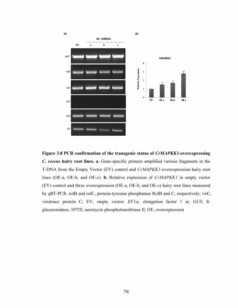
78
Figure 3.8 PCR confirmation of the transgenic status of CrMAPKK1-overexpressing
C. roseus hairy root lines. a. Gene-specific primers amplified various fragments in the
T-DNA from the Empty Vector (EV) control and CrMAPKK1-overexpression hairy root
lines (OE-a, OE-b, and OE-c). b. Relative expression of CrMAPKK1 in empty vector
(EV) control and three overexpression (OE-a, OE-b, and OE-c) hairy root lines measured
by qRT-PCR. rolB and rolC, protein-tyrosine phosphatase RolB and C, respectively; virC,
virulence protein C; EV, empty vector; EF1œ, elongation factor 1 œ; GUS, ß-
glucuronidase; NPTII, neomycin phosphotransferase II; OE, overexpression

79
Figure 3.9 Increase in TIA pathway gene expression and alkaloid accumulation in C.
roseus hairy roots overexpressing CrMAPPKK1. a. Relative expression of TIA
pathway genes, in EV control and three CrMAPKK1-overexpression (OE-a, OE-b, and
OE-c) hairy root lines, were measured by qRT-PCR. b. Tabersonine, ajmalicine, and
catharanthine were extracted from the EV control and the three CrMAPKK1-OE lines and
analyzed by LC-MS/MS. The levels of tabersonine, ajmalicine, and catharanthine were
estimated based on peak areas compared to the standards. Alkaloid levels are indicated in
ng/mg dry weight (DW). Data represented here are the means ± SDs from two biological

80
replicates. Statistical significance was calculated using the Student’s t-test, * p value
<0.05, ** p value <0.01.
3.4 Conclusion
In this report, we illustrate that both the ORCA gene cluster and CrMYC2 are subject to
an additional layer of regulation by a CrMAPK cascade that consists of CrMAPK3/6,
CrMAPKK1, and, possibly, CrMAPKKK1. The involvement of the CrMAPK cascade in
TIA biosynthesis is substantiated by overexpression of CrMAPKK1 in C. roseus hairy
roots. This study represents a detailed characterization of post-translational regulatory
mechanisms that govern a plant TF cluster and is summarized in this model (Figure 3.10).
Figure 3.10 A simplified model depicting the posttranslational regulation of the
CrMYC2, ORCA gene cluster, and TIA pathway genes. Upon perception of JA, the

81
MAP kinase cascade, consisting of CrMAPKKK1, CrMAPKK1, and CrMAPK3/6,
phosphorylates CrMYC2 and the ORCA gene cluster (gray arrows). Phosphorylated
CrMYC2 (labeled by orange flags) activates ORCA3 and possibly another unidentified
TF, which in turn activates ORCA4 and ORCA5. Solid red arrows indicate direct
activation; broken arrows represent indirect or undetermined activation. The
phosphorylated ORCA 3/4/5 (labeled by orange flags) activate two subsets of TIA
pathway genes (in parentheses). ORCA3, ORCA4 and ORCA5 regulate several genes in
the indole and downstream TIA pathways, whereas ORCA4 and 5 activate an additional
set of genes in the seco-iridoid pathway (IS and G10H). CrMYC2, in addition to
activating ORCA3, co-regulates TDC and CPR with ORCAs by binding to the T/G-Box
of the gene promoters. BIS1, binding to the G-Box, activates IS and G10H. ORCA4/5
also activates the same genes, likely by binding to the ORCA-binding sites (BS). A G-
box binding factor (GBF) along with ORCA3/4/5 co-regulates STR. Inset: A simplified
TIA pathway with key genes highlighted in blue.
Copyright © Priyanka Paul 2017

82
Chapter 4
Genome-wide identification of the Catharanthus roseus MAP kinome and functional
characterization of two MAP kinases involved in TIA regulation
4.1 Introduction
Plants are constantly subjected to a number of environmental challenges, including
drought, cold, salinity, pathogen and herbivore attacks. To cope with such challenges
plants evolved sophisticated mechanisms to adapt their metabolism, growth, and
development. Genes encoding proteins involved in signal perception and transduction
have been identified and characterized in a number of plant species. Among them, protein
kinases (PKs) and protein phosphatases (PPs) are significantly abundant in number.
Protein phosphorylation mediated by PKs is one of the most important post-translational
modifications, to transduce external signals into internal responses. One of the most
common protein kinase cascades is the MAPK cascade. The MAPK cascade is typically
comprising of three successively acting protein kinases, such as MAPK Kinase Kinases
(MAPKKKs/MAP3Ks/MEKKs/MPKKK), MAPK Kinases (MAPKKs/MAP2Ks/ MEKs/
MPKKs) and MAP Kinases (MAPKs/MAP1Ks/MPKs), connected to each other by
phosphorylation (Smékalová et al., 2014). MAPKs phosphorylate specific substrates,
including transcription factors (TFs), enzymes, or other kinases in response to external
stimuli resulting in specific cellular responses (Cristina et al., 2010). MAPKs are
activated by dual phosphorylation of conserved threonine (T) and tyrosine (Y) residues in
the TXY motif located at their activation loop (or T-loop) by upstream dual specificity
MAPKKs, which are in turn activated by MAPKKKs via phosphorylation of conserved
serine (S) and / or threonine (T) residues in S/T-X3-5-S/T motif (Cristina et al., 2010).
The MAP1Ks are divided into four groups based on protein structure and sequence of the
TXY sequence motif. The MAP1Ks with TEY motif are grouped into A, B and C,
whereas that having motif TDY are into group D (Ichimura et al., 2002). The possible
number of MAP1Ks in group D varies between different species, such as 10 in rice (Rao

83
et al., 2010), 8 in Arabidopsis (Cardinale et al., 2002), 14 in soybean (Neupane et al.,
2013), and 11 in maize (Kong et al., 2013b; Liu et al., 2013). Group A MAP1Ks are well
characterized in plants and are mostly involved in hormonal and environmental stress
responses. For instance, tobacco SIPK and WIPK, which are orthologous to AtMAPK6
and AtMAPK3 of Arabidopsis respectively, are activated during both biotic and abiotic
stress responses (Seo et al., 1995; Zhang and Klessig, 2001; Meng et al., 2013; Vogel et
al., 2014). On the contrary, group B MAP1Ks have been relatively less studied and are
reported to be involved in both environmental stress responses and cell division (Kosetsu
et al., 2010; Takahashi et al., 2010; Berriri et al., 2012). The function of Group C
MAP1Ks is also less studied. For instance, in Arabidopsis, MAPK7 is reported to be
involved in circadian rhythm whereas in rice the orthologue mediates resistance against
Xanthomonas oryzae (Schaffer et al., 2001; Jalmi and Sinha, 2016). Very little is known
about the biological roles of the Group D MAPKs. In rice, the OsMAPK20-4 has been
shown to be involved in both developmental regulation and environmental stress
responses (Agrawal et al., 2003).
Phylogenetic analysis has grouped plant MAP2K genes into four groups: A, B, C, and D
(Ichimura et al., 2002) and are involved in a wide range of biological processes, including
pathogen defense and secondary metabolism. The Group A MAP2Ks, including
MAPKK1 and MAPKK2 act upstream of MAPK4 in Arabidopsis (Ichimura et al., 1998;
Huang et al., 2000; Cardinale et al., 2002) to negatively regulate the defense responses
against biotrophic pathogens (Petersen et al., 2000; Qiu et al., 2008). Arabidopsis
MAPKK6 is reported to be involved in cell division (Takahashi et al., 2010). Group B
MAP2Ks, such as Arabidopsis MAPKK3 and tobacco NPK2 are involved in nuclear
transport in yeast cells (Quimby et al., 2000). Group C MAP2Ks, such as AtMEK4 and 5
are involved in various environmental stress responses (Ren et al., 2002; Liu et al., 2008).
Group D MAP2Ks, such as Arabidopsis MAPKK9 activates the MAPK3/MAPK6 to
induce camalexin and ethylene biosynthesis (Liu and Zhang, 2004; Xu et al., 2008; Yoo
et al., 2008); MAPKK9-MAPK3/MAPK6 cascade is also known to regulate leaf
senescence in Arabidopsis (Zhou et al., 2009). CrMAPKK1 and JAM1, which are

84
orthologous to AtMAPKK9, are involved in regulation of TIA and nicotine biosynthesis
in C. roseus and tobacco, respectively (De Boer et al., 2011; Paul et al., 2017).
MAP3Ks constitute a large and diverse family of kinases that have been classified into
three subfamilies, MEKK-like, Raf-like, and ZIK-like family, based on the sequence of
the catalytic domain (Wrzaczek and Hirt, 2001). Similar to the MEKKs in yeast (e.g.
STE11 and BCK1) and mammal (e.g., MEKK1), the plant MEKK-like subfamily
members also have a conserved catalytic domain, and the majority of them are involved
in stress responses by activating downstream MAP2Ks (Champion et al., 2004). In
Arabidopsis, AtMEKK1 activates MAPKK4/MAPKK5-MAPK3/MAPK6 cascade to
regulate innate immunity in response to bacterial and fungal pathogens (Asai et al., 2002).
Similar to mammalian RAF1, the plant Raf-like MAP3Ks also share the GTXX (W/Y)
MAPE signature motif (Ichimura et al., 2002; Jonak et al., 2002). In Arabidopsis, the
Raf-like MAP3Ks including Enhanced Disease Resistance 1 (EDR1) and Constitutive
Triple Response 1 (CTR1) are reported to be involved in defense responses and ethylene
signaling, respectively (Kieber et al., 1993; Frye and Innes, 1998; Frye et al., 2001;
Huang et al., 2003). Though ZIK-like kinases are identified in plant MAP3Ks
phylogenetic analyses, these enzymes have not been reported to phosphorylate the
downstream MAP2Ks (Champion et al., 2004; Xu et al., 2005).
Plant MAPK cascades are involved in numerous biological processes, such as cell
division (Jiménez et al., 2007), growth and development (Xu and Zhang, 2015),
hormonal responses (Tena et al., 2001), and environmental stress responses, including
pathogen infection (Pitzschke et al., 2009), wounding (Heinrich et al., 2011), drought,
salinity (Chávez Suárez and Ramírez Fernández, 2010), UV irradiation (Parages et al.,
2013), reactive oxygen species (ROS) (Pitzschke et al., 2009), and ozone (Samuel et al.,
2000). To date, numerous MAPK cascades have been identified and characterized in
plants. In Arabidopsis, the MEKK1-MAPKK4/5-MAPK3/6-WRKY22/WRKY29
cascade is involved in plant innate immunity (Asai et al., 2002; Galletti et al., 2011), the
MEKK1-MAPKK1/2-MAPK4 positively regulates defense responses against
necrotrophic fungi and negatively control defenses against biotrophic pathogens

85
(Petersen et al., 2000; Qiu et al., 2008), and YDA-MAPKK4/5-MAPPK3/6 regulates
stomatal development (Eckardt, 2007). In rice, the OsMAPKK4-OsMAPK1-OsWRKY53
cascade is involved in the wounding signaling pathway (Yoo et al., 2014), and
OsMAPKK3-OsMAPK7-OsWRK30 module acts against Xanthomonas oryzae , which
causes leaf blight disease in rice (Jalmi and Sinha, 2016). Recently, a MAPK cascade
consisting of GhMAPKKK15–GhMAPKK4–GhMPK6 has been reported to directly
phosphorylates and activates GhWRKY59 that regulates drought response in cotton (Li et
al., 2017). The genome sequencing has made it possible to identify and characterize
MAPK families in a number of other plant species, including maize (Kong et al., 2013b;
Liu et al., 2013), wheat (Wang et al., 2016), tomato (Kong et al., 2012; Wu et al., 2014),
Brachypodium distachyon (Chen et al., 2012), Vitis vinifera (Çakır and Kılıçkaya, 2015),
cucumber (Wang et al., 2015), and Capsicum annuum (Liu et al., 2014).
Catharanthus roseus of the family Apocynaceae is the exclusive source of an array of
bioactive TIAs, including the anticancer drugs, vincristine and vinblastine (Kellner et al.,
2015). However, the natural production of TIAs in C. roseus is extremely limited. In
recent years, significant progress has been made in studying the transcriptional regulation
of the TIA biosynthesis. TFs belonging to AP2/ERF (Menke et al., 1999b; van der Fits
and Memelink, 2000; Paul et al., 2017), bHLH (Zhang et al., 2011; Van Moerkercke et al.,
2015; Van Moerkercke et al., 2016) and WRKY (Suttipanta et al., 2011) families have
been shown as the key regulators of the TIA pathway. Previously, two members of
AP2/ERF gene family such as ORCA2 and ORCA3 were reported to regulate the TIA
biosynthesis in C. roseus (Menke et al., 1999b; van der Fits and Memelink, 2000).
Recently, I cloned and characterized two additional AP2/ERF genes, ORCA4 and ORCA5,
which are physically clustered with ORCA3 at the same genomic scaffold (Kellner et al.,
2015). I have shown that though ORCA4 and ORCA5 functionally overlap with ORCA3,
they regulate an additional set of TIA pathway genes. ORCA4 or ORCA5 overexpression
resulted in significant increase of TIA accumulation in C. roseus hairy roots (Paul et al.,
2017). The genes of indole branch are mostly regulated by the ORCA gene cluster
(Menke et al., 1999b; van der Fits and Memelink, 2000; Paul et al., 2017) and CrMYC2
(Zhang et al., 2011); however the majority of iridoid pathway genes are controlled by two

86
newly identified bHLH TFs, BIS1 and BIS2 (Van Moerkercke et al., 2015; Van
Moerkercke et al., 2016).
Post-transcriptional and post-translational modulations of TFs add another layer of
regulatory complexity to the biosynthesis of specialized metabolites in plants (Maier et
al., 2013; Patra et al., 2013a; Patra et al., 2013b; Shen et al., 2017). Post-translational
regulatory mechanisms, including phosphorylation of TFs have been implicated in the
biosynthesis of plant specialized metabolites, including camalexin and nicotine
biosynthesis in Arabidopsis and tobacco, respectively (Xu et al., 2008; De Boer et al.,
2011; Mao et al., 2011). In C. roseus, JA-induced expressions of TIA pathway gene
transcripts are reduced significantly in the presence of protein kinase inhibitor, K-252a,
but are not inhibited by the protein synthesis inhibitor, cycloheximide (Menke et al.,
1999a). This observation indicates the possible role of protein phosphorylation in the JA
signal transduction cascade (Menke et al., 1999a). Moreover, a JA-inducible MAPK,
CrMAPK3, has been reported in C. roseus (Raina et al., 2012). Recently, we
demonstrated that in C. roseus, the ORCA gene cluster and CrMYC2 act downstream of
a MAP kinase cascade that includes CrMAPKKK1, CrMAPKK1, CrMAPK3 and 6.
Overexpression of CrMAPKK1 in C. roseus hairy roots upregulated TIA pathway gene
expression and boosted TIA accumulation (Paul et al., 2017). To date, no systematic and
genome-wide investigations of the MAPK gene families have been reported in C. roseus.
In this study, we performed a genome-wide analysis to identify all MAPKs in C. roseus.
A total of 83 putative C. roseus MAPKs, including 12 CrMAP1Ks, 6 CrMAP2Ks, 59
CrMAP3Ks, and 6 CrMAP4Ks were identified. Their domain organization and
expression profiles in different tissues were analyzed. We next identified and functionally
characterized two previously uncharacterized MAPK cascades that regulate two different
branches of TIA pathway. The MAPKK6-MAPK13 positively regulates the iridoid
branch of TIA pathway in C. roseus. Overexpression of CrMAPK13 in C. roseus hairy
roots upregulated the BIS-regulated iridoid pathway genes and increased tabersonine
accumulation. The other MAPK cascade consists of CrMAPKK1 and CrMAPK20, which
regulates the genes in the indole branch of TIA pathway. Overexpression of CrMAPK20

87
in C. roseus hairy roots down-regulated the CrMYC2-ORCA regulated indole pathway
genes and reduced catharanthine accumulation. In conclusion, this work provided a
detailed understanding of the post-translational regulatory network in TIA biosynthesis in
C. roseus.
4.2 Materials and Methods
4.2.1 Plant materials
C. roseus (L.) G. Don var. ‘Little Bright Eye’ was used for gene expression and cloning,
and generation of transgenic hairy roots. Tobacco var. Xanthi cell line was used for
protoplast transient expression assays.
4.2.2 RNA isolation and cDNA synthesis
C. roseus var. ‘Little Bright Eye’ seeds were surface-sterilized with 30% commercial
bleach, then washed five times with sterile water and afterward germinated on half-
strength Murashige and Skoog (MS) medium (Caisson Labs, USA). Ten-day-old C.
roseus seedlings were next treated with 100µM methyl jasmonate (MeJA) for 2 h and
mock-treated samples were used as a control. Both samples were used for total RNA
isolation and cDNA synthesis as described previously (Suttipanta et al., 2007).
CrMAPKK6, CrMAPK13, and CrMAPK20 were PCR amplified from cDNA using gene-
specific primers (Table 4.1), and then cloned into the pGEM-T Easy vector (Promega,
USA) followed by sequencing confirmation.
4.2.3 Multiple sequence alignment and phylogenetic analysis
Alignment of deduced amino acid sequences was performed using the ClustalOmega
program (Larkin et al., 2007) with the default parameters. Phylogenetic trees were
constructed and visualized using the neighbor-joining (NJ) method through MEGA
version 6 software (Tamura et al., 2011). The statistical reliability of individual nodes of
the newly constructed tree was assessed by bootstrap analyses with 1,000 replications.

88
4.2.4 Identification and co-expression analysis of MAPK cascade genes in C. roseus
The method used to identify all the putative MAP1K, MAP2K, MAP3K and MAP4K
family genes were similar to that used previously for other plants (Chen et al., 2012;
Kong et al., 2013a). Briefly, to identify C. roseus MAPK cascade genes, all protein-
coding genes were obtained from the C. roseus draft genome, the Dryad Digital
Repository (Kellner et al., 2015). Next, the MAPK cascade genes from Arabidopsis and
rice (Oryza sativa) were used as queries to search against the C. roseus protein databases
with the BLASTP program (Wang et al., 2015) with an e-value of 1e−10 as the threshold.
Putative MAP kinases in the C. roseus proteome were identified using the signature
motifs previously described (Jonak et al., 2002) and a total of 91 putative MAPK cascade
genes were identified. The Conserved Domain Database of The National Center for
Biotechnology Information (NCBI) (Wang et al., 2015) and Pfam (Lehti-Shiu and Shiu,
2012) databases were then used to confirm further and to predict the conserved domains
in MAPK cascade genes. Finally, a custom PERL script was used to calculate the
isoelectric points (pIs) and molecular weights (MWs) of MAPK cascade proteins.
To study the expression patterns of C. roseus MAPK cascade genes and TIA pathway
genes, transcriptome data for five different tissues, including flower, mature leaf,
immature leaf, stem, and root were obtained from the Sequence Read Archive (SRA)
database at the NCBI (accession number SRA030483). Raw reads were further processed
and reads per kilobase of transcript per million mapped reads (RPKM) value was
calculated as described previously (Singh et al., 2015). Pairwise Pearson correlation
coefficient for each transcript was calculated using RPKM. Matrix distances for
expression heatmap were computed over Pearson correlations of gene expression values
(RPKM) by the heatmap.2 function of gplots (version 3.0.1) Bioconductor package in R
(version 3.2.2) (Wickham, 2009).
4.2.5 Quantitative RT-PCR (qRT-PCR)
The qRT-PCR was performed as described previously (Suttipanta et al., 2011). The
primers used in qRT-PCR are listed in Table 4.1. The C. roseus 40S Ribosomal Protein

89
S9 (RPS9) gene was used as an internal control (Liscombe et al., 2010). All PCRs were
performed in triplicate and repeated twice.
Table 4.1 Oligonucleotides used in this study
RPS9 5’-GAGGGCCAAAACAAACTTA-3’ 5’-CCCTTATGTGCCTTTGCCTA-3’ EF1œ 5’-TACTGTCCCTGTTGGTCGTG-3’ 5’-AAGAGCTTCGTGGTGCATCT-3’ ASa GCGAACATTTGCAGATCCAT GGCCGATTTGTTATTGTTCC TDC 5’-ATCCGATCAAACCCATACCA-3’ 5’-CGTCATCCTCGACCATTTTT-3’ G10H 5’-TTATTCGGATTCTGCCAAGG-3’ 5’-TCCCCAAAGTGAATCGTCAT-3’ CPR 5’-TGGCAGAAAAGGCTTCTGAT-3’ 5’-CTCAGCCTGTGTGCTATCCA-3’ SLS 5’-GTTCCTTCTCACCGGAGTTG-3’ 5’-CCCATTTGGTCAACATGTCA-3’ STR 5’-ACCATTGTGTGGGAGGACAT-3’ 5’-ATTTGAATGGCACTCCTTGC-3’ LAMT 5’- AGCCAAGGCAGTGATTGTTG-3’ 5’-CTGCAATGCGGAAAGGTTTG-3’ IS 5’-CCACATGATTCGCCTTTTACCG-3’ 5’- AAACCCGAAAACCAGAGCTG-3’
ORCA3 5’-CGGGATCCGAAATACAGAAA-3’ 5’-GCCCTTATACCGGTTCCAAT-3’ ORCA4 5’-ATAGTAGTACTGCCGCCGAAAG-3’ 5’-ATCTCCGCCGCAAATTTTCC-3’ ORCA5 5’-TCTTCCAACGGAGGTTAACGG-3’ 5’-AATGTTGTCTCCAGGGCTTG-3’ CrMAPKK1 5’-AAGGGCAAAGACCTGATTGG-3’ 5’-TCAGGCAAACTTGGTGGTTC-3’ ZCT1 5’-AGCCGAAAACTCATGCTTGT-3’ 5’-CGCCTTTGCAACAGGTTTAT-3’ ZCT2 5’-CGTCAATTTCCATCGTTTCA-3’ 5’-CCGATAGCGAATTCAAGTCC-3’ ZCT3 5’-GACAAGCTTTGGGAGGACAC-3’ 5’-GGCAAGGCAGGTAAGTTCAA-3’ MAPK13 5’-ATCAATCCTGTTGGCAGTGG -3’ 5’- TCTCGAAGGGTTCTTTTGGC -3’ MAPKK6 5’- AGCCATTGACGGAACTCAAG -3’ 5’- TAAGGCGCAATCCTTTCTGG-3’ MAPK19 5’-TCGCCAAAACCACTGATTGC-3’ 5’- TGCATATTCTGAGCCAAGGC-3’ MAPK20 5’- GCTTGGCCAGAGTTGCATTC -3’ 5’- AGATGGCTCCCTCTCGACTT-3’ CrMAPKK1 5’-gcggatcc ATGGCATTAGTTA
AAGACCGTC-3’ 5’-atgctgcag TTATGGCCTTTTA ACCTTTTGC-3’
CrMAPKK6 5’-gcgaattcATGAAGCTTAAGAAGCCAT TG -3’
5’-atggtcgacTTACCTCGGAAAATTCG T AGG -3’
CrMAPK13 5’- gcggatccATGGAAATGGAAAAGG AAAAG-3’
5’- atgtctagaATCATGCCATCTCATGA GCTTG-3’
CrMAPK20 5’- gcaaggatcc ATGCAGCCCGATCACAGGA -3’
5’- accggctgcagTTACCTA GGAAGAGAGA CATGTTTTCTC -3’
rolB 5’CTTATGACAAACTCATAGATAAAAGGTT-3’
5’- TCGTAACTATCCAACTCACATCA C-3’
rolC 5’-CAACCTGTTTCCTACTTTGTTAAC-3’
5’- AAACAAGTGACACACTCAGCTT C-3’
virC 5’-TTTTGCTCCTTCAAGGGAGGTGCC-3’
5’-GGCTTCGCCAACCAATTTGGAGA T-3’
GUS 5’-ATGGTAGATCTGAGGGTAAATTT C-3’
5’- GCTAGCTTGTTTGCCTCCCTG-3’
ntpII 5’-ATGGGGATTGAACAAGATGGA-3’ 5’-TCAGAAGAACTCGTCAAGAAG -3’

90
4.2.6 Protoplast isolation and electroporation
For transient protoplast assays, the reporter plasmids were generated by cloning the STR,
G10H, or IS promoter into a modified pUC vector containing a firefly luciferase (LUC)
reporter and rbcS terminator. The (5XGALRE)-35Spro:LUC was described previously
(Patra et al., 2013a). The effector plasmids were constructed by cloning ORCA3/4/5, BIS1,
CrMAPK13, CrMAPK20 and CrMAPKK6 into a modified pBS vector under the control
of the CaMV35S promoter and rbcS terminator. For the protoplast-based two-hybrid
assay, CrMAPK13 and CrMAPK20 were fused to the GAL4 DNA binding domain in a
pBS plasmid containing MMV promoter and rbcS terminator. A GUS reporter, driven by
the CaMV35S promoter and rbcS terminator, was used as an internal control in all
protoplast assays. Protoplast isolation from tobacco cell suspension cultures and
electroporation with plasmid DNA were performed as previously described (Pattanaik et
al., 2010b; Pattanaik et al., 2010a). The reporter, effector, and internal control plasmids
were electroporated into tobacco protoplasts in different combinations; luciferase and
GUS activities in transfected protoplasts were measured as described previously
(Suttipanta et al., 2007; Paul et al., 2017). Each experiment repeated three times.
4.2.7 Yeast two-hybrid Assay
In order to detect the kinase-kinase or kinase-substrate interaction, yeast two-hybrid
(Y2H) assay was performed. The plasmids, pAD-GAL4-2.1 and pBD-GAL4-Cam
(Stratagene, USA) containing the GAL4 activation and GAL4 DNA-binding domains,
respectively were used. The full-length coding region of CrMAPK13 and CrMAPKK6
were cloned into both pAD-GAL4 and pBD-GAL4 vectors. Similarly, the full-length
coding region of CrMAPK20 was cloned into pBD-GAL4 vector, whereas the
CrMAPKK1, ORCA3, ORCA4, ORCA5 and CrMAPK3 were cloned into pAD-GAL4
vector. The resultant plasmids were co-transformed into yeast strain AH109 using the
PEG/LiAC method (Pattanaik et al., 2010b). Co-transformation of the empty vector pAD-
GAL4 and pBD-CrMAPK13/CrMAPKK6/CrMAPK20 were used as negative control.
Transformed colonies were selected on synthetic dropout (SD) medium lacking leucine
and tryptophan (SD-Leu-Trp). Colonies from double selection plates were then screened
for growth on triple selection SD medium lacking histidine, leucine and tryptophan (SD-

91
His-Leu-Trp) with a required concentration of 3-amino-1,2,4-triazole (3AT) (Pattanaik et
al., 2010b). Each experiment was repeated at least two times.
4.2.8 Construction of plant expression vectors and generation of hairy roots
For plant transformation, CrMAPK20 and CrMAPK13 cDNAs were PCR-amplified from
C. roseus seedling cDNA and then cloned into a modified pCAMBIA2301 vector
containing the CaMV35S promoter and the rbcS terminator (Pattanaik et al., 2010b). The
pCAMBIA2301 vector only was used as an empty vector (EV) control. The plasmids
were mobilized into Agrobacterium rhizogenes R1000 using freeze-thaw method (Weigel
and Glazebrook, 2006). Transformation of C. roseus seedlings and generation of hairy
roots were performed using the protocol described previously (Suttipanta et al., 2011;
Paul et al., 2017). Transgenic status of the hairy root lines was verified by PCR
amplification of the rolB, rolC, virC, nptII and GUS genes. Primers used in this study are
listed in Table 4.1. Three independent hairy root lines were selected for further analysis.
4.2.9 Alkaloid extraction and analysis
To extract alkaloid, transgenic hairy roots were frozen in liquid nitrogen and ground to a
powder. Samples were extracted in methanol (1:100 w/v) twice for 24 h on a shaker.
Pooled extracts were then dried via a rotary evaporator and diluted in methanol 10 µL/mg
of the initial sample. The samples were then analyzed using high-performance liquid
chromatography (HPLC), followed by electrospray-injection in a tandem mass
spectrometry, as previously described (Suttipanta et al., 2011; Paul et al., 2017). The
known alkaloid standards were run to identify elution times and mass fragments.
4.3 Results and Discussion
4.3.1 Identification of the MAP1K, MAP2K, MAP3K and MAP4K families in C.
roseus
MAPK cascades are ubiquitous in all eukaryotes and are responsible for various cellular
responses (Yoo et al., 2008). We took advantage of the C. roseus draft genome (Kellner
et al., 2015) to identify MAP1K, MAP2K, MAP3K and MAP4K genes in its genome. We
conducted BLASTP searches against the C. roseus protein database using MAP kinase

92
cascade proteins from Arabidopsis and rice, as a query. The candidate sequences were
further evaluated for the putative functional domains and motifs. Sequences that did not
contain the known conserved motifs of the MAP1K, MAP2K MAP3K and MAP4K
family proteins were excluded from further analysis. We, finally, identified 12
CrMAP1Ks, 6 CrMAP2Ks, 67 CrMAP3Ks and 6 CrMAP4Ks genes in C. roseus. Out of
67 MAP3Ks, we could retrieve full-length sequences for 59 and these were used for
further analysis.
The CrMAP1K genes were predicted to encode proteins from 219 to 753 amino acids in
length with calculated molecular weights (MWs) ranging from 25.2 to 85.0 kDa and
theoretical isoelectric points (pIs) ranging from 5.19 to 9.2. The sub-cellular localization
predication indicated that most of the members of CrMAP1K are located in the
cytoplasm, except CRO_T006826 and CRO_T008379, which were present in
mitochondria (Table 4.2). The 6 CrMAP2Ks proteins are 341–606 amino acid in length,
with calculated MWs ranging from 38.26 to 68.0 kDa and pIs from 5.57 to 8.81. They
were predicted to be localized in the cytoplasm and nucleus (Table 4.3). The polypeptide
lengths of the CrMAP3K genes ranged from 138 to 1426 amino acids with MWs ranging
from 15.5 to 154.4 kDa and theoretical pIs ranging from 4.66 to 9.97. Similarly,
CrMAP3K proteins were located in the various cellular compartments, including
cytoplasm, nucleus, plasma membrane, mitochondria, or chloroplast (Table 4.4). The
polypeptide lengths of the MAP4K genes ranged from 507 to 883 amino acids, and their
predicted MWs ranged from 56.68 to 97.96 kDa. The theoretical pIs ranged from 5.77 to
8.88 (Tables 4.5).
Table 4.2. List of MAP1Ks identified in C. roseus. Gene Full
length MAP1K
group T-
loop Length (amino acids)
Molecular weight (kDa)
Isoelectric point
Best Arabidopsis
Match
Subcellular localization:score
CRO_T025299 Yes A TEY 375 43.06 5.26 AT3G45640 cytoplasmic: 0.83
CRO_T025830 Yes A TEY 398 45.69 5.51 AT2G43790 cytoplasmic: 0.86
CRO_T003948 Yes B TEY 340 39.21 6.06 AT4G01370 cytoplasmic: 0.83
CRO_T006826 Yes B TEY 387 43.91 6.27 AT4G01370 mitochondrial: 0.63
CRO_T026682 Yes B TEY 380 43.27 5.19 AT1G07880 cytoplasmic: 0.85
CRO_T024779 Yes C TEY 630 71.95 6.37 AT3G18040 cytoplasmic: 0.84

93
Table 4.2. List of MAP1Ks identified in C. roseus (continued.) CRO_T022398 Yes C TEY 371 42.06 7.16 AT2G18170 cytoplasmic: 0.84
CRO_T032753 Yes D TDY 568 64.37 8.98 AT5G19010 cytoplasmic: 0.74
CRO_T008379 Yes D TDY 373 42.92 6.53 AT1G10210 mitochondrial: 0.62
CRO_T007816 Yes D TDY 445 51.75 9.2 AT2G42880 cytoplasmic: 0.77
CRO_T005275 Yes D TDY 753 85.00 8.9 AT3G14720 cytoplasmic: 0.42
CRO_T033572 Yes D TDY 219 25.19 7.7 AT3G18040 cytoplasmic: 0.64
Table 4.3. List of MAP2Ks identified in C. roseus. Gene Full
length MAP2K
group Length (amino acids)
Molecular weight (kDa)
Isoelectric point
Best Arabidopsis
Match
Subcellular localization:score
CRO_T004930 Yes A 355 39.76 6.01 AT5G56580 cytoplasmic: 0.85
CRO_T005978 Yes A 354 39.25 5.57 AT4G29810 cytoplasmic: 0.66
CRO_T019539 Yes B 606 68.03 5.78 AT5G40440 cytoplasmic: 0.66
CRO_T030701 Yes C 361 40.94 6.03 AT1G32320 nuclear: 0.67
CRO_T011505 Yes D 341 38.26 8.04 AT1G73500 nuclear: 0.51
CRO_T020175 Yes D 377 41.60 8.81 AT1G51660 cytoplasmic: 0.65
Table 4.4. List of MAP3Ks identified in C. roseus. Gene Full
length MAP3K
group Length (amino acids)
Molecular weight (kDa)
Isoelectric point
Best Arabidopsis
Match
Subcellular localization:score
CRO_T022055 No MEKK 361 38.35 10.41 AT1G63700 cytoplasmic: 0.72
CRO_T025162 No MEKK 332 36.90 8.08 AT3G50310 nuclear: 0.82
CRO_T025486 No MEKK 181 20.06 4.83 AT3G07980 cytoplasmic: 0.52
CRO_T003904 Yes MEKK 294 32.39 9.08 AT1G09000 cytoplasmic: 0.67
CRO_T007266 Yes MEKK 553 60.73 5.32 AT4G08500 nuclear: 0.75
CRO_T008488 Yes MEKK 353 38.23 8.96 AT1G63700 cytoplasmic: 0.64
CRO_T009969 Yes MEKK 437 48.90 8.87 AT5G66850 cytoplasmic: 0.57
CRO_T012002 Yes MEKK 1031 113.73 5.72 AT5G55090 cytoplasmic: 0.69
CRO_T014277 Yes MEKK 711 77.82 5.57 AT4G08500 nuclear: 0.77
CRO_T014486 Yes MEKK 280 31.36 4.98 AT4G36950 nuclear: 0.39
CRO_T015682 Yes MEKK 296 32.28 4.66 AT4G36950 mitochondrial: 0.46
CRO_T017735 Yes MEKK 266 29.42 4.92 AT1G07150 nuclear: 0.66
CRO_T020201 Yes MEKK 722 80.10 7.23 AT3G06030 cytoplasmic: 0.46
CRO_T020390 Yes MEKK 206 23.07 4.88 AT5G50180 plasma membrane: 0.32
CRO_T020775 Yes MEKK 707 76.47 9.16 AT1G53570 nuclear: 0.71
CRO_T022731 Yes MEKK 436 48.27 5.5 AT2G32510 nuclear: 0.42
CRO_T024393 Yes MEKK 404 45.32 4.85 AT4G36950 nuclear: 0.9
CRO_T027542 Yes MEKK 138 15.50 6.27 AT1G63700 cytoplasmic: 0.52
CRO_T029124 Yes MEKK 1297 143.95 7.99 AT5G66850 nuclear: 0.38
CRO_T031663 Yes MEKK 286 31.27 6.01 AT3G13530 cytoplasmic: 0.66
CRO_T001547 No Raf 150 16.92 8.05 AT5G01850 cytoplasmic: 0.54
CRO_T013561 No Raf 981 108.96 6.64 AT5G57610 nuclear: 0.55

94
Table 4.4. List of MAP3Ks identified in C. roseus (continued.) CRO_T025685 No Raf 688 76.76 5.66 AT2G31010 cytoplasmic: 0.62
CRO_T032003 No Raf 597 66.62 5.99 AT4G24480 cytoplasmic: 0.6
CRO_T000074 Yes Raf 830 91.10 5.62 AT5G03730 nuclear: 0.47
CRO_T000665 Yes Raf 218 24.82 9.17 AT1G08720 cytoplasmic: 0.79
CRO_T001188 Yes Raf 732 82.29 7.3 AT4G23050 cytoplasmic: 0.55
CRO_T001963 Yes Raf 1426 154.40 5.28 AT3G46920 cytoplasmic: 0.54
CRO_T002980 Yes Raf 388 43.92 9.14 AT1G62400 mitochondrial: 0.91
CRO_T006759 Yes Raf 402 44.71 7.92 AT3G22750 cytoplasmic: 0.81
CRO_T007171 Yes Raf 315 36.53 8.51 AT4G14780 cytoplasmic: 0.89
CRO_T007789 Yes Raf 1287 142.50 5.58 AT5G11850 nuclear: 0.53
CRO_T008177 Yes Raf 469 52.81 4.71 AT4G35780 cytoplasmic: 0.81
CRO_T008654 Yes Raf 425 47.42 7.53 AT5G50000 cytoplasmic: 0.78
CRO_T009390 Yes Raf 511 57.91 8.68 AT2G43850 mitochondrial: 0.8
CRO_T009758 Yes Raf 225 25.59 8.46 AT5G11850 cytoplasmic: 0.28
CRO_T009765 Yes Raf 467 53.14 6.27 AT4G18950 cytoplasmic: 0.56
CRO_T011578 Yes Raf 1299 143.72 5.12 AT1G16270 ER: 0.39
CRO_T012915 Yes Raf 841 94.65 6.34 AT3G58640 nuclear: 0.49
CRO_T013026 Yes Raf 352 40.45 8.42 AT1G62400 cytoplasmic: 0.8
CRO_T013539 Yes Raf 1281 141.06 5.39 AT1G79570 nuclear: 0.57
CRO_T013653 Yes Raf 1237 137.80 5.07 AT5G57610 cytoplasmic: 0.63
CRO_T014965 Yes Raf 459 52.09 5.69 AT3G58760 cytoplasmic: 0.77
CRO_T015407 Yes Raf 1364 150.86 5.7 AT3G24715 nuclear: 0.52
CRO_T016949 Yes Raf 370 42.36 8.32 AT1G14000 cytoplasmic: 0.34
CRO_T017479 Yes Raf 200 21.96 7.66 AT5G01850 cytoplasmic: 0.83
CRO_T017535 Yes Raf 365 40.50 6.55 AT5G66710 cytoplasmic: 0.82
CRO_T018707 Yes Raf 437 49.03 7.78 AT1G14000 cytoplasmic: 0.65
CRO_T018906 Yes Raf 571 64.69 5.81 AT4G38470 cytoplasmic: 0.84
CRO_T019753 Yes Raf 672 72.36 5.17 AT5G11850 cytoplasmic: 0.5
CRO_T021479 Yes Raf 281 32.04 9.26 AT4G38470 mitochondrial: 0.66
CRO_T023512 Yes Raf 282 31.53 9.97 AT4G24480 mitochondrial: 0.76
CRO_T025952 Yes Raf 421 47.08 6.52 AT3G01490 cytoplasmic: 0.75
CRO_T028356 Yes Raf 428 47.58 9.08 AT2G24360 cytoplasmic: 0.52
CRO_T029878 Yes Raf 1184 133.01 6.1 AT1G04700 nuclear: 0.6
CRO_T031633 Yes Raf 505 56.83 5.84 AT4G35780 cytoplasmic: 0.67
CRO_T032564 Yes Raf 464 52.52 9.2 AT5G58950 cytoplasmic: 0.63
CRO_T032849 Yes Raf 812 89.01 7.11 AT3G06620 cytoplasmic: 0.52
CRO_T015959 No ZIK 772 88.80 5.61 AT3G04910 cytoplasmic: 0.74
CRO_T002574 Yes ZIK 708 80.69 5.31 AT3G04910 cytoplasmic: 0.77
CRO_T003421 Yes ZIK 304 34.70 5.95 AT5G55560 cytoplasmic: 0.77
CRO_T009022 Yes ZIK 631 71.48 5.48 AT5G58350 nuclear: 0.34
CRO_T015596 Yes ZIK 411 46.55 5.24 AT3G18750 cytoplasmic: 0.85
CRO_T016097 Yes ZIK 780 87.40 5.4 AT3G18750 ER: 0.46
CRO_T018206 Yes ZIK 588 67.58 6.09 AT3G51630 cytoplasmic: 0.76
CRO_T028948 Yes ZIK 303 34.49 5.48 AT5G55560 cytoplasmic: 0.72
CRO_T033582 Yes ZIK 589 66.75 5.31 AT5G41990 cytoplasmic: 0.57

95
Table 4.5. List of MAP4Ks identified in C. roseus. Gene Full
length Length (amino acids)
Molecular weight (kDa)
Isoelectric point
Best Arabidopsis
Match
Subcellular localization:score
CRO_T001030 Yes 696 77.20 6.14 AT5G14720 cytoplasmic: 0.76
CRO_T019613 Yes 682 75.70 5.77 AT1G79640 cytoplasmic: 0.76
CRO_T015367 Yes 507 56.68 5.97 AT4G14480 nuclear: 0.8
CRO_T033821 Yes 883 97.96 6.89 AT4G10730 cytoplasmic: 0.66
CRO_T029825 Yes 853 93.48 5.22 AT1G69220 nuclear: 0.61
CRO_T005088 Yes 847 93.61 8.88 AT3G15220 mitochondrial: 0.84
To investigate the distribution of MAPK cascade genes in the plant kingdom, we chose
plant species from different evolutionarily important groups, including monocot, dicot,
and primitive plants, such as Physcomitrella patens (moss), Selaginella moellendorffii
(spikemoss) and Amborella trichopoda. The results suggest that genes encoding MAPK
cascade components are widely present in the plant kingdom and, therefore, may have
potential roles in the regulation of various cellular functions (Figure 4.1). Typically,
MAP2K gene members constitute the smallest fraction of MAPK cascade (Figure 4.1).
For instance, MAP2Ks in Arabidopsis and C. roseus is roughly half the number of the
MAP1Ks. This observation suggests that MAP2Ks probably activate multiple MAP1Ks
downstream, and the cross talk between different signal transduction pathways might be
converged at this level in plant MAPK cascades (Ichimura et al., 2002). MAP3K genes
transmit every type of upstream signals to the MAPK cascade. MAP3K showed greater
variations in structures and domain composition (Yin et al., 2013). This is probably the
reason that the MAP3K family constitutes the largest members. In yeast, humans and
other mammals, MAP3Ks are activated either through phosphorylation by MAP4Ks
(Posas and Saito, 1997; Sells et al., 1997) or by G protein and G protein-coupled
receptors (Fanger et al., 1997; Sugden and Clerk, 1997). Several MAP4Ks have been
identified in plant genomes based on phylogenetic analyses of their kinase domain. For
instance, a MAP4K, MIK, was characterized from Zea mays (Wang et al., 2014b), and
ScMAP4K1, from Solanum chacoense, was reported to play important roles in the ovule,
seed, and fruit development (Major et al., 2009).

96
Figure 4.1 Distribution of MAPK cascade kinase genes across the plant kingdom. The
total number of MAP1Ks, MAP2Ks, MAP3Ks and MAP4Ks found in C. roseus, as well
as in other monocot, dicot, Physcomitrella patens (moss), Selaginella moellendorffii
(spikemoss) and Amborella trichopoda.
4.3.2 Phylogenetic relationship and conserved domain analysis
To further characterize the MAPK cascade genes, un-rooted phylogenetic trees were
produced by aligning the full-length protein sequences of all 12 CrMAP1Ks, 6
CrMAP2Ks, 59 CrMAP3Ks, and 6 CrMAP4Ks using the NJ method. Plant MAP1Ks are
commonly grouped into four major subfamilies (A, B, C, and D) based on their
phosphorylation motifs and the phylogenetic relationships between amino acid sequences
(Ichimura et al., 2002). Similarly, all the 12 CrMAP1Ks were also classified into groups
A (2), B (3), C (2) and D (5) (Figure 4.2a). Furthermore, the groups A, B, and C
CrMAP1Ks harbor the TEY (Thr-Glu-Tyr) motif at the phosphorylation site, whereas the
members of group D contain a TDY (Thr-Asp-Tyr) motif (Table 4.2). The group D
MAP1Ks form a distant clade, which is consistent with that described previously in other
plants including Arabidopsis and rice (Ichimura et al., 2002; Hamel et al., 2006). All the
CrMAP1Ks contained a single Pkinase domain (Figure 4.2c).

97
Likewise, the six CrMAP2Ks were also divided into four groups, namely, the A (2), B (1),
C (2), and D (1) subfamilies, which is consistent with the MAP2Ks in other plant species
including Arabidopsis and rice (Hamel et al., 2006; Chen et al., 2012; Liang et al., 2013)
(Figure 4.3a). Like CrMAP1Ks, all the CrMAP2Ks also contain a single Pkinase domain
(Figure 4.3c). Consistent with previous reports, a proline-rich region and extended C-
terminal region was also found in the members of group B of CrMAP2Ks subfamily
(Wang et al., 2015).
The MAP3K gene family in plants is usually divided into three subfamilies, namely
MEKK, Raf, and ZIK (Wrzaczek and Hirt, 2001). Similarly, the 59 CrMAP3K genes in
C. roseus were clustered into three groups (17 MEKKs, 34 Rafs, and 8 ZIKs) (Figure
4.4a), which is consistent with those reported for other plant species (Rao et al., 2010;
Kong et al., 2013a; Sun et al., 2014; Wang et al., 2014a; Wu et al., 2014). All the
CrMAP3K proteins have at least a kinase domain, and most of them have a
serine/threonine protein kinase active site. In C. roseus, most of the CrMAP3Ks in Raf
subfamily have a long N-terminal regulatory domain and C-terminal Pkinase_Tyr domain
(Figure 4.4c). It has been proposed that the long regulatory domain in the N-terminal
region of the Raf subfamily may be involved in protein-protein interactions and regulate
or specify the kinase activity (Jouannic et al., 1999). By contrast, the majority of the
members of the ZIK and MEKK subfamilies have single Pkinase domain (Figure 4.4a).
These results are consistent with the previous findings in Arabidopsis, rice, and tomato
(Rao et al., 2010; Wu et al., 2014). The CrMAP4K gene family in C. roseus formed two
major cluster and harbor single Pkinase domain (Figure 4.4a,b).

98
Figure 4.2 Phylogenetic relationships (a), expression profiles in five different tissues
(flower, immature leaf, mature leaf, stem and root) (b), and protein structures (c) of
CrMAP1Ks in C. roseus.
Figure 4.3 Phylogenetic relationships (a), expression profiles in five different tissues
including (flower, immature leaf, mature leaf, stem and root) (b), and protein structures
(c) of CrMAP2Ks in C. roseus.

99
Figure 4.4 Phylogenetic relationships (a), expression profiles in five different tissues
(flower, immature leaf, mature leaf, stem and root) (b), and protein structures (c) of
CrMAP3Ks in C. roseus.

100
Figure 4.5 Phylogenetic relationships (a), expression profiles in five different tissues
(flower, immature leaf, mature leaf, stem and root) (b), and protein structures (c) of
CrMAP4Ks in C. roseus.
4.3.3 Expression profiles of CrMAP kinase cascade genes in different tissues
To gain insight into the spatial expression patterns and putative functions of CrMAPK
cascade genes in C. roseus, the expression pattern of CrMAPK cascade genes was
investigated using available RNA-seq data (Góngora-Castillo et al., 2012). FPKM values
were obtained from five different tissues, including the root, stem, leaf and flower of
plants. Based on the log2-transformed (FPKM + 1) values, expression levels of CrMAPK
cascade genes were classified as low (≤1 FPKM), moderate (≥1 FPKM and ≤6 FPKM),
and high (≥6 FPKM) in all tissues as described before (Lu et al., 2015; Shen et al., 2017).
The expression levels of these genes are presented in heat-maps (Figure 4.2b, 4.3b, 4.4b
and 4.5b). In general, higher numbers of transcripts were detected in mature leaf, stem
and root. All the CrMAPK cascade members were expressed in at least one tissue (Figure
4.2b, 4.3b, 4.4b and 4.5b). However, four genes, including CRO_T030701
(CrMP2K), CRO_T014486, CRO_T016949 and CRO_T027542 (CrMP3Ks), showed
very low expression in all the tested tissues (Figure 4.2b, 4.3b, 4.4b and 4.5b). Eleven
genes, including the homologs of Arabidopsis AtMAPK1, AtMAPK9, AtMAPK19, and
AtMAPK16 were found to be highly expressed in all tissues suggesting their vital role in
plant growth and development (Figure 4.2b, 4.3b, 4.4b and 4.5b). Some of the members
of CrMAP1K (e.g. CRO_T006826), CrMAP2K (e.g. CRO_T019539, CRO_T030701),
CrMAP3K (e.g. CRO_T014486, CRO_T015682, CRO_T012002), and CrMAP4K

101
(CRO_T018206, CRO_T033582, CRO_T002574) families were preferentially expressed
in specific tissues (Figure 4.2b, 4.3b, 4.4b and 4.5b), suggesting their function in tissue-
specific processes in C. roseus.
Previous studies showed that MAPK cascade genes are widely involved in the growth
and development of higher plants (Xu and Zhang, 2015). In Arabidopsis, AtMAPK3 and
AtMAPK6 play important roles in anther cell differentiation and normal anther lobe
formation (Hord et al., 2008). The MAPK cascade AtMAPKK9-AtMAPK6 in
Arabidopsis plays an important role in leaf senescence (Zhou et al., 2009). In soybean,
MAPK4 and MAPK7 regulate plant growth and development (Shi et al., 2010; Liu et al.,
2011). The NPK1 in tobacco has been suggested to regulate cell size, cytokinesis, and
plant growth (Soyano et al., 2003). Duan et al. identified OsMAPKK4 as a factor for
grain size in rice and suggested a possible link between the brassinosteroids (BRs) and
MAPK pathways during grain growth (Duan et al., 2014). The spatial expression profile
of C. roseus MAPKs suggests their involvement in a number of biological processes.
Recently, CrMAPKK1, CrMAPK3 and CrMAPK6 are reported to be involved in
regulation of TIA in C. roseus (Raina et al., 2012; Paul et al., 2017). However, the
involvement of MAPK cascades in the regulation of plant growth and development has
not been demonstrated in C. roseus.
4.3.4 Multiple sequence alignment and motif analysis of CrMAP kinase cascade
components
MAPKs from several plant species, including Arabidopsis and Brassica rapa (Ichimura
et al., 2002; Lu et al., 2015) have been identified based on the presence of specific
features, such as the conserved MAPK motifs and the phosphorylation-activation TXY
motifs (Cristina et al., 2010). The amino acid sequence alignment of the 12 CrMAP1Ks
identified in this study is shown in Figure 4.6a. Most of these CrMAP1Ks contain the
conserved motifs, such a catalytic loop (C-loop), phosphate-binding loop (P-loop),
activation loop (T-loop), and a T[D/E]Y motif in the activation loop, which are known to
be important for function of MAP1Ks. Of the 12 MAP1Ks, five contain the TDY motif
and the other seven contain the TEY motif. Furthermore, the N-terminal regions of most
of the CrMAP1Ks have a glycine-rich motif (G[R/K]G[A/S]YG) located in the P-loop

102
region, which acts as an ATP- and GTP-binding site (Saraste et al., 1990) (Figure 4.6a).
The conserved C-loop, composed of the HRDLKP[G/S/K]N motif which contains an Asp
(D) residue critical for phosphorylation reaction. This signature motif is present in most
of the CrMAP1Ks (Figure 4.6a). The MAPK common docking (CD) domain consists of
an acidic amino acid cluster (LH[D/E]XX[D/E]EPXC), which establishes electrostatic
interactions with upstream factors and functions as a docking site for MAP2Ks,
substrates, and negative regulators (Tanoue et al., 2000; Paul et al., 2017). This conserved
CD domain, was detected in the extended C-terminal region of the CrMAP1K members
belonging to the A and B groups but was not found in members of group C and D
CrMAP1Ks (Figure 4.6a).
Multiple sequence alignment of the CrMAP2Ks showed that each CrMAP2K contains
the active site motif (D [L/I/V] K), as well as a highly conserved phosphorylation target
site (S/T-XXXXX-S/T) within the activation loop. Additionally, a conserved amino acid
signature motif, VGTxxYMSPER, is present in the catalytic domain (Figure 4.6b) as
previously described in Arabidopsis and other plant MAP2K proteins (Ichimura et al.,
2002; Chen et al., 2012; Liang et al., 2013).
As reported in Arabidopsis and other plant MAP3Ks, the MEKK subfamily in C. roseus
harbors the conserved (G[T/S]Px[W/Y/F]MAPEV) motif. The Raf subfamily in C. roseus
has the (GTXX[W/Y]MAPE) motif, whereas the ZIK subfamily has the
(GTPEFMAPE[L/V]Y) signature motif (Jonak et al., 2002; Rao et al., 2010) (Figure
4.6c). The ZIK proteins have been reported to be involved in circadian rhythm. The ZIK
protein, with-no-lysine (K) 1 (WNK1) in Arabidopsis is involved in the control of
circadian rhythms (Murakami-Kojima et al., 2002). The WNK2/5/8 proteins are found to
regulate the Arabidopsis flowering time via modulating the photoperiod pathway (Wang
et al., 2008). A rice WNK1 was reported to be involved in circadian rhythm, and also
differentially responded to diverse abiotic stress responses (Kumar et al., 2011). However,
the role of CrZIK proteins in C. roseus has yet to be elucidated.
Little is known about the roles of MAP4Ks in plants. Six ORFs showing strong similarity
with the Arabidopsis MAP4Ks were identified in C. roseus genome (Figure 4.6d). Most

103
of the CrMAP4K also have similar signature sequence (TFVGTPXWMAPEV) described
previously for Arabidopsis (Champion et al., 2004).
Figure 4.6a Multiple sequence alignment of the 12 CrMAP1K proteins in C. roseus.
Multiple sequence alignment was performed using Clustal Omega and displayed using
Jalview. Sequence regions with highly conserved and encompassing important motifs are
shown. The alignment is color-coded by ClustalX color scheme that integrates amino
acid properties and evolutionary conservation. The P-loop, C-loop, activation-loop motifs
and common docking (CD) domain are indicated by roman numbers I to IV, respectively.

104
The conserved motifs described previously (Bailey et al., 2006) are indicated by lines
below the alignments. The phosphorylation-activation motif TXY is indicated by
asterisks.
Figure 4.6b Multiple sequence alignment of the 6 CroMAP2K proteins.
Alignment was performed using Clustal Omega and displayed using Jalview. Sequence
regions, which are highly conserved and encompassing important motifs are shown here.
The alignment is color-coded by ClustalX color scheme that integrates amino acid
properties and evolutionary conservation. The active site motif (D [L/I/V] K), conserved
phosphorylation target site (S/T-XXXXX-S/T) within the activation loop and signature
VGTxxYMSPER motif described previously (Bailey et al., 2006) are indicated by roman
numbers I to III, respectively.

105
Figure 4.6c Multiple sequence alignment of the CroMAP3K proteins.
Alignment was performed using Clustal Omega and displayed using Jalview. Only
sequence regions, which are highly conserved and encompassing important motifs are
shown. The alignment is color-coded by a ClustalX color scheme that integrates amino
acid properties and evolutionary conservation. The signature motifs described previously
(Bailey et al., 2006) are shown.
Figure 4.6d Multiple sequence alignment of the conserved motif in CroMAP4K
proteins.
Alignment was performed using Clustal Omega and displayed using Jalview. Only
sequence regions, which are highly conserved, and encompassing important motifs are
shown. The alignment is color-coded by a ClustalX color scheme that integrates amino
acid properties and evolutionary conservation. The signature motifs described by Bailey
et al., 2006 for MAP4K proteins are shown.

106
4.3.5 Identification of a CrMAPK cascade, CrMAPKK6-CrMAPK13, involved in
the regulation of iridoid branch of TIA pathway
I next performed co-expression analysis of the all CrMAPK members with key TIA
pathway structural genes and regulators using C. roseus transcriptomes data (Góngora-
Castillo et al., 2012). Co-expression analysis revealed that CRO_T026682,
CRO_T004930, CRO_T003904 and CRO_T020201 were tightly co-expressed with
iridoid pathway genes and TFs, including G10H, IS, IO, 7DLGT, and BIS1/BIS2 (Figure
4.7). Of these four candidates, CRO_T026682 and CRO_T004930 are homologous to the
Arabidopsis AtMAPK13 and AtMAPKK6, respectively. CRO_T026682 is a member of
group B CrMAP1Ks and CRO_T004930 belongs to the group A CrMAP2Ks (Figure 4.2a,
4.3a). On the other hand, CRO_T003904 and CRO_T020201 belong to the MEKK
subfamily of C. roseus CrMAP3Ks and are homologous to the Arabidopsis
AtMAPKKK1 and AtMAPKKK12, respectively (Figure 4.4a). To investigate their
biological function in C. roseus, I cloned CRO_T026682 and CRO_T004930 and
designated them as CrMAPK13 and CrMAPKK6, respectively. CrMAPK13 is 379 amino
acid long with a calculated MW of 43.28 kDa. The CrMAPKK6 is 354 amino acid long
and calculated MW is 39.77 kDa.

107
Figure 4.7 Co-expression analyses of CrMAPK cascade genes with iridoid pathway
regulatory and structural genes. Hierarchical clustering and heat-map show that
CrMAPK13 and CrMAPKK6 are co-expressed with BIS1 and BIS2, and their targets,
G10H, IS, IO, 7DLH and 7DLGT.
Figure 4.8 Sequence alignments of CrMAPK13 and AtMAPK13. Black colors
showing the amino acids, which are identical and gray colors showing the amino acids
that are similar among the sequences. ATP-binding and Kinase domains are underlined.
The asterisks represent TEY motif in kinase domain.
4.3.6 CrMAPK13 significantly enhances the transactivation of iridoid pathway gene
promoters through BIS1
BIS1 is a key regulator of the iridoid branch of the TIA pathway in C. roseus (Van
Moerkercke et al., 2015). We thus examined whether CrMAPK13 can enhance the
activities of BIS1 on iridoid pathway gene promoters. The IS or G10H promoter-reporter
vector was electroporated into protoplasts with various combinations of vectors
expressing BIS1 and CrMAPK13. Individually, BIS1 activated the expression of the IS or
G10H promoters; however, co-expression with CrMAPK13 significantly increased their

108
activities (Figure 4.9a). To determine whether the enhanced transactivation resulted from
CrMAPK13 activity, we substituted CrMAPK13 with the inactivated mutant,
CrMAPK13K173M, in the IS promoter activation assay. CrMAPKK1K173M was unable to
enhance the promoter activation by BIS1 (Figure 4.9b). These results suggest that the
CrMAPK13 can potentially enhance activation of the BIS1 in TIA biosynthesis.
Figure 4.9 Enhancement of the transactivation activity of BIS1 on TIA pathway
gene promoters by CrMAPK13. a. Transactivation of the G10H and IS promoters
(G10H-pro and IS-pro, respectively) by BIS1 in the presence or absence of CrMAPK13.
b. Influence of the inactive mutant, CrMAPK13K173M on transactivation activity of BIS1
on the IS promoter. The IS or G10H promoter, fused to the luciferase reporter, was
electroporated alone or with different effectors into tobacco protoplasts. Control
represents the promoters alone. A plasmid containing GUS reporter, driven by the
CaMV35S promoter and rbcS terminator, was used for normalization. Luciferase and
GUS activities were measured 20 h after electroporation. Luciferase activity was
normalized against GUS activity. Data presented here are means ± SDs of three
biological replicates. Statistical significance was calculated using the Student’s t-test; * p
value <0.05.

109
4.3.7 CrMAPK13 likely phosphorylates the amino acids S134 and T113 in BIS1
Next, we used BIS1 to determine whether phosphorylation events are responsible for the
enhanced activities of the BIS1 on IS. First, we scanned BIS1 for the minimal MAPK
phosphorylation motifs (Ser or Thr followed by Pro; SP or TP motif) (Sharrocks et al.,
2000; Paul et al., 2017) using the NetPhos 2.0 Server
(http://www.cbs.dtu.dk/services/NetPhos-2.0/). We found that BIS1 contains two
potential MAPK phosphorylation sites, at Ser134 and Thr113. We substituted both
Ser134 and Thr113 with Ala independently and in combination by site-directed
mutagenesis, and tested the BIS1 mutants (BIS1S134A, BIS1T113A, and BIS1S134A/T113A) for
activation of the IS promoter in the presence or absence of CrMAPK13. In the protoplast
assay, activation of the IS promoter by CrMAPK13-BIS1 (WT) was significantly higher
than for CrMAPK13-BIS1S134A/BIS1T113A/BIS1S134A/T113A (Figure 4.10), suggesting that
CrMAPK13 likely phosphorylates BIS1 and modulates its transactivation activity.

110
Figure 4.10 CrMAPK13 likely phosphorylates BIS1. The IS promoter-reporter vector
was co-electroporated with wild-type (WT) BIS1, BIS1S134A, BIS1T113A and
BIS1S134A/T113A, with or without CrMAPK13. Luciferase and GUS activities were
measured 20 h after electroporation. Luciferase activity was normalized against GUS
activity. Data presented here are means ± SDs of three biological replicates. Statistical
significance was calculated using the Student’s t-test; * p value <0.05.
4.3.8 CrMAPK13 interacts with BIS1 and CrMAPKK6 in tobacco cells
MAP1Ks have been shown to interact with their substrates as well as upstream MAP2Ks
in yeast (Lee et al., 2008; Ding et al., 2009) and plant cells (Paul et al., 2017). To
determine whether CrMAPK13 interacts with CrMAPKK6 and BIS1, we used a plant
cell-based two-hybrid system (Patra et al., 2013a). Plasmids expressing CrMAPK13 or

111
BIS1 fused to the GAL4 DNA binding domain were co-electroporated into tobacco
protoplasts with a reporter vector expressing the firefly luciferase with 5XGAL response
elements (5XGALRE-35Spro:LUC). DB-CrMAPK13 or DB-BIS1 expression in protoplasts
also induced luciferase activity (approximately 3- and 14- fold, respectively, compared to
the basal level activity of the reporter plasmid). However, the luciferase activity was
significantly increased in protoplasts co-expressing CrMAPKK6 and CrMAPK13
(approximately 5- and 16.5- fold, respectively) (Figure 4.11). These results suggest that
CrMAPKK6 interacts with and CrMAPK13, and CrMAPK13 interact with and likely
phosphorylates BIS1 in plant cells.
Figure 4.11 Interaction of CrMAPK13 with CrMPKK6 and BIS1. CrMAPK13 and
BIS1 fused to the GAL4 DNA binding domain (DB) (amino acid 1–147), were cloned
into a modified pBS vector containing the MMV promoter and rbcS terminator to
generate DB-CrMAPK13 and DB-BIS1, respectively. CrMAPKK6 and CrMAPK13 were
cloned into a modified pBS vector containing the CaMV35S promoter and rbcS
terminator. Effector plasmids were co-electroporated into tobacco protoplasts with a

112
reporter vector containing the firefly luciferase gene under the control of a minimal
CaMV35S promoter and 5XGAL response elements. The control represents the reporter
alone. A plasmid containing the GUS reporter was used for normalization. Luciferase
activity was normalized against GUS activity. Data presented here are means ± SDs of
three biological replicates. Statistical significance was calculated using the Student’s t-
test; * p value <0.05.
4.3.9 CrMAPK13 interacts with CrMAPKK6 in yeast cells
We next used yeast two-hybrid (Y2H) assay, to establish interaction between CrMAPK13
and CrMAPKK6. AD-MAPK13 and AD-MAPKK6 constructs were co-transformed into
yeast cells with the BD-MAPKK6 and BD-MAPK13 constructs, respectively. Yeast cells
containing any one of these two combinations, grew well on both double (SD/−Leu/−Trp)
and triple (SD/−His/−Leu/−Trp) selection medium. Yeast cells containing BD-
CrMAPK13 or BD-CrMAPKK6 with pAD-GAL4 (AD) did not grow on triple selection
medium (Figure 4.12). The Y2H result demonstrated that CrMAPK13 could interact with
CrMAPKK6 in yeast cells.
Figure 4.12 Physical interaction between CrMAPK13 and CrMAPKK6 in yeast cell.
AH109 yeast strains were transformed with the corresponding plasmids 1. pAD-
CrMAPK13 + pBD-CrMAPKK6, 2. pAD-CrMAPKK6 + pBD-CrMAPK13, 3. pAD
(EV) + pBD-CrMAPK13, and 4. pAD (EV) + pBD-CrMAPKK6. The yeast
transformants were grown in SD/-Leu/-Trp (-LT) and SD/-His/-Leu/-Trp (-LTH)
selection media. EV, empty vector

113
4.3.10 Ectopic expression of CrMAPK13 activates key iridoid pathway genes and
boosts alkaloid accumulation in C. roseus hairy roots
The ability of MAPK13 to enhance transactivation of BIS1 on iridoid pathway gene
promoters in tobacco cells (Figure 4.9), prompted us to investigate its effects on gene
expression and TIA biosynthesis in C. roseus. We generated transgenic C. roseus hairy
root lines overexpressing CrMAPK13. The transgenic status of the hairy roots was
verified by PCR (Figure 4.13a), and three transgenic lines (OE-1, -2, and -3) were
selected for further analysis. CrMAPK13 expressions in transgenic lines were
approximately 50 to 150-fold higher compared to the EV control (Figure 4.13b). Next,
we measured the expression of regulatory and structural genes from the iridoid pathway.
The expression of seven BIS1-targeted genes, such as GES, G10H, 8HGO, IS, IO,
7DLGT, and 7DLH, were significantly increased in the CrMAPK13-overexpression lines
compared to the EV control. Interestingly, expressions of both BIS1 and BIS2 were also
increased significantly in CrMAPK13 overexpression lines (Figure 4.14a). To determine
whether the changes in gene expression were also accompanied by increased TIA
accumulation, alkaloids were extracted from independent transgenic hairy root lines and
measured using LC-ESI-MS/MS as described previously (Suttipanta et al., 2011; Paul et
al., 2017). While accumulation of ajmalicine and catharanthine were not significantly
affected, the accumulation of tabersonine was significantly increased in all three
transgenic lines compared to the EV control (Figure 4.14b). Taken together, our findings
support our hypothesis that the CrMAPKK6-MAPK13 cascade acts upstream of BIS1 to
modulate the expression of iridoid pathway genes.

114
Figure 4.13 PCR confirmation of the transgenic status of CrMAPK13-overexpressing
C. roseus hairy root lines. a. Gene-specific primers amplified various fragments in the
T-DNA from the Empty Vector (EV) control and CrMAPK13-overexpression hairy root
lines (OE-1, OE-2, and OE-3). b. Relative expression of CrMAPK13 in empty vector
(EV) control and three overexpression (OE-1, OE-2, and OE-3) hairy root lines measured
by qRT-PCR. rolB and rolC, protein-tyrosine phosphatase RolB and C, respectively; virC,
virulence protein C; EV, empty vector; EF1œ, elongation factor 1 œ; GUS, ß-
glucuronidase; NPTII, neomycin phosphotransferase II; OE, overexpression

115
Figure 4.14 Increase of iridoid pathway gene expression and TIA accumulation in C.
roseus hairy roots overexpressing CrMAPK13. a. Relative expression of iridoid
pathway genes; GES, G10H, 8HGO, IS, IO, 7DLGT and 7DLH in EV control and three
CrMAPK13-overexpression (OE-1, OE-2, and OE-3) hairy root lines were measured by
qRT-PCR. b. Ajmalicine, catharanthine, and tabersonine were extracted from the EV

116
control and the three CrMAPK13-OE lines and analyzed by LC-MS/MS. The levels of
ajmalicine, catharanthine, and tabersonine were estimated based on peak areas compared
to the standards. Alkaloid levels are indicated in ng/mg dry weight (DW). Data
represented here are the means ± SDs from three biological replicates. Statistical
significance was calculated using the Student’s t-test, * p value <0.05, ** p value <0.01,
and *** p value <0.001.
4.3.11 Identification of a CrMAPK cascade, CrMAPKK1-CrMAPK20, involved in
the regulation of indole branch of TIA pathway.
I have previously demonstrated that the CrMAPKK1-CrMAPK3 cascade positively
regulates the indole branch of TIA pathway in C. roseus (Paul et al., 2017) (Chapter 3).
Based on the co-expression analysis of MAPK cascade components in different tissues,
including root, stem, flower, immature and mature leaves (Figure 4.15a) we selected two
additional MAP1K genes, CrMAPK19, and CrMAPK20. These two kinases were co-
expressed with CrMAPKK1. To examine the effects of MeJA on CrMAPK19, and
CrMAPK20 expression, a key inducer of TIA biosynthesis (Menke et al., 1999a), C.
roseus seedlings were treated with 100 µM MeJA for 2h, and then gene expression was
measured in whole seedlings using qRT-PCR. Compared to the control, CrMAPK19
transcripts did not change significantly in MeJA-treated seedlings. Interestingly,
expression of CrMAPK20 was reduced significantly (about 7-fold) in MeJA-treated
seedlings compared with the untreated control (Figure 4.15b). We cloned the CrMAPK20
(CRO T007816; Figure 4.2a) for further characterization. CrMAPK20, a member of
group D CrMAP1K, is 444 amino acid long with calculated MW of 51.76 kDa.

117
Figure 4.15 Spatial expression analysis of the C. roseus MAPK cascade components
and repression of CrMAPK20 by MeJA treatment. a. Co-expression heat-map of the C.
roseus MAPK in different tissues, including roots, stems, immature leaves, mature leaves,
and flowers. b. Ten-day-old C. roseus seedlings were treated with 100 µM MeJA for 2 h,
and relative gene expression was measured by qRT-PCR. Mock-treated seedlings were
used as a control. Data represent means ± SDs of two biological replicates.
4.3.12 CrMAPK20 significantly represses the transactivation potential of ORCAs on
STR promoter in protoplasts
We next performed a co-expression analysis of CrMAPK20 with TIA biosynthetic and
regulatory genes. At least two independent transcriptional cascades are known to regulate
the TIA pathway genes (Paul et al., 2017). BIS1 and BIS2 regulate the seco-iridoid
pathway genes, including G10H and IS (Van Moerkercke et al., 2015; Van Moerkercke et
al., 2016). CrMYC2 and ORCAs regulate genes in the indole pathway, such as TDC, as
well as genes downstream in TIAs pathway, such as LAMT, SLS, and STR. The co-
expression analysis showed that CrMAPK20 was clustered with ASa, ZCT3, BPF-1, and

118
CrMAPK6 (Figure 4.16a), suggesting that CrMAPK20 may be involved in regulation of
the indole branch of the TIA pathway.
To test this hypothesis, the STR promoter-reporter vector was electroporated into
protoplasts with various combinations of vectors expressing ORCA3, ORCA4, and
CrMAPK20. As expected, individually, both TFs activated the expression of the STR
promoter; however, co-expression with CrMAPK20 significantly repressed activation of
the STR promoter (Figure 4.16b). These results suggest that the CrMAPK20 can repress
activation of the ORCA cluster on TIA pathway gene promoter. We next used ORCA3, a
member of the ORCA cluster, to determine whether phosphorylation events are
responsible for the repressed activities ORCAs on STR promoter. Using the NetPhos 2.0
server (http://www.cbs.dtu.dk/services/NetPhos-2.0/), we previously showed that
ORCA3 contains three putative MAPK phosphorylation sites at Ser60, Ser83 and Thr132.
Putative phosphorylation sites were substituted individually with Ala by site-directed
mutagenesis. We also showed that the Ser60 and Thr132 sites are likely phosphorylayed
by CrMAPK3 (Paul et al., 2017). Here, we evaluated the activity of ORCA3S83A on STR
promoter in the presence or absence of CrMAPK3 or CrMAPK20. In a protoplast assay,
CrMAPK3 significantly induced transactivation activity ORCA3S83A on STR promoter;
however, the activation STR promoter by ORCA3S83A was not significantly affected by
CrMAPK20 (Figure 4.16c), suggesting that CrMAPK20 likely phosphorylates ORCA3 at
S83 and modulates its transactivation activity. Differential phosphorylation may be one
of the methods for fine-tuning ORCA3 activation of target promoters. However, more
experiments are needed to validate this hypothesis. Because similar phosphorylation sites
are also found in ORCA4 and ORCA5, it is assumed that they are also substrates of
CrMAPK20.
4.3.13 CrMAPK20 interacts with ORCAs in plant cells
The activation of MAPKs cascade, in response to specific stimuli, is both complex and
often redundant. Moreover, a single MAP1K can activate a number of substrates (Lee et
al., 2008; Andreasson and Ellis, 2010). MAP1Ks have been shown to interact with their
substrates in yeast and plant cells (Lee et al., 2008; Wankhede et al., 2013; Paul et al.,

119
2017). We speculated that ORCAs are substrates of CrMAPK20. To establish a kinase
interaction, we first used a plant cell-based two-hybrid system (Paul et al., 2017).
Plasmids expressing CrMAPK20, fused to the GAL4 DNA binding domain, were co-
electroporated into tobacco protoplasts with a reporter vector expressing the firefly
luciferase with 5XGAL response elements (5XGALRE-35Spro:LUC). CrMAPK20
expression in protoplasts induced luciferase activity (approximately 2-fold, compared to
the basal level activity of the reporter plasmid); however, the luciferase activity was
significantly increased in protoplasts co-expressing ORCA3/4/5 and CrMAPK20
(approximately 3.5-, 3.25-, and 5.5-fold, respectively) (Figure 4.16d). These results
suggest that CrMAPK20 interacts with, and likely phosphorylates, ORCAs in plant cells.

120
Figure 4.16 Co-expression of CrMAPK20 with TIA regulatory and structural genes,
CrMAPK20 repression of the ORCAs transactivation activity on TIA pathway gene
promoter, phosphorylation of ORCA3 by CrMAPK20, and interaction of
CrMAPK20 with ORCAs. a. A hierarchical clustering and heat-map show that
CrMAPK20 was co-expressed with BPF-1, ZCT3 ASα and CrMAPK6. b. Transactivation
of the STR promoter (STR-pro) by ORCA3 and ORCA4 in the presence or absence of
CrMAPK20. c. The STR promoter-reporter vector was co-electroporated with ORCA3
(wt) or the mutant, ORCA3T83A with or without CrMAPK3/CrMAPK20. d. CrMAPK20,
fused to the GAL4 DNA binding domain, was cloned into a modified pBS vector
containing the MMV promoter and rbcS terminator to generate BD-CrMAPK20.

121
ORCA3/4/5 were cloned into a modified pBS vector containing the CaMV35S promoter
and rbcS terminator. Effector plasmids were co-electroporated into tobacco protoplasts
with a reporter vector containing the firefly luciferase gene under the control of a
minimal CaMV35S promoter and 5XGAL response elements. Luciferase activity was
normalized against GUS activity. Data presented here are means ± SDs of three
biological replicates. Statistical significance was calculated using the Student’s t-test; * p
value <0.05.
4.3.14 MAPK20 interacts with ORCAs and CrMAPKK1 in yeast cell
To further confirm the interaction of CrMAPK20 and CrMAPKK1, ORCA3, ORCA4
and ORCA5, we used Y2H assay. The BD-MAPK20 construct was co-transformed into
yeast cells with the AD-CrMAPKK1, AD-ORCA3, AD-ORCA4, and AD-ORCA5
constructs. Yeast cells containing any one of four combinations except ORCA4, grew
well on both double (SD/−Leu/−Trp) and triple (SD/−His/−Leu/−Trp) selection medium
with 0.5mM 3AT. The negative control, BD-CrMAPK20, and pAD, did not grow on
triple selection medium, but grew well in double selection medium (Figure 4.17). The
Y2H results demonstrated that CrMAPK20 could interact with CrMAPKK1, ORCA3 and
5 in yeast cells. Therefore, the components of the ORCA cluster are potential substrates
of the CrMAPKK1-CrMAPK20 cascade.
Figure 4.17 Physical interaction of CrMAPK20 with the CrMAPKK1 and ORCAs
detected in yeast two-hybrid assay. AH109 yeast strains were transformed with
plasmids pAD-CrMAPKK1, ORCA3, ORCA4, or ORCA5 + pBD-CrMAPK20, and

122
control i.e. pAD (EV) + pBD-CrMAPK20. The yeast transformants were grown in SD/-
Leu/-Trp (-LT), and SD/-His/-Leu/-Trp (-LTH) selection media with 0.5mM 3AT (3-
amino-1,2,4-triazole). EV, empty vector.
4.3.15 Ectopic expression of CrMAPK20 represses key indole pathway genes and
reduces TIA accumulation in C. roseus hairy roots
To further substantiate our findings and to demonstrate the role of CrMAPK20 in TIA
production, we generated transgenic C. roseus hairy roots overexpressing CrMAPK20.
The transgenic status of the hairy roots was confirmed by PCR (Figure 4.18a) and two
transgenic lines (OE-1 and OE-2) were selected for further analysis. Expression of
CrMAPK20 was 150-200 fold higher in the transgenic lines compared to the empty
vector (EV) control (Figure 4.18b). Expression of structural genes, including TDC, LAMT,
SLS, STR, and SGD as well as regulators, such as ORCAs and CrMYC2 were reduced
significantly in the CrMAPK20-overexpression lines compared with the EV control.
STR catalyzes the condensation of indole and iridoid precursors, such as tryptamine and
secologanin respectively to form the core TIA precursor, strictosidine. SGD removes the
glucose moiety from strictosidine to form the stictosidine aglycone which subsequently
yields cathenamine, the alleged precursor of key TIAs, including catharanthine,
tabersonine and ajmalicine. Therefore, to determine the metabolic outcomes of
CrMAPK20-overexpression, we measured the alkaloids in two independent hairy root
lines using HPLC-ESI-MS/MS as described previously (Suttipanta et al., 2011; Paul et al.,
2017). Interestingly, the catharanthine accumulation reduced significantly (about 5-25
fold respectively) in CrMAPK20 hairy root lines compared with EV lines; however, no
significant changes were observed for tabersonine and ajmalicine accumulation (Figure
4.18c). Thus, our findings supported our hypothesis that the CrMAPKK1-MAPK20
cascade acts upstream of the ORCA cluster to negatively modulate the expression of
indole pathway genes and alkaloid accumulation.

123
Figure 4.18 Repression of TIA pathway gene expression and alkaloid accumulation
in C. roseus hairy roots overexpressing CrMAPK20. a. PCR confirmation of the
transgenic status of CrMAPK20-overexpressing C. roseus hairy root lines. Gene-specific
primers amplified various fragments in the T-DNA from the Empty Vector (EV) control

124
and CrMAPK20-overexpression hairy root lines (OE-1 and OE-2). rolB and rolC,
protein-tyrosine phosphatase RolB and C, respectively; virC, virulence protein C; EV,
empty vector; EF1œ, elongation factor 1 œ; GUS, ß-glucuronidase; NPTII, neomycin
phosphotransferase II; OE, overexpression. b. Relative expression of CrMAPK20 and
indole pathway genes: TDC, LAMT, SLS, STR and SGD in EV control and two
CrMAPK20-overexpression (OE-1 and OE-2) hairy root lines, were measured using qRT-
PCR. c. Ajmalicine, catharanthine, and tabersonine were extracted from the EV control
and the two CrMAPK20-OE lines and analyzed by HPLC-MS/MS. The levels of
ajmalicine, catharanthine, and tabersonine were estimated based on peak areas compared
to the standards. Alkaloid levels are indicated in ng/mg dry weight (DW). Data
represented here are the means ± SDs from two biological replicates. Statistical
significance was calculated using the Student’s t-test, * p value <0.05, ** p value <0.01,
and *** p value <0.001.
4.3.16 CrMAPK20 interacts with CrMAPK3 in yeast cell
We demonstrated that CrMAPK20 interacts with CrMAPKK1 and ORCA gene cluster
and negatively affects the TIA biosynthesis. Our previous study showed that CrMAPKK1
interacts with CrMAPK3 and positively regulates the TIA biosynthesis by affecting
transactivation activity of ORCA gene cluster (Paul et al., 2017). We speculated that
CrMAPK20 and CrMAPK3 act as molecular switch to modulate TIA biosynthesis.
However, the interconnection between these two kinases and underlying molecular
mechanism are not known. A recent report showed the physical interaction between
OsMPK20-4 and OsMPK3 in rice and indicated their possible role in stomatal defense
(Sheikh et al., 2013). To test whether CrMAPK20 interacts with CrMAPK3, we
performed Y2H assay. The BD-MAPK20 construct was co-transformed into yeast cells
with the AD-MAPK3. Yeast cells containing BD-MAPK20 and AD-MAPK3, grew on
both double (SD/−Leu/−Trp) and triple (SD/−His/−Leu/−Trp) selection medium.
However, yeast cell harboring BD-MAPK20 with empty pAD vector did not grow on
triple selection medium, but grew well on double selection medium (Figure 4.19). The
Y2H result demonstrated that CrMAPK20 could interact with CrMAPK3 in yeast cells.

125
Figure 4.19 Physical interaction of CrMAPK20 and CrMAPK3 detected in yeast
two-hybrid assay. AH109 yeast strains were transformed with plasmids pAD-CrMAPK3
+ pBD-CrMAPK20 and control, i.e., pAD-GAL4 + pBD-CrMAPK20. The yeast
transformants were grown in SD/-Leu/-Trp (-LT), and SD/-His/-Leu/-Trp (-LTH)
selection media.
4.4 Conclusion
The posttranslational regulation of TIA pathway has been poorly explored in C. roseus.
Previous studies speculated the involvement of protein kinases, especially MAPKs, in
TIA biosynthesis (Menke et al., 1999a). Moreover, Raina et al. have isolated and
characterized a MAPK, CrMAPK3 from C. roseus (Raina et al., 2012). CrMAPK3
expression was induced by wounding and UV treatment. In addition, transient
overexpression of CrMAPK3 upregulated TIA biosynthetic pathway genes, such as TDC,
STR, D4H and DAT, and regulators (ORCA3) and resulted in higher TIAs accumulation
(Raina et al., 2012). In our previous report, we described a previously uncharacterized
MAPK cascade consisting of CrMAPK3/6, CrMAPKK1, and, possibly CrMAPKKK1
that act upstream of a bHLH TF, CrMYC2 and an AP2/ERF gene cluster (ORCA gene
cluster) to positively regulate the TIA biosynthesis (Paul et al., 2017). We took advantage
of the available C. roseus draft genome sequences (Kellner et al., 2015) to identify all

126
possible members of MAPK cascade. Moreover, we performed phylogenetic analysis,
domain organization and expression profile in different organs of all the identified
MAPKs in C. roseus. In addition, we functionally characterized two unique MAPK
cascades that regulate two different branches of TIA biosynthesis. The MAPKK6-
MAPK13-BIS1 positively regulates the genes of iridoid branch of TIA pathway in C.
roseus. The involvement of this cascade in TIA biosynthesis was validated by
overexpression of CrMAPK13 in C. roseus hairy roots. We also identified a negative
MAPK cascade comprising of MAPKK1-MAPK20-ORCAs that regulates the indole
branch in TIA biosynthesis. As shown in the proposed model (Figure 4.20), (1) the
MAPK cascade comprising of CrMAPKK6-CrMAPK13 activates BIS1, which regulates
the key iridoid pathway genes, (2) in the presence of MeJA, the CrMAPKK1-
CrMAPK3/6 activates the key TFs of indole branch, such as ORCA cluster and CrMYC2,
which in turn activates the TIA pathway genes (Paul et al., 2017). However, (3) in the
absence of MeJA, a third MAPK cascade consisting of CrMAPKK1-CrMAPK20 was
activated and repressed the transactivation activity of ORCA gene cluster. CrMAPK20
and CrMAPK3 act as a molecular switch. The physical interaction between the
CrMAPK20 and CrMAPK3 was the possible mechanism behind this molecular switch of
these two MAPKs; however, a detailed work is needed in future to confirm this unique
mechanism. In conclusion this work described a detailed understanding of
posttranslational regulation in TIA biosynthesis in C. roseus.
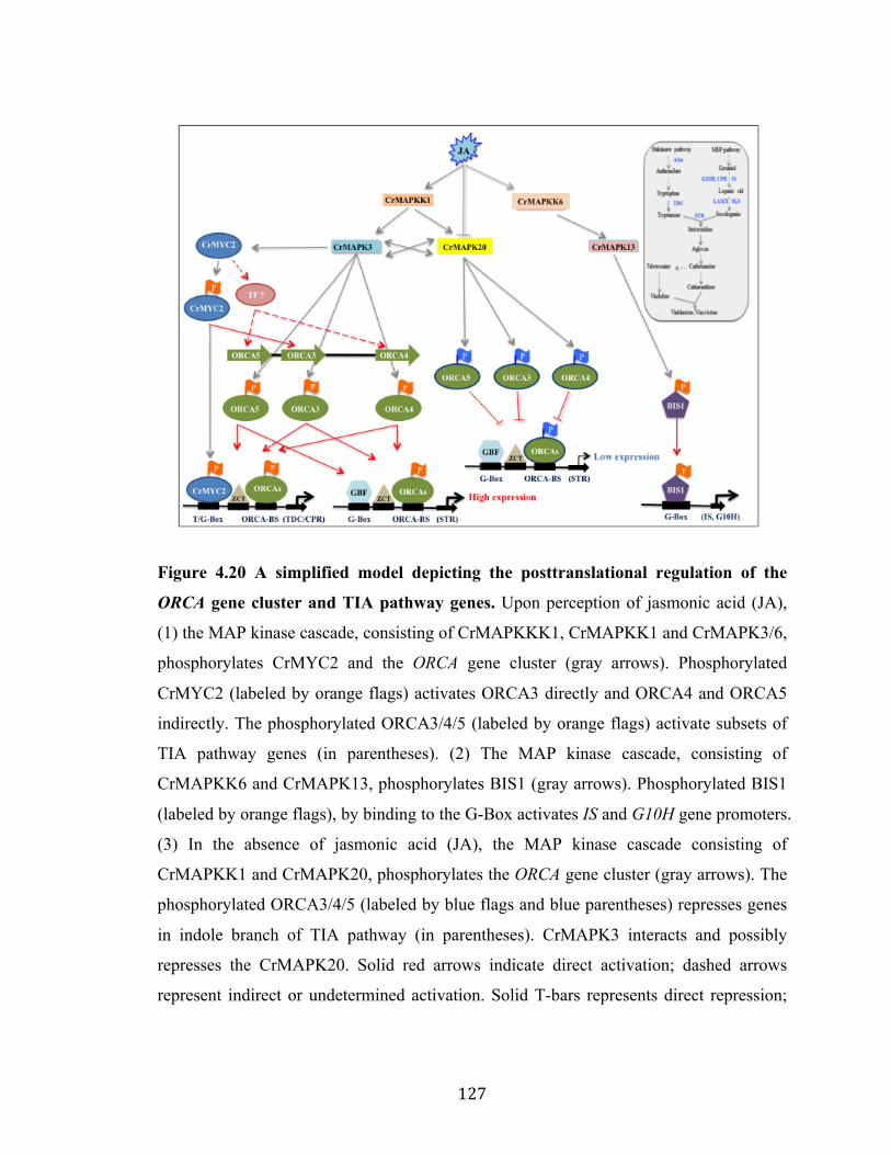
127
Figure 4.20 A simplified model depicting the posttranslational regulation of the
ORCA gene cluster and TIA pathway genes. Upon perception of jasmonic acid (JA),
(1) the MAP kinase cascade, consisting of CrMAPKKK1, CrMAPKK1 and CrMAPK3/6,
phosphorylates CrMYC2 and the ORCA gene cluster (gray arrows). Phosphorylated
CrMYC2 (labeled by orange flags) activates ORCA3 directly and ORCA4 and ORCA5
indirectly. The phosphorylated ORCA3/4/5 (labeled by orange flags) activate subsets of
TIA pathway genes (in parentheses). (2) The MAP kinase cascade, consisting of
CrMAPKK6 and CrMAPK13, phosphorylates BIS1 (gray arrows). Phosphorylated BIS1
(labeled by orange flags), by binding to the G-Box activates IS and G10H gene promoters.
(3) In the absence of jasmonic acid (JA), the MAP kinase cascade consisting of
CrMAPKK1 and CrMAPK20, phosphorylates the ORCA gene cluster (gray arrows). The
phosphorylated ORCA3/4/5 (labeled by blue flags and blue parentheses) represses genes
in indole branch of TIA pathway (in parentheses). CrMAPK3 interacts and possibly
represses the CrMAPK20. Solid red arrows indicate direct activation; dashed arrows
represent indirect or undetermined activation. Solid T-bars represents direct repression;

128
dashed T-bars indicate indirect or unknown repression. The X- indicates protein-protein
interaction. Inset: a simplified TIA pathway with key genes highlighted in blue.
Copyright © Priyanka Paul 2017

129
Chapter 5
Summary and Future directions
Summary: Catharanthus roseus (a.k.a Madagascar periwinkle), of the family
Apocynaceae, produces over a hundred terpenoid indole alkaloids (TIAs) including,
ajmalicine, serpentine, vinblastine, vincristine, which are used in the treatment of
hypertension and cancer (Kellner et al., 2015). TIAs are produced in a very limited
amount in plants. Although a significant progress has been made in isolation and
functional characterization of genes encoding major biosynthetic enzymes in the TIA
pathway (Liu et al., 2007; Asada et al., 2013; Besseau et al., 2013; Salim et al., 2014; Qu
et al., 2015), knowledge on the transcriptional and posttranslational regulation of TIA
pathway is still fragmented. The current knowledge on TIA biosynthesis and its
regulation, including transcriptional and posttranslational regulation, are discussed in
details in Chapter 1.
Previous studies showed that the TIA pathway is regulated by three major class of
transcription factors (TFs), such as AP2/ERFs (ORCA2 and 3) (Menke et al., 1999b; van
der Fits and Memelink, 2000), bHLH (CrMYC2, BIS1 and BIS2) (Zhang et al., 2011;
Van Moerkercke et al., 2015; Van Moerkercke et al., 2016), and WRKY (CrWRKY1)
(Suttipanta et al., 2011) TFs. Recently, analysis of C. roseus draft genome sequence
revealed that the JA-responsive AP2/ERF TF, ORCA3 forms a physical cluster with two
uncharacterized AP2/ERFs, AP2 TF1 030272 and AP2 TF2 030274 (Kellner et al., 2015).
In plants, physically linked clusters of TFs are less characterized. Moreover, the
regulation of TF clusters is relatively unexplored. In Chapter 2, my research uncovered
that the ORCA gene cluster is differentially regulated. ORCA4 and 5, though functionally
overlapping with ORCA3, regulate an additional set of TIA pathway genes. ORCA4 or
ORCA5 overexpression has resulted in significant increase of TIA accumulation in C.
roseus hairy roots. In addition, ORCA5 directly regulates the expression of ORCA4 and
indirectly ORCA3, likely via an unknown factor(s). ORCA3 and ORCA5 bind to a GC-
rich element in ORCA4 promoter. Interestingly, ORCA5 also activates the expression of
ZCT3, a negative regulator of the TIA pathway. In addition CrMYC2 shows dual

130
regulation: directly activating ORCA3 and co-regulating pathway genes concomitantly
with ORCA3.
Previous studies suggest that biosynthesis of TIAs is also regulated at the
posttranslational level, including protein phosphorylation (Menke et al., 1999a; Van Der
Fits and Memelink, 2001). A recent report also indicates that a mitogen-activated protein
kinase (MAPK) is involved in this process (Raina et al., 2012). In Chapter 3, my
research revealed that both the ORCA gene cluster and CrMYC2 act downstream of a
MAP kinase cascade consisting of CrMAPKK1, CrMAPK3 and 6. Overexpression of
CrMAPKK1 in C. roseus hairy roots upregulates key TIA pathway genes expressions and
boosts TIA accumulation.
Taking advantage of the C. roseus draft genome sequence, I identified all the putative
MAPK members and determined their phylogenetic classification, domain organization
and expression patterns in different organs in C. roseus in Chapter 4. Moreover, I
functionally characterized two previously uncharacterized MAPK cascades that regulate
two main branches of the TIA pathway: CrMAPKK6-CrMAPK13 regulates the iridoid
branch of the TIA pathway and overexpression of CrMAPK13 in C. roseus hairy roots
significantly up-regulates iridoid pathway genes and increases tabersonine accumulation.
The other MAPK cascade, CrMAPKK1-CrMAPK20 negatively regulates the indole
branch of the TIA pathway. CrMAPK20 overexpression resulted in repression of
CrMYC2-ORCAs regulated genes and reduced catharanthine accumulation in
CrMAPK20 overexpressing hairy roots.
In conclusion, the overall findings of my research project significantly advance our
understanding of the transcriptional and post-translational regulatory mechanisms that
govern TIA biosynthesis in C. roseus. Moreover, this knowledge could be utilized to
engineer in vivo or heterologous systems to produce these valuable TIAs for the benefits
of mankind.
Future direction 1: Protein phosphorylation and de-phosphorylation facilitated by
protein kinases (PKs) and protein phosphatases, respectively is a common signal

131
transduction mechanism in a number of biological processes. In contrasts to PKs, protein
phosphatases show more diversity and are divided into two major classes: protein
tyrosine phosphatases (PTPs) and protein serine (S)/threonine (T) phosphatases. The PTP
family includes tyrosine-specific and dual-specificity phosphatases (DSPTPs) (Denu et
al., 1996; Schweighofer et al., 2004). The protein serine/threonine phosphatases, on the
other hand, are classified into the protein phosphatases P (PPP) and protein phosphatases
M (PPM) gene families (Schweighofer et al., 2004). The PPP family includes type 1
phosphatases (PP1), and type 2 phosphatases that can be further divided into PP2A and
PP2B. The PPM family contains PP2C and pyruvate dehydrogenase phosphatase (Cohen,
1997). In Arabidopsis, 76 genes encoding PP2C-type phosphatases have been identified.
These PP2Cs are again classified into 10 groups (A–J) (Kerk et al., 2002). A previous
report suggested that in Arabidopsis a type 2C Ser/Thr phosphatase, AtPP2C1, change
the levels of JA and ethylene during wounding, negatively modulates MAPK4 and
MAPK6 to regulate the innate immunity. Therefore, AP2C1 is also considered as MAPK
phosphatases (MKP) (Schweighofer et al., 2007).
A potential homolog of AtPP2C1 was identified from C. roseus and designated here as
CrPP2C1. I cloned the CrPP2C1 that shares 51.74% amino acid sequence identity with
AP2C1 (Figure 5.1). To experimentally verify the involvement of CrPP2C1 in TIA
biosynthesis, I used a protoplast-based transactivation system (Pattanaik et al., 2010b).
The STR promoters, fused to a firefly luciferase reporter gene, was co-electroporated into
tobacco protoplasts with/without the constructs expressing ORCA3 and/or CrPP2C1. As
expected ORCA3 activated the STR promoter; however the activation was significantly
reduced when CrPP2C1 was co-expressed with ORCA3 (Figure 5.2). Thus, CrPP2C1
significantly reduces the transactivation potential of ORCA3 on STR promoter in a plant
cell. More experimental evidence are needed in future to demonstrate whether CrPP2C1
can dephosphorylate the CrMAPKK1-CrMAPK3/6 cascade to regulate the TIA
biosynthesis in C. roseus.

132
Figure 5.1. Multiple sequence alignment of CrPP2C1 with AtPP2C1 (AP2C1). The
identical amino acid residues in the alignment are in black shade and similar residues are
in the gray shade.
Figure 5.2. CrPP2C1 significantly reduces the transactivation potential of ORCA3
on STR promoter in a protoplast assay.

133
Future direction 2: The posttranscriptional modification also gives another layer of
complexity to the regulation of secondary metabolism, including TIAs biosynthesis in C.
roseus (Patra et al., 2013a; Shen et al., 2017). miRNAs, 21 to 24 nucleotides long, are a
major class of endogenous non-coding small regulatory RNAs that are ubiquitous to
plants (Bartel, 2009). In plants, miRNAs are involved in a number of biological processes,
such as hormone homeostasis (Meng et al., 2009), embryogenesis (Lin and Lai, 2013;
Zhai et al., 2014), root (Meng et al., 2010; Khan et al., 2011), and flower development
(Wang et al., 2012). However, the role of miRNA in the regulation of secondary
metabolism is less studied (Gou et al., 2011; Jia et al., 2015; Sharma et al., 2016).
Moreover, the regulatory roles miRNAs in TIAs biosynthesis in C. roseus is relatively
unexplored. In our previous studies, we identified 181 conserved and 173 novel cro-
miRNAs in C. roseus seedlings by using small RNA-sequencing. Interestingly, genome-
wide expression analysis and qRT-PCR validation identified a set of cro-miRNAs (cro-
miR393d, cro-miR164a, cro-miR164b, cro-miR168a etc.), which are differentially
regulated in response to MeJA, the key inducer of TIA biosynthesis (Shen et al., 2017).
Identification and functional characterization of candidate cro-miRNA(s), which directly
regulate(s) the structural and/or regulatory genes in TIA pathway will be promising future
aspects in TIA regulation studies.
Copyright © Priyanka Paul 2017

134
APPENDIX-A
LIST OF ABBREVIATIONS Acronym/abbreviation Expansion ACC 1-aminocyclopropane-1-carboxylic acid cDNA Complementary DNA DMSO Dimethyl sulfoxide DNA Deoxyribonucleic acid dNTP Deoxyribo nucleic triphosphate DTT Dithiothreitol DW Dry weight EDTA Ethylene diamine tetra acetic acid EF1∞ Elongation Factor 1∞ eGFP Enhanced green fluorescent protein EMSA Electrophoretic mobility shift assays g/mg/µg/ng Gram/ milligram/ microgram/ nanogram GFP Green fluorescent protein GUS ß-glucuronidase h/min/sec Hours/minutes/seconds HMM Hidden Markov Models HPLC High performance liquid chromatography JA Jasmonic acid kDa KiloDalton L/mL/µL Liter/ milliliter/ microliter LC-ESI-MS/MS Liquid Chromatography Electrospray
Ionization Tandem Mass Spectrometric M/mM/µM Molar/millimolar/ micromolar MeJA Methyl jasmonic acid MS Murashige and skoog MS media Murashige & Skoog media NaCl Sodium chloride NaOH Sodium hydroxide oC Degree centigrade PBS Phosphate buffered saline pBS pBlueScript PCR Polymerase chain reaction qRT-PCR Quantitative real-time PCR RNA Ribonucleic acid RPKM Reads per kilobase of transcript per million
mapped reads RPS9 40S Ribosomal Protein S9 SD Standard deviation SE Standard error SRA Sequence read archive TBE Tris- borate/ EDTA electrophoresis buffer TE TRIS-EDTA

135
TRIS Hydroxymethyl Aminomethane var. Variety Wt Wild-type µM Micron meter

136
References Agrawal, G.K., Agrawal, S.K., Shibato, J., Iwahashi, H., and Rakwal, R. (2003).
Novel rice MAP kinases OsMSRMK3 and OsWJUMK1 involved in encountering diverse environmental stresses and developmental regulation. Biochemical and biophysical research communications 300, 775-783.
Alves, M.S., Dadalto, S.P., Gonçalves, A.B., De Souza, G.B., Barros, V.A., and Fietto, L.G. (2013). Plant bZIP Transcription Factors Responsive to Pathogens: A Review. International Journal of Molecular Sciences 14, 7815-7828.
An, J.-P., Qu, F.-J., Yao, J.-F., Wang, X.-N., You, C.-X., Wang, X.-F., and Hao, Y.-J. (2017). The bZIP transcription factor MdHY5 regulates anthocyanin accumulation and nitrate assimilation in apple 4, 17023.
Andreasson, E., and Ellis, B. (2010). Convergence and specificity in the Arabidopsis MAPK nexus. Trends in plant science 15, 106-113.
Asada, K., Salim, V., Masada-Atsumi, S., Edmunds, E., Nagatoshi, M., Terasaka, K., Mizukami, H., and De Luca, V. (2013). A 7-deoxyloganetic acid glucosyltransferase contributes a key step in Secologanin Biosynthesis in Madagascar periwinkle. The Plant Cell Online 25, 4123-4134.
Asai, T., Tena, G., Plotnikova, J., Willmann, M.R., Chiu, W.-L., Gomez-Gomez, L., Boller, T., Ausubel, F.M., and Sheen, J. (2002). MAP kinase signalling cascade in Arabidopsis innate immunity. Nature 415, 977-983.
Atchley, W.R., and Fitch, W.M. (1997). A natural classification of the basic helix–loop–helix class of transcription factors. Proceedings of the National Academy of Sciences 94, 5172-5176.
Bailey, T.L., Williams, N., Misleh, C., and Li, W.W. (2006). MEME: discovering and analyzing DNA and protein sequence motifs. Nucleic Acids Res 34.
Bartel, D.P. (2009). MicroRNAs: target recognition and regulatory functions. cell 136, 215-233.
Berriri, S., Garcia, A.V., dit Frey, N.F., Rozhon, W., Pateyron, S., Leonhardt, N., Montillet, J.-L., Leung, J., Hirt, H., and Colcombet, J. (2012). Constitutively active mitogen-activated protein kinase versions reveal functions of Arabidopsis MPK4 in pathogen defense signaling. The Plant cell 24, 4281-4293.
Besseau, S., Kellner, F., Lanoue, A., Thamm, A.M., Salim, V., Schneider, B., Geu-Flores, F., Höfer, R., Guirimand, G., and Guihur, A. (2013). A pair of tabersonine 16-hydroxylases initiates the synthesis of vindoline in an organ-dependent manner in Catharanthus roseus. Plant physiology 163, 1792-1803.
Blom, T.J.M., Sierra, M., van Vliet, T.B., Franke-van Dijk, M.E.I., de Koning, P., van Iren, F., Verpoorte, R., and Libbenga, K.R. (1991). Uptake and accumulation of ajmalicine into isolated vacuoles of cultured cells of Catharanthus roseus (L.) G. Don. and its conversion into serpentine. Planta 183, 170-177.
Brown, S., Clastre, M., Courdavault, V., and O’Connor, S.E. (2015). De novo production of the plant-derived alkaloid strictosidine in yeast. Proceedings of the National Academy of Sciences 112, 3205-3210.
Burlat, V., Oudin, A., Courtois, M., Rideau, M., and St‐Pierre, B. (2004). Co‐expression of three MEP pathway genes and geraniol 10‐hydroxylase in internal

137
phloem parenchyma of Catharanthus roseus implicates multicellular translocation of intermediates during the biosynthesis of monoterpene indole alkaloids and isoprenoid‐derived primary metabolites. The Plant Journal 38, 131-141.
Çakır, B., and Kılıçkaya, O. (2015). Mitogen-activated protein kinase cascades in Vitis vinifera. Frontiers in Plant Science 6, 556.
Cárdenas, P.D., Sonawane, P.D., Pollier, J., Bossche, R.V., Dewangan, V., Weithorn, E., Tal, L., Meir, S., Rogachev, I., and Malitsky, S. (2016). GAME9 regulates the biosynthesis of steroidal alkaloids and upstream isoprenoids in the plant mevalonate pathway. Nature communications 7.
Cardinale, F., Meskiene, I., Ouaked, F., and Hirt, H. (2002). Convergence and divergence of stress-induced mitogen-activated protein kinase signaling pathways at the level of two distinct mitogen-activated protein kinase kinases. The Plant cell 14, 703-711.
Cardoso, M.I.L., Meijer, A.H., Rueb, S., Machado, J.Q., Memelink, J., and Hoge, J.H.C. (1997). A promoter region that controls basal and elicitor-inducible expression levels of the NADPH:cytochrome P450 reductase gene (Cpr ) from Catharanthus roseus binds nuclear factor GT-1. Molecular and General Genetics MGG 256, 674-681.
Carretero-Paulet, L., Galstyan, A., Roig-Villanova, I., Martínez-García, J.F., Bilbao-Castro, J.R., and Robertson, D.L. (2010). Genome-wide classification and evolutionary analysis of the bHLH family of transcription factors in Arabidopsis, poplar, rice, moss, and algae. Plant physiology 153, 1398-1412.
Chahed, K., Oudin, A., Guivarc’h, N., Hamdi, S., Chénieux, J.-C., Rideau, M., and Clastre, M. (2000). 1-Deoxy-D-xylulose 5-phosphate synthase from periwinkle: cDNA identification and induced gene expression in terpenoid indole alkaloid-producing cells. Plant Physiology and Biochemistry 38, 559-566.
Champion, A., Picaud, A., and Henry, Y. (2004). Reassessing the MAP3K and MAP4K relationships. Trends in plant science 9, 123-129.
Chávez Suárez, L., and Ramírez Fernández, R. (2010). Signalling pathway in plants affected by salinity and drought. ITEA 106, 157-169.
Chen, L., Hu, W., Tan, S., Wang, M., Ma, Z., Zhou, S., Deng, X., Zhang, Y., Huang, C., and Yang, G. (2012). Genome-wide identification and analysis of MAPK and MAPKK gene families in Brachypodium distachyon. PLoS One 7, e46744.
Cohen, P.T. (1997). Novel protein serine/threonine phosphatases: variety is the spice of life. Trends in biochemical sciences 22, 245-251.
Collu, G., Unver, N., Peltenburg-Looman, A.M., van der Heijden, R., Verpoorte, R., and Memelink, J. (2001). Geraniol 10‐hydroxylase1, a cytochrome P450 enzyme involved in terpenoid indole alkaloid biosynthesis. FEBS letters 508, 215-220.
Contin, A., van der Heijden, R., Lefeber, A.W., and Verpoorte, R. (1998). The iridoid glucoside secologanin is derived from the novel triose phosphate/pyruvate pathway in a Catharanthus roseus cell culture. FEBS letters 434, 413-416.
Costa, M.M.R., Hilliou, F., Duarte, P., Pereira, L.G., Almeida, I., Leech, M., Memelink, J., Barceló, A.R., and Sottomayor, M. (2008). Molecular cloning and characterization of a vacuolar class III peroxidase involved in the metabolism of anticancer alkaloids in Catharanthus roseus. Plant Physiology 146, 403-417.

138
Cristina, M.S., Petersen, M., and Mundy, J. (2010). Mitogen-activated protein kinase signaling in plants. Annual review of plant biology 61, 621-649.
De Boer, K., Tilleman, S., Pauwels, L., Vanden Bossche, R., De Sutter, V., Vanderhaeghen, R., Hilson, P., Hamill, J.D., and Goossens, A. (2011). APETALA2/ETHYLENE RESPONSE FACTOR and basic helix–loop–helix tobacco transcription factors cooperatively mediate jasmonate‐elicited nicotine biosynthesis. The Plant Journal 66, 1053-1065.
De Carolis, E., and De Luca, V. (1993). Purification, characterization, and kinetic analysis of a 2-oxoglutarate- dependent dioxygenase involved in vindoline biosynthesis from Catharanthus roseus. Journal of Biological Chemistry 268, 5504-5511.
De Carolis, E., Chan, F., Balsevich, J., and De Luca, V. (1990). Isolation and characterization of a 2-oxoglutarate dependent dioxygenase involved in the second-to-last step in vindoline biosynthesis. Plant physiology 94, 1323-1329.
De Luca, V., and Cutler, A.J. (1987). Subcellular localization of enzymes involved in indole alkaloid biosynthesis in Catharanthus roseus. Plant physiology 85, 1099-1102.
De Luca, V., Marineau, C., and Brisson, N. (1989). Molecular cloning and analysis of cDNA encoding a plant tryptophan decarboxylase: comparison with animal dopa decarboxylases. Proceedings of the National Academy of Sciences 86, 2582-2586.
Denu, J.M., Stuckey, J.A., Saper, M.A., and Dixon, J.E. (1996). Form and Function in Protein Dephosphorylation. Cell 87, 361-364.
Ding, X., Richter, T., Chen, M., Fujii, H., Seo, Y.S., Xie, M., Zheng, X., Kanrar, S., Stevenson, R.A., and Dardick, C. (2009). A rice kinase-protein interaction map. Plant physiology 149, 1478-1492.
Duan, P.G., Rao, Y.C., Zeng, D.L., Yang, Y.L., Xu, R., and Zhang, B.L. (2014). SMALL GRAIN 1, which encodes a mitogen-activated protein kinase kinase 4, influences grain size in rice. The Plant journal : for cell and molecular biology 77.
Duarte, J.M., Cui, L., Wall, P.K., Zhang, Q., Zhang, X., Leebens-Mack, J., Ma, H., and Altman, N. (2006). Expression pattern shifts following duplication indicative of subfunctionalization and neofunctionalization in regulatory genes of Arabidopsis. Molecular biology and evolution 23, 469-478.
Eckardt, N.A. (2007). A complete MAPK signaling cascade that functions in stomatal development and patterning in Arabidopsis (Am Soc Plant Biol).
El-Sayed, M., and Verpoorte, R. (2007). Catharanthus terpenoid indole alkaloids: biosynthesis and regulation. Phytochemistry Reviews 6, 277-305.
Ellenberger, T.E., Brandl, C.J., Struhl, K., and Harrison, S.C. The GCN4 basic region leucine zipper binds DNA as a dimer of uninterrupted α Helices: Crystal structure of the protein-DNA complex. Cell 71, 1223-1237.
Facchini, P.J., and De Luca, V. (2008). Opium poppy and Madagascar periwinkle: model non‐model systems to investigate alkaloid biosynthesis in plants. The Plant Journal 54, 763-784.
Fahn, W., Laußermair, E., Deus-Neumann, B., and Stöckigt, J. (1985). Late enzymes of vindoline biosynthesis. Plant Cell Reports 4, 337-340.
Fanger, G.R., Johnson, N.L., and Johnson, G.L. (1997). MEK kinases are regulated by EGF and selectively interact with Rac/Cdc42. The EMBO Journal 16, 4961-4972.

139
Fernández-Calvo, P., Chini, A., Fernández-Barbero, G., Chico, J.-M., Gimenez-Ibanez, S., Geerinck, J., Eeckhout, D., Schweizer, F., Godoy, M., and Franco-Zorrilla, J.M. (2011). The Arabidopsis bHLH transcription factors MYC3 and MYC4 are targets of JAZ repressors and act additively with MYC2 in the activation of jasmonate responses. The Plant cell 23, 701-715.
Field, B., Fiston-Lavier, A.S., Kemen, A., Geisler, K., Quesneville, H., and Osbourn, A.E. (2011). Formation of plant metabolic gene clusters within dynamic chromosomal regions. Proceedings of the National Academy of Sciences USA 108, 16116-16121.
Finn, R.D., Clements, J., and Eddy, S.R. (2011). HMMER web server: interactive sequence similarity searching. Nucleic acids research 39, W29-37.
Finn, R.D., Mistry, J., Tate, J., Coggill, P., Heger, A., Pollington, J.E., Gavin, O.L., Gunasekaran, P., Ceric, G., Forslund, K., Holm, L., Sonnhammer, E.L., Eddy, S.R., and Bateman, A. (2010). The Pfam protein families database. Nucleic acids research 38, D211-222.
Frey, M., Chomet, P., Glawischnig, E., Stettner, C., Grun, S., Winklmair, A., Eisenreich, W., Bacher, A., Meeley, R.B., Briggs, S.P., Simcox, K., and Gierl, A. (1997). Analysis of a chemical plant defense mechanism in grasses. Science 277, 696-699.
Frye, C.A., and Innes, R.W. (1998). An Arabidopsis mutant with enhanced resistance to powdery mildew. The Plant cell 10, 947-956.
Frye, C.A., Tang, D., and Innes, R.W. (2001). Negative regulation of defense responses in plants by a conserved MAPKK kinase. Proceedings of the National Academy of Sciences 98, 373-378.
Fujimoto, S.Y., Ohta, M., Usui, A., Shinshi, H., and Ohme-Takagi, M. (2000). Arabidopsis ethylene-responsive element binding factors act as transcriptional activators or repressors of GCC box–mediated gene expression. The Plant Cell Online 12, 393-404.
Galletti, R., Ferrari, S., and De Lorenzo, G. (2011). Arabidopsis MPK3 and MPK6 play different roles in basal and oligogalacturonide-or flagellin-induced resistance against Botrytis cinerea. Plant Physiology 157, 804-814.
Geu-Flores, F., Sherden, N.H., Courdavault, V., Burlat, V., Glenn, W.S., Wu, C., Nims, E., Cui, Y., and O’Connor, S.E. (2012). An alternative route to cyclic terpenes by reductive cyclization in iridoid biosynthesis. Nature 492, 138-142.
Giddings, L.-A., Liscombe, D.K., Hamilton, J.P., Childs, K.L., DellaPenna, D., Buell, C.R., and O'Connor, S.E. (2011). A stereoselective hydroxylation step of alkaloid biosynthesis by a unique cytochrome P450 in Catharanthus roseus. Journal of Biological Chemistry 286, 16751-16757.
Góngora-Castillo, E., Childs, K.L., Fedewa, G., Hamilton, J.P., Liscombe, D.K., Magallanes-Lundback, M., Mandadi, K.K., Nims, E., Runguphan, W., and Vaillancourt, B. (2012). Development of transcriptomic resources for interrogating the biosynthesis of monoterpene indole alkaloids in medicinal plant species. PLoS One 7, e52506.
Gou, J.-Y., Felippes, F.F., Liu, C.-J., Weigel, D., and Wang, J.-W. (2011). Negative regulation of anthocyanin biosynthesis in Arabidopsis by a miR156-targeted SPL transcription factor. The Plant cell 23, 1512-1522.

140
Hallahan, D.L., West, J.M., Wallsgrove, R.M., Smiley, D.W., Dawson, G.W., Pickett, J.A., and Hamilton, J.G. (1995). Purification and characterization of an acyclic monoterpene primary alcohol: NADP+ oxidoreductase from catmint (Nepeta racemosa). Archives of biochemistry and biophysics 318, 105-112.
Hamel, L., Nicole, M., Sritubtim, S., Morency, M., Ellis, M., Ehlting, J., Beaudoin, N., Barbazuk, B., Klessig, D., Lee, J., Martin, G., Mundy, J., Ohashi, Y., Scheel, D., Sheen, J., Xing, T., Zhang, S., Seguin, A., and Ellis, B. (2006). Ancient signals: comparative genomics of plant MAPK and MAPKK gene families. Trends Plant Sci 11, 192 - 198.
Hanada, K., Zou, C., Lehti-Shiu, M.D., Shinozaki, K., and Shiu, S.-H. (2008). Importance of lineage-specific expansion of plant tandem duplicates in the adaptive response to environmental stimuli. Plant physiology 148, 993-1003.
Hanks, S.K., and Hunter, T. (1995). Protein kinases 6. The eukaryotic protein kinase superfamily: kinase (catalytic) domain structure and classification. The FASEB journal 9, 576-596.
Heim, M.A., Jakoby, M., Werber, M., Martin, C., Weisshaar, B., and Bailey, P.C. (2003). The Basic Helix–Loop–Helix Transcription Factor Family in Plants: A Genome-Wide Study of Protein Structure and Functional Diversity. Molecular Biology and Evolution 20, 735-747.
Heinrich, M., Baldwin, I.T., and Wu, J. (2011). Two mitogen-activated protein kinase kinases, MKK1 and MEK2, are involved in wounding-and specialist lepidopteran herbivore Manduca sexta-induced responses in Nicotiana attenuata. Journal of experimental botany, err162.
Hemscheidt, T., and Zenk, M. (1985). Partial purification and characterization of a NADPH dependent tetrahydroalstonine synthase from Catharanthus roseus cell suspension cultures. Plant cell reports 4, 216-219.
Hiratsu, K., Matsui, K., Koyama, T., and Ohme‐Takagi, M. (2003). Dominant repression of target genes by chimeric repressors that include the EAR motif, a repression domain, in Arabidopsis. The Plant Journal 34, 733-739.
Hord, C.L.H., Suna, Y.J., Pillitteri, L.J., Torii, K.U., Wang, H.C., and Zhang, S.Q. (2008). Regulation of Arabidopsis early anther development by the mitogen-activated protein kinases, MPK3 and MPK6, and the ERECTA and related receptor-like kinases. Mol Plant 1.
Huang, S., Li, R., Zhang, Z., Li, L., Gu, X., Fan, W., Lucas, W.J., Wang, X., Xie, B., Ni, P., Ren, Y., Zhu, H., Li, J., Lin, K., Jin, W., Fei, Z., Li, G., Staub, J., Kilian, A., van der Vossen, E.A., Wu, Y., Guo, J., He, J., Jia, Z., Ren, Y., Tian, G., Lu, Y., Ruan, J., Qian, W., Wang, M., Huang, Q., Li, B., Xuan, Z., Cao, J., Asan, Wu, Z., Zhang, J., Cai, Q., Bai, Y., Zhao, B., Han, Y., Li, Y., Li, X., Wang, S., Shi, Q., Liu, S., Cho, W.K., Kim, J.Y., Xu, Y., Heller-Uszynska, K., Miao, H., Cheng, Z., Zhang, S., Wu, J., Yang, Y., Kang, H., Li, M., Liang, H., Ren, X., Shi, Z., Wen, M., Jian, M., Yang, H., Zhang, G., Yang, Z., Chen, R., Liu, S., Li, J., Ma, L., Liu, H., Zhou, Y., Zhao, J., Fang, X., Li, G., Fang, L., Li, Y., Liu, D., Zheng, H., Zhang, Y., Qin, N., Li, Z., Yang, G., Yang, S., Bolund, L., Kristiansen, K., Zheng, H., Li, S., Zhang, X., Yang, H., Wang, J., Sun, R., Zhang, B., Jiang, S., Wang, J., Du, Y., and Li, S. (2009). The genome of the cucumber, Cucumis sativus L. Nat Genet 41, 1275-1281.

141
Huang, Y., Li, H., Hutchison, C.E., Laskey, J., and Kieber, J.J. (2003). Biochemical and functional analysis of CTR1, a protein kinase that negatively regulates ethylene signaling in Arabidopsis. The Plant Journal 33, 221-233.
Huang, Y., Li, H., Gupta, R., Morris, P.C., Luan, S., and Kieber, J.J. (2000). ATMPK4, an Arabidopsis homolog of mitogen-activated protein kinase, is activated in vitro by AtMEK1 through threonine phosphorylation. Plant Physiology 122, 1301-1310.
Ichimura, K., Mizoguchi, T., Irie, K., Morris, P., Giraudat, J., Matsumoto, K., and Shinozaki, K. (1998). Isolation of ATMEKK1 (a MAP Kinase Kinase Kinase)-interacting proteins and analysis of a MAP kinase cascade inArabidopsis. Biochemical and biophysical research communications 253, 532-543.
Ichimura, K., Shinozaki, K., Tena, G., Sheen, J., Henry, Y., Champion, A., Kreis, M., Zhang, S., Hirt, H., and Wilson, C. (2002). Mitogen-activated protein kinase cascades in plants: a new nomenclature. Trends in plant science 7, 301-308.
Ikeda, H., Esaki, N., Nakai, S., Hashimoto, K., Uesato, S., Soda, K., and Fujita, T. (1991). Acyclic monoterpene primary alcohol: NADP+ oxidoreductase of Rauwolfia serpentina cells: the key enzyme in biosynthesis of monoterpene alcohols.
Irmler, S., Schröder, G., St‐Pierre, B., Crouch, N.P., Hotze, M., Schmidt, J., Strack, D., Matern, U., and Schröder, J. (2000). Indole alkaloid biosynthesis in Catharanthus roseus: new enzyme activities and identification of cytochrome P450 CYP72A1 as secologanin synthase. The Plant Journal 24, 797-804.
Itkin, M., Heinig, U., Tzfadia, O., Bhide, A.J., Shinde, B., Cardenas, P.D., Bocobza, S.E., Unger, T., Malitsky, S., Finkers, R., Tikunov, Y., Bovy, A., Chikate, Y., Singh, P., Rogachev, I., Beekwilder, J., Giri, A.P., and Aharoni, A. (2013). Biosynthesis of antinutritional alkaloids in solanaceous crops is mediated by clustered genes. Science 341, 175-179.
Iyer, G.H., Moore, M.J., and Taylor, S.S. (2005). Consequences of lysine 72 mutation on the phosphorylation and activation state of cAMP-dependent kinase. Journal of Biological Chemistry 280, 8800-8807.
Jakoby, M., Weisshaar, B., Dröge-Laser, W., Vicente-Carbajosa, J., Tiedemann, J., Kroj, T., and Parcy, F. (2002). bZIP transcription factors in Arabidopsis. Trends in Plant Science 7, 106-111.
Jalmi, S.K., and Sinha, A.K. (2016). Functional involvement of a mitogen activated protein kinase module, OsMKK3-OsMPK7-OsWRK30 in mediating resistance against Xanthomonas oryzae in rice. Scientific reports 6.
Jander, G., and Barth, C. (2007). Tandem gene arrays: a challenge for functional genomics. Trends in Plant Science 12, 203-210.
Jia, X., Shen, J., Liu, H., Li, F., Ding, N., Gao, C., Pattanaik, S., Patra, B., Li, R., and Yuan, L. (2015). Small tandem target mimic-mediated blockage of microRNA858 induces anthocyanin accumulation in tomato. Planta 242, 283-293.
Jiménez, C., Cossío, B.R., Rivard, C.J., Berl, T., and Capasso, J.M. (2007). Cell division in the unicellular microalga Dunaliella viridis depends on phosphorylation of extracellular signal-regulated kinases (ERKs). Journal of experimental botany 58, 1001-1011.

142
Jin, H., Liu, Y., Yang, K.Y., Kim, C.Y., Baker, B., and Zhang, S. (2003). Function of a mitogen-activated protein kinase pathway in N gene-mediated resistance in tobacco. Plant Journal 33, 719-731.
Jonak, C., Ökrész, L., Bögre, L., and Hirt, H. (2002). Complexity, cross talk and integration of plant MAP kinase signalling. Current opinion in plant biology 5, 415-424.
Jouannic, S., Hamal, A., Leprince, A.-S., Tregear, J.W., Kreis, M., and Henry, Y. (1999). Characterisation of novel plant genes encoding MEKK/STE11 and RAF-related protein kinases. Gene 229, 171-181.
Jun, J.H., Ha, C.M., and Nam, H.G. (2002). Involvement of the VEP1 gene in vascular strand development in Arabidopsis thaliana. Plant and cell physiology 43, 323-330.
Kato, N., Dubouzet, E., Kokabu, Y., Yoshida, S., Taniguchi, Y., Dubouzet, J.G., Yazaki, K., and Sato, F. (2007). Identification of a WRKY protein as a transcriptional regulator of benzylisoquinoline alkaloid biosynthesis in Coptis japonica. Plant and cell physiology 48, 8-18.
Keat, H., Elizabeth, G., and McKnight, T. (2000). Characterization and cloning of 10-hydroxygeraniol oxidoreductase. Plant Biology.
Kellner, F., Kim, J., Clavijo, B.J., Hamilton, J.P., Childs, K.L., Vaillancourt, B., Cepela, J., Habermann, M., Steuernagel, B., and Clissold, L. (2015). Genome‐guided investigation of plant natural product biosynthesis. The Plant Journal 82, 680-692.
Kerk, D., Bulgrien, J., Smith, D.W., Barsam, B., Veretnik, S., and Gribskov, M. (2002). The Complement of Protein Phosphatase Catalytic Subunits Encoded in the Genome of Arabidopsis. Plant Physiology 129, 908.
Khan, G.A., Declerck, M., Sorin, C., Hartmann, C., Crespi, M., and Lelandais-Brière, C. (2011). MicroRNAs as regulators of root development and architecture. Plant molecular biology 77, 47-58.
Kieber, J.J., Rothenberg, M., Roman, G., Feldmann, K.A., and Ecker, J.R. (1993). CTR1, a negative regulator of the ethylene response pathway in Arabidopsis, encodes a member of the raf family of protein kinases. Cell 72, 427-441.
Kiegerl, S., Cardinale, F., Siligan, C., Gross, A., Baudouin, E., Liwosz, A., Eklof, S., Till, S., Bogre, L., Hirt, H., and Meskiene, I. (2000). SIMKK, a mitogen-activated protein kinase (MAPK) kinase, is a specific activator of the salt stress-induced MAPK, SIMK. The Plant cell 12, 2247-2258.
Kolch, W. (2000). Meaningful relationships: the regulation of the Ras/Raf/MEK/ERK pathway by protein interactions. Biochem. J 351, 289-305.
Kong, F., Wang, J., Cheng, L., Liu, S., Wu, J., Peng, Z., and Lu, G. (2012). Genome-wide analysis of the mitogen-activated protein kinase gene family in Solanum lycopersicum. Gene 499, 108-120.
Kong, X., Lv, W., Zhang, D., Jiang, S., Zhang, S., and Li, D. (2013a). Genome-wide identification and analysis of expression profiles of maize mitogen-activated protein kinase kinase kinase. PloS one 8, e57714.
Kong, X., Pan, J., Zhang, D., Jiang, S., Cai, G., Wang, L., and Li, D. (2013b). Identification of mitogen-activated protein kinase kinase gene family and MKK–

143
MAPK interaction network in maize. Biochemical and biophysical research communications 441, 964-969.
Kosetsu, K., Matsunaga, S., Nakagami, H., Colcombet, J., Sasabe, M., Soyano, T., Takahashi, Y., Hirt, H., and Machida, Y. (2010). The MAP kinase MPK4 is required for cytokinesis in Arabidopsis thaliana. The Plant cell 22, 3778-3790.
Krithika, R., Srivastava, P.L., Rani, B., Kolet, S.P., Chopade, M., Soniya, M., and Thulasiram, H.V. (2015). Characterization of 10-Hydroxygeraniol Dehydrogenase from Catharanthus roseus Reveals Cascaded Enzymatic Activity in Iridoid Biosynthesis. Scientific reports 5.
Kumar, K., Rao, K.P., Biswas, D.K., and Sinha, A.K. (2011). Rice WNK1 is regulated by abiotic stress and involved in internal circadian rhythm. Plant Signal Behav 6.
Laflamme, P., St-Pierre, B., and De Luca, V. (2001). Molecular and biochemical analysis of a Madagascar periwinkle root-specific minovincinine-19-hydroxy-O-acetyltransferase. Plant physiology 125, 189-198.
Larkin, M.A., Blackshields, G., Brown, N., Chenna, R., McGettigan, P.A., McWilliam, H., Valentin, F., Wallace, I.M., Wilm, A., and Lopez, R. (2007). Clustal W and Clustal X version 2.0. Bioinformatics 23, 2947-2948.
Larsen, B., Fuller, V.L., Pollier, J., Van Moerkercke, A., Schweizer, F., Payne, R., Colinas, M., O’Connor, S.E., Goossens, A., and Halkier, B.A. (2017). Identification of Iridoid Glucoside Transporters in Catharanthus roseus. Plant and Cell Physiology.
Lee, J.S., Huh, K.W., Bhargava, A., and Ellis, B.E. (2008). Comprehensive analysis of protein-protein interactions between Arabidopsis MAPKs and MAPK kinases helps define potential MAPK signalling modules. Plant signaling & behavior 3, 1037-1041.
Lehti-Shiu, M.D., and Shiu, S.-H. (2012). Diversity, classification and function of the plant protein kinase superfamily. Philosophical Transactions of the Royal Society of London B: Biological Sciences 367, 2619-2639.
Lehti-Shiu, M.D., Zou, C., Hanada, K., and Shiu, S.-H. (2009). Evolutionary history and stress regulation of plant receptor-like kinase/pelle genes. Plant Physiology 150, 12-26.
Levac, D., Murata, J., Kim, W.S., and De Luca, V. (2008). Application of carborundum abrasion for investigating the leaf epidermis: Molecular cloning of Catharanthus roseus 16-hydroxytabersonine-16-O- methyltransferase. Plant Journal 53, 225-236.
Li, F., Li, M., Wang, P., Cox, K.L., Duan, L., Dever, J.K., Shan, L., Li, Z., and He, P. (2017). Regulation of cotton (Gossypium hirsutum) drought responses by mitogen-activated protein (MAP) kinase cascade-mediated phosphorylation of GhWRKY59. New Phytologist 215, 1462-1475.
Li, M., and Sack, F.D. (2014). Myrosin Idioblast Cell Fate and Development Are Regulated by the <em>Arabidopsis</em> Transcription Factor FAMA, the Auxin Pathway, and Vesicular Trafficking. The Plant cell 26, 4053.
Liang, W., Yang, B., Yu, B.-J., Zhou, Z., Li, C., Jia, M., Sun, Y., Zhang, Y., Wu, F., and Zhang, H. (2013). Identification and analysis of MKK and MPK gene families in canola (Brassica napus L.). BMC genomics 14, 392.

144
Lichtenthaler, H.K. (1999). The 1-deoxy-D-xylulose-5-phosphate pathway of isoprenoid biosynthesis in plants. Annual review of plant biology 50, 47-65.
Lin, Y., and Lai, Z. (2013). Comparative analysis reveals dynamic changes in miRNAs and their targets and expression during somatic embryogenesis in longan (Dimocarpus longan Lour.). PLoS One 8, e60337.
Liscombe, D.K., Usera, A.R., and O'Connor, S.E. (2010). Homolog of tocopherol C methyltransferases catalyzes N methylation in anticancer alkaloid biosynthesis. Proceedings of the National Academy of Sciences USA 107, 18793-18798.
Liu, D.H., Jin, H.B., Chen, Y.H., Cui, L.J., Ren, W.W., Gong, Y.F., and Tang, K.X. (2007). Terpenoid indole alkaloids biosynthesis and metabolic engineering in Catharanthus roseus. Journal of integrative plant biology 49, 961-974.
Liu, H., Wang, Y., Xu, J., Su, T., Liu, G., and Ren, D. (2008). Ethylene signaling is required for the acceleration of cell death induced by the activation of AtMEK5 in Arabidopsis. Cell research 18, 422.
Liu, J.Z., Horstman, H.D., Braun, E., Graham, M.A., Zhang, C.Q., and Navarre, D. (2011). Soybean homologs of MPK4 negatively regulate defense responses and positively regulate growth and development. Plant Physiol 157.
Liu, Y., and Zhang, S. (2004). Phosphorylation of 1-aminocyclopropane-1-carboxylic acid synthase by MPK6, a stress-responsive mitogen-activated protein kinase, induces ethylene biosynthesis in Arabidopsis. The Plant cell 16, 3386 - 3399.
Liu, Y., Zhang, D., Wang, L., and Li, D. (2013). Genome-wide analysis of mitogen-activated protein kinase gene family in maize. Plant molecular biology reporter 31, 1446-1460.
Liu, Z., Shi, L., Liu, Y., Tang, Q., Shen, L., Yang, S., Cai, J., Yu, H., Wang, R., and Wen, J. (2014). Genome-wide identification and transcriptional expression analysis of mitogen-activated protein kinase and mitogen-activated protein kinase kinase genes in Capsicum annuum. Frontiers in plant science 6, 780-780.
Lu, K., Guo, W., Lu, J., Yu, H., Qu, C., Tang, Z., Li, J., Chai, Y., and Liang, Y. (2015). Genome-wide survey and expression profile analysis of the mitogen-activated protein kinase (MAPK) gene family in Brassica rapa. PloS one 10, e0132051.
Ma, D., Pu, G., Lei, C., Ma, L., Wang, H., Guo, Y., Chen, J., Du, Z., Wang, H., and Li, G. (2009). Isolation and characterization of AaWRKY1, an Artemisia annua transcription factor that regulates the amorpha-4, 11-diene synthase gene, a key gene of artemisinin biosynthesis. Plant and Cell Physiology 50, 2146-2161.
MacAlister, C.A., and Bergmann, D.C. (2011). Sequence and function of basic helix–loop–helix proteins required for stomatal development in Arabidopsis are deeply conserved in land plants. Evolution & Development 13, 182-192.
Maier, A., Schrader, A., Kokkelink, L., Falke, C., Welter, B., Iniesto, E., Rubio, V., Uhrig, J.F., Hülskamp, M., and Hoecker, U. (2013). Light and the E3 ubiquitin ligase COP1/SPA control the protein stability of the MYB transcription factors PAP1 and PAP2 involved in anthocyanin accumulation in Arabidopsis. The Plant Journal 74, 638-651.
Major, G., Daigle, C., Stafford-Richard, T., Tebbji, F., Lafleur, É., Caron, S., and Matton, D.P. (2009). Characterization of ScMAP4K1, a MAP kinase kinase

145
kinase kinase involved in ovule, seed and fruit development in Solanum chacoense Bitt. Plant Biology 10.
Mao, G., Meng, X., Liu, Y., Zheng, Z., Chen, Z., and Zhang, S. (2011). Phosphorylation of a WRKY transcription factor by two pathogen-responsive MAPKs drives phytoalexin biosynthesis in Arabidopsis. The Plant cell 23, 1639-1653.
McGrath, K.C., Dombrecht, B., Manners, J.M., Schenk, P.M., Edgar, C.I., Maclean, D.J., Scheible, W.-R., Udvardi, M.K., and Kazan, K. (2005). Repressor-and activator-type ethylene response factors functioning in jasmonate signaling and disease resistance identified via a genome-wide screen of Arabidopsis transcription factor gene expression. Plant Physiology 139, 949-959.
McKnight, T., Roessner, C., Devagupta, R., Scott, A., and Nessler, C. (1990). Nucleotide sequence of a cDNA encoding the vacuolar protein strictosidine synthase from Catharanthus roseus. Nucleic acids research 18, 4939-4939.
McKnight, T.D., Bergey, D.R., Burnett, R.J., and Nessler, C.L. (1991). Expression of enzymatically active and correctly targeted strictosidine synthase in transgenic tobacco plants. Planta 185, 148-152.
Meijer, A.H., Cardoso, M., Voskuilen, J.T., Waal, A., Verpoorte, R., and Hoge, J.H.C. (1993). Isolation and characterization of a cDNA clone from Catharanthus roseus encoding NADPH: cytochrome P‐450 reductase, an enzyme essential for reactions catalysed by cytochrome P‐450 mono‐oxygenases in plants. The Plant Journal 4, 47-60.
Meng, X., Xu, J., He, Y., Yang, K.-Y., Mordorski, B., Liu, Y., and Zhang, S. (2013). Phosphorylation of an ERF transcription factor by Arabidopsis MPK3/MPK6 regulates plant defense gene induction and fungal resistance. The Plant cell 25, 1126-1142.
Meng, Y., Ma, X., Chen, D., Wu, P., and Chen, M. (2010). MicroRNA-mediated signaling involved in plant root development. Biochemical and biophysical research communications 393, 345-349.
Meng, Y., Huang, F., Shi, Q., Cao, J., Chen, D., Zhang, J., Ni, J., Wu, P., and Chen, M. (2009). Genome-wide survey of rice microRNAs and microRNA–target pairs in the root of a novel auxin-resistant mutant. Planta 230, 883-898.
Menke, F., Stefanie, P., Mueller, M., Kijne, J., and Memelink, J. (1999a). Involvement of the Octadecanoid pathway and protein phosphorylation in fungal elicitor induced expression of Terpenoid Indole Alkaloid biosynthetic genes in Catharanthus roseus. Plant Physiol 119, 1289 - 1296.
Menke, F.L., Champion, A., Kijne, J.W., and Memelink, J. (1999b). A novel jasmonate- and elicitor-responsive element in the periwinkle secondary metabolite biosynthetic gene Str interacts with a jasmonate- and elicitor-inducible AP2-domain transcription factor, ORCA2. EMBO Journal 18, 4455-4463.
Miettinen, K., Dong, L., Navrot, N., Schneider, T., Burlat, V., Pollier, J., Woittiez, L., Van Der Krol, S., Lugan, R., and Ilc, T. (2014). The seco-iridoid pathway from Catharanthus roseus. Nature communications 5.
Mirjalili, N., and Linden, J.C. (1996). Methyl jasmonate induced production of taxol in suspension cultures of Taxus cuspidata: ethylene interaction and induction models. Biotechnology progress 12, 110-118.

146
Mizoi, J., Shinozaki, K., and Yamaguchi-Shinozaki, K. (2012). AP2/ERF family transcription factors in plant abiotic stress responses. Biochimica et Biophysica Acta (BBA)-Gene Regulatory Mechanisms 1819, 86-96.
Munkert, J., Pollier, J., Miettinen, K., Van Moerkercke, A., Payne, R., Müller-Uri, F., Burlat, V., O’Connor, S.E., Memelink, J., and Kreis, W. (2015). Iridoid synthase activity is common among the plant progesterone 5β-reductase family. Molecular plant 8, 136-152.
Murakami-Kojima, M., Nakamichi, N., Yamashino, T., and Mizuno, T. (2002). The APRR3 component of the clock-associated APRR1/TOC1 quintet is phosphorylated by a novel protein kinase belonging to the WNK family, the gene for which is also transcribed rhythmically in Arabidopsis thaliana. Plant Cell Physiol 43.
Murata, J., Roepke, J., Gordon, H., and De Luca, V. (2008). The leaf epidermome of Catharanthus roseus reveals its biochemical specialization. The Plant cell 20, 524-542.
Nakano, T., Suzuki, K., Fujimura, T., and Shinshi, H. (2006). Genome-wide analysis of the ERF gene family in Arabidopsis and rice. Plant physiology 140, 411-432.
Neupane, A., Nepal, M.P., Piya, S., Subramanian, S., Rohila, J.S., Reese, R.N., and Benson, B.V. (2013). Identification, nomenclature, and evolutionary relationships of mitogen-activated protein kinase (MAPK) genes in soybean. Evolutionary Bioinformatics 9, 363.
Nutzmann, H.W., Huang, A., and Osbourn, A. (2016). Plant metabolic clusters - from genetics to genomics. New Phytologist.
Ohme-Takagi, M., and Shinshi, H. (1995). Ethylene-inducible DNA binding proteins that interact with an ethylene-responsive element. The Plant Cell Online 7, 173-182.
Oudin, A., Courtois, M., Rideau, M., and Clastre, M. (2007). The iridoid pathway in Catharanthus roseus alkaloid biosynthesis. Phytochemistry Reviews 6, 259-276.
Ouwerkerk, P.B., and Memelink, J. (1999). Elicitor-responsive promoter regions in the tryptophan decarboxylase gene from Catharanthus roseus. Plant molecular biology 39, 129-136.
Parages, M.L., Heinrich, S., Wiencke, C., and Jiménez, C. (2013). Rapid phosphorylation of MAP kinase-like proteins in two species of Arctic kelps in response to temperature and UV radiation stress. Environmental and Experimental Botany 91, 30-37.
Patra, B., Pattanaik, S., and Yuan, L. (2013a). Ubiquitin protein ligase 3 mediates the proteasomal degradation of GLABROUS 3 and ENHANCER OF GLABROUS 3, regulators of trichome development and flavonoid biosynthesis in Arabidopsis. The Plant journal : for cell and molecular biology 74, 435-447.
Patra, B., Schluttenhofer, C., Wu, Y., Pattanaik, S., and Yuan, L. (2013b). Transcriptional regulation of secondary metabolite biosynthesis in plants. Biochimica et Biophysica Acta (BBA)-Gene Regulatory Mechanisms 1829, 1236-1247.
Pattanaik, S., Werkman, J.R., Kong, Q., and Yuan, L. (2010a). Site-Directed Mutagenesis and Saturation Mutagenesis for the Functional Study of Transcription Factors Involved in Plant Secondary Metabolite Biosynthesis. In

147
Plant Secondary Metabolism Engineering: Methods and Applications, A.G. Fett-Neto, ed (Totowa, NJ: Humana Press), pp. 47-57.
Pattanaik, S., Kong, Q., Zaitlin, D., Werkman, J., Xie, C., Patra, B., and Yuan, L. (2010b). Isolation and functional characterization of a floral tissue-specific R2R3 MYB regulator from tobacco. Planta 231, 1061-1076.
Paul, P., Singh, S.K., Patra, B., Sui, X., Pattanaik, S., and Yuan, L. (2017). A differentially regulated AP2/ERF transcription factor gene cluster acts downstream of a MAP kinase cascade to modulate terpenoid indole alkaloid biosynthesis in Catharanthus roseus. New Phytologist 213, 1107-1123.
Pauw, B., Hilliou, F., Sandonis Martin, V., Chatel, G., de Wolf, C., Champion, A., Pre, M., Van Duijn, B., Kijne, J., van der Fits, L., and Memelink, J. (2004). Zinc finger proteins act as transcriptional repressors of alkaloid biosynthesis genes in Catharanthus roseus. J Biol Chem 279, 52940 - 52948.
Payne, R.M.E., Xu, D., Foureau, E., Teto Carqueijeiro, M.I.S., Oudin, A., Bernonville, T.D.d., Novak, V., Burow, M., Olsen, C.-E., Jones, D.M., Tatsis, E.C., Pendle, A., Ann Halkier, B., Geu-Flores, F., Courdavault, V., Nour-Eldin, H.H., and O'Connor, S.E. (2017). An NPF transporter exports a central monoterpene indole alkaloid intermediate from the vacuole. Nature Plants 3, 16208.
Peebles, C.A., Hughes, E.H., Shanks, J.V., and San, K.Y. (2009). Transcriptional response of the terpenoid indole alkaloid pathway to the overexpression of ORCA3 along with jasmonic acid elicitation of Catharanthus roseus hairy roots over time. Metabolic Engineering 11, 76-86.
Petersen, M., Brodersen, P., Naested, H., Andreasson, E., Lindhart, U., Johansen, B., Nielsen, H.B., Lacy, M., Austin, M.J., and Parker, J.E. (2000). Arabidopsis MAP kinase 4 negatively regulates systemic acquired resistance. Cell 103, 1111-1120.
Pitzschke, A., Schikora, A., and Hirt, H. (2009). MAPK cascade signalling networks in plant defence. Current opinion in plant biology 12, 421-426.
Posas, F., and Saito, H. (1997). Osmotic activation of the HOG MAPK pathway via Ste11p MAPKKK: scaffold role of Pbs2p MAPKK. Science 276, 1702-1705.
Qi, X., Bakht, S., Leggett, M., Maxwell, C., Melton, R., and Osbourn, A. (2004). A gene cluster for secondary metabolism in oat: implications for the evolution of metabolic diversity in plants. Proc Natl Acad Sci U S A 101, 8233-8238.
Qiu, J.-L., Zhou, L., Yun, B.-W., Nielsen, H.B., Fiil, B.K., Petersen, K., MacKinlay, J., Loake, G.J., Mundy, J., and Morris, P.C. (2008). Arabidopsis mitogen-activated protein kinase kinases MKK1 and MKK2 have overlapping functions in defense signaling mediated by MEKK1, MPK4, and MKS1. Plant physiology 148, 212-222.
Qu, Y., Easson, M.L., Froese, J., Simionescu, R., Hudlicky, T., and De Luca, V. (2015). Completion of the seven-step pathway from tabersonine to the anticancer drug precursor vindoline and its assembly in yeast. Proceedings of the National Academy of Sciences 112, 6224-6229.
Quimby, B.B., Wilson, C.A., and Corbett, A.H. (2000). The interaction between Ran and NTF2 is required for cell cycle progression. Molecular biology of the cell 11, 2617-2629.

148
Qureshi, A., and Scott, A. (1968). Interconversion of Corynanthe, Aspidosperma, and Iboga alkaloids. A model for indole alkaloid biosynthesis. Chemical Communications (London), 945-946.
Raina, S., Wankhede, D., Jaggi, M., Singh, P., Jalmi, S., Raghuram, B., Sheikh, A., and Sinha, A. (2012). CrMPK3, a mitogen activated protein kinase from Catharanthus roseus and its possible role in stress induced biosynthesis of monoterpenoid indole alkaloids. BMC plant biology 12, 134.
Rao, K., Vani, G., Kumar, K., and Sinha, A. (2009). Rhythmic expression of Mitogen Activated Protein Kinase activity in rice. Mol Cells 28, 417 - 422.
Rao, K.P., Richa, T., Kumar, K., Raghuram, B., and Sinha, A.K. (2010). In silico analysis reveals 75 members of mitogen-activated protein kinase kinase kinase gene family in rice. DNA research 17, 139-153.
Ren, D., Yang, H., and Zhang, S. (2002). Cell death mediated by MAPK is associated with hydrogen peroxide production in Arabidopsis. Journal of Biological Chemistry 277, 559-565.
Rizvi, N.F., Weaver, J.D., Cram, E.J., and Lee-Parsons, C.W. (2016). Silencing the transcriptional repressor, ZCT1, illustrates the tight regulation of terpenoid indole alkaloid biosynthesis in Catharanthus roseus hairy roots. PloS one 11, e0159712.
Rizzon, C., Ponger, L., and Gaut, B.S. (2006). Striking similarities in the genomic distribution of tandemly arrayed genes in Arabidopsis and rice. PLoS computational biology 2, e115.
Rodriguez, S., Compagnon, V., Crouch, N.P., St-Pierre, B., and De Luca, V. (2003). Jasmonate-induced epoxidation of tabersonine by a cytochrome P-450 in hairy root cultures of Catharanthus roseus. Phytochemistry 64, 401-409.
Roepke, J., Salim, V., Wu, M., Thamm, A.M., Murata, J., Ploss, K., Boland, W., and De Luca, V. (2010). Vinca drug components accumulate exclusively in leaf exudates of Madagascar periwinkle. Proceedings of the National Academy of Sciences 107, 15287-15292.
Roytrakul, S., and Verpoorte, R. (2007). Role of vacuolar transporter proteins in plant secondary metabolism: Catharanthus roseus cell culture. Phytochemistry Reviews 6, 383-396.
Sakuma, Y., Liu, Q., Dubouzet, J.G., Abe, H., Shinozaki, K., and Yamaguchi-Shinozaki, K. (2002). DNA-binding specificity of the ERF/AP2 domain of Arabidopsis DREBs, transcription factors involved in dehydration-and cold-inducible gene expression. Biochemical and biophysical research communications 290, 998-1009.
Salim, V., Yu, F., Altarejos, J., and Luca, V. (2013). Virus‐induced gene silencing identifies Catharanthus roseus 7‐deoxyloganic acid‐7‐hydroxylase, a step in iridoid and monoterpene indole alkaloid biosynthesis. The Plant Journal 76, 754-765.
Salim, V., Wiens, B., Masada-Atsumi, S., Yu, F., and De Luca, V. (2014). 7-deoxyloganetic acid synthase catalyzes a key 3 step oxidation to form 7-deoxyloganetic acid in Catharanthus roseus iridoid biosynthesis. Phytochemistry 101, 23-31.
Samuel, M.A., Miles, G.P., and Ellis, B.E. (2000). Ozone treatment rapidly activates MAP kinase signalling in plants. The Plant Journal 22, 367-376.

149
Saraste, M., Sibbald, P.R., and Wittinghofer, A. (1990). The P-loop—a common motif in ATP-and GTP-binding proteins. Trends in biochemical sciences 15, 430-434.
Schaffer, R., Landgraf, J., Accerbi, M., Simon, V., Larson, M., and Wisman, E. (2001). Microarray analysis of diurnal and circadian-regulated genes in Arabidopsis. The Plant cell 13, 113-123.
Schluttenhofer, C., and Yuan, L. (2015). Regulation of Specialized Metabolism by WRKY Transcription Factors. Plant Physiology 167, 295.
Schluttenhofer, C., Pattanaik, S., Patra, B., and Yuan, L. (2014). Analyses of Catharanthus roseus and Arabidopsis thaliana WRKY transcription factors reveal involvement in jasmonate signaling. BMC Genomics 15, 502.
Schmutz, J., Cannon, S.B., Schlueter, J., Ma, J., Mitros, T., Nelson, W., Hyten, D.L., Song, Q., Thelen, J.J., Cheng, J., Xu, D., Hellsten, U., May, G.D., Yu, Y., Sakurai, T., Umezawa, T., Bhattacharyya, M.K., Sandhu, D., Valliyodan, B., Lindquist, E., Peto, M., Grant, D., Shu, S., Goodstein, D., Barry, K., Futrell-Griggs, M., Abernathy, B., Du, J., Tian, Z., Zhu, L., Gill, N., Joshi, T., Libault, M., Sethuraman, A., Zhang, X.C., Shinozaki, K., Nguyen, H.T., Wing, R.A., Cregan, P., Specht, J., Grimwood, J., Rokhsar, D., Stacey, G., Shoemaker, R.C., and Jackson, S.A. (2010). Genome sequence of the palaeopolyploid soybean. Nature 463, 178-183.
Schröder, G., Unterbusch, E., Kaltenbach, M., Schmidt, J., Strack, D., De Luca, V., and Schröder, J. (1999). Light‐induced cytochrome P450‐dependent enzyme in indole alkaloid biosynthesis: tabersonine 16‐hydroxylase. FEBS letters 458, 97-102.
Schweighofer, A., Hirt, H., and Meskiene, I. (2004). Plant PP2C phosphatases: emerging functions in stress signaling. Trends in Plant Science 9, 236-243.
Schweighofer, A., Kazanaviciute, V., Scheikl, E., Teige, M., Doczi, R., Hirt, H., Schwanninger, M., Kant, M., Schuurink, R., Mauch, F., Buchala, A., Cardinale, F., and Meskiene, I. (2007). The PP2C-Type Phosphatase AP2C1, Which Negatively Regulates MPK4 and MPK6, Modulates Innate Immunity, Jasmonic Acid, and Ethylene Levels in Arabidopsis. The Plant cell 19, 2213-2224.
Sells, M.A., Knaus, U.G., Bagrodia, S., Ambrose, D.M., Bokoch, G.M., and Chernoff, J. (1997). Human p21-activated kinase (Pak1) regulates actin organization in mammalian cells. Current Biology 7, 202-210.
Seo, S., Okamoto, M., Seto, H., Ishizuka, K., Sano, H., and Ohashi, Y. (1995). Tobacco MAP kinase: a possible mediator in wound signal transduction pathways. SCIENCE-NEW YORK THEN WASHINGTON-, 1988-1991.
Sharma, D., Tiwari, M., Pandey, A., Bhatia, C., Sharma, A., and Trivedi, P.K. (2016). MicroRNA858 is a potential regulator of phenylpropanoid pathway and plant development in Arabidopsis. Plant physiology, pp. 01831.02015.
Sharrocks, A.D., Yang, S.-H., and Galanis, A. (2000). Docking domains and substrate-specificity determination for MAP kinases. Trends in biochemical sciences 25, 448-453.
Sheikh, A.H., Raghuram, B., Jalmi, S.K., Wankhede, D.P., Singh, P., and Sinha, A.K. (2013). Interaction between two rice mitogen activated protein kinases and its possible role in plant defense. BMC plant biology 13, 121.

150
Shen, E.M., Singh, S.K., Ghosh, J.S., Patra, B., Paul, P., Yuan, L., and Pattanaik, S. (2017). The miRNAome of Catharanthus roseus: identification, expression analysis, and potential roles of microRNAs in regulation of terpenoid indole alkaloid biosynthesis. Sci Rep 7.
Shen, Q., Lu, X., Yan, T., Fu, X., Lv, Z., Zhang, F., Pan, Q., Wang, G., Sun, X., and Tang, K. (2016). The jasmonate-responsive AaMYC2 transcription factor positively regulates artemisinin biosynthesis in Artemisia annua. The New phytologist 210, 1269-1281.
Shi, J., An, H.L., Zhang, L.A., Gao, Z., and Guo, X.Q. (2010). GhMPK7, a novel multiple stress-responsive cotton group C MAPK gene, has a role in broad spectrum disease resistance and plant development. Plant Mol Biol 74.
Shimura, K., Okada, A., Okada, K., Jikumaru, Y., Ko, K.W., Toyomasu, T., Sassa, T., Hasegawa, M., Kodama, O., Shibuya, N., Koga, J., Nojiri, H., and Yamane, H. (2007). Identification of a biosynthetic gene cluster in rice for momilactones. Journal of Biological Chemistry 282, 34013-34018.
Shiu, S.H., Shih, M.C., and Li, W.H. (2005). Transcription factor families have much higher expansion rates in plants than in animals. Plant Physiol 139, 18-26.
Shoji, T., and Hashimoto, T. (2011). Tobacco MYC2 regulates jasmonate-inducible nicotine biosynthesis genes directly and by way of the NIC2-locus ERF genes. Plant and cell physiology 52, 1117-1130.
Shoji, T., Nakajima, K., and Hashimoto, T. (2000). Ethylene Suppresses Jasmonate-Induced Gene Expression in Nicotine Biosynthesis. Plant and Cell Physiology 41, 1072-1076.
Shoji, T., Kajikawa, M., and Hashimoto, T. (2010). Clustered transcription factor genes regulate nicotine biosynthesis in tobacco. The Plant cell 22, 3390-3409.
Shoji, T., Mishima, M., and Hashimoto, T. (2013). Divergent DNA-binding specificities of a group of ETHYLENE RESPONSE FACTOR transcription factors involved in plant defense. Plant physiology 162, 977-990.
Sibéril, Y., Benhamron, S., Memelink, J., Giglioli-Guivarc'h, N., Thiersault, M., Boisson, B., Doireau, P., and Gantet, P. (2001). Catharanthus roseus G-box binding factors 1 and 2 act as repressors of strictosidine synthase gene expression in cell cultures. Plant molecular biology 45, 477-488.
Simkin, A.J., Miettinen, K., Claudel, P., Burlat, V., Guirimand, G., Courdavault, V., Papon, N., Meyer, S., Godet, S., and St-Pierre, B. (2013). Characterization of the plastidial geraniol synthase from Madagascar periwinkle which initiates the monoterpenoid branch of the alkaloid pathway in internal phloem associated parenchyma. Phytochemistry 85, 36-43.
Singh, A.K., Kumar, S.R., Dwivedi, V., Rai, A., Pal, S., Shasany, A.K., and Nagegowda, D.A. (2017). A WRKY transcription factor from Withania somnifera regulates triterpenoid withanolide accumulation and biotic stress tolerance through modulation of phytosterol and defense pathways. New Phytologist.
Singh, S.K., Wu, Y., Ghosh, J.S., Pattanaik, S., Fisher, C., Wang, Y., Lawson, D., and Yuan, L. (2015). RNA-sequencing Reveals Global Transcriptomic Changes in Nicotiana tabacum Responding to Topping and Treatment of Axillary-shoot Control Chemicals. Scientific Reports 5, 18148.

151
Smékalová, V., Doskočilová, A., Komis, G., and Šamaj, J. (2014). Crosstalk between secondary messengers, hormones and MAPK modules during abiotic stress signalling in plants. Biotechnology advances 32, 2-11.
Song, S., Qi, T., Fan, M., Zhang, X., Gao, H., Huang, H., Wu, D., Guo, H., and Xie, D. (2013). The bHLH Subgroup IIId Factors Negatively Regulate Jasmonate-Mediated Plant Defense and Development. PLoS Genetics 9, e1003653.
Sottomayor, M., and Barceló, A.R. (2003). Peroxidase from Catharanthus roseus (L.) G. Don and the biosynthesis of α-3′, 4′-anhydrovinblastine: a specific role for a multifunctional enzyme. Protoplasma 222, 97-105.
Sottomayor, M., Lopez-Serrano, M., DiCosmo, F., and Barceló, A.R. (1998). Purification and characterization of α-3′ , 4′ -anhydrovinblastine synthase (peroxidase-like) from Catharanthus roseus (L.) G. Don. FEBS letters 428, 299-303.
Sottomayor, M., Cardoso, I.L., Pereira, L., and Barceló, A.R. (2004). Peroxidase and the biosynthesis of terpenoid indole alkaloids in the medicinal plant Catharanthus roseus (L.) G. Don. Phytochemistry Reviews 3, 159-171.
Soyano, T., Nishihama, R., Morikiyo, K., Ishikawa, M., and Machida, Y. (2003). NQK1/NtMEK1 is a MAPKK that acts in the NPK1 MAPKKK-mediated MAPK cascade and is required for plant cytokinesis. Gene Dev 17.
St-Pierre, B., and De Luca, V. (1995). A cytochrome P-450 monooxygenase catalyzes the first step in the conversion of tabersonine to vindoline in Catharanthus roseus. Plant physiology 109, 131-139.
St-Pierre, B., Vazquez-Flota, F.A., and De Luca, V. (1999). Multicellular compartmentation of Catharanthus roseus alkaloid biosynthesis predicts intercellular translocation of a pathway intermediate. The Plant cell 11, 887-900.
St-Pierre, B., Besseau, S., Clastre, M., Courdavault, V., Courtois, M., Crèche, J., Ducos, E., de Bernonville, T.D., Dutilleul, C., and Glévarec, G. (2013). Deciphering the evolution, cell biology and regulation of monoterpene indole alkaloids. Adv. Bot. Res 68, 73-109.
St‐Pierre, B., Laflamme, P., Alarco, A.M., and Luca, E. (1998). The terminal O‐acetyltransferase involved in vindoline biosynthesis defines a new class of proteins responsible for coenzyme A‐dependent acyl transfer. The Plant Journal 14, 703-713.
Sugden, P.H., and Clerk, A. (1997). Regulation of the ERK subgroup of MAP kinase cascades through G protein-coupled receptors. Cellular signalling 9, 337-351.
Sun, Y., Wang, C., Yang, B., Wu, F., Hao, X., Liang, W., Niu, F., Yan, J., Zhang, H., and Wang, B. (2014). Identification and functional analysis of mitogen-activated protein kinase kinase kinase (MAPKKK) genes in canola (Brassica napus L.). Journal of experimental botany 65, 2171-2188.
Suttipanta, N., Pattanaik, S., Gunjan, S., Xie, C.H., Littleton, J., and Yuan, L. (2007). Promoter analysis of the< i> Catharanthus roseus</i> geraniol 10-hydroxylase gene involved in terpenoid indole alkaloid biosynthesis. Biochimica et Biophysica Acta (BBA)-Gene Structure and Expression 1769, 139-148.
Suttipanta, N., Pattanaik, S., Kulshrestha, M., Patra, B., Singh, S.K., and Yuan, L. (2011). The Transcription Factor CrWRKY1 Positively Regulates the Terpenoid

152
Indole Alkaloid Biosynthesis in Catharanthus roseus. Plant physiology 157, 2081-2093.
Szabó, L.F. (2008). Molecular evolutionary lines in the formation of indole alkaloids derived from secologanin. Arkivoc 2008, 167-181.
Takahashi, Y., Soyano, T., Kosetsu, K., Sasabe, M., and Machida, Y. (2010). HINKEL kinesin, ANP MAPKKKs and MKK6/ANQ MAPKK, which phosphorylates and activates MPK4 MAPK, constitute a pathway that is required for cytokinesis in Arabidopsis thaliana. Plant and cell physiology 51, 1766-1776.
Tamura, K., Peterson, D., Peterson, N., Stecher, G., Nei, M., and Kumar, S. (2011). MEGA5: molecular evolutionary genetics analysis using maximum likelihood, evolutionary distance, and maximum parsimony methods. Molecular biology and evolution 28, 2731-2739.
Tanoue, T., Adachi, M., Moriguchi, T., and Nishida, E. (2000). A conserved docking motif in MAP kinases common to substrates, activators and regulators. Nature cell biology 2, 110-116.
Tena, G., Asai, T., Chiu, W.-L., and Sheen, J. (2001). Plant mitogen-activated protein kinase signaling cascades. Current opinion in plant biology 4, 392-400.
Thagun, C., Imanishi, S., Kudo, T., Nakabayashi, R., Ohyama, K., Mori, T., Kawamoto, K., Nakamura, Y., Katayama, M., Nonaka, S., Matsukura, C., Yano, K., Ezura, H., Saito, K., Hashimoto, T., and Shoji, T. (2016). Jasmonate-Responsive ERF Transcription Factors Regulate Steroidal Glycoalkaloid Biosynthesis in Tomato. Plant and Cell Physiology 57, 961-975.
Thamm, A.M., Qu, Y., and De Luca, V. (2016). Discovery and metabolic engineering of iridoid/secoiridoid and monoterpenoid indole alkaloid biosynthesis. Phytochemistry Reviews 15, 339-361.
van der Fits, L., and Memelink, J. (2000). ORCA3, a Jasmonate-Responsive Transcriptional Regulator of Plant Primary and Secondary Metabolism. Science 289, 295-297.
Van Der Fits, L., and Memelink, J. (2001). The jasmonate‐inducible AP2/ERF‐domain transcription factor ORCA3 activates gene expression via interaction with a jasmonate‐responsive promoter element. The Plant Journal 25, 43-53.
van der Fits, L., Zhang, H., Menke, F.L., Deneka, M., and Memelink, J. (2000). A Catharanthus roseus BPF-1 homologue interacts with an elicitor-responsive region of the secondary metabolite biosynthetic gene Str and is induced by elicitor via a JA-independent signal transduction pathway. Plant molecular biology 44, 675-685.
van der Heijden, R., Schripsema, J., and Verpoorte, R. (2004). The Catharanthus alkaloids: pharmacognosy and biotechnology. Curr Med Chem 11, 607-628.
Van Moerkercke, A., Steensma, P., Schweizer, F., Pollier, J., Gariboldi, I., Payne, R., Bossche, R.V., Miettinen, K., Espoz, J., and Purnama, P.C. (2015). The bHLH transcription factor BIS1 controls the iridoid branch of the monoterpenoid indole alkaloid pathway in Catharanthus roseus. Proceedings of the National Academy of Sciences 112, 8130-8135.
Van Moerkercke, A., Steensma, P., Gariboldi, I., Espoz, J., Purnama, P.C., Schweizer, F., Miettinen, K., Vanden Bossche, R., De Clercq, R., Memelink, J., and Goossens, A. (2016). The basic helix-loop-helix transcription factor BIS2

153
is essential for monoterpenoid indole alkaloid production in the medicinal plant Catharanthus roseus. Plant Journal.
Vazquez-Flota, F., De Carolis, E., Alarco, A.-M., and De Luca, V. (1997). Molecular cloning and characterization of desacetoxyvindoline-4-hydroxylase, a 2-oxoglutarate dependent-dioxygenase involved in the biosynthesis of vindoline in Catharanthus roseus (L.) G. Don. Plant Molecular Biology 34, 935-948.
Veau, B., Courtois, M., Oudin, A., Chénieux, J.-C., Rideau, M., and Clastre, M. (2000). Cloning and expression of cDNAs encoding two enzymes of the MEP pathway in Catharanthus roseus. Biochimica et Biophysica Acta (BBA)-Gene Structure and Expression 1517, 159-163.
Vinson, C., Acharya, A., and Taparowsky, E.J. (2006). Deciphering B-ZIP transcription factor interactions in vitro and in vivo. Biochimica et Biophysica Acta (BBA) - Gene Structure and Expression 1759, 4-12.
Vinson, C.R., Sigler, P.B., and McKnight, S.L. (1989). Scissors-grip model for DNA recognition by a family of leucine zipper proteins. Science 246, 911.
Vogel, M.O., Moore, M., König, K., Pecher, P., Alsharafa, K., Lee, J., and Dietz, K.-J. (2014). Fast retrograde signaling in response to high light involves metabolite export, MITOGEN-ACTIVATED PROTEIN KINASE6, and AP2/ERF transcription factors in Arabidopsis. The Plant cell 26, 1151-1165.
Vom Endt, D., Soares e Silva, M., Kijne, J.W., Pasquali, G., and Memelink, J. (2007). Identification of a Bipartite Jasmonate-Responsive Promoter Element in the Catharanthus roseus ORCA3 Transcription Factor Gene That Interacts Specifically with AT-Hook DNA-Binding Proteins. Plant physiology 144, 1680-1689.
Wang, G., Lovato, A., Polverari, A., Wang, M., Liang, Y.-H., Ma, Y.-C., and Cheng, Z.-M. (2014a). Genome-wide identification and analysis of mitogen activated protein kinase kinase kinase gene family in grapevine (Vitis vinifera). BMC plant biology 14, 219.
Wang, J., Pan, C., Wang, Y., Ye, L., Wu, J., Chen, L., Zou, T., and Lu, G. (2015). Genome-wide identification of MAPK, MAPKK, and MAPKKK gene families and transcriptional profiling analysis during development and stress response in cucumber. BMC genomics 16, 386.
Wang, L., Liu, Y., Cai, G., Jiang, S., Pan, J., and Li, D. (2014b). Ectopic expression of ZmSIMK1 leads to improved drought tolerance and activation of systematic acquired resistance in transgenic tobacco. Journal of biotechnology 172, 18-29.
Wang, M., Yue, H., Feng, K., Deng, P., Song, W., and Nie, X. (2016). Genome-wide identification, phylogeny and expressional profiles of mitogen activated protein kinase kinase kinase (MAPKKK) gene family in bread wheat (Triticum aestivum L.). BMC genomics 17, 668.
Wang, Y., Liu, K., Liao, H., Zhuang, C., Ma, H., and Yan, X. (2008). The plant WNK gene family and regulation of flowering time in Arabidopsis. Plant Biol 10.
Wang, Z.J., Huang, J.Q., Huang, Y.J., Li, Z., and Zheng, B.S. (2012). Discovery and profiling of novel and conserved microRNAs during flower development in Carya cathayensis via deep sequencing. Planta 236, 613-621.

154
Wankhede, D.P., Misra, M., Singh, P., and Sinha, A.K. (2013). Rice mitogen activated protein kinase kinase and mitogen activated protein kinase interaction network revealed by in-silico docking and yeast two-hybrid approaches. PLoS One 8.
Weigel, D., and Glazebrook, J. (2006). Transformation of agrobacterium using the freeze-thaw method. CSH protocols 2006, 1031-1036.
Wickham, H. (2009). ggplot2: elegant graphics for data analysis. (Springer Science & Business Media).
Widmann, C., Gibson, S., Jarpe, M.B., and Johnson, G.L. (1999). Mitogen-activated protein kinase: conservation of a three-kinase module from yeast to human. Physiol Rev 79, 143-180.
Winzer, T., Gazda, V., He, Z., Kaminski, F., Kern, M., Larson, T.R., Li, Y., Meade, F., Teodor, R., Vaistij, F.E., Walker, C., Bowser, T.A., and Graham, I.A. (2012). A Papaver somniferum 10-gene cluster for synthesis of the anticancer alkaloid noscapine. Science 336, 1704-1708.
Wrzaczek, M., and Hirt, H. (2001). Plant MAP kinase pathways: how many and what for? Biology of the Cell 93, 81-87.
Wu, J., Wang, J., Pan, C., Guan, X., Wang, Y., Liu, S., He, Y., Chen, J., Chen, L., and Lu, G. (2014). Genome-wide identification of MAPKK and MAPKKK gene families in tomato and transcriptional profiling analysis during development and stress response. PLoS One 9, e103032.
Xu, B.-e., Byung-Hoon, L., Xiaoshan, M., Lenertz, L., Heise, C.J., Stippec, S., Goldsmith, E.J., and Melanie, H.C. (2005). WNK1: analysis of protein kinase structure, downstream targets, and potential roles in hypertension. Cell research 15, 6-10.
Xu, J., and Zhang, S. (2015). Mitogen-activated protein kinase cascades in signaling plant growth and development. Trends in plant science 20, 56-64.
Xu, J., Li, Y., Wang, Y., Liu, H., Lei, L., Yang, H., Liu, G., and Ren, D. (2008). Activation of MAP kinase kinase 9 induces ethylene and camalexin biosynthesis and enhances sensitivity to salt stress in Arabidopsis. J Biol Chem 283, 26996 - 27006.
Yamamoto, S., Nakano, T., Suzuki, K., and Shinshi, H. (2004). Elicitor-induced activation of transcription via W box-related cis-acting elements from a basic chitinase gene by WRKY transcription factors in tobacco. Biochimica et Biophysica Acta (BBA)-Gene Structure and Expression 1679, 279-287.
Yang, K., Moon, Y., Choi, K., Kim, Y., Eun, M., Guh, J., Kim, K., and Cho, B. (1997). Structure and expression of the AWI 31 gene specifically induced by wounding in Arabidopsis thaliana. Molecules and cells 7, 131-135.
Yang, Z., Patra, B., Li, R., Pattanaik, S., and Yuan, L. (2013). Promoter analysis reveals cis-regulatory motifs associated with the expression of the WRKY transcription factor CrWRKY1 in Catharanthus roseus. Planta 238, 1039-1049.
Yin, Z., Wang, J., Wang, D., Fan, W., Wang, S., and Ye, W. (2013). The MAPKKK gene family in Gossypium raimondii: genome-wide identification, classification and expression analysis. International journal of molecular sciences 14, 18740-18757.
Yoo, S.-D., Cho, Y.-H., Tena, G., Xiong, Y., and Sheen, J. (2008). Dual control of nuclear EIN3 by bifurcate MAPK cascades in C2H4 signalling. Nature 451, 789.

155
Yoo, S.J., Kim, S.-H., Kim, M.-J., Ryu, C.-M., Kim, Y.C., Cho, B.H., and Yang, K.-Y. (2014). Involvement of the OsMKK4-OsMPK1 cascade and its downstream transcription factor OsWRKY53 in the wounding response in rice. The plant pathology journal 30, 168.
Yu, F., and De Luca, V. (2013). ATP-binding cassette transporter controls leaf surface secretion of anticancer drug components in Catharanthus roseus. Proceedings of the National Academy of Sciences 110, 15830-15835.
Yu, Z.-X., Li, J.-X., Yang, C.-Q., Hu, W.-L., Wang, L.-J., and Chen, X.-Y. (2012). The jasmonate-responsive AP2/ERF transcription factors AaERF1 and AaERF2 positively regulate artemisinin biosynthesis in Artemisia annua L. Molecular Plant 5, 353-365.
Zhai, L., Xu, L., Wang, Y., Huang, D., Yu, R., Limera, C., Gong, Y., and Liu, L. (2014). Genome-wide identification of embryogenesis-associated microRNAs in radish (Raphanus sativus L.) by high-throughput sequencing. Plant molecular biology reporter 32, 900-915.
Zhang, H., Hedhili, S., Montiel, G., Zhang, Y., Chatel, G., Pré, M., Gantet, P., and Memelink, J. (2011). The basic helix‐loop‐helix transcription factor CrMYC2 controls the jasmonate‐responsive expression of the ORCA genes that regulate alkaloid biosynthesis in Catharanthus roseus. The Plant Journal 67, 61-71.
Zhang, S., and Klessig, D.F. (2001). MAPK cascades in plant defense signaling. Trends in plant science 6, 520-527.
Zhang, S., and Liu, Y. (2001). Activation of salicylic acid–induced protein kinase, a mitogen-activated protein kinase, induces multiple defense responses in tobacco. The Plant Cell Online 13, 1877-1889.
Zheng, L., Baumann, U., and Reymond, J.L. (2004). An efficient one-step site-directed and site-saturation mutagenesis protocol. Nucleic acids research 32, e115.
Zhou, C., Cai, Z., Guo, Y., and Gan, S. (2009). An Arabidopsis mitogen-activated protein kinase cascade, MKK9-MPK6, plays a role in leaf senescence. Plant physiology 150, 167-177.
Zhou, M., and Memelink, J. (2016). Jasmonate-responsive transcription factors regulating plant secondary metabolism. Biotechnology Advances 34, 441-449.
Zhu, J.-P., Guggisberg, A., Kalt-Hadamowsky, M., and Hesse, M. (1990). Chemotaxonomic study of the genusTabernaemontana (Apocynaceae) based on their indole alkaloid content. Plant Systematics and Evolution 172, 13-34.
Ziegler, J., and Facchini, P.J. (2008). Alkaloid biosynthesis: metabolism and trafficking. Annu. Rev. Plant Biol. 59, 735-769.

156
Vita
Name Priyanka Paul
Education University of Calcutta, Bethune College (2006-2008)
n Masters in Plant Physiology, Biochemistry and Molecular Biology n 1st Class (74.4%)
University of Calcutta, Rammohan College (2003-2006) n Bachelor of Science in Botany (Honours) with Chemistry and Zoology (Minor) n 1st Class (60.1%)
Fellowships Received § Obtained National Scholarship on merit in Secondary Examination from State
Government of India, 2001 § Obtained National Scholarship on merit cum means in B.Sc. Examination from
State Government of India, 2006 § Qualified the Graduate Aptitude Test in Engineering (GATE) in Life Sciences
with 93.27 percentile score (February 2009) and obtained junior research fellowship.
§ Qualified the National Eligibility Test (NET), jointly conducted by Council of Scientific and Industrial Research (CSIR) and University Grants Commission (UGC) of India for junior research fellowship and eligibility for lectureship (December 2009).
Membership
§ American Society of Plant Biologists (ASPB)
Meetings, presentations and workshops
§ Participated in KBRIN Next Generation Sequencing Workshop, 2017
§ Presented poster entitled “Transcriptional and post-translational regulation of terpenoid indole alkaloid biosynthesis in Catharanthus roseus (Madagascar periwinkle)”, Priyanka Paul, Sanjay K. Singh, Barunava Patra, Xueyi Sui, Sitakanta Pattanaik, and Ling Yuan, in American Society of Plant Biologists meeting, Hawaii, 2017.
§ Presented poster entitled “Gene regulation of complex alkaloid biosynthesis”, Priyanka Paul, Sanjay K. Singh, Barunava Patra, Xueyi Sui, Sitakanta Pattanaik, and Ling Yuan, in Super Collider 2017, Organized by Kentucky NSF EPSCoR, in Lexington, February 24, 2017.

157
§ Oral presentation entitled “Jasmonate-Inducible Mitogen-Activated Protein
Kinase Kinase 1 (MAPKK1) Modulates Terpenoid Indole Alkaloid Biosynthesis in Catharanthus roseus” at the IPSS Graduate Student Mini symposium in University of Kentucky, August 2016.
§ Oral presentation entitled “Potential role of protein phosphorylation in TIA
pathway gene regulation in Catharanthus roseus” at the IPSS Graduate Student Mini symposium in the University of Kentucky, August 2014.
§ Attended the Southern Section of the American Society of Plant Biologists (SS-
ASPB), Lexington 2014 meeting. § Presented poster entitled “Regulation of gene expression during fruit ripening”,
Sudip K. Sinha, Priyanka Paul, Saptadwipa Sanyal, Swarup Roy Choudhury and Dibyendu Narayan Sengupta, in XXXIV All India Cell Biology Conference and Symposium on Quantitative Biology: From Molecules to cells, Bose Institute, India, December, 2010.
§ Presented poster entitled “Expression of the rin gene involved in developmental
programme of tomato fruit ripening”, Sudip K. Sinha, Priyanka Paul, and Dibyendu Narayan Sengupta, in National Symposium on Plant Cell Tissue and Organ Culture: The Present Scenario University of Calcutta, India, March 2010.
Publications § Paul, P., Singh, S. K., Patra, B., Sui, X., Pattanaik, S., & Yuan, L. (2017). A
differentially regulated AP2/ERF transcription factor gene cluster acts downstream of a MAP kinase cascade to modulate terpenoid indole alkaloid biosynthesis in Catharanthus roseus. New Phytologist, 213(3), 1107-1123.
§ Shen, E. M., Singh, S. K., Ghosh, J. S., Patra, B., Paul, P., Yuan, L., & Pattanaik, S. (2017). The miRNAome of Catharanthus roseus: identification, expression analysis, and potential roles of microRNAs in regulation of terpenoid indole alkaloid biosynthesis. Scientific Reports, 7.
Awards received • Outstanding graduate student scholarship (Sashi Sathaye Memorial Scholarship
2017) given by The Bluegrass Indo-American Civic Society (BIACS), USA.
• UK Graduate School Travel Award for attending American Society of Plant Biologists (ASPB) conference 2017.
• Travel award by Department of Plant and Soil Sciences (PSS) for attending American Society of Plant Biologists (ASPB) conference 2017.
P r i y a n k a P a u l