IDENTIFICATION OF SIGNALING FACTORS ... - UKnowledge
144
University of Kentucky University of Kentucky UKnowledge UKnowledge University of Kentucky Doctoral Dissertations Graduate School 2004 IDENTIFICATION OF SIGNALING FACTORS INVOLVED IN THE IDENTIFICATION OF SIGNALING FACTORS INVOLVED IN THE REGULATION OF ALKALOID METABOLISM IN N. TABACUM REGULATION OF ALKALOID METABOLISM IN N. TABACUM Nita Sachan University of Kentucky, [email protected] Right click to open a feedback form in a new tab to let us know how this document benefits you. Right click to open a feedback form in a new tab to let us know how this document benefits you. Recommended Citation Recommended Citation Sachan, Nita, "IDENTIFICATION OF SIGNALING FACTORS INVOLVED IN THE REGULATION OF ALKALOID METABOLISM IN N. TABACUM" (2004). University of Kentucky Doctoral Dissertations. 442. https://uknowledge.uky.edu/gradschool_diss/442 This Dissertation is brought to you for free and open access by the Graduate School at UKnowledge. It has been accepted for inclusion in University of Kentucky Doctoral Dissertations by an authorized administrator of UKnowledge. For more information, please contact [email protected].
-
Upload
khangminh22 -
Category
Documents
-
view
4 -
download
0
Transcript of IDENTIFICATION OF SIGNALING FACTORS ... - UKnowledge

University of Kentucky University of Kentucky
UKnowledge UKnowledge
University of Kentucky Doctoral Dissertations Graduate School
2004
IDENTIFICATION OF SIGNALING FACTORS INVOLVED IN THE IDENTIFICATION OF SIGNALING FACTORS INVOLVED IN THE
REGULATION OF ALKALOID METABOLISM IN N. TABACUM REGULATION OF ALKALOID METABOLISM IN N. TABACUM
Nita Sachan University of Kentucky, [email protected]
Right click to open a feedback form in a new tab to let us know how this document benefits you. Right click to open a feedback form in a new tab to let us know how this document benefits you.
Recommended Citation Recommended Citation Sachan, Nita, "IDENTIFICATION OF SIGNALING FACTORS INVOLVED IN THE REGULATION OF ALKALOID METABOLISM IN N. TABACUM" (2004). University of Kentucky Doctoral Dissertations. 442. https://uknowledge.uky.edu/gradschool_diss/442
This Dissertation is brought to you for free and open access by the Graduate School at UKnowledge. It has been accepted for inclusion in University of Kentucky Doctoral Dissertations by an authorized administrator of UKnowledge. For more information, please contact [email protected].

Nita Sachan
The Graduate School
University of Kentucky
2004
ABSTRACT OF DISSERTATION

IDENTIFICATION OF SIGNALING FACTORS INVOLVED IN THE REGULATION OF ALKALOID METABOLISM IN N. TABACUM
____________________________________
ABSTRACT OF DISSERTATION ____________________________________
A dissertation submitted in partial fulfillment of the
requirements for the degree of Doctor of Philosophy in the College of Agriculture
at the University of Kentucky
By Nita Sachan
Lexington, Kentucky
Co-Directors: Dr. Deane Falcone, Assistant Professor of Agronomy
and Dr. George Wagner, Professor of Agronomy Lexington, Kentucky
2004 Copyright Nita Sachan 2004

IDENTIFICATION OF SIGNALING FACTORS INVOLVED IN THE REGULATION OF ALKALOID METABOLISM IN N. TABACUM
To identify the signaling mechanisms and components that are involved in regulation of a promoter for a gene involved in a secondary pathway I studied the nicotinic alkaloid biosynthetic pathway using various N. tabacum tissues. Nicotine and tropane alkaloids are widely known to be synthesized predominantly in the roots of species that produce pyrrolinium ring containing alkaloids. Putrescine N-methyltransferase (PMT) catalyzes the first committed step in the biosynthesis of these alkaloid secondary products and earlier studies have indicated that PMT gene expression is restricted to root tissue in Solanaceae plants. To further elucidate the factors that govern the regulation of alkaloid synthesis, expression patterns dictated by the 5'-flanking region of one of the members of the PMT -gene family, NsPMT3, using the β-glucuronidase (GUS) reporter gene were examined. Various treatments were used to characterize the nature of signaling in various tissues of seedlings, whole plants and callus. High expression levels were detected in root tissue and no expression was detected in leaves, in agreement with previous studies. However, mechanically wounded leaves resulted in highly localized PMT expression. This wound- induced expression was transient, with maximum levels occurring immediately after wounding and diminishing after approximately 2–4 h. RT-PCR analysis of mRNA isolated from wild-type plants also indicated upregulation of PMT expression in leaves upon wounding as well as very low transcript levels in unwounded leaves. Low levels of PMT activity were detected in leaf tissue, and this activity did not increase significantly upon wounding.
Transgenic callus material showed strong repression of PMT promoter activity in the presence of light and auxin, whereas dark conditions and the absence of auxin upregulated PMT promoter activity. Reactive oxygen species have been implicated in signaling. When treated with the scavengers of reactive oxygen species (ROS), dimethylthiourea (DMTU) or catalase, tobacco callus tissue, which displays highly repressed alkaloid synthesis under normal culture conditions in the light, exhibited significant induction of PMT promoter activity and alkaloid accumulation. It is thought that light repression signals through an ROS intermediate to affect changes in alkaloid pathway gene expression.
ABSTRACT OF DISSERTATION

Upregulation of PMT-promoter activity was observed upon treatment with JA (jasmonic acid) or darkness in roots of very young transgenic seedlings. Treatment with auxin, salicylic acid (SA) and H2O2, on the other hand, was found to highly repress PMT promoter activity. Action of other ROS such as nitric oxide and superoxide radicals on PMT expression is not clear but probably play less of a role, compared to H2O2.
Consistent with this content ion, treatment with light or glucose oxidase (GOX) and glucose to generate H2O2, also repressed alkaloid accumulation, and treatment of seedlings to dark conditions, the ROS scavenger DMTU, or jasmonic acid resulted in alkaloid accumulation. Long distance signaling from leaves to roots is also suspected to involve ROS, as leaves treated with GOX and glucose exhibited repressed PMT promoter activity in roots. The responses of the PMT promoter to auxin, salicylic acid and H2O2 treatments were conserved as shown by similar responses of the N. tabacum PMT promoter when examined in transgenic Arabidopsis, thereby suggesting that these molecules signal through a conserved mechanism. Thus, ROS is strongly implicated in acting as an intermediate in these signaling processes with H2O2 proposed as a major signaling component.
KEYWORDS: Alkaloids, putrescine N-methyltransferase, wounding, reactive oxygen species, signaling in different tissues.
07/27/04
Nita Sachan
IDENTIFICATION OF SIGNALING FACTORS INVOLVED IN THE REGULATION OF ALKALOID METABOLISM IN N. TABACUM
By
Nita Sachan
Deane L. Falcone Co-Director of Disseration
George J. Wagner Co-Director of Disseration
Arthur G. Hunt Director of Graduate Studies
07/27/04

RULES FOR THE USE OF DISSERTATIONS Unpublished dissertations submitted for the Doctor’s degree and deposited in the University of Kentucky Library are as a rule open for inspection, but are to be used only with due regard to the rights of the authors. Bibliographical references may be noted, but quotations or summaries of parts may be published only with the permission of the author, and with usual scholarly acknowledgements. Extensive copying or publication of the dissertation in whole or in part also requires the consent of the Dean of the Graduate School of the University of Kentucky.
DISSERTATION
Nita Sachan
The Graduate School
University of Kentucky
2004

____________________________________
DISSERTATION ____________________________________
A dissertation submitted in partial fulfillment of the
requirements for the degree of Doctor of Philosophy in the College of Agriculture
at the University of Kentucky
By Nita Sachan
Lexington, Kentucky
Co-Directors: Dr. Deane Falcone, Assistant Professor of Agronomy and Dr. George Wagner, Professor of Agronomy
Lexington, Kentucky 2004
Copyright Nita Sachan 2004
IDENTIFICATION OF SIGNALING FACTORS INVOLVED IN THE REGULATION OF ALKALOID METABOLISM IN N. TABACUM
iii
ACKNOWLEDGEMENTS
First of all I would like to thank my family, who have been the rock of my life. This work
would not have been possible without their love and sacrifice. My parents have been the guiding
force throughout my education and have always encouraged me to reach for higher goals. My
husband, Manish, for having made endless sacrifices to help me reach this stage. His research
ideas and intellectual stimulation has helped me tremendously. Biggest thanks to my five month
old daughter, Diya, who is now the shining light of my life, for her patience during writing of
this thesis.
My advisor and mentor, Dr. Deane Falcone, for his constant encouragement, guidance
and support throughout this thesis. This work would not have been possible without his
relentless efforts to guide me through new areas. I would also like to thank Dr. George Wagner,
for his constant encouragement and for constantly reminding me of the value of patience.
Thanks are also due to Dr. Arthur Hunt, Dr. Robert Houtz and Dr. Said Ghabrial for agreeing to
be on my Ph.D. committee and for their helpful suggestions. I am grateful to Dr. Harold Burton
for agreeing to be my external examiner.
Thanks are also due to all the past and present Falcone lab members. They have been
like my second family. This work would not have been possible without their support and
encouragement. In particular, I would like to thank Dr. Jingxian Zhang, for her help in coming
up with new ideas and Irinia Artiushin for her help in lab. I would also like to thank my other
lab members, Dr. Kil-Young Yun, Dr. Suchada Sukrong and Gabriela Diniello. It goes without
saying that I could not have survived my initial years at UK without the help of my dear friends
Annaps and Vigyan and my brother and friend, Sunil, have helped me get over the worst of
graduate school. There are so many others that I have not mentioned, to whom I am grateful for
their help and kindness throughout my stay at Lexington.

iv
TABLE OF CONTENTS
Acknowledgements ...........................................................................................................iii
List of Tables .....................................................................................................................vi
List of Figures……………………………………………………………………………vii
List of File………………………………………………………………………………...x
Chapter 1: Introduction
Plant secondary metabolism ................................................................................1
Plant alkaloids......................................................................................................2
Nicotinic alkaloids ...............................................................................................3
Biochemistry of nicotinic alkaloids .....................................................................5
Genetics involved in alkaloids biosynthesis ........................................................8
Regulation of PMT during alkaloid biosynthesis ................................................10
References ............................................................................................................15
Chapter 2: Wound-induced gene expression of putrescine -N-methyltransferase
in leaves of Nicotiana tabacum
Introduction..........................................................................................................23
Materials and Methods.........................................................................................26
Results and Discussion ........................................................................................30
Conclusions ..........................................................................................................41
References ............................................................................................................43

v
Chapter 3: Identification of signaling factors that impact the regulation of
alkaloid biosynthesis in N. tabacum callus
Introduction..........................................................................................................46
Materials and Methods.........................................................................................49
Results ..................................................................................................................55
Discussion............................................................................................................68
Conclusions ..........................................................................................................72
References ............................................................................................................74
Chapter 4: Characterization of signaling involved in regulation of
alkaloid biosynthesis in N. tabacum young seedlings
Introduction..........................................................................................................77
Materials and Methods.........................................................................................79
Results ..................................................................................................................81
Discussion……………………………………………………………………...101
Conclusions…………………………………………………………………….107
References……………………………………………………………………...108
Chapter 5: Conclusion……………….. ………………………………………………..112
References……………………………………………………………………...117
References……………………………………………………………………………...118
Vita……………………………………………………………………………………...126

vi
LIST OF TABLES
Table 2.1, PMT activities in root and wounded leaf tissues………………………..……39
Table 3.1, Different concentrations of α-naphthalene acetic acid
(α-NAA) used for maintenance of callus cultures……………………….57
Table 3.2, Top six alkaloids detected in GC-MS profile conducted on
untreated callus material…………………………………………………67

vii
LIST OF FIGURES
Figure 1.1, Structures of the main nicotinic alkaloids present in N. tabacum .....................6
Figure 1.2, Biosynthetic pathway for major alkaloids in N. tabacum .................................7
Figure 2.1, Transgenic tobacco lines expressing PMT promoter GUS fusions
after staining with X-Gluc……………………………………….………..31
Figure 2.2 A, Time course of wound induction of PMT promoter activity by GUS
histochemical staining in leaf tissue………………………………….…...33
Figure 2.2 B, Localization of wound- induced PMT expression in an
intact leaf.…………………………………………………………….…....33
Figure 2.2 C, Quantification of the wound- induced response by GUS
fluorometric assay………………………………………………….….…..35
Figure 2.3, RT-PCR analysis of PMT expression at various time points after
wounding………………………………………………………………..…36
Figure 3.1, GUS histochemical analysis of surface-embedded
transgenic callus………………………………………………………..….58
Figure 3.2, GUS histochemical analysis microcalli derived from protoplasts……….…...59
Figure 3.3, Treatment of callus material to different auxin concentrations
in light and dark conditions………………………………………….….…60
Figure 3.4, Treatment of transgenic callus to jasmonic acid causes transient
upregulation…………………………………………………………….....62
Figure 3.5, PMT promoter activity in different light conditions and
ROS scavengers……..…………………………………………………..63

viii
Figure 3.6, Alkaloid activity levels in callus tissue is affected by treatments
that impact reactive oxygen species levels………………………………66
Figure 3.7, A hypothetical model showing the involvement of ROS
(mainly H2O2) in regulating alkaloid pathway expression in
undifferentiated tobacco tissue...……..…………………….……………73
Figure 4.1 PMT promoter activity in roots in response to auxin treatment………..….…82
Figure 4.2 PMT promoter activity in roots in response to jasmonic acid treatment…..…83
Figure 4.3 PMT promoter activity in roots in response to salicylic acid treatment...…...85
Figure 4.4 Hydrogen peroxide downregulates PMT promoter activity in roots…..….…86
Figure 4.5 PMT promoter activity responds to treatments that elevate or diminish
hydrogen peroxide levels………………………………..…………….…87
Figure 4.6 PMT promoter activity is not responsive to treatments that affect
nitric oxide levels……………………………………………………..….89
Figure 4.7.A Affect of superoxide radicals on PMT promoter activity…………………90
Figure 4.7.B. Treatment with NaOH to show that pH changes can effect
PMT expression levels…………………………………………………...90
Figure 4.8 Reactive oxygen scavenging shows little effect on PMT promoter
activity in six-week-old tobacco roots…………………………………..92
Figure 4.9 PMT promoter activity in roots can respond to ROS signals that
originate in leaf tissue…………………………………………………. .93
Figure 4.10 Alkaloid levels in leaves respond to treatments that
alter reactive oxygen oxygen levels………………………………….….96
Figure 4.11 Light conditions slightly affect alkaloid accumulation………………… …97
ix
Figure 4.12 Effect of ROS and scavengers on alkaloid accumulation……………. …..98
Figure 4.13. A. Effect of auxin on PMT promoter in a heterologous host
Arabidopsis thaliana………………….………………………………..99
Figure 4.13 B. Effect of salicylic acid on PMT promoter in a
heterologous host Arabidopsis thaliana………...………………..…….99
Figure 4.14 PMT promoter activity in A. thaliana responds to treatments
that elevate or diminish hydrogen peroxide levels……………………..100
Figure 5.1 A model depicting ROS generated in leaves affecting gene expression
in roots and nicotine accumulation……………………………………..116

x
LIST OF FILE
NS_Diss.pdf………………………………………………………………………………1.4 MB

1
Chapter 1
Introduction
Plant secondary metabolism
About 100,000 different secondary metabolites have been isolated from plants to date
(Verpoorte, 2000a; Nugroho et al., 2002; Verpoorte et al., 2002). Secondary metabolites or plant
natural products can generally be defined as those small-molecule products that are not essential
for normal growth and life of the producing organism but might play a role in enhancing their
fitness. They are biosynthesized from one or more general metabolites by a wider variety of
pathways than is typically available in “primary” metabolism (Kutchan, 1995; Facchini, 2001).
Based on their biosynthetic origin, secondary metabolites can be classified into three main
groups: terpenoids, phenylpropanoids and alkaloids.
For many years, secondary metabolites were considered to be more or less waste
products, with no apparent use to the plant. However, increasing knowledge of secondary
metabolism has led to the greater acceptance of their involvement in the relationship of an
organism with its environment (Verpoorte, 2000b). Also, some essential growth regulators are
still considered to be plant secondary metabolites such as gibberellins, abscisic acid, ethylene,
indole acetic acid and kinetin (Seigler, 1998).
Plant secondary metabolites are of commercial importance. They are used in the form of
drugs, flavors, fragrances, insecticides and dyes. Twenty-five percent of prescribed drugs in
western medicine are derived from plants (Verpoorte, 2000b). Due to the huge diversity of
organisms in nature, there is at least an equally enormous variety of secondary metabolites, and
these has potential to be screened for new activities for drug development.
Diversity of secondary metabolites provides the plants with the capability to improve
defenses against microbial attack or insect/ animal predators. The amounts of secondary
compounds found in an organism is a result of an equilibrium between synthesis, storage and
degradation. The developmental stage of the organism is also often a determinant in triggering
the onset of secondary metabolite accumulation (Haslam, 1986). It was earlier thought that

2
secondary metabolites arose spontaneously or via the action of non-specific enzymes. However,
it is now generally accepted that highly specific enzymes are involved in the biosynthesis of the
vast majority of secondary metabolite products (Wink, 1999).
Water soluble compounds are normally stored in plant vacuoles (Boller and Weimken,
1986) while lipophilic substances are sequestered to resin ducts, lactifers, trichomes or cuticles
(Wiermann, 1981). The site of biosynthesis can be restricted to a single organ and accumulation
might occur in different plant tissue. Long distance transport of metabolites can take place via
xylem or phloem to the site of storage (Wink, 1999).
Plant Alkaloids
The term alkaloid is derived from Arabic word al-qali, the plant from which “soda” was
first obtained (Kutchan, 1995). Alkaloids are a group of naturally occurring low-molecular
weight nitrogen-containing compounds, found in 20% of plant species. This nitrogen is derived
from amino acids, is incorporated into a heterocyclic ring, and is basic (Pelletier, 1983). The
classification of alkaloids is based on their carbon-nitrogen skeletons. Amongst the basic ring
structures are pyridine, isoquinoline, pyrrole, indole, piperidine, and pyrrolidine (Petterson et al.,
1981). Alkaloids have potent biological activity, which makes them suitable for use as
pharmaceuticals, stimulants, narcotics and poisons. In nature, alkaloids are mainly found in
higher plants, but they can also found in club mosses (Lycopodium spp.), horsetails (Equisetum
spp.), and fungi (Robinson, 1981). Alkaloids are also found in microorganisms and animals.
Alkaloids occur in 34 of the 60 Orders in higher plants, about 40% of all plant families
(Petterson et al., 1981).
The majority of alkaloids in plants are derived from amino acids, such as tyrosine,
phenylalanine, ornithine/ arginine, lysine etc. However, in insects and microorganisms,
increasing numbers of alkaloids have been found to be of terpenoid or polyketide origin (Roberts
and Strack, 1999). The discovery of morphine in 1806 began the field of plant alkaloid
biochemistry. However, the structure of morphine was not determined until 1952 due to its
stereochemical complexity. The fact that morphine is still derived from plant sources today
dramatically illustrates the challenges of chemical synthesis and the importance of natural
sources. Major technical advances have occurred in this field that allowed the elucidation of the

3
alkaloid biosynthetic pathways. Among these were the introduction of radiolabeled precursors
and the use of plant cell cultures as an abundant source of enzymes that could be isolated,
purified and characterized. Molecular techniques made it possible to isolate genes involved in
alkaloid secondary pathways. More recent studies have addressed topics regarding the events of
signal perception, pathways of signal transduction and the function of gene promoters in relation
to the regulation of alkaloid biosynthesis (Facchini, 2001).
Nicotinic alklaloids
Nicotine and related alkaloids are found in genus Nicotiana. Nicotiana tabacum is
cultivated throughout the world for preparation of cigars, cigarettes and chewing tobacco.
Nicotiana tabacum was indigenous to tropical America. Native Indians smoked the dried leaves
when Columbus discovered the New World. The history of chemical identification of nicotine
has been well described (Holmstedt, 1988; Domino, 1999). In 1807, Cerioli and Vauquelin in
1808 isolated the ‘essential oil’ or ‘essence of tobacco’. Posselt and Reimann in 1828 isolated
nicotine from tobacco and in 1843 Melsens described its empirical formula. Pictet and Crepieux
synthesized nicotine in 1893 and in 1904, Pictet and Rotschy described the chemical isomerism
of nicotine. In 1978, Pitner et al. identified the stereoisomeric orientation of natural (S)-nicotine
(Domino, 1999).
Nicotine is also found in other plant species such as Erythoxylon coca (coca),
Lycopersicon esculentum (tomato), Atropa belladonna (deadly nightshade) and Asclepia syriaca
(milkweed). Other tobacco alkaloids include nornicotine, anatabine and anabasine and are
important because of their chemical properties that confer physiological stimulant activities in
the brain, making them addictive products (Wink, 1998). Nicotine constitutes about 2-8% of the
total dry weight of the cured commercial tobacco leaf, although a much larger range exists in
different Nicotiana species. Nicotine is the most abundant alkaloid in a majority of Nicotiana
species, constituting about 90% of the total alkaloid fraction. Related alkaloids occur in much
lower concentrations. For example anabasine, anatabine and nornicotine constitute less than 5%
of the total alkaloid fraction. The amount of alkaloids in different tobacco species varies. In N.
glauca for example, anabasine is the major alkaloid (Sisson and Severson, 1990). Nornicotine is
produced mainly during the curing or aging of tobacco leaves. High levels of nornicotine yield a

4
tobacco product that has undesirable smoking quality. Many minor alkaloids are not produced by
specific enzymes, but are probably produced after the tobacco leaves are harvested. The
alkaloids formed nonenzymatically from nicotine and nornicotine include myosmine, cotinine,
nicotyrine, nicotine-1′- N -oxide, N′-methylmyosmine and 2,3′-bipyridine. Other species besides
Solanaceae genera containing nicotine include Duboisia, Equisetum (horsetails), Lycopersicum
(tomatoes), Lycopodium (club mosses), Sedum (succulent plants) and Solanum (potatoes).
Efficient synthesis of complex alkaloids would benefit from the physical association of a
series of complex enzymes or subcellular compartmentation of some of the enzymes so that
pathway intermediates do not inefficiently diffuse into the cytoplasm. No data has yet been
obtained to suggest metabolic channeling in alkaloid biosynthesis. Electron micrographs of
Berberis spp. cells show spherical vesicles corresponding to isolated alkaloid vesicles as clusters
in small vacuoles (Amann et al. 1986). Nicotine production can be elicited in tobacco BY-2 cell
cultures (Imanishi et al., 1998). Goossens et al., (Goossens et al., 2003) used pleiotropic drug
resistance (PDR)-type ATP-binding cassette (ABC) transporters of yeast (Saccharomyces
cerevisiae) to study the secretion of both exogenously applied and endogenously synthesized
secondary metabolites. Yeast strains deficient for one of the multidrug resistance-associated
protein-type transporters was used to assess sensitivity to hyosyamine and scopolamine. pdr5
mutant strain showed substrate specificity for tropane alkaloids. Thus, it was shown that Pdr5p is
the only ABC transporter involved in transport of these alkaloids in yeast. Involvement of Pdr5p
in nicotine transport is unclear because pdr5 mutant strains showed only a weak tolerance to
nicotine. Numerous approaches have been used to increase the production of alkaloids (Fecker et
al., 1993; Bird et al., 2003). Problems related to obtaining useful metabolites from whole plant
sources can be circumvented using cell cultures (Kutchan, 1995).
Biochemistry of nicotinic alkaloids
The four principal tobacco alkaloids all have a pyridine ring in common (see figure 1.1
and 1.2). Production of both nicotine and anabasine involves the condensation of the primary
metabolite nicotinic acid and another nitrogen-containing metabolite. In the case of nicotine, it is
N-methyl pyrrolinium (Feth et al., 1986). The pyridine ring of these alkaloids is derived from
nicotinic acid via the pyridine nucleotide cycle. The rate-limiting step in this cycle is catalyzed

5
by quinolinic acid phosphoribosyltransferase (QPRTase). QPRTase activity is found in roots of
tobacco, but not in leaves. QPRTase activity in roots is proportional to the increase in its activity
upon decapitation of the plant to increase the amount of nicotine (Wagner et al., 1986). QPRTase
seems to have less influence on the nicotine biosynthesis, since the nicotinamide adenine
dinucleotide
(NAD) formed by this cycle, is mainly required by primary metabolic pathways (Wagner et al.,
1986).
The pyrrolidine ring of nicotine is derived from putrescine. Nicotine is synthesized in
tobacco roots from ornithine and/or arginine by the way of putrescine (Leete and Liu, 1973).
Formation of putrescine, which is possible from either of the enzymes ornithine decarboxylase
(ODC) or arginine decarboxylase (ADC) has been shown to occur mostly via ADC. This was
determined experimentally using inhibitors of the decarboxylases and by monitoring changes in
enzyme activities during the induction of alkaloid biosynthesis (Tiburcio and Galston, 1986). At
the whole plant level, roots of decapitated plants show a 4.5-fold higher ADC activity than ODC,
along with increased alkaloids levels (Yang et al., 1984).
Putrescine is either metabolized to higher polyamines, such as spermidine or spermine, or
conjugated with cinnamic acid derivatives or fatty acid conjugates in higher plants (Smith,
1981). It is also converted to N-methylputrescine in plants producing nicotine to tropane
alkaloids. Putrescine N-methyltransferase (PMT; EC 2.1.1.53) catalyzes the first committed step
in the biosynthesis of pyrrolinium ring-containing

6
Figure 1.1 Structures of the main nicotinic alkaloids present in N. tabacum

7
NH2
COOH
H
NH2
Lys ineOrnithine
CC
CCNH2
H
COOHNH2
CC
CC
H
NH2COOH
NH
CNHNH
2
Arginine
N
N
H
H
H
3,6-dihydroanatabine
Cadaverine
NH2 NH2
NH2
H O
5-aminopentanal
N
COOH
Nicotinic acid
N
H
T
HCOOH
3,6-dihydronicotrinic acid
NH
1,2-dihydropyridine
N
2,5-dihydropyridine
CC
CCNH2
NH
CNHNH2
Agmatine
NPiperidine
N
N
Anabasine
HN
NCH3
Nicotine
N+
CH3
N-methyl-d-1-Pyrrolinium ion+
CC
CCNH
2
NH
CH3
N-methylputrescine
CC
CC
NH2NH
2
Putrescine
C COOHC
COOHNH2
Aspartate acid
N
N
H H
Anatabine
QuinilinatePhosphotransferaseQPRTase
Pyridine Nucloetide cycle
NADOrnithinedecarboxylase (ODC)
Argininedecaroxylase(ADC)
LysineDecarboxylase (LDC)
Putrescine N-methyltransferase (PMT)
N- methylputrescineOxidase (MPO)
Nicotine synthase
Figure 1.2 Biosynthetic pathways leading to the major alkaloids in N. tabacum with the
enzymes catalyzing the major steps.

8
alkaloids (Hibi et al 1992) by incorporating the methyl group donated by S-adenosyl-
methionine (SAM). N-methylputrescine is oxidized by N-methylputrescine oxidase (MPO; EC
1.4.3.6) to form the N-methyl-∆1-pyrrolinium cation. Activity of MPO in roots is not
proportional to nicotine accumulation in tobacco (Saunders and Bush, 1979). Nicotine synthase
catalyzes the last step in nicotine biosynthesis, the condensation of N-methyl-∆1-pyrrolinium ion
with nicotinic acid to form nicotine. Nicotine synthase was reported to have been isolated by
(Friesen and leete, 1990), but others have not been successful in reproducing this report (Bush et
al., 1999).
Because of the central position of putrescine N-methyltransferase (PMT) as the
catalyst for the committed step in the nicotine biosynthetic pathway, this study employs the
expression of PMT as an indicator of the entire nicotine pathway expression.
Genetics involved in alkaloid biosynthesis
In the early 1930s, Cuban cigar varieties were found that had low nicotine content.
Backcrossing these varieties lead to the identification of two semidominant mutant genes, nic1
and nic2, that controlled the low-alkaloid phenotype (Legg et al., 1969). Nic genes are expressed
in plant roots and the resulting alkaloid is found in leaves. The semidominant nature of nic1 and
nic2 is shown by their effects on alkaloid accumulation. nic1 locus is ~2.4 times stronger than
nic2 (Legg and Collins, 1971), and nic1 has stronger effect on PMT expression than the nic2
mutation. In tobacco plants containing both nic1 nic2, PMT transcripts are detected only in roots,
but at levels much lower than in wild-type roots (wild type>nic1>nic2>nic1nic2) (Legg et al.,
1969; Legg and Collins, 1971). Enzyme activities of PMT and quinolinic acid
phosphoribosyltransferase are reported to be simultaneously reduced by either of the two
mutations. Both mutations caused a decrease in PMT activity and simultaneous increase in
polyamine content, indicating that for putrescine formation, arginine or ornithine is much less
affected by the Nic genes than nicotine synthesis. This suggests that Nic genes may be regulatory
in nature that control the multiple biosynthetic enzymes required for nicotine synthesis (Saunders
and Bush, 1979). Nic1 and Nic2 maybe duplicate genes originating from one of the two tobacco
progenitors. The stronger allele, nic1 might be a null mutation while the weaker nic 2 might be a
9
non-null mutation with residual Nic function (Hibi et al., 1994). Although no information is
available on the nature of their encoded products, it has been speculated that Nic1 and Nic2
likely encode transcriptional regulators controlling genes involved in the alkaloid biosynthesis
(Hibi et al., 1994).
The N-terminal amino acid sequence of PMT is highly homologous to mammalian spermidine
synthase (SPDS), which transfers the aminopropyl moiety of decarboxylated S-
adenosylmethionine to putrescine, producing spermidine (Hibi et al., 1994). The PMT gene is
thought to originate from SPDS gene during diversification of Solanaceae because of this
homology (Hashimoto et al., 1998).
N. tabacum is a natural amphidiploid and is thought to have originated from two wild
progenitors (Gerstel, 1960, 1963). Kenton et al. (1993), based on cytological evidence, have
shown that a portion of the N. tabacum genome originated from N. sylvestris and an introgressed
hybrid between N. tomentosiformis and N. otophora. Five genes encoding PMT exist in N.
tabacum whose primary structure supports its origin from N. tabacum as proposed by Kenton et
al. (1993) (Riechers and Timko, 1999). Riechers et al. (1999) have also shown that the five PMT
genes are expressed in roots of wild-type N. tabacum. Root pericycle of Atropa Belladonna
shows expression of the AbPMT1 5' gene flanking region fused to the GUS reporter gene (Suzuki
et al., 1999).However, we have shown wound- induced expression of NtPMT3 in leaves (chapter
2; (Sachan and Falcone, 2002).
Hashimoto et al. (1998), based on a molecular analysis of the exon1 region, has shown
that N. sylvestris contributes three genes and N. tomentosiformis contributes one gene to the PMT
family in the N. tabacum genome. NtPMT2, NtPMT3 and NtPMT4 of N. tabacum are likely
derived from NsPMT1, NtPMT2 and NtPMT3 genes of N. sylvestris. These NtPMT1a and
NtPMT1b genes are thought to be derived from PMT genes in N. tomentosiformis and N.
otophora, respectively (Riechers and Timko, 1999). Promoter analysis of the PMT genes
(Riechers and Timko, 1999) did not reveal any similarity to reported sequence motifs for methyl
jasmonate responsiveness (Mason et al., 1995; Rouster et al., 1997) or auxin response elements
reported in other plant genes (Ottoline, 2002). Amongst all PMT genes, NtPMT1a is the most
highly expressed gene in N. tabacum (Riechers and Timko, 1999).
Overexpression of A. belladonna PMT cDNA driven by an enhanced cauliflower mosaic
virus (CaMV) 35S promoter caused the PMT transcript level to increase 1.3 to 3.3-fold, however,

10
no change in alkaloid level was seen. When PMT was overexpressed in N. sylvestris, PMT
transcript levels went up 4- to 8-fold. These plants had 40% higher nicotine accumulation. Co-
suppression of PMT caused the nicotine levels to be highly reduced, to about 2% of control (Sato
et al., 2000). These plants had lower PMT transcript level in roots, but higher polyamines in
leaves. When an N. tabacum PMT gene was overexpressed using a CaMV 35S promoter in
Duboisia plants, a 2-4-fold increase in N-methylputrescine levels was seen compared to wild-
type, but no significant changes in tropane or pyridine-type alkaloids resulted (Moyano et al.,
2002). Another report on antisense-PMT manipulation showed a reduction in the nicotine content
at the same time an increase occurred in the anatabine content. It was hypothesized that the
increase in nicotinic acid accumulation in the antisense-PMT lines resulted from a decrease in the
1-methyl-∆1-pyrrolinium cation, which resulted an increased synthesis of anatabine
(Chintapakorn and Hamill, 2003). These studies show widely differing results in different
alkaloid producing species.
Regulation of PMT during alkaloid biosynthesis
PMT gene expression is primarily restricted to root tissue in Solanaceae plants. However,
PMT expression has also been shown to occur upon wounding in leaves (Sachan and Falcone,
2002). PMT expression is known to be repressed by auxin and stimulated by jasmonic acid (Hibi
et al., 1994; Imanishi et al., 1998). It was also shown that PMT enzyme activities and nicotine
content in the roots increase several- fold 1 day after decapitation (Mizusaki et al., 1973). PMT
mRNA increases within 30 minutes after decapitation, peaks after 1 hr, and then declines (Hibi et
al., 1994). Cultured roots of tobacco when grown in the presence of 3 µM indolebutyric acid
(IBA) and then shifted to auxin-free medium, had increased PMT mRNA levels in less than 30
minutes, which peaked at 1 hr, and then slowly decreased. Mizusaki et al. (1973) have shown
that decapitation of tobacco shoots causes a rapid stimulation of nicotine biosynthesis 24 hours
after decapitation and then a decline after 3 days to basal levels of non-decapitated control plants.
Activity of the ODC, PMT and MPO enzymes have been shown to increase 24 hrs after
decapitation. The transient nature of the enzyme activities might be due to decreased auxin
synthesis and transport in the shoot apex and therefore reduced basipetal auxin movement
(Goldsmith, 1968; Mizusaki et al., 1973).

11
Tobacco callus cultures derived from petioles showed lower alkaloid contents when
grown in the presence of high α-naphthalene acetic acid (11.5 µM α-NAA). In contrast, when
the tissue was placed in a lower auxin concentration (1.5 µM α-NAA), the amounts of
nornicotine and nicotine increased (Tiburcino et al., 1985). Polyamine levels have been shown to
be affected by auxin treatment within the same time frames (Tiburcino et al., 1985). Yang et al.
(1984) have reported a decrease in putrescine, spermine and spermidine by 50 to 80 % after 2
days and 12 days, respectively, after decapitation of the tobacco plant.
Nicotine and related alkaloids play an important role in protecting the plant against
insects and herbivores, and also as a regulatory substance for plant growth. Nicotine is
synthesized in roots and is transported via the xylem to the aerial parts where it is stored in
vacuoles (Wink, 1998). Nicotine is present in most tobacco tissues. In the tobacco leaf, nicotine
content is higher around the edges and at the tip (Hashimoto and Yamada, 1995). (Karban and
Baldwin, 1997) proposed that the reason for production of nicotine in roots and then its
subsequent translocation to the shoots is that nicotine protects roots from herbivory. If the leaves
were the site of nicotine production then increased removal by herbivory would diminish the
alkaloidal response to the damage. But with roots as the site of nicotine synthesis, even if
majority of leaves are grazed, there is a continuous supply of nicotine to the remaining leaves,
thus increasing overall alkaloid concentration. Alkaloids other than nicotine may be produced in
organs other than roots. A large number of endogenous and exogenous factors are known to
affect gene expression leading to alkaloid formation in plants including developmental stage,
phytohormones, and various biotic and abiotic stresses (Kutchan, 1995). Tobacco farmers
traditionally remove the flowering parts (a process called ‘topping’) of commercial Nicotiana
tabacum before seed set, thereby removing the principal source of auxin and breaking apical
dominance. This results in increase of leaf mass and also a rise in alkaloid levels. An increase in
alkaloid levels in leaves is due to a decrease in auxin flow from the apex to the roots, the site of
alkaloid biosynthesis (Bush et al., 1999). Auxin has been shown to decrease nicotine synthesis in
cultured tobacco callus (Takahashi and Yamada, 1973; Tiburcino et al., 1985) and also reduce
the enzyme activities in the nicotine biosynthetic pathway (Mizusaki et al., 1973). Alkaloids
have also been shown to accumulate in the aerial parts of Nicotiana species not as a result of
removal of apical dominance but as a result of damage to leaf. Such an increase is thought to
12
confer a survival advantage on the plant against herbivory (Baldwin, 1994; Karban and Baldwin,
1997; Bush et al., 1999).
Plants have evolved several defense strategies against herbivores. Accumulation of
defense proteins and secondary metabolites in damaged tissues is thought to prevent excessive
herbivory (Baldwin and Preston, 1999). Herbivore attack results in the induction of both local
and systemic defense proteins and chemicals. Studies on tomato proteinase inhibitor (PI) genes
have shown their induction upon insect feeding or mechanical damage via signal transduction
pathways involving systemin, oligosaccharides and jasmonates (Koiwa et al., 1997). Jasmonic
acid (JA) and its methyl ester (MeJA) are fatty acid derivatives of linolenic acid via the
octadecanoid pathway (Creelman and Mullet, 1997). JA inhibits wound- induced expression of
proteinase inhibitor genes (Doares et al., 1995; Pena-Cortes et al., 1995) and at the same time
induces accumulation of defense related compounds such as phenolics, flavonoids and alkaloids
(Gundlach et al., 1992; Muller et al., 1993). Upon leaf injury by insect or mechanical wounding,
the signal, which is most probably jasmonate, is transmitted to the roots where the genes for
nicotine synthesis are activated. Baldwin et al., (Baldwin et al., 1997; Baldwin, 2001) have done
whole plant research into the relationship between herbivory in plants and increases in the
endogenous JA pools and nicotine levels.
Accumulation of JA and nicotine was inhibited by application of salicylic acid (SA)
directly to the wounded tissue. SA is thought to be an important signal that activates plant
defense genes after pathogen attack. However, application of SA to the tissue adjacent to
wounded tissue does not affect nicotine accumulation (Baldwin, 1999). So it is thought that SA
may inhibit the JA signal cascade (Baldwin et al., 1997; Preston et al., 1999). However,
(Nugroho et al., 2002) have shown tha t the leaves from transgenic tobacco that express
constitutive salicylic acid (CSA) do not accumulate enough SA to inhibit the production of
nicotine, suggesting that the levels of SA in the CSA plants were not enough to inhibit the JA
signal cascade.
Hydrogen peroxide (H2O2) was viewed as a toxic byproduct, until it was shown to be a
signaling molecule in both plants and animals (Neil et al., 2002). High intracellular
concentrations of H2O2 concentrations are balanced by activity of efficient antioxidant systems
(Orozco-Cardenas and Ryan, 2002). Increasing evidence indicates that H2O2 functions as a
transient signaling molecule in plants. H2O2 generation during the oxidative burst is one of the
13
earliest cellular responses to potential pathogens and elicitor molecules (Lamb and Dixon, 1997).
H2O2 induces the expression of defense-related genes such as glutathione S-transferase and
phenylalanine ammonia lyase (Levine et al., 1994). H2O2 also activates mitogen-activated protein
kinases (MAPKs), which are conserved signaling kinases that modulate gene expression and
transduce cellular responses to extracellular stimuli (Desikan et al., 1999). Transgenic potato
plants overexpressing a fungal glucose oxidase exhibit constitutive production of sub- lethal
levels of H2O2 and these plants exhibit enhanced disease resistance (Wu et al., 1997).
Recently an ornamental tobacco NEC5 (Nectarin 5) gene, which encodes a flavin-
containing berberine bridge enzyme (BBE)- like protein, has been shown to possess glucose
oxidase (GOX) activity (Carter and Thornburg, 2004). BBE catalyzes the conversion of s-
reticuline into s-scoulerine and is a major branch point in benzophenanthridine alkaloid
biosynthesis in plants (Bird and Facchini, 2001). However, most BBE-like proteins have
unknown functions. The nectar of ornamental tobacco lacks alkaloids, hence it is thought that
NEC5 may not play a role in alkaloid metabolism (Carter and Thornburg, 2004). Tobacco nectar
has a high concentration of sugars (35% [w/v]), and NEC5 GOX activity might contribute to the
antimicrobial levels of hydrogen peroxide. Honey is known for its antimicrobial nature and has
been used in wound dressings. It is thought that GOX is secreted from the hypopharyngeal gland
of worker honeybees (Apismellifera) into the nectar. Stud ies have demonstrated that the
hydrogen peroxide present in honey might be responsible for its antiseptic powers (Bang et al.,
2003; Carter and Thornburg, 2004). Thus, analogous systems may exist in tobacco nicotine
biosynthesis and the Helicoverpa zea (corn earworm) caterpillar which has GOX activity in its
saliva (Musser et al., 2002). Catterpillar feeding on the “Nicotiana” leaves causes degradation
of nicotine by production of H2O2.
Studies involving the overexpression of Vitreoscilla hemoglobin (VHb) in
tobacco resulted in higher chlorophyll content and increased accumulation of nicotine (up
to ~34%) while anabasine was decreased (Holmberg et al., 1997). It was proposed that nicotine
synthase, which is oxygen dependent and one of the least studied enzymes in the nicotine
biosynthetic pathway, might be involved. VHb is known to increase oxygen availability, and has
also been shown to scavenge oxygen radicals, act as terminal oxidase and could reduce
peroxidase (Holmberg et al., 1997). Higher oxygenation during germination results in an
enhanced respiration rate and reduced toxic byproducts (Holmberg et al., 1997).

14
Apart from H2O2, salicylic acid (SA) and nitric oxide (NO) also play important roles in
the hypersensitive responses or programmed cell death in plants, and in systemic acquired
resistance (Shirasu et al., 1997; Delledonne et al., 1998). Salicylic acid can inhibit catalase
activity in vitro and cause increased H2O2 concentration in vivo (Conrath et al., 1995; Durner and
Klessing, 1996). Nitric oxide (NO) and reactive oxygen species (ROS) play important roles in
the activation of defense responses against pathogen attacks (Durner and Klessing, 1999). NO
activates or inhibits certain heme-containing enzymes (Durner and Klessing, 1999) and it has
been shown to stimulate plant defense responses through cGMP-dependent signaling cascade
involving generation of cADPR and activation of mitogen-activated protein kinases (Klessing et
al., 2000; Kumar and Klessing, 2000; Orozco-Cardenas and Ryan, 2002).
In the work described here regulation of alkaloid metabolism in N. tabacum is
further studied in the context of intact plants and also undifferentiated callus tissue. New
findings that indicate a role of ROS in alkaloid biosynthesis is presented and discussed in
terms of the role of ROS in alkaloid regulation.
15
References
Baldwin, I.T. (1994). Chemical changes rapidly induced by folivory. In Insect-Plant
Interactions, E.A. Bernays, ed (Boca Raton: CRC press), pp. 1-23.
Baldwin, I.T. (1999). Inducible nicotine production in native Nicotiana as an example of
adaptive phenotypic plastic ity. J Chem Ecol 25, 3-30.
Baldwin, I.T. (2001). An ecologically motivated analysis of plant-herbivore interactions
in native tobacco. Plant Physiol 127, 1449-1458.
Baldwin, I.T., and Preston, C.A. (1999). The eco-physiological complexity of plant
responses to insect herbivores. Planta 208, 137-145.
Baldwin, I.T., Zhang, Z.-P., Diab, N., Ohnmeiss, T.E., McCloud, E.S., Lynds, G.Y.,
and Schmelz, E.A. (1997). Quantification, correlations and manipulations of wound-
induced changes in jasmonic acid and nicotine in Nicotiana sylvestris. Planta 201, 397-
404.
Bang, L.M., Buntting, C., and Molan, P. (2003). The effect of dilution on the rate of
hydrogen peroxide production in honey and its implication for wound healing. J. Altem.
Complement Med. 9, 276-273.
Bird, D.A., Franceschi, V.R., and Facchini, P.J. (2003). A Tale of Three Cell Types:
Alkaloid Biosynthesis Is Localized to Sieve Elements in Opium Poppy. Plant Cell 15,
2626-2635.
Boller, T., and Weimken, A. (1986). Dynamics of vacoular compartmentation. Annu
Rev Plant Physiol 37, 137-164.
Bush, L., Hempfling, W.P., and Burton, H. (1999). Biosynthesis of nicotine and related
compounds. In Analytical determination of Nicotine and related compounds and their
metabolites., P. Jacob, ed (Amsterdam: Elsevier), pp. 13-44.
Carter, C.J., and Thornburg, R.W. (2004). Tobacco nectarin V is a flavin-contianing
berberinebridge enzyme-like protein with glucose oxidase activity. Plant Physiol 134,
460-469.

16
Chintapakorn, Y., and Hamill, J.D. (2003). Antisense-mediated down-regulation of
putrescine N-methyltransferase activity in transgenic Nicotiana tabacum L. can lead to
elevated levels of anatabine at the expense of nicotine. Plant Mol Biol 53, 87-105.
Conrath, U., Chen, Z., Ricigliano, J.R., and Klessing, D. (1995). Two inducers of plant
defense responses, 2, 6-dichloroisonicotinic acid and salicylic acid, inhibit catalase
acitivity in tobacco. Proc Natl Acad Sci U S A 92, 7143-7147.
Creelman, R.A., and Mullet, J.E. (1997). Biosynthesis and action of jasmonates in
plants. Annu. Rev. Plant Physiol. Plant Mol. Biol. 48, 355-381.
Delledonne, M., Xia, Y., Dixon, R.A., and Lamb, C. (1998). Nitric oxide funcitons as a
signal in plant disease resistance. Nature 394, 585-588.
Desikan, R., Clarke, A., Hancock, J., and Neill, S.J. (1999). H2O2 activates a MAP
kinase- like enzyme in Arabidopsis thaliana suspension cultures. J. Expt. Bot. 50, 1863-
1866.
Doares, S.H., Syrovets, T., Weiler, E.W., and Ryan, C.A. (1995). Oligogalacturonides
and chitosan activate plant defense gene through the octadecanoid pathway. Proc Natl
Acad Sci USA 92, 4095-4098.
Domino, E.F. (1999). Pharmacological significance of nicotine. In Analytical
determination of Nicotine and related compounds and their metabolites, P. Jacob, ed
(Amsterdam, Netherlands: Elsevier Press), pp. 1-12.
Durner, J., and Klessing, D. (1996). Salicylic acid is a modulator of tobacco and
mammalian catalases. The Journal of Biological Chemistry 271, 28492-28501.
Durner, J., and Klessing, D. (1999). Nitric oxide as a signal in plants. Curr. Opin. Plant
Biol. 2.
Facchini, P.J. (2001). Alkaloid biosynthesis in plants: biochemistry, cell biology,
molecular regulation , and metabolic engineering applications. Annu Rev Plant Physiol
Plant Mol Biol 52, 29-66.
Fecker, L.F., Rugenhagen, C., and Berlin, J. (1993). Increased production of
cadaverine and anabasine in hairy root cultures of Nicotiana tabacum expressing a
bacterial lysine decarboxylase gene. Plant Molecular Biology 23, 11-21.
Feth, F., Wagner, R., and Wagner, K.G. (1986). Regulation in tobacco callus of
enzyme activites of nicotine pathway. Planta 168, 402-407.

17
Friesen, J.B., and leete, E. (1990). Nicotine synthase- An enzyme from Nicotiana
species which catalyses the formation of (S)-nicotine from nicotinic acid and 1- methyl-1-
pyrrolinium chloride. Tetrahedron Lett. 31, 6295-6298.
Gerstel, D.U. (1960). Segregation in new allopolyploids of Nicotiana I. Comparison of
6X (N. tabacum X N. tomentosiformis) and 6X (N. tabacum X N. otophora). Genetics 45,
1723-1734.
Gerstel, D.U. (1963). Segregation in new alloployploids of Nicotiana II. Discordant
ratios from individual loci in 6X (N. tabacum X N. sylvestris). Genetics 48, 677-689.
Goldsmith, M.H.M. (1968). The transport of auxin. Annu Rev Plant Physiol 19, 347-
369.
Goossens, A., Hakkinen, S.T., Laakso, I., Oksman-Caldentey, K.M., and Inze, D.
(2003). Secretion of secondary metabolites by ATP-binding cassette transporters in plant
cell suspension cultures. Plant Physiol 131, 1161-1164.
Gundlach, H., Muller, M.J., Kutchan, T.M., and Zenk, M.H. (1992). Jasmonic acid is
a signal transducer in elicitor- induced plant cell cultures. Proc Natl Acad Sci USA 89,
2389-2393.
Hashimoto, T., and Yamada, Y. (1995). Regulation of tobacco alkaloid biosynthesis. In
Phytochemicals and health, H.E. Flores, ed (American society of plant physiologists), pp.
130-144.
Hashimoto, T., Shoji, T., Mihara, T., Oguri, H., Tamaki, K., Suzuki, K., and
Yamada, Y. (1998). Intraspecific variability of tandem repeats in Nicotiana putrescine
N-methyltransferases. Plant Mol Biol 37, 25-37.
Haslam, E. (1986). Secondary metabolism- Fact or fiction. Nat Prod. Rep. 3, 217-249.
Hibi, N., Higashiguchi, S., Hashimoto, T., and Yamada, Y. (1994). Gene expression in
tobacco low-nicotine mutants. Plant Cell 6, 723-735.
Holmberg, N., Lilius, G., Bailey, J.E., and Buelow, L. (1997). Transgenic tobacco
expressing Vitreoscilla hemoglobin exhibits enhanced growth and altered metabolite
production. Nature biotechnology 15, 244-247.
Holmstedt, B. (1988). Toxicity of nicotine and related compounds. In:The pharmacology
of nicotine. In ICSU Symposium Series, K. Rand M.J. Thurau, ed (McLean, V. A. IRL
press), pp. 61-68.
18
Imanishi, S., Hashizume, K., Nakakita, M., Kojima, H., Matsubayashi, Y.,
Hashimoto, T., Sakagami, Y., Yamada, Y., and Nakamura, K. (1998). Differential
induction by methyl jasmonate of genes encoding ornithine decarboxylase and other
enzymes involved in nicotine biosynthesis in tobacco cell cultures. Plant Mol Biol 38,
1101-1111.
Karban, R., and Baldwin, I.T. (1997). Induced reponses to herbivory. (Chicago:
Univ.Chicago press).
Klessing, D.F., Durner, J., Noad, R., Navare, D.A., Wendehenne, D., Kumar, D.,
Zhou, J.M., Shah, J., Zhang, S., Kachroo, P., and etal. (2000). Nitric oxide and
salicylic acid signaling in plant defense. Proc Natl Acad Sci USA 97, 8849-8855.
Koiwa, H., Bessan, r.a., and Hasegawa, P.M. (1997). Regulation of protease inhibitors
and plant defense. Trends Plant Sci. 2, 379-384.
Kumar, D., and Klessing, D. (2000). Differential induction of tobacco MAP kinase by
the defense signals nitric oxide, salicylic acid, ethylene and jasmonic acid. Mol. Plant-
Microbe Interact. 48, 251-275.
Kutchan, T.M. (1995). Alkaloid biosynthesis- the basis for metabolic engineering of
medicinal plants. Plant Cell 6, 723-735.
Lamb, C., and Dixon, R.A. (1997). The oxidative burst in plant disease resistance. Annu
Rev Plant Physiol Plant Mol Biol 48, 251-275.
Leete, E., and Liu, Y.Y. (1973). Metabolism of [2-3H]- and [6H3]-nicotinic acid in intact
Nicotiana tabacum plants. Phytochemistry 12, 593-596.
Legg, P.D., and Collins, G.B. (1971). Inheritance of total allkaloid in Nicotiana tabacum
L. II. Genetic effect of two loci in Burley 21X LA burley 21 populations. Can. J. Genet.
Cytol. 13, 287-291.
Legg, P.D., Chaplin, J.F., and Collins, G.B. (1969). Inheritance of percent total
alkaloids in Nicotiana tabacum L. J. Hered. 60, 213-217.
Levine, A., Tenhaken, R., Dixon, R.A., and Lamb, C.J. (1994). H2O2 from the
oxidative burst orchestrates the plant hypersensitive disease resistance response. Cell 79,
583-593.

19
Mason, H.S., Schindler, U., and Cashmore, A.R. (1995). The G-box; a ubiquitous
regulatory element in plants bound by the GBF family of bZip proteins. Trends Biochem.
Sci. 20, 506-510.
Mizusaki, S., Tanebe, Y., Noguchi, M., and Tamaki, E. (1973). Changes in activities
of ornithine decarboxylase, putrescine N-methyltransferase and N-methylputrescine
oxidase in tobacco roots in relation to nicotine biosynthesis. Plant Cell Physiol 14, 103-
110.
Moyano, E., Fornale, S., Palazon, J., Cusido, R.M., Bagni, N., and Pinol, M.T.
(2002). Alkaloid production in Duboisia hybrid root cultures over expressing the pmt
gene. Phytochemistry 59, 697-702.
Muller, M.J., Brodschelm, W., Spannagl, E., and Zenk, M.H. (1993). Signaling in the
elicitation process is mediated through the octadenoid pathway leading to jasmonic acid.
Proc Natl Acad Sci USA 90, 7490-7494.
Musser, R.O., Hum-Musser, S.M., Eichenseer, H., Peiffer, M., Ervin, G., Murphy,
J.B., and Felton, G.W. (2002). Herbivory: caterpillar saliva beats plant defences. Nature
416, 599-600.
Neil, S.J., Desikan, R., Clarke, A., Hurst, R.D., and Hancock, J.T. (2002). Hydrogen
peroxide and nitric oxide as signaling molecules in plants. J Exp. Bot. 53, 1237-1247.
Nugroho, L.H., Anja, M.G., Looman-Peltenburg, Vos, H., Verberne, M.C., and
Verpoorte, R. (2002). Nicotine and related alkaloids accumulation in constitutive
salicylic acid producing tobacco plants. Plant Science 162, 575-581.
Orozco-Cardenas, M.L., and Ryan, C.A. (2002). Nitric oxide negatively modulates
wound singaling in tomato plants. Plant Physiol 130, 487-493.
Ottoline, L. (2002). Molecular genetics of auxin signaling. Annu Rev Plant Biol 53, 377-
398.
Pelletier, S.W. (1983). Alkaloids: Chemical and biological perspectives. (New york:
Wiley-Interscience).
Pena-Cortes, H., Fisahn, J., and Willmitzer, L. (1995). Signals involved in wound-
induced proteinase inhibitor II gene expression in tomato and potato plants. Proc Natl
Acad Sci USA 92, 4106-4113.
20
Petterson, D.S., Harris, D.J., and Allen, D.G. (1981). Alkaloids. In Introduction to
alkaloids, G.A. Cordell, ed (New York: John Wiley and Sons), pp. 148-179.
Preston, C.A., Lewandowski, C., Enyendi, A.J., and Baldwin, I.T. (1999). Tobacco
mosaic virus inoculation inhibits wound- induced jasmonate acid-mediated responses
within but not between plants. Planta 209, 87-95.
Riechers, D.E., and Timko, M.P. (1999). Structures and expression of gene family
encoding putrescine N-methyltansferase in Nicotiana tabacum: new clues to the
evolutionary origin of cultivated tobacco. Plant Mol Biol 41, 387-401.
Roberts, M.F., and Strack, D. (1999). Biochemistry and physiology of alkaloids and
betalains. In Biochemistry of plant secondary metabolism, M. Wink, ed (Sheffield:
Sheffield Acad. Press), pp. 17-78.
Robinson, T. (1981). Introduction. In The biochemistry of alkaloids, T. Robinson, ed
(New York: Springer-verlag), pp. 1-11.
Rouster, J., Leah, R., Mundy, J., and Cameron-Mills, V. (1997). Identification of a
methyl jasmonate-responsive region in the promoter of a lipoxygenase I gene expressed
in barley grain. Plant J. 11, 513-523.
Sachan, N., and Falcone, D.L. (2002). Wound induced gene expression of putrescine N-
methyl transferase in leaves of Nicotiana tabacum. Phytochemistry 61, 797-805.
Sato, F., Hashimoto, T., Hachiya, A., Tamura, K., Choi, K., Morishinge, T.,
Fijimoto, H., and Yamada, Y. (2000). Metabolic engineering of plant alkaloid
biosynthesis. PNAS 98, 367-372.
Saunders, J.W., and Bush, L.P. (1979). Nicotine biosynthesis enzyme activites in
Nicotiana tabacum L. genotypes with different alkaloid levels. Plant Physiol 64, 236-240.
Seigler, D.S. (1998). Plant secondary metabolism. (Boston/Dordrecht/London: Kluwer
academic publishers).
Shirasu, K., Nakajima, K., Rajasekhar, V.K., Dixon, R.A., and Lamb, C. (1997).
Salicylic acid potentiates an agonist-dependent gain control that amplifies pathogen
signals in the activation of defense mechanisms. Plant Cell 9, 261-270.
Sisson, V.A., and Severson, R.F. (1990). Alkaloid composition of the Nicotiana species.
Beitr. Tabakforsch. Int. 14, 327-339.

21
Smith, T.A. (1981). Amines. In The biochemistry of plants, E.E. Conn, ed (London:
Academic press), pp. 249-268.
Suzuki, K., Yamada, Y., and Hashimoto, T. (1999). Expression of Atropa belladonna
putrescine N-methyltransferase gene in root pericycle. Plant Cell Physiol 40, 289-297.
Takahashi, M., and Yamada, Y. (1973). Regulation of nicotine procduction by auxins
in tobacco cultured cells in vitro. Agric. Biol. Chem. 37, 1755-1757.
Tiburcino, A.F., Sawhney-Kaur, R., Ingersoll, R.B., and Galston, A.W. (1985).
Correlation between polyamines and pyrrolidine alkaloids in developing tobacco callus.
Plant physiology 78, 323-326.
Tiburcio, A.F., and Galston, A.W. (1986). Arginine decarboxylase as the source of
putrescine for tobacco alkaloids. Phtyochemistry 25, 107-110.
Verpoorte, R. (2000a). Secondary metabolism. (Dordrecht, The Netherlands: Kluwer
academic publishers).
Verpoorte, R. (2000b). Secondary metabolism. In Metabolic engineering of plant
secondary metabolism, A.W.e. Alfermann, ed (Dordrecht, The Netherlands: Kluwer
academic publishers), pp. 1-87.
Verpoorte, R., Nugroho, L.H., Anja, M.G., Looman-Peltenburg, Vos, H., and
Verberne, M.C. (2002). Nicotine ad related alkaloids accumulation in constitutive
salicylic acid producing tobacco plants. Plant Science 162, 575-581.
Wagner, R., Feth, F., and Wagner, K.G. (1986). Regulation in tobacco callus of
enzyme activities of the nicotine pathway. II. The pyridine-nucleotide cycle. Planta 168,
408-413.
Wiermann, R. (1981). Secondary plant products and cell and tissue differentiation. In
The biochemistry of plants (New York: Academic Press), pp. 85-116.
Wink, M. (1998). Modes of action of alkaloids. In In Alkaloids, M. Wink, ed (New
York: Plenum press), pp. 301-326.
Wink, M. (1999). Introduction: biochemistry, role and biotechnology of secondary
metabolites. In Biochemistry of plant secondary metabolism, M. Wink, ed (Sheffield:
Sheffied Acad. Press), pp. 1-16.

22
Wu, G., Shortt, B.J., Lawrence, E.B., Leon, J., Fritzsimmons, K.C., Levine, E.B.,
Raskin, I., and Shah, D.M. (1997). Activation of host defense mechanisms by elevated
production of H2O2 in transgenic plants. Plant Physiol 115, 427-435.
Yang, Y.P., Lin, P.P.C., and Bush, L.P. (1984). Induction of putrescine biosynthetic
enzymes and its relation to polyamine and nicotine synthesis in roots of tobacoo. Plant
Physiol 75, S-118.

23
Chapter 2
Wound-induced gene expression of putrescine-N-
methyltransferase in leaves of Nicotiana tabacum
Introduction
Plants are continually exposed to diverse environmental challenges, including attacks
from a wide range of herbivores. Signals elicited by such attacks arise from a number of specific
perturbations associated with tissue injury. Among these, mechanical wounding may be one of
the most critical signals because of its immediacy, causing the initiation of further plant
responses. Thus, mechanical injury, pathogen attack, and damage from herbivores and insects
can all set into motion defined self-defense systems, with some wound signals being transmitted
from damaged tissues to the whole plant systemically or some acting locally, thus induc ing
changes only in the vicinity of the wound site. These responses enable the precise elaboration of
defense reactions to counter specific challenges at the plant’s first line of defense (Baldwin et al.,
1994; Baldwin et al., 1997; Walling, 2000).
Nicotine and related alkaloids are predominant secondary metabolites produced by a
number of Nicotiana species and may function as defensive toxins by acting at specific receptors
in the nervous system of herbivores (Wink, 1998; Shoji et al., 2000). Pyrrolinium alkaloids,
including nicotine, are synthesized in the tobacco root from arginine and/or ornithine by the way
of putrescine (Hashimoto and Yamada, 1994). Putrescine N-methyltransferase (PMT; EC
2.1.1.53) catalyzes the S-adenosyl methionine-dependent N-methylation of putrescine and is the
first committed step in the biosynthetic pathways that lead to nicotine and tropane alkaloids.
Elevated nicotine levels in leaves of transgenic pmt-overexpressing lines of N. sylvestris confirm
its role as a key regulatory step in the pathway (Sato et al., 2001). Consistent with the location of
the pathway based on precursor labeling studies, tobacco PMT enzyme activity and gene
24
expression is detected mainly in root tissues, where it has been established to be negatively
regulated by auxin (Saunders and Bush, 1979; Hashimoto and Yamada, 1994; Hibi et al., 1994).
After synthesis, alkaloids are translocated from the roots through the xylem to the leaves, where
they accumulate. In other alkaloid-producing species, including Atropa belladonna,
Hyoscyamus niger and Datura stramonium, PMT enzyme activity has been detected in the root,
but not in the leaf, stem or flower tissues (Hibi et al., 1992). Histochemical studies of A.
belladonna plants containing PMT -promoter GUS fusions indicated specific PMT gene
expression in the root pericycle. Unlike the response in N. tabacum, the expression of PMT in A.
belladonna roots was not responsive to treatment with MeJA (Suzuki et al., 1999).
Alkaloid synthesis is increased in tobacco roots when auxin production is diminished by
removal of the floral meristem. Alkaloid production is also stimulated by JA, implicating the
involvement of JA in wound- induced signaling. Baldwin et al. (1997) have clearly shown the
role of JA signaling in alkaloid accumulation after leaf damage by insects or wounding.
Quinolinic acid phosphoribosyltransferase (QPRTase) is another enzyme that plays a key
role in pyridine alkaloid metabolism in Nicotiana species. QPRTase ensures the ava ilability of
the primary metabolite, nicotinic acid, which is used as an intermediate in the synthesis of
alkaloids, since it is responsible for the biosynthesis of nicotinamide adenine dinucleotide
(NAD). QPRTase gene expression has been examined in N. tabacum and N. sylvestris, where
nicotine is the most abundant alkaloid. In these species, QPRTase transcripts are detected in
roots, but not in leaves, and are found to increase sharply in roots within 12-24 hrs after damage
to aerial tissue along with increases in pmt transcript levels (Sinclair et al., 2000). In N. glauca,
where anabasine is the most abundant alkaloid, QPRTase expression occurs in both leaves and
roots. However, 12-24 hr after mechanical damage, an increase in QPRTase transcripts was
observed only in leaf tissues (Sinclair et al., 2000).
N. tabacum has five PMT-encoding genes: NtPMT1, NtPMT2, NtPMT3, Nt PMT4 and
NtPMT1a (Riechers and Timko, 1999). Many studies have documented the rapid induction of
PMT -encoding genes by herbivore attack, wounding, or exogenous applications of the
endogenous wound hormone, JA, or its methyl ester, MeJA, in roots of the plant (Baldwin et al.,
1997; Hashimoto et al., 1998; Riechers and Timko, 1999; Hara et al., 2000). During the course
of our study of the regulation of alkaloid biosynthesis in Nicotiana, we obtained evidence for a

25
low level of PMT expression in leaves the transcripts of which are abruptly upregulated upon
wounding. This upregulated expression was found to be highly localized and short- lived.

26
Materials and Methods
Plant material
N. tabacum cv. SR1 was used in all studies (Maliga et al., 1973). All plants were grown
either in sterile Magenta boxes in a growth room at 22°C on Murashige and Skoog (Sigma)
medium containing 1% sucrose, or in standard Farfard (Conrad Fafard, Inc., Agawam, MA)
potting mix in a greenhouse.
Promoter isolation, DNA constructs, plant transformation
A N. tabacum genomic library (Clontech, Palo Alto, CA) was screened using a probe
prepared by PCR from SR1 genomic DNA using published cDNA sequences of the N. tabacum
NtPMT1 cDNA (Hibi et al., 1994). DNA was labeled with 32P using a random primer DNA
labeling system (Life Technologies, Inc.). Eight distinct genomic λ plaques were obtained and
the clone possessing the longest insert was chosen for further studies. A DNA fragment
possessing 2.6-kb upstream of PMT was derived from the λ clone and a fragment encompassing
627-bp immediately upstream of the PMT coding region was subcloned into the pBlueScript KS
vector (Stratagene, Inc.). The 627-bp insert was sequenced by automated capillary sequencing
using an ABI prism 310 sequencer and used for further studies. Upon comparison to published
PMT sequences, the 627 bp PMT clone was shown to be similar to NtPMT3 (GenBank accession
number AF126811, Riechers and Timko, 1999). The 627-bp upstream region of NtPMT3 was
fused upstream of a GUS coding sequence (derived from pBI121) construct in a final construct
using pZP211 as the vector (Hajdukiewicz et al., 1994). The resulting construct was transferred
into Agrobacterium tumefaciens strain GV3101 by electroporation (Biorad Micropulser) and
used for transformation of N. tabacum by the leaf-disc method performed according to (Horsch
et al., 1985). Resulting callus material was selected for kanamycin-resistance (300 µg/ml) and
transferred onto fresh MS kanamycin-containing plates every 2 wk. Transgenic plants were
regenerated from calli derived from leaf discs and transferred to Magenta boxes containing MS
media plus kanamycin (50 µg/ml). After a period of 5 wk, plants were transferred to a
greenhouse.
27
The PMT -promoter GUS fusion construct was also transformed into Arabidopsis plants
by the “floral-dip” method (Clough and Bent, 1998).
Wounding treatments and histochemical assays
Approximately two-month-old greenhouse-grown plants were used for the wound
treatments. For a given experiment, leaf discs were taken from a single leaf to ensure uniformity.
A cork borer (1 cm diameter) was used to excise uniform-sized leaf discs. Discs were taken over
a 24 hr period of time at following intervals: 24, 12, 8, 4, 2, 1 and 0 hr. The 24-hr disc was
removed first and floated in 1 X MS medium for 24 hr before staining for GUS activity.
Similarly, 12-hr discs were floated in 1 X MS medium for 12 hr before undergoing the staining
regime, while a 0-hr leaf disc was removed and put directly into the X-gluc solution for 12 hr.
Histochemical staining for GUS activity was performed according to (Jefferson et al., 1987)).
Plant tissue was infiltrated for 15 min in a solution of (1 mg/ml) 5-bromo-4-chloro-3-indolyl-β-
D-glucuronide (X-Gluc), 100 mM K-phosphate buffer (pH 7.0) and incubated at 37°C overnight.
After staining, samples were treated with 70% ethanol for 24 hr to enhance tissue transparency.
Fluorometric GUS assays were conducted by taking one-cm diameter leaf sections from a
one-month-old plant, each punched with a needle (varying from 0 to 4-5 punches to heavily
smashed) to induce wounding. GUS enzyme activity assays were performed according to the
96-well plate determination as described by (He and Gan, 2001). Individual leaf discs were
placed into wells of a Costar 96-well plate (with opaque walls) containing 10 µl of distilled water
and 50 µl of GUS extraction buffer (50 mM NaHP04 pH 7.0, 10 mM Na2EDTA, 0.1% sarcosyl,
0.1% Triton X-100) and were incubated at 37°C for 5 min. Fifteen µl of 5 mM 4-MUB-β-D-
glucuronide (MUG) in extraction buffer was added and further incubated for 1 hr at 37°C. The
reaction was stopped by adding Na2CO3 to a concentration of 180 mM to the wells. The leaf
discs were removed from the wells and fluorescence was measured at 365 nm excitation and 455
nm emission using a Perkin Elmer luminescence spectrophotometer LS50B (Perkin-Elmer,
Beaconsfield, UK). Since grind ing the tissue to extract proteins would have influenced the
injury response, the GUS activity was calculated as fluorescent units per mg fresh weight of the
tissue instead of per total soluble protein.

28
RNA isolation, RT-PCR
Leaf and root tissue samples were frozen in liquid nitrogen at different time points after
injury. RNA was isolated using a kit (RNeasy, QIAGEN, Inc., Valencia, CA) according to the
manufacturers instructions and the final elution was done with DEPC-treated water. RT-PCR
was performed using the One-Step RT-PCR kit (QIAGEN). Fifty-six day-old plant leaves were
injured over a time period using a sharp forceps and tissue was frozen in liquid nitrogen. Leaf
(200 µg) and root total RNA (20 µg) was amplified using two PMT-specific sense and antisense
primers:
5′-CCTCGAGATGAACGGCCACCAAAAT-3′ and
5′-GGGGATCCGCCGATGATCAAAACCTT-3′.
Following DNAase treatment for 30 min at 37°C and inactivation for 10 min at 75°C,
RT-PCR was conducted with a reverse transcription cycle consisting of 50°C, 30 min. Then
a PCR cycle was performed with the following program: 95°C for 15 min, denaturation at
94°C for 30 sec, annealing at 52°C for 1 min and extension at 72°C for 1 min. Final
extension occurred for 10 min at 72°C and end at 4°C. Internal standards used were18 S
rRNA PCR probes included in the kit (QuantumRNA 18S internal standards, Ambion,
Austin, TX). Five µL of the total reaction mix was loaded onto a 1% agarose gel. SYBR
Green I nucleic acid gel stain (Molecular Probes, Eugene, Oregon, USA) was used for
staining the gel for 30 min at a 1:10,000 dilution in TAE buffer in a light proof container.
Fluorescing bands were visualized using a Fuji phosphorimager.
PMT enzyme assay
At 13 weeks of growth, flowering greenhouse-grown transgenic plants were decapitated
to promote induction of root PMT activity. Tissue was harvested 3 d after topping and frozen in
liquid nitrogen. Extracts were prepared from roots and both wounded and unwounded leaves
according to Mizusaki et al., (1973). One and a half g of tissue was ground in 5 ml extraction
buffer with a mortar and pestle. Homogenates were filtered through 4 layers of cheesecloth and
clarified by centrifugation at 12,000×g for 15 minutes. Supernatants were applied to Centriplus
YM-10 centrifugal concentrators (Fisher scientific) to concentrate proteins. All operations were

29
performed at 4°C. Total protein content in plant extracts were determined by dye binding
according to Bradford (Bradford, 1976) using a kit provided by Sigma.
Putrescine N-methyltransferase activity was determined by measuring the radioactivity of 14C-labeled N-methylputrescine formed from putrescine and S-adenosyl-L-methionine-14CH3.
Reaction mixtures contained 50µM of Tris-HCl (pH 8.3), 2.5 µM of β-mercaptoethanol, 2 µM of
putrescine, 680 nmoles of S-adenosyl-L-methionine-14CH3 (0.04 µCi) and 0.31 ml of enzyme
extract in a final volume of 0.355 ml. After 30 min incubation at room temperature, the reaction
was stopped by adding 0.145 ml of 20% NaOH saturated with NaCl. The labeled N-
methylputrescine reaction product was extracted with 0.98 ml of chloroform. The chloroform
extract was evaporated using a gentle stream of nitrogen gas and its radioactivity then measured
by counting in a liquid scintillation counter (Packard Tri-carb 2100TR) after the addition of
liquid scintillation cocktail.
Cycloheximide/ JA/ auxin/ SA/ cellulase treatment
As a control, leaf discs were excised from a whole leaf using a 10-mm diameter cork
borer and left unwounded, or injury was inflicted by puncturing them with sharp forceps at
various time points. All discs were vacuum-infiltrated for 15 min with MS solution containing
the chemical or additive under a vacuum pressure of 15-mm Hg and then floated in the same
solution for various times. At the end of the treatments, leaf discs were vacuum infiltrated for 15
min at 15-mm Hg in X-Gluc solution (1 mg/ml X-gluc in 100 mM potassium phosphate buffer
pH 7.0) followed by incubation at 37°C overnight for staining. The final concentrations of
substances used were: jasmonic acid (100 µM, 400 µM), cycloheximide (50µg/ml), auxin (0.1
µg/ml) cellulase (1.5%), and SA (100 mM).
30
Results and Discussion
A 627-bp region upstream from the ATG start site of the pmt gene was isolated from an
N. tabacum genomic library and fused to a GUS reporter gene. The isolated sequence was
identical to the NtPMT3 published sequence (GenBank accession number AF126811, Riechers
and Timko, 1999). Previous studies with N. tabacum cv. Burley 21 showed the expression of the
five PMT genes to be localized mainly to the roots (Riechers and Timko, 1999).
Leaf pmt expression: Localized and transient
Numerous studies have established that PMT gene expression in tobacco is restricted to
the roots (Leete and Liu, 1973; Hibi et al., 1994; Riechers and Timko, 1999; Wang et al., 2000).
Sequences upstream of the PMT gene were isolated by screening an N. tabacum genomic library
with a probe derived by PCR using a published cDNA sequence of NtPMT1 (GenBank accession
number D28506; Hibi et al., 1994). To confirm that this PMT promoter region exhibited root-
specific expression, 13 independently derived transgenic N. tabacum plants harboring the PMT -
promoter GUS reporter construct were examined. GUS histochemical staining indicated that
PMT was expressed strongly in roots as it was detectable after 1 hr of exposure to the X-gluc
substrate (Fig. 2.1 A, B); intense staining was observed in roots and root hairs after 12 hr (Fig.
2.1 C, D). To verify that PMT -promoter activity from this construct was also consistent with
established expression patterns in leaf tissue, leaves were examined by taking small leaf sections
and incubating them for 12 hr in the X-gluc solution. In general, GUS staining was not evident
on surfaces of most leaves (Fig. 2.1 E to H). However, histochemical staining was apparent in
leaves as a narrow area around the perimeter of sections that had been cut or punctured. Thus it
appeared that pmt promoter activity was induced in leaf tissue by wounding, and GUS staining
was observed only 2 to 3 mm beyond the wound site (Fig. 2.1 E). Even though the solution was
vacuum infiltrated into the leaf sections, one explanation for the localized staining pattern could
be due to inadequate access of the X-gluc substrate to sites distal from the primary wound.
Substrate accessibility was partly addressed by examining the staining pattern obtained in
wounded leaves from transgenic plants that harbored a strong, constitutive promoter from the
Figwort mosaic virus fused to a GUS reporter gene (Bhattacharyya et al., 2002). In 3-4 week-old

31
Figure 2.1 Transgenic tobacco lines expressing PMT promoter GUS fusions after staining
with X-Gluc. (A–D) Histochemical reaction of GUS activity in roots after 1 h staining in X-
Gluc solution (A, B) or after 12 h staining (C, D). (E–H) Histochemical GUS localization in
leaf discs from four independently derived transgenic lines showing activity in tissues upon
injury, line 1 (E), line 3 (F), line 4 (G) and line 5 (H) after 12 h staining. Control transgenics
containing no PMT promoter sequences showed no staining (not shown). Arrow in (G)
indicates the staining pattern surrounding a puncture wound within a leaf section,
illustrating the presence of GUS activity only in a zone of intact cells close to the wound.
A B C D
E F G H

32
plants, GUS staining was evident at the wound site and throughout the leaves and persisted for
extended periods (> 12 hr) after wounding (data not shown). Thus these findings indicate that
the methods used to infiltrate the X-gluc substrate into the leaf sections is sufficient to visualize
GUS activity beyond the wound site and that the pattern observed in the PMT -promoter GUS
transgenic lines upon wounding is distinct.
To examine the temporal nature of this wound-induced expression, single leaf sections were
punctured with a needle in a time-course fashion. As shown in figure 2.2 A, leaf discs were cut
out from a single leaf of a two-month-old transgenic tobacco plant, punctured, and incubated in
1X MS liquid media for various times before being subjected to GUS histochemical staining.
The staining pattern indicated that PMT expression was activated immediately after wounding,
persisted for several hours and then visibly tapered off at 4 hr. Leaf discs punctured and stained
after about 4 hr showed essentially no GUS staining around the puncture or cut sites (Fig. 2.2 A).
It should be noted that the histochemical staining pattern shown in the time course experiments
reflects an additional 12 hr incubation period in the X-gluc solution after the wound punctures
were made. Therefore, it is possible that additional expression of the GUS gene driven by the
PMT promoter could also have occurred during this staining period.
Since the histochemical staining was evident only around the wounded sites in excised
leaf discs, we wished to obtain evidence for wound-induced signaling of PMT expression within
an intact leaf still attached to the whole plant. A wounding experiment was conducted by cutting
leaf discs out of a single leaf over a 24-hr period. Following the removal of the final leaf disc (0-
hr wounding), the leaf was detached from the plant and stained for GUS expression (Fig. 2.2, B).
GUS staining here was observed only in cells within a narrow band a few mm beyond the area of
injury. Thus, in the aerial tissues examined, wound activation of the PMT promoter was
restricted to cells adjacent to the wound site, including the last injury made by cutting the leaf
from the stem. Injury- induced expression of PMT in isolated leaf discs followed a time course
similar to that obtained for PMT promoter activity upon wounding in intact leaves. Maximum
staining was evident in leaf discs wounded at 0-hr and the signal gradually faded away after 2-4
hr (Fig. 2.2 A, B). Staining at the 8-hr wound site was likely due to pressure applied to the leaf
surface, as we have observed that additional handling of wounded tissue tended to prolong the
wound response. This result also reflected the highly localized nature of the wound- induced
expression since the GUS staining was not influenced by prior leaf injuries located close to the

33
Figure 2.2 (A) Time course of wound induction of PMT promoter activity by GUS
histochemical staining in leaf tissue. Leaf sections were made with a cork borer and
punctured with sharp forceps. Numbers below the panels indicate the time in hours after
puncturing each disc. Punctured discs were placed on MS media during the times
indicated and then subjected to GUS histochemical staining for 12 h for each disc.
Results were typical in at least four independently derived transgenic lines
(4×magnification). (B) Localization of wound- induced PMT expression in an intact leaf.
Injuries were made to a whole leaf still attached to the plant by cutting discs at various
time points, then removing the entire leaf from the plant and staining for GUS
histochemical activity.
A B
0 1 4
8 12 24
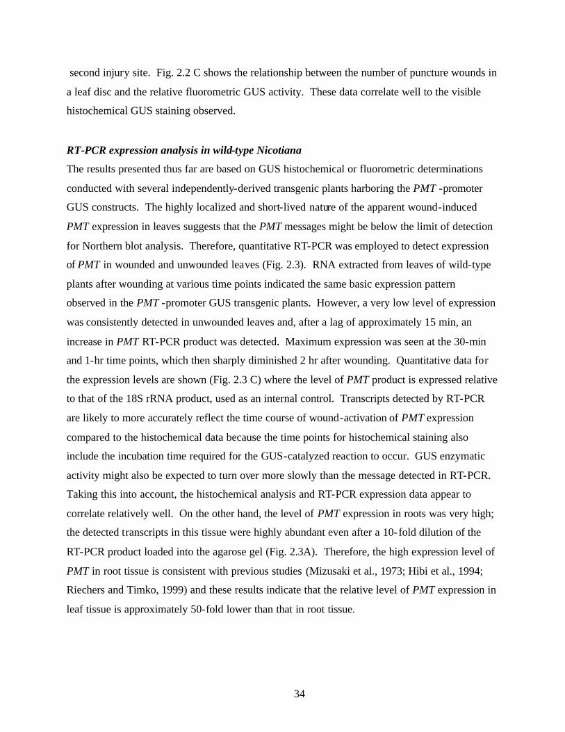
34
second injury site. Fig. 2.2 C shows the relationship between the number of puncture wounds in
a leaf disc and the relative fluorometric GUS activity. These data correlate well to the visible
histochemical GUS staining observed.
RT-PCR expression analysis in wild-type Nicotiana
The results presented thus far are based on GUS histochemical or fluorometric determinations
conducted with several independently-derived transgenic plants harboring the PMT -promoter
GUS constructs. The highly localized and short-lived nature of the apparent wound-induced
PMT expression in leaves suggests that the PMT messages might be below the limit of detection
for Northern blot analysis. Therefore, quantitative RT-PCR was employed to detect expression
of PMT in wounded and unwounded leaves (Fig. 2.3). RNA extracted from leaves of wild-type
plants after wounding at various time points indicated the same basic expression pattern
observed in the PMT -promoter GUS transgenic plants. However, a very low level of expression
was consistently detected in unwounded leaves and, after a lag of approximately 15 min, an
increase in PMT RT-PCR product was detected. Maximum expression was seen at the 30-min
and 1-hr time points, which then sharply diminished 2 hr after wounding. Quantitative data for
the expression levels are shown (Fig. 2.3 C) where the level of PMT product is expressed relative
to that of the 18S rRNA product, used as an internal control. Transcripts detected by RT-PCR
are likely to more accurately reflect the time course of wound-activation of PMT expression
compared to the histochemical data because the time points for histochemical staining also
include the incubation time required for the GUS-catalyzed reaction to occur. GUS enzymatic
activity might also be expected to turn over more slowly than the message detected in RT-PCR.
Taking this into account, the histochemical analysis and RT-PCR expression data appear to
correlate relatively well. On the other hand, the level of PMT expression in roots was very high;
the detected transcripts in this tissue were highly abundant even after a 10-fold dilution of the
RT-PCR product loaded into the agarose gel (Fig. 2.3A). Therefore, the high expression level of
PMT in root tissue is consistent with previous studies (Mizusaki et al., 1973; Hibi et al., 1994;
Riechers and Timko, 1999) and these results indicate that the relative level of PMT expression in
leaf tissue is approximately 50-fold lower than that in root tissue.
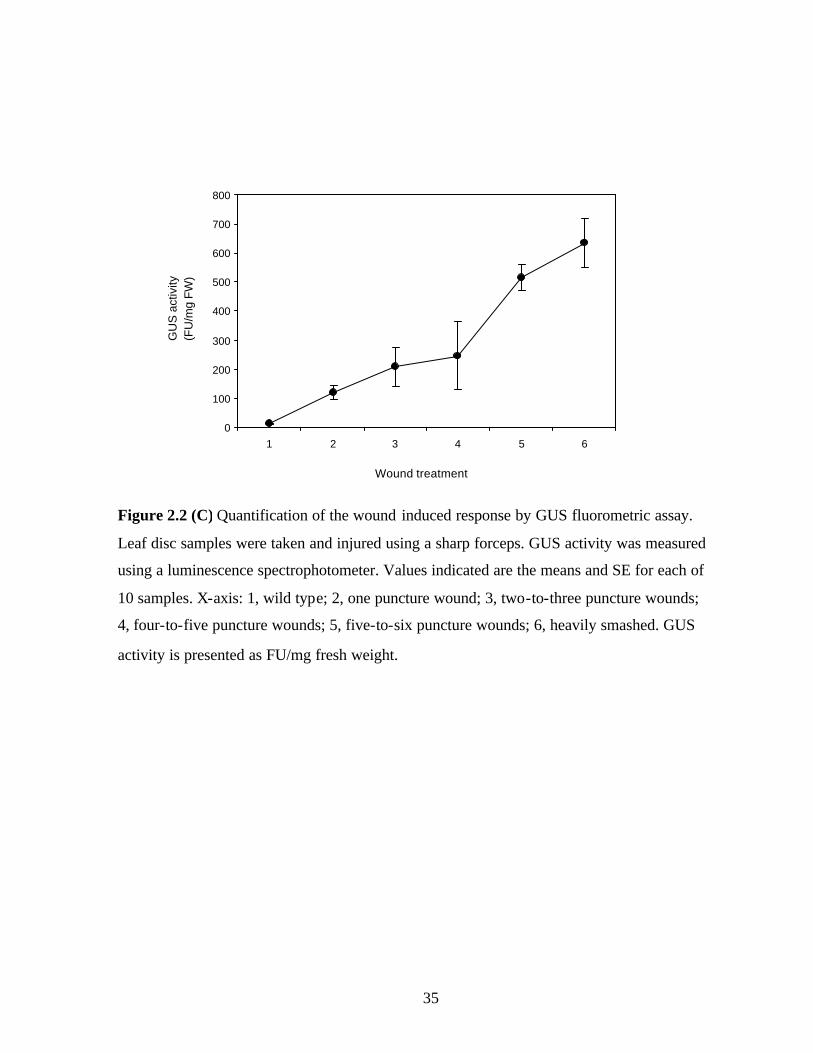
35
Figure 2.2 (C) Quantification of the wound induced response by GUS fluorometric assay.
Leaf disc samples were taken and injured using a sharp forceps. GUS activity was measured
using a luminescence spectrophotometer. Values indicated are the means and SE for each of
10 samples. X-axis: 1, wild type; 2, one puncture wound; 3, two-to-three puncture wounds;
4, four-to-five puncture wounds; 5, five-to-six puncture wounds; 6, heavily smashed. GUS
activity is presented as FU/mg fresh weight.
0
100
200
300
400
500
600
700
800
1 2 3 4 5 6
Wound treatment
GU
S a
ctiv
ity (F
U/m
g FW
)
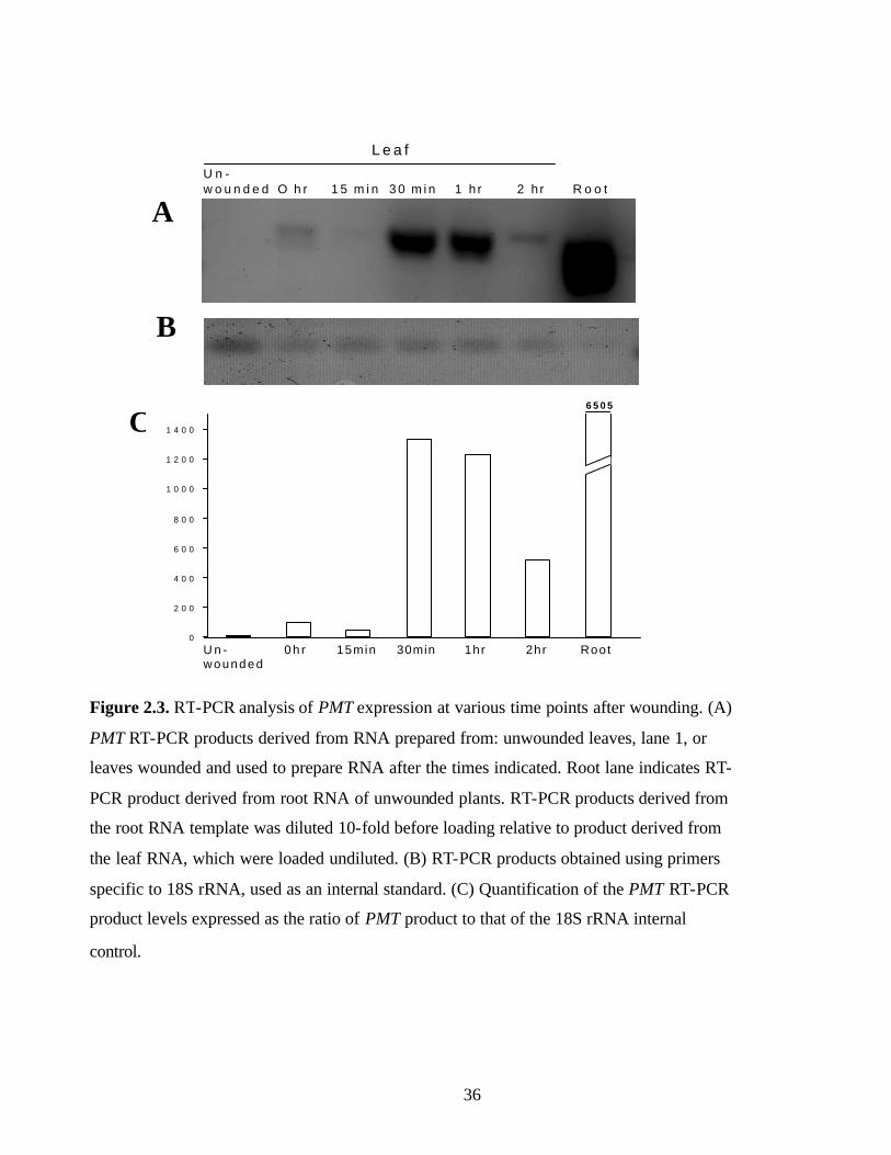
36
U n -w o u n d e d 1 5 m i n 3 0 m i n 1 h r 2 h r R o o t
L e a f
A
B
O h r
0
2 0 0
4 0 0
6 0 0
8 0 0
1 0 0 0
1 2 0 0
1 4 0 0
6 5 0 5
0 h r 15min 1hr30min 2hr RootU n -wounded
Figure 2.3. RT-PCR analysis of PMT expression at various time points after wounding. (A)
PMT RT-PCR products derived from RNA prepared from: unwounded leaves, lane 1, or
leaves wounded and used to prepare RNA after the times indicated. Root lane indicates RT-
PCR product derived from root RNA of unwounded plants. RT-PCR products derived from
the root RNA template was diluted 10-fold before loading relative to product derived from
the leaf RNA, which were loaded undiluted. (B) RT-PCR products obtained using primers
specific to 18S rRNA, used as an internal standard. (C) Quantification of the PMT RT-PCR
product levels expressed as the ratio of PMT product to that of the 18S rRNA internal
control.
C

37
In order to fully confirm that the RT-PCR product obtained was at least one of the known
PMT genes, we cloned the wound- induced RT-PCR product from leaf tissue into a pSTSBlue-1
vector (Novagen, Inc.) and sequenced it. Alignment results indicated that the RT-PCR product
was identical to PMT sequences, including NtPMT1 and NtPMT4 (A411, (Hibi et al., 1994;
Riechers and Timko, 1999) from tobacco (data not shown). Since the five PMT genes in tobacco
share extensive homology, it is possible that they could be amplified with the specific primers
used in this RT-PCR experiment (Riechers and Timko, 1999). Therefore, the detection of these
specific PMT members does not imply that they are the only members whose expression is
upregulated; it does, however, substantiate that PMT is indeed expressed in tobacco leaves and
is transiently upregulated upon wounding. In addition, the GUS analyses performed in this study
employed transgenic plants that harbored PMT -promoter fusions using the region derived
upstream of NtPMT3. The RT-PCR result also reveals why earlier studies (Sinclair et al., 2000)
examining PMT expression in leaves were unable to detect any PMT transcripts since extremely
low levels are expressed in leaves and only a transient and localized induction occurs upon
wounding.
PMT enzyme activity is detectable in leaves
To determine if putrescine N-methyltransferase activity could be detected in wounded
and unwounded leaf tissue, enzyme assays were conducted. In several separate experiments, low
levels of PMT enzymatic activity were detected in leaf tissue (Table 2.1). However, a significant
increase in PMT activity in leaves upon wounding was not consistently obtained. Typically,
leaves wounded and assayed 1 hr later showed a slight increase in activity when compared to the
unwounded tissues. PMT enzyme activity determined in leaves 2 hr after wounding dropped to
levels below those obtained in unwounded leaves; this might be a consequence of inactivation of
the enzyme after prolonged exposure within the damaged tissues. The absence of a clear
correlation between increased PMT transcrip t levels and significantly elevated PMT activity
levels may be the result of assaying at time points that do not coincide with a short- lived activity.
It may also be possible that only highly localized PMT increases occur, and any increase in
activity may be masked by the large amounts of tissue required to prepare the extracts.
Detection of low levels of PMT enzyme activity in unwounded leaves however, does coincide
38
with the low transcript levels detected in leaves. Further study of this low level of PMT
expression in leaves is required to determine its metabolic significance.
Further characterizations of leaf pmt expression
Wounding is known to cause an increase in PMT transcripts involved in alkaloid biosynthesis in
roots of mature plants in a JA-dependent manner (Baldwin et al., 1997). These responses are
thought to fulfill the need to upregulate the synthesis of alkaloids to deter leaf-eating insects.
Therefore, the effects of JA, as well as other chemicals implicated as components in wound- or
pathogenesis-responsive signaling pathways were investigated to determine if these influence
PMT expression in leaves. Leaf discs were treated with JA by infiltrating 100 µM or 400 µM JA
in MS medium, puncturing, and after various periods of time, subjecting them to GUS
histochemical staining. Treatment with JA did not influence the wound induction pattern shown
previously to any degree (data not shown). Therefore, it appears that the wound- induced
stimulation of PMT promoter activity in leaves occurs by way of a jasmonate- independent
pathway. Leaf discs were also treated in this manner with several other substances known to
mediate wound or pathogen- induced signaling. Treatment with auxin, salicylic acid and
cellulase did not show any effects on leaf PMT expression based on histochemical GUS staining
(data not shown) and did not influence the observed wound- induction. PMT expression in leaves
therefore does not seem to be affected by JA or other factors previously shown to affect its
expression in roots or that are known to function as general elicitors (Winz and Baldwin, 2001).
To further characterize the PMT wound responsiveness in leaves, leaf discs were treated
with cycloheximide (CHX), to assess the requirement of protein synthesis during the wound-
induced response. Somewhat unexpectedly, the response to mechanical wounding was not
inhibited and instead appeared to cause the wound- induced expression of PMT to persist (data
not shown). The ability of cycloheximide to cause a normally transient transcriptional signal to
persist has been observed in a number of instances (Usami et al., 1995; Hara et al., 2000).
Therefore, its effect on PMT expression suggests a similar response. A possible explanation for
how CHX could eliminate the abatement of PMT transcription during extended periods
following wounding is that new protein synthesis is required for the rapid decrease of PMT -
promoter activity. An additional explanation is that the decrease in PMT expression observed

39
Table 2. 1. PMT activities in root and wounded leaf tissues.
Leaves
Roots Unwounded 1-hr wounded 2-hr wounded
1.87 ± 0.18 0.16± 0.06 0.20 ± 0.01 0.10 ± 0.02
Putrescine N-methyltransferase (PMT) enzyme activity (nmoles/µg protein) was determined in
roots and wounded leaves. Leaves were wounded by puncturing the entire surface of a leaf with
a wire brush and extracts were prepared from the indicated tissues as described in Experimental
Protocols. Values are the average of three determinations ± SE.

40
after wounding coincides with wound healing that occurs to repair tissues after injury. Inhibition
of protein synthesis at this stage would prevent healing, allowing PMT expression to persist.
NtPMT promoter responses in Arabidopsis
Evaluation of the PMT promoter activity in Arabidopsis, a non-alkaloid producing plant,
was performed to determine its expression characteristics in a heterologous species. The same
NtPMT3 promoter-GUS construct used in tobacco was introduced into Arabidopsis to obtain
stable transgenic plants. GUS histochemical staining revealed PMT-promoter activity in the
resulting lines in roots and trichomes of Arabidopsis leaves but displayed no evidence of wound
induction (data not shown). The lack of a similar wound- induced expression in a different
species suggests that the PMT-transcriptional response to wounding is a specific phenomenon in
tobacco leaves.

41
Conclusions
The spatial expression patterns of many defensive genes are affected by wounding
(Chang et al., 1995; Grantz et al., 1995; McGurl et al., 1995; Nishiuchi et al., 1997) and the
assessment described by Nishiuchi (1997) pertains to the situation we have observed for PMT
expression in N. tabacum leaves. Nishiuchi (1997) points out that these defensive genes are
constitutively expressed at high levels in roots but at lower levels in leaves. One type of wound-
regulated expression is the induction of those genes in tissues in which the genes are normally
not highly expressed under non- induced conditions (Nishiuchi et al., 1997). Such is the situation
described here with the highly localized PMT expression in leaves. The enhanced PMT
expression seen in response to wounding in root tissues corresponds to the type of wound
response pattern for those genes that are constitutively expressed at higher levels (Hibi et al.,
1994; Riechers and Timko, 1999; Winz and Baldwin, 2001).
The observations made here of wounding inducing a transient, highly localized
upregulation of PMT transcription in leaves, but with little evidence of major increases in PMT
activity, are not fully understood. The low level of PMT transcript and enzyme activity in
unwounded leaves suggests the possibility that it may play an additional, subtle role in alkaloid
metabolism in leaf tissue. In this regard, Sato, et al. (2001) observed that transgenic N. sylvestris
lines overexpressing PMT using the 35S CaMV promoter not only exhibited increased leaf
nicotine contents but also increases in accumulated methylputrescine along with slight decreases
in the level of spermidine and spermine in leaves. On the other hand, in a line containing
cosuppressed PMT, an elevation of putrescine and spermidine levels was observed in leaves,
which was postulated to be a result of transport of these polyamines from the root to the leaves
(Sato et al., 2001). The low levels of PMT expression and activity in leaves described in our
work suggests an additional possibility to explain the polyamine increases observed by Sato
(2001). Specifically, putrescine and spermidine accumulate directly in leaves as a result of
cosuppression of the low level of PMT activity that is present in leaves.
Another possible function for low levels of leaf-based PMT expression in Nicotiana may
be related to the diversity of alkaloid metabolism in distinct species and in distinct cell types.
For example in N. repanda, the presence of a specific N-acylated nornicotine derivative has been

42
detected, and the site of accumulation as well as synthesis is exclusive to trichomes on the leaf
surface (Laue et al., 2000). This suggests that further metabolism of alkaloids can occur after
their synthesis in roots and translocation to the leaf. In this regard, the low level of PMT
expression and its transient upregulation in leaves may provide an additional means to alter the
diversity of alkaloidal components in different species. Future studies on establishing PMT
expression profiles in leaves of different alkaloid-accumulating species of Nicotiana may shed
light on this possibility.

43
References
Baldwin, I.T., Schmelz, E.A., and Ohnmeiss, T.E. (1994). Wound-induced changes in root and
shoot jasmonic acid pools correlate with induced nicotine synthesis in Nicotiana sylvestris
Spegazzini and Comes. J Chem Ecol 20, 2139-2157.
Baldwin, I.T., Zhang, Z.-P., Diab, N., Ohnmeiss, T.E., McCloud, E.S., Lynds, G.Y., and
Schmelz, E.A. (1997). Quantification, correlations and manipulations of wound-induced changes
in jasmonic acid and nicotine in Nicotiana sylvestris. Planta 201, 397-404.
Bhattacharyya, S., Dey, N., and Maiti, I.B. (2002). Analysis of cis-sequence transcript (Sgt)
promoter from the figwort mosaic virus (FMV) and comparison of promoter activity with
cauliflower mosaic virus (CaMV) promoters in monocot and dicot cells. Virus Research In
press.
Bradford, M.M. (1976). A rapid and sensitive method for quantification of microgram
quantities of protein utilizing the principle of protein dye binding. Anal. Biochem. 72, 248-254.
Chang, M.-M., Horovitz, D., Culley, D., and Hadwiger, L.A. (1995). Molecular cloning and
characterization of pea chitinase gene expressed in response to wounding, fungal infection and
elicitor chitosan. Plant Mol Biol 28, 105-111.
Clough, S.J., and Bent, A.F. (1998). Floral dip: a simplified method for agrobacterium–
mediated transformation of Arabidopsis thaliana. Plant J 16.
Grantz, A.A., Brummell, D.A., and Bennett, A.B. (1995). Ascorbate free radical reducatse
mRNA levels are induced by wounding. Plant Physiol 108, 411-418.
Hajdukiewicz, P., Svab, Z., and Maliga, P. (1994). The small, versatile pPZP family of
agrobacterium binary vectors for plant transformation. Plant Mol Biol 25, 989-994.
Hara, K., Yagi, M., Koizumi, N., Kusano, T., and Sano, H. (2000). Screening of wound-
responsive genes identifies an immediate-early expressed gene encoding a highly charged
protein mechanically wounded tobacco plants. Plant Cell Physiol 41, 684-691.
Hashimoto, T., and Yamada, Y. (1994). Alkaloid biogenesis: molecular aspects. Annu Rev
Plant Physiol Plant Mol Biol 45, 257-285.
44
Hashimoto, T., Shoji, T., Mihara, T., Oguri, H., Tamaki, K., Suzuki, K., and Yamada, Y.
(1998). Intraspecific variability of tandem repeats in Nicotiana putrescine N-methyltransferases.
Plant Mol Biol 37, 25-37.
He, Y., and Gan, S. (2001). Identical promoter elements are involved in regulation of OPR1
gene by senescence and jasmonic acid in Arabidopsis. Plant Mol Biol 47, 595-605.
Hibi, N., Higashiguchi, S., Hashimoto, T., and Yamada, Y. (1994). Gene expression in
tobacco low-nicotine mutants. Plant Cell 6, 723-735.
Hibi, N., Fujita, T., Hatano, M., Hashimoto, T., and Yamada, Y. (1992). Putrescine N-
methyltransferase in cultured roots of Hyoscyamus albus. Plant Physiol. 100, 826-835.
Horsch, R.B., Fry, J.E., Hoffman, N.L., Eicholts, D., Rogers, S.G., and Frayley, R.T. (1985).
A simple and general method for transferring genes into plants. Science 227, 1229-1231.
Jefferson, R.A., Kavanagh, T.A., and Bevan, M. (1987). GUS fusions: β-glucuronidase as a
sensitive and versatile gene fusion marker in higher plants. EMBO 6, 3901-3907.
Laue, G., Preston, C.A., and Baldwin, I.T. (2000). Fast track to the trichome: induction of N-
acyl nornicotines precedes nicotine induction in Nicotiana repanda. Planta 210, 510-514.
Leete, E., and Liu, Y.Y. (1973). Metabolism of [2-3H]- and [6H3]-nicotinic acid in intact
Nicotiana tabacum plants. Phytochemistry 12, 593-596.
Maliga, P., Sz-Breznovits, A., and Marton, L. (1973). Streptomycin-resistant plants from
callus culture of haploid tobacco. Nat New Biol 244, 29-30.
McGurl, B., Mukherjee, S., Kahn, M., and Ryan, C.A. (1995). Characterization of two protein
inhibitors (ATI) cDNAs from alfalfa leaves (Medicago sativa var Vernema): The expression of
ATI genes in response to wounding and soil microorganisms. Plant Mol Biol 27, 995-1001.
Mizusaki, S., Tanebe, Y., Noguchi, M., and Tamaki, E. (1973). Changes in activities of
ornithine decarboxylase, putrescine N-methyltransferase and N-methylputrescine oxidase in
tobacco roots in relation to nicotine biosynthesis. Plant Cell Physiol 14, 103-110.
Nishiuchi, T., Hamada, T., Kodama, H., and Iba, K. (1997). Wounding changes the spatial
expression pattern of the Arabidopsis plastid ω-3 fatty acid desaturase gene (FAD7) through
different signal transduction pathways. Plant Cell 9, 1701-1712.
Riechers, D.E., and Timko, M.P. (1999). Structures and expression of gene family encoding
putrescine N-methyltansferase in Nicotiana tabacum: new clues to the evolutionary origin of
cultivated tobacco. Plant Mol Biol 41, 387-401.

45
Sato, F., Hashimoto, T., Hachiya, A., Tamura, K., Choi, K.B., Morishige, T., Fujimoto, H.,
and Yamada, Y. (2001). Metabolic engineering of plant alkaloid biosynthesis. Proc Natl Acad
Sci U S A 98, 367-372.
Saunders, J.W., and Bush, L.P. (1979). Nicotine biosynthesis enzyme activites in Nicotiana
tabacum L. genotypes with different alkaloid levels. Plant Physiol 64, 236-240.
Shoji, T., Yamada, Y., and Hashimoto, T. (2000). Jasmonate induction of Putrescine N-
methyltransferase genes in the root of Nicotiana sylvestris. Plant Cell Physiol 41, 831-839.
Sinclair, S.J., Murphy, K.J., Birch, C.D., and Hamil, J.D. (2000). Molecular characterization
of quinolinate phosphoribosyltransferase (QPRTase) in Nicotiana. Plant Mol Biol 44, 603-617.
Suzuki, K.-I., Yamada, Y., and Hashimoto, T. (1999). Expression of Atropa belladonna
putrescine N-methlytrasferase gene in root pericycle. Plant Physiol. 40, 289-297.
Usami, S., Banno, H., Ito, Y., Nishihama, R., and Machida, Y. (1995). Cutting activates a 46-
kilodalton protein kinase in plants. Proc Natl Acad Sci USA 92, 8660-8664.
Walling, L.L. (2000). The myriad plant responses to herbivores. Journal of Plant Growth
Regulation 19, 195-216.
Wang, J., Sheehan, M., Brookman, H., and Timko, M.P. (2000). Characterization of cDNA
diffferentially expressed in roots of tobacco (Nicotiana tabacum cv burley 21) during the early
stages of alkaloid biosynthesis. plant Science 158, 19-32.
Wink, M. (1998). Modes of action of alkaloids. In Alkaloids, M.F. Roberts and M. Wink, eds
(New York: Plenum press), pp. 301-326.
Winz, R.A., and Baldwin, I.T. (2001). Molecular Interactions between the specialist herbivore
Manduca sexta (Lepidoptera, sphingidae) and its natural host Nicotiana attenuata IV. Insect-
induced ethlyene reduces jasmonate-induced nicotine accumulation by regulation putrescine N-
methyltransferase transcripts. Plant Physiol. 125, 2189-2202.

46
Chapter 3
Identification of signaling factors that impact the regulation of
alkaloid biosynthesis in N. tabacum callus
Introduction
Overview of alkaloid biosynthesis in intact plants
Nicotine and related alkaloids are predominant secondary metabolites synthesized by a
number of Nicotiana species and may function as defensive toxins by acting on the nervous
system of herbivores (Wink, 1998; Shoji et al., 2000). Pyrrolinium alkaloids, including nicotine,
are synthesized in the tobacco root from arginine and/or ornithine by the way of putrescine
(Hashimoto and Yamada, 1994). Putrescine N-methyltransferase (PMT; EC 2.1.1.53) is a key
enzyme catalyzing the committed step for the biosynthesis of tropane alkaloids and nicotine.
PMT catalyzes the S-adenosyl methionine-dependent N-methylation of putrescine directing the
conversion of a primary metabolite into the biosynthetic pathways that leads to the secondary
metabolic products nicotine and tropane alkaloids (as shown in fig. 1.1, chapter 1). Putrescine is
therefore a precursor not only of polyamines but also to important alkaloids such as the
pyrrolidine alkaloids of tobacco (nicotine and nornicotine), tropane alkaloids of Datura,
Hyoscyamus (hyoscyamine, hyoscine, and meteloidine), pyrrolizidine alkaloids (retronecine),
and possibly phenanthroindolizidines (tylophorine) (Tiburcino, 1985; Leete, 1980).
Nicotine and tropane alkaloids are formed in the roots and transported to the aerial parts
of the plant. PMT transcripts are found exclusively in roots of tobacco and are localized
specifically in the cortex, endodermis and xylem of young roots of N. sylvestris (Shoji et al.,
2000). In A. belladonna, a tropane alkaloid producing plant, PMT genes are exclusively
expressed in root pericycle adjacent to xylem tissue (Suzuki et al., 1999).
N. tabacum has five PMT-encoding genes: NtPMT1, NtPMT2, NtPMT3, Nt PMT4 and
NtPMT1a (Riechers and Timko, 1999). Many studies have also documented a rapid induction of
PMT-encoding genes by herbivore attack, wounding, or exogenous applications of the

47
endogenous wound hormone, jasmonic acid (JA), or its methyl ester, MeJA, to roots of the plant
(Baldwin et al., 1997; Hashimoto et al., 1998; Riechers and Timko, 1999; Hara et al., 2000).
Alkaloid regulation in undifferentiated tissues
Regulation of alkaloid biosynthesis in whole plants is different from its regulation in
callus cultures. Some studies have shown that light stimulated the production of a number of
secondary metabolites in plant cell cultures. In tobacco cell cultures, there have been reports that
light both enhances (Tabata et al., 1971) and suppresses nicotine accumulation (Hobbs et al.,
1991; Ohta and Yatazawa, 1978). Cell suspension cultures derived from N. tabacum cv.
Wisconsin accumulated less total alkaloids than the intact plant (Hobbs, 1989). In N. tabacum,
nicotine accounted for two-thirds of the total alkaloid content, and the ratio of nornicotine to
nicotine was 0.2 (Hobbs, 1989) whereas in cell cultures, nicotine accounted for one-third of the
alkaloid content and ratio of nornicotine to nicotine was found to be 1.0 (Holden et al., 1988).
The amount of nicotine was reduced by 80% when tobacco callus tissues previous ly grown in
darkness were shifted to light for three weeks (Ohta and Yatazawa, 1978). Callus cultures in
general have been reported to have lower alkaloid content compared to intact tobacco plants
(Ohta et al., 1978). Thus, the range of alkaloid levels found in different studies is likely to be due
to various culturing methods and environmental conditions used to propagate undifferentiated
callus.
Several reports have indicated that auxin levels are involved in regulation of nicotine
production in callus and root cultures (Takahashi, 1973; Solt, 1957). Tiburcio et al., (1985) have
shown that callus tissue obtained from petiolar explants of N. tabacum showed increased alkaloid
levels when grown on high α -naphthalene acetic acid (α-NAA) medium, then were transferred
to a medium containing lower concentrations of auxin. However, some inconsistencies have
been reported in the ability to synthesize alkaloids in undifferentiated callus tissue derived from
different sources. Callus cultures derived from shoots showed lower alkaloid production when
compared to those derived from roots of N. tabacum (Hobbs and Yeoman, 1991). Nicotine,
nornicotine, anabasine and traces of anatabine were detected in callus. Shoot cultures of N.
tabacum had only small amounts of nicotine and nornicotine, indicating that they may be able to
synthesize alkaloids (Dawson and Solt, 1959). More recently, gene expression studies have

48
shown that PMT expression levels are reduced when tobacco callus cultures are treated with
auxin (Hibi et al., 1994; Imanishi et al., 1998).
As indicated above, the biosynthesis of secondary metabolites in plants is complex and
highly variable results have been obtained using tissue culture systems. In addition, few studies
have focused on determining the nature of signaling components involved in the regulation of
secondary metabolite biosynthesis. Undifferentiated tissue from plants would represent a
convenient and economical means to obtain useful plant metabolites, if the production of these
metabolites was predictable. Therefore the studies described in this chapter were undertaken to
begin to decipher the major signaling components that regulate a secondary pathway in callus
tissue. Using transgenic plant material that enables an efficient means to report pathway-specific
gene expression, a number of conditions were tested. The results obtained enabled the
identification of a primary signaling factor that seems to be involved in governing pathway
expression.

49
MATERIALS AND METHODS
Plant Material
N. tabacum cv. SR1 was used in all studies (Maliga et al., 1973). All plants were
grown in sterile Magenta boxes in a growth room at 22°C with continuous light (80 µmol
m-2 s-1) on Murashige and Skoog (Sigma) medium containing 1% sucrose.
Promoter isolation and plant transformation
A N. tabacum genomic library (Clontech, Palo Alto, CA) was screened using a
probe prepared by PCR from SR1 genomic DNA using published cDNA sequences of the
N. tabacum NtPMT1 cDNA (Hibi et al., 1994). A 4.5-kb NtPMT3 clone was isolated as
described previously (chapter 2, Sachan and Falcone, 2002). A sequence containing 627
bp of the NtPMT3 promoter region was fused to the GUS reporter gene. The resulting
construct was introduced into Agrobacterium tumefaciens GV3101 strain and used for
transformation of N. tabacum as described previously (chapter 2, Sachan and Falcone,
2002).
Protoplast isolation and callus preparation
N. tabacum plants were grown in sterile Magenta boxes on solid 1X MS medium
for 5-6 weeks. Leaves were excised from the plants and cut into sections of 1 cm2 using a
sharp razor blade. Approximately 3-5 g of leaf tissue was added to 20-ml filter-sterilized
enzyme solution (1.5% cellulase and 0.5% pectinase (Sigma) in 0.4 M K-3 medium and
incubated in dark at 26°C overnight. The digested leaf enzyme solution was passed
through a 100 µM sieve and 12 ml was transferred to 17×100 mm polystyrene snap cap
culture tubes. The tubes were centrifuged at approximately 2000 rpm in a Jouan-MR22
swing-out bucket centrifuge for 6 minutes (0 brake; 0 acceleration). After centrifugation,
tubes were carefully removed from the centrifuge so as not to disturb the intact
protoplasts that floated on top of the medium, cell debris was pelleted to the bottom of
50
the tube. The cell debris pellet and the solution were removed with a 1.5-mm diameter
capillary tube connected to a peristaltic pump. Medium was carefully withdrawn until
approximately two ml of medium containing intact protoplasts remained. The tube was
refilled with fresh 0.4 M K-3 medium (Fritze and Walden, 1995), mixed gently by
inversion and centrifuged again. This was repeated at least 2-3 times to completely
remove the enzyme solution. In the last wash, the medium was removed until three ml
remained and the protoplasts from all tubes were mixed together. The amount of
protoplasts in the final pooled medium was estimated by counting under a microscope.
The final concentration of protoplasts transferred to a 10×60-mm petri dish was adjusted
to 1 million cells per 10 ml of 0.4 M K-3 medium (1 mg/ml or 5.3 µM final concentration
of α -naphthalene acetic acid (α-NAA) and 0.2 mg/ml or final concentration of 0.93 µM
kinetin). Sealed dishes containing the fresh protoplasts were incubated in dark for 4 days
at 26°C for recovery after isolation.
Protoplast washing
Nine days after cutting leaves, protoplasts were washed in W-5 medium. Five
milliliters of W-5 medium (a medium containing, NaCl, CaCl2, KCl and 100 mM
glucose) was added to polystyrene snap cap culture tubes and to this 5 ml of protoplasts
in K-3 medium was slowly added. Tubes were centrifuged in a Jouan swing-out bucket
rotor at 1,000 rpm for 3 min at 24°C (acceleration-9, brake-3, ∆G-157). The protoplast
pellet at the bottom of the tube was washed with W-5 medium, and the wash was
repeated twice. In the last wash, a 5-ml pipette was used to remove the majority of the
media and leaving washed cells in a small volume of media. Protoplasts are then
resuspended in 0.4 M K-4 medium, (a medium containing asparagine, arginine and
glutamine as nitrogenous sources instead of NH4NO3 and NaNO3 as the nitrogen source
in K-3 medium). Two tubes of protoplasts were combined to give protoplasts in 10 ml of
0.4 M K-4 medium, which was transferred to 60×15-mm plates. When viewed under the
microscope at this stage, protoplasts float on top, have elongated shapes, and are ready to
divide.

51
Embedding of protoplasts to generate callus
On day 14 after the isolation, protoplasts were embedded in low-melting agarose
(Seaplaque agarose, FMC bioproducts; melting temperature ≤ 65°C, gelling temperature
26°-30°C). Each 60×15-mm petri dish of protoplasts was embedded to give one 100×15-
mm petri dish of protoplasts. To embed protoplasts in 0.3 M K-4 medium, a final
concentration of 0.3% low melting agarose was added to protoplasts and the suspension
was quickly poured into a 100×15-mm petri dish and allowed to solidify for one hour.
Subsequently 10 ml of 0.3 M K-4 medium was added and dishes were placed at 26°C in
light.
After one week, the medium containing the embedded protoplasts was changed to
0.2 M K-4 medium. The following week, the medium was changed to 0.1 M K-4
medium. After incubation in 0.1 M K-4 medium for another 2 weeks, the microcalli
appear and are ready to be surface embedded. These media changes were undertaken to
decrease the osmulants to induce callus formation. All media contained α-NAA and
kinetin at the same concentrations mentioned above.
Surface embedding of microcalli
Microcalli embedded within agarose from a single plate were pooled together and
divided into approximately 16-18 parts for surface embedding on 100×15-mm-sized petri
plates. To each part, 0.6% of low-melting agarose and 0.1 M K-4 medium was added and
quickly stirred. This was poured over 0.1 M K-4 medium solidified with 0.2% phytagel
(Sigma) in 150-mm petri dishes. The microcalli were allowed to embed on the surface of
the media in these plates for 3 hrs. Ten millilitres of 0.1 M K-4 liquid medium was added
on top of the embedded material on each plate. Plates were sealed and incubated at 26°C.
Callus treatments
Two weeks after surface embedding microcalli, now grown larger callus material
was used for various treatments. All callus tissue was vacuum-infiltrated with 0.1 M K-4
medium containing the chemical under a vacuum pressure of 10-mm hg for 15 min. The
final concentrations of the substances used were 5 mM DMTU (1, 3-dimethyl 2-thiourea,
Aldrich), 200 units/ml catalase (Sigma), 40 µM jasmonic acid, 10 mM tiron (4,5-

52
dihydroxy 1,3-benzene disulfonic acid, Sigma), 30 mM ascorbate and 10 mM N-acetyl
cysteine. For normal growth of callus, light was provided continuously at 15-20 µmol m-2
s-1, while “high light” was 100 µmol m-2 s-1. For dark treatments, callus plates were
wrapped in aluminum foil.
GUS fluorometric assay
GUS enzyme activity assays were performed in 96-well plates as described by
(He and Gan, 2001) with modifications. Callus material was taken and removed of all
media and the tissue was ground in 200 µl GUS extraction buffer (50 mM NaHPO4 pH
7.0, 10 mM Na2EDTA, 0.1% sarcosyl, 0.1% Triton X-100). One-hundred microlitres of
the extract was taken for the assay to which 30 µl of 5 mM 4-MUB-β-D-glucuronide
(MUG) in extraction buffer was added and further incubated for 20 minutes at 37°C. The
reaction was stopped by adding Na2CO3 to a concentration of 180 mM to the wells.
Fluorescence was measured at 365 nm excitation and 455 nm emission using a Perkin
Elmer luminescence spectrophotometer model LS50B (Perkin-Elmer, Beaconsfield, UK).
Protein estimation was done colorimetrically by dye binding according to a kit provided
by BioRad.
Measurement of H2O2 activity
Measurement of H2O2 activity made using callus treated with varying
concentrations of auxin (as indicated in table 1) for 2 weeks. A kit provided by Molecular
Probes- Amplex red hydrogen peroxide/ peroxidase assay kit (A-22188) was used. In a
96-well plates 50 µl of the medium, in which the callus was maintained, was diluted with
50 µl horseradish peroxidase (HRP) solution. The reaction was incubated at room
temperature for 30 minutes. Fluorescence was measured at 530 nm excitation and 590 nm
emission using a Perkin Elmer luminescence spectrophotometer LS50B (Perkin-Elmer,
Beaconsfield, UK).
Extract preparation for alkaloid displacement assay
Approximately 50 mg of callus material was removed with a pair of scoop ring
forceps for preparing the callus extracts. Callus was ground in eppendorf tubes containing
500 µl water using a small pestle connected to a drill press for one minute. An additional
53
500 µl of water was added afterwards. Extracts were centrifuged at 14,000 rpm for 10
minutes. Supernatants were used for conducting the alkaloid displacement assay.
Alkaloid displacement assay
Radioligand binding displacement were conducted in 96-well filter plates (350 µL
well volume) at room temperature (unless otherwise noted), using methods modified after
those described elsewhere (Houghtling et al., 1995; Mahanandeeshwar et al., 1995).
Briefly, competitor plant tissue sample extract, or unlabelled ligand for non-specific
binding measurement was pre-incubated with a homogenized brain tissue preparation
prepared by Dr. Trent Rogers in Prof. John Littleton’s laboratory. Following appropriate
pre-incubation of competitor, radiolabelled ligand (168 pM [3H]-epibatidine, S.A. 66.6
Ci/mmole, New England Nuclear) was added to each well and incubated further.
Following incubation, samples were harvested onto 96-well GF/B filter plates and rapidly
washed three times with 350 µL 50 mM Tris-HCl buffer (pH 7.4). After overnight
drying, scintillation fluid was added to each filter and the filterplate was counted using a
96-well plate scintillation counter. Specific binding in the presence of competitor was
converted to percentage of total specific binding of radioligand alone. Non-specific
binding was calculated based upon the amount of radioligand binding in the presence of
excess competitor ligand (300 µM nicotine). Significant differences between individual
samples were calculated from radioactivity remaining in counts per minute (CPM)
average and from triplicate measurements per sample using Students 2-tailed t-tests.
Extract preparation and GC-MS analysis of callus alkaloids
Callus was maintained in light conditions (15-20 µmol m-2 s-1) and in the presence
of auxin. The media in which the callus was maintained was removed and the callus was
macerated with a drill machine and then centrifuged. The supernatant was subsequently
lyophilized in a vacuum drier. 1.5 g of the lyophilized tissue was added to 10 ml of
acidified methanol (pH 1.5 with HCl). Quinaldine (2-methyl quinoline) 100 ng/ml was
added as an internal standard and the suspension was shaken on an orbital shaker
overnight. Two ml of the above suspension was mixed with an equal volume of a 50:50
mixture of hexane:diethyl ether. This was centrifuged at 5000 g for 20 min in a sorvall
centrifuge. The aqueous layer was collected and the pH adjusted to 11 with NH4OH. An

54
equal amount of CH2Cl2 solution was vortexed and then centrifuged. The CH2Cl2 layer is
collected in a separate tube. While the upper aqueous layer is again treated with equal
volume of CH2Cl2 and centrifuged. The above process was repeated twice; total of three
times. All the CH2Cl2 layers were combined and dried under argon gas. The dried residue
was dissolved in 100µl methanol. An Agilent 6890 GC with 5973 mass spectrometer
fitted with a HP5- MS 30 x 25 x 25 column was used for the analysis. Injection volumes
were 1µl.

55
RESULTS
NtPMT3 gene promoter-GUS fusion constructs and preparation of transgenic N.
tabacum plants were described in chapter 2. Stable transgenic line number nine was
chosen based on its reproducible GUS expression characteristics. Seeds from this line
were germinated on MS plates with kanamycin selection. Seedlings were then transferred
to Magenta boxes to obtain larger plants, and these were used to isolate protoplasts and
subsequently to prepare undifferentiated callus.
Fresh protoplasts were isolated from leaf tissue by Irina Artiushin. Callus cultures
derived from freshly isolated leaf protoplasts were then grown by surface plating. Callus
tissue prepared in this way was actively growing and healthy as indicated by its vigorous
growth on agar surfaces and its green appearance. Surface plating was done to maintain
regenerable tissue and to provide a supply of equivalently grown tissue to ensure
consistent responses. Alkaloid production has been shown to be tissue specific. In this
study callus tissue was used to evaluate the expression characteristics of the alkaloid
pathway at the transcriptional level and at the level of alkaloid accumulation.
Effect of light and auxin on alkaloid pathway expression
Because the earlier literature has described variable results of alkaloid accumulation in N.
tabacum callus tissue we conducted experiments to evaluate the effect of light and auxin
on PMT-promoter expression to provide initial information regarding regulation of the
pathway at the transcriptional level. All callus materials were grown in the presence of
normal concentrations of auxin and kinetin (1 mg/ml or 5.3 µmol final concentration of
α-NAA and 0.2 mg/ml or final concentration of 0.93 µM of kinetin) unless otherwise
indicated. Callus materials were maintained in continuous light conditions (15-20 µmol
m-2 s-1). Five-week-old agarose embedded callus pieces were then transferred to fresh
medium containing different auxin concentrations (indicated in table 3.1) for 2 weeks in
light or dark conditions. Callus material that was placed in the light for 2 weeks with
normal auxin concentrations showed little or no PMT expression based on GUS

56
histochemical and fluorometric data (figure 3.1, 3.2 and 3.3 respectively). PMT
expression levels are indicative of nicotine biosynthesis and total alkaloid levels as has
been shown previously (Yun, Littleton and Falcone, unpublished results). The is
consistent with the conclusion that PMT enzyme catalyzes the first committed step of
nicotine production. This low background of PMT expression indicates that there is no or
low alkaloid pathway expression as well as alkaloid synthesis in tissue-cultured callus
material derived from isolated leaf protoplasts. Earlier studies involving shoot cultures of
N. tabacum, N. glauca and N. rustica found lower alkaloid in light conditions when
compared to dark (Hobbs and Yeoman, 1991). We used different auxin concentrations in
both light and dark to directly examine their effects on alkaloid pathway expression.
When the callus was maintained in dark conditions with reduced concentrations of auxin
(0.79 µM - 0.15-fold auxin), a marked increase in PMT expression was observed (figure
3.2). However, when the microcalli were plated a little more densely and at a later stage
of development, the callus material with no auxin in dark conditions showed greater PMT
expression based on histochemical anaysis (figure 3.1). In the experiment of figure 3.1
callus tissue was transferred as agarose pieces and surface-embedded while in figure 3.2
the tissue was much younger and were not surface embedded. Even with differing
concentrations of auxin little differences in PMT expression as indicated by PMT
promoter activity was observed in presence of light based on GUS histochemical data and
fluorometric analysis (figure 3.1 and 3.3). However, when placed in the dark conditions
with reduced auxin concentrations, dramatic upregulation of PMT expression based on
GUS histochemical data was apparent in the callus tissue (figure 3.2). Under the light
levels used for normal propagation of the tissue, PMT expression was very low, as shown
by low levels of GUS activity (figure 3.5). Higher light causes expression to be reduced
further (figure 3.5). In dark conditions, even in the presence of typical levels of auxin,
PMT expression was higher compared to the normal growth condition of callus. This was
not that evident in the histochemical analys is, presumably because the fluorometric
method detection is more sensitive to changes in experimental conditions.
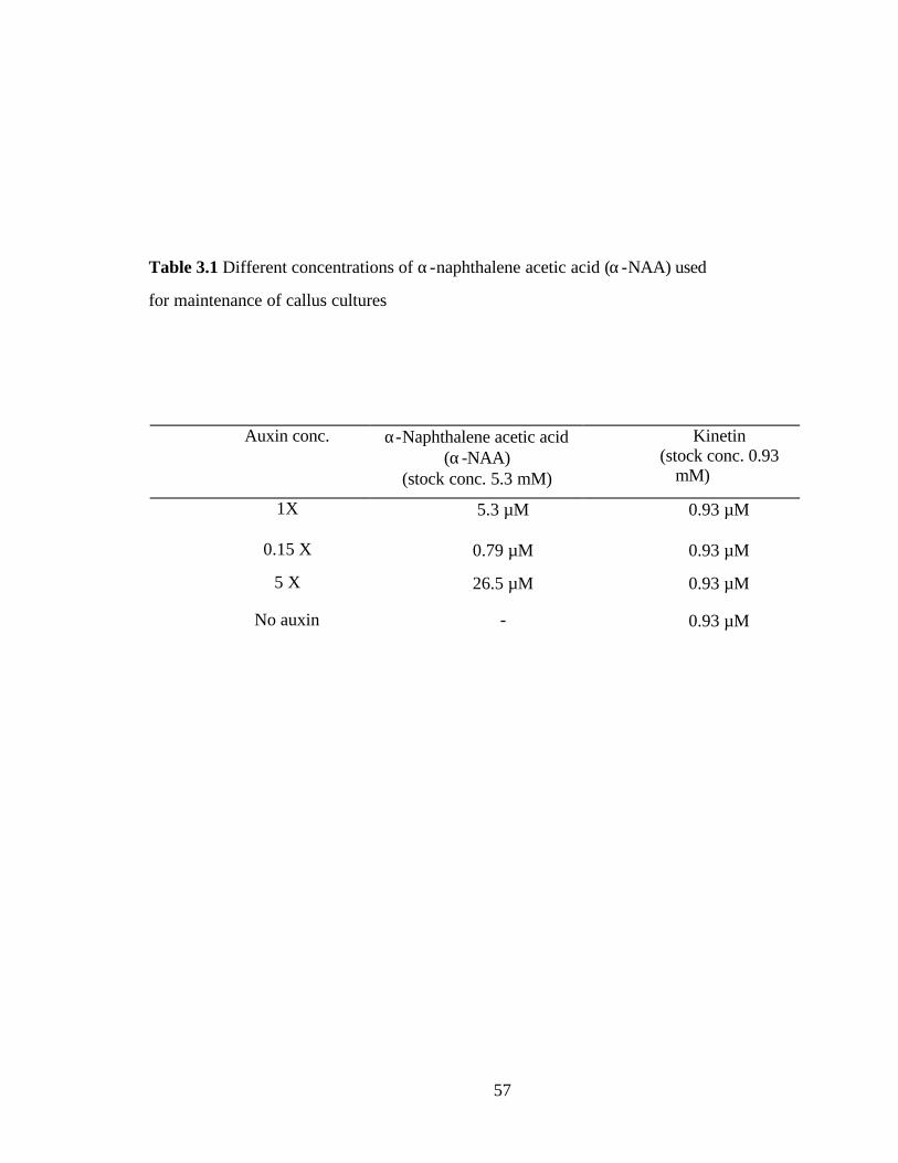
57
Auxin conc. α-Naphthalene acetic acid (α-NAA)
(stock conc. 5.3 mM)
Kinetin (stock conc. 0.93
mM)
1X 5.3 µM 0.93 µM
0.15 X 0.79 µM 0.93 µM
5 X 26.5 µM 0.93 µM
No auxin - 0.93 µM
Table 3.1 Different concentrations of α-naphthalene acetic acid (α-NAA) used
for maintenance of callus cultures

58
Figure 3.1 GUS histochemical analysis of surface-embedded transgenic callus. The callus
material generated from protoplasts isolated from transgenic tobacco leaves containing the
NtPMT 3 promoter-GUS fusion. Five-week-old callus embedded in agarose was treated for one
week and stained with X-gluc to assess PMT promoter activity. The surface-embedded callus
was cut out as sections to ensure equivalent tissue for all treatments. Callus were treated to
varying concentrations of α-NAA (for details see table 3.1).
Auxin α-NAA
concentration

59
Figure 3.2 GUS histochemical analysis microcalli derived from protoplasts. Callus
material was generated from protoplasts isolated from transgenic tobacco leaves
containing the NtPMT 3 promoter-GUS fusion. Three-week-old callus embedded in
agarose was treated for one week and stained with X-gluc to assess PMT promoter
activity. This callus tissue was not surface-embedded to avoid handling of the tissue
and thereby affecting PMT expression. Callus were treated to varying concentrations
of α-NAA (for details see table 3.1)

60
0
2
4
6
8
10
12
14
NO AUXIN 0.15 X 0.5 X 1 X
Auxin concentration
nmol
es M
U/µ
g pr
/min
LightDark
Figure 3.3 Treatment of callus material to different auxin concentrations in light and dark
conditions. Callus material was generated from protoplasts isolated from transgenic tobacco
leaves containing the NtPMT 3 promoter GUS fusion were examined for β-glucuronidase
activity after treatment with varying concentrations of auxin (as indicated in table 3.1) in light
and dark conditions after one week of treatment. Dark gray bars, dark treated callus; white
bars callus kept in light conditions. All treatments were conducted by vacuum infiltrating the
callus material at 10 mm Hg for 15 minutes.

61
Response of alkaloid expression in callus to jasmonic acid
The responses of alkaloid pathway expression in callus tissue to jasmonic acid (JA),
which is known to cause upregulation of alkaloid synthesis in intact plants, was
examined. When callus tissue was treated with jasmonic acid in the presence of light and
auxin, upregulation of the NtPMT 3 promoter occurred, based on GUS fluorometric
expression. However, this expression was transient. After one day of treatment, an
increase in PMT promoter activity was seen in callus treated with JA (figure 3.4). This
activity dropped to below that of non-treated levels after 2 days. This transient effect is
similar to the NtPMT 3 transient expression in wounded leaves (chapter 2).
Involvement of light in expression of NtPMT in callus tissue
Surface-embedded callus material was treated for two weeks in the presence of
auxin and low light conditions (15-20 µmol m-2 s-1) normally used for growth, high light
(100 µ mol m-2 s-1), or darkness. Earlier studies using light to repress alkaloid
biosynthesis did not use varying light intensity and dark for the experiments. Here we
utilized differing light conditions to examine its effect on callus cultures derived from
leaf protoplasts, and used GUS fluorometric analysis to measure NtPMT 3 promoter
activity. Hobbs et al., (Hobbs and Yeoman, 1991) determined the effect of light and dark
on N. tabacum callus cultures. The alkaloid content in that study of green illuminated
callus cultures was greatly reduced compared to non-green cultures grown in the dark
(Hobbs and Yeoman, 1991). In our work callus grown in low (“normal”) illumination
showed extremely low expression based on the GUS fluorometric analysis (figure 3.5).
This low PMT expression was further repressed (or inhibited) when cultures were
exposed to high light conditions (100 µmol m-2 s-1) (figure 3.5). All the cultures were
grown in presence of auxin (5.3 µM), which is known to have some repressive effect on
PMT expression and also alkaloid accumulation (Hibi et al., 1994). When these cultures
were placed in the dark for a week, both GUS histochemical and fluorometric analysis
indicated that PMT promoter expression increased (figure 3.1 and 3.5 respectively).
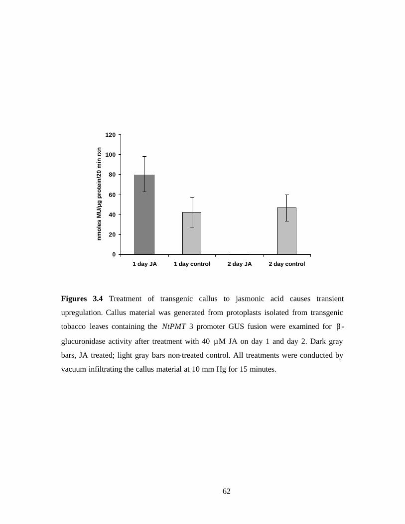
62
0
20
40
60
80
100
120
1 day JA 1 day control 2 day JA 2 day control
nm
ole
s M
U/µ
g p
rote
in/2
0 m
in r
xn
Figures 3.4 Treatment of transgenic callus to jasmonic acid causes transient
upregulation. Callus material was generated from protoplasts isolated from transgenic
tobacco leaves containing the NtPMT 3 promoter GUS fusion were examined for β-
glucuronidase activity after treatment with 40 µM JA on day 1 and day 2. Dark gray
bars, JA treated; light gray bars non-treated control. All treatments were conducted by
vacuum infiltrating the callus material at 10 mm Hg for 15 minutes.

63
0
10
20
30
40
50
60
70
normal light dark high light Catalase 5 mM DMTU
nm
ole
s M
U/u
g p
r.
Figure 3.5 PMT promoter activity in different light conditions and ROS scavengers.
Tobacco callus tissue, which displays highly repressed alkaloid synthesis under normal
culture conditions in light, exhibits significant induction of PMT promoter activity and
alkaloid accumulation when treated with an ROS scavenger, DMTU and catalase.
Transgenic tobacco containing the PMT 3 promoter-GUS fusion was used to obtain callus
material and were treated in the following ways 1) low light (normal) conditions (15-20
µmol m-2 s-1); 2) in darkness; 3) high- light conditions (100 µmol m-2 s-1); 4) 100 units/ml
catalase.; 5) in the presence of 5 mM dimethylthiourea (DMTU). Callus treated with
catalase and DMTU were maintained in low-light conditions in presence of auxin. All
treatments were conducted by vacuum infiltrating the callus material at 10 mm Hg for 15
minutes.
In presence of light and auxin

64
Involvement of reactive oxygen species in alkaloid pathway regulation
We hypothesized that light may elicit its effect on alkaloid pathway expression
through reactive oxygen species (ROS), such as H2O2, superoxide radicals, etc. and these
might function as signals, ROS are known to be produced during normal metabolism
such as photorespiration and photosynthesis. Thus we tested the involvement of ROS on
pathway expression by utilizing ROS scavenging chemicals such as DMTU (1,3 dimethyl
2-thiourea, an ROS scavenger for OH- and O2- radicals), and a H2O2 degrading enzyme,
catalase. DMTU is a well-known scavenger of ROS. DMTU also traps H2O2 (Jiang and
Zhang, 2002). Tobacco callus cultures, which display highly repressed alkaloid synthesis
under normal conditions in the light, exhibited significant (8-fold) induction of PMT
promoter activity when treated with 5 mM DMTU (figure 3.5). These cultures were
maintained in the presence of light and auxin, which are known repressors of PMT
expression. However, the presence of DMTU apparently effectively overcame this light
and auxin-mediated repression. Quantification of β–glucuronidase activity using GUS
fluorometric analysis on the DMTU-treated callus showed activity higher than that
observed when callus was maintained dark (figure 3.5).
An additional means of lowering ROS levels, specifically H2O2, was applied by
treating with 200 units/ml of catalase, an enzyme which catalyzes the breakdown of
H2O2. This treatment might provide evidence as to the effectiveness of H2O2 as potential
ROS species involved in signaling. Upon infiltrating the soft callus tissue with medium
containing catalase, the repression due to the presence of light and auxin was, like the
DMTU effect, overcome (approximately 6-fold) as indicated by GUS fluorometric
analysis (figure 3.5).
To test the hypothesis that the external media in which the callus material was
maintained had caused leaching of H2O2 from tissue, perhaps via by porin channels, we
tested for the H2O2 in the medium.
Other chemicals such as10 mM Tiron (4,5 dihydroxy 1,3 benzene disulfonic acid,
an ROS scavenger) and 100 µM DPI (diphenylene iodinium chloride, a well known
inhibitor of NAD(P)H oxidase) and 10 µM Rose Bengal diacetate (a superoxide anion
generator), were used to attempt to differentiate among distinct reactive oxygen species
as potential signals. However, treatment with these agents for 1 week killed the callus

65
tissue. Also 10 mM N-acetyl cysteine along with 30 mM ascorbic acid was used as a
reactive oxygen species scavenger, but these agents also did not effect PMT promoter
activity in callus tissue (data not shown).
Effect of ROS on alkaloid biosynthesis
To determine if a correlation existed between PMT expression after ROS
scavenging and alkaloid accumulation, the sensitive epibatidine binding displacement
assay was used to detect the presence of alkaloids.
In low-light conditions, alkaloid accumulation was very low, averaging
approximately 3 nM in a five-week-old callus tissue that was surface embedded (figure
3.6). In high light the amount of alkaloids detected in the callus was lower. However in
dark conditions, the alkaloid content increased even in presence of auxin (figure 3.6).
When the callus tissue was infiltrated with catalase, the alkaloid content was also
elevated (figure 3.6). However, when DMTU was added the increase in alkaloid levels
was the same as in untreated callus (data not shown). This might be because of the fact
that NtPMT3 might not be able to contribute a sufficient level of activity to increase
alkaloid synthesis. GC-MS analysis of callus tissue treated with DMTU and catalase was
done to confirm the epibatidine displacement data. An initial gas chromatographic (GC)
examination of the alkaloids produced in callus tissue was undertaken and are shown for
untreated callus samples in Table 3.2. The identification of these alkaloids supports their
detection by epibatidine-displacement assays. GC-MS analysis of DMTU and catalase-
treated callus tissues are in progress.

66
Figure 3.6 Alkaloid activity levels in callus tissue is affected by treatments that impact
reactive oxygen species levels. Callus material was generated from protoplasts isolated
from transgenic tobacco leaves containing NtPMT 3 promoter-GUS fusion construct.
Five-week-old callus grown on agar plates was treated by infiltrating tissue with catalase
and analyzed for alkaloid activity two weeks later. Callus extracts were prepared by
grinding the tissue in water and using the supernatant for the displacement assay.
Number above the bar indicates the estimated concentration of “nicotine equivalents”
derived from curves generated using pure nicotine in the assay. Concentration values
therefore indicate the amount of displacement activity that is equivalent to the given
concentration of nicotine.
0
5
10
15
20
25
30
35
40
45
Normal Lt Dark High lt (100µE) Catalase (200units/ml)
% E
B d
isp
lace
men
t
~3.3 nM
~ 16 nM
~ 5.5 nM
~ 14 nM
In presence of light and auxin
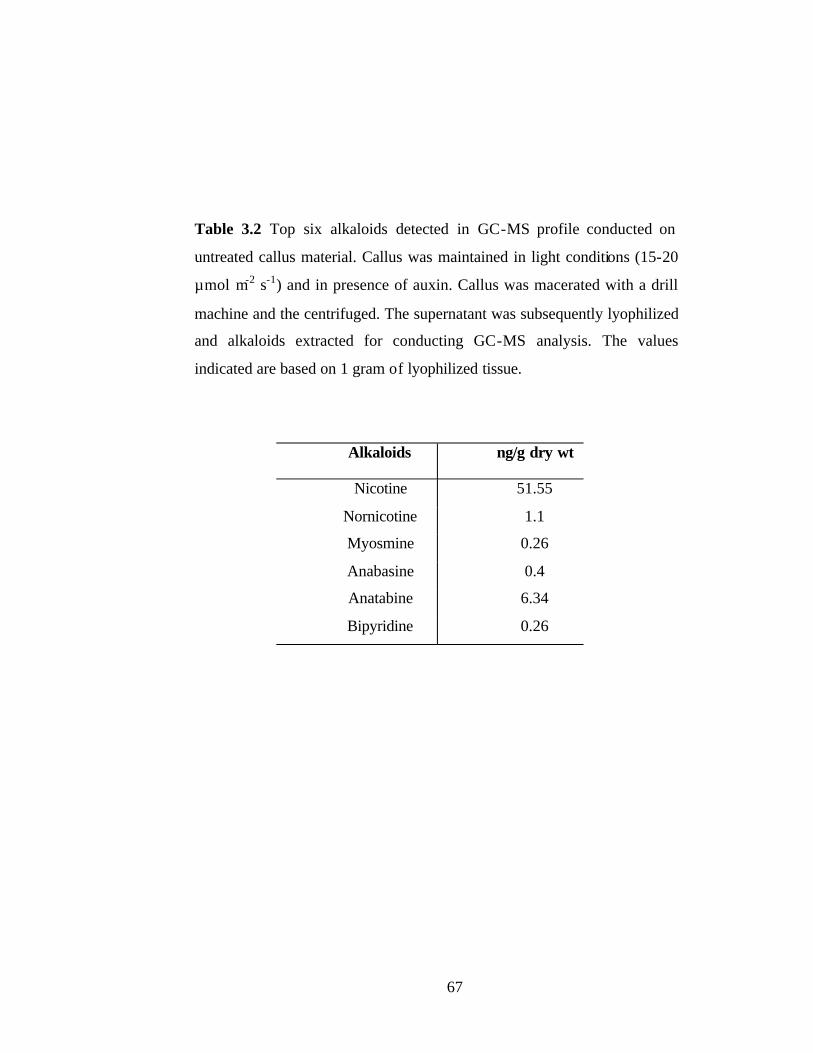
67
Alkaloids ng/g dry wt
Nicotine 51.55
Nornicotine 1.1
Myosmine 0.26
Anabasine 0.4
Anatabine 6.34
Bipyridine 0.26
Table 3.2 Top six alkaloids detected in GC-MS profile conducted on
untreated callus material. Callus was maintained in light conditions (15-20
µmol m-2 s-1) and in presence of auxin. Callus was macerated with a drill
machine and the centrifuged. The supernatant was subsequently lyophilized
and alkaloids extracted for conducting GC-MS analysis. The values
indicated are based on 1 gram of lyophilized tissue.

68
DISCUSSION
Treatment with factors known to affect the regulation of alkaloid biosynthesis
were first tested with our undifferentiated callus system. Auxin and jasmonic acid are
known to affect PMT expression in N. tabacum (Hibi et al., 1994; Imanishi et al., 1998).
Treatments with jasmonic acid resulted in an increase in alkaloid levels as studied earlier
in callus cultures (Imanishi et al., 1998) and in intact plants (Baldwin et al., 1994). Light
and auxin have been also shown to have a negative affect on alkaloid accumulation in
callus (Hobbs and Yeoman, 1991; Hibi et al., 1994) and intact plants (Mizusaki et al.,
1973; Hobbs and Yeoman, 1991; Hibi et al., 1994).
In our studies here with undifferentiated tobacco callus, we established that the
repressive effect of light and auxin on alkaloid expression occurred at the level of PMT
promoter activity. GUS histochemical and quantitative fluorometric data revealed a
strong repression of PMT promoter activity in the presence of both light and auxin (figure
3.1 and 3.3). Growing the callus material in the dark and in the absence of auxin gave
higher levels of PMT promoter expression. Collectively, these results show that light is
the primary factor in alkaloid pathway expression because a lower degree of change was
seen by reducing auxin levels, whereas the absence of light consistently gave the
strongest inductive effect, even in the presence of auxin. In some experiments, such as
with younger, plated callus tissue, it appeared that darkness plus a low level of auxin
provides the maximum PMT promoter expression, probably because a low level of auxin
was required to maintain growth.
Also there was an upregulation of PMT promoter activity in presence of JA, but
this was shortlived in that elevated PMT expression lasted only 24 hours (figure 3.4).
This transient effect has not been noted in other studies, which focused particularly on
studies with NtPMT1 (Imanishi et al., 1998) using tobacco BY-2 cells. The cells showed
an increase in PMT transcripts 6 h after treatment with MeJA (methyl jasmonate), which
lasted up to 24 h. However, the cells were not examined further. In the same report a
transient induction of ODC was seen upon treatment with MeJA (Imanishi et al., 1998).
69
One major difference between these studies and the results described here is that Iminashi
et al., used transformed BY-2 cells which are dissimilar to the freshly prepared callus
cultures used here. The protoplast-derived callus cultures are fully regenerable whereas
BY-2 cells are not. Since a different PMT gene belonging to N. slyvestris was examined
by Iminashi et al., and also the time period during which the PMT expression was
examined was different, being only 24 hours, results might be expected to differ from
those found here. A transient expression of NtPMT3 has been reported in leaves upon
wounding (Sachan and Falcone, 2002).
The conditions used to promote the formation of undifferentiated tissue from
single leaf cells, namely the presence of the growth regulators cytokinin and auxin,
results in a tissue dissimilar to either root or leaf tissue in regard to expression of alkaloid
metabolism. Therefore, the regulation of alkaloid biosynthesis is probably also unique to
this tissue. The identification of signaling intermediates that play a role in alkaloid
biosynthesis in undifferentiated tissue may have relevance in production of secondary
metabolites in plant cell cultures.
During a plant’s life cycle, it is subjected to a range of internal and external
stresses. Its ability to adapt to metabolic and environmental changes is essential for its
survival. Production of reactive oxygen species (ROS) such as hydrogen peroxide
(H2O2), superoxide (O ?̄2) and its toxic derivatives, hydroxyl radicals (OH?) and singlet
oxygen (1O2) occurs at all times during plant growth and development, but when the plant
is exposed to biotic and abiotic stresses production of these ROS increases (Navabpour,
2003). Several studies have indicated the production of ROS in presence of increased
light conditions (Karpinski et al., 1999). Prolonged exposure to conditions that cause
excess excitation energy can result in an increase in the generation of ROS. ROS are an
integral part of intracellular signaling in essentially all biological systems (Finkel, 1998).
Treatments of callus tissue with light causes a decrease in alkaloid accumulation (figure
3.6). It is possible that elevated levels of reactive oxygen species produced in the
presence of light causes repression of PMT gene expression (figure 3.5). This repression
in turn causes a decrease in alkaloid accumulation. Thus ROS, H2O2, might act as
intermediates in light signaling by changing levels of PMT expression. The use of the
ROS scavenger, DMTU, resulted in increased expression of PMT based on GUS
70
fluorometric analysis and an increase in alkaloid accumulation (fig. 3.5 and 3.6). This
increase could be a result of scavenging the ROS released in the media through the
various porins. It is hypothesized that free radicals are in equilibrium between the interior
and exterior of the cells and hence DMTU is able to scavenge the ones in apoplast and
the media in which the callus is maintained. It should be kept in mind that the epibatidine
assay is able to detect all nicotine related alkaloids. Hence the GUS expression data for
PMT gene expression generally correlate with the alkaloid accumulation data. Catalase is
not as effective as DMTU in causing an upregulation of promoter activity perhaps
because catalase cannot cross the cell wall and plasma membrane barriers, but with callus
tissue being basically heterogenous it might be able to penetrate a poorly developed cell
wall. H2O2 may also rapidly move into and out of cells. Whereas H2O2 is generally
considered to be capable of crossing the plasma membrane, peroxiporins or aquaporins
have also been recently implicated in such transmembrane transport of H2O2 (Henzler and
Steudle, 2000; Neill et al., 2002). Thus we hypothesize that there is an equilibrium
between the levels of H2O2 inside and outside the cell and catalase is able to scavenge
H2O2 which is present in the external medium, thereby lowering the overall concentration
of H2O2. The decreased H2O2 level then results in relieving the repression of PMT gene
expression. These results indicate a strong presence of ROS as intermediates in signaling
involved in alkaloid biosynthesis.
Another explanation for the greater PMT promoter activity in the presence of
DMTU compared to the catalase treatment might be that catalase only reduces H2O2
levels and DMTU is capable of scavenging a broader range of ROS. However, other
general chemical scavengers did not show equivalent effects on PMT promoter activity.
This might be due to highly specific scavenging of ROS. Also use of the H2O2 generating
enzyme, glucose oxidase, caused death of callus tissue probably because H2O2 is lethal
for long-term maintenance of tissues and callus is a very fragile tissue that survives
poorly in adverse conditions.
Tobacco callus tissue, which displays highly repressed alkaloid synthesis under
our typical laboratory conditions in the light, exhibited significant induction of PMT
promoter activity and alkaloid accumulation when treated with an ROS scavenger-
dimethylthiourea (DMTU) or with the H2O2 degrading enzyme, catalase. Based on GUS

71
fluorometric data, it was determined that treatment with these compounds relieves the
suppression caused by light and auxin in the callus tissue. Indeed, the level of PMT
promoter induction is even greater than that observed when callus tissue is incubated in
complete darkness. This perhaps suggests that a low level of ROS formation even in
dark- grown tissue limits the full extent of PMT promoter activity.
Earlier reports have reported very low levels of alkaloids in callus tissue derived
from shoots (Hobbs and Yeoman, 1991). Callus which was treated to the different light
conditions for changes in expression of PMT was also used to determine if the levels of
alkaloids are affected. For this, a very sensitive methodology was used to determine
various alkaloids in N. tabacum callus tissue. Thus the epibatidine displacement assay
was used to measure the very low levels of alkaloids present in callus cultures. This assay
is very useful since it is highly sensitive and able to detect slight changes in alkaloid
levels in extremely small amounts of tissue. The assay is also able to detect most of the
nicotine-related alkaloids present in N. tabacum (Rogers and Littleton, unpublished).
Callus exposed to low-light or high- light conditions did not show appreciably higher
alkaloid contents. However, when under dark conditions or in the presence of the H2O2
degrading enzyme, catalase, an increase in alkaloid accumulation resulted (figure 3.6).
72
Conclusions
Callus tissue is good model for studying alkaloid regulation even though it is
distinct from other plant tissues. Undifferentiated plant tissues can be highly useful as a
vehicle for natural product synthesis. In callus tissue, light plays a major role in
regulating both PMT gene expression and total alkaloid biosynthesis. Auxin and light
completely repress PMT gene expression in callus cultures. ROS scavengers, such as
DMTU and catalase, were able to overcome the repressive effect of light and auxin.
Thus, ROS is strongly implicated as an intermediate in signaling PMT gene response.
Also, treatment with catalase resulted in increased alkaloid production even in the
presence of light and auxin. We thus infer that H2O2 acts downstream of auxin and light
in this signaling pathway, which in turn negatively regulates PMT gene expression. A
model showing this ROS involvement in regulating alkaloid expression in tobacco
undifferentiated tissue is shown in figure 3.7.
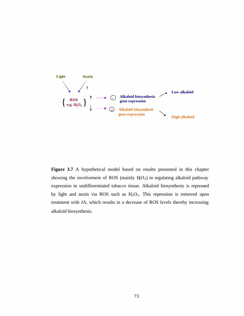
73
Figure 3.7 A hypothetical model based on results presented in this chapter
showing the involvement of ROS (mainly H2O2) in regulating alkaloid pathway
expression in undifferentiated tobacco tissue. Alkaloid biosynthesis is repressed
by light and auxin via ROS such as H2O2. This repression is removed upon
treatment with JA, which results in a decrease of ROS levels thereby increasing
alkaloid biosynthesis.
Light Auxin
ROS e.g. H2O2
?
{ }-
+
Alkaloid biosynthesis gene expression
Alkaloid biosynthesis gene expression
Low alkaloid
High alkaloid
74
References
Baldwin, I.T., Schmelz, E.A., and Ohnmeiss, T.E. (1994). Wound-induced changes in
root and shoot jasmonic acid pools correlate with induced nicotine synthesis in Nicotiana
sylvestris Spegazzini and Comes. J Chem Ecol 20, 2139-2157.
Baldwin, I.T., Zhang, Z.-P., Diab, N., Ohnmeiss, T.E., McCloud, E.S., Lynds, G.Y.,
and Schmelz, E.A. (1997). Quantification, correlations and manipulations of wound-
induced changes in jasmonic acid and nicotine in Nicotiana sylvestris. Planta 201, 397-
404.
Finkel, T. (1998). Oxygen radicals and signaling. Curr. Opin. Cell Biol. 10, 248-253.
Fritze, K., and Walden, R. (1995). Gene activation by T-DNA tagging. In Methods in
molecular biology (Clifton, N. J.: Humana Press), pp. 281-294.
Hara, K., Yagi, M., Koizumi, N., Kusano, T., and Sano, H. (2000). Screening of
wound-responsive genes identifies an immediate-early expressed gene encoding a highly
charged protein mechanically wounded tobacco plants. Plant Cell Physiol 41, 684-691.
Hashimoto, T., and Yamada, Y. (1994). Alkaloid biogenesis: molecular aspects. Annu
Rev Plant Physiol Plant Mol Biol 45, 257-285.
Hashimoto, T., Shoji, T., Mihara, T., Oguri, H., Tamaki, K., Suzuki, K., and
Yamada, Y. (1998). Intraspecific variability of tandem repeats in Nicotiana putrescine
N-methyltransferases. Plant Mol Biol 37, 25-37.
He, Y., and Gan, S. (2001). Identical promoter elements are involved in regulation of
OPR1 gene by senescence and jasmonic acid in Arabidopsis. Plant Mol Biol 47, 595-605.
Henzler, T., and Steudle, E. (2000). Transport and metabolic degradation of hydrogen
peroxide in Chara corallina: model calculations and mesurements with the pressure probe
suggest transport of H2O2 across water channel. Journal of Experimental Botany 51.
Hibi, N., Higashiguchi, S., Hashimoto, T., and Yamada, Y. (1994). Gene expression in
tobacco low-nicotine mutants. Plant Cell 6, 723-735.
Hobbs, M.C., and Yeoman, M.M. (1991). Effect of light on alkaloid accumulation in
cell cultures of Nicotiana species. Journal of Experimental Botany 42, 1371-1378.
Houghtling, R.A., Davila-Garcia, M.I., and Kellar, K.J. (1995). Characterization of
(±)-[3H]epibatidine binding to nicotinic cholinergic
receptors in rat and human brain. Molecular pharmacology 48, 280-287.

75
Imanishi, S., Hashizume, K., Nakakita, M., Kojima, H., Matsubayashi, Y.,
Hashimoto, T., Sakagami, Y., Yamada, Y., and Nakamura, K. (1998). Differential
induction by methyl jasmonate of genes encoding ornithine decarboxylase and other
enzymes involved in nicotine biosynthesis in tobacco cell cultures. Plant Mol Biol 38,
1101-1111.
Jiang, M., and Zhang, J. (2002). Water stress- induced abscisic acid accumulation
triggers the increased generation of reactive oxygen species and up-regulates the activites
of antioxidant enzymes in maize leaves. Journal of Experimental Botany 53, 2401-2410.
Karpinski, S., Reynolds, H., Karpinska, B., Wingsle, G., Creissen, G., and
Mullineaux, P. (1999). Systemic signalling and acclimation in response to excess
excitation energy in Arabidopsis. Science 284, 654-657.
Mahanandeeshwar, G., Alvin, T.V.J., and Buccafusco, J.J. (1995). A rapid
microtechnique for the estimation of muscarinic and nicotinic receptor binding
parameters using 96-well filtration plates. Journal of Neuroscience 63, 121-125.
Maliga, P., Sz-Breznovits, A., and Marton, L. (1973). Streptomycin-resistant plants
from callus culture of haploid tobacco. Nat New Biol 244, 29-30.
Mizusaki, S., Tanebe, Y., Noguchi, M., and Tamaki, E. (1973). Changes in activities
of ornithine decarboxylase, putrescine N-methyltransferase and N-methylputrescine
oxidase in tobacco roots in relation to nicotine biosynthesis. Plant Cell Physiol 14, 103-
110.
Neill, S., Desikan, R., and Hancock, J. (2002). Hydrogen peroxide signalling. Curr.
Opin. Cell Biol. 5, 388-395.
Ohta, S., Matsui, O., and Yatazawa, M. (1978). Culture conditions for nicotime
production in tobacco tissue culture. Agric. Biol. Chem. 42, 1245-1251.
Riechers, D.E., and Timko, M.P. (1999). Structures and expression of gene family
encoding putrescine N-methyltansferase in Nicotiana tabacum: new clues to the
evolutionary origin of cultivated tobacco. Plant Mol Biol 41, 387-401.
Sachan, N., and Falcone, D.L. (2002). Wound induced gene expression of putrescine N-
methyl transferase in leaves of Nicotiana tabacum. Phytochemistry 61, 797-805.

76
Shoji, T., Yamada, Y., and Hashimoto, T. (2000). Jasmonate induction of Putrescine
N-methyltransferase genes in the root of Nicotiana sylvestris. Plant Cell Physiol 41, 831-
839.
Wink, M. (1998). Modes of action of alkaloids. In Alkaloids, M. Wink, ed (New York:
Plenum press), pp. 301-326.

77
Chapter 4
Characterization of signaling involved in regulation of alkaloid
biosynthesis in young N. tabacum seedlings
Introduction
Survival of young seedlings is critical for the propagation of individuals of the next
generation. Extensive herbivory at the seedling stage seriously restrict survival. Synthesis of
nicotine in especially young, small seedlings has not been evaluated previously. Thus we
decided to study the regulation of nicotine production in young seedlings to determine whether
significant alkaloid production occurs. Based on the elucidation of signaling factors that
impacted alkaloid biosynthesis in N. tabacum callus tissue, we decided to evaluate if similar
factors also play a role in roots of young seedlings. In chapter 3 it was found that the expression
of the alkaloid biosynthetic pathway was strongly influenced by the levels of reactive oxygen
species (ROS). Also, recent studies by Musser et al. (Musser et al., 2002), showed that a
reduction in nicotine levels in tobacco occurred upon Helicoverpa zea caterpillar feeding. This
stimulated our investigation of factors that might affect regulation at the gene level.
Removal of tobacco flower initials and young leaves is a common practice during
tobacco cultivation. Decapitation in this way eliminates apical dominance and increases root
growth and nicotine content (Akehurst, 1981). Auxin synthesized in young meristematic tissue
is transported down the stem to the roots (Phillips, 1975), hence removal of the tip results in a
reduced flow of auxin to the roots and thereby may cause activation or repression of genes
involved in nicotine biosynthesis (Hibi et al., 1994). Repression has been shown to function at
the level of transcription in roots of whole plants. Seedlings were used to determine if similar
responses occurred in roots, thus implicating the presence of nicotine and related alkaloids for
survival of such a young tissue. Details regarding synthesis of nicotine in young seedlings are
unknown. In more mature plants several studies conducted by Baldwin et al. (Baldwin et al.,
1994; Baldwin et al., 1997; Preston et al., 1999) report an upregulation of PMT expression along

78
with alkaloid accumulation in N. sylvestris upon treatment with jasmonic acid (JA). Baldwin et
al. (Baldwin et al., 1994) hypothesize that upon injury to leaves the signal which is transmitted to
the root for increased nicotine accumulation is most probably JA.
In this chapter the characterization of signals is conducted using the findings obtained in
chapter 3 as a starting point to determine if ROS are also involved in alkaloid pathway
expression in differentiated tobacco seedling roots.

79
Materials and Methods
The transgenic plants harboring the PMT promoter-GUS fusion construct described in
chapter 2 (Sachan and Falcone, 2002) were used to generate stable transgenic N. tabacum plants,
and these were used as a source of seeds. These seeds were germinated to yield young seedlings.
Plant material
N. tabacum SR1was transformed with a PMT promoter-GUS fusion that included 627 bp
of genomic sequence of the NtPMT 3 gene. Details of the plasmid construction and plant
transformation were as described in chapter 2. Tobacco seeds were sterilized by a two minute
treatment in 70% ethanol, followed by two 20-minute treatments with 30% (v/v) household
bleach (active ingredient 6% sodium hypochlorite) and 0.02% tritonX-100 with continuous
rocking. Seeds were then rinsed 5 times with sterile water, followed by resuspension in 0.15%
agar.
Treatment of seedlings
Seedlings were grown on solid MS medium plates in the light (80 µmol m-2 s-1) for
fourteen days. Subsequently seedlings were removed from the agar medium and transferred to
liquid MS medium containing various chemicals for treatment. All seedlings were vacuum-
infiltrated with MS medium containing the chemical under a vacuum pressure of 10-mm hg for
15 min. The final concentrations of the substances used were 40 µM jasmonic acid (Sigma), 5
mM hydrogen peroxide, glucose oxidase, glucose (all Sigma), salicylic acid, 5 mM DMTU (1, 3-
dimethyl 2-thiourea, Aldrich), 200 units/ml catalase (Sigma), 10 mM tiron (4,5-dihydroxy 1,3-
benzene disulfonic acid, Sigma), 30 mM ascorbate and 10 mM N-acetylcysteine.
GUS fluorometric assay
GUS enzyme activity assays were performed in 96-well plates as described in chapter 3.
Seedling roots were excised from whole seedlings and the tissue was ground in 100 µl GUS

80
extraction buffer (50 mM NaHPO4 pH 7.0, 10 mM Na2EDTA, 0.1% sarcosyl, 0.1% Triton X-
100). Five microliters of the extract was diluted with 95 µl of GUS extraction buffer and to this,
30 µl of 5 mM 4-MUB-β-D-glucuronide (MUG) in extraction buffer was added and incubated
for 20 minutes at 37°C. The reaction was stopped by adding Na2CO3 to a concentration of 180
mM. Fluorescence was measured at 365 nm excitation and 455 nm emission using a Perkin
Elmer luminescence spectrophotometer model LS50B (Perkin-Elmer, Beaconsfield, UK).
Protein estimation was done colorimetrically by dye binding using a kit provided by BioRad
(Bradford, 1976).
Alkaloid displacement assay and extract preparation
Equal amounts of leaf material were removed from seedlings for preparation of the
extracts. Leaf material was ground for one minute in eppendorf tubes containing 500 µl water
using a drill press and micropestle for one minute. An additional 500 µl of water was added
afterwards. Extracts were centrifuged at 14,000 rpm for 10 minutes and supernatants were used
for conducting the alkaloid displacement assays and radioligand binding displacement assays as
described in chapter 3.
81
RESULTS
Primary transgenic plants harboring the PMT promoter-GUS fusion construct described
in chapter 2 were used to generate T1 seeds. Seeds were germinated on MS medium agar plates
with kanamycin selection to yield young seedlings. Agar was removed from the roots and
seedlings were subsequently transferred to MS liquid medium for conducting further
experiments.
Treatment with auxin, jasmonic acid
Various plant hormones have been known to affect the production of nicotine in leaves of
whole Nicotiana tabacum plants (Hibi et al., 1994; Baldwin, 1998), and these effects have been
shown to impact key enzyme activities in the nicotine biosynthetic pathway (Mizusaki et al.,
1973; Feth et al., 1986). To determine if these hormone-mediated effects occur at the level of
transcription, we treated young seedlings with auxin and jasmonic acid and monitored PMT
promoter activity. Treatment of fourteen-day-old seedlings with α-naphthalene acetic acid
(auxin) for 12 hours in light conditions resulted in a decrease in PMT promoter activity based on
GUS fluorometric analysis (figure 4.1). However there was no decrease in activity upon
treatment for five hours (data not shown).
Fourteen-day-old seedlings were treated with jasmonic acid in the same way they were
treated with auxin. Seedlings treated with jasmonic acid showed some variation in PMT
promoter activity after 12 hours following treatment based on GUS fluorometric activity but
showed pronounced upregulation at 24 hours after the treatment (figure 4.2). This PMT promoter
activity was then decreased 48 hours after treatment. Thus, the upregulation of NtPMT 3
expression is transient, similar to the effect of wounding leaf tissues in chapter 2. A difference in
PMT promoter activity at the zero time between control and JA-treated seedlings is clear. This
may be due to removal of agar from the tissue, thereby causing minor injury to the roots, and
susequently upregulating PMT expression.
Salicylic acid downregulates PMT promoter activity
Since salicylic acid (SA) and jasmonic acid are antagonistic to each other, we also
examined the effect of salicylic acid on PMT promoter activity in roots of young seedling. Five

82
0
100
200
300
400
500
600
700
800
auxin control
nm
ole
s M
U/µ
g p
rote
in/ 2
0 m
in r
xn
Figure 4.1 PMT promoter activity in roots in response to auxin treatment. Roots of
nineteen-day-old transgenic seedlings containing the PMT-promoter GUS fusion
were examined for β–glucuronidase activity after treatment with 30 µM α-
napthalene acetic acid, (α-NAA, auxin) for 12 h.

83
0
200
400
600
800
1000
1200
1400
0 1 2 12 24 48
Hours
nmol
es M
U/u
g pr
./20
min
rxn
Figure 4.2 PMT promoter activity in roots in response to jasmonic acid treatment. Roots of
nineteen-day-old transgenic seedlings containing the NtPMT 3 promoter GUS fusion were
examined for β-glucuronidase activity after treatment with 40 µM JA for various times.
Dark gray bars, JA treated; light gray bars non-treated control.

84
hours after treatment with 1 mM salicylic acid a high degree of suppression of PMT promoter
activity was found (Figure 4.3). Lower levels of salicylic acid (0.5 and 0.25 mM)
correspondingly also diminished NtPMT3 promoter activity with approximately 25% of the β–
glucuronidase activity detectable after treatment with 0.25 mM salicylic acid.
Hydrogen peroxide downregulates PMT promoter activity
An earlier report on reduction/ degradation of tobacco leaf nicotine contents after insect feeding
postulated that ROS was an active component of insect regurgitant (Musser et al., 2002). To
examine whether ROS has a direct effect on PMT expression, 50 mM H2O2 was added to the
incubation medium containing transgenic tobacco seedlings. Extracts prepared from roots of the
seedlings indicated no detectable β–glucuronidase activity after one hour of treatment (data not
shown). Five mM H2O2 also inhibited NtPMT 3 promoter activity as measured by β–
glucuronidase activity to approximately two-thirds the level of untreated controls and this
inhibition was found to persist for several hours (figure 4.4).
The half- life of exogenously added H2O2 has been reported to be 2 min in plant cell
cultures, and after only 5 min, no H2O2 is detectable (Desikan et al., 1998). Hence, we provided a
continuous supply of H2O2, using glucose oxidase and glucose to generate it catalytically.
Addition of the H2O2-generating system, glucose (0.5 mM)/glucose oxidase (0.5 U/ml), to P.
syringae pv. glycinea cell suspension cultures gives a steady state accumulation of ~6 µM for
approximately 3 hours (Delledonne et al., 1998b). Treatment of seedlings with 50 units/ml of
glucose oxidase in presence of 25 mM glucose caused a strong repression of PMT promoter
expression based on GUS fluorometric analysis conducted on extracts prepared from root tissues
(figure 4.5). This amount of glucose oxidase activity gave almost the same degree of NtPMT3
promoter repression as treatment with 20 mM H2O2 in seedling roots (figure 4.5).
Effect of ROS scavengers
Based on the effects of ROS scavengers on PMT promoter activity observed with N.
tabacum callus tissue (chapter 3), dimethylthiourea (DMTU) was utilized as a scavenging
molecule to reduce the levels of ROS, such as H2O2. Seedling roots were treated with MS
medium containing DMTU, which was vacuum infiltrated. After treatment of the seedling roots

85
0
200
400
600
800
1000
1200
1 0.5 0.25 0
nm
ole
s M
U/µ
g p
rote
in
Figure 4.3 PMT promoter activity in roots in response to salicylic acid treatment. Roots of
nineteen-day-old transgenic seedlings containing the NtPMT 3 promoter GUS fusion were
examined for β-glucuronidase activity 5 hours after treatment with differing concentrations of
salicylic acid.
Salicylic acid (mM)
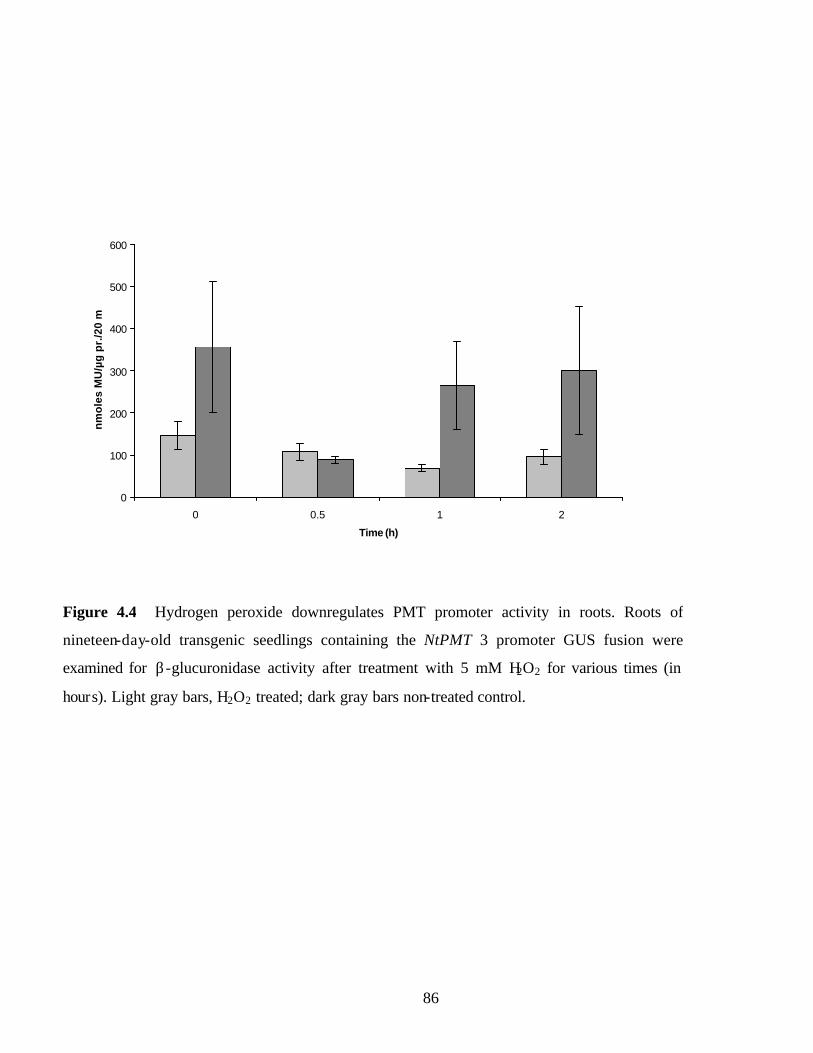
86
0
100
200
300
400
500
600
0 0.5 1 2
Time (h)
nm
ole
s M
U/µ
g p
r./2
0 m
in
Figure 4.4 Hydrogen peroxide downregulates PMT promoter activity in roots. Roots of
nineteen-day-old transgenic seedlings containing the NtPMT 3 promoter GUS fusion were
examined for β-glucuronidase activity after treatment with 5 mM H2O2 for various times (in
hours). Light gray bars, H2O2 treated; dark gray bars non-treated control.

87
0
100
200
300
400
500
600
control 20 mM H2O2 50units/mlGOX+25mM
gluc
GOX only 5 mM DMTU 100 units/mlCatalase
nm
ole
s M
U/µ
g p
r/20
min
rxn
Figure 4.5 PMT promoter activity responds to treatments that elevate or diminish hydrogen
peroxide levels. Roots of 21-day-old transgenic tobacco seedlings containing the PMT 3
promoter-GUS fusion were treated with the following: 1) control (no treatment); 2) in the
presence of 20 mM hydrogen peroxide; 3) in the presence of 50 units/ml glucose oxidase
(GOX) and 25 mM glucose; 4) 50 units/ml glucose oxidase only; 5) in the presence of 5
mM dimethylthiourea (DMTU); 6) 100 units/ml catalase. All treatments were conducted by
vacuum infiltrating the seedlings in MS liquid medium followed by a 5-h incubation.
Treatment
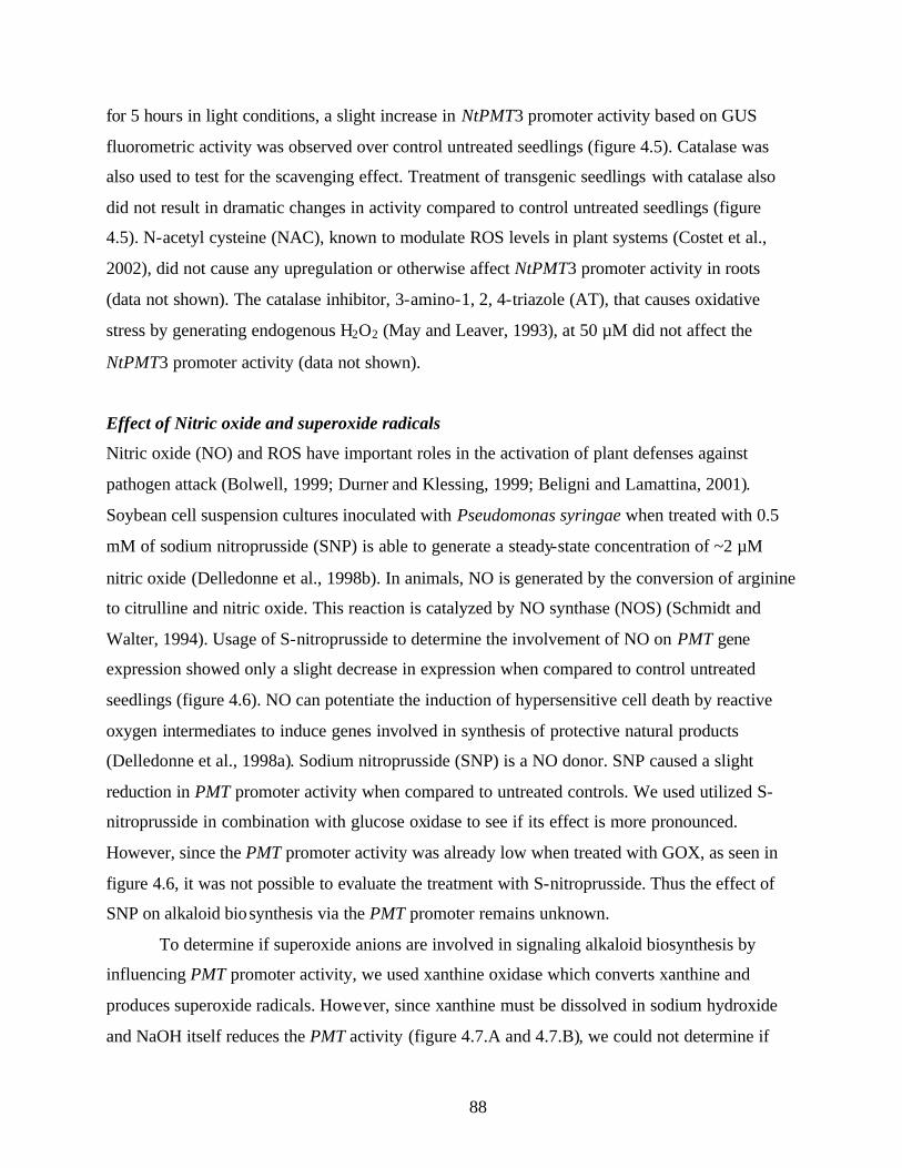
88
for 5 hours in light conditions, a slight increase in NtPMT3 promoter activity based on GUS
fluorometric activity was observed over control untreated seedlings (figure 4.5). Catalase was
also used to test for the scavenging effect. Treatment of transgenic seedlings with catalase also
did not result in dramatic changes in activity compared to control untreated seedlings (figure
4.5). N-acetyl cysteine (NAC), known to modulate ROS levels in plant systems (Costet et al.,
2002), did not cause any upregulation or otherwise affect NtPMT3 promoter activity in roots
(data not shown). The catalase inhibitor, 3-amino-1, 2, 4-triazole (AT), that causes oxidative
stress by generating endogenous H2O2 (May and Leaver, 1993), at 50 µM did not affect the
NtPMT3 promoter activity (data not shown).
Effect of Nitric oxide and superoxide radicals
Nitric oxide (NO) and ROS have important roles in the activation of plant defenses against
pathogen attack (Bolwell, 1999; Durner and Klessing, 1999; Beligni and Lamattina, 2001).
Soybean cell suspension cultures inoculated with Pseudomonas syringae when treated with 0.5
mM of sodium nitroprusside (SNP) is able to generate a steady-state concentration of ~2 µM
nitric oxide (Delledonne et al., 1998b). In animals, NO is generated by the conversion of arginine
to citrulline and nitric oxide. This reaction is catalyzed by NO synthase (NOS) (Schmidt and
Walter, 1994). Usage of S-nitroprusside to determine the involvement of NO on PMT gene
expression showed only a slight decrease in expression when compared to control untreated
seedlings (figure 4.6). NO can potentiate the induction of hypersensitive cell death by reactive
oxygen intermediates to induce genes involved in synthesis of protective natural products
(Delledonne et al., 1998a). Sodium nitroprusside (SNP) is a NO donor. SNP caused a slight
reduction in PMT promoter activity when compared to untreated controls. We used utilized S-
nitroprusside in combination with glucose oxidase to see if its effect is more pronounced.
However, since the PMT promoter activity was already low when treated with GOX, as seen in
figure 4.6, it was not possible to evaluate the treatment with S-nitroprusside. Thus the effect of
SNP on alkaloid biosynthesis via the PMT promoter remains unknown.
To determine if superoxide anions are involved in signaling alkaloid biosynthesis by
influencing PMT promoter activity, we used xanthine oxidase which converts xanthine and
produces superoxide radicals. However, since xanthine must be dissolved in sodium hydroxide
and NaOH itself reduces the PMT activity (figure 4.7.A and 4.7.B), we could not determine if

89
0
100
200
300
400
500
600
1 2 3 4 5 6
nm
ole
s M
U/µ
g p
r/ 2
0 m
in r
xn
Figure 4.6 PMT promoter activity is not responsive to treatments that affect nitric oxide
levels. Roots of 21-day-old transgenic tobacco seedling containing PMT 3 promoter-GUS
fusion were treated with the following: 1) control (no treatment); 2) in the presence of 15
units/ml glucose oxidase (GOX) and 12.5 mM glucose ; 3) in the presence of 5 mM sodium
nitroprusside (SNP); 4) in the presence of 5 mM SNP, 15 units/ml GOX and 12.5 mM
glucose. All treatments were conducted by vacuum infiltrating the seedlings in MS liquid
medium followed by a 5-hour incubation.
Treatments

90
0
200
400
600
800
1000
1200
control 50 units/ml GOX+ 25mM Gluc 2 units/ml XOX+ 2 mMXanthine
nm
ole
s M
U/u
g p
r/20
min
rxn
0
200
400
600
800
1000
1200
1400
1600
1800
Control NaOH
nm
ole
s M
U/µ
g p
rote
in/ 2
0 m
in r
xn
A
B
Figure 4.7.A PMT promoter activity is not responsive to treatments that affect nitric oxide
levels. Roots of 21-day-old transgenic tobacco seedling containing PMT 3 promoter-GUS
fusion were treated with the following: 1) control (no treatment); 2) in the presence of 50
units/ml glucose oxidase (GOX) and 25 mM glucose ; 3) in the presence of 2 units/ml xanthine
oxidase (XOX) and 2 mM xanthine. All treatments were conducted by vacuum infiltrating the
seedlings in MS liquid medium followed by 5-hour incubation.
Figure 4.7.B. Treatment with NaOH (same dilution as that used to prepare XOX) to show that
pH changes can effect PMT expression levels
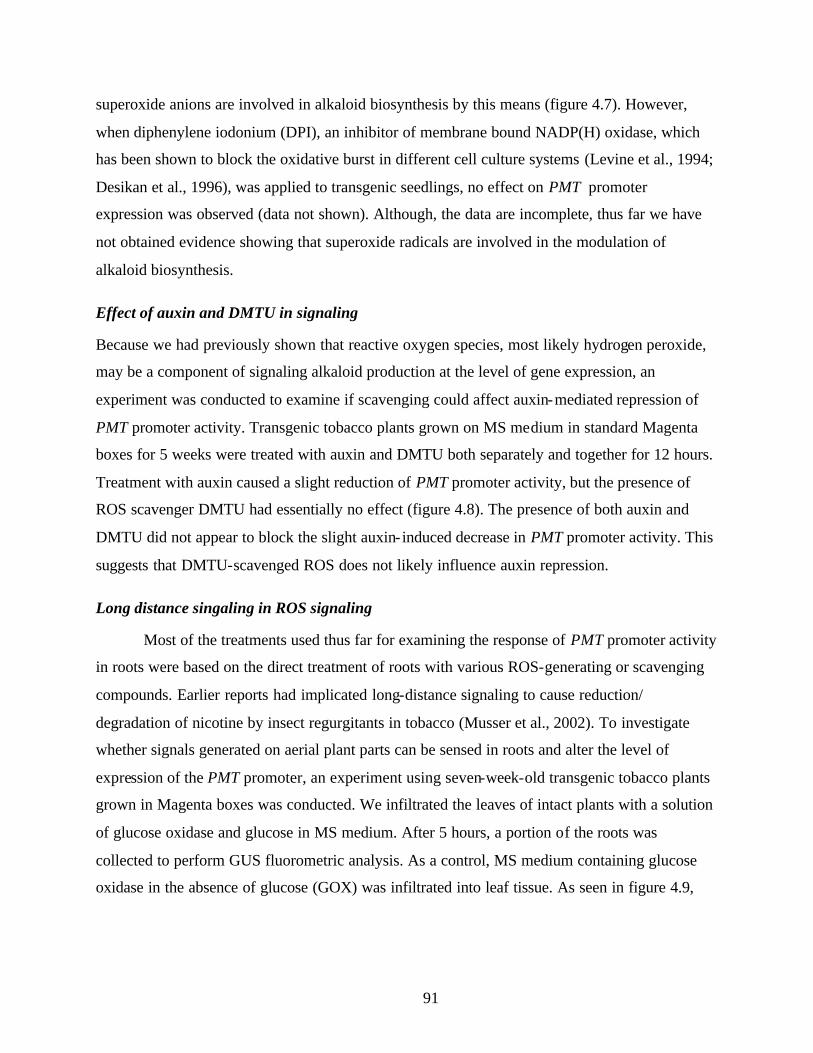
91
superoxide anions are involved in alkaloid biosynthesis by this means (figure 4.7). However,
when diphenylene iodonium (DPI), an inhibitor of membrane bound NADP(H) oxidase, which
has been shown to block the oxidative burst in different cell culture systems (Levine et al., 1994;
Desikan et al., 1996), was applied to transgenic seedlings, no effect on PMT promoter
expression was observed (data not shown). Although, the data are incomplete, thus far we have
not obtained evidence showing that superoxide radicals are involved in the modulation of
alkaloid biosynthesis.
Effect of auxin and DMTU in signaling
Because we had previously shown that reactive oxygen species, most likely hydrogen peroxide,
may be a component of signaling alkaloid production at the level of gene expression, an
experiment was conducted to examine if scavenging could affect auxin-mediated repression of
PMT promoter activity. Transgenic tobacco plants grown on MS medium in standard Magenta
boxes for 5 weeks were treated with auxin and DMTU both separately and together for 12 hours.
Treatment with auxin caused a slight reduction of PMT promoter activity, but the presence of
ROS scavenger DMTU had essentially no effect (figure 4.8). The presence of both auxin and
DMTU did not appear to block the slight auxin- induced decrease in PMT promoter activity. This
suggests that DMTU-scavenged ROS does not likely influence auxin repression.
Long distance singaling in ROS signaling
Most of the treatments used thus far for examining the response of PMT promoter activity
in roots were based on the direct treatment of roots with various ROS-generating or scavenging
compounds. Earlier reports had implicated long-distance signaling to cause reduction/
degradation of nicotine by insect regurgitants in tobacco (Musser et al., 2002). To investigate
whether signals generated on aerial plant parts can be sensed in roots and alter the level of
expression of the PMT promoter, an experiment using seven-week-old transgenic tobacco plants
grown in Magenta boxes was conducted. We infiltrated the leaves of intact plants with a solution
of glucose oxidase and glucose in MS medium. After 5 hours, a portion of the roots was
collected to perform GUS fluorometric analysis. As a control, MS medium containing glucose
oxidase in the absence of glucose (GOX) was infiltrated into leaf tissue. As seen in figure 4.9,
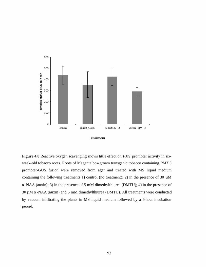
92
Treatment
Figure 4.8 Reactive oxygen scavenging shows little effect on PMT promoter activity in six-
week-old tobacco roots. Roots of Magenta box-grown transgenic tobacco containing PMT 3
promoter-GUS fusion were removed from agar and treated with MS liquid medium
containing the following treatments 1) control (no treatment); 2) in the presence of 30 µM
α-NAA (auxin); 3) in the presence of 5 mM dimethylthiurea (DMTU); 4) in the presence of
30 µM α-NAA (auxin) and 5 mM dimethylthiurea (DMTU). All treatments were conducted
by vacuum infiltrating the plants in MS liquid medium followed by a 5-hour incubation
peroid.
0
100
200
300
400
500
600
Control 30uM Auxin 5 mM DMTU Auxin +DMTU
nm
ole
s M
U/µ
g p
r/20
min
rxn

93
0
20
40
60
80
100
120
140
160
Control (100units/ml GOX
only)
50 units/ml GOXand glucose
100 units/mlGOX andglucose
nm
ole
s M
U/u
g p
r/20
min
rxn
after 5 hrs averageafter 24 hrs average
Figure 4.9 PMT promoter activity in roots can respond to ROS signals that originate in leaf
tissue. Magenta box-grown leaves of transgenic tobacco containing the PMT 3 promoter-GUS
fusion were infiltrated with a syringe containing MS medium the following: 1) control (100
units/ml glucose oxidase (GOX)); 2) 50 units/ml glucose GOX and 25 mM glucose; 3) 100
units/ml glucose GOX and 50 mM glucose. Treatments incubated for 5 hours (light gray) and
24 hours (dark gray).

94
after just 5 h, PMT promoter activity was reduced by approximately 30% after treatment with 50
units/ml of GOX. Treatment of leaves with a higher GOX concentration (100 units/ml) led to
further repression of PMT promoter activity within 5 h of treatment. After 24 h of leaf treatment,
the level of PMT promoter activity was reduced to below detectable levels suggesting that the
signal persisted and resulted in a further reduction of PMT promoter activity. Treatment with
glucose oxidase only also resulted in a complete repression of PMT promoter activity after 24 h,
perhaps due to availability of its substrate. Thus, it appears that alkaloid biosynthetic genes in the
root can respond to signals generated within leaves. This signaling may be mediated by H2O2 or
some other signaling molecule.
Alkaloid levels respond to treatments that lower or raise ROS
To determine whether the PMT promoter activities observed in response to treatments
that affect ROS levels, correlation with the levels of alkaloids synthesized was determined using
radioligand binding-displacement assays (described in chapter 3). The alkaloid activity was
determined in extracts prepared from seedling leaves, the site of alkaloid deposition after
synthesis in roots. Treatment of seven-day-old seedlings with jasmonic acid for four days caused
an ~10-fold increase in the alkaloid levels. However, treatment with salicylic acid also caused
higher (~2.5-fold) accumulation of alkaloids (figure 4.10) which was unlike the response of PMT
promoter activity to salicylic acid treatment (figure 4.3).
Treatment of seven-day-old seedlings to different light conditions caused the alkaloid
accumulation to be lower when compared to dark treatments (figure 4.11). Seedlings were
treated to light conditions normally used during their propagation on synthetic medium (60 µmol
m-2 s-1), or “high light” (150 µ mol m-2 s-1) or darkness for four days. Under dark conditions,
alkaloids accumulated ~ 2 fold higher in leaves than in leaves of plants treated to continuous
light, as determined by alkaloid displacement activity. Seedlings treated to higher light
conditions showed a slight decrease in alkaloid activity levels (figure 4.11). A similar treatment
experiment was also conducted on three-week-old seedlings grown in soil. In seedlings treated to
dark conditions, there was higher alkaloid accumulation. However, light did not influence
alkaloid accumulation in these older plants (data not shown).

95
Treatment of seedlings to varying concentrations of ROS as generated by glucose oxidase
and glucose caused lower accumulation of alkaloid activity in leaf extracts. Four days after
treatment with higher concentrations of GOX, the seedlings were stunted, indicating that the
treatment used was deleterious to seedling growth. Thus, equal amounts of seedling leaves were
taken to prepare the extracts. Seedlings treated with DMTU exhibited a higher (~1.5-fold)
accumulation of alkaloids (figure 4.12). The DMTU treated seedlings were similar in appearance
to control- treated seedlings.
PMT-promoter regulation in transgenic Arabidopsis
To determine if the regulatory responses of the PMT promoter observed in N. tabacum is similar
in a different plant species, we introduced the PMT promoter-GUS fusion construct into A.
thaliana. Root extracts from treated seedlings were used for the GUS fluorometric assay. When
fourteen-day-old Arabidopsis seedlings were treated with auxin, the response was similar to that
seen in N. tabacum. A decrease in PMT promoter- GUS activity was seen upon treatment with
auxin for 12 hours in light conditions (figure 4.13 A).
When Arabidopsis seedling roots were treated with SA for five hours, it resulted in the
repression of PMT promoter expression (figure 4.13 B). Unlike the expression seen in N.
tabacum, Arabidopsis seedlings did not respond to JA treatments (data not shown). PMT
promoter activity in Arabidopsis responded to ROS treatments in a manner similar to that seen in
tobacco. Treatment with H2O2 or glucose oxidase caused repression of PMT promoter
expression. However like tobacco seedlings, treatment of Arabidopsis seedlings with DMTU or
catalase did not seem to cause an upregulation but rather a slight downregulation (figure 4.14).
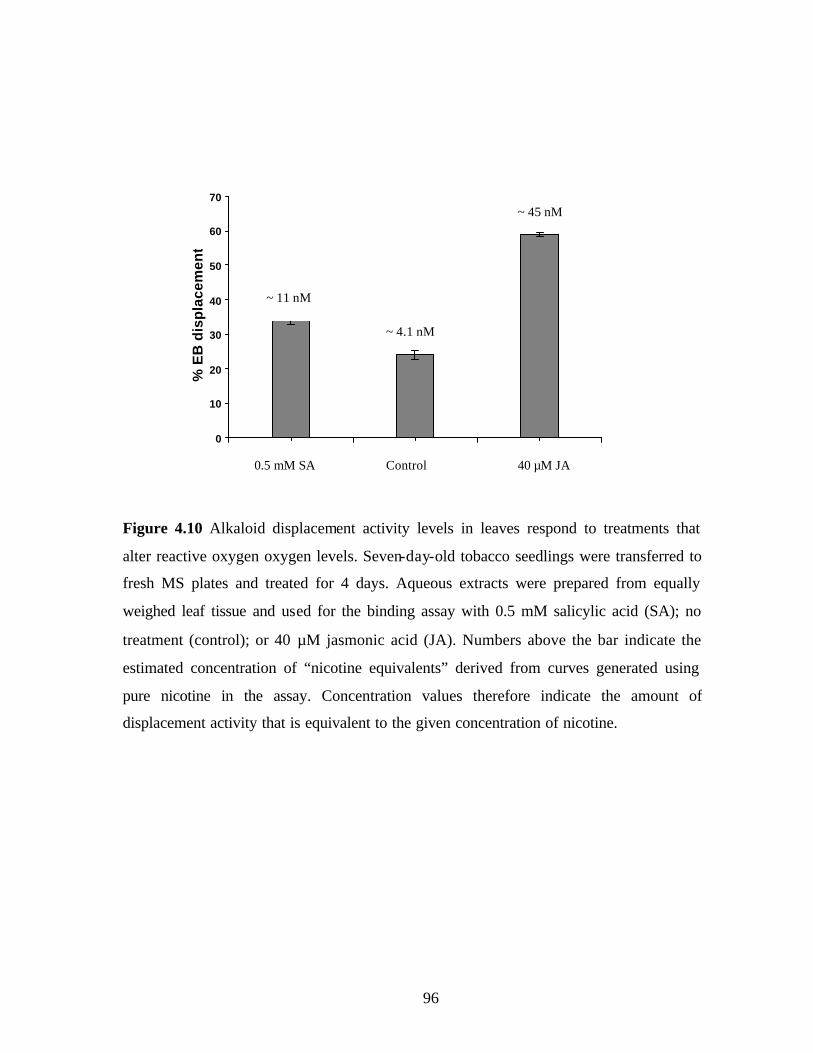
96
0
10
20
30
40
50
60
70
0.5 mM SA Control 40 µM JA
% E
B d
isp
lace
men
t
Figure 4.10 Alkaloid displacement activity levels in leaves respond to treatments that
alter reactive oxygen oxygen levels. Seven-day-old tobacco seedlings were transferred to
fresh MS plates and treated for 4 days. Aqueous extracts were prepared from equally
weighed leaf tissue and used for the binding assay with 0.5 mM salicylic acid (SA); no
treatment (control); or 40 µM jasmonic acid (JA). Numbers above the bar indicate the
estimated concentration of “nicotine equivalents” derived from curves generated using
pure nicotine in the assay. Concentration values therefore indicate the amount of
displacement activity that is equivalent to the given concentration of nicotine.
~ 11 nM
~ 4.1 nM
~ 45 nM
0.5 mM SA Control 40 µM JA
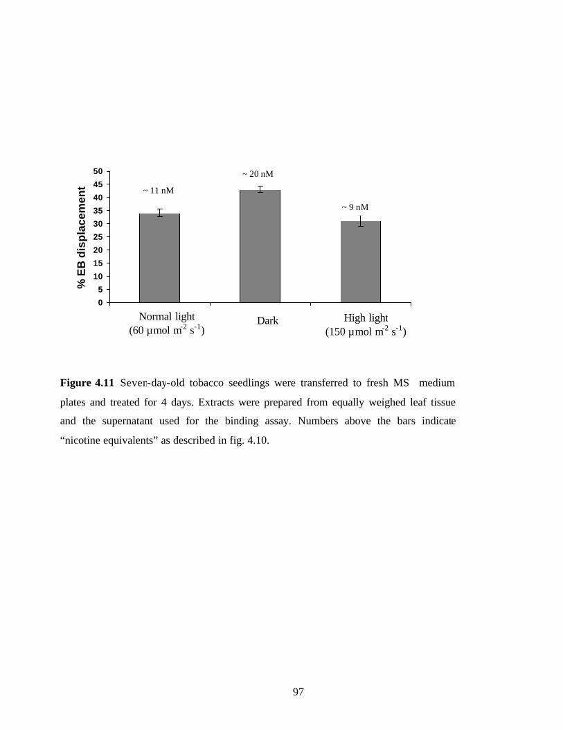
97
0
5
10
15
20
25
30
35
40
45
50
Normal light (60 µmol m-2 s-1)
Dark High light (150 µmol m-2s-1)
% E
B d
isp
lace
men
t
Figure 4.11 Seven-day-old tobacco seedlings were transferred to fresh MS medium
plates and treated for 4 days. Extracts were prepared from equally weighed leaf tissue
and the supernatant used for the binding assay. Numbers above the bars indicate
“nicotine equivalents” as described in fig. 4.10.
~ 11 nM
~ 20 nM
~ 9 nM
Normal light (60 µmol m-2 s-1)
High light (150 µmol m-2 s-1)
Dark

98
Figure 4.12 Seven-day-old tobacco seedlings were transferred to fresh MS medium
plates and treated for 4 days. Extracts were prepared from equally weighed leaf
tissue and the supernatant used for the binding assay. Numbers above the bars
indicate “nicotine equivalents” as described in fig. 4.10.
0
10
20
30
40
50
60
70
50units/mlGOX+25mM Gluc
16 units/mlGOX+12.5mM Gluc
Control 5 mM DMTU
% E
B d
ispl
acem
ent
~ 50 nM ~ 33 nM
~ 17 nM
~ 7 nM

99
0
100
200
300
400
500
600
700
C o n t r o l 30 µM Auxin
nm
ole
s M
U/µ
g p
r/20
min
rxn
0
50
100
150
200
250
300
Control 1 mM SA
nm
ole
s M
U/µ
g p
r/20
min
rxn
Figure 4.13 PMT promoter activity responds similarly in a heterologous host background,
Arabidopsis thaliana. (A) Transgenic Arabidopsis seedlings grown on MS medium were
removed and the seedlings treated in liquid MS medium with and without auxin overnight.
(B) Transgenic seedlings grown on MS medium were removed and the seedlings treated in
liquid MS medium with and without salicylic acid (SA) for five hours. GUS fluorometric
analysis was conducted on extracts prepared from roots.
A
B
Control 30 µM Auxin

100
0
100
200
300
400
500
600
700
control 20 mM H2O2 50 unitsmlglucose
oxidase+ 50mM glucose
5 mM DMTU 200 units/mlcatalase
10 mM tiron
nm
ole
s M
u/µ
g p
r/20
min
rxn
Figure 4.14 Transgenic Arabidopsis seedlings grown on MS media were removed
and the seedlings treated in liquid MS media for five hours. GUS fluorometric
analysis was conducted on extracts prepared from roots. Roots of 21-day-old
transgenic Arabidopsis seedlings containing the NtPMT3 promoter-GUS fusion
were treated with the following:
1) control (no treatment); 2) in the presence of 20 mM hydrogen peroxide; 3) in the
presence of 50 units/ml glucose oxidase (GOX) and 25 mM glucose; 4) in presence
of 5 mM dimethylthiourea (DMTU); 5) 100 units/ml catalase 6) 10 mM Tiron. All
treatments were conducted by vacuum infiltrating the seedlings in MS liquid
medium followed by a 5-hour incubation.
1 2 3 4 5 6
101
Discussion
Treatment with known effectors of alkaloid biosynthesis
Earlier studies showed a repression of NtPMT 1 expression upon treatment with auxin in
cultured tobacco roots (Hibi et al., 1994). Decapitation of tobacco plants cause an increase in
PMT enzyme activity and in nicotine content (Mizusaki et al., 1973). The effect of auxin on
NtPMT3 expression, as determined here, is also repressive, but it takes 12 hours to detect this
decrease in the root tissues. Thus a slight delay seems to occur in the response to auxin, which
may indicate an additional intermediary step(s) or signal(s) for the auxin-mediated regulation of
PMT. In addition, the downregulation observed in the presence of auxin is only partial, with
some PMT promoter expression still occurring, consistent with other studies (Hibi et al., 1994).
Several studies have been conducted to indicate the responsiveness of alkaloid synthesis
or PMT expression to JA treatments (Baldwin, 1994, 1998; Imanishi et al., 1998; Shoji et al.,
2000). A rapid increase in PMT transcript levels and nicotine contents are typically observed.
Most of these studies were done with Nt PMT1. However, in our studies we observed a transient
upregulation of NtPMT3 upon treatment with 40 µM of JA. But this is inconsistent with PMT
expression in N. sylvestris in which treatment with JA does not result in a transient but
apparently continuous expression (Shoji et al., 2000). Shoji et al., (1998) have shown the levels
of NsPMT transcripts remain elevated up to 72 h after treatment in a gel blot analysis using the
NtPMT 1 as a probe. Jasmonic acid (JA) has been implicated as a wounding hormone and the JA
levels are thought to increase rapidly and transiently, and with pools waxing and waning within
minutes upon wounding (Baldwin, 1998). However, several studies investigating the JA effect
on wounding have examined JA levels in plants up to 24 h but not beyond (Baldwin et al., 1997;
Preston et al., 1999). It is possible that the transient upregulation seen with Nt PMT 3 upon
treatment with JA, could also be true for other PMT clones. Only one report, Shoji et al. (2002)
reports that the NsPMT expression does not wane even after 72 hours of JA application (Shoji et
al., 2000). Another factor could be that different species of Nicotiana are involved i.e., N.
tabacum and N. sylvestris. NtPMT3 transient expression when induced by JA is similar to
transient effect of NtPMT3 expression in leaves upon wounding (Sachan and Falcone, 2002).

102
Involvement of ROS molecules in alkaloid regulation
An earlier study reported on the reduction or degradation of nicotine in tobacco leaf
tissue by the action of components in insect regurgitants that are released upon insect feeding
(Musser et al., 2002). We tested whether the effect of ROS contributed directly to the reduction
of nicotine and related alkaloids by acting at the level of gene expression. Treatment with 5 mM
H2O2 was found to repress NtPMT3 promoter expression. Similar repression of NtPMT3 occurs
upon treatment with glucose oxidase. Glucose oxidase is the principal salivary enzyme in
Helicoverpa zea (the common, corn earworm), and converts D-glucose and molecular oxygen to
D-gluconic acid and H2O2 (Eichenseer et al., 1999; Felton and Eichenseer, 1999). H2O2 is
continually generated from various sources during normal plant metabolism. A wide range of
steady-state H2O2 concentrations (e. g. 60 µM-7mM in Arabidopsis (Karpinski et al., 1999; D. et
al., 2001) and 1-2 mM in maize and rice has been reported in plants (Jiang and Zhang, 2001; Lin
and Kao, 2001). We verified that the pronounced reduction of the NtPMT3 gene promoter, one of
the five PMT gene family members in tobacco, actually reflected whole alkaloid pathway
expression by measuring total alkaloid activity levels after the ROS treatments. The effect of
H2O2 on alkaloid accumulation was also inhibitory (figure 4.12), confirming the use of NtPMT3
gene as a useful indicator of alkaloid pathway expression.
Salicylic acid (SA) is antagonistic to JA. Application of SA at the site of wounding
reduced the amount of JA and nicotine produced (Baldwin, 1999). However application of SA
adjacent to the site of wounding does not inhibit nicotine production (Baldwin, 1999). However,
no correlation was seen between alkalo id accumulation and SA levels in constitutive salicylic
acid producing transgenic tobacco plants (Nugroho et al., 2002). This finding might have been
due to the fact that the SA was produced in chloroplastic tissue where the substrate, chorismate,
is synthesized, while the alkaloid is synthesized in roots which had little evidence of occurrence
of chloroplasts (Nugroho et al., 2002).
Based on GUS fluorometric analysis, NtPMT3 promoter-GUS fusion transgenic seedlings
reproducibly showed a high degree of promoter repression upon SA application (figure 4.3).
However, repression did not correlate to the alkaloid levels in SA untreated seedlings, and an
opposite response occurred i.e., alkaloid activity actually increasing (figure 4.10). This lack of a
correlation between NtPMT3 promoter activity and alkaloid levels thus uncouples alkaloid
production from at least expression of the PMT 3 promoter. This result is in contrast to the

103
results of previous experiments employing ROS generators or scavengers. We added a similar
final concentration of SA to the β–glucuronidase enzyme assay mixture to make sure that this
effect is not simply due to addition of SA on β-glucuronidase enzyme activity. Northern analysis
of seedlings treated with SA similarly did not show any difference compared to the untreated
seedlings. The probe used for the analysis was prepared by PCR of genomic DNA used NtPMT1
primers. Thus it appears that SA acts by a distinct mechanism that causes a reduction in PMT3
expression. It is to be determined if it does effect other PMT family members. Another
possibility may be that SA treatment downregulates all PMT gene expression but mechanisms of
transport and/or alkaloid degradation are not effected thereby leading to alkaloid levels that are
relatively unchanged. It is possible that SA does not directly affect the NtPMT3, but might be
targeting a regulatory protein interacting with the NtPMT3 promoter. Also, SA may be
suppressing PMT gene expression by suppression of ascorbate peroxidase and catalase that are
responsible for degradation of H2O2. SA has been shown to inhibit catalases (Willenkens et al.,
1994).
A related study employed transgenic potato that was transformed with fungal glucose
oxidase to produce constitutively sub- lethal levels of H2O2. The plants showed increased disease
resistance and a several- fold increase in SA levels (Wu et al., 1997). GOX-transgenic plants
show broad spectrum disease resistance to Phtyophtora infectans late blight, Alternaria solani
early blight and Verticillium dahliae wilt (Wu et al., 1997). Thus, intracellularly generated H2O2
is implicated in elevating SA levels (Slaymaker et al., 2002). On the other hand SA’s
involvement in generating H2O2 cannot be ruled out (Conrath et al., 1995; Durner and Klessing,
1996).
Dimethylthiourea was utilized as a scavenging molecule to reduce the levels of ROS such
as H2O2, as it functions as a trap for H2O2 (Levine et al., 1994) as well as other ROS species such
as superoxide anions and hydroxyl radicals. Catalase was also used to decrease specifically H2O2.
Because catalases are enzymes thay cannot cross cell wall or plasma membranes, we hypothesize
that ROS such as H2O2 leach out into the media and thereby can be reduced by catalase to
remove the repression of the PMT promoter. However, the amount of ROS production in roots
may be very low to begin with, or that a distinct type of regulation occurs in tobacco callus.
Hence we do not see a high degree of PMT overexpression as seen in callus tissue in response to
ROS scavengers (Chapter 3). In addition the level of PMT expression in roots is already

104
relatively high. In regard to sources of ROS in seedling roots, peroxisomes are known to be a
main source of ROS, and most of them occur in leaves. Peroxisomes play a key role in
photorespiration that occurs in leaves of C3 plants such as tobacco. Because photorespiration is
an inevitable consequence of photosynthesis, leaf peroxisomes are large and abundant in
photosynthetic tissues (Masters and Crane, 1995; Olsen and Harada, 1995). Mitochondria, on the
other hand, are also abundant in roots and ROS production from these during oxidative electron
transport may generate a very small and localized amount of ROS, such that a minimal affect is
seen on the PMT promoter.
Nitric oxide (NO) and ROS have important roles in the activation of defenses against
pathogen attack (Bolwell, 1999; Durner and Klessing, 1999; Beligni and Lamattina, 2001).
When the plant recognizes an avirulent pathogen it triggers a rapid production of ROS such as
H2O2 and O2-, which results in initiation of several defense responses in the plant (Delledonne et
al., 1998b). However, Delledonne et al., (1998) have shown that to produce a strong disease-
resistance response, nitric oxide is also required as a signal. It has been shown to act
independently of these intermediates to induce genes involved in production of protective natural
products. It is not clear if NO has a role in alkaloid signaling since S-nitroprusside affects PMT
only slightly (figure 4.6). Also SNP along with GOX did not potentiate the effect of GOX on
NtPMT3 promoter expression.
No evidence was obtained to suggest that superoxides affect NtPMT3 promoter
expression based on the effect of DPI and xanthine oxidase (figure 4.7). The evidence thus far
suggests that H2O2 might be the primary signal affecting alkaloid regulation.
Long distance transport of signal
Based on the experiments involving infiltration of intact plant leaves with glucose
oxidase and glucose (figure 4.9) it is clear that a signal can be transferred from leaves to roots,
the site of alkaloid biosynthesis, after just 5 hours of treatment. It is not known if this signal is
H2O2 or if it acts via other means. H2O2 is not generally known to be a long distance signaling
molecule, thus additional signaling mechanisms are likely to be involved. However, one
speculation is that ROS initiates a cell-to-cell signaling cascade, in which case a means to
propagate the signal must also be involved. This aerial-to-root signaling however, is significant

105
in that the signal generated within a single leaf resulted in a potent reduction of PMT promoter
activity in roots.
Downstream signals
In callus tissue, it is thought that auxin, JA and light act via an ROS signaling
intermediate, such as H2O2 to regulate alkaloid synthesis (chapter 3). However, in whole plants
where auxin was applied in combination with DMTU (figure 4.8), it did not appear that results
were consistent with auxin acting via an ROS intermediate. Results of this experiment suggests
that ROS might not act downstream of the auxin-mediated downregulation of PMT expression or
that ROS is not involved in this process. Another explanation is that earlier studies of treatments
with DMTU were done for five hours while the time required for auxin to elicit an effect on the
plants to be lower (overnight). Thus the duration for the two studies differed. Also, as observed
with GUS fluorometric assays conducted on young seedlings treated with DMTU (figure 4.5),
little difference was seen between controls. Thus, DMTU does not affect PMT expression in
seedling roots. From these experiments, it does not appear that the partial repression of PMT
promoter activity by auxin occurs via an ROS intermediate.
Expression in A. thaliana
To determine if the same regulatory responses of the PMT promoter we have observed
might occur in different plant species, we introduced the PMT promoter-GUS fusion construct
into A. thaliana. The responses of the PMT promoter to auxin, salicylic acid and H2O2 treatments
were similar to the inhibitory responses observed in tobacco seedlings as determined by GUS
fluorometric assays, suggesting that these molecules signal through a conserved mechanism in
these two unrelated plants.
Alkaloid accumulation
Alkaloid displacement activity assays in extracts derived from very small tissue samples
of seedling leaves was used to detect very low (i. e., picogram) levels of alkaloids. The assay is
based on radioligand binding-displacement, using an alkaloid drug with high affinity for specific

106
receptor proteins (nicotine acetylcholine receptors) in the mammalian brain. The assay utilizes
tritium-labeled epibatidine, a highly specific alkaloid that binds with high affinity to these
receptors. By comparing the amount of binding displaced to the displacement produced by
standard concentrations of nicotine, one can get an estimate of the concentration of the alkaloids
within the extract. This assay is able to recognize nicotine and nicotine- like compounds in the
extract, as essentially all nicotine alkaloids tested in pure form show similar displacement
activities.
Treatment with JA and darkness resulted in increased alkaloid levels. However, a
reduction in accumulation was seen when the seedlings were treated with varying amounts of
H2O2 generated catalytically with glucose oxidase and glucose. In addition, high light also caused
a decrease in alkaloid accumulation in young (11-day-old) seedlings (figure 4.11). However, this
apparent light- induced decrease in alkaloid accumulation was not found reproducibly in older
plants, suggesting that if this represents a genuine physiological response, it is occuring in
relatively in young seedlings. Thus, the role of ROS in signaling alkaloid expression in
differentiated seedling tissue is not as straightforward as was found in undifferentiated callus
tissue. It is clear that GOX-generated H2O2 can result in a decreased alkaloid accumulation in
young seedlings but it is unknown how this might play a role in the life of the plant. The finding
that leaf application of an ROS generating signal has a pronounced effect on PMT promoter
activity certainly shows that some aspects of ROS are involved in alkaloid pathway expression
repression.

107
Conclusions
Study of young seedling tissue was made to address whether significant alkaloid
production occurred and also to study alkaloid regulation at the level of PMT gene expression in
a differentiated tissue. PMT gene expression is observed in roots at a very early stage along with
alkaloid accumulation. Unlike the callus tissue, in young seedlings light does not seem to play a
major role in regulating PMT expression and alkaloid accumulation. However, ROS, particularly
H2O2, is implicated in signaling PMT gene expression and also alkaloid production. Low-level
H2O2 treatment generated enzymatically by the GOX-glucose reaction was found to reduce
alkaloid production in very young seedlings. Seedlings treated to a dark period were able to
produce increased alkaloid levels. In seedlings, H2O2 might act downstream of auxin, SA and
light in signaling, thereby acting as an intermediate in negatively regulating PMT gene
expression. Also signal generated in the aerial part of plants by application of glucose oxidase
and glucose (thereby generating H2O2) results in altering PMT expression in roots

108
References
Akehurst, B.C. (1981). The growth, plant structure and genetics. (London: Longman).
Baldwin, I.T. (1994). Chemical changes rapidly induced by folivory. In Insect-Plant
Interactions, E.A. Bernays, ed (Boca Raton: CRC press), pp. 1-23.
Baldwin, I.T. (1998). Jasmonate- induced responses are costly but benefit plants under attack in
native populations. Proc Natl Acad Sci USA 95, 8113-8118.
Baldwin, I.T. (1999). Inducible nicotine production in native Nicotiana as an example of
adaptive phenotypic plasticity. J Chem Ecol 25, 3-30.
Baldwin, I.T., Schmelz, E.A., and Ohnmeiss, T.E. (1994). Wound-induced changes in root and
shoot jasmonic acid pools correlate with induced nicotine synthesis in Nicotiana sylvestris
Spegazzini and Comes. J Chem Ecol 20, 2139-2157.
Baldwin, I.T., Zhang, Z.-P., Diab, N., Ohnmeiss, T.E., McCloud, E.S., Lynds, G.Y., and
Schmelz, E.A. (1997). Quantification, correlations and manipulations of wound-induced changes
in jasmonic acid and nicotine in Nicotiana sylvestris. Planta 201, 397-404.
Beligni, M.V., and Lamattina, L. (2001). Nitric oxide in plants: The history is just beginning.
Plant Cell Environ. 24, 267-278.
Bolwell, G.P. (1999). Role of active oxygen species and NO in plant defense responses. Curr.
Opin. Plant Biol. 2, 287-294.
Bradford, M.M. (1976). A rapid and sensitive method for quantification of microgram
quantities of protein utilizing the principle of protein dye binding. Anal. Biochem. 72, 248-254.
Conrath, U., Chen, Z., Ricigliano, J.R., and Klessing, D. (1995). Two inducers of plant
defense responses, 2, 6-dichloroisonicotinic acid and salicylic acid, inhibit catalase acitivity in
tobacco. Proc Natl Acad Sci U S A 92, 7143-7147.
Costet, L., Dorey, S., Friting, B., and Kauffmann, S. (2002). A pharmacological approach to
test the diffusible signal activity of reactive oxygen intermediates in elicitor-treated tobacoo
leaves. Plant Cell Physiol 43, 91-98.
D., V.-J.S., Pignocchi, C., Noctor, G., and Foyer, C.H. (2001). Low ascorbic acid in the vtc-1
mutant of Arabidopsis is associated with decreased growth and intracellular redistribution of
antioxidant system 127, 426-435.

109
Delledonne, M., Xia, Y., Dixon, R.A., and Lamb, C. (1998). Nitric oxide functions as a signal
in plant disease resistance. Nature 394, 585-588.
Desikan, R., Hancock, J., Coffey, S., and Neill, S.J. (1996). Generation of active oxygen in
elicited cells of Arabidopsis thaliana is mediated by a NADPH oxidase- like enzyme. FEBS
letters 382, 213-217.
Desikan, R., Reynolds, A., Hancock, J.T., and Neill, S.J. (1998). Hairpin and hyrdrogen
peroxide both initiate programmed cell death but have differential effects on gene expression in
Arabidopsis suspension cultures. Biochem. J. 330, 115-120.
Durner, J., and Klessing, D. (1996). Salicylic acid is a modulator of tobacco and mammalian
catalases. The Journal of Biological Chemistry 271, 28492-28501.
Durner, J., and Klessing, D. (1999). Nitric oxide as a signal in plants. Curr. Opin. Plant Biol. 2.
Eichenseer, H., Mathews, M.C., Bi, J.L., Murphy, J.B., and Felton, G.W. (1999). Salivary
glucose oxidase: Multifunctional roles for Helicoverpa zea. Arch. Insect Biochem. Physiol. 42.
Felton, G.W., and Eichenseer, H. (1999). Induced plant defense against pathogens and
herbivores. In Am. Phytopathol. Soc. (St. Paul, Minnesota).
Feth, F., Wagner, R., and Wagner, K.G. (1986). Regulation in tobacco callus of enzyme
activites of nicotine pathway. Planta 168, 402-407.
Hibi, N., Higashiguchi, S., Hashimoto, T., and Yamada, Y. (1994). Gene expression in
tobacco low-nicotine mutants. Plant Cell 6, 723-735.
Imanishi, S., Hashizume, K., Nakakita, M., Kojima, H., Matsubayashi, Y., Hashimoto, T.,
Sakagami, Y., Yamada, Y., and Nakamura, K. (1998). Differential induction by methyl
jasmonate of genes encoding ornithine decarboxylase and other enzymes involved in nicotine
biosynthesis in tobacco cell cultures. Plant Mol Biol 38, 1101-1111.
Jiang, M., and Zhang, J. (2001). Effect of abscisic acid on active oxygen species, antioxidant
defense system and damage in leaves of maize seedlings. Plant Cell Physiol 42, 1265-1273.
Karpinski, S., Reynolds, H., Karpinska, B., Wingsle, G., Creissen, G., and Mullineaux, P.
(1999). Systemic signalling and acclimation in response to excess excitation energy in
Arabidopsis. Science 284, 654-657.
Levine, A., Tenhaken, R., Dixon, R.A., and Lamb, C.J. (1994). H2O2 from the oxidative burst
orchestrates the plant hypersensitive disease resistance response. Cell 79, 583-593.
110
Lin, C.C., and Kao, C.H. (2001). Abscisic acid induced changes in cell wall peroxidase activity
and hydrogen peroxide level in roots of rice seedlings. Plant Sci. 160, 323-329.
Masters, C., and Crane, D. (1995). The Peroxisome: A Vital Organelle. (Cambridge, UK:
Cambridge Univ. Press).
May, M.J., and Leaver, C.J. (1993). Oxidative stimulation of glutathione synthesis in
Arabidopsis thaliana suspension cultures. Plant Physiol 103, 621-627.
Mizusaki, S., Tanebe, Y., Noguchi, M., and Tamaki, E. (1973). Changes in activities of
ornithine decarboxylase, putrescine N-methyltransferase and N-methylputrescine oxidase in
tobacco roots in relation to nicotine biosynthesis. Plant Cell Physiol 14, 103-110.
Musser, R.O., Hum-Musser, S.M., Eichenseer, H., Peiffer, M., Ervin, G., Murphy, J.B., and
Felton, G.W. (2002). Herbivory: caterpillar saliva beats plant defences. Nature 416, 599-600.
Nugroho, L.H., Anja, M.G., Looman-Peltenburg, Vos, H., Verberne, M.C., and Verpoorte,
R. (2002). Nicotine and related alkaloids accumulation in constitutive salicylic acid producing
tobacco plants. Plant Science 162, 575-581.
Olsen, L., and Harada, J. (1995). Peroxisomes and their assembly in higher plants. Annu Rev
Plant Physiol Plant Mol Biol 46, 123-146.
Phillips, I.D.J. (1975). Apical dominance. Annu Rev Plant Physiol 26, 341-365.
Preston, C.A., Lewandowski, C., Enyendi, A.J., and Baldwin, I.T. (1999). Tobacco mosaic
virus inoculation inhibits wound-induced jasmonate acid-mediated responses within but not
between plants. Planta 209, 87-95.
Sachan, N., and Falcone, D.L. (2002). Wound induced gene expression of putrescine N-methyl
transferase in leaves of Nicotiana tabacum. Phytochemistry 61, 787-805.
Schmidt, H.H.H.W., and Walter, U. (1994). NO at work. Cell 78, 919-925.
Shoji, T., Yamada, Y., and Hashimoto, T. (2000). Jasmonate induction of Putrescine N-
methyltransferase genes in the root of Nicotiana sylvestris. Plant Cell Physiol 41, 831-839.
Slaymaker, D.H., Navarre, D.A., Clark, D., Pozo, O.D., Martin, G.B., and Klessing, D.
(2002). The tobacco salicylic acid-binding protein 3 (SABP3) is the chloroplast carbonic
anhydrase, which exhibits antioxidant activity and plays a role in the hypersensitive defense
response. PNAS 99, 11640-11645.
Willenkens, H., Villarroel, R., Montagu, M.V., Inze, D., and Camp, W.V. (1994). Molecular
identification of catalases from Nicotiana plumbaginifolia (L.). FEBS letters 352, 79-83.

111
Wu, G., Shortt, B.J., Lawrence, E.B., Leon, J., Fritzsimmons, K.C., Levine, E.B., Raskin, I.,
and Shah, D.M. (1997). Activation of host defense mechanisms by elevated production of H2O2
in transgenic plants. Plant Physiol 115, 427-435.

112
Chapter 5
Conclusions
This work was conducted to investigate various factors involved in the regulation of the
alkaloid metabolism. Alkaloid biosynthesis and gene expression occurs only in specific tissues
and cells and often in response to specific developmental cues. We wished to improve our
understanding of the regulation of alkaloid biosynthesis and did this study by investigating
signaling factors that might regulate pathway expression. For this we monitered the gene that is
responsible for catalyzing the first committed step in nicotine biosynthesis, putrescine N-
methyltransferase (PMT). The PMT gene was isolated from a genomic DNA library of N.
tabacum. One of the clones was NtPMT 3, which was not previously isolated, was sequenced in
its entirety including the upstream promoter region. A 627-bp fragment encompassing the
promoter of NtPMT 3 was fused to the GUS reporter gene to obtain transgenic N. tabacum plants
that could be used to study the regulation of alkaloid biosynthesis in N. tabacum plants and
tissues.
Earlier studies indicated that expression of PMT genes is restricted to roots (Hibi et al.,
1992). During the course of our analysis of the transgenic N. tabacum plants, we found a novel
upregulation of the NtPMT 3 promoter in leaves in response to mechanical wounding. This
expression was transient i.e., the expression declined 2-4 h after wounding. RT-PCR analysis of
this phenomenon indicated that wounding of leaves upregulates the PMT transcripts within 15
minutes of injury and reaches a maximum levels at 30 minutes. Cloning and subsequent
sequencing of the RT-PCR product revealed the presence of other PMT clones, indicating that
wound-inducible expression in leaves is not just limited to NtPMT 3. A low level of PMT
enzyme activity was seen in unwounded leaves. Efforts to analyze nicotine biosynthesis in leaves
were not conclusive, since extensive wounding was required to see a slight increase in the level
of nicotine biosynthesis. From these results we discovered that a trace amount of constitutive
PMT activity exists in leaves. This may afford young leaves a slight advantage against insect
herbivory.

113
Callus prepared from protoplasts freshly isolated from transgenic N. tabacum plants was
used to study PMT gene regulation in an undifferentiated tissue. Light and auxin have a strong
repressive effect on PMT promoter activity. However, growing callus in the dark and with
lowered concentrations of auxin gave high levels of PMT promoter expression. Based on several
studies showing the production of ROS when plants are exposed to high light conditions (Salin,
1987; Holmberg et al., 1997; Karpinski et al., 1997; Karpinski et al., 1999), we tested the effect
of oxidants and ROS scavengers directly on callus tissue to evaluate if these treatments impacted
both alkaloid accumulation and PMT promoter activity. Tobacco callus tissue, which displays
highly repressed alkaloid synthesis under typical laboratory cond itions in the light, exhibited
significant induction of PMT promoter activity and alkaloid accumulation (indirectly measured)
when treated with an ROS scavenger- dimethylthiourea (DMTU) or with the H2O2 degrading
enzyme, catalase.
Most previous studies examining the regulation of alkaloid biosynthesis and
accumulation employed whole plants or callus cell suspension cultures (Baldwin et al., 1997;
Imanishi et al., 1998; Baldwin, 2001). Nicotine assists in enhancing the plants fitness by
deterring insect herbivory, however, how early the biosynthesis of nicotine begins to confer a
survival advantage to young seedlings is a question that has not been addressed thus far. Hence,
we analyzed fourteen-day-old seedling roots to study the regulation of alkaloid biosynthesis. All
the factors regulating the expression in fully-grown plants were conserved in young seedling
roots. Auxin has been shown to have a repressive effect on NtPMT3 promoter and jasmonic acid
upregulates the NtPMT3 promoter in roots, although transiently. In a recent report (Musser et al.,
2002), products of glucose oxidase were proposed to act in suppressing the synthesis of
insecticidal alkaloids such as nicotine in tobacco. That study proposed that hydrogen peroxide
might interfere with the jasmonic acid induced wound signal that enhances nicotine
accumulation in leaves. The studies presented here confirm this result and extend it by
establishing that ROS levels have a direct influence on pathway expression, at the level of
transcription, at least for NtPMT3. We propose that ROS constitute part of the signaling that
represses the PMT gene and thereby nicotine production. The inability of ROS scavengers to
result in detectable effects in roots may be due to low levels of H2O2. It is not clear from our
experiments if other ROS such as nitric oxide or superoxide anions are involved in the regulation
of PMT. Treatment of seedlings to dark conditions, resulted in increased alkaloid accumulation.

114
H2O2 has been clearly shown to play a key role in alkaloid regulation. We postulate that the
mechanism of signaling involved in N. tabacum seedlings utilizes H2O2 as a downstream
intermediate to SA and light, thereby acting in negatively regulating PMT gene expression. We
were able to show that within hours of application glucose oxidase-generated H2O2 to the leaf
tissue, the PMT promoter activity was reduced in roots. H2O2, produced by glucose oxidase and
glucose treatments, also reduces alkaloid accumulation. Thus, we have shown that ROS is
implicated in alkaloid biosynthesis by acting as an intermediate in the metabolism of alkaloid
biosynthesis and that this control operates at the level of gene expression. A model depicting the
essential features of these results are shown in figure 5.1.
To determine if the same regulatory responses of the PMT promoter we have uncovered
were held in common with expression in a heterologous species, we introduced the PMT
promoter-GUS fusion construct into A. thaliana. The responses of the PMT promoter to auxin,
salicylic acid and H2O2 treatments were similar to the inhibitory responses observed in tobacco
seedlings as determined by GUS fluorometric assays, thereby suggesting that these molecules
signal through a conserved mechanism.
From our studies of PMT regulation in different N. tabacum tissues, we have unveiled
several novel characteristics. In leaves tha t normally do not show any PMT expression, we found
a transient increase in PMT expression upon mechanical wounding. Also, we have shown that
ROS is an important signaling intermediate in alkaloid synthesis in undifferentiated callus tissue.
Increased concentrations of ROS that might be produced in presence of light and auxin causes
downregulation of PMT and, in turn, decreased alkaloid accumulation. This is strongly suggested
from experiments using ROS scavengers, which were found to remove the repression of PMT
gene expression in the presence of light and auxin. Treatment with catalase also resulted in
increased alkaloid accumulation. In seedling roots, ROS were observed to decrease PMT
expression as well as alkaloid accumulation. Long distance transduction of this signal from
leaves to roots has also been implicated. A possibility is that this ROS signal might be H2O2. It is
unknown whether ROS species such as H2O2 themselves, or other signaling molecules elicited
by ROS in leaves are responsible for the potent effect on root PMT expression.
Involvement of ROS in signaling in alkaloid metabolism is further suggested by a report
on transgenic tobacco overexpressing Vitreoscilla hemoglobin (VHb), which results in increased
nicotine accumulation. An increased accumulation was attributed to higher oxygenation for

115
nicotine synthase, an oxygen-requiring enzyme, and also scavenging of oxygen radicals by the
VHb (Holmberg et al., 1997). The results of this work by Holmberg, et al., can now be
considered in light of the findings presented here, which show that modulation of ROS can have
dramatic effects on alkaloid pathway expressions.
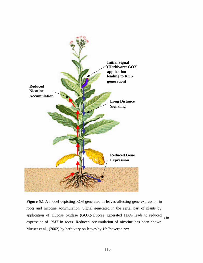
116
References
Baldwin, I.T. (2001). An ecologically motivated analysis of plant-herbivore interactions in
native tobacco. Plant Physiol 127, 1449-1458.
Initial Signal (Herbivory/ GOX application leading to ROS generation)
Reduced Gene Expression
Reduced Nicotine Accumulation
Long Distance Signaling
Figure 5.1 A model depicting ROS generated in leaves affecting gene expression in
roots and nicotine accumulation. Signal generated in the aerial part of plants by
application of glucose oxidase (GOX)-glucose generated H2O2 leads to reduced
expression of PMT in roots. Reduced accumulation of nicotine has been shown
Musser et al., (2002) by herbivory on leaves by Helicoverpa zea.

117
References
Baldwin, I.T., Zhang, Z.-P., Diab, N., Ohnmeiss, T.E., McCloud, E.S., Lynds, G.Y., and
Schmelz, E.A. (1997). Quantification, correlations and manipulations of wound-induced changes
in jasmonic acid and nicotine in Nicotiana sylvestris. Planta 201, 397-404.
Hibi, N., Fujita, T., Hatano, M., Hashimoto, t., and Yamada, Y. (1992). Putrescine N-
methyltransferase in cultured roots of Hyoscyamus albus. Plant Physiol. 100, 826-835.
Holmberg, N., Lilius, G., Bailey, J.E., and Buelow, L. (1997). Transgenic tobacco expressing
Vitreoscilla hemoglobin exhibits enhanced growth and altered metabolite production. Nature
biotechnology 15, 244-247.
Imanishi, S., Hashizume, K., Nakakita, M., Kojima, H., Matsubayashi, Y., Hashimoto, T.,
Sakagami, Y., Yamada, Y., and Nakamura, K. (1998). Differential induction by methyl
jasmonate of genes encoding ornithine decarboxylase and other enzymes involved in nicotine
biosynthesis in tobacco cell cultures. Plant Mol Biol 38, 1101-1111.
Karpinski, S., Escobar, C., Karpinska, B., Creissen, G., and Mullineaux, P. (1997).
Photosynthetic electron transport regulates the expression of cytosolic ascorbate peroxidase
genes in Arabidopsis during excess light stress. Plant Cell 9, 627-640.
Karpinski, S., Reynolds, H., Karpinska, B., Wingsle, G., Creissen, G., and Mullineaux, P.
(1999). Systemic signalling and acclimation in response to excess excitation energy in
Arabidopsis. Science 284, 654-657.
Musser, R.O., Hum-Musser, S.M., Eichenseer, H., Peiffer, M., Ervin, G., Murphy, J.B., and
Felton, G.W. (2002). Herbivory: caterpillar saliva beats plant defences. Nature 416, 599-600.
Salin, M.L. (1987). Toxic oxygen species and protective systems of the chloroplasts. Plant
Physiol 72.

118
References
Baldwin, I.T. (1994). Chemical changes rapidly induced by folivory. In Insect-Plant
Interactions, E.A. Bernays, ed (Boca Raton: CRC press), pp. 1-23.
Baldwin, I.T. (1999). Inducible nicotine production in native Nicotiana as an example of
adaptive phenotypic plasticity. J Chem Ecol 25, 3-30.
Baldwin, I.T. (2001). An ecologically motivated analysis of plant-herbivore interactions in
native tobacco. Plant Physiol 127, 1449-1458.
Baldwin, I.T., and Preston, C.A. (1999). The eco-physiological complexity of plant responses
to insect herbivores. Planta 208, 137-145.
Baldwin, I.T., Zhang, Z.-P., Diab, N., Ohnmeiss, T.E., McCloud, E.S., Lynds, G.Y., and
Schmelz, E.A. (1997). Quantification, correlations and manipulations of wound-induced changes
in jasmonic acid and nicotine in Nicotiana sylvestris. Planta 201, 397-404.
Bang, L.M., Buntting, C., and Molan, P. (2003). The effect of dilution on the rate of hydrogen
peroxide
Beligni, M.V., and Lamattina, L. (2001). Nitric oxide in plants: The history is just beginning.
Plant Cell Environ. 24, 267-278.
Bhattacharyya, S., Dey, N., and Maiti, I.B. (2002). Analysis of cis-sequence transcript (Sgt)
promoter from the figwort mosaic virus (FMV) and comparison of promoter activity with
cauliflower mosaic virus (CaMV) promoters in monocot and dicot cells. Virus Research In
press.
Bolwell, G.P. (1999). Role of active oxygen species and NO in plant defense responses. Curr.
Opin. Plant Biol. 2, 287-294.
Bradford, M.M. (1976). A rapid and sensitive method for quantification of microgram
quantities of protein utilizing the principle of protein dye binding. Anal. Biochem. 72, 248-254.
Bush, L., Hempfling, W.P., and Burton, H. (1999). Biosynthesis of nicotine and related
compounds. In Analytical determination of Nicotine and related compounds and their
metabolites., P. Jacob, ed (Amsterdam: Elsevier), pp. 13-44.

119
Carter, C.J., and Thornburg, R.W. (2004). Tobacco nectarin V is a flavin-contianing
berberinebridge enzyme-like protein with glucose oxidase activity. Plant Physiol 134, 460-469.
Chintapakorn, Y., and Hamill, J.D. (2003). Antisense-mediated down-regulation of putrescine
N-methyltransferase activity in transgenic Nicotiana tabacum L. can lead to elevated levels of
anatabine at the expense of nicotine. Plant Mol Biol 53, 87-105.
Chang, M.-M., Horovitz, D., Culley, D., and Hadwiger, L.A. (1995). Molecular cloning and
characterization of pea chitinase gene expressed in response to wounding, fungal infection and
elicitor chitosan. Plant Mol Biol 28, 105-111.
Clough, S.J., and Bent, A.F. (1998). Floral dip: a simplified method for agrobacterium–
mediated transformation of Arabidopsis thaliana. Plant J 16.
Conrath, U., Chen, Z., Ricigliano, J.R., and Klessing, D. (1995). Two inducers of plant
defense responses, 2, 6-dichloroisonicotinic acid and salicylic acid, inhibit catalase acitivity in
tobacco. Proc Natl Acad Sci U S A 92, 7143-7147.
Costet, L., Dorey, S., Friting, B., and Kauffmann, S. (2002). A pharmacological approach to
test the diffusible signal activity of reactive oxygen intermediates in elicitor-treated tobacoo
leaves. Plant Cell Physiol 43, 91-98.
Creelman, R.A., and Mullet, J.E. (1997). Biosynthesis and action of jasmonates in plants.
Annu. Rev. Plant Physiol. Plant Mol. Biol. 48, 355-381.
Delledonne, M., Xia, Y., Dixon, R.A., and Lamb, C. (1998). Nitric oxide funcitons as a signal
in plant disease resistance. Nature 394, 585-588.
Desikan, R., Clarke, A., Hancock, J., and Neill, S.J. (1999). H2O2 activates a MAP kinase-
like enzyme in Arabidopsis thaliana suspension cultures. J. Expt. Bot. 50, 1863-1866.
D., V.-J.S., Pignocchi, C., Noctor, G., and Foyer, C.H. (2001). Low ascorbic acid in the vtc-1
mutant of Arabidopsis is associated with decreased growth and intracellular redistribution of
antioxidant system 127, 426-435.
Desikan, R., Hancock, J., Coffey, S., and Neill, S.J. (1996). Generation of active oxygen in
elicited cells of Arabidopsis thaliana is mediated by a NADPH oxidase- like enzyme. FEBS
letters 382, 213-217.
Desikan, R., Reynolds, A., Hancock, J.T., and Neill, S.J. (1998). Hairpin and hyrdrogen
peroxide both initiate programmed cell death but have differential effects on gene expression in
Arabidopsis suspension cultures. Biochem. J. 330, 115-120.

120
Durner, J., and Klessing, D. (1996). Salicylic acid is a modulator of tobacco and mammalian
catalases. The Journal of Biological Chemistry 271, 28492-28501.
Durner, J., and Klessing, D. (1999). Nitric oxide as a signal in plants. Curr. Opin. Plant Biol. 2.
Doares, S.H., Syrovets, T., Weiler, E.W., and Ryan, C.A. (1995). Oligogalacturonides and
chitosan activate plant defense gene through the octadecanoid pathway. Proc Natl Acad Sci USA
92, 4095-4098.
Durner, J., and Klessing, D. (1996). Salicylic acid is a modulator of tobacco and mammalian
catalases. The Journal of Biological Chemistry 271, 28492-28501.
Durner, J., and Klessing, D. (1999). Nitric oxide as a signal in plants. Curr. Opin. Plant Biol. 2.
Eichenseer, H., Mathews, M.C., Bi, J.L., Murphy, J.B., and Felton, G.W. (1999). Salivary
glucose oxidase: Multifunctional roles for Helicoverpa zea. Arch. Insect Biochem. Physiol. 42.
Feth, F., Wagner, R., and Wagner, K.G. (1986). Regulation in tobacco callus of enzyme
activites of nicotine pathway. Planta 168, 402-407.
Felton, G.W., and Eichenseer, H. (1999). Induced plant defense against pathogens and
herbivores. In Am. Phytopathol. Soc. (St. Paul, Minnesota).
Finkel, T. (1998). Oxygen radicals and signaling. Curr. Opin. Cell Biol. 10, 248-253.
Friesen, J.B., and leete, E. (1990). Nicotine synthase- An enzyme from Nicotiana species which
catalyses the formation of (S)-nicotine from nicotinic acid and 1- methyl-1-pyrrolinium chloride.
Tetrahedron Lett. 31, 6295-6298.
Grantz, A.A., Brummell, D.A., and Bennett, A.B. (1995). Ascorbate free radical reducatse
mRNA levels are induced by wounding. Plant Physiol 108, 411-418.
Gerstel, D.U. (1960). Segregation in new allopolyploids of Nicotiana I. Comparison of 6X (N.
tabacum X N. tomentosiformis) and 6X (N. tabacum X N. otophora). Genetics 45, 1723-1734.
Gerstel, D.U. (1963). Segregation in new alloployploids of Nicotiana II. Discordant ratios from
individual loci in 6X (N. tabacum X N. sylvestris). Genetics 48, 677-689.
Goldsmith, M.H.M. (1968). The transport of auxin. Annu Rev Plant Physiol 19, 347-369.
Gundlach, H., Muller, M.J., Kutchan, T.M., and Zenk, M.H. (1992). Jasmonic acid is a
signal transducer in elicitor- induced plant cell cultures. Proc Natl Acad Sci USA 89, 2389-2393.
Hara, K., Yagi, M., Koizumi, N., Kusano, T., and Sano, H. (2000). Screening of wound-
responsive genes identifies an immediate-early expressed gene encoding a highly charged
protein mechanically wounded tobacco plants. Plant Cell Physiol 41, 684-691.

121
Hajdukiewicz, P., Svab, Z., and Maliga, P. (1994). The small, versatile pPZP family of
agrobacterium binary vectors for plant transformation. Plant Mol Biol 25, 989-994.
Hashimoto, T., and Yamada, Y. (1995). Regulation of tobacco alkaloid biosynthesis. In
Phytochemicals and health, H.E. Flores, ed (American society of plant physiologists), pp. 130-
144.
Hashimoto, T., and Yamada, Y. (1994). Alkaloid biogenesis: molecular aspects. Annu Rev
Plant Physiol Plant Mol Biol 45, 257-285.
Hashimoto, T., Shoji, T., Mihara, T., Oguri, H., Tamaki, K., Suzuki, K., and Yamada, Y.
(1998). Intraspecific variability of tandem repeats in Nicotiana putrescine N-methyltransferases.
Plant Mol Biol 37, 25-37.
He, Y., and Gan, S. (2001). Identical promoter elements are involved in regulation of OPR1
gene by senescence and jasmonic acid in Arabidopsis. Plant Mol Biol 47, 595-605.
Henzler, T., and Steudle, E. (2000). Transport and metabolic degradation of hydrogen peroxide
in Chara corallina: model calculations and mesurements with the pressure probe suggest
transport of H2O2 across water channel. Journal of Experimental Botany 51.
Hibi, N., Higashiguchi, S., Hashimoto, T., and Yamada, Y. (1994). Gene expression in
tobacco low-nicotine mutants. Plant Cell 6, 723-735.
Hibi, N., Fujita, T., Hatano, M., Hashimoto, T., and Yamada, Y. (1992). Putrescine N-
methyltransferase in cultured roots of Hyoscyamus albus. Plant Physiol. 100, 826-835.
Horsch, R.B., Fry, J.E., Hoffman, N.L., Eicholts, D., Rogers, S.G., and Frayley, R.T. (1985).
A simple and general method for transferring genes into plants. Science 227, 1229-1231.
Holmberg, N., Lilius, G., Bailey, J.E., and Buelow, L. (1997). Transgenic tobacco expressing
Vitreoscilla hemoglobin exhibits enhanced growth and altered metabolite production. Nature
biotechnology 15, 244-247.
Houghtling, R.A., Davila-Garcia, M.I., and Kellar, K.J. (1995). Characterization of (±)-
[3H]epibatidine binding to nicotinic cholinergic
receptors in rat and human brain. Molecular pharmacology 48, 280-287.
Imanishi, S., Hashizume, K., Nakakita, M., Kojima, H., Matsubayashi, Y., Hashimoto, T.,
Sakagami, Y., Yamada, Y., and Nakamura, K. (1998). Differential induction by methyl
jasmonate of genes encoding ornithine decarboxylase and other enzymes involved in nicotine
biosynthesis in tobacco cell cultures. Plant Mol Biol 38, 1101-1111.

122
Jefferson, R.A., Kavanagh, T.A., and Bevan, M. (1987). GUS fusions: β-glucuronidase as a
sensitive and versatile gene fusion marker in higher plants. EMBO 6, 3901-3907.
Jiang, M., and Zhang, J. (2002). Water stress- induced abscisic acid accumulation triggers the
increased generation of reactive oxygen species and up-regulates the activites of antioxidant
enzymes in maize leaves. Journal of Experimental Botany 53, 2401-2410.
Karban, R., and Baldwin, I.T. (1997). Induced reponses to herbivory. (Chicago: Univ.Chicago
press).
Karpinski, S., Reynolds, H., Karpinska, B., Wingsle, G., Creissen, G., and Mullineaux, P.
(1999). Systemic signalling and acclimation in response to excess excitation energy in
Arabidopsis. Science 284, 654-657.
Klessing, D.F., Durner, J., Noad, R., Navare, D.A., Wendehenne, D., Kumar, D., Zhou,
J.M., Shah, J., Zhang, S., Kachroo, P., and etal. (2000). Nitric oxide and salicylic acid
signaling in plant defense. Proc Natl Acad Sci USA 97, 8849-8855.
Koiwa, H., Bessan, r.a., and Hasegawa, P.M. (1997). Regula tion of protease inhibitors and
plant defense. Trends Plant Sci. 2, 379-384.
Kumar, D., and Klessing, D. (2000). Differential induction of tobacco MAP kinase by the
defense signals nitric oxide, salicylic acid, ethylene and jasmonic acid. Mol. Plant-Microbe
Interact. 48, 251-275.
Kutchan, T.M. (1995). Alkaloid biosynthesis- the basis for metabolic engineering of medicinal
plants. Plant Cell 6, 723-735.
Leete, E., and Liu, Y.Y. (1973). Metabolism of [2-3H]- and [6H3]-nicotinic acid in intact
Nicotiana tabacum plants. Phytochemistry 12, 593-596.
Lamb, C., and Dixon, R.A. (1997). The oxidative burst in plant disease resistance. Annu Rev
Plant Physiol Plant Mol Biol 48, 251-275.
Legg, P.D., and Collins, G.B. (1971). Inheritance of total allkaloid in Nicotiana tabacum L. II.
Genetic effect of two loci in Burley 21X LA burley 21 populations. Can. J. Genet. Cytol. 13,
287-291.
Legg, P.D., Chaplin, J.F., and Collins, G.B. (1969). Inheritance of percent total alkaloids in
Nicotiana tabacum L. J. Hered. 60, 213-217.
Laue, G., Preston, C.A., and Baldwin, I.T. (2000). Fast track to the trichome: induction of N-
acyl nornicotines precedes nicotine induction in Nicotiana repanda. Planta 210, 510-514.

123
Levine, A., Tenhaken, R., Dixon, R.A., and Lamb, C.J. (1994). H2O2 from the oxidative burst
orchestrates the plant hypersensitive disease resistance response. Cell 79, 583-593.
Lin, C.C., and Kao, C.H. (2001). Abscisic acid induced changes in cell wall peroxidase activity
and hydrogen peroxide level in roots of rice seedlings. Plant Sci. 160, 323-329.
Mahanandeeshwar, G., Alvin, T.V.J., and Buccafusco, J.J. (1995). A rapid microtechnique
for the estimation of muscarinic and nicotinic receptor binding parameters using 96-well
filtration plates. Journal of Neuroscience 63, 121-125.
Maliga, P., Sz-Breznovits, A., and Marton, L. (1973). Streptomycin-resistant plants from
callus culture of haploid tobacco. Nat New Biol 244, 29-30.Maliga, P., Sz-Breznovits, A., and
Marton, L. (1973). Streptomycin-resistant plants from callus culture of haploid tobacco. Nat
New Biol 244, 29-30.
Mason, H.S., Schindler, U., and Cashmore, A.R. (1995). The G-box; a ubiquitous regulatory
element in plants bound by the GBF family of bZip proteins. Trends Biochem. Sci. 20, 506-510.
Masters, C., and Crane, D. (1995). The Peroxisome: A Vital Organelle. (Cambridge, UK:
Cambridge Univ. Press).
May, M.J., and Leaver, C.J. (1993). Oxidative stimulation of glutathione synthesis in
Arabidopsis thaliana suspension cultures. Plant Physiol 103, 621-627.
McGurl, B., Mukherjee, S., Kahn, M., and Ryan, C.A. (1995). Characterization of two protein
inhibitors (ATI) cDNAs from alfalfa leaves (Medicago sativa var Vernema): The expression of
ATI genes in response to wounding and soil microorganisms. Plant Mol Biol 27, 995-1001.
Mizusaki, S., Tanebe, Y., Noguchi, M., and Tamaki, E. (1973). Changes in activities of
ornithine decarboxylase, putrescine N-methyltransferase and N-methylputrescine oxidase in
tobacco roots in relation to nicotine biosynthesis. Plant Cell Physiol 14, 103-110.
Moyano, E., Fornale, S., Palazon, J., Cusido, R.M., Bagni, N., and Pinol, M.T. (2002).
Alkaloid production in Duboisia hybrid root cultures over expressing the pmt gene.
Phytochemistry 59, 697-702.
Muller, M.J., Brodschelm, W., Spannagl, E., and Zenk, M.H. (1993). Signaling in the
elicitation process is mediated through the octadenoid pathway leading to jasmonic acid. Proc
Natl Acad Sci USA 90, 7490-7494.
Musser, R.O., Hum-Musser, S.M., Eichenseer, H., Peiffer, M., Ervin, G., Murphy, J.B., and
Felton, G.W. (2002). Herbivory: caterpillar saliva beats plant defences. Nature 416, 599-600.

124
Neil, S.J., Desikan, R., Clarke, A., Hurst, R.D., and Hancock, J.T. (2002). Hydrogen
peroxide and nitric oxide as signaling molecules in plants. J Exp. Bot. 53, 1237-1247.
Nishiuchi, T., Hamada, T., Kodama, H., and Iba, K. (1997). Wounding changes the spatial
expression pattern of the Arabidopsis plastid ω-3 fatty acid desaturase gene (FAD7) through
different signal transduction pathways. Plant Cell 9, 1701-1712.
Nugroho, L.H., Anja, M.G., Looman-Peltenburg, Vos, H., Verberne, M.C., and Verpoorte,
R. (2002). Nicotine and related alkaloids accumulation in constitutive salicylic acid producing
tobacco plants. Plant Science 162, 575-581.
Ohta, S., Matsui, O., and Yatazawa, M. (1978). Culture conditions for nicotime production in
tobacco tissue culture. Agric. Biol. Chem. 42, 1245-1251.
Olsen, L., and Harada, J. (1995). Peroxisomes and their assembly in higher plants. Annu Rev
Plant Physiol Plant Mol Biol 46, 123-146.
Orozco-Cardenas, M.L., and Ryan, C.A. (2002). Nitric oxide negatively modulates wound
singaling in tomato plants. Plant Physiol 130, 487-493.
Ottoline, L. (2002). Molecular genetics of auxin signaling. Annu Rev Plant Biol 53, 377-398.
Pena-Cortes, H., Fisahn, J., and Willmitzer, L. (1995). Signals involved in wound- induced
proteinase inhibitor II gene expression in tomato and potato plants. Proc Natl Acad Sci USA 92,
4106-4113.
Phillips, I.D.J. (1975). Apical dominance. Annu Rev Plant Physiol 26, 341-365.
Preston, C.A., Lewandowski, C., Enyendi, A.J., and Baldwin, I.T. (1999). Tobacco mosaic
virus inoculation inhibits wound-induced jasmonate acid-mediated responses within but not
between plants. Planta 209, 87-95.
Riechers, D.E., and Timko, M.P. (1999). Structures and expression of gene family encoding
putrescine N-methyltansferase in Nicotiana tabacum: new clues to the evolutionary origin of
cultivated tobacco. Plant Mol Biol 41, 387-401.
Rouster, J., Leah, R., Mundy, J., and Cameron-Mills, V. (1997). Identification of a methyl
jasmonate-responsive region in the promoter of a lipoxygenase I gene expressed in barley grain.
Plant J. 11, 513-523.
Sachan, N., and Falcone, D.L. (2002). Wound induced gene expression of putrescine N-methyl
transferase in leaves of Nicotiana tabacum. Phytochemistry 61, 797-805.
125
Sato, F., Hashimoto, T., Hachiya, A., Tamura, K., Choi, K., Morishinge, T., Fijimoto, H.,
and Yamada, Y. (2000). Metabolic engineering of plant alkaloid biosynthesis. PNAS 98, 367-
372.
Saunders, J.W., and Bush, L.P. (1979). Nicotine biosynthesis enzyme activites in Nicotiana
tabacum L. genotypes with different alkaloid levels. Plant Physiol 64, 236-240.
Schmidt, H.H.H.W., and Walter, U. (1994). NO at work. Cell 78, 919-925.
Shirasu, K., Nakajima, K., Rajasekhar, V.K., Dixon, R.A., and Lamb, C. (1997). Salicylic
acid potentiates an agonist-dependent gain control that amplifies pathogen signals in the
activation of defense mechanisms. Plant Cell 9, 261-270.
Shoji, T., Yamada, Y., and Hashimoto, T. (2000). Jasmonate induction of Putrescine N-
methyltransferase genes in the root of Nicotiana sylvestris. Plant Cell Physiol 41, 831-839.
Sinclair, S.J., Murphy, K.J., Birch, C.D., and Hamil, J.D. (2000). Molecular characterization
of quinolinate phosphoribosyltransferase (QPRTase) in Nicotiana. Plant Mol Biol 44, 603-617.
Slaymaker, D.H., Navarre, D.A., Clark, D., Pozo, O.D., Martin, G.B., and Klessing, D.
(2002). The tobacco salicylic acid-binding protein 3 (SABP3) is the chloroplast carbonic
anhydrase, which exhibits antioxidant activity and plays a role in the hypersensitive defense
response. PNAS 99, 11640-11645.
Suzuki, K.-I., Yamada, Y., and Hashimoto, T. (1999). Expression of Atropa belladonna
putrescine N-methlytrasferase gene in root pericycle. Plant Physiol. 40, 289-297.
Takahashi, M., and Yamada, Y. (1973). Regulation of nicotine procduction by auxins in
tobacco cultured cells in vitro. Agric. Biol. Chem. 37, 1755-1757.
Tiburcino, A.F., Sawhney-Kaur, R., Ingersoll, R.B., and Galston, A.W. (1985). Correlation
between polyamines and pyrrolidine alkaloids in developing tobacco callus. Plant physiology 78,
323-326.
Usami, S., Banno, H., Ito, Y., Nishihama, R., and Machida, Y. (1995). Cutting activates a 46-
kilodalton protein kinase in plants. Proc Natl Acad Sci USA 92, 8660-8664.
Walling, L.L. (2000). The myriad plant responses to herbivores. Journal of Plant Growth
Regulation 19, 195-216.
Wang, J., Sheehan, M., Brookman, H., and Timko, M.P. (2000). Characterization of cDNA
diffferentially expressed in roots of tobacco (Nicotiana tabacum cv burley 21) during the early
126
stages of alkaloid biosynthesis. plant Science 158, 19-32.Wink, M. (1998). Modes of action of
alkaloids. In Alkaloids, M. Wink, ed (New York: Plenum press), pp. 301-326.
Willenkens, H., Villarroel, R., Montagu, M.V., Inze, D., and Camp, W.V. (1994). Molecular
identification of catalases from Nicotiana plumbaginifolia (L.). FEBS letters 352, 79-83.
Wink, M. (1998). Modes of action of alkaloids. In Alkaloids, M.F. Roberts and M. Wink, eds
(New York: Plenum press), pp. 301-326.
Wu, G., Shortt, B.J., Lawrence, E.B., Leon, J., Fritzsimmons, K.C., Levine, E.B., Raskin, I.,
and Shah, D.M. (1997). Activation of host defense mechanisms by elevated production of H2O2
in transgenic plants. Plant Physiol 115, 427-435.
127
VITA
Nita Sachan was born on 25th June 1973 in India. She completed her Bachelor of Science in
Genetics from Osmania University, Hyderabad, India with first class distinction in 1994. Later
she joined one of the most prestigious institutes in India, Ind ian Institute of Technology,
Roorkee, (IITR) and completed her Master of Science in Bioscience with first class distinction,
in 1996. At IITR she was ranked 2nd in the class and was awarded a scholarship for academic
excellence. To pursue her PhD, she joined the Plant Physiology/Molecular Biology/
Biochemistry Program in the Department of Agronomy, University of Kentucky. She worked as
a graduate research assistant at the Kentucky Tobacco Research and Development Centre
(KTRDC). She worked under the guidance of Dr. Deane Falcone from 1997 to 2004 at
University of Kentucky.