IDENTIFICATION AND COMPARATIVE GENOMIC ANALYSIS OF SIGNALING AND REGULATORY COMPONENTS IN THE DIATOM...
20
IDENTIFICATION AND COMPARATIVE GENOMIC ANALYSIS OF SIGNALING AND REGULATORY COMPONENTS IN THE DIATOM THALASSIOSIRA PSEUDONANA 1 Anton Montsant Laboratory of Molecular Plant Biology, CNRS UMR 8186, Department of Biology, Ecole Normale Supe ´rieure, 75230 Paris, France Laboratory of Cell Signalling, Stazione Zoologica, Villa Comunale, I 80121 Naples, Italy Andrew E. Allen Laboratory of Molecular Plant Biology, CNRS UMR 8186, Department of Biology, Ecole Normale Supe ´rieure, 75230 Paris, France The Institute for Genomic Research, 9712 Medical Center Drive, Rockville, MD 20850, USA Sacha Coesel Laboratory of Cell Signalling, Stazione Zoologica, Villa Comunale, I 80121 Naples, Italy Centre of Marine Sciences, University of Algarve, Campus de Gambelas, 8005-139 Faro, Portugal Alessandra De Martino Laboratory of Molecular Plant Biology, CNRS UMR 8186, Department of Biology, Ecole Normale Supe ´rieure, 75230 Paris, France Laboratory of Cell Signalling, Stazione Zoologica, Villa Comunale, I 80121 Naples, Italy Angela Falciatore, Manuela Mangogna, Magali Siaut Laboratory of Cell Signalling, Stazione Zoologica, Villa Comunale, I 80121 Naples, Italy Marc Heijde, Kamel Jabbari, Uma Maheswari, Edda Rayko, Assaf Vardi Laboratory of Molecular Plant Biology, CNRS UMR 8186, Department of Biology, Ecole Normale Supe ´rieure, 75230 Paris, France Kirk E. Apt Martek Biosciences Corp., 6480 Dobbin Road, MD 21045, USA John A. Berges Department of Biological Sciences, University of Wisconsin-Milwaukee, Milwaukee, WI 53201, USA Anthony Chiovitti School of Botany, University of Melbourne, Victoria 3010, Australia Aubrey K. Davis, Kimberlee Thamatrakoln Scripps Institution of Oceanography, University of California San Diego, La Jolla, CA 92093, USA Masood Z. Hadi Lockheed Martin Corporation, Sandia National Laboratory, PO Box 969, MS-9951, Livermore, CA 94551, USA Todd W. Lane Biosystems Research Department, Sandia National Labs, Livermore, CA 94551-0969, USA J. Casey Lippmeier Martek Biosciences Corp., 6480 Dobbin Road, MD 21045, USA Department of Biological Sciences, University of Hull, Hull HU6 7RX, UK Diego Martinez DOE Joint Genome Institute, Los Alamos National Laboratory, Los Alamos, NM 87545, USA Micaela S. Parker School of Oceanography, University of Washington, Seattle, WA 98195, USA J. Phycol. 43, 585–604 (2007) ȑ 2007 Phycological Society of America DOI: 10.1111/j.1529-8817.2007.00342.x 585
-
Upload
independent -
Category
Documents
-
view
3 -
download
0
Transcript of IDENTIFICATION AND COMPARATIVE GENOMIC ANALYSIS OF SIGNALING AND REGULATORY COMPONENTS IN THE DIATOM...
IDENTIFICATION AND COMPARATIVE GENOMIC ANALYSIS OF SIGNALING ANDREGULATORY COMPONENTS IN THE DIATOM THALASSIOSIRA PSEUDONANA1
Anton Montsant
Laboratory of Molecular Plant Biology, CNRS UMR 8186, Department of Biology, Ecole Normale Superieure, 75230 Paris, France
Laboratory of Cell Signalling, Stazione Zoologica, Villa Comunale, I 80121 Naples, Italy
Andrew E. Allen
Laboratory of Molecular Plant Biology, CNRS UMR 8186, Department of Biology, Ecole Normale Superieure, 75230 Paris, France
The Institute for Genomic Research, 9712 Medical Center Drive, Rockville, MD 20850, USA
Sacha Coesel
Laboratory of Cell Signalling, Stazione Zoologica, Villa Comunale, I 80121 Naples, Italy
Centre of Marine Sciences, University of Algarve, Campus de Gambelas, 8005-139 Faro, Portugal
Alessandra De Martino
Laboratory of Molecular Plant Biology, CNRS UMR 8186, Department of Biology, Ecole Normale Superieure, 75230 Paris, France
Laboratory of Cell Signalling, Stazione Zoologica, Villa Comunale, I 80121 Naples, Italy
Angela Falciatore, Manuela Mangogna, Magali Siaut
Laboratory of Cell Signalling, Stazione Zoologica, Villa Comunale, I 80121 Naples, Italy
Marc Heijde, Kamel Jabbari, Uma Maheswari, Edda Rayko, Assaf Vardi
Laboratory of Molecular Plant Biology, CNRS UMR 8186, Department of Biology, Ecole Normale Superieure, 75230 Paris,
France
Kirk E. Apt
Martek Biosciences Corp., 6480 Dobbin Road, MD 21045, USA
John A. Berges
Department of Biological Sciences, University of Wisconsin-Milwaukee, Milwaukee, WI 53201, USA
Anthony Chiovitti
School of Botany, University of Melbourne, Victoria 3010, Australia
Aubrey K. Davis, Kimberlee Thamatrakoln
Scripps Institution of Oceanography, University of California San Diego, La Jolla, CA 92093, USA
Masood Z. Hadi
Lockheed Martin Corporation, Sandia National Laboratory, PO Box 969, MS-9951, Livermore, CA 94551, USA
Todd W. Lane
Biosystems Research Department, Sandia National Labs, Livermore, CA 94551-0969, USA
J. Casey Lippmeier
Martek Biosciences Corp., 6480 Dobbin Road, MD 21045, USA
Department of Biological Sciences, University of Hull, Hull HU6 7RX, UK
Diego Martinez
DOE Joint Genome Institute, Los Alamos National Laboratory, Los Alamos, NM 87545, USA
Micaela S. Parker
School of Oceanography, University of Washington, Seattle, WA 98195, USA
J. Phycol. 43, 585–604 (2007)� 2007 Phycological Society of AmericaDOI: 10.1111/j.1529-8817.2007.00342.x
585
Gregory J. Pazour
Program in Molecular Medicine, University of Massachusetts Medical School, Worcester, MA 01605, USA
Mak A. Saito
Department of Marine Chemistry and Geochemistry, Woods Hole Oceanographic Institution, Woods Hole, MA 02543, USA
Dan S. Rokhsar
Center for Integrative Genomics, University of California Berkeley, Berkeley, CA, USA
DOE Joint Genome Institute, Walnut Creek, CA 94598, USA
E. Virginia Armbrust2
School of Oceanography, University of Washington, Seattle, WA 98195, USA
and
Chris Bowler3
Laboratory of Molecular Plant Biology, CNRS UMR 8186, Department of Biology, Ecole Normale Superieure, 75230 Paris, France
Laboratory of Cell Signalling, Stazione Zoologica, Villa Comunale, I 80121 Naples, Italy
Diatoms are unicellular brown algae that likelyarose from the endocytobiosis of a red alga into asingle-celled heterotroph and that constitute an algalclass of major importance in phytoplankton commu-nities around the globe. The first whole-genomesequence from a diatom species, Thalassiosira pseu-donana Hasle et Heimdal, was recently reported,and features that are central to diatom physiologyand ecology, such as silicon and nitrogen metabo-lism, iron uptake, and carbon concentration mecha-nisms, were described. Following this initial study,the basic cellular systems controlling cell signaling,gene expression, cytoskeletal structures, andresponse to stress have been cataloged in an attemptto obtain a global view of the molecular foundationsthat sustain such an ecologically successful group oforganisms. Comparative analysis with several micro-bial, plant, and metazoan complete genomesequences allowed the identification of putativemembrane receptors, signaling proteins, and othercomponents of central interest to diatom ecophysiol-ogy and evolution. Thalassiosira pseudonana likelyperceives light through a novel phytochrome andseveral cryptochrome photoreceptors; it may lackthe conserved RHO small-GTPase subfamily of cell-polarity regulators, despite undergoing polarizedcell-wall synthesis; and it possesses an unusuallylarge number of heat-shock transcription factors,which may indicate the central importance of tran-scriptional responses to environmental stress. Theavailability of the complete gene repertoire will per-mit a detailed biochemical and genetic analysis ofhow diatoms prosper in aquatic environments and
will contribute to the understanding of eukaryoticevolution.
Key index words: apoptosis; cell cycle; compara-tive genomics; cryptochrome; cytoskeleton; dia-tom; flagella; genome; GTP-ase; lateral genetransfer; myosin; oxidative stress; Phaeodactylumtricornutum; phytochrome; secondary endosym-biosis; Thalassiosira pseudonana; whole-genomeanalysis; xanthophyll cycle
Abbreviations: AP, apurinic ⁄ apyrimidinic; BV, bili-verdin; CDKs, cyclin-dependent kinases; CSDs,cold-shock domains; ESTs, expressed sequencetags; GAPs, GTPase-activating proteins; GEFs,guanyl-nucleotide exchange factors; GPCR,G-protein-coupled receptors; HATs, histone acetyltransferases; HDACs, histone deacetylases; HK,histidine kinase; Hpt, histidine phosphotransfer;HSFs, heat-shock factors; IFT, intraflagellar trans-port; LRR, leucine-rich repeats; PCB, phycocy-anobilin; P/B, phytochromobilin; RCC, regulatorof chromsome condensation; RIP, repeat inducedpoint; RR, response regulator; SDV, silica depos-ition vesicle; SODs, superoxide dismutases; TF,transcription factor; VDE, violaxanthin de-epoxidase;ZEP, zeaxanthin epoxidase
The contemporary oceans cover 70% of the sur-face of our planet, and even though the photosyn-thetic organisms living within them constitute only1% of the earth’s photosynthetic biomass, marineecosystems are responsible for about one-half of glo-bal primary productivity (Field et al. 1998, Irigoienet al. 2002). Besides photosynthetic prokaryotes, themost successful organisms are thought to be a classof eukaryotic unicellular algae, known as diatoms.
1Received 10 October 2006. Accepted 19 January 2007.2Author for correspondence: e-mail [email protected].
edu.3Author for correspondence: e-mail [email protected].
586 ANTON MONTSANT ET AL.
On a global scale, diatoms are thought to contributeat least 20% of the annual primary productivity andto play major roles in key biogeochemical cycles(Smetacek 1999).
Two major groups of diatoms can be distin-guished according to cell shape. Centric diatomsare radially symmetrical and are thought to haveappeared �200 mya, whereas the bilaterally symmet-rical pennate diatoms evolved from the centric dia-toms �70 mya (Kooistra et al. 2003). A hallmark ofdiatoms is their beautifully ornamented silicified cellwall, known as the frustule. The precision of thenanoscale pattern and architecture of the frustulesuggests that understanding diatom cell-wallbiosynthesis eventually may be exploitable in nano-technological applications (Parkinson and Gordon1999, Lopez et al. 2005). Although in recent yearssome key components involved in silicon transportand precipitation have been described (Hildebrandet al. 1998, Kroger et al. 1999), and also identifiedin the T. pseudonana genome (Poulsen and Kroger2004), the mechanisms by which the siliceous mater-ial of the frustule is laid down remain largelyunknown.
The algal groups of the Heterokontophyta(e.g., diatoms and brown seaweeds), the Haptophyta(e.g., coccolithophorids), and the Cryptophyta (e.g.,Guillardia theta D. R. A. Hill et Wetherbee) are col-lectively known as the chromist algae because of thecolors conferred by their distinct pigment composi-tion. Together with the Alveolates (dinoflagellatealgae and apicomplexan parasites), these diverseeukaryotic groups likely arose from a commonsecondary endosymbiotic event in which a hetero-trophic eukaryote engulfed (or was invaded by) ared eukaryotic alga more than one billion years ago(Patron et al. 2004, Yoon et al. 2004, Li et al. 2006).At the initial stage of this endosymbiosis, thecomplex cell likely contained at least five differentgenomes: the nuclear and mitochondrial genomesof the host cell and the nuclear, mitochondrial, andplastid genomes of the red alga. Over evolutionarytime most of the cellular structures of the endo-symbiont have been lost, and extant diatoms nowcontain plastids surrounded by four rather than twomembranes and a nucleus and mitochondria thatare derived from the original heterotrophic host.Diatom cells, therefore, have a number of featuresthat make them highly divergent from the classicalcellular structures of higher plants (e.g., a signalpeptide-mediated import of proteins into plastids[Apt et al. 2002] or a hypothesized single-cell C4carbon concentration mechanism [Reinfelder et al.2000]).
The nuclear, plastid, and mitochondrial genomesequences of the centric diatom Thalassiosira pseudo-nana were recently reported (Armbrust et al. 2004).The nuclear genome is 34 Mb and consists of 24chromosomes, and the plastid and mitochondrialgenomes are 129 kb and 44 kb, respectively. A range
of novel metabolic features have been described,such as genes encoding components for formationof silica-based cell walls, biosynthetic enzymes forseveral types of polyunsaturated fatty acids, andenzymes for utilization of various nitrogenous com-pounds, including a complete urea cycle (Allenet al. 2006). The T. pseudonana genome thereforeencodes a unique combination of factors previouslythought to be restricted either to photosynthetic orheterotrophic eukaryotes. In this study, explorationof the T. pseudonana genome has continued to pro-vide insights into the mechanisms used by diatomsto regulate their cellular homeostasis. Specifically,we have investigated the recognizable gene familiescontrolling cell signaling, transcription, cytoskeletalstructures, cell cycle, and stress responses.
MATERIALS AND METHODS
Source of sequence data sets. The complete predicted pro-teome of T. pseudonana, strain CCMP 1335, is available fromthe Joint Genome Institute (JGI; http://genome.jgi-psf.org/thaps3/thaps3.download.html). In Figure 1 and Tables 1, 3,and S1 (in the supplementary material), the predicted proteo-mes of several organisms were analyzed along with the diatompredicted proteome for comparison. Their sources werehttp://merolae.biol.s.u-tokyo.ac.jp/download ⁄ for the unicel-lular red alga Cyanidioschyzon merolae P. DeLuca, R. Taddei etL. Varno (Matsuzaki et al., 2004); http://www.broad.mit.edu/cgibin/annotation/neurospora/downloadlicense.cgi for thefungus Neurospora crassa (Galagan et al. 2003); http://genome.jgi-psf.org/ for the parasitic Stramenopiles Phytophthora ramo-rum and Phytophthora sojae (Tyler et al. 2006), the green algaChlamydomonas reinhardtii P. A. Dang, the fungus Phanerochaetechrysosporium, and the animals Ciona intestinalis and Xenopuslaevis; and http://www.ebi.ac.uk/proteome/index.html forthe yeast Saccharomyces cerevisiae, the plant Arabidopsis thaliana,and Caenorhabditis elegans, Drosophila melanogaster, and Musmusculus.
Protein domain counts. The T. pseudonana predicted prote-ome and the proteome of six reference organisms werecompared with the Conserved Domain Database (http://www.ncbi.nlm.nih.gov/entrez/query.fcgi?db = cdd) to identifyInterPro domains by means of the RPS-Blast algorithm asdescribed previously (Marchler-Bauer et al. 2003).
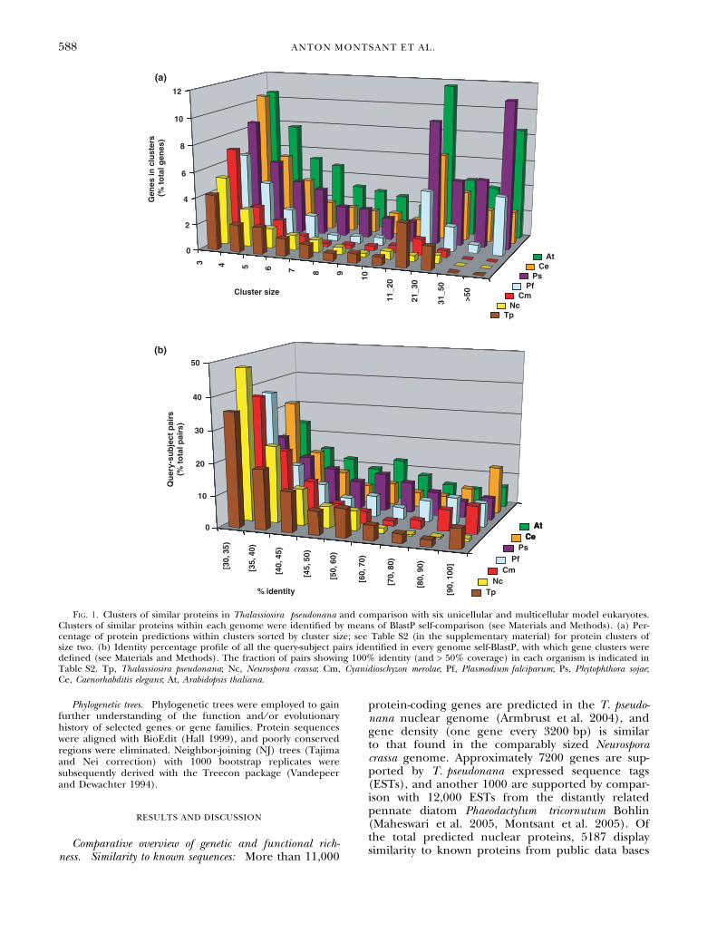
Analysis of intragenomic coding sequence similarity. The pro-teomes of the diatom and six other sequenced eukaryotes, forreference, were compared with themselves using BlastP with aBLOSUM62 matrix (Altschul et al. 1990) and an E-valuethreshold of E-20. Clusters of similar genes, loosely represent-ing ‘‘gene families,’’ were defined as those groups of sequencesthat presented > 30% identity for at least 50% of their lengthto a common query. The number of genes within clusters ofsimilar genes was plotted as a function of gene cluster size(Fig. 1a). The query–match pairs employed to build the geneclusters were classified by identity percentage intervals tovisualize the level of similarity shared by the components of thegene clusters within each genome (Fig. 1b).
Counts of putative transmembrane kinases and leucine-rich-repeatproteins. Protein predictions that contained protein kinase(IPR000719) or leucine-rich repeat (LRR; IPR001611)domains were identified by means of the InterProScan outputkeyword search tool (http://www.ebi.ac.uk/InterProScan)implemented in the genome browsers of the organismsin study, and subsequently submitted to the TMHMM algo-rithm (Krogh et al. 2001) to detect putative transmembranesegments.
SIGNALING AND REGULATORY COMPONENTS IN T. PSEUDONANA 587
Phylogenetic trees. Phylogenetic trees were employed to gainfurther understanding of the function and ⁄ or evolutionaryhistory of selected genes or gene families. Protein sequenceswere aligned with BioEdit (Hall 1999), and poorly conservedregions were eliminated. Neighbor-joining (NJ) trees (Tajimaand Nei correction) with 1000 bootstrap replicates weresubsequently derived with the Treecon package (Vandepeerand Dewachter 1994).
RESULTS AND DISCUSSION
Comparative overview of genetic and functional rich-ness. Similarity to known sequences: More than 11,000
protein-coding genes are predicted in the T. pseudo-nana nuclear genome (Armbrust et al. 2004), andgene density (one gene every 3200 bp) is similarto that found in the comparably sized Neurosporacrassa genome. Approximately 7200 genes are sup-ported by T. pseudonana expressed sequence tags(ESTs), and another 1000 are supported by compar-ison with 12,000 ESTs from the distantly relatedpennate diatom Phaeodactylum tricornutum Bohlin(Maheswari et al. 2005, Montsant et al. 2005). Ofthe total predicted nuclear proteins, 5187 displaysimilarity to known proteins from public data bases
AtCe
TpNc
Cm
Ps
AtCe
Pf
Gen
es in
clu
ster
s(%
to
tal g
enes
)
Cluster size
3 4 5 6 7 8 9
10
11_2
0
21_3
0
31_5
0
>50
0
2
4
6
8
10
12
Qu
ery-
sub
ject
pai
rs(%
to
tal p
airs
)
% identity
[30,
35)
[35,
40)
[40,
45)
[45,
50)
[50,
60)
[60,
70)
[70,
80)
[80,
90)
[90,
100
]
0
10
20
30
40
50
AtCe
TpNc
Cm
Ps
AtAtCeCe
Pf
(a)
(b)
Fig. 1. Clusters of similar proteins in Thalassiosira pseudonana and comparison with six unicellular and multicellular model eukaryotes.Clusters of similar proteins within each genome were identified by means of BlastP self-comparison (see Materials and Methods). (a) Per-centage of protein predictions within clusters sorted by cluster size; see Table S2 (in the supplementary material) for protein clusters ofsize two. (b) Identity percentage profile of all the query-subject pairs identified in every genome self-BlastP, with which gene clusters weredefined (see Materials and Methods). The fraction of pairs showing 100% identity (and > 50% coverage) in each organism is indicated inTable S2. Tp, Thalassiosira pseudonana; Nc, Neurospora crassa; Cm, Cyanidioschyzon merolae; Pf, Plasmodium falciparum; Ps, Phytophthora sojae;Ce, Caenorhabditis elegans; At, Arabidopsis thaliana.
588 ANTON MONTSANT ET AL.
(score > 200, E-value < E-20) and 6661 have recog-nizable domains in the InterPro data base (Armbrustet al. 2004, and data not shown). To obtain aview of functional domain diversity within theT. pseudonana predicted proteome relative to otherorganisms, we divided these domains into five majorcategories according to whether they are equallyabundant in other eukaryotes or whether they areover- or underrepresented in the diatom (Table S1in the supplementary material). Such a comparisonrevealed a number of characteristics relevant tobiological processes involving signaling, cell cycle,cytoskeleton, or transcription, all of which werestudied in detail by in silico analysis and manualcuration (see the following sections).
Within-genome coding sequence diversity: In studying‘‘functional richness,’’ we further wondered howmany different sequences existed within the totalpool of predicted genes. The sequence diversitywithin the T. pseudonana predicted proteome wascharacterized using approaches similar to those pre-viously employed for N. crassa (Galagan et al. 2003).The complete predicted proteome of the diatomwas compared with itself by means of BlastP, andprotein predictions sharing at least 30% amino acididentity over 50% of their lengths were groupedwith each other. In doing so, a ‘‘nonredundant’’ setof genes was obtained in which most genes constitu-ting a family or derived from recent duplicationswere grouped together. Clusters of similar proteinswere defined in the same fashion within the predic-ted proteomes of another six unicellular and multi-cellular eukaryotes (Table S2 in the supplementarymaterial). When gene number was plotted as a func-tion of gene-cluster size, a similar profile wasobtained for all the referenced microbes, with multi-cellular organisms showing a higher proportion ofgenes in clusters of large size (i.e., multicellularorganisms have more multigene families, and these
are composed of more members; Fig. 1a). The clo-sest relative of the diatoms among the referenceorganisms, the oomycete Phytophthora sojae, showed aprotein family-size profile similar to that of the mul-ticellular organisms (Fig. 1a). However, the diatomand the oomycete have similar numbers of singletongenes (i.e., genes that do not belong to a gene fam-ily) and gene clusters (Table S2 in the supplement-ary material), indicating that the higher genenumber of P. sojae does not translate into higherwithin-genome sequence diversity, possibly becausegene number increased in the oomycete throughmultiplication of previously existing families.
In order to estimate the degree of similarityamong genes within gene clusters for each of thegenomes, the query–subject pairs with which clusterswere defined were sorted by % identity, and the cor-responding histogram was plotted. As shown previ-ously (Galagan et al. 2003), a repeat-induced-point(RIP) mutation mechanism causes N. crassa to havea negligible amount of query–match pairs show-ing > 70% identity, a feature that was not detectedin the diatom or in any other reference organism(Fig. 1b). Besides Neurospora, all eukaryotes dis-played similar profiles, with C. elegans containing alarger fraction of very highly conserved pairs(> 90% identity). Up to 3% of the diatom query–match pairs showed 100% identity (and > 50% cov-erage), a fraction comparable to that of the flat-worm (3.61%) and higher than in the remainingorganisms (Table S2 in the supplementary mater-ial), some of which may correspond to very recentgene duplication events.
Signal perception. Membrane-bound receptors: Varioustypes of sensing mechanisms have been discoveredand extensively studied in plants and animals in thepast decades, and a number of transmembranereceptor families can be identified by similaritysearches. G-protein-coupled receptors (GPCR) are anextremely important class of receptor proteins in ani-mal cells (FANTOM Consortium & RIKEN group2002), but Arabidopsis is known to contain only one(Arabidopsis Genome Initiative 2000, Colucci et al.2002). The T. pseudonana genome was found toencode two putative GPCRs, and consistently twoputative heterotrimeric G proteins, through whichGPCRs connect with downstream signal transduc-tion pathways, were also identified (two genes enco-ding the a and b subunits, none encoding the csubunit; Table S3, annotations 1–6, in the supple-mentary material).
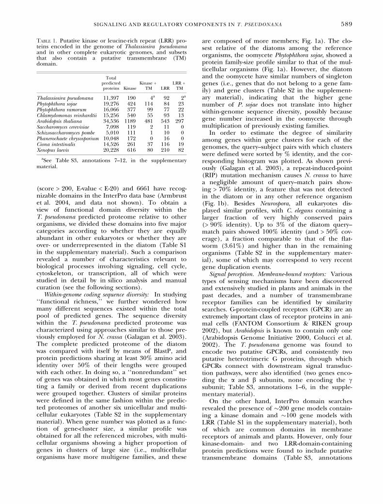
On the other hand, InterPro domain searchesrevealed the presence of �200 gene models contain-ing a kinase domain and �100 gene models withLRR (Table S1 in the supplementary material), bothof which are common domains in membranereceptors of animals and plants. However, only fourkinase-domain- and two LRR-domain-containingprotein predictions were found to include putativetransmembrane domains (Table S3, annotations
Table 1. Putative kinase or leucine-rich repeat (LRR) pro-teins encoded in the genome of Thalassiosira pseudonanaand in other complete eukaryotic genomes, and subsetsthat also contain a putative transmembrane (TM)domain.
Totalpredictedproteins Kinase
Kinase +TM LRR
LRR +TM
Thalassiosira pseudonana 11,397 190 4a 92 2a
Phytophthora sojae 19,276 424 114 84 23Phytophthora ramorum 16,066 377 99 77 22Chlamydomonas reinhardtii 15,256 540 55 93 13Arabidopsis thaliana 34,536 1189 481 543 297Saccharomyces cerevisiae 7,098 119 2 11 0Schizosaccharomyces pombe 5,010 111 1 10 0Phanerochaete chrysosporium 10,048 172 0 16 0Ciona intestinalis 14,526 261 37 116 19Xenopus laevis 20,228 616 80 210 82
aSee Table S3, annotations 7–12, in the supplementarymaterial.
SIGNALING AND REGULATORY COMPONENTS IN T. PSEUDONANA 589
7–12, in the supplementary material). When thesecounts were obtained for a variety of other genomesfor comparison, the oomycetes P. ramorum and P.sojae (close relatives of the diatoms) showed a sim-ilar total number of kinase- or LRR-domain-contain-ing proteins, but the subset of them with atransmembrane region was found to be one to twoorders of magnitude higher (Table 1). Completegenome sequences representative of other majorphytoplankton taxa, such as coccolithophores, dino-flagellates, and pennate diatoms, will be necessaryto conclude whether the utilization of transmem-brane kinases and LRR receptors in chromalveolatesis, as the parasitic oomycetes would suggest, compar-able to that in animals, or whether it is sparse likein the T. pseudonana predicted proteome. On theother hand, these types of transmembrane receptorsappear to be conspicuously rare in fungi, given thatonly three putative transmembrane kinases weredetected in the three fungal species examined(Table 1).
Understanding the roles of the T. pseudonanaputative GPCRs and membrane-bound kinases andLRR proteins, and whether these constitute the coreof diatom membrane-bound receptors, will requirefurther examination, but it seems likely that otherreceptor classes await identification. Predicted pro-teins that may have receptor functions are those ofunknown function containing membrane-localizingdomains (e.g., domains that define the prenylationamino acid motif CAAX are abundant in theT. pseudonana genome).
Photoperception: In addition to being a source ofenergy, light constitutes an important sensory stimu-lus. Terrestrial plants utilize three major classesof photoreceptors to sense ambient light in theirenvironment: the red- or far-red-light-absorbingphytochromes and the blue-light-absorbing crypto-chromes and phototropins (Falciatore and Bowler2005). Furthermore, some green algae containgreen-light-absorbing rhodopsins (Nagel et al. 2002,Sineshchekov et al. 2002). Green light persists tothe greatest depths in coastal waters, but no rhodop-sins were identified in the diatom genome. How-ever, the diatom appears likely to use cryptochromephotoreceptors for perception of blue light, and aputative phytochrome was also identified (seebelow).
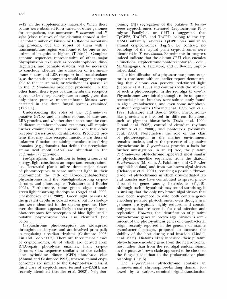
Cryptochrome photoreceptors are widespreadthroughout eukaryotes and are involved principallyin regulating circadian rhythms (Cashmore 2003,Lin and Todo 2005). There are three major classesof cryptochromes, all of which are derived fromDNA-repair photolyase enzymes. Plant crypto-chromes show sequence similarity to the cyclobu-tane pyrimidine dimer (CPD)–photolyase class(Ahmad and Cashmore 1993), whereas animal crypt-ochromes are similar to the 6,4-photolyase class. Athird class of cryptochrome, termed cry-DASH, wasrecently identified (Brudler et al. 2003). Neighbor-
joining (NJ) segregation of the putative T. pseudo-nana cryptochromes (denoted Cryptochrome Pho-tolyase Family1-4, or CPF1-4) suggested thatTpCPF2, TpCPF3, and TpCPF4 belong to the cry-DASH subfamily, whereas TpCPF1 was similar toanimal cryptochromes (Fig. 2). By contrast, noorthologs of the typical plant cryptochromes wereidentified in T. pseudonana. Experiments in progressindeed indicate that the diatom CPF1 class encodesa functional cryptochrome photoreceptor (S. Coesel,M. Mangogna, A. Falciatore, and C. Bowler, unpub-lished data).
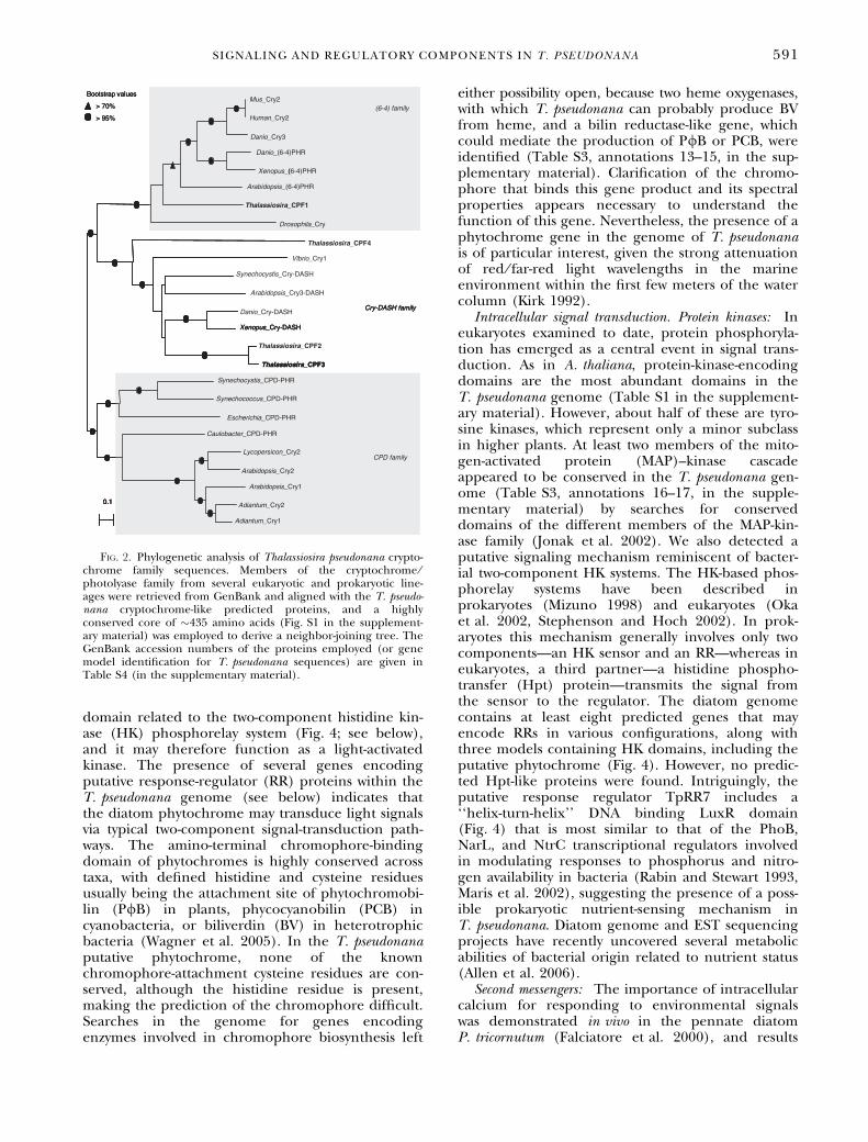
The identification of a phytochrome photorecep-tor is consistent with an earlier report demonstra-ting that diatoms can perceive red ⁄ far-red light(Leblanc et al. 1999) and contrasts with the absenceof such a photoreceptor in the red alga C. merolae.Phytochromes were initially thought to be unique toterrestrial plants, but they were subsequently foundin algae, cyanobacteria, and even some nonphoto-synthetic organisms (Morand et al. 1993, Yeh et al.1997, Falciatore and Bowler 2005). Phytochrome-like proteins are involved in different functions,such as pigment biosynthesis (Davis et al. 1999,Giraud et al. 2002), control of circadian rhythms(Schmitz et al. 2000), and phototaxis (Yoshiharaet al. 2000). Nonetheless, the role of this classof photoreceptor in the marine environmentremains unclear, and so the presence of a putativephytochrome in T. pseudonana provides a basis forfurther investigation. In an NJ tree, the putativeT. pseudonana phytochrome appeared most similarto phytochrome-like sequences from the diatomP. tricornutum (M. Siaut, A. Falciatore, and C. Bowlerunpublished data) and from two brown algal viruses(Delaroque et al. 2001), revealing a possible ‘‘brownclade’’ of phytochromes in which virus-mediated lat-eral transfer may have contributed to spread phyto-chrome-like genes among heterokonts (Fig. 3).Although such a hypothesis may sound surprising, itis striking that the only two brown algal viruses thathave been sequenced to date both contain genesencoding putative phytochromes, even though viralgenomes are typically highly reduced and containonly genes that are essential for viral infection andreplication. However, the identification of putativephytochrome genes in brown algal viruses is remi-niscent of the photosynthesis genes of cyanobacterialorigin recently reported in the genome of marinecyanobacterial phages, proposed to increase theviability of the host during viral invasion (Lindellet al. 2005). Diatoms likely inherited their putativephytochrome-encoding gene from the heterotrophichost rather than from the red algal endosymbiont,as the putative brown clade appeared to be closer tothe fungal clade than to the prokaryotic or plantorthologs (Fig. 3).
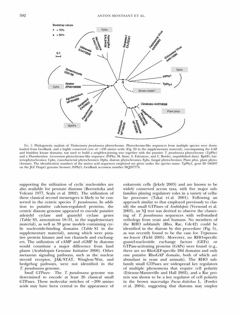
The T. pseudonana phytochrome contains anamino-terminal chromophore-binding domain fol-lowed by a carboxy-terminal signal-transduction
590 ANTON MONTSANT ET AL.
domain related to the two-component histidine kin-ase (HK) phosphorelay system (Fig. 4; see below),and it may therefore function as a light-activatedkinase. The presence of several genes encodingputative response-regulator (RR) proteins within theT. pseudonana genome (see below) indicates thatthe diatom phytochrome may transduce light signalsvia typical two-component signal-transduction path-ways. The amino-terminal chromophore-bindingdomain of phytochromes is highly conserved acrosstaxa, with defined histidine and cysteine residuesusually being the attachment site of phytochromobi-lin (P/B) in plants, phycocyanobilin (PCB) incyanobacteria, or biliverdin (BV) in heterotrophicbacteria (Wagner et al. 2005). In the T. pseudonanaputative phytochrome, none of the knownchromophore-attachment cysteine residues are con-served, although the histidine residue is present,making the prediction of the chromophore difficult.Searches in the genome for genes encodingenzymes involved in chromophore biosynthesis left
either possibility open, because two heme oxygenases,with which T. pseudonana can probably produce BVfrom heme, and a bilin reductase-like gene, whichcould mediate the production of P/B or PCB, wereidentified (Table S3, annotations 13–15, in the sup-plementary material). Clarification of the chromo-phore that binds this gene product and its spectralproperties appears necessary to understand thefunction of this gene. Nevertheless, the presence of aphytochrome gene in the genome of T. pseudonanais of particular interest, given the strong attenuationof red ⁄ far-red light wavelengths in the marineenvironment within the first few meters of the watercolumn (Kirk 1992).
Intracellular signal transduction. Protein kinases: Ineukaryotes examined to date, protein phosphoryla-tion has emerged as a central event in signal trans-duction. As in A. thaliana, protein-kinase-encodingdomains are the most abundant domains in theT. pseudonana genome (Table S1 in the supplement-ary material). However, about half of these are tyro-sine kinases, which represent only a minor subclassin higher plants. At least two members of the mito-gen-activated protein (MAP)–kinase cascadeappeared to be conserved in the T. pseudonana gen-ome (Table S3, annotations 16–17, in the supple-mentary material) by searches for conserveddomains of the different members of the MAP-kin-ase family (Jonak et al. 2002). We also detected aputative signaling mechanism reminiscent of bacter-ial two-component HK systems. The HK-based phos-phorelay systems have been described inprokaryotes (Mizuno 1998) and eukaryotes (Okaet al. 2002, Stephenson and Hoch 2002). In prok-aryotes this mechanism generally involves only twocomponents—an HK sensor and an RR—whereas ineukaryotes, a third partner—a histidine phospho-transfer (Hpt) protein—transmits the signal fromthe sensor to the regulator. The diatom genomecontains at least eight predicted genes that mayencode RRs in various configurations, along withthree models containing HK domains, including theputative phytochrome (Fig. 4). However, no predic-ted Hpt-like proteins were found. Intriguingly, theputative response regulator TpRR7 includes a‘‘helix-turn-helix’’ DNA binding LuxR domain(Fig. 4) that is most similar to that of the PhoB,NarL, and NtrC transcriptional regulators involvedin modulating responses to phosphorus and nitro-gen availability in bacteria (Rabin and Stewart 1993,Maris et al. 2002), suggesting the presence of a poss-ible prokaryotic nutrient-sensing mechanism inT. pseudonana. Diatom genome and EST sequencingprojects have recently uncovered several metabolicabilities of bacterial origin related to nutrient status(Allen et al. 2006).
Second messengers: The importance of intracellularcalcium for responding to environmental signalswas demonstrated in vivo in the pennate diatomP. tricornutum (Falciatore et al. 2000), and results
Fig. 2. Phylogenetic analysis of Thalassiosira pseudonana crypto-chrome family sequences. Members of the cryptochrome ⁄photolyase family from several eukaryotic and prokaryotic line-ages were retrieved from GenBank and aligned with the T. pseudo-nana cryptochrome-like predicted proteins, and a highlyconserved core of �435 amino acids (Fig. S1 in the supplement-ary material) was employed to derive a neighbor-joining tree. TheGenBank accession numbers of the proteins employed (or genemodel identification for T. pseudonana sequences) are given inTable S4 (in the supplementary material).
SIGNALING AND REGULATORY COMPONENTS IN T. PSEUDONANA 591
supporting the utilization of cyclic nucleotides arealso available for pennate diatoms (Borowitzka andVolcani 1977, Scala et al. 2002). The utilization ofthese classical second messengers is likely to be con-served in the centric species T. pseudonana. In addi-tion to putative calcium-regulated proteins, thecentric diatom genome appeared to encode putativeadenylyl cyclase and guanylyl cyclase genes(Table S3, annotations 18–31, in the supplementarymaterial), as well as 39 gene models containing cyc-lic nucleotide-binding domains (Table S1 in thesupplementary material), among which were puta-tive protein kinases and ion channels and exchang-ers. The utilization of cAMP and cGMP by diatomswould constitute a major difference from landplants (Arabidopsis Genome Initiative 2000). Othermetazoan signaling pathways, such as the nuclearsteroid receptor, JAK ⁄ STAT, Wingless ⁄ Wnt, andhedgehog pathways, were not identified in theT. pseudonana genome.
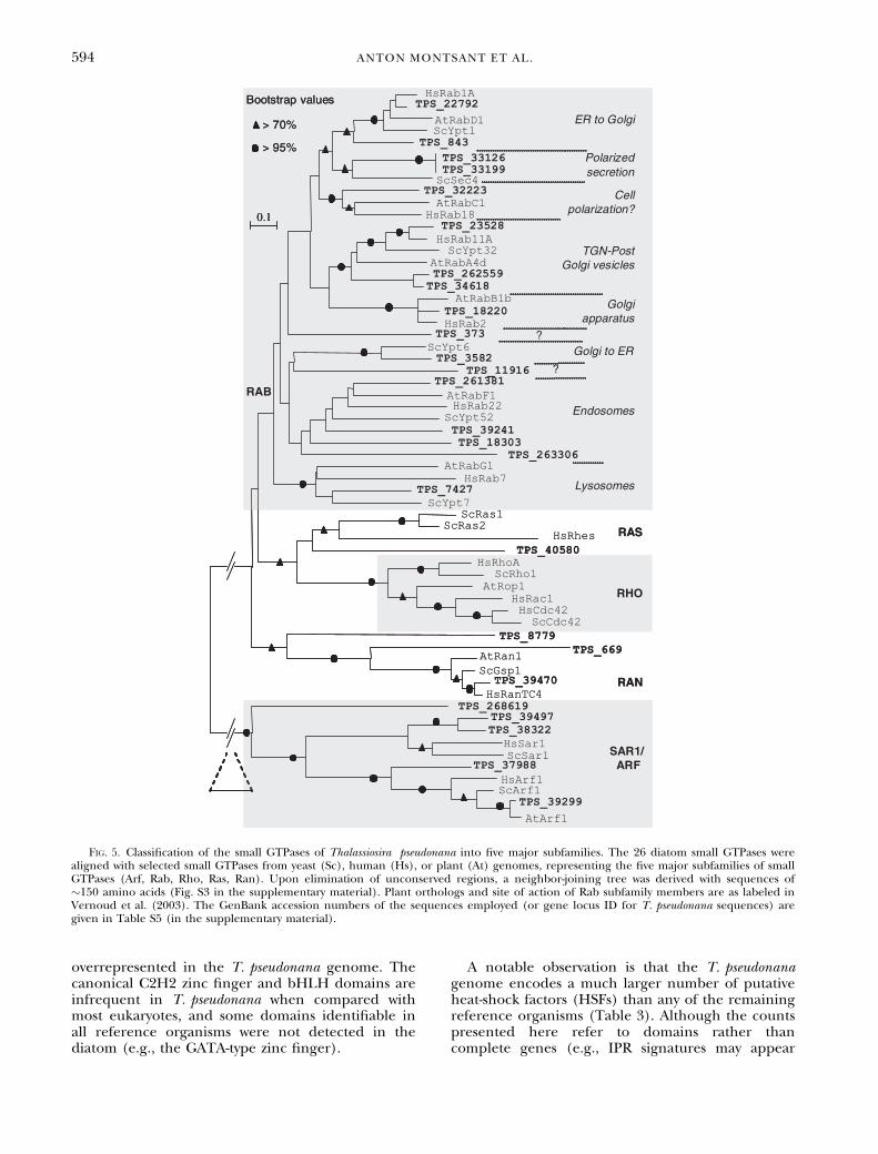
Small GTPases: The T. pseudonana genome wasdetermined to encode at least 26 classical smallGTPases. These molecular switches of �200 aminoacids may have been central to the appearance of
eukaryotic cells (Jekely 2003) and are known to bewidely conserved across taxa, with five major sub-families playing regulatory roles in a variety of cellu-lar processes (Takai et al. 2001). Following anapproach similar to that employed previously to clas-sify the small GTPases of Arabidopsis (Vernoud et al.2003), an NJ tree was derived to observe the cluster-ing of T. pseudonana sequences with well-studiedorthologs from yeast and humans. No members ofthe RHO subfamily (Rho, Rac, Cdc42) could beidentified in the diatom by this procedure (Fig. 5),as was recently found to be the case for Trypanoso-ma bruceii (Field 2005). Moreover, no RHO-specificguanyl-nucleotide exchange factors (GEFs) orGTPase-activating proteins (GAPs) were found (e.g.,there are no RhoGEF-specific Dbl domains and onlyone putative RhoGAP domain, both of which areabundant in yeast and animals). The RHO sub-family small GTPases are widespread key regulatorsof multiple phenomena that require cell polarity(Etienne-Manneville and Hall 2002), and a Rac pro-tein was shown to be a key regulator of cell polarityin the brown macroalga Fucus distichus L. (Fowleret al. 2004), suggesting that diatoms may employ
Fig. 3. Phylogenetic analysis of Thalassiosira pseudonana phytochrome. Phytochrome-like sequences from multiple species were down-loaded from GenBank, and a highly conserved core of �430 amino acids (Fig. S2 in the supplementary material), encompassing the GAFand histidine kinase domains, was used to build a neighbor-joining tree together with the putative T. pseudonana phytochrome (TpPhy)and a Phaeodactylum tricornutum phytochrome-like sequence (PtPhy; M. Siaut, A. Falciatore, and C. Bowler, unpublished data). BphPs, bac-teriophytochromes; Cphs, cyanobacterial phytochromes; Dphs, diatom phytochromes; Fphs, fungal phytochromes; Plant phys, plant phyto-chromes. The identification numbers of the amino acid sequences employed are given under the species name. TpPhy1, gene ID 106207on the JGI Thaps1 genome browser; PtPhy1, GenBank accession number DQ287774.
592 ANTON MONTSANT ET AL.
novel mechanisms for controlling polarized proces-ses such as phototactic movement and transport tothe silica deposition vesicle (SDV; Zurzolo and Bow-ler 2001).
When small GTPase-encoding genes weresearched in the genomes of other model eukaryotesand aligned and classified in the same fashion(Figs. S4 and S5 in the supplementary material), theapicomplexan Plasmodium falciparum was also foundto lack RHO subfamily GTPases (Table 2; Fig. S5ain the supplementary material). However, the oomy-cete P. sojae, more closely related to the diatoms,appeared to have a probable Rac1 ortholog, alongwith another putative member of the RHO sub-family (Fig. S5b in supplementary material; Table 2).The apicomplexan, in addition, lacks a Ras1-likeprotein, a feature previously noted in plants(Vernoud et al. 2003), and this was found to be thecase also for the green microalga Ostreococcus tauriC. Courties et Chret.-Dinet, but not for the red algaC. merolae (Fig. S5, c and d, in the supplementarymaterial; Table 2). Relative to the small-GTPase pro-file of the reference organisms, the regulators ofvesicle-trafficking Rab subfamily proteins constitutea high proportion of the total small GTPases ofT. pseudonana (Table 2). Some unicellular parasites
have recently been shown to have much larger Rabsubfamilies, possibly reflecting a functional special-ization for their specific lifestyles (Lal et al. 2005,Saito-Nakano et al. 2005). Two of the diatom small-GTPases clustered clearly with all Rab proteins, butnot within any conserved subclade (Fig. 5). Suchdiatom-specific Rab variants may play a role in bio-genesis or maintenance of membrane structurespeculiar to diatoms or closely related organisms(e.g., SDV or plastid outer membranes).
Control of gene expression. Chromatin structure: Thenuclear DNA of eukaryotes is typically organizedaround nucleosomes, which contain two subunits ofeach of the core histones H2A, H2B, H3, and H4.The T. pseudonana genome contains several genesencoding the core histones (as well as the linker his-tone H1), which are, as frequently observed in otherorganisms, organized together into gene clusters(Fig. S6 in the supplementary material). A histoneH3.3 variant was also not clustered with otherhistones, in agreement with what is typicallyfound in other eukaryotes (Malik and Henikoff2003). When components regulating the epigeneticcontrol of gene expression by modification of nucle-osome packaging were studied, several putativehistone acetyl transferases (HATs) of the GCN5,MYST, and CBP ⁄ p300 superfamilies, as well ashistone deacetylases (HDACs) belonging to theRpd3p, HDA1p, and Sir2p families, were identified(Table S3, annotations 32–49, in the supplementarymaterial). Histone methylation is known to be medi-ated by SET domain proteins (Xiao et al. 2003), ofwhich up to 30 may be encoded in the T. pseudo-nana genome (Table S3, annotations 50–55, in thesupplementary material). Methylated and acetylatedhistone tails are typically recognized by chromodo-mains and bromodomains, identified, respectively,in 4 and 27 diatom predicted proteins. Four genesencoding putative DNA methyl transferases werealso detected (Table S3, annotations 56–59, in thesupplementary material), consistent with previousdata showing that diatom DNA is methylated (Jarviset al. 1992).
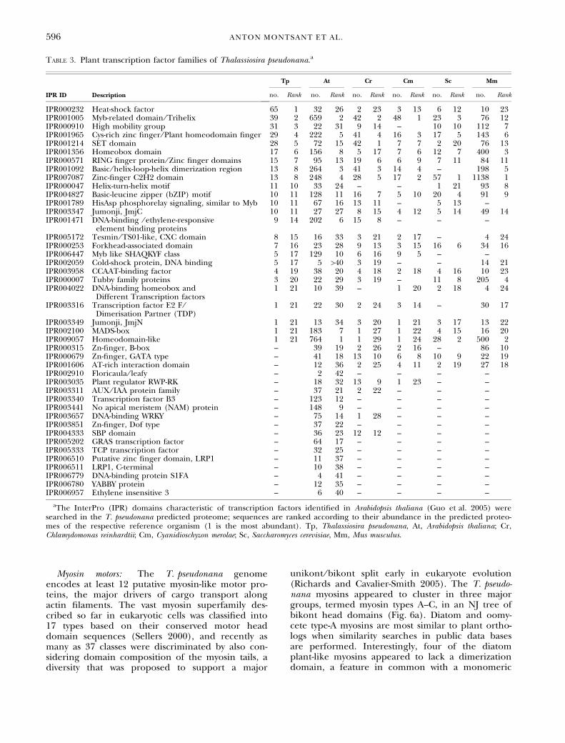
Transcription factors: Genes encoding the basalmachinery for RNA polymerase I-, II-, and III-direc-ted transcription are present within the diatomgenome. To examine T. pseudonana transcriptionfactor families, we searched for the InterPro (IPR)transcription factor (TF) domains recently summari-zed in the A. thaliana DATF data base (http://datf.cbi.pku.edu.cn; Guo et al. 2005) and comparedthe abundance of these domains with five speciescomprising unicellular and multicellular photo-synthetic and heterotrophic organisms (Table 3).Twenty-four of the 44 TF domains identified inA. thaliana were present in T. pseudonana. Most ofthese are also present in the rest of the referencedeukaryotic organisms, with zinc fingers being of sim-ilar relative abundance in all six species. However,some domains widespread in eukaryotes are under- or
Fig. 4. Schematic of two-component signaling modules withinpredicted proteins identified in Thalassiosira pseudonana. GAF,sensor domain; HK, histidine kinase domain; RR, response-regulator receiver domain; CAAX, prenylation site (for mem-brane localization); PAS, protein-protein interaction domain; TF,transcription factor DNA-binding motif. Conserved histidine au-tophosphorylation and aspartate phosphoacceptor sites are indi-cated, where present. The depicted gene models have thefollowing locus IDs: TpPHY1, TPS_22848; TpHK1, TPS_262298;TpRR1, TPS_20939; TpRR2, TPS_264268; TpRR3, TPS_263389;TpRR4, TPS_264726; TpRR5, TPS_3877; TpRR6, TPS_11819;TpRR7, TPS_33288.
SIGNALING AND REGULATORY COMPONENTS IN T. PSEUDONANA 593
overrepresented in the T. pseudonana genome. Thecanonical C2H2 zinc finger and bHLH domains areinfrequent in T. pseudonana when compared withmost eukaryotes, and some domains identifiable inall reference organisms were not detected in thediatom (e.g., the GATA-type zinc finger).
A notable observation is that the T. pseudonanagenome encodes a much larger number of putativeheat-shock factors (HSFs) than any of the remainingreference organisms (Table 3). Although the countspresented here refer to domains rather thancomplete genes (e.g., IPR signatures may appear
Fig. 5. Classification of the small GTPases of Thalassiosira pseudonana into five major subfamilies. The 26 diatom small GTPases werealigned with selected small GTPases from yeast (Sc), human (Hs), or plant (At) genomes, representing the five major subfamilies of smallGTPases (Arf, Rab, Rho, Ras, Ran). Upon elimination of unconserved regions, a neighbor-joining tree was derived with sequences of�150 amino acids (Fig. S3 in the supplementary material). Plant orthologs and site of action of Rab subfamily members are as labeled inVernoud et al. (2003). The GenBank accession numbers of the sequences employed (or gene locus ID for T. pseudonana sequences) aregiven in Table S5 (in the supplementary material).
594 ANTON MONTSANT ET AL.
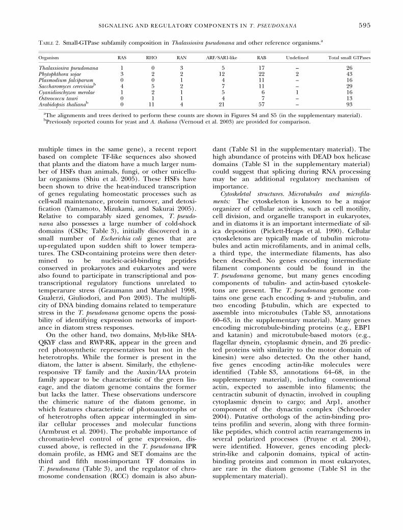
multiple times in the same gene), a recent reportbased on complete TF-like sequences also showedthat plants and the diatom have a much larger num-ber of HSFs than animals, fungi, or other unicellu-lar organisms (Shiu et al. 2005). These HSFs havebeen shown to drive the heat-induced transcriptionof genes regulating homeostatic processes such ascell-wall maintenance, protein turnover, and detoxi-fication (Yamamoto, Mizukami, and Sakurai 2005).Relative to comparably sized genomes, T. pseudo-nana also possesses a large number of cold-shockdomains (CSDs; Table 3), initially discovered in asmall number of Escherichia coli genes that areup-regulated upon sudden shift to lower tempera-tures. The CSD-containing proteins were then deter-mined to be nucleic-acid-binding peptidesconserved in prokaryotes and eukaryotes and werealso found to participate in transcriptional and pos-transcriptional regulatory functions unrelated totemperarure stress (Graumann and Marahiel 1998,Gualerzi, Giuliodori, and Pon 2003). The multipli-city of DNA binding domains related to temperaturestress in the T. pseudonana genome opens the possi-bility of identifying expression networks of import-ance in diatom stress responses.
On the other hand, two domains, Myb-like SHA-QKYF class and RWP-RK, appear in the green andred photosynthetic representatives but not in theheterotrophs. While the former is present in thediatom, the latter is absent. Similarly, the ethylene-responsive TF family and the Auxin ⁄ IAA proteinfamily appear to be characteristic of the green lin-eage, and the diatom genome contains the formerbut lacks the latter. These observations underscorethe chimeric nature of the diatom genome, inwhich features characteristic of photoautotrophs orof heterotrophs often appear intermingled in sim-ilar cellular processes and molecular functions(Armbrust et al. 2004). The probable importance ofchromatin-level control of gene expression, dis-cussed above, is reflected in the T. pseudonana IPRdomain profile, as HMG and SET domains are thethird and fifth most-important TF domains inT. pseudonana (Table 3), and the regulator of chro-mosome condensation (RCC) domain is also abun-
dant (Table S1 in the supplementary material). Thehigh abundance of proteins with DEAD box helicasedomains (Table S1 in the supplementary material)could suggest that splicing during RNA processingmay be an additional regulatory mechanism ofimportance.
Cytoskeletal structures. Microtubules and microfila-ments: The cytoskeleton is known to be a majororganizer of cellular activities, such as cell motility,cell division, and organelle transport in eukaryotes,and in diatoms it is an important intermediate of sil-ica deposition (Pickett-Heaps et al. 1990). Cellularcytoskeletons are typically made of tubulin microtu-bules and actin microfilaments, and in animal cells,a third type, the intermediate filaments, has alsobeen described. No genes encoding intermediatefilament components could be found in theT. pseudonana genome, but many genes encodingcomponents of tubulin- and actin-based cytoskele-tons are present. The T. pseudonana genome con-tains one gene each encoding a- and c-tubulin, andtwo encoding b-tubulin, which are expected toassemble into microtubules (Table S3, annotations60–63, in the supplementary material). Many genesencoding microtubule-binding proteins (e.g., EBP1and katanin) and microtubule-based motors (e.g.,flagellar dynein, cytoplasmic dynein, and 26 predic-ted proteins with similarity to the motor domain ofkinesin) were also detected. On the other hand,five genes encoding actin-like molecules wereidentified (Table S3, annotations 64–68, in thesupplementary material), including conventionalactin, expected to assemble into filaments; thecentractin subunit of dynactin, involved in couplingcytoplasmic dynein to cargo; and Arp1, anothercomponent of the dynactin complex (Schroeder2004). Putative orthologs of the actin-binding pro-teins profilin and severin, along with three formin-like peptides, which control actin rearrangements inseveral polarized processes (Pruyne et al. 2004),were identified. However, genes encoding pleck-strin-like and calponin domains, typical of actin-binding proteins and common in most eukaryotes,are rare in the diatom genome (Table S1 in thesupplementary material).
Table 2. Small-GTPase subfamily composition in Thalassiosira pseudonana and other reference organisms.a
Organism RAS RHO RAN ARF ⁄ SAR1-like RAB Undefined Total small GTPases
Thalassiosira pseudonana 1 0 3 5 17 – 26Phytophthora sojae 3 2 2 12 22 2 43Plasmodium falciparum 0 0 1 4 11 – 16Saccharomyces cerevisiaeb 4 5 2 7 11 – 29Cyanidioschyzon merolae 1 2 1 5 6 1 16Ostreococcu tauri 0 1 1 4 7 – 13Arabidopsis thalianab 0 11 4 21 57 – 93
aThe alignments and trees derived to perform these counts are shown in Figures S4 and S5 (in the supplementary material).bPreviously reported counts for yeast and A. thaliana (Vernoud et al. 2003) are provided for comparison.
SIGNALING AND REGULATORY COMPONENTS IN T. PSEUDONANA 595
Myosin motors: The T. pseudonana genomeencodes at least 12 putative myosin-like motor pro-teins, the major drivers of cargo transport alongactin filaments. The vast myosin superfamily des-cribed so far in eukaryotic cells was classified into17 types based on their conserved motor headdomain sequences (Sellers 2000), and recently asmany as 37 classes were discriminated by also con-sidering domain composition of the myosin tails, adiversity that was proposed to support a major
unikont ⁄ bikont split early in eukaryote evolution(Richards and Cavalier-Smith 2005). The T. pseudo-nana myosins appeared to cluster in three majorgroups, termed myosin types A–C, in an NJ tree ofbikont head domains (Fig. 6a). Diatom and oomy-cete type-A myosins are most similar to plant ortho-logs when similarity searches in public data basesare performed. Interestingly, four of the diatomplant-like myosins appeared to lack a dimerizationdomain, a feature in common with a monomeric
Table 3. Plant transcription factor families of Thalassiosira pseudonana.a
IPR ID Description
Tp At Cr Cm Sc Mm
no. Rank no. Rank no. Rank no. Rank no. Rank no. Rank
IPR000232 Heat-shock factor 65 1 32 26 2 23 3 13 6 12 10 23IPR001005 Myb-related domain ⁄ Trihelix 39 2 659 2 42 2 48 1 23 3 76 12IPR000910 High mobility group 31 3 22 31 9 14 – 10 10 112 7IPR001965 Cys-rich zinc finger ⁄ Plant homeodomain finger 29 4 222 5 41 4 16 3 17 5 143 6IPR001214 SET domain 28 5 72 15 42 1 7 7 2 20 76 13IPR001356 Homeobox domain 17 6 156 8 5 17 7 6 12 7 400 3IPR000571 RING finger protein ⁄ Zinc finger domains 15 7 95 13 19 6 6 9 7 11 84 11IPR001092 Basic ⁄ helix-loop-helix dimerization region 13 8 264 3 41 3 14 4 – 198 5IPR007087 Zinc-finger C2H2 domain 13 8 248 4 28 5 17 2 57 1 1138 1IPR000047 Helix-turn-helix motif 11 10 33 24 – – 1 21 93 8IPR004827 Basic-leucine zipper (bZIP) motif 10 11 128 11 16 7 5 10 20 4 91 9IPR001789 HisAsp phosphorelay signaling, similar to Myb 10 11 67 16 13 11 – 5 13 –IPR003347 Jumonji, JmjC 10 11 27 27 8 15 4 12 5 14 49 14IPR001471 DNA-binding ⁄ ethylene-responsive
element binding proteins9 14 202 6 15 8 – – –
IPR005172 Tesmin ⁄ TS01-like, CXC domain 8 15 16 33 3 21 2 17 – 4 24IPR000253 Forkhead-associated domain 7 16 23 28 9 13 3 15 16 6 34 16IPR006447 Myb like SHAQKYF class 5 17 129 10 6 16 9 5 – –IPR002059 Cold-shock protein, DNA binding 5 17 5 >40 3 19 – – 14 21IPR003958 CCAAT-binding factor 4 19 38 20 4 18 2 18 4 16 10 23IPR000007 Tubby family proteins 3 20 22 29 3 19 – 11 8 205 4IPR004022 DNA-binding homeobox and
Different Transcription factors1 21 10 39 – 1 20 2 18 4 24
IPR003316 Transcription factor E2 F ⁄Dimerisation Partner (TDP)
1 21 22 30 2 24 3 14 – 30 17
IPR003349 Jumonji, JmjN 1 21 13 34 3 20 1 21 3 17 13 22IPR002100 MADS-box 1 21 183 7 1 27 1 22 4 15 16 20IPR009057 Homeodomain-like 1 21 764 1 1 29 1 24 28 2 500 2IPR000315 Zn-finger, B-box – 39 19 2 26 2 16 – 86 10IPR000679 Zn-finger, GATA type – 41 18 13 10 6 8 10 9 22 19IPR001606 AT-rich interaction domain – 12 36 2 25 4 11 2 19 27 18IPR002910 Floricaula ⁄ leafy – 2 42 – – – –IPR003035 Plant regulator RWP-RK – 18 32 13 9 1 23 – –IPR003311 AUX ⁄ IAA protein family – 37 21 2 22 – – –IPR003340 Transcription factor B3 – 123 12 – – – –IPR003441 No apical meristem (NAM) protein – 148 9 – – – –IPR003657 DNA-binding WRKY – 75 14 1 28 – – –IPR003851 Zn-finger, Dof type – 37 22 – – – –IPR004333 SBP domain – 36 23 12 12 – – –IPR005202 GRAS transcription factor – 64 17 – – – –IPR005333 TCP transcription factor – 32 25 – – – –IPR006510 Putative zinc finger domain, LRP1 – 11 37 – – – –IPR006511 LRP1, C-terminal – 10 38 – – – –IPR006779 DNA-binding protein S1FA – 4 41 – – – –IPR006780 YABBY protein – 12 35 – – – –IPR006957 Ethylene insensitive 3 – 6 40 – – – –
aThe InterPro (IPR) domains characteristic of transcription factors identified in Arabidopsis thaliana (Guo et al. 2005) weresearched in the T. pseudonana predicted proteome; sequences are ranked according to their abundance in the predicted proteo-mes of the respective reference organism (1 is the most abundant). Tp, Thalassiosira pseudonana, At, Arabidopsis thaliana; Cr,Chlamydomonas reinhardtii; Cm, Cyanidioschyzon merolae; Sc, Saccharomyces cerevisiae, Mm, Mus musculus.
596 ANTON MONTSANT ET AL.
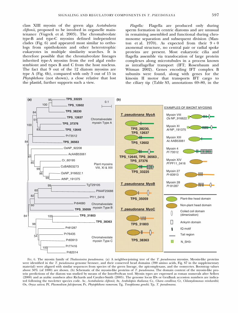
class XIII myosin of the green alga Acetabulariacliftonii, proposed to be involved in organelle main-tenance (Vugrek et al. 2003). The chromalveolatetype-B and type-C myosins defined independentclades (Fig. 6) and appeared most similar to ortho-logs from opisthokonts and other heterotrophiceukaryotes in multiple similarity searches. It istherefore possible that the chromalveolate lineagesinherited type-A myosins from the red algal endo-symbiont and types B and C from the host nucleus.The fact that 9 out of the 12 diatom myosins aretype A (Fig. 6b), compared with only 3 out of 15 inPhytophthora (not shown), a close relative that lostthe plastid, further supports such a view.
Flagella: Flagella are produced only duringsperm formation in centric diatoms and are unusualin remaining assembled and functional during chro-mosome separation and subsequent division (Man-ton et al. 1970). As expected from their 9 + 0axonemal structure, no central pair or radial spokeproteins are present. Most eukaryotic cilia andflagella assemble via translocation of large proteincomplexes along microtubules in a process knownas intraflagellar transport (IFT; Rosenbaum andWitman 2002). Genes encoding IFT complex Bsubunits were found, along with genes for thekinesin II motor that transports IFT cargo tothe ciliary tip (Table S3, annotations 69–80, in the
Fig. 6. The myosin family of Thalassiosira pseudonana. (a) A neighbor-joining tree of the T. pseudonana myosins. Myosin-like proteinswere identified in the T. pseudonana genome browser, and their conserved head domains (390 amino acids, Fig. S7 in the supplementarymaterial) were aligned with similar sequences from species of the green lineage, the apicomplexans, and the oomycetes. Bootstrap valuesabove 50% (of 1000) are shown. (b) Schematic of the myosin-like proteins of T. pseudonana. The domain content of the myosin-like pro-tein predictions of the diatom was studied by means of the InterProScan tool. Myosin types are expressed as roman numerals after Sellers(2000) and as arabic numbers after Richards and Cavalier-Smith (2005). The genome locus IDs or GenBank accession numbers are indica-ted following the two-letter species code. Ac, Acetabularia cliftonii; At, Arabidopsis thaliana; Cc, Chara corallina; Cr, Chlamydomonas reinhardtii;Os, Oryza sativa; Pf, Plasmodium falciparum; Pr, Phytophthora ramorum; Tg, Toxoplasma gondii; Tp, T. pseudonana.
SIGNALING AND REGULATORY COMPONENTS IN T. PSEUDONANA 597
supplementary material). However, no componentsof a cell-body-directed IFT motor (heavy andlight-intermediate chain of cytoplasmic dynein2 ⁄ 1b) or IFT complex A subunits were found,suggesting that T. pseudonana either uses a differentmotor for retrograde transport or that its IFT doesnot run in both directions. The lack of IFT complexA proteins in a ciliated species is surprising and sup-ports the idea that IFT complex A and complex Bhave fundamentally different functions (Pipernoet al. 1998).
Tracking absolute and subjective time. Circadian clock:The circadian clock allows organisms to adapt todaily changing light conditions by anticipating thenight–day transitions and is therefore of particularimportance for photosynthetic organisms. Althoughthe clocks of different eukaryotic organisms consistof different individual components, the mechanismis generally based on a core oscillator that receivesinputs from sensors of extracellular stimuli (e.g.,photoreceptors) and signals downstream to anumber of output pathways that control cell cycleand production of photosynthesis-related peptides(Salome and McClung 2004, Unsal-Kacmaz et al.2005). Although no complete conserved circadianoscillator was distinguished from the T. pseudonanagenome, domains common in plant core oscillators(e.g., MYB, PAS, CCT motif, B-Box or F-Box; Somers2001) appear in multiple predicted proteins, and aputative ortholog of the Circadian Phase ModifierCpmA, a member of a cyanobacterial output path-way thought to regulate the photosystem proteinspsbAI, psbAII, and KaiA (Katayama et al. 1999), wasidentified (Table S3, annotation 81, in the supple-mentary material). Together with the aforemen-tioned photoreceptors, likely major input sourcesfor the core oscillator, the fragmentary pictureobtained from the genome sequence may provide astarting point for experimental work.
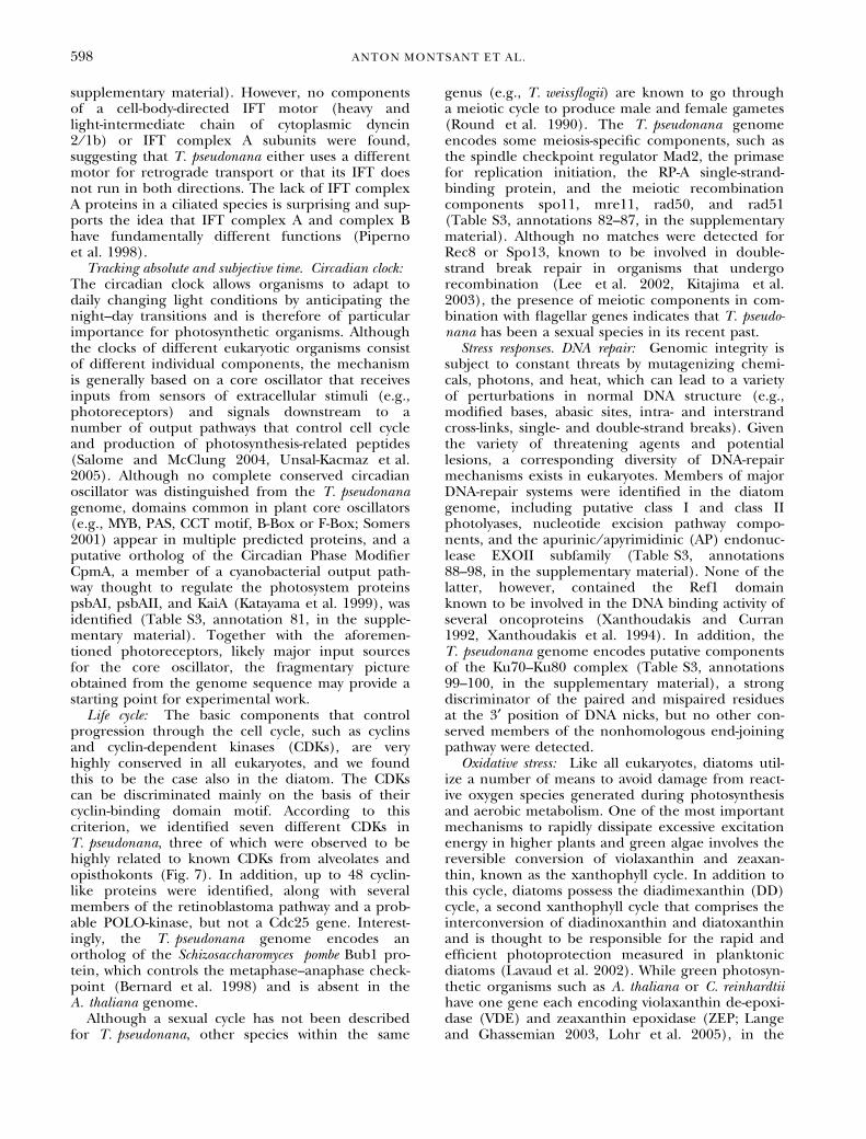
Life cycle: The basic components that controlprogression through the cell cycle, such as cyclinsand cyclin-dependent kinases (CDKs), are veryhighly conserved in all eukaryotes, and we foundthis to be the case also in the diatom. The CDKscan be discriminated mainly on the basis of theircyclin-binding domain motif. According to thiscriterion, we identified seven different CDKs inT. pseudonana, three of which were observed to behighly related to known CDKs from alveolates andopisthokonts (Fig. 7). In addition, up to 48 cyclin-like proteins were identified, along with severalmembers of the retinoblastoma pathway and a prob-able POLO-kinase, but not a Cdc25 gene. Interest-ingly, the T. pseudonana genome encodes anortholog of the Schizosaccharomyces pombe Bub1 pro-tein, which controls the metaphase–anaphase check-point (Bernard et al. 1998) and is absent in theA. thaliana genome.
Although a sexual cycle has not been describedfor T. pseudonana, other species within the same
genus (e.g., T. weissflogii) are known to go througha meiotic cycle to produce male and female gametes(Round et al. 1990). The T. pseudonana genomeencodes some meiosis-specific components, such asthe spindle checkpoint regulator Mad2, the primasefor replication initiation, the RP-A single-strand-binding protein, and the meiotic recombinationcomponents spo11, mre11, rad50, and rad51(Table S3, annotations 82–87, in the supplementarymaterial). Although no matches were detected forRec8 or Spo13, known to be involved in double-strand break repair in organisms that undergorecombination (Lee et al. 2002, Kitajima et al.2003), the presence of meiotic components in com-bination with flagellar genes indicates that T. pseudo-nana has been a sexual species in its recent past.
Stress responses. DNA repair: Genomic integrity issubject to constant threats by mutagenizing chemi-cals, photons, and heat, which can lead to a varietyof perturbations in normal DNA structure (e.g.,modified bases, abasic sites, intra- and interstrandcross-links, single- and double-strand breaks). Giventhe variety of threatening agents and potentiallesions, a corresponding diversity of DNA-repairmechanisms exists in eukaryotes. Members of majorDNA-repair systems were identified in the diatomgenome, including putative class I and class IIphotolyases, nucleotide excision pathway compo-nents, and the apurinic ⁄ apyrimidinic (AP) endonuc-lease EXOII subfamily (Table S3, annotations88–98, in the supplementary material). None of thelatter, however, contained the Ref1 domainknown to be involved in the DNA binding activity ofseveral oncoproteins (Xanthoudakis and Curran1992, Xanthoudakis et al. 1994). In addition, theT. pseudonana genome encodes putative componentsof the Ku70–Ku80 complex (Table S3, annotations99–100, in the supplementary material), a strongdiscriminator of the paired and mispaired residuesat the 3¢ position of DNA nicks, but no other con-served members of the nonhomologous end-joiningpathway were detected.
Oxidative stress: Like all eukaryotes, diatoms util-ize a number of means to avoid damage from react-ive oxygen species generated during photosynthesisand aerobic metabolism. One of the most importantmechanisms to rapidly dissipate excessive excitationenergy in higher plants and green algae involves thereversible conversion of violaxanthin and zeaxan-thin, known as the xanthophyll cycle. In addition tothis cycle, diatoms possess the diadimexanthin (DD)cycle, a second xanthophyll cycle that comprises theinterconversion of diadinoxanthin and diatoxanthinand is thought to be responsible for the rapid andefficient photoprotection measured in planktonicdiatoms (Lavaud et al. 2002). While green photosyn-thetic organisms such as A. thaliana or C. reinhardtiihave one gene each encoding violaxanthin de-epoxi-dase (VDE) and zeaxanthin epoxidase (ZEP; Langeand Ghassemian 2003, Lohr et al. 2005), in the
598 ANTON MONTSANT ET AL.
T. pseudonana genome we found two copies of VDEand two copies of ZEP (Table S3, annotations 101–104, in the supplementary material), possibly reflect-ing the presence of this second diatom-specific xan-thophyll cycle. We also recognized components ofpathways associated with the utilization of antioxi-dants and antioxidative enzymes such as ascorbate,a-tocopherol, and glutathione peroxidases andreductases (Table S3, annotations 105–116, in thesupplementary material), in agreement with theenzyme activities measured in T. pseudonana as aresponse to UV radiation (Rijstenbil 2001). A puta-
tive prokaryotic-type catalase ⁄ peroxidase was identi-fied that may break down the H2O2 generatedduring the b-oxidation of fatty acids and the glyoxy-late cycle, which likely takes place within the perox-isome (Armbrust et al. 2004). The T. pseudonanagenome encodes two Fe-type superoxide dismutases(SODs) and two Mn-type SODs (at least one likelyto be localized to mitochondria) that act to convertoxygen radicals to hydrogen peroxide (Table S3,annotations 117–120, in the supplementary mater-ial). Similar to P. falciparum, no obvious match wasfound for a Cu ⁄ Zn-type SOD or for a Ni-containing
Fig. 7. Phylogenetic analysis of the cyclin-dependent kinase (CDK)–like proteins encoded in the Thalassiosira pseudonana genome. Theseven putative CDKs identified in the diatom genome were trimmed to a conserved core (�280 amino acids; Fig. S8 in the supplementarymaterial), along with CDKs from animal, protozoan, and plant species, and used to build a neighbor-joining tree. Conserved motifs areindicated under the CDK subfamily name. In orthologs with variations of the canonical motif, the motif sequence is indicated after thesequence name. GenBank accession numbers or protein prediction identification numbers of all sequences employed are indicated inTable S6 (in the supplementary material).
SIGNALING AND REGULATORY COMPONENTS IN T. PSEUDONANA 599
SOD recently discovered in marine cyanobacteria(Eitinger 2004).
Biotic stress: In their aquatic environments, dia-toms are exposed to a variety of predators andpathogenic organisms (Holfeld 2000, Nagasakiet al. 2004, Nagasaki et al. 2005). Diatom cellsdamaged by copepod feeding release reactiveunsaturated aldehydes shown to have an antiprolif-erative effect on copepod egg hatching and larvaldevelopment, although the ecological implicationsof this remain controversial (Miralto et al. 1999, I-rigoien et al. 2002, Ianora et al. 2004). Such a def-ense mechanism has not been reported inT. pseudonana, but its genome encodes severalputative components of oxylipin production path-ways (e.g., phospholipases and cytochrome p450–like predicted proteins; Table S1, in the supple-mentary material). However, no lipoxygenase ⁄hydroperoxide lyases, hypothesized to be requiredfor this pathway (Pohnert 2002), could be detec-ted. The presence of genes with similarity to com-ponents of the prostaglandin biosynthesis pathway(Table S3, annotations 122–123, in the supple-mentary material), associated with the inflamma-tory response of the mammalian immune system,may suggest other metabolic pathways related todefense-signaling cascades. As pathogens threatento become established, higher plants commonlyactivate a hypersensitive response that derives froman oxidative burst and limits pathogen spread bykilling infected cells and activating transcription ofcell-defense proteins. The T. pseudonana genomeencodes multiple proteins with LRR motifs(Table 1), but none contain the nucleotide-binding or TOLL ⁄ TIR domains that additionallydefine disease-defense R-proteins. However, weidentified more than 10 plantlike pathogen-related or hypersensitive response-induced proteins;two putative subunits of the NADPH respiratoryburst oxidase, recently found to be up-regulatedduring pathogen attack in a red alga (Herve et al.2005); and two putative nitric oxide synthases,which may play a central role in detection of bio-chemical signals from wounded neighboring cellsin natural bloom conditions (Vardi et al. 2006).At least five copies of genes that encode proteinsbelonging to the Multi Antimicrobial and ToxinExtrusion (MATE) family, recently shown tobe part of a disease-resistance signal transductionpathway in plants (Nawrath et al. 2002), werealso found (Table S3, annotations 141–145, in thesupplementary material).
Unicellular apoptosis: When oxidative stressexceeds antioxidant capacity, apoptotic pathwayscan be induced. Internally triggered cell death hasbeen identified in unicellular organisms (Ameisen2002), and it may play a major role in phytoplank-ton bloom succession and collapse (Vardi et al.1999, Bidle and Falkowski 2004, Vardi et al. 2006).No clear homologs of important elements of meta-
zoan apoptotic pathways, such as p53, caspases, orthe BCL2 family of apoptosis regulators, wereobserved in T. pseudonana. However, we could iden-tify at least five apoptosis-related metacaspases(Table S3, annotations 146–150, in the supplement-ary material), present in A. thaliana, fungi, and sev-eral eukaryotic microbes (Koonin and Aravind 2002,Madeo et al. 2002). Furthermore, we identifiedapoptosis-associated nuclear factors E2 F and DP1,found in metazoans and plants, but not previouslyreported in unicellular organisms, as well as threeputative serpins (serine proteinase inhibitors) thathave been implicated in host defense in animals(Irving et al. 2000; Table S3, annotations 151–165,in the supplementary material). Interestingly, a ser-pin for which there is abundant EST support and aplantlike pathogenesis-related protein were recentlydetected in a proteomic study of a T. pseudonana cell-wall protein-enriched fraction, and their pattern ofexpression through the cell cycle correlated withthat of cell-wall-synthesis marker genes (Frigeri et al.2006). Although the apoptotic elements present inT. pseudonana appear largely similar to those ofother eukaryotic microbes and plants, no homologswere found for TIR adaptor proteins or AP-ATPases,both of which are abundant in A. thaliana. Furtherexamination of these features in other chromalveo-lates should help elucidate the origin of cell-deathpathways and their ecological role in phytoplanktonbloom termination.
CONCLUSION
This study complements the overview initiated byArmbrust et al. (2004) of major features of diatombiology that can be investigated from the availabilityof a first whole-genome sequence for this type oforganism. In both reports, novel assortments of meta-bolic and regulatory components have been inferredfrom the genome sequence. Whereas Armbrust et al.(2004) focused on the general features of diatom pri-mary metabolism (e.g., carbon, nitrogen, and silicametabolism), the current manuscript describes mech-anisms of cell regulation and maintenance of cellhomeostasis. More specifically, we have highlightedthe basic features of signal transduction, includingreceptors and canonical signaling cascades, transcrip-tion factors and chromatin-level control, cytoskeleton,cell cycle, and stress-response pathways. The analysisreveals an unusual assortment of pathways andcomponents that have never previously been foundtogether in the same organism, reflecting theunusual phylogenetic history of diatoms.
Of potential major significance is that diatomsmay possess a novel class of phytochrome that repre-sents a ‘‘brown’’ clade, and that the T. pseudonanagenome appears to lack conventional Rho-type smallGTPases, generally considered to be essential regula-tors of polarized processes such as localized cell-wallsynthesis and tactic movement in eukaryotes. The
600 ANTON MONTSANT ET AL.
extraordinarily high number of heat-shock TFs maybe of significant interest for future work on diatomstress responses. Finally, the putative membrane-bound environmental sensing mechanisms des-cribed here are also very promising experimentaltargets, in view of recent studies that have revealedsophisticated chemical-based systems for the trans-duction of external signals in marine diatoms (Fal-ciatore et al. 2000, Vardi et al. 2006). However, therelative paucity of environmental sensing mecha-nisms will be a major challenge to address experi-mentally. Another highly successful group ofphotosynthetic organisms in the marine environ-ment, the prokaryotic prochlorophytes, were alsodetermined to have a poor content of classical sens-ing mechanisms for the perception of external sig-nals (as opposed to their freshwater counterparts),which may suggest that the marine environment issufficiently uniform and unchanging that a largenumber of receptor-based signaling systems is notrequired (Mary and Vaulot 2003).
In summary, the results of the in silico–basedanalyses described here and in Armbrust et al. pro-vide a rather complete blueprint of T. pseudonanabiology that should serve as cornerstones for futurelaboratory-based and in situ–based experimentalstudies aimed at understanding diatom biology andthe position of these organisms in the eukaryoticevolutionary tree.
This work was performed under the auspices of the U.S.Department of Energy’s (DOE) Office of Science, Biologicaland Environmental Research Program and the University ofCalifornia, Lawrence Livermore National Laboratory underContract No. W-7405-Eng-48; Lawrence Berkeley NationalLaboratory under contract No. DE-AC03-76SF00098; and LosAlamos National Laboratory under contract No. W-7405-ENG-36.Additional funding was provided by the European Union(contracts QLRT-2001-01226, LSHG-CT-2004-512035, andGOCE-CT-2004-505403 to C. B.), the CNRS ATIP ‘‘Blanche’’programme (2JE144 to C. B.), and the U.S. DOE (DE-FG03-02ER63471 to E. V. A.).
Ahmad, M. & Cashmore, A. R. 1993. HY4 gene of A. thalianaencodes a protein with characteristics of a blue-light photo-receptor. Nature 366:162–6.
Allen, A. E., Vardi, A. & Bowler, C. 2006. An ecological and evo-lutionary context for integrated nitrogen metabolism andrelated signaling pathways in marine diatoms. Curr. Opin. PlantBiol. 9:264–73.
Altschul, S. F., Gish, W., Miller, W., Myers, E. W. & Lipman, D. J.1990. Basic local alignment search tool. J. Mol. Evol. 215: 403–10.
Ameisen, J. C. 2002. On the origin, evolution, and nature of pro-grammed cell death: a timeline of four billion years. Cell DeathDiffer. 9:367–93.
Apt, K. E., Zaslavkaia, L., Lippmeier, J. C., Lang, M., Kilian, O.,Wetherbee, R., Grossman, A. R. & Kroth, P. G. 2002. In vivocharacterization of diatom multipartite plastid targeting sig-nals. J. Cell Sci. 115:4061–9.
Arabidopsis Genome Initiative. 2000. Analysis of the genomesequence of the flowering plant Arabidopsis thaliana. Nature408:796–815.
Armbrust, E. V., Berges, J. B., Bowler, C., Green, B. R., Martinez, D.,Putnam, N. H., Zhou, S., et al. 2004. The genome of the
diatom Thalassiosira pseudonana: ecology, evolution, andmetabolism. Science 306:79–86.
Bernard, P., Hardwick, K. & Javerzat, J. P. 1998. Fission yeast bub1 isa mitotic centromere protein essential for the spindle check-point and the preservation of correct ploidy through mitosis.J. Cell Biol. 143:1775–87.
Bidle, K. D. & Falkowski, P. G. 2004. Cell death in planktonic,photosynthetic microorganisms. Nat. Rev. Microbiol. 2:643–55.
Borowitzka, L. J. & Volcani, B. E. 1977. Role of silicon in diatommetabolism. VIII. Cyclic AMP and cyclic GMP in synchronizedcultures of Cylindrotheca fusiformis. Arch. Microbiol. 112:147–52.
Brudler, R., Hitomi, K., Daiyasu, H., Toh, H., Kucho, K., Ishiura,M., Kanehisa, M., Roberts, V. A., Todo, T., Tainer, J. A. &Getzoff, E. D. 2003. Identification of a new cryptochrome class.Structure, function, and evolution. Mol. Cell 11:59–67.
Cashmore, A. R. 2003. Cryptochromes: enabling plants and ani-mals to determine circadian time. Cell. Mol. Life Sci. 114:537–43.
Colucci, G., Apone, F., Alyeshmerni, N., Chalmers, D. & Chrispeels,M. J. 2002. GCR1, the putative Arabidopsis G protein-coupledreceptor gene is cell cycle-regulated, and its overexpressionabolishes seed dormancy and shortens time to flowering. Proc.Natl. Acad. Sci. U. S. A. 99:4736–41.
Davis, S. J., Vener, A. V. & Vierstra, R. D. 1999. Bacter-iophytochromes: phytochrome-like photoreceptors fromnonphotosynthetic eubacteria. Science 286:2517–20.
Delaroque, N., Mueller, D. G., Bothe, G., Pohl, T., Knippers, R. &Boland, W. 2001. The complete DNA sequence of theEctocarpus siliculosus Virus EsV-1 genome. Virology 287:112–32.
Eitinger, T. 2004. In vivo production of active nickel superoxidedismutase from Prochlorococcus marinus MIT9313 is dependenton its cognate peptidase. J. Bacteriol. 186:7821–5.
Etienne-Manneville, S. & Hall, A. 2002. Rho GTPases in cell biol-ogy. Nature 420:629–35.
Falciatore, A. & Bowler, C. 2005. The evolution and function ofblue and red light photoreceptors. Curr. Top. Dev. Biol. 68:317–50.
Falciatore, A., d’Alcala, M. R., Croot, P. & Bowler, C. 2000. Per-ception of environmental signals by a marine diatom. Science288:2363–6.
FANTOM Consortium & the RIKEN Genome Exploration ResearchGroup Phase I and II Team. 2002. Analysis of the mousetranscriptome based on functional annotation of 60,770full-length cDNAs. Nature 420: 563–73.
Field, C. B., Behrenfeld, M. J., Randerson, J. T. & Falkowski, P.1998. Primary production of the biosphere: integrating ter-restrial and oceanic components. Science 281:237–40.
Field, M. C. 2005. Signalling the genome: the Ras-like small GTPasefamily of trypanosomatids. Trends Parasitol. 21:447–50.
Fowler, J. E., Vejlupkova, Z., Goodner, B. W., Lu, G. & Quatrano, R.S. 2004. Localization to the rhizoid tip implicates a Fucus dis-tichus Rho family GTPase in a conserved cell polarity pathway.Planta 219:566–866.
Frigeri, L. G., Radabaugh, T. R., Haynes, P. A. & Hildebrand, M.2006. Identification of proteins from a cell wall fraction of thediatom Thalassiosira pseudonana: insights into silica structureformation. Mol. Cell. Proteomics 5:182–93.
Galagan, J. E., Calvo, S. E., Borkovich, K. A., Selker, E. U., Read, N. D.,Jaffe, D., FitzHugh, W., et al. 2003. The genome sequence of thefilamentous fungus Neurospora crassa. Nature 422:859–68.
Giraud, E., Fardoux, J., Fourrier, N., Hannibal, L., Genty, B.,Bouyer, P., Dreyfus, B. & Vermeglio, A. 2002. Bacter-iophytochrome controls photosystem synthesis in anoxygenicbacteria. Nature 417:202–5.
Graumann, P. L. & Marahiel, M. A. 1998. A superfamily of proteinsthat contain the cold-shock domain. Trends Biochem. Sci. 23:286–90.
Gualerzi, C. O., Giuliodori, A. M. & Pon, C. L. 2003. Transcriptionaland post-transcriptional control of cold-shock genes. J. Mol.Biol. 331:527–39.
SIGNALING AND REGULATORY COMPONENTS IN T. PSEUDONANA 601
Guo, A., He, K., Liu, D., Bai, S., Gu, X., Wei, L. & Luo, J. 2005.DATF: a database of Arabidopsis transcription factors. Bioinfor-matics 21:2568–9.
Hall, T. A. 1999. BioEdit: a user-friendly biological sequencealignment editor and analysis program for Windows 95/98/NT. Nucleic Acids Symp. Ser. 41: 95–8.
Herve, C., Tonon, T., Collen, J., Corre, E. & Boyen, C. 2005.NADPH oxidases in eukaryotes: red algae provide new hints!Curr. Genet. 49:190–204.
Hildebrand, M., Dahlin, K. & Volcani, B. E. 1998. Characterizationof a silicon transporter gene family in Cylindrotheca fusiformis:sequences, expression analysis, and identification of homologsin other diatoms. Mol. Gen. Genet. 260:480–6.
Holfeld, H. 2000. Infection of the single-celled diatom Stephano-discus alpinus by the chytrid Zygorhizidium: parasite distributionwithin host population, changes in host cell size, and host-parasite size relationship. Limnol. Oceanogr. 45:1440–4.
Ianora, A., Miralto, A., Poulet, S. A., Carotenuto, Y., Buttino, I.,Romano, G., Casotti, R., Pohnert, G., Wichard, T., Colucci-D’Amato, L., Terrazzano, G. & Smetacek, V. 2004. Aldehydesuppression of copepod recruitment in blooms of a ubiquitousplanktonic diatom. Nature 429:403–7.
Irigoien, X., Harris, R. P., Verheye, H. M., Joly, P., Runge, J., Starr, M.,Pond, D., Campbell, R., Shreeve, R., Ward, P., Smith, A. N., Dam,H. G., Peterson, W., Tirelli, V., Koski, M., Smith, T., Harbour, D.& Davidson, R. 2002. Copepod hatching success in marineecosystems with high diatom concentrations. Nature 419:387–9.
Irving, J. A., Pike, R. N., Lesk, A. M. & Whisstock, J. C. 2000. Phy-logeny of the serpin superfamily: implications of patterns ofamino acid conservation for structure and function. GenomeRes. 10:1845–64.
Jarvis, E. E., Dunahay, T. G. & Brown, L. M. 1992. DNA nucleosideand methylation in several species of microalgae. J. Phycol.28:356–62.
Jekely, G. 2003. Small GTPases and the evolution of the eukaryoticcell. Bioessays 25:1129–38.
Jonak, C., Okresz, L., Bogre, L. & Hirt, H. 2002. Complexity, crosstalk and integration of plant MAP kinase signalling. Curr. Opin.Plant Biol. 5:415–24.
Katayama, M., Tsinoremas, N. F., Kondo, T. & Golden, S. S. 1999.CpmA, a gene involved in an output pathway of the cyano-bacterial circadian system. J. Bacteriol. 181:3516–24.
Kirk, J. 1992. The nature and measurement of the light environ-ment in the ocean. In Falkowski, P. & Woodhead, A. [Eds.]Primary Productivity and Biogeochemical Cycles in the Sea. PlenumPress, New York, pp. 9–29.
Kitajima, T. S., Yokobayashi, S., Yamamoto, M. & Watanabe, Y.2003. Distinct cohesin complexes organize meiotic chromo-some domains. Science 300:1152–5.
Kooistra, W. H. C. F., DeStefano, M., Mann, D. G. & Medlin, L. K.2003. The phylogeny of the diatoms. Prog. Mol. Subcell. Biol.33:59–97.
Koonin, E. V. & Aravind, L. 2002. Origin and evolution ofeukaryotic apoptosis: the bacterial connection. Cell Death Differ.9:394–404.
Kroger, N., Deutzmann, R. & Sumper, M. 1999. Polycationicpeptides from diatom biosilica that direct silica nanosphereformation. Science 286:1129–32.
Krogh, A., Larsson, B., von Heijne, G. & Sonnhammer, E. L. L.2001. Predicting transmembrane protein topology with ahidden Markov model: application to complete genomes.J. Mol. Biol. 305: 567–80.
Lal, K., Field, M. C., Carlton, J. M., Warwicker, J. & Hirt, R. P. 2005.Identification of a very large Rab GTPase family in the parasiticprotozoan Trichomonas vaginalis. Mol. Biochem. Parasitol.143:226–35.
Lange, B. M. & Ghassemian, M. 2003. Genome organization inArabidopsis thaliana: a survey for genes involved in isoprenoidand chlorophyll metabolism. Plant Mol. Biol. 51:925–48.
Lavaud, J., Rousseau, B., van Gorkom, H. J. & Etienne, A. L. 2002.Influence of the diadinoxanthin pool size on photoprotectionin the marine planktonic diatom Phaeodactylum tricornutum.Plant Physiol. 129:1398–406.
Leblanc, C., Falciatore, A., Watanabe, M. & Bowler, C. 1999. Semi-quantitative RT-PCR analysis of photoregulated gene expres-sion in marine diatoms. Plant Mol. Biol. 40:1031–44.
Lee, B. H., Amon, A. & Prinz, S. 2002. Spo13 regulates cohesincleavage. Genes Dev. 16:1672–81.
Li, S., Nosenko, T., Hackett, J. D. & Bhattacharya, D. 2006. Phylo-genomic analysis identifies red algal genes of endosymbioticorigin in the chromalveolates. Mol. Biol. Evol. 23:663–74.
Lin, C. & Todo, T. 2005. The cryptochromes. Genome Biol. 6:220–8.
Lindell, D., Jaffe, J. D., Johnson, Z. I., Church, G. M. & Chisholm, S.W. 2005. Photosynthesis genes in marine viruses yield proteinsduring host infection. Nature 438:86–9.
Lohr, M., Im, C. S. & Grossman, A. R. 2005. Genome-based ex-amination of chlorophyll and carotenoid biosynthesis inChlamydomonas reinhardtii. Plant Physiol. 138:490–515.
Lopez, P. J., Descles, J., Allen, A. E. & Bowler, C. 2005. Prospects indiatom research. Curr. Opin. Biotechnol. 16:180–6.
Madeo, F., Herker, E., Maldener, C., Wissing, S., Lachelt, S., Her-lan, M., Fehr, M., Lauber, K., Sigrist, S. J., Wesselborg, S. &Frohlich, K. U. 2002. A caspase-related protease regulatesapoptosis in yeast. Mol. Cell 9:911–7.
Maheswari, U., Montsant, A., Goll, J., Krishnaswamy, S., Rajyashri,K. R., Patell, V. M. & Bowler, C. 2005. The diatom EST data-base. Nucleic Acids Res. 33:D344–7.
Malik, H. S. & Henikoff, S. 2003. Phylogenomics of the nucleo-some. Nat. Struct. Biol. 10:882–91.
Manton, I., Kowallik, K. & von Stosch, H. A. 1970. Observations onthe fine structure and development of the spindle at mitosisand meiosis in a marine centric diatom (Lithodesmium undu-latum). IV. The second meiotic division and conclusion. J. CellSci. 7:407–43.
Marchler-Bauer, A., Anderson, J. B., DeWeese-Scott, C., Fedorova,N. D., Geer, L. Y., He, S. Q., Hurwitz, D. I., et al. 2003. CDD: acurated Entrez database of conserved domain alignments.Nucleic Acids Res. 31:383–7.
Maris, A. E., Sawaya, M. R., Kaczor-grzeskowiak, M., Jarvis, M. R.,Bearson, S. M., Kopka, M. L., Schroder, I., Gunsalus, R. P. &Dickerson, R. E. 2002. Dimerization allows DNA target siterecognition by the NarL response regulator. Nat. Struct. Biol.9:771–8.
Mary, I. & Vaulot, D. 2003. Two-component systems in Pro-chlorococcus MED4: genomic analysis and differentialexpression under stress. FEMS Microbiol. Lett. 226:135–44.
Matsuzaki, M., Misumi, O., Shin-I, T., Maruyama, S., Takahara, M.,Miyagishima, S. Y., Mori, T., et al. 2004. Genome sequence ofthe ultrasmall unicellular red alga Cyanidioschyzon merolae 10D.Nature 428:653–57.
Miralto, A., Barone, G., Romano, G., Poulet, S. A., Ianora, A., Russo,G. L., Buttino, I., Mazzarella, G., Laabir, M., Cabrini, M. &Giacobbe, M. G. 1999. The insidious effect of diatoms oncopepod reproduction. Nature 402:173–6.
Mizuno, T. 1998. His-Asp phosphotransfer signal transduction.J. Biochem. 123:555–63.
Montsant, A., Jabbari, K., Maheswari, U. & Bowler, C. 2005. Com-parative genomics of the pennate diatom Phaeodactylum tri-cornutum. Plant Physiol. 137:500–13.
Morand, L. Z., Kidd, D. G. & Lagarias, J. C. 1993. Phytochromelevels in the green alga Mesotaenium caldariorum are lightregulated. Plant Physiol. 101:97–104.
Nagasaki, K., Tomaru, Y., Katanozaka, N., Shirai, Y., Nishida, K.,Itakura, S. & Yamaguchi, M. 2004. Isolation and characteriza-tion of a novel single-stranded RNA virus infecting the bloom-forming diatom Rhizosolenia setigera. Appl. Environ. Microbiol.70:704–11.
Nagasaki, K., Tomaru, Y., Takao, Y., Nishida, K., Shirai, Y., Suzuki,H. & Nagumo, T. 2005. Previously unknown virus infectsmarine diatom. Appl. Environ. Microbiol. 71:3528–35.
Nagel, G., Ollig, D., Fuhrmann, M., Kateriya, S., Musti, A. M.,Bamberg, E. & Hegemann, P. 2002. Channelrhodopsin-1: alight-gated proton channel in green algae. Science 296:2395–8.
Nawrath, C., Heck, S., Parinthawong, N. & Metraux, J. P. 2002.EDS5, an essential component of salicylic acid-dependent
602 ANTON MONTSANT ET AL.
signaling for disease resistance in Arabidopsis, is a member ofthe MATE transporter family. Plant Cell 14:275–86.
Oka, A., Sakai, H. & Iwakoshi, S. 2002. His-Asp phosphorelay signaltransduction in higher plants: receptors and response reg-ulators for cytokinin signaling in Arabidopsis thaliana. GenesGenet. Syst. 77:383–91.
Parkinson, J. & Gordon, R. 1999. Beyond micromachining: thepotential of diatoms. Trends Biotechnol. 17:190–6.
Patron, N. J., Rogers, M. B. & Keeling, P. J. 2004. Gene replacementof fructose-1,6-bisphosphate aldolase supports the hypothesisof a single photosynthetic ancestor of chromalveolates. Eu-karyot. Cell 3:1169–75.
Pickett-Heaps, J., Schmid, A. -M. M. & Edgar, L. A. 1990. The cellbiology of diatom valve formation. In Round, F. E. & Chap-man, D. J. [Eds.] Progress in Phycological Research. Biopress Ltd.,Bristol, UK, pp. 1–168.
Piperno, G., Siuda, E., Henderson, S., Segil, M., Vaananen, H. &Sassaroli, M. 1998. Distinct mutants of retrograde intraflagellartransport (IFT) share similar morphological and moleculardefects. J. Cell Biol. 143:1591–601.
Pohnert, G. 2002. Phospholipase A2 activity triggers the wound-activated chemical defense in the diatom Thalassiosira rotula.Plant Physiol. 129:103–11.
Poulsen, N. & Kroger, N. 2004. Silica morphogenesis by alternativeprocessing of silaffins in the diatom Thalassiosira pseudonana.J. Biol. Chem. 279:42993–9.
Pruyne, D., Legesse-Miller, A., Gao, L., Dong, Y. & Bretscher, A.2004. Mechanisms of polarized growth and organelle segre-gation in yeast. Annu. Rev. Cell Dev. Biol. 20:559–91.
Rabin, R. S. & Stewart, V. 1993. Dual response regulators (NarL andNarP) interact with dual sensors (NarX and NarQ) to controlnitrate- and nitrite-regulated gene expression in Escherichia coliK-12. J. Bacteriol. 175:3259–68.
Reinfelder, J. R., Kraepiel, A. M. L. & Morel, F. M. M. 2000. Uni-cellular C-4 photosynthesis in a marine diatom. Nature407:996–9.
Richards, T. A. & Cavalier-Smith, T. 2005. Myosin domain evolutionand the primary divergence of eukaryotes. Nature 436:1113–8.
Rijstenbil, J. W. 2001. Effects of periodic, low UVA radiation on cellcharacteristics and oxidative stress in the marine planktonicdiatom Ditylum brightwellii. Eur. J. Phycol. 36:1–8.
Rosenbaum, J. L. & Witman, G. B. 2002. Intraflagellar transport.Nat. Rev. Mol. Cell Biol. 3:813–25.
Round, F. E., Crawford, R. M. & Mann, D. G. 1990. The Diatoms:Biology and Morphology of the Genera. Cambridge UniversityPress, London, 747 pp.
Saito-Nakano, Y., Loftus, B. J., Hall, N. & Nozaki, T. 2005. Thediversity of Rab GTPases in Entamoeba histolytica. Exp. Parasitol.110:244–52.
Salome, P. A. & McClung, C. R. 2004. The Arabidopsis thaliana clock.J. Biol. Rhythms 19:425–35.
Scala, S., Carels, N., Falciatore, A., Chiusano, M. L. & Bowler, C.2002. Genome properties of the diatom Phaeodactylum tri-cornutum. Plant Physiol. 129:993–1002.
Schmitz, O., Katayama, M., Williams, S. B., Kondo, T. & Golden, S.S. 2000. CikA, a bacteriophytochrome that resets the cyano-bacterial circadian clock. Science 289:765–8.
Schroeder, T. A. 2004. Dynactin. Annu. Rev. Cell Dev. Biol. 20:759–79.Sellers, J. R. 2000. Myosins: a diverse superfamily. Biochim. Biophys.
Acta 1496:3–22.Shiu, S. H., Shih, M. C. & Li, W. H. 2005. Transcription factor
families have much higher expansion rates in plants than inanimals. Plant Physiol. 139:18–26.
Sineshchekov, O. A., Jung, K. H. & Spudich, J. L. 2002. Two rho-dopsins mediate phototaxis to low- and high-intensity light in
Chlamydomonas reinhardtii. Proc. Natl. Acad. Sci. U. S. A.99:8689–94.
Smetacek, V. 1999. Diatoms and the ocean carbon cycle. Protist150:25–32.
Somers, D. E. 2001. Clock-associated genes in Arabidopsis: a familyaffair. Philos. Trans. R. Soc. Lond. B Biol. Sci. 356:1745–53.
Stephenson, K. & Hoch, J. A. 2002. Evolution of signalling in thesporulation phosphorelay. Mol. Microbiol. 46:297–304.
Takai, Y., Sasaki, T. & Matozaki, T. 2001. Small GTP-binding pro-teins. Physiol. Rev. 81:153–208.
Tyler, B. M., Tripathy, S., Zhang, X., Dehal, P., Jiang, R. H., Aerts,A., Arredondo, F. D., et al. 2006. Phytophthora genome se-quences uncover evolutionary origins and mechanisms ofpathogenesis. Science 313:1261–6.
Unsal-Kacmaz, K., Mullen, T. E., Kaufmann, W. K. & Sancar, A.2005. Coupling of human circadian and cell cycles by thetimeless protein. Mol. Cell. Biol. 25:3109–16.
Vandepeer, Y. & Dewachter, R. 1994. Treecon for Windows: asoftware package for the construction and drawing of evolu-tionary trees for the Microsoft Windows environment. Comput.Appl. Biosci. 10:569–70.
Vardi, A., Berman-Frank, I., Rozenberg, T., Hadas, O., Kaplan, A. &Levine, A. 1999. Programmed cell death of the dinoflagellatePeridinium gatunense is mediated by CO2 limitation and oxi-dative stress. Curr. Biol. 9:1061–4.
Vardi, A., Formiggini, F., Casotti, R., De Martino, A., Ribalet, F.,Miralto, A. & Bowler, C. 2006. A stress surveillance systembased on calcium and nitric oxide in marine diatoms. PLoSBiol. 4:e60 (e-pub ahead of print).
Vernoud, V., Horton, A. C., Yang, Z. & Nielsen, E. 2003. Analysis ofthe small GTPase gene superfamily of Arabidopsis. Plant Physiol.131:1191–208.
Vugrek, O., Sawitzky, H. & Menzel, D. 2003. Class XIII myosinsfrom the green alga Acetabularia: driving force in organelletransport and tip growth? J. Muscle Res. Cell Motil. 24:87–97.
Wagner, J. R., Brunzelle, J. S., Forest, K. T. & Vierstra, R. D.2005. A light-sensing knot revealed by the structure of thechromophore-binding domain of phytochrome. Nature438:325–31.
Xanthoudakis, S. & Curran, T. 1992. Identification and character-ization of Ref-1, a nuclear protein that facilitates AP-1 DNA-binding activity. EMBO J. 11:653–65.
Xanthoudakis, S., Miao, G. G. & Curran, T. 1994. The redox andDNA-repair activities of Ref-1 are encoded by nonoverlappingdomains. Proc. Natl. Acad. Sci. U. S. A. 4:23–7.
Xiao, B., Wilson, J. R. & Gamblin, S. J. 2003. SET domains andhistone methylation. Curr. Opin. Struct. Biol. 13:699–705.
Yamamoto, A., Mizukami, Y. & Sakurai, H. 2005. Identification of anovel class of target genes and a novel type of bindingsequence of heat shock transcription factor in Saccharomycescerevisiae. J. Biol. Chem. 280: 11911–9.
Yeh, K. C., Wu, S. H., Murphy, J. T. & Lagarias, J. C. 1997. A cya-nobacterial phytochrome two-component light sensory system.Science 277:1505–8.
Yoon, H. S., Hackett, J. D., Ciniglia, C., Pinto, G. & Bhattacharya, D.2004. A molecular timeline for the origin of photosyntheticeukaryotes. Mol. Biol. Evol. 21:809–18.
Yoshihara, S., Suzuki, F., Fujita, H., Geng, X. X. & Ikeuchi, M. 2000.Novel putative photoreceptor and regulatory genes requiredfor the positive phototactic movement of the unicellular mo-tile cyanobacterium Synechocystis sp. PCC 6803. Plant Cell Phy-siol. 41:1299–304.
Zurzolo, C. & Bowler, C. 2001. Exploring bioinorganic patternformation in diatoms. A story of polarized trafficking. PlantPhysiol. 127:1339–45.
SIGNALING AND REGULATORY COMPONENTS IN T. PSEUDONANA 603
Supplementary Material
Tables of InterPro domain counts (Table S1),data complementing the analysis of similar-geneclusters (Table S2), annotated gene models(Table S3), and accession numbers of sequencesemployed in Figures 2, 5, and 7 (Tables S4–S6)are available online. Alignments used to deriveneighbor-joining trees (Figs. S1–S4, S7, and S8),the small-GTPase trees for reference microbes onwhich Table 2 counts are based (Fig. S5), and ascheme of histone clusters (Fig. S6) are also pro-vided as supplementary figures.
The following supplementary materials areavailable as part of the online article fromhttp://www.blackwell-synergy.com:
Table S1. InterPro conserved domains identi-fied in Thalassiosira pseudonana and comparisonwith six reference eukaryotes.
Table S2. Clusters of similar coding sequencesin Thalassiosira pseudonana and in other modeleukaryotes.
Table S3. A selection of annotated gene pre-dictions relevant to signaling, gene expression,cytoskeletal regulation, and stress response.
Table S4. Identification numbers of the pro-tein sequences used to derive the cryptochrometree in Figure 2.
Table S5. GenBank accession numbers of theprotein sequences used to derive Figures 5 andS4 (in the supplementary material).
Table S6. Identification numbers of the pro-tein sequences used to derive Figure 7.
Figure S1. Alignment of the cryptochromesequences used to derive the tree shown inFigure 2 of main text.
Figure S2. Alignment of the phytochromesequences used to derive the tree shown inFigure 3 of main text.
Figure S3. Alignment of the small GTPasesequences used to derive the tree shown inFigure 5 of main text.
Figure S4. A. Alignment of the sequences usedto derive a small-GTPase tree for Plasmodium falci-parum. B. Alignment of the sequences used toderive a small-GTPase tree for Phytophthora sojae.C. Alignment of the sequences used to derive asmall-GTPase tree for Ostreococcus tauri. D. Align-ment of the sequences used to derive a small-GTPase tree for Cyanidioschyzon merolae.
Figure S5. Neighbor-joining trees of the smallGTPases of Plasmodium falciparum (a), Phytophtho-ra sojae (b), Ostreococcus tauri (c), and Cyanidios-chyzon merolae (d) along with well-characterizedsmall GTPases from yeast and humans.
Figure S6. Schematic of histone clusters identi-fied in the Thalassiosira pseudonana genome.
Figure S7. Alignment of the myosin headdomain sequences used to derive the tree shownin Figure 6 of main text.
Figure S8. Alignment of the CDK sequencesused to derive the tree shown in Figure 7 ofmain text.
This material is available as part of the onlinearticle from: http://www.blackwell-synergy.com/doi/abs/10.1111/j.1529-8817.2007.00342.x (Thislink will take you to the article abstract).
Please note: Blackwell Publishing is notresponsible for the content or functionality ofany supplementary materials supplied by theauthors. Any queries (other than missing mate-rial) should be directed to the correspondingauthor for the article.
604 ANTON MONTSANT ET AL.