Epidemic spreading over networks - A view from neighbourhoods
Upload
independentCategory
view
4download
0
Identification of a Domain on the Integrin a5 Subunit Implicated inCell Spreading and Signaling*
(Received for publication, April 9, 1998, and in revised form, August 6, 1998)
Zuojun Cao‡, Kun Huang‡, and Alan F. Horwitz‡§¶
From the Departments of ‡Biochemistry and §Cell and Structural Biology, University of Illinois at Urbana-Champaign,Urbana, Illinois 61801
The a5b1 integrin is a cell surface receptor for fi-bronectin implicated in several cellular activities in-cluding cell proliferation, differentiation, and migra-tion. The primary site at which the a5b1 integrininteracts with fibronectin is the RGD (Arg-Gly-Asp)amino acid sequence. In general, the sites on the inte-grin a subunits involved in ligand binding are not wellcharacterized. Based on previous cross-linking studies,sequence alignment, predicted conformation, and in-tron-exon boundaries, we identified a 144-residue re-gion (positions 223–367) on the a5 subunit as a putativebinding region and divided it into four subdomainsnamed domains I, II, III, and IV. Chimeric receptorswere prepared in which sequences on the a5 subunitwere exchanged with the corresponding sequences onthe a6 subunit, which is specific for laminin and does notbind via an RGD sequence. The mutated human a5 inte-grin gene was transfected into CHO B2 cells, which aredeficient in a5 expression. Only chimeras of domain IIIor IV express on the cell surface. Both of these chimerasdecreased the adhesion, spreading, focal adhesion as-sembly, and migration on fibronectin. The adhesion ofthe chimeric receptors to fibronectin remained sensi-tive to the RGD peptide, and antibodies that inhibitinteraction with the fibronectin synergy site and RGDloop remain inhibitory for the chimeras, indicating thatour chimeras do not inhibit binding to either the RGD orsynergy sites. Finally, the affinity of soluble fibronectinto cells via the a5b1 receptor decreased only about3-fold. This decrease is substantially less than the ob-served effects on migration and spreading, which werenot altered by changes in substrate concentration. Thus,the alteration in binding sites does not easily accountfor the changes in cell spreading and focal adhesionassembly. The tyrosine phosphorylation and focal adhe-sion assembly that are seen when cells expressing thewild type a5 receptor adhere to fibronectin were inhib-ited in cells expressing the chimeric receptors. There-fore, our results suggest that the chimeras of these do-mains likely interrupt a5-mediated conformationalsignaling.
Integrins are a large family of heterodimeric adhesive recep-tors of major importance in diverse biological processes thatinclude embryonic development, tumorigenesis, blood clotting,
inflammation, and wound healing (1). Through their dual func-tions as receptors for molecules associated with the cytoskele-ton and for adhesive molecules in the extracellular matrix, onother cells, and in the circulation, they provide anchorage forcell attachment and spreading. Integrins also transduce sig-nals from the outside to the inside of the cells in cooperationwith growth factor receptors (2, 3).
The interactions of integrins with their extracellular ligandsare subjects of intensive study. The binding affinity is regu-lated by a process called inside-out signaling, in which theintracellular signals affect affinity and, for some integrins,specificity as well (4). Ligand affinity can also be modulated byexogenous agents, which include the nature of the cations thatbind to the integrin; antibodies that induce high affinity ligandbinding; and alterations in the membrane proximal region ofintegrin cytoplasmic domains (5, 6). These observations havebeen incorporated into the hinge model of integrin function. Inthis model, affinity/specificity-determining conformationalchanges are propagated from the inside to the outside via acritical hinge region, which resides near the membrane on thecytoplasmic domains of the integrin subunits (4, 7, 8).
Due to their potential for the design of therapeutics, theligand-binding sites on integrins are under intensive study.Peptide cross-linking analyses of mutant integrins, peptidescorresponding to integrin sequences, and epitope mapping ofinhibitory mAbs1 have been used to identify the integrin li-gand-binding sites. From these studies, three regions are im-plicated in ligand binding: the N-terminal portions of both thea and b subunit and the A (or I) domain in the integrins thatcontain it (9).
The ligand-binding sites on the b1 and b3 subunits are thebest understood. RGD-containing peptides cross-link to theN-terminal portion of the b3 (positions 109–171) subunit (10).The importance of this region in ligand binding of b3 and someother integrins has been confirmed in several studies. A labeledRGD peptide cross-links to a peptide corresponding to theamino acids 61–203 on the sequence of the b3 subunit (11). Anaturally occurring mutation on b3 (Asp119 3 Tyr) and muta-tions of the homologous residue on b1 and b6 also result inreduced ligand binding (12, 13). In addition, site-directed mu-tagenesis within this domain inhibits ligand binding (14).
For integrin a subunits that do not possess an A domain,ligand binding localizes to the N-terminal portion. This regioncorresponds roughly to the amino-terminal 400 amino acids onseveral a subunits including a4, a5, and aIIb (9). It containsmetal ion binding sites and is composed of seven homologous,repeated domains, recently termed FG-GAP repeats (15). Pep-* This study was supported by National Institutes of Health Grant
GM23244. The costs of publication of this article were defrayed in partby the payment of page charges. This article must therefore be herebymarked “advertisement” in accordance with 18 U.S.C. Section 1734solely to indicate this fact.
¶ To whom correspondence should be addressed: Dept. of Cell andStructural Biology, B107 CLSL, 601 S. Goodwin Ave., Urbana, IL61801. Tel.: 217-333-6118; Fax: 217-244-1648; E-mail: [email protected].
1 The abbreviations used are: mAb, monoclonal antibody; CHO, Chi-nese hamster ovary; CMF-HH buffer, Ca21-, Mg21-free Hepes-Hanks’buffer; FITC, fluorescein isothiocyanate; PBS, phosphate-buffered sa-line; BSA, bovine serum albumin; ICAM, intercellular adhesion mole-cule; VCAM, vascular cell adhesion molecule.
THE JOURNAL OF BIOLOGICAL CHEMISTRY Vol. 273, No. 48, Issue of November 27, pp. 31670–31679, 1998© 1998 by The American Society for Biochemistry and Molecular Biology, Inc. Printed in U.S.A.
This paper is available on line at http://www.jbc.org31670
by guest on February 16, 2016http://w
ww
.jbc.org/D
ownloaded from

tide ligands cross-link to this region on both the aIIb and av
subunits (16, 17). Point mutations in repeat III on a4, forexample, inhibit ligand binding (18). Repeats V–VII (or IV–VIIin some integrins) encompass sequences that resemble theclassical Ca21 binding motifs known as EF-hands. Mutationsin the EF-hand motifs on the a4 integrin impair ligand binding(19). A peptide derived from repeat V on aIIb inhibits plateletaggregation and binding of fibrinogen to purified aIIbb3 (20),and peptides corresponding to repeat V/VI on aL interfere withthe interaction between ICAM-1 and aLb2 (21).
The importance of divalent metal-binding sites is also seen inthe cation dependence of integrin ligand binding. Mn21 andMg21 generally elevate ligand binding, whereas Ca21 inhibitsit (22). Alterations in some EF-hand motifs of the a subunitinhibit ligand binding. The missing 12th coordination site onthe partial EF-hand motif on a subunits has led to the hypoth-esis that integrin ligands may directly bind to cations by pro-viding an oxygenated residue, e.g. the Asp in the RGD sequence(23). Another model suggests that divalent cations promoteligand binding by switching the integrin into an active confor-mation (6, 24). In most integrins, the binding of Mn21 increasesthe binding affinity. In addition, changing any one of severaldifferent cation-binding sites on a4 reduces ligand binding sim-ilarly, suggesting a structural rather than a direct ligand bind-ing role (19). Although it is not clear which model reflects thereal function of cations in integrin-ligand interaction, cationbinding is very likely a key factor in this process.
Despite the characterization of cation binding as critical toligand binding, ligand-binding motifs on integrin a subunits(outside of the I domain on some a subunits) are not wellcharacterized. This is due, in part, to the sensitivity of a sub-unit to conformational changes that result in improper foldingand/or impaired dimerization. In an attempt to avoid this prob-lem, we constructed chimeric integrins to study the ligandbinding domain on the a5 integrin. We selected four consecu-tive small fragments near the first and second cation-bindingsite on the a subunit, exchanged each segment with the homol-ogous sequence on the a6 subunit, and expressed the resultingchimera in CHO B2 cells, which are deficient in expression ofthe a5 subunit (25). We chose a5 and a6 integrins because bothdimerize with the b1 subunit but have distinct ligand specific-ity; the former binds to fibronectin, while the later binds tolaminin. We found that two of the chimeras express stably onthe cell surface and exhibit defects in adhesion, spreading, andmigration on fibronectin. Interestingly, the binding to fibronec-tin via the RGD and synergy sites was not grossly perturbed.However, the mutations did impair focal adhesion formationand adhesion-mediated protein tyrosine phosphorylation. Thedata suggest that the regions of these mutations are probablyinvolved in outside-in signal transduction.
EXPERIMENTAL PROCEDURES
Construction of Wild Type and Chimeric a Subunits—The a5 cDNAwas originally obtained in the pbApr-1-neo vector (26) and subclonedinto the eucaryotic expression vector, pRSVneo (27). Five restrictionsites, RsrII, SacII, NheI, BssHII, and NsiI, were introduced at theborders of the swapped regions by site-directed mutagenesis as de-scribed in the Muta-GeneTM in vitro mutagenesis kit (Bio-Rad). The a5
cDNA bearing the generated restriction sites was cloned into the pTZ-18R vector, and the fragments of interest were exchanged between a5
and a6 by restriction enzyme digestion and ligation. The chimericcDNAs were cloned into the expression vector pRSVneo. To constructchimeras with cytoplasmic domain truncations, stop codon sequencewas introduced after the GFFKR sequence by site-directed mutagene-sis. The chimeric cDNAs were also cloned into pRSVneo.
Transfection and Selection of a5-Expressing Cells—CHO B2 cellswere grown in minimal essential medium (Life Technologies, Inc.) with10% fetal bovine serum (full medium). 3 3 105 cells were plated onto60-mm tissue culture plates for 16–20 h. 8 mg of plasmid DNA and 25
ml of lipofectamine (Life Technologies) were mixed and diluted in min-imal essential medium to a final volume of 250 ml. The mixture wasincubated for 45 min before addition to the cell cultures. Cells wereincubated for 5 h, and the DNA-lipofectamine mixture was replaced byfull medium. After 24 h, the cells were moved to selection mediumcontaining 1 mg/ml G418 (Life Technologies), selected for 10–14 days,and then maintained in 0.2 mg/ml G418. The expression of a5 on the cellsurface was analyzed by flow cytometry. The cells were first incubatedfor 30 min with 20 mg/ml VIF4 antibody in Ca21-, Mg21-free Hepes-Hanks’ buffer (CMF-HH buffer) containing 2% BSA and were detachedfrom the plates using 0.2 g/ml EDTA in CMF-HH buffer. The cells werethen washed three times with CMF-HH buffer followed by incubationwith a fluorescein isothiocyanate (FITC)-labeled sheep anti-mouse IgG(Cappell, Durham, NC). Cells were then washed with CMF-HH bufferand resuspended in the same buffer. The cell sorting was performed onan EPICSTM (Coulter Electronics, Inc., Miami Lakes, FL) flow cytome-ter equipped with Cicero software for data analysis. Untransfected cellswere used as controls. The transfected cells expressing 20-fold over thatof the untransfected cells were sorted and enriched.
Adhesion Assays—96-well, non-tissue culture plates were coatedwith fibronectin in phosphate-buffered saline (PBS) at the indicatedconcentration for 2 h at 37 °C. The wells were then washed with PBSand blocked with 2% BSA in PBS for 1 h. Cells were detached fromtissue culture plates using 1 mM EDTA in PBS, washed once in PBS,and resuspended in serum-free medium at a concentration of 3 3 105
cells/ml except for the Mn21 activation assays. 100 ml of the suspensionwas added to each coated well. After a 45-min incubation at 37 °C, theplates were placed on an orbital shaker and pulsed three times for 10 sat 300 rpm to remove the nonadherent cells. The wells were thenwashed with PBS once, and 60 ml of 7.5 mM p-nitrophenyl-N-acetyl-b-D-glucosaminide (Sigma) was added in a buffer containing 0.1 M citratebuffer (pH 5.0) with 0.5% Triton X-100. After a 6-h incubation, 90 ml ofdeveloper solution was added (50 mM glycine, 5 mM EDTA, pH 10.4),and the absorbance was read at 410 nM. In parallel, 100 ml of the cellsuspension was centrifuged, and the pellet was resuspended in 60 ml ofp-nitrophenyl-N-acetyl-b-D-glucosaminide solution as the 100% cellcontrol. The percentage of adhesion was expressed as the ratio betweenthe mean absorbance of three wells and the absorbance of the 100% cellcontrol. To analyze the effect of Mn21 on cell adhesion, cells weredetached from dishes, washed with 1 mM EDTA and calcium-, magne-sium-free Tyrode’s buffer (150 mM NaCl, 2.5 mM KCl, 12 mM NaCO3, 2%BSA, pH 7.4), and then resuspended in the calcium-, magnesium-freeTyrode buffer at 6 3 105 cells/ml. 50 ml of cell suspension were mixedwith an equal volume of MnCl2 at the desired concentration. Cells werethen plated, and the standard adhesion assay was performed as de-scribed above.
Migration Assay—Migration was assayed using a transwell assay asdescribed previously (28). Transwells were coated with fibronectin atdifferent concentrations for 2 h followed by a 1-h incubation with 2%BSA. Cells were detached with 1 mM EDTA in PBS, washed once withPBS, and resuspended in serum-free hybridoma medium CCMI (Hy-clone Laboratories, Inc., Logan, UT). 1 3 105 cells were added to eachwell. After a 3-h incubation at 37 °C, the cells remaining on the topmembrane were carefully removed using a cotton swab. The cells thathad migrated cells onto the lower surface of the membrane were fixedwith methanol and stained with methylene blue for 30 min. Cells on thelower surface of the membrane were counted under a phase-contrastmicroscope. Six fields were scored for each well. The numbers of cellsthat had migrated in each field were expressed as the mean of two setsof wells.
Cell Spreading—Cell spreading assays were performed as describedpreviously (29, 37). 96-well, non-tissue culture plates were coated with10 mg/ml fibronectin. Cells were detached, washed as described abovefor adhesion assays, and then resuspended in serum-free medium at2 3 105 cells/ml. 50-ml aliquots of cell suspension were mixed with anequal volume of antibody solution and added to the fibronectin-coatedwells. The cells were incubated for 1 h at 37 °C and fixed in 3%formaldehyde, and then six random fields were scored using a phase-contrast microscope for spreading as described previously (29). Onlycells with fully formed lamina were counted as spread cells. The per-centage of spreading was determined from the ratio of the number ofspread cells and the number of total cells in each field.
Fluorescence Staining—12-mm glass coverslips (Fisher) were coatedwith different fibronectin concentrations. Cells were detached fromplates using 0.2 g/ml EDTA in PBS and resuspended in serum-freemedium (minimal essential medium with 2% BSA). 2–3 3 104 cells wereadded to each coverslip and incubated at 37 °C for 3 h. The coverslipswere washed twice with PBS, fixed with 3% formaldehyde in PBS, and
a5 Integrin Domain Implicated in Cell Spreading and Signaling 31671
by guest on February 16, 2016http://w
ww
.jbc.org/D
ownloaded from

permeablized with 0.2% Triton X-100 for 10 min. The cells were incu-bated with primary antibody (20 mg/ml in blocking buffer) for 30 minfollowed by incubation with FITC-conjugated sheep anti-mouse IgG.Coverslips were blocked with blocking buffer (5% horse serum in PBS)before each incubation and then washed extensively. The coverslipswere mounted, and the cells were viewed on an Axioplan fluorescencemicroscope (Carl Zeiss, Inc., Thornwood, NY).
Cell Extracts and Western Blotting—Cells were prepared as de-scribed above for adhesion assays. 1.5 3 106 cells were added to 60-mmfibronectin-coated dishes. After a 3-h incubation at 37 °C, cells werelysed in 100 ml of cold radioimmune precipitation extraction buffer (20mM Tris, pH 7.4, 150 mM NaCl, 0.5% Nonidet P-40, 1.0% Triton X-100,0.25% sodium deoxycholate, 2 mM EDTA, 2 mM EGTA) with proteaseinhibitors (20 mg/ml leupeptin, 0.7 mg/ml pepstatin, 1 mM phenanthro-line, 0.05 units of aprotinin, 2 mM pheylmethylsulfonyl chloride), andphosphatase inhibitors (30 mM sodium pyrophosphate, 40 mM NaF, 1mM sodium orthovanadate). Cells were scraped and incubated on ice for15 min followed by centrifugation at 14,000 3 g for 5 min. Proteinconcentration was determined using the bicinchoninic acid (Pierce)method. The appropriate volume of 43 protein loading buffer (0.25 M
Tris-HCl, 8% SDS, 40% glycerol, 0.1% bromphenol blue, 20% 2-mercap-toethanol) was added to 30 mg of protein. The sample was then im-mersed in boiling water for 5–10 min, loaded on a 12% SDS-polyacryl-amide gel, and transferred to a nitrocellulose membrane. Themembrane was blocked with 1% BSA in TST buffer (10 mM Tris pH 7.5,100 mM NaCl, 0.1% Tween 20) for 1 h. The PY20 antibody was dilutedto 1:1000 in blocking buffer and incubated with the membrane for 1 h.After a TST buffer washing, the PY20 blot was incubated for 30 minwith a horseradish peroxidase-conjugated antibody (diluted to 1:10,000in blocking buffer) followed by a washing with TST buffer. The blot wasvisualized by chemiluminescence (Pierce Super SignalTM; Pierce) usingx-ray film (Eastman Kodak Co.).
Fibronectin Binding Assay—Fibronectin was first conjugated withFITC as follows. The fibronectin was diluted to a concentration of 0.5mg/ml in carbonate saline buffer (0.015 M Na2CO3, 0.035 NaHCO3, 0.15NaCl), and FITC was dissolved in the same buffer at 0.5 mg/ml. 50 mlof the FITC solution was added to 0.5 mg of fibronectin, and the mixtureincubated at room temperature for 1 h. The free FITC was removedusing a Sephadex G-25 column equilibrated with PBS. The concentra-tion of the collected FITC-labeled fibronectin was calculated by theequation [FITC-fibronectin] 5 (A280 2 0.35 3 A495)/1.23. A280 and A495
are the absorbance of FITC-fibronectin at 280 and 495 nm, respectively.1.23 is the extinction coefficient of the FITC-labeled fibronectin (30).The fibronectin binding assay was performed as follows. Cells weredetached from plates using 0.2 g/ml EDTA in CMF-HH buffer andwashed once with serum-free medium. Cells were then blocked with 2%BSA in minimal essential medium for 30 min and incubated withdifferent concentrations of FITC-fibronectin for 30 min at room temper-ature. Cells were analyzed by flow cytometry as described above. Theamount of fibronectin bound was estimated from the fluorescence in-tensity of the cells. The specific binding was obtained by subtracting thebackground binding (the fluorescence intensity of the untransfectedCHO B2 cells) from the total binding of the transfectants. The data wereanalyzed using a Scatchard plot (31). The ratio of the bound to the freeconcentration of fibronectin was plotted as a function of the boundconcentration of fibronectin. The data were fit to a straight line, and theslope was used to estimate the apparent dissociation constant (Kd).
Materials—The VIF4, VD1, IIA1, and VD10 mAbs directed againsthuman a5 integrins were gifts from Dr. Ralph Isberg (Tufts University,Boston, MA), and BIID2 mAb was a gift from Dr. C. Damsky (Univer-sity of California, San Francisco, CA). The mAbs 16G3 and 3B8 werefrom Dr. K. Yamada (NCI, National Institutes of Health, Bethesda,MD). The anti-phosphotyrosine mAb, PY20, was purchased from Trans-duction Laboratories (Lexington, KY). The anti-vinculin mAb, FITC,and poly-L-lysine were purchased from Sigma. Transwells were pur-chased from Corning Costar Co. (Cambridge, MA). Fibronectin for thesoluble ligand assay was a gift from Dr. Deane Mosher (University ofWisconsin, Madison, WI). The fibronectin used for other assays waspurified from human plasma by affinity chromatography as describedelsewhere (61). The human a5 and chicken a6 cDNA were kindly pro-vided by Dr. L. Reichardt (University of California, San Francisco, CA).The CHO B2 cell line was a gift from Dr. R. L. Juliano (University ofNorth Carolina, Chapel Hill, NC).
RESULTS
Construction and Expression of a5 Chimeras—Previous pub-lications suggest that the N-terminal portion of some a sub-
units is involved in integrin-ligand interaction. For our studies,we selected a 144-residue fragment on the a5 subunit as theputative ligand-binding region (Fig. 1). This region encom-passes two cation-binding motifs. It is homologous to the puta-tive ligand-binding site, which cross-links to a radiolabeledGRGDSPK peptide on the av subunit (16). It also shares se-quence homology with the putative ligand interaction sites onthe aIIb and aL subunits, respectively (20, 21). Chou-Fasman(32) and Robson-Garnier folding algorithms (33, 34) predictloop structures in this region (data not shown). The region wasdivided into four smaller fragments. These were based on theknown exon-intron boundaries on integrin a subunits and shortnonhomologous peptide regions surrounded by sequences ofhigh homology. To preserve the structure, the four segments,designated as domains I, II, III, and IV on the a5 integrin, wereexchanged with their counterparts on chicken a6 subunit (Ta-ble I). The a6 subunit was chosen because it also dimerizes withthe b1 subunit but has a different ligand specificity.
The wild type and chimeric a5 subunits, designated as a5I,a5II, a5III, and a5IV, were transfected into the CHO B2 cell line,which expresses minimal endogenous a5 integrin (25). Cellswere grown in selective medium to achieve stable transfectantsand then sorted by flow cytometry for those expressing similarlevels of a5. Neither the a5I nor a5II could be detected on the cellsurface following either transient or stable transfections (TableI). This was seen in multiple, independent transfections. Incontrast, both a5III and a5IV express on the cell surface. Cellsexpressing comparable levels of wild type and mutant a5 werecollected by flow cytometry (Fig. 2). Swapping the four subdo-mains in different combinations, including domains III and IV,did not result in proteins expressed on the cell surface.
CHO B2 Cells Expressing a5III and a5IV Chimeras ExhibitImpaired Adhesion, Spreading, and Migration—CHO B2 cellsexpressing chimeric integrins showed impaired adhesion whencompared with wild type transfectants (Fig. 3). Whereas wildtype a5 transfected CHO B2 cells readily attach to fibronectin,the adhesion of a5III- and a5IV-transfected cells was signifi-cantly reduced (p , 0.05). Untransfected CHO B2 cells showedminimal attachment. The inhibition observed in the mutantswas not reversed by plating cells on increased fibronectin con-centrations. The adhesion of the a5 transfectants to fibronectinwas specifically mediated by exogenous a5, since adhesion-perturbing antibodies specific for human a5 integrin inhibitedadhesion (data not shown).
CHO B2 cells transfected with chimeric a5 subunits also
FIG. 1. A diagram of the chimeric a5 subunits used in thisstudy. A schematic model of the a5 integrin is shown. A 144-residueregion near the first and second cation binding motifs, which are rep-resented by the thick bars, was selected and divided into four segments(indicated as I, II, III, and IV). Residues at the boundaries betweensegments are shown. Each segment was exchanged with its counterparton the a6 subunit.
a5 Integrin Domain Implicated in Cell Spreading and Signaling31672
by guest on February 16, 2016http://w
ww
.jbc.org/D
ownloaded from

showed altered spreading. After 1 h of incubation on fibronec-tin, most of the wild type a5 transfectants were fully spread. Incontrast, only a small fraction of the a5III- and a5IV-transfectedcells were spread (Fig. 4). Most remained round even on highconcentrations of fibronectin (data not shown). The differencesin spreading were still apparent after longer incubation timesboth in the presence and absence of serum.
Migration of CHO B2 cells transfected with the a5 chimera wasassayed using a transwell assay. The migration of a5III and a5IV
was substantially lower than that of the wild type a5 transfectanton either high or low concentrations of fibronectin (Fig. 5). a5IV-expressing cells migrated less than a5III-expressing cells.
Cells Expressing a5 Chimeras Show Small Alterations inTheir Affinity for Fibronectin—Two straightforward mecha-nisms can explain the differences in adhesion, spreading, andmigration observed in the wild type and chimeric a5 receptors.1) Domains III and IV may be directly involved in ligandbinding by either interacting with fibronectin or providing crit-ical conformational support for the ligand-binding pocket. Mu-tations in this region, then, would lead to a significant decrease
in receptor affinity for fibronectin. 2) Domains III and IV me-diate a ligand-induced transmembrane signal transductionevent that affects cytoplasmic function. Perturbing this path-way would lead to inhibited signal propagation, which wouldaffect integrin avidity and spreading.
The first hypothesis was investigated by assaying the bind-ing of soluble fibronectin to cells expressing wild type andchimeric a5 subunits. Cells were incubated with different con-centrations of purified FITC-fibronectin, and binding was as-sayed by flow cytometry. The binding could be inhibited by anadhesion-perturbing antibody, which demonstrates the speci-ficity of the interaction. The data were analyzed using a Scat-
TABLE IConstruction of a5 chimeras
A 144-amino acid region on the a5 subunit was divided into foursmaller segments. These regions were swapped with homologous re-gions on the a6 subunit. The boundary residues of each segment on a5and a6 are shown. The chimeric a5 cDNAs were transfected into CHOB2 cells, and the surface expression of each chimera, as assayed by flowcytometry, is shown in the table.
Domain(s)swapped
Residues replacedon a5
Counterpart froma6
Expression onCHO B2
I Gly223–Ser275 Ala201–Ser255 No expressionII Val276–Ala323 Leu256–Ala307 No expressionIII Ser324–Ala347 Ser308–Ala331 ExpressedIV Pro348–Val367 Pro332–Ile847 ExpressedI and II Gly223–Met322 Ala201–Ala307 No expressionII and III Val276–Ala347 Leu256–Ala331 No expressionIII and IV Ala323–Val367 Ser308–Ile347 No expressionI, II, and III Gly223–Ala347 Ala201–Ala331 No expression
Domain Sequence Subunit
Domain III SYFGYAVAATDVNGDGLDDLLVGA a5SSFGYDVAVVDLNSDGWQDIVIGA a6
Domain IV PLLMDRTPDGRPQEV-GRVYV a5PQYFDRSG—–DIGGAVYI a6
FIG. 2. Expression of wild type and chimeric a5b1 on cell sur-face. CHO B2 cells expressing wild type or mutant a5 were stained withthe primary mAb, VIF4, which recognizes the human a5 integrin sub-unit, followed by FITC-sheep anti-mouse IgG. Cells were selected forsimilar expression levels by fluorescence-activated cell sorting.
FIG. 3. Adhesion of a5III and a5IV chimeras to fibronectin. 96-well plates were coated with fibronectin at different concentrations. 3 3104 cells were plated in each well and incubated for 45 min, and theweakly adherent and nonadherent cells were removed by shaking theplates on an orbital shaker (see “Experimental Procedures”). The ad-herent cells were quantitated by a colorimetric assay. The results areexpressed as the percentage of cells that adhered relative to the totalnumber of cells added to the well. 100% is where all of the added cellsadhere. Error bars represent the S.D. E, wild type a5; M, untransfectedCHO B2 cells; L, a5III; 3, a5IV.
FIG. 4. Morphology of cells expressing wild type and chimerica5. 96-well plates were coated with 10 mg/ml fibronectin. 3 3 104 cellswere plated into each well and incubated for 45 min. Cells were ob-served under a phase-contrast microscope, and representative fieldswere photographed. A, untransfected CHO B2 cells remained rounded.B, most of the wild type a5 cells were fully spread on fibronectin. A largefraction of mutant a5 cells were adherent but not spread. C, a5III; D,a5IV.
a5 Integrin Domain Implicated in Cell Spreading and Signaling 31673
by guest on February 16, 2016http://w
ww
.jbc.org/D
ownloaded from

chard plot as shown in Fig. 6. The data were fit with a linearcurve. The R2 values for the wild type a5, a5III, and a5IV are0.94, 0.86, and 0.84, respectively. As shown in Fig. 6, the Kd forwild type a5 is 150 nM, which is consistent with previous pub-lications (31). The affinity of the a5III and a5IV chimeras isabout 3-fold lower. This alteration is considerably less thanthat reported for adhesion-inhibiting mutants of the a4 and bsubunits (13, 18, 35). A b1 mutant has been reported thatshows significantly decreased affinity to fibronectin but stilllocalizes to focal adhesions (13). Therefore, while the decreasein adhesion is consistent with the 3-fold change in affinity, therelatively large inhibitions of spreading and migration are lessobviously explained solely by the change in affinity but mayreflect a change in avidity. This conclusion is supported by theadhesion and migration assays in which the difference betweenthe wild type and the mutant is large and persistent even athigher fibronectin concentrations.
The affinity hypothesis was investigated further using adhe-sion-inhibiting antibodies and peptides. The a5b1 integrin in-teracts with fibronectin at two sites: the main (RGD, Arg-Gly-Asp) and the synergy (PHSRN, Phe-His-Ser-Arg-Asn) sites(36). 16G3 is an anti-fibronectin mAb that recognizes the RGDsequence and inhibits its interaction with the a5b1 integrin(37). If the RGD binding domain on a5b1 was altered by ourswap mutations, the interaction between fibronectin anda5IIIb1 or a5IVb1 would be insensitive to 16G3 and independentof the RGD sequence. Spreading and adhesion of the wild typeand chimeric a5 transfectants to fibronectin was inhibited byboth the 16G3 mAb (Fig. 7) and the presence of the GRGDSPpeptide (Fig. 8). The chimeras were also more rounded in thepresence of the peptide. This inhibition was specific, since thecontrol peptide, GRGESP, did not similarly affect cell adhesion(not shown). It is significant that the dose dependence of theGRGDSP inhibition of attachment was similar for both the wildtype a5 and the a5III and a5IV chimeras. This shows that theRGD-binding site is intact and indicates that its affinity is notgrossly altered.
Another anti-fibronectin mAb, 3B8, inhibits the interaction
between a5b1 and the PHRSN synergy site (37). Using therationale described above, we used this mAb to determinewhether 3B8 inhibited recognition of the PHRSN site on fi-bronectin (Fig. 7). As reported above for the RGD site, the 3B8mAb inhibited the spreading of CHO cells expressing either thewild type or chimeric a5 receptors. The antibody concentrationrequired for inhibition was similar in the cells expressing thea5 and chimeric receptors. These data suggest that thePHSRN-binding site, like that for RGD, on the chimeric a5
subunit is not grossly affected.We also investigated whether the chimeras produced gross
conformational changes in the chimeric a5 subunits. A panel offour adhesion-perturbing mAbs all inhibited, with similar con-centration optima, the adhesion of cells expressing wild type orchimeric receptors. A non-adhesion-perturbing mAb, 6F4, wasassayed for binding at different concentrations; both the mu-tants and wild type a5 showed similar concentration optima(data not shown). As discussed above, most of the mutations weconstructed resulted in subunits that did not appear on the cellsurface, suggesting a constrained conformation in this region.Thus, this observation along with the antibody binding andconcentration dependence for inhibition of adhesion supportsthe view that the chimeras do not exhibit gross conformationalalterations.
Finally, we investigated the divalent cation requirement for
FIG. 5. Migration of CHO cells expressing wild type and mu-tant a5b1 on fibronectin. The bottom of a transwell membrane wascoated with various concentrations of fibronectin. 105 cells were platedon top of the well and incubated for 3 h. Cells remaining on top of themembrane were removed. The cells that migrated onto the bottom werestained and counted under a phase-contrast microscope. The number ofcells that migrated to the bottom are shown in the graph as the meanof the six fields in two sets of wells. In general 10–20% of the cellsmigrate from the top to the bottom in this time period. Error barsrepresent S.D. M, wild type a5; E, untransfected CHO B2 cells; L, a5III; 3, a5IV.
FIG. 6. Fibronectin binding affinities to wild type and chi-meric subunits. Transfected and untransfected CHO B2 cells wereincubated with FITC-conjugated fibronectin. Fibronectin binding wasthen assayed by fluorescence-activated cell sorting. Nonspecific bindingwas estimated by fluorescence-activated cell sorting analysis of FITC-conjugated fibronectin binding to CHO B2 cells. Data were analyzed asby Scatchard analysis as described under “Experimental Procedures”and “Results.” Fluorescence intensity represents the amount of fi-bronectin bound per cell. The slope of the curve is the apparent disso-ciation constant (Kd). E, wild type a5; L, a5III; M, a5IV.
a5 Integrin Domain Implicated in Cell Spreading and Signaling31674
by guest on February 16, 2016http://w
ww
.jbc.org/D
ownloaded from

adhesion to fibronectin. Integrins have several sites that binddivalent ions, which are critical for ligand binding. We ana-lyzed the effects of Mn21, an activator of integrin binding, onadhesion to fibronectin. The effect of Mn21 on cells expressingeither the wild type or a5 chimera is similar (Fig. 9); i.e. theadhesion of cells expressing either the wild type or mutant a5 isenhanced by Mn21 and shows a similar concentration depend-ence. Thus, the mutations do not cause a gross change in theinteractions with divalent cations.
Taken together, our data suggest that domains III and IV aremost likely not directly involved in ligand binding. Althoughthe mutations in these domains caused small alterations of thebinding affinity in the chimeric receptors, the perturbations donot readily explain the large differences observed in migrationand spreading. This notion is further supported by the similarfibronectin concentration dependence of adhesion and spread-ing for both wild type and chimeric a5 subunits.
Cells Expressing the a5 Chimeras Show Inhibited Focal Ad-hesion Formation and Tyrosine Phosphorylation—In the ab-sence of clear evidence for major alterations of ligand binding,an alternative hypothesis was investigated, i.e. that the chime-ras exhibited inhibited ligand-induced signal transductionand/or focal adhesion formation. The latter was assayed byplating cells on fibronectin for 4 h and staining with an anti-vinculin antibody (Fig. 10). CHO B2 cells expressing the wildtype a5 spread on the substrate and formed distinct focal ad-hesions at the cell periphery. Untransfected cells remainedrounded, and no prominent focal adhesions were observed. Alarge proportion of a5III- and a5IV-transfected cells, althoughadherent, were not well spread and formed only a few, weaklystaining focal adhesions.
Since focal adhesions are hot spots for tyrosine phosphoryl-ation, a major event that is initiated by cell attachment, westained for phosphotyrosine in focal adhesions using PY20, amAb specific for phosphotyrosine residues (Fig. 11). As re-ported above with vinculin staining, cells expressing the chi-meras showed weak phosphotyrosine staining that was notorganized into focal adhesion-like structures. In contrast, cellsexpressing wild type a5 stained brightly and localized in focaladhesions. This could result from inhibited focal adhesion for-mation or from inhibited tyrosine phosphorylation. We assayedfor phosphorylation of cellular proteins by incubating cells onfibronectin for 3 h and immunoblotting extracts using PY20(Fig. 12). Large differences in the tyrosine phosphorylationpattern were observed between wild type a5 and a5III and a5IV
mutants. In wild type a5 transfectants, prominent tyrosine-phosphorylated proteins were evident at ;125 and ;70 kDaafter incubation on fibronectin. There was little increase in
FIG. 7. Effect of RGD and synergy site-perturbing mAbs on thespreading of a5 transfectants. 96-well plates were coated with 20mg/ml fibronectin. Wells were incubated with 16G3 mAb, which per-turbs adhesion via the RGD site, or the 3B8 mAb, which interacts withthe synergy site for 10 min. 1 3 104 cells were then plated into the eachwell. After a 1-h incubation, cells were fixed and scored using a phase-contrast microscope as described under “Experimental Procedures.”The number of spread cells (see “Experimental Procedures”) and num-ber of total cells in each view field were counted. The percentage ofspreading was from the ratio of the two numbers. u, wild type a5;
, a5III; p, a5IV.
FIG. 8. Effect of the GRGDSP peptide on the adhesion of a5transfectants to fibronectin. 96-well plates were coated with 20mg/ml fibronectin. Cells were resuspended at a density of 6 3 105
cells/ml. 50 ml of cell suspension was mixed with an equal volume ofGRGDSP peptide and plated into each well. After a 45-min incubation,unbound and weakly adherent cells were removed by shaking, and theremaining, adherent cells were quantitated by a colorimetric reaction.The result is expressed as the percentage of the initially plated cellsthat are adherent. 100% is when all of the cells adhere. The error barsindicate the S.D. E, wild type a5; L, a5III; 3, a5IV.
FIG. 9. Effect of Mn21 on adhesion to fibronectin. 96-well plateswere coated with 20 mg/ml fibronectin. Cells were resuspended at adensity of 6 3 105 cells/ml. 50 ml of the cell suspension was mixed withan equal volume of MnCl2 at the designated concentration and platedinto each well. After a 45-min incubation, unbound and weakly adher-ent cells were removed by shaking, and the remaining adherent cellswere quantitated by a colorimetric reaction as described under “Exper-imental Procedures.” The result is expressed as the percentage of theinitially plated cells that are bound. 100% is when all of the cellsadhere. E, wild type a5; L, a5III; 3, a5IV.
a5 Integrin Domain Implicated in Cell Spreading and Signaling 31675
by guest on February 16, 2016http://w
ww
.jbc.org/D
ownloaded from

tyrosine phosphorylation in CHO B2 cells plated onto fibronec-tin. Protein phosphorylation in a5III- and a5IV-transfected cellswas also weak upon adhesion to fibronectin. The decreasedprotein phosphorylation seen in a5IV was larger than that seenwith a5III.
In summary, CHO B2 cells expressing a5III and a5IV chime-ras showed significantly reduced phosphorylation on tyrosineand inhibited focal adhesion formation. This points to inhibitedtransmembrane signal propagation as a likely effect of thechimeric a5 subunits.
Role of the a5 Subunit Cytoplasmic Domains in Focal Adhe-sion Formation—Post-receptor occupancy events, including as-sociation with focal adhesion components and tyrosine phos-phorylations, are regulated by the outside-in signal
propagation, which is thought to involve a conformationalchange in the cytoplasmic domain. Considerable evidence sup-ports the view that the b subunit cytoplasmic domain is themajor functional moiety. It has binding sites for several focaladhesion molecules including focal adhesion kinase, a tyrosinekinase, and isolated b1 cytoplasmic domains (fused to receptorslike interleukin-2) localize to focal adhesions (8, 38–42). In thisview, the a subunit cytoplasmic domain regulates integrinfunction by inhibiting interactions of focal adhesion compo-nents with the b subunit cytoplasmic domain. We hypothesizedthat removal of the a5 cytoplasmic domain from our chimericmutants would free the b1 subunit and restore spreading andtyrosine phosphorylation. Therefore, we deleted the cytoplas-mic domains on the a5III and a5IV chimera, which is present onmost a subunits. The new chimeras, designated as a5III/cyto anda5IV/cyto, were transfected into CHO B2 cells. The deletionresulted in unstable expression of a5III/cyto. a5IV/cyto expressedstably and was selected for similar expression levels. The cellswere assayed for adhesion, spreading, migration, and focaladhesion formation. As summarized in Table II, the pheno-types observed with the new chimera were indistinguishablefrom that of the chimeras with an intact a subunit cytoplasmicdomain. Thus, cells expressing chimeric integrins are unable toform focal adhesions either in the presence or absence of acomplete a subunit cytoplasmic domain. This contrasts withthe behavior of receptors with interleukin-2 chimeras contain-ing isolated b1 subunit cytoplasmic domains, which associatewith preformed focal adhesions.
DISCUSSION
In this study, we identified domains on the integrin a5 sub-unit that mediate cell spreading and signaling on fibronectin.Genetic approaches to identifying functional regions on inte-grin a subunits have been frustrated by the sensitivity of theirextracellular domains to mutation. Such mutant a subunitsoften do not dimerize or appear on the surface (19). To avoidthis, we divided a putative ligand binding region on the inte-grin a5 subunit into four smaller segments that are flanked byconserved sequences and form predicted loops. We made chi-meric a5 subunits by substituting each segment with its coun-terpart on the a6 subunit. Since the a5 and a6 subunits dimer-ize with the same b1 subunit, the a5 chimeras should minimizeconformational perturbations. Two of the a5 chimeras that weconstructed express on the cell surface. Both contain swapsnear putative cation binding regions. When introduced into theCHO B2 cell line, which is deficient in expression of endoge-nous a5 integrin, the cells showed inhibited spreading, adhe-
FIG. 10. Focal adhesion formation of the transfectants on fi-bronectin. Cells were plated on coverslips coated with 20 mg/ml fi-bronectin. After 4 h of incubation, the cells were fixed with 3% formal-dehyde, permeablized in 0.2% Triton X-100, and stained first with ananti-vinculin mAb and then with FITC-labeled sheep anti-mouse IgG.Stained cells were observed in a fluorescence microscope. A, wild typea5; B, untransfected CHO B2 cells; C, a5III; D, a5IV.
FIG. 11. Tyrosine phosphorylation in focal adhesions. Cellswere plated on coverslips coated with 20 mg/ml fibronectin. After 4 h ofincubation, the cells were fixed, permeablized, and stained with thePY20 mAb followed by FITC-labeled sheep anti-mouse IgG. Stainedcells were observed using a fluorescence microscope. A, wild type a5; B,untransfected CHO B2 cells; C, a5III; D, a5IV.
FIG. 12. Protein tyrosine phosphorylation subsequent to adhe-sion on fibronectin. 1.5 3 106 cells were incubated in suspension(lanes 1–4) or on 6-cm dishes coated with 10 mg/ml fibronectin for 3 h(lanes 5–8). Cells were detached from the substrate and lysed as de-scribed under “Experimental Procedures.” A 30-mg aliquot of total pro-tein of each sample was loaded on an SDS-polyacrylamide gel andblotted using PY20 and horseradish peroxidase-conjugated anti-mouseIgG. The immunoblot was detected by incubating with chemilumines-cent substrate. Lanes 1 and 5, wild type a5; lanes 2 and 6, untransfectedCHO B2 cells; lanes 3 and 7, a5III; lanes 4 and 8, a5IV.
a5 Integrin Domain Implicated in Cell Spreading and Signaling31676
by guest on February 16, 2016http://w
ww
.jbc.org/D
ownloaded from
sion, migration, and focal adhesion formation when comparedwith CHO B2 cells expressing the wild type a5 subunit. How-ever, the affinity of the mutants for binding to fibronectindecreased only 3-fold. The recognition sites for RGD andPHSRN were not grossly perturbed on the chimeric a5 sub-units, since RGD peptides and antibodies directed against boththe RGD and PHSRN sites inhibited adhesion. Metal ions stillpromoted cell adhesion at concentrations similar to those forthe wild type a5 subunit. These data suggest that the defectsmost likely do not arise solely from alterations in receptoraffinity. However, as indicated by phosphotyrosine blotting andinhibited spreading, signal propagation mediated by a5 wasdisrupted by the mutations, suggesting a model in which thechimeras inhibit the putative conformation changes that me-diate ligand-induced transmembrane signal propagation.
It has been previously reported that the N-terminal one-third of integrin a and b subunits mediate integrin-ligandrecognition (9). This region is also involved in the interactionsbetween a and b subunits. Despite the conservative nature ofour mutations, it is interesting that most of the chimeric a5
subunits that we made failed to express on cell surface. Fur-thermore, even some conserved point mutations in the EF-hands, such as D284E, D334E, N336E, D338E, and G398S,also resulted in no detectable surface expression (data notshown). Similar inhibitions of dimerization and surface expres-sion in mutant a subunits have been reported by others (19).These observations all point to a high conformational sensitiv-ity in this region. Presumably, the encompassed cation-bindingmotifs are crucial for proper folding and/or dimerization.
The altered spreading, migration, and focal adhesion forma-tion observed with CHO B2 cells expressing the a5III and a5IV
do not likely arise from major conformational changes. Aspointed out above, any major conformational alteration in thistightly folded structure would probably abolish its surface ex-pression. In our experiments, both a5III and a5IV express on thecell surface. Furthermore, cells transfected with either the wildtype or mutant a5 subunits showed similar concentration op-tima for antibody binding and inhibition of adhesion, indicatingthat the mutations do not grossly alter the affinity of a5III anda5IV to several antibodies.
Our data also suggest that the interaction of the chimeric a5
receptors with fibronectin is perturbed only about 3-fold. Boththe RGD and PHSRN sequences, which are the importantmotifs on fibronectin for adhesion to fibronectin, are also notgrossly altered. Adhesion-perturbing antibodies directedagainst these sites on fibronectin inhibit the adhesion andspreading of both wild type and mutant cells. Furthermore,RGD-containing peptides inhibit adhesion of chimeric integrinsat concentrations that are similar to those that inhibit adhe-sion of the wild type subunit. In addition, increasing the sub-strate concentration did not enhance adhesion, spreading, ormigration in cells expressing the chimeric a5 subunits. Massaction predicts that decreased affinity would be compensated
for by an increase in substrate concentration as observed pre-viously for migration and spreading (28, 43, 44). Taken to-gether, these results demonstrate that major recognition siteson a5III and a5IV are intact.
It is unlikely that the inhibited adhesion and spreading arisesolely from the 3-fold affinity alteration caused by the muta-tions. The evidence for this conclusion includes the following.First, the affinity change is much smaller than that observedfor spreading, migration, and focal adhesion formation. A swapmutation on the b3 subunit has been reported that increasesthe affinity of integrin aIIbb3 to fibrinogen over 30-fold. How-ever, the adhesion of the mutant and the wild type cells tofibrinogen, while different on low concentrations of substrate,showed no difference at higher concentrations (35). Cells trans-fected with aIIb, when assayed in the presence of LIBS6, anactivating antibody, showed a 10-fold increased affinity forfibrinogen; however, the spreading of the cells was similar tothose not treated with LIBS6.2 Second, it has been shownpreviously that some mutations affect cell adhesion andspreading without altering the ligand binding affinity. Muta-tions within the divalent cation-binding regions of integrin a4,for example, alter cell adhesion significantly but have no effecton the binding of soluble VCAM-1 (45). Another mutant in theregion outside of the cation-binding region also showed inhib-ited adhesion while still bound to VCAM-1 (46). Thus, changesin cell adhesion and spreading do necessarily arise from largeaffinity changes. Conversely, small affinity alterations also donot necessarily lead to impaired cell spreading.2 Third, recentpublications (28, 43, 44) show that alterations in receptor af-finity shift the concentration optimum for cell migration in amanner predictable by force and mass action considerations. Inthese studies, 3-fold changes in receptor affinity would notshow the magnitude and type of inhibitions of migration andspreading seen in cells expressing the chimeric integrins. Inthis context, it is particularly important to note that the inhi-bition of migration, for example, cannot be compensated for byincreased substrate fibronectin concentration. Thus, althoughthe affinity change of a5III and a5IV chimeras to fibronectin maycontribute to the altered spreading, migration, and focal adhe-sion formation of the chimera, it is unlikely that they are theprimary contributors. However, since adhesion is a highly co-operative process, we cannot rigorously exclude a contributionfrom the relatively small change in affinity.
Alterations in a post-ligand-binding event provide a morelikely explanation for the defects in the chimeric a5 subunits,since we observed inhibition of well characterized post-ligand-binding processes. Adhesion mediated by the a5 integrin in-duces spreading, focal adhesion formation, and phosphoryla-tion of several prominent proteins migrating in the molecularmass area of 130 and 70 kDa. All of these were inhibited inCHO B2 cells expressing the chimeric integrins. Therefore, wehypothesize that the chimeras disrupt ligand-initiated signalpropagation.
The mechanism by which ligand binding to integrins ini-tiates a signal transduction process that propagates from theextracellular to the cytoplasmic side of the membrane is notknown. Considerable evidence points to a major, long rangeconformation change involving cation binding regions in theintegrin molecule as a key event in this process (47, 48). Ligandbinding to the aIIbb3 integrin, for example, induces a confor-mation that is recognized by certain mAbs (49). PAC-1 is a mAbdirected against the b3 integrin subunit that recognizes anactive conformation of aIIbb3. The affinity of the PAC-1 mAb foraIIbb3 increases 20-fold following the addition of peptide li-
2 A. Huttenlocher, unpublished data.
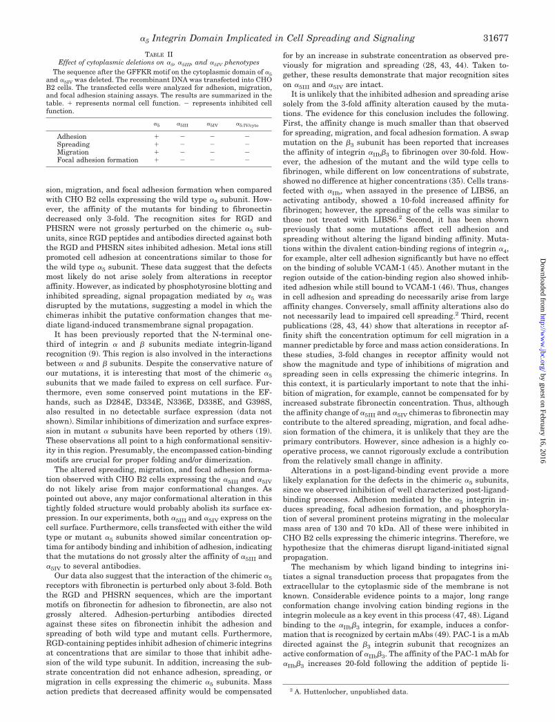
TABLE IIEffect of cytoplasmic deletions on a5, a5III, and a5IV phenotypes
The sequence after the GFFKR motif on the cytoplasmic domain of a5and a5IV was deleted. The recombinant DNA was transfected into CHOB2 cells. The transfected cells were analyzed for adhesion, migration,and focal adhesion staining assays. The results are summarized in thetable. 1 represents normal cell function. 2 represents inhibited cellfunction.
a5 a5III a5IV a5:IV/cyto
Adhesion 1 2 2 2Spreading 1 2 2 2Migration 1 2 2 2Focal adhesion formation 1 2 2 2
a5 Integrin Domain Implicated in Cell Spreading and Signaling 31677
by guest on February 16, 2016http://w
ww
.jbc.org/D
ownloaded from

gands (50). This change has been hypothesized to propagate tothe cytoplasmic domain via a “hinge” where the membraneproximal regions of the a and b subunit cytoplasmic domainsreside.
The regions swapped in our chimeras represent regions in ornear putative cation binding motifs. Our domain III encom-passes a cation-binding site, while domain IV is adjacent toone. These regions are sensitive to conformational changes andimplicated in regulating the affinity of integrin-ligand binding(22, 51, 52). Changes in the cations bound to integrins alter theaffinity of several integrins and can expose new epitopes. Re-placing Ca21 with Mn21, for example, increases the affinity ofseveral different integrins, while the binding of Mg21 exposesan epitope on integrin aLb2 (22, 51, 52). The affinity of integrinscan also be affected through alterations of the cytoplasmicdomains or by signals from the cytoplasm (53, 54). Thus, con-formational information appears to travel reciprocally from thecytoplasmic domains to the ligand-binding site and vice versa.The cation binding domain appears to lie on this pathway.
How might the domains III and IV participate in the confor-mation changes that mediate ligand-initiated signal transduc-tion? The b-propeller model provides potential insights (15). Inthis model, the N-terminal region of the integrin a subunitfolds into a b-propeller domain, which is composed of sevenfour-stranded b-sheet structures. It has been proposed thatintegrins bind to ligands and a putative Mg21 ion at the upperface of the propeller. The Ca21 binding motifs are predicted tobe on the lower face of the propeller. Domain III in our studyencompasses strand 1 and part of strand 2 of the fifth b-sheetstructure and is predicted to interact with Ca21 at the lowerface of the propeller. Domain IV contains part of strands 2 and3 of the fifth b-sheet and is located on top of the propeller,possibly at or close to the interface between the ligand and theintegrin. One hypothesis is that when a5b1 interacts with fi-bronectin, domain IV, which is adjacent to the ligand bindingpocket, undergoes a conformational change and causes achange in domain III, which in turn propagates the alterationtoward to the cytoplasmic domain. The metal ion associatedwith domain III helps to stabilize the proper conformation orassist in the transformation. Domains III and IV thereforeconstitute a mediator that directs the signal from ligand-rec-ognition site to the cytoplasm. Mutations in a5III and a5IV
would disrupt such a pathway and result in attenuated signal-ing. It is likely that the signal-propagation domain(s) would becoupled to the ligand-binding domain in the sterically con-strained environment that appear to characterize integrin ex-tracellular domains. Considering the proximity of domains IIIand IV to the ligand-binding site, mutations in this region mayalso cause a small conformational alteration of the ligand-binding pocket, which could produce the 3-fold effects that weobserve.
A related hypothesis is that domains III and IV reside at oradjacent to the interface between the a and b subunits. It isknown that ligands and divalent cations induce a conforma-tional change in the integrin heterodimer, and the interactionbetween the a and b subunit may be involved in this confor-mational change. In the model discussed above, the b-propellerdomain on the a subunit interacts with the b subunit (55). Ithas also been reported that aIIbb3 subunits dissociate when theCa21 is removed (56). In this context, domains III and IV in ourstudy may be part of the interface between the subunits. In thislocation, the a5IIIb1 and aIVb1 chimeras would inhibit intersub-unit conformation changes that mediate ligand-initiated signaltransduction.
The mechanisms by which ligand binding to the extracellulardomain couples to the cytoplasmic responses that mediate ad-
hesion, spreading, and tyrosine phosphorylation are anenigma. The available evidence points to ligand occupancy andreceptor aggregation being required for a complete response(38, 39, 57). Ligand binding alone is sufficient to target inte-grins to focal adhesions but probably not to assemble them (39).Similarly, receptor aggregation alone, while sufficient to pro-mote the association of a large number of signaling molecules,does not lead to focal adhesion formation. Focal adhesion as-sembly appears to require both ligation and aggregation (58).Considerable evidence also points to the cytoplasmic domain ofseveral integrin b subunits as critical for both integrin-medi-ated tyrosine phosphorylation and association with focal adhe-sion molecules. The a subunits, in turn, appear to modulate thefunctionality of the b subunit cytoplasmic domain, perhapsthrough a steric interaction (59, 60). In this context, it is inter-esting that removing the a5 cytoplasmic domain from the mu-tants does not alter the phenotype of the a5IV chimera. Itunderscores a difference between the ability to localize in focaladhesions and to form or organize focal adhesions. We can onlyspeculate on why our mutations alter the ability to organizefocal adhesions. Perhaps ligand binding in the mutants resultsin inhibited receptor aggregation or accessibility of the b1 cy-toplasmic domain. In either case, these mutants should be veryuseful for understanding the roles of a5 subunit structure andfunction in adhesion-induced signaling in a variety of cellularprocesses.
Acknowledgments—We thank R. Juliano for providing CHO B2 cellline, D. Mosher for fibronectin, and L. Reichardt for the a5 and a6cDNAs. We also thank M. Humphries for useful discussions, M. Warefor comments on the manuscript, Q. Zhang for discussion on fibronectinbinding, and Margot Lakonishok for advice throughout the study.
REFERENCES
1. Hynes, R. (1992) Cell 69, 11–252. Hemler, M. E. (1990) Annu. Rev. Immunol. 8, 365–4003. Springer, T. A. (1994) Cell 76, 301–3044. Ginsberg, M. H., Du, X., and Plow, E. F. (1992) Curr. Opin. Cell. Biol. 4,
766–7715. Humphries, M. J. (1996) Curr. Opin. Cell. Biol. 8, 632–6406. Mould, A. P. (1996) J. Cell Sci. 109, 2613–26187. Hughes, P. E., Diaz-Gonzalez, F., Leong, L., Wu, C., McDonald, J. A., Shattil,
S. J., and Ginsberg, M. H. (1996) J. Biol. Chem. 271, 6571–65748. Sastry, S. K., and Horwitz, A. F. (1993) Curr. Opin. Cell. Biol. 5, 819–8319. Loftus, J. C., Smith, J. W., and Ginsberg, M. H. (1994) J. Biol. Chem. 269,
25235–2523810. D’Souza, S. E., Ginsberg, M. H., Burke, T. A., Lam, S. C., and Plow, E. F. (1988)
Science 242, 91–9311. Smith, J. W., and Cheresh, D. A. (1988) J. Biol. Chem. 263, 18726–1873112. Loftus, J. C., O’Toole. T., Plow, E. F., Glass, A., Frelinger, A. L. D., and
Ginsberg, M. H. (1990) Science 249, 915–91813. Takada, Y., Ylanne, J., Mandelman, D., Puzon, W., and Ginsberg, M. H. (1992)
J. Cell Biol. 119, 913–92114. Bajt, M. L., and Loftus, J. C. (1994) J. Biol. Chem. 269, 20913–2091915. Springer, T. A. (1997) Proc. Natl. Acad. Sci. U. S. A. 94, 65–7216. Smith, J. W., and Cheresh, D. A. (1990) J. Biol. Chem. 265, 2168–217217. D’Souza, S. E., Ginsberg, M. H., Burke, T. A., and Plow, E. F. (1990) J. Biol.
Chem. 265, 3440–344618. Irie, A., Kamata, T., Puzon-McLaughlin, W., and Takada, Y. (1995) EMBO J.
14, 5550–555619. Masumoto, A., and Hemler, M. E. (1993) J. Cell Biol. 123, 245–25320. D’Souza, S. E., Ginsberg, M. H., Matsueda, G. R., and Plow, E. F. (1991) Nature
350, 66–6821. Paula, S., Bates, P. A., Harvey, J., Bennett, R. I., and Hogg, N. (1994) EMBO
J. 13, 1790–179822. Mould, A. P., Akiyama, S. K., and Humphries, M. J. (1995) J. Biol. Chem. 270,
26270–2627723. Humphries, M. J. (1990) J. Cell Sci. 97, 585–59224. Mould, A. P., Garatt, A. N., Askari, J. A., Akiyama, S. K., and Humphries,
M. J. (1995) Biochem. Soc. Trans. 23, (suppl.) 39525. Schreiner, C. L., Bauer, J. S., Danilov, Y. N., Hussein, S., Sczekan, M. M., and
Juliano, R. L. (1989) J. Cell Biol. 109, 3157–316726. Gunning, P., Leavitt, J., Muscat, G., Ng, S. Y., and Kedes, L. (1987) Proc. Natl.
Acad. Sci. U. S. A. 84, 4831–483527. Reszka, A. A., Hayashi, Y., and Horwitz, A. F. (1992) J. Cell Biol. 117,
1321–133028. Huttenlocher, A., Ginsberg, M. H., and Horwitz, A. F. (1996) J. Cell Biol. 134,
1551–156229. Humphries, M. J., Akiyama, S. K., Komoriya, A., Olden, K., and Yamada,
K. M. (1986) J. Cell Biol. 103, 2637–264730. Puzon-McLaughlin, W., and Takada, Y. (1996) J. Biol. Chem. 271,
20438–20443
a5 Integrin Domain Implicated in Cell Spreading and Signaling31678
by guest on February 16, 2016http://w
ww
.jbc.org/D
ownloaded from

31. Akiyama, S. K., and Yamada, K. M. (1985) J. Biol. Chem. 260, 4492–450032. Chou, P. Y., and Fasman, G. D. (1978) Annu. Rev. Biochem. 47, 251–27633. Robson, B., and Suzuki, E. (1976) J. Mol. Biol. 107, 327–35634. Garnier, J., Osguthorpe, D. J., and Robson, B. (1978) J. Mol. Biol. 120, 97–12035. Bajt, M. L., Loftus, J. C., Gawaz, M. P., and Ginsberg, M. H. (1992) J. Biol.
Chem. 267, 22211–2221636. Aota, S., Nomizu, M., and Yamada, K. M. (1994) J. Biol. Chem. 269,
24756–2476137. Nagai, T., Yamakawa, N., Aota, S., Yamada, S. S., Akiyama, S. K., Olden, K.,
and Yamada, K. M. (1991) J. Cell Biol. 114, 1295–130538. Yamada, K. M., and Miyamoto, S. (1995) Curr. Opin. Cell Biol. 7, 681–68939. LaFlamme, S. E., Thomas, L. A., Yamada, S. S., and Yamada, K. M. (1994)
J. Cell Biol. 126, 1287–129840. Otey, C. A., Pavalko, F. M., and Burridge, K. (1990) J. Cell Biol. 111, 721–72941. Horwitz, A. F., Duggan, K., Buck, C., Becklerle, M. C., and Burridge, K. (1985)
Nature 320, 531–53342. Schaller, M. D., Otey, C. A., Hildebrand, J. D., and Parsons, J. T. (1995) J. Cell
Biol. 130, 1181–118743. Palecek, S. P., Loftus, J. C., Ginsberg, M. H., Lauffenburger, D. A., and
Horwitz, A. F. (1997) Nature 385, 537–53944. Palecek, S. P., Schmidt, C. E., Lauffenburger, D. A., and Horwitz, A. F. (1996)
J. Cell Sci. 109, 941–95245. Pujades, C., Alon, R., Yauch, R. L., Masumoto, A., Burkly, L. C., Chen, C.,
Springer, T. A., Lobb, R. R., and Hemler, M. E. (1997) Mol. Biol. Cell 8,2647–2657
46. Ma, L., Conrad, P. J., Webb, D. L., and Blue, M. L. (1995) J. Biol. Chem. 270,18401–18407
47. Pelletier, A. J., Kunicki, T., Ruggeri, Z. M., and Quaranta, V. (1995) J. Biol.
Chem. 270, 18133–1814048. Fox, J. E. (1994) Annu. N. Y. Acad. Sci. 714, 75–8749. Mazurov, A. V., Khaspekova, S. G., Byzova, T. V., Tikhomirov, O., Berndt,
M. C., Steiner, B., and Kouns, W. C. (1996) FEBS Lett. 391, 84–8850. Du, X., Gu, M., Weisel, J. W., Nagaswami, C., Bennett, J. S., Bowditch, R., and
Ginsberg, M. H. (1993) J. Biol. Chem. 268, 23087–2309251. Dransfield, I., Cabanas, C., Craig, A., and Hogg, N. (1992) J. Cell Biol. 116,
219–22652. Hogg, N., Harvey, J., Cabanas, C., and Landis, R. C. (1993) Am. Rev. Respir.
Dis. 148, S55–S5953. Hughes, P. E., O’Toole. T. E., Ylanne, J., Shattil, S. J., and Ginsberg, M. H.
(1995) J. Biol. Chem. 270, 12411–1241754. O’Toole, T. E., Katagiri, Y., Faull, R. J., Peter, K., Tamura, R., Quaranta, V.,
Loftus, J. C., Shattil, S. J., and Ginsberg, M. H. (1994) J. Cell Biol. 124,1047–1059
55. Huang, C., and Springer, T. A. (1997) Proc. Natl. Acad. Sci. U. S. A. 94,3162–3167
56. Jennings, L. K., and Philips, D. R. (11982) J. Biol. Chem. 257, 10458–1046657. Shwartz, M. A., Schaller, M. D., and Ginsberg, M. H. (1995) Annu. Rev. Cell.
Dev. Biol. 11, 549–59958. Miyamoto, S., Akiyama, S. K., and Yamada, K. M. (1995) Science 267, 883–88559. Briesewitz, R., Kern, A., Smilenov, L. B., David, F. S., and Marcantonio, E. E.
(1996) Mol. Biol. Cell 7, 1499–150960. Ylanne, J., Chen, Y., O’Toole. T. E., Loftus, J. C., Takada, Y., and Ginsberg,
M. H. (1993) J. Cell Biol. 122, 223–23361. Ruoslanti, E., Hayman, E. G., Pierschbacher, M., and Engwall, E. (1982)
Methods Enzymol. 82, 803–831
a5 Integrin Domain Implicated in Cell Spreading and Signaling 31679
by guest on February 16, 2016http://w
ww
.jbc.org/D
ownloaded from

Zuojun Cao, Kun Huang and Alan F. Horwitzand Signaling
Subunit Implicated in Cell Spreading5αIdentification of a Domain on the Integrin
doi: 10.1074/jbc.273.48.316701998, 273:31670-31679.J. Biol. Chem.
http://www.jbc.org/content/273/48/31670Access the most updated version of this article at
Alerts:
When a correction for this article is posted•
When this article is cited•
to choose from all of JBC's e-mail alertsClick here
http://www.jbc.org/content/273/48/31670.full.html#ref-list-1
This article cites 61 references, 40 of which can be accessed free at
by guest on February 16, 2016http://w
ww
.jbc.org/D
ownloaded from
Copyright © 2022 FDOKUMEN