Autonomic Dysreflexia After Spinal Cord Injury - UKnowledge
33
University of Kentucky University of Kentucky UKnowledge UKnowledge Physiology Faculty Publications Physiology 1-2018 Autonomic Dysreflexia After Spinal Cord Injury: Systemic Autonomic Dysreflexia After Spinal Cord Injury: Systemic Pathophysiology and Methods of Management Pathophysiology and Methods of Management Khalid C. Eldahan University of Kentucky, [email protected] Alexander G. Rabchevsky University of Kentucky, [email protected] Follow this and additional works at: https://uknowledge.uky.edu/physiology_facpub Part of the Neuroscience and Neurobiology Commons, and the Physiology Commons Right click to open a feedback form in a new tab to let us know how this document benefits you. Right click to open a feedback form in a new tab to let us know how this document benefits you. Repository Citation Repository Citation Eldahan, Khalid C. and Rabchevsky, Alexander G., "Autonomic Dysreflexia After Spinal Cord Injury: Systemic Pathophysiology and Methods of Management" (2018). Physiology Faculty Publications. 136. https://uknowledge.uky.edu/physiology_facpub/136 This Article is brought to you for free and open access by the Physiology at UKnowledge. It has been accepted for inclusion in Physiology Faculty Publications by an authorized administrator of UKnowledge. For more information, please contact [email protected].
-
Upload
khangminh22 -
Category
Documents
-
view
0 -
download
0
Transcript of Autonomic Dysreflexia After Spinal Cord Injury - UKnowledge
University of Kentucky University of Kentucky
UKnowledge UKnowledge
Physiology Faculty Publications Physiology
1-2018
Autonomic Dysreflexia After Spinal Cord Injury: Systemic Autonomic Dysreflexia After Spinal Cord Injury: Systemic
Pathophysiology and Methods of Management Pathophysiology and Methods of Management
Khalid C. Eldahan University of Kentucky, [email protected]
Alexander G. Rabchevsky University of Kentucky, [email protected]
Follow this and additional works at: https://uknowledge.uky.edu/physiology_facpub
Part of the Neuroscience and Neurobiology Commons, and the Physiology Commons
Right click to open a feedback form in a new tab to let us know how this document benefits you. Right click to open a feedback form in a new tab to let us know how this document benefits you.
Repository Citation Repository Citation Eldahan, Khalid C. and Rabchevsky, Alexander G., "Autonomic Dysreflexia After Spinal Cord Injury: Systemic Pathophysiology and Methods of Management" (2018). Physiology Faculty Publications. 136. https://uknowledge.uky.edu/physiology_facpub/136
This Article is brought to you for free and open access by the Physiology at UKnowledge. It has been accepted for inclusion in Physiology Faculty Publications by an authorized administrator of UKnowledge. For more information, please contact [email protected].

Autonomic Dysreflexia After Spinal Cord Injury: Systemic Pathophysiology and Autonomic Dysreflexia After Spinal Cord Injury: Systemic Pathophysiology and Methods of Management Methods of Management
Digital Object Identifier (DOI) https://doi.org/10.1016/j.autneu.2017.05.002
Notes/Citation Information Notes/Citation Information Published in Autonomic Neuroscience: Basic & Clinical, v. 209, p. 59-70.
© 2017 Elsevier B.V. All rights reserved.
This manuscript version is made available under the CC‐BY‐NC‐ND 4.0 license https://creativecommons.org/licenses/by-nc-nd/4.0/.
The document available for download is the author's post-peer-review final draft of the article.
This article is available at UKnowledge: https://uknowledge.uky.edu/physiology_facpub/136

Autonomic Dysreflexia after Spinal Cord Injury: Systemic Pathophysiology and Methods of Management
Khalid C. Eldahana,b and Alexander G. Rabchevskya,b,*
aDepartment of Physiology, University of Kentucky, Lexington, KY 40536, United States
bSpinal Cord and Brain Injury Research Center, University of Kentucky, Lexington, KY 40536, United States
Abstract
Traumatic spinal cord injury (SCI) has widespread physiological effects beyond the disruption of
sensory and motor function, notably the loss of normal autonomic and cardiovascular control.
Injury at or above the sixth thoracic spinal cord segment segregates critical spinal sympathetic
neurons from supraspinal modulation which can result in a syndrome known as autonomic
dysreflexia (AD). AD is defined as episodic hypertension and concomitant baroreflex-mediated
bradycardia initiated by unmodulated sympathetic reflexes in the decentralized cord. This
condition is often triggered by noxious yet unperceived visceral or somatic stimuli below the
injury level and if severe enough can require immediate medical attention. Herein, we review the
pathophysiological mechanisms germane to the development of AD, including maladaptive
plasticity of neural circuits mediating abnormal sympathetic reflexes and hypersensitization of
peripheral vasculature that collectively contribute to abnormal hemodynamics after SCI. Further,
we discuss the systemic effects of recurrent AD and pharmacological treatments used to manage
such episodes. Contemporary research avenues are then presented to better understand the relative
contributions of underlying mechanisms and to elucidate the effects of recurring AD on
cardiovascular and immune functions for developing more targeted and effective treatments to
attenuate the development of this insidious syndrome following high-level SCI.
Keywords
maladaptive plasticity; sprouting; sympathetic; hypertension; propriospinal; primary afferent
1. Introduction
In addition to motor and sensory deficits, traumatic spinal cord injury (SCI) causes a
constellation of interrelated autonomic and cardiovascular abnormalities. Cardiovascular
*Corresponding author: University of Kentucky, Spinal Cord and Brain Injury Research Center (SCoBIRC), B471, Biomedical and Biological Sciences Research Building, 741 South Limestone Street, Lexington, KY 40536-0509. [email protected].
Publisher's Disclaimer: This is a PDF file of an unedited manuscript that has been accepted for publication. As a service to our customers we are providing this early version of the manuscript. The manuscript will undergo copyediting, typesetting, and review of the resulting proof before it is published in its final citable form. Please note that during the production process errors may be discovered which could affect the content, and all legal disclaimers that apply to the journal pertain.
Conflicts of interest: none.
HHS Public AccessAuthor manuscriptAuton Neurosci. Author manuscript; available in PMC 2019 January 01.
Published in final edited form as:Auton Neurosci. 2018 January ; 209: 59–70. doi:10.1016/j.autneu.2017.05.002.
Author M
anuscriptA
uthor Manuscript
Author M
anuscriptA
uthor Manuscript

complications secondary to SCI are among the leading causes of mortality and morbidity in
this population, underscoring the necessity to understand and properly manage resultant
comorbidities (Cragg et al., 2013; Garshick et al., 2005; Myers et al., 2007; Sabre et al.,
2013). In humans, SCI at or above the sixth thoracic (T6) spinal cord segment often results
in the development of a potentially life-threatening syndrome called autonomic dysreflexia
(AD). AD is clinically defined as acute hypertension generated by unmodulated sympathetic
reflexes below the injury level that is often accompanied by baroreceptor-mediated
bradycardia, which provides short-term control of blood pressure (Karlsson, 1999). In
response to hypertension, the baroreflex system lowers blood pressure by reducing heart rate
and decreasing activity of vasoconstrictor sympathetic preganglionic neurons (SPN) located
throughout the thoracolumbar spinal cord that regulate peripheral vascular resistance.
However, while vagal parasympathetic innervation of the heart remains intact after SCI, the
disruption of descending vasomotor pathways to SPN produces an incomplete compensatory
decrease in peripheral vascular resistance so that hypertension persists until the triggering
stimulus is removed (see section 3.1).
Typically, AD is precipitated by noxious visceral or somatic stimulation below the level of
injury that activates a massive sympathetic reflex causing widespread vasoconstriction.
While the most common triggers are over-distension of the bowel or bladder (Canon et al.,
2015; Lindan et al., 1980; Snow et al., 1978), other noxious stimuli including skin
lacerations, ingrown toenails, pressure sores, tight clothing and certain medical procedures
such as bladder catheterization and cystometry are also reported to cause AD (reviewed in
Karlsson, 1999). During an episode of AD, arterial blood pressure can reach devastating
levels, with systolic values as high as 325 mmHg (McBride et al., 2003), exemplifying that
AD is a hypertensive crisis that requires immediate medical attention (Muzumdar, 1982;
Showkathali et al., 2007; Verghese, 1989). Severe cases that do not receive rapid and
appropriate treatment can have serious consequences such as hypertensive encephalopathy,
stroke, cardiac arrest, seizure and even death (Bjelakovic et al., 2014; Colachis et al., 1997;
Eltorai et al., 1992;Fausel et al., 2014; Jain et al., 2013; Valles et al., 2005). Bouts of AD can
arise multiple times daily due to the noxious yet unperceived afferent stimulation produced
by normal, intermittent filling of the bladder and bowels (Fougere et al., 2016; Hubli et al.,
2015; Popok et al., 2016). In light of this, it is perhaps not surprising that eliminating AD is
one of the highest priorities of the SCI population, based on a large national survey
(Anderson, 2004) which reported that both quadriplegics and paraplegics prioritize the
recovery of bowel/bladder function and elimination of AD over regaining walking
movements, highlighting the need for research strategies to mitigate the development of AD
altogether.
In this review, we provide a clinical description of AD along with its pharmacological
management, and discuss the underlying pathophysiological changes that contribute to such
dangerous, episodic hypertension after high-level SCI. We further describe recent studies
revealing body-wide disturbances that result from chronic recurring episodes of AD,
including vascular, cardiac and immunological dysfunctions. Contemporary research
strategies will be considered to understand more comprehensively the underlying
mechanisms, the full physiological impact of this syndrome that typically occurs multiple
times daily, and potential therapeutic approaches to abrogate its development.
Eldahan and Rabchevsky Page 2
Auton Neurosci. Author manuscript; available in PMC 2019 January 01.
Author M
anuscriptA
uthor Manuscript
Author M
anuscriptA
uthor Manuscript

2. Clinical Description of AD
2.1. Who is at risk for developing AD?
The T6 spinal segment is critical to the development of AD (Lindan et al., 1980;Mathias et
al., 1988; Snow et al., 1978), as damage at or above this level interrupts descending
modulation of the thoracolumbar SPN that regulate vasomotor tone, notably in the extensive
splanchnic vascular bed (Blackmer, 2003; Gao et al., 2002). These vessels are innervated by
the splanchnic nerves arising from the T5-T12 levels (Loukas et al., 2010) and receive
approximately 25% of the cardiac output (Greenway et al., 1974; Rowell, 1990), which can
have a large influence on total peripheral resistance and blood pressure. There are, however,
uncommon reports of AD occurring after lesions below T6 but the magnitude of
hypertension and changes in heart rate tend to be relatively mild since some degree of
control over splanchnic sympathetic outflow remains intact (Moeller et al., 1973).
Not all individuals with SCI at or above the T6 level develop AD, with the prevalence
reported between 48% and 91% (Curt et al., 1997; Lindan et al., 1980; Snow et al., 1978).
This discrepancy is likely attributed to differences in the completeness of SCI, time elapsed
since injury, and differences in the criteria used to confirm the presence of AD used among
studies (Furusawa et al., 2011). Indeed, the clinical definition of AD is somewhat
inconsistent (see section 2.2). Interestingly, AD has been documented in cases of non-
traumatic abnormalities of the spinal cord such as intramedullary astrocytoma (Furlan et al.,
2003) and multiple sclerosis (Kulcu et al., 2009), indicating that disruption of descending
vasomotor pathways in any manner may contribute to the development of this syndrome.
2.2. Characteristic features of AD
The magnitude of hypertension required to be considered AD varies across studies. Snow et
al. (1978) classified AD in adults as an increase of 40 mmHg systolic blood pressure
whereas Popok et al. (2016) defined AD as an increase of 20 mmHg systolic blood pressure.
Others (Lindan et al., 1980) diagnosed AD as a sudden rise in both systolic and diastolic
blood pressure of any magnitude. Veteran’s Affairs guidelines recommend that AD in adults
is considered following abrupt elevation in systolic blood pressure of 20–40 mmHg above
baseline, whereas in pediatric SCI an increase of 15–20 mmHg systolic pressure warrants
consideration (Canon et al., 2015; Consortium for Spinal Cord, 2002).
In addition to elevated blood pressure, individuals with an acute episode of AD can
experience a diverse set of symptoms including debilitating headache, sweating, flushing of
the skin above the injury level, piloerection, stuffy nose, blurred vision and anxiety
(Karlsson, 1999). While these symptoms are not simultaneously present in all cases,
headache and sweating above the lesion occurs 88% of the time (Lindan et al., 1980).
Although the classical definition of AD is acute hypertension coincident with bradycardia
(Erickson, 1980; Guttmann et al., 1947; Trop et al., 1991), the importance of heart rate in the
diagnosis of AD is a matter of controversy. Lindan et al. (1980) reported an equal incidence
of bradycardia and tachycardia (increase in heart rate) in documented cases of AD, whereas
others report that tachycardia is more common (Hickey et al., 2004; Kewalramani, 1980;
Scott et al., 1978). Whether an episode of AD is concomitant with an increase or decrease in
Eldahan and Rabchevsky Page 3
Auton Neurosci. Author manuscript; available in PMC 2019 January 01.
Author M
anuscriptA
uthor Manuscript
Author M
anuscriptA
uthor Manuscript

heart rate may depend on the injury level (Collins et al., 2006; Karlsson, 1999; Krassioukov
et al., 2003). As suggested by Karlsson (1999), activation of sympathetic circuits in the
spinal cord below a cervical injury may propagate rostrally towards cardiac-innervating SPN
(i.e., T1-T4), explaining why tachycardia is frequently observed during AD. However, the
correlation between injury level and the direction of heart rate change during episodes of AD
has not been formally investigated.
2.3 Temporal development of AD after injury
AD most often presents in the chronic phase of SCI, with a majority of cases first occurring
3–6 months after injury in humans (Lindan et al., 1980). While it may also occur in earlier
stages of injury, the incidence of early AD is relatively low, with only 5.7% of individuals
with SCI above T6 having clinically documented AD within the first month post-injury
(Krassioukov et al., 2003). Though uncommon, the manifestation of AD in the early stage of
injury is significant considering that treatment of acute, high-level SCI often includes
pressor agents to help combat the profound hypotension associated with such injuries
(reviewed in Ploumis et al., 2010). In cases of acute AD occurring within days of injury,
systolic blood pressure as high as 210 mmHg has been reported (Silver, 2000), suggesting
that concurrent vasopressor support may compound damage during episodic hypertension.
In experimental SCI, the development of telemetric monitoring techniques, along with the
advent of computer algorithms capable of processing large amounts of hemodynamic data,
have allowed for detailed analysis of the temporal development of AD in conscious animals
(Laird et al., 2006; Mayorov et al., 2001; Rabchevsky et al., 2012; West et al., 2015). In both
rats and mice with high-thoracic SCI, spontaneous AD triggered by naturally occurring
stimuli emerges in a biphasic pattern with a transient surge of events occurring within the
first week, followed by a gradual rise in frequency beginning around 2-weeks post-injury
(Rabchevsky et al., 2012; West et al., 2015; Zhang et al., 2013). Moreover, the magnitude of
change in systolic pressure during these events increases over time (West et al., 2015). These
observations generally correspond to the development of AD in humans, where the initial
disruption to descending vasomotor pathways allows for early AD (Krassioukov et al., 2003)
before maladaptive changes in viscerosympathetic circuitry associated with more frequent
and severe cases occur during the more chronic stages (West et al., 2015). Recent
capabilities of screening ambulatory blood pressure recordings for daily AD events in
humans (Hubli et al., 2014; Popok et al., 2016) will enable detailed analyses of the temporal
progression of this syndrome in humans, as well as the efficacy of drugs to reduce the
frequency or magnitude of recurrent AD.
3. Mechanisms contributing to AD
3.1. Loss of supraspinal control over sympathetic preganglionic neurons
Within the intact nervous system, supraspinal vasomotor neurons residing in the
paraventricular nucleus, rostral ventrolateral medulla, rostral ventromedial medulla, caudal
raphe nuclei and A5 cell group (Calaresu et al., 1988; Chalmers et al., 1994; Hosoya et al.,
1991; Jansen et al., 1995; Llewellyn-Smith, 2009; Strack et al., 1989; Sved et al., 2001) send
projections to the intermediolateral cell column (IML), which is comprised of spinal nuclei
Eldahan and Rabchevsky Page 4
Auton Neurosci. Author manuscript; available in PMC 2019 January 01.
Author M
anuscriptA
uthor Manuscript
Author M
anuscriptA
uthor Manuscript

containing sympathetic preganglionic neurons (SPN) that extends throughout the T1-L2
segments (Pyner et al., 1994; Tang et al., 1995; Zagon et al., 1993). These supraspinal
neurons modulate the tonic firing of SPN which, in turn, send projections to the peripheral
sympathetic chain ganglia or directly to the adrenal medulla (see Figure 1). The sympathetic
ganglia act as the final sympathetic effector cells and innervate blood vessels throughout the
body, whereas stimulation of the adrenal medulla secretes epinephrine and norepinephrine
(NE) into the circulation. Together, this provides both direct and indirect control of blood
vessel diameter and peripheral resistance to facilitate hemodynamic homeostasis (reviewed
in Thomas, 2011).
After high-level SCI, the descending autonomic pathways responsible for supraspinal
modulation of SPN become interrupted, reducing sympathetic tone below the injury
(Stjernberg et al., 1986) and leaving SPN under the control of spinal influences alone. In the
initial “spinal shock” phase of injury, which can last for weeks in humans (Ditunno et al.,
2004), this loss of descending control manifests as significantly reduced blood pressure and
depression of sympathetic reflexes (Frankel et al., 1972). Over time, however, plasticity of
SPN and reorganization of spinal circuitry create a hyper-excitable state that contributes to
the aberrant reflex activation of SPN in response to afferent stimulation (see Figure 1)
(Krassioukov et al., 2002;Llewellyn-Smith et al., 2001; Rabchevsky, 2006). Because of the
interruption to descending modulatory pathways, which would normally inhibit the SPN
during hypertension via the baroreflex (Guyenet et al., 1981), AD persists until the stimulus
is withdrawn.
3.2 Synaptic reorganization of sympathetic preganglionic neurons (SPN)
The damage to descending vasomotor pathways caused by SCI leaves many SPN partially
denervated, causing a number of histological and functional changes in these neurons in both
the human and rat (Krassioukov et al., 1999; Krassioukov et al., 1995; Krassioukov et al.,
1996). Whereas SPN activity is normally regulated by a confluence of supraspinal and
intraspinal inputs, their ongoing activity after complete SCI depends solely on the influence
of spinal sympathetic interneurons (Schramm, 2006). Spinally derived sources of input to
SPN include interneurons residing in laminae V, VII and X (Cabot et al., 1994; Cano et al.,
2001; Clarke et al., 1998; Deuchars et al., 2001;Deuchars et al., 2015; Joshi et al., 1995;
Tang et al., 2004a). While there are no direct synaptic inputs to SPN from primary afferents,
it is thought that sensory neurons can influence the SPN via such spinal interneurons
(Laskey et al., 1988; Schramm, 2006). The integration of supraspinal and intraspinal inputs
on SPN is complex and involves both monosynaptic and polysynaptic pathways (reviewed in
Deuchars et al., 2015).
After experimental T4 spinal cord transection in adult rats, profound morphological changes
occur to SPN within the IML below the lesion. Within 3 days of injury, the dendritic length
and diameter of SPN soma decrease dramatically in response to the degeneration of
terminals with supraspinal origins (Llewellyn-Smith et al., 2001). By two-weeks, however,
the somatic size and dendritic arbor of SPN appears normal again (Krassioukov et al., 1996;
Krenz et al., 1998a). These dynamic changes in gross SPN morphology correspond
temporally with the evolution of cardiovascular dysfunction in rats with high-level SCI,
Eldahan and Rabchevsky Page 5
Auton Neurosci. Author manuscript; available in PMC 2019 January 01.
Author M
anuscriptA
uthor Manuscript
Author M
anuscriptA
uthor Manuscript

where pronounced resting hypotension occurs in the first days, followed by a gradual
increase in basal blood pressure and the appearance of recurrent AD by two weeks post-
injury (Laird et al., 2006; Mayorov et al., 2001; Rabchevsky et al., 2012; West et al., 2015).
While atrophied SPN regain a normal morphology in the weeks after injury, experimental
evidence suggests that a radical reorganization of synaptic inputs controlling their activity
occurs. Weaver et al. (1997) demonstrated altered expression patterns of GAP-43 (growth
associated protein-43), a marker of reactive sprouting, in both mature axons and growth
cones in close apposition to SPN caudal to a complete mid-thoracic transection weeks after
injury. Because of the complete transection model used and absence of degenerating
supraspinal axons in the IML after 7 days, the source of these GAP-43 immunoreactive
axons was limited to spinal neurons below the injury, suggesting reorganization of
intraspinal sympathetic circuits that may contribute to the exaggerated sympathetic reflex
during an AD event. Llewellyn-Smith et al., (2001) also observed a switch in the ratio of
synapses on SPN containing glutamate or γ-aminobutyric acid (GABA), indicating a shift in
the balance of excitatory and inhibitory influences, respectively. Specifically, Transection
caused a decrease in the percentage of glutamatergic inputs versus an increase in the
percentage of GABAergic inputs on SPN in the IML at the T8 spinal level after two-weeks,
implying a predominantly inhibitory synaptic integration. While this observation is
consistent with the resting hypotension and reduced sympathetic outflow seen after SCI,
how this relates to the intense sympathetic outbursts that occur in response to noxious
visceral stimulation is uncertain. Recent work by Huang et al. (2016) indicates that complete
high-thoracic (T2) spinal cord transection in rats causes GAB A neurotransmission to
convert from an inhibitory to excitatory role in nociceptive circuits. While the influence of
SCI on GABAergic regulation of SPNs was beyond the scope of this work, it provides
evidence that GABAergic inputs onto SPN may provide excitatory drive after SCI
(Rabchevsky, 2006).
An alternative explanation may be that glutamatergic synapses on SPN are the predominate
input recruited by noxious visceral stimulation. It has been documented that glutamatergic
signaling through both NMDA and AMPA receptors is important for the initiation of AD in
response to noxious colorectal distension (CRD) (Maiorov et al., 1997). Ueno et al. (2016)
recently demonstrated that chemogenetic silencing of Vglut2+ (vesicular glutamate
transporter 2) interneurons after T3 SCI suppressed anti-inflammatory sympathetic reflexes
related to AD (see section 5.2). This is further supported by studies testing the anti-epileptic
and neuropathic pain medication gabapentin (GBP) as an experimental treatment for AD
(Rabchevsky et al., 2011). GBP is known to inhibit pre-synaptic glutamate release by
binding to the a251 subunit of voltage-gated calcium channels (Coderre et al., 2005; Coderre
et al., 2007; Gee et al., 1996; Shimoyama et al., 2000) and has been shown to reduce both
the magnitude and frequency of AD in rats with complete Transection, specifically after
acute but not chronic administration (Rabchevsky et al., 2011; Rabchevsky et al., 2012).
Notably, however, the site(s) of action and mechanism(s) through which GBP mitigates AD
remains uncertain. It is feasible that GBP acts by reducing glutamatergic transmission at the
level of both primary afferents entering the spinal cord as well as glutamatergic spinal
interneurons synapsing with SPN. GBP has also been reported to inhibit excitatory
synaptogenesis both in-vitro and in-vivo by blocking the binding of synaptogenic
Eldahan and Rabchevsky Page 6
Auton Neurosci. Author manuscript; available in PMC 2019 January 01.
Author M
anuscriptA
uthor Manuscript
Author M
anuscriptA
uthor Manuscript

thrombospondin proteins to the a251 subunit (Eroglu et al., 2009). This suggests that GBP
may work to reduce aberrant excitatory synapse formation in viscerosympathetic circuits
after SCI. However, the short 2–3 hour half-life of GBP in rats (Radulovic et al., 1995;
Vollmer et al., 1986) likely explains why once daily GBP treatment is insufficient to
significantly reduce the frequency of spontaneous AD in the hours following administration;
and likely insufficient to alter putative synaptic formation (Rabchevsky et al., 2012). Further
studies into the synaptic mechanisms controlling SPN during noxious stimulation, as well as
the precise means through which GBP mitigates AD may help identify novel therapeutic
targets for prophylactically preventing the development of this syndrome.
3.3 Primary afferent sprouting
Intraspinal sprouting of primary afferent nociceptive fibers is also thought to contribute to
the development of AD. After experimental SCI, the central arbors of unmyelinated c-fiber
afferents expressing calcitonin gene-related peptide (CGRP), a marker of nociceptive fibers,
sprout into the dorsal horn caudal to the injury (Hou et al., 2009; Krenz et al., 1998b). The
extent of CGRP+ fiber sprouting into the lumbosacral spinal cord correlates with the
magnitude of AD elicited by noxious CRD (Cameron et al., 2006; Krenz et al., 1999),
suggesting that enhanced sprouting of nociceptive fibers increases reflex activation of SPN
through propriospinal “relay” neurons (Figure 1, see section 3.4). Increased CGRP+ fiber
density becomes apparent two weeks after experimental injury (Hou et al., 2009; Krenz et
al., 1998b), a time at which AD develops reliably in spinal rats (Krassioukov et al., 1995;
Laird et al., 2006). Importantly, increased CGRP+ fiber sprouting in the dorsal horn is also
seen in the chronic stages of human SCI, and in one case has been associated with a well-
documented history of AD (Ackery et al., 2007).
The sprouting of c-fibers appears to be primarily dependent on nerve growth factor (NGF)
signaling. After transection or contusion SCI, resident neurons and glia upregulate the
expression of intraspinal NGF rostral and caudal to the lesion (Bakhit et al., 1991; Brown et
al., 2004). Blockade of NGF signaling through intrathecal delivery of anti-NGF antibodies
for two-weeks after T4-transection SCI effectively mitigates injury-induced sprouting of
CGRP+ fibers throughout the spinal cord. Furthermore, animals receiving anti-NGF therapy
had less severe AD in response to noxious CRD (Krenz et al., 1999), although this treatment
did not abolish it completely. Cameron et al. (2006) used a proof-of-principle approach to
overexpress NGF in the cord after T4-transection using adenovirus injections into either the
T13/L1 or L6/S1 spinal levels innervating the distal colon and found that the pressor and
bradycardic responses to CRD were significantly exacerbated. Conversely, injured cords
injected with adenovirus encoding semaphorin 3a, a specific chemorepellant for both c-
fibers and sympathetic fibers (Tang et al., 2004b), had significantly diminished pressor and
bradycardic responses during CRD. Such exacerbations or ameliorations were significantly
correlated with increased or decreased CGRP+ fiber sprouting, respectively.
3.4 Propriospinal plasticity
Plasticity of ascending lumbosacral propriospinal fibers that relay pelvic sensory
information rostrally towards SPN in the thoracic cord is also thought to contribute to the
development of AD. There is compelling evidence for functional plasticity of propriospinal
Eldahan and Rabchevsky Page 7
Auton Neurosci. Author manuscript; available in PMC 2019 January 01.
Author M
anuscriptA
uthor Manuscript
Author M
anuscriptA
uthor Manuscript

interneurons which comprise spinal sympathetic circuits after transection SCI in the rat.
Krassioukov et al. (2002) investigated the response of sympathetically-correlated
interneurons to both noxious and innocuous stimuli hours following T3 transection (acute)
as well as after one month (chronic). In the chronic but not acute stage of injury,
interneurons whose electrical activity was cross-correlated with that of simultaneously
recorded renal sympathetic nerve activity were excited by noxious CRD and cutaneous
pinching, as well as non-noxious brushing of the skin caudal to the injury. This seminal
study showed that in the weeks after injury, plasticity occurs within somatosensory relay
neurons that influence the activity of SPN.
This electrophysiological evidence was supported histologically following anterograde
tracer injections into the lumbosacral cord after complete T4-transection which significantly
increased labeling of ascending propriospinal fibers originating in the dorsal gray
commissure (DGC), notably found in juxtaposition to thoracic SPN labeled with Fluorogold
(Hou et al., 2008). The DGC is a region in which pelvic visceral afferents terminate and
send relay projections rostrally toward supraspinal targets (Al-Chaer et al., 1996; Hosoya et
al., 1994; Matsushita, 1998; Pascual et al., 1993; Vizzard, 2000). Hou et al. (2008) found
that a single session of prolonged, intermittent CRD performed two weeks after injury
caused enhanced neuronal activation throughout the lumbosacral DGC, as indicated by
increased expression of the immediate early gene c-fos in comparison to sham animals in
which intact descending modulation prevents such expression. It remains unclear, however,
whether the DGC neurons project directly to SPN or whether they project to excitatory
interneurons to indirectly modulate the excitability of SPN (Rabchevsky, 2006).
Collectively, these findings support a model of AD in which plasticity of ascending
propriospinal fibers, in conjunction with sprouting of primary afferents, creates an
amplification of afferent stimuli below the injury such that noxious inputs are more likely to
activate SPN to initiate unmodulated sympathetic reflexes resulting in an episode of AD
(Figure 1). Previous technical limitations made it difficult to directly test the
neuroanatomical basis of the AD reflex in live animals. However, with the recent
development of elegant genetic and chemogenetic silencing techniques (Kinoshita et al.,
2012; Stachniak et al., 2014; Zhu et al., 2014), it may now be feasible to directly assess the
role of specific populations of ascending propriospinal neurons in conveying lumbosacral
afferent input to rostral segments containing cardiovascular SPN (Figure 1, see section 6).
3.5 Peripheral adrenergic hypersensitivity
In addition to maladaptive plasticity of viscerosympathetic circuitry, there is also evidence of
peripheral changes responsible for the exaggerated pressor response to afferent stimulation.
In quadriplegics, resting blood pressure and plasma catecholamine levels are both
significantly lower than in intact subjects (Mathias et al., 1976). This is consistent with the
observation that sympathetic outflow below the lesion is lower both acutely and chronically
after SCI (Mathias et al., 1979; Wallin et al., 1984), which would be expected to produce
lower concentrations of circulating catecholamines (Goldstein et al., 1983). However, it has
been demonstrated that the vasomotor response to intravenously infused NE is enhanced in
quadriplegics with a documented history of AD. Specifically, by measuring the diameter of
Eldahan and Rabchevsky Page 8
Auton Neurosci. Author manuscript; available in PMC 2019 January 01.
Author M
anuscriptA
uthor Manuscript
Author M
anuscriptA
uthor Manuscript

the dorsal foot vein before and after local intravenous administration of NE, Arnold et al.
(1995) found that the concentration of NE required to induce a 50% reduction in resting
vessel diameter was significantly lower in tetraplegics compared to normal controls (1.6
mg/min vs. 10.9 ng/min). Similarly, Krum et al. (1992b) found that the dose of intravenously
infused phenylephrine (a peripheral α1-adrenergic receptor agonist) required to produce a
pressor response of 20 mmHg is significantly lower in functionally complete quadriplegics
compared to normal individuals. Furthermore, Krum et al. (1992a) reported that even modest
increases in plasma NE during bladder cystometry after high-level SCI were sufficient to
produce a significant pressor response greater than 20 mmHg systolic. Taken together, these
studies indicate that SCI promotes hypersensitivity of the vasculature to catecholamines
released through aberrant, unmodulated sympathetic reflexes, and that this
hypersensitization may contribute to the severity of episodic hypertension after SCI.
There is indication that peripheral sensitization following SCI is limited to the vasculature
caudal to the lesion and not within rostral vascular beds in which central sympathetic control
remains intact. Following T4 transection in rats, Rummery et al. (2010) found evidence of
enhanced neurovascular transmission in the saphenous artery which receives sympathetic
outflow from the lower thoracolumbar cord. However, SCI-related neurovascular
potentiation was not seen in the median artery which receives sympathetic outflow from the
cord rostral to the lesion. Brock et al. (2006) similarly found that T4 transection in rats leads
to increased neurovascular transmission within the mesenteric vasculature, with perivascular
nerve stimulation producing five-fold increased contractions of mesenteric arteries seven
weeks after SCI. This increased reactivity of mesenteric arteries was blunted by the
adrenergic antagonist prazosin, further implicating that peripheral hypersensitization is
secondary to changes in adrenergic transmission after injury. While the mechanism of this
phenomenon is not fully known, Brock et al. (2006) suggest that decreased norepinephrine
reuptake may be a contributing factor. Alternatively, Al Dera et al. (2012) report that
complete SCI in rats also alters L-type calcium channels, with their activation playing a
larger role in smooth muscle contraction of tail arteries isolated after injury. Whether or not
these two mechanisms are unique to specific vascular beds caudal to the lesion remains
unclear.
Consistent with the hypothesis that SCI causes enhanced adrenergic sensitivity in peripheral
vasculature, Lee et al. (2016) demonstrated increased protein expression of the α1-
adrenergic receptor in the femoral artery of rats after T10 spinal contusion. Furthermore, this
increased receptor expression corresponded to an enhanced pressor response and
vasoconstriction of isolated femoral arteries in response to phenylephrine stimulation.
Whether or not the time-course of these changes corresponds to the timeline over which AD
develops in rodents or humans remains unknown. Moreover, it is unclear whether these
findings are applicable to AD since this syndrome does not develop after T10 contusion SCI.
Although there is compelling evidence that peripheral sensitization is important in the
pathophysiology of AD, many questions remain regarding the underlying cellular processes
and time-course over which adrenergic hyper-responsiveness develops, as well as the relative
contribution compared to central maladaptive plasticity discussed.
Eldahan and Rabchevsky Page 9
Auton Neurosci. Author manuscript; available in PMC 2019 January 01.
Author M
anuscriptA
uthor Manuscript
Author M
anuscriptA
uthor Manuscript

4. Clinical management of AD
A variety of non-pharmacological and pharmacological strategies can be used to treat AD.
Since AD often resolves once the inciting stimulus is removed, the current standard of care
recommends identifying and eliminating the inciting factors, when possible, before
pharmacological approaches are considered (Consortium for Spinal Cord, 2002). Immediate
measures involve assessment of resting arterial pressure and monitoring for other symptoms
associated with AD (see section 2.2). If hypertension is observed, it is advised to move
patients into an upright position to facilitate the lowering of arterial pressure through
hydrostatic redistribution of blood to the lower extremities. This postural maneuver causes a
reduction of blood pressure in high-level SCI patients (Krassioukov et al., 2006), although
the efficacy to alleviate hypertension specifically during AD has not yet been formally
investigated (Krassioukov et al., 2009). Since filling of the bladder and impaction of the
bowel are the most common triggers of AD (Lindan et al., 1980), bladder voiding or bowel
care routines should next be considered along with a general inspection for other possible
sources of noxious stimulation such as pressure sores, tight clothing and skin laceration.
Given that blood pressure can fluctuate rapidly in patients with AD, it is important to
monitor blood pressure every 2–5 minutes for resolution or exacerbation of hypertension
during physical examination (Consortium for Spinal Cord, 2002).
While some cases of AD are mild and resolve relatively easily, more severe cases can
produce profound levels of hypertension and have no readily identifiable cause.
Furthermore, it may be difficult to identify and remove the triggering stimuli, particularly for
individuals with high thoracic and cervical injuries who have limited dexterity and mobility.
Therefore, a variety of drugs and medical procedures have been employed for the control of
AD, primarily through managing hypertension. Some of the most common remedies are
briefly discussed below.
4.1 Nitrates
Organic nitrates, such as nitroglycerine paste, are currently the most commonly prescribed
medications for mitigating acute episodes of AD (Braddom et al., 1991; Caruso et al., 2015).
Nitrates are a class of drugs that are converted to or release nitric oxide (NO), an
endogenous molecule that induces smooth muscle relaxation in the vasculature (Moncada et
al., 1993). For acute episodes of AD, it is recommended to apply 2% nitroglycerin paste
onto the skin above the level of SCI (Braddom et al., 1991). Vasodilation and subsequent
reduction of blood pressure occurs rapidly after transdermal application and the paste can be
wiped off once the therapeutic effect has been achieved (Grobecker, 1990). Moreover, a
variety of oral and transdermal patch preparations are available and easy for patients and
caregivers to administer. In severe cases of AD that are difficult to control, notably in a
clinical setting, intravenous sodium nitroprusside may also be used to rapidly resolve
hypertension (Valles et al., 2005).
4.2 Nifedipine
Nifedipine is an L-type calcium (Ca2+) channel blocker that reduces the influx of Ca2+ into
vascular smooth muscle cells, leading to a reduction in peripheral resistance and
Eldahan and Rabchevsky Page 10
Auton Neurosci. Author manuscript; available in PMC 2019 January 01.
Author M
anuscriptA
uthor Manuscript
Author M
anuscriptA
uthor Manuscript

consequently blood pressure. Despite few controlled clinical trials, nifedipine has been
widely used to treat AD in SCI patients (Braddom et al., 1991; Caruso et al., 2015; Dykstra
et al., 1987; Esmail et al., 2002; Krassioukov et al., 2009; Thyberg et al., 1994). In a survey
of clinicians with extensive experience treating AD, Braddom et al., 1991) found that
nifedipine was the most commonly prescribed agent to manage minor or severe symptoms of
AD. More recently, nitrates have become slightly more prominent than nifedipine in treating
minor or severe episodes of AD (Caruso et al., 2015), likely stemming from safety concerns
regarding the use of nifedipine during hypertensive crises because it may overshoot the
therapeutic drop in blood pressure and cause severe hypotension and ischemia (Chobanian et
al., 2003). Although there are no formal descriptions in the literature of adverse effects
related to the use of nifedipine specifically for AD, it is possible that it may reduce arterial
pressure below the range for critical renal, cerebral and cardiac vascular beds to autoregulate
their perfusion, particularly since many with AD are predisposed to resting and orthostatic
hypotension (Furlan et al., 2008).
4.3 Prazosin
The adrenergic receptor antagonist, prazosin, is another drug used to treat AD (Krum et al.,
1992c; Phillips et al., 2015). Prazosin is an antihypertensive agent that works by specifically
blocking α1-adrenergic receptors located on peripheral vasculature (Cavero et al., 1980).
Unlike other hypertension medications, prazosin has little effect on cardiac function or
resting blood pressure (Jaillon, 1980), making it safer to use in patients with chronic
hypotension. Krum et al. (1992c) reported that, compared to placebo treatment, twice daily
prazosin significantly decreased the magnitude of hypertension and severity of secondary
symptoms during AD caused by a variety of genitourinary and colorectal stimuli. More
recently, Phillips et al. (2015) reported that prazosin reduced the magnitude of hypertension
during AD during sperm retrieval procedures involving penile vibrostimulation, which is a
well-documented cause of AD. Collectively, research on the use of prazosin indicates that it
may be useful as a prophylactic treatment for AD triggered by a variety of iatrogenic stimuli
(Krum et al., 1992c; Phillips et al., 2015) .
4.4 Botulinum Toxin
Conservative pharmacological approaches are not always effective, and some individuals
require more aggressive treatments (Krassioukov et al., 2009). One such treatment is
botulinum toxin (BTX), a neurotoxin derived from Clostridium botulinum that suppresses
neuromuscular transmission by cleaving synaptic proteins required for neurotransmitter
exocytosis (reviewed in Dolly, 2003). Due to its ability to temporarily cause detrusor muscle
paralysis by preventing parasympathetic post-ganglionic acetylcholine release, delivery of
BTX into the detrusor muscle has been used to treat bladder dysfunction secondary to SCI,
such as neurogenic detrusor overactivity (NDO) (Dykstra, 2003; Dykstra et al., 1988;
Orasanu et al., 2013; Schurch et al., 2005). As these conditions disrupt normal micturition
and can lead to noxious distension of the bladder, they are associated with the occurrence of
AD. While there have been no large-scale clinical trials for BTX as a prophylactic treatment
of AD, there is indication that this could be a safe and tenable approach to minimize AD
associated with bladder dysfunction and certain medical procedures such as cystoscopy.
Schurch et al. (2000) reported a disappearance of AD caused by bladder voiding in a small
Eldahan and Rabchevsky Page 11
Auton Neurosci. Author manuscript; available in PMC 2019 January 01.
Author M
anuscriptA
uthor Manuscript
Author M
anuscriptA
uthor Manuscript

subset of patients receiving BTX injections into the detrusor muscle to treat neurogenic
bladder, though the duration of this improvement was not specified. Recently, Fougere et al.
(2016) conducted a small clinical trial specifically to assess the efficacy of intradetrusor
injections of BTX to reduce bladder-related AD and observed a decrease in the severity of
AD during cystometric filling of the bladder. Moreover, ambulatory blood pressure
recordings revealed a decrease in the frequency of daily bladder-related AD for at least one
month, accompanied by an increase in quality of life measures. Such reports indicate that
BTX treatment may be a safe and potentially long-lasting means of prophylactically treating
bladder-related AD. Notably, the duration of effect after BTX treatment is limited due to a
combination of compensatory sprouting of peripheral nerve terminals and the gradual
recovery of synaptic proteins necessary for neurotransmitter release (Dolly, 2003). While the
duration of detrusor paresis is approximately 9 months (Schurch et al., 1996), it remains
unknown how long intradetrusor BTX treatment can provide relief of AD.
In addition to temporary paralysis of bladder smooth muscle, there is also evidence that
BTX treatment reduces AD by modulating sensory transmission into the spinal cord.
Apostolidis et al. (2005) reported that intradetrusor injections of BTX in patients with NDO
caused a decrease in the expression of both purinoceptors (P2X3) and capsaicin receptors
(TRPV1) in sensory fibers innervating the bladder, and that this decrease was correlated
temporally with a significant improvement in bladder function. These receptors are involved
in bladder nociception (Birder et al., 2002; Cockayne et al., 2000), suggesting that decreased
afferent signaling contributes to the improvement in urodynamic function after BTX
treatment. In an experimental model, Elkelini et al. (2012) instilled BTX into the bladder of
rats after T4-transection and observed a significant reduction in the magnitude of AD
induced by bladder distension during cystometry, which was associated with reduced NGF
expression in the bladder and sensory neurons in the dorsal root ganglia. As NGF regulates
c-fiber density in the bladder (Schnegelsberg et al., 2010), it is also possible that BTX
reduces bladder-related AD by decreasing the innervation or distribution of nociceptive
fibers. Notably, intradetrusor BTX treatment has also been used to alleviate refractory AD in
a pediatric SCI patient (Lockwood et al., 2016).
5. Systemic effects of recurrent AD
5.1 Cardiovascular Changes
Emerging evidence suggests that the recurrence of AD, which can occur more than 40 times
a day for some individuals (Hubli et al., 2015), adversely affects multiple physiological
systems over time. Alan et al. (2010) demonstrated that repeated instances of AD after
experimental SCI exacerbate injury-induced peripheral vascular dysfunction. Isolated
mesenteric arteries from rats which underwent 30-minutes of continuous CRD daily for two-
weeks after T3 transection SCI had a more pronounced vasoconstrictor response to
phenylephrine (an α1-adrenergic agonist) compared to arteries from animals with SCI only.
This suggests that injury-induced adrenergic hypersensitization, which itself is a mechanism
involved in the etiology of AD (see section 3.5), is exacerbated by repeated instances of AD
and may help explain why the severity of AD increases over time (West et al., 2015). If this
is the case, it should be possible to dampen the temporal increase in AD severity by
Eldahan and Rabchevsky Page 12
Auton Neurosci. Author manuscript; available in PMC 2019 January 01.
Author M
anuscriptA
uthor Manuscript
Author M
anuscriptA
uthor Manuscript

pharmacologically controlling adrenergic hypersensitization. However, as discussed in
section 3.5, the cellular processes leading to adrenergic hyper-responsiveness have yet to be
elucidated.
Repetitive surges in blood pressure from recurrent AD may also induce maladaptive
structural changes in peripheral vasculature (West et al. (2013). Vascular remodeling is a
dynamic process that responds to changes in shear stress from normal hemodynamic
fluctuations as well as hypertensive disease (Baeyens et al., 2015; Schiffrin, 2004; Silver et
al., 2006). While SCI alone is associated with changes in vascular structure, this is believed
to be an adaptation to the decreased metabolic demands of atrophied tissue below the injury
(Olive et al., 2003). The hypothesis that blood pressure fluctuations from recurrent AD cause
further remodeling of peripheral vasculature requires additional study, particularly since
structural changes to resistance vessels, such as alterations in the ratio of wall thickness/
lumen diameter, are strongly associated with cardiovascular disease (Rizzoni et al., 2003).
Deleterious changes in cardiac function are also associated with daily repeated bouts of
experimental AD. West et al. (2016) investigated the effects of recurrent AD on cardiac
structure and function in rats following T3 transection. Beginning 2-weeks after injury, rats
received 60-minutes of repetitive CRD daily for 4 weeks. In-vivo echocardiography revealed
that animals with daily induced AD (SCI-AD) had diminished basal contractility and their
hearts developed significantly lower left ventricular pressure with a slower rate of ventricular
contraction. These alterations in cardiac mechanics were accompanied by a dampened
inotropic response to β-adrenergic stimulation during isoproterenol challenge in the SCI-AD
group compared to SCI alone, despite no differences in β-adrenergic receptor expression.
Taken together, this data suggests that recurrent AD leads to aberrant cardiac mechanics,
which may be due to desensitization of adrenergic receptors in the heart. Notably, these
experimental findings were corroborated with clinical data which indicated a relationship
between the number of daily AD events and impaired cardiac mechanics in a small sample
of humans with mid-cervical SCI (C4-C8). As suggested by the authors (West et al., 2016),
this consequence of recurrent AD may also contribute to the increased risk of cardiovascular
disease in people with SCI (Cragg et al., 2013; Garshick et al., 2005).
In addition to peripheral vasculature, there are emerging reports of associations between AD
and cerebrovascular function. Phillips et al. (2016b) found that high-thoracic SCI in rats
causes maladaptive changes in middle cerebral artery (MCA) structure and function. Seven
weeks after T3 transection, ex-vivo analysis of MCA revealed decreased vessel compliance
and diminished vascular reactivity to vasoconstrictive 5-hydroxytryptamine. The functional
changes in MCA preparations were seen in conjunction with increased collagen deposition
and wall thickness, suggesting that arterial stiffening and structural remodeling after SCI
may impair cerebral autoregulation. Considering that T3 transection in rats reliably causes
AD to develop within two weeks, and given the seven-week duration of this study, it is likely
that recurrent AD contributed to these findings. Potential changes in cerebrovascular
function may help explain cognitive dysfunction which has been documented after high-
level SCI (Davidoff et al., 1990; Phillips et al., 2014; Wecht et al., 2013). Phillips et al.
(2016a) provided preliminary data suggesting that cerebral autoregulation is capable of
maintaining sufficient blood flow to the brain during mild and slowly evolving episodes of
Eldahan and Rabchevsky Page 13
Auton Neurosci. Author manuscript; available in PMC 2019 January 01.
Author M
anuscriptA
uthor Manuscript
Author M
anuscriptA
uthor Manuscript

AD, though it remains unknown if the cerebral vasculature can respond appropriately during
more severe and rapidly occurring episodes. Ultimately, it will be important for future
studies to determine whether recurrent AD causes structural and functional modifications to
peripheral and cerebral vasculature since these maladaptations have been proposed to
contribute to cardiovascular and cognitive dysfunction after SCI (Phillips et al., 2016b;
Wecht et al., 2013).
5.2 Immunomodulatory effects of AD
In addition to cardiovascular anomalies, AD is linked to aberrant functioning of the immune
system (Ueno et al., 2016; Zhang et al., 2013). Compared to the general population, people
with chronic SCI have a weakened immune system capacity (Campagnolo et al., 1994;
Iversen et al., 2000) and are more susceptible to lethal infections such as pneumonia
(Brommer et al., 2016). This phenomenon, known as spinal cord injury-induced immune
depression syndrome (SCI-IDS) (Riegger et al., 2007), is dependent on the level of SCI,
with injuries above the level of the major sympathetic outflow being associated with more
severe immunosuppression (Brommer et al., 2016; Campagnolo et al., 1997; Iversen et al.,
2000; Lucin et al., 2007; Zhang et al., 2013). Iversen et al. (2000) collected blood and bone
marrow samples from individuals with SCI and observed diminished lymphocyte activity
and impaired proliferation of hematopoietic progenitor cells, particularly in tetraplegics with
mid-cervical injuries. Similarly, Lucin et al. (2007) found that T3 but not T9 transection in
mice causes a loss of splenocytes and B cells in association with enhanced levels of splenic
NE and blood cortisol levels.
In intact subjects, the immune system is modulated by the hypothalamic-pituitary-adrenal
axis (HPA) and sympathetic nervous system (SNS) (reviewed in Irwin et al., 2011). In
response to physical or psychological stress, the anterior pituitary gland releases
adrenocorticotropic hormone to stimulate the release of glucocorticoids (GCs) from the
adrenal cortex into the blood. There is also evidence for direct neural innervation of the
adrenal cortex by SPN (Engeland et al., 2005), suggesting that sympathetic activity during
AD may also promote GC release. GCs subsequently bind to the intracellular glucocorticoid
receptor in leukocytes to alter the transcriptional programming and limit inflammatory
responses (reviewed in Rhen et al., 2005). Additionally, activation of the SNS modulates the
immune system through the release of NE directly into lymphoid tissues, where it binds to
β2-adrenergic receptors on a variety of immune cells, including lymphocytes and
macrophages. Like GCs, NE can have immunosuppressive effects on a variety of immune
cells, including B and T lymphocytes, by activating intracellular signaling cascades such as
the cyclic adenosine monophosphate (cAMP) pathway. Ultimately, the result of such
stimulation is to shunt cellular activity away from a pro-inflammatory state (reviewed in
Lorton et al., 2015).
Because of the immunosuppressive role of SNS stimulation, it is thought that SCI-IDS is
caused by heightened sympathetic activity and catecholamine release during recurrent bouts
of AD. Zhang et al. (2013) reported a causal link between AD and chronic
immunosuppression. The temporal development of AD in mice with T3 transection SCI
corresponded to an elevated level of circulating GCs and accumulation of NE within the
Eldahan and Rabchevsky Page 14
Auton Neurosci. Author manuscript; available in PMC 2019 January 01.
Author M
anuscriptA
uthor Manuscript
Author M
anuscriptA
uthor Manuscript

spleen, along with a profound loss of greater than 50% of splenic leukocytes by 5-weeks
post-injury. Compared to sham operated or low thoracic (T9) SCI mice which do not
develop AD, those with spontaneously occurring AD measured telemetrically and quantified
with a computer detection algorithm had a limited capacity to produce antibodies after
injection of immunogenic ovalbumin. Additionally, this immunodeficiency was exacerbated
by experimentally inducing AD with repeated bouts of noxious CRD. The
immunosuppressive effects associated with established AD were reversed after treating
injured mice with a cocktail of a selective β2-adrenergic receptor antagonist and a GC
receptor antagonist, concluding that immunosuppression after T3 SCI in mice is caused by
AD-related elevations in NE and GC. Notably, however, this treatment cocktail was not
reported to alter the occurrence of AD. These experimental findings in mice were
corroborated with data from a mid-cervical SCI patient showing that tapping of the abdomen
overlying the urinary bladder, which stimulated an episode of AD, caused an increase in
catecholamine release with a concomitant reduction in lymphocyte proliferation (Zhang et
al., 2013). More recently, Ueno et al. (2016) used an inducible chemogenetic silencing
approach in mice with T3 SCI to inhibit glutamatergic interneurons thought to facilitate
reflex activation of SPN innervating the spleen during AD. After silencing these neurons
daily for 2-weeks, the quantity of splenic B and T cells was restored to pre-injury levels.
Together, these studies may explain why people with high-level SCI, many of whom
experience AD, have increased susceptibility to a variety of infectious diseases, and further
implies that controlling or preventing AD is essential for overall well-being.
Importantly, the immunomodulatory effects of recurrent AD may only be a contributing
factor to SCI-IDS rather than the primary driver. For example, direct sympathetic
innervation of the bone marrow (Bjurholm et al., 1988; Denes et al., 2005) is thought to help
regulate hematopoiesis and immune function in the normal, uninjured state (reviewed in
Jung et al., 2017). Therefore, dysregulated activity of decentralized sympathetic fibers
innervating bone marrow following SCI may also contribute to SCI-IDS. Moreover, it is well
documented that SCI leads to decreased bone density as a result of diminished gravitational
load (Biering-Sorensen et al., 1990; Zehnder et al., 2004). As the bone marrow is a major
site of hematopoiesis, it is possible that bone atrophy after SCI compromises the
hematopoietic niche, which may help to explain the reduced immune cell production seen
after injury (Iversen et al., 2000). The relative contribution of these potential mechanisms
warrants future investigation.
6. Emerging research strategies
Important questions remain regarding the underlying mechanisms responsible for episodic
AD that develops after SCI. While strong correlative evidence supports a role in maladaptive
plasticity of lumbosacral primary afferents and ascending propriospinal neurons, the relative
contribution of these modified pathways to the AD syndrome is unclear. Recently developed
genetic and chemogenetic neuronal silencing techniques should allow future investigations
aimed at dissecting the precise role of these distinct neuroanatomical pathways. For
example, Kinoshita et al. (2012) injected a complimentary set of viral vectors into regions of
the spinal cord containing the terminals and soma of propriospinal neurons thought to be
critical for the execution of dexterous hand movements. In their model, a highly efficient
Eldahan and Rabchevsky Page 15
Auton Neurosci. Author manuscript; available in PMC 2019 January 01.
Author M
anuscriptA
uthor Manuscript
Author M
anuscriptA
uthor Manuscript

retrogradely transported lentiviral vector carrying enhanced tetanus toxin (HiRET-eTeNT)
under the control of the tetracycline-responsive element was injected into the spinal
segments containing the terminals of their target neurons. In addition, a second adeno-
associated viral (AAV) vector carrying a reverse tetracycline transactivation sequence (AAV-
rtTAV) was injected into the spinal cord segment containing the cell bodies of those same
neurons. With this approach, it was possible to silence neurons by inducing the expression of
tetanus toxin through systemic delivery of doxycycline. Notably, only the neurons which
were doubly infected with both HiRET-eTeNT and AAV-rtTAV were silenced, providing a
reversible and spatially targeted method for investigating the role of specific neural
pathways. Based on such an approach, it is potentially feasible to doubly infect and silence
ascending propriospinal relay neurons which originate in the lumbosacral cord that project
rostrally towards thoracic SPN to directly test their contribution to AD induced through
stimulation of lumbosacral afferents (see Figure 1). As discussed in section 5.2, Ueno et al.
(2016) employed a novel chemogenetic silencing approach to prevent the development of
SCI-IDS associated with AD by blocking neurotransmission of glutamatergic spinal
interneurons, though it is unknown whether this approach would also ameliorate
hemodynamic changes during AD. Moreover, this method silenced all glutamatergic
interneurons in the injected regions (T5-T7) and, therefore, does not allow for the precise
dissection of specific intraspinal pathways.
Alternatively, a recent report by Iyer et al. (2016) demonstrated the ability to silence specific
nociceptive primary afferents using a chemogenetic method in which AAV carrying the
engineered inhibitory hMD4(Gi) DREADD receptor (designer receptor exclusively activated
by a designer drug) was injected directly into the sciatic nerve. Upon activation with the
highly specific ligand clozapine-n-oxide (CNO), the hMD4(Gi) receptor hyperpolarizes
neurons through induction of the G-protein inward-rectifying potassium channel
(Armbruster et al., 2007). They demonstrated that intraperitoneal injection of clozapine-n-
oxide (CNO) significantly increased mechanical and thermal nociceptive thresholds in the
foot pads of animals with DREADD infected afferents. Because the genetic and
chemogenetic silencing methods discussed here have distinct ligand-receptor interactions, it
may be possible within a single animal to serially silence propriospinal relay neurons and
primary afferents to directly investigate their contributions to both spontaneous and CRD-
induced AD.
In addition to further mechanistic studies, it will also be important to fully elucidate the
systemic effects of recurrent AD. The metabolic consequences of sporadic, uncontrolled
fluctuations in catecholamines and GCs released during AD are not well known. As
suggested by Karlsson (1999), frequent daily bouts of AD accompanied with such abnormal
surges in GCs and catecholamines may contribute to the high reported incidence of
metabolic abnormalities in persons with SCI (Duckworth et al., 1980; Gorgey et al., 2014;
Maruyama et al., 2008; Yekutiel et al., 1989). Considering that GC and catecholamines have
a number of effects on energy metabolism in various tissues throughout the body, including
the liver, spleen, pancreas, adipose tissue and skeletal muscle (reviewed in Barth et al., 2007;
Vegiopoulos et al., 2007), it is possible that recurring AD predisposes or exacerbates the
development of metabolic disease, such as insulin resistance and dyslipidemia. In support of
this, Bluvshtein et al. (2011) reported that postprandial insulin resistance is present in
Eldahan and Rabchevsky Page 16
Auton Neurosci. Author manuscript; available in PMC 2019 January 01.
Author M
anuscriptA
uthor Manuscript
Author M
anuscriptA
uthor Manuscript

tetraplegics with cervical SCI, but not in paraplegics with thoracic SCI that preserves some
level of supraspinal control over sympathetic outflow. Moreover, manual percussion of the
abdomen overlying the bladder in people with high-thoracic or cervical SCI was found to
activate lipolysis in tissues below the injury level in association with increased blood
pressure and catecholamine release (Karlsson et al., 1997). Whether a relationship exists
between the magnitude and frequency of AD and the incidence of insulin resistance or
abnormal lipid metabolism has not yet been formally investigated.
7. Conclusions
AD can have a significant impact on the quality of daily living and, if not treated properly
and timely, this hypertensive syndrome can have deleterious cardiophysiological and
systemic consequences. The most common treatment paradigms involve vasoactive drugs
intended to resolve the acute hypertensive crises rather than preventing them from occurring,
though some evidence suggests that prazosin and botulinum toxin may provide prophylactic
management of AD associated with normal and iatrogenic urogenital stimulation. While
well documented, the intracellular signaling processes underlying adrenergic
hypersensitization in the context of AD are unknown. Recent methodological advances
should allow for more detailed investigation into the relative contribution of specific
propriospinal neurons and primary afferent fibers to this aberrant viscerosympathetic reflex
after SCI. The widespread physiological consequences of recurrent AD, which can happen
dozens of times daily, have only recently been investigated. Animal models and clinical data
indicate that repeated episodes of AD exacerbate peripheral adrenergic hypersensitization,
suppress immune function, compromise cardiac mechanics, and potentially alter
cerebrovascular and cognitive function. However, the potential effects of recurrent AD on
peripheral and central vasculature structure, as well as widespread metabolism have only
begun to be investigated. In light of recent data describing profound systemic effects of
chronic AD, it is increasingly important to develop more targeted therapies capable of
preventing the development of this syndrome and its associated maladies altogether in order
to improve quality of lives for individuals with SCI who are predisposed to this syndrome.
Acknowledgments
Funding
This work was supported by SCoBIRC Chair Endowment (AGR), 5T32 NS077889 (KCE)
Glossary of acronyms
SCI spinal cord injury
AD autonomic dysreflexia
SCI-IDS spinal cord injury-induced immune depression syndrome
SPN sympathetic preganglionic neurons
IML intermediolateral cell column
Eldahan and Rabchevsky Page 17
Auton Neurosci. Author manuscript; available in PMC 2019 January 01.
Author M
anuscriptA
uthor Manuscript
Author M
anuscriptA
uthor Manuscript

NE norepinephrine
BTX botulinum toxin
NDO neurogenic detrusor overactivity
NGF nerve growth factor
GABA gamma-aminobutyric acid
GBP gabapentin
CRD colorectal distension
SNS sympathetic nervous system
HPA hypothalamic pituitary axis
GC glucocorticoid
DREADD designer receptor exclusively activated by designer drug
CNO clozapine-n-oxide
DGC dorsal gray commissure
CGRP calcitonin gene related peptide
NMDA N-methyl-D-aspartate receptor
AMPA α-amino-3-hydroxy-5-methyl-4-isoxazolepropionic acid receptor
References
Ackery AD, Norenberg MD, Krassioukov A. Calcitonin gene-related peptide immunoreactivity in chronic human spinal cord injury. Spinal cord. 2007; 45:678–686. [PubMed: 17339890]
Al Dera H, Habgood MD, Furness JB, Brock JA. Prominent contribution of L-type Ca2+ channels to cutaneous neurovascular transmission that is revealed after spinal cord injury augments vasoconstriction. Am J Physiol Heart Circ Physiol. 2012; 302:H752–762. [PubMed: 22081708]
Al-Chaer ED, Lawand NB, Westlund KN, Willis WD. Pelvic visceral input into the nucleus gracilis is largely mediated by the postsynaptic dorsal column pathway. J Neurophysiol. 1996; 76:2675–2690. [PubMed: 8899637]
Alan N, Ramer LM, Inskip JA, Golbidi S, Ramer MS, Laher I, Krassioukov AV. Recurrent autonomic dysreflexia exacerbates vascular dysfunction after spinal cord injury. Spine J. 2010; 10:1108–1117. [PubMed: 21094471]
Anderson KD. Targeting recovery: priorities of the spinal cord-injured population. Journal of neurotrauma. 2004; 21:1371–1383. [PubMed: 15672628]
Apostolidis A, Popat R, Yiangou Y, Cockayne D, Ford AP, Davis JB, Dasgupta P, Fowler CJ, Anand P. Decreased sensory receptors P2×3 and TRPV1 in suburothelial nerve fibers following intradetrusor injections of botulinum toxin for human detrusor overactivity. J Urol. 2005; 174:977–982. discussion 982–973. [PubMed: 16094018]
Armbruster BN, Li X, Pausch MH, Herlitze S, Roth BL. Evolving the lock to fit the key to create a family of G protein-coupled receptors potently activated by an inert ligand. Proc Natl Acad Sci U S A. 2007; 104:5163–5168. [PubMed: 17360345]
Eldahan and Rabchevsky Page 18
Auton Neurosci. Author manuscript; available in PMC 2019 January 01.
Author M
anuscriptA
uthor Manuscript
Author M
anuscriptA
uthor Manuscript
Arnold JM, Feng QP, Delaney GA, Teasell RW. Autonomic dysreflexia in tetraplegic patients: evidence for alpha-adrenoceptor hyper-responsiveness. Clin Auton Res. 1995; 5:267–270. [PubMed: 8563459]
Baeyens N, Nicoli S, Coon BG, Ross TD, Van den Dries K, Han J, Lauridsen HM, Mejean CO, Eichmann A, Thomas JL, Humphrey JD, Schwartz MA. Vascular remodeling is governed by a VEGFR3-dependent fluid shear stress set point. Elife. 2015:4.
Bakhit C, Armanini M, Wong WL, Bennett GL, Wrathall JR. Increase in nerve growth factor-like immunoreactivity and decrease in choline acetyltransferase following contusive spinal cord injury. Brain Res. 1991; 554:264–271. [PubMed: 1933308]
Barth E, Albuszies G, Baumgart K, Matejovic M, Wachter U, Vogt J, Radermacher P, Calzia E. Glucose metabolism and catecholamines. Crit Care Med. 2007; 35:S508–518. [PubMed: 17713401]
Biering-Sorensen F, Bohr HH, Schaadt OP. Longitudinal study of bone mineral content in the lumbar spine, the forearm and the lower extremities after spinal cord injury. Eur J Clin Invest. 1990; 20:330–335. [PubMed: 2114994]
Birder LA, Nakamura Y, Kiss S, Nealen ML, Barrick S, Kanai AJ, Wang E, Ruiz G, De Groat WC, Apodaca G, Watkins S, Caterina MJ. Altered urinary bladder function in mice lacking the vanilloid receptor TRPV1. Nature neuroscience. 2002; 5:856–860. [PubMed: 12161756]
Bjelakovic B, Dimitrijevic L, Lukic S, Golubovic E. Hypertensive encephalopathy as a late complication of autonomic dysreflexia in a 12-year-old boy with a previous spinal cord injury. Eur J Pediatr. 2014; 173:1683–1684. [PubMed: 24535713]
Bjurholm A, Kreicbergs A, Terenius L, Goldstein M, Schultzberg M. Neuropeptide Y-, tyrosine hydroxylase- and vasoactive intestinal polypeptide-immunoreactive nerves in bone and surrounding tissues. J Auton Nerv Syst. 1988; 25:119–125. [PubMed: 2906951]
Blackmer J. Rehabilitation medicine: 1. Autonomic dysreflexia. CMAJ. 2003; 169:931–935. [PubMed: 14581313]
Bluvshtein V, Korczyn AD, Pinhas I, Vered Y, Gelernter I, Catz A. Insulin resistance in tetraplegia but not in mid-thoracic paraplegia: is the mid-thoracic spinal cord involved in glucose regulation? Spinal cord. 2011; 49:648–652. [PubMed: 21042331]
Braddom RL, Rocco JF. Autonomic dysreflexia. A survey of current treatment. Am J Phys Med Rehabil. 1991; 70:234–241. [PubMed: 1910647]
Brock JA, Yeoh M, McLachlan EM. Enhanced neurally evoked responses and inhibition of norepinephrine reuptake in rat mesenteric arteries after spinal transection. Am J Physiol Heart Circ Physiol. 2006; 290:H398–405. [PubMed: 16143650]
Brommer B, Engel O, Kopp MA, Watzlawick R, Muller S, Pruss H, Chen Y, DeVivo MJ, Finkenstaedt FW, Dirnagl U, Liebscher T, Meisel A, Schwab JM. Spinal cord injury-induced immune deficiency syndrome enhances infection susceptibility dependent on lesion level. Brain. 2016; 139:692–707. [PubMed: 26754788]
Brown A, Ricci MJ, Weaver LC. NGF message and protein distribution in the injured rat spinal cord. Experimental neurology. 2004; 188:115–127. [PubMed: 15191808]
Cabot JB, Alessi V, Carroll J, Ligorio M. Spinal cord lamina V and lamina VII interneuronal projections to sympathetic preganglionic neurons. The Journal of comparative neurology. 1994; 347:515–530. [PubMed: 7814672]
Calaresu FR, Yardley CP. Medullary basal sympathetic tone. Annu Rev Physiol. 1988; 50:511–524. [PubMed: 3288103]
Cameron AA, Smith GM, Randall DC, Brown DR, Rabchevsky AG. Genetic manipulation of intraspinal plasticity after spinal cord injury alters the severity of autonomic dysreflexia. The Journal of neuroscience : the official journal of the Society for Neuroscience. 2006; 26:2923–2932. [PubMed: 16540569]
Campagnolo DI, Bartlett JA, Keller SE, Sanchez W, Oza R. Impaired phagocytosis of Staphylococcus aureus in complete tetraplegics. Am J Phys Med Rehabil. 1997; 76:276–280. [PubMed: 9267186]
Campagnolo DI, Keller SE, DeLisa JA, Glick TJ, Sipski ML, Schleifer SJ. Alteration of immune system function in tetraplegics. A pilot study. Am J Phys Med Rehabil. 1994; 73:387–393. [PubMed: 7993612]
Eldahan and Rabchevsky Page 19
Auton Neurosci. Author manuscript; available in PMC 2019 January 01.
Author M
anuscriptA
uthor Manuscript
Author M
anuscriptA
uthor Manuscript

Cano G, Sved AF, Rinaman L, Rabin BS, Card JP. Characterization of the central nervous system innervation of the rat spleen using viral transneuronal tracing. The Journal of comparative neurology. 2001; 439:1–18. [PubMed: 11579378]
Canon S, Shera A, Phan NM, Lapicz L, Scheidweiler T, Batchelor L, Swearingen C. Autonomic dysreflexia during urodynamics in children and adolescents with spinal cord injury or severe neurologic disease. J Pediatr Urol. 2015; 11:31–34. 32. [PubMed: 25459389]
Caruso D, Gater D, Harnish C. Prevention of recurrent autonomic dysreflexia: a survey of current practice. Clin Auton Res. 2015; 25:293–300. [PubMed: 26280219]
Cavero I, Roach AG. The pharmacology of prazosin, a novel antihypertensive agent. Life Sci. 1980; 27:1525–1540. [PubMed: 6255278]
Chalmers J, Arnolda L, Llewellyn-Smith I, Minson J, Pilowsky P, Suzuki S. Central neurons and neurotransmitters in the control of blood pressure. Clin Exp Pharmacol Physiol. 1994; 21:819–829. [PubMed: 7867233]
Chobanian AV, Bakris GL, Black HR, Cushman WC, Green LA, Izzo JL, Jones DW, Materson BJ, Oparil S, Wright JT, Roccella EJ. Seventh Report of the Joint National Committee on Prevention, Detection, Evaluation, and Treatment of High Blood Pressure. Hypertension. 2003; 42:1206–1252. [PubMed: 14656957]
Clarke HA, Dekaban GA, Weaver LC. Identification of lamina V and VII interneurons presynaptic to adrenal sympathetic preganglionic neurons in rats using a recombinant herpes simplex virus type 1. Neuroscience. 1998; 85:863–872. [PubMed: 9639279]
Cockayne DA, Hamilton SG, Zhu QM, Dunn PM, Zhong Y, Novakovic S, Malmberg AB, Cain G, Berson A, Kassotakis L, Hedley L, Lachnit WG, Burnstock G, McMahon SB, Ford AP. Urinary bladder hyporeflexia and reduced pain-related behaviour in P2×3-deficient mice. Nature. 2000; 407:1011–1015. [PubMed: 11069181]
Coderre TJ, Kumar N, Lefebvre CD, Yu JS. Evidence that gabapentin reduces neuropathic pain by inhibiting the spinal release of glutamate. J Neurochem. 2005; 94:1131–1139. [PubMed: 16092950]
Coderre TJ, Kumar N, Lefebvre CD, Yu JS. A comparison of the glutamate release inhibition and anti-allodynic effects of gabapentin, lamotrigine, and riluzole in a model of neuropathic pain. J Neurochem. 2007; 100:1289–1299. [PubMed: 17241130]
Colachis SC 3rd, Clinchot DM. Autonomic hyperreflexia associated with recurrent cardiac arrest: case report. Spinal cord. 1997; 35:256–257. [PubMed: 9143091]
Collins HL, Rodenbaugh DW, DiCarlo SE. Spinal cord injury alters cardiac electrophysiology and increases the susceptibility to ventricular arrhythmias. Prog Brain Res. 2006; 152:275–288. [PubMed: 16198707]
Consortium for Spinal Cord, M. Acute management of autonomic dysreflexia: individuals with spinal cord injury presenting to health-care facilities. J Spinal Cord Med. 2002; 25(Suppl 1):S67–88. [PubMed: 12051242]
Cragg JJ, Noonan VK, Krassioukov A, Borisoff J. Cardiovascular disease and spinal cord injury: results from a national population health survey. Neurology. 2013; 81:723–728. [PubMed: 23884034]
Curt A, Nitsche B, Rodic B, Schurch B, Dietz V. Assessment of autonomic dysreflexia in patients with spinal cord injury. J Neurol Neurosurg Psychiatry. 1997; 62:473–477. [PubMed: 9153603]
Davidoff GN, Roth EJ, Haughton JS, Ardner MS. Cognitive dysfunction in spinal cord injury patients: sensitivity of the Functional Independence Measure subscales vs neuropsychologic assessment. Archives of physical medicine and rehabilitation. 1990; 71:326–329. [PubMed: 2327886]
Denes A, Boldogkoi Z, Uhereczky G, Hornyak A, Rusvai M, Palkovits M, Kovacs KJ. Central autonomic control of the bone marrow: multisynaptic tract tracing by recombinant pseudorabies virus. Neuroscience. 2005; 134:947–963. [PubMed: 15994021]
Deuchars SA, Brooke RE, Frater B, Deuchars J. Properties of interneurones in the intermediolateral cell column of the rat spinal cord: role of the potassium channel subunit Kv3.1. Neuroscience. 2001; 106:433–446. [PubMed: 11566512]
Deuchars SA, Lall VK. Sympathetic preganglionic neurons: properties and inputs. Compr Physiol. 2015; 5:829–869. [PubMed: 25880515]
Eldahan and Rabchevsky Page 20
Auton Neurosci. Author manuscript; available in PMC 2019 January 01.
Author M
anuscriptA
uthor Manuscript
Author M
anuscriptA
uthor Manuscript

Ditunno JF, Little JW, Tessler A, Burns AS. Spinal shock revisited: a four-phase model. Spinal cord. 2004; 42:383–395. [PubMed: 15037862]
Dolly O. Synaptic transmission: inhibition of neurotransmitter release by botulinum toxins. Headache. 2003; 43(Suppl 1):S16–24. [PubMed: 12887390]
Duckworth WC, Solomon SS, Jallepalli P, Heckemeyer C, Finnern J, Powers A. Glucose intolerance due to insulin resistance in patients with spinal cord injuries. Diabetes. 1980; 29:906–910. [PubMed: 7429029]
Dykstra DD. Botulinum toxin in the management of bowel and bladder function in spinal cord injury and other neurologic disorders. Phys Med Rehabil Clin N Am. 2003; 14:793–804. vi. [PubMed: 14580038]
Dykstra DD, Sidi AA, Anderson LC. The effect of nifedipine on cystoscopy-induced autonomic hyperreflexia in patients with high spinal cord injuries. J Urol. 1987; 138:1155–1157. [PubMed: 3669159]
Dykstra DD, Sidi AA, Scott AB, Pagel JM, Goldish GD. Effects of botulinum A toxin on detrusor-sphincter dyssynergia in spinal cord injury patients. J Urol. 1988; 139:919–922. [PubMed: 3361663]
Elkelini MS, Bagli DJ, Fehlings M, Hassouna M. Effects of intravesical onabotulinumtoxinA on bladder dysfunction and autonomic dysreflexia after spinal cord injury: role of nerve growth factor. BJU Int. 2012; 109:402–407. [PubMed: 21733076]
Eltorai I, Kim R, Vulpe M, Kasravi H, Ho W. Fatal cerebral hemorrhage due to autonomic dysreflexia in a tetraplegic patient: case report and review. Paraplegia. 1992; 30:355–360. [PubMed: 1598177]
Engeland WC, Arnhold MM. Neural circuitry in the regulation of adrenal corticosterone rhythmicity. Endocrine. 2005; 28:325–332. [PubMed: 16388123]
Erickson RP. Autonomic hyperreflexia: pathophysiology and medical management. Archives of physical medicine and rehabilitation. 1980; 61:431–440. [PubMed: 6107074]
Eroglu C, Allen NJ, Susman MW, O’Rourke NA, Park CY, Ozkan E, Chakraborty C, Mulinyawe SB, Annis DS, Huberman AD, Green EM, Lawler J, Dolmetsch R, Garcia KC, Smith SJ, Luo ZD, Rosenthal A, Mosher DF, Barres BA. Gabapentin receptor alpha2delta-1 is a neuronal thrombospondin receptor responsible for excitatory CNS synaptogenesis. Cell. 2009; 139:380–392. [PubMed: 19818485]
Esmail Z, Shalansky KF, Sunderji R, Anton H, Chambers K, Fish W. Evaluation of captopril for the management of hypertension in autonomic dysreflexia: a pilot study. Archives of physical medicine and rehabilitation. 2002; 83:604–608. [PubMed: 11994798]
Fausel RA, Paski SC. Autonomic Dysreflexia Resulting in Seizure After Colonoscopy in a Patient With Spinal Cord Injury. ACG Case Rep J. 2014; 1:187–188. [PubMed: 26157871]
Fougere RJ, Currie KD, Nigro MK, Stothers L, Rapoport D, Krassioukov AV. Reduction in Bladder-Related Autonomic Dysreflexia after OnabotulinumtoxinA Treatment in Spinal Cord Injury. Journal of neurotrauma. 2016
Frankel HL, Michaelis LS, Golding DR, Beral V. The blood pressure in paraplegia. I. Paraplegia. 1972; 10:193–200. [PubMed: 4636920]
Furlan JC, Fehlings MG. Cardiovascular complications after acute spinal cord injury: pathophysiology, diagnosis, and management. Neurosurg Focus. 2008; 25:E13.
Furlan JC, Fehlings MG, Halliday W, Krassioukov AV. Autonomic dysreflexia associated with intramedullary astrocytoma of the spinal cord. Lancet Oncol. 2003; 4:574–575. [PubMed: 12965279]
Furusawa K, Tokuhiro A, Sugiyama H, Ikeda A, Tajima F, Genda E, Uchida R, Tominaga T, Tanaka H, Magara A, Sumida M. Incidence of symptomatic autonomic dysreflexia varies according to the bowel and bladder management techniques in patients with spinal cord injury. Spinal cord. 2011; 49:49–54. [PubMed: 20697419]
Gao SA, Ambring A, Lambert G, Karlsson AK. Autonomic control of the heart and renal vascular bed during autonomic dysreflexia in high spinal cord injury. Clin Auton Res. 2002; 12:457–464. [PubMed: 12598950]
Eldahan and Rabchevsky Page 21
Auton Neurosci. Author manuscript; available in PMC 2019 January 01.
Author M
anuscriptA
uthor Manuscript
Author M
anuscriptA
uthor Manuscript

Garshick E, Kelley A, Cohen SA, Garrison A, Tun CG, Gagnon D, Brown R. A prospective assessment of mortality in chronic spinal cord injury. Spinal cord. 2005; 43:408–416. [PubMed: 15711609]
Gee NS, Brown JP, Dissanayake VU, Offord J, Thurlow R, Woodruff GN. The novel anticonvulsant drug, gabapentin (Neurontin), binds to the alpha2delta subunit of a calcium channel. The Journal of biological chemistry. 1996; 271:5768–5776. [PubMed: 8621444]
Goldstein DS, McCarty R, Polinsky RJ, Kopin IJ. Relationship between plasma norepinephrine and sympathetic neural activity. Hypertension. 1983; 5:552–559. [PubMed: 6345364]
Gorgey AS, Dolbow DR, Dolbow JD, Khalil RK, Castillo C, Gater DR. Effects of spinal cord injury on body composition and metabolic profile - part I. J Spinal Cord Med. 2014; 37:693–702. [PubMed: 25001559]
Greenway CV, Lister GE. Capacitance effects and blood reservoir function in the splanchnic vascular bed during non-hypotensive haemorrhage and blood volume expansion in anaesthetized cats. The Journal of physiology. 1974; 237:279–294. [PubMed: 4825450]
Grobecker H. Pharmacology and clinical pharmacology of organic nitrates. Eur J Clin Pharmacol. 1990; 38(Suppl 1):S3–7. [PubMed: 1972358]
Guttmann L, Whitteridge D. Effects of bladder distension on autonomic mechanisms after spinal cord injuries. Brain. 1947; 70:361–404. [PubMed: 18903252]
Guyenet P, Cabot J. Inhibition of sympathetic preganglionic neurons by catecholamines and clonidine: mediation by an alpha-adrenergic receptor. The Journal of Neuroscience. 1981; 1:908–917. [PubMed: 6286903]
Hickey KJ, Vogel LC, Willis KM, Anderson CJ. Prevalence and etiology of autonomic dysreflexia in children with spinal cord injuries. J Spinal Cord Med. 2004; 27(Suppl 1):S54–60. [PubMed: 15503704]
Hosoya Y, Nadelhaft I, Wang D, Kohno K. Thoracolumbar sympathetic preganglionic neurons in the dorsal commissural nucleus of the male rat: an immunohistochemical study using retrograde labeling of cholera toxin subunit B. Exp Brain Res. 1994; 98:21–30. [PubMed: 8013588]
Hosoya Y, Sugiura Y, Okado N, Loewy AD, Kohno K. Descending input from the hypothalamic paraventricular nucleus to sympathetic preganglionic neurons in the rat. Exp Brain Res. 1991; 85:10–20. [PubMed: 1884750]
Hou S, Duale H, Cameron AA, Abshire SM, Lyttle TS, Rabchevsky AG. Plasticity of lumbosacral propriospinal neurons is associated with the development of autonomic dysreflexia after thoracic spinal cord transection. The Journal of comparative neurology. 2008; 509:382–399. [PubMed: 18512692]
Hou S, Duale H, Rabchevsky AG. Intraspinal sprouting of unmyelinated pelvic afferents after complete spinal cord injury is correlated with autonomic dysreflexia induced by visceral pain. Neuroscience. 2009; 159:369–379. [PubMed: 19146928]
Huang YJ, Lee KH, Murphy L, Garraway SM, Grau JW. Acute spinal cord injury (SCI) transforms how GABA affects nociceptive sensitization. Experimental neurology. 2016; 285:82–95. [PubMed: 27639636]
Hubli M, Gee CM, Krassioukov AV. Refined assessment of blood pressure instability after spinal cord injury. Am J Hypertens. 2015; 28:173–181. [PubMed: 24990527]
Hubli M, Krassioukov AV. Ambulatory blood pressure monitoring in spinal cord injury: clinical practicability. Journal of neurotrauma. 2014; 31:789–797. [PubMed: 24175653]
Irwin MR, Cole SW. Reciprocal regulation of the neural and innate immune systems. Nat Rev Immunol. 2011; 11:625–632. [PubMed: 21818124]
Iversen PO, Hjeltnes N, Holm B, Flatebo T, Strom-Gundersen I, Ronning W, Stanghelle J, Benestad HB. Depressed immunity and impaired proliferation of hematopoietic progenitor cells in patients with complete spinal cord injury. Blood. 2000; 96:2081–2083. [PubMed: 10979951]
Iyer SM, Vesuna S, Ramakrishnan C, Huynh K, Young S, Berndt A, Lee SY, Gorini CJ, Deisseroth K, Delp SL. Optogenetic and chemogenetic strategies for sustained inhibition of pain. Sci Rep. 2016; 6:30570. [PubMed: 27484850]
Jaillon P. Clinical pharmacokinetics of prazosin. Clin Pharmacokinet. 1980; 5:365–376. [PubMed: 6994981]
Eldahan and Rabchevsky Page 22
Auton Neurosci. Author manuscript; available in PMC 2019 January 01.
Author M
anuscriptA
uthor Manuscript
Author M
anuscriptA
uthor Manuscript

Jain A, Ghai B, Jain K, Makkar JK, Mangal K, Sampley S. Severe autonomic dysreflexia induced cardiac arrest under isoflurane anesthesia in a patient with lower thoracic spine injury. J Anaesthesiol Clin Pharmacol. 2013; 29:241–243. [PubMed: 23878450]
Jansen AS, Nguyen XV, Karpitskiy V, Mettenleiter TC, Loewy AD. Central command neurons of the sympathetic nervous system: basis of the fight-or-flight response. Science. 1995; 270:644–646. [PubMed: 7570024]
Joshi S, Levatte MA, Dekaban GA, Weaver LC. Identification of spinal interneurons antecedent to adrenal sympathetic preganglionic neurons using trans-synaptic transport of herpes simplex virus type 1. Neuroscience. 1995; 65:893–903. [PubMed: 7609886]
Jung WC, Levesque JP, Ruitenberg MJ. It takes nerve to fight back: The significance of neural innervation of the bone marrow and spleen for immune function. Semin Cell Dev Biol. 2017; 61:60–70. [PubMed: 27523920]
Karlsson AK. Autonomic dysreflexia. Spinal cord. 1999; 37:383. [PubMed: 10432257]
Karlsson AK, Elam M, Friberg P, Sullivan L, Attvall S, Lonnroth P. Peripheral afferent stimulation of decentralized sympathetic neurons activates lipolysis in spinal cord-injured subjects. Metabolism. 1997; 46:1465–1469. [PubMed: 9439544]
Kewalramani LS. Autonomic dysreflexia in traumatic myelopathy. Am J Phys Med. 1980; 59:1–21. [PubMed: 6986791]
Kinoshita M, Matsui R, Kato S, Hasegawa T, Kasahara H, Isa K, Watakabe A, Yamamori T, Nishimura Y, Alstermark B, Watanabe D, Kobayashi K, Isa T. Genetic dissection of the circuit for hand dexterity in primates. Nature. 2012; 487:235–238. [PubMed: 22722837]
Krassioukov A, Warburton DE, Teasell R, Eng JJ. Spinal Cord Injury Rehabilitation Evidence Research, T. A systematic review of the management of autonomic dysreflexia after spinal cord injury. Archives of physical medicine and rehabilitation. 2009; 90:682–695. [PubMed: 19345787]
Krassioukov AV, Bunge RP, Pucket WR, Bygrave MA. The changes in human spinal sympathetic preganglionic neurons after spinal cord injury. Spinal cord. 1999; 37:6–13. [PubMed: 10025688]
Krassioukov AV, Furlan JC, Fehlings MG. Autonomic dysreflexia in acute spinal cord injury: an under-recognized clinical entity. Journal of neurotrauma. 2003; 20:707–716. [PubMed: 12965050]
Krassioukov AV, Harkema SJ. Effect of harness application and postural changes on cardiovascular parameters of individuals with spinal cord injury. Spinal cord. 2006; 44:780–786. [PubMed: 16801934]
Krassioukov AV, Johns DG, Schramm LP. Sensitivity of sympathetically correlated spinal interneurons, renal sympathetic nerve activity, and arterial pressure to somatic and visceral stimuli after chronic spinal injury. Journal of neurotrauma. 2002; 19:1521–1529. [PubMed: 12542854]
Krassioukov AV, Weaver LC. Episodic hypertension due to autonomic dysreflexia in acute and chronic spinal cord-injured rats. American Journal of Physiology - Heart and Circulatory Physiology. 1995; 268:H2077–H2083.
Krassioukov AV, Weaver LC. Morphological changes in sympathetic preganglionic neurons after spinal cord injury in rats. Neuroscience. 1996; 70:211–225. [PubMed: 8848126]
Krenz NR, Meakin SO, Krassioukov AV, Weaver LC. Neutralizing intraspinal nerve growth factor blocks autonomic dysreflexia caused by spinal cord injury. The Journal of neuroscience : the official journal of the Society for Neuroscience. 1999; 19:7405–7414. [PubMed: 10460247]
Krenz NR, Weaver LC. Changes in the morphology of sympathetic preganglionic neurons parallel the development of autonomic dysreflexia after spinal cord injury in rats. Neuroscience letters. 1998a; 243:61–64. [PubMed: 9535113]
Krenz NR, Weaver LC. Sprouting of primary afferent fibers after spinal cord transection in the rat. Neuroscience. 1998b; 85:443–458. [PubMed: 9622243]
Krum H, Louis WJ, Brown DJ, Clarke SJ, Fleming JA, Howes LG. Cardiovascular and vasoactive hormone responses to bladder distension in spinal and normal man. Paraplegia. 1992a; 30:348–354. [PubMed: 1598176]
Krum H, Louis WJ, Brown DJ, Howes LG. Pressor dose responses and baroreflex sensitivity in quadriplegic spinal cord injury patients. J Hypertens. 1992b; 10:245–250. [PubMed: 1315821]
Eldahan and Rabchevsky Page 23
Auton Neurosci. Author manuscript; available in PMC 2019 January 01.
Author M
anuscriptA
uthor Manuscript
Author M
anuscriptA
uthor Manuscript

Krum H, Louis WJ, Brown DJ, Howes LG. A study of the alpha-1 adrenoceptor blocker prazosin in the prophylactic management of autonomic dysreflexia in high spinal cord injury patients. Clin Auton Res. 1992c; 2:83–88. [PubMed: 1353386]
Kulcu DG, Akbas B, Citci B, Cihangiroglu M. Autonomic dysreflexia in a man with multiple sclerosis. J Spinal Cord Med. 2009; 32:198–203. [PubMed: 19569468]
Laird AS, Carrive P, Waite PM. Cardiovascular and temperature changes in spinal cord injured rats at rest and during autonomic dysreflexia. The Journal of physiology. 2006; 577:539–548. [PubMed: 16973703]
Laskey W, Polosa C. Characteristics of the sympathetic preganglionic neuron and its synaptic input. Progress in neurobiology. 1988; 31:47–84. [PubMed: 3287455]
Lee JS, Fang SY, Roan JN, Jou IM, Lam CF. Spinal cord injury enhances arterial expression and reactivity of alpha1-adrenergic receptors-mechanistic investigation into autonomic dysreflexia. Spine J. 2016; 16:65–71. [PubMed: 26433037]
Lindan R, Joiner E, Freehafer A, Hazel C. Incidence and clinical features of autonomic dysreflexia in patients with spinal cord injury. Spinal cord. 1980; 18:285–292.
Llewellyn-Smith IJ. Anatomy of synaptic circuits controlling the activity of sympathetic preganglionic neurons. J Chem Neuroanat. 2009; 38:231–239. [PubMed: 19524034]
Llewellyn-Smith IJ, Weaver LC. Changes in synaptic inputs to sympathetic preganglionic neurons after spinal cord injury. The Journal of comparative neurology. 2001; 435:226–240. [PubMed: 11391643]
Lockwood G, Durkee C, Groth T. Intravesical Botulinum Toxin for Persistent Autonomic Dysreflexia in a Pediatric Patient. Case Rep Urol. 2016; 2016:4569684. [PubMed: 27006855]
Lorton D, Bellinger DL. Molecular mechanisms underlying beta-adrenergic receptor-mediated cross-talk between sympathetic neurons and immune cells. Int J Mol Sci. 2015; 16:5635–5665. [PubMed: 25768345]
Loukas M, Klaassen Z, Merbs W, Tubbs RS, Gielecki J, Zurada A. A review of the thoracic splanchnic nerves and celiac ganglia. Clin Anat. 2010; 23:512–522. [PubMed: 20235178]
Lucin KM, Sanders VM, Jones TB, Malarkey WB, Popovich PG. Impaired antibody synthesis after spinal cord injury is level dependent and is due to sympathetic nervous system dysregulation. Experimental neurology. 2007; 207:75–84. [PubMed: 17597612]
Maiorov DN, Krenz NR, Krassioukov AV, Weaver LC. Role of spinal NMDA and AMPA receptors in episodic hypertension in conscious spinal rats. Am J Physiol. 1997; 273:H1266–1274. [PubMed: 9321815]
Maruyama Y, Mizuguchi M, Yaginuma T, Kusaka M, Yoshida H, Yokoyama K, Kasahara Y, Hosoya T. Serum leptin, abdominal obesity and the metabolic syndrome in individuals with chronic spinal cord injury. Spinal cord. 2008; 46:494–499. [PubMed: 18209743]
Mathias CJ, Christensen NJ, Corbett JL, Frankel HL, Spalding JM. Plasma catecholamines during paroxysmal neurogenic hypertension in quadriplegic man. Circ Res. 1976; 39:204–208. [PubMed: 939005]
Mathias CJ, Christensen NJ, Frankel HL, Spalding JM. Cardiovascular control in recently injured tetraplegics in spinal shock. Q J Med. 1979; 48:273–287. [PubMed: 504551]
Mathias CJ, Frankel HL. Cardiovascular control in spinal man. Annu Rev Physiol. 1988; 50:577–592. [PubMed: 3288107]
Matsushita M. Ascending propriospinal afferents to area X (substantia grisea centralis) of the spinal cord in the rat. Exp Brain Res. 1998; 119:356–366. [PubMed: 9551836]
Mayorov DN, Adams MA, Krassioukov AV. Telemetric blood pressure monitoring in conscious rats before and after compression injury of spinal cord. Journal of neurotrauma. 2001; 18:727–736. [PubMed: 11497098]
McBride F, Quah SP, Scott ME, Dinsmore WW. Tripling of blood pressure by sexual stimulation in a man with spinal cord injury. J R Soc Med. 2003; 96:349–350. [PubMed: 12835450]
Moeller BA Jr, Scheinberg D. Autonomic dysreflexia in injuries below the sixth thoracic segment. JAMA. 1973; 224:1295.
Moncada S, Higgs A. The L-arginine-nitric oxide pathway. N Engl J Med. 1993; 329:2002–2012. [PubMed: 7504210]
Eldahan and Rabchevsky Page 24
Auton Neurosci. Author manuscript; available in PMC 2019 January 01.
Author M
anuscriptA
uthor Manuscript
Author M
anuscriptA
uthor Manuscript

Muzumdar AS. The mass reflex: an emergency in a quadriplegic patients. Can Med Assoc J. 1982; 126:369–370. 376. [PubMed: 7066793]
Myers J, Lee M, Kiratli J. Cardiovascular disease in spinal cord injury: an overview of prevalence, risk, evaluation, and management. Am J Phys Med Rehabil. 2007; 86:142–152. [PubMed: 17251696]
Olive JL, Dudley GA, McCully KK. Vascular remodeling after spinal cord injury. Med Sci Sports Exerc. 2003; 35:901–907. [PubMed: 12783036]
Orasanu B, Mahajan ST. The use of botulinum toxin for the treatment of overactive bladder syndrome. Indian J Urol. 2013; 29:2–11. [PubMed: 23671356]
Pascual JI, Insausti R, Gonzalo LM. Urinary bladder innervation in male rat: termination of primary afferents in the spinal cord as determined by transganglionic transport of WGA-HRP. J Urol. 1993; 150:500–504. [PubMed: 7686986]
Phillips AA, Ainslie PN, Warburton DE, Krassioukov AV. Cerebral Blood Flow Responses to Autonomic Dysreflexia in Humans with Spinal Cord Injury. Journal of neurotrauma. 2016a; 33:315–318. [PubMed: 26077616]
Phillips AA, Elliott SL, Zheng MM, Krassioukov AV. Selective alpha adrenergic antagonist reduces severity of transient hypertension during sexual stimulation after spinal cord injury. Journal of neurotrauma. 2015; 32:392–396. [PubMed: 25093677]
Phillips AA, Matin N, Frias B, Zheng MM, Jia M, West C, Dorrance AM, Laher I, Krassioukov AV. Rigid and remodelled: cerebrovascular structure and function after experimental high-thoracic spinal cord transection. The Journal of physiology. 2016b; 594:1677–1688. [PubMed: 26634420]
Phillips AA, Warburton DE, Ainslie PN, Krassioukov AV. Regional neurovascular coupling and cognitive performance in those with low blood pressure secondary to high-level spinal cord injury: improved by alpha-1 agonist midodrine hydrochloride. J Cereb Blood Flow Metab. 2014; 34:794–801. [PubMed: 24473484]
Ploumis A, Yadlapalli N, Fehlings MG, Kwon BK, Vaccaro AR. A systematic review of the evidence supporting a role for vasopressor support in acute SCI. Spinal cord. 2010; 48:356–362. [PubMed: 19935758]
Popok D, West CR, Hubli M, Currie KD, Krassioukov AV. Characterising the severity of autonomic cardiovascular dysfunction after spinal cord injury using a novel 24 hour ambulatory blood pressure analysis software. Journal of neurotrauma. 2016
Pyner S, Coote JH. Evidence that sympathetic preganglionic neurones are arranged in target-specific columns in the thoracic spinal cord of the rat. The Journal of comparative neurology. 1994; 342:15–22. [PubMed: 8207125]
Rabchevsky AG. Segmental organization of spinal reflexes mediating autonomic dysreflexia after spinal cord injury. Prog Brain Res. 2006; 152:265–274. [PubMed: 16198706]
Rabchevsky AG, Patel SP, Duale H, Lyttle TS, O’Dell CR, Kitzman PH. Gabapentin for spasticity and autonomic dysreflexia after severe spinal cord injury. Spinal cord. 2011; 49:99–105. [PubMed: 20514053]
Rabchevsky AG, Patel SP, Lyttle TS, Eldahan KC, O’Dell CR, Zhang Y, Popovich PG, Kitzman PH, Donohue KD. Effects of gabapentin on muscle spasticity and both induced as well as spontaneous autonomic dysreflexia after complete spinal cord injury. Frontiers in physiology. 2012; 3:329. [PubMed: 22934077]
Radulovic LL, Turck D, von Hodenberg A, Vollmer KO, McNally WP, DeHart PD, Hanson BJ, Bockbrader HN, Chang T. Disposition of gabapentin (neurontin) in mice, rats, dogs, and monkeys. Drug Metab Dispos. 1995; 23:441–448. [PubMed: 7600909]
Rhen T, Cidlowski JA. Antiinflammatory action of glucocorticoids--new mechanisms for old drugs. N Engl J Med. 2005; 353:1711–1723. [PubMed: 16236742]
Riegger T, Conrad S, Liu K, Schluesener HJ, Adibzahdeh M, Schwab JM. Spinal cord injury-induced immune depression syndrome (SCI-IDS). Eur J Neurosci. 2007; 25:1743–1747. [PubMed: 17432962]
Rizzoni D, Porteri E, Boari GE, De Ciuceis C, Sleiman I, Muiesan ML, Castellano M, Miclini M, Agabiti-Rosei E. Prognostic significance of small-artery structure in hypertension. Circulation. 2003; 108:2230–2235. [PubMed: 14557363]
Eldahan and Rabchevsky Page 25
Auton Neurosci. Author manuscript; available in PMC 2019 January 01.
Author M
anuscriptA
uthor Manuscript
Author M
anuscriptA
uthor Manuscript

Rowell LB. Importance of scintigraphic measurements of human splanchnic blood volume. J Nucl Med. 1990; 31:160–162. [PubMed: 2313354]
Rummery NM, Tripovic D, McLachlan EM, Brock JA. Sympathetic vasoconstriction is potentiated in arteries caudal but not rostral to a spinal cord transection in rats. Journal of neurotrauma. 2010; 27:2077–2089. [PubMed: 20822463]
Sabre L, Rekand T, Asser T, Korv J. Mortality and causes of death after traumatic spinal cord injury in Estonia. J Spinal Cord Med. 2013; 36:687–694. [PubMed: 24090049]
Schiffrin EL. Remodeling of resistance arteries in essential hypertension and effects of antihypertensive treatment. Am J Hypertens. 2004; 17:1192–1200. [PubMed: 15607629]
Schnegelsberg B, Sun TT, Cain G, Bhattacharya A, Nunn PA, Ford AP, Vizzard MA, Cockayne DA. Overexpression of NGF in mouse urothelium leads to neuronal hyperinnervation, pelvic sensitivity, and changes in urinary bladder function. Am J Physiol Regul Integr Comp Physiol. 2010; 298:R534–547. [PubMed: 20032263]
Schramm LP. Spinal sympathetic interneurons: their identification and roles after spinal cord injury. Prog Brain Res. 2006; 152:27–37. [PubMed: 16198691]
Schurch B, de Seze M, Denys P, Chartier-Kastler E, Haab F, Everaert K, Plante P, Perrouin-Verbe B, Kumar C, Fraczek S, Brin MF. Botox Detrusor Hyperreflexia Study, T. Botulinum toxin type a is a safe and effective treatment for neurogenic urinary incontinence: results of a single treatment, randomized, placebo controlled 6-month study. J Urol. 2005; 174:196–200. [PubMed: 15947626]
Schurch B, Hauri D, Rodic B, Curt A, Meyer M, Rossier AB. Botulinum-A toxin as a treatment of detrusor-sphincter dyssynergia: a prospective study in 24 spinal cord injury patients. J Urol. 1996; 155:1023–1029. [PubMed: 8583552]
Schurch B, Stohrer M, Kramer G, Schmid DM, Gaul G, Hauri D. Botulinum-A toxin for treating detrusor hyperreflexia in spinal cord injured patients: a new alternative to anticholinergic drugs? Preliminary results. J Urol. 2000; 164:692–697. [PubMed: 10953127]
Scott MB, Morrow JW. Phenoxybenzamine in neurogenic bladder dysfunction after spinal cord injury. II. Autonomic dysreflexia. J Urol. 1978; 119:483–484. [PubMed: 650750]
Shimoyama M, Shimoyama N, Hori Y. Gabapentin affects glutamatergic excitatory neurotransmission in the rat dorsal horn. Pain. 2000; 85:405–414. [PubMed: 10781913]
Showkathali R, Antionios TF. Autonomic dysreflexia; a medical emergency. J R Soc Med. 2007; 100:382–383. [PubMed: 17682033]
Silver AE, Vita JA. Shear-stress-mediated arterial remodeling in atherosclerosis: too much of a good thing? Circulation. 2006; 113:2787–2789. [PubMed: 16785350]
Silver JR. Early autonomic dysreflexia. Spinal cord. 2000; 38:229–233. [PubMed: 10822393]
Snow JC, Sideropoulos HP, Kripke BJ, Freed MM, Shah NK, Schlesinger RM. Autonomic hyperreflexia during cystoscopy in patients with high spinal cord injuries. Paraplegia. 1978; 15:327–332. [PubMed: 625432]
Stachniak TJ, Ghosh A, Sternson SM. Chemogenetic synaptic silencing of neural circuits localizes a hypothalamus-->midbrain pathway for feeding behavior. Neuron. 2014; 82:797–808. [PubMed: 24768300]
Stjernberg L, Blumberg H, Wallin BG. Sympathetic activity in man after spinal cord injury. Outflow to muscle below the lesion. Brain. 1986; 109(Pt 4):695–715. [PubMed: 3730811]
Strack AM, Sawyer WB, Hughes JH, Platt KB, Loewy AD. A general pattern of CNS innervation of the sympathetic outflow demonstrated by transneuronal pseudorabies viral infections. Brain Res. 1989; 491:156–162. [PubMed: 2569907]
Sved AF, Cano G, Card JP. Neuroanatomical specificity of the circuits controlling sympathetic outflow to different targets. Clin Exp Pharmacol Physiol. 2001; 28:115–119. [PubMed: 11153526]
Tang FR, Tan CK, Ling EA. A light-microscopic study of the intermediolateral nucleus following injection of CB-HRP and fluorogold into the superior cervical ganglion of the rat. J Auton Nerv Syst. 1995; 50:333–338. [PubMed: 7714327]
Tang X, Neckel ND, Schramm LP. Spinal interneurons infected by renal injection of pseudorabies virus in the rat. Brain Res. 2004a; 1004:1–7. [PubMed: 15033414]
Eldahan and Rabchevsky Page 26
Auton Neurosci. Author manuscript; available in PMC 2019 January 01.
Author M
anuscriptA
uthor Manuscript
Author M
anuscriptA
uthor Manuscript

Tang XQ, Tanelian DL, Smith GM. Semaphorin3A inhibits nerve growth factor-induced sprouting of nociceptive afferents in adult rat spinal cord. The Journal of neuroscience : the official journal of the Society for Neuroscience. 2004b; 24:819–827. [PubMed: 14749426]
Thomas GD. Neural control of the circulation. Adv Physiol Educ. 2011; 35:28–32. [PubMed: 21385998]
Thyberg M, Ertzgaard P, Gylling M, Granerus G. Effect of nifedipine on cystometry-induced elevation of blood pressure in patients with a reflex urinary bladder after a high level spinal cord injury. Paraplegia. 1994; 32:308–313. [PubMed: 8058347]
Trop CS, Bennett CJ. Autonomic dysreflexia and its urological implications: a review. J Urol. 1991; 146:1461–1469. [PubMed: 1942319]
Ueno M, Ueno-Nakamura Y, Niehaus J, Popovich PG, Yoshida Y. Silencing spinal interneurons inhibits immune suppressive autonomic reflexes caused by spinal cord injury. Nature neuroscience. 2016; 19:784–787. [PubMed: 27089020]
Valles M, Benito J, Portell E, Vidal J. Cerebral hemorrhage due to autonomic dysreflexia in a spinal cord injury patient. Spinal cord. 2005; 43:738–740. [PubMed: 16010281]
Vegiopoulos A, Herzig S. Glucocorticoids, metabolism and metabolic diseases. Mol Cell Endocrinol. 2007; 275:43–61. [PubMed: 17624658]
Verghese M. Autonomic dysreflexia: a life threatening emergency. Nurs J India. 1989; 80:134–135. [PubMed: 2762179]
Vizzard MA. Increased expression of spinal cord Fos protein induced by bladder stimulation after spinal cord injury. Am J Physiol Regul Integr Comp Physiol. 2000; 279:R295–305. [PubMed: 10896894]
Vollmer KO, von Hodenberg A, Kolle EU. Pharmacokinetics and metabolism of gabapentin in rat, dog and man. Arzneimittelforschung. 1986; 36:830–839. [PubMed: 3730018]
Wallin BG, Stjernberg L. Sympathetic activity in man after spinal cord injury. Outflow to skin below the lesion. Brain. 1984; 107(Pt 1):183–198. [PubMed: 6697155]
Weaver LC, Cassam AK, Krassioukov AV, Llewellyn-Smith IJ. Changes in immunoreactivity for growth associated protein-43 suggest reorganization of synapses on spinal sympathetic neurons after cord transection. Neuroscience. 1997; 81:535–551. [PubMed: 9300440]
Wecht JM, Bauman WA. Decentralized cardiovascular autonomic control and cognitive deficits in persons with spinal cord injury. J Spinal Cord Med. 2013; 36:74–81. [PubMed: 23809520]
West CR, Alyahya A, Laher I, Krassioukov A. Peripheral vascular function in spinal cord injury: a systematic review. Spinal cord. 2013; 51:10–19. [PubMed: 23184028]
West CR, Popok D, Crawford MA, Krassioukov AV. Characterizing the Temporal Development of Cardiovascular Dysfunction in Response to Spinal Cord Injury. Journal of neurotrauma. 2015; 32:922–930. [PubMed: 25630034]
West CR, Squair JW, McCracken L, Currie KD, Somvanshi R, Yuen V, Phillips AA, Kumar U, McNeill JH, Krassioukov AV. Cardiac Consequences of Autonomic Dysreflexia in Spinal Cord Injury. Hypertension. 2016
Yekutiel M, Brooks ME, Ohry A, Yarom J, Carel R. The prevalence of hypertension, ischaemic heart disease and diabetes in traumatic spinal cord injured patients and amputees. Paraplegia. 1989; 27:58–62. [PubMed: 2784200]
Zagon A, Smith AD. Monosynaptic projections from the rostral ventrolateral medulla oblongata to identified sympathetic preganglionic neurons. Neuroscience. 1993; 54:729–743. [PubMed: 8332259]
Zehnder Y, Luthi M, Michel D, Knecht H, Perrelet R, Neto I, Kraenzlin M, Zach G, Lippuner K. Long-term changes in bone metabolism, bone mineral density, quantitative ultrasound parameters, and fracture incidence after spinal cord injury: a cross-sectional observational study in 100 paraplegic men. Osteoporos Int. 2004; 15:180–189. [PubMed: 14722626]
Zhang Y, Guan Z, Reader B, Shawler T, Mandrekar-Colucci S, Huang K, Weil Z, Bratasz A, Wells J, Powell ND, Sheridan JF, Whitacre CC, Rabchevsky AG, Nash MS, Popovich PG. Autonomic dysreflexia causes chronic immune suppression after spinal cord injury. The Journal of neuroscience : the official journal of the Society for Neuroscience. 2013; 33:12970–12981. [PubMed: 23926252]
Eldahan and Rabchevsky Page 27
Auton Neurosci. Author manuscript; available in PMC 2019 January 01.
Author M
anuscriptA
uthor Manuscript
Author M
anuscriptA
uthor Manuscript

Zhu H, Roth BL. Silencing synapses with DREADDs. Neuron. 2014; 82:723–725. [PubMed: 24853931]
Eldahan and Rabchevsky Page 28
Auton Neurosci. Author manuscript; available in PMC 2019 January 01.
Author M
anuscriptA
uthor Manuscript
Author M
anuscriptA
uthor Manuscript
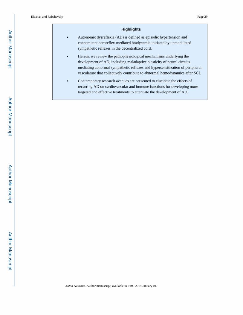
Highlights
• Autonomic dysreflexia (AD) is defined as episodic hypertension and
concomitant baroreflex-mediated bradycardia initiated by unmodulated
sympathetic reflexes in the decentralized cord.
• Herein, we review the pathophysiological mechanisms underlying the
development of AD, including maladaptive plasticity of neural circuits
mediating abnormal sympathetic reflexes and hypersensitization of peripheral
vasculature that collectively contribute to abnormal hemodynamics after SCI.
• Contemporary research avenues are presented to elucidate the effects of
recurring AD on cardiovascular and immune functions for developing more
targeted and effective treatments to attenuate the development of AD.
Eldahan and Rabchevsky Page 29
Auton Neurosci. Author manuscript; available in PMC 2019 January 01.
Author M
anuscriptA
uthor Manuscript
Author M
anuscriptA
uthor Manuscript

Figure 1. Diagrammatic representation of the neuroanatomical circuitry thought to be involved in
autonomic dysreflexia triggered by pelvic visceral stimulation, as well as potential neuronal
pathway silencing paradigms. Descending vasomotor fibers (blue) originating in the
brainstem and hypothalamus modulate the tonic activity of sympathetic preganglionic
neurons (SPN; red) throughout the intermediolateral cell column (IML) in the thoracolumbar
spinal cord. After complete spinal cord injury at or above the T6 segment, SPN which
innervate the adrenal medulla and blood vessels below the injury are segregated from
descending control pathways, allowing for unrestrained sympathetic reflex activity that leads
to hypertension. Injury further leads to maladaptive sprouting of both primary afferent c-
fibers (purple) and ascending propriospinal tracts (green) originating in the lumbosacral
dorsal gray commissure (DGC). These ascending “relay” neurons are thought to convey
sensory information from the bladder and colon (i.e., distension) rostrally towards SPN.
Emerging neuronal silencing techniques may allow for investigations into the role of specific
neuroanatomical pathways involved in AD. One example might be to employ double
infection of ascending lumbosacral propriospinal neurons with the HiRET-eTeNT vector
(red syringe), which is retrogradely transported (dotted black lines) from nerve terminals in
Eldahan and Rabchevsky Page 30
Auton Neurosci. Author manuscript; available in PMC 2019 January 01.
Author M
anuscriptA
uthor Manuscript
Author M
anuscriptA
uthor Manuscript

the IML to lumbosacral DGC also infected with AAV-rtTAV vector (blue syringe).
Doxycycline-induced silencing of these neurons could then allow for direct investigation
into their roles in facilitating AD during pelvic visceral stimulation.
Eldahan and Rabchevsky Page 31
Auton Neurosci. Author manuscript; available in PMC 2019 January 01.
Author M
anuscriptA
uthor Manuscript
Author M
anuscriptA
uthor Manuscript