Tyrannobdella rex N. Gen. N. Sp. and the Evolutionary Origins of Mucosal Leech Infestations
9
See discussions, stats, and author profiles for this publication at: https://www.researchgate.net/publication/43343284 Tyrannobdella rex N. Gen. N. Sp. and the Evolutionary Origins of Mucosal Leech Infestations Article in PLoS ONE · April 2010 DOI: 10.1371/journal.pone.0010057 · Source: PubMed CITATIONS 19 READS 85 7 authors, including: Some of the authors of this publication are also working on these related projects: Species Diversity of Asian Macrophagous Leeches View project Anna J Phillips Smithsonian Institution 21 PUBLICATIONS 144 CITATIONS SEE PROFILE Alejandro Oceguera-Figueroa Universidad Nacional Autónoma de México 38 PUBLICATIONS 195 CITATIONS SEE PROFILE Gloria P Gomez-Perez Instituto de Salud Global de Barcelona 7 PUBLICATIONS 44 CITATIONS SEE PROFILE Yite Lai National Taiwan University 20 PUBLICATIONS 112 CITATIONS SEE PROFILE All content following this page was uploaded by Yite Lai on 03 December 2016. The user has requested enhancement of the downloaded file. All in-text references underlined in blue are added to the original document and are linked to publications on ResearchGate, letting you access and read them immediately.
Transcript of Tyrannobdella rex N. Gen. N. Sp. and the Evolutionary Origins of Mucosal Leech Infestations

Seediscussions,stats,andauthorprofilesforthispublicationat:https://www.researchgate.net/publication/43343284
TyrannobdellarexN.Gen.N.Sp.andtheEvolutionaryOriginsofMucosalLeechInfestations
ArticleinPLoSONE·April2010
DOI:10.1371/journal.pone.0010057·Source:PubMed
CITATIONS
19
READS
85
7authors,including:
Someoftheauthorsofthispublicationarealsoworkingontheserelatedprojects:
SpeciesDiversityofAsianMacrophagousLeechesViewproject
AnnaJPhillips
SmithsonianInstitution
21PUBLICATIONS144CITATIONS
SEEPROFILE
AlejandroOceguera-Figueroa
UniversidadNacionalAutónomadeMéxico
38PUBLICATIONS195CITATIONS
SEEPROFILE
GloriaPGomez-Perez
InstitutodeSaludGlobaldeBarcelona
7PUBLICATIONS44CITATIONS
SEEPROFILE
YiteLai
NationalTaiwanUniversity
20PUBLICATIONS112CITATIONS
SEEPROFILE
AllcontentfollowingthispagewasuploadedbyYiteLaion03December2016.
Theuserhasrequestedenhancementofthedownloadedfile.Allin-textreferencesunderlinedinblueareaddedtotheoriginaldocument
andarelinkedtopublicationsonResearchGate,lettingyouaccessandreadthemimmediately.
Tyrannobdella rex N. Gen. N. Sp. and the EvolutionaryOrigins of Mucosal Leech InfestationsAnna J. Phillips1,2, Renzo Arauco-Brown3, Alejandro Oceguera-Figueroa1,2, Gloria P. Gomez4, Marıa
Beltran5, Yi-Te Lai6, Mark E. Siddall2*
1 Department of Biology, The Graduate Center, The City University of New York, New York, New York, United States of America, 2 Sackler Institute of Comparative
Genomics, American Museum of Natural History, New York, New York, United States of America, 3 School of Medicine, Universidad Peruana Cayetano Heredia, Lima, Peru,
4 Department of Microbiology, Universidad Peruana Cayetano Heredia, Lima, Peru, 5 Enteroparasitology Laboratory, Peruvian Public Health Center, Peruvian Health
Institute, Lima, Peru, 6 Institute of Zoology, National Taiwan University, Taipei, Taiwan
Abstract
Background: Leeches have gained a fearsome reputation by feeding externally on blood, often from human hosts. Orificialhirudiniasis is a condition in which a leech enters a body orifice, most often the nasopharyngeal region, but there are manycases of leeches infesting the eyes, urethra, vagina, or rectum. Several leech species particularly in Africa and Asia are well-known for their propensity to afflict humans. Because there has not previously been any data suggesting a closerelationship for such geographically disparate species, this unnerving tendency to be invasive has been regarded only as aloathsome oddity and not a unifying character for a group of related organisms.
Principal Findings: A new genus and species of leech from Peru was found feeding from the nasopharynx of humans.Unlike any other leech previously described, this new taxon has but a single jaw with very large teeth. Phylogenetic analysesof nuclear and mitochondrial genes using parsimony and Bayesian inference demonstrate that the new species belongsamong a larger, global clade of leeches, all of which feed from the mucosal surfaces of mammals.
Conclusions: This new species, found feeding from the upper respiratory tract of humans in Peru, clarifies an expansion ofthe family Praobdellidae to include the new species Tyrannobdella rex n. gen. n.sp., along with others in the generaDinobdella, Myxobdella, Praobdella and Pintobdella. Moreover, the results clarify a single evolutionary origin of a group ofleeches that specializes on mucous membranes, thus, posing a distinct threat to human health.
Citation: Phillips AJ, Arauco-Brown R, Oceguera-Figueroa A, Gomez GP, Beltran M, et al. (2010) Tyrannobdella rex N. Gen. N. Sp. and the Evolutionary Origins ofMucosal Leech Infestations. PLoS ONE 5(4): e10057. doi:10.1371/journal.pone.0010057
Editor: Robert DeSalle, American Museum of Natural History, United States of America
Received December 4, 2009; Accepted February 24, 2010; Published April 14, 2010
Copyright: � 2010 Phillips et al. This is an open-access article distributed under the terms of the Creative Commons Attribution License, which permitsunrestricted use, distribution, and reproduction in any medium, provided the original author and source are credited.
Funding: This work was financially supported by the National Science Foundation (DEB-0640463), the Stavros Niarchos fund for Expeditionary Research, aTheodore Roosevelt Memorial Grant, and a CUNY Science Fellowship. The funders had no role in study design, data collection and analysis, decision to publish, orpreparation of the manuscript.
Competing Interests: The authors have declared that no competing interests exist.
* E-mail: [email protected]
Introduction
Most people realize that they are being parasitized by a leech
upon finding the worm attached to their skin. Disturbingly, leeches
occasionally enter human orifices–a condition known as mucosal,
orifical, vesical, or internal hirudiniasis depending on the
localization of the leech (Fig. 1). Whereas most bloodfeeding
leeches feed as ecto-parasites for short periods of time, those that
feed on mucous membranes have been known to stay in an orifice
for days or weeks on end [1,2]. Cases of hirudiniasis are
underreported because patients suffering from orificial hirudiniasis
may only resort to medical attention if they are personally
unsuccessful in extracting the leech [3]. Whereas invasive leeches
are usually found in the nasopharyngeal region, there are many
cases of leeches infesting various body orifices such as the eyes,
urethra, vagina, or rectum [4]. Depending on the exact site of the
bite in the nasopharyngeal region, symptoms may include
hemorrhaging, hemoptysis, dysphonia, coughing, a tickling
sensation, dyspnea or, in extreme cases, severe anemia and death
[5,6]. Hemorrhaging from leeches in the urethra, or even in the
bladder, also poses a particular problem in that clot formation is
inhibited by urine flow [7]. Underlying conditions, such as
coagulation disorders or secondary bacterial infections can cause a
patient’s condition to escalate from relatively minor to life-
threatening very quickly [2,8,7].
Reported cases of human orificial hirudiniasis are most
common in rural areas of the Middle East, Africa, and Asia,
however cases have been recorded from almost all continents [2].
Domestic and wild mammals in these regions, especially livestock,
are at the greatest risk for orificial hirudiniasis in relation to the
amount of time such animals spend at leech-inhabited watering
holes [1]. Some species are more likely to afflict humans, such as
Dinobdella ferox (Blanchard, 1896; literally translated to ‘‘terrible
ferocious leech’’), or species of Limnatis, Praobdella, and Myxobdella
[9].
Leech systematics has been transformed with the addition of
comparative sequence data and most of the groups historically
recognized by taxonomists have been redefined or eliminated
PLoS ONE | www.plosone.org 1 April 2010 | Volume 5 | Issue 4 | e10057

[e.g., 10, 11]. That said, Phillips and Siddall expressed reluctance
to fully reorganize the systematics of New World hirudinoid
leeches under Semiscolescidae in light of the unexpected finding of
several interrelated Old World species in that group [10]. Our
discovery of a species that is new to science, found feeding from
the upper respiratory tract of humans in Peru, leads to a reanalysis
of the phylogeny and classification of one clade of hirudinoid
leeches, clarifying a single evolutionary origin of a group that
specializes on mucous membranes and poses a threat to human
health.
Results
Clinical PresentationsIn 1997, a previously healthy six-year-old boy was admitted to a
health center in Lamas province, department of San Martın, Peru
complaining of frontal cephalgia. The patient’s history revealed
that, prior to admission, he frequently bathed in local lakes and
natural streams. The patient reported neither bleeding nor
respiratory distress. A 25 mm long leech was removed from the
right nostril and preserved in formalin.
Again, in 1997, a 16-month-old boy was admitted to a local
heath center in Yochegua province, San Francisco district,
department of Ayacucho, Peru also complaining of frontal
cephalgia and also without respiratory symptoms. It was
ascertained that prior to admission the boy had bathed in small
local lakes. A 60 mm leech was removed from the patient’s nasal
cavity, washed with saline solution and preserved in 10% formalin.
Nasal bleeding continued for two days.
In 2007, a nine-year-old girl was admitted to La Merced
hospital in Chanchamayo province, department of Junın, Peru
following a two-week history of frontal cephalgia and a ‘‘sliding’’
sensation inside her nose. The patient’s parents noticed a black
worm moving inside her right nostril and sought medical
attention. No other respiratory symptoms presented. The patient
volunteered that she had been traveling in Satipo province,
department of Junın, Peru where she frequently bathed in lakes,
rivers and streams. Physical examination was remarkable only for
nasal pain with hand pressure and a black mass inside the right
nasal cavity. With some effort, a 65–70 mm black leech was
removed without significant bleeding from the patient’s nasal
cavity, and was preserved in ethanol.
Figure 1. Mucosally invasive hirudinoid leeches. Known from a wide variety of anatomical sites including eyes (A) as in this case involvingDinobdella ferox (B), mucosal leech species, as in a case involving Myxobdella annandalei (C), more frequently feed from the nasopharyngeal surfacesof mammals (D).doi:10.1371/journal.pone.0010057.g001
Origins of Leech Infestations
PLoS ONE | www.plosone.org 2 April 2010 | Volume 5 | Issue 4 | e10057

DescriptionTyrannobdella n. gen. One dorsal monostichodont jaw
armed with few, large denticular teeth. Mouth velar with single
slot for jaw. Ventrolateral jaws absent. Complete somite five-
annulate. Cephalic eyespots, five pair in parabolic arc. Anus
between last annulus and caudal sucker. Caudal sucker wider than
posterior of body. Reproductive organs micromorphic. Feeds from
mucosal surfaces of mammals. ZooBank LSID for the genus
Tyrannobdella is urn:lsid:zoobank.org:act:43D55B49-C888-4D6B-
AF6F-61238EC1339B. Type species: Tyrannobdella rex n. sp.
Tyrannobdella rex n. sp. Holotype: Preserved body length
44.5 mm, maximal width 0.95 mm, fixed and stored in 90%
ethanol; dissected. Collected at La Merced Chanchamayo Junın,
Peru in 2007 by Dr. Renzo Arauco-Brown; deposited in the
Museum of Natural History of San Marcos University, Peru
(catalogue number 2841).
Paratypes: Two mature specimens fixed in formalin and stored
in 90% ethanol. Collected in departments San Martın and
Ayacucho, Peru in 1997 by Dra. Marıa Beltran; one specimen
deposited in the Enteroparasitology laboratory at the Peruvian
Health Institute and another at the Museum of Natural History of
San Marcos University, Peru (catalogue number 2842).
One dorsal jaw armed with eight large (up to 130 mm high)
teeth forming a single (i.e., monostichodont) row (Fig. 2a,c). Two
of eight teeth may be sub-cuticular and observable only with
compound microscopy (Fig. 2c). Pharynx muscular and tubular.
Crop from IX to XXV, first nine cecal pairs in IX through XIX,
post-ceca extend blitaterally to XXV. First and second cecal
chambers subdivided into two unequal sub-ceca with the larger
being posterior, otherwise one cecal pair per somite. Intestine
tubular, acecate.
Body muscular, uniformly pigmented brown to grey without
stripes or other ornamentation after preservation. Papillae absent.
Oral sucker small and velar (Fig. 2b). Oral opening central and
dorsoventrally oval. Posterior sucker large, wider than posterior of
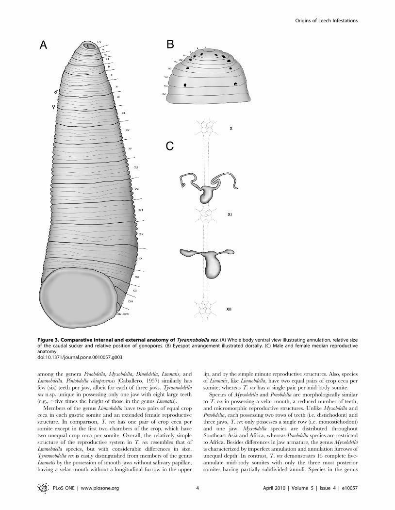
body (Fig. 3a). Somites I - III uniannulate, somites IV – V
biannulate, somites VI - VIII triannulate, somites IX - XXIV
quinqueannulate, somites XXV triannulate with annulus a1
dorsally subdivided, somite XXVI biannulate with annulus a1
dorsally subdivided, and somite XXVII unianulate with a faint
dorsal furrow visible. Anus between last annulus and caudal
sucker. Eyespots, five pairs on II, III, IV a1, V a1 and VI a2,
forming a parabolic arc (Fig. 3b). Male gonopore on XI b6, female
gonopore on XII b6, gonopores separated by 1/2+4+1/2 annuli.
Nephropores 17 pairs from VIII-XXV, each pair ventral on
posterior margins of annulus b2 of somite.
Male and female reproductive organs extremely micromorphic,
same size as or smaller than ventral ganglia (Fig. 3c). Penis sheath
U-shaped, with initial posterior disposition and subsequent
anterior procurrent portion leading to small epidydimis. Ejacula-
tory bulbs absent. Glandular prostate absent. Vagina present, U-
shaped, no common oviduct and oviducts half the size of vagina.
Vaginal cecum absent. Ovaries simple, bulbous.
The ZooBank LSID for the species T. rex is urn:lsid:zoobank.
org:act:F8C0E97B-F525-4EB3-B11B-B8CBA1CB8F5F.
Etymology: Tyrannobdella: tyrannos (G.) – ‘‘tyrant’’ + bdella (G.)
– ‘‘leech’’; rex: rex (G.) – ‘‘king’’.
Remarks: No other leech species is known to have but a single
armed jaw with such large teeth. The reduced number of teeth, a
caudal sucker wider than the posterior of the body, and preference
for feeding on mucous membranes of mammals all indicate the
placement of this new taxon within the family Praobdellidae
Figure 2. Comparative jaw morphology of Tyrannobdella rex. (A) Stereomicrograph of the single dorsal jaw of T. rex with large teeth. Scale baris 100 mm. (B) Tyrannobdella rex anterior sucker exhibiting velar mouth and longitudinal slit through which the dorsal jaw protrudes when feeding.Scale bar is 1 mm. (C) Compound micrograph in lateral view of eight large teeth of T. rex. Scale bar is 100 mm. (D) Lateral view of jaw of Limnatispaluda illustrating typical size of hirudinoid teeth. Scale bar is 100 mm.doi:10.1371/journal.pone.0010057.g002
Origins of Leech Infestations
PLoS ONE | www.plosone.org 3 April 2010 | Volume 5 | Issue 4 | e10057
among the genera Praobdella, Myxobdella, Dinobdella, Limnatis, and
Limnobdella. Pintobdella chiapasensis (Caballero, 1957) similarly has
few (six) teeth per jaw, albeit for each of three jaws. Tyrannobdella
rex n.sp. unique in possessing only one jaw with eight large teeth
(e.g., ,five times the height of those in the genus Limnatis).
Members of the genus Limnobdella have two pairs of equal crop
ceca in each gastric somite and an extended female reproductive
structure. In comparison, T. rex has one pair of crop ceca per
somite except in the first two chambers of the crop, which have
two unequal crop ceca per somite. Overall, the relatively simple
structure of the reproductive system in T. rex resembles that of
Limnobdella species, but with considerable differences in size.
Tyrannobdella rex is easily distinguished from members of the genus
Limnatis by the possession of smooth jaws without salivary papillae,
having a velar mouth without a longitudinal furrow in the upper
lip, and by the simple minute reproductive structures. Also, species
of Limnatis, like Limnobdella, have two equal pairs of crop ceca per
somite, whereas T. rex has a single pair per mid-body somite.
Species of Myxobdella and Praobdella are morphologically similar
to T. rex in possessing a velar mouth, a reduced number of teeth,
and micromorphic reproductive structures. Unlike Myxobdella and
Praobdella, each possessing two rows of teeth (i.e. distichodont) and
three jaws, T. rex only possesses a single row (i.e. monostichodont)
and one jaw. Myxobdella species are distributed throughout
Southeast Asia and Africa, whereas Praobdella species are restricted
to Africa. Besides differences in jaw armature, the genus Myxobdella
is characterized by imperfect annulation and annulation furrows of
unequal depth. In contrast, T. rex demonstrates 15 complete five-
annulate mid-body somites with only the three most posterior
somites having partially subdivided annuli. Species in the genus
Figure 3. Comparative internal and external anatomy of Tyrannobdella rex. (A) Whole body ventral view illustrating annulation, relative sizeof the caudal sucker and relative position of gonopores. (B) Eyespot arrangement illustrated dorsally. (C) Male and female median reproductiveanatomy.doi:10.1371/journal.pone.0010057.g003
Origins of Leech Infestations
PLoS ONE | www.plosone.org 4 April 2010 | Volume 5 | Issue 4 | e10057

Myxobdella have gonopores separated by five or five and a half
annuli, whereas T. rex has gonopores separated by 1/2+4+1/2
annuli. Species in the genus Praobdella lack the velar mouth and
have at least seven annuli between gonopores.
Phylogenetic analysesThe combined dataset included a total of 5256 molecular
characters (18S rDNA: 2041 characters, 28S rDNA: 2189
characters, 12S mt rDNA: 367 characters, COI: 657 characters).
Parsimony recovered a single tree with 2725 steps and the two
runs of the Bayesian analysis had a harmonic mean of log
likelihood values that averaged to–19830.23. Phylogenetic analy-
ses recovered identical topologies regardless of method (Fig. 4).
A clade of hirudinoid leeches (including the genera Limnobdella,
Limnatis, Dinobdella, Myxobdella, Pintobdella and Tyrannobdella),
distinguished by their propensity for feeding on mammalian
mucous membranes, was recovered as monophyletic with strong
support (bs = 100; pp = 1.00). Sister to this was a strictly New
World clade (bs = 80; pp = 1.00) comprising both the families
Semiscolescidae (Semiscolex + Patagoniobdella) and Macrobdellidae
(Macrobdella + Philobdella + Oxyptychus). The new Peruvian species,
T. rex, was sister to the Mexican P. chiapasensis (bs = 82; pp = 1.00).
Dinobdella ferox and Myxobdella annandalei Oka, 1917 were most
closely related (bs = 100; pp = 1.00). Representatives of the Old
World genus Limnatis formed a monophyletic clade sister to the
Mexican genus Limnobdella (bs = 94; pp = 1.00). The clades (T. rex +P. chiapasensis) and (D. ferox + M. annandalei) also form their own
clade sister to (Limnatis spp. + Limnobdella spp.).
Discussion
Hirudinoid leeches that show a preference for mammalian
mucosal surfaces all appear to have descended from a common
ancestor millions of years ago. Among these, the new species
Tyrannobdella rex is the first from South America and one with a
particularly unpleasant habit of infesting humans [12]. Another
New World orifice-invading leech known from southern Mexico,
P. chiapasensis, and sister taxon to T. rex, has only been found to
parasitize the nasal passages of tapirs [13]. Limnobdella species from
central and northern Mexico are known to be pests of livestock
[14]. The consistency with which pain was reported by its victims
may relate to the relatively enormous teeth T. rex has on its jaw.
Most of the documented cases of leech infestation are in tropical
regions. Such cases are closely related to unsafe drinking water
habits and people swimming in natural sources. It is in these
situations that these worms enter the rectum, vagina or upper
airway and attach to the mucosa [15]. A recent study revealed that
the nose is the most common site of infestation (71%), followed by
the hypopharynx (14%) [16]. Less often, leech infestations affect
the lower airways causing haemoptysis, haematemesis, severe
anaemia, airway obstruction or death [17]. While little is known of
the symbiotic fauna for praobdellids, species of Aeromonas are
known to inhabit the gastric ceca of various hirudinoid leeches
[18,19]. Insofar as praobdellids have been reported to remain
attached for prolonged periods [1], there may also be a serious risk
of bacterial infection to the extent that prophylactic antibiotic
treatments is indicated in all cases of orificial hirudiniasis.
Several species of leech are known to invade human orifices, most
notably various Old World species in the genera Myxobdella,
Praobdella, and Dinobdella. Until now, the family Praobdellidae (sensu
Sawyer, 1986) included only those three genera, two representatives
of which were monophyletic in our analyses: Myxobdella annandalei
and Dinobdella ferox. We found strong support for monophyly of that
pair in a broader clade that also includes species of Limnatis,
Limnobdella, T. rex and P. chiapasensis. This clade is defined not only by
our molecular evidence, but also by three morphological and
behavioral synapomorphies: reduced number of teeth–less than 12
in Myxobdella, Dinobdella, Praobdella, Tyrannobdella, and Pintobdella, and
less than 40 in Limnatis and Limnobdella; the caudal sucker is wider
than the posterior of the body; and a preference for feeding
primarily on mammalian mucous membranes. The enlarged caudal
sucker seen throughout this family may well be an adaptation that
mediates attachment to moist mucous membranes [2]. Only once
has a praobdellid been reported feeding opportunistically on
amphibians when mammals were not available [20].
The systematics of the family Praobdellidae (sensu Sawyer, 1986)
has been plagued by ill-defined groups and by substandard type
specimens being the sole representatives for some species [1,2].
The characteristics of the oral sucker, the color pattern, and the
location of the gonopores seem to hold the most phylogenetic
information among species, but organizing these species within
genera has been confused. The Terrible Ferocious Leech,
Dinobdella ferox, for example, was initially described as a species
of Whitmania, a genus of non-bloodfeeding leeches more closely
related to Hirudo [10]. Several morphological similarities have
been noted [21] between Praobdella radiata Moore, 1958 and
Myxobdella africana Moore, 1939, while Praobdella guineensis Blanch-
ard, 1896, Praobdella buettneri Blanchard, 1896, and Praobdella
Figure 4. Single most parsimonious tree based on combined18S rDNA, 28s rDNA, 12s rDNA, and COI datasets. The familyPraobdellidae formed a well-supported monophyletic group of leechesthat exhibits a predilection for mammalian mucosa. All groups received100 percent bootstrap support and posterior probabilities of 1.00except as noted on the tree. Branches are drawn proportional toamount of change.doi:10.1371/journal.pone.0010057.g004
Origins of Leech Infestations
PLoS ONE | www.plosone.org 5 April 2010 | Volume 5 | Issue 4 | e10057

maculata (Moore, 1939) have each been considered potential
synonyms of D. ferox (Moore, 1958). It has generally been agreed
that these taxonomic conundra will only be resolved with the
addition of fresh specimens [2,21]. Nonetheless, the monophyly in
our phylogenetic analyses of the genera Myxobdella, Dinobdella,
Limnatis, Limnobdella, Tyrannobdella, and Pintobdella agree with the
morphological and behavioral synapomorphies observed through-
out the clade suggesting that the family Praobdellidae should be
expanded to include them all. In turn, this settles the problem
faced by Phillips and Siddall [10], and allows Semiscolecidae
Scriban and Autrum, 1934 to retain its traditional scope
comprising non-bloodfeeding South American taxa and allows
Macrobdellidae Richardson, 1969 to encompass the bloodfeeding
genera Macrobdella, Philobdella and Oxyptychus.
Representatives of the genus Praobdella, preferably the type
species P. buettneri, are sorely needed to definitively establish the
relationships of members of the family Praobdellidae. Praobdella
buettneri has not been collected since its description in 1896 (from
Bismarksburg, Togoland, now the Togolese Republic) along with
P. guineensis, which shares the same type locality [22]. Only
external morphology was mentioned in Blanchard’s (1896)
description and the type specimens are long-since dried out
making it difficult to relate them to newly collected material not
found at the type locality. Additional species of this family that
warrant scrutiny are M. africana, Myxobdella sinanensis Oka, 1925,
Myxobdella weberi (Blanchard, 1897), Myxobdella nepalica Nesemann
and Sharma, 2001, P. maculata, and P. radiata. Further collection
efforts in Africa and Asia may yet successfully provide the required
material, though our standard methods of attracting leeches to our
exposed selves may prove awkward given their established
propensity for particular anatomical feeding sites.
Methods
Specimens of T. rex were collected from two states of Peru in
1997; one from a health center in Lamas province, department of
San Martin, Peru, and one from a local heath center in Yochegua
province, San Francisco district. Both of these specimens were
preserved in formalin. A third specimen collected from a clinic in
La Merced Chanchamayo Junin, Peru in 2007, was preserved in
ethanol, and was the specimen chosen both for the holotype and
for sequencing in these analyses. Specimens of P. chiapasensis were
collected from forest streams leading to the lakes of Montebello,
State of Chiapas, Mexico between 6 and 18 July, 2008. One M.
annandalei was received in December, 2008 from Dharamsala,
India. Tissue samples of D. ferox were collected on 13 April, 2008
in Taiwan. Examination of external and internal morphology was
accomplished with a Nikon SMZ-U stereo microscope on whole
and dissected specimens. Photographs were taken with a SPOT-
RT digital camera. Drawings were made by superposition of
vector art over images placed in Adobe IllustratorH 10 and Adobe
PhotoshopH 7.
DNA sequencing and alignmentTissue was collected from the caudal sucker in order to
avoid contamination from host DNA in gastric or intestinal
regions of the leech. DNeasy Tissue Kit (Qiagen Valencia, CA)
was used for tissue lysis and DNA purification. Primers for the
PCR amplification of nuclear 18S rDNA and 28S rDNA and
mitochondrial cytochrome oxidase I (COI) and 12S rDNA
gene fragments were adapted from published protocols [23,24,
25,26,27] and are listed in Table 1. Amplification reactions of gene
fragments were conducted using either Ready-To-Go PCR Beads
Table 1. Genes and primer sequences used in phylogenetic analyses.
Gene Primer Name Primer Sequence Reference
Nuclear
18S rDNA
1 A 59-AACCTGGTTGATCCTGCCAGT-39 Apakupakul et. al., 1999
L 59-CCAACTACGAGCTTTTTAACTG-39 Apakupakul et. al., 1999
2 C 59-CGGTAATTCCAGCTCCAATAG-39 Apakupakul et. al., 1999
Y 59-CAGACAAATCGCTCCACCAAC-39 Apakupakul et. al., 1999
3 O 59-AAGGGCACCACCAGGAGTGGAG-39 Apakupakul et. al., 1999
B 59-TGATCCTTCCGCAGGTTCACCT-39 Apakupakul et. al., 1999
28S rDNA
1 28srD1a 59-CCCSCGTAAYTTAAGCATAT-39 Prendini et al., 2005
28sB 59-TCGGAAGGAACCAGCTAC-39 Whiting, 2002
2 28sA 59-GACCCGTCTTGAAGCACG-39 Whiting, 2002
28SBout 59-CCCACAGCGCCAGTTCTGCTTACC-39 Prendini et al., 2005
3 28srD5a 59-GGYGTTGGTTGCTTAAGACAG-39 Whiting, 2002
28srD7b1 59-GACTTCCCTTACCTACAT-39 Whiting, 2002
Mitochondrial
12s rDNA
12Sa 59-AAACTAGGATTAGATACCCTATTAT-39 Simon et al., 1994
12Sb 59-AAGAGCGACGGGCGATGTGT-39 Simon et al., 1994
COI
LCO1490 59-GGTCAACAAATCATAAAGATATTGG-39 Folmer et al., 1994
HCO2198 59-TAAACTTCAGGGTGACCAAAAAATCA-39 Folmer et al., 1994
doi:10.1371/journal.pone.0010057.t001
Origins of Leech Infestations
PLoS ONE | www.plosone.org 6 April 2010 | Volume 5 | Issue 4 | e10057

(GE Healthcare, Piscataway, NJ) with 0.5 ml of each 10 mM
primer, 1 ml DNA template, and 23 ml Rnase-free H2O (total
volume 25 ml), or homemade Taq with 1.0 ml Taq, 2.5 ml MgCl,
2.5 ml 10x Buffer A, 1.0 ml dNTPs, 0.5 ml of each 10 mM primer,
2.0 ml template, and 15 ml H2O) (total volume 25 ml). PCR
reactions were performed in an Eppendorf Mastercycler. The
following amplification protocols were used: for 18S, 94uC (1 min)
followed by 35 cycles of 94uC (30 sec), 49uC (30 sec), 68uC (2 min
30 sec) and final extension at 68uC (1 min); for 28S and 12S, 94uC(5 min), followed by 39 cycles of 95uC (1 min), 52uC (1 min), 70uC(1 min) and final extension of 72u (7 min); for COI, 94uC (1 min),
followed by 30 cycles of 94uC (30 sec), 48uC (30 sec), 68uC(45 sec), 68uC (1 min) and final extension of 68uC (1 min). PCR
amplification products were purified with AMPureTM (Agencourt
Bioscience Corporation). Cycle sequencing reactions were per-
formed with an Eppendorf MastercyclerH using 1 ml Big DyeTM
Extender Buffer v3.1, 1 ml of 1 mM primer and 3 ml of cleaned
PCR template (13 ml total volume). Sequences were purified by
70% isopropanol/70% ethanol precipitation and analyzed with an
ABI PRISMH 3730 sequencer (Applied Biosystems). CodonCode
Aligner (CodonCode Corporation) was used to edit and reconcile
sequences. GenBank accession numbers are listed for sequences
derived from each taxon in Table 2. Alignments of all genes were
accomplished using the European Bioinformatics Institute server
for MUSCLE v. 3.7 applying default settings [28].
Phylogenetic analysesA total of 17 species comprising 19 terminals were used in the
analyses with Hirudo medicinalis specified as the outgroup (Table 1).
Phylogenetic analyses were conducted using two approaches:
Parsimony and Bayesian Inference (BI). Parsimony analyses were
conducted in TNT v 1.1 [29] using 10 replicates of random taxon
addition, sectorial searching, the Ratchet [30], and tree-bisection-
reconnection branch swapping for each gene as well as for the
combined dataset (18S, 28S, 12S, COI). Bootstrap values for
combined analyses were obtained using 10 heuristic pseudorepli-
cates and the same analytical settings. Bayesian analyses were
conducted in MrBayes v. 3.1.2 [31]. The data were partitioned by
gene for 18S, 28S, 12S, and by codon position for COI (three
partition; 3p). A GTR+I+C model was applied to each unlinked
data partition based on the Akaike Information Criterion [via
ModelTest v. 3.7; 32, 33]. For the Metropolis-Coupled Markov
Chain Monte Carlo (MCMCMC) analyses, default prior distribu-
tions of parameters were used twice with one cold chain and three
hot chains for 10 million generations and sampled every 1000th
generation. The BI analyses burned-in before 100,000 genera-
tions. Split frequencies of the standard deviation of simultaneous
BI analyses were well below 0.01. As such, the burn-in was set to
discard the first 100,000 generations, leaving 9,900 trees sampled
for estimation of posterior probabilities.
Nomenclatural ActsThe electronic version of this document does not represent a
published work according to the International Code of Zoological
Nomenclature (ICZN), and hence the nomenclatural acts
contained in the electronic version are not available under that
Code from the electronic edition. Therefore, a separate edition of
this document was produced by a method that assures numerous
Table 2. Taxa used for the phylogenetic analyses, collection localities, and GenBank accession numbers.
Taxon Locality GenBank Accession Numbers
18S 28S 12S CO1
Ingroup
Dinobdella ferox Taiwan GU394006 GU394010 GU394002 ________
Limnatis cf. nilotica Namibia GQ368795 GQ368774 GQ368815 GQ368754
Limnatis paluda 1 Afghanistan GQ368796 GQ368775 ________ GQ368755
Limnatis paluda 2 Israel AY425470 AY425389 AY425430 AY425452
Limnobdella mexicana 1* Mexico GQ368798 GQ368777 GQ368816 GQ368756
Limnobdella mexicana 2* Mexico GQ368799 GQ368778 GQ368817 GQ368757
Myxobdella annandalei India GU394007 GU394011 GU394003 GU39414
Pintobdella chiapasensis Chiapas, Mexico GU394008 GU394012 GU394004 GU394015
Tyrannobdella rex Peru GU394009 GU394013 GU394005 GU394016
Outgroup
Haemadipsa sylvestris Vietnam AF116005 AY425373 AY425416 AF003266
Haemopis sanguisuga* Sweden AF099941 AY425381 AF099960 AF462021
Hirudo medicinalis* BioPharm, UK AF116011 AY425385 AF099961 AF003272
Macobdella decora* MI, USA AF116007 AY425390 AY425431 AF003271
Macrobdella ditetra GA, USA AY425471 AY425391 AY425432 AY425453
Oxyptychus brasiliensis Brazil AY425473 AY425398 AY425436 AY425455
Patagoniobdella fraterna Chile AY425477 AY425405 AY425441 AY425459
Philobdella floridana* SC, USA DQ097210-13 DQ097201-14 DQ097226 DQ097219-22
Philobdella gracilis LA, USA DQ097209 DQ097200 DQ097225 DQ097218
Semiscolex similis Bolivia AY425475 AY425402 AY42543 AY425475
Type species of genera are indicated with an asterisk.doi:10.1371/journal.pone.0010057.t002
Origins of Leech Infestations
PLoS ONE | www.plosone.org 7 April 2010 | Volume 5 | Issue 4 | e10057

identical and durable copies, and those copies were simultaneously
obtainable (from the publication date noted on the first page of this
article) for the purpose of providing a public and permanent
scientific record, in accordance with Article 8.1 of the Code. The
separate print-only edition is available on request from PLoS by
sending a request to PLoS ONE, 185 Berry Street, Suite 3100, San
Francisco, CA 94107, USA along with a check for $10 (to cover
printing and postage) payable to ‘‘Public Library of Science’’.
In addition, this published work and the nomenclatural acts it
contains have been registered in ZooBank, the proposed online
registration system for the ICZN. The ZooBank LSIDs (Life
Science Identifiers) can be resolved, and the associated informa-
tion viewed, through any standard web browser by appending the
LSID to the prefix ‘‘http://zoobank.org/’’. The ZooBank LSID
for this publication is: urn:lsid:zoobank.org:pub:8D431ED1-B837-
4781-A591-D3886285283A
Acknowledgments
We thank Sebastian Kvist and Colleen Ingram for their comments on
drafts of this manuscript. For providing specimens, we thank Will Reeves
and Virginia Leon-Regagnon. Also, we thank the Willi Hennig Society for
subsidizing the program TNT and making it freely available. We thank
Major Thomas P. Ward (US Armed Forces Institute of Pathology;
presently Consulting Opthomologists Farmington, CT) who provided the
photographs of the ocular Dinobdella ferox affliction, as well as Catherine
Schuetze (University of Queensland) and Jan Slapeta (University of
Sydney) for both photographs and specimens of Myxobdella annandalei from a
dogs nose.
RAB would like to give special thanks to Julio Demarini MD, from the
Infectious Diseases Department, Hospital La Merced, Junın, Peru, for
helping with the initial approach to the treatment of the first case of
Tyranobdella rex infestation that was found and for encouraging us to delve
as deep as possible into this type of parasitism of humans. RAB also thanks
his family, for believing in him as a medical student, without clearly
understanding what he has been doing, and for supporting the export of
the first specimen of Tyranobdella rex from the Peruvian jungle to the
American Museum of Natural History, NYC.
Author Contributions
Conceived and designed the experiments: AJP AOF MES. Performed the
experiments: AJP AOF MES. Analyzed the data: AJP AOF MES.
Contributed reagents/materials/analysis tools: AJP RAB GPG MB YTL
MES. Wrote the paper: AJP RAB AOF MES.
References
1. Harding WA, Moore JP (1927) Fauna of British India: Hirudinea. London:Taylor and Francis. 185 p.
2. Cundall DB, Whitehead SM, Hechtel FOP (1986) Severe anaemia and death
due to the pharyngeal leech Myxobdella africana. Trans R Soc Trop Med Hyg 80:940–944.
3. Montazeri F, Bedayat A, Jamali L, Salehian M, Montazeri G (2008) Leechendoparasitism: report of a case and review of the literature. Eur J Pediatr 168:
39–42.4. Almallah Z (1968) Internal hirudiniasis in man with Limnatis nilotica, in Iraq.
J Parasitol 54: 637–638.
5. Masterman EWG (1908) Hirudinea as human parasites in Palestine.Parasitology 1: 182–185.
6. Turner FM (1969) Pharyngeal leeches. Lancet ii: 1400–1401.7. Alam S, Das Choudhary MK, Islam K (2008) Leech in urinary bladder causing
hematuria. J Pediatr Urol 4: 70–73.
8. Kose A (2008) Leech bites, massive bleeding, coagulation profile disorders, andsevere anemia. Am J Emerg Med 26: 1067.e3–1067.e6.
9. Sawyer RT (1986) Leech Biology and Behaviour. Oxford: Clarendon Press. 685 p.10. Phillips AJ, Siddall ME (2009) Polyparaphyly of Hirudinidae: many lineages of
medicinal leeches. BMC Evol Bio 9: 246.11. Borda E, Oceguera-Figueroa A, Siddall ME (2008) On the classification,
evolution and biogeography of terrestrial haemadipsoid leeches (Hirudinida:
Arhynchobdellida: Hirudiniformes). Mol Phylogenet Evol 46: 142–154.12. Beltran M, Melgar R, Tello R (1997) Infeccion nasal por Hirudo medicinalis y
breve revision del tema. Rev Med Exp INS 14(2): 42–46.13. Caballero CE (1957) Description of a new species of leech, coming from the
forests of the state of Chiapas. Ana Inst Biol Univ Nac Auton Mexico 28:
241–245.14. Cabellero CE (1932) Limnobdella tehuacanea (Jimenez, 1865) Caballero, 1931. Syn:
Hirudo tehuacanea Jimenez, 1865. Anales Inst Biol Univ Nac Auton Mexico 3:43–47.
15. Hamid MS, Mohd Nar GR (1996) Severe urological complication of leech bite
in the tropics. Br J Urol 77: 164–165.16. Raza SN, Shabbir SM, Anwar-ul-Haq (2006) Leech infestation and its
association with water drinking habits. J Coll Physicians Surg Pakistan 16:175–178.
17. Singh M, Naim AF (1979) Respiratory obstruction and haematemesis due toleech. Lancet 2: 1374.
18. Siddall M, Worthen PL, Johnson M, Graf J (2007) Novel role for Aeromonas
jandaei as a digestive tract symbiont of the North American medicinal leech. Appl
Env Microbiol 73: 655–658.
19. Laufer AS, Siddall ME, Graf J (2008) Characterization of the digestive-tract
microbiota of Hirudo orientalis, a European medicinal leech. Appl Env Microbiol
74: 6151–6154.
20. Lukin EI (1976) Pijavki [Leeches] In: Fauna USSR. Leningrad: Academy of
Science of the USSR Vol. 1. 484 p.
21. Moore JP (1958) The leeches (Hirudinidae) in the collection of the Natal
Museum. Ann Natal Mus 14: 327–328.
22. Blanchard R (1896) Hirudineen aus dem Togoland. Arch Naturgesch 63: 49–53.
23. Apakupakul K, Siddall ME, Burreson EM (1999) Higher-level relationships of
leeches (Annelida: Clitellata: Euhirudinea) based on morphology and gene
sequences. Mol Phylogenet Evol 12: 350–359.
24. Predini L (2005) Comment on ‘‘Identifying spiders through DNA barcodes’’.
Can J Zool 83: 498–504.
25. Whiting MF (2002) Mecoptera is paraphyletic: multiple genes and phylogeny of
Mecoptera and Siphonaptera. Zool Scripta 31: 93–104.
26. Simon C, Frati F, Beckenbach A, Crespi B, Liu H, et al. (1994) Evolution,
weighting, and phylogenetic utility of mitochondrial gene sequences and a
compilation of conserved PCR primers. Annals Entomol Soc Am 87: 651–701.
27. Folmer O, Back M, Hoeh W, Lutz R, Vrijenhoek R (1994) DNA primers for
amplification of mitochondrial cytochrome c oxidase subunit I from diverse
metazoan invertebrates. Mol Mar Biol Biotechnol 3: 294–299.
28. Edgar RC (2004) MUSCLE: multiple sequence alignment with high accuracy
and high throughput. Nucleic Acids Res 32: 1792–1797.
29. Goloboff PA, Farris JS, Kallersjo M, Oxelman B, Ramırez MJ, et al. (2003)
Improvements to resampling measures of group support. Cladistics 19: 324–332.
30. Nixon K (1999) The parsimony ratchet, a new method for rapid parsimony
analysis. Cladistics 15: 407–414.
31. Ronquist F, Huelsenbeck JP (2003) MRBAYES 3: Bayesian phylogenetic
inference under mixed models. Bioinformatics 19: 1572–1574.
32. Posada D, Crandall KA (1998) Modeltest: testing the model of DNA
substitution. Bioinformatics 14: 817–818.
33. Posada D, Buckley TR (2004) Model selection and model averaging in
phylogenetics: advantages of Akaike Information Criterion and Bayesian
approaches over likelihood ratio tests. Syst Biol 53: 793–808.
Origins of Leech Infestations
PLoS ONE | www.plosone.org 8 April 2010 | Volume 5 | Issue 4 | e10057


![N -[4-( N -Cyclohexylsulfamoyl)phenyl]acetamide](https://static.fdokumen.com/doc/165x107/632f4f4de68feab59a0210b7/n-4-n-cyclohexylsulfamoylphenylacetamide.jpg)