N Cyclohexyl N -methylbenzenesulfonamide
9
N-Cyclohexyl-N-methylbenzene- sulfonamide Zeeshan Haider, a Islam Ullah Khan, a * Muhammad Nadeem Arshad, a Muhammad Shafiq a and Caoyuan Niu b * a Materials Chemistry Laboratory, Department of Chemistry, GC University, Lahore 54000, Pakistan, and b College of Sciences, Henan Agricultural University, Zhengzhou 450002, People’s Republic of China Correspondence e-mail: [email protected], [email protected] Received 9 October 2009; accepted 13 October 2009 Key indicators: single-crystal X-ray study; T = 296 K; mean (C–C) = 0.003 A ˚ ; R factor = 0.038; wR factor = 0.113; data-to-parameter ratio = 16.1. The title compound, C 13 H 19 NO 2 S, was synthesized by the reaction of N-cyclohexylaminebenzenesulfonamide and methyl iodide. The crystal packing is stabilized by weak intermolecular C—HO hydrogen bonds. Related literature Compounds containing cyclohexylamine have been reported to be activators of dopamine receptors in the central nervous system, see: Hacksell et al. (1981). For related structures, see: Arshad et al. (2008, 2009). Experimental Crystal data C 13 H 19 NO 2 S M r = 253.35 Monoclinic, P2 1 =c a = 9.2729 (5) A ˚ b = 12.1182 (7) A ˚ c = 12.5801 (7) A ˚ = 109.103 (2) V = 1335.79 (13) A ˚ 3 Z =4 Mo K radiation = 0.23 mm 1 T = 296 K 0.28 0.12 0.09 mm Data collection Bruker APEXII CCD detector diffractometer Absorption correction: multi-scan (SADABS; Bruker, 2005) T min = 0.938, T max = 0.979 12741 measured reflections 2489 independent reflections 1864 reflections with I >2(I) R int = 0.030 Refinement R[F 2 >2(F 2 )] = 0.038 wR(F 2 ) = 0.113 S = 1.08 2489 reflections 155 parameters H-atom parameters constrained max = 0.16 e A ˚ 3 min = 0.25 e A ˚ 3 Table 1 Hydrogen-bond geometry (A ˚ , ). D—HA D—H HA DA D—HA C2—H2O2 i 0.93 2.52 3.268 (3) 137 Symmetry code: (i) x; y þ 1 2 ; z þ 1 2 . Data collection: APEX2 (Bruker, 2005); cell refinement: SAINT (Bruker, 2005); data reduction: SAINT; program(s) used to solve structure: SHELXS97 (Sheldrick, 2008); program(s) used to refine structure: SHELXL97 (Sheldrick, 2008); molecular graphics: SHELXL97 and DIAMOND (Brandenburg, 2005); software used to prepare material for publication: SHELXL97. Supplementary data and figures for this paper are available from the IUCr electronic archives (Reference: BT5092). References Arshad, M. N., Tahir, M. N., Khan, I. U., Ahmad, E. & Shafiq, M. (2008). Acta Cryst. E64, o2380. Arshad, M. N., Tahir,M. N., Khan, I. U., Shafiq, M. & Ahmad, S. (2009). Acta Cryst. E65, o940. Brandenburg, K. (2005). DIAMOND. Crystal Impact GbR. Bonn, Germany. Bruker (2005). APEX2, SAINT, and SADABS. Bruker AXS Inc., Madison, Wisconsin, USA. Hacksell, U., Arvidsson, L.-E., Svensson, U., Nilsson, J. L. G., Sanchez, D., Wikstroem, H., Lindberg, P., Hjorth, S. & Carlsson, A. (1981). J. Med. Chem. 24, 1475–1482. Sheldrick, G. M. (2008). Acta Cryst. A64, 112–122. organic compounds o2892 Haider et al. doi:10.1107/S1600536809041762 Acta Cryst. (2009). E65, o2892 Acta Crystallographica Section E Structure Reports Online ISSN 1600-5368
-
Upload
independent -
Category
Documents
-
view
0 -
download
0
Transcript of N Cyclohexyl N -methylbenzenesulfonamide

N-Cyclohexyl-N-methylbenzene-sulfonamide
Zeeshan Haider,a Islam Ullah Khan,a* Muhammad
Nadeem Arshad,a Muhammad Shafiqa and Caoyuan Niub*
aMaterials Chemistry Laboratory, Department of Chemistry, GC University, Lahore
54000, Pakistan, and bCollege of Sciences, Henan Agricultural University,
Zhengzhou 450002, People’s Republic of China
Correspondence e-mail: [email protected], [email protected]
Received 9 October 2009; accepted 13 October 2009
Key indicators: single-crystal X-ray study; T = 296 K; mean �(C–C) = 0.003 A;
R factor = 0.038; wR factor = 0.113; data-to-parameter ratio = 16.1.
The title compound, C13H19NO2S, was synthesized by the
reaction of N-cyclohexylaminebenzenesulfonamide and
methyl iodide. The crystal packing is stabilized by weak
intermolecular C—H� � �O hydrogen bonds.
Related literature
Compounds containing cyclohexylamine have been reported
to be activators of dopamine receptors in the central nervous
system, see: Hacksell et al. (1981). For related structures, see:
Arshad et al. (2008, 2009).
Experimental
Crystal data
C13H19NO2S Mr = 253.35
Monoclinic, P21=ca = 9.2729 (5) Ab = 12.1182 (7) Ac = 12.5801 (7) A� = 109.103 (2)�
V = 1335.79 (13) A3
Z = 4Mo K� radiation� = 0.23 mm�1
T = 296 K0.28 � 0.12 � 0.09 mm
Data collection
Bruker APEXII CCD detectordiffractometer
Absorption correction: multi-scan(SADABS; Bruker, 2005)Tmin = 0.938, Tmax = 0.979
12741 measured reflections2489 independent reflections1864 reflections with I > 2�(I)Rint = 0.030
Refinement
R[F 2 > 2�(F 2)] = 0.038wR(F 2) = 0.113S = 1.082489 reflections
155 parametersH-atom parameters constrained��max = 0.16 e A�3
��min = �0.25 e A�3
Table 1Hydrogen-bond geometry (A, �).
D—H� � �A D—H H� � �A D� � �A D—H� � �A
C2—H2� � �O2i 0.93 2.52 3.268 (3) 137
Symmetry code: (i) x;�yþ 12; zþ 1
2.
Data collection: APEX2 (Bruker, 2005); cell refinement: SAINT
(Bruker, 2005); data reduction: SAINT; program(s) used to solve
structure: SHELXS97 (Sheldrick, 2008); program(s) used to refine
structure: SHELXL97 (Sheldrick, 2008); molecular graphics:
SHELXL97 and DIAMOND (Brandenburg, 2005); software used to
prepare material for publication: SHELXL97.
Supplementary data and figures for this paper are available from theIUCr electronic archives (Reference: BT5092).
References
Arshad, M. N., Tahir, M. N., Khan, I. U., Ahmad, E. & Shafiq, M. (2008). ActaCryst. E64, o2380.
Arshad, M. N., Tahir, M. N., Khan, I. U., Shafiq, M. & Ahmad, S. (2009). ActaCryst. E65, o940.
Brandenburg, K. (2005). DIAMOND. Crystal Impact GbR. Bonn, Germany.Bruker (2005). APEX2, SAINT, and SADABS. Bruker AXS Inc., Madison,
Wisconsin, USA.Hacksell, U., Arvidsson, L.-E., Svensson, U., Nilsson, J. L. G., Sanchez, D.,
Wikstroem, H., Lindberg, P., Hjorth, S. & Carlsson, A. (1981). J. Med. Chem.24, 1475–1482.
Sheldrick, G. M. (2008). Acta Cryst. A64, 112–122.
organic compounds
o2892 Haider et al. doi:10.1107/S1600536809041762 Acta Cryst. (2009). E65, o2892
Acta Crystallographica Section E
Structure ReportsOnline
ISSN 1600-5368

supplementary materials

supplementary materials
sup-1
Acta Cryst. (2009). E65, o2892 [ doi:10.1107/S1600536809041762 ]
N-Cyclohexyl-N-methylbenzenesulfonamide
Z. Haider, I. U. Khan, M. N. Arshad, M. Shafiq and C. Niu
Comment
Sulfonamide compounds have gained much importance due to their therapeutic applications. The compound containingcyclohexylamine has been reported to be an activator of dopamine receptors in the CNS (Hacksell et al., 1981). The titlecompound is a sulfonamide derivative of cyclohexylamine in continuation to our previous work (Arshad et al., 2008; Arshadet al., 2009).
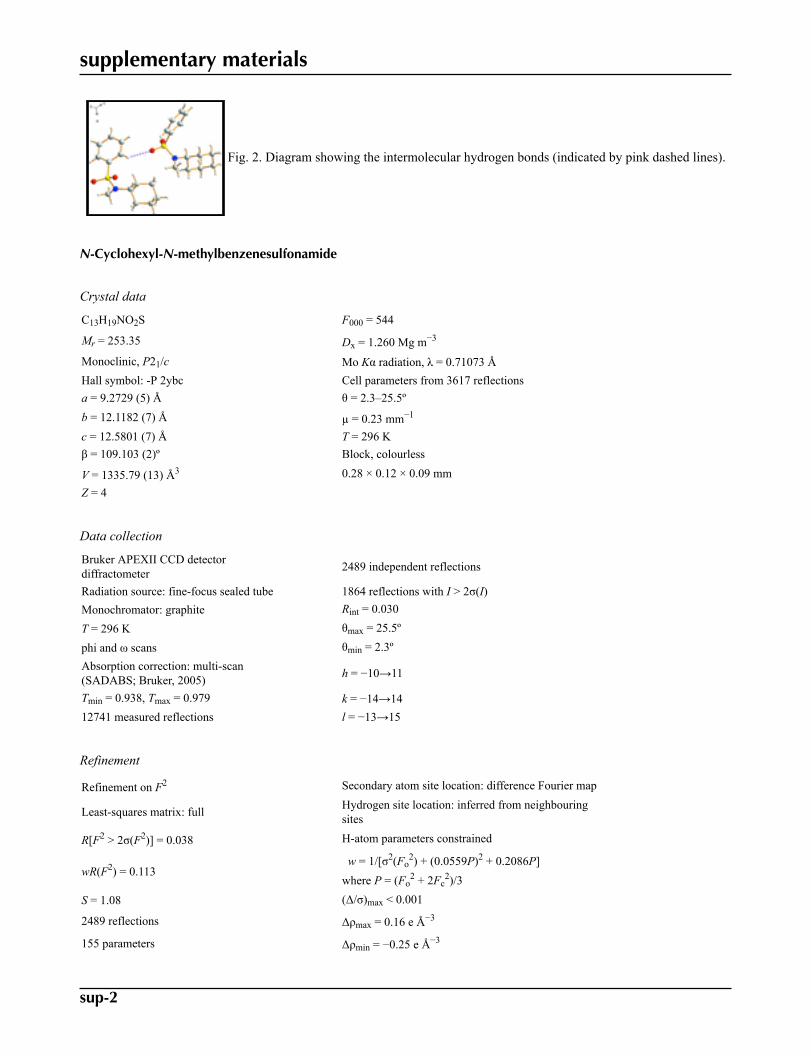
The molecular structure of the title compound (I) is shown in Fig. 1. The mean plane of the benzene ring and that ofthe four essentially planar C atoms (C8, C9, C11, C12. Maximum deviation, 0.0132 Å) of the chair-form cyclohexyl ringhave a dihedral angle of 24.26 (9)°. Furthermore, there are intermolecular C—H···O hydrogen bonds between the aromatic
H atom (H2) and one sulfonamide O atom (O2i, symmetric code: see table 1) of neighboring molecules that contribute tothe three-dimensional packing (Fig. 2).
Experimental
Sodium hydride (0.88 mmol) was taken in a round bottom flask and washed with n-hexane so as to remove the mineraloil dispersant. A solution of N-cyclohexylamine benzene sulfonamide (0.43 mmol) in 5 ml of N,N dimethyl formamidewas added. The mixture was stirred for half an hour at room temperature. Then, methyl iodide (0.86 mmol) was added andstirring was continued for about 3 hrs until the complete consumption of sulfonamide. The reaction was monitored by TLC.After the completion of the reaction the contents were transferred into the distilled water ice. The product precipitated andwas separated by filtration and recrystallized from methanol. The melting point of the product was observed to be 353 Kuncorrected.
Refinement
All H atoms were placed in calculated positions and refined using a riding model [C—H = 0.93 Å and Uiso(H) = 1.2Ueq(C)
for aromatic H atoms; C—H = 0.98 Å and Uiso(H) = 1.2Ueq(C) for tertiary CH; C—H = 0.97 Å and Uiso(H) = 1.2Ueq(C)
for CH2; C—H = 0.96 Å and Uiso(H) = 1.5Ueq(C) for the methyl H atoms]. The final difference Fourier map had a highest
peak at 0.71 Å from atom C1 and a deepest hole at 0.71 A Å from atom S1, but were otherwise featureless.
Figures
Fig. 1. The molecular structure of the title compound showing the atom-labelling scheme.Displacement ellipsoids are drawn at the 30% probability level.
supplementary materials
sup-2
Fig. 2. Diagram showing the intermolecular hydrogen bonds (indicated by pink dashed lines).
N-Cyclohexyl-N-methylbenzenesulfonamide
Crystal data
C13H19NO2S F000 = 544
Mr = 253.35 Dx = 1.260 Mg m−3
Monoclinic, P21/c Mo Kα radiation, λ = 0.71073 ÅHall symbol: -P 2ybc Cell parameters from 3617 reflectionsa = 9.2729 (5) Å θ = 2.3–25.5ºb = 12.1182 (7) Å µ = 0.23 mm−1
c = 12.5801 (7) Å T = 296 Kβ = 109.103 (2)º Block, colourless
V = 1335.79 (13) Å3 0.28 × 0.12 × 0.09 mmZ = 4
Data collection
Bruker APEXII CCD detectordiffractometer 2489 independent reflections
Radiation source: fine-focus sealed tube 1864 reflections with I > 2σ(I)Monochromator: graphite Rint = 0.030
T = 296 K θmax = 25.5º
phi and ω scans θmin = 2.3ºAbsorption correction: multi-scan(SADABS; Bruker, 2005) h = −10→11
Tmin = 0.938, Tmax = 0.979 k = −14→1412741 measured reflections l = −13→15
Refinement
Refinement on F2 Secondary atom site location: difference Fourier map
Least-squares matrix: full Hydrogen site location: inferred from neighbouringsites
R[F2 > 2σ(F2)] = 0.038 H-atom parameters constrained
wR(F2) = 0.113 w = 1/[σ2(Fo
2) + (0.0559P)2 + 0.2086P]where P = (Fo
2 + 2Fc2)/3
S = 1.08 (Δ/σ)max < 0.001
2489 reflections Δρmax = 0.16 e Å−3
155 parameters Δρmin = −0.25 e Å−3

supplementary materials
sup-3
Primary atom site location: structure-invariant directmethods Extinction correction: none
Special details
Geometry. All e.s.d.'s (except the e.s.d. in the dihedral angle between two l.s. planes) are estimated using the full covariance mat-rix. The cell e.s.d.'s are taken into account individually in the estimation of e.s.d.'s in distances, angles and torsion angles; correlationsbetween e.s.d.'s in cell parameters are only used when they are defined by crystal symmetry. An approximate (isotropic) treatment ofcell e.s.d.'s is used for estimating e.s.d.'s involving l.s. planes.
Refinement. Refinement of F2 against ALL reflections. The weighted R-factor wR and goodness of fit S are based on F2, convention-
al R-factors R are based on F, with F set to zero for negative F2. The threshold expression of F2 > σ(F2) is used only for calculating R-
factors(gt) etc. and is not relevant to the choice of reflections for refinement. R-factors based on F2 are statistically about twice as largeas those based on F, and R- factors based on ALL data will be even larger.
Fractional atomic coordinates and isotropic or equivalent isotropic displacement parameters (Å2)
x y z Uiso*/Ueq
C1 0.67239 (19) 0.36402 (15) 0.21562 (15) 0.0443 (5)C2 0.6576 (2) 0.33212 (18) 0.31729 (17) 0.0556 (5)H2 0.7103 0.2710 0.3556 0.067*C3 0.5644 (2) 0.3918 (2) 0.3605 (2) 0.0694 (7)H3 0.5552 0.3717 0.4294 0.083*C4 0.4849 (3) 0.4804 (2) 0.3038 (2) 0.0738 (7)H4 0.4200 0.5192 0.3332 0.089*C5 0.5005 (3) 0.51236 (19) 0.2034 (2) 0.0732 (7)H5 0.4467 0.5731 0.1652 0.088*C6 0.5951 (2) 0.45494 (17) 0.15921 (18) 0.0568 (5)H6 0.6070 0.4772 0.0918 0.068*C7 1.0474 (2) 0.29629 (15) 0.33830 (14) 0.0424 (4)H7 0.9836 0.2401 0.3570 0.051*C8 1.0716 (2) 0.38752 (18) 0.42469 (16) 0.0568 (5)H8A 0.9739 0.4194 0.4203 0.068*H8B 1.1338 0.4452 0.4084 0.068*C9 1.1495 (2) 0.3434 (2) 0.54232 (16) 0.0632 (6)H9A 1.1678 0.4037 0.5958 0.076*H9B 1.0829 0.2907 0.5610 0.076*C10 1.2988 (2) 0.2885 (2) 0.55192 (17) 0.0651 (6)H10A 1.3416 0.2563 0.6262 0.078*H10B 1.3701 0.3434 0.5430 0.078*C11 1.2786 (3) 0.1995 (2) 0.46425 (18) 0.0679 (7)H11A 1.2203 0.1392 0.4805 0.082*H11B 1.3780 0.1710 0.4682 0.082*C12 1.1976 (2) 0.24159 (18) 0.34615 (16) 0.0546 (5)H12A 1.1787 0.1805 0.2936 0.065*H12B 1.2625 0.2943 0.3255 0.065*C13 1.0145 (3) 0.4353 (2) 0.18252 (19) 0.0719 (7)H13A 0.9857 0.4965 0.2197 0.108*H13B 0.9659 0.4421 0.1027 0.108*

supplementary materials
sup-4
H13C 1.1233 0.4350 0.1996 0.108*N1 0.96659 (17) 0.33232 (13) 0.22141 (13) 0.0485 (4)O1 0.78750 (17) 0.17730 (12) 0.18902 (14) 0.0710 (5)O2 0.75416 (18) 0.32058 (15) 0.04381 (11) 0.0769 (5)S1 0.79429 (6) 0.29004 (4) 0.15969 (4) 0.0523 (2)
Atomic displacement parameters (Å2)
U11 U22 U33 U12 U13 U23
C1 0.0369 (9) 0.0421 (11) 0.0442 (10) −0.0061 (8) 0.0002 (7) −0.0009 (8)C2 0.0458 (11) 0.0601 (14) 0.0563 (12) −0.0031 (10) 0.0104 (9) 0.0119 (10)C3 0.0505 (12) 0.0910 (19) 0.0690 (15) −0.0087 (13) 0.0227 (11) −0.0028 (13)C4 0.0447 (12) 0.0776 (18) 0.098 (2) −0.0046 (12) 0.0213 (13) −0.0252 (15)C5 0.0569 (14) 0.0515 (14) 0.098 (2) 0.0069 (11) 0.0078 (13) 0.0027 (13)C6 0.0513 (12) 0.0506 (13) 0.0579 (12) 0.0002 (10) 0.0033 (10) 0.0078 (10)C7 0.0397 (10) 0.0442 (11) 0.0410 (10) −0.0014 (8) 0.0101 (8) 0.0003 (8)C8 0.0558 (12) 0.0591 (13) 0.0535 (12) 0.0128 (10) 0.0151 (9) −0.0100 (10)C9 0.0655 (14) 0.0758 (16) 0.0468 (12) 0.0075 (11) 0.0163 (10) −0.0124 (10)C10 0.0573 (13) 0.0805 (17) 0.0477 (12) 0.0090 (11) 0.0039 (10) −0.0097 (11)C11 0.0618 (14) 0.0700 (16) 0.0583 (13) 0.0212 (11) 0.0009 (10) −0.0093 (11)C12 0.0519 (12) 0.0567 (13) 0.0507 (12) 0.0091 (9) 0.0108 (9) −0.0131 (9)C13 0.0738 (15) 0.0693 (16) 0.0675 (15) −0.0071 (12) 0.0162 (12) 0.0176 (12)N1 0.0454 (9) 0.0507 (10) 0.0462 (9) −0.0010 (7) 0.0108 (7) 0.0024 (7)O1 0.0658 (10) 0.0425 (9) 0.0905 (11) −0.0052 (7) 0.0064 (8) −0.0131 (8)O2 0.0797 (11) 0.1027 (13) 0.0379 (8) 0.0063 (9) 0.0050 (7) −0.0119 (8)S1 0.0513 (3) 0.0512 (3) 0.0449 (3) −0.0004 (2) 0.0027 (2) −0.0090 (2)
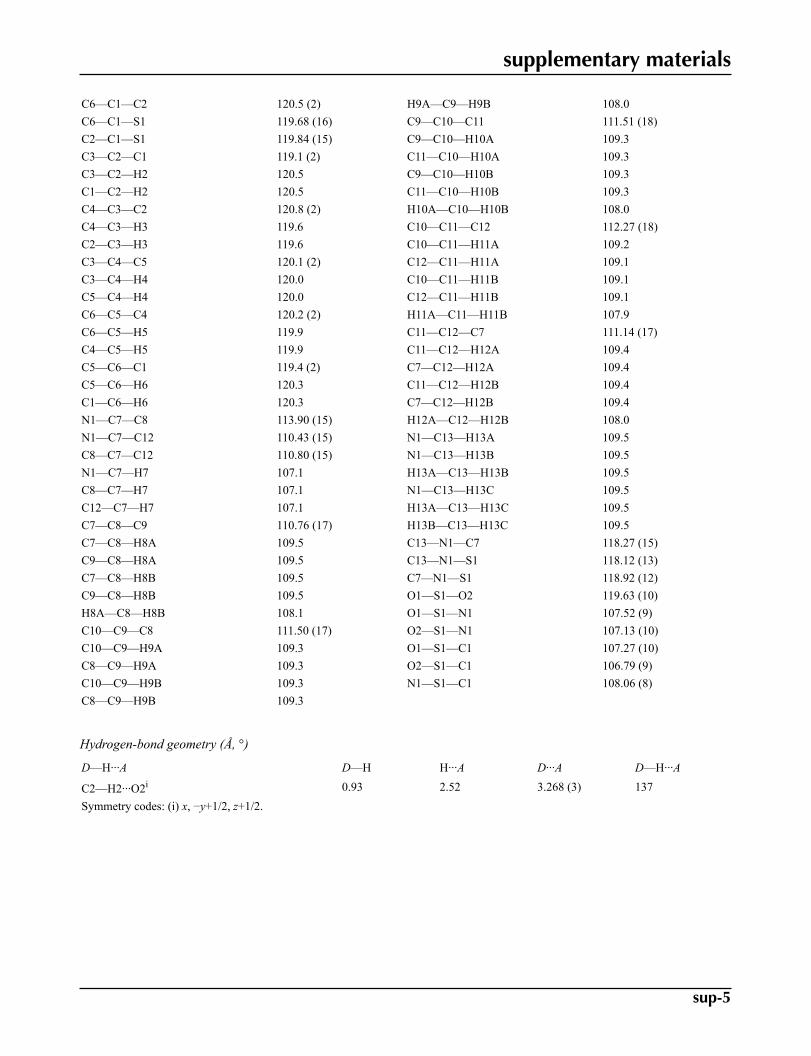
Geometric parameters (Å, °)
C1—C6 1.378 (3) C9—C10 1.505 (3)C1—C2 1.385 (3) C9—H9A 0.9700C1—S1 1.760 (2) C9—H9B 0.9700C2—C3 1.369 (3) C10—C11 1.509 (3)C2—H2 0.9300 C10—H10A 0.9700C3—C4 1.364 (3) C10—H10B 0.9700C3—H3 0.9300 C11—C12 1.517 (3)C4—C5 1.374 (4) C11—H11A 0.9700C4—H4 0.9300 C11—H11B 0.9700C5—C6 1.372 (3) C12—H12A 0.9700C5—H5 0.9300 C12—H12B 0.9700C6—H6 0.9300 C13—N1 1.462 (3)C7—N1 1.481 (2) C13—H13A 0.9600C7—C8 1.515 (3) C13—H13B 0.9600C7—C12 1.516 (3) C13—H13C 0.9600C7—H7 0.9800 N1—S1 1.6144 (16)C8—C9 1.516 (3) O1—S1 1.4218 (16)C8—H8A 0.9700 O2—S1 1.4302 (15)C8—H8B 0.9700
supplementary materials
sup-5
C6—C1—C2 120.5 (2) H9A—C9—H9B 108.0C6—C1—S1 119.68 (16) C9—C10—C11 111.51 (18)C2—C1—S1 119.84 (15) C9—C10—H10A 109.3C3—C2—C1 119.1 (2) C11—C10—H10A 109.3C3—C2—H2 120.5 C9—C10—H10B 109.3C1—C2—H2 120.5 C11—C10—H10B 109.3C4—C3—C2 120.8 (2) H10A—C10—H10B 108.0C4—C3—H3 119.6 C10—C11—C12 112.27 (18)C2—C3—H3 119.6 C10—C11—H11A 109.2C3—C4—C5 120.1 (2) C12—C11—H11A 109.1C3—C4—H4 120.0 C10—C11—H11B 109.1C5—C4—H4 120.0 C12—C11—H11B 109.1C6—C5—C4 120.2 (2) H11A—C11—H11B 107.9C6—C5—H5 119.9 C11—C12—C7 111.14 (17)C4—C5—H5 119.9 C11—C12—H12A 109.4C5—C6—C1 119.4 (2) C7—C12—H12A 109.4C5—C6—H6 120.3 C11—C12—H12B 109.4C1—C6—H6 120.3 C7—C12—H12B 109.4N1—C7—C8 113.90 (15) H12A—C12—H12B 108.0N1—C7—C12 110.43 (15) N1—C13—H13A 109.5C8—C7—C12 110.80 (15) N1—C13—H13B 109.5N1—C7—H7 107.1 H13A—C13—H13B 109.5C8—C7—H7 107.1 N1—C13—H13C 109.5C12—C7—H7 107.1 H13A—C13—H13C 109.5C7—C8—C9 110.76 (17) H13B—C13—H13C 109.5C7—C8—H8A 109.5 C13—N1—C7 118.27 (15)C9—C8—H8A 109.5 C13—N1—S1 118.12 (13)C7—C8—H8B 109.5 C7—N1—S1 118.92 (12)C9—C8—H8B 109.5 O1—S1—O2 119.63 (10)H8A—C8—H8B 108.1 O1—S1—N1 107.52 (9)C10—C9—C8 111.50 (17) O2—S1—N1 107.13 (10)C10—C9—H9A 109.3 O1—S1—C1 107.27 (10)C8—C9—H9A 109.3 O2—S1—C1 106.79 (9)C10—C9—H9B 109.3 N1—S1—C1 108.06 (8)C8—C9—H9B 109.3
Hydrogen-bond geometry (Å, °)
D—H···A D—H H···A D···A D—H···A
C2—H2···O2i 0.93 2.52 3.268 (3) 137Symmetry codes: (i) x, −y+1/2, z+1/2.

supplementary materials
sup-6
Fig. 1

supplementary materials
sup-7
Fig. 2