N -Acetonylsaccharin
9
N-Acetonylsaccharin Matloob Ahmad, a Hamid Latif Siddiqui, a Muhammad Azam, b Waseeq Ahmad Siddiqui c and Masood Parvez d * a Institute of Chemistry, University of the Punjab, Lahore 54590, Pakistan, b Institute of Biochemistry, University of Balochistan, Quetta, Pakistan, c Department of Chemistry, University of Sargodha, Sargodha, Pakistan, and d Department of Chemistry, The University of Calgary, 2500 University Drive NW, Calgary, Alberta, T2N 1N4, Canada Correspondence e-mail: [email protected] Received 14 July 2009; accepted 3 August 2009 Key indicators: single-crystal X-ray study; T = 200 K; mean (C–C) = 0.003 A ˚ ; R factor = 0.041; wR factor = 0.110; data-to-parameter ratio = 16.3. In the title compound [systematic name: 2-(2-oxopropyl)-1,2- benzothiazol-3(2H)-one 1,1-dioxide], C 10 H 9 NO 4 S, the benzo- thiazole unit is essentially planar [maximum deviation = 0.0490 (9) A ˚ for the S atom] and the oxopropyl group is inclined at an angle 75.61 (8) with respect to its mean plane. In the crystal, molecules are held together by weak inter- molecular C—HO non-classical hydrogen bonds, resulting in centrosymmetric dimeric units, forming 14-membered ring systems which may be described as R 2 2 (14) ring motifs. Moreover, molecules lying about inversion centers show – interactions, with centroid–centroid separations between the benzene rings of 3.676 (2) A ˚ . Related literature For the crystal structure of a benzothiazine, see: Ahmad et al. (2008). For the biological activity of sacharine derivatives, see: Kapui et al. (2003); Singh et al. (2007); Vaccarino et al. (2007). For graph-set notation of ring motifs, see: Bernstein et al. (1994). Experimental Crystal data C 10 H 9 NO 4 S M r = 239.24 Monoclinic, P2 1 =c a = 7.475 (3) A ˚ b = 8.975 (4) A ˚ c = 15.923 (7) A ˚ = 101.028 (18) V = 1048.5 (8) A ˚ 3 Z =4 Mo K radiation = 0.31 mm 1 T = 200 K 0.12 0.12 0.06 mm Data collection Nonius KappaCCD diffractometer Absorption correction: multi-scan (SORTAV; Blessing, 1997) T min = 0.964, T max = 0.982 3984 measured reflections 2382 independent reflections 2106 reflections with I >2(I) R int = 0.023 Refinement R[F 2 >2(F 2 )] = 0.041 wR(F 2 ) = 0.110 S = 1.04 2382 reflections 146 parameters H-atom parameters constrained max = 0.30 e A ˚ 3 min = 0.41 e A ˚ 3 Table 1 Hydrogen-bond geometry (A ˚ , ). D—HA D—H HA DA D—HA C10—H10BO2 i 0.98 2.57 3.324 (3) 134 Symmetry code: (i) x; y; z þ 1. Data collection: COLLECT (Hooft, 1998); cell refinement: DENZO (Otwinowski & Minor, 1997); data reduction: SCALE- PACK (Otwinowski & Minor, 1997); program(s) used to solve structure: SHELXS97 (Sheldrick, 2008); program(s) used to refine structure: SHELXL97 (Sheldrick, 2008); molecular graphics: ORTEP-3 for Windows (Farrugia, 1997); software used to prepare material for publication: SHELXL97. Supplementary data and figures for this paper are available from the IUCr electronic archives (Reference: SI2191). References Ahmad, M., Siddiqui, H. L., Ahmad, S., Farooq, S. U. & Parvez, M. (2008). Acta Cryst. E64, o1213–o1214. Bernstein, J., Etter, M. C. & Leiserowitz, L. (1994). Structure Correlation, edited by H. -B. Bu ¨rgi & J. D. Dunitz, Vol. 2. 431–507. New York: VCH. Blessing, R. H. (1997). J. Appl. Cryst. 30, 421–426. Farrugia, L. J. (1997). J. Appl. Cryst. 30, 565. Hooft, R. (1998). COLLECT. Nonius BV, Delft, The Netherlands. Kapui, Z., Varga, M., Urban-Szabo, K., Mikus, E., Szabo, T., Szeredi, J., Finance, O. & Aranyi, P. (2003). J. Pharmacol. Exp. Ther. 305, 1–9. Otwinowski, Z. & Minor, W. (1997). Methods in Enzymology, Vol. 276, Macromolecular Crystallography, Part A, edited by C. W. Carter Jr & R. M. Sweet, pp. 307–326. New York: Academic Press. Sheldrick, G. M. (2008). Acta Cryst. A64, 112–122. Singh, S. K., Shivaramakrishna, S., Saibaba, V., Rao, K. S., Ganesh, K. R., Vasudev, R., Kumar, P. P., Babu, J. M., Vyas, K., Rao, Y. K. & Iqbal, J. (2007). Eur. J. Med. Chem. 42, 456–462. Vaccarino, A. L., Paul, D., Mukherjee, P. K., de Turco, E. B. R., Marcheselli, V. L., Xu, L., Trudell, M. L., Minguez, J. M., Matia, M. P., Sunkel, C., Alvarez- Builla, J. & Bazan, N. G. (2007). Bioorg. Med. Chem. 15, 2206–2215. organic compounds Acta Cryst. (2009). E65, o2185 doi:10.1107/S1600536809030773 Ahmad et al. o2185 Acta Crystallographica Section E Structure Reports Online ISSN 1600-5368
-
Upload
independent -
Category
Documents
-
view
6 -
download
0
Transcript of N -Acetonylsaccharin

N-Acetonylsaccharin
Matloob Ahmad,a Hamid Latif Siddiqui,a Muhammad
Azam,b Waseeq Ahmad Siddiquic and Masood Parvezd*
aInstitute of Chemistry, University of the Punjab, Lahore 54590, Pakistan, bInstitute of
Biochemistry, University of Balochistan, Quetta, Pakistan, cDepartment of
Chemistry, University of Sargodha, Sargodha, Pakistan, and dDepartment of
Chemistry, The University of Calgary, 2500 University Drive NW, Calgary, Alberta,
T2N 1N4, Canada
Correspondence e-mail: [email protected]
Received 14 July 2009; accepted 3 August 2009
Key indicators: single-crystal X-ray study; T = 200 K; mean �(C–C) = 0.003 A;
R factor = 0.041; wR factor = 0.110; data-to-parameter ratio = 16.3.
In the title compound [systematic name: 2-(2-oxopropyl)-1,2-
benzothiazol-3(2H)-one 1,1-dioxide], C10H9NO4S, the benzo-
thiazole unit is essentially planar [maximum deviation =
0.0490 (9) A for the S atom] and the oxopropyl group is
inclined at an angle 75.61 (8)� with respect to its mean plane.
In the crystal, molecules are held together by weak inter-
molecular C—H� � �O non-classical hydrogen bonds, resulting
in centrosymmetric dimeric units, forming 14-membered ring
systems which may be described as R22(14) ring motifs.
Moreover, molecules lying about inversion centers show �–�interactions, with centroid–centroid separations between the
benzene rings of 3.676 (2) A.
Related literature
For the crystal structure of a benzothiazine, see: Ahmad et al.
(2008). For the biological activity of sacharine derivatives, see:
Kapui et al. (2003); Singh et al. (2007); Vaccarino et al. (2007).
For graph-set notation of ring motifs, see: Bernstein et al.
(1994).
Experimental
Crystal data
C10H9NO4SMr = 239.24Monoclinic, P21=ca = 7.475 (3) Ab = 8.975 (4) Ac = 15.923 (7) A� = 101.028 (18)�
V = 1048.5 (8) A3
Z = 4Mo K� radiation� = 0.31 mm�1
T = 200 K0.12 � 0.12 � 0.06 mm
Data collection
Nonius KappaCCD diffractometerAbsorption correction: multi-scan
(SORTAV; Blessing, 1997)Tmin = 0.964, Tmax = 0.982
3984 measured reflections2382 independent reflections2106 reflections with I > 2�(I)Rint = 0.023
Refinement
R[F 2 > 2�(F 2)] = 0.041wR(F 2) = 0.110S = 1.042382 reflections
146 parametersH-atom parameters constrained��max = 0.30 e A�3
��min = �0.41 e A�3
Table 1Hydrogen-bond geometry (A, �).
D—H� � �A D—H H� � �A D� � �A D—H� � �A
C10—H10B� � �O2i 0.98 2.57 3.324 (3) 134
Symmetry code: (i) �x;�y;�z þ 1.
Data collection: COLLECT (Hooft, 1998); cell refinement:
DENZO (Otwinowski & Minor, 1997); data reduction: SCALE-
PACK (Otwinowski & Minor, 1997); program(s) used to solve
structure: SHELXS97 (Sheldrick, 2008); program(s) used to refine
structure: SHELXL97 (Sheldrick, 2008); molecular graphics:
ORTEP-3 for Windows (Farrugia, 1997); software used to prepare
material for publication: SHELXL97.
Supplementary data and figures for this paper are available from theIUCr electronic archives (Reference: SI2191).
References
Ahmad, M., Siddiqui, H. L., Ahmad, S., Farooq, S. U. & Parvez, M. (2008).Acta Cryst. E64, o1213–o1214.
Bernstein, J., Etter, M. C. & Leiserowitz, L. (1994). Structure Correlation,edited by H. -B. Burgi & J. D. Dunitz, Vol. 2. 431–507. New York: VCH.
Blessing, R. H. (1997). J. Appl. Cryst. 30, 421–426.Farrugia, L. J. (1997). J. Appl. Cryst. 30, 565.Hooft, R. (1998). COLLECT. Nonius BV, Delft, The Netherlands.Kapui, Z., Varga, M., Urban-Szabo, K., Mikus, E., Szabo, T., Szeredi, J.,
Finance, O. & Aranyi, P. (2003). J. Pharmacol. Exp. Ther. 305, 1–9.Otwinowski, Z. & Minor, W. (1997). Methods in Enzymology, Vol. 276,
Macromolecular Crystallography, Part A, edited by C. W. Carter Jr & R. M.Sweet, pp. 307–326. New York: Academic Press.
Sheldrick, G. M. (2008). Acta Cryst. A64, 112–122.Singh, S. K., Shivaramakrishna, S., Saibaba, V., Rao, K. S., Ganesh, K. R.,
Vasudev, R., Kumar, P. P., Babu, J. M., Vyas, K., Rao, Y. K. & Iqbal, J. (2007).Eur. J. Med. Chem. 42, 456–462.
Vaccarino, A. L., Paul, D., Mukherjee, P. K., de Turco, E. B. R., Marcheselli, V.L., Xu, L., Trudell, M. L., Minguez, J. M., Matia, M. P., Sunkel, C., Alvarez-Builla, J. & Bazan, N. G. (2007). Bioorg. Med. Chem. 15, 2206–2215.
organic compounds
Acta Cryst. (2009). E65, o2185 doi:10.1107/S1600536809030773 Ahmad et al. o2185
Acta Crystallographica Section E
Structure ReportsOnline
ISSN 1600-5368

supplementary materials

supplementary materials
sup-1
Acta Cryst. (2009). E65, o2185 [ doi:10.1107/S1600536809030773 ]
N-Acetonylsaccharin
M. Ahmad, H. L. Siddiqui, M. Azam, W. A. Siddiqui and M. Parvez
Comment
Saccharine derivatives are extensively reported in the literature for their diverse range of biological activities like cyclooxy-genase-2 (COX-2) inhibitors (Singh et al. 2007), analgesic (Vaccarino et al. 2007), human leucocyte elastase (HLE) inhibit-ors (Kapui et al. 2003). In continuation to our project to explore potentially biologically active derivatives of benzothiazines(Ahmad et al. 2008), we herein report the crystal structure of the title compound, (I), in this paper.
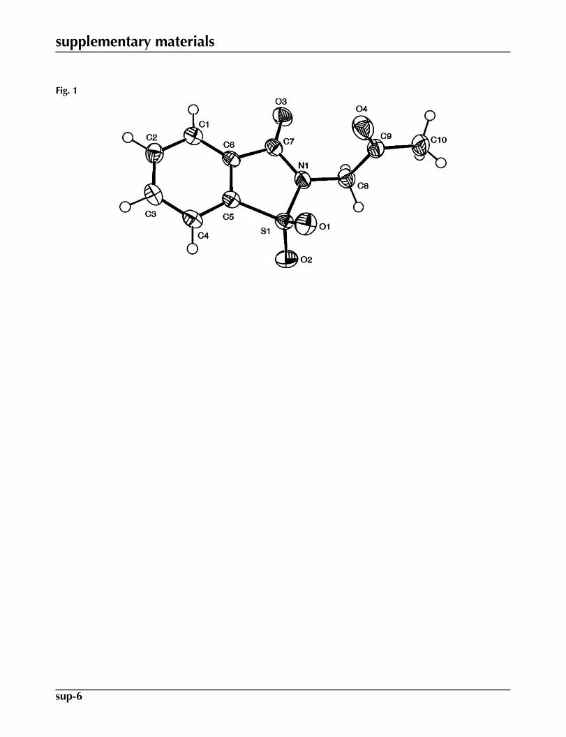
The structure of the title compound is depicted in Figure 1. The benzothiazol moiety (C1—C7/N1/S1) is essentaillyplanar with maximum deviation observed for S1 (0.0490 (9) Å) and the oxopropyl group (C8/C9/C10/O4) forms an angle75.61 (8)° with the mean-plane of the former. The molecular dimensions in (I) agree with the corresponding moleculardimensions reported for a closely related compound (Ahmad et al. 2008). In the crystal structure, the molecules of (I) areheld together by rather weak intermolecular C—H···O type non-classical hydrogen bonds resulting in dimeric units aboutinversion centers, forming fourteen membered ring systems which may be described in terms of graph set notation (Bernstein
et al. 1994) as R22(14) ring motif; details are given in Table 1 and Figure 2. The molecules lying about inversion centers show
π-π interactions with the separation between the centroids of the benzene rings (C1–C6) which are related by the symmetryoperation: 1-x, 1-y, 1-z, is 3.676 (2) Å (Spek, 2009); with perpendicular distance being 3.354 Å and the slippage of 1.504 Å.
Experimental
Sodium saccharin (73.2 mmoles, 15.0 g) and chloroacetone (87.8 mmole, 7.0 ml) were added in a round bottom flaskcontaining 30 ml of anhydrous DMF. The mixture was stirred under inert atmosphere for one hour at 393 K. The contentsof the flask were poured in ice cold water. Brownish ppts. formed were filtered and washed with excess of water. Crystalssuitable for XRD were grown in chloroform. Yield: 15.2 g, 87%; m.p. 386–387 K.
Refinement
Though all the H atoms could be distinguished in the difference Fourier map the H-atoms were included at geometricallyidealized positions and refined in riding-model approximation with the following constraints: C—H distances were set to0.95–0.99 Å and Uiso(H) = 1.2Ueq(C). The final difference map was free of any chemically significant features.
Figures
Fig. 1. ORTEP-3 (Farrugia, 1997) drawing of (I) with displacement ellipsoids plotted at 50%probability level.

supplementary materials
sup-2
Fig. 2. Unit cell of (I) showing dimers of molecules formed by C—H···O interactions; H-atoms not involved in H-bonding interactions have been excluded.
2-(2-oxopropyl)-1,2-benzothiazol-3(2H)-one 1,1-dioxide
Crystal data
C10H9NO4S F000 = 496
Mr = 239.24 Dx = 1.516 Mg m−3
Monoclinic, P21/c Mo Kα radiation, λ = 0.71073 ÅHall symbol: -P 2ybc Cell parameters from 3984 reflectionsa = 7.475 (3) Å θ = 3.4–27.5ºb = 8.975 (4) Å µ = 0.31 mm−1
c = 15.923 (7) Å T = 200 Kβ = 101.028 (18)º Block, colorless
V = 1048.5 (8) Å3 0.12 × 0.12 × 0.06 mmZ = 4
Data collection
Nonius KappaCCDdiffractometer 2382 independent reflections
Radiation source: fine-focus sealed tube 2106 reflections with I > 2σ(I)Monochromator: graphite Rint = 0.023
T = 200 K θmax = 27.5º
ω and φ scans θmin = 3.4ºAbsorption correction: multi-scan(SORTAV; Blessing, 1997) h = −9→9
Tmin = 0.964, Tmax = 0.982 k = −8→113984 measured reflections l = −20→20
Refinement
Refinement on F2 Secondary atom site location: difference Fourier map
Least-squares matrix: full Hydrogen site location: inferred from neighbouringsites
R[F2 > 2σ(F2)] = 0.041 H-atom parameters constrained
wR(F2) = 0.110 w = 1/[σ2(Fo
2) + (0.0444P)2 + 0.8524P]where P = (Fo
2 + 2Fc2)/3
S = 1.04 (Δ/σ)max = 0.005
2382 reflections Δρmax = 0.30 e Å−3
146 parameters Δρmin = −0.41 e Å−3
supplementary materials
sup-3
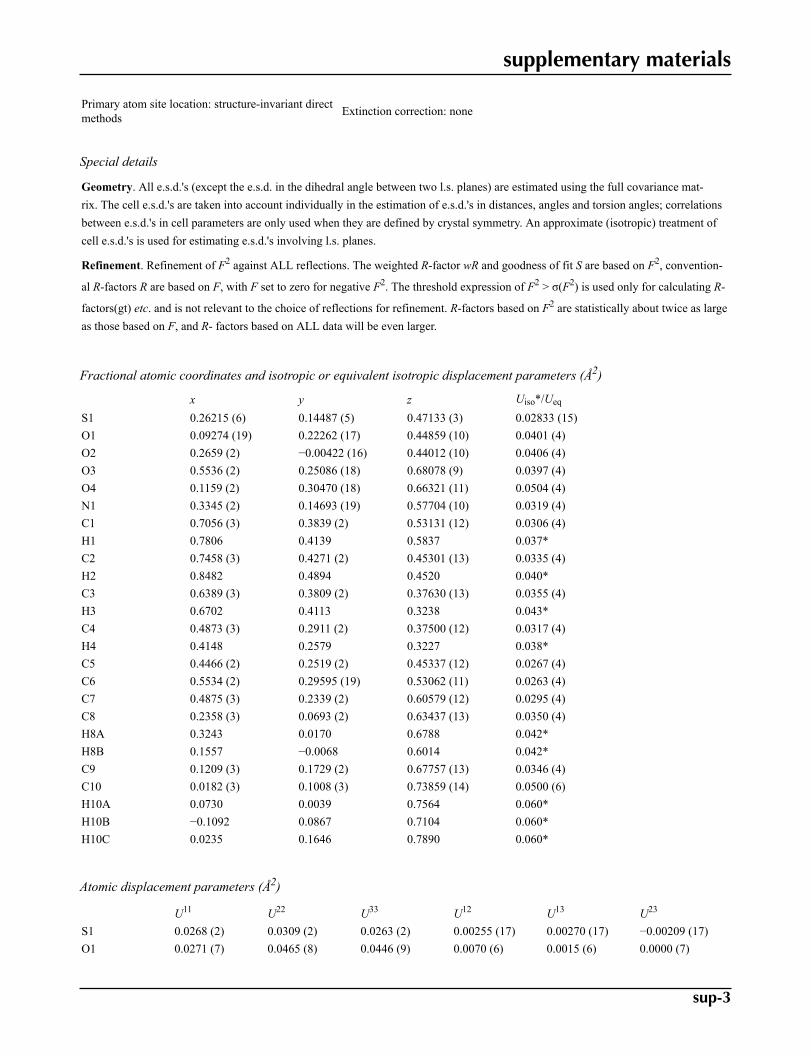
Primary atom site location: structure-invariant directmethods Extinction correction: none
Special details
Geometry. All e.s.d.'s (except the e.s.d. in the dihedral angle between two l.s. planes) are estimated using the full covariance mat-rix. The cell e.s.d.'s are taken into account individually in the estimation of e.s.d.'s in distances, angles and torsion angles; correlationsbetween e.s.d.'s in cell parameters are only used when they are defined by crystal symmetry. An approximate (isotropic) treatment ofcell e.s.d.'s is used for estimating e.s.d.'s involving l.s. planes.
Refinement. Refinement of F2 against ALL reflections. The weighted R-factor wR and goodness of fit S are based on F2, convention-
al R-factors R are based on F, with F set to zero for negative F2. The threshold expression of F2 > σ(F2) is used only for calculating R-
factors(gt) etc. and is not relevant to the choice of reflections for refinement. R-factors based on F2 are statistically about twice as largeas those based on F, and R- factors based on ALL data will be even larger.
Fractional atomic coordinates and isotropic or equivalent isotropic displacement parameters (Å2)
x y z Uiso*/Ueq
S1 0.26215 (6) 0.14487 (5) 0.47133 (3) 0.02833 (15)O1 0.09274 (19) 0.22262 (17) 0.44859 (10) 0.0401 (4)O2 0.2659 (2) −0.00422 (16) 0.44012 (10) 0.0406 (4)O3 0.5536 (2) 0.25086 (18) 0.68078 (9) 0.0397 (4)O4 0.1159 (2) 0.30470 (18) 0.66321 (11) 0.0504 (4)N1 0.3345 (2) 0.14693 (19) 0.57704 (10) 0.0319 (4)C1 0.7056 (3) 0.3839 (2) 0.53131 (12) 0.0306 (4)H1 0.7806 0.4139 0.5837 0.037*C2 0.7458 (3) 0.4271 (2) 0.45301 (13) 0.0335 (4)H2 0.8482 0.4894 0.4520 0.040*C3 0.6389 (3) 0.3809 (2) 0.37630 (13) 0.0355 (4)H3 0.6702 0.4113 0.3238 0.043*C4 0.4873 (3) 0.2911 (2) 0.37500 (12) 0.0317 (4)H4 0.4148 0.2579 0.3227 0.038*C5 0.4466 (2) 0.2519 (2) 0.45337 (12) 0.0267 (4)C6 0.5534 (2) 0.29595 (19) 0.53062 (11) 0.0263 (4)C7 0.4875 (3) 0.2339 (2) 0.60579 (12) 0.0295 (4)C8 0.2358 (3) 0.0693 (2) 0.63437 (13) 0.0350 (4)H8A 0.3243 0.0170 0.6788 0.042*H8B 0.1557 −0.0068 0.6014 0.042*C9 0.1209 (3) 0.1729 (2) 0.67757 (13) 0.0346 (4)C10 0.0182 (3) 0.1008 (3) 0.73859 (14) 0.0500 (6)H10A 0.0730 0.0039 0.7564 0.060*H10B −0.1092 0.0867 0.7104 0.060*H10C 0.0235 0.1646 0.7890 0.060*
Atomic displacement parameters (Å2)
U11 U22 U33 U12 U13 U23
S1 0.0268 (2) 0.0309 (2) 0.0263 (2) 0.00255 (17) 0.00270 (17) −0.00209 (17)O1 0.0271 (7) 0.0465 (8) 0.0446 (9) 0.0070 (6) 0.0015 (6) 0.0000 (7)

supplementary materials
sup-4
O2 0.0477 (9) 0.0311 (7) 0.0415 (8) −0.0004 (6) 0.0049 (7) −0.0067 (6)O3 0.0419 (8) 0.0536 (9) 0.0221 (7) −0.0003 (7) 0.0024 (6) 0.0009 (6)O4 0.0612 (11) 0.0388 (8) 0.0582 (11) 0.0046 (8) 0.0288 (9) 0.0006 (7)N1 0.0303 (8) 0.0411 (9) 0.0252 (8) −0.0019 (7) 0.0077 (6) −0.0004 (6)C1 0.0278 (9) 0.0337 (9) 0.0289 (9) 0.0050 (7) 0.0021 (7) −0.0009 (7)C2 0.0286 (9) 0.0344 (10) 0.0393 (11) 0.0029 (8) 0.0105 (8) 0.0018 (8)C3 0.0367 (10) 0.0430 (11) 0.0291 (10) 0.0065 (8) 0.0124 (8) 0.0051 (8)C4 0.0334 (9) 0.0382 (10) 0.0230 (9) 0.0071 (8) 0.0043 (7) −0.0016 (7)C5 0.0251 (8) 0.0287 (8) 0.0259 (9) 0.0051 (7) 0.0040 (7) −0.0006 (7)C6 0.0278 (9) 0.0280 (8) 0.0229 (8) 0.0061 (7) 0.0043 (7) −0.0008 (7)C7 0.0290 (9) 0.0345 (9) 0.0249 (9) 0.0052 (7) 0.0045 (7) 0.0005 (7)C8 0.0389 (10) 0.0345 (10) 0.0330 (10) −0.0006 (8) 0.0103 (8) 0.0053 (8)C9 0.0322 (10) 0.0435 (11) 0.0288 (10) −0.0055 (9) 0.0075 (8) 0.0006 (8)C10 0.0500 (13) 0.0703 (16) 0.0330 (12) −0.0204 (12) 0.0159 (10) −0.0010 (11)
Geometric parameters (Å, °)
S1—O2 1.4295 (15) C3—C4 1.388 (3)S1—O1 1.4305 (15) C3—H3 0.9500S1—N1 1.6667 (18) C4—C5 1.385 (3)S1—C5 1.7485 (19) C4—H4 0.9500O3—C7 1.211 (2) C5—C6 1.389 (3)O4—C9 1.204 (3) C6—C7 1.487 (3)N1—C7 1.388 (3) C8—C9 1.517 (3)N1—C8 1.456 (2) C8—H8A 0.9900C1—C6 1.383 (3) C8—H8B 0.9900C1—C2 1.392 (3) C9—C10 1.496 (3)C1—H1 0.9500 C10—H10A 0.9800C2—C3 1.389 (3) C10—H10B 0.9800C2—H2 0.9500 C10—H10C 0.9800
O2—S1—O1 116.35 (9) C6—C5—S1 110.42 (14)O2—S1—N1 109.71 (9) C1—C6—C5 120.11 (17)O1—S1—N1 110.53 (9) C1—C6—C7 127.31 (17)O2—S1—C5 112.90 (9) C5—C6—C7 112.55 (16)O1—S1—C5 112.22 (9) O3—C7—N1 123.44 (18)N1—S1—C5 92.60 (8) O3—C7—C6 127.64 (18)C7—N1—C8 123.13 (17) N1—C7—C6 108.91 (16)C7—N1—S1 115.29 (13) N1—C8—C9 112.95 (16)C8—N1—S1 121.49 (14) N1—C8—H8A 109.0C6—C1—C2 118.03 (18) C9—C8—H8A 109.0C6—C1—H1 121.0 N1—C8—H8B 109.0C2—C1—H1 121.0 C9—C8—H8B 109.0C3—C2—C1 121.19 (19) H8A—C8—H8B 107.8C3—C2—H2 119.4 O4—C9—C10 123.2 (2)C1—C2—H2 119.4 O4—C9—C8 121.05 (18)C4—C3—C2 121.16 (18) C10—C9—C8 115.78 (19)C4—C3—H3 119.4 C9—C10—H10A 109.5C2—C3—H3 119.4 C9—C10—H10B 109.5C5—C4—C3 116.97 (18) H10A—C10—H10B 109.5

supplementary materials
sup-5
C5—C4—H4 121.5 C9—C10—H10C 109.5C3—C4—H4 121.5 H10A—C10—H10C 109.5C4—C5—C6 122.52 (18) H10B—C10—H10C 109.5C4—C5—S1 127.07 (15)
O2—S1—N1—C7 120.12 (15) C2—C1—C6—C7 178.29 (17)O1—S1—N1—C7 −110.26 (15) C4—C5—C6—C1 0.9 (3)C5—S1—N1—C7 4.66 (15) S1—C5—C6—C1 −179.02 (13)O2—S1—N1—C8 −63.36 (17) C4—C5—C6—C7 −177.06 (16)O1—S1—N1—C8 66.26 (17) S1—C5—C6—C7 3.04 (19)C5—S1—N1—C8 −178.83 (15) C8—N1—C7—O3 1.2 (3)C6—C1—C2—C3 −1.4 (3) S1—N1—C7—O3 177.61 (15)C1—C2—C3—C4 0.6 (3) C8—N1—C7—C6 179.96 (16)C2—C3—C4—C5 0.9 (3) S1—N1—C7—C6 −3.59 (19)C3—C4—C5—C6 −1.7 (3) C1—C6—C7—O3 1.1 (3)C3—C4—C5—S1 178.21 (14) C5—C6—C7—O3 178.91 (18)O2—S1—C5—C4 63.10 (19) C1—C6—C7—N1 −177.59 (17)O1—S1—C5—C4 −70.77 (19) C5—C6—C7—N1 0.2 (2)N1—S1—C5—C4 175.78 (17) C7—N1—C8—C9 74.6 (2)O2—S1—C5—C6 −117.01 (13) S1—N1—C8—C9 −101.65 (19)O1—S1—C5—C6 109.12 (14) N1—C8—C9—O4 0.4 (3)N1—S1—C5—C6 −4.32 (14) N1—C8—C9—C10 −178.98 (18)C2—C1—C6—C5 0.7 (3)
Hydrogen-bond geometry (Å, °)
D—H···A D—H H···A D···A D—H···A
C10—H10B···O2i 0.98 2.57 3.324 (3) 134Symmetry codes: (i) −x, −y, −z+1.
supplementary materials
sup-6
Fig. 1

supplementary materials
sup-7
Fig. 2


![N -[4-( N -Cyclohexylsulfamoyl)phenyl]acetamide](https://static.fdokumen.com/doc/165x107/632f4f4de68feab59a0210b7/n-4-n-cyclohexylsulfamoylphenylacetamide.jpg)