DNA Methylation Analysis of the Angiotensin Converting Enzyme (ACE) Gene in Major Depression
11
DNA Methylation Analysis of the Angiotensin Converting Enzyme (ACE) Gene in Major Depression Peter Zill 1,2 *, Thomas C. Baghai 1,3 , Cornelius Schu ¨ le 1 , Christoph Born 1 , Clemens Fru ¨ stu ¨ ck 1 , Andreas Bu ¨ ttner 4,5 , Wolfgang Eisenmenger 4 , Gabriella Varallo-Bedarida 6 , Rainer Rupprecht 1,3 , Hans- Ju ¨ rgen Mo ¨ ller 1 , Brigitta Bondy 1,2 1 Department of Psychiatry and Psychotherapy, Ludwig-Maximilians-University Munich, Munich, Germany, 2 Division of Psychiatric Genetics and Neurochemistry, Ludwig- Maximilians-University Munich, Munich, Germany, 3 Department of Psychiatry and Psychotherapy, University Regensburg, Regensburg, Germany, 4 Institute for Legal Medicine, Ludwig-Maximilians-University Munich, Munich, Germany, 5 Institute for Legal Medicine, University Rostock, Rostock, Germany, 6 Department of Internal Medicine-Preventive Cardiology, Ludwig-Maximilians-University Munich, Munich, Germany Abstract Background: The angiotensin converting enzyme (ACE) has been repeatedly discussed as susceptibility factor for major depression (MD) and the bi-directional relation between MD and cardiovascular disorders (CVD). In this context, functional polymorphisms of the ACE gene have been linked to depression, to antidepressant treatment response, to ACE serum concentrations, as well as to hypertension, myocardial infarction and CVD risk markers. The mostly investigated ACE Ins/Del polymorphism accounts for ,40%–50% of the ACE serum concentration variance, the remaining half is probably determined by other genetic, environmental or epigenetic factors, but these are poorly understood. Materials and Methods: The main aim of the present study was the analysis of the DNA methylation pattern in the regulatory region of the ACE gene in peripheral leukocytes of 81 MD patients and 81 healthy controls. Results: We detected intensive DNA methylation within a recently described, functional important region of the ACE gene promoter including hypermethylation in depressed patients (p = 0.008) and a significant inverse correlation between the ACE serum concentration and ACE promoter methylation frequency in the total sample (p = 0.02). Furthermore, a significant inverse correlation between the concentrations of the inflammatory CVD risk markers ICAM-1, E-selectin and P-selectin and the degree of ACE promoter methylation in MD patients could be demonstrated (p = 0.01 - 0.04). Conclusion: The results of the present study suggest that aberrations in ACE promoter DNA methylation may be an underlying cause of MD and probably a common pathogenic factor for the bi-directional relationship between MD and cardiovascular disorders. Citation: Zill P, Baghai TC, Schu ¨le C, Born C, Fru ¨ stu ¨ ck C, et al. (2012) DNA Methylation Analysis of the Angiotensin Converting Enzyme (ACE) Gene in Major Depression. PLoS ONE 7(7): e40479. doi:10.1371/journal.pone.0040479 Editor: Marianna Mazza, Catholic University of Sacred Heart of Rome, Italy Received January 31, 2012; Accepted June 8, 2012; Published July 13, 2012 Copyright: ß 2012 Zill et al. This is an open-access article distributed under the terms of the Creative Commons Attribution License, which permits unrestricted use, distribution, and reproduction in any medium, provided the original author and source are credited. Funding: The present project was in part supported by a grant from the Deutsche Forschungsgemeinschaft (DFG BA 2309/1-1). No additional external funding received for this study. The funders had no role in study design, data collection and analysis, decision to publish, or preparation of the manuscript. Competing Interests: The authors have declared that no competing interests exist. * E-mail: [email protected] Introduction Major depression (MD) is one of the most common psychiatric diseases with a lifetime prevalence between 5% and 17% [1]. Although the monoamine hypothesis has been consistently linked to the etiology of MD, the exact pathophysiology remains largely unknown [2]. Actual hypothesis suggest complex interactions between multiple genetic, environmental and epigenetic risk factors that impact the development of MD and severity of symptomatology [3,4]. Epigenetic processes, mediated through modifications of DNA and histones by non mutagenic mechanisms play a major role in the context of development, but recent studies indicate that epigenetic alterations can also occur due to environmental stimuli during the whole life [5,6]. One of the most intensely investigated epigenetic mechanisms is the DNA methylation of cytosine residues in regulatory CpG islands of genes. Thus, recent studies have suggested that a dysfunction of DNA methylation in the brain may account for psychiatric disorders, including MD [4,7,8]. In this context animal models have shown that early life events in terms of stress, recognized as a major susceptibility factor for depression, can change the DNA methylation level of genes, being implicated in the neurobiology of depression, as glial cell-derived neurotrophic factor (GDNF), glutamic acid decarboxylase 1 (GAD1), estrogen receptor alpha 1b (ERA1B), arginine vasopres- sin (AVP), glucocrtiocoid receptor (NR3C1) and serotonin transporter (5-HTTP) [9–14]. In humans only a few studies of differential DNA methylation patterns in depression have been carried out so far, but the data seem to support those from animal studies. In vitro analysis of lymphoblastoid cell lines of depressed patients revealed an association between MD and increased DNA methylation levels of the 5-HTTP gene [15]. These findings have PLoS ONE | www.plosone.org 1 July 2012 | Volume 7 | Issue 7 | e40479
-
Upload
independent -
Category
Documents
-
view
0 -
download
0
Transcript of DNA Methylation Analysis of the Angiotensin Converting Enzyme (ACE) Gene in Major Depression

DNA Methylation Analysis of the Angiotensin ConvertingEnzyme (ACE) Gene in Major DepressionPeter Zill1,2*, Thomas C. Baghai1,3, Cornelius Schule1, Christoph Born1, Clemens Frustuck1,
Andreas Buttner4,5, Wolfgang Eisenmenger4, Gabriella Varallo-Bedarida6, Rainer Rupprecht1,3, Hans-
Jurgen Moller1, Brigitta Bondy1,2
1 Department of Psychiatry and Psychotherapy, Ludwig-Maximilians-University Munich, Munich, Germany, 2 Division of Psychiatric Genetics and Neurochemistry, Ludwig-
Maximilians-University Munich, Munich, Germany, 3 Department of Psychiatry and Psychotherapy, University Regensburg, Regensburg, Germany, 4 Institute for Legal
Medicine, Ludwig-Maximilians-University Munich, Munich, Germany, 5 Institute for Legal Medicine, University Rostock, Rostock, Germany, 6 Department of Internal
Medicine-Preventive Cardiology, Ludwig-Maximilians-University Munich, Munich, Germany
Abstract
Background: The angiotensin converting enzyme (ACE) has been repeatedly discussed as susceptibility factor for majordepression (MD) and the bi-directional relation between MD and cardiovascular disorders (CVD). In this context, functionalpolymorphisms of the ACE gene have been linked to depression, to antidepressant treatment response, to ACE serumconcentrations, as well as to hypertension, myocardial infarction and CVD risk markers. The mostly investigated ACE Ins/Delpolymorphism accounts for ,40%–50% of the ACE serum concentration variance, the remaining half is probablydetermined by other genetic, environmental or epigenetic factors, but these are poorly understood.
Materials and Methods: The main aim of the present study was the analysis of the DNA methylation pattern in theregulatory region of the ACE gene in peripheral leukocytes of 81 MD patients and 81 healthy controls.
Results: We detected intensive DNA methylation within a recently described, functional important region of the ACE genepromoter including hypermethylation in depressed patients (p = 0.008) and a significant inverse correlation between theACE serum concentration and ACE promoter methylation frequency in the total sample (p = 0.02). Furthermore, a significantinverse correlation between the concentrations of the inflammatory CVD risk markers ICAM-1, E-selectin and P-selectin andthe degree of ACE promoter methylation in MD patients could be demonstrated (p = 0.01 - 0.04).
Conclusion: The results of the present study suggest that aberrations in ACE promoter DNA methylation may be anunderlying cause of MD and probably a common pathogenic factor for the bi-directional relationship between MD andcardiovascular disorders.
Citation: Zill P, Baghai TC, Schule C, Born C, Frustuck C, et al. (2012) DNA Methylation Analysis of the Angiotensin Converting Enzyme (ACE) Gene in MajorDepression. PLoS ONE 7(7): e40479. doi:10.1371/journal.pone.0040479
Editor: Marianna Mazza, Catholic University of Sacred Heart of Rome, Italy
Received January 31, 2012; Accepted June 8, 2012; Published July 13, 2012
Copyright: � 2012 Zill et al. This is an open-access article distributed under the terms of the Creative Commons Attribution License, which permits unrestricteduse, distribution, and reproduction in any medium, provided the original author and source are credited.
Funding: The present project was in part supported by a grant from the Deutsche Forschungsgemeinschaft (DFG BA 2309/1-1). No additional external fundingreceived for this study. The funders had no role in study design, data collection and analysis, decision to publish, or preparation of the manuscript.
Competing Interests: The authors have declared that no competing interests exist.
* E-mail: [email protected]
Introduction
Major depression (MD) is one of the most common psychiatric
diseases with a lifetime prevalence between 5% and 17% [1].
Although the monoamine hypothesis has been consistently linked
to the etiology of MD, the exact pathophysiology remains largely
unknown [2]. Actual hypothesis suggest complex interactions
between multiple genetic, environmental and epigenetic risk
factors that impact the development of MD and severity of
symptomatology [3,4].
Epigenetic processes, mediated through modifications of DNA
and histones by non mutagenic mechanisms play a major role in
the context of development, but recent studies indicate that
epigenetic alterations can also occur due to environmental stimuli
during the whole life [5,6]. One of the most intensely investigated
epigenetic mechanisms is the DNA methylation of cytosine
residues in regulatory CpG islands of genes. Thus, recent studies
have suggested that a dysfunction of DNA methylation in the brain
may account for psychiatric disorders, including MD [4,7,8].
In this context animal models have shown that early life events
in terms of stress, recognized as a major susceptibility factor for
depression, can change the DNA methylation level of genes, being
implicated in the neurobiology of depression, as glial cell-derived
neurotrophic factor (GDNF), glutamic acid decarboxylase 1
(GAD1), estrogen receptor alpha 1b (ERA1B), arginine vasopres-
sin (AVP), glucocrtiocoid receptor (NR3C1) and serotonin
transporter (5-HTTP) [9–14]. In humans only a few studies of
differential DNA methylation patterns in depression have been
carried out so far, but the data seem to support those from animal
studies. In vitro analysis of lymphoblastoid cell lines of depressed
patients revealed an association between MD and increased DNA
methylation levels of the 5-HTTP gene [15]. These findings have
PLoS ONE | www.plosone.org 1 July 2012 | Volume 7 | Issue 7 | e40479

recently been confirmed in depressed patients [16]. Furthermore,
two studies could demonstrate that prenatal exposure to maternal
depressed mood decreases the DNA methylation pattern of the
methylenetetrahydro-folate reductase (MTHFR) gene and increas-
es the NR3C1 gene methylation in newborn infants [17,18].
Within a human post mortem study a hypermethylation of the
gamma-aminobutyric acid receptor alpha 1 subunit (GABRA1)
gene has been reported in the frontal cortex of depressed suicide
victims [19]. A genome wide approach to analyze the methylation
profile of over 14.000 genes in 33 depressed patients and 67
healthy probands showed coordinated signals with pathophysio-
logical mechanisms previously implicated in the etiology of
depression [8]. The findings from these first studies suggest that
DNA methylation profiles in several candidate genes may be
associated with depression, but the detailed patterns and
mechanisms are still unknown.
The angiotensin converting enzyme (ACE), a member of the
renin-angiotensin system (RAS) has been repeatedly discussed
both as a major candidate gene for depression and as a common
susceptibility factor for the bi-directional relation between
depression and cardiovascular disorders (CVD) [20]. Cross-
sectional and prospective analyses have shown that depression
may increase mortality and morbidity in patients with heart
failure, regardless of its etiology, and independently of traditional
cardiovascular factors. There is now compelling evidence that a
reciprocal relationship between both disorders exists. The
presence of cardiovascular disease can influence mood states and
some of the factors associated with depression, especially the
multiple alterations associated with acute and chronic stress, may
give rise to vascular disorders such as atherosclerosis, microcircu-
latory endothelial dysfunction, or metabolic conditions such as
diabetes and dyslipidemia [21]. As both disorders are complex and
multifactorial in origin, involving multiple genes with interactive or
additive effects together with environmental factors, depression
and cardiovascular disease could be different manifestations of the
same genetic and/or epigentic substrate.
Several genetic studies, especially with the functional ACE Ins/
Del polymorphism, from our and other groups propose an
involvement of ACE gene polymorphisms in the pathophysiology
of major depression, in the therapeutic outcome, in hypothalamic-
pituitary-adrenal (HPA)-axis dysregulation and expression of ACE
serum concentrations [22–27]. The mostly analyzed ACE Ins/Del
polymorphism accounts for ,40%–50% of the ACE serum
concentration variance, the remaining half is probably determined
by other genetic, environmental or epigenetic factors, but these
have not been successfully identified so far.
In this context, within an interdisciplinary study investigating
the relation between CVD and MD, we determined the serum
concentrations of ACE and several inflammatory biomarkers in
depressed patients as probable CVD risk markers. Besides the
observation that many inflammatory markers were increased in
depressed patients, we observed a significant correlation between
the ACE serum concentrations and several of these markers as
ICAM-1, VCAM-1, E-selectin, P-selectin and MCP-1 in these
patients, independent from the ACE Ins/Del polymorphism
(unpublished data).
Based on the above described findings, we hypothesize that
epigenetic mechanisms of the ACE gene might be a reason for
these observations. Therefore the main aim of the present study
was to analyze the DNA methylation in the regulatory region of
the ACE gene in peripheral leukocyte DNA of 81 depressed
patients compared to 81 healthy controls. The identified
methylation pattern was additionally investigated in human
post mortem brain tissue (cortex, hippocampus) of 13 control
individuals. Furthermore, the DNA methylation pattern was
correlated to ACE serum concentrations, to the above described
alterations in CVD markers and to clinical characteristics.
Results
We investigated the DNA methylation pattern in the 59-region
of the ACE gene in peripheral leukocyte DNA from blood of 81
depressed patients and 81 healthy controls. There were no
significant differences between patients and controls regarding
gender or age (table 1).
DNA Methylation Levels of the ACE Gene in PatientsControls and Post Mortem Brain Tissue
The CpG island analysis of a 7540 bp region, which contains
putative promoter sequences and exon 1, revealed a 1398 bp CpG
island (2567 to 831, related to the transcription start site). After
bisulphite sequencing intensive methylation was found from
nucleotide 2456 to 2255. In the other parts of the CpG island
there was no, respectively very rare, negligible methylation. Thus,
the following analysis and evaluations refer to the methylated
region from nucleotide 2456 to 2255.
The (2456/2255) region contains 25 CpG sites; 24 of them
could be analyzed by sequencing in 81 patients and 81 controls
(figure 1b). Depressive patients showed a hypermethylation pattern
at all CpG sites (mean: 11.9% 622.8% in average over all 24 CpG
sites) compared to healthy controls (mean: 6.8% 615.9%). This
observation resulted in statistical significant differences at three
CpG sites (site 1, 5, 12) and a trend for significance (0.05,p,0.09)
at 5 CpG sites (site 7, 10, 11, 13, 21) (figure 1a). In 38 patients out
of 81 (47%) methylation between one and 20 CpG sites with mean
methylation frequencies between 1% and 96% (determined as
mean value of the methylation frequency over all 24 CpG sites)
was detectable. In the control group only 21 subjects out of 81
(26%) revealed a methylation pattern between one and 11 CpGs
with lower mean methylation frequencies between 1% and 73%
(over all 24 CpG sites). Figure 1c summarizes this significant
difference between methylated patients and controls (p = 0.008,
x2 = 7.1, df = 1).
Due to the partial tissue specificity of DNA methylation, we
additionally analyzed the ACE promoter methylation in post
mortem brain tissue to check whether the (2456/2255) ACE
gene region is also a methylation ‘‘hot-spot’’ in brain which can be
detected in neural tissues or whether the observed pattern is
leukocyte specific. Data from hippocampus and cortex of 13
control individuals could clearly demonstrate a similar methylation
Table 1. Demographic characteristics of the depressedpatients (MD) and healthy controls (CON).
MD (N = 81) CON (N = 81)
Sex [males/females] 30/51 40/41
Age [years, mean 6 SD];(range)
45.8614.3 (21–76) 46.2614.2 (19–73)
Age of onset[years, mean 6 SD]
37.2612.6
Episodes [mean 6 SD] 3.762.4
HAMD-17 [mean 6 SD] 22.565.5
ACE-Concentration[Units 6 SD]
44.4615.4 46.2614.2
doi:10.1371/journal.pone.0040479.t001
ACE Gene, Epigenetics and Depression
PLoS ONE | www.plosone.org 2 July 2012 | Volume 7 | Issue 7 | e40479

pattern as in leukocytes of controls, but with somewhat higher
frequency (figure 2). In average, methylation levels of 18% (over all
24 CpG sites) in cortex (mean: 17.8% 619.9%) and 13% in
hippocampus (mean: 12.6% 614.0%) could be measured.
Further statistical analysis revealed no correlation between
number of methylated CpG sites or methylation frequency and
age, gender or post mortem interval in the analyzed patient-,
control- and post mortem samples.
Figure 1. Frequency of DNA methylation in the investigated ACE promoter region. (a) Frequency of DNA methylation at 24 CpG sites,located in the (2456/2255) region of the ACE gene in depressive patients (MD) and healthy controls (CON); *: p,0.05 (x2-test). (b) Sequence of the(2456/2255) region (italic, underlined) including the 24 investigated CpG sites (bold). (c) Frequency of methylation and non-methylation at the 24CpG sites, located in the (2456/2255) region of the ACE gene in depressive patients (MD) and healthy controls (CON). Patients have a significantlyhigher methylation status than the healthy controls (p = 0.008, x2 = 7.1, df = 1). *: p,0.05 (x2-test).doi:10.1371/journal.pone.0040479.g001
ACE Gene, Epigenetics and Depression
PLoS ONE | www.plosone.org 3 July 2012 | Volume 7 | Issue 7 | e40479

Correlation between DNA Methylation of the ACE Geneand ACE Serum Concentration
ACE serum concentrations could be measured at baseline in 74
medication-free depressive patients and 68 controls with no
differences between the groups (44.4 units 615.4 units versus 46.2
units 614.2 units). 36 patients and 19 controls were methylated,
whereas 38 patients and 49 controls had no methylation in the
investigated gene region. Analysis in groups of methylated versus
unmethylated samples yielded a significant difference in the total
sample in terms of lower ACE concentrations in methylated
probes (p = 0.005, F = 8.1). This effect was also obvious in
depressive patient, but with lower significance due the smaller
sample size (p = 0.03, F = 5.1). In the control group the observed
pattern was not statistically significant (figure 3). These results
could be supported by correlation analysis. We found a significant
inverse correlation between methylation frequency (determined as
mean value of the methylation frequency over all 24 CpG sites for
each probe) and the ACE serum concentration in the total sample
(r = 2197, p = 0.02) (figure 4). We could not identify a direct
correlation between the number of methylated CpG sites and the
ACE serum concentration neither in patients nor in controls.
Moreover, there was no relation between age or gender and ACE
levels in both groups.
Correlation between DNA Methylation of the ACE Geneand Serum Concentration of Inflammatory CVD Markers
We found a significant positive correlation between the serum
concentrations of ACE and the inflammatory CVD risk markers
ICAM-1 (r = 0.26, p = 0.05), VCAM-1 (r = 0.27, p = 0.04), E-
selectin (r = 0.48, p,0.001), P-selectin (r = 0.33, p = 0.02) and
MCP-1 (r = 0.30, p = 0.03) in depressive patients, independent
from the functional ACE Ins/Del polymorphism (data not shown).
Assuming that this observation might be caused by epigenetic
mechanisms, we analyzed a possible relation between the
methylation pattern in the (2456/2255) ACE gene region and
the inflammatory marker concentrations. Data were available
from 66 medication-free depressive patients (baseline).
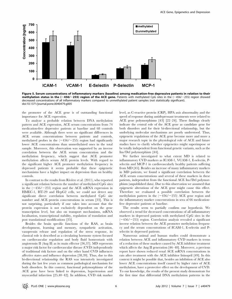
As shown in figure 5 patients with methylated CpG sites in the
(2456/2255) region showed only a trend for decreased concen-
trations of all inflammatory markers compared to unmethylated
patient samples (not statistically significant). Additional correlation
analyses revealed a significant inverse relation between the
methylation frequency (determined as mean value of the
methylation frequency over all 24 CpG sites for each probe) and
the serum baseline concentrations of ICAM-1 (r = 20.289,
p = 0.02), E-selectin (r = 20.249, p = 0.04) and P-selectin
(r = 20.333, p = 0.01) (figure 6).
We could not identify a direct correlation between the number
of methylated CpG sites and any of the inflammatory marker
concentrations. Furthermore there was no relation between age or
gender and the inflammatory marker concentrations.
Further analysis of patient’s methylation pattern or number of
methylated CpG sites in relation to clinical variables as HAMD-
17-total scores, age of onset, number of episodes, duration of
clinical stay or internal parameters as weight, body mass index
(BMI), blood glucose and lipids revealed no significant correlation.
Discussion
Angiotensin-converting-enzyme (ACE) is a membrane bound
endopeptidase which is involved in the metabolism of angiotensin
I and bradykinin, both being important for the regulation of
vascular tone and cardiac functions [28]. Besides its crucial role in
cardiovascular homeostasis, the ACE gene confers also suscepti-
bility for depression, as demonstrated in several genetic and
expression studies [23,25,26]. Moreover the ACE gene is discussed
as a common factor for the known relation between MD and
CVD, but the underlying mechanisms are poorly understood [21].
Epigenetic alterations, probably via environmental influences as
early stressful life events might be one explanation for these
observations.
Therefore we analyzed the DNA methylation in the promoter
region of the ACE gene in peripheral leukocyte DNA of 81
depressed patients and 81 healthy controls in relation to ACE
serum concentrations, to inflammatory CVD risk markers and to
clinical characteristics.
Bisulfite sequencing of a 1398 bp CpG island in the proximal
ACE promoter revealed an intensive methylation pattern of 24
CpG sites from nucleotide 2456 to 2255 with significantly higher
Figure 2. Frequency of DNA methylation at 24 CpG sites, located in the (2456/2255) region of the ACE gene in human post mortemsamples of hippocampus and cortex from 13 control individuals.doi:10.1371/journal.pone.0040479.g002
ACE Gene, Epigenetics and Depression
PLoS ONE | www.plosone.org 4 July 2012 | Volume 7 | Issue 7 | e40479

methylation levels in depressive patients versus controls. DNA
methylation within this (2456/2255) region could also be
demonstrated in post mortem hippocampus and cortex tissue of
13 control individuals. Taken together, these findings demonstrate
that in almost 50% of depressed patients promoter methylation of
the ACE gene is detectable with a two-fold higher frequency as in
controls. Our data further demonstrate that a comparable
methylation pattern as in peripheral lymphocytes is present in
cortex and hippocampus, which suggest also an important role of
ACE promoter methylation in the brain.
The anterior cingulate cortex, amygdala and hippocampus form
part of an interconnected prefrontal-limbic network (PLN) that is
dysregulated in depressive disorders [29]. The PLN is modulated
by the basal ganglia and midbrain structures, as well as by the
HPA-axis. The ACE gene is not only expressed in the PLN
[30,31], but also involved in HPA-axis dysregulation in depressed
patients, as we have reported in previous studies [22,25].
Moreover, epigenetic alterations within the PLN in relation to
early life stress, a known risk factor for depression, have been
reported for the glucocorticoid receptor gene in animal studies
[32]. Therefore it seems probable that epigenetic alterations of the
ACE gene in terms of hypermethylation might contribute to the
dysregulated PLN in depression, at least in a subgroup of patients.
We are aware that these conclusions from our data are highly
speculative because only post mortem brain samples of controls,
which died mostly from cardiovascular diseases, were included in
the analysis due to the lack of brain tissue from MD patients. Thus
it is not possible to draw meaningful conclusions.
Interestingly our findings of intensive DNA methylation in the
(2456/2255) ACE gene region are consistent with a recent report
by Riviere et al. (2011), who investigated the influence of DNA
methylation and chromatin condensation state on the expression
of the human ACE gene in human liver (HepG2), colon (HT-29),
microvascular endothelial (HMEC-1) and lung (SUT) cell lines
[33]. Within a 3340 bp fragment of the putative ACE promoter
region, they described two CpG island (2562/2244 and 2202/
216) after in silico analysis, which are both located in the CpG
island, identified in our study. Similarly to our results they found
almost exclusively intensive DNA methylation at the 25 CpG sites
in the (2456/2255) region, with highest levels in HepG2 cells
followed by HMEC-1, HT-29 and SUT cells. This proximal
region contains several transcription factor response elements as
EGR-1, SP 1, CP 2 and was also reported in an earlier study to be
sufficient to drive high transcriptional activity [34]. Furthermore,
within luciferase reporter gene assays Riviere et al. (2011) could
demonstrate that a 3340 bp fragment of the 59-ACE region which
includes the (2456/2255) fragment induces cell type specific
promoter activity which is dependent of the methylation level.
Additionally, the methylation pattern was clearly related to the
ACE mRNA concentrations in vitro as well as in vivo (rats) [33].
These observations confirms that the (2456/2255) region within
Figure 3. ACE serum concentrations (baseline) among the total sample, medication-free depressive patients (MD) and controls(CON) in relation to their methylation status in the (2456/2255) region of the ACE gene. Methylated probes showed lower ACEconcentrations (p = 0.005, F = 8.1). This effect was also obvious in depressive patient (p = 0.03, F = 5.1). In the control group there was a similar pattern,but without statistical significance. *: p,0.05 (ANCOVA). Data are mean values 6 s.e.m.doi:10.1371/journal.pone.0040479.g003
Figure 4. Correlation analysis between the ACE serum concen-trations (baseline) and the ACE methylation frequency over all24 CpG sites of each probe. We found a significant inversecorrelation in the total sample (r = 2197, p = 0.02; Pearson’s correlation).doi:10.1371/journal.pone.0040479.g004
ACE Gene, Epigenetics and Depression
PLoS ONE | www.plosone.org 5 July 2012 | Volume 7 | Issue 7 | e40479
the promoter of the ACE gene is of outstanding functional
importance for ACE expression.
To analyze a probable relation between DNA methylation
pattern and ACE expression, ACE serum concentrations from 74
medication-free depressive patients at baseline and 68 controls
were available. Although there were no significant differences in
ACE serum concentrations between patients and controls,
methylated probes in the (2456/2255) region had significantly
lower ACE concentrations than unmethylated ones in the total
sample. Moreover, this observation was supported by an inverse
correlation between the ACE serum concentration and the
methylation frequency, which suggest that ACE promoter
methylation affects serum ACE protein levels. With regard to
the significant higher ACE promoter methylation frequency in
depressed patients it seems probable that these epigenetic
mechanisms have a higher impact on depression than on healthy
controls.
In contrast to the results from Riviere et al. (2011), who reported
a significant relation between the number of methylated CpG sites
in the (2456/2255) region and the ACE mRNA expression in
HMEC-1, HT-29 and HepG2 cells, we could not detect any
significant direct correlation between methylated CpG site
number and ACE protein concentrations in serum [33]. This is
not surprising, particularly if one takes into account that the
protein expression is not exclusively dependent on the gene
transcription level, but also on transport mechanisms, mRNA
localisation, transcriptional stability, regulation of translation and
post translational modifications [35].
Besides the brain specific functions of the RAS, as brain
development, learning and memory, sympathetic activation,
vasopressin release and regulation of the stress response, its
classical role is described as a circulating hormone system focused
on cardiovascular regulation and body fluid homeostasis with
angiotensin II (Ang II) as its main effector [36,37]. MD represents
a major risk factor for cardiovascular disease (CVD) independently
of traditional risk factors and on the other hand CVD influences
affective states and influence depression [38,39]. Thus, due to this
bi-directional relationship the RAS was intensively investigated
during the last few years as common pathological mechanism for
both disorders. In this context, functional polymorphisms of the
ACE gene have been linked to depression, hypertension and
myocardial infarction [25,40–42]. In addition, CVD risk marker
level, as C-reactive protein (CRP), HPA axis abnormality and the
speed of response during antidepressant treatments were related to
ACE gene polymorphisms [43] [22–24]. These findings clearly
indicate the central role of the ACE gene as candidate gene for
both disorders and for their bi-directional relationship, but the
underlying molecular mechanisms are poorly understood. Thus,
epigenetic regulations of the ACE gene become more and more a
major research topic in the physiological role of ACE and future
studies have to clarify whether epigenetics might superimpose or
be totally independent from functional genetic variants, such as the
Ins/Del polymorphism [44].
We further investigated to what extent MD is related to
inflammatory CVD markers as ICAM-1, VCAM-1, E-selectin, P-
selectin and MCP-1 in cardiovascularly healthy patients suffering
from MD [45]. Besides an increase of many inflammatory markers
in MD patients, we found a significant correlation between the
ACE serum concentrations and several of these markers in these
patients, independent from the functional ACE Ins/Del polymor-
phism (unpublished data). Due to this observation we assumed that
epigenetic alterations of the ACE gene might cause this effect.
Therefore we evaluated a possible correlation between the
methylation pattern in the (2456/2255) ACE gene region and
the inflammatory marker concentrations in sera of 66 medication-
free depressive patients at baseline.
The results seem to partially confirm our hypothesis. We
observed a trend for decreased concentrations of all inflammatory
markers in depressed patients with methylated CpG sites in the
(2456/2255) region. Correlation analysis revealed a significant
inverse relation between the ACE promoter methylation frequen-
cy and the serum concentrations of ICAM-1, E-selectin and P-
selectin in depressed patients.
Numerous animal and human studies could demonstrate a
relation between ACE and inflammatory CVD markers in terms
of a reduction of these markers caused by ACE inhibitor treatment
which affects the Ang II generation [46–48]. Moreover, a previous
report have shown reduced renal ACE mRNA concentrations in
rats after treatment with the ACE inhibitor lisinopril [49]. In this
context it might be possible that, besides an inhibition of ACE also
lower ACE concentrations itsself caused by higher rates of ACE
methylation, have a protective effect on the development of CVD.
To our knowledge, the results of the present study demonstrate for
the first time that differential DNA methylation patterns in the
Figure 5. Serum concentrations of inflammatory markers (baseline) among medication-free depressive patients in relation to theirmethylation status in the (2456/2255) region of the ACE gene. Patients with methylated CpG sites in the (2456/2255) region showeddecreased concentrations of all inflammatory markers compared to unmethylated patient samples (not statistically significant).doi:10.1371/journal.pone.0040479.g005
ACE Gene, Epigenetics and Depression
PLoS ONE | www.plosone.org 6 July 2012 | Volume 7 | Issue 7 | e40479

promoter region of the ACE gene seem to affect the expression of
inflammatory CVD risk marker concentrations in depression. This
interaction might be involved in the pathophysiological mecha-
nisms which predispose a subgroup of depressed patients to
develop CVD or not. This could be one of the missing links of the
bi-directional relation between MD and CVD.
We are aware that several limitations should be considered
when interpreting our findings and that our conclusions are
partially very speculative at the moment. First, the sizes of patient
and control samples are probably not large enough for final
conclusions. This might also be a reason for the lack of a
significant relation between clinical variables like HAMD-17-total
scores, treatment response, age of onset, number of episodes or
duration of clinical stay and the methylation frequency in the ACE
promoter.
Second, the use of peripheral leukocyte DNA limits the
inference that can be drawn regarding pathways involving the
brain. Nevertheless, a growing body of research confirms the
promise of using blood cells as peripheral model [50,51]. Human
and animal studies in neurological disorders such as stroke,
multiple sclerosis or autism using microarrays could clearly
demonstrate specific disease related gene profiles in blood of
patients including many brain specific genes [52]. Moreover,
numerous epigenetic studies of psychiatric disorders have been
performed in peripheral leukocyte DNA [53], and there is
evidence suggesting a DNA methylation concordance between
peripheral tissues and brain for genes like catechyl-o-methyltrans-
ferase (COMT) [54,55]. Only a limited number of studies have
investigated common central and peripheral ACE alterations. For
example, Tan et al. (2004) measured increases in brain and cardiac
ACE densities after myocardial infarct in rats [56].
In the present study we compared brain and blood ACE
promoter methylation in controls using post mortem brain tissues
of hippocampus and frontal cortex. Total methylation levels of
18% in cortex and 13% in hippocampus could be measured
compared to ,7% in peripheral leukocytes of controls. Although
the post mortem samples came from individuals who died mostly
from cardiovascular diseases which hampers any conclusions
regarding differences in methylation frequencies between blood
and brain controls, it appears that the (2456/2255) ACE gene
region is also a probable ‘‘hot-spot’’ for methylation in these brain
regions. On the other hand the differences in methylation
frequencies between post mortem and blood samples might be a
result of tissue specificity of ACE regulation, suggesting less ACE
gene expression in cortex and hippocampus than in peripheral
leukocytes. Therefore, it remains the question how brain and
peripheral ACE features parallel to each other. From the present
data we cannot answer this question in full detail, but we
hypothesize that the observed methylation pattern in peripheral
blood cells might represent a disease specific profile. Further
studies in different brain tissues of depressed patients and healthy
controls are needed to validate this hypothesis.
Moreover, the peripheral DNA samples are a mix of
mononuclear cells, which derive from two distinct lineages and
compartments. Since cell count information was not available, we
cannot completely rule out whether the differences in methylation
Figure 6. Correlation analysis between the inflammatorymarker serum concentrations (baseline) and the ACE methyl-ation frequency over all 24 CpG sites of each probe. We found asignificant inverse correlation between the methylation frequency andthe serum baseline concentrations of ICAM-1 (r = 20.289, p = 0.02), E-selectin (r = 20.249, p = 0.04) and P-selectin (r = 20.333, p = 0.01);(Pearson’s correlation).doi:10.1371/journal.pone.0040479.g006
ACE Gene, Epigenetics and Depression
PLoS ONE | www.plosone.org 7 July 2012 | Volume 7 | Issue 7 | e40479

frequencies reflect different relative numbers of mononuclear cells
in the cases versus the controls.
Further, our data represent correlative findings and thus cannot
prove a causal relationship between ACE DNA methylation –
ACE mRNA expression – ACE protein expression, respectively
inflammatory CVD marker expression, because we had no mRNA
from patients and controls of the present study. It has also to be
considered that soluble serum ACE originates from endothelial
cells, mainly from lung shed from the cell membrane by
proteolytic cleavage, but at physiologic conditions the concentra-
tion of ACE in blood is very stable and thus serum ACE
measurements are an essential tool for monitoring the level,
respectively activity of ACE [57,58].
Furthermore, DNA methylation represents a mixture of state-
and trait dependent effects. Further prospective studies have to
clarify whether the observed DNA methylation revert to control
levels after remission, respectively whether there are similar
abnormalities in first degree relatives of the patients.
Otherwise, despite these limitations, the functional significance
of our data is widely supported by the recent distinguished work of
Riviere et al. (2011) who could clearly demonstrate the functional
significance of DNA methylation in the (2456/2255) ACE gene
region for promoter activity as well as for mRNA expression in
vitro and in vivo [33]. Thus, we believe that the limitations of our
study do not detract from the major significance of its results,
which will need further confirmation in independent studies.
In summary, this study is the first to demonstrate that the
human ACE expression is under a strong epigenetic influence by
DNA methylation in MD. Approximately 50% of depressed
patients of the present study showed DNA methylation in a
functional relevant 201 bp region of the ACE gene proximal
promoter in peripheral leukocytes compared to only ,25% of
healthy controls. This methylation pattern seems to have an
influence on serum ACE protein expression in the total sample
and additionally on the amount of inflammatory risk markers for
CVD, as ICAM-1, E-selectin and P-selectin in depressed patients.
Although several questions remain to be resolved regarding the
precise functional consequences of the identified ACE promoter
DNA methylation, our data are consistent with the hypothesis that
DNA methylation aberrations may be an underlying cause of
depressive disorders. Furthermore, the present findings support the
importance of CpG methylation of the ACE gene as a common
pathogenic factor in MD and CVD and may lead to a better
understanding of the neurobiology of MD, as well as of the bi-
directional relationship between MD and CVD.
Materials and Methods
Ethics StatementAll clinical investigations have been conducted according to the
principles expressed in the Declaration of Helsinki and approved
by the Ethics Committee of the Medical Faculty of the Ludwigs
Maximilians University (LMU) Munich (Head: Prof. Dr. Wolf-
gang Eisenmenger, Members: Prof. Dr. Eckhard Held, Prof. Dr.
Gustav Paumgartner, PD Dr. Thomas Beinert, Prof. Dr. Hans
Ulrich Gallwas, Prof. Dr. Detlef Kunze, Dr. Viktoria Monch, Prof.
Dr. Randolph Penning, Prof. Dr. Klaus Hahn, Prof. Dr. Klaus
Jurgen Pfeifer, and Dr. Christian Zach). Ethics proposal, Project
No. 213/00; positive vote from: 12.05.2005 ‘‘Genetische, bio-
chemische und funktionelle Untersuchungen an depressiven
Patienten und gesunden Kontrollpersonen’’. Written informed
consent was given by the patients and healthy volunteers. Autopsy
samples: The autopsies were court ordered from the state attorney
according to the German legal situation due to unknown causes of
death. In that case informed consent from the next of kin is not
required, because relatives have no possibility for intervention.
Within these autopsies it is necessary to take routinely additional
tissue probes for probable further investigations. The probes of the
present study originate from these investigations. The Ethics
Committee of the LMU Munich approved this procedure. All
autopsies were performed according to the legal requirements. For
the control individuals the natural cause of death was verified
finally by these autopsies. Blood and brain samples were
exclusively taken during the routine autopsies to perform the
court ordered analysis. Furthermore post mortem material will be
preserved for subsequently necessary investigations on behalf of
the state attorney. Blood and brain samples were never taken for
research. For research projects we use only remaining post
mortem samples which have been released and approved for use in
research by the Ethics Committee of the LMU Munich. As
described, the consent for research use of autopsy tissues will be
given by the local Ethics Committees of the universities. This is the
current procedure in legal medicine in Germany.
SubjectsDepressed patients and healthy controls were taken from two
cohorts recently published in an interdisciplinary study by Baghai
et al. (2010) [45]. In brief, 81 unrelated Caucasian patients
suffering from unipolar major depression (30 males, 51 females,
mean age 45.8 years 614.3 years) were recruited from in-patients
at the Department of Psychiatry and Psychotherapy of the
Ludwig-Maximilian-University of Munich (LMU). Patients were
diagnosed by experienced and trained psychiatrists according to
DSM-IV using the Structured Clinical Interview for DSM-IV
disorders (SCID-I) [59]. The main inclusion criteria were unipolar
depression and a score in the Hamilton Rating Scale for
Depression (HAM-D17) of at least 17. Prior to inclusion in the
study blood samples were obtained for routine laboratory
screening, a medical history was taken and a physical examination
was performed by a physician to exclude severe medical disorders.
Clinically relevant medical illness and the concomitant use of
antihypertensive medications such as ACE inhibitors or angioten-
sin receptor blockers as well as beta blockers and hormone
replacement therapies, alcohol or drug abuse within the last
6 month prior to study inclusion or withdrawal signs led to
exclusion from the study. Blood samples were taken after a
washout period of at least 3 days.
81 ethnically matched subjects from the general population (40
males, 41 females, mean age 46.2 years 614.2 years) served as
control group. All subjects were psychiatrically screened by a short
structured interview with a psychiatrist to rule out psychiatric
problems. Subjects with known history of psychiatric disorders
were excluded from the study.
All patients and controls were of Caucasian origin from the
German population and came from the same geographical area in
southern Germany. Demographic data of patients and controls are
given in table 1.
Post Mortem SamplesBrain specimens derived from 13 individuals (7 males,
6 females, mean age 41.9 years 616.3 years) who died suddenly
from diseases not directly involving the CNS were obtained from
the Institute for Legal Medicine of the LMU. Causes of death were
the following: acute cardiac failure (n = 4), accident (n = 4), aortic
aneurysm (n = 2) and homicide (n = 3). The clinical, respectively
medical data sheets of the individuals were available to the
Institute for Legal Medicine of the LMU and did not give any hint
on lifetime psychiatric or neurological disorders. Accordingly to
ACE Gene, Epigenetics and Depression
PLoS ONE | www.plosone.org 8 July 2012 | Volume 7 | Issue 7 | e40479

the medical records there was no history of psychopharmacolog-
ical medication, alcohol or drug abuse. All individuals were
Caucasian from the same geographical region in southern
Germany.
The unfixed brain specimens were obtained 4–29 hours
(average post mortem delay of 15.8 hours 67.3 hours) after death
during routine autopsy. Sections were taken from the posterior
hippocampus at the coronal level of the lateral geniculate nucleus
and from the cerebellar cortex. The tissue probes were collected
using the RNAlater kit (Qiagen, Hilden, Germany) and immedi-
ately frozen at 280 C until used for the DNA extraction.
Promoter CpG Island AnalysisWe selected a 7540 bp region (25989/+1551, related to the
transcription start site), containing the putative promoter and exon
1 of the ACE gene (NG_011648: nt 1 to 6540). CpG content
analysis was performed applying the CpG Island Searcher (http://
www. uscnorris.com/cpgislands2/cpg.aspx) [60]. The CpG is-
lands were defined as a region with at least 500 bp, with a GC
percentage that is greater than 55% and with an observed/
expected CpG ratio that is greater than 65%.
Bisulfite Sequencing of Genomic DNAGenomic DNA was extracted from whole blood of patients and
controls, as well as from the post mortem brain samples with the
Invisorb Blood Giga Kit (Invitek, Berlin, Germany) according to
the manufacturer’s instructions. The peripheral DNA samples
originated from several genetic projects. Therefore, cell counts to
differentiate between the different mononuclear blood cells were
not available.
750 ng of genomic DNA were treated with sodium bisulfite
using the EpiTect Bisulfite Kit (Qiagen, Hilden, Germany)
according to the manufacturer’s instructions.
Five primer sets were designed to amplify the CpG island region
in overlapping fragments. Given that we exclusively found high
degree of DNA methylation in the PCR fragment of primer set 2,
the following method description refers only to primer set 2.
Detailed information about the further PCR sets, as well as the
PCR and sequencing conditions can be obtained on request.
Both primers harboured universal primer sequences at their 59-
end to facilitate sequencing. The following sequences were
designed for primer set 2: forward primer (NG_011648: 2465/
2447, related to the transcription start site): 59-M13(-21)-TTA
TGG TTT GGT GAA GAA GT-39; reverse primer:
(NG_011648: 2231/2210, related to the transcription start site):
59-M13(-29)-AAA AAA ACC TCC TCT CTT TAA A-39.
PCR was carried out in a final volume of 10 ml containing 1 ml
of bisulfite treated DNA, 200 mM of each dNTP, 5 mM MgCl2,
2 ml Q-Solution, 0.5 mM of the forward and reverse primer and
0.5 units Taq DNA polymerase (Qiagen, Hilden Germany). After
an initial denaturation step of 95uC for 15 min, there was a first
round of 5 cycles of denaturation at 95uC for 30 sec, annealing at
58uC for 90 sec and extension at 72uC for 120 sec. The second
PCR round consisted of 35 cycles of denaturation at 95uC for
30 sec, annealing at 54uC for 90 sec and extension at 72uC for
90 sec. A final step was performed at 72uC for 5 min. PCR
products were purified by SAP (shrimp alkaline phosphatase)
treatment according to standard protocols (Applied Biosystems,
Foster City, CA, USA).
50 ng of the purified PCR product was used for cycle
sequencing with the BigDye Terminator 3.1 Cycle Sequencing
Kit (Applied Biosystems, Foster City, CA, USA) in a final volume
of 20 ml containing 3 ml reaction mix and 0.16 mM pimer. Results
were verified by bi-directional sequencing. Cycling conditions
were: 35 cycles of denaturation at 96uC for 10 sec, annealing at
50uC for 5 sec and extension at 50uC for 4 min. The amplified
products were ethanol/EDTA purified according to the described
conditions by Applied Biosystems. Samples were sequenced on an
ABI 310 capillary sequencer (Applied Biosystems, Foster City, CA,
USA). Data were processed by using Sequencing Analysis version
5.3.1 applying the KB-Base Caller 1.3 (Applied Biosystems, Foster
City, CA, USA).
The approximate methylation frequency of cytosine of each
CpG site was calculated by comparing the peak height of the
cytosine signal with the sum of the cytosine and thymidine peak
height signals, as described by Melki et al. (1999) [61]. This
method was validated by Jiang et al. (2010) [62], who could
confirm after comparison with pyrosequencing and bisulfite-clonig
sequencing that this method is a simple, high-throughput and a
reliable technology for determining the methylation status of
specific genes. However, when using direct sequencing, it is often
difficult to assess methylation levels ,15% due to variable
sequencing background signal. Thus, CpG sites with ratio ranges
0.00–0.20 were considered as unmethylated (0%) and ranges
between 0.81–1.0 as fully methylated (100%). For the ratio range
0.21–0.80 the calculated frequencies were used.
Measurement of ACE ConcentrationBlood samples for ACE determination were drawn from
medication-free MD patients and controls before the first
application of antidepressant treatment (baseline). Aprotenin
treated serum was stored frozen at 280uC until assessment.
ACE was quantified using a commercial radio enzyme assay
(REA) (ACE-REA, Diagnostic Products Corporation Biermann,
Bad Nauheim, Germany). The lower detection limit was 3.8 U/l.
Measurement of Inflammatory MarkersThe inflammatory markers were analyzed in serum of
medication-free patients before pharmacological treatment (base-
line). MCP-1, VCAM-1, ICAM-1, E-selectin, and P-selectin, were
determined using ELISAs obtained from IBL (R&D Systems,
Minneapolis, USA) according to manufacturer’s instruction.
All laboratory analyses were carried out blind to case control
status.
Statistical AnalysisAll statistical analyses were performed with SPSS for Windows
(Version 19.0; SPSS; Chicago, IL).
All dependent variables (concentrations of ACE and inflamma-
tory markers) were examined by the Kolmogorov-Smirnov test on
normality. Stepwise linear regression analyses (pin = 0.05, pout
= 0.10) with the independent variables gender and age were
performed for all dependent variables. Correlations analyses
between dependent variables and age, as well as methylation
pattern and between methylation pattern and post mortem
interval (PMI) in post mortem samples were performed with the
Pearson’s test.
Analyses of covariance (ANCOVA) were conducted to test
group differences with regard to DNA methylation pattern with
age and gender as covariates. The level of significance was set at
0.05.
Acknowledgments
The authors thank Stefanie Behrens for her excellent assistance in
laboratory work and data analysis.
ACE Gene, Epigenetics and Depression
PLoS ONE | www.plosone.org 9 July 2012 | Volume 7 | Issue 7 | e40479

Author Contributions
Conceived and designed the experiments: PZ BB. Performed the
experiments: PZ AB. Analyzed the data: PZ. Contributed reagents/
materials/analysis tools: TCB CS CB CF WE GVB RR HJM. Wrote the
paper: PZ BB.
References
1. Kessler RC, McGonagle KA, Zhao S, Nelson CB, Hughes M, et al. (1994)
Lifetime and 12-month prevalence of DSM-III-R psychiatric disorders in theUnited States. Results from the National Comorbidity Survey. Arch Gen
Psychiatry 51: 8–19.
2. Lee S, Jeong J, Kwak Y, Park SK (2010) Depression research: where are we
now? Mol Brain 3: 8.: 8.
3. Shyn SI, Hamilton SP (2010) The genetics of major depression: moving beyond
the monoamine hypothesis. Psychiatr Clin North Am 33: 125–140.
4. Mill J, Petronis A (2007) Molecular studies of major depressive disorder: theepigenetic perspective. Mol Psychiatry 12: 799–814.
5. Li E (2002) Chromatin modification and epigenetic reprogramming inmammalian development. Nat Rev Genet 3: 662–673.
6. Jirtle RL, Skinner MK (2007) Environmental epigenomics and diseasesusceptibility. Nat Rev Genet 8: 253–262.
7. Tsankova N, Renthal W, Kumar A, Nestler EJ (2007) Epigenetic regulation in
psychiatric disorders. Nat Rev Neurosci 8: 355–367.
8. Uddin M, Koenen KC, Aiello AE, Wildman DE, de Los SR, et al. (2011)
Epigenetic and inflammatory marker profiles associated with depression in acommunity-based epidemiologic sample. Psychol Med 41: 997–1007.
9. Kinnally EL, Capitanio JP, Leibel R, Deng L, LeDuc C, et al. (2010) Epigeneticregulation of serotonin transporter expression and behavior in infant rhesus
macaques. Genes Brain Behav 9: 575–582.
10. Weaver IC, Meaney MJ, Szyf M (2006) Maternal care effects on the
hippocampal transcriptome and anxiety-mediated behaviors in the offspring
that are reversible in adulthood. Proc Natl Acad Sci U S A 103: 3480–3485.
11. Uchida S, Hara K, Kobayashi A, Otsuki K, Yamagata H, et al. (2011)
Epigenetic status of Gdnf in the ventral striatum determines susceptibility andadaptation to daily stressful events. Neuron 69: 359–372.
12. Zhang TY, Hellstrom IC, Bagot RC, Wen X, Diorio J, et al. (2010) Maternalcare and DNA methylation of a glutamic acid decarboxylase 1 promoter in rat
hippocampus. J Neurosci 30: 13130–13137.
13. Murgatroyd C, Patchev AV, Wu Y, Micale V, Bockmuhl Y, et al. (2009)Dynamic DNA methylation programs persistent adverse effects of early-life
stress. Nat Neurosci 12: 1559–1566.
14. Champagne FA, Weaver IC, Diorio J, Dymov S, Szyf M, et al. (2006) Maternal
care associated with methylation of the estrogen receptor-alpha1b promoter andestrogen receptor-alpha expression in the medial preoptic area of female
offspring. Endocrinology 147: 2909–2915.
15. Philibert RA, Sandhu H, Hollenbeck N, Gunter T, Adams W, et al. (2008) The
relationship of 5HTT (SLC6A4) methylation and genotype on mRNA
expression and liability to major depression and alcohol dependence in subjectsfrom the Iowa Adoption Studies. Am J Med Genet B Neuropsychiatr Genet
147B: 543–549.
16. Olsson CA, Foley DL, Parkinson-Bates M, Byrnes G, McKenzie M, et al. (2010)
Prospects for epigenetic research within cohort studies of psychological disorder:
a pilot investigation of a peripheral cell marker of epigenetic risk for depression.Biol Psychol 83: 159–165.
17. Devlin AM, Brain U, Austin J, Oberlander TF (2010) Prenatal exposure tomaternal depressed mood and the MTHFR C677T variant affect SLC6A4
methylation in infants at birth. PLoS One 5: e12201.
18. Oberlander TF, Weinberg J, Papsdorf M, Grunau R, Misri S, et al. (2008)
Prenatal exposure to maternal depression, neonatal methylation of human
glucocorticoid receptor gene (NR3C1) and infant cortisol stress responses.Epigenetics 3: 97–106.
19. Poulter MO, Du L, Weaver IC, Palkovits M, Faludi G, et al. (2008) GABAAreceptor promoter hypermethylation in suicide brain: implications for the
involvement of epigenetic processes. Biol Psychiatry 64: 645–652.
20. Phillips MI, de Oliveira EM (2008) Brain renin angiotensin in disease. J Mol
Med 86: 715–722.
21. Bondy B (2007) Common genetic factors for depression and cardiovascular
disease. Dialogues Clin Neurosci 9: 19–28.
22. Baghai TC, Schule C, Zwanzger P, Minov C, Zill P, et al. (2002) Hypothalamic-pituitary-adrenocortical axis dysregulation in patients with major depression is
influenced by the insertion/deletion polymorphism in the angiotensin I-converting enzyme gene. Neurosci Lett 328: 299–303.
23. Baghai TC, Schule C, Zill P, Deiml T, Eser D, et al. (2004) The angiotensin Iconverting enzyme insertion/deletion polymorphism influences therapeutic
outcome in major depressed women, but not in men. Neurosci Lett 363: 38–42.
24. Baghai TC, Schule C, Zwanzger P, Minov C, Schwarz MJ, et al. (2001) Possibleinfluence of the insertion/deletion polymorphism in the angiotensin I-converting
enzyme gene on therapeutic outcome in affective disorders. Mol Psychiatry 6:258–259.
25. Baghai TC, Binder EB, Schule C, Salyakina D, Eser D, et al. (2006)Polymorphisms in the angiotensin-converting enzyme gene are associated with
unipolar depression, ACE activity and hypercortisolism. Mol Psychiatry 11:
1003–1015.
26. Angunsri R, Sritharathikhun T, Suttirat S, Tencomnao T (2009) Association of
angiotensin-converting enzyme gene promoter single nucleotide polymorphismsand haplotype with major depression in a northeastern Thai population. J Renin
Angiotensin Aldosterone Syst 10: 179–184.
27. Mendlewicz J, Oswald P, Claes S, Massat I, Souery D, et al. (2005) Patient-
control association study of substance P-related genes in unipolar and bipolaraffective disorders. Int J Neuropsychopharmacol 8: 505–513.
28. Nantel P, Rene de CP (2010) The evolution of angiotensin blockade in themanagement of cardiovascular disease. Can J Cardiol 26 Suppl E: 7E-13E.: 7E-
13E.
29. Bennett MR (2011) The prefrontal-limbic network in depression: Modulation by
hypothalamus, basal ganglia and midbrain. Prog Neurobiol 93: 468–487.
30. Arganaraz GA, Konno AC, Perosa SR, Santiago JF, Boim MA, et al. (2008) The
renin-angiotensin system is upregulated in the cortex and hippocampus ofpatients with temporal lobe epilepsy related to mesial temporal sclerosis.
Epilepsia 49: 1348–1357.
31. McKinley MJ, Allen AM, Mathai ML, May C, McAllen RM, et al. (2001) Brain
angiotensin and body fluid homeostasis. Jpn J Physiol 51: 281–289.
32. Weaver IC (2009) Epigenetic effects of glucocorticoids. Semin Fetal Neonatal
Med 14: 143–150.
33. Riviere G, Lienhard D, Andrieu T, Vieau D, Frey BM, et al. (2011) Epigenetic
regulation of somatic angiotensin-converting enzyme by DNA methylation andhistone acetylation. Epigenetics 6: 478–489.
34. Testut P, Soubrier F, Corvol P, Hubert C (1993) Functional analysis of thehuman somatic angiotensin I-converting enzyme gene promoter. Biochem J 293:
843–848.
35. Ehlers MD (2003) Activity level controls postsynaptic composition and signaling
via the ubiquitin-proteasome system. Nat Neurosci 6: 231–242.
36. Wright JW, Harding JW (2011) Brain renin-angiotensin-A new look at an old
system. Prog Neurobiol 95: 49–67.
37. Baltatu OC, Campos LA, Bader M (2011) Local renin-angiotensin system and
the brain–a continuous quest for knowledge. Peptides 32: 1083–1086.
38. Musselman DL, Evans DL, Nemeroff CB (1998) The relationship of depression
to cardiovascular disease: epidemiology, biology, and treatment. Arch GenPsychiatry 55: 580–592.
39. Grippo AJ, Johnson AK (2002) Biological mechanisms in the relationshipbetween depression and heart disease. Neurosci Biobehav Rev 26: 941–962.
40. Arinami T, Li L, Mitsushio H, Itokawa M, Hamaguchi H, et al. (1996) Aninsertion/deletion polymorphism in the angiotensin converting enzyme gene is
associated with both brain substance P contents and affective disorders. BiolPsychiatry 40: 1122–1127.
41. Taverne K, de GM, de BA, Klungel O (2010) Genetic polymorphisms related tothe renin-angiotensin-aldosterone system and response to antihypertensive
drugs. Expert Opin Drug Metab Toxicol 6: 439–460.
42. Hamelin BA, Zakrzewski-Jakubiak M, Robitaille NM, Bogaty P, Labbe L, et al.
(2011) Increased risk of myocardial infarction associated with Angiotensin-converting enzyme gene polymorphism is age dependent. J Clin Pharmacol 51:
1286–1292.
43. Hafner S, Baghai TC, Eser D, Schule C, Rupprecht R, et al. (2008) C-reactive
protein is associated with polymorphisms of the angiotensin-converting enzyme
gene in major depressed patients. J Psychiatr Res 42: 163–165.
44. Raleigh SM (2012) Epigenetic regulation of the ACE gene might be morerelevant to endurance physiology than the I/D polymorphism. J Appl Physiol
112: 1082–1083.
45. Baghai TC, Varallo-Bedarida G, Born C, Hafner S, Schule C, et al. (2010)
Major depressive disorder is associated with cardiovascular risk factors and low
Omega-3 index. J Clin Psychiatry.
46. Tsikouris JP, Cox CD (2003) Pharmacologic blockade of the renin-angiotensin
system: vascular benefits beyond commonly understood pharmacologic actions.Pharmacotherapy 23: 1141–1152.
47. da Cunha V, Tham DM, Martin-McNulty B, Deng G, Ho JJ, et al. (2005)Enalapril attenuates angiotensin II-induced atherosclerosis and vascular
inflammation. Atherosclerosis 178: 9–17.
48. Soehnlein O, Schmeisser A, Cicha I, Reiss C, Ulbrich H, et al. (2005) ACE
inhibition lowers angiotensin-II-induced monocyte adhesion to HUVEC byreduction of p65 translocation and AT 1 expression. J Vasc Res 42: 399–407.
49. Hamming I, van Goor GH, Turner AJ, Rushworth CA, Michaud AA, et al.(2008) Differential regulation of renal angiotensin-converting enzyme (ACE) and
ACE2 during ACE inhibition and dietary sodium restriction in healthy rats. ExpPhysiol 93: 631–638.
50. Gladkevich A, Kauffman HF, Korf J (2004) Lymphocytes as a neural probe:potential for studying psychiatric disorders. Prog Neuropsychopharmacol Biol
Psychiatry 28: 559–576.
51. Kurian SM, Le-Niculescu H, Patel SD, Bertram D, Davis J, et al. (2011)
Identification of blood biomarkers for psychosis using convergent functionalgenomics. Mol Psychiatry 16: 37–58.
ACE Gene, Epigenetics and Depression
PLoS ONE | www.plosone.org 10 July 2012 | Volume 7 | Issue 7 | e40479

52. Sharp FR, Xu H, Lit L, Walker W, Apperson M, et al. (2006) The future of
genomic profiling of neurological diseases using blood. Arch Neurol 63: 1529–1536.
53. Pidsley R, Mill J (2011) Epigenetic studies of psychosis: current findings,
methodological approaches, and implications for postmortem research. BiolPsychiatry 69: 146–156.
54. Murphy BC, O’Reilly RL, Singh SM (2005) Site-specific cytosine methylation inS-COMT promoter in 31 brain regions with implications for studies involving
schizophrenia. Am J Med Genet B Neuropsychiatr Genet 133B: 37–42.
55. Nohesara S, Ghadirivasfi M, Mostafavi S, Eskandari MR, Ahmadkhaniha H, etal. (2011) DNA hypomethylation of MB-COMT promoter in the DNA derived
from saliva in schizophrenia and bipolar disorder. J Psychiatr Res 45: 1432–1438.
56. Tan J, Wang H, Leenen FH (2004) Increases in brain and cardiac AT1 receptorand ACE densities after myocardial infarct in rats. Am J Physiol Heart Circ
Physiol 286: H1665–H1671.
57. Hooper NM, Karran EH, Turner AJ (1997) Membrane protein secretases.
Biochem J 321: 265–279.
58. Alhenc-Gelas F, Richard J, Courbon D, Warnet JM, Corvol P (1991)
Distribution of plasma angiotensin I-converting enzyme levels in healthy men:
relationship to environmental and hormonal parameters. J Lab Clin Med 117:
33–39.
59. First MB, Spitzer RL, Gibbon M, Williams JBW (1997) Structured clinical
interview for DSM-IV axis I disorders: patient edition. New York.
60. Takai D, Jones PA (2003) The CpG island searcher: a new WWW resource. In
Silico Biol 3: 235–240.
61. Melki JR, Vincent PC, Clark SJ (1999) Concurrent DNA hypermethylation of
multiple genes in acute myeloid leukemia. Cancer Res 59: 3730–3740.
62. Jiang M, Zhang Y, Fei J, Chang X, Fan W, et al. (2010) Rapid quantification of
DNA methylation by measuring relative peak heights in direct bisulfite-PCR
sequencing traces. Lab Invest 90: 282–290.
ACE Gene, Epigenetics and Depression
PLoS ONE | www.plosone.org 11 July 2012 | Volume 7 | Issue 7 | e40479