Semantic Perception: Converting Sensory Observations to Abstractions
Upload
independentCategory
view
5download
0
Effect of Angiotensin-Converting Enzyme Inhibition onGlomerular Basement Membrane Permeability andDistribution of Zonula Occludens-1 in MWF Rats
DANIELA MACCONI,* MARINA GHILARDI,* MARIA ENRICA BONASSI,*EHAB I. MOHAMED,* MAURO ABBATE,* FRANCESCA COLOMBI,*GIUSEPPE REMUZZI,*† and ANDREA REMUZZI**Department of Kidney Research, Mario Negri Institute for Pharmacological Research, and†Unit ofNephrology and Dialysis, Azienda Ospedaliera, Ospedali Riuniti di Bergamo, Bergamo, Italy.
Abstract.The mechanism(s) by which angiotensin-convertingenzyme (ACE) inhibitors prevent glomerular membrane loss ofpermselective function is still not understood. In male MWFrats, which develop spontaneous proteinuria with age, ACEinhibitors prevent proteinuria and increase glomerular ultrafil-tration coefficient. These renoprotective effects are not asso-ciated with ultrastructural changes of capillary wall compo-nents. This study was undertaken to investigate whether ACEinhibitors modulate functional properties of glomerular base-ment membrane (GBM) and/or of epithelial cells, both ofwhich have been suggested to play a role in the maintenance ofthe glomerular filtration barrier. The hydraulic and macromo-lecular permeability of the GBM were determined, by aninvitro filtration system, in untreated or lisinopril-treated rats andin Wistar rats taken as controls. By indirect immunofluores-
cence and immunoelectron microscopy, glomerular distribu-tion of the tight junction protein zonula occludens-1 (ZO-1), acomponent of the slit diaphragm, was also studied. Resultsdocument that spontaneous proteinuria in MWF rats developswithout significant changes in the permeability of the GBM towater and albumin, or in the ultrastructure of the podocyte footprocesses, but is associated with an important alteration in thedistribution of ZO-1 at the glomerular level. Lisinopril, whichprevented proteinuria, also prevented glomerular redistributionof the protein. Thus, renoprotective effects of ACE inhibitorsare not associated with changes in intrinsic functional proper-ties of GBM, or ultrastructural changes of the epithelial cells,but rather with preservation of glomerular ZO-1 distributionand slit diaphragm function, which are essential for maintain-ing the filtration barrier.
Under normal circumstances, the glomerular capillary wallfunctions as an efficient and selective barrier that allows a highflow rate of filtration for plasma water and small solutes, butalmost completely retains macromolecules the size of albuminor larger (1,2). Several experimental studies and clinical inves-tigations in the past have focused on understanding how glo-merular membrane permselective function is damaged or lostduring development of proteinuric glomerular diseases. This isstill an open question and is of crucial importance, because agrowing body of evidence suggests that abnormal filtration ofplasma proteins through the glomerular capillary wall has anintrinsic toxicity on the proximal tubule and subsequently on
the whole kidney, and seems to represent a key factor in renaldisease progression (3–6). Additional interest in this area hasbeen generated by the observation that inhibition of angioten-sin-converting enzyme (ACE) prevents or reduces urinary pro-tein excretion, glomerular injury, and renal function deteriora-tion in experimental animals (7–10) and in humans (11–15).
The experimental investigation of glomerular capillary wallpermeability properties has suggested that in an experimentalmodel characterized by massive proteinuria, such as passiveHeymann nephritis, proteinuria depends on the recruitment ofpreviously unexposed, large, nonselective pores, which consti-tute an escape pathway permeable to macromolecules (16).Studies by our group in male MWF rats, which develop spon-taneous proteinuria with age (17,18), have shown that angio-tensin II inhibition or antagonism prevented proteinuria bypreserving size-selective function of the glomerular capillary(10,19). This effect of angiotensin II inhibition has also beendemonstrated in human renal diseases (12,20–22). Using mi-cropuncture technique, we have observed that in the MWF ratACE inhibition is associated with increased glomerular ultra-filtration coefficient (Kf, the product of hydraulic permeabilityand the filtering surface area of glomerular membrane) (10).Morphometric studies in the same model indicated that thiselevation ofKf induced by ACE inhibitors was not the conse-quence of increased glomerular filtering surface area (23), but
Received March 18, 1999. Accepted August 25, 1999.Dr. Timothy Meyer served as Guest Editor and supervised the review and finaldisposition of this manuscript.This study was presented in part at the 29th Annual Meeting of the AmericanSociety of Nephrology, November 3–6, 1996, New Orleans, LA, and at the31st Annual Meeting of the American Society of Nephrology, October 25–28,1998, Philadelphia, PA.Correspondence to Dr. Daniela Macconi, “Mario Negri” Institute for Pharma-cological Research, Via Gavazzeni, 11, 24125 Bergamo, Italy. Phone:139035 319 888; Fax:139 035 319 331; E-mail: [email protected]
1046-6673/1103-0477Journal of the American Society of NephrologyCopyright © 2000 by the American Society of Nephrology
J Am Soc Nephrol 11: 477–489, 2000

must be attributed primarily to an elevation of the hydraulicpermeability of the glomerular capillary wall. Structural andtheoretical evidence suggests that the hydraulic resistance ofthe glomerular capillary wall is attributed half to the glomer-ular basement membrane (GBM) and half to the slit diaphragmof the epithelial podocytes, and we investigated in a previousstudy whether ACE inhibitor treatment modified GBM thick-ness and/or the epithelial podocyte ultrastructure in MWF rats.In that study, no changes were found in GBM thickness,configuration of epithelial podocytes, or in the width andfrequency of the epithelial slit diaphragms between treated anduntreated animals (23). Thus, maintenance of the glomerularfiltration function induced by ACE inhibition does not derivefrom detectable ultrastructural changes of capillary wall com-ponents, but rather must derive from action on its intrinsicfunctional properties.
The aim of the present study was to address this issue inmore depth. We first focused our attention on the GBM, whichseems to play a role in determining, at least in part, bothhydraulic and macromolecular permeability properties of theglomerular membrane (24,25). To evaluate the permeabilityproperties of the GBM, independently from glomerular hemo-dynamics and glomerular cells, we used anin vitro filtrationtechnique, based on water and macromolecule filtration acrossisolated glomerular membrane fragments consolidated underpressure to form a continuous film (26,27). We investigatedwhether permeability to water and albumin of reconstitutedGBM is different in MWF rats compared with normal Wistarrats used as controls. We also studied whether the renoprotec-tive effect of ACE inhibitors, observedin vivo in these animals,depends on the modulation of hydraulic and macromolecularpermeability of the GBM.
More recent evidence suggests that glomerular epithelialcells, besides the GBM, also play an important role in main-taining the permselective function of the glomerular capillarywall and in particular that the slit diaphragms strongly influ-ence water and protein filtration across the glomerular mem-brane (28–32). In line with these findings, it has been docu-mented that experimental nephrosis is associated with areorganization of the filtration slits and glomerular redistribu-tion of the tight junction protein zonula occludens-1 (ZO-1), acomponent of the slit diaphragm, which seems to play a pivotalrole in maintaining the permselective properties of the glomer-ular capillary wall (33). Thus, on the basis of the aboveobservations, we investigated whether proteinuria, in maleMWF rats, is associated with alterations of ZO-1 distributionwithin the glomerular membrane and/or changes in the ultra-structure of the epithelial foot processes, and whether ACEinhibition influences both parameters. Wistar rats were used asnormal controls for these comparisons.
Materials and MethodsStudy Design
Forty-five male MWF rats of 10 wk of age and 24 male Wistar ratswere used for these studies. MWF rats were bred and raised in ourfacilities (17). Wistar rats were purchased from Charles River (Calco,Italy). All animals were allowed free access to food (standard rat chow
containing 23% protein by weight) and water. For filtration experi-ments, three groups of animals were used. Twelve male Wistar ratsweighing 350 to 420 g were used as normal controls (group 1); 11male MWF rats received no therapy and were followed until the ageof 25 to 30 wk (group 2); eight male MWF rats of 10 wk of age weregiven the ACE inhibitor lisinopril (Zeneca Pharmaceutical, Maccles-field, United Kingdom) at a dose of 40 mg/L in the drinking water for15 to 18 wk (group 3). Before treatment and at the end of theobservation period, systolic BP (SBP) was measured by tail plethys-mography in awake animals, and urinary protein excretion was deter-mined by 24-h urine collections and Coomassie blue G dye-bindingassay as described previously (10). Filtration experiments were per-formed at the end of the observation or treatment period, the time atwhich we have previously shown that untreated rats are proteinuricbut develop only minor changes of the glomerular structure (18). Forthe evaluation of glomerular distribution of ZO-1 and ultrastructuralcomponents of the glomerular capillary wall, three other groups ofanimals of 20 wk of age were used: 12 age-matched Wistar rats wereused as normal controls (group 4), 15 MWF rats were left untreated(group 5), and 11 MWF rats were treated with the ACE inhibitorlisinopril from 10 to 20 wk of age (group 6). Experimental work andanimal care procedures were conducted in accordance with the insti-tutional guidelines outlined by national and international laws andpolicies (EEC Council Directive 86/609, OJ L 358-1, NIH Guide forthe Care and Use of Laboratory Animals).
GBM IsolationRats of groups 1 to 3 were anesthetized with inactin (100 mg/kg,
intraperitoneally), and both kidneys were perfusedin situ at 120mmHg with Tris-buffered saline (0.15 M NaCl, 0.05 M Tris(hy-droxymethyl) aminomethane/HCl buffer, pH 7.4) until blanching.Kidneys were removed and weighed. All subsequent preparative stepswere performed at 4°C. The renal capsules were dissected away, andthe cortexes were separated from the medulla, weighed, and finelyminced. The homogenate was sequentially pressed through 180- and150-mm sieves, which excluded most of the tubules, and then washedwith Tris-buffered saline, pH 7.4, with 0.1% Tween over a 75-mmsieve to retain glomeruli. The purity of the resulting preparation,consisting of intact decapsulated glomeruli, was determined micro-scopically and was higher than 90%.
The GBM was prepared by detergent lysis of isolated glomeruliwith N-lauroyl-sarcosine as described previously (34). Briefly,N-lauroyl-sarcosine (0.5%, wt/vol, in Tris-buffered saline, 20 ml/g cor-tex) was added to the glomerular pellet and vigorously homogenizedfor 2 min. After standing for 10 min, the suspension was centrifugedat 20003 g for 2 min and the sediment was resuspended in freshdetergent (4 ml/g cortex), vigorously shaken, and again centrifuged asabove. The residue was washed once in Tris-buffered saline, sus-pended in unbuffered 0.15 M NaCl containing 0.01% (wt/vol) de-oxyribonuclease 1 (1.5 ml/g cortex, Type DN 25; Sigma ChemicalCo., St. Louis, MO), vortexed, and allowed to stand for 30 min atroom temperature. The capillary “skeletons” were centrifuged, sus-pended in 4 ml of Krebs buffer (120 mM NaCl, 4.8 mM KCl, 1 mMKH2PO4, 1.2 mM CaCl2, 0.6 mM MgSO4, 24 mM NaHCO3, 180mg/dl glucose; pH adjusted to 7.4 before use), and sonicated. Mem-brane fragments were then used for filtration experiments. All solu-tions were filtered (0.22-mm pore size filters; Millipore, Bedford,MA) immediately before use to remove contaminants, and glasswarewere precoated with silicon (Sigmacote; Sigma Chemical Co.).
478 Journal of the American Society of Nephrology J Am Soc Nephrol 11: 477–489, 2000
Filtration ExperimentsFor filtration experiments, an Amicon mini-ultrafiltration cell
(model 3) was modified to include a sampling port connected by athree-way stopcock to a roller pump for periodic sampling of theretentate and buffer refilling. A Whatman 50 hardened filter paper anda HAWP 0.45-mm Millipore membrane were layered at the base of theultrafiltration cell. GBM suspended in Krebs buffer, pH 7.4, wasloaded into the cell, and pressure in the cell increased with N2 gas to1.5 atm for 1 h toform a continuous film of membrane fragments onthe Millipore surface. Once GBM was consolidated, we examined theeffect of transmembrane hydraulic pressure difference (DP) concen-tration on hydraulic permeability of the GBM in the absence ofalbumin. The buffer was filtered through the cell at the appliedpressure of 50, 100, 200, and 300 mmHg. At each pressure level, aftera 3-min equilibration period, 5 min of collection were carried out.Filtrate was collected in preweighed tubes to calculate water filtrationrate. Buffer was then removed, and the cell was rinsed with retentatesolution containing bovine serum albumin (BSA) (in Krebs buffer, pH7.4) and then assembled with the magnetic stirrer. Filtration of albu-min was first performed atDP of 300 mmHg (under continuousstirring at 220 rpm) for 20 min for equilibration. Then a collectionperiod of 10 min was performed. Pressure was subsequently decreasedto 200, 100, and 50 mmHg, and, for each pressure level, the clearanceof albumin was determined preceded by a 10-min equilibration period.The retentate was sampled at the start and completion of the collectionperiod, and the filtrate was collected throughout the 10-min clearancein preweighed tubes. The flux of water and albumin and the clearanceand the fractional clearance of albumin were calculated. Albuminconcentration was determined in both filtrate and retentate fractionsby Coomassie blue G dye-binding assay (35). All of the experimentswere performed at 25°C. At the end of each filtration experiment, theGBM-Millipore filter was perfused with buffer for 10 min atDP of 50mmHg, fixed with glutaraldehyde 2.5% for 20 min at the sameDP,and processed for light microscopy and morphometric evaluation ofGBM layer. Light microscopic slides of the GBM-Millipore filterembedded with random orientation were stained with periodic acid-Schiff for determination of the thickness of GBM layers. In GBMlayers obtained from animals of group 1, the effect of pH in thefiltration fluid was studied. To this purpose, an identical procedurewas used to evaluate the permeability properties of the GBM to waterand albumin at pH 5.7 (n 5 5) and 7.4 (n 5 7).
CalculationsVolumetric flow (Q) was measured by collecting the filtrate during
timed intervals in preweighed test tubes. Volume flux (Jv) was thencalculated as:
Jv 5Q
A,
whereA is the effective membrane surface area available for filtration(1.54 cm2). Hydraulic permeability (Lp) of the reconstructed GBMlayer was calculated as:
Lp 5Jv
DP,
where DP is the hydraulic pressure difference across the filtrationmembrane. According to Daniels and coworkers (27), hydraulic per-meability in presence of albumin (Lpalb
) was calculated taking into
account the effect of transmembrane oncotic pressure of albumin(DPalb):
Lpalb 5Jv
~DP 2 salbDPalb),
wheresalb is the albumin reflection coefficient. Assuming that albu-min transport across the filter is mainly due to convection, the albuminreflection coefficient was calculated as:
salb 5 1 2 ualb ,
where ualb is the membrane sieving coefficient for albumin that isgiven by the ratio between the albumin concentrations at the two sidesof the membrane. Thus,
Lpalb 5Jv
~DP 2 ~1 2 ualb)DPalb),
whereDPalb was calculated fromCalb using the Landis–Pappenhei-mer equation.
To take into account the concentration polarization effects ofalbumin at the surface of GBM, which increases albumin concentra-tion near the GBM layer, we used the formulation proposed byDaniels and coworkers (27) for the calculation of the membraneualb
from the overallu9alb, the ratio between albumin concentration in thefiltrate fluid over that in the retentate.
ualb 5u9alb
~1 2 u9alb! B 1 u9alb,
where the polarization coefficientB has the form:
B 5 expSJv
kcD ,
andkc is a mass transfer coefficient assumed to bekc 5 4.363 1024
cm/s, and is a function of albumin diffusivity, viscosity of the solu-tion, and speed of rotation of the stirrer (27).
To estimate local hydraulic permeability properties of the extracel-lular matrix that composes GBM, we calculated the Darcy permeabil-ity (KD) as:
KD 5 Lp 3 m 3 Dx# or KDalb 5 Lpalb 3 malb 3 Dx# ,
respectively, in the absence or presence of albumin, whereDx is thethickness of the GBM layer, andm andmalb are the dynamic viscosityof filtration solution without or with albumin, respectively. Theseviscosities were calculated from direct estimations of cinematic vis-cosity at room temperature using a glass viscometer.
Morphometry of GBM LayersThickness of GBM films used for filtration experiments (Dx) was
determined by conventional morphometric technique of GBM-Milli-pore filter sections using a light microscope connected to a computer-based image analysis system as described previously (23). Briefly,images of the GBM layers were systematically acquired, randomlyoriented, and digitally overlaid with a hortogonal grid (93 11 lines),and GBM layer thickness was measured in screen pixels over the gridlines intersecting the membrane in vertical and horizontal direction. Aminimum of 500 measurements in both directions was made for each
J Am Soc Nephrol 11: 477–489, 2000 Glomerular ZO-1 Distribution in MWF Rats 479

filter. The mean thickness of GBM layers was then calculated usingthe harmonic meanTh as described previously (23):
Dx# 58
3p3 Th# ,
where 8/3p is a correction factor for variation in the angle of sectionthrough GBM.
ImmunohistochemistryFor immunohistochemistry, the left kidney in each animal was
flushed with Dulbecco’s minimal essential medium through the ab-dominal aorta and fixedin situ by perfusion with aldehyde fixative(3% paraformaldehyde, 0.05% glutaraldehyde buffered with 0.1 Msodium phosphate buffer, pH 7.4) at room temperature. The kidneywas then removed, cut along the midcoronal section, and immersed inthe same fixative for an additional hour at 4°C. Thereafter, tissuespecimens were immersed in 30% sucrose in phosphate-bufferedsaline (PBS) for at least 2 h at4°C, embedded in OCT medium, andfrozen in liquid nitrogen. Tissue sections (3mm) were cut using aMikrom cryostat (HM 500 O; Walldorf, Germany) and stored at280°C until further processing.
Before ZO-1 staining, the structural integrity of tissue sections waschecked by direct immunofluorescence with FITC-conjugated wheatgerm agglutinin (12.5mg/ml in PBS; Vector Laboratories, Burlin-game, CA). ZO-1 was detected with the affinity purified-rabbit anti-ZO-1 (10mg/ml in PBS; Zymed Laboratories, South San Francisco,CA) overnight at 4°C, followed by three 5-min washes with PBS atroom temperature and by incubation with Cy3-conjugated goat anti-rabbit IgG antibodies (affinity-purified, 7.5mg/ml in PBS; JacksonImmunoResearch Laboratories, West Grove, PA) for 1 h at roomtemperature. For each section, nonspecific binding of antibodies wasblocked with PBS/1% BSA. After final washing with PBS, slideswere mounted using 100 mM Tris HCl:glycerol (50:50), 2%N-propylgallate, pH 8.0. Sections were examined with a light microscope(BH2; Olympus Optical Co., Tokyo, Japan) equipped with epifluo-rescence and appropriate filters. In each section, the glomerular dis-tribution of immunofluorescence staining for ZO-1 was classified aspreviously reported (33). A score was assigned to each individualglomerulus in the section tissue. The scores 0, 0.5, and 1.0 were used,respectively, for: continuous distribution along the glomerular capil-lary wall; heterogeneous distribution of ZO-1 along the glomerularmembrane, with staining intensity variable from one region to anotherwithin the same glomerulus; and markedly discontinuous distributionof ZO-1. The final score (DSZO-1) per section was then calculated asthe weighted mean:
DSZO-1 5~N1 3 0 1 N2 3 0.51 N3 3 1!
~N1 1 N2 1 N3!,
whereNi (i 5 1 to 3) is the number of glomeruli in each category. Thescores were assigned by two observers, blinded to the nature of theexperimental groups, and the mean of the two estimations was calcu-lated. More than 80 glomeruli on average per section were evaluatedby each observer. As negative control, incubation of the sections withthe secondary antibody alone was performed and resulted in a com-plete prevention of staining at the glomerular level.
Immunoelectron MicroscopyThree animals of groups 4, 5, and 6 were used for immunogold
studies and Western blot analysis. For immunogold staining, the leftkidney was flushed with PBS through the abdominal aorta and fixed
in situ by perfusion with paraformaldehyde-lysine-periodate fixative(2% paraformaldehyde, 0.075 M lysine, 0.01 M sodium periodate in0.0375 M sodium phosphate buffer) at room temperature (36). Thekidney was then removed, cut along the midcoronal section, andimmersed in the same fixative overnight at 4°C. Thereafter, fragmentsof periodate-lysine paraformaldehyde-fixed kidneys were infiltratedwith 2.3 M sucrose for 1 h and sectioned at approximately2100°C,using a Reichert fetal calf serum ultracryomicrotome (37). Ultrathinsections were transferred to nickel grids (200 mesh) coated withFormvar and stored at 4°C on 1% gelatin until use. After quenchingfree aldehyde groups with 10% fetal calf serum containing 0.01 Mglycine, sections were incubated overnight (4°C) with the affinity-purified rabbit anti-ZO-1 (10mg/ml in PBS/1% BSA) followed by 1 hincubation at room temperature with 10-nm gold-conjugated goatanti-rabbit IgG (1:50, in PBS/1% BSA, Sigma Chemical Co.). Afterincubation, the grids were washed once with high-salt PBS and twicewith PBS, fixed for 10 min in 1% glutaraldehyde, washed again indistilled water, and stained for 5 min with 2% aqueous uranyl acetate.Grids were then destained and embedded in methylcellulose beforeexamining with a Zeiss EM 109 electron microscope (Carl Zeiss,Oberkochen, Germany).
Western Blot AnalysisThe right kidney of the above animals was perfused with ice-cold
PBS containing protease inhibitors (1 mM each benzamidine, phenyl-methylsulfonyl fluoride, 10mg/ml each leupeptin, antipain, pepstatinA, and 5 mM EDTA), after which isolated glomeruli were obtained bydifferential sieving as described above. All isolation steps were per-formed at 4°C in the presence of protease inhibitors. The glomerularpellet was solubilized in SDS-PAGE sample buffer (2% SDS, 10%glycerol, 0.08 M (hydroxymethyl)aminomethane (Tris) hydrochlo-ride, pH 6.8) without bromphenol blue and centrifuged at 14,0003 gfor 5 min to remove insoluble material. The protein concentration wasmeasured by Lowry. Forty micrograms of protein per lane wereelectrophoresed under reducing conditions on 5% polyacrylamide slabgels (0.75 mm thick) (38) and transferred to nitrocellulose membranes(39). The membranes were incubated 1 h atroom temperature withblocking buffer (5% skim milk, 0.2% Tween in PBS) followed byovernight incubation at 4°C with rabbit anti-ZO-1 (1:1000 in 3% skimmilk, 0.1% Tween in PBS). The bound IgG was detected by incuba-tion for 1 h atroom temperature with alkaline phosphatase-conjugatedgoat anti-rabbit IgG F(ab9)2 fragments (1:5000 in 3% skim milk, 0.1%Tween in PBS) followed by Fast Red (Boehringer Mannheim).
Electron MicroscopyAfter in situ fixation, the right kidney was removed and cut in small
pieces (1 mm3), which were immersed in 2.5% glutaraldehyde buff-ered with 0.1 M sodium cacodylate buffer, pH 7.4, for 4 h at roomtemperature, washed in cacodylate buffer, and then post-fixed with1% OsO4 for an additional hour. Fixed specimens were dehydratedwith graded concentrations of ethanol and embedded in Epon resin.Ultrathin sections (100 nm) were cut on an ultramicrotome (LKB2088 UltrotomeV; LKB-Produkter, Bromma, Sweden), collected onFormvar-coated slot grids, and stained with uranyl acetate and leadcitrate. Ultrastructural evaluations were done by a pathologist blindedto the nature of the study, using a Zeiss EM 109 electron microscope.
Evaluation of Glomerular Epithelial UltrastructuralChanges
Epithelial foot process width (WFP) was measured according topreviously described methods (23,40), using computer-based mor-
480 Journal of the American Society of Nephrology J Am Soc Nephrol 11: 477–489, 2000

phometry. Briefly, eight photomicrographs were systematically ob-tained from two to three glomerular cross-sections per animal usingelectron microscopy. Photomicrographs were printed at a final mag-nification of 312,800, and a calibration grid with 2160 lines/mm(Fullam, Inc., Schenectady, NY) was used to monitor exact magnifi-cation. The micrographs were then digitized using a flat scanner andtransferred to the computer-based morphometric system. The meanwidth of the podocyte foot processes (WFP) was calculated as theharmonic mean (23,41):
W# FP 58
3p3
N
Oi
1
Wi
,
whereN is the number of foot processes measured andWi is the lengthof the segments connecting the center line of two adjacent filtrationslits, measured using image processing software (NIH Image version1.60) with final magnification on the computer screen of336,000.The harmonic mean takes into account that epithelial foot processesare sectioned with random orientation. More than 500 foot processeswere measured in each glomerulus.
Statistical AnalysesAll results are expressed as mean6 SD. Data were analyzed by
one-way ANOVA, and differences between individual group meanswere assessed by Tukey–Cicchetti test for multiple comparisons (42).Scores for glomerular distribution of ZO-1 along the glomerularcapillary wall by indirect immunofluorescence in the three groupswere compared with the Kruskal–Wallis test for nonparametric data.Statistical significance was defined asP , 0.05.
ResultsMean SBP and urinary protein excretion of rats used for
filtration experiments performed at pH 7.4 are reported inFigure 1. As expected (10), SBP was elevated in untreatedMWF rats (group 2) of 25 to 30 wk of age above the meanvalue measured in control Wistar rats (group 1) (Figure 1, toppanel). Proteinuria developed spontaneously with age in un-treated MWF rats (group 2), increasing from 686 16 mg/d(basal value) to 2766 131 mg/d at 25 to 30 wk of age (Figure1, bottom panel). ACE inhibition with lisinopril, besides con-trolling BP, significantly prevented the increase in urinaryprotein excretion (Figure 1). Body weight was comparable inthese three groups of rats, whereas kidney weight was signif-icantly lower in lisinopril-treated rats compared to untreatedMWF (1.236 0.08versus1.416 0.17 g,P , 0.05) and Wistarrats (1.496 0.16 g,P , 0.01).
The permeability properties of GBM to water and albumin atpH 7.4 were evaluated using thein vitro filtration technique attransmembrane hydraulic pressure difference (DP) rangingfrom 50 to 300 mmHg. From water and albumin filtration rateat eachDP level, and measured mean thickness of GBM layersDx, the Darcy permeability (KD) was calculated. As shown inTable 1,KD, either in the absence or presence of albumin, wascomparable between untreated MWF and Wistar rats. Further-more, treatment of MWF rats with lisinopril for about 18 wkdid not affect significantly the hydraulic permeability of GBM.Albumin permeability of GBM in proteinuric MWF rats cal-culated as albumin clearance corrected for concentration po-
larization effect (ualb) was comparable to that measured inGBM from Wistar animals taken as normal controls (Table 2).Lisinopril effectively prevented proteinuria in MWF rats, butdid not affect albumin permeability through the GBM at thestatistically significant extent (Table 2). As shown by theresults reported in Table 3, albumin permeability (ualb) ofGBM in Wistar rats was comparable at pH 7.4 and pH 5.7,indicating that electric charge did not affect the filtration be-havior of albumin.
The results on glomerular ZO-1 distribution are reported inFigures 2 and 3. In normal kidneys from Wistar rats (group 4),the immunofluorescence labeling for ZO-1 was uniformly dis-tributed along the peripheral capillary loop in almost the entirenumber of glomeruli examined (Figure 2, A and B). An aver-age score of 0.226 0.20 was calculated for glomerular ZO-1distribution (Figure 3). The pattern of glomerular staining ofZO-1 was markedly different in proteinuric MWF rats (group5), appearing more heterogeneous and discontinuous (Figure 2,C and D). The score of ZO-1 distribution averaged 0.586 0.24in this group, a value significantly higher than that computed
Figure 1.Systolic BP (SBP) and urinary protein excretion in Wistarrats (n 5 7) and in untreated (n 5 12) or lisinopril-treated (n 5 8)MWF rats at the end of the observation period or treatment. Resultsare mean6 SD. *P , 0.01versusWistar rats andversuslisinopril-treated MWF rats.
J Am Soc Nephrol 11: 477–489, 2000 Glomerular ZO-1 Distribution in MWF Rats 481

for group 4 (P , 0.01) (Figure 3). These changes in ZO-1distribution cannot be attributed to structural changes of theglomerular tuft, since we measured that focal areas of glomeru-losclerotic changes affected on average only 4% of glomerularpopulation in untreated MWF rats.
The glomerular distribution of ZO-1 was also examined atthe ultrastructural level by immunoelectron microscopy (Fig-ure 4). In Wistar rats, immunogold labeling for ZO-1 localizedalmost exclusively on the cytoplasmic face of the podocyteplasma membrane near the point of insertion of the slit dia-phragm (Figure 4A). In proteinuric MWF rats, immunogoldlabeling for ZO-1 was observed in the same region. In addition,
frequent clusters of gold particles concentrated along the cy-toplasmic side of the podocyte plasma membrane were alsoobserved, suggesting a greater level of labeling of ZO-1 inthese animals. Moreover, labeling for ZO-1 was also observedwithin the cytoplasm of podocyte foot processes (Figure 4, Band C). Such a localization of ZO-1 is in line with the altereddistribution of the protein we found by immunofluorescence inMWF compared with Wistar rats. We also investigatedwhether glomerular expression of ZO-1 protein was differentin Wistar and MWF rats. Western blot analysis of ZO-1 inglomerular lysate showed a comparable expression of the pro-tein between the two groups (Figure 5). This finding suggeststhat proteinuria in MWF rats was associated with a redistribu-tion of ZO-1 at the glomerular level rather than with changesin the amount of protein present.
Despite the development of proteinuria, and the importantalteration of ZO-1 distribution, morphologic observation ofglomerular membrane ultrastructure at the electron microscopylevel did not show evident differences between untreated MWFanimals and Wistar controls. No signs of abnormality wereobserved in GBM, podocyte foot processes, and filtration slits,except for small focal areas of epithelial cell effacement, whichwere observed to a similar extent in Wistar rats. Figure 6 showsrepresentative micrographs of glomerular membrane at high-power electron microscopy in a normal Wistar rat and in aproteinuric MWF rat. Quantification of epithelial foot processwidth (WFP) with morphometric analysis confirmed qualitativeobservation. As shown in Figure 7, a slight but not statisticallysignificant difference was found between the two rat strains.Actually, WFP averaged 3066 19 nm in the control Wistargroup and 3506 28 nm in untreated MWF rats.
Treatment with lisinopril completely prevented proteinuriain MWF rats of group 6, compared with untreated MWF rats ofgroup 5 (urinary protein excretion averaged 356 14 and2336 63 mg/d, respectively, in the two groups). The antipro-teinuric effect of lisinopril was associated with important pre-vention of the alteration in glomerular distribution of ZO-1. Asshown in Figure 2, E and F, the distribution of ZO-1 wasmarkedly different from untreated MWF and more similar toWistar animals. Indeed, the majority of glomeruli from lisino-
Table 1. Darcy permeability of GBM layers in the absence (KD) or presence of albumin (KDalb)a
DP (mmHg)
KD (nm2) KDalb(nm2)
Wistar (n 5 7) MWF (n 5 11)MWF 1Lisinopril(n 5 8)
Wistar (n 5 7) MWF (n 5 11)MWF 1Lisinopril(n 5 8)
50 1.766 0.43 1.466 0.49 1.746 0.63 1.086 0.16 1.096 0.31 0.936 0.38100 1.376 0.35 1.136 0.55 1.536 0.70 0.586 0.10b 0.586 0.21b 0.546 0.12b
200 1.046 0.27 0.866 0.42 1.056 0.39 0.336 0.06b 0.336 0.08b 0.306 0.07b
300 0.856 0.21b 0.716 0.35b 0.876 0.33b 0.236 0.04b,c 0.256 0.08b,c 0.216 0.05b,c
Dx (mm) 7 6 2 76 2 66 1 76 2 76 2 66 1
a Results are mean6 SD. GBM, glomerular basement membrane;DP, transmembrane hydraulic pressure difference;Dx, mean thicknessof GBM layers.
b P , 0.01versussame group atDP 5 50 mmHg.c P , 0.01versussame group atDP 5 100 mmHg.
Table 2. Permeability properties of GBM to albumina
DP (mmHg)
ualb
Wistar(n 5 7)
MWF(n 5 11)
MWF 1 Lisinopril(n 5 8)
50 0.206 0.05 0.196 0.08 0.226 0.04100 0.176 0.03 0.176 0.07 0.186 0.04200 0.156 0.03 0.146 0.06 0.166 0.05300 0.156 0.04 0.126 0.03 0.186 0.06
a Results are mean6 SD. ualb, albumin clearance corrected forconcentration polarization effect. Other abbreviations as in Table 1.
Table 3. Effect of pH on albumin permeability of GBMfrom Wistar ratsa
DP (mmHg)
ualb
pH 7.4(n 5 7)
pH 5.7(n 5 5)
50 0.206 0.05 0.196 0.01100 0.176 0.03 0.156 0.02200 0.156 0.03 0.136 0.02300 0.156 0.04 0.136 0.03
a Results are mean6 SD. Abbreviations as in Tables 1 and 2.
482 Journal of the American Society of Nephrology J Am Soc Nephrol 11: 477–489, 2000

pril-treated rats showed a continuous distribution of the proteinalong the glomerular membrane, comparable to the patternobserved in normal kidneys of Wistar rats (group 4), and theaverage score for ZO-1 distribution was 0.186 0.07 (P , 0.01versusuntreated MWF rats) (Figure 3). Results of immunoflu-orescence were confirmed at the ultrastructural level (Figure4). Immunogold labeling for ZO-1 in ultrathin cryosections oflisinopril-treated MWF rats revealed gold particles almost ex-
clusively on the cytoplasmic side of the podocyte plasmamembrane, as found in control Wistar rats, without clusters ofgold particles and cytoplasmic labeling as observed in un-treated MWF rats. As expected, ACE inhibition did not affectthe expression of the protein in glomerular lysate, as shown inFigure 5.
The ultrastructure of the epithelial foot process in theseanimals was comparable to untreated MWF rats and to the
Figure 2.Representative immunofluorescence micrographs showing the distribution of zonula occludens-1 (ZO-1) in glomeruli from Wistarrats (A and B) and from untreated (C and D) or lisinopril-treated MWF rats (E and F).
J Am Soc Nephrol 11: 477–489, 2000 Glomerular ZO-1 Distribution in MWF Rats 483
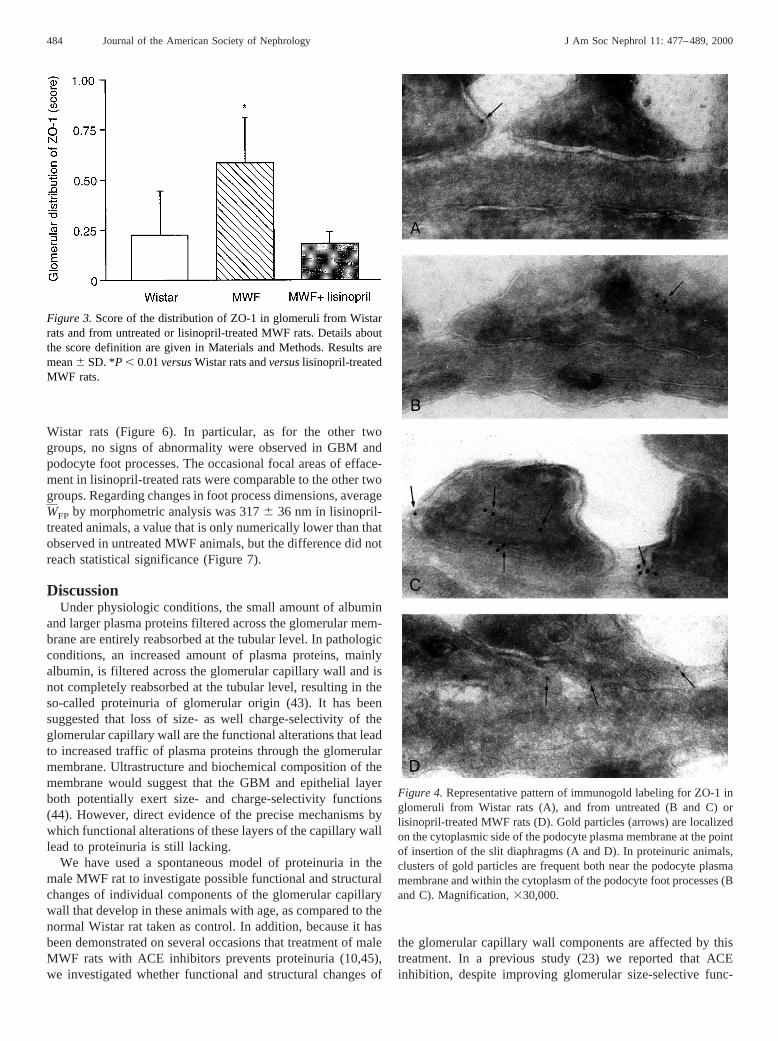
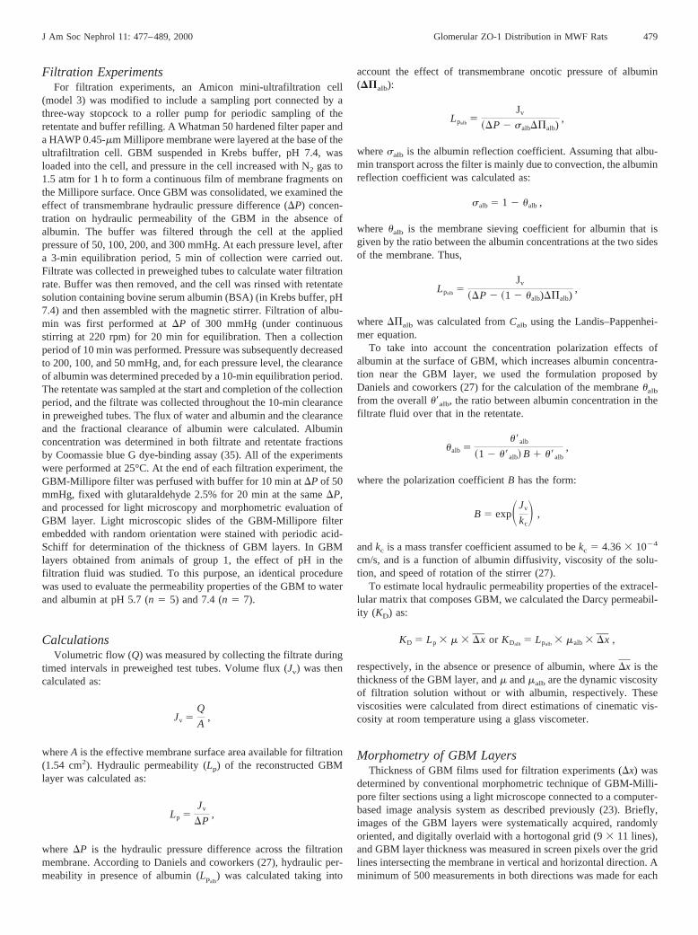
Wistar rats (Figure 6). In particular, as for the other twogroups, no signs of abnormality were observed in GBM andpodocyte foot processes. The occasional focal areas of efface-ment in lisinopril-treated rats were comparable to the other twogroups. Regarding changes in foot process dimensions, averageWFP by morphometric analysis was 3176 36 nm in lisinopril-treated animals, a value that is only numerically lower than thatobserved in untreated MWF animals, but the difference did notreach statistical significance (Figure 7).
DiscussionUnder physiologic conditions, the small amount of albumin
and larger plasma proteins filtered across the glomerular mem-brane are entirely reabsorbed at the tubular level. In pathologicconditions, an increased amount of plasma proteins, mainlyalbumin, is filtered across the glomerular capillary wall and isnot completely reabsorbed at the tubular level, resulting in theso-called proteinuria of glomerular origin (43). It has beensuggested that loss of size- as well charge-selectivity of theglomerular capillary wall are the functional alterations that leadto increased traffic of plasma proteins through the glomerularmembrane. Ultrastructure and biochemical composition of themembrane would suggest that the GBM and epithelial layerboth potentially exert size- and charge-selectivity functions(44). However, direct evidence of the precise mechanisms bywhich functional alterations of these layers of the capillary walllead to proteinuria is still lacking.
We have used a spontaneous model of proteinuria in themale MWF rat to investigate possible functional and structuralchanges of individual components of the glomerular capillarywall that develop in these animals with age, as compared to thenormal Wistar rat taken as control. In addition, because it hasbeen demonstrated on several occasions that treatment of maleMWF rats with ACE inhibitors prevents proteinuria (10,45),we investigated whether functional and structural changes of
the glomerular capillary wall components are affected by thistreatment. In a previous study (23) we reported that ACEinhibition, despite improving glomerular size-selective func-
Figure 3.Score of the distribution of ZO-1 in glomeruli from Wistarrats and from untreated or lisinopril-treated MWF rats. Details aboutthe score definition are given in Materials and Methods. Results aremean6 SD. *P , 0.01versusWistar rats andversuslisinopril-treatedMWF rats.
Figure 4.Representative pattern of immunogold labeling for ZO-1 inglomeruli from Wistar rats (A), and from untreated (B and C) orlisinopril-treated MWF rats (D). Gold particles (arrows) are localizedon the cytoplasmic side of the podocyte plasma membrane at the pointof insertion of the slit diaphragms (A and D). In proteinuric animals,clusters of gold particles are frequent both near the podocyte plasmamembrane and within the cytoplasm of the podocyte foot processes (Band C). Magnification,330,000.
484 Journal of the American Society of Nephrology J Am Soc Nephrol 11: 477–489, 2000

tion in male MWF rats (10), did not affect ultrastructure of themembrane components as observed by quantitative morpho-metric analysis, or the glomerular capillary surface area avail-able for filtration.
On the basis of the above observation, here we first inves-tigated whether development of proteinuria might depend onchanges in permeability properties of the GBM to water andalbumin. The results of ourin vitro evaluations document thatpermeability properties of the GBM to water and albumin arecomparable in proteinuric MWF rats and in normal Wistarrats—bothKD andualb were not significantly different in thetwo groups—suggesting that functional alterations of GBMcannot explain the permselectivity defect of the glomerularmembrane in proteinuric animals. In line with this result, ACEinhibition did not affect significantly the hydraulic permeabil-ity of GBM matrix and albumin filtration.
The maintenance of the GBM filtration barrier depends onthe charge and size-based selectivity toward filtered macro-molecules. In our system, in line with previous reports byRobinson and Walton (46), the electrostatic charge did notaffect the behavior of albumin filtration through the GBM layerreconstructed from normal rats, indicating that size rather thancharge is more prominent for restricting albumin passagethrough the GBM in normal circumstances. Very recently,Bolton and coworkers have elegantly documented that thesieving coefficients of Ficoll sulfate, in anin vitro filtrationsystem, were not different from those of neutral Ficoll atphysiologic ionic strength, but reduced at low ionic strength,suggesting that density of fixed negative charges of GBM isinsufficient to confer significant charge selectivity under phys-iologic conditions (47). All of the above evidence supports theconcept that the charge selectivity of the glomerular capillarywall does not depend on the GBM. Our present filtration studyshowing that alterations in GBM permeability to water andmacromolecules do not develop in proteinuric MWF rats, andACE inhibition does not change permeability properties of theGBM layer, strongly suggests that structural and/or functionalchanges of other components of the glomerular membranemust be responsible for glomerular permselectivity defect.
Several recent reports indicate that glomerular epithelialcells play a major role in water and protein permeability of the
glomerular capillary wall (28–32). We then investigated thedistribution of a tight junction component, the ZO-1 protein,which seems to play a crucial role in maintaining functionalproperties of the epithelial slit diaphragms (48–50), and mor-phologically evaluated the ultrastructure of the epithelial cellfoot processes in the three experimental conditions adopted.Our present immunohistochemistry study documents that innormal Wistar rats, ZO-1 is uniformly distributed along theouter surface of the filtering membrane, whereas this pattern ismarkedly altered in proteinuric MWF rats, with a predominantdiscontinuous distribution of the ZO-1, with aggregation infocal points within the glomerular membrane (Figure 2). At theimmunoelectron microscopy level, ZO-1 protein appears to belocated not only at the cytoplasmic side of podocyte mem-brane, near the filtration slit, but also is present within thecytoplasm of the foot processes. The reason for the observedcytoplasmic labeling of the protein is behind the objective ofour work, but it is suggestive of possible alteration in transportor compartmentalization of the protein within the podocytes.Independently of its origin, the abnormal protein distributionobserved at electron microscopy level correlates with the ab-normalities in the distribution observed in the immunofluores-cence study in untreated MWF rats. Of interest, our Westernblot analysis did not demonstrate changes in ZO-1 expressionat the glomerular level, suggesting that the pattern observed atimmunofluorescence derives mainly from a redistribution ofthe protein rather than from changes in its expression.
The redistribution of ZO-1 both at the optical and electronmicroscopy level that we have observed in proteinuric MWFrats has already been documented in experimental nephrosisinduced in rats by puromycin aminonucleoside and in normalrat kidney perfused by the polycation protamine sulfate (33). Inboth cases, ZO-1 redistribution was associated with changes inthe foot process ultrastructure and apical displacement of theglomerular slit diaphragm above the newly formed intercellularoccluding-type junctions (33). At variance, in our model ofspontaneous glomerular injury, the altered glomerular distribu-tion of ZO-1 was not associated with any apparent ultrastruc-tural change of the foot processes and the epithelial slit dia-phragms on electron microscopy. More detailed investigationof the ultrastructure of the podocytes did not show a statisti-cally significant difference in mean foot process width that wasonly numerically different in proteinuric animals comparedwith normal controls, as measured by morphometric technique.However, the qualitative and quantitative observations that wehave obtained on electron microscopy are in line with a recentstudy by Kawachi and coworkers showing altered immunore-activity of ZO-1 in glomeruli from rats injected with mono-clonal antibody 5-1-6, which recognizes a 51-kD protein on theslit diaphragm and causes severe proteinuria, but does notinduce changes in the structural integrity of the slit diaphragms(51). Of note, other conditions that induce abnormal urinaryprotein excretion, such as elevation of intrarenal angiotensin II(52) and reactive oxygen metabolites (53), appear not to beassociated with changes in epithelial foot processes ultrastruc-ture. On the other hand, in line with the present results, wehave previously reported that prevention of proteinuria by ACE
Figure 5. Representative Western blot analysis of glomerular lysatefrom Wistar rats and from untreated or lisinopril-treated MWF rats.For details, see Materials and Methods.
J Am Soc Nephrol 11: 477–489, 2000 Glomerular ZO-1 Distribution in MWF Rats 485
inhibition in MWF rats was not associated with epithelialultrastructural changes (23). Along this line, a human study(41) aimed at evaluating the relationship between glomerularfoot process width and proteinuria in patients with glomerulo-nephritis has already documented that severe proteinuria candevelop in the absence of ultrastructural changes of the foot
processes, suggesting that foot process fusion is not a prereq-uisite for proteinuria.
Our present results further strengthen the concept that amolecular rather than a structural impairment must be respon-sible for abnormal glomerular filtration of plasma proteins. Inaddition, our findings that ACE inhibition, which is known to
Figure 6. Transmission electron micrographs of representative glomeruli from Wistar rats (A and B) and from untreated (C and D) orlisinopril-treated MWF rats (E and F). Structural integrity of the slit diaphragms is maintained in proteinuric MWF rats. Magnification:329,400 in A, C, and E;34400 in B, D, and F.
486 Journal of the American Society of Nephrology J Am Soc Nephrol 11: 477–489, 2000

prevent proteinuria and increaseKf (10), also prevented alter-ation in the glomerular distribution of ZO-1 both at the opticaland electron microscopy level, strongly suggest that preserva-tion of glomerular distribution of this protein and probably ofsome other not yet identified component of the slit diaphragmis essential for the maintenance of hydraulic permeability andpermselective properties of the glomerular capillary wall. Themechanisms by which ZO-1 is redistributed and aggregatedwithin the glomerular capillary wall in proteinuric conditionsare behind the scope of the present study and are presentlyunder investigation in our laboratory.
Altered distribution of ZO-1 has been documented in cul-tured podocytes exposedin vitro to cytokines, which cause adecrease in transmonolayer electrical resistance (54). It hasbeen suggested that increased tyrosine phosphorylation of pro-teins at cell-cell junctions including ZO-1 protein may beresponsible for the reorganization of the junctional complexand increase in paracellular permeability (55,56). ZO-1 hasalso been found to be phosphorylated in the tyrosine residuesduring rapid rearrangement of the glomerular filtration slits inexperimental nephrosis (57). Finally, angiotensin II has beenshown to induce tyrosine phosphorylation of proteins such aspaxillin and PLCg (58,59). On the basis of this evidence, it istempting to speculate that angiotensin II causes a biochemicalmodification of ZO-1 in proteinuric MWF rats that may beresponsible for impairment of the filtration slit diaphragmfunction and consequently of the filtration barrier.
In conclusion, our present investigations indicate that: (1)development of proteinuria in male MWF rats takes placewithout significant changes in GBM hydraulic and permselec-tive function, or with changes in glomerular epithelial cellultrastructure; (2) proteinuria in this model is associated withimportant alterations in glomerular distribution of the junctionprotein ZO-1; and (3) the antiproteinuric effect of ACE inhib-itors is associated with preservation of glomerular ZO-1 dis-tribution.
AcknowledgmentsThe authors thank Dr. Tullio Bertani for morphologic evaluation of
glomerular membrane ultrastructure at electron microscopy level andDr. Marina Morigi and Dr. Anna Fassi for helpful discussion.
References1. Galaske R, Baladamus CA, Stolte H: Plasma protein handling in
the rat kidney: Micropuncture experiments in the acute heterol-ogous phase of anti-GBM-nephritis.Pflugers Arch375: 269–277, 1978
2. Tojo A, Hitoshi E: Intrarenal handling of proteins in rats usingfractional micropuncture technique.Am J Physiol263: F601–F606, 1992
3. Remuzzi G: Abnormal protein traffic through the glomerularbarrier induces proximal tubular cell dysfunction and causesrenal injury.Curr Opin Nephrol Hypertens4: 339–342, 1995
4. Remuzzi G, Bertani T: Is glomerulosclerosis a consequence ofaltered glomerular permeability to macromolecules?Kidney Int38: 384–394, 1990
5. Hirschberg R: Bioactivity of glomerular ultrafiltrate duringheavy proteinuria may contribute to renal tubulo-interstitial le-sions.J Clin Invest98: 116–124, 1996
6. Eddy AA: Experimental insights into the tubulointerstitial dis-ease accompanying primary glomerular lesions.J Am Soc Neph-rol 5: 1273–1278, 1994
7. Anderson S, Meyer TW, Rennke HG, Brenner BM: Control ofglomerular hypertension limits glomerular injury in rats withreduced renal mass.J Clin Invest76: 612–619, 1985
8. Zatz R, Dunn BR, Meyer TW, Anderson S, Rennke HG, BrennerBM: Prevention of diabetic glomerulopathy by pharmacologicalamelioration of glomerular capillary hypertension.J Clin Invest77: 1925–1930, 1986
9. Anderson S, Rennke HG, Brenner BM: Therapeutic advantage ofconverting enzyme inhibitors in arresting progressive renal dis-ease associated with systemic hypertension in the rat.J ClinInvest77: 1993–2000, 1986
10. Remuzzi A, Puntorieri S, Battaglia C, Bertani T, Remuzzi G:Angiotensin converting enzyme inhibition ameliorates glomeru-lar filtration of macromolecules and water and lessens glomeru-lar injury in the rat.J Clin Invest85: 541–549, 1990
11. Hommel E, Parving HH, Mathiesen E, Edsberg B, Damkjær M,Giese J: Effect of captopril on kidney function in insulin-depen-dent diabetic patients with nephropathy.Br Med J293: 467–470,1986
12. Parving HH, Hommel E, Smidt UM: Protection of kidney func-tion and decrease in albuminuria by captopril in insulin depen-dent diabetics with nephropathy.Br Med J 297: 1086–1091,1988
13. Bjorck S, Nyberg G, Mulec H, Granerus G, Herlitz H, Aurell M:Beneficial effects of angiotensin converting enzyme inhibitionon renal function in patients with diabetic nephropathy.Br Med J293: 471–474, 1986
14. Marre M, Le Blanc H, Suarez L: Converting enzyme inhibitionand kidney function in normotensive diabetic patients with per-sistent microalbuminuria.Br Med J294: 1448–1452, 1987
15. The GISEN Group: Randomised placebo-controlled trial of ef-fect of ramipril on decline in glomerular filtration rate and risk ofterminal renal failure in proteinuric, non-diabetic nephropathy.Lancet349: 1857–1863, 1997
16. Yoshioka T, Rennke HG, Salant DJ, Deen WM, Ichikawa I: Roleof abnormally high transmural pressure in the permselectivity
Figure 7.Mean foot process width in Wistar rats and in untreated orlisinopril-treated MWF rats. Results are mean6 SD.
J Am Soc Nephrol 11: 477–489, 2000 Glomerular ZO-1 Distribution in MWF Rats 487
defect of glomerular capillary wall: A study in early passiveHeymann nephritis.Circ Res61: 531–538, 1987
17. Halbach GR, Alt JM, Brunkhorst R, Frei U, Kuhn K, Stolte H:Single nephron hyperfiltration and proteinuria in a newly se-lected rat strain with superficial glomeruli.Ren Physiol9: 317–325, 1986
18. Remuzzi A, Puntorieri S, Alfano M, Macconi D, Abbate M,Bertani T, Remuzzi G: Pathophysiologic implications of protein-uria in a rat model of progressive glomerular injury.Lab Invest67: 572–579, 1992
19. Remuzzi A, Malanchini B, Battaglia C, Bertani T, Remuzzi G:Comparison of the effects of angiotensin-converting enzymeinhibition and angiotensin II receptor blockade on the evolutionof spontaneous glomerular injury in male MWF/Ztm rats.ExpNephrol4: 19–25, 1996
20. Heeg JE, de Jong PE, van der Hem GK, de Zeeuw D: Reductionof proteinuria by angiotensin converting enzyme inhibition.Kid-ney Int32: 78–83, 1987
21. Morelli E, Loon N, Meyer TW, Peters W, Myers BD: Effects ofconverting-enzyme inhibition on barrier function in diabetic glo-merulopathy.Diabetes39: 76–82, 1990
22. Remuzzi A, Perticucci E, Ruggenenti P, Mosconi L, Limonta M,Remuzzi G: Angiotensin converting enzyme inhibition improvesglomerular size-selectivity in IgA nephropathy.Kidney Int 39:1267–1273, 1991
23. Ene-Iordache B, Imberti O, Foglieni C, Remuzzi G, Bertani T,Remuzzi A: Effects of angiotensin-converting enzyme inhibitionon glomerular capillary wall ultrastructure in MWF/Ztm rats.J Am Soc Nephrol5: 1378–1384, 1994
24. Farquhar MG: The glomerular basement membrane: A selectivemacromolecular filter. In:Cell Biology of Extracellular Matrix,edited by Hay ED, New York, Plenum Press, 1981, pp 335–378
25. Fujigaki Y, Nagase M, Kobayasi S, Hidaka S, Shimomura M,Hishida A: Intra-GBM site of the functional filtration barrier forendogenous proteins in rats.Kidney Int43: 567–574, 1993
26. Robinson GB, Walton HA: Glomerular basement membrane as acompressible ultrafilter.Microvasc Res38: 36–48, 1989
27. Daniels BS, Hauser EB, Deen WM, Hostetter TH: Glomerularbasement membrane: In vitro studies of water and protein per-meability.Am J Physiol262: F919–F926, 1992
28. Daniels BS: Increased albumin permeability in vitro followingalterations of glomerular charge is mediated by the cells of thefiltration barrier.J Lab Clin Med124: 224–230, 1994
29. Daniels BS: The role of the glomerular epithelial cell in themaintenance of the glomerular filtration barrier.Am J Nephrol13: 318–323, 1993
30. Rennke HG: How does glomerular epithelial cell injury contrib-ute to progressive glomerular damage?Kidney Int45: S58–S63,1994
31. Laurens W, Battaglia C, Foglieni C, De Vos R, Malanchini B,Van Damme B, Vanrenterghem Y, Remuzzi G, Remuzzi A:Direct podocyte damage in the single nephron leads to albumin-uria in vivo. Kidney Int47: 1078–1086, 1995
32. Blantz RC, Gabbai FB, Peterson O, Wilson CB, Kihara I, Kawa-chi H, Shimizu F, Yamamoto T: Water and protein permeabilityis regulated by the glomerular epithelial slit diaphragm.J Am SocNephrol4: 1957–1964, 1994
33. Kurihara H, Anderson JM, Kerjaschki D, Farquhar MG: Thealtered glomerular filtration slits seen in puromycin amino-nucleoside nephrosis and protamine sulfate-treated rats containthe tight junction protein ZO-1.Am J Pathol141: 805–816, 1992
34. Ligler FS, Robinson GB, Byrne J: A new method for the isola-
tion of renal basement membranes.Biochim Biophys Acta468:327–340, 1977
35. Read SM, Northcote DH: Minimization of variations in theresponse to different proteins of the Coomassie blue G dye-binding assay for protein.Anal Biochem116: 53–64, 1981
36. McLean IW, Nakane PF: Periodate-lysine paraformaldehyde fix-ative: A new fixative for immunoelectron microscopy.J Histo-chem Cytochem22: 1077–1083, 1974
37. Tokuyasu K: Immunocytochemistry on ultrathin frozen sections.Histochem J12: 381–403, 1980
38. Laemnli UK: Cleavage of structural proteins during the assemblyof the head of bacteriophage T4.Nature227: 680–685, 1970
39. Towbin H, Staehelin T, Gordon J: Electrophoretic transfer ofproteins from polyacrylamide gels to nitrocellulose sheets: Pro-cedure and some applications.Proc Natl Acad Sci USA76:4350–4354, 1979
40. Pagtalunan ME, Rasch R, Rennke HG, Meyer TW: Morphomet-ric analysis of effects of angiotensin II on glomerular structure inrats.Am J Physiol268: F82–F88, 1995
41. Seefeldt T, Bohman S-O, Gundersen HJG, Maunsbach AB,Petersen VP, Olsen S: Quantitative relationship between glomer-ular foot process width and proteinuria in glomerulonephritis.Lab Invest44: 541–546, 1981
42. Wallenstein S, Zucker CL, Fleiss JL: Some statistical methodsuseful in circulation research.Circ Res47: 1–9, 1980
43. Fitzgibbon WR, Webster SK, Imamura A, Ploth DW, HutchisonFN: Effect of dietary protein and enalapril on proximal tubulardelivery and absorption of albumin in nephrotic rats.Am JPhysiol260: F986–F996, 1996
44. Remuzzi A, Remuzzi G: Glomerular perm-selective function.Kidney Int45: 398–402, 1994
45. Remuzzi A, Imberti O, Puntorieri S, Malanchini B, Macconi D,Magrini L, Bertani T, Remuzzi G: Dissociation between antipro-teinuric and antihypertensive effect of angiotensin convertingenzyme inhibitors in the rat.Am J Physiol267: F1034–F1044,1994
46. Robinson GB, Walton HA: Ultrafiltration through basementmembrane. In:Renal Basement Membranes in Health and Dis-eases, edited by Price RG, Hudson BG, New York, London,Academic Press, 1987, pp 147–161
47. Bolton GR, Deen WM, Daniels BS: Assessment of the chargeselectivity of glomerular basement membrane using Ficoll sul-fate.Am J Physiol274: F889–F896, 1998
48. Schnabel E, Anderson JM, Farquhar MG: The tight junctionprotein ZO-1 is concentrated along slit diaphragms of the glo-merular epithelium.J Cell Biol 111: 1255–1263, 1990
49. Kurihara H, Anderson JM, Farquhar MG: Diversity among tightjunctions in rat kidney: Glomerular slit diaphragms and endothe-lial junctions express only one isoform of the tight junctionprotein ZO-1.Proc Natl Acad Sci USA89: 7075–7079, 1992
50. Anderson JM, Van Itallie CM: Tight junctions and the molecularbasis for regulation of paracellular permeability.Am J Physiol269: G467–G475, 1995
51. Kawachi H, Kurihara H, Topham PS, Brown D, Shia MA,Orikasa M, Shimizu F, Salant DJ: Slit diaphragm-reactive ne-phritogenic MAb 5-1-6 alters expression of ZO-1 in rat podo-cytes.Am J Physiol273: F984–F993, 1997
52. Fujiwara Y, Kitamura E, Ueda N, Fukunaga M, Orita Y, KamadaT: Mechanism of action of angiotensin II on isolated rat glomer-uli. Kidney Int36: 985–991, 1989
53. Yoshioka T, Ichikawa A, Fogo A: Reactive oxygen metabolitescauses massive, reversible proteinuria and glomerular sieving
488 Journal of the American Society of Nephrology J Am Soc Nephrol 11: 477–489, 2000

defect without apparent ultrastructural abnormality.J Am SocNephrol2: 902–912, 1991
54. Coers W, Vos JTWM, Van Der Meide PH, Van Der Horst MLC,Huitema S, Weening JJ: Interferon-gamma (IFN-g) and IL-4expressed during mercury-induced membranous nephropathy aretoxic for cultured podocytes.Clin Exp Immunol102: 297–307,1995
55. Staddon JM, Herrenknecht K, Smales C, Rubin LL: Evidencethat tyrosine phosphorylation may increase tight junction perme-ability. J Cell Sci108: 609–619, 1995
56. Collares-Buzato CB, Jepson MA, Simmons NL, Hirst BH: In-creased tyrosine phosphorylation causes redistribution of adher-ens junction and tight junction proteins and perturbs paracellular
barrier function in MDCK epithelia.Eur J Cell Biol 76: 85–92,1998
57. Kurihara H, Anderson JM, Farquhar MG: Increased tyr phos-phorylation of ZO-1 during modification of tight junctions be-tween glomerular foot processes.Am J Physiol268: F514–F524,1995
58. Leduc I, Meloche S: Angiotensin II stimulates tyrosine phos-phorylation of the focal adhesion-associated protein paxillin inaortic smooth muscle cells.J Biol Chem270: 4401–4404, 1995
59. Marrero MB, Schieffer B, Ma H, Bernstein KE, Ling BN: ANGII-induced tyrosine phosphorylation stimulates phospholipaseC-g1 and Cl2 channels in mesangial cells.Am J Physiol270:C1834–C1842, 1996
Access to UpToDate on-line is available for additional clinical information at http://www.lww.com/JASN.
J Am Soc Nephrol 11: 477–489, 2000 Glomerular ZO-1 Distribution in MWF Rats 489
Copyright © 2022 FDOKUMEN