Prospects for aminoacyl-tRNA synthetase inhibitors as new antimicrobial agents
Upload
sorbonne-frCategory
view
0download
0
10.1101/gad.207701Access the most recent version at doi: 2001 15: 2295-2306 Genes & Dev.
Damien Brégeon, Vincent Colot, Miroslav Radman and François Taddei
ribosomal frameshiftTranslational misreading: a tRNA modification counteracts a +2
References
http://www.genesdev.org/cgi/content/full/15/17/2295#otherarticlesArticle cited in:
http://www.genesdev.org/cgi/content/full/15/17/2295#ReferencesThis article cites 56 articles, 24 of which can be accessed free at:
serviceEmail alerting
click heretop right corner of the article or Receive free email alerts when new articles cite this article - sign up in the box at the
Notes
http://www.genesdev.org/subscriptions/ go to: Genes and DevelopmentTo subscribe to
© 2001 Cold Spring Harbor Laboratory Press
Cold Spring Harbor Laboratory Press on December 25, 2007 - Published by www.genesdev.orgDownloaded from

Translational misreading:a tRNA modification counteractsa +2 ribosomal frameshiftDamien Brégeon,1,4 Vincent Colot,2,3 Miroslav Radman,1 and François Taddei1
1INSERM EPI9916, Faculté de Médecine Necker-Enfants Malades, 75730 Paris Cedex 15, France; 2Institut Jacques Monod,CNRS-Universités Paris 6 et Paris 7, 75251 Paris Cedex 05, France
Errors during gene expression from DNA to proteins via transcription and translation may be deleterious forthe functional maintenance of cells. In this paper, extensive genetic studies of the misreading of a GA repeatintroduced into the lacZ gene of Escherichia coli indicate that in this bacteria, errors occur predominantly bya +2 translational frameshift, which is controlled by a tRNA modification involving the MnmE and GidAproteins. This ribosomal frameshift results from the coincidence of three events: (1) decreased codon–anticodon affinity at the P-site, which is caused by tRNA hypomodification in mnmE− and gidA− strains; (2) arepetitive mRNA sequence predisposing to slippage; and (3) increased translational pausing attributable to thepresence of a rare codon at the A-site. Based on genetic analysis, we propose that GidA and MnmE act in thesame pathway of tRNA modification, the absence of which is responsible for the +2 translational frameshift.The difference in the impact of the mutant gene on cell growth, however, indicates that GidA has at least oneother function.
[Key Words: Translational fidelity; 5-methylaminomethyl-2-thio-uridine; gidA; mnmE; thdF; argU]
Received May 10, 2001; revised version accepted July 2, 2001.
Processes ensuring the fidelity of DNA replication havebeen studied extensively, presumably because of theoverwhelming importance of gene mutations in evolu-tion, hereditary diseases and cancer (Friedberg et al.1995). In contrast, less is known about the fidelity oftranscription and translation, which must be crucial forthe functional maintenance of cells and organisms.Functional degeneration of nondividing cells limits thelifespan of organs such as the heart or brain. Therefore,decreasing fidelity of gene expression may be an under-lying cause of cellular aging, degenerative diseases, anddeath. Accurate gene expression is dependent on the fi-delity of both transcription of DNA and translation ofmRNA.Fidelity of transcription depends on proofreading by a
3� → 5� exonuclease activity found in a number of eu-karyotic and prokaryotic RNA polymerases (Thomas etal. 1998). The Escherichia coli RNA polymerase exhibitserror rates ranging from 1 to 10 misincorporations per105 synthesized nucleotides (for review, see Libby andGallant 1991). Aberrant RNAs also include the presenceof altered nucleotides in the mRNA caused either by
direct alteration of the mRNA or by misincorporation ofaltered mutagenic nucleotides, such as 8-oxo-GTP, bythe RNA polymerase (Taddei et al. 1997). The presenceof 8-oxo-G in cellular RNA was shown to be restricted tovulnerable neurons in dementia such as Alzheimer’s dis-ease (AD) (Nunomura et al. 1999) or Parkinson’s disease(Zhang et al. 1999) and to be correlated with the synthe-sis of altered proteins (Dukan et al. 2000). The rate oftranscriptional frameshifting is unknown but it was pro-posed that it may lead to some neurodegenerative dis-eases (van Leeuwen et al. 1998b). In patients with AD,the accumulation of aberrant protein species shifted tothe +1 frame with no related mutation in the DNA blue-print has been reported. The appearance of those proteinswas shown to be caused by a molecular misreading of acoding GA repeat resulting in the deletion of a dinucleo-tide GA in the mRNA (Hol et al. 1998; van Leeuwen etal. 1998a,b).Translational errors have been characterized in E. coli
and measured in vivo and/or in vitro (for review, seeKurland 1992). These include missense errors resultingin the substitution of one amino acid for another, orincorporation of extra amino acids caused by read-through of termination codons. Overall, translational er-rors occur at a frequency of 10−3 to 10−4 (Loftfield andVanderjagt 1972; Edelmann and Gallant 1977; Ellis andGallant 1982; Parker 1989) and are limited by the abilityof the ribosome to discriminate between correct and in-correct tRNAs entering the A-site (Thompson 1988; Ni-
3Present address: Unité de Recherches en Génomique Végétale(INRA-CNRS), 91057 Evry cedex, France.4Corresponding author.E-MAIL [email protected]; FAX 33-1-40-61-53-22.Article and publication are at http://www.genesdev.org/cgi/doi/10.1101/gad.207701.
GENES & DEVELOPMENT 15:2295–2306 © 2001 by Cold Spring Harbor Laboratory Press ISSN 0890-9369/01 $5.00; www.genesdev.org 2295
Cold Spring Harbor Laboratory Press on December 25, 2007 - Published by www.genesdev.orgDownloaded from

erhaus 1990; Czworkowski and Moore 1996). When in-duced by the addition of some antibiotics (e.g., strepto-mycin), translational misreading leads to increasedconcentrations of oxidized proteins (Dukan et al. 2000),which were found to accumulate in aging cells (Stadt-man 1992). Other translational errors include prematuretermination (Menninger 1977; Kurland et al. 1996) andtranslational frameshifting. The latter generally givesrise to a shorter peptide because of the subsequent en-counter of stop codons in the shifted frame. Spontaneousframeshifts resulting from translational errors occur at afrequency of <10−5 per codon (Kurland 1992), but wereshown to increase in E. coli cells entering stationaryphase (Barak et al. 1996).Basic mechanisms involved in DNA replication, re-
combination, transcription, and translation are oftenconserved phylogenetically (Lewin 1994). Therefore, weset out to study in E. coli the source of errors broughtabout by the presence of a GA repeat within an engi-neered frameshifted allele of the lacZ gene (Table 1). Thecharacterization of E. coli mutants expressing an inter-mediate level of �-galactosidase led to the identificationof three genes that are involved in the faithful transla-tion of the GA repeat. Two of these genes, mnmE andgidA, are involved in tRNA modification and the third,argU, encodes the tRNAArgmnm5UCU. The analysis of themechanism of this unusual +2 translational frameshift atthis sequence is consistent with the recently proposed+1/−1 translational frameshift dual error model (Qian etal. 1998; Björk et al. 1999; Farabaugh and Björk 1999),which can be extended to explain larger ribosomalframeshifting and short tRNA hopping. Measurementsof the growth rate of strains carrying a mutation in thegidA or mnmE genes showed that the impact of tRNAhypomodification varies with nutritional conditions andis not equivalent for both strains (Table 2). These obser-vations suggest that GidA may also be involved in otherpathways.
Results
The reporter system
We studied the control of error rates of expression (tran-scription or translation) of a gene containing a coding GA
repeat in E. coli. A reporter system for such errors wasconstructed using the lacZ gene encoding the �-galacto-sidase enzyme.The constructed lacZ(+GA) frameshifted allele con-
tains a repetition of the dinucleotide GA starting fromthe first base of codon 461 to the first base of codon 463(Table 1). These codons normally encode amino acidsinvolved in the active site of �-galactosidase (Cupplesand Miller 1988). This insertion results in a sequenceshifted to the +2 reading frame when compared with thewild-type sequence and the emergence of a prematurestop codon 210 nucleotides downstream, thereby gener-ating a mRNA encoding a 534-amino-acid polypeptide.The �-galactosidase activity of NECB1 cells, which carrythis new allele, is 125,000 times lower than the activityof the isogenic lacZ+ cells (Table 1). As a result, NECB1cells are not able to grow on minimal media containinglactose as the sole source of carbon, and form white colo-nies on LB plates containing X-Gal (5-bromo-4-chloro-3-indolyl-�-D-galactopyranoside).
Only deletion of a dinucleotide GA can restorean active �-galactosidase
To study errors on this GA repeat, we first wanted toestablish the spectrum of mutants that could restore par-tially or totally the �-galactosidase activity. This ques-tion was addressed by selecting for spontaneous rever-
Table 1. Sequences of alleles used and their corresponding �-galactosidase activities
Allele SequenceaStop codonpositionb
�-Galactosidaseactivityc
lacZ+ AAT GAA TCA GGC 3073 12400 ± 10A s n G l u S e r G l y
lacZ (+GA) AAT GAG AGA GTG GC 1603 0.10 ± 0.01A s n G l u A r g V a l
lacZ+ revertants AAT GAG AGT GGC 3073 12600 ± 240A s n G l u S e r G l y
lacZ (+ Arg462) AAT GAG AGA AGT GGC 3076 0.01 ± 0.001A s n G l u A r g S e r G l y
aAll sequences are shown from codon 460 of the lacZ gene.bNucleotide number of the first base of the following stop codon.cMiller units ± standard error.
Table 2. Generation time of strains in various growth mediaat 40°C
Relevant genotypes
Mediaa
LB Mm + 0.4% glucose
Wild type (NECB1) 32.9 ± 0.7 75.7 ± 3.7gidA�Tn10 38.0 ± 1.2b 91.1 ± 3.4e
mnmE�Tn10 38.2 ± 0.4c 77.7 ± 2.5gidA�Tn10, mnmE�kan 45.2 ± 0.7d 95.8 ± 2.0f
aNumbers represent the average generation time of 5 culturesgiven in minutes ± SEM.b–fGeneration times that are significantly different from that ofthe wild type in the same medium. T-test comparison betweenWT and mutants grown in the same media have the followingP-values: b, 0.0054; c, 0.0002; d, <0.0001; e, 0.0154; f, 0.0013.
Brégeon et al.
2296 GENES & DEVELOPMENT
Cold Spring Harbor Laboratory Press on December 25, 2007 - Published by www.genesdev.orgDownloaded from

sion mutants at the DNA level, which restore an active�-galactosidase.NECB1 cells were plated on minimal medium con-
taining lactose and Lac+ revertants were scored. The fre-quency of appearance of Lac+ colonies was about 10−9 perviable cell. The lacZ gene of 20 of those clones takenfrom three different cultures was sequenced and all ofthem had a deletion of a GA dinucleotide that restoresthe wild-type sequence of the protein (Table 1). The �-ga-lactosidase activity of those revertants was measuredand is comparable with the activity of the isogenic lacZ+
strain. With this screen we found no single nucleotideinsertions that could potentially have restored the read-ing frame and generated a polypeptide with an extraamino acid. A mutant we constructed containing an ex-tra A downstream to the GA repeat had a �-galactosidaseactivity about 106-fold lower than the lacZ+ strain (Table1). The expected protein encoded by this lacZ (+Arg462)mutant contains an extra arginine in position 462 (Table1). This indicates that only a mutation restoring thewild-type frame of the protein can result in an active�-galactosidase, consistent with the central position ofthese amino acids in the enzyme active site (Cupples andMiller 1988). As a consequence, mutation rate being verylow, production of an active �-galactosidase should re-sult mostly from a deletion of a dinucleotide GA at thetranscription level or +2 frameshift at the translationlevel.
Isolation of mutants affected in the phenotypicexpression of lacZ(+GA)
The low level of �-galactosidase activity of thelacZ(+GA) strain suggests that this type of misreading(−GA) at the level of transcription or translation, produc-ing occasional active proteins, are very rare in wild-typeE. coli. Therefore, a cell with a mutation that increasesthese error rates is expected to express only a low tointermediate level active �-galactosidase. Following mu-tagenic treatments, mutants were scored for their abilityto form pale blue colonies on plates containing X-Gal.The first mutagenesis of the NECB1 strain was a ran-
dom mini-Tn10 insertion using the �NK1323 phage(Kleckner et al. 1991). Following mutagenesis, about20,000 tetracycline-resistant colonies were screened and10 of them presented a light blue coloration. Nine of the10 were stable and eight of those conferred an interme-diate level of �-galactosidase when the Tn10was retrans-duced by P1 phage in NECB1 cells. For these clones, thetransduction of the tetracycline resistance into Lac+ cellsdoes not detectably affect the �-galactosidase-specific ac-tivity (data not shown), making it unlikely that the in-creased �-galactosidase level is attributable to a muta-tion affecting the level of transcription or translation ofthe lacZ gene.As transposon mutagenesis will not reveal essential
genes that may be involved in the control of gene expres-sion, we performed a random point mutagenesis using2-aminopurine (2-AP). This molecule is an adenine ana-log, which can mispair with cytosine and generates
mostly transitions. Approximately 30,000 colonies fromthree different cultures of NECB1 cells grown with 2-APwere screened and 133 mutants harboring a stable paleblue coloration were isolated. These mutants are de-scribed below.
Characterization of mutants containing a Tn10insertion
The eight mutants containing a Tn10 insertion that wasassociated with increased �-galactosidase activity wereidentified by inverse PCR (Triglia et al. 1988) and se-quencing of the amplified fragments. Mutants 3, 4, 8, and10 contained a Tn10 insertion in the gidA gene at posi-tion 707, 1025, 5, and 51, respectively. The function ofthis gene, the closest to the origin of replication oriC inE. coli, is as yet unidentified but has been associatedwith a glucose inhibited division phenotype (von Mey-enburg and Hansen 1980). In all cases, �-galactosidaseactivity returned to the level of the parental strain whentransformed with a plasmid containing the gidA gene(data not shown), suggesting that all insertions result inthe lack of production of an active GidA protein. Mutant3, hereafter designated as gidA::Tn10, having a �-galac-tosidase activity ∼70-fold higher than the parental strain(Fig. 1) was chosen for further analysis.Mutants 2, 6, and 7 had Tn10 insertions in the mnmE
gene, at positions 798, 707, and 43, respectively. ThemnmE gene was previously called trmE, because of itsinvolvement in tRNA modification (Elseviers et al.1984). For the three mutants, transformation with a plas-mid containing the mnmE gene decreased the �-galacto-sidase activity to the level of the parental strain (data notshown). Mutant 2, hereafter designated as mnmE::Tn10,has a �-galactosidase activity ∼50-fold higher than theparental strain (Fig. 1). This mutant was used as a repre-sentative of mnmE::Tn10 mutants for further analysis.Mutant 9 has a Tn10 insertion in the ebgR gene,
Figure 1. Error rate measurement with �-galactosidase assays.�-galactosidase assays were performed on cells grown overnightin LB containing the proper antibiotic. Bars represent the aver-age of �-galactosidase measurements ± standard errors. Theactivity of mnmE::kan is not statistically different frommnmE::Tn10 (t-test = −0.526, P = 0.604). Double mutantgidA::Tn10,mnmE::kan activity is not different from the activ-ity of mnmE::kan (t-test = −1.556, P = 0.134) but is differentfrom gidA::Tn10 (t-test = 4.451, P = 0.0001) suggesting thatmnmE is epistatic on gidA.
tRNA modification and ribosomal frameshifting
GENES & DEVELOPMENT 2297
Cold Spring Harbor Laboratory Press on December 25, 2007 - Published by www.genesdev.orgDownloaded from
known to be the repressor of the evolved �-galactosidaseoperon. When this operon is derepressed, the EbgA en-zyme is produced, resulting in a weakly positive lactoseutilization phenotype (Campbell et al. 1973; Arraj andCampbell 1975). This mutant was not analyzed further.
Characterization of mutants obtained after2-AP mutagenesis
As the majority of the insertion mutants were in mnmEand gidA, we first identified which of the point mutantswere complemented with plasmids expressing thesegenes. Transformed cells were scored for the disappear-ance of the pale blue coloration. Of the 133 mutants, 36were complemented by mnmE and 56 were comple-mented with gidA. This accounted for ∼70% of all theclones, confirming the importance of these two genes inthe fidelity of gene expression. The �-galactosidase ac-tivity of gidA mutants has an average of 5.7 ± 1.4 Millerunits, whereas that for mnmE mutants is 3.7 ± 0.3, bothof which are comparable with the activities in the strainswith Tn10 disruptions of those genes.To determine if the 41 mutants not complemented by
mnmE or gidA contained mutations altering phenotypicexpression, we eliminated mutations in cis in the lacZgene and those causing derepression of genes coding for�-galactosidase-like activities. These two types of mu-tants were identified by transduction using P1 stocksmade on lacI42::Tn10 lacZ(+GA) and lacI42::Tn10lacZ�M15 strains, respectively. When the lacZ(+GA) al-lele was introduced into its genome, one mutant lost theblue coloration showing that the intermediate �-galac-tosidase activity was attributable to a cismutation of thelacZ(+GA) allele (not sequenced). Six mutants remainedconstantly blue when their lacZ gene was replaced by adeletion of lacZ, which is the hallmark of a strain ex-pressing �-galactosidase-like activity.To characterize mutations in genes not identified pre-
viously, the seven mutants out of the 34 remaining thatshowed the highest �-galactosidase level (140-fold higherthan the parental strain on average) were transformedwith a genomic DNA library made from the parentallacZ(+GA) strain. Plasmids that suppressed the bluecolor all contained argU, which encodes tRNAArg. Toconfirm whether this gene was deficient in these sevenmutants, a plasmid containing the argU gene was con-structed and used to transform cells of those sevenclones. The �-galactosidase activity of all clones trans-formed with this plasmid was reduced to a level compa-rable with that of the NECB1 parental strain. The pres-ence of mutations in the argU structural gene or in itspromoter was confirmed by sequencing.The 27 mutants harboring the lowest �-galactosidase
activity (sevenfold higher than the parental strain on av-erage) have not been characterized further.
GidA and MnmE may be in the same pathway
The mnmE gene product is known to be involved in thebiosynthesis of the hypermodified nucleoside 5-methyl-aminomethyl-2-thio-uridine (mnm5s2U), which is found
in the wobble position of bacterial tRNAs specific forglutamate, lysine, glutamine, arginine, and leucine(Björk 1996). It is unclear howmany enzymatic reactionsare involved in the first step of this process (Hagervall etal. 1987). There is no clear evidence for the function ofGidA, but thermosensitive mutants of that gene werefound in a tRNA6
Leu (supP) mutant background (Nakay-ashiki and Inokuchi 1998), which we interpret as a rolefor this protein in tRNA biogenesis.To determine whether GidA is involved in the same
pathway as MnmE, we transduced a mutant allele ofmnmE interrupted with a kanamycin cassette(o454::kan1, called mnmE::kan hereafter; Cabedo et al.1999) into lacZ(+GA) parental and derivative gidA::Tn10strains. The �-galactosidase activity of mnmE::kan wasnot different from that of mnmE::Tn10, and the activityof the double mutant (gidA::Tn10, mnmE::kan) is notdifferent from that of the singlemnmE::kanmutant (Fig.1) but is significantly lower than the activity of the singlegidA::Tn10 mutant. This implies that the gene productsfrom gidA and mnmE are likely to be in the same biosyn-thetic pathway, and furthermore, that the MnmE proteinactivity precedes the activity of the GidA protein.
+2 Ribosomal frameshifting caused by a less efficientcodon–anticodon recognition
The +2 translational frameshifting, observed in mn-mE::Tn10 or gidA::Tn10, could be explained by an ex-tension of the recently described +1/−1 translationalframeshift dual error model (Qian et al. 1998), whichpostulates such a slipping of the ribosome when a hypo-modified tRNA present in the P-site results in a weakercodon–anticodon interaction. The tRNAGlumnm5s2UUC,which decodes the first codon of the GA repeat, is one ofthe tRNAs that carry an MnmE-dependent mnm5s2U34modification (Björk 1996), which allows it to decode ei-ther GAG or GAA codons with similar efficiencies(Kruger et al. 1998). In themnmE::Tn10 strain, and likelyin the gidA::Tn10 strain, the U34 is hypomodified result-ing in tRNA with a less efficient base-pairing in thewobble position (Kruger et al. 1998). The observed frame-shifting could result from the presence of this hypomodi-fied tRNAGluU�UC (U� is a hypomodified form of the uridine)in the P-site of the ribosome.To test the effect of the pairing stability of this tRNA
on frameshift errors, we engineered gltT to have a CUCanticodon. This mutant and the gltT+ gene were clonedinto the pBAD33 vector under the control of the arabi-nose inducible promoter (Guzman et al. 1995), generat-ing the pNEC2 plasmid series (Fig. 2; Table 3). Whencoexpressed with the four wild-type copies on the chro-mosome, the tRNA harboring the CUC anticodonreduced the translational frameshifting by 30% inmnmE::Tn10 and gidA::Tn10 strains (Fig. 2B,C) as wellas in the wild-type strain (t-test induced vs. non-in-duced = −2.8, P = 0.02; data not shown). This mutatedtRNA therefore appears to be able to increase the codon–anticodon interaction, thereby reducing the frameshift-ing of the ribosome. The selective competition for trans-
Brégeon et al.
2298 GENES & DEVELOPMENT
Cold Spring Harbor Laboratory Press on December 25, 2007 - Published by www.genesdev.orgDownloaded from

lational machinery (e.g., aminoacylation) between thetRNA produced by the plasmid and the wild-type tRNAproduced from the four genomic genes could explainwhy the error rate was not reduced to the level of theparental lacZ(+GA) strain.
+2 Translational frameshift caused by the presenceof a rare codon in the A-site
The +1/−1 translational frameshift dual error model alsoproposes that frameshifting could occur at a pausing siteduring translation, which could be caused by the pres-ence of a slowly translated codon in the A-site. The ex-tent of vacancy of the A-site has been shown to correlateto a +1 translational frameshift (Curran and Yarus 1989;Sipley and Goldman 1993). In the case of lacZ(+GA), the
AGA codon (codon 462; Table 1) is one of the rarestcodons in E. coli (Hénaut and Danchin 1996; Nakamuraet al. 2000) and is decoded by a minor tRNA encoded byargU (Ikemura 1981; Dong et al. 1996). The AGA codonmay therefore constitute a pausing site and lead toframeshifting.To test this hypothesis, the lacZ(+GA) and the deriva-
tives gidA::Tn10 and mnmE::Tn10 were transformedwith a plasmid containing the argU gene. The overex-pression of the tRNAArgmnm5UCU encoded by argU in gi-dA::Tn10 or mnmE::Tn10 strains abolishes the errorphenotype observed in those strains (Fig. 3), as the �-ga-lactosidase activities for transformed gidA::Tn10 andmnmE::Tn10 strains are comparable with the parentalstrain. It should be noted that the presence of the vectoralone slightly reduced ribosomal frameshifting. This de-crease may be attributable to the chloramphenicol,which was added to the growth media to select for theplasmid and is known to be a translation inhibitor (Berg-mann and Sicher 1952). The error rate is significantlyreduced by the overproduction of argU, even in thetransformed parental strain (Fig. 3), showing that thissame +2 frameshift occurs in wild-type cells (NECB1) bypausing of the ribosome caused by the scarcity oftRNAArgmnm5UCU. These results show that the abolition ofthe ribosome frameshifting in parental, mnmE::Tn10,and gidA::Tn10 strains is caused by the filling of theA-site with the overproduced tRNAArgmnm5UCU.
The absence of the mnm5s2U modificationand the cell growth rate
The gidA gene was first isolated in association with aglucose-inhibited division phenotype (von Meyenburgand Hansen 1980) and is located next to the origin ofreplication (oriC). It was therefore proposed to have arole in the control of cell division. As it is likely to be-long to the same pathway as the mnmE gene in the con-trol of the +2GA frameshift, we have tested the effect ofmutant alleles of these genes on growth rate.To estimate the growth rate of these strains defective
for the mnm5s2U modification, we measured the gen-eration time of the parental strain, gidA::Tn10,mnmE::Tn10, and the double mutant (gidA::Tn10mnmE::kan) in LB and in minimal medium at 40°C.Comparison of generation times showed that gidA::Tn10mnmE::Tn10 and (gidA::Tn10 mnmE::kan) strains had agrowth rate lower than that of the parental strain duringgrowth in LB (Table 2) or in LB containing glucose (datanot shown). Under conditions of slower growth (minimalmedium), we failed to detect a significant difference be-tween the parental and themnmE::Tn10 strains, but thegidA::Tn10 and (gidA::Tn10 mnmE::kan) strains had ageneration time 16% and 20% longer, respectively, thanthat of the parental or mnmE::Tn10 strains. Unlike inthe �-galactosidase assays showing the translational er-ror rate of the +2GA frameshift, the double mutant didnot have the same phenotype as themnmE::Tn10 strain.These results indicate that mutations in gidA or mnmEhave an independent impact on growth rate.
Figure 2. Expression of a tRNA with an exact anticodon re-duces the errors. Mutant gltT tRNAGlu was created to deter-mine the role of the anticodon loop sequence in translationalerrors. (A) Sequence from nucleotides 30–40 (anticodon loop) ofthe tRNA encoded by gltT cloned in pNEC2-WT. The pNEC2–U34C plasmid carries the mutation U34 to C (bold letters, U*designates the modified uridine), thereby generating a CUC an-ticodon, which is an exact match to the GAG codon. (B,C)Transformed NECB10 (mnmE::Tn10 �ara leu::Tn5) andNECB11 (gidA::Tn10 �ara leu::Tn5) cells were grown overnightin LB containing 0%, 0.005%, or 0.05% arabinose to induceexpression of the gltT gene that is under the control of thearabinose promoter in pBAD33 (see Table 3). For each strain andplasmid pair, the �-galactosidase values were normalized to thevalue at 0% arabinose induction. The decreased �-galactosidaseactivity observed with the expression of the gltT allele carriedby pNEC2–U34C (�) is significant in mnmE::Tn10 (t-test in-duced vs. noninduced = −4.2, P = 0.002) (B) and in gidA::Tn10(t-test induced vs. noninduced = −4.8, P = 0.001) (C). In bothstrains the expression of the gltT allele carried by the pNEC2-WT (�) has no effect on ribosomal frameshifting. These resultssuggest that translational errors in mnmE::Tn10 and gidA::Tn10are attributable to a less efficient hypomodified tRNAGlu
U�UC
(U� represents the hypomodified form of the uridine). Each pointrepresents the average of normalized �-galactosidase activity± standard error.
tRNA modification and ribosomal frameshifting
GENES & DEVELOPMENT 2299
Cold Spring Harbor Laboratory Press on December 25, 2007 - Published by www.genesdev.orgDownloaded from
These results suggest that GidA may be acting in an-other pathway, such as a different tRNA modificationpath, or in DNA replication as suggested previously(Ogawa and Okazaki 1994).
Discussion
We studied the potential causes of errors during expres-sion of a GA repeat sequence inserted out of frame in thelacZ gene of E. coli. Mutants having an increased errorrate of gene expression were isolated based on the pro-duction of a moderate amount of active �-galactosidase.Approximately 83% of all these mutants (generated byboth insertional and point mutagenesis) were alleles of
mnmE and gidA, both apparently involved in the sametRNA modification pathway. The preponderance ofthese two genes suggests that they are the predominantgenetic targets for the increased frameshift (−GA) mis-reading in gene expression of E. coli. The impact of theobserved ribosomal errors on cell growth rate is largelydependent on nutritional conditions and is different inboth strains.
Misreading of a GA repeat in E. coli mostly occursby translational frameshifting
The elucidation of the observed (−GA) misreading sug-gests that the strong increase in +2GA translational
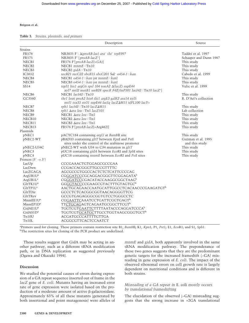
Table 3. Strains, plasmids, and primers
Description Source
StrainsFR576 NR3835 F−: �(proAB-lac) ara− thi− trpE997 Taddei et al. 1997FR575 NR3835 F� [proAB-lacZ+] Schaaper and Dunn 1987NECB1 FR576 F�[proAB-lacZ(+GA)] This studyNECB2 NECB1 mnmE�Tn10 This studyNECB3 NECB2 gidA�Tn10 This studyIC3652 recB21 recC22 sbcB15 sbcC201 Sal− o454-1�kan Cabedo et al. 1999NECB4 NECB1 o454-1�kan (or mnmE�kan) This studyNECB5 NECB3 o454-1�kan (or mnmE�kan) This studySS14 trp31 his1 argG6 rpsl 104 tonA2 �(lacZ) supE44 Vulic et al. 1999
xyl7 mtl2 metB1 serB28 specR F42[ finP301 lacI42�Tn10 lacZ+]NECB6 NECB1 lacI42�Tn10 This studyGC3560 thr1 leu6 proA2 his4 thi1 argE3 galK2 ara14 xyl5 R. D’Ari’s collection
mtl1 tsx33 str31 supE44 lacIq lacZ�M15 �JFL100 lacY+NECB7 rph1 lacI42�Tn10 lacZ�M15 This studyNECB8 rph1 �ara leu�Tn5 lacZ101 Lab collectionNECB9 NECB1 �ara leu�Tn5 This studyNECB10 NECB2 �ara leu�Tn5 This studyNECB11 NECB3 �ara leu�Tn5 This studyNECB13 FR576 F�[proAB-lacZ(+Arg462)] This study
PlasmidspNEC1 pACYC184 containing argU at BamHI site This studypNEC2-WT pBAD33 containing gltT between KpnI and PstI Guzman et al. 1995
sites under the control of the arabinose promoter and this studypNEC2-U34C pNEC2-WT with U34 to C34 mutation in gltT This studypNEC3 pUC18 containing gidA between EcoRI and SphI sites This studypNEC4 pUC18 containing mnmE between EcoRI and PstI sites This study
Primers (5� → 3�)LacUp CCCGAAACTGTGGAGCGCCGAALacDwn CCGACCACGGGTTGCCGTTTTCLacZGAGA AGCGCCGTGGCCACTCTCTCATTCCCCAGArgUB1Ua CGGGATCCCGCAGGACGGGTTCGGAGATAb
ArgUB1La CGGGATCCCGACATACCAAGGCGGCTAAGb
GltTK1Ua GGGGTACCCCGAAGCGTACTTTGTAGTGCb
GltTP1La AACTGCAGAACCAATGCATTGGCCTCACAACCCGAAGATGTb
GltTDir GCCCTCTCACGGCGGTAACAGGGGTTCGGltTRev GCCGTGAGAGGGCGGTGTCCTGGGCCTCMnmEE1Ua CGGAATTCAAATCCTGATTCGGTGAGTb
MnmEP1Da TTCTGCAGAGTCAGAATGCGGCTTCGTb
GidAE1Ua TGGTCGTGAATTCTTTTAATACCCAGGATCCCAb
GidAS1Da TGGTCGTGCATGCTTGCCTGGTAAGCGGGTGCTb
Tn10U ACGATGCCCATTTTGTTGATn10L ACGGCGTTCACTCCAATCT
aPrimers used for cloning. These primers contain restriction site B1, BamHI; K1, Kpn1; P1, Pst1; E1, EcoR1; and S1, Sph1.bThe restriction sites for cloning of the PCR product are underlined.
Brégeon et al.
2300 GENES & DEVELOPMENT
Cold Spring Harbor Laboratory Press on December 25, 2007 - Published by www.genesdev.orgDownloaded from
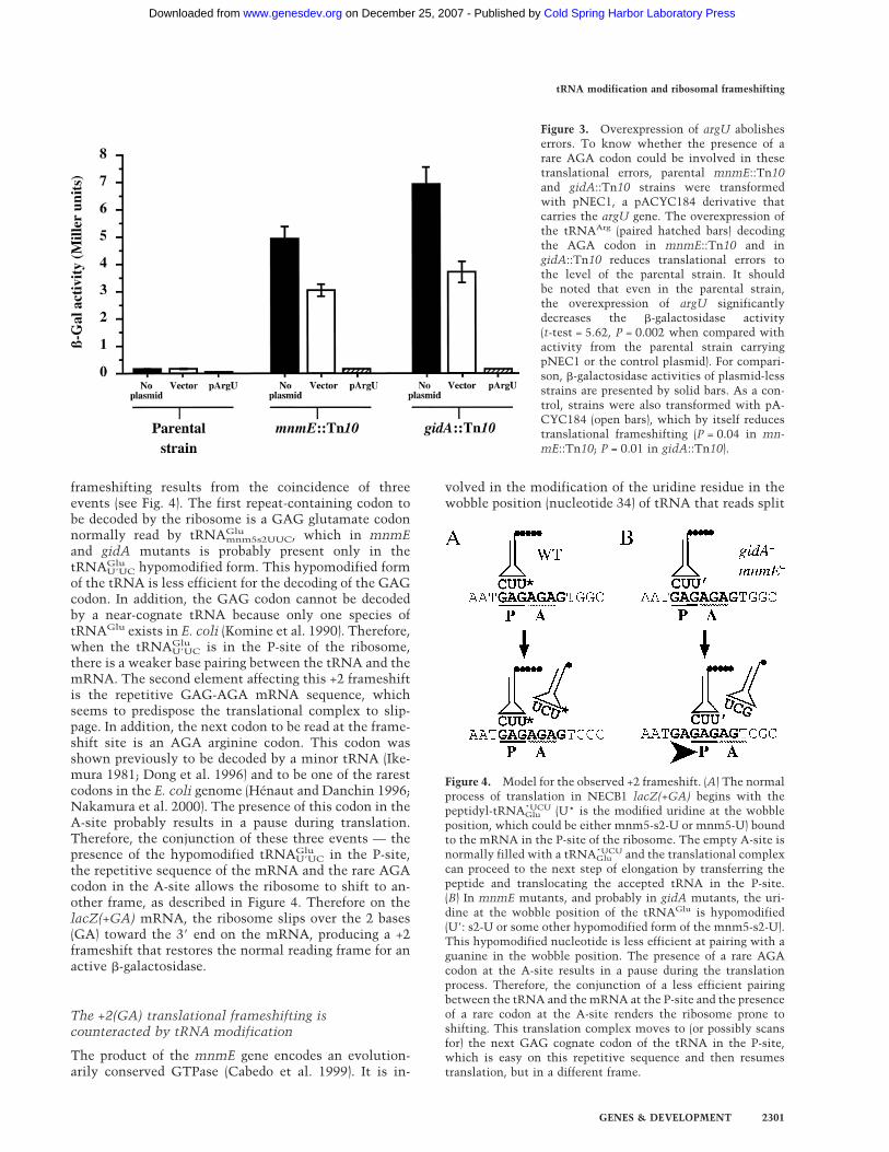
frameshifting results from the coincidence of threeevents (see Fig. 4). The first repeat-containing codon tobe decoded by the ribosome is a GAG glutamate codonnormally read by tRNAGlumnm5s2UUC, which in mnmEand gidA mutants is probably present only in thetRNAGluU�UC hypomodified form. This hypomodified formof the tRNA is less efficient for the decoding of the GAGcodon. In addition, the GAG codon cannot be decodedby a near-cognate tRNA because only one species oftRNAGlu exists in E. coli (Komine et al. 1990). Therefore,when the tRNAGluU�UC is in the P-site of the ribosome,there is a weaker base pairing between the tRNA and themRNA. The second element affecting this +2 frameshiftis the repetitive GAG-AGA mRNA sequence, whichseems to predispose the translational complex to slip-page. In addition, the next codon to be read at the frame-shift site is an AGA arginine codon. This codon wasshown previously to be decoded by a minor tRNA (Ike-mura 1981; Dong et al. 1996) and to be one of the rarestcodons in the E. coli genome (Hénaut and Danchin 1996;Nakamura et al. 2000). The presence of this codon in theA-site probably results in a pause during translation.Therefore, the conjunction of these three events — thepresence of the hypomodified tRNAGluU�UC in the P-site,the repetitive sequence of the mRNA and the rare AGAcodon in the A-site allows the ribosome to shift to an-other frame, as described in Figure 4. Therefore on thelacZ(+GA) mRNA, the ribosome slips over the 2 bases(GA) toward the 3� end on the mRNA, producing a +2frameshift that restores the normal reading frame for anactive �-galactosidase.
The +2(GA) translational frameshifting iscounteracted by tRNA modification
The product of the mnmE gene encodes an evolution-arily conserved GTPase (Cabedo et al. 1999). It is in-
volved in the modification of the uridine residue in thewobble position (nucleotide 34) of tRNA that reads split
Figure 4. Model for the observed +2 frameshift. (A) The normalprocess of translation in NECB1 lacZ(+GA) begins with thepeptidyl-tRNA*UCUGlu (U* is the modified uridine at the wobbleposition, which could be either mnm5-s2-U or mnm5-U) boundto the mRNA in the P-site of the ribosome. The empty A-site isnormally filled with a tRNA*UCUGlu and the translational complexcan proceed to the next step of elongation by transferring thepeptide and translocating the accepted tRNA in the P-site.(B) In mnmE mutants, and probably in gidA mutants, the uri-dine at the wobble position of the tRNAGlu is hypomodified(U�: s2-U or some other hypomodified form of the mnm5-s2-U).This hypomodified nucleotide is less efficient at pairing with aguanine in the wobble position. The presence of a rare AGAcodon at the A-site results in a pause during the translationprocess. Therefore, the conjunction of a less efficient pairingbetween the tRNA and themRNA at the P-site and the presenceof a rare codon at the A-site renders the ribosome prone toshifting. This translation complex moves to (or possibly scansfor) the next GAG cognate codon of the tRNA in the P-site,which is easy on this repetitive sequence and then resumestranslation, but in a different frame.
Figure 3. Overexpression of argU abolisheserrors. To know whether the presence of arare AGA codon could be involved in thesetranslational errors, parental mnmE::Tn10and gidA::Tn10 strains were transformedwith pNEC1, a pACYC184 derivative thatcarries the argU gene. The overexpression ofthe tRNAArg (paired hatched bars) decodingthe AGA codon in mnmE::Tn10 and ingidA::Tn10 reduces translational errors tothe level of the parental strain. It shouldbe noted that even in the parental strain,the overexpression of argU significantlydecreases the �-galactosidase activity(t-test = 5.62, P = 0.002 when compared withactivity from the parental strain carryingpNEC1 or the control plasmid). For compari-son, �-galactosidase activities of plasmid-lessstrains are presented by solid bars. As a con-trol, strains were also transformed with pA-CYC184 (open bars), which by itself reducestranslational frameshifting (P = 0.04 in mn-mE::Tn10; P = 0.01 in gidA::Tn10).
tRNA modification and ribosomal frameshifting
GENES & DEVELOPMENT 2301
Cold Spring Harbor Laboratory Press on December 25, 2007 - Published by www.genesdev.orgDownloaded from

codon boxes (a codon box in which the first two nucleo-tides are the same, while the third nucleotide determineswhich of the two alternative amino acids are encoded). Inthose tRNAs, the modification is of the type xm5U (or athiolated derivative xm5s2U) where x can be an amino(m), methylamino (mn), or carboxymethylamino (cmn)group. The first reaction in this process, catalyzed byMnmE, is the addition of the cmnm group in position 5of the uridine. For the tRNAGlu of E. coli, which decodesGAA and GAG codons, it was shown that 5-methylami-nomethyl group on the uracil facilitates its base-pairingwith G while decreasing the base-pairing with A. Thisresults in rates of translation of GAG and GAA that ap-proach the average speed of the ribosome (Kruger et al.1998). Therefore, in the case of lacZ(+GA) inmnmE::Tn10 strain, this tRNAGlu is less efficientlypaired with the first codon of the GA repeat, which islikely to facilitate the observed ribosomal frameshifting(see above; Fig. 4).
GidA protein may be involved in the mnm5s2U34modification pathway
There is no clearly identified function for the gidA gene,which has been described to have the phenotype of glu-cose inhibited division (von Meyenburg and Hansen1980) and is located next to the chromosomal origin ofreplication, oriC, of E. coli. Results obtained in thisstudy with the mnmE::kan gidA::Tn10 double mutantshow that the products of both genes are likely to beinvolved in the same tRNA modification pathway and,furthermore, that theMnmE activity precedes that of theGidA. The synthesis of mnm5s2U could occur in thefollowing steps:
U34 →mnmA s2U34 →
mnmE X →gidA cmnm5s2U34 →
mnmCl
nm5s2U34 →mnmC2 mnm5s2U34
It was recently suggested that the genetic symbols forgenes involved in this pathway should be revised andthat trmU (or asuE) be replaced bymnmA, trmE (or thdF)by mnmE, and trmC by mnmC (Leung et al. 1998). ThemnmA gene product is responsible for the thiolation ofposition 2 of the uridine (Sullivan et al. 1985). No mu-tant of this gene has been isolated with our screen, prob-ably because the 2-thio group added by this enzyme isimportant for aminoacylation and does not have a sig-nificant role for the decoding of the GAG codon (Krugeret al. 1998). MnmC is composed of two activities (Hager-vall et al. 1987), which transform the cmnm intermedi-ate into the final mnm modification. The fact that wedid not isolate mutants of this gene and the fact that acmnm intermediate is used as the final form of onetRNALeu, suggest that this step may not change the de-coding properties of the modified tRNA. The mnmEgene product was shown to be responsible for the firstmodification of the 5-position of the uracil, but it is un-clear how many steps precede the formation of cmnm5(Elseviers et al. 1984; Hagervall et al. 1987).
The role we propose for GidA in this modificationpathway could explain the recent results of Nakayashikiand others. Thermosensitive lethal mutants of this genehave been isolated in a strain harboring the amber sup-pressor mutant gene tRNA6
Leu (supP) (Nakayashiki andInokuchi 1998), which changes the anticodon of this dis-pensable tRNA from CAA to CUA. Normally, tRNA6
Leu
recognizes the UUG codon, whereas the mutated supPtRNA recognizes the UAG amber codon. The UUGcodon is also decoded by tRNA4
Leu, which has acmnm5m2UAA (m2: methyl group in position 2) in itsanticodon such that it can recognize both UUG andUUA (Horie et al. 1999). By analogy with tRNAGlu, onecould speculate that the absence of cmnm5 at nonper-missive temperature may decrease the base pairing of thetRNA4
Leu with the UUG codon, thereby requiring thetRNA6
Leu under these conditions for normal translation.Furthermore, the first attempt at isolating mutants af-fected in the biosynthesis of mnm5s2U allowed the char-acterization of two loci, named trmE and trmF. The phe-notype of these two mutants was not clearly differentbut they were mapped to two different locations on theE. coli chromosome. The trmE gene was shown to be thesame gene as mnmE (Cabedo et al. 1999) and trmF wasmapped near 83 min on the E. coli map between uncAand bglR in the region of oriC (Elseviers et al. 1984). Themap location of gidA is 84.5 min, close to oriC. Wetherefore propose that trmF is an allele of gidA and thatthis locus may be renamed mnmG as suggested previ-ously (Leung et al. 1998).The growth rate of both gidA::Tn10 and mnmE::Tn10
strains is reduced in LB, but, in minimal medium, thisdecrease was only observed for the gidA::Tn10 strain.This difference of phenotype may suggest that the un-known product of MnmE (indicated by an X in the modi-fication pathway presented above), which is likely to beaccumulated in a gidA− strain, could be toxic for cellsunder this slow growth condition. At this stage, how-ever, we can not exclude that GidA may be involved inother tRNA modification pathways or in DNA replica-tion as suggested previously (Ogawa and Okazaki 1994).
Extension of the dual error model may explaintRNA hopping
Most of the described nonprogrammed ribosomal frame-shifts are thought to slip in either +1 or −1 frame (Far-abaugh and Björk 1999, and references therein). The +1/−1 dual error model proposed previously (Qian et al.1998) suggests several explanations for nonprogrammed+1 and −1 frameshifts that could occur, especially atCCCN and GGGN sites (N, any nucleotide). This modelassumes that slipping of the ribosome would happen at aprecise stage after the three nucleotide translocation. Atthis step, the ribosome is prone to slip on the mRNA, inparticular when a near-cognate tRNA or a hypomodifiedtRNA is present in the P-site. In this case, the shift isattributable to a weaker codon–anticodon pairing at thewobble position. Even if a normal cognate tRNA is in theP-site, the ribosome could slip when a translational
Brégeon et al.
2302 GENES & DEVELOPMENT
Cold Spring Harbor Laboratory Press on December 25, 2007 - Published by www.genesdev.orgDownloaded from

pause is induced because of the presence of a rare codonin the A-site.An extended version of the +1/−1 dual error model
could also explain the tRNA hopping described by Weisset al. (1987). The mutation they studied is a leaky −1frameshift allele trpE91 in Salmonella (Riyasaty and At-kins 1968). The low-level translational frameshiftingthat occurs at this site was studied by transferring theregion surrounding the mutation into a lacZ expressionvector (Weiss et al. 1987). The sequence of the expressedpeptide revealed that the frameshift occurred by a trans-lational hopping, creating a +2 rather than a −1 frame-shift, via a tRNAVal hopping by +2. Two other sequenceswere identified on which the ribosome is prone to slip (orhop) over 5 or 6 nucleotides. Protein sequencing con-firmed that tRNAAsn hops 5 nucleotides between cog-nate codons and that tRNALeu hops over an in-framestop codon between near-cognate codons. The presenceof an in-frame stop codon for both +2 and +6 hoppingapparently creates a pausing site during translation, afavorable condition for the frameshifting (Farabaugh1996). The +5 event, however, occurred at an in-frameUCA codon that is not predicted to cause a translationalpause. The strain used by Weiss and colleagues wasCSH26 with a deletion of the pro–lac region, where thestrC and trmB genes have been mapped, respectively, byP1 co-transduction (Roberts and Reeve 1970) and conju-gation (Marinus et al. 1975), which may have an influ-ence on this +5 frameshift. The strC gene is known to beinvolved in streptomycin resistance and to modify theribosome structure (Roberts and Reeve 1970). The TrmBprotein is a tRNA methyltransferase, which catalyzesthe formation of methyl-7-guanosine (m7G) in sometRNAs, including the tRNAAsn hopping over 5 nucleo-tides. Whether this modification is important to stabi-lize the translation complex remains to be tested. Allobservations of ribosome frameshifting with hypomodi-fied tRNA or in presence of a pausing site during trans-lation are therefore in favor of the extended version ofthe dual error model described above.
An evolutionarily conserved modification pathway
The mnmE and gidA gene products display significantsimilarities with proteins of Saccharomyces cerevisiaeand Caenorhabditis elegans as well as with several ex-pressed sequence tags from Arabidopsis thaliana, mouseand human. In S. cerevisiae, homologs of MnmE andGidA are respectively Mss1p and Mto1p and have beenshown to be nuclear-encoded mitochondrial genes (De-coster et al. 1993; Colby et al. 1998). Therefore, MSS1and MTO1 genes are likely to have evolved from mito-chondrial genes translocated into the nucleus.Mutants of Mto1p and Mss1p were reported to be im-
plicated in a respiratory deficiency phenotype only whenthe 15S rRNA gene carries a mutation that confers resis-tance to paromomycin (Decoster et al. 1993; Colby et al.1998). This mutation is localized to helix 47, which ispart of the A-site of the ribosome (Li et al. 1982; DeStasio and Dahlberg 1990). Those mutant ribosomes
were also shown to have a decreased level of naturalframeshifting, suggestive of a more stringent proofread-ing (Weiss-Brummer and Huttenhofer 1989). It is signifi-cant that mitochondrial tRNALeu and tRNATrp contain amodified U at the wobble position (Björk 1998). Thismodification is of the type cmnm5Um and allows thesetRNA to translate codons terminating in either A or G.We propose that Mss1p and Mto1p are responsible forthis modification and that hypomodified tRNAs are lessefficient for the decoding of the codon ending in G. Thisdeficiency could be stronger with the more stringentparomomycin-resistant ribosome, thereby causing therespiratory defect observed in mss1 and mto1 mutantcells. Furthermore, the products of these two genes in S.cerevisiae were shown to be in the same complex, andare therefore likely to act in the same pathway (Colby etal. 1998). Whether GidA and MnmE are also in the sameprotein complex, which may counteract the previouslyproposed toxicity of the unknown MnmE product (X),remains to be determined in E. coli.The alteration of growth rate in both gidA::Tn10 and
mnmE::Tn10 strains and the respiratory deficient phe-notype of yeast mutants could be linked to errors madeduring translation, which are likely to be processivityerrors. As this pathway seems to be conserved in manyorganisms, mutation of these genes may be involved ingenetic disorders such as mitochondrial diseases.
Materials and methods
Growth conditions
The cell growth media are liquid LB supplemented with thefollowing antibiotics, when appropriate, at the following con-centrations: tetracycline 12.5 µg/mL, chloramphenicol 30 µg/mL, rifampicin 100 µg/mL, kanamycin 100 µg/mL, and ampi-cillin 100 µg/mL. For strain constructions, clones were selectedon LB plates supplemented with the appropriate antibiotic and/or X-Gal (200 µg/mL) and 20 mM sodium citrate. Selections forauxotrophy, or carbon source, were made on minimal M9 plates(1.5% agar) supplemented with 0.4% of the relevant carbonsource and 0.1% of the suitable amino acids. Growth rates weremeasured by optical density at 530 nm in 96-well plates.
Strain constructions
The genotypes and sources of the strains of E. coli used in thisstudy are listed in Table 3. The NECB1 was constructed asdescribed in Cupples and Miller (1988) using LacZGAGA asmutagenic primer. The introduced mutation was verified bysequencing.Derivative strains were constructed by P1-mediated transduc-
tion as described by Miller (1972). Conjugations were made bymixing 500 µL of overnight cultures of donor and recipient cellsin 5 mL of LB and placed at 37°C without shaking for 2 h.Transconjugants were then selected by plating on the appropri-ate media.
Mutagenic treatments and identification of transposoninsertion site
The random insertion mutants were created by transposition ofmini-Tn10 from �NK1323 (Kleckner et al. 1991). Phage stocks
tRNA modification and ribosomal frameshifting
GENES & DEVELOPMENT 2303
Cold Spring Harbor Laboratory Press on December 25, 2007 - Published by www.genesdev.orgDownloaded from

and infections were made as described in (Miller 1992). Trans-fected cells were plated on LB plates containing tetracycline andX-Gal to screen for light blue colonies. The Tn10 insertion siteswere identified by inverse PCR in bacterial clones showing in-termediate levels of �-galactosidase activity. Genomic DNA(500 ng) from such clones was digested with PstI and purifiedwith a QIAquick PCR purification kit (QIAGEN). Fragmentswere then incubated overnight at 12°C with 40,000 units of T4DNA ligase in a final volume of 1 mL. DNA was precipitatedwith ethanol and resuspended in 20 µL of water. The DNAfragments were then amplified by PCR with VentR(exo−) DNApolymerase (New England Biolabs) using Tn10U and Tn10Lprimers (Genset). Comparison of the sequence of the PCR prod-uct with the sequence of the E. coli genome allowed us to de-termine the insertion site for each clone.The point mutations were made by growing NECB1 cells
overnight in LB containing 700 µg/mL of 2-AP as described byMiller (1992). Mutagenesis was monitored by determining thefrequency of rifampicin resistant mutants as compared withnontreated culture. After the treatment, cells were plated on LBplates containing X-Gal, and mutants having a pale blue color-ation were re-streaked on the same type of medium.
Plasmid construction
All plasmids and their constructs were transformed into theappropriate E. coli cells by electrotransformation. Ligationswere carried out by T4 DNA ligase (New England Biolabs) ac-cording to the manufacturer’s instructions and the ElectromaxDH10B cells (Life Technologies) were used for primary transfor-mations. For gene amplifications, a single colony of NECB1 wasresuspended in 50 µL of water, and 1 µL of the suspension wasadded to the PCR mix. Cells were lysed during the first cycle ofPCR by 5-min incubation at 95°C.To construct the pNEC1 plasmid, the region of argU was
amplified under standard conditions with the Pwo polymerase,using ArgUB1U and ArgUB1L as primers. The amplified frag-ment was digested with BamHI and inserted into pACYC184 atthe BamHI site. Positive clones were checked to be chloram-phenicol-resistant and tetracycline-sensitive and the presenceof the insert was monitored by digestion with BamHI.The gltT gene was amplified in the same manner as argU by
using GltTK1U and GltTP1L as primers for the constructionpNEC2-WT. The linear fragment was digested with KpnI andPstI and inserted into pBAD33 (Guzman et al. 1995) betweenKpnI and PstI sites. Plasmids from transformed clones were ex-tracted using a QIAquick Miniprep kit (QIAGEN) and digestedwith KpnI and PstI to monitor the presence of the inserted frag-ment. Linear fragments for the construction of pNEC2-U34Cwere generated by sequential PCR steps using GltTK1U, Glt-TDir, GltTP1L, and GltTRev (Table 3).Linear fragments for the cloning of gidA (pNEC3) or mnmE
(pNEC4) were obtained by amplification with Pwo DNA poly-merase (Roche) using GidAE1U and GidAS1D, respectively, orMnmEE1U and MnmEP1D as primers. Fragments were thendigested with the appropriate enzyme (Table 3) and inserted intopUC18. Positive clones were checked for the lack of �-comple-mentation (Yanisch-Perron et al. 1985) and were verified by re-striction.For the genomic library, 2 µg of genomic DNA from NECB1
prepared with a Wizard Genomic DNA Preparation kit (Pro-mega) was partially digested with Sau3AI. Fragments rangingfrom 3–4 kb were eluted from agarose with a QIAquick gelextraction kit (QIAGEN). Fragments were then cloned into theBamHI site of pACYC184 using a fragment:plasmid ratio of 10:1for the ligation and the transformed cells were selected on LB
plates containing chloramphenicol. Plasmid DNA of 30 singlecolonies was isolated and digested with EcoRV and SalI to es-timate the efficiency of ligation, which was ∼90%. Plasmidswere then extracted from 20,000 colonies resuspended inMgSO4 10 mM with a QIAfilter Maxiprep kit (QIAGEN).
�-Galactosidase activity
For all �-galactosidase assays, 200 µL of an overnight culturewas used. Activities were measured and were expressed as de-scribed by Miller (1972). The OD530 was not measured as allsamples were centrifuged before the reading of the OD420. Tomeasure low levels of �-galactosidase activities, longer incuba-tion times were necessary (up to 1 d, always controlling forspontaneous hydrolysis of O-nitrophenyl-�-D-galactoside). All�-galactosidase activities are given in Miller units ± standarderror.
Acknowledgments
We thank P. Leroy, M.F. Bredeche, and M. Kobayashi for tech-nical assistance and R. D’Ari and M.E. Armengod for providingstrains. We thank I. Matic and O. Tenaillon for constant en-couragement. We thank E. Stewart, F.W. Stahl, M.S. Fox, C.G.Kurland, G.R. Björk, T.G. Hagervall, and J.A. Gallant for criticalreading of the manuscript and F. VanLeeuwen, H. Grosjean, andP. Farabaugh for their interest in this work. This work wassupported by grants from ACI blanche, MENRT, INSERM andLa Ligue contre le cancer.The publication costs of this article were defrayed in part by
payment of page charges. This article must therefore be herebymarked “advertisement” in accordance with 18 USC section1734 solely to indicate this fact.
References
Arraj, J.A. and Campbell, J.H. 1975. Isolation and characteriza-tion of the newly evolved ebg �- galactosidase of Escherichiacoli K-12. J. Bacteriol. 124: 849–856.
Barak, Z., Gallant, J., Lindsley, D., Kwieciszewki, B., andHeidel, D. 1996. Enhanced ribosome frameshifting in sta-tionary phase cells. J. Mol. Biol. 263: 140–148.
Bergmann, E.D. and Sicher, S. 1952. Mode of action of chloram-phenicol. Nature 170: 931–932.
Björk, G.R. 1996. Stable RNA modification. In Escherichia coliand Salmonella: Cellular and molecular biology (ed. F.C.Neidhardt, R. Curtiss III, J.L. Ingraham, E.C.C. Lin, B.K.Low, B. Magasanik, W.S. Reznikoff, M. Riley, M. Schaechter,and H.E. Umbarger), pp. 861–886. American Society for Mi-crobiology, Washington, DC.
———. 1998. Modified nucleosides at position 34 and 37 oftRNAs and their predicted coding capacities. InModifiationand editing of RNA (ed. H. Grosjean and R. Benne), pp. 577–581. American Society for Microbiology, Washington, DC.
Björk, G.R., Durand, J.M., Hagervall, T.G., Leipuviene, R.,Lundgren, H.K., Nilsson, K., Chen, P., Qian, Q., and Urbo-navicius, J. 1999. Transfer RNA modification: Influence ontranslational frameshifting and metabolism. FEBS Lett.452: 47–51.
Cabedo, H., Macian, F., Villarroya, M., Escudero, J.C., Martinez-Vicente, M., Knecht, E., and Armengod, M.E. 1999. TheEscherichia coli trmE (mnmE) gene, involved in tRNAmodi-fication, codes for an evolutionarily conserved GTPase withunusual biochemical properties. EMBO J. 18: 7063–7076.
Campbell, J.H., Lengyel, J.A., and Langridge, J. 1973. Evolution
Brégeon et al.
2304 GENES & DEVELOPMENT
Cold Spring Harbor Laboratory Press on December 25, 2007 - Published by www.genesdev.orgDownloaded from

of a second gene for �-galactosidase in Escherichia coli. Proc.Natl. Acad. Sci. 70: 1841–1845.
Colby, G., Wu, M., and Tzagoloff, A. 1998. MTO1 codes for amitochondrial protein required for respiration in paromomy-cin-resistant mutants of Saccharomyces cerevisiae. J. Biol.Chem. 273: 27945–27952.
Cupples, C.G. and Miller, J.H. 1988. Effects of amino acid sub-stitutions at the active site in Escherichia coli �-galactosi-dase. Genetics 120: 637–644.
Curran, J.F. and Yarus, M. 1989. Rates of aminoacyl-tRNA se-lection at 29 sense codons in vivo. J. Mol. Biol. 209: 65–77.
Czworkowski, J. and Moore, P.B. 1996. The elongation phase ofprotein synthesis. Prog. Nucleic Acid Res. Mol. Biol.54: 293–332.
De Stasio, E.A. and Dahlberg, A.E. 1990. Effects of mutagenesisof a conserved base-paired site near the decoding region ofEscherichia coli 16 S ribosomal RNA. J. Mol. Biol. 212: 127–133.
Decoster, E., Vassal, A., and Faye, G. 1993. MSS1, a nuclear-encoded mitochondrial GTPase involved in the expressionof COX1 subunit of cytochrome c oxidase. J. Mol. Biol.232: 79–88.
Dong, H., Nilsson, L., and Kurland, C.G. 1996. Co-variation oftRNA abundance and codon usage in Escherichia coli at dif-ferent growth rates. J. Mol. Biol. 260: 649–663.
Dukan, S., Farewell, A., Ballesteros, M., Taddei, F., Radman, M.,and Nystrom, T. 2000. Protein oxidation in response to in-creased transcriptional or translational errors. Proc. Natl.Acad. Sci. 97: 5746–5749.
Edelmann, P. and Gallant, J. 1977. Mistranslation in E. coli.Cell10: 131–137.
Ellis, N. and Gallant, J. 1982. An estimate of the global errorfrequency in translation. Mol. Gen. Genet. 188: 169–172.
Elseviers, D., Petrullo, L.A., and Gallagher, P.J. 1984. Novel E.coli mutants deficient in biosynthesis of 5-methylamino-methyl- 2-thiouridine. Nucleic Acids Res. 12: 3521–3534.
Farabaugh, P.J. 1996. Programmed translational frameshifting.Microbiol. Rev. 60: 103–134.
Farabaugh, P.J. and Björk, G.R. 1999. How translational accu-racy influences reading frame maintenance. EMBO J.18: 1427–1434.
Friedberg, E.C., Walker, G.C., and Siede, W. 1995. DNA repairand mutagenesis. American Society for Microbiology,Washington, DC.
Guzman, L.M., Belin, D., Carson, M.J., and Beckwith, J. 1995.Tight regulation, modulation, and high-level expression byvectors containing the arabinose PBAD promoter. J. Bacte-riol. 177: 4121–4130.
Hagervall, T.G., Edmonds, C.G., McCloskey, J.A., and Björk,G.R. 1987. Transfer RNA(5-methylaminomethyl-2-thiouri-dine)-methyltransferase from Escherichia coli K-12 has twoenzymatic activities. J. Biol. Chem. 262: 8488–8495.
Hénaut, A. and Danchin, A. 1996. Analysis and prediction fromEscherichia coli sequences, or E. coli In Silico. In Escherichiacoli and Salmonella: Cellular and Molecular Biology (ed.F.C. Neidhardt, R. Curtiss III, J.L. Ingraham, E.C.C. Lin, B.K.Low, B. Magasanik, W.S. Reznikoff, M. Riley, M. Schaechter,and H.E. Umbarger), pp. 2047–2066. American Society forMicrobiology, Washington, DC.
Hol, E.M., Neubauer, A., de Kleijn, D.P., Sluijs, J.A., Ramdjielal,R.D., Sonnemans, M.A., and van Leeuwen, F.W. 1998. Di-nucleotide deletions in neuronal transcripts: A novel type ofmutation in non-familial Alzheimer’s disease and Downsyndrome patients. Prog. Brain Res. 117: 379–395.
Horie, N., Yamaizumi, Z., Kuchino, Y., Takai, K., Goldman, E.,Miyazawa, T., Nishimura, S., and Yokoyama, S. 1999. Modi-
fied nucleosides in the first positions of the anticodons oftRNA(Leu)4 and tRNA(Leu)5 from Escherichia coli. Bio-chemistry 38: 207–217.
Ikemura, T. 1981. Correlation between the abundance of Esch-erichia coli transfer RNAs and the occurrence of the respec-tive codons in its protein genes. J. Mol. Biol. 146: 1–21.
Kleckner, N., Bender, J., and Gottesman, S. 1991. Uses of trans-posons with emphasis on Tn10. Methods Enzymol.204: 139–180.
Komine, Y., Adachi, T., Inokuchi, H., and Ozeki, H. 1990. Ge-nomic organization and physical mapping of the transferRNA genes in Escherichia coli K12. J. Mol. Biol. 212: 579–598.
Kruger, M.K., Pedersen, S., Hagervall, T.G., and Sorensen, M.A.1998. The modification of the wobble base of tRNAGlumodulates the translation rate of glutamic acid codons invivo. J. Mol. Biol. 284: 621–631.
Kurland, C.G. 1992. Translational accuracy and the fitness ofbacteria. Annu. Rev. Genet. 26: 29–50.
Kurland, C., Hughes, D., and Ehrenberg, M. 1996. Limitations oftranslational accuracy. In Escherichia coli and Salmonella:Cellular and molecular biology (ed. F.C. Neidhardt, R. Cur-tiss III, J.L. Ingraham, E.C.C. Lin, B.K. Low, B. Magasanik,W.S. Reznikoff, M. Riley, M. Schaechter, and H.E. Um-barger), pp. 979–1004. American Society for Microbiology,Washington, DC.
Leung, H.C.E., Hagervall, T.G., Björk, G.R., and Winkler, M.E.1998. Genetic locations and database accession numbers ofRNA-modifying and -editing enzymes. In Modification andediting of RNA (ed. H. Grosjean and R. Benne), pp. 561–568.American Society for Microbiology, Washington, DC.
Lewin, B. 1994. Genes V. Oxford University Press, New York,NY.
Li, M., Tzagoloff, A., Underbrink-Lyon, K., and Martin, N.C.1982. Identification of the paromomycin-resistance muta-tion in the 15 S rRNA gene of yeast mitochondria. J. Biol.Chem. 257: 5921–5928.
Libby, R.T. and Gallant, J.A. 1991. The role of RNA polymerasein transcriptional fidelity. Mol. Microbiol. 5: 999–1004.
Loftfield, R.B. and Vanderjagt, D. 1972. The frequency of errorsin protein biosynthesis. Biochem. J. 128: 1353–1356.
Marinus, M.G., Morris, N.R., Soll, D., and Kwong, T.C. 1975.Isolation and partial characterization of three Escherichiacoli mutants with altered transfer ribonucleic acid methyl-ases. J. Bacteriol. 122: 257–265.
Menninger, J.R. 1977. Ribosome editing and the error catastro-phe hypothesis of cellular aging.Mech. Ageing. Dev. 6: 131–142.
Miller, J.H. 1972. Experiments in molecular genetics. ColdSpring Harbor Laboratory Press, Cold Spring Harbor, NY.
———. 1992. A short course in bacterial genetics. A laboratorymanual for Escherichia coli and related bacteria. ColdSpring Harbor Laboratory Press, Cold Spring Harbor, NY.
Nakamura, Y., Gojobori, T., and Ikemura, T. 2000. Codon usagetabulated from international DNA sequence databases: Sta-tus for the year 2000. Nucleic Acids Res. 28: 292.
Nakayashiki, T. and Inokuchi, H. 1998. Novel temperature-sen-sitive mutants of Escherichia coli that are unable to grow inthe absence of wild-type tRNA6Leu. J. Bacteriol. 180: 2931–2935.
Nierhaus, K.H. 1990. The allosteric three-site model for theribosomal elongation cycle: Features and future. Biochemis-try 29: 4997–5008.
Nunomura, A., Perry, G., Pappolla, M.A., Wade, R., Hirai, K.,Chiba, S., and Smith, M.A. 1999. RNA oxidation is a promi-nent feature of vulnerable neurons in Alzheimer’s disease. J.
tRNA modification and ribosomal frameshifting
GENES & DEVELOPMENT 2305
Cold Spring Harbor Laboratory Press on December 25, 2007 - Published by www.genesdev.orgDownloaded from

Neurosci. 19: 1959–1964.Ogawa, T. and Okazaki, T. 1994. Cell cycle-dependent tran-scription from the gid and mioC promoters of Escherichiacoli. J. Bacteriol. 176: 1609–1615.
Parker, J. 1989. Errors and alternatives in reading the universalgenetic code. Microbiol. Rev. 53: 273–298.
Qian, Q., Li, J.N., Zhao, H., Hagervall, T.G., Farabaugh, P.J., andBjörk., G.R. 1998. A new model for phenotypic suppressionof frameshift mutations by mutant tRNAs.Mol. Cell 1: 471–482.
Riyasaty, S. and Atkins, J.F. 1968. External suppression of aframeshift mutant in Salmonella. J. Mol. Biol. 34: 541–557.
Roberts, L.M. and Reeve, E.C. 1970. Two mutations giving low-level streptomycin resistance in Escherichia coli K 12.Genet. Res. 16: 359–365.
Schaaper, R.M. and Dunn, R.L. 1987. Spectra of spontaneousmutations in Escherichia coli strains defective in mismatchcorrection: The nature of in vivo DNA replication errors.Proc. Natl. Acad. Sci. 84: 6220–6224.
Sipley, J. and Goldman, E. 1993. Increased ribosomal accuracyincreases a programmed translational frameshift in Esche-richia coli. Proc. Natl. Acad. Sci. 90: 2315–2319.
Stadtman, E.R. 1992. Protein oxidation and aging. Science257: 1220–1224.
Sullivan, M.A., Cannon, J.F., Webb, F.H., and Bock, R.M. 1985.Antisuppressor mutation in Escherichia coli defective inbiosynthesis of 5-methylaminomethyl-2-thiouridine. J. Bac-teriol. 161: 368–376.
Taddei, F., Hayakawa, H., Bouton, M.F., Cirinesi, A.M., Matic,I., Sekiguchi, M., and Radman, M. 1997. Counteraction byMutT protein of transcriptional errors caused by oxidativedamage. Science 278: 128–130.
Thomas, M.J., Platas, A.A., and Hawley, D.K. 1998. Transcrip-tional fidelity and proofreading by RNA polymerase II. Cell93: 627–637.
Thompson, R.C. 1988. EFTu provides an internal kinetic stan-dard for translational accuracy. Trends Biochem. Sci. 13: 91–93.
Triglia, T., Peterson, M.G., and Kemp, D.J. 1988. A procedurefor in vitro amplification of DNA segments that lie outsidethe boundaries of known sequences. Nucleic Acids Res.16: 81–86.
van Leeuwen, F.W., Burbach, J.P., and Hol, E.M. 1998a. Muta-tions in RNA: A first example of molecular misreading inAlzheimer’s disease. Trends Neurosci. 21: 331–335.
van Leeuwen, F.W., de Kleijn, D.P., van den Hurk, H.H., Neu-bauer, A., Sonnemans, M.A., Sluijs, J.A., Koycu, S., Ramd-jielal, R.D.J., Salehi, A., Martens, G.J.M., et al. 1998b. Frame-shift mutants of beta amyloid precursor protein and ubiqui-tin-B in Alzheimer’s and Down patients. Science 279: 242–247.
von Meyenburg, K. and Hansen, F.G. 1980. The origin of repli-cation, oriC, of the Escherichia coli chromosome: Genesnear to oriC and construction of oriC deletion mutations. InMechanistic studies of DNA replication and genetic recom-bination, pp. 137–159. Academic Press, New York, NY.
Vulic, M., Lenski, R.E., and Radman, M. 1999. Mutation, re-combination, and incipient speciation of bacteria in the labo-ratory. Proc. Natl. Acad. Sci. 96: 7348–7351.
Weiss, R.B., Dunn, D.M., Atkins, J.F., and Gesteland, R.F. 1987.Slippery runs, shifty stops, backward steps, and forwardhops: −2, −1, +1, +2, +5, and +6 ribosomal frameshifting.ColdSpring Harb. Symp. Quant. Biol. 52: 687–693.
Weiss-Brummer, B. and Huttenhofer, A. 1989. The paromomy-cin resistance mutation (parR-454) in the 15 S rRNA gene ofthe yeast Saccharomyces cerevisiae is involved in ribosomal
frameshifting. Mol. Gen. Genet. 217: 362–369.Yanisch-Perron, C., Vieira, J., and Messing, J. 1985. ImprovedM13 phage cloning vectors and host strains: Nucleotide se-quences of the M13mp18 and pUC19 vectors. Gene 33: 103–119.
Zhang, J., Perry, G., Smith, M.A., Robertson, D., Olson, S.J.,Graham, D.G., and Montine, T.J. 1999. Parkinson’s diseaseis associated with oxidative damage to cytoplasmic DNAand RNA in substantia nigra neurons. Am. J. Pathol.154: 1423–1429.
Brégeon et al.
2306 GENES & DEVELOPMENT
Cold Spring Harbor Laboratory Press on December 25, 2007 - Published by www.genesdev.orgDownloaded from
Copyright © 2022 FDOKUMEN